Fibroma, Ossifying
Fibroma, Desmoplastic
Fibrous Dysplasia of Bone
Jaw Neoplasms
Poxviridae
Gingival Neoplasms
Cementoma
Chondroma
Thecoma
Leporipoxvirus
Chondroblastoma
Radiography, Panoramic
Osteoma
Palatal Neoplasms
Rhabdomyoma
Maxillary Sinus Neoplasms
Heart Neoplasms
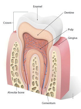
Dental Cementum
Myxoma virus
Neoplasms, Connective Tissue
Collagen Type XII
Femoral Neoplasms
Gingival Diseases
Chromium Isotopes
Ethmoid Sinus
Jaw Cysts
Odontoma
Frontal Bone
Tumor Virus Infections
Granuloma, Giant Cell
Curettage
Togo
Tooth, Unerupted
Cicatricial fibromatosis mimics metastatic medulloblastoma. (1/412)
Cicatricial fibromatoses usually occur in the anterior abdominal wall or in the extremities, but rarely in the scalp or the soft tissues of the neck. We report a case of desmoid fibromatosis that developed in a 15-year-old boy 8 months after surgery for cerebellar medulloblastoma. (+info)Overgrowth of oral mucosa and facial skin, a novel feature of aspartylglucosaminuria. (2/412)
Aspartylglucosaminuria (AGU) is a lysosomal storage disorder caused by deficiency of aspartylglucosaminidase (AGA). The main symptom is progressive mental retardation. A spectrum of different mutations has been reported in this disease, one missense mutation (Cys163Ser) being responsible for the majority of Finnish cases. We were able to examine 66 Finnish AGU patients for changes in the oral mucosa and 44 of these for changes in facial skin. Biopsy specimens of 16 oral lesions, 12 of them associated with the teeth, plus two facial lesions were studied histologically. Immunohistochemical staining for AGA was performed on 15 oral specimens. Skin was seborrhoeic in adolescent and adult patients, with erythema of the facial skin already common in childhood. Of 44 patients, nine (20%) had facial angiofibromas, tumours primarily occurring in association with tuberous sclerosis. Oedemic buccal mucosa (leucoedema) and gingival overgrowths were more frequent in AGU patients than in controls (p<0.001). Of 16 oral mucosal lesions studied histologically, 15 represented fibroepithelial or epithelial hyperplasias and were reactive in nature. Cytoplasmic vacuolisation was evident in four. Immunohistochemically, expression of AGA in AGU patients' mucosal lesions did not differ from that seen in corresponding lesions of normal subjects. Thus, the high frequency of mucosal overgrowth in AGU patients does not appear to be directly associated with lysosomal storage or with alterations in the level of AGA expression. (+info)Infantile fibromatosis of the neck with intracranial involvement: MR and CT findings. (3/412)
CT and MR imaging studies were performed in a 3-year-old boy with infantile fibromatosis arising from the infratemporal fossa and extending into the middle cranial fossa. On CT scans, the lesion was hyperattenuating (44-49 Hounsfield units [HU]), enhancing significantly after application of contrast material (63-66 HU). The MR images showed a multilobulated lesion of heterogeneous signal intensity. The tumor was markedly hypointense on T2-weighted images and slightly hypointense on T1-weighted images relative to brain tissue, iso- or slightly hyperintense relative to tongue muscle on both T2- and T1-weighted images, and enhanced strongly after administration of gadopentetate dimeglumine. (+info)Association of herpesvirus with fibropapillomatosis of the green turtle Chelonia mydas and the loggerhead turtle Caretta caretta in Florida. (4/412)
Sea turtle fibropapillomatosis (FP) is a disease marked by proliferation of benign but debilitating cutaneous fibropapillomas and occasional visceral fibromas. Transmission experiments have implicated a chloroform-sensitive transforming agent present in filtered cell-free tumor homogenates in the etiology of FP. In this study, consensus primer PCR methodology was used to test the association of a chelonian herpesvirus with fibropapillomatosis. Fibropapilloma and skin samples were obtained from 17 green and 2 loggerhead turtles affected with FP stranded along the Florida coastline. Ninety-three cutaneous and visceral tumors from the 19 turtles, and 33 skin samples from 16 of the turtles, were tested. All turtles affected with FP had herpesvirus associated with their tumors as detected by PCR. Ninety-six percent (89/93) of the tumors, but only 9% (3/33) of the skin samples, from affected turtles contained detectable herpesvirus. The skin samples that contained herpesvirus were all within 2 cm of a fibropapilloma. Also, 1 of 11 scar tissue samples from sites where fibropapillomas had been removed 2 to 51 wk earlier from 5 green turtles contained detectable herpesvirus. None of 18 normal skin samples from 2 green and 2 loggerhead turtles stranded without FP contained herpesvirus. The data indicated that herpesvirus was detectable only within or close to tumors. To determine if the same virus infected both turtle species, partial nucleotide sequences of the herpesvirus DNA polymerase gene were determined from 6 loggerhead and 2 green turtle samples. The sequences predicted that herpesvirus of loggerhead turtles differed from those of green turtles by only 1 of 60 amino acids in the sequence examined, indicating that a chelonian herpesvirus exhibiting minor intratypic variation was the only herpesvirus present in tumors of both green and loggerhead turtles. The FP-associated herpesvirus resisted cultivation on chelonian cell lines which support the replication of other chelonian herpesviruses. These results lead to the conclusion that a chelonian herpesvirus is regularly associated with fibropapillomatosis and is not merely an incidental finding in affected turtles. (+info)Predominance of beta-catenin mutations and beta-catenin dysregulation in sporadic aggressive fibromatosis (desmoid tumor). (5/412)
Aggressive fibromatosis (also called desmoid tumor) occurs as a sporadic lesion or as part of Familial Adenomatous Polyposis, which is caused by germ line mutations in the Adenomatous polyposis Coli (APC) gene. APC is involved in the regulation of the cellular level of beta-catenin, which is a mediator in Wnt signaling. Mutational analysis of the beta-catenin and APC genes was performed in 42 sporadic aggressive fibromatoses. Nine tumors had mutations in APC, and 22 had a point mutation in beta-catenin at either codon 45 or codon 41 (producing a stabilized beta-catenin protein product). Immunohistochemistry showed an elevated beta-catenin protein level in all tumors, regardless of mutational status. Beta-catenin localized to the nucleus, and was not tyrosine phosphorylated in the six tumors in which this was tested. The demonstration of mutations in two mediators in the Wnt-APC-beta-catenin pathway implicates beta-catenin stabilization as the key factor in the pathogenesis of aggressive fibromatosis. This is the first demonstration of somatic beta-catenin mutations in a locally invasive, but non metastatic lesion composed of spindle cells, illustrating the importance of beta-catenin stabilization in a variety of cell types and neoplastic processes. Moreover, this tumor has one of the highest reported frequencies of beta-catenin mutations of any tumor type. (+info)Cardiac valvular papillary fibroelastoma: a report of 2 cases. (6/412)
Papillary fibroelastomas are rare cardiac valve tumors with potential for life-threatening complications such as stroke or sudden death. We report 2 cases of papillary fibroelastoma that were found by echocardiography. The 1st tumor arose from the mitral valve in a patient who presented after multiple transient neurologic events. The 2nd tumor arose from the aortic valve and was found incidentally during coronary artery bypass grafting. Both patients underwent successful surgical removal of the tumor. (+info)Benign small bowel tumor. (7/412)
The clinical record and histologic sections of 84 cases of benign small bowel tumor are reviewed. Manifestations of systemic diseases, congenital anomalies, and lesions of either the ileocecal valve or periampullary region were excluded. In the same time span there were 96 small bowel malignancies. Clinical presentation, pathologic findings, management and result are compared to the collected published experience of about 2000 cases. There were 36 leiomyomas, 22 lipomas, 9 angiomas, 6 neurofibromas and 4 fibromas. Thirty-six men and 48 women were affected; the majority in their fifth and sixth decade. Seventy-eight were operative and 6 autopsy diagnoses. The most common symptom was obstruction (42%) followed by hemorrhage (34%) and pain (22%), relative frequency differing for the various specific tumors. There were rarely significant physical findings. A diagnosis of small bowel tumor was made radiologically in 30 patients. Because of the nonspecificity of other signs and symptoms, an acute awareness of the possibility of small bowel tumor is mandatory for preoperative anticipation of the diagnosis. Local resection was performed in all with no deaths or significant postoperative complications. (+info)Frequent activation of AKT2 and induction of apoptosis by inhibition of phosphoinositide-3-OH kinase/Akt pathway in human ovarian cancer. (8/412)
We previously demonstrated that AKT2, a member of protein kinase B family, is activated by a number of growth factors via Ras and PI 3-kinase signaling pathways. Here, we report the frequent activation of AKT2 in human primary ovarian cancer and induction of apoptosis by inhibition of phosphoinositide-3-OH kinase (PI 3-kinase)/Akt pathway. In vitro AKT2 kinase assay analyses in 91 ovarian cancer specimens revealed elevated levels of AKT2 activity (>3-fold) in 33 cases (36.3%). The majority of tumors displaying activated AKT2 were high grade and stages III and IV. Immunostaining and Western blot analyses using a phospho-ser-473 Akt antibody that detects the activated form of AKT2 (AKT2 phosphorylated at serine-474) confirmed the frequent activation of AKT2 in ovarian cancer specimens. Phosphorylated AKT2 in tumor specimens localized to the cell membrane and cytoplasm but not the nucleus. To address the mechanism of AKT2 activation, we measured in vitro PI 3-kinase activity in 43 ovarian cancer specimens, including the 33 cases displaying elevated AKT2 activation. High levels of PI 3-kinase activity were observed in 20 cases, 15 of which also exhibited AKT2 activation. The remaining five cases displayed elevated AKT1 activation. Among the cases with elevated AKT2, but not PI 3-kinase activity (18 cases), three showed down-regulation of PTEN protein expression. Inhibition of PI 3-kinase/AKT2 by wortmannin or LY294002 induces apoptosis in ovarian cancer cells exhibiting activation of the PI 3-kinase/AKT2 pathway. These findings demonstrate for the first time that activation of AKT2 is a common occurrence in human ovarian cancer and that PI 3-kinase/Akt pathway may be an important target for ovarian cancer intervention. (+info)A fibroma is a benign (non-cancerous) tumor that consists primarily of fibrous or connective tissue. It can occur in various parts of the body, including the skin, mouth, and internal organs. The term "fibroma" is often used to describe any benign fibrous growth, but there are specific types of fibromas such as dermatofibroma (found in the skin), oral fibroma (found in the mouth), and benign fibrous histiocytoma (found in soft tissues).
It's important to note that while fibromas are generally harmless, they can cause discomfort or problems depending on their size and location. If a fibroma is causing issues or there's concern about its growth or malignancy, it should be evaluated by a healthcare professional for potential removal or further assessment.
A fibroma, ossifying is a benign (non-cancerous) tumor that typically develops in the periodontal ligament, which is the tissue that connects the tooth to the jawbone. This type of fibroma is characterized by the formation of bone-like tissue within the tumor. It usually appears as a firm, slow-growing nodule or mass that can cause pain or discomfort, particularly when biting down on the affected tooth.
The exact cause of ossifying fibromas is not well understood, but they are thought to arise from an overgrowth of cells in the periodontal ligament. They are more common in women than men and typically occur in people between the ages of 20 and 40. Treatment usually involves surgical removal of the tumor, along with any affected tissue or teeth. In some cases, recurrence may occur, so regular follow-up appointments with a dental professional are recommended.
Desmoplastic fibroma is a very rare benign (non-cancerous) tumor of the connective tissue. It typically develops in the bones, but can also occur in soft tissues. The tumor is characterized by the overgrowth of collagen-producing cells (fibroblasts), leading to the formation of a firm, fibrous mass. Desmoplastic fibromas are slow-growing and typically do not spread to other parts of the body (metastasize). However, they can cause significant damage to the affected bone or tissue as they grow, potentially leading to fractures or deformities. Treatment usually involves surgical removal of the tumor.
Fibrous Dysplasia of Bone is a rare, benign bone disorder that is characterized by the replacement of normal bone tissue with fibrous (scar-like) and immature bone tissue. This results in weakened bones that are prone to fractures, deformities, and pain. The condition can affect any bone in the body but most commonly involves the long bones of the legs, arms, and skull. It can occur as an isolated finding or as part of a genetic disorder called McCune-Albright syndrome. The exact cause of fibrous dysplasia is not fully understood, but it is believed to result from a genetic mutation that occurs during early bone development. There is no cure for fibrous dysplasia, and treatment typically focuses on managing symptoms and preventing complications.
Odontogenic tumors are a group of neoplasms that originate from the dental tissues or their remnants, including the odontogenic epithelium, ectomesenchyme, and/or their derivatives. These tumors can be benign or malignant and may affect the jaw bones and surrounding structures. They can cause various symptoms, such as swelling, pain, loosening of teeth, and altered bite. The classification of odontogenic tumors includes a wide range of entities with different biological behaviors, clinical features, and treatment approaches. Accurate diagnosis is essential for proper management and prognosis.
Mandibular neoplasms refer to abnormal growths or tumors that develop in the mandible, which is the lower jawbone. These growths can be benign (non-cancerous) or malignant (cancerous). Benign neoplasms are typically slow-growing and rarely spread to other parts of the body, while malignant neoplasms can invade surrounding tissues and may metastasize (spread) to distant sites.
Mandibular neoplasms can have various causes, including genetic mutations, exposure to certain chemicals or radiation, and infection with certain viruses. The symptoms of mandibular neoplasms may include swelling or pain in the jaw, difficulty chewing or speaking, numbness in the lower lip or chin, loose teeth, and/or a lump or mass in the mouth or neck.
The diagnosis of mandibular neoplasms typically involves a thorough clinical examination, imaging studies such as X-rays, CT scans, or MRI scans, and sometimes a biopsy to confirm the type and extent of the tumor. Treatment options depend on the type, stage, and location of the neoplasm, and may include surgery, radiation therapy, chemotherapy, or a combination of these approaches. Regular follow-up care is essential to monitor for recurrence or metastasis.
Jaw neoplasms refer to abnormal growths or tumors in the jawbone (mandible) or maxilla (upper jaw). These growths can be benign (non-cancerous) or malignant (cancerous). Benign neoplasms are not considered life-threatening, but they can still cause problems by invading nearby tissues and causing damage. Malignant neoplasms, on the other hand, can spread to other parts of the body and can be life-threatening if not treated promptly and effectively.
Jaw neoplasms can present with various symptoms such as swelling, pain, loose teeth, numbness or tingling in the lips or tongue, difficulty chewing or swallowing, and jaw stiffness or limited movement. The diagnosis of jaw neoplasms typically involves a thorough clinical examination, imaging studies such as X-rays, CT scans, or MRI, and sometimes a biopsy to determine the type and extent of the tumor.
Treatment options for jaw neoplasms depend on several factors, including the type, size, location, and stage of the tumor, as well as the patient's overall health and medical history. Treatment may involve surgery, radiation therapy, chemotherapy, or a combination of these modalities. Regular follow-up care is essential to monitor for recurrence or metastasis (spread) of the neoplasm.
Maxillary neoplasms refer to abnormal growths or tumors in the maxilla, which is the upper jaw bone. These growths can be benign (non-cancerous) or malignant (cancerous). Benign neoplasms are slow-growing and do not spread to other parts of the body, while malignant neoplasms can invade surrounding tissues and spread to distant sites.
Maxillary neoplasms can cause various symptoms such as swelling, pain, numbness, loose teeth, or difficulty in chewing or swallowing. They may also cause nasal congestion, nosebleeds, or visual changes if they affect the eye or orbit. The diagnosis of maxillary neoplasms usually involves a combination of clinical examination, imaging studies such as CT or MRI scans, and biopsy to determine the type and extent of the tumor.
Treatment options for maxillary neoplasms depend on several factors, including the type, size, location, and stage of the tumor, as well as the patient's overall health and preferences. Treatment may include surgery, radiation therapy, chemotherapy, or a combination of these modalities. Regular follow-up care is essential to monitor for recurrence or metastasis and ensure optimal outcomes.
Poxviridae is a family of large, complex, double-stranded DNA viruses that includes many significant pathogens affecting humans and animals. The most well-known member of this family is the Variola virus, which causes smallpox in humans, a highly contagious and deadly disease that has been eradicated through global vaccination efforts. Other important human pathogens in this family include the Monkeypox virus, which can cause a smallpox-like illness, and the Molluscum contagiosum virus, which causes benign skin tumors.
Poxviruses have a unique ability to replicate in the cytoplasm of host cells, rather than in the nucleus like many other DNA viruses. They also have a complex structure, with a large, brick-shaped virion that contains a lateral body, a core, and an outer envelope. The genome of poxviruses is relatively large, ranging from 130 to 375 kilobases in length, and encodes many genes involved in viral replication, host immune evasion, and modulation of host cell processes.
Poxviridae is further divided into two subfamilies: Chordopoxvirinae, which includes viruses that infect vertebrates, and Entomopoxvirinae, which includes viruses that infect insects. The Chordopoxvirinae subfamily is divided into several genera, including Orthopoxvirus (which includes Variola, Monkeypox, and Vaccinia viruses), Parapoxvirus (which includes Orf virus and Bovine papular stomatitis virus), and Yatapoxvirus (which includes Yaba monkey tumor virus and Tanapox virus).
Overall, Poxviridae is a diverse family of viruses that pose significant public health and agricultural threats, and continue to be the subject of ongoing research and development efforts aimed at understanding their biology and developing new vaccines and therapies.
Gingival neoplasms refer to abnormal growths or tumors that occur in the gingiva, which are the part of the gums that surround the teeth. These growths can be benign (non-cancerous) or malignant (cancerous). Benign neoplasms include conditions such as fibromas, papillomas, and hemangiomas, while malignant neoplasms are typically squamous cell carcinomas.
Gingival neoplasms can present with a variety of symptoms, including swelling, bleeding, pain, and loose teeth. They may also cause difficulty with chewing, speaking, or swallowing. The exact cause of these neoplasms is not always known, but risk factors include tobacco use, alcohol consumption, poor oral hygiene, and certain viral infections.
Diagnosis of gingival neoplasms typically involves a thorough clinical examination, including a dental exam and biopsy. Treatment options depend on the type and stage of the neoplasm, but may include surgery, radiation therapy, chemotherapy, or a combination of these approaches. Regular dental check-ups and good oral hygiene practices can help to detect gingival neoplasms at an early stage and improve treatment outcomes.
Cementoma is a benign (non-cancerous) tumor that primarily affects the jaw bones, particularly the lower jaw (mandible). It is characterized by the growth of abnormal cementum-like tissue within the bone. Cementum is a hard tissue that covers the roots of teeth and helps anchor them to the jawbone.
There are different types of cementomas, including:
1. Periapical cemental dysplasia (PCD): This type of cementoma usually affects the anterior region of the lower jaw and is often associated with non-vital teeth. It typically presents as a small, radiopaque (dark) area on an X-ray.
2. Florid cemento-osseous dysplasia (FCOD): FCOD is a more widespread form of cementoma that affects multiple areas of the jawbones. It primarily affects middle-aged women and can cause significant bone remodeling, leading to radiopaque lesions on X-rays.
3. Gigantiform cementoma: This rare, aggressive type of cementoma typically affects children and adolescents. It can cause rapid bone growth and expansion, resulting in facial deformities and functional impairments.
4. Ossifying fibroma: Although not strictly a cementoma, ossifying fibroma shares some similarities with these tumors. It is characterized by the formation of both bone and cementum-like tissue within the lesion.
Treatment for cementomas depends on their size, location, and growth rate. Small, asymptomatic lesions may not require treatment, while larger or symptomatic ones might need surgical removal to prevent complications such as tooth displacement, infection, or pathological fractures. Regular follow-ups with dental X-rays are essential to monitor the progression of these lesions.
A chondroma is a benign, slow-growing tumor that develops in the cartilage. Cartilage is a type of connective tissue found in various parts of the body, including the joints, ribcage, and nose. Chondromas are most commonly found in the hands and feet.
Chondromas are typically small, measuring less than 2 centimeters in diameter, and they usually do not cause any symptoms. However, if a chondroma grows large enough to press on nearby nerves or blood vessels, it may cause pain, numbness, or weakness in the affected area.
Chondromas are usually diagnosed through imaging tests such as X-rays, CT scans, or MRI scans. If a chondroma is suspected based on these tests, a biopsy may be performed to confirm the diagnosis and rule out other types of tumors.
Treatment for chondromas typically involves surgical removal of the tumor. In most cases, this can be done using minimally invasive techniques that allow for quicker recovery times. After surgery, patients will need to follow up with their healthcare provider to ensure that the tumor has been completely removed and to monitor for any signs of recurrence.
A thecoma is a type of ovarian sex cord-stromal tumor, which are rare tumors that develop from the supporting cells (stromal cells) or the cells that produce hormones (sex cord cells) in the ovary. These tumors account for about 2% of all ovarian tumors.
Thecomas specifically arise from stromal cells that produce estrogen and other sex hormones. They are typically slow-growing and may not cause any symptoms, or they may cause symptoms related to hormonal imbalances such as irregular menstrual periods, vaginal bleeding, or postmenopausal bleeding. In some cases, thecomas can also grow large enough to cause abdominal discomfort or bloating.
Most thecomas are benign (non-cancerous), but a small percentage of them can be malignant (cancerous) and may spread to other parts of the body. Treatment for thecomas typically involves surgical removal of the tumor, and in some cases, hormonal therapy or chemotherapy may also be recommended.
Leporipoxvirus is a genus of viruses in the Poxviridae family, which includes double-stranded DNA viruses. This genus primarily consists of pathogens that infect rabbits and hares. Two well-known examples of Leporipoxviruses are myxoma virus and rabbit (hare) fibroma virus.
1. Myxoma Virus: It is the causative agent of myxomatosis, a often fatal disease in European rabbits (Oryctolagus cuniculus). The virus is transmitted through insect vectors, primarily mosquitoes and fleas. Infected rabbits develop skin lesions, swelling around the eyes and genitals, and eventually die due to internal organ failure.
2. Rabbit (Hare) Fibroma Virus: This Leporipoxvirus causes benign tumors called fibromas in rabbits and hares. The tumors typically develop on the skin or mucous membranes but can also occur internally. While these growths are not fatal, they can cause significant stress and discomfort for affected animals.
It is important to note that Leporipoxviruses do not pose a direct threat to humans as they primarily infect rabbits and hares. However, researchers study these viruses due to their potential applications in cancer therapy and vaccine development.
Oncogenic viruses are a type of viruses that have the ability to cause cancer in host cells. They do this by integrating their genetic material into the DNA of the infected host cell, which can lead to the disruption of normal cellular functions and the activation of oncogenes (genes that have the potential to cause cancer). This can result in uncontrolled cell growth and division, ultimately leading to the formation of tumors. Examples of oncogenic viruses include human papillomavirus (HPV), hepatitis B virus (HBV), and human T-cell leukemia virus type 1 (HTLV-1). It is important to note that only a small proportion of viral infections lead to cancer, and the majority of cancers are not caused by viruses.
Chondroblastoma is a rare, benign (non-cancerous) bone tumor that typically develops in the epiphysis, which is the rounded end of a long bone near a joint. It primarily affects children and adolescents, with around 90% of cases occurring before the age of 20.
The tumor arises from chondroblasts, cells responsible for producing cartilage during bone growth. Chondroblastoma is usually slow-growing and typically causes localized pain, swelling, or tenderness in the affected area. In some cases, it may weaken the bone and lead to fractures.
Treatment generally involves surgical removal of the tumor, followed by curettage (scraping) of the surrounding bone tissue and replacement with bone grafts or substitutes. Recurrence is possible but rare, and long-term prognosis is usually favorable.
Panoramic radiography is a specialized type of dental X-ray imaging that captures a panoramic view of the entire mouth, including the teeth, upper and lower jaws, and surrounding structures. It uses a special machine that rotates around the head, capturing images as it moves. This technique provides a two-dimensional image that is helpful in diagnosing and planning treatment for various dental conditions such as impacted teeth, bone abnormalities, and jaw disorders.
The panoramic radiograph can also be used to assess the development and positioning of wisdom teeth, detect cysts or tumors in the jaws, and evaluate the effects of trauma or injury to the mouth. It is a valuable tool for dental professionals as it allows them to see a comprehensive view of the oral structures, which may not be visible with traditional X-ray techniques.
It's important to note that while panoramic radiography provides valuable information, it should be used in conjunction with other diagnostic tools and clinical examinations to ensure accurate diagnosis and treatment planning.
Osteoma is a benign (noncancerous) tumor that is made up of mature bone tissue. It usually grows slowly over a period of years and is most commonly found in the skull or jaw, although it can occur in other bones of the body as well. Osteomas are typically small, but they can grow to be several centimeters in size. They may cause symptoms if they press on nearby tissues or structures, such as nerves or blood vessels. In some cases, osteomas may not cause any symptoms and may only be discovered during routine imaging studies. Treatment for osteoma is typically not necessary unless it is causing problems or growing rapidly. If treatment is needed, it may involve surgical removal of the tumor.
Palatal neoplasms refer to abnormal growths or tumors that occur on the palate, which is the roof of the mouth. These growths can be benign (non-cancerous) or malignant (cancerous). Benign neoplasms are typically slower growing and less likely to spread, while malignant neoplasms are more aggressive and can invade nearby tissues and organs.
Palatal neoplasms can have various causes, including genetic factors, environmental exposures, and viral infections. They may present with symptoms such as mouth pain, difficulty swallowing, swelling or lumps in the mouth, bleeding, or numbness in the mouth or face.
The diagnosis of palatal neoplasms typically involves a thorough clinical examination, imaging studies, and sometimes biopsy to determine the type and extent of the growth. Treatment options depend on the type, size, location, and stage of the neoplasm but may include surgery, radiation therapy, chemotherapy, or a combination of these approaches. Regular follow-up care is essential to monitor for recurrence or spread of the neoplasm.
Rhabdomyoma is a rare, benign tumor that arises from the striated muscle tissue, which is the type of muscle that enables movement and action in the body. These tumors most commonly occur in the heart (cardiac rhabdomyomas) or in the head and neck region (extracardiac rhabdomyomas). Cardiac rhabdomyomas are often associated with genetic disorders such as tuberous sclerosis complex, while extracardiac rhabdomyomas can be found in various locations like the skin, tongue, or skeletal muscles.
Cardiac rhabdomyomas typically appear in infancy or early childhood and may not cause any symptoms. However, they can potentially lead to complications such as heart rhythm abnormalities, obstruction of blood flow, or heart failure. Extracardiac rhabdomyomas are usually slow-growing and asymptomatic but can cause issues depending on their size and location. Surgical removal may be necessary if the tumor interferes with vital functions or causes discomfort.
It is essential to note that while rhabdomyomas are generally benign, they can undergo malignant transformation in rare cases, leading to a more aggressive form called rhabdomyosarcoma. Regular follow-ups and monitoring are crucial for early detection and management of any changes in the tumor's behavior.
Maxillary sinus neoplasms refer to abnormal growths or tumors that develop in the maxillary sinuses, which are located in the upper part of your cheekbones, below your eyes. These growths can be benign (non-cancerous) or malignant (cancerous).
Benign neoplasms may include conditions such as an osteoma (a benign bone tumor), a papilloma (a benign growth of the lining of the sinus), or a fibrous dysplasia (a condition where bone is replaced by fibrous tissue).
Malignant neoplasms, on the other hand, can be primary (originating in the maxillary sinuses) or secondary (spreading to the maxillary sinuses from another site in the body). Common types of malignant tumors that arise in the maxillary sinus include squamous cell carcinoma, adenocarcinoma, and mucoepidermoid carcinoma.
Symptoms of maxillary sinus neoplasms may include nasal congestion, nosebleeds, facial pain or numbness, vision changes, and difficulty swallowing or speaking. Treatment options depend on the type, size, and location of the tumor but may include surgery, radiation therapy, chemotherapy, or a combination of these approaches.
Heart neoplasms are abnormal growths or tumors that develop within the heart tissue. They can be benign (noncancerous) or malignant (cancerous). Benign tumors, such as myxomas and rhabdomyomas, are typically slower growing and less likely to spread, but they can still cause serious complications if they obstruct blood flow or damage heart valves. Malignant tumors, such as angiosarcomas and rhabdomyosarcomas, are fast-growing and have a higher risk of spreading to other parts of the body. Symptoms of heart neoplasms can include shortness of breath, chest pain, fatigue, and irregular heart rhythms. Treatment options depend on the type, size, and location of the tumor, and may include surgery, radiation therapy, or chemotherapy.
Dental cementum is a type of hard connective tissue that covers the root of a tooth. It is primarily composed of calcium salts and collagen fibers, and it serves to attach the periodontal ligaments (the fibers that help secure the tooth in its socket) to the tooth's root. Cementum also helps protect the root of the tooth and contributes to the maintenance of tooth stability. It continues to grow and deposit new layers throughout an individual's life, which can be seen as incremental lines called "cementum annulations."
Myxoma virus (MYXV) is a member of the Poxviridae family, specifically in the Leporipoxvirus genus. It is a double-stranded DNA virus that naturally infects European rabbits (Oryctolagus cuniculus) and causes a fatal disease called myxomatosis. The virus is transmitted through insect vectors such as mosquitoes and fleas, and it replicates in the cytoplasm of infected cells.
Myxoma virus has been studied extensively as a model organism for viral pathogenesis and host-pathogen interactions. It has also been explored as a potential oncolytic virus for cancer therapy due to its ability to selectively infect and kill certain types of cancer cells while leaving normal cells unharmed. However, it is important to note that the use of Myxoma virus in humans is still experimental and requires further research and development before it can be considered safe and effective for therapeutic purposes.
Neoplasms of connective tissue are abnormal growths or tumors that develop from the cells that form the body's supportive framework, including bones, cartilage, tendons, ligaments, and other connective tissues. These neoplasms can be benign (non-cancerous) or malignant (cancerous), and they can cause various symptoms depending on their location and size.
There are several types of connective tissue neoplasms, including:
1. Fibroma: A benign tumor that arises from fibrous connective tissue.
2. Fibrosarcoma: A malignant tumor that develops from fibrous connective tissue.
3. Lipoma: A benign tumor that arises from fat cells.
4. Liposarcoma: A malignant tumor that develops from fat cells.
5. Chondroma: A benign tumor that arises from cartilage.
6. Chondrosarcoma: A malignant tumor that develops from cartilage.
7. Osteoma: A benign tumor that arises from bone.
8. Osteosarcoma: A malignant tumor that develops from bone.
9. Giant cell tumors: Benign or malignant tumors that contain many giant cells, which are large, multinucleated cells.
10. Synovial sarcoma: A malignant tumor that arises from the synovial tissue that lines joints and tendons.
Connective tissue neoplasms can cause various symptoms depending on their location and size. For example, a benign lipoma may cause a painless lump under the skin, while a malignant osteosarcoma may cause bone pain, swelling, and fractures. Treatment options for connective tissue neoplasms include surgery, radiation therapy, chemotherapy, or a combination of these approaches.
Bone neoplasms are abnormal growths or tumors that develop in the bone. They can be benign (non-cancerous) or malignant (cancerous). Benign bone neoplasms do not spread to other parts of the body and are rarely a threat to life, although they may cause problems if they grow large enough to press on surrounding tissues or cause fractures. Malignant bone neoplasms, on the other hand, can invade and destroy nearby tissue and may spread (metastasize) to other parts of the body.
There are many different types of bone neoplasms, including:
1. Osteochondroma - a benign tumor that develops from cartilage and bone
2. Enchondroma - a benign tumor that forms in the cartilage that lines the inside of the bones
3. Chondrosarcoma - a malignant tumor that develops from cartilage
4. Osteosarcoma - a malignant tumor that develops from bone cells
5. Ewing sarcoma - a malignant tumor that develops in the bones or soft tissues around the bones
6. Giant cell tumor of bone - a benign or occasionally malignant tumor that develops from bone tissue
7. Fibrosarcoma - a malignant tumor that develops from fibrous tissue in the bone
The symptoms of bone neoplasms vary depending on the type, size, and location of the tumor. They may include pain, swelling, stiffness, fractures, or limited mobility. Treatment options depend on the type and stage of the tumor but may include surgery, radiation therapy, chemotherapy, or a combination of these treatments.
Collagen type XII is a type of collagen that is found in the extracellular matrix of various tissues, including tendons, ligaments, and skin. It is a fibril-associated collagen that is closely associated with collagens type I and III. Collagen type XII has been shown to play a role in regulating the organization and diameter of collagen fibrils. Mutations in the gene for collagen type XII have been associated with certain types of muscular dystrophy and Bethlem myopathy, which are genetic disorders that affect muscle strength and tone. Additionally, it has been suggested to play a role in the development of osteoarthritis.
Femoral neoplasms refer to abnormal growths or tumors that develop in the femur, which is the long thigh bone in the human body. These neoplasms can be benign (non-cancerous) or malignant (cancerous). Benign femoral neoplasms are slow-growing and rarely spread to other parts of the body, while malignant neoplasms are aggressive and can invade nearby tissues and organs, as well as metastasize (spread) to distant sites.
There are various types of femoral neoplasms, including osteochondromas, enchondromas, chondrosarcomas, osteosarcomas, and Ewing sarcomas, among others. The specific type of neoplasm is determined by the cell type from which it arises and its behavior.
Symptoms of femoral neoplasms may include pain, swelling, stiffness, or weakness in the thigh, as well as a palpable mass or limited mobility. Diagnosis typically involves imaging studies such as X-rays, CT scans, or MRI, as well as biopsy to determine the type and grade of the tumor. Treatment options may include surgery, radiation therapy, chemotherapy, or a combination of these approaches, depending on the type, size, location, and stage of the neoplasm.
Gingival diseases are infections or inflammations that affect the gingiva, which is the part of the gum around the base of the teeth. These diseases can be caused by bacteria found in dental plaque and can lead to symptoms such as redness, swelling, bleeding, and receding gums. If left untreated, gingival diseases can progress to periodontal disease, a more serious condition that can result in tooth loss. Common types of gingival diseases include gingivitis and periodontitis.
Skull neoplasms refer to abnormal growths or tumors that develop within the skull. These growths can be benign (non-cancerous) or malignant (cancerous). They can originate from various types of cells, such as bone cells, nerve cells, or soft tissues. Skull neoplasms can cause various symptoms depending on their size and location, including headaches, seizures, vision problems, hearing loss, and neurological deficits. Treatment options include surgery, radiation therapy, and chemotherapy. It is important to note that a neoplasm in the skull can also refer to metastatic cancer, which has spread from another part of the body to the skull.
Chromium isotopes are different forms of the chemical element Chromium (Cr), which have different numbers of neutrons in their atomic nuclei. This results in each isotope having a different atomic mass, although they all have the same number of protons (24) and therefore share the same chemical properties.
The most common and stable chromium isotopes are Chromium-52 (Cr-52), Chromium-53 (Cr-53), Chromium-54 (Cr-54), and Chromium-56 (Cr-56). The other less abundant isotopes of Chromium, such as Chromium-50 (Cr-50) and Chromium-51 (Cr-51), are radioactive and undergo decay to become stable isotopes.
Chromium is an essential trace element for human health, playing a role in the metabolism of carbohydrates, lipids, and proteins. It is also used in various industrial applications, such as in the production of stainless steel and other alloys.
The ethmoid sinuses are a pair of air-filled spaces located in the ethmoid bone, which is a part of the skull that forms the upper portion of the nasal cavity and the inner eye socket. These sinuses are divided into anterior and posterior groups and are present in adults, but not at birth. They continue to grow and develop until early adulthood.
The ethmoid sinuses are lined with mucous membrane, which helps to warm, humidify, and filter the air we breathe. They are surrounded by a network of blood vessels and nerves, making them susceptible to inflammation and infection. Inflammation of the ethmoid sinuses can lead to conditions such as sinusitis, which can cause symptoms such as nasal congestion, headache, and facial pain.
Paranasal sinus neoplasms refer to abnormal growths or tumors that develop within the paranasal sinuses, which are air-filled cavities located inside the skull near the nasal cavity. These tumors can be benign (noncancerous) or malignant (cancerous), and they can arise from various types of tissue within the sinuses, such as the lining of the sinuses (mucosa), bone, or other soft tissues.
Paranasal sinus neoplasms can cause a variety of symptoms, including nasal congestion, nosebleeds, facial pain or numbness, and visual disturbances. The diagnosis of these tumors typically involves a combination of imaging studies (such as CT or MRI scans) and biopsy to determine the type and extent of the tumor. Treatment options may include surgery, radiation therapy, chemotherapy, or a combination of these approaches, depending on the specific type and stage of the neoplasm.
A jaw cyst is a pathological cavity filled with fluid or semi-fluid material, which forms within the jaw bones. They are typically classified as odontogenic (developing from tooth-forming tissues) or non-odontogenic (developing from other tissues). The most common types of odontogenic jaw cysts include dentigerous cysts (formed around the crown of an unerupted tooth) and follicular cysts (formed from the inflammation of a developing tooth's tissue). Non-odontogenic cysts, such as nasopalatine duct cysts and keratocystic odontogenic tumors, can also occur in the jaw bones. Jaw cysts may cause symptoms like swelling, pain, or displacement of teeth, but some may not present any symptoms until they grow large enough to be detected on a radiographic examination. Treatment typically involves surgical removal of the cyst and, if necessary, reconstruction of the affected bone.
Chondromatosis is a medical condition characterized by the abnormal formation of multiple cartilaginous nodules or masses within a joint or soft tissue. It is often seen in synovial joints, where the synovial membrane that lines the joint cavity produces these cartilage nodules.
There are two types of chondromatosis: primary and secondary. Primary chondromatosis, also known as synovial chondromatosis, is a rare condition where the cartilaginous nodules develop spontaneously within the synovium. Over time, these nodules may calcify or ossify, turning into bone-like structures. Secondary chondromatosis occurs as a result of degenerative joint disease, trauma, or other underlying conditions that cause cartilage to break off and float freely in the synovial fluid, eventually forming nodules.
Symptoms of chondromatosis may include joint pain, swelling, stiffness, and limited range of motion. In some cases, the condition may lead to osteoarthritis or other joint complications if left untreated. Treatment typically involves surgical removal of the cartilaginous nodules, followed by management of any underlying conditions that may have contributed to the development of chondromatosis.
Odontoma is a type of odontogenic tumor, which means it arises from the tissues that form teeth. It is considered a benign or non-cancerous tumor and is typically slow-growing. Odontomas are usually asymptomatic and are often discovered on routine dental X-rays or during procedures such as wisdom tooth removal.
Odontomas can be classified into two types: complex and compound. Complex odontomas are composed of a haphazard mixture of dental tissue, including enamel, dentin, and cementum, while compound odontomas contain small tooth-like structures called denticles.
These tumors typically occur in the posterior region of the jaw, and while they are usually asymptomatic, some patients may experience symptoms such as swelling, pain, or displacement of teeth. Treatment for odontomas typically involves surgical removal of the tumor.
The frontal bone is the bone that forms the forehead and the upper part of the eye sockets (orbits) in the skull. It is a single, flat bone that has a prominent ridge in the middle called the superior sagittal sinus, which contains venous blood. The frontal bone articulates with several other bones, including the parietal bones at the sides and back, the nasal bones in the center of the face, and the zygomatic (cheek) bones at the lower sides of the orbits.
A myoma, also known as a leiomyoma or fibroid, is a benign (noncancerous) tumor that originates from the smooth muscle cells in the wall of a visceral organ. The term "myoma" is often used to describe these growths when they occur in the uterus, where they are typically referred to as uterine fibroids. Uterine fibroids can vary in size, shape, and location within the uterine wall. They are quite common, especially among women of reproductive age, and may not always cause symptoms. However, in some cases, they can lead to issues such as heavy menstrual bleeding, pelvic pain, or infertility. Myomas can also occur in other organs, like the gastrointestinal tract, but they are most frequently found in the uterus.
A tumor virus infection is a condition in which a person's cells become cancerous or transformed due to the integration and disruption of normal cellular functions by a viral pathogen. These viruses are also known as oncoviruses, and they can cause tumors or cancer by altering the host cell's genetic material, promoting uncontrolled cell growth and division, evading immune surveillance, and inhibiting apoptosis (programmed cell death).
Examples of tumor viruses include:
1. DNA tumor viruses: These are double-stranded DNA viruses that can cause cancer in humans. Examples include human papillomavirus (HPV), hepatitis B virus (HBV), and Merkel cell polyomavirus (MCV).
2. RNA tumor viruses: Also known as retroviruses, these single-stranded RNA viruses can cause cancer in humans. Examples include human T-cell leukemia virus type 1 (HTLV-1) and human immunodeficiency virus (HIV).
Tumor virus infections are responsible for approximately 15-20% of all cancer cases worldwide, making them a significant public health concern. Prevention strategies, such as vaccination against HPV and HBV, have been shown to reduce the incidence of associated cancers.
A giant cell granuloma is a type of non-cancerous (benign) lesion characterized by the presence of large collections of immune cells called macrophages, which have fused together to form multinucleated giant cells. These lesions can occur in various tissues throughout the body but are most commonly found in the oral cavity and jawbone.
Giant cell granulomas can be further classified into two types: central (or bone) giant cell granuloma and peripheral giant cell granuloma. Central giant cell granulomas arise from the bone, while peripheral giant cell granulomas occur in the soft tissues of the gingiva or mouth lining.
Central giant cell granulomas are more aggressive than peripheral ones and can cause significant bone destruction if left untreated. They typically affect younger individuals, with a higher prevalence in females than males. The exact cause of central giant cell granulomas is not well understood but may be associated with local trauma, hormonal imbalances, or genetic factors.
Peripheral giant cell granulomas are less aggressive and usually present as painless, slow-growing nodules on the gums. They typically affect adults, with a higher prevalence in females than males. Peripheral giant cell granulomas may be associated with local irritants such as plaque, calculus, or dental restorations.
Treatment for giant cell granulomas depends on their size, location, and aggressiveness. Surgical excision is the most common treatment approach, but other options such as curettage, corticosteroid injections, or medication therapy may also be considered. Regular follow-up appointments with a healthcare provider are essential to monitor for recurrence.
Curettage is a medical procedure that involves scraping or removing tissue from the lining of an organ or body cavity, typically performed using a curette, which is a long, thin surgical instrument with a looped or sharp end. In gynecology, curettage is often used to remove tissue from the uterus during a procedure called dilation and curettage (D&C) to diagnose or treat abnormal uterine bleeding, or to remove residual placental or fetal tissue following a miscarriage or abortion. Curettage may also be used in other medical specialties to remove damaged or diseased tissue from areas such as the nose, throat, or skin.
I'm sorry for any confusion, but "Togo" is not a medical term. It is the name of a country in West Africa, known officially as the Togolese Republic. If you have any questions about medical terminology or health-related topics, please don't hesitate to ask!
A tooth is classified as "unerupted" when it has not yet penetrated through the gums and entered the oral cavity. This can apply to both primary (baby) teeth and permanent (adult) teeth. The reasons for a tooth's failure to erupt can vary, including crowding of teeth, lack of sufficient space, or anatomical barriers such as bone or soft tissue. In some cases, unerupted teeth may need to be monitored or treated, depending on the specific situation and any symptoms experienced by the individual.
A lipoma is a common, benign (non-cancerous) soft tissue growth. It is composed of adipose or fatty tissue and typically found just beneath the skin, but they can also occur deeper within the body. Lipomas are usually round, moveable, and painless, although they may cause discomfort if they grow large enough to put pressure on nearby nerves or if they're located in a sensitive area. They generally grow slowly over time. Surgical removal is an option if the lipoma becomes bothersome or grows significantly in size. It's important to note that while lipomas are typically harmless, any new lumps or bumps should be evaluated by a healthcare professional to confirm the diagnosis and rule out other more serious conditions.
 Fibroma
 Chondromyxoid Fibroma: Practice Essentials, Pathophysiology, Etiology
 Gigantic Nasal Fibroma: CT and MR Imaging Findings with Histopathological Correlation
 Pathology Outlines - Desmoplastic fibroma
 fibromata - definition and meaning
 Deer Fibroma: Wildlife Diseases: Living with Wildlife: Wildlife: Fish & Wildlife: Maine Dept of Inland Fisheries and Wildlife
 View source for Fibroma other diagnostic studies - wikidoc
 Hypocellular Plaque-Like CD34-Positive Dermal Fibroma (Medallion-Like Dermal Dendrocyte Hamartoma) Presenting as a Skin-Colored...
 Image: Oral Fibroma - MSD Manual Consumer Version
 Desmoplastic fibroma or bone desmoid tumor: two cases - The Desmoid Tumor Research Foundation
 Essential Oils: Fibroma Tumors Shrunk With Frankincense Oil
 Periungual fibroma - Chemwatch
 Chondromyxoid Fibroma
 Peripheral Ossifying Fibroma: Case Report
 Chondromyxoid Fibroma - Agile Ortho
 Left Ventricular Fibroma Excision
 chondromyxoid fibroma Archives - BoneCancer.in
 What Causes a Plantar Fibroma?
 What Is a Plantar Fibroma?
 What Is An Oral Fibroma? | AZ Dentist