Flow Cytometry
Image Cytometry
Blood Flow Velocity
Laser Scanning Cytometry
Immunophenotyping
Apoptosis
Antigens, CD
Cells, Cultured
Cell Separation
Fluorescent Dyes
Cell Cycle
Pulsatile Flow
Staining and Labeling
Gene Flow
Microspheres
Propidium
Monocytes
Cell Survival
T-Lymphocytes
Fluorescein-5-isothiocyanate
CD4-Positive T-Lymphocytes
Antigens, CD45
Fluoresceins
Leukocytes, Mononuclear
Microscopy, Fluorescence
Cell Division
Fluorescence
Tumor Cells, Cultured
Reverse Transcriptase Polymerase Chain Reaction
Annexin A5
RNA, Messenger
Cytokines
Leukocytes
Lymphocytes
Lymphocyte Subsets
Cell Count
Microscopy, Confocal
Dose-Response Relationship, Drug
Sensitivity and Specificity
Blotting, Western
B-Lymphocytes
Platelet Activation
Immunohistochemistry
Rheology
Lymphocyte Activation
T-Lymphocyte Subsets
CD8-Positive T-Lymphocytes
Neutrophils
Bone Marrow Cells
Enzyme-Linked Immunosorbent Assay
Interferon-gamma
Cell Differentiation
Mice, Inbred BALB C
Biological Markers
Antigens, CD3
Blood Platelets
Fluorescent Antibody Technique
Reproducibility of Results
Green Fluorescent Proteins
Phenotype
Aneuploidy
Mice, Inbred C57BL
Antigens, Surface
Antigens, CD34
Up-Regulation
Gene Expression
Endothelial Cells
Dendritic Cells
Flow Injection Analysis
Antigens, CD14
Caspase 3
Phycoerythrin
P-Selectin
DNA
Signal Transduction
Neoplasm, Residual
Tumor Necrosis Factor-alpha
HLA-DR Antigens
Killer Cells, Natural
Receptors, IgG
Macrophages
Antibodies
Endothelium, Vascular
Cytophotometry
T-Lymphocytes, Regulatory
Cell Membrane
Erythrocytes
Disease Models, Animal
Stem Cells
Rhodamine 123
Transfection
HL-60 Cells
Leukemia, Lymphocytic, Chronic, B-Cell
Antigens, CD19
Cell Adhesion Molecules
Antigens, CD56
Bone Marrow
Proto-Oncogene Proteins c-bcl-2
Laser-Doppler Flowmetry
Cell Movement
DNA Fragmentation
Case-Control Studies
Leukocyte Count
Microfluidic Analytical Techniques
Down-Regulation
Antigens, CD4
Acridine Orange
Immunomagnetic Separation
Interleukin-2 Receptor alpha Subunit
Antigens, Differentiation, T-Lymphocyte
CD4-CD8 Ratio
Coculture Techniques
K562 Cells
Peak Expiratory Flow Rate
S Phase
Single-Cell Analysis
Phagocytosis
Antigens, Differentiation
Gene Expression Regulation
Epithelial Cells
Spermatozoa
Antigens, CD38
Microfluidics
Coloring Agents
Antigens, CD11b
Carbocyanines
Polymerase Chain Reaction
Diploidy
Antigens, Neoplasm
Real-Time Polymerase Chain Reaction
Immunoglobulin G
Lectins, C-Type
Lasers
Organic Chemicals
Hemorheology
Reactive Oxygen Species
Antigens, CD95
Molecular Sequence Data
Mice, Nude
Membrane Potential, Mitochondrial
Caspases
Lymph Nodes
Models, Biological
Rhodamines
Reference Values
Sialic Acid Binding Ig-like Lectin 3
Macrophage-1 Antigen
Cricetinae
Jurkat Cells
Gene Expression Regulation, Neoplastic
Photoacoustic Techniques
Fixatives
Antigens, Differentiation, Myelomonocytic
Tumor Markers, Biological
Neoplasm Proteins
Antigens, CD8
Polyploidy
Membrane Proteins
Lung
Vascular Resistance
Peptides
Antigens, CD44
Intercellular Adhesion Molecule-1
Granulocytes
Lipopolysaccharides
Succinimides
In Situ Hybridization, Fluorescence
Forkhead Transcription Factors
bcl-2-Associated X Protein
Interleukin-2
Fibroblasts
Fluorescein
Mice, SCID
Interleukin-8 receptor modulates IgE production and B-cell expansion and trafficking in allergen-induced pulmonary inflammation. (1/36125)
We examined the role of the interleukin-8 (IL-8) receptor in a murine model of allergen-induced pulmonary inflammation using mice with a targeted deletion of the murine IL-8 receptor homologue (IL-8r-/-). Wild-type (Wt) and IL-8r-/- mice were systemically immunized to ovalbumin (OVA) and were exposed with either single or multiple challenge of aerosolized phosphate-buffered saline (OVA/PBS) or OVA (OVA/OVA). Analysis of cells recovered from bronchoalveolar lavage (BAL) revealed a diminished recruitment of neutrophils to the airway lumen after single challenge in IL-8r-/- mice compared with Wt mice, whereas multiply challenged IL-8r-/- mice had increased B cells and fewer neutrophils compared with Wt mice. Both Wt and IL-8r-/- OVA/OVA mice recruited similar numbers of eosinophils to the BAL fluid and exhibited comparable degrees of pulmonary inflammation histologically. Both total and OVA-specific IgE levels were greater in multiply challenged IL-8r-/- OVA/OVA mice than in Wt mice. Both the IL-8r-/- OVA/OVA and OVA/PBS mice were significantly less responsive to methacholine than their respective Wt groups, but both Wt and IL-8r mice showed similar degrees of enhancement after multiple allergen challenge. The data demonstrate that the IL-8r modulates IgE production, airway responsiveness, and the composition of the cells (B cells and neutrophils) recruited to the airway lumen in response to antigen. (+info)Cystic fibrosis transmembrane conductance regulator-mediated corneal epithelial cell ingestion of Pseudomonas aeruginosa is a key component in the pathogenesis of experimental murine keratitis. (2/36125)
Previous findings indicate that the cystic fibrosis transmembrane conductance regulator (CFTR) is a ligand for Pseudomonas aeruginosa ingestion into respiratory epithelial cells. In experimental murine keratitis, P. aeruginosa enters corneal epithelial cells. We determined the importance of CFTR-mediated uptake of P. aeruginosa by corneal cells in experimental eye infections. Entry of noncytotoxic (exoU) P. aeruginosa into human and rabbit corneal cell cultures was inhibited with monoclonal antibodies and peptides specific to CFTR amino acids 108 to 117. Immunofluorescence microscopy and flow cytometry demonstrated CFTR in the intact murine corneal epithelium, and electron microscopy showed that CFTR binds to P. aeruginosa following corneal cell ingestion. In experimental murine eye infections, multiple additions of 5 nM CFTR peptide 103-117 to inocula of either cytotoxic (exoU+) or noncytotoxic P. aeruginosa resulted in large reductions in bacteria in the eye and markedly lessened eye pathology. Compared with wild-type C57BL/6 mice, heterozygous DeltaF508 Cftr mice infected with P. aeruginosa had an approximately 10-fold reduction in bacterial levels in the eye and consequent reductions in eye pathology. Homozygous DeltaF508 Cftr mice were nearly completely resistant to P. aeruginosa corneal infection. CFTR-mediated internalization of P. aeruginosa by buried corneal epithelial cells is critical to the pathogenesis of experimental eye infection, while in the lung, P. aeruginosa uptake by surface epithelial cells enhances P. aeruginosa clearance from this tissue. (+info)Altered trafficking of lysosomal proteins in Hermansky-Pudlak syndrome due to mutations in the beta 3A subunit of the AP-3 adaptor. (3/36125)
Hermansky-Pudlak syndrome (HPS) is a genetic disorder characterized by defective lysosome-related organelles. Here, we report the identification of two HPS patients with mutations in the beta 3A subunit of the heterotetrameric AP-3 complex. The patients' fibroblasts exhibit drastically reduced levels of AP-3 due to enhanced degradation of mutant beta 3A. The AP-3 deficiency results in increased surface expression of the lysosomal membrane proteins CD63, lamp-1, and lamp-2, but not of nonlysosomal proteins. These differential effects are consistent with the preferential interaction of the AP-3 mu 3A subunit with tyrosine-based signals involved in lysosomal targeting. Our results suggest that AP-3 functions in protein sorting to lysosomes and provide an example of a human disease in which altered trafficking of integral membrane proteins is due to mutations in a component of the sorting machinery. (+info)Enhanced myocardial glucose use in patients with a deficiency in long-chain fatty acid transport (CD36 deficiency). (4/36125)
CD36 is a multifunctional, 88 kDa glycoprotein that is expressed on platelets and monocytes/macrophages. CD36 also has high homology with the long-chain fatty acid (LFA) transporter in the myocardium. Although platelet and monocyte CD36 levels can indicate a CD36 deficiency, they cannot predict specific clinical manifestations in the myocardium of a given person. We examined the hypothesis that a deficiency in LFA transport augments myocardial glucose uptake in patients with a type I CD36 deficiency. METHODS: Seven fasting patients with a type I CD36 deficiency and 9 controls were assessed by cardiac radionuclide imaging using beta-methyl-p-iodophenyl-pentadecanoic acid (BMIPP) as a LFA tracer and by PET with 18F-fluorodeoxyglucose (FDG). RESULTS: None of the patients with a CD36 deficiency showed myocardial uptake of BMIPP. The percentage dose uptake of BMIPP in these subjects was significantly lower than that in normal controls (1.31+/-0.24 versus 2.90+/-0.2; P < 0.005). PET studies revealed that myocardial FDG accumulation was substantially increased in patients with a CD36 deficiency. Quantitative analysis showed that the percentage dose uptake of FDG in patients with a CD36 deficiency was significantly higher than that in normal controls (1.28+/-0.35 versus 0.43+/-0.22; P< 0.01). CONCLUSION: CD36 functions as a major myocardial LFA transporter and its absence may cause a compensatory upregulation of myocardial glucose uptake. (+info)Proliferation and differentiation of rat theca-interstitial cells: comparison of effects induced by platelet-derived growth factor and insulin-like growth factor-I. (5/36125)
This study was designed to evaluate mechanisms regulating proliferation of steroidogenically active and steroidogenically inactive theca-interstitial (T-I) cells, and, specifically, to evaluate the effects of platelet-derived growth factor (PDGF) and insulin-like growth factor-I (IGF-I). T-I cells obtained from immature Sprague-Dawley rats were cultured in chemically defined media. Proliferation was assayed by thymidine incorporation and cell counting. Steroidogenically active cells were identified by the presence of 3beta-hydroxysteroid dehydrogenase activity. Flow cytometry facilitated separation of dividing cells (in S and G2/M phases of the cell cycle) from nondividing cells (in G0 and G1 phases of the cell cycle). PDGF alone (0.1-1 nM) produced a dose-dependent increase in DNA synthesis by up to 136%. IGF-I alone (10 nM) increased DNA synthesis by 56%. In the presence of both IGF-I (10 nM) and PDGF (0.1-1 nM), DNA synthesis increased by 108-214%. PDGF (1 nM) increased the total number of T-I cells by 43%; this effect was due to an increase in the number of steroidogenically inactive cells (47%). In contrast, the stimulatory effect of IGF-I (10 nM) was predominantly due to an increase in the number of steroidogenically active cells (163%). Separation of dividing cells from nondividing cells was accomplished with the aid of flow cytometry. In the absence of growth factors, the proportion of steroidogenically active cells was 35% lower among proliferating than resting cells. PDGF (1 nM) decreased the proportion of steroidogenically active cells among both proliferating and resting cells (by 43% and 16%, respectively). In contrast, IGF-I (10 nM) increased the proportion of steroidogenically active cells among proliferating cells by 56%. These findings indicate that differentiated/steroidogenically active cells divide; furthermore, PDGF and IGF-I may selectively stimulate proliferation of individual subpopulations of T-I cells, thereby providing a mechanism for development of structural and steroidogenically active components of the T-I compartment. (+info)Use of RhD fusion protein expressed on K562 cell surface in the study of molecular basis for D antigenic epitopes. (6/36125)
The human D antigens, one of the most clinically important blood groups, are presented by RhD protein with a putative 12 transmembrane topology. To understand the molecular basis for the complex antigenic profile of RhD protein, we expressed a series of RhD fusion proteins using different portions of Duffy protein as a tag in erythroleukemic K562 cells. Because the reactivity of monoclonal anti-RhD antibody, LOR15C9, depends mainly on the sequence coded by exon 7 of RhD, we altered DNA sequence corresponding to the amino acid residues 323-331(A) and 350-354(B) in the exon 7. The mutation in region B resulted in a severe reduction in LOR15C9 binding by flow cytometry analysis, suggesting that region B may play an important role in constituting antigen epitopes recognized by LOR15C9. On the other hand, a slight decrease in the antibody binding was observed for the region A mutant, suggesting that the intracellularly located region A may elicit a long distance effect on the formation of exofacial antigen epitopes. In addition, using various monoclonal antibodies against RhD, we compared the antigenic profile of expressed RhD fusion protein with that of endogenous RhD in K562 cells as well as in erythrocytes. (+info)Phenotypic and functional characterisation of myofibroblasts, macrophages, and lymphocytes migrating out of the human gastric lamina propria following the loss of epithelial cells. (7/36125)
BACKGROUND: The basement membrane of human colonic mucosa contains numerous discrete pores. We have recently shown that following loss of the surface epithelium, many cells migrate out of the colonic lamina propria via basement membrane pores. AIMS: To characterise cells migrating out via basement membrane pores of the human gastric lamina propria, following loss of the surface epithelium. METHODS: Fresh human gastric mucosal samples were completely denuded of epithelial cells and placed in culture. Tissue samples were studied by electron microscopy (EM) and cells by EM, FACS analysis, immunohistochemistry, and reverse transcription polymerase chain reaction (RT-PCR). RESULTS: EM showed numerous discrete pores (0. 65-8.29 microm in diameter) in the subepithelial basement membrane. During culture of mucosal samples denuded of epithelial cells, lymphocytes, macrophages, and myofibroblasts migrated out of the lamina propria via the basement membrane pores. The lymphocytes were predominantly CD45RO+ and CD69+ T cells. Macrophages were shown to express cyclooxygenase (COX) 1 and 2 enzymes. Myofibroblasts were established in culture and, despite prolonged culture and passage, retained their phenotype. They expressed mRNA and protein for COX 1 and 2 enzymes and their release of prostaglandin E2 was inhibited by selective COX 1 and 2 inhibitors. CONCLUSIONS: Lamina propria cells migrating out of cultured denuded gastric mucosal samples have been characterised phenotypically and functionally. Such cells would be suitable for studies of their interactions with epithelial cells and also with Helicobacter pylori and its products. (+info)Adenovirus mediated p53 tumour suppressor gene therapy for human gastric cancer cells in vitro and in vivo. (8/36125)
BACKGROUND/AIMS: Gastric cancer is one of the most prevalent forms of cancer in East Asia. Point mutation of the p53 gene has been reported in more than 60% of cases of gastric cancer and can lead to genetic instability and uncontrolled cell proliferation. The purpose of this investigation was to evaluate the potential of p53 gene therapy for gastric cancer. METHODS: The responses of human gastric cancer cell lines, MKN1, MKN7, MKN28, MKN45, and TMK-1, to recombinant adenoviruses encoding wild type p53 (AdCAp53) were analysed in vitro. The efficacy of the AdCAp53 treatment for MKN1 and MKN45 subcutaneous tumours in nude mice was assessed in vivo. RESULTS: p53-specific growth inhibition was observed in vitro in two of four gastric cancer cell lines with mutated p53, but not in the wild type p53 cell line. The mechanism of the killing of gastric cancer cells by AdCAp53 was found, by flow cytometric analysis and detection of DNA fragmentation, to be apoptosis. In vivo studies showed that the growth of subcutaneous tumours of p53 mutant MKN1 cells was significantly inhibited by direct injection of AdCAp53, but no significant growth inhibition was detected in the growth of p53 wild type MKN45 tumours. CONCLUSIONS: Adenovirus mediated reintroduction of wild type p53 is a potential clinical utility in gene therapy for gastric cancers. (+info)Flow cytometry is a medical and research technique used to measure physical and chemical characteristics of cells or particles, one cell at a time, as they flow in a fluid stream through a beam of light. The properties measured include:
* Cell size (light scatter)
* Cell internal complexity (granularity, also light scatter)
* Presence or absence of specific proteins or other molecules on the cell surface or inside the cell (using fluorescent antibodies or other fluorescent probes)
The technique is widely used in cell counting, cell sorting, protein engineering, biomarker discovery and monitoring disease progression, particularly in hematology, immunology, and cancer research.
Image cytometry is a technique that combines imaging and cytometry to analyze individual cells within a population. It involves capturing digital images of cells, followed by the extraction and analysis of quantitative data from those images. This can include measurements of cell size, shape, and fluorescence intensity, which can be used to identify and characterize specific cell types or functional states. Image cytometry has applications in basic research, diagnostics, and drug development, particularly in the fields of oncology and immunology.
The term "image cytometry" is often used interchangeably with "cellular imaging," although some sources distinguish between the two based on the level of automation and quantitative analysis involved. In general, image cytometry involves more automated and standardized methods for acquiring and analyzing large numbers of cell images, while cellular imaging may involve more manual or qualitative assessment of individual cells.
Regional blood flow (RBF) refers to the rate at which blood flows through a specific region or organ in the body, typically expressed in milliliters per minute per 100 grams of tissue (ml/min/100g). It is an essential physiological parameter that reflects the delivery of oxygen and nutrients to tissues while removing waste products. RBF can be affected by various factors such as metabolic demands, neural regulation, hormonal influences, and changes in blood pressure or vascular resistance. Measuring RBF is crucial for understanding organ function, diagnosing diseases, and evaluating the effectiveness of treatments.
Blood flow velocity is the speed at which blood travels through a specific part of the vascular system. It is typically measured in units of distance per time, such as centimeters per second (cm/s) or meters per second (m/s). Blood flow velocity can be affected by various factors, including cardiac output, vessel diameter, and viscosity of the blood. Measuring blood flow velocity is important in diagnosing and monitoring various medical conditions, such as heart disease, stroke, and peripheral vascular disease.
Laser scanning cytometry (LSC) is a technology that combines flow cytometry and microscope-based imaging to enable the quantitative analysis of cellular components or molecules at a single-cell level. In LSC, a laser beam is used to scan and excite fluorescently labeled cells or tissue sections on a glass slide, and the emitted light is collected and analyzed to determine the amount and distribution of specific markers within each cell. This technique allows for high-resolution spatial analysis of cells, making it useful in various research fields such as cell biology, cancer research, and drug development.
Immunophenotyping is a medical laboratory technique used to identify and classify cells, usually in the context of hematologic (blood) disorders and malignancies (cancers), based on their surface or intracellular expression of various proteins and antigens. This technique utilizes specific antibodies tagged with fluorochromes, which bind to the target antigens on the cell surface or within the cells. The labeled cells are then analyzed using flow cytometry, allowing for the detection and quantification of multiple antigenic markers simultaneously.
Immunophenotyping helps in understanding the distribution of different cell types, their subsets, and activation status, which can be crucial in diagnosing various hematological disorders, immunodeficiencies, and distinguishing between different types of leukemias, lymphomas, and other malignancies. Additionally, it can also be used to monitor the progression of diseases, evaluate the effectiveness of treatments, and detect minimal residual disease (MRD) during follow-up care.
Apoptosis is a programmed and controlled cell death process that occurs in multicellular organisms. It is a natural process that helps maintain tissue homeostasis by eliminating damaged, infected, or unwanted cells. During apoptosis, the cell undergoes a series of morphological changes, including cell shrinkage, chromatin condensation, and fragmentation into membrane-bound vesicles called apoptotic bodies. These bodies are then recognized and engulfed by neighboring cells or phagocytic cells, preventing an inflammatory response. Apoptosis is regulated by a complex network of intracellular signaling pathways that involve proteins such as caspases, Bcl-2 family members, and inhibitors of apoptosis (IAPs).
CD (cluster of differentiation) antigens are cell-surface proteins that are expressed on leukocytes (white blood cells) and can be used to identify and distinguish different subsets of these cells. They are important markers in the field of immunology and hematology, and are commonly used to diagnose and monitor various diseases, including cancer, autoimmune disorders, and infectious diseases.
CD antigens are designated by numbers, such as CD4, CD8, CD19, etc., which refer to specific proteins found on the surface of different types of leukocytes. For example, CD4 is a protein found on the surface of helper T cells, while CD8 is found on cytotoxic T cells.
CD antigens can be used as targets for immunotherapy, such as monoclonal antibody therapy, in which antibodies are designed to bind to specific CD antigens and trigger an immune response against cancer cells or infected cells. They can also be used as markers to monitor the effectiveness of treatments and to detect minimal residual disease (MRD) after treatment.
It's important to note that not all CD antigens are exclusive to leukocytes, some can be found on other cell types as well, and their expression can vary depending on the activation state or differentiation stage of the cells.
"Cells, cultured" is a medical term that refers to cells that have been removed from an organism and grown in controlled laboratory conditions outside of the body. This process is called cell culture and it allows scientists to study cells in a more controlled and accessible environment than they would have inside the body. Cultured cells can be derived from a variety of sources, including tissues, organs, or fluids from humans, animals, or cell lines that have been previously established in the laboratory.
Cell culture involves several steps, including isolation of the cells from the tissue, purification and characterization of the cells, and maintenance of the cells in appropriate growth conditions. The cells are typically grown in specialized media that contain nutrients, growth factors, and other components necessary for their survival and proliferation. Cultured cells can be used for a variety of purposes, including basic research, drug development and testing, and production of biological products such as vaccines and gene therapies.
It is important to note that cultured cells may behave differently than they do in the body, and results obtained from cell culture studies may not always translate directly to human physiology or disease. Therefore, it is essential to validate findings from cell culture experiments using additional models and ultimately in clinical trials involving human subjects.
Cell separation is a process used to separate and isolate specific cell types from a heterogeneous mixture of cells. This can be accomplished through various physical or biological methods, depending on the characteristics of the cells of interest. Some common techniques for cell separation include:
1. Density gradient centrifugation: In this method, a sample containing a mixture of cells is layered onto a density gradient medium and then centrifuged. The cells are separated based on their size, density, and sedimentation rate, with denser cells settling closer to the bottom of the tube and less dense cells remaining near the top.
2. Magnetic-activated cell sorting (MACS): This technique uses magnetic beads coated with antibodies that bind to specific cell surface markers. The labeled cells are then passed through a column placed in a magnetic field, which retains the magnetically labeled cells while allowing unlabeled cells to flow through.
3. Fluorescence-activated cell sorting (FACS): In this method, cells are stained with fluorochrome-conjugated antibodies that recognize specific cell surface or intracellular markers. The stained cells are then passed through a laser beam, which excites the fluorophores and allows for the detection and sorting of individual cells based on their fluorescence profile.
4. Filtration: This simple method relies on the physical size differences between cells to separate them. Cells can be passed through filters with pore sizes that allow smaller cells to pass through while retaining larger cells.
5. Enzymatic digestion: In some cases, cells can be separated by enzymatically dissociating tissues into single-cell suspensions and then using various separation techniques to isolate specific cell types.
These methods are widely used in research and clinical settings for applications such as isolating immune cells, stem cells, or tumor cells from biological samples.
Fluorescent dyes are substances that emit light upon excitation by absorbing light of a shorter wavelength. In a medical context, these dyes are often used in various diagnostic tests and procedures to highlight or mark certain structures or substances within the body. For example, fluorescent dyes may be used in imaging techniques such as fluorescence microscopy or fluorescence angiography to help visualize cells, tissues, or blood vessels. These dyes can also be used in flow cytometry to identify and sort specific types of cells. The choice of fluorescent dye depends on the specific application and the desired properties, such as excitation and emission spectra, quantum yield, and photostability.
A cell line that is derived from tumor cells and has been adapted to grow in culture. These cell lines are often used in research to study the characteristics of cancer cells, including their growth patterns, genetic changes, and responses to various treatments. They can be established from many different types of tumors, such as carcinomas, sarcomas, and leukemias. Once established, these cell lines can be grown and maintained indefinitely in the laboratory, allowing researchers to conduct experiments and studies that would not be feasible using primary tumor cells. It is important to note that tumor cell lines may not always accurately represent the behavior of the original tumor, as they can undergo genetic changes during their time in culture.
Ploidy is a term used in genetics to describe the number of sets of chromosomes in a cell or an organism. The ploidy level can have important implications for genetic inheritance and expression, as well as for evolutionary processes such as speciation and hybridization.
In most animals, including humans, the normal ploidy level is diploid, meaning that each cell contains two sets of chromosomes - one set inherited from each parent. However, there are also many examples of polyploidy, in which an organism has more than two sets of chromosomes.
Polyploidy can arise through various mechanisms, such as genome duplication or hybridization between different species. In some cases, polyploidy may confer evolutionary advantages, such as increased genetic diversity and adaptability to new environments. However, it can also lead to reproductive isolation and the formation of new species.
In plants, polyploidy is relatively common and has played a significant role in their evolution and diversification. Many crop plants are polyploids, including wheat, cotton, and tobacco. In some cases, artificial induction of polyploidy has been used to create new varieties with desirable traits for agriculture and horticulture.
Overall, ploidy is an important concept in genetics and evolution, with implications for a wide range of biological processes and phenomena.
The cell cycle is a series of events that take place in a cell leading to its division and duplication. It consists of four main phases: G1 phase, S phase, G2 phase, and M phase.
During the G1 phase, the cell grows in size and synthesizes mRNA and proteins in preparation for DNA replication. In the S phase, the cell's DNA is copied, resulting in two complete sets of chromosomes. During the G2 phase, the cell continues to grow and produces more proteins and organelles necessary for cell division.
The M phase is the final stage of the cell cycle and consists of mitosis (nuclear division) and cytokinesis (cytoplasmic division). Mitosis results in two genetically identical daughter nuclei, while cytokinesis divides the cytoplasm and creates two separate daughter cells.
The cell cycle is regulated by various checkpoints that ensure the proper completion of each phase before progressing to the next. These checkpoints help prevent errors in DNA replication and division, which can lead to mutations and cancer.
Pulsatile flow is a type of fluid flow that occurs in a rhythmic, wave-like pattern, typically seen in the cardiovascular system. It refers to the periodic variation in the volume or velocity of a fluid (such as blood) that is caused by the regular beating of the heart. In pulsatile flow, there are periods of high flow followed by periods of low or no flow, which creates a distinct pattern on a graph or tracing. This type of flow is important for maintaining proper function and health in organs and tissues throughout the body.
'Staining and labeling' are techniques commonly used in pathology, histology, cytology, and molecular biology to highlight or identify specific components or structures within tissues, cells, or molecules. These methods enable researchers and medical professionals to visualize and analyze the distribution, localization, and interaction of biological entities, contributing to a better understanding of diseases, cellular processes, and potential therapeutic targets.
Medical definitions for 'staining' and 'labeling' are as follows:
1. Staining: A process that involves applying dyes or stains to tissues, cells, or molecules to enhance their contrast and reveal specific structures or components. Stains can be categorized into basic stains (which highlight acidic structures) and acidic stains (which highlight basic structures). Common staining techniques include Hematoxylin and Eosin (H&E), which differentiates cell nuclei from the surrounding cytoplasm and extracellular matrix; special stains, such as PAS (Periodic Acid-Schiff) for carbohydrates or Masson's trichrome for collagen fibers; and immunostains, which use antibodies to target specific proteins.
2. Labeling: A process that involves attaching a detectable marker or tag to a molecule of interest, allowing its identification, quantification, or tracking within a biological system. Labels can be direct, where the marker is directly conjugated to the targeting molecule, or indirect, where an intermediate linker molecule is used to attach the label to the target. Common labeling techniques include fluorescent labels (such as FITC, TRITC, or Alexa Fluor), enzymatic labels (such as horseradish peroxidase or alkaline phosphatase), and radioactive labels (such as ³²P or ¹⁴C). Labeling is often used in conjunction with staining techniques to enhance the specificity and sensitivity of detection.
Together, staining and labeling provide valuable tools for medical research, diagnostics, and therapeutic development, offering insights into cellular and molecular processes that underlie health and disease.
Cell proliferation is the process by which cells increase in number, typically through the process of cell division. In the context of biology and medicine, it refers to the reproduction of cells that makes up living tissue, allowing growth, maintenance, and repair. It involves several stages including the transition from a phase of quiescence (G0 phase) to an active phase (G1 phase), DNA replication in the S phase, and mitosis or M phase, where the cell divides into two daughter cells.
Abnormal or uncontrolled cell proliferation is a characteristic feature of many diseases, including cancer, where deregulated cell cycle control leads to excessive and unregulated growth of cells, forming tumors that can invade surrounding tissues and metastasize to distant sites in the body.
Gene flow, also known as genetic migration or gene admixture, refers to the transfer of genetic variation from one population to another. It occurs when individuals reproduce and exchange genes with members of other populations through processes such as migration and interbreeding. This can result in an alteration of the genetic composition of both populations, increasing genetic diversity and reducing the differences between them. Gene flow is an important mechanism in evolutionary biology and population genetics, contributing to the distribution and frequency of alleles (versions of a gene) within and across populations.
Monoclonal antibodies are a type of antibody that are identical because they are produced by a single clone of cells. They are laboratory-produced molecules that act like human antibodies in the immune system. They can be designed to attach to specific proteins found on the surface of cancer cells, making them useful for targeting and treating cancer. Monoclonal antibodies can also be used as a therapy for other diseases, such as autoimmune disorders and inflammatory conditions.
Monoclonal antibodies are produced by fusing a single type of immune cell, called a B cell, with a tumor cell to create a hybrid cell, or hybridoma. This hybrid cell is then able to replicate indefinitely, producing a large number of identical copies of the original antibody. These antibodies can be further modified and engineered to enhance their ability to bind to specific targets, increase their stability, and improve their effectiveness as therapeutic agents.
Monoclonal antibodies have several mechanisms of action in cancer therapy. They can directly kill cancer cells by binding to them and triggering an immune response. They can also block the signals that promote cancer growth and survival. Additionally, monoclonal antibodies can be used to deliver drugs or radiation directly to cancer cells, increasing the effectiveness of these treatments while minimizing their side effects on healthy tissues.
Monoclonal antibodies have become an important tool in modern medicine, with several approved for use in cancer therapy and other diseases. They are continuing to be studied and developed as a promising approach to treating a wide range of medical conditions.
Microspheres are tiny, spherical particles that range in size from 1 to 1000 micrometers in diameter. They are made of biocompatible and biodegradable materials such as polymers, glass, or ceramics. In medical terms, microspheres have various applications, including drug delivery systems, medical imaging, and tissue engineering.
In drug delivery, microspheres can be used to encapsulate drugs and release them slowly over time, improving the efficacy of the treatment while reducing side effects. They can also be used for targeted drug delivery, where the microspheres are designed to accumulate in specific tissues or organs.
In medical imaging, microspheres can be labeled with radioactive isotopes or magnetic materials and used as contrast agents to enhance the visibility of tissues or organs during imaging procedures such as X-ray, CT, MRI, or PET scans.
In tissue engineering, microspheres can serve as a scaffold for cell growth and differentiation, promoting the regeneration of damaged tissues or organs. Overall, microspheres have great potential in various medical applications due to their unique properties and versatility.
Propidium is not a medical condition or diagnosis, but rather it is a fluorescent dye that is used in medical and scientific research. It is often used in procedures such as flow cytometry and microscopy to stain and label cells or nucleic acids (DNA or RNA). Propidium iodide is the most commonly used form of propidium, which binds to DNA by intercalating between the bases.
Once stained with propidium iodide, cells with damaged membranes will take up the dye and can be detected and analyzed based on their fluorescence intensity. This makes it possible to identify and quantify dead or damaged cells in a population, as well as to analyze DNA content and cell cycle status.
Overall, propidium is an important tool in medical research and diagnostics, providing valuable information about cell health, viability, and genetic material.
Monocytes are a type of white blood cell that are part of the immune system. They are large cells with a round or oval shape and a nucleus that is typically indented or horseshoe-shaped. Monocytes are produced in the bone marrow and then circulate in the bloodstream, where they can differentiate into other types of immune cells such as macrophages and dendritic cells.
Monocytes play an important role in the body's defense against infection and tissue damage. They are able to engulf and digest foreign particles, microorganisms, and dead or damaged cells, which helps to clear them from the body. Monocytes also produce cytokines, which are signaling molecules that help to coordinate the immune response.
Elevated levels of monocytes in the bloodstream can be a sign of an ongoing infection, inflammation, or other medical conditions such as cancer or autoimmune disorders.
Cell survival refers to the ability of a cell to continue living and functioning normally, despite being exposed to potentially harmful conditions or treatments. This can include exposure to toxins, radiation, chemotherapeutic drugs, or other stressors that can damage cells or interfere with their normal processes.
In scientific research, measures of cell survival are often used to evaluate the effectiveness of various therapies or treatments. For example, researchers may expose cells to a particular drug or treatment and then measure the percentage of cells that survive to assess its potential therapeutic value. Similarly, in toxicology studies, measures of cell survival can help to determine the safety of various chemicals or substances.
It's important to note that cell survival is not the same as cell proliferation, which refers to the ability of cells to divide and multiply. While some treatments may promote cell survival, they may also inhibit cell proliferation, making them useful for treating diseases such as cancer. Conversely, other treatments may be designed to specifically target and kill cancer cells, even if it means sacrificing some healthy cells in the process.
T-lymphocytes, also known as T-cells, are a type of white blood cell that plays a key role in the adaptive immune system's response to infection. They are produced in the bone marrow and mature in the thymus gland. There are several different types of T-cells, including CD4+ helper T-cells, CD8+ cytotoxic T-cells, and regulatory T-cells (Tregs).
CD4+ helper T-cells assist in activating other immune cells, such as B-lymphocytes and macrophages. They also produce cytokines, which are signaling molecules that help coordinate the immune response. CD8+ cytotoxic T-cells directly kill infected cells by releasing toxic substances. Regulatory T-cells help maintain immune tolerance and prevent autoimmune diseases by suppressing the activity of other immune cells.
T-lymphocytes are important in the immune response to viral infections, cancer, and other diseases. Dysfunction or depletion of T-cells can lead to immunodeficiency and increased susceptibility to infections. On the other hand, an overactive T-cell response can contribute to autoimmune diseases and chronic inflammation.
Fluorescein-5-isothiocyanate (FITC) is not a medical term per se, but a chemical compound commonly used in biomedical research and clinical diagnostics. Therefore, I will provide a general definition of this term:
Fluorescein-5-isothiocyanate (FITC) is a fluorescent dye with an absorption maximum at approximately 492-495 nm and an emission maximum at around 518-525 nm. It is widely used as a labeling reagent for various biological molecules, such as antibodies, proteins, and nucleic acids, to study their structure, function, and interactions in techniques like flow cytometry, immunofluorescence microscopy, and western blotting. The isothiocyanate group (-N=C=S) in the FITC molecule reacts with primary amines (-NH2) present in biological molecules to form a stable thiourea bond, enabling specific labeling of target molecules for detection and analysis.
In the field of medicine, "time factors" refer to the duration of symptoms or time elapsed since the onset of a medical condition, which can have significant implications for diagnosis and treatment. Understanding time factors is crucial in determining the progression of a disease, evaluating the effectiveness of treatments, and making critical decisions regarding patient care.
For example, in stroke management, "time is brain," meaning that rapid intervention within a specific time frame (usually within 4.5 hours) is essential to administering tissue plasminogen activator (tPA), a clot-busting drug that can minimize brain damage and improve patient outcomes. Similarly, in trauma care, the "golden hour" concept emphasizes the importance of providing definitive care within the first 60 minutes after injury to increase survival rates and reduce morbidity.
Time factors also play a role in monitoring the progression of chronic conditions like diabetes or heart disease, where regular follow-ups and assessments help determine appropriate treatment adjustments and prevent complications. In infectious diseases, time factors are crucial for initiating antibiotic therapy and identifying potential outbreaks to control their spread.
Overall, "time factors" encompass the significance of recognizing and acting promptly in various medical scenarios to optimize patient outcomes and provide effective care.
CD4-positive T-lymphocytes, also known as CD4+ T cells or helper T cells, are a type of white blood cell that plays a crucial role in the immune response. They express the CD4 receptor on their surface and help coordinate the immune system's response to infectious agents such as viruses and bacteria.
CD4+ T cells recognize and bind to specific antigens presented by antigen-presenting cells, such as dendritic cells or macrophages. Once activated, they can differentiate into various subsets of effector cells, including Th1, Th2, Th17, and Treg cells, each with distinct functions in the immune response.
CD4+ T cells are particularly important in the immune response to HIV (human immunodeficiency virus), which targets and destroys these cells, leading to a weakened immune system and increased susceptibility to opportunistic infections. The number of CD4+ T cells is often used as a marker of disease progression in HIV infection, with lower counts indicating more advanced disease.
CD45 is a protein that is found on the surface of many types of white blood cells, including T-cells, B-cells, and natural killer (NK) cells. It is also known as leukocyte common antigen because it is present on almost all leukocytes. CD45 is a tyrosine phosphatase that plays a role in regulating the activity of various proteins involved in cell signaling pathways.
As an antigen, CD45 is used as a marker to identify and distinguish different types of white blood cells. It has several isoforms that are generated by alternative splicing of its mRNA, resulting in different molecular weights. The size of the CD45 isoform can be used to distinguish between different subsets of T-cells and B-cells.
CD45 is an important molecule in the immune system, and abnormalities in its expression or function have been implicated in various diseases, including autoimmune disorders and cancer.
Fluorescein is not a medical condition, but rather a diagnostic dye that is used in various medical tests and procedures. It is a fluorescent compound that absorbs light at one wavelength and emits light at another wavelength, which makes it useful for imaging and detecting various conditions.
In ophthalmology, fluorescein is commonly used in eye examinations to evaluate the health of the cornea, conjunctiva, and anterior chamber of the eye. A fluorescein dye is applied to the surface of the eye, and then the eye is examined under a blue light. The dye highlights any damage or abnormalities on the surface of the eye, such as scratches, ulcers, or inflammation.
Fluorescein is also used in angiography, a medical imaging technique used to examine blood vessels in the body. A fluorescein dye is injected into a vein, and then a special camera takes pictures of the dye as it flows through the blood vessels. This can help doctors diagnose and monitor conditions such as cancer, diabetes, and macular degeneration.
Overall, fluorescein is a valuable diagnostic tool that helps medical professionals detect and monitor various conditions in the body.
Mononuclear leukocytes are a type of white blood cells (leukocytes) that have a single, large nucleus. They include lymphocytes (B-cells, T-cells, and natural killer cells), monocytes, and dendritic cells. These cells play important roles in the body's immune system, including defending against infection and disease, and participating in immune responses and surveillance. Mononuclear leukocytes can be found in the bloodstream as well as in tissues throughout the body. They are involved in both innate and adaptive immunity, providing specific and nonspecific defense mechanisms to protect the body from harmful pathogens and other threats.
A cell line is a culture of cells that are grown in a laboratory for use in research. These cells are usually taken from a single cell or group of cells, and they are able to divide and grow continuously in the lab. Cell lines can come from many different sources, including animals, plants, and humans. They are often used in scientific research to study cellular processes, disease mechanisms, and to test new drugs or treatments. Some common types of human cell lines include HeLa cells (which come from a cancer patient named Henrietta Lacks), HEK293 cells (which come from embryonic kidney cells), and HUVEC cells (which come from umbilical vein endothelial cells). It is important to note that cell lines are not the same as primary cells, which are cells that are taken directly from a living organism and have not been grown in the lab.
Fluorescence microscopy is a type of microscopy that uses fluorescent dyes or proteins to highlight and visualize specific components within a sample. In this technique, the sample is illuminated with high-energy light, typically ultraviolet (UV) or blue light, which excites the fluorescent molecules causing them to emit lower-energy, longer-wavelength light, usually visible light in the form of various colors. This emitted light is then collected by the microscope and detected to produce an image.
Fluorescence microscopy has several advantages over traditional brightfield microscopy, including the ability to visualize specific structures or molecules within a complex sample, increased sensitivity, and the potential for quantitative analysis. It is widely used in various fields of biology and medicine, such as cell biology, neuroscience, and pathology, to study the structure, function, and interactions of cells and proteins.
There are several types of fluorescence microscopy techniques, including widefield fluorescence microscopy, confocal microscopy, two-photon microscopy, and total internal reflection fluorescence (TIRF) microscopy, each with its own strengths and limitations. These techniques can provide valuable insights into the behavior of cells and proteins in health and disease.
A lymphocyte count is a laboratory test that measures the number of white blood cells called lymphocytes in a sample of blood. Lymphocytes are a vital part of the immune system and help fight off infections and diseases. A normal lymphocyte count ranges from 1,000 to 4,800 cells per microliter (µL) of blood for adults.
An abnormal lymphocyte count can indicate an infection, immune disorder, or blood cancer. A low lymphocyte count is called lymphopenia, while a high lymphocyte count is called lymphocytosis. The cause of an abnormal lymphocyte count should be investigated through further testing and clinical evaluation.
Cell division is the process by which a single eukaryotic cell (a cell with a true nucleus) divides into two identical daughter cells. This complex process involves several stages, including replication of DNA, separation of chromosomes, and division of the cytoplasm. There are two main types of cell division: mitosis and meiosis.
Mitosis is the type of cell division that results in two genetically identical daughter cells. It is a fundamental process for growth, development, and tissue repair in multicellular organisms. The stages of mitosis include prophase, prometaphase, metaphase, anaphase, and telophase, followed by cytokinesis, which divides the cytoplasm.
Meiosis, on the other hand, is a type of cell division that occurs in the gonads (ovaries and testes) during the production of gametes (sex cells). Meiosis results in four genetically unique daughter cells, each with half the number of chromosomes as the parent cell. This process is essential for sexual reproduction and genetic diversity. The stages of meiosis include meiosis I and meiosis II, which are further divided into prophase, prometaphase, metaphase, anaphase, and telophase.
In summary, cell division is the process by which a single cell divides into two daughter cells, either through mitosis or meiosis. This process is critical for growth, development, tissue repair, and sexual reproduction in multicellular organisms.
Fluorescence is not a medical term per se, but it is widely used in the medical field, particularly in diagnostic tests, medical devices, and research. Fluorescence is a physical phenomenon where a substance absorbs light at a specific wavelength and then emits light at a longer wavelength. This process, often referred to as fluorescing, results in the emission of visible light that can be detected and measured.
In medical terms, fluorescence is used in various applications such as:
1. In-vivo imaging: Fluorescent dyes or probes are introduced into the body to highlight specific structures, cells, or molecules during imaging procedures. This technique can help doctors detect and diagnose diseases such as cancer, inflammation, or infection.
2. Microscopy: Fluorescence microscopy is a powerful tool for visualizing biological samples at the cellular and molecular level. By labeling specific proteins, nucleic acids, or other molecules with fluorescent dyes, researchers can observe their distribution, interactions, and dynamics within cells and tissues.
3. Surgical guidance: Fluorescence-guided surgery is a technique where surgeons use fluorescent markers to identify critical structures such as blood vessels, nerves, or tumors during surgical procedures. This helps ensure precise and safe surgical interventions.
4. Diagnostic tests: Fluorescence-based assays are used in various diagnostic tests to detect and quantify specific biomarkers or analytes. These assays can be performed using techniques such as enzyme-linked immunosorbent assay (ELISA), polymerase chain reaction (PCR), or flow cytometry.
In summary, fluorescence is a physical process where a substance absorbs and emits light at different wavelengths. In the medical field, this phenomenon is harnessed for various applications such as in-vivo imaging, microscopy, surgical guidance, and diagnostic tests.
'Tumor cells, cultured' refers to the process of removing cancerous cells from a tumor and growing them in controlled laboratory conditions. This is typically done by isolating the tumor cells from a patient's tissue sample, then placing them in a nutrient-rich environment that promotes their growth and multiplication.
The resulting cultured tumor cells can be used for various research purposes, including the study of cancer biology, drug development, and toxicity testing. They provide a valuable tool for researchers to better understand the behavior and characteristics of cancer cells outside of the human body, which can lead to the development of more effective cancer treatments.
It is important to note that cultured tumor cells may not always behave exactly the same way as they do in the human body, so findings from cell culture studies must be validated through further research, such as animal models or clinical trials.
Cell-derived microparticles (CDMs), also known as microvesicles or microparticles, are small membrane-bound particles that are released from the cell surface upon activation or apoptosis of various cell types, including platelets, leukocytes, endothelial cells, and red blood cells. CDMs range in size from 0.1 to 1.0 micrometers in diameter and contain a variety of bioactive molecules, such as lipids, proteins, and nucleic acids, which can be transferred to neighboring or distant cells, thereby modulating their function.
CDMs have been implicated in various physiological and pathological processes, including coagulation, inflammation, immune response, angiogenesis, and cancer progression. They have also emerged as potential biomarkers for various diseases, such as cardiovascular disease, sepsis, and cancer, due to their distinct molecular signature and abundance in body fluids, such as blood, urine, and cerebrospinal fluid.
The mechanisms of CDM formation and release are complex and involve several cellular processes, including cytoskeletal rearrangement, membrane budding, and vesicle shedding. The molecular composition of CDMs reflects their cellular origin and activation state, and can be analyzed by various techniques, such as flow cytometry, proteomics, and transcriptomics, to gain insights into their biological functions and clinical relevance.
Reverse Transcriptase Polymerase Chain Reaction (RT-PCR) is a laboratory technique used in molecular biology to amplify and detect specific DNA sequences. This technique is particularly useful for the detection and quantification of RNA viruses, as well as for the analysis of gene expression.
The process involves two main steps: reverse transcription and polymerase chain reaction (PCR). In the first step, reverse transcriptase enzyme is used to convert RNA into complementary DNA (cDNA) by reading the template provided by the RNA molecule. This cDNA then serves as a template for the PCR amplification step.
In the second step, the PCR reaction uses two primers that flank the target DNA sequence and a thermostable polymerase enzyme to repeatedly copy the targeted cDNA sequence. The reaction mixture is heated and cooled in cycles, allowing the primers to anneal to the template, and the polymerase to extend the new strand. This results in exponential amplification of the target DNA sequence, making it possible to detect even small amounts of RNA or cDNA.
RT-PCR is a sensitive and specific technique that has many applications in medical research and diagnostics, including the detection of viruses such as HIV, hepatitis C virus, and SARS-CoV-2 (the virus that causes COVID-19). It can also be used to study gene expression, identify genetic mutations, and diagnose genetic disorders.
Annexin A5 is a protein that belongs to the annexin family, which are calcium-dependent phospholipid-binding proteins. Annexin A5 has high affinity for phosphatidylserine, a type of phospholipid that is usually located on the inner leaflet of the plasma membrane in healthy cells. However, when cells undergo apoptosis (programmed cell death), phosphatidylserine is exposed on the outer leaflet of the plasma membrane.
Annexin A5 can bind to exposed phosphatidylserine on the surface of apoptotic cells and is commonly used as a marker for detecting apoptosis in various experimental settings, including flow cytometry, immunohistochemistry, and imaging techniques. Annexin A5-based assays are widely used in research and clinical settings to study the mechanisms of apoptosis and to develop diagnostic tools for various diseases, such as cancer, neurodegenerative disorders, and cardiovascular diseases.
Messenger RNA (mRNA) is a type of RNA (ribonucleic acid) that carries genetic information copied from DNA in the form of a series of three-base code "words," each of which specifies a particular amino acid. This information is used by the cell's machinery to construct proteins, a process known as translation. After being transcribed from DNA, mRNA travels out of the nucleus to the ribosomes in the cytoplasm where protein synthesis occurs. Once the protein has been synthesized, the mRNA may be degraded and recycled. Post-transcriptional modifications can also occur to mRNA, such as alternative splicing and addition of a 5' cap and a poly(A) tail, which can affect its stability, localization, and translation efficiency.
Cytokines are a broad and diverse category of small signaling proteins that are secreted by various cells, including immune cells, in response to different stimuli. They play crucial roles in regulating the immune response, inflammation, hematopoiesis, and cellular communication.
Cytokines mediate their effects by binding to specific receptors on the surface of target cells, which triggers intracellular signaling pathways that ultimately result in changes in gene expression, cell behavior, and function. Some key functions of cytokines include:
1. Regulating the activation, differentiation, and proliferation of immune cells such as T cells, B cells, natural killer (NK) cells, and macrophages.
2. Coordinating the inflammatory response by recruiting immune cells to sites of infection or tissue damage and modulating their effector functions.
3. Regulating hematopoiesis, the process of blood cell formation in the bone marrow, by controlling the proliferation, differentiation, and survival of hematopoietic stem and progenitor cells.
4. Modulating the development and function of the nervous system, including neuroinflammation, neuroprotection, and neuroregeneration.
Cytokines can be classified into several categories based on their structure, function, or cellular origin. Some common types of cytokines include interleukins (ILs), interferons (IFNs), tumor necrosis factors (TNFs), chemokines, colony-stimulating factors (CSFs), and transforming growth factors (TGFs). Dysregulation of cytokine production and signaling has been implicated in various pathological conditions, such as autoimmune diseases, chronic inflammation, cancer, and neurodegenerative disorders.
Leukocytes, also known as white blood cells (WBCs), are a crucial component of the human immune system. They are responsible for protecting the body against infections and foreign substances. Leukocytes are produced in the bone marrow and circulate throughout the body in the bloodstream and lymphatic system.
There are several types of leukocytes, including:
1. Neutrophils - These are the most abundant type of leukocyte and are primarily responsible for fighting bacterial infections. They contain enzymes that can destroy bacteria.
2. Lymphocytes - These are responsible for producing antibodies and destroying virus-infected cells, as well as cancer cells. There are two main types of lymphocytes: B-lymphocytes and T-lymphocytes.
3. Monocytes - These are the largest type of leukocyte and help to break down and remove dead or damaged tissues, as well as microorganisms.
4. Eosinophils - These play a role in fighting parasitic infections and are also involved in allergic reactions and inflammation.
5. Basophils - These release histamine and other chemicals that cause inflammation in response to allergens or irritants.
An abnormal increase or decrease in the number of leukocytes can indicate an underlying medical condition, such as an infection, inflammation, or a blood disorder.
Lymphocytes are a type of white blood cell that is an essential part of the immune system. They are responsible for recognizing and responding to potentially harmful substances such as viruses, bacteria, and other foreign invaders. There are two main types of lymphocytes: B-lymphocytes (B-cells) and T-lymphocytes (T-cells).
B-lymphocytes produce antibodies, which are proteins that help to neutralize or destroy foreign substances. When a B-cell encounters a foreign substance, it becomes activated and begins to divide and differentiate into plasma cells, which produce and secrete large amounts of antibodies. These antibodies bind to the foreign substance, marking it for destruction by other immune cells.
T-lymphocytes, on the other hand, are involved in cell-mediated immunity. They directly attack and destroy infected cells or cancerous cells. T-cells can also help to regulate the immune response by producing chemical signals that activate or inhibit other immune cells.
Lymphocytes are produced in the bone marrow and mature in either the bone marrow (B-cells) or the thymus gland (T-cells). They circulate throughout the body in the blood and lymphatic system, where they can be found in high concentrations in lymph nodes, the spleen, and other lymphoid organs.
Abnormalities in the number or function of lymphocytes can lead to a variety of immune-related disorders, including immunodeficiency diseases, autoimmune disorders, and cancer.
Lymphocyte subsets refer to distinct populations of white blood cells called lymphocytes, which are crucial components of the adaptive immune system. There are two main types of lymphocytes: T cells and B cells, and each type has several subsets based on their surface receptors, functions, and activation status.
1. T cell subsets: These include CD4+ T helper cells (Th cells), CD8+ cytotoxic T cells (Tc cells), regulatory T cells (Tregs), and memory T cells. Th cells are further divided into Th1, Th2, Th17, and Tfh cells based on their cytokine production profiles and functions.
* CD4+ T helper cells (Th cells) play a central role in orchestrating the immune response by producing various cytokines that activate other immune cells.
* CD8+ cytotoxic T cells (Tc cells) directly kill virus-infected or malignant cells upon recognition of specific antigens presented on their surface.
* Regulatory T cells (Tregs) suppress the activation and proliferation of other immune cells to maintain self-tolerance and prevent autoimmunity.
* Memory T cells are long-lived cells that remain in the body after an initial infection or immunization, providing rapid protection upon subsequent encounters with the same pathogen.
2. B cell subsets: These include naïve B cells, memory B cells, and plasma cells. Upon activation by antigens, B cells differentiate into antibody-secreting plasma cells that produce specific antibodies to neutralize or eliminate pathogens.
* Naïve B cells are resting cells that have not yet encountered their specific antigen.
* Memory B cells are long-lived cells generated after initial antigen exposure, which can quickly differentiate into antibody-secreting plasma cells upon re-exposure to the same antigen.
* Plasma cells are terminally differentiated B cells that secrete large amounts of specific antibodies.
Analyzing lymphocyte subsets is essential for understanding immune system function and dysfunction, as well as monitoring the effectiveness of immunotherapies and vaccinations.
The term "DNA, neoplasm" is not a standard medical term or concept. DNA refers to deoxyribonucleic acid, which is the genetic material present in the cells of living organisms. A neoplasm, on the other hand, is a tumor or growth of abnormal tissue that can be benign (non-cancerous) or malignant (cancerous).
In some contexts, "DNA, neoplasm" may refer to genetic alterations found in cancer cells. These genetic changes can include mutations, amplifications, deletions, or rearrangements of DNA sequences that contribute to the development and progression of cancer. Identifying these genetic abnormalities can help doctors diagnose and treat certain types of cancer more effectively.
However, it's important to note that "DNA, neoplasm" is not a term that would typically be used in medical reports or research papers without further clarification. If you have any specific questions about DNA changes in cancer cells or neoplasms, I would recommend consulting with a healthcare professional or conducting further research on the topic.
"Cell count" is a medical term that refers to the process of determining the number of cells present in a given volume or sample of fluid or tissue. This can be done through various laboratory methods, such as counting individual cells under a microscope using a specialized grid called a hemocytometer, or using automated cell counters that use light scattering and electrical impedance techniques to count and classify different types of cells.
Cell counts are used in a variety of medical contexts, including hematology (the study of blood and blood-forming tissues), microbiology (the study of microscopic organisms), and pathology (the study of diseases and their causes). For example, a complete blood count (CBC) is a routine laboratory test that includes a white blood cell (WBC) count, red blood cell (RBC) count, hemoglobin level, hematocrit value, and platelet count. Abnormal cell counts can indicate the presence of various medical conditions, such as infections, anemia, or leukemia.
Confocal microscopy is a powerful imaging technique used in medical and biological research to obtain high-resolution, contrast-rich images of thick samples. This super-resolution technology provides detailed visualization of cellular structures and processes at various depths within a specimen.
In confocal microscopy, a laser beam focused through a pinhole illuminates a small spot within the sample. The emitted fluorescence or reflected light from this spot is then collected by a detector, passing through a second pinhole that ensures only light from the focal plane reaches the detector. This process eliminates out-of-focus light, resulting in sharp images with improved contrast compared to conventional widefield microscopy.
By scanning the laser beam across the sample in a raster pattern and collecting fluorescence at each point, confocal microscopy generates optical sections of the specimen. These sections can be combined to create three-dimensional reconstructions, allowing researchers to study cellular architecture and interactions within complex tissues.
Confocal microscopy has numerous applications in medical research, including studying protein localization, tracking intracellular dynamics, analyzing cell morphology, and investigating disease mechanisms at the cellular level. Additionally, it is widely used in clinical settings for diagnostic purposes, such as analyzing skin lesions or detecting pathogens in patient samples.
A dose-response relationship in the context of drugs refers to the changes in the effects or symptoms that occur as the dose of a drug is increased or decreased. Generally, as the dose of a drug is increased, the severity or intensity of its effects also increases. Conversely, as the dose is decreased, the effects of the drug become less severe or may disappear altogether.
The dose-response relationship is an important concept in pharmacology and toxicology because it helps to establish the safe and effective dosage range for a drug. By understanding how changes in the dose of a drug affect its therapeutic and adverse effects, healthcare providers can optimize treatment plans for their patients while minimizing the risk of harm.
The dose-response relationship is typically depicted as a curve that shows the relationship between the dose of a drug and its effect. The shape of the curve may vary depending on the drug and the specific effect being measured. Some drugs may have a steep dose-response curve, meaning that small changes in the dose can result in large differences in the effect. Other drugs may have a more gradual dose-response curve, where larger changes in the dose are needed to produce significant effects.
In addition to helping establish safe and effective dosages, the dose-response relationship is also used to evaluate the potential therapeutic benefits and risks of new drugs during clinical trials. By systematically testing different doses of a drug in controlled studies, researchers can identify the optimal dosage range for the drug and assess its safety and efficacy.
Sensitivity and specificity are statistical measures used to describe the performance of a diagnostic test or screening tool in identifying true positive and true negative results.
* Sensitivity refers to the proportion of people who have a particular condition (true positives) who are correctly identified by the test. It is also known as the "true positive rate" or "recall." A highly sensitive test will identify most or all of the people with the condition, but may also produce more false positives.
* Specificity refers to the proportion of people who do not have a particular condition (true negatives) who are correctly identified by the test. It is also known as the "true negative rate." A highly specific test will identify most or all of the people without the condition, but may also produce more false negatives.
In medical testing, both sensitivity and specificity are important considerations when evaluating a diagnostic test. High sensitivity is desirable for screening tests that aim to identify as many cases of a condition as possible, while high specificity is desirable for confirmatory tests that aim to rule out the condition in people who do not have it.
It's worth noting that sensitivity and specificity are often influenced by factors such as the prevalence of the condition in the population being tested, the threshold used to define a positive result, and the reliability and validity of the test itself. Therefore, it's important to consider these factors when interpreting the results of a diagnostic test.
Western blotting is a laboratory technique used in molecular biology to detect and quantify specific proteins in a mixture of many different proteins. This technique is commonly used to confirm the expression of a protein of interest, determine its size, and investigate its post-translational modifications. The name "Western" blotting distinguishes this technique from Southern blotting (for DNA) and Northern blotting (for RNA).
The Western blotting procedure involves several steps:
1. Protein extraction: The sample containing the proteins of interest is first extracted, often by breaking open cells or tissues and using a buffer to extract the proteins.
2. Separation of proteins by electrophoresis: The extracted proteins are then separated based on their size by loading them onto a polyacrylamide gel and running an electric current through the gel (a process called sodium dodecyl sulfate-polyacrylamide gel electrophoresis or SDS-PAGE). This separates the proteins according to their molecular weight, with smaller proteins migrating faster than larger ones.
3. Transfer of proteins to a membrane: After separation, the proteins are transferred from the gel onto a nitrocellulose or polyvinylidene fluoride (PVDF) membrane using an electric current in a process called blotting. This creates a replica of the protein pattern on the gel but now immobilized on the membrane for further analysis.
4. Blocking: The membrane is then blocked with a blocking agent, such as non-fat dry milk or bovine serum albumin (BSA), to prevent non-specific binding of antibodies in subsequent steps.
5. Primary antibody incubation: A primary antibody that specifically recognizes the protein of interest is added and allowed to bind to its target protein on the membrane. This step may be performed at room temperature or 4°C overnight, depending on the antibody's properties.
6. Washing: The membrane is washed with a buffer to remove unbound primary antibodies.
7. Secondary antibody incubation: A secondary antibody that recognizes the primary antibody (often coupled to an enzyme or fluorophore) is added and allowed to bind to the primary antibody. This step may involve using a horseradish peroxidase (HRP)-conjugated or alkaline phosphatase (AP)-conjugated secondary antibody, depending on the detection method used later.
8. Washing: The membrane is washed again to remove unbound secondary antibodies.
9. Detection: A detection reagent is added to visualize the protein of interest by detecting the signal generated from the enzyme-conjugated or fluorophore-conjugated secondary antibody. This can be done using chemiluminescent, colorimetric, or fluorescent methods.
10. Analysis: The resulting image is analyzed to determine the presence and quantity of the protein of interest in the sample.
Western blotting is a powerful technique for identifying and quantifying specific proteins within complex mixtures. It can be used to study protein expression, post-translational modifications, protein-protein interactions, and more. However, it requires careful optimization and validation to ensure accurate and reproducible results.
B-lymphocytes, also known as B-cells, are a type of white blood cell that plays a key role in the immune system's response to infection. They are responsible for producing antibodies, which are proteins that help to neutralize or destroy pathogens such as bacteria and viruses.
When a B-lymphocyte encounters a pathogen, it becomes activated and begins to divide and differentiate into plasma cells, which produce and secrete large amounts of antibodies specific to the antigens on the surface of the pathogen. These antibodies bind to the pathogen, marking it for destruction by other immune cells such as neutrophils and macrophages.
B-lymphocytes also have a role in presenting antigens to T-lymphocytes, another type of white blood cell involved in the immune response. This helps to stimulate the activation and proliferation of T-lymphocytes, which can then go on to destroy infected cells or help to coordinate the overall immune response.
Overall, B-lymphocytes are an essential part of the adaptive immune system, providing long-lasting immunity to previously encountered pathogens and helping to protect against future infections.
Platelet activation is the process by which platelets (also known as thrombocytes) become biologically active and change from their inactive discoid shape to a spherical shape with pseudopodia, resulting in the release of chemical mediators that are involved in hemostasis and thrombosis. This process is initiated by various stimuli such as exposure to subendothelial collagen, von Willebrand factor, or thrombin during vascular injury, leading to platelet aggregation and the formation of a platelet plug to stop bleeding. Platelet activation also plays a role in inflammation, immune response, and wound healing.
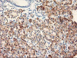
Immunohistochemistry (IHC) is a technique used in pathology and laboratory medicine to identify specific proteins or antigens in tissue sections. It combines the principles of immunology and histology to detect the presence and location of these target molecules within cells and tissues. This technique utilizes antibodies that are specific to the protein or antigen of interest, which are then tagged with a detection system such as a chromogen or fluorophore. The stained tissue sections can be examined under a microscope, allowing for the visualization and analysis of the distribution and expression patterns of the target molecule in the context of the tissue architecture. Immunohistochemistry is widely used in diagnostic pathology to help identify various diseases, including cancer, infectious diseases, and immune-mediated disorders.
Rheology is not a term that is specific to medicine, but rather it is a term used in the field of physics to describe the flow and deformation of matter. It specifically refers to the study of how materials flow or deform under various stresses or strains. This concept can be applied to various medical fields such as studying the flow properties of blood (hematology), understanding the movement of tissues and organs during surgical procedures, or analyzing the mechanical behavior of biological materials like bones and cartilages.
Lymphocyte activation is the process by which B-cells and T-cells (types of lymphocytes) become activated to perform effector functions in an immune response. This process involves the recognition of specific antigens presented on the surface of antigen-presenting cells, such as dendritic cells or macrophages.
The activation of B-cells leads to their differentiation into plasma cells that produce antibodies, while the activation of T-cells results in the production of cytotoxic T-cells (CD8+ T-cells) that can directly kill infected cells or helper T-cells (CD4+ T-cells) that assist other immune cells.
Lymphocyte activation involves a series of intracellular signaling events, including the binding of co-stimulatory molecules and the release of cytokines, which ultimately result in the expression of genes involved in cell proliferation, differentiation, and effector functions. The activation process is tightly regulated to prevent excessive or inappropriate immune responses that can lead to autoimmunity or chronic inflammation.
T-lymphocyte subsets refer to distinct populations of T-cells, which are a type of white blood cell that plays a central role in cell-mediated immunity. The two main types of T-lymphocytes are CD4+ and CD8+ cells, which are defined by the presence or absence of specific proteins called cluster differentiation (CD) molecules on their surface.
CD4+ T-cells, also known as helper T-cells, play a crucial role in activating other immune cells, such as B-lymphocytes and macrophages, to mount an immune response against pathogens. They also produce cytokines that help regulate the immune response.
CD8+ T-cells, also known as cytotoxic T-cells, directly kill infected cells or tumor cells by releasing toxic substances such as perforins and granzymes.
The balance between these two subsets of T-cells is critical for maintaining immune homeostasis and mounting effective immune responses against pathogens while avoiding excessive inflammation and autoimmunity. Therefore, the measurement of T-lymphocyte subsets is essential in diagnosing and monitoring various immunological disorders, including HIV infection, cancer, and autoimmune diseases.
CD8-positive T-lymphocytes, also known as CD8+ T cells or cytotoxic T cells, are a type of white blood cell that plays a crucial role in the adaptive immune system. They are named after the CD8 molecule found on their surface, which is a protein involved in cell signaling and recognition.
CD8+ T cells are primarily responsible for identifying and destroying virus-infected cells or cancerous cells. When activated, they release cytotoxic granules that contain enzymes capable of inducing apoptosis (programmed cell death) in the target cells. They also produce cytokines such as interferon-gamma, which can help coordinate the immune response and activate other immune cells.
CD8+ T cells are generated in the thymus gland and are a type of T cell, which is a lymphocyte that matures in the thymus and plays a central role in cell-mediated immunity. They recognize and respond to specific antigens presented on the surface of infected or cancerous cells in conjunction with major histocompatibility complex (MHC) class I molecules.
Overall, CD8+ T cells are an essential component of the immune system's defense against viral infections and cancer.
Neutrophils are a type of white blood cell that are part of the immune system's response to infection. They are produced in the bone marrow and released into the bloodstream where they circulate and are able to move quickly to sites of infection or inflammation in the body. Neutrophils are capable of engulfing and destroying bacteria, viruses, and other foreign substances through a process called phagocytosis. They are also involved in the release of inflammatory mediators, which can contribute to tissue damage in some cases. Neutrophils are characterized by the presence of granules in their cytoplasm, which contain enzymes and other proteins that help them carry out their immune functions.
Bone marrow cells are the types of cells found within the bone marrow, which is the spongy tissue inside certain bones in the body. The main function of bone marrow is to produce blood cells. There are two types of bone marrow: red and yellow. Red bone marrow is where most blood cell production takes place, while yellow bone marrow serves as a fat storage site.
The three main types of bone marrow cells are:
1. Hematopoietic stem cells (HSCs): These are immature cells that can differentiate into any type of blood cell, including red blood cells, white blood cells, and platelets. They have the ability to self-renew, meaning they can divide and create more hematopoietic stem cells.
2. Red blood cell progenitors: These are immature cells that will develop into mature red blood cells, also known as erythrocytes. Red blood cells carry oxygen from the lungs to the body's tissues and carbon dioxide back to the lungs.
3. Myeloid and lymphoid white blood cell progenitors: These are immature cells that will develop into various types of white blood cells, which play a crucial role in the body's immune system by fighting infections and diseases. Myeloid progenitors give rise to granulocytes (neutrophils, eosinophils, and basophils), monocytes, and megakaryocytes (which eventually become platelets). Lymphoid progenitors differentiate into B cells, T cells, and natural killer (NK) cells.
Bone marrow cells are essential for maintaining a healthy blood cell count and immune system function. Abnormalities in bone marrow cells can lead to various medical conditions, such as anemia, leukopenia, leukocytosis, thrombocytopenia, or thrombocytosis, depending on the specific type of blood cell affected. Additionally, bone marrow cells are often used in transplantation procedures to treat patients with certain types of cancer, such as leukemia and lymphoma, or other hematologic disorders.
An Enzyme-Linked Immunosorbent Assay (ELISA) is a type of analytical biochemistry assay used to detect and quantify the presence of a substance, typically a protein or peptide, in a liquid sample. It takes its name from the enzyme-linked antibodies used in the assay.
In an ELISA, the sample is added to a well containing a surface that has been treated to capture the target substance. If the target substance is present in the sample, it will bind to the surface. Next, an enzyme-linked antibody specific to the target substance is added. This antibody will bind to the captured target substance if it is present. After washing away any unbound material, a substrate for the enzyme is added. If the enzyme is present due to its linkage to the antibody, it will catalyze a reaction that produces a detectable signal, such as a color change or fluorescence. The intensity of this signal is proportional to the amount of target substance present in the sample, allowing for quantification.
ELISAs are widely used in research and clinical settings to detect and measure various substances, including hormones, viruses, and bacteria. They offer high sensitivity, specificity, and reproducibility, making them a reliable choice for many applications.
Interferon-gamma (IFN-γ) is a soluble cytokine that is primarily produced by the activation of natural killer (NK) cells and T lymphocytes, especially CD4+ Th1 cells and CD8+ cytotoxic T cells. It plays a crucial role in the regulation of the immune response against viral and intracellular bacterial infections, as well as tumor cells. IFN-γ has several functions, including activating macrophages to enhance their microbicidal activity, increasing the presentation of major histocompatibility complex (MHC) class I and II molecules on antigen-presenting cells, stimulating the proliferation and differentiation of T cells and NK cells, and inducing the production of other cytokines and chemokines. Additionally, IFN-γ has direct antiproliferative effects on certain types of tumor cells and can enhance the cytotoxic activity of immune cells against infected or malignant cells.
Cell differentiation is the process by which a less specialized cell, or stem cell, becomes a more specialized cell type with specific functions and structures. This process involves changes in gene expression, which are regulated by various intracellular signaling pathways and transcription factors. Differentiation results in the development of distinct cell types that make up tissues and organs in multicellular organisms. It is a crucial aspect of embryonic development, tissue repair, and maintenance of homeostasis in the body.
BALB/c is an inbred strain of laboratory mouse that is widely used in biomedical research. The strain was developed at the Institute of Cancer Research in London by Henry Baldwin and his colleagues in the 1920s, and it has since become one of the most commonly used inbred strains in the world.
BALB/c mice are characterized by their black coat color, which is determined by a recessive allele at the tyrosinase locus. They are also known for their docile and friendly temperament, making them easy to handle and work with in the laboratory.
One of the key features of BALB/c mice that makes them useful for research is their susceptibility to certain types of tumors and immune responses. For example, they are highly susceptible to developing mammary tumors, which can be induced by chemical carcinogens or viral infection. They also have a strong Th2-biased immune response, which makes them useful models for studying allergic diseases and asthma.
BALB/c mice are also commonly used in studies of genetics, neuroscience, behavior, and infectious diseases. Because they are an inbred strain, they have a uniform genetic background, which makes it easier to control for genetic factors in experiments. Additionally, because they have been bred in the laboratory for many generations, they are highly standardized and reproducible, making them ideal subjects for scientific research.
A biological marker, often referred to as a biomarker, is a measurable indicator that reflects the presence or severity of a disease state, or a response to a therapeutic intervention. Biomarkers can be found in various materials such as blood, tissues, or bodily fluids, and they can take many forms, including molecular, histologic, radiographic, or physiological measurements.
In the context of medical research and clinical practice, biomarkers are used for a variety of purposes, such as:
1. Diagnosis: Biomarkers can help diagnose a disease by indicating the presence or absence of a particular condition. For example, prostate-specific antigen (PSA) is a biomarker used to detect prostate cancer.
2. Monitoring: Biomarkers can be used to monitor the progression or regression of a disease over time. For instance, hemoglobin A1c (HbA1c) levels are monitored in diabetes patients to assess long-term blood glucose control.
3. Predicting: Biomarkers can help predict the likelihood of developing a particular disease or the risk of a negative outcome. For example, the presence of certain genetic mutations can indicate an increased risk for breast cancer.
4. Response to treatment: Biomarkers can be used to evaluate the effectiveness of a specific treatment by measuring changes in the biomarker levels before and after the intervention. This is particularly useful in personalized medicine, where treatments are tailored to individual patients based on their unique biomarker profiles.
It's important to note that for a biomarker to be considered clinically valid and useful, it must undergo rigorous validation through well-designed studies, including demonstrating sensitivity, specificity, reproducibility, and clinical relevance.
CD3 antigens are a group of proteins found on the surface of T-cells, which are a type of white blood cell that plays a central role in the immune response. The CD3 antigens are composed of several different subunits (ε, δ, γ, and α) that associate to form the CD3 complex, which is involved in T-cell activation and signal transduction.
The CD3 complex is associated with the T-cell receptor (TCR), which recognizes and binds to specific antigens presented by antigen-presenting cells. When the TCR binds to an antigen, it triggers a series of intracellular signaling events that lead to T-cell activation and the initiation of an immune response.
CD3 antigens are important targets for immunotherapy in some diseases, such as certain types of cancer. For example, monoclonal antibodies that target CD3 have been developed to activate T-cells and enhance their ability to recognize and destroy tumor cells. However, CD3-targeted therapies can also cause side effects, such as cytokine release syndrome, which can be serious or life-threatening in some cases.
Cerebrovascular circulation refers to the network of blood vessels that supply oxygenated blood and nutrients to the brain tissue, and remove waste products. It includes the internal carotid arteries, vertebral arteries, circle of Willis, and the intracranial arteries that branch off from them.
The internal carotid arteries and vertebral arteries merge to form the circle of Willis, a polygonal network of vessels located at the base of the brain. The anterior cerebral artery, middle cerebral artery, posterior cerebral artery, and communicating arteries are the major vessels that branch off from the circle of Willis and supply blood to different regions of the brain.
Interruptions or abnormalities in the cerebrovascular circulation can lead to various neurological conditions such as stroke, transient ischemic attack (TIA), and vascular dementia.
Blood platelets, also known as thrombocytes, are small, colorless cell fragments in our blood that play an essential role in normal blood clotting. They are formed in the bone marrow from large cells called megakaryocytes and circulate in the blood in an inactive state until they are needed to help stop bleeding. When a blood vessel is damaged, platelets become activated and change shape, releasing chemicals that attract more platelets to the site of injury. These activated platelets then stick together to form a plug, or clot, that seals the wound and prevents further blood loss. In addition to their role in clotting, platelets also help to promote healing by releasing growth factors that stimulate the growth of new tissue.
Membrane glycoproteins are proteins that contain oligosaccharide chains (glycans) covalently attached to their polypeptide backbone. They are integral components of biological membranes, spanning the lipid bilayer and playing crucial roles in various cellular processes.
The glycosylation of these proteins occurs in the endoplasmic reticulum (ER) and Golgi apparatus during protein folding and trafficking. The attached glycans can vary in structure, length, and composition, which contributes to the diversity of membrane glycoproteins.
Membrane glycoproteins can be classified into two main types based on their orientation within the lipid bilayer:
1. Type I (N-linked): These glycoproteins have a single transmembrane domain and an extracellular N-terminus, where the oligosaccharides are predominantly attached via asparagine residues (Asn-X-Ser/Thr sequon).
2. Type II (C-linked): These glycoproteins possess two transmembrane domains and an intracellular C-terminus, with the oligosaccharides linked to tryptophan residues via a mannose moiety.
Membrane glycoproteins are involved in various cellular functions, such as:
* Cell adhesion and recognition
* Receptor-mediated signal transduction
* Enzymatic catalysis
* Transport of molecules across membranes
* Cell-cell communication
* Immunological responses
Some examples of membrane glycoproteins include cell surface receptors (e.g., growth factor receptors, cytokine receptors), adhesion molecules (e.g., integrins, cadherins), and transporters (e.g., ion channels, ABC transporters).
The Fluorescent Antibody Technique (FAT) is a type of immunofluorescence assay used in laboratory medicine and pathology for the detection and localization of specific antigens or antibodies in tissues, cells, or microorganisms. In this technique, a fluorescein-labeled antibody is used to selectively bind to the target antigen or antibody, forming an immune complex. When excited by light of a specific wavelength, the fluorescein label emits light at a longer wavelength, typically visualized as green fluorescence under a fluorescence microscope.
The FAT is widely used in diagnostic microbiology for the identification and characterization of various bacteria, viruses, fungi, and parasites. It has also been applied in the diagnosis of autoimmune diseases and certain cancers by detecting specific antibodies or antigens in patient samples. The main advantage of FAT is its high sensitivity and specificity, allowing for accurate detection and differentiation of various pathogens and disease markers. However, it requires specialized equipment and trained personnel to perform and interpret the results.
Reproducibility of results in a medical context refers to the ability to obtain consistent and comparable findings when a particular experiment or study is repeated, either by the same researcher or by different researchers, following the same experimental protocol. It is an essential principle in scientific research that helps to ensure the validity and reliability of research findings.
In medical research, reproducibility of results is crucial for establishing the effectiveness and safety of new treatments, interventions, or diagnostic tools. It involves conducting well-designed studies with adequate sample sizes, appropriate statistical analyses, and transparent reporting of methods and findings to allow other researchers to replicate the study and confirm or refute the results.
The lack of reproducibility in medical research has become a significant concern in recent years, as several high-profile studies have failed to produce consistent findings when replicated by other researchers. This has led to increased scrutiny of research practices and a call for greater transparency, rigor, and standardization in the conduct and reporting of medical research.
Green Fluorescent Protein (GFP) is not a medical term per se, but a scientific term used in the field of molecular biology. GFP is a protein that exhibits bright green fluorescence when exposed to light, particularly blue or ultraviolet light. It was originally discovered in the jellyfish Aequorea victoria.
In medical and biological research, scientists often use recombinant DNA technology to introduce the gene for GFP into other organisms, including bacteria, plants, and animals, including humans. This allows them to track the expression and localization of specific genes or proteins of interest in living cells, tissues, or even whole organisms.
The ability to visualize specific cellular structures or processes in real-time has proven invaluable for a wide range of research areas, from studying the development and function of organs and organ systems to understanding the mechanisms of diseases and the effects of therapeutic interventions.
A phenotype is the physical or biochemical expression of an organism's genes, or the observable traits and characteristics resulting from the interaction of its genetic constitution (genotype) with environmental factors. These characteristics can include appearance, development, behavior, and resistance to disease, among others. Phenotypes can vary widely, even among individuals with identical genotypes, due to differences in environmental influences, gene expression, and genetic interactions.
Aneuploidy is a medical term that refers to an abnormal number of chromosomes in a cell. Chromosomes are thread-like structures located inside the nucleus of cells that contain genetic information in the form of genes.
In humans, the normal number of chromosomes in a cell is 46, arranged in 23 pairs. Aneuploidy occurs when there is an extra or missing chromosome in one or more of these pairs. For example, Down syndrome is a condition that results from an extra copy of chromosome 21, also known as trisomy 21.
Aneuploidy can arise during the formation of gametes (sperm or egg cells) due to errors in the process of cell division called meiosis. These errors can result in eggs or sperm with an abnormal number of chromosomes, which can then lead to aneuploidy in the resulting embryo.
Aneuploidy is a significant cause of birth defects and miscarriages. The severity of the condition depends on which chromosomes are affected and the extent of the abnormality. In some cases, aneuploidy may have no noticeable effects, while in others it can lead to serious health problems or developmental delays.
C57BL/6 (C57 Black 6) is an inbred strain of laboratory mouse that is widely used in biomedical research. The term "inbred" refers to a strain of animals where matings have been carried out between siblings or other closely related individuals for many generations, resulting in a population that is highly homozygous at most genetic loci.
The C57BL/6 strain was established in 1920 by crossing a female mouse from the dilute brown (DBA) strain with a male mouse from the black strain. The resulting offspring were then interbred for many generations to create the inbred C57BL/6 strain.
C57BL/6 mice are known for their robust health, longevity, and ease of handling, making them a popular choice for researchers. They have been used in a wide range of biomedical research areas, including studies of cancer, immunology, neuroscience, cardiovascular disease, and metabolism.
One of the most notable features of the C57BL/6 strain is its sensitivity to certain genetic modifications, such as the introduction of mutations that lead to obesity or impaired glucose tolerance. This has made it a valuable tool for studying the genetic basis of complex diseases and traits.
Overall, the C57BL/6 inbred mouse strain is an important model organism in biomedical research, providing a valuable resource for understanding the genetic and molecular mechanisms underlying human health and disease.
Blood cells are the formed elements in the blood, including red blood cells (erythrocytes), white blood cells (leukocytes), and platelets (thrombocytes). These cells are produced in the bone marrow and play crucial roles in the body's functions. Red blood cells are responsible for carrying oxygen to tissues and carbon dioxide away from them, while white blood cells are part of the immune system and help defend against infection and disease. Platelets are cell fragments that are essential for normal blood clotting.
Coronary circulation refers to the circulation of blood in the coronary vessels, which supply oxygenated blood to the heart muscle (myocardium) and drain deoxygenated blood from it. The coronary circulation system includes two main coronary arteries - the left main coronary artery and the right coronary artery - that branch off from the aorta just above the aortic valve. These arteries further divide into smaller branches, which supply blood to different regions of the heart muscle.
The left main coronary artery divides into two branches: the left anterior descending (LAD) artery and the left circumflex (LCx) artery. The LAD supplies blood to the front and sides of the heart, while the LCx supplies blood to the back and sides of the heart. The right coronary artery supplies blood to the lower part of the heart, including the right ventricle and the bottom portion of the left ventricle.
The veins that drain the heart muscle include the great cardiac vein, the middle cardiac vein, and the small cardiac vein, which merge to form the coronary sinus. The coronary sinus empties into the right atrium, allowing deoxygenated blood to enter the right side of the heart and be pumped to the lungs for oxygenation.
Coronary circulation is essential for maintaining the health and function of the heart muscle, as it provides the necessary oxygen and nutrients required for proper contraction and relaxation of the myocardium. Any disruption or blockage in the coronary circulation system can lead to serious consequences, such as angina, heart attack, or even death.
Surface antigens are molecules found on the surface of cells that can be recognized by the immune system as being foreign or different from the host's own cells. Antigens are typically proteins or polysaccharides that are capable of stimulating an immune response, leading to the production of antibodies and activation of immune cells such as T-cells.
Surface antigens are important in the context of infectious diseases because they allow the immune system to identify and target infected cells for destruction. For example, viruses and bacteria often display surface antigens that are distinct from those found on host cells, allowing the immune system to recognize and attack them. In some cases, these surface antigens can also be used as targets for vaccines or other immunotherapies.
In addition to their role in infectious diseases, surface antigens are also important in the context of cancer. Tumor cells often display abnormal surface antigens that differ from those found on normal cells, allowing the immune system to potentially recognize and attack them. However, tumors can also develop mechanisms to evade the immune system, making it difficult to mount an effective response.
Overall, understanding the properties and behavior of surface antigens is crucial for developing effective immunotherapies and vaccines against infectious diseases and cancer.
Antineoplastic agents are a class of drugs used to treat malignant neoplasms or cancer. These agents work by inhibiting the growth and proliferation of cancer cells, either by killing them or preventing their division and replication. Antineoplastic agents can be classified based on their mechanism of action, such as alkylating agents, antimetabolites, topoisomerase inhibitors, mitotic inhibitors, and targeted therapy agents.
Alkylating agents work by adding alkyl groups to DNA, which can cause cross-linking of DNA strands and ultimately lead to cell death. Antimetabolites interfere with the metabolic processes necessary for DNA synthesis and replication, while topoisomerase inhibitors prevent the relaxation of supercoiled DNA during replication. Mitotic inhibitors disrupt the normal functioning of the mitotic spindle, which is essential for cell division. Targeted therapy agents are designed to target specific molecular abnormalities in cancer cells, such as mutated oncogenes or dysregulated signaling pathways.
It's important to note that antineoplastic agents can also affect normal cells and tissues, leading to various side effects such as nausea, vomiting, hair loss, and myelosuppression (suppression of bone marrow function). Therefore, the use of these drugs requires careful monitoring and management of their potential adverse effects.
CD34 is a type of antigen that is found on the surface of certain cells in the human body. Specifically, CD34 antigens are present on hematopoietic stem cells, which are immature cells that can develop into different types of blood cells. These stem cells are found in the bone marrow and are responsible for producing red blood cells, white blood cells, and platelets.
CD34 antigens are a type of cell surface marker that is used in medical research and clinical settings to identify and isolate hematopoietic stem cells. They are also used in the development of stem cell therapies and transplantation procedures. CD34 antigens can be detected using various laboratory techniques, such as flow cytometry or immunohistochemistry.
It's important to note that while CD34 is a useful marker for identifying hematopoietic stem cells, it is not exclusive to these cells and can also be found on other cell types, such as endothelial cells that line blood vessels. Therefore, additional markers are often used in combination with CD34 to more specifically identify and isolate hematopoietic stem cells.
Cell adhesion refers to the binding of cells to extracellular matrices or to other cells, a process that is fundamental to the development, function, and maintenance of multicellular organisms. Cell adhesion is mediated by various cell surface receptors, such as integrins, cadherins, and immunoglobulin-like cell adhesion molecules (Ig-CAMs), which interact with specific ligands in the extracellular environment. These interactions lead to the formation of specialized junctions, such as tight junctions, adherens junctions, and desmosomes, that help to maintain tissue architecture and regulate various cellular processes, including proliferation, differentiation, migration, and survival. Disruptions in cell adhesion can contribute to a variety of diseases, including cancer, inflammation, and degenerative disorders.
Up-regulation is a term used in molecular biology and medicine to describe an increase in the expression or activity of a gene, protein, or receptor in response to a stimulus. This can occur through various mechanisms such as increased transcription, translation, or reduced degradation of the molecule. Up-regulation can have important functional consequences, for example, enhancing the sensitivity or response of a cell to a hormone, neurotransmitter, or drug. It is a normal physiological process that can also be induced by disease or pharmacological interventions.
Gene expression is the process by which the information encoded in a gene is used to synthesize a functional gene product, such as a protein or RNA molecule. This process involves several steps: transcription, RNA processing, and translation. During transcription, the genetic information in DNA is copied into a complementary RNA molecule, known as messenger RNA (mRNA). The mRNA then undergoes RNA processing, which includes adding a cap and tail to the mRNA and splicing out non-coding regions called introns. The resulting mature mRNA is then translated into a protein on ribosomes in the cytoplasm through the process of translation.
The regulation of gene expression is a complex and highly controlled process that allows cells to respond to changes in their environment, such as growth factors, hormones, and stress signals. This regulation can occur at various stages of gene expression, including transcriptional activation or repression, RNA processing, mRNA stability, and translation. Dysregulation of gene expression has been implicated in many diseases, including cancer, genetic disorders, and neurological conditions.
Endothelial cells are the type of cells that line the inner surface of blood vessels, lymphatic vessels, and heart chambers. They play a crucial role in maintaining vascular homeostasis by controlling vasomotor tone, coagulation, platelet activation, and inflammation. Endothelial cells also regulate the transport of molecules between the blood and surrounding tissues, and contribute to the maintenance of the structural integrity of the vasculature. They are flat, elongated cells with a unique morphology that allows them to form a continuous, nonthrombogenic lining inside the vessels. Endothelial cells can be isolated from various tissues and cultured in vitro for research purposes.
Dendritic cells (DCs) are a type of immune cell that play a critical role in the body's defense against infection and cancer. They are named for their dendrite-like projections, which they use to interact with and sample their environment. DCs are responsible for processing antigens (foreign substances that trigger an immune response) and presenting them to T cells, a type of white blood cell that plays a central role in the immune system's response to infection and cancer.
DCs can be found throughout the body, including in the skin, mucous membranes, and lymphoid organs. They are able to recognize and respond to a wide variety of antigens, including those from bacteria, viruses, fungi, and parasites. Once they have processed an antigen, DCs migrate to the lymph nodes, where they present the antigen to T cells. This interaction activates the T cells, which then go on to mount a targeted immune response against the invading pathogen or cancerous cells.
DCs are a diverse group of cells that can be divided into several subsets based on their surface markers and function. Some DCs, such as Langerhans cells and dermal DCs, are found in the skin and mucous membranes, where they serve as sentinels for invading pathogens. Other DCs, such as plasmacytoid DCs and conventional DCs, are found in the lymphoid organs, where they play a role in activating T cells and initiating an immune response.
Overall, dendritic cells are essential for the proper functioning of the immune system, and dysregulation of these cells has been implicated in a variety of diseases, including autoimmune disorders and cancer.
Flow Injection Analysis (FIA) is not a medical term, but a technique used in analytical chemistry. However, I can provide you with the definition of this term:
Flow Injection Analysis (FIA) is a method for automated, continuous monitoring and quantitative analysis of chemical substances. It involves the injection of a sample into a flowing carrier stream, which transports it to a detector after mixing and reaction in a flow-through cell or system. The analyte interacts with reagents to produce a signal that can be measured and related to the concentration of the substance being analyzed. FIA is widely used for environmental monitoring, quality control, process control, and clinical analysis.
CD14 is a type of protein found on the surface of certain cells in the human body, including monocytes, macrophages, and some types of dendritic cells. These cells are part of the immune system and play a crucial role in detecting and responding to infections and other threats.
CD14 is not an antigen itself, but it can bind to certain types of antigens, such as lipopolysaccharides (LPS) found on the surface of gram-negative bacteria. When CD14 binds to an LPS molecule, it helps to activate the immune response and trigger the production of cytokines and other inflammatory mediators.
CD14 can also be found in soluble form in the bloodstream, where it can help to neutralize LPS and prevent it from causing damage to tissues and organs.
It's worth noting that while CD14 plays an important role in the immune response, it is not typically used as a target for vaccines or other immunotherapies. Instead, it is often studied as a marker of immune activation and inflammation in various diseases, including sepsis, atherosclerosis, and Alzheimer's disease.
Caspase-3 is a type of protease enzyme that plays a central role in the execution-phase of cell apoptosis, or programmed cell death. It's also known as CPP32 (CPP for ced-3 protease precursor) or apopain. Caspase-3 is produced as an inactive protein that is activated when cleaved by other caspases during the early stages of apoptosis. Once activated, it cleaves a variety of cellular proteins, including structural proteins, enzymes, and signal transduction proteins, leading to the characteristic morphological and biochemical changes associated with apoptotic cell death. Caspase-3 is often referred to as the "death protease" because of its crucial role in executing the cell death program.
Phycoerythrin is not a medical term, but a term used in biochemistry and cell biology. It refers to a type of protein found in certain algae and cyanobacteria that binds phycobilins, which are linear tetrapyrrole chromophores. Phycoerythrin is a light-harvesting pigment that absorbs light energy and transfers it to the photosynthetic reaction centers. It is often used in research and clinical settings as a fluorescent label for various applications, such as flow cytometry, immunohistochemistry, and microscopy.
P-Selectin is a type of cell adhesion molecule, specifically a member of the selectin family, that is involved in the inflammatory response. It is primarily expressed on the surface of activated platelets and endothelial cells. P-Selectin plays a crucial role in the initial interaction between leukocytes (white blood cells) and the vascular endothelium, which is an essential step in the recruitment of leukocytes to sites of inflammation or injury. This process helps to mediate the rolling and adhesion of leukocytes to the endothelial surface, facilitating their extravasation into the surrounding tissue. P-Selectin's function is regulated by its interaction with specific ligands on the surface of leukocytes, such as PSGL-1 (P-Selectin Glycoprotein Ligand-1).
Deoxyribonucleic acid (DNA) is the genetic material present in the cells of organisms where it is responsible for the storage and transmission of hereditary information. DNA is a long molecule that consists of two strands coiled together to form a double helix. Each strand is made up of a series of four nucleotide bases - adenine (A), guanine (G), cytosine (C), and thymine (T) - that are linked together by phosphate and sugar groups. The sequence of these bases along the length of the molecule encodes genetic information, with A always pairing with T and C always pairing with G. This base-pairing allows for the replication and transcription of DNA, which are essential processes in the functioning and reproduction of all living organisms.
Signal transduction is the process by which a cell converts an extracellular signal, such as a hormone or neurotransmitter, into an intracellular response. This involves a series of molecular events that transmit the signal from the cell surface to the interior of the cell, ultimately resulting in changes in gene expression, protein activity, or metabolism.
The process typically begins with the binding of the extracellular signal to a receptor located on the cell membrane. This binding event activates the receptor, which then triggers a cascade of intracellular signaling molecules, such as second messengers, protein kinases, and ion channels. These molecules amplify and propagate the signal, ultimately leading to the activation or inhibition of specific cellular responses.
Signal transduction pathways are highly regulated and can be modulated by various factors, including other signaling molecules, post-translational modifications, and feedback mechanisms. Dysregulation of these pathways has been implicated in a variety of diseases, including cancer, diabetes, and neurological disorders.
A residual neoplasm is a term used in pathology and oncology to describe the remaining abnormal tissue or cancer cells after a surgical procedure or treatment aimed at completely removing a tumor. This means that some cancer cells have been left behind and continue to persist in the body. The presence of residual neoplasm can increase the risk of recurrence or progression of the disease, as these remaining cells may continue to grow and divide.
Residual neoplasm is often assessed during follow-up appointments and monitoring, using imaging techniques like CT scans, MRIs, or PET scans, and sometimes through biopsies. The extent of residual neoplasm can influence the choice of further treatment options, such as additional surgery, radiation therapy, chemotherapy, or targeted therapies, to eliminate the remaining cancer cells and reduce the risk of recurrence.
Tumor Necrosis Factor-alpha (TNF-α) is a cytokine, a type of small signaling protein involved in immune response and inflammation. It is primarily produced by activated macrophages, although other cell types such as T-cells, natural killer cells, and mast cells can also produce it.
TNF-α plays a crucial role in the body's defense against infection and tissue injury by mediating inflammatory responses, activating immune cells, and inducing apoptosis (programmed cell death) in certain types of cells. It does this by binding to its receptors, TNFR1 and TNFR2, which are found on the surface of many cell types.
In addition to its role in the immune response, TNF-α has been implicated in the pathogenesis of several diseases, including autoimmune disorders such as rheumatoid arthritis, inflammatory bowel disease, and psoriasis, as well as cancer, where it can promote tumor growth and metastasis.
Therapeutic agents that target TNF-α, such as infliximab, adalimumab, and etanercept, have been developed to treat these conditions. However, these drugs can also increase the risk of infections and other side effects, so their use must be carefully monitored.
HLA-DR antigens are a type of human leukocyte antigen (HLA) class II molecule that plays a crucial role in the immune system. They are found on the surface of antigen-presenting cells, such as dendritic cells, macrophages, and B lymphocytes. HLA-DR molecules present peptide antigens to CD4+ T cells, also known as helper T cells, thereby initiating an immune response.
HLA-DR antigens are highly polymorphic, meaning that there are many different variants of these molecules in the human population. This diversity allows for a wide range of potential peptide antigens to be presented and recognized by the immune system. HLA-DR antigens are encoded by genes located on chromosome 6 in the major histocompatibility complex (MHC) region.
In transplantation, HLA-DR compatibility between donor and recipient is an important factor in determining the success of the transplant. Incompatibility can lead to a heightened immune response against the transplanted organ or tissue, resulting in rejection. Additionally, certain HLA-DR types have been associated with increased susceptibility to autoimmune diseases, such as rheumatoid arthritis and multiple sclerosis.
Natural Killer (NK) cells are a type of lymphocyte, which are large granular innate immune cells that play a crucial role in the host's defense against viral infections and malignant transformations. They do not require prior sensitization to target and destroy abnormal cells, such as virus-infected cells or tumor cells. NK cells recognize their targets through an array of germline-encoded activating and inhibitory receptors that detect the alterations in the cell surface molecules of potential targets. Upon activation, NK cells release cytotoxic granules containing perforins and granzymes to induce target cell apoptosis, and they also produce a variety of cytokines and chemokines to modulate immune responses. Overall, natural killer cells serve as a critical component of the innate immune system, providing rapid and effective responses against infected or malignant cells.
IgG receptors, also known as Fcγ receptors (Fc gamma receptors), are specialized protein molecules found on the surface of various immune cells, such as neutrophils, monocytes, macrophages, and some lymphocytes. These receptors recognize and bind to the Fc region of IgG antibodies, one of the five classes of immunoglobulins in the human body.
IgG receptors play a crucial role in immune responses by mediating different effector functions, including:
1. Antibody-dependent cellular cytotoxicity (ADCC): IgG receptors on natural killer (NK) cells and other immune cells bind to IgG antibodies coated on the surface of virus-infected or cancer cells, leading to their destruction.
2. Phagocytosis: When IgG antibodies tag pathogens or foreign particles, phagocytes like neutrophils and macrophages recognize and bind to these immune complexes via IgG receptors, facilitating the engulfment and removal of the targeted particles.
3. Antigen presentation: IgG receptors on antigen-presenting cells (APCs) can internalize immune complexes, process the antigens, and present them to T cells, thereby initiating adaptive immune responses.
4. Inflammatory response regulation: IgG receptors can modulate inflammation by activating or inhibiting downstream signaling pathways in immune cells, depending on the specific type of Fcγ receptor and its activation state.
There are several types of IgG receptors (FcγRI, FcγRII, FcγRIII, and FcγRIV) with varying affinities for different subclasses of IgG antibodies (IgG1, IgG2, IgG3, and IgG4). The distinct functions and expression patterns of these receptors contribute to the complexity and fine-tuning of immune responses in the human body.
Optic flow is not a medical term per se, but rather a term used in the field of visual perception and neuroscience. It refers to the pattern of motion of objects in the visual field that occurs as an observer moves through the environment. This pattern of motion is important for the perception of self-motion and the estimation of egocentric distance (the distance of objects in the environment relative to the observer). Optic flow has been studied in relation to various clinical populations, such as individuals with vestibular disorders or visual impairments, who may have difficulty processing optic flow information.
Macrophages are a type of white blood cell that are an essential part of the immune system. They are large, specialized cells that engulf and destroy foreign substances, such as bacteria, viruses, parasites, and fungi, as well as damaged or dead cells. Macrophages are found throughout the body, including in the bloodstream, lymph nodes, spleen, liver, lungs, and connective tissues. They play a critical role in inflammation, immune response, and tissue repair and remodeling.
Macrophages originate from monocytes, which are a type of white blood cell produced in the bone marrow. When monocytes enter the tissues, they differentiate into macrophages, which have a larger size and more specialized functions than monocytes. Macrophages can change their shape and move through tissues to reach sites of infection or injury. They also produce cytokines, chemokines, and other signaling molecules that help coordinate the immune response and recruit other immune cells to the site of infection or injury.
Macrophages have a variety of surface receptors that allow them to recognize and respond to different types of foreign substances and signals from other cells. They can engulf and digest foreign particles, bacteria, and viruses through a process called phagocytosis. Macrophages also play a role in presenting antigens to T cells, which are another type of immune cell that helps coordinate the immune response.
Overall, macrophages are crucial for maintaining tissue homeostasis, defending against infection, and promoting wound healing and tissue repair. Dysregulation of macrophage function has been implicated in a variety of diseases, including cancer, autoimmune disorders, and chronic inflammatory conditions.
Antibodies are proteins produced by the immune system in response to the presence of a foreign substance, such as a bacterium or virus. They are capable of identifying and binding to specific antigens (foreign substances) on the surface of these invaders, marking them for destruction by other immune cells. Antibodies are also known as immunoglobulins and come in several different types, including IgA, IgD, IgE, IgG, and IgM, each with a unique function in the immune response. They are composed of four polypeptide chains, two heavy chains and two light chains, that are held together by disulfide bonds. The variable regions of the heavy and light chains form the antigen-binding site, which is specific to a particular antigen.
The endothelium is a thin layer of simple squamous epithelial cells that lines the interior surface of blood vessels, lymphatic vessels, and heart chambers. The vascular endothelium, specifically, refers to the endothelial cells that line the blood vessels. These cells play a crucial role in maintaining vascular homeostasis by regulating vasomotor tone, coagulation, platelet activation, inflammation, and permeability of the vessel wall. They also contribute to the growth and repair of the vascular system and are involved in various pathological processes such as atherosclerosis, hypertension, and diabetes.
Cytophotometry is a medical analytical technique that involves the measurement of light intensity or absorbance by individual cells, allowing for the quantitative analysis of cellular components such as DNA, RNA, and proteins. This method can be used to study cell cycle phase distribution, chromosome abnormalities, and changes in nuclear structure associated with various pathological conditions, including cancer and genetic disorders.
In cytophotometry, cells are typically stained with a fluorescent dye that specifically binds to the target molecule of interest, such as DNA or RNA. The cells are then placed on a microscope slide and illuminated with light at an appropriate wavelength for the dye used. A photodetector is used to measure the intensity of the emitted fluorescent light, which is proportional to the amount of target molecule present in each cell.
The resulting data can be analyzed using specialized software to generate histograms or other visual representations of the distribution of target molecules within a population of cells. This information can be used to identify abnormalities or changes in cellular composition, providing valuable insights into the underlying biology of various diseases and conditions.
Regulatory T-lymphocytes (Tregs), also known as suppressor T cells, are a subpopulation of T-cells that play a critical role in maintaining immune tolerance and preventing autoimmune diseases. They function to suppress the activation and proliferation of other immune cells, thereby regulating the immune response and preventing it from attacking the body's own tissues.
Tregs constitutively express the surface markers CD4 and CD25, as well as the transcription factor Foxp3, which is essential for their development and function. They can be further divided into subsets based on their expression of other markers, such as CD127 and CD45RA.
Tregs are critical for maintaining self-tolerance by suppressing the activation of self-reactive T cells that have escaped negative selection in the thymus. They also play a role in regulating immune responses to foreign antigens, such as those encountered during infection or cancer, and can contribute to the immunosuppressive microenvironment found in tumors.
Dysregulation of Tregs has been implicated in various autoimmune diseases, including type 1 diabetes, rheumatoid arthritis, and multiple sclerosis, as well as in cancer and infectious diseases. Therefore, understanding the mechanisms that regulate Treg function is an important area of research with potential therapeutic implications.
A cell membrane, also known as the plasma membrane, is a thin semi-permeable phospholipid bilayer that surrounds all cells in animals, plants, and microorganisms. It functions as a barrier to control the movement of substances in and out of the cell, allowing necessary molecules such as nutrients, oxygen, and signaling molecules to enter while keeping out harmful substances and waste products. The cell membrane is composed mainly of phospholipids, which have hydrophilic (water-loving) heads and hydrophobic (water-fearing) tails. This unique structure allows the membrane to be flexible and fluid, yet selectively permeable. Additionally, various proteins are embedded in the membrane that serve as channels, pumps, receptors, and enzymes, contributing to the cell's overall functionality and communication with its environment.
Erythrocytes, also known as red blood cells (RBCs), are the most common type of blood cell in circulating blood in mammals. They are responsible for transporting oxygen from the lungs to the body's tissues and carbon dioxide from the tissues to the lungs.
Erythrocytes are formed in the bone marrow and have a biconcave shape, which allows them to fold and bend easily as they pass through narrow blood vessels. They do not have a nucleus or mitochondria, which makes them more flexible but also limits their ability to reproduce or repair themselves.
In humans, erythrocytes are typically disc-shaped and measure about 7 micrometers in diameter. They contain the protein hemoglobin, which binds to oxygen and gives blood its red color. The lifespan of an erythrocyte is approximately 120 days, after which it is broken down in the liver and spleen.
Abnormalities in erythrocyte count or function can lead to various medical conditions, such as anemia, polycythemia, and sickle cell disease.
Animal disease models are specialized animals, typically rodents such as mice or rats, that have been genetically engineered or exposed to certain conditions to develop symptoms and physiological changes similar to those seen in human diseases. These models are used in medical research to study the pathophysiology of diseases, identify potential therapeutic targets, test drug efficacy and safety, and understand disease mechanisms.
The genetic modifications can include knockout or knock-in mutations, transgenic expression of specific genes, or RNA interference techniques. The animals may also be exposed to environmental factors such as chemicals, radiation, or infectious agents to induce the disease state.
Examples of animal disease models include:
1. Mouse models of cancer: Genetically engineered mice that develop various types of tumors, allowing researchers to study cancer initiation, progression, and metastasis.
2. Alzheimer's disease models: Transgenic mice expressing mutant human genes associated with Alzheimer's disease, which exhibit amyloid plaque formation and cognitive decline.
3. Diabetes models: Obese and diabetic mouse strains like the NOD (non-obese diabetic) or db/db mice, used to study the development of type 1 and type 2 diabetes, respectively.
4. Cardiovascular disease models: Atherosclerosis-prone mice, such as ApoE-deficient or LDLR-deficient mice, that develop plaque buildup in their arteries when fed a high-fat diet.
5. Inflammatory bowel disease models: Mice with genetic mutations affecting intestinal barrier function and immune response, such as IL-10 knockout or SAMP1/YitFc mice, which develop colitis.
Animal disease models are essential tools in preclinical research, but it is important to recognize their limitations. Differences between species can affect the translatability of results from animal studies to human patients. Therefore, researchers must carefully consider the choice of model and interpret findings cautiously when applying them to human diseases.
According to the National Institutes of Health (NIH), stem cells are "initial cells" or "precursor cells" that have the ability to differentiate into many different cell types in the body. They can also divide without limit to replenish other cells for as long as the person or animal is still alive.
There are two main types of stem cells: embryonic stem cells, which come from human embryos, and adult stem cells, which are found in various tissues throughout the body. Embryonic stem cells have the ability to differentiate into all cell types in the body, while adult stem cells have more limited differentiation potential.
Stem cells play an essential role in the development and repair of various tissues and organs in the body. They are currently being studied for their potential use in the treatment of a wide range of diseases and conditions, including cancer, diabetes, heart disease, and neurological disorders. However, more research is needed to fully understand the properties and capabilities of these cells before they can be used safely and effectively in clinical settings.
The spleen is an organ in the upper left side of the abdomen, next to the stomach and behind the ribs. It plays multiple supporting roles in the body:
1. It fights infection by acting as a filter for the blood. Old red blood cells are recycled in the spleen, and platelets and white blood cells are stored there.
2. The spleen also helps to control the amount of blood in the body by removing excess red blood cells and storing platelets.
3. It has an important role in immune function, producing antibodies and removing microorganisms and damaged red blood cells from the bloodstream.
The spleen can be removed without causing any significant problems, as other organs take over its functions. This is known as a splenectomy and may be necessary if the spleen is damaged or diseased.
Rhodamine 123 is not a medical term, but a chemical compound. It's a fluorescent dye used in various scientific and research applications, particularly in the field of cell biology. Rhodamine 123 has an affinity for mitochondria, the energy-producing structures in cells, making it useful as a marker to study mitochondrial function and distribution within cells.
In summary, Rhodamine 123 is not a medical definition itself, but it can be used in medical research contexts to investigate cellular processes.
Transfection is a term used in molecular biology that refers to the process of deliberately introducing foreign genetic material (DNA, RNA or artificial gene constructs) into cells. This is typically done using chemical or physical methods, such as lipofection or electroporation. Transfection is widely used in research and medical settings for various purposes, including studying gene function, producing proteins, developing gene therapies, and creating genetically modified organisms. It's important to note that transfection is different from transduction, which is the process of introducing genetic material into cells using viruses as vectors.
HL-60 cells are a type of human promyelocytic leukemia cell line that is commonly used in scientific research. They are named after the hospital where they were first isolated, the Hospital of the University of Pennsylvania (HUP) and the 60th culture attempt to grow these cells.
HL-60 cells have the ability to differentiate into various types of blood cells, such as granulocytes, monocytes, and macrophages, when exposed to certain chemical compounds or under specific culturing conditions. This makes them a valuable tool for studying the mechanisms of cell differentiation, proliferation, and apoptosis (programmed cell death).
HL-60 cells are also often used in toxicity studies, drug discovery and development, and research on cancer, inflammation, and infectious diseases. They can be easily grown in the lab and have a stable genotype, making them ideal for use in standardized experiments and comparisons between different studies.
Cell culture is a technique used in scientific research to grow and maintain cells from plants, animals, or humans in a controlled environment outside of their original organism. This environment typically consists of a sterile container called a cell culture flask or plate, and a nutrient-rich liquid medium that provides the necessary components for the cells' growth and survival, such as amino acids, vitamins, minerals, and hormones.
There are several different types of cell culture techniques used in research, including:
1. Adherent cell culture: In this technique, cells are grown on a flat surface, such as the bottom of a tissue culture dish or flask. The cells attach to the surface and spread out, forming a monolayer that can be observed and manipulated under a microscope.
2. Suspension cell culture: In suspension culture, cells are grown in liquid medium without any attachment to a solid surface. These cells remain suspended in the medium and can be agitated or mixed to ensure even distribution of nutrients.
3. Organoid culture: Organoids are three-dimensional structures that resemble miniature organs and are grown from stem cells or other progenitor cells. They can be used to study organ development, disease processes, and drug responses.
4. Co-culture: In co-culture, two or more different types of cells are grown together in the same culture dish or flask. This technique is used to study cell-cell interactions and communication.
5. Conditioned medium culture: In this technique, cells are grown in a medium that has been conditioned by previous cultures of other cells. The conditioned medium contains factors secreted by the previous cells that can influence the growth and behavior of the new cells.
Cell culture techniques are widely used in biomedical research to study cellular processes, develop drugs, test toxicity, and investigate disease mechanisms. However, it is important to note that cell cultures may not always accurately represent the behavior of cells in a living organism, and results from cell culture experiments should be validated using other methods.
Chronic lymphocytic leukemia (CLL) is a type of cancer that starts from cells that become certain white blood cells (called lymphocytes) in the bone marrow. The cancer (leukemia) cells start in the bone marrow but then go into the blood.
In CLL, the leukemia cells often build up slowly. Many people don't have any symptoms for at least a few years. But over time, the cells can spread to other parts of the body, including the lymph nodes, liver, and spleen.
The "B-cell" part of the name refers to the fact that the cancer starts in a type of white blood cell called a B lymphocyte or B cell. The "chronic" part means that this leukemia usually progresses more slowly than other types of leukemia.
It's important to note that chronic lymphocytic leukemia is different from chronic myelogenous leukemia (CML). Although both are cancers of the white blood cells, they start in different types of white blood cells and progress differently.
CD19 is a type of protein found on the surface of B cells, which are a type of white blood cell that plays a key role in the body's immune response. CD19 is a marker that helps identify and distinguish B cells from other types of cells in the body. It is also a target for immunotherapy in certain diseases, such as B-cell malignancies.
An antigen is any substance that can stimulate an immune response, particularly the production of antibodies. In the context of CD19, antigens refer to substances that can bind to CD19 and trigger a response from the immune system. This can include proteins, carbohydrates, or other molecules found on the surface of bacteria, viruses, or cancer cells.
Therefore, 'antigens, CD19' refers to any substances that can bind to the CD19 protein on B cells and trigger an immune response. These antigens may be used in the development of immunotherapies for the treatment of B-cell malignancies or other diseases.
Cell adhesion molecules (CAMs) are a type of protein found on the surface of cells that mediate the attachment or adhesion of cells to either other cells or to the extracellular matrix (ECM), which is the network of proteins and carbohydrates that provides structural and biochemical support to surrounding cells.
CAMs play crucial roles in various biological processes, including tissue development, differentiation, repair, and maintenance of tissue architecture and function. They are also involved in cell signaling, migration, and regulation of the immune response.
There are several types of CAMs, classified based on their structure and function, such as immunoglobulin-like CAMs (IgCAMs), cadherins, integrins, and selectins. Dysregulation of CAMs has been implicated in various diseases, including cancer, inflammation, and neurological disorders.
CD56 is a type of antigen that is found on the surface of certain cells in the human body. It is also known as neural cell adhesion molecule 1 (NCAM-1) and is a member of the immunoglobulin superfamily. CD56 antigens are primarily expressed on natural killer (NK) cells, a type of immune cell that plays a role in the body's defense against viruses and cancer.
CD56 antigens help NK cells recognize and bind to other cells in the body, such as infected or abnormal cells. This binding can trigger the NK cells to release chemicals that can kill the target cells. CD56 antigens also play a role in the development and function of NK cells, including their ability to communicate with other immune cells and coordinate an effective response to threats.
In addition to NK cells, CD56 antigens are also found on some subsets of T cells, another type of immune cell. In these cells, CD56 antigens help regulate the activation and function of the T cells.
Abnormalities in the expression of CD56 antigens have been associated with various diseases, including certain types of cancer and autoimmune disorders.
Bone marrow is the spongy tissue found inside certain bones in the body, such as the hips, thighs, and vertebrae. It is responsible for producing blood-forming cells, including red blood cells, white blood cells, and platelets. There are two types of bone marrow: red marrow, which is involved in blood cell production, and yellow marrow, which contains fatty tissue.
Red bone marrow contains hematopoietic stem cells, which can differentiate into various types of blood cells. These stem cells continuously divide and mature to produce new blood cells that are released into the circulation. Red blood cells carry oxygen throughout the body, white blood cells help fight infections, and platelets play a crucial role in blood clotting.
Bone marrow also serves as a site for immune cell development and maturation. It contains various types of immune cells, such as lymphocytes, macrophages, and dendritic cells, which help protect the body against infections and diseases.
Abnormalities in bone marrow function can lead to several medical conditions, including anemia, leukopenia, thrombocytopenia, and various types of cancer, such as leukemia and multiple myeloma. Bone marrow aspiration and biopsy are common diagnostic procedures used to evaluate bone marrow health and function.
Proto-oncogene proteins c-bcl-2 are a group of proteins that play a role in regulating cell death (apoptosis). The c-bcl-2 gene produces one of these proteins, which helps to prevent cells from undergoing apoptosis. This protein is located on the membrane of mitochondria and endoplasmic reticulum and it can inhibit the release of cytochrome c, a key player in the activation of caspases, which are enzymes that trigger apoptosis.
In normal cells, the regulation of c-bcl-2 protein helps to maintain a balance between cell proliferation and cell death, ensuring proper tissue homeostasis. However, when the c-bcl-2 gene is mutated or its expression is dysregulated, it can contribute to cancer development by allowing cancer cells to survive and proliferate. High levels of c-bcl-2 protein have been found in many types of cancer, including leukemia, lymphoma, and carcinomas, and are often associated with a poor prognosis.
Laser-Doppler flowmetry (LDF) is a non-invasive, investigative technique used to measure microcirculatory blood flow in real time. It is based on the principle of the Doppler effect, which describes the change in frequency or wavelength of light or sound waves as they encounter a moving object or reflect off a moving surface.
In LDF, a low-power laser beam is directed at the skin or other transparent tissue. The light penetrates the tissue and scatters off the moving red blood cells within the microvasculature. As the light scatters, it undergoes a slight frequency shift due to the movement of the red blood cells. This frequency shift is then detected by a photodetector, which converts it into an electrical signal. The magnitude of this signal is directly proportional to the speed and concentration of the moving red blood cells, providing a measure of microcirculatory blood flow.
LDF has various clinical applications, including the assessment of skin perfusion in patients with peripheral arterial disease, burn injuries, and flaps used in reconstructive surgery. It can also be used to study the effects of drugs or other interventions on microcirculation in research settings.
Cell movement, also known as cell motility, refers to the ability of cells to move independently and change their location within tissue or inside the body. This process is essential for various biological functions, including embryonic development, wound healing, immune responses, and cancer metastasis.
There are several types of cell movement, including:
1. **Crawling or mesenchymal migration:** Cells move by extending and retracting protrusions called pseudopodia or filopodia, which contain actin filaments. This type of movement is common in fibroblasts, immune cells, and cancer cells during tissue invasion and metastasis.
2. **Amoeboid migration:** Cells move by changing their shape and squeezing through tight spaces without forming protrusions. This type of movement is often observed in white blood cells (leukocytes) as they migrate through the body to fight infections.
3. **Pseudopodial extension:** Cells extend pseudopodia, which are temporary cytoplasmic projections containing actin filaments. These protrusions help the cell explore its environment and move forward.
4. **Bacterial flagellar motion:** Bacteria use a whip-like structure called a flagellum to propel themselves through their environment. The rotation of the flagellum is driven by a molecular motor in the bacterial cell membrane.
5. **Ciliary and ependymal movement:** Ciliated cells, such as those lining the respiratory tract and fallopian tubes, have hair-like structures called cilia that beat in coordinated waves to move fluids or mucus across the cell surface.
Cell movement is regulated by a complex interplay of signaling pathways, cytoskeletal rearrangements, and adhesion molecules, which enable cells to respond to environmental cues and navigate through tissues.
DNA fragmentation is the breaking of DNA strands into smaller pieces. This process can occur naturally during apoptosis, or programmed cell death, where the DNA is broken down and packaged into apoptotic bodies to be safely eliminated from the body. However, excessive or abnormal DNA fragmentation can also occur due to various factors such as oxidative stress, exposure to genotoxic agents, or certain medical conditions. This can lead to genetic instability, cellular dysfunction, and increased risk of diseases such as cancer. In the context of reproductive medicine, high levels of DNA fragmentation in sperm cells have been linked to male infertility and poor assisted reproductive technology outcomes.
A case-control study is an observational research design used to identify risk factors or causes of a disease or health outcome. In this type of study, individuals with the disease or condition (cases) are compared with similar individuals who do not have the disease or condition (controls). The exposure history or other characteristics of interest are then compared between the two groups to determine if there is an association between the exposure and the disease.
Case-control studies are often used when it is not feasible or ethical to conduct a randomized controlled trial, as they can provide valuable insights into potential causes of diseases or health outcomes in a relatively short period of time and at a lower cost than other study designs. However, because case-control studies rely on retrospective data collection, they are subject to biases such as recall bias and selection bias, which can affect the validity of the results. Therefore, it is important to carefully design and conduct case-control studies to minimize these potential sources of bias.
A leukocyte count, also known as a white blood cell (WBC) count, is a laboratory test that measures the number of leukocytes in a sample of blood. Leukocytes are a vital part of the body's immune system and help fight infection and inflammation. A high or low leukocyte count may indicate an underlying medical condition, such as an infection, inflammation, or a bone marrow disorder. The normal range for a leukocyte count in adults is typically between 4,500 and 11,000 cells per microliter (mcL) of blood. However, the normal range can vary slightly depending on the laboratory and the individual's age and sex.
Microfluidic analytical techniques refer to the use of microfluidics, which is the manipulation of fluids in channels with dimensions of tens to hundreds of micrometers, for analytical measurements and applications. These techniques involve the integration of various functional components such as pumps, valves, mixers, and detectors onto a single chip or platform to perform chemical, biochemical, or biological analyses.
Microfluidic analytical techniques offer several advantages over traditional analytical methods, including reduced sample and reagent consumption, faster analysis times, increased sensitivity and throughput, and improved automation and portability. Examples of microfluidic analytical techniques include lab-on-a-chip devices, digital microfluidics, bead-based assays, and micro total analysis systems (μTAS). These techniques have found applications in various fields such as diagnostics, drug discovery, environmental monitoring, and food safety.
Down-regulation is a process that occurs in response to various stimuli, where the number or sensitivity of cell surface receptors or the expression of specific genes is decreased. This process helps maintain homeostasis within cells and tissues by reducing the ability of cells to respond to certain signals or molecules.
In the context of cell surface receptors, down-regulation can occur through several mechanisms:
1. Receptor internalization: After binding to their ligands, receptors can be internalized into the cell through endocytosis. Once inside the cell, these receptors may be degraded or recycled back to the cell surface in smaller numbers.
2. Reduced receptor synthesis: Down-regulation can also occur at the transcriptional level, where the expression of genes encoding for specific receptors is decreased, leading to fewer receptors being produced.
3. Receptor desensitization: Prolonged exposure to a ligand can lead to a decrease in receptor sensitivity or affinity, making it more difficult for the cell to respond to the signal.
In the context of gene expression, down-regulation refers to the decreased transcription and/or stability of specific mRNAs, leading to reduced protein levels. This process can be induced by various factors, including microRNA (miRNA)-mediated regulation, histone modification, or DNA methylation.
Down-regulation is an essential mechanism in many physiological processes and can also contribute to the development of several diseases, such as cancer and neurodegenerative disorders.
Hematopoietic stem cells (HSCs) are immature, self-renewing cells that give rise to all the mature blood and immune cells in the body. They are capable of both producing more hematopoietic stem cells (self-renewal) and differentiating into early progenitor cells that eventually develop into red blood cells, white blood cells, and platelets. HSCs are found in the bone marrow, umbilical cord blood, and peripheral blood. They have the ability to repair damaged tissues and offer significant therapeutic potential for treating various diseases, including hematological disorders, genetic diseases, and cancer.
CD4 antigens, also known as CD4 proteins or CD4 molecules, are a type of cell surface receptor found on certain immune cells, including T-helper cells and monocytes. They play a critical role in the immune response by binding to class II major histocompatibility complex (MHC) molecules on the surface of antigen-presenting cells and helping to activate T-cells. CD4 antigens are also the primary target of the human immunodeficiency virus (HIV), which causes AIDS, leading to the destruction of CD4-positive T-cells and a weakened immune system.
Acridine Orange is a fluorescent dye commonly used in various scientific applications, particularly in the field of cytology and microbiology. Its chemical formula is C17H19N3O.
In medical terms, Acridine Orange is often used as a supravital stain to differentiate between live and dead cells or to identify bacteria, fungi, and other microorganisms in samples. It can also be used to detect abnormalities in DNA and RNA, making it useful in the identification of certain types of cancerous cells.
When exposed to ultraviolet light, Acridine Orange exhibits a green fluorescence when bound to double-stranded DNA and a red or orange-red fluorescence when bound to single-stranded RNA. This property makes it a valuable tool in the study of cell division, gene expression, and other biological processes that involve nucleic acids.
However, it is important to note that Acridine Orange can be toxic to living cells in high concentrations or with prolonged exposure, so it must be used carefully and in accordance with established safety protocols.
Immunomagnetic separation (IMS) is a medical diagnostic technique that combines the specificity of antibodies with the magnetic properties of nanoparticles to isolate and concentrate target cells or molecules from a sample. This method is widely used in research and clinical laboratories for the detection and analysis of various biological components, including bacteria, viruses, parasites, and tumor cells.
The process involves the use of magnetic beads coated with specific antibodies that bind to the target cells or molecules. Once bound, an external magnetic field is applied to separate the labeled cells or molecules from the unbound components in the sample. The isolated targets can then be washed, concentrated, and further analyzed using various methods such as polymerase chain reaction (PCR), flow cytometry, or microscopy.
IMS offers several advantages over traditional separation techniques, including high specificity, gentle handling of cells, minimal sample manipulation, and the ability to process large volumes of samples. These features make IMS a valuable tool in various fields, such as immunology, microbiology, hematology, oncology, and molecular biology.
The Interleukin-2 Receptor alpha Subunit (IL-2Rα), also known as CD25, is a protein that is expressed on the surface of certain immune cells, such as activated T-cells and B-cells. It is a subunit of the interleukin-2 receptor, which plays a crucial role in the activation and regulation of the immune response. The IL-2Rα binds to interleukin-2 (IL-2) with high affinity, forming a complex that initiates intracellular signaling pathways involved in T-cell proliferation, differentiation, and survival. IL-2Rα is also a target for immunosuppressive therapies used to prevent rejection of transplanted organs and to treat autoimmune diseases.
Antigens are substances (usually proteins) on the surface of cells, viruses, fungi, or bacteria that the immune system recognizes as foreign and mounts a response against.
Differentiation in the context of T-lymphocytes refers to the process by which immature T-cells mature and develop into different types of T-cells with specific functions, such as CD4+ helper T-cells or CD8+ cytotoxic T-cells.
T-lymphocytes, also known as T-cells, are a type of white blood cell that plays a central role in cell-mediated immunity. They are produced in the bone marrow and mature in the thymus gland. Once mature, they circulate throughout the body in search of foreign antigens to attack and destroy.
Therefore, 'Antigens, Differentiation, T-Lymphocyte' refers to the process by which T-lymphocytes mature and develop the ability to recognize and respond to specific foreign antigens.
The CD4-CD8 ratio is a measurement of the relative numbers of two types of immune cells, CD4+ T cells (also known as helper T cells) and CD8+ T cells (also known as cytotoxic T cells), in the blood. The CD4-CD8 ratio is commonly used as a marker of immune function and health.
CD4+ T cells play an important role in the immune response by helping to coordinate the activity of other immune cells, producing chemical signals that activate them, and producing antibodies. CD8+ T cells are responsible for directly killing infected cells and tumor cells.
A normal CD4-CD8 ratio is typically between 1.0 and 3.0. A lower ratio may indicate an impaired immune system, such as in cases of HIV infection or other immunodeficiency disorders. A higher ratio may be seen in some viral infections, autoimmune diseases, or cancer. It's important to note that the CD4-CD8 ratio should be interpreted in conjunction with other laboratory and clinical findings for a more accurate assessment of immune function.
Coculture techniques refer to a type of experimental setup in which two or more different types of cells or organisms are grown and studied together in a shared culture medium. This method allows researchers to examine the interactions between different cell types or species under controlled conditions, and to study how these interactions may influence various biological processes such as growth, gene expression, metabolism, and signal transduction.
Coculture techniques can be used to investigate a wide range of biological phenomena, including the effects of host-microbe interactions on human health and disease, the impact of different cell types on tissue development and homeostasis, and the role of microbial communities in shaping ecosystems. These techniques can also be used to test the efficacy and safety of new drugs or therapies by examining their effects on cells grown in coculture with other relevant cell types.
There are several different ways to establish cocultures, depending on the specific research question and experimental goals. Some common methods include:
1. Mixed cultures: In this approach, two or more cell types are simply mixed together in a culture dish or flask and allowed to grow and interact freely.
2. Cell-layer cultures: Here, one cell type is grown on a porous membrane or other support structure, while the second cell type is grown on top of it, forming a layered coculture.
3. Conditioned media cultures: In this case, one cell type is grown to confluence and its culture medium is collected and then used to grow a second cell type. This allows the second cell type to be exposed to any factors secreted by the first cell type into the medium.
4. Microfluidic cocultures: These involve growing cells in microfabricated channels or chambers, which allow for precise control over the spatial arrangement and flow of nutrients, waste products, and signaling molecules between different cell types.
Overall, coculture techniques provide a powerful tool for studying complex biological systems and gaining insights into the mechanisms that underlie various physiological and pathological processes.
K562 cells are a type of human cancer cell that are commonly used in scientific research. They are derived from a patient with chronic myelogenous leukemia (CML), a type of cancer that affects the blood and bone marrow.
K562 cells are often used as a model system to study various biological processes, including cell signaling, gene expression, differentiation, and apoptosis (programmed cell death). They are also commonly used in drug discovery and development, as they can be used to test the effectiveness of potential new therapies against cancer.
K562 cells have several characteristics that make them useful for research purposes. They are easy to grow and maintain in culture, and they can be manipulated genetically to express or knock down specific genes. Additionally, K562 cells are capable of differentiating into various cell types, such as red blood cells and megakaryocytes, which allows researchers to study the mechanisms of cell differentiation.
It's important to note that while K562 cells are a valuable tool for research, they do not fully recapitulate the complexity of human CML or other cancers. Therefore, findings from studies using K562 cells should be validated in more complex model systems or in clinical trials before they can be translated into treatments for patients.
Blood circulation, also known as cardiovascular circulation, refers to the process by which blood is pumped by the heart and circulated throughout the body through a network of blood vessels, including arteries, veins, and capillaries. This process ensures that oxygen and nutrients are delivered to cells and tissues, while waste products and carbon dioxide are removed.
The circulation of blood can be divided into two main parts: the pulmonary circulation and the systemic circulation. The pulmonary circulation involves the movement of blood between the heart and the lungs, where it picks up oxygen and releases carbon dioxide. The systemic circulation refers to the movement of blood between the heart and the rest of the body, delivering oxygen and nutrients to cells and tissues while picking up waste products for removal.
The heart plays a central role in blood circulation, acting as a pump that contracts and relaxes to move blood through the body. The contraction of the heart's left ventricle pushes oxygenated blood into the aorta, which then branches off into smaller arteries that carry blood throughout the body. The blood then flows through capillaries, where it exchanges oxygen and nutrients for waste products and carbon dioxide with surrounding cells and tissues. The deoxygenated blood is then collected in veins, which merge together to form larger vessels that eventually return the blood back to the heart's right atrium. From there, the blood is pumped into the lungs to pick up oxygen and release carbon dioxide, completing the cycle of blood circulation.
Peak Expiratory Flow Rate (PEFR) is a measurement of how quickly a person can exhale air from their lungs. It is often used as a quick test to assess breathing difficulties in people with respiratory conditions such as asthma or chronic obstructive pulmonary disease (COPD). PEFR is measured in liters per minute (L/min) and the highest value obtained during a forceful exhalation is recorded as the peak expiratory flow rate. Regular monitoring of PEFR can help to assess the severity of an asthma attack or the effectiveness of treatment.
In the context of cell biology, "S phase" refers to the part of the cell cycle during which DNA replication occurs. The "S" stands for synthesis, reflecting the active DNA synthesis that takes place during this phase. It is preceded by G1 phase (gap 1) and followed by G2 phase (gap 2), with mitosis (M phase) being the final stage of the cell cycle.
During S phase, the cell's DNA content effectively doubles as each chromosome is replicated to ensure that the two resulting daughter cells will have the same genetic material as the parent cell. This process is carefully regulated and coordinated with other events in the cell cycle to maintain genomic stability.
Single-cell analysis is a branch of molecular biology that involves the examination and study of individual cells to reveal their genetic, protein, and functional heterogeneity. This approach allows researchers to understand the unique behaviors and characteristics of single cells within a population, which can be crucial in understanding complex biological systems and diseases such as cancer, where cell-to-cell variability plays an important role.
Single-cell analysis techniques include next-generation sequencing, microfluidics, mass spectrometry, and imaging, among others. These methods enable the measurement of various molecular markers, including DNA, RNA, proteins, and metabolites, at the single-cell level. The resulting data can provide insights into cellular processes such as gene expression, signaling pathways, and cell cycle status, which can help to reveal new biological mechanisms and therapeutic targets.
Overall, single-cell analysis has emerged as a powerful tool for studying complex biological systems and diseases, providing a more detailed and nuanced view of cell behavior than traditional bulk analysis methods.
Phagocytosis is the process by which certain cells in the body, known as phagocytes, engulf and destroy foreign particles, bacteria, or dead cells. This mechanism plays a crucial role in the immune system's response to infection and inflammation. Phagocytes, such as neutrophils, monocytes, and macrophages, have receptors on their surface that recognize and bind to specific molecules (known as antigens) on the target particles or microorganisms.
Once attached, the phagocyte extends pseudopodia (cell extensions) around the particle, forming a vesicle called a phagosome that completely encloses it. The phagosome then fuses with a lysosome, an intracellular organelle containing digestive enzymes and other chemicals. This fusion results in the formation of a phagolysosome, where the engulfed particle is broken down by the action of these enzymes, neutralizing its harmful effects and allowing for the removal of cellular debris or pathogens.
Phagocytosis not only serves as a crucial defense mechanism against infections but also contributes to tissue homeostasis by removing dead cells and debris.
Antigens are substances (usually proteins) on the surface of cells, viruses, fungi, or bacteria that can be recognized by the immune system and provoke an immune response. In the context of differentiation, antigens refer to specific markers that identify the developmental stage or lineage of a cell.
Differentiation antigens are proteins or carbohydrates expressed on the surface of cells during various stages of differentiation, which can be used to distinguish between cells at different maturation stages or of different cell types. These antigens play an essential role in the immune system's ability to recognize and respond to abnormal or infected cells while sparing healthy cells.
Examples of differentiation antigens include:
1. CD (cluster of differentiation) molecules: A group of membrane proteins used to identify and define various cell types, such as T cells, B cells, natural killer cells, monocytes, and granulocytes.
2. Lineage-specific antigens: Antigens that are specific to certain cell lineages, such as CD3 for T cells or CD19 for B cells.
3. Maturation markers: Antigens that indicate the maturation stage of a cell, like CD34 and CD38 on hematopoietic stem cells.
Understanding differentiation antigens is crucial in immunology, cancer research, transplantation medicine, and vaccine development.
'Gene expression regulation' refers to the processes that control whether, when, and where a particular gene is expressed, meaning the production of a specific protein or functional RNA encoded by that gene. This complex mechanism can be influenced by various factors such as transcription factors, chromatin remodeling, DNA methylation, non-coding RNAs, and post-transcriptional modifications, among others. Proper regulation of gene expression is crucial for normal cellular function, development, and maintaining homeostasis in living organisms. Dysregulation of gene expression can lead to various diseases, including cancer and genetic disorders.
Epithelial cells are types of cells that cover the outer surfaces of the body, line the inner surfaces of organs and glands, and form the lining of blood vessels and body cavities. They provide a protective barrier against the external environment, regulate the movement of materials between the internal and external environments, and are involved in the sense of touch, temperature, and pain. Epithelial cells can be squamous (flat and thin), cuboidal (square-shaped and of equal height), or columnar (tall and narrow) in shape and are classified based on their location and function.
Spermatozoa are the male reproductive cells, or gametes, that are produced in the testes. They are microscopic, flagellated (tail-equipped) cells that are highly specialized for fertilization. A spermatozoon consists of a head, neck, and tail. The head contains the genetic material within the nucleus, covered by a cap-like structure called the acrosome which contains enzymes to help the sperm penetrate the female's egg (ovum). The long, thin tail propels the sperm forward through fluid, such as semen, enabling its journey towards the egg for fertilization.
CD38 is a type of antigen that is found on the surface of many different types of cells in the human body, including immune cells such as T-cells and B-cells. Antigens are substances (usually proteins) on the surface of cells that can be recognized by the immune system, triggering an immune response.
CD38 plays a role in several different cellular processes, including the regulation of calcium levels within cells, the production of energy in the form of ATP, and the modulation of immune responses. It is also involved in the activation and proliferation of T-cells and B-cells, which are critical components of the adaptive immune system.
CD38 can be targeted by certain types of immunotherapy, such as monoclonal antibodies, to help stimulate an immune response against cancer cells that express this antigen on their surface.
Microfluidics is a multidisciplinary field that involves the study, manipulation, and control of fluids that are geometrically constrained to a small, typically sub-millimeter scale. It combines elements from physics, chemistry, biology, materials science, and engineering to design and fabricate microscale devices that can handle and analyze small volumes of fluids, often in the range of picoliters to microliters.
In medical contexts, microfluidics has numerous applications, including diagnostic testing, drug discovery, and personalized medicine. For example, microfluidic devices can be used to perform rapid and sensitive molecular assays for detecting pathogens or biomarkers in patient samples, as well as to screen drugs and evaluate their efficacy and toxicity in vitro.
Microfluidics also enables the development of organ-on-a-chip platforms that mimic the structure and function of human tissues and organs, allowing researchers to study disease mechanisms and test new therapies in a more physiologically relevant context than traditional cell culture models. Overall, microfluidics offers significant potential for improving healthcare outcomes by enabling faster, more accurate, and more cost-effective diagnostic and therapeutic strategies.
Coloring agents, also known as food dyes or color additives, are substances that are added to foods, medications, and cosmetics to improve their appearance by giving them a specific color. These agents can be made from both synthetic and natural sources. They must be approved by regulatory agencies such as the U.S. Food and Drug Administration (FDA) before they can be used in products intended for human consumption.
Coloring agents are used for various reasons, including:
* To replace color lost during food processing or preparation
* To make foods more visually appealing
* To help consumers easily identify certain types of food
* To indicate the flavor of a product (e.g., fruit-flavored candies)
It's important to note that while coloring agents can enhance the appearance of products, they do not affect their taste or nutritional value. Some people may have allergic reactions to certain coloring agents, so it's essential to check product labels if you have any known allergies. Additionally, excessive consumption of some synthetic coloring agents has been linked to health concerns, so moderation is key.
CD11b, also known as integrin αM or Mac-1, is not an antigen itself but a protein that forms part of a family of cell surface receptors called integrins. These integrins play a crucial role in various biological processes, including cell adhesion, migration, and signaling.
CD11b combines with CD18 (integrin β2) to form the heterodimeric integrin αMβ2, also known as Mac-1 or CR3 (complement receptor 3). This integrin is primarily expressed on the surface of myeloid cells, such as monocytes, macrophages, and neutrophils.
As an integral part of the immune system, CD11b/CD18 recognizes and binds to various ligands, including:
1. Icosahedral bacterial components like lipopolysaccharides (LPS) and peptidoglycans
2. Fragments of complement component C3b (iC3b)
3. Fibrinogen and other extracellular matrix proteins
4. Certain immune cell receptors, such as ICAM-1 (intercellular adhesion molecule 1)
The binding of CD11b/CD18 to these ligands triggers various intracellular signaling pathways that regulate the immune response and inflammation. In this context, antigens are substances (usually proteins or polysaccharides) found on the surface of cells, viruses, or bacteria that can be recognized by the immune system. CD11b/CD18 plays a role in recognizing and responding to these antigens during an immune response.
Carbocyanines are a class of organic compounds that contain a polymethine chain, which is a type of carbon-based structure with alternating single and double bonds, and one or more cyanine groups. A cyanine group is a functional group consisting of a nitrogen atom connected to two carbon atoms by double bonds, with the remaining valences on the carbon atoms being satisfied by other groups.
Carbocyanines are known for their strong absorption and fluorescence properties in the visible and near-infrared regions of the electromagnetic spectrum. These properties make them useful as dyes and fluorescent labels in various applications, including biomedical research, clinical diagnostics, and material science.
In medicine, carbocyanines are sometimes used as fluorescent contrast agents for imaging purposes. They can be injected into the body and accumulate in certain tissues or organs, where they emit light when excited by a specific wavelength of light. This allows doctors to visualize the distribution of the agent and potentially detect abnormalities such as tumors or inflammation.
It is important to note that while carbocyanines have potential medical applications, they are not themselves medications or drugs. They are tools used in various medical procedures and research.
Polymerase Chain Reaction (PCR) is a laboratory technique used to amplify specific regions of DNA. It enables the production of thousands to millions of copies of a particular DNA sequence in a rapid and efficient manner, making it an essential tool in various fields such as molecular biology, medical diagnostics, forensic science, and research.
The PCR process involves repeated cycles of heating and cooling to separate the DNA strands, allow primers (short sequences of single-stranded DNA) to attach to the target regions, and extend these primers using an enzyme called Taq polymerase, resulting in the exponential amplification of the desired DNA segment.
In a medical context, PCR is often used for detecting and quantifying specific pathogens (viruses, bacteria, fungi, or parasites) in clinical samples, identifying genetic mutations or polymorphisms associated with diseases, monitoring disease progression, and evaluating treatment effectiveness.
Diploidy is a term used in genetics to describe the state of having two sets of chromosomes in each cell. In diploid organisms, one set of chromosomes is inherited from each parent, resulting in a total of 2 sets of chromosomes.
In humans, for example, most cells are diploid and contain 46 chromosomes arranged in 23 pairs. This includes 22 pairs of autosomal chromosomes and one pair of sex chromosomes (XX in females or XY in males). Diploidy is a characteristic feature of many complex organisms, including animals, plants, and fungi.
Diploid cells can undergo a process called meiosis, which results in the formation of haploid cells that contain only one set of chromosomes. These haploid cells can then combine with other haploid cells during fertilization to form a new diploid organism.
Abnormalities in diploidy can lead to genetic disorders, such as Down syndrome, which occurs when an individual has three copies of chromosome 21 instead of the typical two. This extra copy of the chromosome can result in developmental delays and intellectual disabilities.
Neoplasm antigens, also known as tumor antigens, are substances that are produced by cancer cells (neoplasms) and can stimulate an immune response. These antigens can be proteins, carbohydrates, or other molecules that are either unique to the cancer cells or are overexpressed or mutated versions of normal cellular proteins.
Neoplasm antigens can be classified into two main categories: tumor-specific antigens (TSAs) and tumor-associated antigens (TAAs). TSAs are unique to cancer cells and are not expressed by normal cells, while TAAs are present at low levels in normal cells but are overexpressed or altered in cancer cells.
TSAs can be further divided into viral antigens and mutated antigens. Viral antigens are produced when cancer is caused by a virus, such as human papillomavirus (HPV) in cervical cancer. Mutated antigens are the result of genetic mutations that occur during cancer development and are unique to each patient's tumor.
Neoplasm antigens play an important role in the immune response against cancer. They can be recognized by the immune system, leading to the activation of immune cells such as T cells and natural killer (NK) cells, which can then attack and destroy cancer cells. However, cancer cells often develop mechanisms to evade the immune response, allowing them to continue growing and spreading.
Understanding neoplasm antigens is important for the development of cancer immunotherapies, which aim to enhance the body's natural immune response against cancer. These therapies include checkpoint inhibitors, which block proteins that inhibit T cell activation, and therapeutic vaccines, which stimulate an immune response against specific tumor antigens.
Real-Time Polymerase Chain Reaction (RT-PCR) is a laboratory technique used in molecular biology to amplify and detect specific DNA sequences in real-time. It is a sensitive and specific method that allows for the quantification of target nucleic acids, such as DNA or RNA, through the use of fluorescent reporter molecules.
The RT-PCR process involves several steps: first, the template DNA is denatured to separate the double-stranded DNA into single strands. Then, primers (short sequences of DNA) specific to the target sequence are added and allowed to anneal to the template DNA. Next, a heat-stable enzyme called Taq polymerase adds nucleotides to the annealed primers, extending them along the template DNA until a new double-stranded DNA molecule is formed.
During each amplification cycle, fluorescent reporter molecules are added that bind specifically to the newly synthesized DNA. As more and more copies of the target sequence are generated, the amount of fluorescence increases in proportion to the number of copies present. This allows for real-time monitoring of the PCR reaction and quantification of the target nucleic acid.
RT-PCR is commonly used in medical diagnostics, research, and forensics to detect and quantify specific DNA or RNA sequences. It has been widely used in the diagnosis of infectious diseases, genetic disorders, and cancer, as well as in the identification of microbial pathogens and the detection of gene expression.
Immunoglobulin G (IgG) is a type of antibody, which is a protective protein produced by the immune system in response to foreign substances like bacteria or viruses. IgG is the most abundant type of antibody in human blood, making up about 75-80% of all antibodies. It is found in all body fluids and plays a crucial role in fighting infections caused by bacteria, viruses, and toxins.
IgG has several important functions:
1. Neutralization: IgG can bind to the surface of bacteria or viruses, preventing them from attaching to and infecting human cells.
2. Opsonization: IgG coats the surface of pathogens, making them more recognizable and easier for immune cells like neutrophils and macrophages to phagocytose (engulf and destroy) them.
3. Complement activation: IgG can activate the complement system, a group of proteins that work together to help eliminate pathogens from the body. Activation of the complement system leads to the formation of the membrane attack complex, which creates holes in the cell membranes of bacteria, leading to their lysis (destruction).
4. Antibody-dependent cellular cytotoxicity (ADCC): IgG can bind to immune cells like natural killer (NK) cells and trigger them to release substances that cause target cells (such as virus-infected or cancerous cells) to undergo apoptosis (programmed cell death).
5. Immune complex formation: IgG can form immune complexes with antigens, which can then be removed from the body through various mechanisms, such as phagocytosis by immune cells or excretion in urine.
IgG is a critical component of adaptive immunity and provides long-lasting protection against reinfection with many pathogens. It has four subclasses (IgG1, IgG2, IgG3, and IgG4) that differ in their structure, function, and distribution in the body.
C-type lectins are a family of proteins that contain one or more carbohydrate recognition domains (CRDs) with a characteristic pattern of conserved sequence motifs. These proteins are capable of binding to specific carbohydrate structures in a calcium-dependent manner, making them important in various biological processes such as cell adhesion, immune recognition, and initiation of inflammatory responses.
C-type lectins can be further classified into several subfamilies based on their structure and function, including selectins, collectins, and immunoglobulin-like receptors. They play a crucial role in the immune system by recognizing and binding to carbohydrate structures on the surface of pathogens, facilitating their clearance by phagocytic cells. Additionally, C-type lectins are involved in various physiological processes such as cell development, tissue repair, and cancer progression.
It is important to note that some C-type lectins can also bind to self-antigens and contribute to autoimmune diseases. Therefore, understanding the structure and function of these proteins has important implications for developing new therapeutic strategies for various diseases.
Recombinant proteins are artificially created proteins produced through the use of recombinant DNA technology. This process involves combining DNA molecules from different sources to create a new set of genes that encode for a specific protein. The resulting recombinant protein can then be expressed, purified, and used for various applications in research, medicine, and industry.
Recombinant proteins are widely used in biomedical research to study protein function, structure, and interactions. They are also used in the development of diagnostic tests, vaccines, and therapeutic drugs. For example, recombinant insulin is a common treatment for diabetes, while recombinant human growth hormone is used to treat growth disorders.
The production of recombinant proteins typically involves the use of host cells, such as bacteria, yeast, or mammalian cells, which are engineered to express the desired protein. The host cells are transformed with a plasmid vector containing the gene of interest, along with regulatory elements that control its expression. Once the host cells are cultured and the protein is expressed, it can be purified using various chromatography techniques.
Overall, recombinant proteins have revolutionized many areas of biology and medicine, enabling researchers to study and manipulate proteins in ways that were previously impossible.
A laser is not a medical term per se, but a physical concept that has important applications in medicine. The term "LASER" stands for "Light Amplification by Stimulated Emission of Radiation." It refers to a device that produces and amplifies light with specific characteristics, such as monochromaticity (single wavelength), coherence (all waves moving in the same direction), and high intensity.
In medicine, lasers are used for various therapeutic and diagnostic purposes, including surgery, dermatology, ophthalmology, and dentistry. They can be used to cut, coagulate, or vaporize tissues with great precision, minimizing damage to surrounding structures. Additionally, lasers can be used to detect and measure physiological parameters, such as blood flow and oxygen saturation.
It's important to note that while lasers are powerful tools in medicine, they must be used by trained professionals to ensure safe and effective treatment.
I believe there may be some confusion in your question. "Organic chemicals" is a broad term that refers to chemical compounds containing carbon, often bonded to hydrogen. These can include natural substances like sugars and proteins, as well as synthetic materials like plastics and pharmaceuticals.
However, if you're asking about "organic" in the context of farming or food production, it refers to things that are produced without the use of synthetic pesticides, fertilizers, genetically modified organisms, irradiation, and sewage sludge.
In the field of medicine, there isn't a specific definition for 'organic chemicals'. If certain organic chemicals are used in medical contexts, they would be defined by their specific use or function (like a specific drug name).
Hemorheology is the study of the flow properties of blood and its components, including red blood cells, white blood cells, platelets, and plasma. Specifically, it examines how these components interact with each other and with the walls of blood vessels to affect the flow characteristics of blood under different conditions. Hemorheological factors can influence blood viscosity, which is a major determinant of peripheral vascular resistance and cardiac workload. Abnormalities in hemorheology have been implicated in various diseases such as atherosclerosis, hypertension, diabetes, and sickle cell disease.
Reactive Oxygen Species (ROS) are highly reactive molecules containing oxygen, including peroxides, superoxide, hydroxyl radical, and singlet oxygen. They are naturally produced as byproducts of normal cellular metabolism in the mitochondria, and can also be generated by external sources such as ionizing radiation, tobacco smoke, and air pollutants. At low or moderate concentrations, ROS play important roles in cell signaling and homeostasis, but at high concentrations, they can cause significant damage to cell structures, including lipids, proteins, and DNA, leading to oxidative stress and potential cell death.
CD95 (also known as Fas or APO-1) is a type of cell surface receptor that can bind to specific proteins and trigger programmed cell death, also known as apoptosis. It is an important regulator of the immune system and helps to control the activation and deletion of immune cells. CD95 ligand (CD95L), the protein that binds to CD95, is expressed on activated T-cells and can induce apoptosis in other cells that express CD95, including other T-cells and tumor cells.
An antigen is any substance that can stimulate an immune response, leading to the production of antibodies or activation of immune cells. In the context of CD95, antigens may refer to substances that can induce the expression of CD95 on the surface of cells, making them susceptible to CD95L-mediated apoptosis. These antigens could include viral proteins, tumor antigens, or other substances that trigger an immune response.
Therefore, the medical definition of 'antigens, CD95' may refer to substances that can induce the expression of CD95 on the surface of cells and make them targets for CD95L-mediated apoptosis.
Molecular sequence data refers to the specific arrangement of molecules, most commonly nucleotides in DNA or RNA, or amino acids in proteins, that make up a biological macromolecule. This data is generated through laboratory techniques such as sequencing, and provides information about the exact order of the constituent molecules. This data is crucial in various fields of biology, including genetics, evolution, and molecular biology, allowing for comparisons between different organisms, identification of genetic variations, and studies of gene function and regulation.
Cytological techniques refer to the methods and procedures used to study individual cells, known as cytopathology. These techniques are used in the diagnosis and screening of various medical conditions, including cancer. The most common cytological technique is the Pap test, which involves collecting cells from the cervix and examining them for abnormalities. Other cytological techniques include fine-needle aspiration (FNA), which involves using a thin needle to withdraw cells from a tumor or lump, and body fluids analysis, which involves examining cells present in various bodily fluids such as urine, sputum, and pleural effusions. These techniques allow for the examination of cellular structure, morphology, and other characteristics to help diagnose and monitor diseases.
"Nude mice" is a term used in the field of laboratory research to describe a strain of mice that have been genetically engineered to lack a functional immune system. Specifically, nude mice lack a thymus gland and have a mutation in the FOXN1 gene, which results in a failure to develop a mature T-cell population. This means that they are unable to mount an effective immune response against foreign substances or organisms.
The name "nude" refers to the fact that these mice also have a lack of functional hair follicles, resulting in a hairless or partially hairless phenotype. This feature is actually a secondary consequence of the same genetic mutation that causes their immune deficiency.
Nude mice are commonly used in research because their weakened immune system makes them an ideal host for transplanted tumors, tissues, and cells from other species, including humans. This allows researchers to study the behavior of these foreign substances in a living organism without the complication of an immune response. However, it's important to note that because nude mice lack a functional immune system, they must be kept in sterile conditions and are more susceptible to infection than normal mice.
Mitochondrial membrane potential is the electric potential difference (voltage) across the inner mitochondrial membrane. It is negative inside the mitochondria and positive outside. This electrical gradient is established by the active transport of hydrogen ions (protons) out of the mitochondrial matrix and into the intermembrane space by complexes in the electron transport chain during oxidative phosphorylation. The energy stored in this electrochemical gradient is used to generate ATP, which is the main source of energy for cellular metabolism.
Caspases are a family of protease enzymes that play essential roles in programmed cell death, also known as apoptosis. These enzymes are produced as inactive precursors and are activated when cells receive signals to undergo apoptosis. Once activated, caspases cleave specific protein substrates, leading to the characteristic morphological changes and DNA fragmentation associated with apoptotic cell death. Caspases also play roles in other cellular processes, including inflammation and differentiation. There are two types of caspases: initiator caspases (caspase-2, -8, -9, and -10) and effector caspases (caspase-3, -6, and -7). Initiator caspases are activated in response to various apoptotic signals and then activate the effector caspases, which carry out the proteolytic cleavage of cellular proteins. Dysregulation of caspase activity has been implicated in a variety of diseases, including neurodegenerative disorders, ischemic injury, and cancer.
Lymph nodes are small, bean-shaped organs that are part of the immune system. They are found throughout the body, especially in the neck, armpits, groin, and abdomen. Lymph nodes filter lymph fluid, which carries waste and unwanted substances such as bacteria, viruses, and cancer cells. They contain white blood cells called lymphocytes that help fight infections and diseases by attacking and destroying the harmful substances found in the lymph fluid. When an infection or disease is present, lymph nodes may swell due to the increased number of immune cells and fluid accumulation as they work to fight off the invaders.
Biological models, also known as physiological models or organismal models, are simplified representations of biological systems, processes, or mechanisms that are used to understand and explain the underlying principles and relationships. These models can be theoretical (conceptual or mathematical) or physical (such as anatomical models, cell cultures, or animal models). They are widely used in biomedical research to study various phenomena, including disease pathophysiology, drug action, and therapeutic interventions.
Examples of biological models include:
1. Mathematical models: These use mathematical equations and formulas to describe complex biological systems or processes, such as population dynamics, metabolic pathways, or gene regulation networks. They can help predict the behavior of these systems under different conditions and test hypotheses about their underlying mechanisms.
2. Cell cultures: These are collections of cells grown in a controlled environment, typically in a laboratory dish or flask. They can be used to study cellular processes, such as signal transduction, gene expression, or metabolism, and to test the effects of drugs or other treatments on these processes.
3. Animal models: These are living organisms, usually vertebrates like mice, rats, or non-human primates, that are used to study various aspects of human biology and disease. They can provide valuable insights into the pathophysiology of diseases, the mechanisms of drug action, and the safety and efficacy of new therapies.
4. Anatomical models: These are physical representations of biological structures or systems, such as plastic models of organs or tissues, that can be used for educational purposes or to plan surgical procedures. They can also serve as a basis for developing more sophisticated models, such as computer simulations or 3D-printed replicas.
Overall, biological models play a crucial role in advancing our understanding of biology and medicine, helping to identify new targets for therapeutic intervention, develop novel drugs and treatments, and improve human health.
Rhodamines are not a medical term, but rather a class of chemical compounds that are commonly used as dyes and fluorescent tracers in various fields, including biology, chemistry, and material science. They absorb light at one wavelength and emit it at another, longer wavelength, which makes them useful for tracking and visualizing processes in living cells and tissues.
In a medical context, rhodamines may be used as part of diagnostic tests or procedures, such as in fluorescence microscopy or flow cytometry, to label and detect specific cells or molecules of interest. However, they are not typically used as therapeutic agents themselves.
In the context of medicine and pharmacology, "kinetics" refers to the study of how a drug moves throughout the body, including its absorption, distribution, metabolism, and excretion (often abbreviated as ADME). This field is called "pharmacokinetics."
1. Absorption: This is the process of a drug moving from its site of administration into the bloodstream. Factors such as the route of administration (e.g., oral, intravenous, etc.), formulation, and individual physiological differences can affect absorption.
2. Distribution: Once a drug is in the bloodstream, it gets distributed throughout the body to various tissues and organs. This process is influenced by factors like blood flow, protein binding, and lipid solubility of the drug.
3. Metabolism: Drugs are often chemically modified in the body, typically in the liver, through processes known as metabolism. These changes can lead to the formation of active or inactive metabolites, which may then be further distributed, excreted, or undergo additional metabolic transformations.
4. Excretion: This is the process by which drugs and their metabolites are eliminated from the body, primarily through the kidneys (urine) and the liver (bile).
Understanding the kinetics of a drug is crucial for determining its optimal dosing regimen, potential interactions with other medications or foods, and any necessary adjustments for special populations like pediatric or geriatric patients, or those with impaired renal or hepatic function.
Reference values, also known as reference ranges or reference intervals, are the set of values that are considered normal or typical for a particular population or group of people. These values are often used in laboratory tests to help interpret test results and determine whether a patient's value falls within the expected range.
The process of establishing reference values typically involves measuring a particular biomarker or parameter in a large, healthy population and then calculating the mean and standard deviation of the measurements. Based on these statistics, a range is established that includes a certain percentage of the population (often 95%) and excludes extreme outliers.
It's important to note that reference values can vary depending on factors such as age, sex, race, and other demographic characteristics. Therefore, it's essential to use reference values that are specific to the relevant population when interpreting laboratory test results. Additionally, reference values may change over time due to advances in measurement technology or changes in the population being studied.
Siglec-3, also known as CD33, is a type of Siglec (Sialic acid-binding immunoglobulin-like lectin) that is primarily expressed on the surface of myeloid cells, including monocytes, macrophages, and some dendritic cell subsets. It is a transmembrane protein with an extracellular domain containing an N-terminal V-set immunoglobulin-like domain, followed by one to three C2-set immunoglobulin-like domains, a transmembrane region, and a cytoplasmic tail. Siglec-3 selectively binds to sialic acid residues on glycoproteins and gangliosides, and its function is thought to regulate immune cell activation and inflammation. It has been implicated in the pathogenesis of several diseases, including cancer, Alzheimer's disease, and HIV infection.
Blood pressure is the force exerted by circulating blood on the walls of the blood vessels. It is measured in millimeters of mercury (mmHg) and is given as two figures:
1. Systolic pressure: This is the pressure when the heart pushes blood out into the arteries.
2. Diastolic pressure: This is the pressure when the heart rests between beats, allowing it to fill with blood.
Normal blood pressure for adults is typically around 120/80 mmHg, although this can vary slightly depending on age, sex, and other factors. High blood pressure (hypertension) is generally considered to be a reading of 130/80 mmHg or higher, while low blood pressure (hypotension) is usually defined as a reading below 90/60 mmHg. It's important to note that blood pressure can fluctuate throughout the day and may be affected by factors such as stress, physical activity, and medication use.
The Macrophage-1 Antigen (also known as Macrophage Antigen-1 or CD14) is a glycoprotein found on the surface of various cells, including monocytes, macrophages, and some dendritic cells. It functions as a receptor for complexes formed by lipopolysaccharides (LPS) and LPS-binding protein (LBP), which are involved in the immune response to gram-negative bacteria. CD14 plays a crucial role in activating immune cells and initiating the release of proinflammatory cytokines upon recognizing bacterial components.
In summary, Macrophage-1 Antigen is a cell surface receptor that contributes to the recognition and response against gram-negative bacteria by interacting with LPS-LBP complexes.
Cricetinae is a subfamily of rodents that includes hamsters, gerbils, and relatives. These small mammals are characterized by having short limbs, compact bodies, and cheek pouches for storing food. They are native to various parts of the world, particularly in Europe, Asia, and Africa. Some species are popular pets due to their small size, easy care, and friendly nature. In a medical context, understanding the biology and behavior of Cricetinae species can be important for individuals who keep them as pets or for researchers studying their physiology.
Jurkat cells are a type of human immortalized T lymphocyte (a type of white blood cell) cell line that is commonly used in scientific research. They were originally isolated from the peripheral blood of a patient with acute T-cell leukemia. Jurkat cells are widely used as a model system to study T-cell activation, signal transduction, and apoptosis (programmed cell death). They are also used in the study of HIV infection and replication, as they can be infected with the virus and used to investigate viral replication and host cell responses.
Neoplastic gene expression regulation refers to the processes that control the production of proteins and other molecules from genes in neoplastic cells, or cells that are part of a tumor or cancer. In a normal cell, gene expression is tightly regulated to ensure that the right genes are turned on or off at the right time. However, in cancer cells, this regulation can be disrupted, leading to the overexpression or underexpression of certain genes.
Neoplastic gene expression regulation can be affected by a variety of factors, including genetic mutations, epigenetic changes, and signals from the tumor microenvironment. These changes can lead to the activation of oncogenes (genes that promote cancer growth and development) or the inactivation of tumor suppressor genes (genes that prevent cancer).
Understanding neoplastic gene expression regulation is important for developing new therapies for cancer, as targeting specific genes or pathways involved in this process can help to inhibit cancer growth and progression.
Photoacoustic techniques, also known as optoacoustic techniques, refer to a group of diagnostic methods used in medicine and biology that combine the principles of laser-induced ultrasound and optical absorption contrast. These techniques involve illuminating a sample or tissue with a short laser pulse, which is absorbed by chromophores (light-absorbing molecules) within the tissue. The absorption of light energy leads to thermoelastic expansion and generates broadband acoustic waves, which are then detected using ultrasound transducers.
The resulting photoacoustic signals can provide information about the optical absorption properties, concentration, and distribution of chromophores in the tissue, such as hemoglobin, melanin, or exogenous contrast agents. Photoacoustic techniques have several advantages over traditional optical imaging methods, including deeper penetration depths (up to a few centimeters) and higher spatial resolution (down to a few micrometers).
There are various photoacoustic techniques, including photoacoustic microscopy (PAM), photoacoustic tomography (PAT), and functional photoacoustic imaging. These methods have shown great potential in various biomedical applications, such as cancer detection, blood oxygenation mapping, skin melanoma characterization, and brain function monitoring.
Fixatives are substances used in histology and pathology to preserve tissue specimens for microscopic examination. They work by stabilizing the structural components of cells and tissues, preventing decomposition and autolysis. This helps to maintain the original structure and composition of the specimen as closely as possible, allowing for accurate diagnosis and research. Commonly used fixatives include formalin, glutaraldehyde, methanol, and ethanol. The choice of fixative depends on the specific type of tissue being preserved and the intended use of the specimen.
Cell size refers to the volume or spatial dimensions of a cell, which can vary widely depending on the type and function of the cell. In general, eukaryotic cells (cells with a true nucleus) tend to be larger than prokaryotic cells (cells without a true nucleus). The size of a cell is determined by various factors such as genetic makeup, the cell's role in the organism, and its environment.
The study of cell size and its relationship to cell function is an active area of research in biology, with implications for our understanding of cellular processes, evolution, and disease. For example, changes in cell size have been linked to various pathological conditions, including cancer and neurodegenerative disorders. Therefore, measuring and analyzing cell size can provide valuable insights into the health and function of cells and tissues.
Antigens are substances (usually proteins) on the surface of cells, or viruses, bacteria, and other microorganisms, that can stimulate an immune response.
Differentiation in the context of myelomonocytic cells refers to the process by which these cells mature and develop into specific types of immune cells, such as monocytes, macrophages, and neutrophils.
Myelomonocytic cells are a type of white blood cell that originate from stem cells in the bone marrow. They give rise to two main types of immune cells: monocytes and granulocytes (which include neutrophils, eosinophils, and basophils).
Therefore, 'Antigens, Differentiation, Myelomonocytic' refers to the study or examination of how antigens affect the differentiation process of myelomonocytic cells into specific types of immune cells. This is an important area of research in immunology and hematology as it relates to understanding how the body responds to infections, inflammation, and cancer.
Tumor markers are substances that can be found in the body and their presence can indicate the presence of certain types of cancer or other conditions. Biological tumor markers refer to those substances that are produced by cancer cells or by other cells in response to cancer or certain benign (non-cancerous) conditions. These markers can be found in various bodily fluids such as blood, urine, or tissue samples.
Examples of biological tumor markers include:
1. Proteins: Some tumor markers are proteins that are produced by cancer cells or by other cells in response to the presence of cancer. For example, prostate-specific antigen (PSA) is a protein produced by normal prostate cells and in higher amounts by prostate cancer cells.
2. Genetic material: Tumor markers can also include genetic material such as DNA, RNA, or microRNA that are shed by cancer cells into bodily fluids. For example, circulating tumor DNA (ctDNA) is genetic material from cancer cells that can be found in the bloodstream.
3. Metabolites: Tumor markers can also include metabolic products produced by cancer cells or by other cells in response to cancer. For example, lactate dehydrogenase (LDH) is an enzyme that is released into the bloodstream when cancer cells break down glucose for energy.
It's important to note that tumor markers are not specific to cancer and can be elevated in non-cancerous conditions as well. Therefore, they should not be used alone to diagnose cancer but rather as a tool in conjunction with other diagnostic tests and clinical evaluations.
A neoplasm is a tumor or growth that is formed by an abnormal and excessive proliferation of cells, which can be benign or malignant. Neoplasm proteins are therefore any proteins that are expressed or produced in these neoplastic cells. These proteins can play various roles in the development, progression, and maintenance of neoplasms.
Some neoplasm proteins may contribute to the uncontrolled cell growth and division seen in cancer, such as oncogenic proteins that promote cell cycle progression or inhibit apoptosis (programmed cell death). Others may help the neoplastic cells evade the immune system, allowing them to proliferate undetected. Still others may be involved in angiogenesis, the formation of new blood vessels that supply the tumor with nutrients and oxygen.
Neoplasm proteins can also serve as biomarkers for cancer diagnosis, prognosis, or treatment response. For example, the presence or level of certain neoplasm proteins in biological samples such as blood or tissue may indicate the presence of a specific type of cancer, help predict the likelihood of cancer recurrence, or suggest whether a particular therapy will be effective.
Overall, understanding the roles and behaviors of neoplasm proteins can provide valuable insights into the biology of cancer and inform the development of new diagnostic and therapeutic strategies.
CD8 antigens are a type of protein found on the surface of certain immune cells called cytotoxic T lymphocytes or cytotoxic T cells. These cells play a critical role in the adaptive immune response, which is the specific and targeted response of the immune system to foreign substances (antigens) that invade the body.
CD8 antigens help cytotoxic T cells recognize and respond to infected or abnormal cells, such as those that have been infected by a virus or have become cancerous. When a cytotoxic T cell encounters a cell displaying a specific antigen bound to a CD8 molecule, it becomes activated and releases toxic substances that can kill the target cell.
CD8 antigens are also known as cluster of differentiation 8 antigens or CD8 receptors. They belong to a larger family of proteins called major histocompatibility complex class I (MHC class I) molecules, which present antigens to T cells and play a crucial role in the immune system's ability to distinguish between self and non-self.
Polyploidy is a condition in which a cell or an organism has more than two sets of chromosomes, unlike the typical diploid state where there are only two sets (one from each parent). Polyploidy can occur through various mechanisms such as errors during cell division, fusion of egg and sperm cells that have an abnormal number of chromosomes, or through the reproduction process in plants.
Polyploidy is common in the plant kingdom, where it often leads to larger size, increased biomass, and sometimes hybrid vigor. However, in animals, polyploidy is less common and usually occurs in only certain types of cells or tissues, as most animals require a specific number of chromosomes for normal development and reproduction. In humans, polyploidy is typically not compatible with life and can lead to developmental abnormalities and miscarriage.
Membrane proteins are a type of protein that are embedded in the lipid bilayer of biological membranes, such as the plasma membrane of cells or the inner membrane of mitochondria. These proteins play crucial roles in various cellular processes, including:
1. Cell-cell recognition and signaling
2. Transport of molecules across the membrane (selective permeability)
3. Enzymatic reactions at the membrane surface
4. Energy transduction and conversion
5. Mechanosensation and signal transduction
Membrane proteins can be classified into two main categories: integral membrane proteins, which are permanently associated with the lipid bilayer, and peripheral membrane proteins, which are temporarily or loosely attached to the membrane surface. Integral membrane proteins can further be divided into three subcategories based on their topology:
1. Transmembrane proteins, which span the entire width of the lipid bilayer with one or more alpha-helices or beta-barrels.
2. Lipid-anchored proteins, which are covalently attached to lipids in the membrane via a glycosylphosphatidylinositol (GPI) anchor or other lipid modifications.
3. Monotopic proteins, which are partially embedded in the membrane and have one or more domains exposed to either side of the bilayer.
Membrane proteins are essential for maintaining cellular homeostasis and are targets for various therapeutic interventions, including drug development and gene therapy. However, their structural complexity and hydrophobicity make them challenging to study using traditional biochemical methods, requiring specialized techniques such as X-ray crystallography, nuclear magnetic resonance (NMR) spectroscopy, and single-particle cryo-electron microscopy (cryo-EM).
A lung is a pair of spongy, elastic organs in the chest that work together to enable breathing. They are responsible for taking in oxygen and expelling carbon dioxide through the process of respiration. The left lung has two lobes, while the right lung has three lobes. The lungs are protected by the ribcage and are covered by a double-layered membrane called the pleura. The trachea divides into two bronchi, which further divide into smaller bronchioles, leading to millions of tiny air sacs called alveoli, where the exchange of gases occurs.
Vascular resistance is a measure of the opposition to blood flow within a vessel or a group of vessels, typically expressed in units of mmHg/(mL/min) or sometimes as dynes*sec/cm^5. It is determined by the diameter and length of the vessels, as well as the viscosity of the blood flowing through them. In general, a decrease in vessel diameter, an increase in vessel length, or an increase in blood viscosity will result in an increase in vascular resistance, while an increase in vessel diameter, a decrease in vessel length, or a decrease in blood viscosity will result in a decrease in vascular resistance. Vascular resistance is an important concept in the study of circulation and cardiovascular physiology because it plays a key role in determining blood pressure and blood flow within the body.
Microcirculation is the circulation of blood in the smallest blood vessels, including arterioles, venules, and capillaries. It's responsible for the delivery of oxygen and nutrients to the tissues and the removal of waste products. The microcirculation plays a crucial role in maintaining tissue homeostasis and is regulated by various physiological mechanisms such as autonomic nervous system activity, local metabolic factors, and hormones.
Impairment of microcirculation can lead to tissue hypoxia, inflammation, and organ dysfunction, which are common features in several diseases, including diabetes, hypertension, sepsis, and ischemia-reperfusion injury. Therefore, understanding the structure and function of the microcirculation is essential for developing new therapeutic strategies to treat these conditions.
Peptides are short chains of amino acid residues linked by covalent bonds, known as peptide bonds. They are formed when two or more amino acids are joined together through a condensation reaction, which results in the elimination of a water molecule and the formation of an amide bond between the carboxyl group of one amino acid and the amino group of another.
Peptides can vary in length from two to about fifty amino acids, and they are often classified based on their size. For example, dipeptides contain two amino acids, tripeptides contain three, and so on. Oligopeptides typically contain up to ten amino acids, while polypeptides can contain dozens or even hundreds of amino acids.
Peptides play many important roles in the body, including serving as hormones, neurotransmitters, enzymes, and antibiotics. They are also used in medical research and therapeutic applications, such as drug delivery and tissue engineering.
CD44 is a type of protein found on the surface of some cells in the human body. It is a cell adhesion molecule and is involved in various biological processes such as cell-cell interaction, lymphocyte activation, and migration of cells. CD44 also acts as a receptor for hyaluronic acid, a component of the extracellular matrix.
As an antigen, CD44 can be recognized by certain immune cells, including T cells and B cells, and can play a role in the immune response. There are several isoforms of CD44 that exist due to alternative splicing of its mRNA, leading to differences in its structure and function.
CD44 has been studied in the context of cancer, where it can contribute to tumor growth, progression, and metastasis. In some cases, high levels of CD44 have been associated with poor prognosis in certain types of cancer. However, CD44 also has potential roles in tumor suppression and immune surveillance, making its overall role in cancer complex and context-dependent.
Intercellular Adhesion Molecule-1 (ICAM-1), also known as CD54, is a transmembrane glycoprotein expressed on the surface of various cell types including endothelial cells, fibroblasts, and immune cells. ICAM-1 plays a crucial role in the inflammatory response and the immune system by mediating the adhesion of leukocytes (white blood cells) to the endothelium, allowing them to migrate into surrounding tissues during an immune response or inflammation.
ICAM-1 contains five immunoglobulin-like domains in its extracellular region and binds to several integrins present on leukocytes, such as LFA-1 (lymphocyte function-associated antigen 1) and Mac-1 (macrophage-1 antigen). This interaction facilitates the firm adhesion of leukocytes to the endothelium, which is a critical step in the extravasation process.
In addition to its role in inflammation and immunity, ICAM-1 has been implicated in several pathological conditions, including atherosclerosis, cancer, and autoimmune diseases. Increased expression of ICAM-1 on endothelial cells is associated with the recruitment of immune cells to sites of injury or infection, making it an important target for therapeutic interventions in various inflammatory disorders.
Granulocytes are a type of white blood cell that plays a crucial role in the body's immune system. They are called granulocytes because they contain small granules in their cytoplasm, which are filled with various enzymes and proteins that help them fight off infections and destroy foreign substances.
There are three types of granulocytes: neutrophils, eosinophils, and basophils. Neutrophils are the most abundant type and are primarily responsible for fighting bacterial infections. Eosinophils play a role in defending against parasitic infections and regulating immune responses. Basophils are involved in inflammatory reactions and allergic responses.
Granulocytes are produced in the bone marrow and released into the bloodstream, where they circulate and patrol for any signs of infection or foreign substances. When they encounter a threat, they quickly move to the site of infection or injury and release their granules to destroy the invading organisms or substances.
Abnormal levels of granulocytes in the blood can indicate an underlying medical condition, such as an infection, inflammation, or a bone marrow disorder.
Lipopolysaccharides (LPS) are large molecules found in the outer membrane of Gram-negative bacteria. They consist of a hydrophilic polysaccharide called the O-antigen, a core oligosaccharide, and a lipid portion known as Lipid A. The Lipid A component is responsible for the endotoxic activity of LPS, which can trigger a powerful immune response in animals, including humans. This response can lead to symptoms such as fever, inflammation, and septic shock, especially when large amounts of LPS are introduced into the bloodstream.
Succinimides are a group of anticonvulsant medications used to treat various types of seizures. They include drugs such as ethosuximide, methsuximide, and phensuximide. These medications work by reducing the abnormal electrical activity in the brain that leads to seizures.
The name "succinimides" comes from their chemical structure, which contains a five-membered ring containing two nitrogen atoms and a carbonyl group. This structure is similar to that of other anticonvulsant medications, such as barbiturates, but the succinimides have fewer side effects and are less likely to cause sedation or respiratory depression.
Succinimides are primarily used to treat absence seizures, which are characterized by brief periods of staring and lack of responsiveness. They may also be used as adjunctive therapy in the treatment of generalized tonic-clonic seizures and other types of seizures.
Like all medications, succinimides can cause side effects, including nausea, vomiting, dizziness, headache, and rash. More serious side effects, such as blood dyscrasias, liver toxicity, and Stevens-Johnson syndrome, are rare but have been reported. It is important for patients taking succinimides to be monitored regularly by their healthcare provider to ensure safe and effective use of the medication.
In situ hybridization, fluorescence (FISH) is a type of molecular cytogenetic technique used to detect and localize the presence or absence of specific DNA sequences on chromosomes through the use of fluorescent probes. This technique allows for the direct visualization of genetic material at a cellular level, making it possible to identify chromosomal abnormalities such as deletions, duplications, translocations, and other rearrangements.
The process involves denaturing the DNA in the sample to separate the double-stranded molecules into single strands, then adding fluorescently labeled probes that are complementary to the target DNA sequence. The probe hybridizes to the complementary sequence in the sample, and the location of the probe is detected by fluorescence microscopy.
FISH has a wide range of applications in both clinical and research settings, including prenatal diagnosis, cancer diagnosis and monitoring, and the study of gene expression and regulation. It is a powerful tool for identifying genetic abnormalities and understanding their role in human disease.
Forkhead transcription factors (FOX) are a family of proteins that play crucial roles in the regulation of gene expression through the process of binding to specific DNA sequences, thereby controlling various biological processes such as cell growth, differentiation, and apoptosis. These proteins are characterized by a conserved DNA-binding domain, known as the forkhead box or FOX domain, which adopts a winged helix structure that recognizes and binds to the consensus sequence 5'-(G/A)(T/C)AA(C/A)A-3'.
The FOX family is further divided into subfamilies based on the structure of their DNA-binding domains, with each subfamily having distinct functions. For example, FOXP proteins are involved in brain development and function, while FOXO proteins play a key role in regulating cellular responses to stress and metabolism. Dysregulation of forkhead transcription factors has been implicated in various diseases, including cancer, diabetes, and neurodegenerative disorders.
BCL-2-associated X protein, often abbreviated as BAX, is a type of protein belonging to the BCL-2 family. The BCL-2 family of proteins plays a crucial role in regulating programmed cell death, also known as apoptosis. Specifically, BAX is a pro-apoptotic protein, which means that it promotes cell death.
BAX is encoded by the BAX gene, and it functions by forming pores in the outer membrane of the mitochondria, leading to the release of cytochrome c and other pro-apoptotic factors into the cytosol. This triggers a cascade of events that ultimately leads to cell death.
Dysregulation of BAX and other BCL-2 family proteins has been implicated in various diseases, including cancer and neurodegenerative disorders. For example, reduced levels of BAX have been observed in some types of cancer, which may contribute to tumor growth and resistance to chemotherapy. On the other hand, excessive activation of BAX has been linked to neuronal death in conditions such as Alzheimer's disease and Parkinson's disease.
Interleukin-2 (IL-2) is a type of cytokine, which are signaling molecules that mediate and regulate immunity, inflammation, and hematopoiesis. Specifically, IL-2 is a growth factor for T cells, a type of white blood cell that plays a central role in the immune response. It is primarily produced by CD4+ T cells (also known as T helper cells) and stimulates the proliferation and differentiation of activated T cells, including effector T cells and regulatory T cells. IL-2 also has roles in the activation and function of other immune cells, such as B cells, natural killer cells, and dendritic cells. Dysregulation of IL-2 production or signaling can contribute to various pathological conditions, including autoimmune diseases, chronic infections, and cancer.
Fibroblasts are specialized cells that play a critical role in the body's immune response and wound healing process. They are responsible for producing and maintaining the extracellular matrix (ECM), which is the non-cellular component present within all tissues and organs, providing structural support and biochemical signals for surrounding cells.
Fibroblasts produce various ECM proteins such as collagens, elastin, fibronectin, and laminins, forming a complex network of fibers that give tissues their strength and flexibility. They also help in the regulation of tissue homeostasis by controlling the turnover of ECM components through the process of remodeling.
In response to injury or infection, fibroblasts become activated and start to proliferate rapidly, migrating towards the site of damage. Here, they participate in the inflammatory response, releasing cytokines and chemokines that attract immune cells to the area. Additionally, they deposit new ECM components to help repair the damaged tissue and restore its functionality.
Dysregulation of fibroblast activity has been implicated in several pathological conditions, including fibrosis (excessive scarring), cancer (where they can contribute to tumor growth and progression), and autoimmune diseases (such as rheumatoid arthritis).
Fluorescein is not a medical condition or term, but rather a diagnostic dye used in various medical tests and procedures. Medically, it is referred to as Fluorescein Sodium, a fluorescent compound that absorbs light at one wavelength and emits light at another longer wavelength when excited.
In the field of ophthalmology (eye care), Fluorescein is commonly used in:
1. Fluorescein angiography: A diagnostic test to examine blood flow in the retina and choroid, often used to diagnose and manage conditions like diabetic retinopathy, age-related macular degeneration, and retinal vessel occlusions.
2. Tear film assessment: Fluorescein dye is used to evaluate the quality of tear film and diagnose dry eye syndrome by observing the staining pattern on the cornea.
3. Corneal abrasions/foreign body detection: Fluorescein dye can help identify corneal injuries, such as abrasions or foreign bodies, under a cobalt blue light.
In other medical fields, fluorescein is also used in procedures like:
1. Urinary tract imaging: To detect urinary tract abnormalities and evaluate kidney function.
2. Lymphangiography: A procedure to visualize the lymphatic system.
3. Surgical navigation: In some surgical procedures, fluorescein is used as a marker for better visualization of specific structures or areas.
Drug synergism is a pharmacological concept that refers to the interaction between two or more drugs, where the combined effect of the drugs is greater than the sum of their individual effects. This means that when these drugs are administered together, they produce an enhanced therapeutic response compared to when they are given separately.
Drug synergism can occur through various mechanisms, such as:
1. Pharmacodynamic synergism - When two or more drugs interact with the same target site in the body and enhance each other's effects.
2. Pharmacokinetic synergism - When one drug affects the metabolism, absorption, distribution, or excretion of another drug, leading to an increased concentration of the second drug in the body and enhanced therapeutic effect.
3. Physiochemical synergism - When two drugs interact physically, such as when one drug enhances the solubility or permeability of another drug, leading to improved absorption and bioavailability.
It is important to note that while drug synergism can result in enhanced therapeutic effects, it can also increase the risk of adverse reactions and toxicity. Therefore, healthcare providers must carefully consider the potential benefits and risks when prescribing combinations of drugs with known or potential synergistic effects.
SCID mice is an acronym for Severe Combined Immunodeficiency mice. These are genetically modified mice that lack a functional immune system due to the mutation or knockout of several key genes required for immunity. This makes them ideal for studying the human immune system, infectious diseases, and cancer, as well as testing new therapies and treatments in a controlled environment without the risk of interference from the mouse's own immune system. SCID mice are often used in xenotransplantation studies, where human cells or tissues are transplanted into the mouse to study their behavior and interactions with the human immune system.
Antineoplastic agents, phytogenic, also known as plant-derived anticancer drugs, are medications that are derived from plants and used to treat cancer. These agents have natural origins and work by interfering with the growth and multiplication of cancer cells, helping to slow or stop the spread of the disease. Some examples of antineoplastic agents, phytogenic include paclitaxel (Taxol), vincristine, vinblastine, and etoposide. These drugs are often used in combination with other treatments such as surgery, radiation therapy, and other medications to provide a comprehensive approach to cancer care.
Leukemia is a type of cancer that originates from the bone marrow - the soft, inner part of certain bones where new blood cells are made. It is characterized by an abnormal production of white blood cells, known as leukocytes or blasts. These abnormal cells accumulate in the bone marrow and interfere with the production of normal blood cells, leading to a decrease in red blood cells (anemia), platelets (thrombocytopenia), and healthy white blood cells (leukopenia).
There are several types of leukemia, classified based on the specific type of white blood cell affected and the speed at which the disease progresses:
1. Acute Leukemias - These types of leukemia progress rapidly, with symptoms developing over a few weeks or months. They involve the rapid growth and accumulation of immature, nonfunctional white blood cells (blasts) in the bone marrow and peripheral blood. The two main categories are:
- Acute Lymphoblastic Leukemia (ALL) - Originates from lymphoid progenitor cells, primarily affecting children but can also occur in adults.
- Acute Myeloid Leukemia (AML) - Develops from myeloid progenitor cells and is more common in older adults.
2. Chronic Leukemias - These types of leukemia progress slowly, with symptoms developing over a period of months to years. They involve the production of relatively mature, but still abnormal, white blood cells that can accumulate in large numbers in the bone marrow and peripheral blood. The two main categories are:
- Chronic Lymphocytic Leukemia (CLL) - Affects B-lymphocytes and is more common in older adults.
- Chronic Myeloid Leukemia (CML) - Originates from myeloid progenitor cells, characterized by the presence of a specific genetic abnormality called the Philadelphia chromosome. It can occur at any age but is more common in middle-aged and older adults.
Treatment options for leukemia depend on the type, stage, and individual patient factors. Treatments may include chemotherapy, targeted therapy, immunotherapy, stem cell transplantation, or a combination of these approaches.
The Fluorescent Antibody Technique (FAT), Indirect is a type of immunofluorescence assay used to detect the presence of specific antigens in a sample. In this method, the sample is first incubated with a primary antibody that binds to the target antigen. After washing to remove unbound primary antibodies, a secondary fluorescently labeled antibody is added, which recognizes and binds to the primary antibody. This indirect labeling approach allows for amplification of the signal, making it more sensitive than direct methods. The sample is then examined under a fluorescence microscope to visualize the location and amount of antigen based on the emitted light from the fluorescent secondary antibody. It's commonly used in diagnostic laboratories for detection of various bacteria, viruses, and other antigens in clinical specimens.
Cell death is the process by which cells cease to function and eventually die. There are several ways that cells can die, but the two most well-known and well-studied forms of cell death are apoptosis and necrosis.
Apoptosis is a programmed form of cell death that occurs as a normal and necessary process in the development and maintenance of healthy tissues. During apoptosis, the cell's DNA is broken down into small fragments, the cell shrinks, and the membrane around the cell becomes fragmented, allowing the cell to be easily removed by phagocytic cells without causing an inflammatory response.
Necrosis, on the other hand, is a form of cell death that occurs as a result of acute tissue injury or overwhelming stress. During necrosis, the cell's membrane becomes damaged and the contents of the cell are released into the surrounding tissue, causing an inflammatory response.
There are also other forms of cell death, such as autophagy, which is a process by which cells break down their own organelles and proteins to recycle nutrients and maintain energy homeostasis, and pyroptosis, which is a form of programmed cell death that occurs in response to infection and involves the activation of inflammatory caspases.
Cell death is an important process in many physiological and pathological processes, including development, tissue homeostasis, and disease. Dysregulation of cell death can contribute to the development of various diseases, including cancer, neurodegenerative disorders, and autoimmune diseases.
Luminescent proteins are a type of protein that emit light through a chemical reaction, rather than by absorbing and re-emitting light like fluorescent proteins. This process is called bioluminescence. The light emitted by luminescent proteins is often used in scientific research as a way to visualize and track biological processes within cells and organisms.
One of the most well-known luminescent proteins is Green Fluorescent Protein (GFP), which was originally isolated from jellyfish. However, GFP is actually a fluorescent protein, not a luminescent one. A true example of a luminescent protein is the enzyme luciferase, which is found in fireflies and other bioluminescent organisms. When luciferase reacts with its substrate, luciferin, it produces light through a process called oxidation.
Luminescent proteins have many applications in research, including as reporters for gene expression, as markers for protein-protein interactions, and as tools for studying the dynamics of cellular processes. They are also used in medical imaging and diagnostics, as well as in the development of new therapies.
L-Selectin, also known as LECAM-1 (Leukocyte Cell Adhesion Molecule 1), is a type of cell adhesion molecule that is found on the surface of leukocytes (white blood cells). It plays an important role in the immune system by mediating the initial attachment and rolling of leukocytes along the endothelial lining of blood vessels, which is a critical step in the process of inflammation and immune response.
L-Selectin recognizes specific sugar structures called sialyl Lewis x (sLeX) and related structures on the surface of endothelial cells, allowing leukocytes to bind to them. This interaction helps to slow down the leukocytes and facilitate their extravasation from the blood vessels into the surrounding tissues, where they can carry out their immune functions.
L-Selectin is involved in a variety of immunological processes, including the recruitment of leukocytes to sites of infection or injury, the homing of lymphocytes to lymphoid organs, and the regulation of immune cell trafficking under homeostatic conditions.
CD5 is a type of protein found on the surface of certain cells in the human body, including some immune cells like T cells and B cells. It is also known as a cell marker or identifier. Antigens are substances (usually proteins) on the surface of cells that can be recognized by the immune system, triggering an immune response.
In the context of CD5, antigens refer to foreign substances that can bind to the CD5 protein and stimulate an immune response. However, it's important to note that CD5 itself is not typically considered an antigen in the medical community. Instead, it is a marker used to identify certain types of cells and monitor their behavior in health and disease states.
In some cases, abnormal expression or regulation of CD5 has been associated with various diseases, including certain types of cancer. For example, some B-cell lymphomas may overexpress CD5, which can help doctors diagnose and monitor the progression of the disease. However, in these contexts, CD5 is not considered an antigen in the traditional sense.
Small interfering RNA (siRNA) is a type of short, double-stranded RNA molecule that plays a role in the RNA interference (RNAi) pathway. The RNAi pathway is a natural cellular process that regulates gene expression by targeting and destroying specific messenger RNA (mRNA) molecules, thereby preventing the translation of those mRNAs into proteins.
SiRNAs are typically 20-25 base pairs in length and are generated from longer double-stranded RNA precursors called hairpin RNAs or dsRNAs by an enzyme called Dicer. Once generated, siRNAs associate with a protein complex called the RNA-induced silencing complex (RISC), which uses one strand of the siRNA (the guide strand) to recognize and bind to complementary sequences in the target mRNA. The RISC then cleaves the target mRNA, leading to its degradation and the inhibition of protein synthesis.
SiRNAs have emerged as a powerful tool for studying gene function and have shown promise as therapeutic agents for a variety of diseases, including viral infections, cancer, and genetic disorders. However, their use as therapeutics is still in the early stages of development, and there are challenges associated with delivering siRNAs to specific cells and tissues in the body.
Neoplastic stem cells, also known as cancer stem cells (CSCs), are a subpopulation of cells within a tumor that are capable of self-renewal and generating the heterogeneous lineages of cells that comprise the tumor. These cells are believed to be responsible for the initiation, maintenance, and progression of cancer, as well as its recurrence and resistance to therapy.
CSCs share some similarities with normal stem cells, such as their ability to divide asymmetrically and give rise to differentiated progeny. However, they also have distinct characteristics that distinguish them from their normal counterparts, including aberrant gene expression, altered signaling pathways, and increased resistance to apoptosis (programmed cell death).
The existence of CSCs has important implications for cancer diagnosis, treatment, and prevention. Targeting these cells specifically may be necessary to achieve durable remissions and prevent relapse, as they are thought to survive conventional therapies that target the bulk of the tumor. Further research is needed to better understand the biology of CSCs and develop effective strategies for their elimination.
An acute disease is a medical condition that has a rapid onset, develops quickly, and tends to be short in duration. Acute diseases can range from minor illnesses such as a common cold or flu, to more severe conditions such as pneumonia, meningitis, or a heart attack. These types of diseases often have clear symptoms that are easy to identify, and they may require immediate medical attention or treatment.
Acute diseases are typically caused by an external agent or factor, such as a bacterial or viral infection, a toxin, or an injury. They can also be the result of a sudden worsening of an existing chronic condition. In general, acute diseases are distinct from chronic diseases, which are long-term medical conditions that develop slowly over time and may require ongoing management and treatment.
Examples of acute diseases include:
* Acute bronchitis: a sudden inflammation of the airways in the lungs, often caused by a viral infection.
* Appendicitis: an inflammation of the appendix that can cause severe pain and requires surgical removal.
* Gastroenteritis: an inflammation of the stomach and intestines, often caused by a viral or bacterial infection.
* Migraine headaches: intense headaches that can last for hours or days, and are often accompanied by nausea, vomiting, and sensitivity to light and sound.
* Myocardial infarction (heart attack): a sudden blockage of blood flow to the heart muscle, often caused by a buildup of plaque in the coronary arteries.
* Pneumonia: an infection of the lungs that can cause coughing, chest pain, and difficulty breathing.
* Sinusitis: an inflammation of the sinuses, often caused by a viral or bacterial infection.
It's important to note that while some acute diseases may resolve on their own with rest and supportive care, others may require medical intervention or treatment to prevent complications and promote recovery. If you are experiencing symptoms of an acute disease, it is always best to seek medical attention to ensure proper diagnosis and treatment.
GPI-linked proteins are a type of cell surface protein that are attached to the plasma membrane via a glycosylphosphatidylinositol (GPI) anchor. The GPI anchor is a complex glycolipid molecule that acts as a molecular tether, connecting the protein to the outer leaflet of the lipid bilayer of the cell membrane.
The GPI anchor is synthesized in the endoplasmic reticulum (ER) and added to proteins in the ER or Golgi apparatus during protein trafficking. The addition of the GPI anchor to a protein occurs in a post-translational modification process called GPI anchoring, which involves the transfer of the GPI moiety from a lipid carrier to the carboxyl terminus of the protein.
GPI-linked proteins are found on the surface of many different types of cells, including red blood cells, immune cells, and nerve cells. They play important roles in various cellular processes, such as cell signaling, cell adhesion, and enzyme function. Some GPI-linked proteins also serve as receptors for bacterial toxins and viruses, making them potential targets for therapeutic intervention.
Chinese herbal drugs, also known as traditional Chinese medicine (TCM), refer to a system of medicine that has been practiced in China for thousands of years. It is based on the belief that the body's vital energy, called Qi, must be balanced and flowing freely for good health. TCM uses various techniques such as herbal therapy, acupuncture, dietary therapy, and exercise to restore balance and promote healing.
Chinese herbal drugs are usually prescribed in the form of teas, powders, pills, or tinctures and may contain one or a combination of herbs. The herbs used in Chinese medicine are typically derived from plants, minerals, or animal products. Some commonly used Chinese herbs include ginseng, astragalus, licorice root, and cinnamon bark.
It is important to note that the use of Chinese herbal drugs should be under the guidance of a qualified practitioner, as some herbs can interact with prescription medications or have side effects. Additionally, the quality and safety of Chinese herbal products can vary widely depending on the source and manufacturing process.
"Sex preselection," also known as "gender selection" or "family balancing," is the process of influencing the sex of an offspring before birth. It can be achieved through various methods, including preimplantation genetic diagnosis (PGD) in conjunction with in vitro fertilization (IVF), sperm sorting techniques, and embryo manipulation.
PGD is a technique where one or more cells are taken from an embryo created through IVF and tested for genetic disorders or chromosomal abnormalities. During this process, the sex of the embryo can also be determined. Only embryos of the desired sex are then transferred to the uterus for implantation.
Sperm sorting techniques involve separating X-chromosome-bearing sperm (which produce female offspring) from Y-chromosome-bearing sperm (which produce male offspring). The sorted sperm can then be used for artificial insemination or IVF.
It's important to note that sex preselection is a controversial topic due to ethical considerations and legal restrictions in some countries.
Prognosis is a medical term that refers to the prediction of the likely outcome or course of a disease, including the chances of recovery or recurrence, based on the patient's symptoms, medical history, physical examination, and diagnostic tests. It is an important aspect of clinical decision-making and patient communication, as it helps doctors and patients make informed decisions about treatment options, set realistic expectations, and plan for future care.
Prognosis can be expressed in various ways, such as percentages, categories (e.g., good, fair, poor), or survival rates, depending on the nature of the disease and the available evidence. However, it is important to note that prognosis is not an exact science and may vary depending on individual factors, such as age, overall health status, and response to treatment. Therefore, it should be used as a guide rather than a definitive forecast.
Necrosis is the premature death of cells or tissues due to damage or injury, such as from infection, trauma, infarction (lack of blood supply), or toxic substances. It's a pathological process that results in the uncontrolled and passive degradation of cellular components, ultimately leading to the release of intracellular contents into the extracellular space. This can cause local inflammation and may lead to further tissue damage if not treated promptly.
There are different types of necrosis, including coagulative, liquefactive, caseous, fat, fibrinoid, and gangrenous necrosis, each with distinct histological features depending on the underlying cause and the affected tissues or organs.
Bromodeoxyuridine (BrdU) is a synthetic thymidine analog that can be incorporated into DNA during cell replication. It is often used in research and medical settings as a marker for cell proliferation or as a tool to investigate DNA synthesis and repair. When cells are labeled with BrdU and then examined using immunofluorescence or other detection techniques, the presence of BrdU can indicate which cells have recently divided or are actively synthesizing DNA.
In medical contexts, BrdU has been used in cancer research to study tumor growth and response to treatment. It has also been explored as a potential therapeutic agent for certain conditions, such as neurodegenerative diseases, where promoting cell proliferation and replacement of damaged cells may be beneficial. However, its use as a therapeutic agent is still experimental and requires further investigation.
Sprague-Dawley rats are a strain of albino laboratory rats that are widely used in scientific research. They were first developed by researchers H.H. Sprague and R.C. Dawley in the early 20th century, and have since become one of the most commonly used rat strains in biomedical research due to their relatively large size, ease of handling, and consistent genetic background.
Sprague-Dawley rats are outbred, which means that they are genetically diverse and do not suffer from the same limitations as inbred strains, which can have reduced fertility and increased susceptibility to certain diseases. They are also characterized by their docile nature and low levels of aggression, making them easier to handle and study than some other rat strains.
These rats are used in a wide variety of research areas, including toxicology, pharmacology, nutrition, cancer, and behavioral studies. Because they are genetically diverse, Sprague-Dawley rats can be used to model a range of human diseases and conditions, making them an important tool in the development of new drugs and therapies.
Interleukin-4 (IL-4) is a type of cytokine, which is a cell signaling molecule that mediates communication between cells in the immune system. Specifically, IL-4 is produced by activated T cells and mast cells, among other cells, and plays an important role in the differentiation and activation of immune cells called Th2 cells.
Th2 cells are involved in the immune response to parasites, as well as in allergic reactions. IL-4 also promotes the growth and survival of B cells, which produce antibodies, and helps to regulate the production of certain types of antibodies. In addition, IL-4 has anti-inflammatory effects and can help to downregulate the immune response in some contexts.
Defects in IL-4 signaling have been implicated in a number of diseases, including asthma, allergies, and certain types of cancer.
High-throughput screening (HTS) assays are a type of biochemical or cell-based assay that are designed to quickly and efficiently identify potential hits or active compounds from large libraries of chemicals or biological molecules. In HTS, automated equipment is used to perform the assay in a parallel or high-throughput format, allowing for the screening of thousands to millions of compounds in a relatively short period of time.
HTS assays typically involve the use of robotics, liquid handling systems, and detection technologies such as microplate readers, imagers, or flow cytometers. These assays are often used in drug discovery and development to identify lead compounds that modulate specific biological targets, such as enzymes, receptors, or ion channels.
HTS assays can be used to measure a variety of endpoints, including enzyme activity, binding affinity, cell viability, gene expression, and protein-protein interactions. The data generated from HTS assays are typically analyzed using statistical methods and bioinformatics tools to prioritize and optimize hit compounds for further development.
Overall, high-throughput screening assays are a powerful tool in modern drug discovery and development, enabling researchers to rapidly identify and characterize potential therapeutic agents with improved efficiency and accuracy.
Tetrazolium salts are a group of compounds that are commonly used as indicators of cell viability and metabolic activity. These salts are reduced by the action of dehydrogenase enzymes in living cells, resulting in the formation of formazan dyes, which are colored and can be measured spectrophotometrically.
The most commonly used tetrazolium salt is 3-(4,5-dimethylthiazol-2-yl)-2,5-diphenyltetrazolium bromide (MTT), which is reduced to a purple formazan product by mitochondrial dehydrogenases in viable cells. Other tetrazolium salts include 2,3-bis(2-methoxy-4-nitro-5-sulfophenyl)-2H-tetrazolium-5-carboxanilide (XTT), which is reduced to a water-soluble formazan product, and 3-(4,5-dimethylthiazol-2-yl)-5-(3-carboxymethoxyphenyl)-2-(4-sulfophenyl)-2H-tetrazolium (MTS), which is reduced to a water-soluble formazan product by NAD(P)H-dependent dehydrogenases.
Tetrazolium salts are widely used in cell culture studies, toxicity testing, and drug development to assess cell viability, proliferation, and cytotoxicity. However, it is important to note that tetrazolium salt reduction can also occur in some non-viable cells or under certain experimental conditions, which may lead to false positive results. Therefore, these assays should be used with caution and validated for specific applications.
CD63 is a type of protein found on the surface of certain cells, including platelets and some immune cells. It is also known as granulophysin and is a member of the tetraspanin family of proteins. CD63 is often used as a marker for activated immune cells, particularly those involved in the immune response to viruses and other pathogens.
In the context of antigens, CD63 may be referred to as a target antigen, which is a molecule on the surface of a cell that can be recognized by the immune system. In this case, CD63 may be targeted by antibodies produced by the immune system in response to an infection or other stimulus.
It's important to note that while CD63 is often used as a marker for activated immune cells, it is not itself an antigen in the sense of being a foreign molecule that can elicit an immune response. Rather, it is a protein that can be targeted by the immune system in certain contexts.
CD59 is a type of protein found on the surface of many cells in the human body, including red and white blood cells, that functions as an inhibitor of the complement system. The complement system is a part of the immune system that helps to eliminate pathogens such as bacteria and viruses from the body.
CD59 specifically inhibits the formation of the membrane attack complex (MAC), which is a protein structure that forms pores in the cell membrane and can lead to cell lysis or death. By preventing the formation of the MAC, CD59 helps to protect cells from complement-mediated damage.
As an antigen, CD59 is a molecule that can be recognized by the immune system and stimulate an immune response. However, because it is a self-protein found on normal human cells, CD59 is not typically targeted by the immune system unless there is some kind of dysregulation or abnormality.
In certain medical conditions, such as autoimmune disorders or transplant rejection, the immune system may mistakenly target CD59 or other self-proteins, leading to damage to healthy cells and tissues. In these cases, treatments may be necessary to modulate or suppress the immune response and prevent further harm.
Nanoparticles are defined in the field of medicine as tiny particles that have at least one dimension between 1 to 100 nanometers (nm). They are increasingly being used in various medical applications such as drug delivery, diagnostics, and therapeutics. Due to their small size, nanoparticles can penetrate cells, tissues, and organs more efficiently than larger particles, making them ideal for targeted drug delivery and imaging.
Nanoparticles can be made from a variety of materials including metals, polymers, lipids, and dendrimers. The physical and chemical properties of nanoparticles, such as size, shape, charge, and surface chemistry, can greatly affect their behavior in biological systems and their potential medical applications.
It is important to note that the use of nanoparticles in medicine is still a relatively new field, and there are ongoing studies to better understand their safety and efficacy.
Platelet membrane glycoproteins are specialized proteins found on the surface of platelets, which are small blood cells responsible for clotting. These glycoproteins play crucial roles in various processes related to hemostasis and thrombosis, including platelet adhesion, activation, and aggregation.
There are several key platelet membrane glycoproteins, such as:
1. Glycoprotein (GP) Ia/IIa (also known as integrin α2β1): This glycoprotein mediates the binding of platelets to collagen fibers in the extracellular matrix, facilitating platelet adhesion and activation.
2. GP IIb/IIIa (also known as integrin αIIbβ3): This is the most abundant glycoprotein on the platelet surface and functions as a receptor for fibrinogen, von Willebrand factor, and other adhesive proteins. Upon activation, GP IIb/IIIa undergoes conformational changes that enable it to bind these ligands, leading to platelet aggregation and clot formation.
3. GPIb-IX-V: This glycoprotein complex is involved in the initial tethering and adhesion of platelets to von Willebrand factor (vWF) in damaged blood vessels. It consists of four subunits: GPIbα, GPIbβ, GPIX, and GPV.
4. GPVI: This glycoprotein is essential for platelet activation upon contact with collagen. It associates with the Fc receptor γ-chain (FcRγ) to form a signaling complex that triggers intracellular signaling pathways, leading to platelet activation and aggregation.
Abnormalities in these platelet membrane glycoproteins can lead to bleeding disorders or thrombotic conditions. For example, mutations in GPIIb/IIIa can result in Glanzmann's thrombasthenia, a severe bleeding disorder characterized by impaired platelet aggregation. On the other hand, increased expression or activation of these glycoproteins may contribute to the development of arterial thrombosis and cardiovascular diseases.
Plasma cells are a type of white blood cell that are derived from B cells (another type of white blood cell) and are responsible for producing antibodies. Antibodies are proteins that help the body to fight against infections by recognizing and binding to specific antigens, such as bacteria or viruses. Plasma cells are found in the bone marrow, spleen, and lymph nodes, and they play a crucial role in the immune system's response to infection.
Plasma cells are characterized by their large size, eccentric nucleus, and abundant cytoplasm filled with rough endoplasmic reticulum, which is where antibody proteins are synthesized and stored. When activated, plasma cells can produce and secrete large amounts of antibodies into the bloodstream and lymphatic system, where they can help to neutralize or eliminate pathogens.
It's worth noting that while plasma cells play an important role in the immune response, abnormal accumulations of these cells can also be a sign of certain diseases, such as multiple myeloma, a type of cancer that affects plasma cells.
A clone is a group of cells that are genetically identical to each other because they are derived from a common ancestor cell through processes such as mitosis or asexual reproduction. Therefore, the term "clone cells" refers to a population of cells that are genetic copies of a single parent cell.
In the context of laboratory research, cells can be cloned by isolating a single cell and allowing it to divide in culture, creating a population of genetically identical cells. This is useful for studying the behavior and characteristics of individual cell types, as well as for generating large quantities of cells for use in experiments.
It's important to note that while clone cells are genetically identical, they may still exhibit differences in their phenotype (physical traits) due to epigenetic factors or environmental influences.
CD24 is a cell surface glycoprotein that serves as a marker for B cells at various stages of development and differentiation. It is also expressed on the surface of certain other cell types, including neutrophils and some cancer cells. Antigens are substances that can stimulate an immune response and are recognized as foreign by the body's immune system. CD24 is not typically referred to as an antigen itself, but it can be used as a target for immunotherapy in certain types of cancer. In this context, monoclonal antibodies or other immune-based therapies may be developed to specifically recognize and bind to CD24 on the surface of cancer cells, with the goal of triggering an immune response against the cancer cells.
Pregnancy is a physiological state or condition where a fertilized egg (zygote) successfully implants and grows in the uterus of a woman, leading to the development of an embryo and finally a fetus. This process typically spans approximately 40 weeks, divided into three trimesters, and culminates in childbirth. Throughout this period, numerous hormonal and physical changes occur to support the growing offspring, including uterine enlargement, breast development, and various maternal adaptations to ensure the fetus's optimal growth and well-being.
"Swine" is a common term used to refer to even-toed ungulates of the family Suidae, including domestic pigs and wild boars. However, in a medical context, "swine" often appears in the phrase "swine flu," which is a strain of influenza virus that typically infects pigs but can also cause illness in humans. The 2009 H1N1 pandemic was caused by a new strain of swine-origin influenza A virus, which was commonly referred to as "swine flu." It's important to note that this virus is not transmitted through eating cooked pork products; it spreads from person to person, mainly through respiratory droplets produced when an infected person coughs or sneezes.
Paroxysmal nocturnal hemoglobinuria (PNH) is a rare, acquired disorder of the blood characterized by the destruction of red blood cells (hemolysis), which can cause symptoms such as fatigue, dark colored urine (especially in the morning), chest pain, shortness of breath, and an increased risk of blood clots. The hemoglobin from the lysed red blood cells appears in the urine, hence the term "hemoglobinuria."
The paroxysmal nature of the disorder refers to the sudden and recurring episodes of hemolysis that can occur at any time, although they may be more frequent at night. The condition is caused by mutations in a gene called PIG-A, which leads to the production of defective red blood cell membranes that are sensitive to destruction by complement, a component of the immune system.
PNH is a serious and potentially life-threatening condition that can lead to complications such as kidney damage, pulmonary hypertension, and thrombosis. Treatment typically involves supportive care, such as blood transfusions, and medications to manage symptoms and prevent complications. In some cases, stem cell transplantation may be considered as a curative treatment option.
A genetic vector is a vehicle, often a plasmid or a virus, that is used to introduce foreign DNA into a host cell as part of genetic engineering or gene therapy techniques. The vector contains the desired gene or genes, along with regulatory elements such as promoters and enhancers, which are needed for the expression of the gene in the target cells.
The choice of vector depends on several factors, including the size of the DNA to be inserted, the type of cell to be targeted, and the efficiency of uptake and expression required. Commonly used vectors include plasmids, adenoviruses, retroviruses, and lentiviruses.
Plasmids are small circular DNA molecules that can replicate independently in bacteria. They are often used as cloning vectors to amplify and manipulate DNA fragments. Adenoviruses are double-stranded DNA viruses that infect a wide range of host cells, including human cells. They are commonly used as gene therapy vectors because they can efficiently transfer genes into both dividing and non-dividing cells.
Retroviruses and lentiviruses are RNA viruses that integrate their genetic material into the host cell's genome. This allows for stable expression of the transgene over time. Lentiviruses, a subclass of retroviruses, have the advantage of being able to infect non-dividing cells, making them useful for gene therapy applications in post-mitotic tissues such as neurons and muscle cells.
Overall, genetic vectors play a crucial role in modern molecular biology and medicine, enabling researchers to study gene function, develop new therapies, and modify organisms for various purposes.
In the context of medical and health sciences, particle size generally refers to the diameter or dimension of particles, which can be in the form of solid particles, droplets, or aerosols. These particles may include airborne pollutants, pharmaceutical drugs, or medical devices such as nanoparticles used in drug delivery systems.
Particle size is an important factor to consider in various medical applications because it can affect the behavior and interactions of particles with biological systems. For example, smaller particle sizes can lead to greater absorption and distribution throughout the body, while larger particle sizes may be filtered out by the body's natural defense mechanisms. Therefore, understanding particle size and its implications is crucial for optimizing the safety and efficacy of medical treatments and interventions.
In the context of medicine and medical devices, calibration refers to the process of checking, adjusting, or confirming the accuracy of a measurement instrument or system. This is typically done by comparing the measurements taken by the device being calibrated to those taken by a reference standard of known accuracy. The goal of calibration is to ensure that the medical device is providing accurate and reliable measurements, which is critical for making proper diagnoses and delivering effective treatment. Regular calibration is an important part of quality assurance and helps to maintain the overall performance and safety of medical devices.
B-lymphocytes, also known as B-cells, are a type of white blood cell that plays a central role in the humoral immune response. They are responsible for producing antibodies, which are proteins that help to neutralize or destroy pathogens such as viruses and bacteria.
B-lymphocyte subsets refer to distinct populations of B-cells that can be identified based on their surface receptors and functional characteristics. Some common B-lymphocyte subsets include:
1. Naive B-cells: These are mature B-cells that have not yet been exposed to an antigen. They express surface receptors called immunoglobulin M (IgM) and immunoglobulin D (IgD).
2. Memory B-cells: These are B-cells that have previously encountered an antigen and mounted an immune response. They express high levels of surface immunoglobulins and can quickly differentiate into antibody-secreting plasma cells upon re-exposure to the same antigen.
3. Plasma cells: These are fully differentiated B-cells that secrete large amounts of antibodies in response to an antigen. They lack surface immunoglobulins and do not undergo further division.
4. Regulatory B-cells: These are a subset of B-cells that modulate the immune response by producing anti-inflammatory cytokines and suppressing the activation of other immune cells.
5. B-1 cells: These are a population of B-cells that are primarily found in the peripheral blood and mucosal tissues. They produce natural antibodies that provide early protection against pathogens and help to maintain tissue homeostasis.
Understanding the different B-lymphocyte subsets and their functions is important for diagnosing and treating immune-related disorders, including autoimmune diseases, infections, and cancer.
Enzyme inhibitors are substances that bind to an enzyme and decrease its activity, preventing it from catalyzing a chemical reaction in the body. They can work by several mechanisms, including blocking the active site where the substrate binds, or binding to another site on the enzyme to change its shape and prevent substrate binding. Enzyme inhibitors are often used as drugs to treat various medical conditions, such as high blood pressure, abnormal heart rhythms, and bacterial infections. They can also be found naturally in some foods and plants, and can be used in research to understand enzyme function and regulation.
Cell surface receptors, also known as membrane receptors, are proteins located on the cell membrane that bind to specific molecules outside the cell, known as ligands. These receptors play a crucial role in signal transduction, which is the process of converting an extracellular signal into an intracellular response.
Cell surface receptors can be classified into several categories based on their structure and mechanism of action, including:
1. Ion channel receptors: These receptors contain a pore that opens to allow ions to flow across the cell membrane when they bind to their ligands. This ion flux can directly activate or inhibit various cellular processes.
2. G protein-coupled receptors (GPCRs): These receptors consist of seven transmembrane domains and are associated with heterotrimeric G proteins that modulate intracellular signaling pathways upon ligand binding.
3. Enzyme-linked receptors: These receptors possess an intrinsic enzymatic activity or are linked to an enzyme, which becomes activated when the receptor binds to its ligand. This activation can lead to the initiation of various signaling cascades within the cell.
4. Receptor tyrosine kinases (RTKs): These receptors contain intracellular tyrosine kinase domains that become activated upon ligand binding, leading to the phosphorylation and activation of downstream signaling molecules.
5. Integrins: These receptors are transmembrane proteins that mediate cell-cell or cell-matrix interactions by binding to extracellular matrix proteins or counter-receptors on adjacent cells. They play essential roles in cell adhesion, migration, and survival.
Cell surface receptors are involved in various physiological processes, including neurotransmission, hormone signaling, immune response, and cell growth and differentiation. Dysregulation of these receptors can contribute to the development of numerous diseases, such as cancer, diabetes, and neurological disorders.
Microscopy is a technical field in medicine that involves the use of microscopes to observe structures and phenomena that are too small to be seen by the naked eye. It allows for the examination of samples such as tissues, cells, and microorganisms at high magnifications, enabling the detection and analysis of various medical conditions, including infections, diseases, and cellular abnormalities.
There are several types of microscopy used in medicine, including:
1. Light Microscopy: This is the most common type of microscopy, which uses visible light to illuminate and magnify samples. It can be used to examine a wide range of biological specimens, such as tissue sections, blood smears, and bacteria.
2. Electron Microscopy: This type of microscopy uses a beam of electrons instead of light to produce highly detailed images of samples. It is often used in research settings to study the ultrastructure of cells and tissues.
3. Fluorescence Microscopy: This technique involves labeling specific molecules within a sample with fluorescent dyes, allowing for their visualization under a microscope. It can be used to study protein interactions, gene expression, and cell signaling pathways.
4. Confocal Microscopy: This type of microscopy uses a laser beam to scan a sample point by point, producing high-resolution images with reduced background noise. It is often used in medical research to study the structure and function of cells and tissues.
5. Scanning Probe Microscopy: This technique involves scanning a sample with a physical probe, allowing for the measurement of topography, mechanical properties, and other characteristics at the nanoscale. It can be used in medical research to study the structure and function of individual molecules and cells.
CD15 is a type of antigen that is found on the surface of certain types of white blood cells called neutrophils and monocytes. It is also expressed on some types of cancer cells, including myeloid leukemia cells and some lymphomas. CD15 antigens are part of a group of molecules known as carbohydrate antigens because they contain sugar-like substances called carbohydrates.
CD15 antigens play a role in the immune system's response to infection and disease. They can be recognized by certain types of immune cells, such as natural killer (NK) cells and cytotoxic T cells, which can then target and destroy cells that express CD15 antigens. In cancer, the presence of CD15 antigens on the surface of cancer cells can make them more visible to the immune system, potentially triggering an immune response against the cancer.
CD15 antigens are also used as a marker in laboratory tests to help identify and classify different types of white blood cells and cancer cells. For example, CD15 staining is often used in the diagnosis of acute myeloid leukemia (AML) to distinguish it from other types of leukemia.
CD55, also known as Decay-accelerating factor (DAF), is a protein that acts as an inhibitor of the complement system, which is a part of the immune system. It prevents the formation of the membrane attack complex (MAC) on host cells and tissues, thereby protecting them from damage caused by the complement activation. CD55 is found on the surface of many types of cells in the body, including red blood cells, white blood cells, and cells lining the blood vessels.
As an antigen, CD55 is a molecule that can be recognized by the immune system and stimulate an immune response. However, unlike some other antigens, CD55 does not typically elicit a strong immune response because it is a self-antigen, meaning it is normally present in the body and should not be targeted by the immune system.
In certain medical conditions, such as autoimmune disorders or transplant rejection, the immune system may mistakenly attack cells expressing CD55. In these cases, measuring the levels of CD55 antigens can provide valuable diagnostic information and help guide treatment decisions.
Chemokine receptors are a type of G protein-coupled receptor (GPCR) that bind to chemokines, which are small signaling proteins involved in immune cell trafficking and inflammation. These receptors play a crucial role in the regulation of immune responses, hematopoiesis, and development. Chemokine receptors are expressed on the surface of various cells, including leukocytes, endothelial cells, and fibroblasts. Upon binding to their respective chemokines, these receptors activate intracellular signaling pathways that lead to cell migration, activation, or proliferation. There are several subfamilies of chemokine receptors, including CXCR, CCR, CX3CR, and XCR, each with distinct specificities for different chemokines. Dysregulation of chemokine receptor signaling has been implicated in various pathological conditions, such as autoimmune diseases, cancer, and viral infections.
Analysis of Variance (ANOVA) is a statistical technique used to compare the means of two or more groups and determine whether there are any significant differences between them. It is a way to analyze the variance in a dataset to determine whether the variability between groups is greater than the variability within groups, which can indicate that the groups are significantly different from one another.
ANOVA is based on the concept of partitioning the total variance in a dataset into two components: variance due to differences between group means (also known as "between-group variance") and variance due to differences within each group (also known as "within-group variance"). By comparing these two sources of variance, ANOVA can help researchers determine whether any observed differences between groups are statistically significant, or whether they could have occurred by chance.
ANOVA is a widely used technique in many areas of research, including biology, psychology, engineering, and business. It is often used to compare the means of two or more experimental groups, such as a treatment group and a control group, to determine whether the treatment had a significant effect. ANOVA can also be used to compare the means of different populations or subgroups within a population, to identify any differences that may exist between them.
Drug screening assays for antitumor agents are laboratory tests used to identify and evaluate the effectiveness of potential drugs or compounds that can inhibit the growth of tumor cells or induce their death. These assays are typically performed in vitro (in a test tube or petri dish) using cell cultures of various types of cancer cells.
The assays measure different parameters such as cell viability, proliferation, apoptosis (programmed cell death), and cytotoxicity to determine the ability of the drug to kill or inhibit the growth of tumor cells. The results of these assays can help researchers identify promising antitumor agents that can be further developed for clinical use in cancer treatment.
There are different types of drug screening assays for antitumor agents, including high-throughput screening (HTS) assays, which allow for the rapid and automated testing of a large number of compounds against various cancer cell lines. Other types of assays include phenotypic screening assays, target-based screening assays, and functional screening assays, each with its own advantages and limitations.
Overall, drug screening assays for antitumor agents play a critical role in the development of new cancer therapies by providing valuable information on the activity and safety of potential drugs, helping to identify effective treatments and reduce the time and cost associated with bringing new drugs to market.
Neutrophil activation refers to the process by which neutrophils, a type of white blood cell, become activated in response to a signal or stimulus, such as an infection or inflammation. This activation triggers a series of responses within the neutrophil that enable it to carry out its immune functions, including:
1. Degranulation: The release of granules containing enzymes and other proteins that can destroy microbes.
2. Phagocytosis: The engulfment and destruction of microbes through the use of reactive oxygen species (ROS) and other toxic substances.
3. Formation of neutrophil extracellular traps (NETs): A process in which neutrophils release DNA and proteins to trap and kill microbes outside the cell.
4. Release of cytokines and chemokines: Signaling molecules that recruit other immune cells to the site of infection or inflammation.
Neutrophil activation is a critical component of the innate immune response, but excessive or uncontrolled activation can contribute to tissue damage and chronic inflammation.
Mesenchymal Stromal Cells (MSCs) are a type of adult stem cells found in various tissues, including bone marrow, adipose tissue, and umbilical cord blood. They have the ability to differentiate into multiple cell types, such as osteoblasts, chondrocytes, and adipocytes, under specific conditions. MSCs also possess immunomodulatory properties, making them a promising tool in regenerative medicine and therapeutic strategies for various diseases, including autoimmune disorders and tissue injuries. It is important to note that the term "Mesenchymal Stem Cells" has been replaced by "Mesenchymal Stromal Cells" in the scientific community to better reflect their biological characteristics and potential functions.
"Multiple drug resistance" (MDR) is a term used in medicine to describe the condition where a patient's infection becomes resistant to multiple antimicrobial drugs. This means that the bacteria, virus, fungus or parasite that is causing the infection has developed the ability to survive and multiply despite being exposed to medications that were originally designed to kill or inhibit its growth.
In particular, MDR occurs when an organism becomes resistant to at least one drug in three or more antimicrobial categories. This can happen due to genetic changes in the microorganism that allow it to survive in the presence of these drugs. The development of MDR is a significant concern for public health because it limits treatment options and can make infections harder, if not impossible, to treat.
MDR can develop through several mechanisms, including mutations in the genes that encode drug targets or enzymes involved in drug metabolism, as well as the acquisition of genetic elements such as plasmids and transposons that carry resistance genes. The overuse and misuse of antimicrobial drugs are major drivers of MDR, as they create selective pressure for the emergence and spread of resistant strains.
MDR infections can occur in various settings, including hospitals, long-term care facilities, and communities. They can affect people of all ages and backgrounds, although certain populations may be at higher risk, such as those with weakened immune systems or chronic medical conditions. Preventing the spread of MDR requires a multifaceted approach that includes surveillance, infection control, antimicrobial stewardship, and research into new therapies and diagnostics.
Mechanical stress, in the context of physiology and medicine, refers to any type of force that is applied to body tissues or organs, which can cause deformation or displacement of those structures. Mechanical stress can be either external, such as forces exerted on the body during physical activity or trauma, or internal, such as the pressure changes that occur within blood vessels or other hollow organs.
Mechanical stress can have a variety of effects on the body, depending on the type, duration, and magnitude of the force applied. For example, prolonged exposure to mechanical stress can lead to tissue damage, inflammation, and chronic pain. Additionally, abnormal or excessive mechanical stress can contribute to the development of various musculoskeletal disorders, such as tendinitis, osteoarthritis, and herniated discs.
In order to mitigate the negative effects of mechanical stress, the body has a number of adaptive responses that help to distribute forces more evenly across tissues and maintain structural integrity. These responses include changes in muscle tone, joint positioning, and connective tissue stiffness, as well as the remodeling of bone and other tissues over time. However, when these adaptive mechanisms are overwhelmed or impaired, mechanical stress can become a significant factor in the development of various pathological conditions.
Inflammation is a complex biological response of tissues to harmful stimuli, such as pathogens, damaged cells, or irritants. It is characterized by the following signs: rubor (redness), tumor (swelling), calor (heat), dolor (pain), and functio laesa (loss of function). The process involves the activation of the immune system, recruitment of white blood cells, and release of inflammatory mediators, which contribute to the elimination of the injurious stimuli and initiation of the healing process. However, uncontrolled or chronic inflammation can also lead to tissue damage and diseases.
Inhibitory Concentration 50 (IC50) is a measure used in pharmacology, toxicology, and virology to describe the potency of a drug or chemical compound. It refers to the concentration needed to reduce the biological or biochemical activity of a given substance by half. Specifically, it is most commonly used in reference to the inhibition of an enzyme or receptor.
In the context of infectious diseases, IC50 values are often used to compare the effectiveness of antiviral drugs against a particular virus. A lower IC50 value indicates that less of the drug is needed to achieve the desired effect, suggesting greater potency and potentially fewer side effects. Conversely, a higher IC50 value suggests that more of the drug is required to achieve the same effect, indicating lower potency.
It's important to note that IC50 values can vary depending on the specific assay or experimental conditions used, so they should be interpreted with caution and in conjunction with other measures of drug efficacy.
Protein binding, in the context of medical and biological sciences, refers to the interaction between a protein and another molecule (known as the ligand) that results in a stable complex. This process is often reversible and can be influenced by various factors such as pH, temperature, and concentration of the involved molecules.
In clinical chemistry, protein binding is particularly important when it comes to drugs, as many of them bind to proteins (especially albumin) in the bloodstream. The degree of protein binding can affect a drug's distribution, metabolism, and excretion, which in turn influence its therapeutic effectiveness and potential side effects.
Protein-bound drugs may be less available for interaction with their target tissues, as only the unbound or "free" fraction of the drug is active. Therefore, understanding protein binding can help optimize dosing regimens and minimize adverse reactions.
Immunoenzyme techniques are a group of laboratory methods used in immunology and clinical chemistry that combine the specificity of antibody-antigen reactions with the sensitivity and amplification capabilities of enzyme reactions. These techniques are primarily used for the detection, quantitation, or identification of various analytes (such as proteins, hormones, drugs, viruses, or bacteria) in biological samples.
In immunoenzyme techniques, an enzyme is linked to an antibody or antigen, creating a conjugate. This conjugate then interacts with the target analyte in the sample, forming an immune complex. The presence and amount of this immune complex can be visualized or measured by detecting the enzymatic activity associated with it.
There are several types of immunoenzyme techniques, including:
1. Enzyme-linked Immunosorbent Assay (ELISA): A widely used method for detecting and quantifying various analytes in a sample. In ELISA, an enzyme is attached to either the capture antibody or the detection antibody. After the immune complex formation, a substrate is added that reacts with the enzyme, producing a colored product that can be measured spectrophotometrically.
2. Immunoblotting (Western blot): A method used for detecting specific proteins in a complex mixture, such as a protein extract from cells or tissues. In this technique, proteins are separated by gel electrophoresis and transferred to a membrane, where they are probed with an enzyme-conjugated antibody directed against the target protein.
3. Immunohistochemistry (IHC): A method used for detecting specific antigens in tissue sections or cells. In IHC, an enzyme-conjugated primary or secondary antibody is applied to the sample, and the presence of the antigen is visualized using a chromogenic substrate that produces a colored product at the site of the antigen-antibody interaction.
4. Immunofluorescence (IF): A method used for detecting specific antigens in cells or tissues by employing fluorophore-conjugated antibodies. The presence of the antigen is visualized using a fluorescence microscope.
5. Enzyme-linked immunosorbent assay (ELISA): A method used for detecting and quantifying specific antigens or antibodies in liquid samples, such as serum or culture supernatants. In ELISA, an enzyme-conjugated detection antibody is added after the immune complex formation, and a substrate is added that reacts with the enzyme to produce a colored product that can be measured spectrophotometrically.
These techniques are widely used in research and diagnostic laboratories for various applications, including protein characterization, disease diagnosis, and monitoring treatment responses.
Oxygen is a colorless, odorless, tasteless gas that constitutes about 21% of the earth's atmosphere. It is a crucial element for human and most living organisms as it is vital for respiration. Inhaled oxygen enters the lungs and binds to hemoglobin in red blood cells, which carries it to tissues throughout the body where it is used to convert nutrients into energy and carbon dioxide, a waste product that is exhaled.
Medically, supplemental oxygen therapy may be provided to patients with conditions such as chronic obstructive pulmonary disease (COPD), pneumonia, heart failure, or other medical conditions that impair the body's ability to extract sufficient oxygen from the air. Oxygen can be administered through various devices, including nasal cannulas, face masks, and ventilators.
Cell cycle checkpoints are control mechanisms that regulate the cell cycle and ensure the accurate and timely progression through different phases of the cell cycle. These checkpoints monitor specific cellular events, such as DNA replication and damage, chromosome separation, and proper attachment of the mitotic spindle to the chromosomes. If any of these events fail to occur properly or are delayed, the cell cycle checkpoints trigger a response that can halt the cell cycle until the problem is resolved. This helps to prevent cells with damaged or incomplete genomes from dividing and potentially becoming cancerous.
There are three main types of cell cycle checkpoints:
1. G1 Checkpoint: Also known as the restriction point, this checkpoint controls the transition from the G1 phase to the S phase of the cell cycle. It monitors the availability of nutrients, growth factors, and the integrity of the genome before allowing the cell to proceed into DNA replication.
2. G2 Checkpoint: This checkpoint regulates the transition from the G2 phase to the M phase of the cell cycle. It checks for completion of DNA replication and absence of DNA damage before allowing the cell to enter mitosis.
3. Mitotic (M) Checkpoint: Also known as the spindle assembly checkpoint, this checkpoint ensures that all chromosomes are properly attached to the mitotic spindle before anaphase begins. It prevents the separation of sister chromatids until all kinetochores are correctly attached and tension is established between them.
Cell cycle checkpoints play a crucial role in maintaining genomic stability, preventing tumorigenesis, and ensuring proper cell division. Dysregulation of these checkpoints can lead to various diseases, including cancer.
P-glycoprotein (P-gp) is a type of membrane transport protein that plays a crucial role in the efflux (extrusion) of various substrates, including drugs and toxins, out of cells. It is also known as multidrug resistance protein 1 (MDR1).
P-gp is encoded by the ABCB1 gene and is primarily located on the apical membrane of epithelial cells in several tissues, such as the intestine, liver, kidney, and blood-brain barrier. Its main function is to protect these organs from harmful substances by actively pumping them out of the cells and back into the lumen or bloodstream.
In the context of pharmacology, P-gp can contribute to multidrug resistance (MDR) in cancer cells. When overexpressed, P-gp can reduce the intracellular concentration of various anticancer drugs, making them less effective. This has led to extensive research on inhibitors of P-gp as potential adjuvants for cancer therapy.
In summary, P-glycoprotein is a vital efflux transporter that helps maintain homeostasis by removing potentially harmful substances from cells and can impact drug disposition and response in various tissues, including the intestine, liver, kidney, and blood-brain barrier.
Lymphoma is a type of cancer that originates from the white blood cells called lymphocytes, which are part of the immune system. These cells are found in various parts of the body such as the lymph nodes, spleen, bone marrow, and other organs. Lymphoma can be classified into two main types: Hodgkin lymphoma (HL) and non-Hodgkin lymphoma (NHL).
HL is characterized by the presence of a specific type of abnormal lymphocyte called Reed-Sternberg cells, while NHL includes a diverse group of lymphomas that lack these cells. The symptoms of lymphoma may include swollen lymph nodes, fever, night sweats, weight loss, and fatigue.
The exact cause of lymphoma is not known, but it is believed to result from genetic mutations in the lymphocytes that lead to uncontrolled cell growth and division. Exposure to certain viruses, chemicals, and radiation may increase the risk of developing lymphoma. Treatment options for lymphoma depend on various factors such as the type and stage of the disease, age, and overall health of the patient. Common treatments include chemotherapy, radiation therapy, immunotherapy, and stem cell transplantation.
A base sequence in the context of molecular biology refers to the specific order of nucleotides in a DNA or RNA molecule. In DNA, these nucleotides are adenine (A), guanine (G), cytosine (C), and thymine (T). In RNA, uracil (U) takes the place of thymine. The base sequence contains genetic information that is transcribed into RNA and ultimately translated into proteins. It is the exact order of these bases that determines the genetic code and thus the function of the DNA or RNA molecule.
Breast neoplasms refer to abnormal growths in the breast tissue that can be benign or malignant. Benign breast neoplasms are non-cancerous tumors or growths, while malignant breast neoplasms are cancerous tumors that can invade surrounding tissues and spread to other parts of the body.
Breast neoplasms can arise from different types of cells in the breast, including milk ducts, milk sacs (lobules), or connective tissue. The most common type of breast cancer is ductal carcinoma, which starts in the milk ducts and can spread to other parts of the breast and nearby structures.
Breast neoplasms are usually detected through screening methods such as mammography, ultrasound, or MRI, or through self-examination or clinical examination. Treatment options for breast neoplasms depend on several factors, including the type and stage of the tumor, the patient's age and overall health, and personal preferences. Treatment may include surgery, radiation therapy, chemotherapy, hormone therapy, or targeted therapy.
CD20 is not a medical definition of an antigen, but rather it is a cell surface marker that helps identify a specific type of white blood cell called B-lymphocytes or B-cells. These cells are part of the adaptive immune system and play a crucial role in producing antibodies to fight off infections.
CD20 is a protein found on the surface of mature B-cells, and it is used as a target for monoclonal antibody therapies in the treatment of certain types of cancer and autoimmune diseases. Rituximab is an example of a monoclonal antibody that targets CD20 and is used to treat conditions such as non-Hodgkin lymphoma, chronic lymphocytic leukemia, and rheumatoid arthritis.
While CD20 itself is not an antigen, it can be recognized by the immune system as a foreign substance when a monoclonal antibody such as rituximab binds to it. This binding can trigger an immune response, leading to the destruction of the B-cells that express CD20 on their surface.
Immunologic cytotoxicity refers to the damage or destruction of cells that occurs as a result of an immune response. This process involves the activation of immune cells, such as cytotoxic T cells and natural killer (NK) cells, which release toxic substances, such as perforins and granzymes, that can kill target cells.
In addition, antibodies produced by B cells can also contribute to immunologic cytotoxicity by binding to antigens on the surface of target cells and triggering complement-mediated lysis or antibody-dependent cellular cytotoxicity (ADCC) by activating immune effector cells.
Immunologic cytotoxicity plays an important role in the body's defense against viral infections, cancer cells, and other foreign substances. However, it can also contribute to tissue damage and autoimmune diseases if the immune system mistakenly targets healthy cells or tissues.
Platelet aggregation is the clumping together of platelets (thrombocytes) in the blood, which is an essential step in the process of hemostasis (the stopping of bleeding) after injury to a blood vessel. When the inner lining of a blood vessel is damaged, exposure of subendothelial collagen and tissue factor triggers platelet activation. Activated platelets change shape, become sticky, and release the contents of their granules, which include ADP (adenosine diphosphate).
ADP then acts as a chemical mediator to attract and bind additional platelets to the site of injury, leading to platelet aggregation. This forms a plug that seals the damaged vessel and prevents further blood loss. Platelet aggregation is also a crucial component in the formation of blood clots (thrombosis) within blood vessels, which can have pathological consequences such as heart attacks and strokes if they obstruct blood flow to vital organs.
T helper 17 (Th17) cells are a subset of CD4+ T cells, which are a type of white blood cell that plays a crucial role in the immune response. Th17 cells are characterized by their production of certain cytokines, including interleukin-17 (IL-17), IL-21, and IL-22. They are involved in the inflammatory response and play a key role in protecting the body against extracellular bacteria and fungi. However, an overactive Th17 response has been implicated in several autoimmune diseases, such as multiple sclerosis, rheumatoid arthritis, and psoriasis. Therefore, understanding the regulation of Th17 cells is important for developing new therapies to treat these conditions.
Mitochondria are specialized structures located inside cells that convert the energy from food into ATP (adenosine triphosphate), which is the primary form of energy used by cells. They are often referred to as the "powerhouses" of the cell because they generate most of the cell's supply of chemical energy. Mitochondria are also involved in various other cellular processes, such as signaling, differentiation, and apoptosis (programmed cell death).
Mitochondria have their own DNA, known as mitochondrial DNA (mtDNA), which is inherited maternally. This means that mtDNA is passed down from the mother to her offspring through the egg cells. Mitochondrial dysfunction has been linked to a variety of diseases and conditions, including neurodegenerative disorders, diabetes, and aging.
I believe there may be some confusion in your question. "Rabbits" is a common name used to refer to the Lagomorpha species, particularly members of the family Leporidae. They are small mammals known for their long ears, strong legs, and quick reproduction.
However, if you're referring to "rabbits" in a medical context, there is a term called "rabbit syndrome," which is a rare movement disorder characterized by repetitive, involuntary movements of the fingers, resembling those of a rabbit chewing. It is also known as "finger-chewing chorea." This condition is usually associated with certain medications, particularly antipsychotics, and typically resolves when the medication is stopped or adjusted.
Drug resistance in neoplasms (also known as cancer drug resistance) refers to the ability of cancer cells to withstand the effects of chemotherapeutic agents or medications designed to kill or inhibit the growth of cancer cells. This can occur due to various mechanisms, including changes in the cancer cell's genetic makeup, alterations in drug targets, increased activity of drug efflux pumps, and activation of survival pathways.
Drug resistance can be intrinsic (present at the beginning of treatment) or acquired (developed during the course of treatment). It is a significant challenge in cancer therapy as it often leads to reduced treatment effectiveness, disease progression, and poor patient outcomes. Strategies to overcome drug resistance include the use of combination therapies, development of new drugs that target different mechanisms, and personalized medicine approaches that consider individual patient and tumor characteristics.
Interleukin-10 (IL-10) is an anti-inflammatory cytokine that plays a crucial role in the modulation of immune responses. It is produced by various cell types, including T cells, macrophages, and dendritic cells. IL-10 inhibits the production of pro-inflammatory cytokines, such as TNF-α, IL-1, IL-6, IL-8, and IL-12, and downregulates the expression of costimulatory molecules on antigen-presenting cells. This results in the suppression of T cell activation and effector functions, which ultimately helps to limit tissue damage during inflammation and promote tissue repair. Dysregulation of IL-10 has been implicated in various pathological conditions, including chronic infections, autoimmune diseases, and cancer.
Antibody specificity refers to the ability of an antibody to bind to a specific epitope or antigenic determinant on an antigen. Each antibody has a unique structure that allows it to recognize and bind to a specific region of an antigen, typically a small portion of the antigen's surface made up of amino acids or sugar residues. This highly specific binding is mediated by the variable regions of the antibody's heavy and light chains, which form a pocket that recognizes and binds to the epitope.
The specificity of an antibody is determined by its unique complementarity-determining regions (CDRs), which are loops of amino acids located in the variable domains of both the heavy and light chains. The CDRs form a binding site that recognizes and interacts with the epitope on the antigen. The precise fit between the antibody's binding site and the epitope is critical for specificity, as even small changes in the structure of either can prevent binding.
Antibody specificity is important in immune responses because it allows the immune system to distinguish between self and non-self antigens. This helps to prevent autoimmune reactions where the immune system attacks the body's own cells and tissues. Antibody specificity also plays a crucial role in diagnostic tests, such as ELISA assays, where antibodies are used to detect the presence of specific antigens in biological samples.
Rheumatoid arthritis (RA) is a systemic autoimmune disease that primarily affects the joints. It is characterized by persistent inflammation, synovial hyperplasia, and subsequent damage to the articular cartilage and bone. The immune system mistakenly attacks the body's own tissues, specifically targeting the synovial membrane lining the joint capsule. This results in swelling, pain, warmth, and stiffness in affected joints, often most severely in the hands and feet.
RA can also have extra-articular manifestations, affecting other organs such as the lungs, heart, skin, eyes, and blood vessels. The exact cause of RA remains unknown, but it is believed to involve a complex interplay between genetic susceptibility and environmental triggers. Early diagnosis and treatment are crucial in managing rheumatoid arthritis to prevent joint damage, disability, and systemic complications.
Glycoproteins are complex proteins that contain oligosaccharide chains (glycans) covalently attached to their polypeptide backbone. These glycans are linked to the protein through asparagine residues (N-linked) or serine/threonine residues (O-linked). Glycoproteins play crucial roles in various biological processes, including cell recognition, cell-cell interactions, cell adhesion, and signal transduction. They are widely distributed in nature and can be found on the outer surface of cell membranes, in extracellular fluids, and as components of the extracellular matrix. The structure and composition of glycoproteins can vary significantly depending on their function and location within an organism.
'Cell lineage' is a term used in biology and medicine to describe the developmental history or relationship of a cell or group of cells to other cells, tracing back to the original progenitor or stem cell. It refers to the series of cell divisions and differentiation events that give rise to specific types of cells in an organism over time.
In simpler terms, cell lineage is like a family tree for cells, showing how they are related to each other through a chain of cell division and specialization events. This concept is important in understanding the development, growth, and maintenance of tissues and organs in living beings.
Hepatocellular carcinoma (HCC) is the most common type of primary liver cancer in adults. It originates from the hepatocytes, which are the main functional cells of the liver. This type of cancer is often associated with chronic liver diseases such as cirrhosis caused by hepatitis B or C virus infection, alcohol abuse, non-alcoholic fatty liver disease (NAFLD), and aflatoxin exposure.
The symptoms of HCC can vary but may include unexplained weight loss, lack of appetite, abdominal pain or swelling, jaundice, and fatigue. The diagnosis of HCC typically involves imaging tests such as ultrasound, CT scan, or MRI, as well as blood tests to measure alpha-fetoprotein (AFP) levels. Treatment options for Hepatocellular carcinoma depend on the stage and extent of the cancer, as well as the patient's overall health and liver function. Treatment options may include surgery, radiation therapy, chemotherapy, targeted therapy, or liver transplantation.
Equipment design, in the medical context, refers to the process of creating and developing medical equipment and devices, such as surgical instruments, diagnostic machines, or assistive technologies. This process involves several stages, including:
1. Identifying user needs and requirements
2. Concept development and brainstorming
3. Prototyping and testing
4. Design for manufacturing and assembly
5. Safety and regulatory compliance
6. Verification and validation
7. Training and support
The goal of equipment design is to create safe, effective, and efficient medical devices that meet the needs of healthcare providers and patients while complying with relevant regulations and standards. The design process typically involves a multidisciplinary team of engineers, clinicians, designers, and researchers who work together to develop innovative solutions that improve patient care and outcomes.
Fas Ligand Protein (FasL or CD95L) is a type II transmembrane protein belonging to the tumor necrosis factor (TNF) superfamily. It plays a crucial role in programmed cell death, also known as apoptosis. The FasL protein binds to its receptor, Fas (CD95 or APO-1), which is found on the surface of various cells including immune cells. This binding triggers a signaling cascade that leads to apoptosis, helping to regulate the immune response and maintain homeostasis in tissues.
FasL can also be produced as a soluble protein (sFasL) through alternative splicing or proteolytic cleavage of the membrane-bound form. Soluble FasL may have different functions compared to its membrane-bound counterpart, and its role in physiology and disease is still under investigation.
Dysregulation of the Fas/FasL system has been implicated in various pathological conditions, including autoimmune diseases, neurodegenerative disorders, and cancer.
Multiple myeloma is a type of cancer that forms in a type of white blood cell called a plasma cell. Plasma cells help your body fight infection by producing antibodies. In multiple myeloma, cancerous plasma cells accumulate in the bone marrow and crowd out healthy blood cells. Rather than producing useful antibodies, the cancer cells produce abnormal proteins that can cause complications such as kidney damage, bone pain and fractures.
Multiple myeloma is a type of cancer called a plasma cell neoplasm. Plasma cell neoplasms are diseases in which there is an overproduction of a single clone of plasma cells. In multiple myeloma, this results in the crowding out of normal plasma cells, red and white blood cells and platelets, leading to many of the complications associated with the disease.
The abnormal proteins produced by the cancer cells can also cause damage to organs and tissues in the body. These abnormal proteins can be detected in the blood or urine and are often used to monitor the progression of multiple myeloma.
Multiple myeloma is a relatively uncommon cancer, but it is the second most common blood cancer after non-Hodgkin lymphoma. It typically occurs in people over the age of 65, and men are more likely to develop multiple myeloma than women. While there is no cure for multiple myeloma, treatments such as chemotherapy, radiation therapy, and stem cell transplantation can help manage the disease and its symptoms, and improve quality of life.
Fetal blood refers to the blood circulating in a fetus during pregnancy. It is essential for the growth and development of the fetus, as it carries oxygen and nutrients from the placenta to the developing tissues and organs. Fetal blood also removes waste products, such as carbon dioxide, from the fetal tissues and transports them to the placenta for elimination.
Fetal blood has several unique characteristics that distinguish it from adult blood. For example, fetal hemoglobin (HbF) is the primary type of hemoglobin found in fetal blood, whereas adults primarily have adult hemoglobin (HbA). Fetal hemoglobin has a higher affinity for oxygen than adult hemoglobin, which allows it to more efficiently extract oxygen from the maternal blood in the placenta.
Additionally, fetal blood contains a higher proportion of reticulocytes (immature red blood cells) and nucleated red blood cells compared to adult blood. These differences reflect the high turnover rate of red blood cells in the developing fetus and the need for rapid growth and development.
Examination of fetal blood can provide important information about the health and well-being of the fetus during pregnancy. For example, fetal blood sampling (also known as cordocentesis or percutaneous umbilical blood sampling) can be used to diagnose genetic disorders, infections, and other conditions that may affect fetal development. However, this procedure carries risks, including preterm labor, infection, and fetal loss, and is typically only performed when there is a significant risk of fetal compromise or when other diagnostic tests have been inconclusive.
"Cricetulus" is a genus of rodents that includes several species of hamsters. These small, burrowing animals are native to Asia and have a body length of about 8-15 centimeters, with a tail that is usually shorter than the body. They are characterized by their large cheek pouches, which they use to store food. Some common species in this genus include the Chinese hamster (Cricetulus griseus) and the Daurian hamster (Cricetulus dauuricus). These animals are often kept as pets or used in laboratory research.
I'm sorry for any confusion, but "Polystyrenes" is not a medical term. Polystyrene is a type of synthetic aromatic hydrocarbon polymer that is used in a variety of commercial and industrial products, such as packaging materials, insulation, and disposable cutlery. It's important to note that some polystyrene products may contain potentially harmful chemicals, such as styrene, which can leach out into food or drink, posing potential health risks. However, the medical community primarily deals with the health effects of exposure to these chemicals rather than defining the material itself.
CD18 is a type of protein called an integrin that is found on the surface of many different types of cells in the human body, including white blood cells (leukocytes). It plays a crucial role in the immune system by helping these cells to migrate through blood vessel walls and into tissues where they can carry out their various functions, such as fighting infection and inflammation.
CD18 forms a complex with another protein called CD11b, and together they are known as Mac-1 or CR3 (complement receptor 3). This complex is involved in the recognition and binding of various molecules, including bacterial proteins and fragments of complement proteins, which help to trigger an immune response.
CD18 has been implicated in a number of diseases, including certain types of cancer, inflammatory bowel disease, and rheumatoid arthritis. Mutations in the gene that encodes CD18 can lead to a rare disorder called leukocyte adhesion deficiency (LAD) type 1, which is characterized by recurrent bacterial infections and impaired wound healing.
"Cattle" is a term used in the agricultural and veterinary fields to refer to domesticated animals of the genus *Bos*, primarily *Bos taurus* (European cattle) and *Bos indicus* (Zebu). These animals are often raised for meat, milk, leather, and labor. They are also known as bovines or cows (for females), bulls (intact males), and steers/bullocks (castrated males). However, in a strict medical definition, "cattle" does not apply to humans or other animals.
B-cell lymphoma is a type of cancer that originates from the B-lymphocytes, which are a part of the immune system and play a crucial role in fighting infections. These cells can develop mutations in their DNA, leading to uncontrolled growth and division, resulting in the formation of a tumor.
B-cell lymphomas can be classified into two main categories: Hodgkin's lymphoma and non-Hodgkin's lymphoma. B-cell lymphomas are further divided into subtypes based on their specific characteristics, such as the appearance of the cells under a microscope, the genetic changes present in the cancer cells, and the aggressiveness of the disease.
Some common types of B-cell lymphomas include diffuse large B-cell lymphoma, follicular lymphoma, mantle cell lymphoma, and Burkitt lymphoma. Treatment options for B-cell lymphomas depend on the specific subtype, stage of the disease, and other individual factors. Treatment may include chemotherapy, radiation therapy, immunotherapy, targeted therapy, or stem cell transplantation.
The G1 phase, or Gap 1 phase, is the first phase of the cell cycle, during which the cell grows in size and synthesizes mRNA and proteins in preparation for subsequent steps leading to mitosis. During this phase, the cell also checks its growth and makes sure that it is large enough to proceed through the cell cycle. If the cell is not large enough, it will arrest in the G1 phase until it has grown sufficiently. The G1 phase is followed by the S phase, during which DNA replication occurs.
A ligand, in the context of biochemistry and medicine, is a molecule that binds to a specific site on a protein or a larger biomolecule, such as an enzyme or a receptor. This binding interaction can modify the function or activity of the target protein, either activating it or inhibiting it. Ligands can be small molecules, like hormones or neurotransmitters, or larger structures, like antibodies. The study of ligand-protein interactions is crucial for understanding cellular processes and developing drugs, as many therapeutic compounds function by binding to specific targets within the body.
Immunologic receptors are specialized proteins found on the surface of immune cells that recognize and bind to specific molecules, known as antigens, on the surface of pathogens or infected cells. This binding triggers a series of intracellular signaling events that activate the immune cell and initiate an immune response.
There are several types of immunologic receptors, including:
1. T-cell receptors (TCRs): These receptors are found on the surface of T cells and recognize antigens presented in the context of major histocompatibility complex (MHC) molecules.
2. B-cell receptors (BCRs): These receptors are found on the surface of B cells and recognize free antigens in solution.
3. Pattern recognition receptors (PRRs): These receptors are found inside immune cells and recognize conserved molecular patterns associated with pathogens, such as lipopolysaccharides and flagellin.
4. Fc receptors: These receptors are found on the surface of various immune cells and bind to the constant region of antibodies, mediating effector functions such as phagocytosis and antibody-dependent cellular cytotoxicity (ADCC).
Immunologic receptors play a critical role in the recognition and elimination of pathogens and infected cells, and dysregulation of these receptors can lead to immune disorders and diseases.
The cell nucleus is a membrane-bound organelle found in the eukaryotic cells (cells with a true nucleus). It contains most of the cell's genetic material, organized as DNA molecules in complex with proteins, RNA molecules, and histones to form chromosomes.
The primary function of the cell nucleus is to regulate and control the activities of the cell, including growth, metabolism, protein synthesis, and reproduction. It also plays a crucial role in the process of mitosis (cell division) by separating and protecting the genetic material during this process. The nuclear membrane, or nuclear envelope, surrounding the nucleus is composed of two lipid bilayers with numerous pores that allow for the selective transport of molecules between the nucleoplasm (nucleus interior) and the cytoplasm (cell exterior).
The cell nucleus is a vital structure in eukaryotic cells, and its dysfunction can lead to various diseases, including cancer and genetic disorders.
Phosphorylation is the process of adding a phosphate group (a molecule consisting of one phosphorus atom and four oxygen atoms) to a protein or other organic molecule, which is usually done by enzymes called kinases. This post-translational modification can change the function, localization, or activity of the target molecule, playing a crucial role in various cellular processes such as signal transduction, metabolism, and regulation of gene expression. Phosphorylation is reversible, and the removal of the phosphate group is facilitated by enzymes called phosphatases.
Neprilysin (NEP), also known as membrane metallo-endopeptidase or CD10, is a type II transmembrane glycoprotein that functions as a zinc-dependent metalloprotease. It is widely expressed in various tissues, including the kidney, brain, heart, and vasculature. Neprilysin plays a crucial role in the breakdown and regulation of several endogenous bioactive peptides, such as natriuretic peptides, bradykinin, substance P, and angiotensin II. By degrading these peptides, neprilysin helps maintain cardiovascular homeostasis, modulate inflammation, and regulate neurotransmission. In the context of heart failure, neprilysin inhibitors have been developed to increase natriuretic peptide levels, promoting diuresis and vasodilation, ultimately improving cardiac function.
Interphase is a phase in the cell cycle during which the cell primarily performs its functions of growth and DNA replication. It is the longest phase of the cell cycle, consisting of G1 phase (during which the cell grows and prepares for DNA replication), S phase (during which DNA replication occurs), and G2 phase (during which the cell grows further and prepares for mitosis). During interphase, the chromosomes are in their relaxed, extended form and are not visible under the microscope. Interphase is followed by mitosis, during which the chromosomes condense and separate to form two genetically identical daughter cells.
Circulating neoplastic cells (CNCs) are defined as malignant cancer cells that have detached from the primary tumor site and are found circulating in the peripheral blood. These cells have undergone genetic and epigenetic changes, leading to uncontrolled cell growth and division, and can form new tumors at distant sites in the body, a process known as metastasis.
The presence of CNCs has been shown to be a prognostic factor for poor outcomes in various types of cancer, including breast, colon, and prostate cancer. The detection and characterization of CNCs can provide valuable information about the tumor's biology, aggressiveness, and response to therapy, allowing for more personalized treatment approaches.
However, the detection of CNCs is challenging due to their rarity in the bloodstream, with only a few cells present among billions of normal blood cells. Therefore, highly sensitive methods such as flow cytometry, polymerase chain reaction (PCR), and next-generation sequencing are used for their identification and quantification.
CHO cells, or Chinese Hamster Ovary cells, are a type of immortalized cell line that are commonly used in scientific research and biotechnology. They were originally derived from the ovaries of a female Chinese hamster (Cricetulus griseus) in the 1950s.
CHO cells have several characteristics that make them useful for laboratory experiments. They can grow and divide indefinitely under appropriate conditions, which allows researchers to culture large quantities of them for study. Additionally, CHO cells are capable of expressing high levels of recombinant proteins, making them a popular choice for the production of therapeutic drugs, vaccines, and other biologics.
In particular, CHO cells have become a workhorse in the field of biotherapeutics, with many approved monoclonal antibody-based therapies being produced using these cells. The ability to genetically modify CHO cells through various methods has further expanded their utility in research and industrial applications.
It is important to note that while CHO cells are widely used in scientific research, they may not always accurately represent human cell behavior or respond to drugs and other compounds in the same way as human cells do. Therefore, results obtained using CHO cells should be validated in more relevant systems when possible.
CD13, also known as aminopeptidase N, is a type of protein found on the surface of some cells in the human body. It is a type of antigen, which is a molecule that can trigger an immune response when recognized by the immune system. CD13 is found on the surface of various cell types, including certain white blood cells and cells that line the blood vessels. It plays a role in several biological processes, such as breaking down proteins and regulating inflammation.
CD13 is also a target for some cancer therapies because it is overexpressed in certain types of cancer cells. For example, CD13-targeted therapies have been developed to treat acute myeloid leukemia (AML), a type of blood cancer that affects the bone marrow. These therapies work by binding to CD13 on the surface of AML cells and triggering an immune response that helps to destroy the cancer cells.
It's important to note that while CD13 is an antigen, it is not typically associated with infectious diseases or foreign invaders, as other antigens might be. Instead, it is a normal component of human cells that can play a role in various physiological processes and disease states.
Genome size refers to the total amount of genetic material, or DNA, contained within the cell of an organism. It is usually measured in terms of base pair (bp) length and can vary greatly between different species. The genome size includes all the genes, non-coding DNA, and repetitive elements present in the genome.
It's worth noting that genome size does not necessarily correlate with the complexity of an organism. For example, some plants have much larger genomes than humans, while some bacteria have smaller genomes. Additionally, genome size can also vary within a single species due to differences in the amount of repetitive DNA or other genetic elements.
Heterologous transplantation is a type of transplantation where an organ or tissue is transferred from one species to another. This is in contrast to allogeneic transplantation, where the donor and recipient are of the same species, or autologous transplantation, where the donor and recipient are the same individual.
In heterologous transplantation, the immune systems of the donor and recipient are significantly different, which can lead to a strong immune response against the transplanted organ or tissue. This is known as a graft-versus-host disease (GVHD), where the immune cells in the transplanted tissue attack the recipient's body.
Heterologous transplantation is not commonly performed in clinical medicine due to the high risk of rejection and GVHD. However, it may be used in research settings to study the biology of transplantation and to develop new therapies for transplant rejection.
Gene expression profiling is a laboratory technique used to measure the activity (expression) of thousands of genes at once. This technique allows researchers and clinicians to identify which genes are turned on or off in a particular cell, tissue, or organism under specific conditions, such as during health, disease, development, or in response to various treatments.
The process typically involves isolating RNA from the cells or tissues of interest, converting it into complementary DNA (cDNA), and then using microarray or high-throughput sequencing technologies to determine which genes are expressed and at what levels. The resulting data can be used to identify patterns of gene expression that are associated with specific biological states or processes, providing valuable insights into the underlying molecular mechanisms of diseases and potential targets for therapeutic intervention.
In recent years, gene expression profiling has become an essential tool in various fields, including cancer research, drug discovery, and personalized medicine, where it is used to identify biomarkers of disease, predict patient outcomes, and guide treatment decisions.
In situ nick-end labeling (ISEL, also known as TUNEL) is a technique used in pathology and molecular biology to detect DNA fragmentation, which is a characteristic of apoptotic cells (cells undergoing programmed cell death). The method involves labeling the 3'-hydroxyl termini of double or single stranded DNA breaks in situ (within tissue sections or individual cells) using modified nucleotides that are coupled to a detectable marker, such as a fluorophore or an enzyme. This technique allows for the direct visualization and quantification of apoptotic cells within complex tissues or cell populations.
Interleukin-6 (IL-6) is a cytokine, a type of protein that plays a crucial role in communication between cells, especially in the immune system. It is produced by various cells including T-cells, B-cells, fibroblasts, and endothelial cells in response to infection, injury, or inflammation.
IL-6 has diverse effects on different cell types. In the immune system, it stimulates the growth and differentiation of B-cells into plasma cells that produce antibodies. It also promotes the activation and survival of T-cells. Moreover, IL-6 plays a role in fever induction by acting on the hypothalamus to raise body temperature during an immune response.
In addition to its functions in the immune system, IL-6 has been implicated in various physiological processes such as hematopoiesis (the formation of blood cells), bone metabolism, and neural development. However, abnormal levels of IL-6 have also been associated with several diseases, including autoimmune disorders, chronic inflammation, and cancer.
Systemic Lupus Erythematosus (SLE) is a complex autoimmune disease that can affect almost any organ or system in the body. In SLE, the immune system produces an exaggerated response, leading to the production of autoantibodies that attack the body's own cells and tissues, causing inflammation and damage. The symptoms and severity of SLE can vary widely from person to person, but common features include fatigue, joint pain, skin rashes (particularly a "butterfly" rash across the nose and cheeks), fever, hair loss, and sensitivity to sunlight.
Systemic lupus erythematosus can also affect the kidneys, heart, lungs, brain, blood vessels, and other organs, leading to a wide range of symptoms such as kidney dysfunction, chest pain, shortness of breath, seizures, and anemia. The exact cause of SLE is not fully understood, but it is believed to involve a combination of genetic, environmental, and hormonal factors. Treatment typically involves medications to suppress the immune system and manage symptoms, and may require long-term management by a team of healthcare professionals.
Liver neoplasms refer to abnormal growths in the liver that can be benign or malignant. Benign liver neoplasms are non-cancerous tumors that do not spread to other parts of the body, while malignant liver neoplasms are cancerous tumors that can invade and destroy surrounding tissue and spread to other organs.
Liver neoplasms can be primary, meaning they originate in the liver, or secondary, meaning they have metastasized (spread) to the liver from another part of the body. Primary liver neoplasms can be further classified into different types based on their cell of origin and behavior, including hepatocellular carcinoma, cholangiocarcinoma, and hepatic hemangioma.
The diagnosis of liver neoplasms typically involves a combination of imaging studies, such as ultrasound, CT scan, or MRI, and biopsy to confirm the type and stage of the tumor. Treatment options depend on the type and extent of the neoplasm and may include surgery, radiation therapy, chemotherapy, or liver transplantation.
Nonparametric statistics is a branch of statistics that does not rely on assumptions about the distribution of variables in the population from which the sample is drawn. In contrast to parametric methods, nonparametric techniques make fewer assumptions about the data and are therefore more flexible in their application. Nonparametric tests are often used when the data do not meet the assumptions required for parametric tests, such as normality or equal variances.
Nonparametric statistical methods include tests such as the Wilcoxon rank-sum test (also known as the Mann-Whitney U test) for comparing two independent groups, the Wilcoxon signed-rank test for comparing two related groups, and the Kruskal-Wallis test for comparing more than two independent groups. These tests use the ranks of the data rather than the actual values to make comparisons, which allows them to be used with ordinal or continuous data that do not meet the assumptions of parametric tests.
Overall, nonparametric statistics provide a useful set of tools for analyzing data in situations where the assumptions of parametric methods are not met, and can help researchers draw valid conclusions from their data even when the data are not normally distributed or have other characteristics that violate the assumptions of parametric tests.
Cardiovascular models are simplified representations or simulations of the human cardiovascular system used in medical research, education, and training. These models can be physical, computational, or mathematical and are designed to replicate various aspects of the heart, blood vessels, and blood flow. They can help researchers study the structure and function of the cardiovascular system, test new treatments and interventions, and train healthcare professionals in diagnostic and therapeutic techniques.
Physical cardiovascular models may include artificial hearts, blood vessels, or circulation systems made from materials such as plastic, rubber, or silicone. These models can be used to study the mechanics of heart valves, the effects of different surgical procedures, or the impact of various medical devices on blood flow.
Computational and mathematical cardiovascular models use algorithms and equations to simulate the behavior of the cardiovascular system. These models may range from simple representations of a single heart chamber to complex simulations of the entire circulatory system. They can be used to study the electrical activity of the heart, the biomechanics of blood flow, or the distribution of drugs in the body.
Overall, cardiovascular models play an essential role in advancing our understanding of the human body and improving patient care.
Adenocarcinoma is a type of cancer that arises from glandular epithelial cells. These cells line the inside of many internal organs, including the breasts, prostate, colon, and lungs. Adenocarcinomas can occur in any of these organs, as well as in other locations where glands are present.
The term "adenocarcinoma" is used to describe a cancer that has features of glandular tissue, such as mucus-secreting cells or cells that produce hormones. These cancers often form glandular structures within the tumor mass and may produce mucus or other substances.
Adenocarcinomas are typically slow-growing and tend to spread (metastasize) to other parts of the body through the lymphatic system or bloodstream. They can be treated with surgery, radiation therapy, chemotherapy, targeted therapy, or a combination of these treatments. The prognosis for adenocarcinoma depends on several factors, including the location and stage of the cancer, as well as the patient's overall health and age.
Neoplasm transplantation is not a recognized or established medical procedure in the field of oncology. The term "neoplasm" refers to an abnormal growth of cells, which can be benign or malignant (cancerous). "Transplantation" typically refers to the surgical transfer of living cells, tissues, or organs from one part of the body to another or between individuals.
The concept of neoplasm transplantation may imply the transfer of cancerous cells or tissues from a donor to a recipient, which is not a standard practice due to ethical considerations and the potential harm it could cause to the recipient. In some rare instances, researchers might use laboratory animals to study the transmission and growth of human cancer cells, but this is done for scientific research purposes only and under strict regulatory guidelines.
In summary, there is no medical definition for 'Neoplasm Transplantation' as it does not represent a standard or ethical medical practice.
Hematology is a branch of medicine that deals with the study of blood, its physiology, and pathophysiology. It involves the diagnosis, treatment, and prevention of diseases related to the blood and blood-forming organs such as the bone marrow, spleen, and lymphatic system. This includes disorders of red and white blood cells, platelets, hemoglobin, blood vessels, and coagulation (blood clotting). Some common hematological diseases include anemia, leukemia, lymphoma, sickle cell disease, and bleeding disorders like hemophilia.
In medical terms, the skin is the largest organ of the human body. It consists of two main layers: the epidermis (outer layer) and dermis (inner layer), as well as accessory structures like hair follicles, sweat glands, and oil glands. The skin plays a crucial role in protecting us from external factors such as bacteria, viruses, and environmental hazards, while also regulating body temperature and enabling the sense of touch.
U937 cells are a type of human histiocytic lymphoma cell line that is commonly used in scientific research and studies. They are derived from the peripheral blood of a patient with histiocytic lymphoma, which is a rare type of cancer that affects the immune system's cells called histiocytes.
U937 cells have a variety of uses in research, including studying the mechanisms of cancer cell growth and proliferation, testing the effects of various drugs and treatments on cancer cells, and investigating the role of different genes and proteins in cancer development and progression. These cells are easy to culture and maintain in the laboratory, making them a popular choice for researchers in many fields.
It is important to note that while U937 cells can provide valuable insights into the behavior of cancer cells, they do not necessarily reflect the complexity and diversity of human cancers. Therefore, findings from studies using these cells should be validated in more complex models or clinical trials before being applied to patient care.
Synovial fluid is a viscous, clear, and straw-colored fluid found in the cavities of synovial joints, bursae, and tendon sheaths. It is produced by the synovial membrane, which lines the inner surface of the capsule surrounding these structures.
The primary function of synovial fluid is to reduce friction between articulating surfaces, providing lubrication for smooth and painless movement. It also acts as a shock absorber, protecting the joints from external forces during physical activities. Synovial fluid contains nutrients that nourish the articular cartilage, hyaluronic acid, which provides its viscoelastic properties, and lubricin, a protein responsible for boundary lubrication.
Abnormalities in synovial fluid composition or volume can indicate joint-related disorders, such as osteoarthritis, rheumatoid arthritis, gout, infection, or trauma. Analysis of synovial fluid is often used diagnostically to determine the underlying cause of joint pain, inflammation, or dysfunction.
Indicators and reagents are terms commonly used in the field of clinical chemistry and laboratory medicine. Here are their definitions:
1. Indicator: An indicator is a substance that changes its color or other physical properties in response to a chemical change, such as a change in pH, oxidation-reduction potential, or the presence of a particular ion or molecule. Indicators are often used in laboratory tests to monitor or signal the progress of a reaction or to indicate the end point of a titration. A familiar example is the use of phenolphthalein as a pH indicator in acid-base titrations, which turns pink in basic solutions and colorless in acidic solutions.
2. Reagent: A reagent is a substance that is added to a system (such as a sample or a reaction mixture) to bring about a chemical reaction, test for the presence or absence of a particular component, or measure the concentration of a specific analyte. Reagents are typically chemicals with well-defined and consistent properties, allowing them to be used reliably in analytical procedures. Examples of reagents include enzymes, antibodies, dyes, metal ions, and organic compounds. In laboratory settings, reagents are often prepared and standardized according to strict protocols to ensure their quality and performance in diagnostic tests and research applications.
Cell shape refers to the physical form or configuration of a cell, which is determined by the cytoskeleton (the internal framework of the cell) and the extracellular matrix (the external environment surrounding the cell). The shape of a cell can vary widely depending on its type and function. For example, some cells are spherical, such as red blood cells, while others are elongated or irregularly shaped. Changes in cell shape can be indicative of various physiological or pathological processes, including development, differentiation, migration, and disease.
Nitric oxide (NO) is a molecule made up of one nitrogen atom and one oxygen atom. In the body, it is a crucial signaling molecule involved in various physiological processes such as vasodilation, immune response, neurotransmission, and inhibition of platelet aggregation. It is produced naturally by the enzyme nitric oxide synthase (NOS) from the amino acid L-arginine. Inhaled nitric oxide is used medically to treat pulmonary hypertension in newborns and adults, as it helps to relax and widen blood vessels, improving oxygenation and blood flow.
Respiratory burst is a term used in the field of biology, particularly in the context of immunology and cellular processes. It does not have a direct application to clinical medicine, but it is important for understanding certain physiological and pathophysiological mechanisms. Here's a definition of respiratory burst:
Respiratory burst is a rapid increase in oxygen consumption by phagocytic cells (like neutrophils, monocytes, and macrophages) following their activation in response to various stimuli, such as pathogens or inflammatory molecules. This process is part of the innate immune response and serves to eliminate invading microorganisms.
The respiratory burst involves the activation of NADPH oxidase, an enzyme complex present in the membrane of phagosomes (the compartment where pathogens are engulfed). Upon activation, NADPH oxidase catalyzes the reduction of oxygen to superoxide radicals, which then dismutate to form hydrogen peroxide. These reactive oxygen species (ROS) can directly kill or damage microorganisms and also serve as signaling molecules for other immune cells.
While respiratory burst is a crucial part of the immune response, excessive or dysregulated ROS production can contribute to tissue damage and chronic inflammation, which have implications in various pathological conditions, such as atherosclerosis, neurodegenerative diseases, and cancer.
Colonic neoplasms refer to abnormal growths in the large intestine, also known as the colon. These growths can be benign (non-cancerous) or malignant (cancerous). The two most common types of colonic neoplasms are adenomas and carcinomas.
Adenomas are benign tumors that can develop into cancer over time if left untreated. They are often found during routine colonoscopies and can be removed during the procedure.
Carcinomas, on the other hand, are malignant tumors that invade surrounding tissues and can spread to other parts of the body. Colorectal cancer is the third leading cause of cancer-related deaths in the United States, and colonic neoplasms are a significant risk factor for developing this type of cancer.
Regular screenings for colonic neoplasms are recommended for individuals over the age of 50 or those with a family history of colorectal cancer or other risk factors. Early detection and removal of colonic neoplasms can significantly reduce the risk of developing colorectal cancer.
A cell is the basic structural and functional unit of all living organisms, excluding certain viruses. Cells are typically membrane-bound entities that contain genetic material (DNA or RNA), ribosomes, and other organelles that carry out various metabolic functions necessary for the survival and reproduction of the organism.
Cells can vary in size, shape, and complexity depending on the type of organism they belong to. In multicellular organisms, different cells specialize in performing specific functions, leading to a high degree of organization and cooperation within tissues and organs.
There are two main types of cells: prokaryotic cells (such as bacteria) and eukaryotic cells (such as those found in plants, animals, and fungi). Prokaryotic cells are simpler in structure and lack membrane-bound organelles, while eukaryotic cells have a more complex organization and contain various specialized structures enclosed within membranes.
Understanding the properties and behaviors of cells is crucial for understanding life at its most fundamental level and has important implications for fields such as medicine, biotechnology, and agriculture.
Preclinical drug evaluation refers to a series of laboratory tests and studies conducted to determine the safety and effectiveness of a new drug before it is tested in humans. These studies typically involve experiments on cells and animals to evaluate the pharmacological properties, toxicity, and potential interactions with other substances. The goal of preclinical evaluation is to establish a reasonable level of safety and understanding of how the drug works, which helps inform the design and conduct of subsequent clinical trials in humans. It's important to note that while preclinical studies provide valuable information, they may not always predict how a drug will behave in human subjects.
Immunologic memory, also known as adaptive immunity, refers to the ability of the immune system to recognize and mount a more rapid and effective response upon subsequent exposure to a pathogen or antigen that it has encountered before. This is a key feature of the vertebrate immune system and allows for long-term protection against infectious diseases.
Immunologic memory is mediated by specialized cells called memory T cells and B cells, which are produced during the initial response to an infection or immunization. These cells persist in the body after the pathogen has been cleared and can quickly respond to future encounters with the same or similar antigens. This rapid response leads to a more effective and efficient elimination of the pathogen, resulting in fewer symptoms and reduced severity of disease.
Immunologic memory is the basis for vaccines, which work by exposing the immune system to a harmless form of a pathogen or its components, inducing an initial response and generating memory cells that provide long-term protection against future infections.
A plant extract is a preparation containing chemical constituents that have been extracted from a plant using a solvent. The resulting extract may contain a single compound or a mixture of several compounds, depending on the extraction process and the specific plant material used. These extracts are often used in various industries including pharmaceuticals, nutraceuticals, cosmetics, and food and beverage, due to their potential therapeutic or beneficial properties. The composition of plant extracts can vary widely, and it is important to ensure their quality, safety, and efficacy before use in any application.
Radiation scattering is a physical process in which radiation particles or waves deviate from their original direction due to interaction with matter. This phenomenon can occur through various mechanisms such as:
1. Elastic Scattering: Also known as Thomson scattering or Rayleigh scattering, it occurs when the energy of the scattered particle or wave remains unchanged after the collision. In the case of electromagnetic radiation (e.g., light), this results in a change of direction without any loss of energy.
2. Inelastic Scattering: This type of scattering involves an exchange of energy between the scattered particle and the target medium, leading to a change in both direction and energy of the scattered particle or wave. An example is Compton scattering, where high-energy photons (e.g., X-rays or gamma rays) interact with charged particles (usually electrons), resulting in a decrease in photon energy and an increase in electron kinetic energy.
3. Coherent Scattering: In this process, the scattered radiation maintains its phase relationship with the incident radiation, leading to constructive and destructive interference patterns. An example is Bragg scattering, which occurs when X-rays interact with a crystal lattice, resulting in diffraction patterns that reveal information about the crystal structure.
In medical contexts, radiation scattering can have both beneficial and harmful effects. For instance, in diagnostic imaging techniques like computed tomography (CT) scans, radiation scattering contributes to image noise and reduces contrast resolution. However, in radiation therapy for cancer treatment, controlled scattering of therapeutic radiation beams can help ensure that the tumor receives a uniform dose while minimizing exposure to healthy tissues.
A "knockout" mouse is a genetically engineered mouse in which one or more genes have been deleted or "knocked out" using molecular biology techniques. This allows researchers to study the function of specific genes and their role in various biological processes, as well as potential associations with human diseases. The mice are generated by introducing targeted DNA modifications into embryonic stem cells, which are then used to create a live animal. Knockout mice have been widely used in biomedical research to investigate gene function, disease mechanisms, and potential therapeutic targets.
Th1 cells, or Type 1 T helper cells, are a subset of CD4+ T cells that play a crucial role in the cell-mediated immune response. They are characterized by the production of specific cytokines, such as interferon-gamma (IFN-γ), tumor necrosis factor-alpha (TNF-α), and interleukin-2 (IL-2). Th1 cells are essential for protecting against intracellular pathogens, including viruses, bacteria, and parasites. They activate macrophages to destroy ingested microorganisms, stimulate the differentiation of B cells into plasma cells that produce antibodies, and recruit other immune cells to the site of infection. Dysregulation of Th1 cell responses has been implicated in various autoimmune diseases, such as multiple sclerosis, rheumatoid arthritis, and type 1 diabetes.
Molecular imaging is a type of medical imaging that provides detailed pictures of what is happening at the molecular and cellular level in the body. It involves the use of specialized imaging devices and radiopharmaceuticals (radiotracers) to visualize and measure biological processes, such as gene expression, protein expression, or metabolic activity, within cells and tissues. This information can be used to detect disease at its earliest stages, monitor response to therapy, and guide the development of new treatments.
Molecular imaging techniques include positron emission tomography (PET), single-photon emission computed tomography (SPECT), magnetic resonance imaging (MRI), and computed tomography (CT). These techniques differ in their ability to provide functional, anatomical, or molecular information about the body.
Overall, molecular imaging is a powerful tool for non-invasively visualizing and understanding biological processes at the molecular level, which can lead to improved diagnosis, treatment planning, and patient outcomes.
The Predictive Value of Tests, specifically the Positive Predictive Value (PPV) and Negative Predictive Value (NPV), are measures used in diagnostic tests to determine the probability that a positive or negative test result is correct.
Positive Predictive Value (PPV) is the proportion of patients with a positive test result who actually have the disease. It is calculated as the number of true positives divided by the total number of positive results (true positives + false positives). A higher PPV indicates that a positive test result is more likely to be a true positive, and therefore the disease is more likely to be present.
Negative Predictive Value (NPV) is the proportion of patients with a negative test result who do not have the disease. It is calculated as the number of true negatives divided by the total number of negative results (true negatives + false negatives). A higher NPV indicates that a negative test result is more likely to be a true negative, and therefore the disease is less likely to be present.
The predictive value of tests depends on the prevalence of the disease in the population being tested, as well as the sensitivity and specificity of the test. A test with high sensitivity and specificity will generally have higher predictive values than a test with low sensitivity and specificity. However, even a highly sensitive and specific test can have low predictive values if the prevalence of the disease is low in the population being tested.
CD7 is a type of protein found on the surface of certain cells in the human body, including some immune cells like T-cells and natural killer cells. It is a type of antigen that can be recognized by other immune cells and their receptors, and it plays a role in the regulation of the immune response.
CD7 antigens are often used as targets for immunotherapy in certain types of cancer, as they are overexpressed on the surface of some cancer cells. For example, anti-CD7 monoclonal antibodies have been developed to target and kill CD7-positive cancer cells, or to deliver drugs or radiation directly to those cells.
It's important to note that while CD7 is a well-established target for immunotherapy in certain types of cancer, it is not a specific disease or condition itself. Rather, it is a molecular marker that can be used to identify and target certain types of cells in the body.
Acute myeloid leukemia (AML) is a type of cancer that originates in the bone marrow, the soft inner part of certain bones where new blood cells are made. In AML, the immature cells, called blasts, in the bone marrow fail to mature into normal blood cells. Instead, these blasts accumulate and interfere with the production of normal blood cells, leading to a shortage of red blood cells (anemia), platelets (thrombocytopenia), and normal white blood cells (leukopenia).
AML is called "acute" because it can progress quickly and become severe within days or weeks without treatment. It is a type of myeloid leukemia, which means that it affects the myeloid cells in the bone marrow. Myeloid cells are a type of white blood cell that includes monocytes and granulocytes, which help fight infection and defend the body against foreign invaders.
In AML, the blasts can build up in the bone marrow and spread to other parts of the body, including the blood, lymph nodes, liver, spleen, and brain. This can cause a variety of symptoms, such as fatigue, fever, frequent infections, easy bruising or bleeding, and weight loss.
AML is typically treated with a combination of chemotherapy, radiation therapy, and/or stem cell transplantation. The specific treatment plan will depend on several factors, including the patient's age, overall health, and the type and stage of the leukemia.
G0 phase, also known as the resting phase or quiescent stage, is a part of the cell cycle in which cells are not actively preparing to divide. In this phase, cells are metabolically active and can carry out their normal functions, but they are not synthesizing DNA or dividing. Cells in G0 phase have left the cell cycle and may remain in this phase for an indefinite period of time, until they receive signals to re-enter the cell cycle and begin preparing for division again.
It's important to note that not all cells go through the G0 phase. Some cells, such as stem cells and certain types of immune cells, may spend most of their time in G0 phase and only enter the cell cycle when they are needed to replace damaged or dying cells. Other cells, such as those lining the digestive tract, continuously divide and do not have a G0 phase.
The platelet glycoprotein GPIIb-IIIa complex, also known as integrin αIIbβ3 or CD41/CD61, is a heterodimeric transmembrane receptor found on the surface of platelets and megakaryocytes. It plays a crucial role in platelet aggregation and thrombus formation during hemostasis and pathological conditions such as arterial thrombosis.
The GPIIb-IIIa complex is composed of two non-covalently associated subunits, GPIIb (αIIb or CD41) and IIIa (β3 or CD61). Upon platelet activation by various agonists like ADP, thrombin, or collagen, the GPIIb-IIIa complex undergoes a conformational change that allows it to bind fibrinogen, von Willebrand factor, and other adhesive proteins. This binding event leads to platelet aggregation and the formation of a hemostatic plug or pathological thrombus.
Inhibition of the GPIIb-IIIa complex has been a target for antiplatelet therapy in the prevention and treatment of arterial thrombosis, such as myocardial infarction and stroke. Several pharmacological agents, including monoclonal antibodies and small molecule antagonists, have been developed to block this complex and reduce platelet aggregation.
Transmission electron microscopy (TEM) is a type of microscopy in which an electron beam is transmitted through a ultra-thin specimen, interacting with it as it passes through. An image is formed from the interaction of the electrons with the specimen; the image is then magnified and visualized on a fluorescent screen or recorded on an electronic detector (or photographic film in older models).
TEM can provide high-resolution, high-magnification images that can reveal the internal structure of specimens including cells, viruses, and even molecules. It is widely used in biological and materials science research to investigate the ultrastructure of cells, tissues and materials. In medicine, TEM is used for diagnostic purposes in fields such as virology and bacteriology.
It's important to note that preparing a sample for TEM is a complex process, requiring specialized techniques to create thin (50-100 nm) specimens. These include cutting ultrathin sections of embedded samples using an ultramicrotome, staining with heavy metal salts, and positive staining or negative staining methods.
The thymus gland is an essential organ of the immune system, located in the upper chest, behind the sternum and surrounding the heart. It's primarily active until puberty and begins to shrink in size and activity thereafter. The main function of the thymus gland is the production and maturation of T-lymphocytes (T-cells), which are crucial for cell-mediated immunity, helping to protect the body from infection and cancer.
The thymus gland provides a protected environment where immune cells called pre-T cells develop into mature T cells. During this process, they learn to recognize and respond appropriately to foreign substances while remaining tolerant to self-tissues, which is crucial for preventing autoimmune diseases.
Additionally, the thymus gland produces hormones like thymosin that regulate immune cell activities and contribute to the overall immune response.
Interleukin-7 Receptor alpha Subunit (IL-7Rα or CD127) is a protein that is part of the Interleukin-7 receptor complex. It is a type I cytokine receptor that plays a crucial role in the development, survival, and homeostasis of T cells, which are a type of white blood cell important for immune function. IL-7Rα is expressed on the surface of naive T cells, memory T cells, and some innate lymphoid cells. The interaction between Interleukin-7 (IL-7) and IL-7Rα leads to the activation of various signaling pathways, including the JAK-STAT pathway, which promotes T cell survival, proliferation, and differentiation.
DNA primers are short single-stranded DNA molecules that serve as a starting point for DNA synthesis. They are typically used in laboratory techniques such as the polymerase chain reaction (PCR) and DNA sequencing. The primer binds to a complementary sequence on the DNA template through base pairing, providing a free 3'-hydroxyl group for the DNA polymerase enzyme to add nucleotides and synthesize a new strand of DNA. This allows for specific and targeted amplification or analysis of a particular region of interest within a larger DNA molecule.
An epitope is a specific region on the surface of an antigen (a molecule that can trigger an immune response) that is recognized by an antibody, B-cell receptor, or T-cell receptor. It is also commonly referred to as an antigenic determinant. Epitopes are typically composed of linear amino acid sequences or conformational structures made up of discontinuous amino acids in the antigen. They play a crucial role in the immune system's ability to differentiate between self and non-self molecules, leading to the targeted destruction of foreign substances like viruses and bacteria. Understanding epitopes is essential for developing vaccines, diagnostic tests, and immunotherapies.
Recombinant fusion proteins are artificially created biomolecules that combine the functional domains or properties of two or more different proteins into a single protein entity. They are generated through recombinant DNA technology, where the genes encoding the desired protein domains are linked together and expressed as a single, chimeric gene in a host organism, such as bacteria, yeast, or mammalian cells.
The resulting fusion protein retains the functional properties of its individual constituent proteins, allowing for novel applications in research, diagnostics, and therapeutics. For instance, recombinant fusion proteins can be designed to enhance protein stability, solubility, or immunogenicity, making them valuable tools for studying protein-protein interactions, developing targeted therapies, or generating vaccines against infectious diseases or cancer.
Examples of recombinant fusion proteins include:
1. Etaglunatide (ABT-523): A soluble Fc fusion protein that combines the heavy chain fragment crystallizable region (Fc) of an immunoglobulin with the extracellular domain of the human interleukin-6 receptor (IL-6R). This fusion protein functions as a decoy receptor, neutralizing IL-6 and its downstream signaling pathways in rheumatoid arthritis.
2. Etanercept (Enbrel): A soluble TNF receptor p75 Fc fusion protein that binds to tumor necrosis factor-alpha (TNF-α) and inhibits its proinflammatory activity, making it a valuable therapeutic option for treating autoimmune diseases like rheumatoid arthritis, ankylosing spondylitis, and psoriasis.
3. Abatacept (Orencia): A fusion protein consisting of the extracellular domain of cytotoxic T-lymphocyte antigen 4 (CTLA-4) linked to the Fc region of an immunoglobulin, which downregulates T-cell activation and proliferation in autoimmune diseases like rheumatoid arthritis.
4. Belimumab (Benlysta): A monoclonal antibody that targets B-lymphocyte stimulator (BLyS) protein, preventing its interaction with the B-cell surface receptor and inhibiting B-cell activation in systemic lupus erythematosus (SLE).
5. Romiplostim (Nplate): A fusion protein consisting of a thrombopoietin receptor agonist peptide linked to an immunoglobulin Fc region, which stimulates platelet production in patients with chronic immune thrombocytopenia (ITP).
6. Darbepoetin alfa (Aranesp): A hyperglycosylated erythropoiesis-stimulating protein that functions as a longer-acting form of recombinant human erythropoietin, used to treat anemia in patients with chronic kidney disease or cancer.
7. Palivizumab (Synagis): A monoclonal antibody directed against the F protein of respiratory syncytial virus (RSV), which prevents RSV infection and is administered prophylactically to high-risk infants during the RSV season.
8. Ranibizumab (Lucentis): A recombinant humanized monoclonal antibody fragment that binds and inhibits vascular endothelial growth factor A (VEGF-A), used in the treatment of age-related macular degeneration, diabetic retinopathy, and other ocular disorders.
9. Cetuximab (Erbitux): A chimeric monoclonal antibody that binds to epidermal growth factor receptor (EGFR), used in the treatment of colorectal cancer and head and neck squamous cell carcinoma.
10. Adalimumab (Humira): A fully humanized monoclonal antibody that targets tumor necrosis factor-alpha (TNF-α), used in the treatment of various inflammatory diseases, including rheumatoid arthritis, psoriasis, and Crohn's disease.
11. Bevacizumab (Avastin): A recombinant humanized monoclonal antibody that binds to VEGF-A, used in the treatment of various cancers, including colorectal, lung, breast, and kidney cancer.
12. Trastuzumab (Herceptin): A humanized monoclonal antibody that targets HER2/neu receptor, used in the treatment of breast cancer.
13. Rituximab (Rituxan): A chimeric monoclonal antibody that binds to CD20 antigen on B cells, used in the treatment of non-Hodgkin's lymphoma and rheumatoid arthritis.
14. Palivizumab (Synagis): A humanized monoclonal antibody that binds to the F protein of respiratory syncytial virus, used in the prevention of respiratory syncytial virus infection in high-risk infants.
15. Infliximab (Remicade): A chimeric monoclonal antibody that targets TNF-α, used in the treatment of various inflammatory diseases, including Crohn's disease, ulcerative colitis, rheumatoid arthritis, and ankylosing spondylitis.
16. Natalizumab (Tysabri): A humanized monoclonal antibody that binds to α4β1 integrin, used in the treatment of multiple sclerosis and Crohn's disease.
17. Adalimumab (Humira): A fully human monoclonal antibody that targets TNF-α, used in the treatment of various inflammatory diseases, including rheumatoid arthritis, psoriatic arthritis, ankylosing spondylitis, Crohn's disease, and ulcerative colitis.
18. Golimumab (Simponi): A fully human monoclonal antibody that targets TNF-α, used in the treatment of rheumatoid arthritis, psoriatic arthritis, ankylosing spondylitis, and ulcerative colitis.
19. Certolizumab pegol (Cimzia): A PEGylated Fab' fragment of a humanized monoclonal antibody that targets TNF-α, used in the treatment of rheumatoid arthritis, psoriatic arthritis, ankylosing spondylitis, and Crohn's disease.
20. Ustekinumab (Stelara): A fully human monoclonal antibody that targets IL-12 and IL-23, used in the treatment of psoriasis, psoriatic arthritis, and Crohn's disease.
21. Secukinumab (Cosentyx): A fully human monoclonal antibody that targets IL-17A, used in the treatment of psoriasis, psoriatic arthritis, and ankylosing spondylitis.
22. Ixekizumab (Taltz): A fully human monoclonal antibody that targets IL-17A, used in the treatment of psoriasis and psoriatic arthritis.
23. Brodalumab (Siliq): A fully human monoclonal antibody that targets IL-17 receptor A, used in the treatment of psoriasis.
24. Sarilumab (Kevzara): A fully human monoclonal antibody that targets the IL-6 receptor, used in the treatment of rheumatoid arthritis.
25. Tocilizumab (Actemra): A humanized monoclonal antibody that targets the IL-6 receptor, used in the treatment of rheumatoid arthritis, systemic juvenile idiopathic arthritis, polyarticular juvenile idiopathic arthritis, giant cell arteritis, and chimeric antigen receptor T-cell-induced cytokine release syndrome.
26. Siltuximab (Sylvant): A chimeric monoclonal antibody that targets IL-6, used in the treatment of multicentric Castleman disease.
27. Satralizumab (Enspryng): A humanized monoclonal antibody that targets IL-6 receptor alpha, used in the treatment of neuromyelitis optica spectrum disorder.
28. Sirukumab (Plivensia): A human monoclonal antibody that targets IL-6, used in the treatment
Cellular immunity, also known as cell-mediated immunity, is a type of immune response that involves the activation of immune cells, such as T lymphocytes (T cells), to protect the body against infected or damaged cells. This form of immunity is important for fighting off infections caused by viruses and intracellular bacteria, as well as for recognizing and destroying cancer cells.
Cellular immunity involves a complex series of interactions between various immune cells and molecules. When a pathogen infects a cell, the infected cell displays pieces of the pathogen on its surface in a process called antigen presentation. This attracts T cells, which recognize the antigens and become activated. Activated T cells then release cytokines, chemicals that help coordinate the immune response, and can directly attack and kill infected cells or help activate other immune cells to do so.
Cellular immunity is an important component of the adaptive immune system, which is able to learn and remember specific pathogens in order to mount a faster and more effective response upon subsequent exposure. This form of immunity is also critical for the rejection of transplanted organs, as the immune system recognizes the transplanted tissue as foreign and attacks it.
CD86 is a type of protein found on the surface of certain immune cells called antigen-presenting cells (APCs), such as dendritic cells, macrophages, and B cells. These proteins are known as co-stimulatory molecules and play an important role in activating T cells, a type of white blood cell that is crucial for adaptive immunity.
When APCs encounter a pathogen or foreign substance, they engulf it, break it down into smaller peptides, and display these peptides on their surface in conjunction with another protein called the major histocompatibility complex (MHC) class II molecule. This presentation of antigenic peptides to T cells is not sufficient to activate them fully. Instead, APCs must also provide a co-stimulatory signal through interactions between co-stimulatory molecules like CD86 and receptors on the surface of T cells, such as CD28.
CD86 binds to its receptor CD28 on T cells, providing a critical second signal that promotes T cell activation, proliferation, and differentiation into effector cells. This interaction is essential for the development of an effective immune response against pathogens or foreign substances. In addition to its role in activating T cells, CD86 also helps regulate immune tolerance by contributing to the suppression of self-reactive T cells that could otherwise attack the body's own tissues and cause autoimmune diseases.
Overall, CD86 is an important player in the regulation of the immune response, helping to ensure that T cells are activated appropriately in response to pathogens or foreign substances while also contributing to the maintenance of self-tolerance.
Prospective studies, also known as longitudinal studies, are a type of cohort study in which data is collected forward in time, following a group of individuals who share a common characteristic or exposure over a period of time. The researchers clearly define the study population and exposure of interest at the beginning of the study and follow up with the participants to determine the outcomes that develop over time. This type of study design allows for the investigation of causal relationships between exposures and outcomes, as well as the identification of risk factors and the estimation of disease incidence rates. Prospective studies are particularly useful in epidemiology and medical research when studying diseases with long latency periods or rare outcomes.
The intracellular space refers to the interior of a cell, specifically the area enclosed by the plasma membrane that is occupied by organelles, cytoplasm, and other cellular structures. It excludes the extracellular space, which is the area outside the cell surrounded by the plasma membrane. The intracellular space is where various metabolic processes, such as protein synthesis, energy production, and waste removal, occur. It is essential for maintaining the cell's structure, function, and survival.
In the context of medical terminology, 'color' is not defined specifically with a unique meaning. Instead, it generally refers to the characteristic or appearance of something, particularly in relation to the color that a person may observe visually. For instance, doctors may describe the color of a patient's skin, eyes, hair, or bodily fluids to help diagnose medical conditions or monitor their progression.
For example, jaundice is a yellowing of the skin and whites of the eyes that can indicate liver problems, while cyanosis refers to a bluish discoloration of the skin and mucous membranes due to insufficient oxygen in the blood. Similarly, doctors may describe the color of stool or urine to help diagnose digestive or kidney issues.
Therefore, 'color' is not a medical term with a specific definition but rather a general term used to describe various visual characteristics of the body and bodily fluids that can provide important diagnostic clues for healthcare professionals.
"Wistar rats" are a strain of albino rats that are widely used in laboratory research. They were developed at the Wistar Institute in Philadelphia, USA, and were first introduced in 1906. Wistar rats are outbred, which means that they are genetically diverse and do not have a fixed set of genetic characteristics like inbred strains.
Wistar rats are commonly used as animal models in biomedical research because of their size, ease of handling, and relatively low cost. They are used in a wide range of research areas, including toxicology, pharmacology, nutrition, cancer, cardiovascular disease, and behavioral studies. Wistar rats are also used in safety testing of drugs, medical devices, and other products.
Wistar rats are typically larger than many other rat strains, with males weighing between 500-700 grams and females weighing between 250-350 grams. They have a lifespan of approximately 2-3 years. Wistar rats are also known for their docile and friendly nature, making them easy to handle and work with in the laboratory setting.
Stage-Specific Embryonic Antigens (SSEAs) are a type of antigens that are found on the surface of early embryonic cells during specific stages of development. These antigens were first discovered in mouse embryos and are expressed in a stage-specific manner, meaning they appear and disappear at specific times during embryonic development.
SSEAs are classified into different types based on their carbohydrate structures, including SSEA-1, SSEA-3, SSEA-4, and SSEA-5. These antigens have been found to be important markers for identifying the stage of embryonic development and have been used in research to study early embryonic development, stem cell biology, and cancer.
In particular, SSEAs have been identified as markers for pluripotent stem cells, which are cells that have the ability to differentiate into any type of cell in the body. These antigens are often used to isolate and characterize pluripotent stem cells, such as embryonic stem cells (ESCs) and induced pluripotent stem cells (iPSCs).
It's worth noting that SSEAs have also been found to be expressed in some types of cancer cells, suggesting a potential role in tumor growth and progression. However, more research is needed to fully understand the function and significance of these antigens in both embryonic development and cancer.
Doxorubicin is a type of chemotherapy medication known as an anthracycline. It works by interfering with the DNA in cancer cells, which prevents them from growing and multiplying. Doxorubicin is used to treat a wide variety of cancers, including leukemia, lymphoma, breast cancer, lung cancer, ovarian cancer, and many others. It may be given alone or in combination with other chemotherapy drugs.
Doxorubicin is usually administered through a vein (intravenously) and can cause side effects such as nausea, vomiting, hair loss, mouth sores, and increased risk of infection. It can also cause damage to the heart muscle, which can lead to heart failure in some cases. For this reason, doctors may monitor patients' heart function closely while they are receiving doxorubicin treatment.
It is important for patients to discuss the potential risks and benefits of doxorubicin therapy with their healthcare provider before starting treatment.
I'm sorry for any confusion, but "hydrodynamics" is not a term typically used in medical definitions. Hydrodynamics is a branch of physics that deals with the motion of fluids and the forces acting on them. It is commonly used in fields such as engineering, particularly in the design of fluid-handling systems, and in the study of phenomena like water waves and blood flow in certain scientific contexts.
If you have any questions related to medicine or health, I would be happy to try to help answer those for you!
Th2 cells, or T helper 2 cells, are a type of CD4+ T cell that plays a key role in the immune response to parasites and allergens. They produce cytokines such as IL-4, IL-5, IL-13 which promote the activation and proliferation of eosinophils, mast cells, and B cells, leading to the production of antibodies such as IgE. Th2 cells also play a role in the pathogenesis of allergic diseases such as asthma, atopic dermatitis, and allergic rhinitis.
It's important to note that an imbalance in Th1/Th2 response can lead to immune dysregulation and disease states. For example, an overactive Th2 response can lead to allergic reactions while an underactive Th2 response can lead to decreased ability to fight off parasitic infections.
It's also worth noting that there are other subsets of CD4+ T cells such as Th1, Th17, Treg and others, each with their own specific functions and cytokine production profiles.
Melanoma is defined as a type of cancer that develops from the pigment-containing cells known as melanocytes. It typically occurs in the skin but can rarely occur in other parts of the body, including the eyes and internal organs. Melanoma is characterized by the uncontrolled growth and multiplication of melanocytes, which can form malignant tumors that invade and destroy surrounding tissue.
Melanoma is often caused by exposure to ultraviolet (UV) radiation from the sun or tanning beds, but it can also occur in areas of the body not exposed to the sun. It is more likely to develop in people with fair skin, light hair, and blue or green eyes, but it can affect anyone, regardless of their skin type.
Melanoma can be treated effectively if detected early, but if left untreated, it can spread to other parts of the body and become life-threatening. Treatment options for melanoma include surgery, radiation therapy, chemotherapy, immunotherapy, and targeted therapy, depending on the stage and location of the cancer. Regular skin examinations and self-checks are recommended to detect any changes or abnormalities in moles or other pigmented lesions that may indicate melanoma.
The G2 phase, also known as the "gap 2 phase," is a stage in the cell cycle that occurs after DNA replication (S phase) and before cell division (mitosis). During this phase, the cell prepares for mitosis by completing the synthesis of proteins and organelles needed for chromosome separation. The cell also checks for any errors or damage to the DNA before entering mitosis. This phase is a critical point in the cell cycle where proper regulation ensures the faithful transmission of genetic information from one generation of cells to the next. If significant DNA damage is detected during G2, the cell may undergo programmed cell death (apoptosis) instead of dividing.
A xenograft model antitumor assay is a type of preclinical cancer research study that involves transplanting human tumor cells or tissues into an immunodeficient mouse. This model allows researchers to study the effects of various treatments, such as drugs or immune therapies, on human tumors in a living organism.
In this assay, human tumor cells or tissues are implanted into the mouse, typically under the skin or in another organ, where they grow and form a tumor. Once the tumor has established, the mouse is treated with the experimental therapy, and the tumor's growth is monitored over time. The response of the tumor to the treatment is then assessed by measuring changes in tumor size or weight, as well as other parameters such as survival rate and metastasis.
Xenograft model antitumor assays are useful for evaluating the efficacy and safety of new cancer therapies before they are tested in human clinical trials. They provide valuable information on how the tumors respond to treatment, drug pharmacokinetics, and toxicity, which can help researchers optimize dosing regimens and identify potential side effects. However, it is important to note that xenograft models have limitations, such as differences in tumor biology between mice and humans, and may not always predict how well a therapy will work in human patients.
Coronary vessels refer to the network of blood vessels that supply oxygenated blood and nutrients to the heart muscle, also known as the myocardium. The two main coronary arteries are the left main coronary artery and the right coronary artery.
The left main coronary artery branches off into the left anterior descending artery (LAD) and the left circumflex artery (LCx). The LAD supplies blood to the front of the heart, while the LCx supplies blood to the side and back of the heart.
The right coronary artery supplies blood to the right lower part of the heart, including the right atrium and ventricle, as well as the back of the heart.
Coronary vessel disease (CVD) occurs when these vessels become narrowed or blocked due to the buildup of plaque, leading to reduced blood flow to the heart muscle. This can result in chest pain, shortness of breath, or a heart attack.
Oxygen consumption, also known as oxygen uptake, is the amount of oxygen that is consumed or utilized by the body during a specific period of time, usually measured in liters per minute (L/min). It is a common measurement used in exercise physiology and critical care medicine to assess an individual's aerobic metabolism and overall health status.
In clinical settings, oxygen consumption is often measured during cardiopulmonary exercise testing (CPET) to evaluate cardiovascular function, pulmonary function, and exercise capacity in patients with various medical conditions such as heart failure, chronic obstructive pulmonary disease (COPD), and other respiratory or cardiac disorders.
During exercise, oxygen is consumed by the muscles to generate energy through a process called oxidative phosphorylation. The amount of oxygen consumed during exercise can provide important information about an individual's fitness level, exercise capacity, and overall health status. Additionally, measuring oxygen consumption can help healthcare providers assess the effectiveness of treatments and rehabilitation programs in patients with various medical conditions.
Treatment outcome is a term used to describe the result or effect of medical treatment on a patient's health status. It can be measured in various ways, such as through symptoms improvement, disease remission, reduced disability, improved quality of life, or survival rates. The treatment outcome helps healthcare providers evaluate the effectiveness of a particular treatment plan and make informed decisions about future care. It is also used in clinical research to compare the efficacy of different treatments and improve patient care.
The synovial membrane, also known as the synovium, is the soft tissue that lines the inner surface of the capsule of a synovial joint, which is a type of joint that allows for smooth movement between bones. This membrane secretes synovial fluid, a viscous substance that lubricates and nourishes the cartilage and helps to reduce friction within the joint during movement.
The synovial membrane has a highly specialized structure, consisting of two layers: the intima and the subintima. The intima is a thin layer of cells that are in direct contact with the synovial fluid, while the subintima is a more fibrous layer that contains blood vessels and nerves.
The main function of the synovial membrane is to produce and regulate the production of synovial fluid, as well as to provide nutrients to the articular cartilage. It also plays a role in the immune response within the joint, helping to protect against infection and inflammation. However, abnormalities in the synovial membrane can lead to conditions such as rheumatoid arthritis, where the membrane becomes inflamed and produces excess synovial fluid, leading to pain, swelling, and joint damage.
Quantum dots are not a medical term per se, but they are often referred to in the field of medical research and technology. Quantum dots are semiconductor nanocrystals that exhibit unique optical properties, making them useful for various applications in biology and medicine. They can range in size from 1 to 10 nanometers in diameter and can be composed of materials such as cadmium selenide (CdSe), indium arsenide (InAs), or lead sulfide (PbS).
In the medical context, quantum dots have been explored for use in bioimaging, biosensing, and drug delivery. Their small size and tunable optical properties make them ideal for tracking cells, proteins, and other biological molecules in real-time with high sensitivity and specificity. Additionally, quantum dots can be functionalized with various biomolecules, such as antibodies or peptides, to target specific cell types or disease markers.
However, it is important to note that the use of quantum dots in medical applications is still largely in the research stage, and there are concerns about their potential toxicity due to the heavy metals used in their composition. Therefore, further studies are needed to evaluate their safety and efficacy before they can be widely adopted in clinical settings.
An amino acid sequence is the specific order of amino acids in a protein or peptide molecule, formed by the linking of the amino group (-NH2) of one amino acid to the carboxyl group (-COOH) of another amino acid through a peptide bond. The sequence is determined by the genetic code and is unique to each type of protein or peptide. It plays a crucial role in determining the three-dimensional structure and function of proteins.
The umbilical veins are blood vessels in the umbilical cord that carry oxygenated and nutrient-rich blood from the mother to the developing fetus during pregnancy. There are typically two umbilical veins, one of which usually degenerates and becomes obliterated, leaving a single functional vein. This remaining vein is known as the larger umbilical vein or the venous duct. It enters the fetal abdomen through the umbilicus and passes through the liver, where it branches off to form the portal sinus. Ultimately, the blood from the umbilical vein mixes with the blood from the inferior vena cava and is pumped to the heart through the right atrium.
It's important to note that after birth, the umbilical veins are no longer needed and undergo involution, becoming the ligamentum teres in the adult.
Ultrasonography, Doppler, color is a type of diagnostic ultrasound technique that uses the Doppler effect to produce visual images of blood flow in vessels and the heart. The Doppler effect is the change in frequency or wavelength of a wave in relation to an observer who is moving relative to the source of the wave. In this context, it refers to the change in frequency of the ultrasound waves as they reflect off moving red blood cells.
In color Doppler ultrasonography, different colors are used to represent the direction and speed of blood flow. Red typically represents blood flowing toward the transducer (the device that sends and receives sound waves), while blue represents blood flowing away from the transducer. The intensity or brightness of the color is proportional to the velocity of blood flow.
Color Doppler ultrasonography is often used in conjunction with grayscale ultrasound imaging, which provides information about the structure and composition of tissues. Together, these techniques can help diagnose a wide range of conditions, including heart disease, blood clots, and abnormalities in blood flow.
Interleukin-3 (IL-3) receptor alpha subunit, also known as CD123 or IL-3Rα, is a protein that forms part of the receptor for interleukin-3. This receptor is found on the surface of hematopoietic cells, which are cells that give rise to all blood cells. The alpha subunit combines with the beta subunit (IL-3Rβ) to form a high-affinity receptor for IL-3, which plays a crucial role in the survival, proliferation, and differentiation of hematopoietic cells.
IL-3 is a cytokine that regulates the production and function of various blood cells, including mast cells, eosinophils, basophils, megakaryocytes, and hematopoietic stem cells. The binding of IL-3 to its receptor activates several signaling pathways within the cell, leading to changes in gene expression and ultimately influencing the cell's behavior.
Abnormalities in the IL-3 receptor have been implicated in certain diseases, such as hematologic malignancies. For example, an overexpression of CD123 has been observed in some leukemias and lymphomas, making it a potential target for immunotherapy in these cancers.
Computer-assisted image processing is a medical term that refers to the use of computer systems and specialized software to improve, analyze, and interpret medical images obtained through various imaging techniques such as X-ray, CT (computed tomography), MRI (magnetic resonance imaging), ultrasound, and others.
The process typically involves several steps, including image acquisition, enhancement, segmentation, restoration, and analysis. Image processing algorithms can be used to enhance the quality of medical images by adjusting contrast, brightness, and sharpness, as well as removing noise and artifacts that may interfere with accurate diagnosis. Segmentation techniques can be used to isolate specific regions or structures of interest within an image, allowing for more detailed analysis.
Computer-assisted image processing has numerous applications in medical imaging, including detection and characterization of lesions, tumors, and other abnormalities; assessment of organ function and morphology; and guidance of interventional procedures such as biopsies and surgeries. By automating and standardizing image analysis tasks, computer-assisted image processing can help to improve diagnostic accuracy, efficiency, and consistency, while reducing the potential for human error.
Thy-1, also known as Thy-1 antigen or CD90, is a glycosylphosphatidylinositol (GPI)-anchored protein found on the surface of various cells in the body. It was first discovered as a cell surface antigen on thymocytes, hence the name Thy-1.
Thy-1 is a member of the immunoglobulin superfamily and is widely expressed in different tissues, including the brain, where it is found on the surface of neurons and glial cells. In the immune system, Thy-1 is expressed on the surface of T lymphocytes, natural killer (NK) cells, and some subsets of dendritic cells.
The function of Thy-1 is not fully understood, but it has been implicated in various biological processes, including cell adhesion, signal transduction, and regulation of immune responses. Thy-1 has also been shown to play a role in the development and maintenance of the nervous system, as well as in the pathogenesis of certain neurological disorders.
As an antigen, Thy-1 can be recognized by specific antibodies, which can be used in various research and clinical applications, such as immunohistochemistry, flow cytometry, and cell sorting.
CD80 (also known as B7-1) is a cell surface protein that functions as a costimulatory molecule in the immune system. It is primarily expressed on antigen presenting cells such as dendritic cells, macrophages, and B cells. CD80 binds to the CD28 receptor on T cells, providing a critical second signal necessary for T cell activation and proliferation. This interaction plays a crucial role in the initiation of an effective immune response against pathogens and tumors.
CD80 can also interact with another receptor called CTLA-4 (cytotoxic T lymphocyte antigen 4), which is expressed on activated T cells. The binding of CD80 to CTLA-4 delivers a negative signal that helps regulate the immune response and prevent overactivation, contributing to the maintenance of self-tolerance and preventing autoimmunity.
In summary, CD80 is an important antigen involved in the regulation of the adaptive immune response by modulating T cell activation and proliferation through its interactions with CD28 and CTLA-4 receptors.
Transgenic mice are genetically modified rodents that have incorporated foreign DNA (exogenous DNA) into their own genome. This is typically done through the use of recombinant DNA technology, where a specific gene or genetic sequence of interest is isolated and then introduced into the mouse embryo. The resulting transgenic mice can then express the protein encoded by the foreign gene, allowing researchers to study its function in a living organism.
The process of creating transgenic mice usually involves microinjecting the exogenous DNA into the pronucleus of a fertilized egg, which is then implanted into a surrogate mother. The offspring that result from this procedure are screened for the presence of the foreign DNA, and those that carry the desired genetic modification are used to establish a transgenic mouse line.
Transgenic mice have been widely used in biomedical research to model human diseases, study gene function, and test new therapies. They provide a valuable tool for understanding complex biological processes and developing new treatments for a variety of medical conditions.
Glycoprotein IIb (also known as integrin αIIbβ3 or CD41/CD61) is a type of protein found on the surface of platelets, which are small cell fragments involved in blood clotting. This glycoprotein plays a crucial role in the final pathway of platelet activation and aggregation, which ultimately leads to the formation of a clot to stop bleeding.
More specifically, Glycoprotein IIb is responsible for binding fibrinogen, von Willebrand factor, and other adhesive proteins in the blood, allowing platelets to bind together and form a clot. Mutations or defects in this glycoprotein can lead to bleeding disorders such as Glanzmann thrombasthenia, which is characterized by abnormal platelet function and excessive bleeding.
Perfusion, in medical terms, refers to the process of circulating blood through the body's organs and tissues to deliver oxygen and nutrients and remove waste products. It is a measure of the delivery of adequate blood flow to specific areas or tissues in the body. Perfusion can be assessed using various methods, including imaging techniques like computed tomography (CT) scans, magnetic resonance imaging (MRI), and perfusion scintigraphy.
Perfusion is critical for maintaining proper organ function and overall health. When perfusion is impaired or inadequate, it can lead to tissue hypoxia, acidosis, and cell death, which can result in organ dysfunction or failure. Conditions that can affect perfusion include cardiovascular disease, shock, trauma, and certain surgical procedures.
The intestinal mucosa is the innermost layer of the intestines, which comes into direct contact with digested food and microbes. It is a specialized epithelial tissue that plays crucial roles in nutrient absorption, barrier function, and immune defense. The intestinal mucosa is composed of several cell types, including absorptive enterocytes, mucus-secreting goblet cells, hormone-producing enteroendocrine cells, and immune cells such as lymphocytes and macrophages.
The surface of the intestinal mucosa is covered by a single layer of epithelial cells, which are joined together by tight junctions to form a protective barrier against harmful substances and microorganisms. This barrier also allows for the selective absorption of nutrients into the bloodstream. The intestinal mucosa also contains numerous lymphoid follicles, known as Peyer's patches, which are involved in immune surveillance and defense against pathogens.
In addition to its role in absorption and immunity, the intestinal mucosa is also capable of producing hormones that regulate digestion and metabolism. Dysfunction of the intestinal mucosa can lead to various gastrointestinal disorders, such as inflammatory bowel disease, celiac disease, and food allergies.
CD40 ligand (CD40L or CD154) is a type II transmembrane protein and a member of the tumor necrosis factor (TNF) superfamily. It is primarily expressed on activated CD4+ T cells, but can also be found on other immune cells such as activated B cells, macrophages, and dendritic cells.
CD40 ligand binds to its receptor, CD40, which is mainly expressed on the surface of antigen-presenting cells (APCs) such as B cells, dendritic cells, and macrophages. The interaction between CD40L and CD40 plays a crucial role in the activation and regulation of the immune response.
CD40L-CD40 signaling is essential for T cell-dependent B cell activation, antibody production, and class switching. It also contributes to the activation and maturation of dendritic cells, promoting their ability to stimulate T cell responses. Dysregulation of CD40L-CD40 signaling has been implicated in various autoimmune diseases, transplant rejection, and cancer.
C-X-C chemokine receptor type 4 (CXCR4) is a type of protein found on the surface of some cells, including white blood cells, and is a type of G protein-coupled receptor (GPCR). CXCR4 binds specifically to the chemokine ligand CXCL12 (also known as stromal cell-derived factor 1, or SDF-1), which plays a crucial role in the trafficking and homing of immune cells, particularly hematopoietic stem cells and lymphocytes. The binding of CXCL12 to CXCR4 triggers various intracellular signaling pathways that regulate cell migration, proliferation, survival, and differentiation.
In addition to its role in the immune system, CXCR4 has been implicated in several physiological and pathological processes, such as embryonic development, neurogenesis, angiogenesis, cancer metastasis, and HIV infection. In cancer, the overexpression of CXCR4 or increased levels of its ligand CXCL12 have been associated with poor prognosis, tumor growth, and metastasis in various types of malignancies, including breast, lung, prostate, colon, and ovarian cancers. In HIV infection, the CXCR4 coreceptor, together with CD4, facilitates viral entry into host cells, particularly during the later stages of the disease when the virus shifts its preference from CCR5 to CXCR4 as a coreceptor.
In summary, CXCR4 is a cell-surface receptor that binds specifically to the chemokine ligand CXCL12 and plays essential roles in immune cell trafficking, hematopoiesis, cancer metastasis, and HIV infection.
Xenon radioisotopes are unstable isotopes of the element xenon that emit radiation as they decay into more stable forms. These isotopes can be produced through various nuclear reactions and have a wide range of applications, including medical imaging and cancer treatment. Examples of commonly used xenon radioisotopes include xenon-127, xenon-131m, xenon-133, and xenon-135.
It's important to note that the use of radioisotopes in medical settings must be carefully regulated and monitored to ensure safety and minimize potential risks to patients and healthcare workers.
Pulmonary circulation refers to the process of blood flow through the lungs, where blood picks up oxygen and releases carbon dioxide. This is a vital part of the overall circulatory system, which delivers nutrients and oxygen to the body's cells while removing waste products like carbon dioxide.
In pulmonary circulation, deoxygenated blood from the systemic circulation returns to the right atrium of the heart via the superior and inferior vena cava. The blood then moves into the right ventricle through the tricuspid valve and gets pumped into the pulmonary artery when the right ventricle contracts.
The pulmonary artery divides into smaller vessels called arterioles, which further branch into a vast network of tiny capillaries in the lungs. Here, oxygen from the alveoli diffuses into the blood, binding to hemoglobin in red blood cells, while carbon dioxide leaves the blood and is exhaled through the nose or mouth.
The now oxygenated blood collects in venules, which merge to form pulmonary veins. These veins transport the oxygen-rich blood back to the left atrium of the heart, where it enters the systemic circulation once again. This continuous cycle enables the body's cells to receive the necessary oxygen and nutrients for proper functioning while disposing of waste products.
Cell membrane permeability refers to the ability of various substances, such as molecules and ions, to pass through the cell membrane. The cell membrane, also known as the plasma membrane, is a thin, flexible barrier that surrounds all cells, controlling what enters and leaves the cell. Its primary function is to protect the cell's internal environment and maintain homeostasis.
The permeability of the cell membrane depends on its structure, which consists of a phospholipid bilayer interspersed with proteins. The hydrophilic (water-loving) heads of the phospholipids face outward, while the hydrophobic (water-fearing) tails face inward, creating a barrier that is generally impermeable to large, polar, or charged molecules.
However, specific proteins within the membrane, called channels and transporters, allow certain substances to cross the membrane. Channels are protein structures that span the membrane and provide a pore for ions or small uncharged molecules to pass through. Transporters, on the other hand, are proteins that bind to specific molecules and facilitate their movement across the membrane, often using energy in the form of ATP.
The permeability of the cell membrane can be influenced by various factors, such as temperature, pH, and the presence of certain chemicals or drugs. Changes in permeability can have significant consequences for the cell's function and survival, as they can disrupt ion balances, nutrient uptake, waste removal, and signal transduction.
Interleukin-17 (IL-17) is a type of cytokine, which are proteins that play a crucial role in cell signaling and communication during the immune response. IL-17 is primarily produced by a subset of T helper cells called Th17 cells, although other cell types like neutrophils, mast cells, natural killer cells, and innate lymphoid cells can also produce it.
IL-17 has several functions in the immune system, including:
1. Promoting inflammation: IL-17 stimulates the production of various proinflammatory cytokines, chemokines, and enzymes from different cell types, leading to the recruitment of immune cells like neutrophils to the site of infection or injury.
2. Defending against extracellular pathogens: IL-17 plays a critical role in protecting the body against bacterial and fungal infections by enhancing the recruitment and activation of neutrophils, which can engulf and destroy these microorganisms.
3. Regulating tissue homeostasis: IL-17 helps maintain the balance between immune tolerance and immunity in various tissues by regulating the survival, proliferation, and differentiation of epithelial cells, fibroblasts, and other structural components.
However, dysregulated IL-17 production or signaling has been implicated in several inflammatory and autoimmune diseases, such as psoriasis, rheumatoid arthritis, multiple sclerosis, and inflammatory bowel disease. Therefore, targeting the IL-17 pathway with specific therapeutics has emerged as a promising strategy for treating these conditions.
A mutation is a permanent change in the DNA sequence of an organism's genome. Mutations can occur spontaneously or be caused by environmental factors such as exposure to radiation, chemicals, or viruses. They may have various effects on the organism, ranging from benign to harmful, depending on where they occur and whether they alter the function of essential proteins. In some cases, mutations can increase an individual's susceptibility to certain diseases or disorders, while in others, they may confer a survival advantage. Mutations are the driving force behind evolution, as they introduce new genetic variability into populations, which can then be acted upon by natural selection.
Tumor suppressor protein p53, also known as p53 or tumor protein p53, is a nuclear phosphoprotein that plays a crucial role in preventing cancer development and maintaining genomic stability. It does so by regulating the cell cycle and acting as a transcription factor for various genes involved in apoptosis (programmed cell death), DNA repair, and cell senescence (permanent cell growth arrest).
In response to cellular stress, such as DNA damage or oncogene activation, p53 becomes activated and accumulates in the nucleus. Activated p53 can then bind to specific DNA sequences and promote the transcription of target genes that help prevent the proliferation of potentially cancerous cells. These targets include genes involved in cell cycle arrest (e.g., CDKN1A/p21), apoptosis (e.g., BAX, PUMA), and DNA repair (e.g., GADD45).
Mutations in the TP53 gene, which encodes p53, are among the most common genetic alterations found in human cancers. These mutations often lead to a loss or reduction of p53's tumor suppressive functions, allowing cancer cells to proliferate uncontrollably and evade apoptosis. As a result, p53 has been referred to as "the guardian of the genome" due to its essential role in preventing tumorigenesis.
A CD4 lymphocyte count is a laboratory test that measures the number of CD4 T-cells (also known as CD4+ T-cells or helper T-cells) in a sample of blood. CD4 cells are a type of white blood cell that plays a crucial role in the body's immune response, particularly in fighting off infections caused by viruses and other pathogens.
CD4 cells express a protein on their surface called the CD4 receptor, which is used by human immunodeficiency virus (HIV) to infect and destroy these cells. As a result, people with HIV infection or AIDS often have low CD4 lymphocyte counts, which can make them more susceptible to opportunistic infections and other complications.
A normal CD4 lymphocyte count ranges from 500 to 1,200 cells per cubic millimeter of blood (cells/mm3) in healthy adults. A lower than normal CD4 count is often used as a marker for the progression of HIV infection and the development of AIDS. CD4 counts are typically monitored over time to assess the effectiveness of antiretroviral therapy (ART) and to guide clinical decision-making regarding the need for additional interventions, such as prophylaxis against opportunistic infections.
Histocompatibility antigens, class I are proteins found on the surface of most cells in the body. They play a critical role in the immune system's ability to differentiate between "self" and "non-self." These antigens are composed of three polypeptides - two heavy chains and one light chain - and are encoded by genes in the major histocompatibility complex (MHC) on chromosome 6 in humans.
Class I MHC molecules present peptide fragments from inside the cell to CD8+ T cells, also known as cytotoxic T cells. This presentation allows the immune system to detect and destroy cells that have been infected by viruses or other intracellular pathogens, or that have become cancerous.
There are three main types of class I MHC molecules in humans: HLA-A, HLA-B, and HLA-C. The term "HLA" stands for human leukocyte antigen, which reflects the original identification of these proteins on white blood cells (leukocytes). The genes encoding these molecules are highly polymorphic, meaning there are many different variants in the population, and matching HLA types is essential for successful organ transplantation to minimize the risk of rejection.
Eosinophils are a type of white blood cell that play an important role in the body's immune response. They are produced in the bone marrow and released into the bloodstream, where they can travel to different tissues and organs throughout the body. Eosinophils are characterized by their granules, which contain various proteins and enzymes that are toxic to parasites and can contribute to inflammation.
Eosinophils are typically associated with allergic reactions, asthma, and other inflammatory conditions. They can also be involved in the body's response to certain infections, particularly those caused by parasites such as worms. In some cases, elevated levels of eosinophils in the blood or tissues (a condition called eosinophilia) can indicate an underlying medical condition, such as a parasitic infection, autoimmune disorder, or cancer.
Eosinophils are named for their staining properties - they readily take up eosin dye, which is why they appear pink or red under the microscope. They make up only about 1-6% of circulating white blood cells in healthy individuals, but their numbers can increase significantly in response to certain triggers.
A "colony count" is a method used to estimate the number of viable microorganisms, such as bacteria or fungi, in a sample. In this technique, a known volume of the sample is spread onto the surface of a solid nutrient medium in a petri dish and then incubated under conditions that allow the microorganisms to grow and form visible colonies. Each colony that grows on the plate represents an individual cell (or small cluster of cells) from the original sample that was able to divide and grow under the given conditions. By counting the number of colonies that form, researchers can make a rough estimate of the concentration of microorganisms in the original sample.
The term "microbial" simply refers to microscopic organisms, such as bacteria, fungi, or viruses. Therefore, a "colony count, microbial" is a general term that encompasses the use of colony counting techniques to estimate the number of any type of microorganism in a sample.
Colony counts are used in various fields, including medical research, food safety testing, and environmental monitoring, to assess the levels of contamination or the effectiveness of disinfection procedures. However, it is important to note that colony counts may not always provide an accurate measure of the total number of microorganisms present in a sample, as some cells may be injured or unable to grow under the conditions used for counting. Additionally, some microorganisms may form clusters or chains that can appear as single colonies, leading to an overestimation of the true cell count.
CD40 is a type of protein known as a tumor necrosis factor receptor that is found on the surface of various cells in the body, including B cells, dendritic cells, and activated T cells. It plays an important role in the immune system by interacting with another protein called CD154 (also known as CD40 ligand) to activate immune responses.
CD40 antigens are molecules that can stimulate an immune response when introduced into the body because they are recognized as foreign substances by the immune system. They may be used in vaccines or other immunotherapies to induce an immune response against specific targets, such as cancer cells or infectious agents.
CD40 antigens can also be found on some types of tumor cells, and activating CD40 with CD154 has been shown to enhance the anti-tumor immune response in preclinical models. Therefore, CD40 agonists are being investigated as potential cancer therapies.
In summary, CD40 antigens are proteins that can stimulate an immune response and are involved in activating immune cells. They have potential applications in vaccines, immunotherapies, and cancer treatments.
A Colony-Forming Units (CFU) assay is a type of laboratory test used to measure the number of viable, or living, cells in a sample. It is commonly used to enumerate bacteria, yeast, and other microorganisms. The test involves placing a known volume of the sample onto a nutrient-agar plate, which provides a solid growth surface for the cells. The plate is then incubated under conditions that allow the cells to grow and form colonies. Each colony that forms on the plate represents a single viable cell from the original sample. By counting the number of colonies and multiplying by the known volume of the sample, the total number of viable cells in the sample can be calculated. This information is useful in a variety of applications, including monitoring microbial populations, assessing the effectiveness of disinfection procedures, and studying microbial growth and survival.
Erythrocyte count, also known as red blood cell (RBC) count, is a laboratory test that measures the number of red blood cells in a sample of blood. Red blood cells are important because they carry oxygen from the lungs to the rest of the body. A low erythrocyte count may indicate anemia, while a high count may be a sign of certain medical conditions such as polycythemia. The normal range for erythrocyte count varies depending on a person's age, sex, and other factors.
Integrins are a type of cell-adhesion molecule that play a crucial role in cell-cell and cell-extracellular matrix (ECM) interactions. They are heterodimeric transmembrane receptors composed of non-covalently associated α and β subunits, which form more than 24 distinct integrin heterodimers in humans.
Integrins bind to specific ligands, such as ECM proteins (e.g., collagen, fibronectin, laminin), cell surface molecules, and soluble factors, through their extracellular domains. The intracellular domains of integrins interact with the cytoskeleton and various signaling proteins, allowing them to transduce signals from the ECM into the cell (outside-in signaling) and vice versa (inside-out signaling).
These molecular interactions are essential for numerous biological processes, including cell adhesion, migration, proliferation, differentiation, survival, and angiogenesis. Dysregulation of integrin function has been implicated in various pathological conditions, such as cancer, fibrosis, inflammation, and autoimmune diseases.
Ultrasonography, Doppler refers to a non-invasive diagnostic medical procedure that uses high-frequency sound waves to create real-time images of the movement of blood flow through vessels, tissues, or heart valves. The Doppler effect is used to measure the frequency shift of the ultrasound waves as they bounce off moving red blood cells, which allows for the calculation of the speed and direction of blood flow. This technique is commonly used to diagnose and monitor various conditions such as deep vein thrombosis, carotid artery stenosis, heart valve abnormalities, and fetal heart development during pregnancy. It does not use radiation or contrast agents and is considered safe with minimal risks.
HIV (Human Immunodeficiency Virus) infection is a viral illness that progressively attacks and weakens the immune system, making individuals more susceptible to other infections and diseases. The virus primarily infects CD4+ T cells, a type of white blood cell essential for fighting off infections. Over time, as the number of these immune cells declines, the body becomes increasingly vulnerable to opportunistic infections and cancers.
HIV infection has three stages:
1. Acute HIV infection: This is the initial stage that occurs within 2-4 weeks after exposure to the virus. During this period, individuals may experience flu-like symptoms such as fever, fatigue, rash, swollen glands, and muscle aches. The virus replicates rapidly, and the viral load in the body is very high.
2. Chronic HIV infection (Clinical latency): This stage follows the acute infection and can last several years if left untreated. Although individuals may not show any symptoms during this phase, the virus continues to replicate at low levels, and the immune system gradually weakens. The viral load remains relatively stable, but the number of CD4+ T cells declines over time.
3. AIDS (Acquired Immunodeficiency Syndrome): This is the most advanced stage of HIV infection, characterized by a severely damaged immune system and numerous opportunistic infections or cancers. At this stage, the CD4+ T cell count drops below 200 cells/mm3 of blood.
It's important to note that with proper antiretroviral therapy (ART), individuals with HIV infection can effectively manage the virus, maintain a healthy immune system, and significantly reduce the risk of transmission to others. Early diagnosis and treatment are crucial for improving long-term health outcomes and reducing the spread of HIV.
Lung neoplasms refer to abnormal growths or tumors in the lung tissue. These tumors can be benign (non-cancerous) or malignant (cancerous). Malignant lung neoplasms are further classified into two main types: small cell lung carcinoma and non-small cell lung carcinoma. Lung neoplasms can cause symptoms such as cough, chest pain, shortness of breath, and weight loss. They are often caused by smoking or exposure to secondhand smoke, but can also occur due to genetic factors, radiation exposure, and other environmental carcinogens. Early detection and treatment of lung neoplasms is crucial for improving outcomes and survival rates.
The brain is the central organ of the nervous system, responsible for receiving and processing sensory information, regulating vital functions, and controlling behavior, movement, and cognition. It is divided into several distinct regions, each with specific functions:
1. Cerebrum: The largest part of the brain, responsible for higher cognitive functions such as thinking, learning, memory, language, and perception. It is divided into two hemispheres, each controlling the opposite side of the body.
2. Cerebellum: Located at the back of the brain, it is responsible for coordinating muscle movements, maintaining balance, and fine-tuning motor skills.
3. Brainstem: Connects the cerebrum and cerebellum to the spinal cord, controlling vital functions such as breathing, heart rate, and blood pressure. It also serves as a relay center for sensory information and motor commands between the brain and the rest of the body.
4. Diencephalon: A region that includes the thalamus (a major sensory relay station) and hypothalamus (regulates hormones, temperature, hunger, thirst, and sleep).
5. Limbic system: A group of structures involved in emotional processing, memory formation, and motivation, including the hippocampus, amygdala, and cingulate gyrus.
The brain is composed of billions of interconnected neurons that communicate through electrical and chemical signals. It is protected by the skull and surrounded by three layers of membranes called meninges, as well as cerebrospinal fluid that provides cushioning and nutrients.
Immunoblotting, also known as western blotting, is a laboratory technique used in molecular biology and immunogenetics to detect and quantify specific proteins in a complex mixture. This technique combines the electrophoretic separation of proteins by gel electrophoresis with their detection using antibodies that recognize specific epitopes (protein fragments) on the target protein.
The process involves several steps: first, the protein sample is separated based on size through sodium dodecyl sulfate-polyacrylamide gel electrophoresis (SDS-PAGE). Next, the separated proteins are transferred onto a nitrocellulose or polyvinylidene fluoride (PVDF) membrane using an electric field. The membrane is then blocked with a blocking agent to prevent non-specific binding of antibodies.
After blocking, the membrane is incubated with a primary antibody that specifically recognizes the target protein. Following this, the membrane is washed to remove unbound primary antibodies and then incubated with a secondary antibody conjugated to an enzyme such as horseradish peroxidase (HRP) or alkaline phosphatase (AP). The enzyme catalyzes a colorimetric or chemiluminescent reaction that allows for the detection of the target protein.
Immunoblotting is widely used in research and clinical settings to study protein expression, post-translational modifications, protein-protein interactions, and disease biomarkers. It provides high specificity and sensitivity, making it a valuable tool for identifying and quantifying proteins in various biological samples.
Barbiturates are a class of drugs that act as central nervous system depressants, which means they slow down the activity of the brain and nerves. They were commonly used in the past to treat conditions such as anxiety, insomnia, and seizures, but their use has declined due to the risk of addiction, abuse, and serious side effects. Barbiturates can also be used for surgical anesthesia and as a treatment for barbiturate or pentobarbital overdose.
Barbiturates work by enhancing the activity of the neurotransmitter gamma-aminobutyric acid (GABA) in the brain, which results in sedation, hypnosis, and anticonvulsant effects. However, at higher doses, barbiturates can cause respiratory depression, coma, and even death.
Some examples of barbiturates include pentobarbital, phenobarbital, secobarbital, and amobarbital. These drugs are usually available in the form of tablets, capsules, or injectable solutions. It is important to note that barbiturates should only be used under the supervision of a healthcare professional, as they carry a high risk of dependence and abuse.
Precursor Cell Lymphoblastic Leukemia-Lymphoma (previously known as Precursor T-lymphoblastic Leukemia/Lymphoma) is a type of cancer that affects the early stages of T-cell development. It is a subtype of acute lymphoblastic leukemia (ALL), which is characterized by the overproduction of immature white blood cells called lymphoblasts in the bone marrow, blood, and other organs.
In Precursor Cell Lymphoblastic Leukemia-Lymphoma, these abnormal lymphoblasts accumulate primarily in the lymphoid tissues such as the thymus and lymph nodes, leading to the enlargement of these organs. This subtype is more aggressive than other forms of ALL and has a higher risk of spreading to the central nervous system (CNS).
The medical definition of Precursor Cell Lymphoblastic Leukemia-Lymphoma includes:
1. A malignant neoplasm of immature T-cell precursors, also known as lymphoblasts.
2. Characterized by the proliferation and accumulation of these abnormal cells in the bone marrow, blood, and lymphoid tissues such as the thymus and lymph nodes.
3. Often associated with chromosomal abnormalities, genetic mutations, or aberrant gene expression that contribute to its aggressive behavior and poor prognosis.
4. Typically presents with symptoms related to bone marrow failure (anemia, neutropenia, thrombocytopenia), lymphadenopathy (swollen lymph nodes), hepatosplenomegaly (enlarged liver and spleen), and potential CNS involvement.
5. Diagnosed through a combination of clinical evaluation, imaging studies, and laboratory tests, including bone marrow aspiration and biopsy, immunophenotyping, cytogenetic analysis, and molecular genetic testing.
6. Treated with intensive multi-agent chemotherapy regimens, often combined with radiation therapy and/or stem cell transplantation to achieve remission and improve survival outcomes.
Lymphocytosis is a medical term that refers to an abnormal increase in the number of lymphocytes (a type of white blood cell) in the peripheral blood. A normal lymphocyte count ranges from 1,000 to 4,800 cells per microliter (μL) of blood in adults. Lymphocytosis is typically defined as a lymphocyte count greater than 4,800 cells/μL in adults or higher than age-specific normal values in children.
There are various causes of lymphocytosis, including viral infections (such as mononucleosis), bacterial infections, tuberculosis, fungal infections, parasitic infections, autoimmune disorders, allergies, and certain cancers like chronic lymphocytic leukemia or lymphoma. It is essential to investigate the underlying cause of lymphocytosis through a thorough clinical evaluation, medical history, physical examination, and appropriate diagnostic tests, such as blood tests, imaging studies, or biopsies.
It's important to note that an isolated episode of mild lymphocytosis is often not clinically significant and may resolve on its own without any specific treatment. However, persistent or severe lymphocytosis requires further evaluation and management based on the underlying cause.
Basophils are a type of white blood cell that are part of the immune system. They are granulocytes, which means they contain granules filled with chemicals that can be released in response to an infection or inflammation. Basophils are relatively rare, making up less than 1% of all white blood cells.
When basophils become activated, they release histamine and other chemical mediators that can contribute to allergic reactions, such as itching, swelling, and redness. They also play a role in inflammation, helping to recruit other immune cells to the site of an infection or injury.
Basophils can be identified under a microscope based on their characteristic staining properties. They are typically smaller than other granulocytes, such as neutrophils and eosinophils, and have a multi-lobed nucleus with dark purple-staining granules in the cytoplasm.
While basophils play an important role in the immune response, abnormal levels of basophils can be associated with various medical conditions, such as allergies, infections, and certain types of leukemia.
A kidney, in medical terms, is one of two bean-shaped organs located in the lower back region of the body. They are essential for maintaining homeostasis within the body by performing several crucial functions such as:
1. Regulation of water and electrolyte balance: Kidneys help regulate the amount of water and various electrolytes like sodium, potassium, and calcium in the bloodstream to maintain a stable internal environment.
2. Excretion of waste products: They filter waste products from the blood, including urea (a byproduct of protein metabolism), creatinine (a breakdown product of muscle tissue), and other harmful substances that result from normal cellular functions or external sources like medications and toxins.
3. Endocrine function: Kidneys produce several hormones with important roles in the body, such as erythropoietin (stimulates red blood cell production), renin (regulates blood pressure), and calcitriol (activated form of vitamin D that helps regulate calcium homeostasis).
4. pH balance regulation: Kidneys maintain the proper acid-base balance in the body by excreting either hydrogen ions or bicarbonate ions, depending on whether the blood is too acidic or too alkaline.
5. Blood pressure control: The kidneys play a significant role in regulating blood pressure through the renin-angiotensin-aldosterone system (RAAS), which constricts blood vessels and promotes sodium and water retention to increase blood volume and, consequently, blood pressure.
Anatomically, each kidney is approximately 10-12 cm long, 5-7 cm wide, and 3 cm thick, with a weight of about 120-170 grams. They are surrounded by a protective layer of fat and connected to the urinary system through the renal pelvis, ureters, bladder, and urethra.
Vascular Cell Adhesion Molecule-1 (VCAM-1) is a glycoprotein expressed on the surface of endothelial cells that plays a crucial role in the inflammatory response. It is involved in the recruitment and adhesion of leukocytes to the site of inflammation. VCAM-1 interacts with integrins on the surface of leukocytes, particularly very late antigen-4 (VLA-4), to facilitate this adhesion process. This interaction leads to the activation of signaling pathways that promote the migration of leukocytes across the endothelial barrier and into the surrounding tissue, where they can contribute to the immune response and resolution of inflammation. Increased expression of VCAM-1 has been associated with various inflammatory diseases, including atherosclerosis, rheumatoid arthritis, and multiple sclerosis.
1. Receptors: In the context of physiology and medicine, receptors are specialized proteins found on the surface of cells or inside cells that detect and respond to specific molecules, known as ligands. Receptors play a crucial role in signal transduction, enabling cells to communicate with each other and respond to changes in their environment.
2. Antigen: An antigen is any substance (usually a protein) that can be recognized by the immune system and stimulate an immune response. Antigens can be foreign substances such as bacteria, viruses, or pollen, or they can be components of our own cells, such as tumor antigens in cancer cells. Antigens are typically bound and presented to the immune system by specialized cells called antigen-presenting cells (APCs).
3. T-Cell: T-cells, also known as T lymphocytes, are a type of white blood cell that plays a central role in cell-mediated immunity. T-cells are produced in the bone marrow and mature in the thymus gland. There are two main types of T-cells: CD4+ helper T-cells and CD8+ cytotoxic T-cells. Helper T-cells assist other immune cells, such as B-cells and macrophages, in mounting an immune response, while cytotoxic T-cells directly kill infected or cancerous cells.
4. Alpha-Beta: Alpha-beta is a type of T-cell receptor (TCR) that is found on the surface of most mature T-cells. The alpha-beta TCR is composed of two polypeptide chains, an alpha chain and a beta chain, that are held together by disulfide bonds. The alpha-beta TCR recognizes and binds to specific antigens presented in the context of major histocompatibility complex (MHC) molecules on the surface of APCs. This interaction is critical for initiating an immune response against infected or cancerous cells.
Interleukin-8 (IL-8) is a type of cytokine, which is a small signaling protein involved in immune response and inflammation. IL-8 is also known as neutrophil chemotactic factor or NCF because it attracts neutrophils, a type of white blood cell, to the site of infection or injury.
IL-8 is produced by various cells including macrophages, epithelial cells, and endothelial cells in response to bacterial or inflammatory stimuli. It acts by binding to specific receptors called CXCR1 and CXCR2 on the surface of neutrophils, which triggers a series of intracellular signaling events leading to neutrophil activation, migration, and degranulation.
IL-8 plays an important role in the recruitment of neutrophils to the site of infection or tissue damage, where they can phagocytose and destroy invading microorganisms. However, excessive or prolonged production of IL-8 has been implicated in various inflammatory diseases such as chronic obstructive pulmonary disease (COPD), rheumatoid arthritis, and cancer.
Myeloid cells are a type of immune cell that originate from the bone marrow. They develop from hematopoietic stem cells, which can differentiate into various types of blood cells. Myeloid cells include monocytes, macrophages, granulocytes (such as neutrophils, eosinophils, and basophils), dendritic cells, and mast cells. These cells play important roles in the immune system, such as defending against pathogens, modulating inflammation, and participating in tissue repair and remodeling.
Myeloid cell development is a tightly regulated process that involves several stages of differentiation, including the commitment to the myeloid lineage, proliferation, and maturation into specific subtypes. Dysregulation of myeloid cell development or function can contribute to various diseases, such as infections, cancer, and autoimmune disorders.
The Fluorescent Antibody Technique (FAT), Direct is a type of immunofluorescence assay used in laboratory diagnostic tests. It is a method for identifying and locating specific antigens in cells or tissues by using fluorescent-labeled antibodies that directly bind to the target antigen.
In this technique, a sample (such as a tissue section or cell smear) is prepared and then treated with a fluorescently labeled primary antibody that specifically binds to the antigen of interest. After washing away unbound antibodies, the sample is examined under a fluorescence microscope. If the antigen is present in the sample, it will be visible as distinct areas of fluorescence, allowing for the direct visualization and localization of the antigen within the cells or tissues.
Direct FAT is commonly used in diagnostic laboratories to identify and diagnose various infectious diseases, including bacterial, viral, and fungal infections. It can also be used to detect specific proteins or antigens in research and clinical settings.
Antibiotics are a type of medication used to treat infections caused by bacteria. They work by either killing the bacteria or inhibiting their growth.
Antineoplastics, also known as chemotherapeutic agents, are a class of drugs used to treat cancer. These medications target and destroy rapidly dividing cells, such as cancer cells, although they can also affect other quickly dividing cells in the body, such as those in the hair follicles or digestive tract, which can lead to side effects.
Antibiotics and antineoplastics are two different classes of drugs with distinct mechanisms of action and uses. It is important to use them appropriately and under the guidance of a healthcare professional.
Leukemia, myeloid is a type of cancer that originates in the bone marrow, where blood cells are produced. Myeloid leukemia affects the myeloid cells, which include red blood cells, platelets, and most types of white blood cells. In this condition, the bone marrow produces abnormal myeloid cells that do not mature properly and accumulate in the bone marrow and blood. These abnormal cells hinder the production of normal blood cells, leading to various symptoms such as anemia, fatigue, increased risk of infections, and easy bruising or bleeding.
There are several types of myeloid leukemias, including acute myeloid leukemia (AML) and chronic myeloid leukemia (CML). AML progresses rapidly and requires immediate treatment, while CML tends to progress more slowly. The exact causes of myeloid leukemia are not fully understood, but risk factors include exposure to radiation or certain chemicals, smoking, genetic disorders, and a history of chemotherapy or other cancer treatments.
Radiation tolerance, in the context of medicine and particularly radiation oncology, refers to the ability of tissues or organs to withstand and recover from exposure to ionizing radiation without experiencing significant damage or loss of function. It is often used to describe the maximum dose of radiation that can be safely delivered to a specific area of the body during radiotherapy treatments.
Radiation tolerance varies depending on the type and location of the tissue or organ. For example, some tissues such as the brain, spinal cord, and lungs have lower radiation tolerance than others like the skin or bone. Factors that can affect radiation tolerance include the total dose of radiation, the fractionation schedule (the number and size of radiation doses), the volume of tissue treated, and the individual patient's overall health and genetic factors.
Assessing radiation tolerance is critical in designing safe and effective radiotherapy plans for cancer patients, as excessive radiation exposure can lead to serious side effects such as radiation-induced injury, fibrosis, or even secondary malignancies.
The G1 phase cell cycle checkpoint is a point in the cell cycle where the cell checks and regulates its progression from the G1 phase to the S phase. During this checkpoint, the cell evaluates various factors such as availability of nutrients, growth factors, and the absence of DNA damage to determine whether it should proceed with DNA replication or undergo cellular senescence, differentiation, or apoptosis (programmed cell death). The G1 phase checkpoint is controlled by a complex network of signaling pathways, including the p53 and Rb tumor suppressor proteins.
An animal model in medicine refers to the use of non-human animals in experiments to understand, predict, and test responses and effects of various biological and chemical interactions that may also occur in humans. These models are used when studying complex systems or processes that cannot be easily replicated or studied in human subjects, such as genetic manipulation or exposure to harmful substances. The choice of animal model depends on the specific research question being asked and the similarities between the animal's and human's biological and physiological responses. Examples of commonly used animal models include mice, rats, rabbits, guinea pigs, and non-human primates.
Cryopreservation is a medical procedure that involves the preservation of cells, tissues, or organs by cooling them to very low temperatures, typically below -150°C. This is usually achieved using liquid nitrogen. The low temperature slows down or stops biological activity, including chemical reactions and cellular metabolism, which helps to prevent damage and decay.
The cells, tissues, or organs that are being cryopreserved must be treated with a cryoprotectant solution before cooling to prevent the formation of ice crystals, which can cause significant damage. Once cooled, the samples are stored in specialized containers or tanks until they are needed for use.
Cryopreservation is commonly used in assisted reproductive technologies, such as the preservation of sperm, eggs, and embryos for fertility treatments. It is also used in research, including the storage of cell lines and stem cells, and in clinical settings, such as the preservation of skin grafts and corneas for transplantation.
Antigens are substances that can stimulate an immune response, particularly the production of antibodies by B-lymphocytes. Differentiation refers to the process by which cells mature and become more specialized in their functions. In the context of B-lymphocytes, differentiation involves the maturation of naive B-cells into plasma cells that are capable of producing large amounts of antibodies in response to an antigenic stimulus.
B-lymphocytes, also known as B-cells, are a type of white blood cell that plays a critical role in the adaptive immune system. They are responsible for producing antibodies, which are proteins that recognize and bind to specific antigens, marking them for destruction by other immune cells.
When a B-lymphocyte encounters an antigen, it becomes activated and begins to differentiate into a plasma cell. During this process, the B-cell undergoes several changes, including an increase in size, the expression of new surface receptors, and the production of large amounts of antibodies specific to the antigen. These antibodies are then released into the bloodstream, where they can bind to the antigen and help to neutralize or eliminate it.
Overall, the differentiation of B-lymphocytes in response to antigens is a critical component of the adaptive immune system, allowing the body to mount targeted responses to specific pathogens and other foreign substances.
 Flow cytometry
Photoacoustic flow cytometry
Flow Cytometry Standard
Flow cytometry bioinformatics
Tissue cytometry
Cytometry
Compensation (cytometry)
Reticulocyte
LabKey Server
Phycobiliprotein
International Society for Laboratory Hematology
Microfluorimetry
Ronald S. Weinstein
Cell biology
Hematopathology
Los Alamos National Laboratory
Center for American Indian and Rural Health Equity
Autodisplay
Oklahoma Medical Research Foundation
Clarice Yentsch
FlowJo
Australian Institute for Bioengineering and Nanotechnology
White blood cell differential
Hairy cell leukemia
Phorbol
Complete blood count
Lymphoma in animals
12-O-Tetradecanoylphorbol-13-acetate
Mass cytometry
Cell cycle analysis
Flow cytometry - Wikipedia
 Flow Cytometry Version 2
 Flow Cytometry - MeSH - NCBI
 Flow Cytometry
Flow Cytometry Antibodies
 Flow Cytometry Core Facility | National Eye Institute
 Flow Cytometry Products | Fisher Scientific
 High sensitivity flow cytometry of membrane vesicles
 Shop All Flow Cytometry Products
 Flow Cytometry Core Facility: Immunology - UT Southwestern, Dallas, TX
 Pediatric Severe Combined Immunodeficiency Workup: Approach Considerations, Laboratory Studies, Flow Cytometry
 Removing Bottlenecks from Flow Cytometry | Lab Manager
Flow Cytometry and Imaging
 Flow Cytometry and Cellular Imaging Facility - South Campus | MD Anderson Cancer Center
 FlowJo instructions | Yale Flow Cytometry
Using 4-color flow cytometry to identify abnormal myeloid populations
 LMS NIHR Flow Cytometry Facility at Imperial | Faculty of Medicine | Imperial College London
 Flow Cytometry Control and Standardization Beads
Microbiology and flow cytometry
 Introduction to Flow Cytometry
Flow Cytometry Laboratory - Auburn University College of Veterinary Medicine
 Minimizing spectral overlap in multicolor flow cytometry experiments | bioRxiv
 Centre for Flow Cytometry and Microscopy - Publications - Sunnybrook Research Institute
 Intracellular Flow Cytometry | Intracellular Staining
FLOW CYTOMETRY CALIBRATION PARTICLES - Spherotech
Hallway workstation for Diva exporting | Yale Flow Cytometry
 BD launches new digital marketplace for flow cytometry
 Low-cost flow cytometry platform attaches to a camera phone | Laser Focus World
 What is Flow Cytometry and How Does it Work? | Coherent
FLOW CYTOMETRY COMPENSATION PARTICLES - Spherotech
Assays8
- For successful flow cytometry assays and reproducible results, antibodies validated for use in flow cytometry and proper controls are essential. (sigmaaldrich.com)
- Drug screening efforts have traditionally relied primarily on time-consuming or computationally demanding techniques like ELISA-type assays and high-content imaging cytometry. (labmanager.com)
- Researchers are continuously more engaged in high-parameter flow cytometry assays, analyzing twenty or more markers at high throughput, for deeper insights into the immune network of the host organism. (biolegend.com)
- To facilitate intracellular flow cytometry assays, BD has developed several kits, buffers and protocols. (bdbiosciences.com)
- Due to the large volume of flow cytometry assays performed for ITN studies, we utilize two core laboratories, both of which have excellent performance on ITN standardization and QC testing, and follow best practices for data handling and transfer to ITN data managers. (immunetolerance.org)
- The Flow Cytometry & CyTOF Core run by Alice Long specializes in standardization of multiparameter large-scale and high-throughput flow and CyTOF from peripheral blood and tetramer assays. (immunetolerance.org)
- If you are not planning to use the cells in downstream assays, such as in vitro culture, try fixing the cells to extend the time between purification and analysis by flow cytometry. (stemcell.com)
- Our course aims to provide a comprehensive understanding of the underlying principles of flow cytometry, while also emphasising practical considerations related to the most frequently used assays. (embl.org)
Reagents2
- With less than 5 minutes of hands-on time, see how simple it is to create your own optimal flow cytometry reagents with ColorWheel ® flow cytometry antibodies and dyes. (sigmaaldrich.com)
- The Fluorophore and Reagent Selection Guide for Flow Cytometry is a handy reference poster featuring the broad range of Invitrogen labeling reagents available for flow cytometry. (thermofisher.com)
Cytometer22
- In this process, a sample containing cells or particles is suspended in a fluid and injected into the flow cytometer instrument. (wikipedia.org)
- The first label-free high-frequency impedance flow cytometer based on a patented microfluidic "lab-on-chip", Ampha Z30, was introduced by Amphasys (2012). (wikipedia.org)
- At the 5th American Engineering Foundation Conference on Automated Cytology in Pensacola (Florida) in 1976 - eight years after the introduction of the first fluorescence-based flow cytometer (1968) - it was agreed to commonly use the name "flow cytometry", a term that quickly became popular. (wikipedia.org)
- A flow cytometer is similar to a microscope, except that, instead of producing an image of the cell, flow cytometry offers high-throughput, automated quantification of specified optical parameters on a cell-by-cell basis. (wikipedia.org)
- citation needed] A flow cytometer has five main components: a flow cell, a measuring system, a detector, an amplification system, and a computer for analysis of the signals. (wikipedia.org)
- Acquisition is mediated by a computer physically connected to the flow cytometer, and the software which handles the digital interface with the cytometer. (wikipedia.org)
- Explore our flow cytometry guide to uncover flow cytometry basics, traditional flow cytometer components, key flow cytometry protocol steps, and proper controls. (sigmaaldrich.com)
- Numerous types of labels can be used to visualize the antibodies for detection in the flow cytometer depending on the experimental design including nucleic acid dyes, cell viability dyes, polymer dyes, quantum dots, small organic molecules, and fluorescent proteins. (sigmaaldrich.com)
- Products for use with flow cytometer instruments and flow cytometry procedures. (fishersci.com)
- BD Accuri C6 Plus Flow Cytometer, with its compact footprint, light weight, and operational simplicity, supports a wide variety of applications including immunology, cell biology, plant and microbiology, and industrial applications. (fishersci.com)
- To address this limitation, a high sensitivity flow cytometer was constructed and EV measurement approaches that allowed them to enumerate and estimate the size of individual EVs, as well as measure the presence of surface markers to identify phenotypic subsets of EVs. (nih.gov)
- Collecting accurate flow cytometry data is dependent on proper flow cytometer maintenance and experimental setup. (novusbio.com)
- Several types of flow cytometry control beads are available to calibrate, standardize, and control the flow cytometer and resulting data. (novusbio.com)
- When used to adjust a flow cytometer, fluorescence intensity is converted from an arbitrary unit to the standard unit of measurement, molecules of equivalent soluble fluorochrome (MESF) units. (novusbio.com)
- Compensation beads are used to help set reliable and precise flow cytometer gating and voltage constraints to account for fluorescence spillover, which occurs when a fluorophore emits photons in multiple detectors. (novusbio.com)
- The beads serve as a quality control for fluorochrome-conjugated monoclonal antibodies minimizing the effects of spectral spillover between multiple fluorochromes and flow cytometer channels. (novusbio.com)
- Prior to the commercial availability of the Bio-Rad Bryte HS flow cytometer, microbial analysis was limited to custom built instrumentation, or to using other standard flow units at or very near their limits of detection. (purdue.edu)
- The Flow Cytometry Laboratory offers flow cytometry analysis on a Beckman Coulter Analytical Flow Cytometer - a CytoFLEX LX flow cytometer - which is used for the qualitative and quantitative measurement of biological and physical properties of cells and other particles. (auburn.edu)
- The CytoFLEX LX flow cytometer can perform up to 21 color marker analyses simultaneously. (auburn.edu)
- A conventional flow cytometer typically uses the same four laser wavelengths: 405 nm, 488 nm, and 640 nm, with an optional 561 nm. (coherent.com)
- The FACSCanto™ Flow Cytometer is for In Vitro Diagnostic Use for up to six colors. (bdbiosciences.com)
- that describe dual-platform technology, a method in which absolute counts are derived from measurements obtained from two instruments--a flow cytometer and hematology analyzer. (cdc.gov)
Aspects of flow cytometry2
- It is a must for everyone interested in basic and more specific aspects of flow cytometry techniques. (embl.org)
- The course aims to provide a broad understanding of different aspects of flow cytometry, including experimental design, sample material handling, analysis of flow cytometry data, as well as file and image handling prior to research publication. (lu.se)
Cytometers10
- Mack Fulwyler was the inventor of the forerunner to today's flow cytometers - particularly the cell sorter. (wikipedia.org)
- Modern flow cytometers are able to analyze many thousands of particles per second, in "real time" and, if configured as cell sorters, can actively separate and isolate particles with specified optical properties at similar rates. (wikipedia.org)
- Discover how to select flow cytometry dyes with tips for flow cytometry dye selection including matching fluorescence profiles of fluorophores to the configuration of flow cytometers, the factors that can affect fluorophore performance in a multicolor panel, and helpful tools for simple dye selection. (sigmaaldrich.com)
- Traditional flow cytometers need to fill the sample line up to the interrogation point with material from a single sample, which is time consuming. (labmanager.com)
- The mission of the Advanced Cytometry & Sorting Facility at South Campus is to assist researchers by providing access to well maintained, state-of-the-art cytometers for sophisticated multiparametric single cell analysis. (mdanderson.org)
- In addition, the URQP can be used for routine calibration of flow cytometers. (spherotech.com)
- Aid in setting proper compensation to reduce the cross-talk between the channels of multicolor flow cytometers. (spherotech.com)
- 2. Flow Cytometers a. (aarkstore.com)
- BD flow cytometers are Class 1 Laser Products. (bdbiosciences.com)
- Here you will learn about how flow cytometers work and get information to help you get started with your flow cytometry experiments. (thermofisher.com)
Experiments8
- Uncover tips for multicolor flow cytometry experiments, including the challenges in multicolor flow cytometry, steps for how to build a multicolor flow cytometry panel, tandem dyes, and tools for multicolor assay design. (sigmaaldrich.com)
- Check out this flow cytometry troubleshooting guide with possible causes and solutions for most common problems encountered during flow cytometry experiments, including a loss or lack of signal, high background, decreasing acquisition rates, and more. (sigmaaldrich.com)
- We conducted experiments in which enrichment cultures containing various levels of E.coli O157:H7 were analyzed by the epifluorescent technique and by flow cytometry. (purdue.edu)
- Access flow cytometry application resources for more success as you plan and execute your experiments. (thermofisher.com)
- By using dose-response curve principles for both experimental design and analysis, it is possible to increase the sensitivity and robustness, leading to accurate quantification of phagocytosis that is comparable across experiments and systems.Here, we describe how to quantify phagocytosis using flow cytometry with a robust, high-throughput, and easy-to-use approach. (lu.se)
- Through a combination of theoretical instruction and hands-on training, students will gain the knowledge and skills necessary to effectively design, execute, and interpret flow cytometry experiments. (embl.org)
- njoyed the course a lot and learned what I was looking for: an opportunity to learn the principles of flow cytometry that are important for planning and performing experiments. (embl.org)
- FACS experiments: planning, implementation and analysis of flow cytometry data. (lu.se)
Analyzer2
- Uses for flow cytometry include: Cell counting Cell sorting Determining cell characteristics and function Detecting microorganisms Biomarker detection Protein engineering detection Diagnosis of health disorders such as blood cancers Measuring genome size A flow cytometry analyzer is an instrument that provides quantifiable data from a sample. (wikipedia.org)
- Instrument blocking has formed another major concern, according to Richard, who reports further innovation in sample pumps along with increased pressure and speed in flow cells within the ZE 5 Cell Analyzer to mitigate this problem. (labmanager.com)
Particles6
- Flow cytometry (FC) is a technique used to detect and measure physical and chemical characteristics of a population of cells or particles. (wikipedia.org)
- Flow cytometry analysis is a technique commonly used to examine physical and chemical characteristics of cells and particles. (sigmaaldrich.com)
- Flow cytometry is a technique for counting and examining microscopic particles, such as cells and beads, by suspending them in a stream of fluid and passing them by a detection apparatus. (nih.gov)
- Flow cytometry is a popular tool for characterizing heterogeneous mixtures of particles such as cell types within blood, but the small size of EVs makes them difficult to measure using conventional flow cytometry. (nih.gov)
- Flow cytometry uses lasers to count or sort different types of cells and other biological particles. (coherent.com)
- Flow cytometry is a high-throughput technique for counting and studying microscopic particles in suspension by passing them through a laser and an electronic detection apparatus. (lu.se)
Facility6
- The Flow Cytometry Core Facility at the National Eye Institute provides flow cytometry analytical and sorting equipment and services to the NEI Intramural community. (nih.gov)
- The flow cytometry facility is a shared resource facility providing cell analysis and sorting to the entire UT Southwestern campus. (utsouthwestern.edu)
- The LMS/NIHR Flow Cytometry Facility at Imperial aims to facilitate biomedical research by providing a specialist service in flow cytometric analysis and cell sorting. (imperial.ac.uk)
- Use the Flow Cytometry facility description above for grant proposals. (auburn.edu)
- This course is a co-arrangement between the Research School in Stem Cell Biology, the Lund University Neuroscience Graduate School, the StemTherapy FACS Core Facility at Lund Stem Cell Center, and the MultiPark Cellomics and Flow Cytometry Core Facility. (lu.se)
- The Flow Cytometry Core Facility at LUDC provides instrumentation and technical assistance to perform flow cytometric analysis and operator based cell sorting service to all Lund University scientists as well as scientists from other institutes and industry. (lu.se)
Antibodies and dyes3
- Find answers to frequently asked questions (FAQs) about ColorWheel ® flow cytometry antibodies and dyes, straight from the experts. (sigmaaldrich.com)
- Harness the flexibility of ColorWheel® flow cytometry antibodies and dyes to unlock freedom in your flow cytometry multiplex assay design or multicolor panel design and simplify your multiplex flow cytometry workflows. (sigmaaldrich.com)
- A new proprietary flow cytometry technology allows end users to flexibly mix and match independently selected antibodies and dyes to assemble them in any combination, without the use of conjugation kits or secondary antibodies. (sigmaaldrich.com)
Analysis26
- Flow cytometry is a technology that uses single or multiple lasers to provide a multi-parametric analysis of single cells. (sigmaaldrich.com)
- Need tips on how to improve your flow cytometry analysis? (sigmaaldrich.com)
- View ColorWheel® protocol steps for flow cytometry analysis when using ColorWheel® antibodies with ColorWheel® dyes including antibody preparation, PBMC sample preparation, cell surface staining, and intracellular (cytoplasmic) staining. (sigmaaldrich.com)
- Performing flow cytometry analysis involves using various flow cytometry antibodies and labels. (sigmaaldrich.com)
- Flow cytometry is a powerful molecular and cellular biology tool that enables multi-parametric analysis of individual cells. (sigmaaldrich.com)
- Technological advances over the last two decades have turned flow cytometry into a powerful high-throughput tool for a broad range of large-scale applications requiring rapid multi-parameter analysis. (labmanager.com)
- While it has always been ideal for multiparametric analysis of cells based on their physical characteristics and surface protein expression, flow cytometry has traditionally been too slow in processing large sample numbers to gain widespread use in high-volume applications. (labmanager.com)
- The demand for high-throughput flow cytometry solutions offering rapid multiparameter analysis is not surprising, given the context. (labmanager.com)
- Consequently, SYNMIKRO invested in several devices with different key aspects regarding the analysis of fluorescently-tagged molecules for its Flow Cytometry. (uni-marburg.de)
- The CytoFLEX LX is also equipped with a 96-well plate-loader for automated sample analysis or can use sample flow tubes. (auburn.edu)
- Intracellular flow cytometry is a powerful technique for the identification of cell types and the analysis of signaling and functional responses within cell lines and heterogeneous cell samples. (bdbiosciences.com)
- Weighing about half an ounce, costing less than $5, and able to attach to a cell phone, an optofluidic platform that integrates flow cytometry and fluorescent microscopy can be used to rapidly image bodily fluids for cell counts or cell analysis. (laserfocusworld.com)
- For example, when you get a blood count from your doctor, the analysis is done via flow cytometry. (coherent.com)
- The term multi-parameter flow cytometry refers to counting analysis instruments and protocols capable of assaying tens of different parameters. (coherent.com)
- Flow cytometry (CMF) is a technology of multiparametric cell analysis for detection and identification of molecules and cell structures using fluorescent markers and conjugated antibodies. (csic.es)
- We are committed to accelerating your science by providing a comprehensive suite of solutions for the analysis of cells and their function by delivering you our flagship flow cytometry products designed to deliver high-performance results and save you time. (thermofisher.com)
- Protocols that fit your needs in flow cytometry ranging from sample preparation to numerous cell stimulation conditions, staining, immunophenotyping and data analysis strategies. (thermofisher.com)
- EVs were characterized by transmission electron microscopy and fluorescence nanoparticle tracking analysis, and EV surface protein profiles were analyzed by multiplex bead-based flow cytometry to identify tumor- or immune system-related EVs of AML, ALL, CLL, and healthy samples. (nature.com)
- Built on more than 45 years of BD experience and leadership in flow cytometry and multicolor analysis, the BD FACSCanto™ and BD FACSCanto™ II Flow Cytometry Systems deliver reliable performance, accuracy and ease-of-use for today's busy clinical laboratories. (bdbiosciences.com)
- Flow cytometry involves the analysis of the optical and visible light characteristics of a single particle (e.g. cells, nuclei, chromosomes) throughout their passage within a slender, specifically printed liquid stream. (grandviewresearch.com)
- Single cell analysis by flow cytometry can be described as a three step-process: 1) Preparation of the cells, 2) Data acquisition and 3) Data analysis (Figure 2). (europeanpharmaceuticalreview.com)
- Prior basic working knowledge of flow cytometry, data acquisition or analysis software (DIVA + FlowJo) is of advantage, but not strictly a must. (embl.org)
- Flow cytometry is used for single-cell analysis of cell populations and monitoring of population phenotypic heterogeneity as well as for preparative sorting of individual cells and subpopulations. (lu.se)
- Focus of the course will be on getting practical experience of flow cytometry for single-cell analysis and cell sorting of microorganisms. (lu.se)
- Spectral Flow Cytometry Analysis of Resident Tissue Macrophages. (bvsalud.org)
- SPT incorporates internal calibrator beads of known quantity in the analysis of specimens by three- or four-color flow cytometry. (cdc.gov)
Bead-based1
- On the premise of technology, the market has been segmented into cell-based and bead-based flow cytometry. (grandviewresearch.com)
Microbiology2
- Discover the fascinating evolution, available solutions and applications with flow cytometry in microbiology. (thermofisher.com)
- Flow cytometry have important applications in many fields including fundamental and applied microbiology, bioprocess monitoring, protein engineering, plant biology and marine biology. (lu.se)
Multicolor2
- Development of Multicolor Flow Cytometry Calibration Standard: Assignment of Equivalent Reference Fluorophores (ERF) Unit. (spherotech.com)
- Spectral flow cytometry improves flow cytometry panels by resolving the full emission spectra of individual fluorophores, allowing greater flexibility to incorporate more fluorochromes when designing multicolor panels. (bvsalud.org)
Workflow3
- Experience simplicity in your flow cytometry workflow with the 3-Step ColorWheel ® Flow Cytometry Reagent Preparation protocol. (sigmaaldrich.com)
- The ColorWheel ® flow cytometry portfolio of antibodies, dyes, and isotype controls was designed to minimize the complexity of balancing these factors through an oligo-based proprietary technology that lets the user independently pick their antibody and dye without a conjugation kit, bringing simplicity to your flow cytometry workflow without compromising on quality. (sigmaaldrich.com)
- Find tips, troubleshooting help, and resources for your flow cytometry workflow. (thermofisher.com)
Antibody2
- Setting up a successful flow cytometry experiment involves the consideration of many factors such as instrument and dye suitability, primary conjugated antibody availability, secondary antibody cross-reactivity, and more. (sigmaaldrich.com)
- E.coli O157:H7 cells from a beef enrichment culture processed and stained with a FITC anti-E.coli O157:H7 polyclonal antibody from Kirkegaard and Perry Laboratories, Inc. Enrichment cultures were used to bring low numbers of O157:H7 up to the level detectable by flow cytometry. (purdue.edu)
Calibration1
- Used during flow cytometry standardardization and calibration measurements for the further advancement of biological and clinical applications. (spherotech.com)
Immunology2
- Flow cytometry is a powerful technology that is commonly used in molecular and cellular biology research applications, including immunology, cancer biology, and disease research. (sigmaaldrich.com)
- For some research applications such as oncology, immunology, and drug discovery, flow cytometry can also be used to sort cells, i.e., to selectively collect a target type of cell while discarding all the others. (coherent.com)
Populations3
- Numerous fields of study use flow cytometry and often analyze multiple parameters within diverse populations of cells. (sigmaaldrich.com)
- While western blotting and other methods are useful for the examination of single proteins expressed by entire cell populations, flow cytometry allows the detection of multiple proteins simultaneously at the level of individual cells. (bdbiosciences.com)
- By utilising flow cytometry, researchers can accurately identify and measure specific cell populations within complex mixtures, as well as assess cell cycle progression, proliferation status, and expression of fluorescent markers. (embl.org)
Protocols3
- Learn key steps in flow cytometry protocols to make your next flow cytometry experiment run with ease. (sigmaaldrich.com)
- BD Biosciences provides fluorochrome-conjugated antibodies, buffers, kits and protocols to facilitate intracellular flow cytometry. (bdbiosciences.com)
- Find the tools and techniques, including BD fluorochrome-conjugated antibodies, buffers, kits and protocols that support intracellular cytokine staining and phosphoprotein and transcription factor detection by intracellular flow cytometry. (bdbiosciences.com)
Detection6
- Antibodies are used in flow cytometry to help characterize markers on the cell surface since they allow for the detection of specific antigens. (sigmaaldrich.com)
- Vesicle flow cytometry using fluorescence-based detection of EVs has the potential to realize the potential of cell-derived membrane vesicles as functional biomarkers for a variety of applications. (nih.gov)
- No. 558050 ) is the recommended permeabilization buffer for phosphoepitope detection by flow cytometry. (bdbiosciences.com)
- The Flow Cytometry Core run by Alan Kirk specializes in transplantation studies, with particular expertise involving donor-recipient disparities, HLA, and alloantibody detection. (immunetolerance.org)
- Flow cytometry enables the detection and enumeration of specific cellular and sub-cellular elements in complex mixture of cells, such as whole peripheral blood. (europeanpharmaceuticalreview.com)
- Contemporary flow cytometry dates from the early 1970's when the Herzenberg Laboratory at Stanford University used a laser as an excitation source for fluorescein and demonstrated enhanced sensitivity of detection. (europeanpharmaceuticalreview.com)
Viability2
- Flow Cytometry is a technique for quantifying characteristics of cells such as cell number, size and complexity, fluorescence, phenotype, and viability. (novusbio.com)
- Using flow cytometry, researchers can measure cell size and granularity as well as investigate factors including viability, cell cycle, and protein expression. (biolegend.com)
High-throughput flow2
- New attention towards removing the last of the bottlenecks associated with high-throughput flow cytometry has produced accessible, comprehensive commercial solutions. (labmanager.com)
- One of the earliest, and most important, innovations in high-throughput flow cytometry allowed for more rapid cycling through samples. (labmanager.com)
Secondary antibodies1
- We offer high-quality flow cytometry validated primary antibodies, secondary antibodies, and isotype control antibodies to support all your flow cytometry needs. (sigmaaldrich.com)
Sorting Course1
- The Flow Cytometry: Sorting Course aims to provide practical knowledge of droplet based cell sorting (FACS) that can be directly transferred to research questions. (lu.se)
Workup1
- In experienced laboratories, 4-color flow cytometry represents a valuable addition to the workup of non-CML MPDs and MDSs. (nih.gov)
Cell22
- The sample is focused to ideally flow one cell at a time through a laser beam, where the light scattered is characteristic to the cells and their components. (wikipedia.org)
- Other instruments using flow cytometry include cell sorters which physically separate and thereby purify cells of interest based on their optical properties. (wikipedia.org)
- Soon after, flow cytometry instruments were developed, including the Cytofluorograph (1971) from Bio/Physics Systems Inc. (later: Ortho Diagnostics), the PAS 8000 (1973) from Partec, the first FACS (fluorescence-activated cell sorting) instrument from Becton Dickinson (1974), the ICP 22 (1975) from Partec/Phywe and the Epics from Coulter (1977/78). (wikipedia.org)
- The flow cell has a liquid stream (sheath fluid), which carries and aligns the cells so that they pass single file through the light beam for sensing. (wikipedia.org)
- Each cell or particle is analyzed by visible light scatter or by fluorescence as cells rapidly flow past each laser. (sigmaaldrich.com)
- Flow cytometry antibodies are used to specifically bind to different cell markers, helping to characterize the cell type and assess any changes in protein expression as part of the experiment. (sigmaaldrich.com)
- For instance, you've got a protein that would normally be expressed on the surface of the cell-if you take it off the surface of the cell and put it onto a plate, then potentially you're changing the conformation of that protein," explains Richard Cuthbert, PhD, flow cytometry product manager at Bio-Rad. (labmanager.com)
- Flow cytometry is a laser-based technology that allows the simultaneous measurement of fluorescence in single cells at different wavelengths reporting the cellular concentration of several fluorescently-tagged molecules, and of other physical or biological parameters such as cell size or granularity in a statistically robust fashion. (uni-marburg.de)
- Our team of cytometry experts is equipped with wide breadth of expertise to help researchers make new discoveries in the fight against cancer with single cell research using cell sorting, analytical flow cytometry, and imaging flow cytometry. (mdanderson.org)
- To identify normal patterns of antigen expression during myeloid maturation and to determine whether flow cytometric evaluation of myeloid maturation represents an additional objective way to assess the likelihood of a stem cell neoplasm. (nih.gov)
- During a typical flow cytometry experiment, samples are stained using fluorophore-labeled antibodies that recognize and bind specific cell surface or intracellular markers, or a variety of fluorescent chemical probes. (biolegend.com)
- The Flow Cytometry Laboratory at Auburn University provides operator-managed and assisted support for flow cytometry analyses and high-speed cell sorting. (auburn.edu)
- The AU Flow Cytometry & High-Speed Cell Sorting Core Laboratory has recently installed a new state-of-the-art compact benchtop cell sorter that is housed within a BSL2-competant laminar flow biosafety cabinet. (auburn.edu)
- Finally, the URQP can be used to detect laser fluctuations, failed electronics, or obstructions in the flow cell. (spherotech.com)
- Quantitative flow cytometry measurements in antibodies bound per cell based on a CD4 reference. (spherotech.com)
- This involves forcing the cells to pass in single file through a flow cell often using a flow method called hydrodynamic focusing. (coherent.com)
- Cell sorting with flow cytometry. (coherent.com)
- Flow sorting is an extension of this technology, by which any cell or object measured can be separated selectively from the suspension based on properties measured in the flow stream. (csic.es)
- Get the latest in flow cytometry and cell sorting innovation, through these collection of articles. (thermofisher.com)
- The BD FACSCanto™ and BD FACSCanto™ II Systems feature a fixed-alignment flow cell in the fluidics system that minimizes startup time and improves reproducibility. (bdbiosciences.com)
- Comparison of time to first positive serologic test result for tularemia and time to raised CD3+/CD4-/CD8- T-cell percentage determined by flow cytometry relative to the time of symptom onset of 58 patients with probable or confirmed tularemia, Czech Republic, 2003-2015. (cdc.gov)
- The acronym FACS ( fluorescence activated cell sorting) is trademarked and owned by Becton Dickinson, but is more often used as a synonyme for flow cytometry . (emf-portal.org)
Sensitivity2
- In addition, we summarize data, presented in detail elsewhere, from a retrospective comparison of the sensitivity of flow cytometry with conventional cytogenetics for a large number of bone marrow aspirates on which both types of studies were performed. (nih.gov)
- This is an example of how the multiparameter data acquisition of flow cytometry can increase the sensitivity and resolution of the the immunofluorescent method. (purdue.edu)
Tandem dyes1
- When tandem dyes are incorporated into a flow cytometry experiment, it is possible to generate multiple readouts from the same laser. (biolegend.com)
20161
- Cytometry A . 2016 Feb;89(2):196-206. (nih.gov)
Experiment1
- Whatever the aim of a flow cytometry experiment, and regardless of which flow cytometry platform is used, achieving accurate results hinges on experimental design. (biolegend.com)
Scatter2
- An unanticipated benefit of using flow cytometry was the observation that E.coli O157 had a distinctive light scatter profile compared to the background bacterial population. (purdue.edu)
- Nano-TiO2 also formed small agglomerates in medium containing as little as 1% protein and caused cellular interaction as measured by side scatter by flow cytometry and DNA damage (comet assay) in HepG2 cells. (cdc.gov)
ELISA1
- Antibodies in 25 μg or 25 μL small pack sizes for your immunocytochemistry (ICC), immunohistochemistry (IHC), immunofluorescence (IF), immunoprecipitation (IP), flow cytometry, ELISA and western blot applications. (sigmaaldrich.com)
Immunofluorescent1
- We are very interested in using immunofluorescent staining combined with flow cytometry as a method to detect and enumerate food-borne pathogens. (purdue.edu)
Markers1
- Cells expressing differential levels of epithelial and progenitor markers were isolated using flow cytometry sorting and characterised by qPCR and IF. (who.int)
Intracellular1
- Intracellular flow cytometry also provides rich information concerning cellular function and signaling responses. (bdbiosciences.com)
Applications4
- Flow cytometry is also used to detect cancer & immunodeficiency disorders, and in hematology, it is used for organ transplantation studies and in other applications. (grandviewresearch.com)
- Recent developments have placed high dimensional flow cytometry at the forefront of new applications critical for the pharmaceutical sector and clinical laboratories. (europeanpharmaceuticalreview.com)
- Flow cytometry was rated the preferred technology for determining a specific phenotype within a heterogeneous population and thus enabling rapid identification of relevant biomarkers for both discovery and clinical applications. (europeanpharmaceuticalreview.com)
- In the course we will cover the most important applications of microbial flow cytometry and provide a clear view of what kind of information can be obtained, as well as how to analyze and interpret the data. (lu.se)
Multiparametric1
- Like flow cytometry, high-content imaging cytometry is multiparametric, but it requires considerable processing power for analyzing and storing large image data files, and capturing rare events is difficult. (labmanager.com)
Panels1
- Selecting fluorochromes for polychromatic panels for flow cytometry is complex and time-consuming. (biorxiv.org)
Clinical3
- citation needed] Flow cytometry is routinely used in basic research, clinical practice, and clinical trials. (wikipedia.org)
- In addition, increasing incidence of Cancer and HIV/AIDS, usage of flow cytometry in clinical trials, advent of application-specific flow cytometry products are further expected to provide growth platform to this market. (grandviewresearch.com)
- In particular, flow cytometry has become a well-established and routine method in clinical laboratories. (europeanpharmaceuticalreview.com)


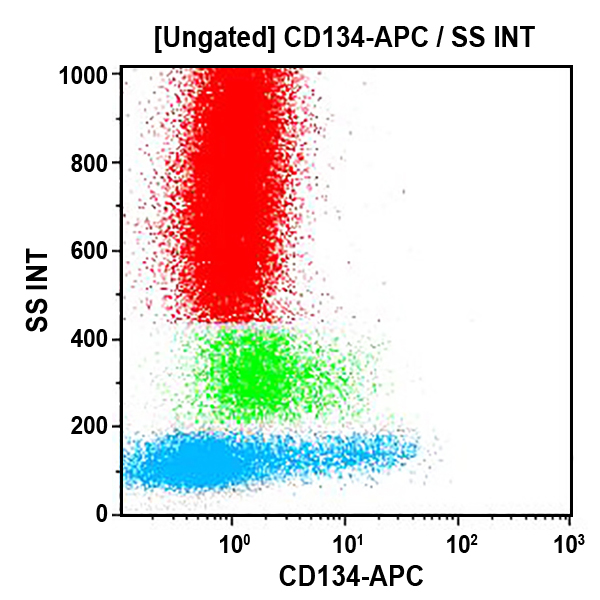
![Recombinant Anti-p53 antibody [E26] KO Tested (ab32389) | Abcam](https://www.abcam.com/ps/products/32/ab32389/Images/ab32389-389819-antip53antibodyimmunocytochemistryimmunofluorescencehap1human.jpg)


![APC Anti-Myc tag antibody [9E10] (ab223896) | Abcam](https://www.abcam.com/ps/products/223/ab223896/Images/ab223896-332330-anti-myc-tag-antibody-9e10-allophycocyanin-flow-cytometry.jpg)


![Alexa Fluor® 647 Anti-PDGFR alpha antibody [EPR22059-270] (ab270085) | Abcam](https://www.abcam.com/ps/products/270/ab270085/Images/ab270085-3-alexa-fluor-647-anti-pdgfr-alpha-antibody-epr22059-270-flow-cytometry-shsy5y-human.jpg)