Fourth Ventricle
Cerebral Ventricle Neoplasms
Cerebral Ventricles
Third Ventricle
Hydrocephalus
Cerebral Ventriculography
Dandy-Walker Syndrome
Cranial Fossa, Posterior
Ventriculoperitoneal Shunt
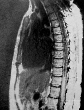
Syringomyelia
Heart Ventricles
Cisterna Magna
Choroid Plexus Neoplasms
Lateral Ventricles
Cerebral Aqueduct
Spina Bifida Cystica
Arnold-Chiari Malformation
Platybasia
Neuroendoscopy
Choroid Plexus
Ganglioglioma
Cerebellar Diseases
Ependymoma
Brain Stem
Cerebrospinal Fluid Shunts
Infratentorial Neoplasms
Cholesteatoma
Cerebellar Neoplasms
Magnetic Resonance Imaging
Cerebellum
Rhombencephalon
Arachnoid Cysts
Cerebrospinal Fluid
Subarachnoid Space
Meningioma
Neurocysticercosis
Tomography, X-Ray Computed
Endoscopic aqueductal plasty via the fourth ventricle through the cerebellar hemisphere under navigating system guidance--technical note. (1/72)
A 1-year 8-month-old boy presented with isolated fourth ventricle after ventriculoperitoneal shunting for hydrocephalus associated with ventricular and subarachnoid hemorrhage. The therapeutic endoscope was inserted through the thin left cerebellar hemisphere. Endoscopic aqueductal plasty was performed via the enlarged fourth ventricle under guidance from a navigating system. Endoscopic aqueductal plasty via the fourth ventricle under navigating system guidance is a useful procedure enabling less invasive surgery for isolated fourth ventricle associated with slit-like ventricle after shunt placement. (+info)Kainic acid lesions to the lateral tegmental field of medulla: effects on cough, expiration and aspiration reflexes in anesthetized cats. (2/72)
We have tested the hypothesis that neurons of both the ventral reticular nucleus and the adjacent parts of the lateral tegmental field (LTF) may be important for the production of motor programs associated with cough, expiration and aspiration reflexes. Our studies were conducted on non-decerebrate, spontaneously breathing cats under pentobarbitone anesthesia. Dysfunction of the medullary LTF region above the obex, produced by uni- or bilateral injections of kainic acid (a neurotoxin), regularly abolished the cough reflex evoked by mechanical stimulation of both the tracheobronchial and laryngeal regions and in most cases also the expiration reflex induced from the glottal area. However, some electrical activity still occurred in the neurogram of the recurrent laryngeal nerve during probing the laryngeal and glottal regions. Interestingly, the aspiration reflex elicited from the nasopharynx regularly persisted, although with lower intensity after the LTF lesion. Nevertheless, successive midcollicular decerebration performed in four cats also abolished the aspiration reflex. These experiments demonstrate the importance of medullary LTF neurons for the normal occurrence of cough and expiration reflexes. One possible explanation for the elimination of these expulsive processes is that the blockade of the LTF neurons may remove an important source of a facilitatory input to the brainstem circuitries that mediate cough and expiration reflexes. In addition, the potential importance of the mesencephalic reticular formation for the occurrence of the aspiration reflex and the role of the LTF in modulating both the eupnoeic breathing and the blood pressure are also discussed. (+info)Hypoglycemia activates orexin neurons and selectively increases hypothalamic orexin-B levels: responses inhibited by feeding and possibly mediated by the nucleus of the solitary tract. (3/72)
Orexins are novel appetite-stimulating peptides expressed in the lateral hypothalamic area (LHA), and their expression is stimulated by hypoglycemia in fasted rats. We investigated activation of orexin and other neurons during insulin-induced hypoglycemia using the immediate early gene product Fos. Insulin (50 U/kg) lowered plasma glucose by >50% after 5 h and stimulated feeding sixfold compared with saline-injected controls. Hypoglycemic rats allowed to feed and normoglycemic controls both showed sparse Fos-positive (Fos+) neurons in the LHA and the paraventricular nucleus (PVN) and arcuate nucleus (ARC) and showed none in the nucleus of the solitary tract (NTS), which relays visceral feeding signals to the LHA. In the LHA, total numbers of Fos+ neurons were comparable in fed hypoglycemic and control groups (60 +/- 6 vs. 52 +/- 4 cells/mm2, P > 0.05), as were Fos+ neurons immunoreactive for orexin (1.4 +/- 0.4 vs. 0.6 +/- 0.4 cells/mm2, P > 0.05). By contrast, hypoglycemic rats that were fasted showed significantly more Fos+ nuclei in the LHA (96 +/- 10 cells/mm2, P < 0.05, vs. both other groups) and Fos+ orexin neurons (8.4 +/- 3.3 cells/mm2, P < 0.001, vs. both other groups). They also showed two- to threefold more Fos+ nuclei (P < 0.001) in the PVN and ARC than both fed hypoglycemic rats and controls and showed strikingly abundant Fos+ neurons in the NTS and dorsal motor nucleus of the vagus. In parallel studies, whole hypothalamic orexin-A levels were not changed in hypoglycemic rats, whether fasted or freely fed, whereas orexin-B levels were 10-fold higher in hypoglycemic fasted rats than in control and hypoglycemic fed groups. These data support our hypothesis that orexin neurons are stimulated by falling glucose levels but are readily inhibited by signals related to nutrient ingestion and suggest that they may functionally link with neuronal activity in the NTS. Orexin-A and -B may play specific roles in behavioral or neuroendocrine responses to hypoglycemia. (+info)Midline posterior fossa teratoma--case report. (4/72)
A 20-day-old female neonate presented with an immature teratoma in the midline posterior fossa. The tumor was totally removed but the patient died of pneumonia. Teratoma is a rare tumor, but very difficult to treat as the patients tend to be young, and the outcome is very poor. (+info)Subdiaphragmatic vagotomy induces NADPH diaphorase in the rat dorsal motor nucleus of the vagus. (5/72)
Axotomy of the vagal motor neurons by cervical vagotomy induces NADPH diaphorase staining due to increased nitric oxide synthase expression in both the rat dorsal motor nucleus and nucleus ambiguous; furthermore, cerical vagotomy leads to cell death of the dorsal motor nucleus cells. Subdiaphragmatic vagotomy axotomizes the vagal motor cells further from the brainstem than cervical vagotomy, and cuts the fibers running only to the abdominal viscera. Here we report that subdiaphragmatic vagotomy is sufficient to induce NADPH diaphorase staining in the dorsal motor nucleus but does not induce staining in the nucleus ambiguus. Because the neurons of the dorsal motor nucleus do not undergo cell death after subdiaphragmatic vagotomy and are able to re-enervate the gut, the increased nitric oxide synthase expression after distal axotomy may be related more to regeneration than degeneration. (+info)Fourth ventricular meningioma in an adult--case report. (6/72)
A 72-year-old female presented with an intra-fourth ventricular meningioma manifesting as truncal ataxia. Computed tomography (CT) showed a slightly high-density, well-demarcated, and homogeneously enhanced mass located in the fourth ventricle and extending to the right lateral recess. T2-weighted magnetic resonance (MR) imaging revealed a peritumoral high-intensity band without dural tail sign. Bilateral vertebral angiography revealed faint tumor staining supplied from the choroidal branches of the posterior inferior cerebellar arteries. The mass was totally resected via a suboccipital approach. CT, T2-weighted MR imaging, and vertebral angiography are informative for the preoperative diagnosis of fourth ventricular meningioma. (+info)Intracranial meningeal involvement in Churg-Strauss syndrome. (7/72)
We describe the case of a 54-year-old woman with a clinical diagnosis of Churg-Strauss syndrome (CSS). The patient had a fever of unknown origin, severe headache, progressing left ophthalmoplegia, and visual acuity disturbance. MR imaging revealed diffuse and thick hypointense lesions on T2-weighted images in the frontal meninges and anterior falx cerebri with diffuse enhancement. Similar lesions were also detected in the left superior ophthalmic fissure to the cavernous sinus. Nodular lesions in the fourth ventricle, which might have been the cause of hydrocephalus, were hypointense on T2-weighted images. These MR imaging findings suggested remote granulomatous involvement in the meninges and choroid plexus associated with CSS. To our knowledge, remote meningeal involvement in association with CSS has not been previously reported. (+info)Intractable vomiting from glioblastoma metastatic to the fourth ventricle: three case studies. (8/72)
Dissemination of malignant glioma to the fourth ventricle with metastatic deposits and intractable vomiting is rare. Leptomeningeal extension of malignant glioma is an uncommon condition that has been reported in patients with end-stage disease and is usually unresponsive to any treatment modality. We describe 3 patients with progressing recurrent glioblastoma multiforme in whom leptomeningeal invasion manifested itself as intractable vomiting due to tumor metastases in the floor of the fourth ventricle. All patients received additional radiation therapy focused to the posterior fossa, with complete resolution of vomiting occurring within 10 days after irradiation. The remission of symptoms in these patients persisted until their death 3-4 months after the repeat radiation therapy. These reports indicate that additional focused radiation should be considered because of its significant therapeutic effect in alleviating intractable nausea and vomiting in patients with glioma metastasized to the posterior fossa. (+info)The fourth ventricle is a part of the cerebrospinal fluid-filled system in the brain, located in the posterior cranial fossa and continuous with the central canal of the medulla oblongata and the cerebral aqueduct. It is shaped like a cavity with a roof, floor, and lateral walls, and it communicates rostrally with the third ventricle through the cerebral aqueduct and caudally with the subarachnoid space through the median and lateral apertures (foramina of Luschka and Magendie). The fourth ventricle contains choroid plexus tissue, which produces cerebrospinal fluid. Its roof is formed by the cerebellar vermis and the superior medullary velum, while its floor is composed of the rhomboid fossa, which includes several important structures such as the vagal trigone, hypoglossal trigone, and striae medullares.
Cerebral ventricle neoplasms refer to tumors that develop within the cerebral ventricles, which are fluid-filled spaces in the brain. These tumors can arise from various types of cells within the ventricular system, including the ependymal cells that line the ventricles, choroid plexus cells that produce cerebrospinal fluid, or other surrounding tissues.
Cerebral ventricle neoplasms can cause a variety of symptoms depending on their size and location, such as headaches, nausea, vomiting, vision changes, imbalance, weakness, or difficulty with mental tasks. The treatment options for these tumors may include surgical resection, radiation therapy, and chemotherapy, depending on the type and extent of the tumor. Regular follow-up care is essential to monitor for recurrence and manage any long-term effects of treatment.
The cerebral ventricles are a system of interconnected fluid-filled cavities within the brain. They are located in the center of the brain and are filled with cerebrospinal fluid (CSF), which provides protection to the brain by cushioning it from impacts and helping to maintain its stability within the skull.
There are four ventricles in total: two lateral ventricles, one third ventricle, and one fourth ventricle. The lateral ventricles are located in each cerebral hemisphere, while the third ventricle is located between the thalami of the two hemispheres. The fourth ventricle is located at the base of the brain, above the spinal cord.
CSF flows from the lateral ventricles into the third ventricle through narrow passageways called the interventricular foramen. From there, it flows into the fourth ventricle through another narrow passageway called the cerebral aqueduct. CSF then leaves the fourth ventricle and enters the subarachnoid space surrounding the brain and spinal cord, where it can be absorbed into the bloodstream.
Abnormalities in the size or shape of the cerebral ventricles can indicate underlying neurological conditions, such as hydrocephalus (excessive accumulation of CSF) or atrophy (shrinkage) of brain tissue. Imaging techniques, such as computed tomography (CT) or magnetic resonance imaging (MRI), are often used to assess the size and shape of the cerebral ventricles in clinical settings.
The third ventricle is a narrow, fluid-filled cavity in the brain that is located between the thalamus and hypothalamus. It is one of the four ventricles in the ventricular system of the brain, which produces and circulates cerebrospinal fluid (CSF) around the brain and spinal cord.
The third ventricle is shaped like a slit and communicates with the lateral ventricles through the interventricular foramen (also known as the foramen of Monro), and with the fourth ventricle through the cerebral aqueduct (also known as the aqueduct of Sylvius).
The third ventricle contains choroid plexus tissue, which produces CSF. The fluid flows from the lateral ventricles into the third ventricle, then through the cerebral aqueduct and into the fourth ventricle, where it can circulate around the brainstem and spinal cord before being absorbed back into the bloodstream.
Abnormalities in the third ventricle, such as enlargement or obstruction of the cerebral aqueduct, can lead to hydrocephalus, a condition characterized by an accumulation of CSF in the brain.
Hydrocephalus is a medical condition characterized by an abnormal accumulation of cerebrospinal fluid (CSF) within the brain, leading to an increase in intracranial pressure and potentially causing damage to the brain tissues. This excessive buildup of CSF can result from either overproduction or impaired absorption of the fluid, which typically causes the ventricles (fluid-filled spaces) inside the brain to expand and put pressure on surrounding brain structures.
The condition can be congenital, present at birth due to genetic factors or abnormalities during fetal development, or acquired later in life as a result of injuries, infections, tumors, or other disorders affecting the brain's ability to regulate CSF flow and absorption. Symptoms may vary depending on age, severity, and duration but often include headaches, vomiting, balance problems, vision issues, cognitive impairment, and changes in behavior or personality.
Treatment for hydrocephalus typically involves surgically implanting a shunt system that diverts the excess CSF from the brain to another part of the body where it can be absorbed, such as the abdominal cavity. In some cases, endoscopic third ventriculostomy (ETV) might be an alternative treatment option, creating a new pathway for CSF flow within the brain. Regular follow-ups with neurosurgeons and other healthcare professionals are essential to monitor the condition and make any necessary adjustments to the treatment plan.
Cerebral ventriculography is a medical imaging technique that involves the injection of a contrast material into the cerebral ventricles, which are fluid-filled spaces within the brain. The purpose of this procedure is to produce detailed images of the ventricular system and the surrounding structures in order to diagnose and evaluate various neurological conditions, such as hydrocephalus (excessive accumulation of cerebrospinal fluid in the ventricles), tumors, or other abnormalities that may be causing obstruction or compression of the ventricular system.
The procedure typically involves inserting a thin, flexible tube called a catheter into the lateral ventricle of the brain through a small hole drilled in the skull. The contrast material is then injected through the catheter and X-ray images are taken as the contrast material flows through the ventricular system. These images can help to identify any abnormalities or blockages that may be present.
Cerebral ventriculography has largely been replaced by non-invasive imaging techniques, such as computed tomography (CT) and magnetic resonance imaging (MRI), which provide similar information without the need for invasive procedures. However, cerebral ventriculography may still be used in certain cases where these other methods are not sufficient to make a definitive diagnosis.
Dandy-Walker Syndrome is a congenital brain malformation characterized by the absence or underdevelopment of the cerebellar vermis (the part of the brain that helps coordinate movement) and an enlarged fluid-filled space (fourth ventricle) surrounding it. This condition can also be associated with an upward bulging of the back of the skull (occipital bone), and in some cases, hydrocephalus (excessive accumulation of cerebrospinal fluid in the brain). The syndrome can vary in severity, and symptoms may include problems with balance, coordination, developmental delays, and increased intracranial pressure. It is usually diagnosed through imaging tests such as ultrasound, CT scan, or MRI. Treatment typically involves managing symptoms and addressing complications, which may include surgical procedures to relieve hydrocephalus if present.
The posterior cranial fossa is a term used in anatomy to refer to the portion of the skull that forms the lower, back part of the cranial cavity. It is located between the occipital bone and the temporal bones, and it contains several important structures including the cerebellum, pons, medulla oblongata, and the lower cranial nerves (IX-XII). The posterior fossa also contains the foramen magnum, which is a large opening through which the spinal cord connects to the brainstem. This region of the skull is protected by the occipital bone, which forms the base of the skull and provides attachment for several neck muscles.
A Ventriculoperitoneal (VP) shunt is a surgical procedure that involves the insertion of a long, flexible tube (shunt) into the cerebral ventricles of the brain to drain excess cerebrospinal fluid (CSF). The other end of the shunt is directed into the peritoneal cavity, where the CSF can be absorbed.
The VP shunt is typically used to treat hydrocephalus, a condition characterized by an abnormal accumulation of CSF within the ventricles of the brain, which can cause increased intracranial pressure and damage to the brain. By diverting the excess CSF from the ventricles into the peritoneal cavity, the VP shunt helps to relieve the symptoms of hydrocephalus and prevent further neurological damage.
The shunt system consists of several components, including a ventricular catheter that is placed in the ventricle, a one-way valve that regulates the flow of CSF, and a distal catheter that is directed into the peritoneal cavity. The valve helps to prevent backflow of CSF into the brain and ensures that the fluid flows in only one direction, from the ventricles to the peritoneal cavity.
VP shunts are generally safe and effective, but they can be associated with complications such as infection, obstruction, or malfunction of the shunt system. Regular follow-up with a healthcare provider is necessary to monitor the function of the shunt and ensure that any potential issues are addressed promptly.
Syringomyelia is a medical condition characterized by the formation of a fluid-filled cavity or cavities (syrinx) within the spinal cord. This syrinx can lead to various symptoms depending on its size and location, which may include pain, muscle weakness, numbness, and stiffness in the neck, back, shoulders, arms, or legs. In some cases, it may also affect bladder and bowel function, sexual performance, and the ability to maintain normal body temperature. Syringomyelia is often associated with Chiari malformation, a condition where the lower part of the brain extends into the spinal canal. However, other conditions such as spinal cord injuries, tumors, or infections may also cause syringomyelia.
The heart ventricles are the two lower chambers of the heart that receive blood from the atria and pump it to the lungs or the rest of the body. The right ventricle pumps deoxygenated blood to the lungs, while the left ventricle pumps oxygenated blood to the rest of the body. Both ventricles have thick, muscular walls to generate the pressure necessary to pump blood through the circulatory system.
The term "cisterna magna" is derived from Latin, where "cisterna" means "reservoir" or "receptacle," and "magna" means "large." In medical anatomy, the cisterna magna refers to a large, sac-like space located near the lower part of the brainstem. It is a subarachnoid cistern, which means it is a space that contains cerebrospinal fluid (CSF) between the arachnoid and pia mater membranes covering the brain and spinal cord.
More specifically, the cisterna magna is situated between the cerebellum (the lower part of the brain responsible for coordinating muscle movements and maintaining balance) and the occipital bone (the bone at the back of the skull). This space contains a significant amount of CSF, which serves as a protective cushion for the brain and spinal cord, helps regulate intracranial pressure, and facilitates the circulation of nutrients and waste products.
The cisterna magna is an essential structure in neurosurgical procedures and diagnostic imaging techniques like lumbar puncture (spinal tap) or myelograms, where contrast agents are introduced into the CSF to visualize the spinal cord and surrounding structures. Additionally, it serves as a crucial landmark for various surgical approaches to the posterior fossa (the lower part of the skull that houses the cerebellum and brainstem).
Choroid plexus neoplasms are rare types of brain tumors that arise from the choroid plexus, which are clusters of blood vessels in the ventricles (fluid-filled spaces) of the brain. These tumors can be benign (choroid plexus papilloma) or malignant (choroid plexus carcinoma). Choroid plexus neoplasms most commonly occur in children under the age of 2, but they can also affect adults. Symptoms may include increased head circumference, hydrocephalus (fluid buildup in the brain), vomiting, and developmental delays. Treatment typically involves surgical removal of the tumor, followed by radiation therapy or chemotherapy for malignant tumors.
The lateral ventricles are a pair of fluid-filled cavities located within the brain. They are part of the ventricular system, which is a series of interconnected spaces filled with cerebrospinal fluid (CSF). The lateral ventricles are situated in the left and right hemispheres of the brain and are among the largest of the ventricles.
Each lateral ventricle has a complex structure and can be divided into several parts:
1. Anterior horn: This is the front part of the lateral ventricle, located in the frontal lobe of the brain.
2. Body: The central part of the lateral ventricle, which is continuous with the anterior horn and posterior horn.
3. Posterior horn: The back part of the lateral ventricle, located in the occipital lobe of the brain.
4. Temporal horn: An extension that projects into the temporal lobe of the brain.
The lateral ventricles are lined with ependymal cells, which produce cerebrospinal fluid. CSF circulates through the ventricular system, providing buoyancy and protection to the brain, and is eventually absorbed into the bloodstream. Abnormalities in the size or shape of the lateral ventricles can be associated with various neurological conditions, such as hydrocephalus, brain tumors, or neurodegenerative diseases.
The cerebral aqueduct, also known as the aqueduct of Sylvius, is a narrow canal that connects the third and fourth ventricles (cavities) of the brain. It allows for the flow of cerebrospinal fluid (CSF) from the third ventricle to the fourth ventricle. The cerebral aqueduct is a critical component of the ventricular system of the brain, and any obstruction or abnormality in this region can result in an accumulation of CSF and increased pressure within the brain, which can lead to serious neurological symptoms and conditions such as hydrocephalus.
Spina Bifida Cystica is a type of neural tube defect that occurs when the bones of the spine (vertebrae) do not form properly around the developing spinal cord, resulting in a sac-like protrusion of the spinal cord and its surrounding membranes through an opening in the spine. This sac, called a meningocele or myelomeningocele, can be covered with skin or exposed, and it may contain cerebrospinal fluid, nerve roots, or portions of the spinal cord.
Myelomeningocele is the most severe form of Spina Bifida Cystica, where the sac contains a portion of the spinal cord and nerves. This can lead to various neurological complications such as weakness or paralysis below the level of the spine affected, loss of sensation, bladder and bowel dysfunction, and hydrocephalus (accumulation of cerebrospinal fluid in the brain). Early diagnosis and intervention, including prenatal surgery, can help improve outcomes for individuals with Spina Bifida Cystica.
Arnold-Chiari malformation is a structural abnormality of the brain and skull base, specifically the cerebellum and brainstem. It is characterized by the descent of the cerebellar tonsils and sometimes parts of the brainstem through the foramen magnum (the opening at the base of the skull) into the upper spinal canal. This can cause pressure on the brainstem and cerebellum, potentially leading to a range of symptoms such as headaches, neck pain, unsteady gait, swallowing difficulties, hearing or balance problems, and in severe cases, neurological deficits. There are four types of Arnold-Chiari malformations, with type I being the most common and least severe form. Types II, III, and IV are progressively more severe and involve varying degrees of hindbrain herniation and associated neural tissue damage. Surgical intervention is often required to alleviate symptoms and prevent further neurological deterioration.
Intraventricular injections are a type of medical procedure where medication is administered directly into the cerebral ventricles of the brain. The cerebral ventricles are fluid-filled spaces within the brain that contain cerebrospinal fluid (CSF). This procedure is typically used to deliver drugs that target conditions affecting the central nervous system, such as infections or tumors.
Intraventricular injections are usually performed using a thin, hollow needle that is inserted through a small hole drilled into the skull. The medication is then injected directly into the ventricles, allowing it to circulate throughout the CSF and reach the brain tissue more efficiently than other routes of administration.
This type of injection is typically reserved for situations where other methods of drug delivery are not effective or feasible. It carries a higher risk of complications, such as bleeding, infection, or damage to surrounding tissues, compared to other routes of administration. Therefore, it is usually performed by trained medical professionals in a controlled clinical setting.
Platybasia is a medical term that refers to a condition where the base of the skull is flattened or broadened, resulting in an abnormal increase in the angle between the clivus (a part of the sphenoid bone) and the posterior aspect of the upper surface of the palatine bone. This condition can be congenital or acquired and is often associated with other skeletal abnormalities. In some cases, platybasia may lead to neurological symptoms such as headaches, neck pain, or even brainstem compression.
Neuroendoscopy is a minimally invasive surgical technique that involves the use of an endoscope to access and treat various conditions within the brain and spinal column. An endoscope is a long, flexible tube with a light and camera at its tip, which allows surgeons to view and operate on internal structures through small incisions or natural openings in the body.
In neuroendoscopy, the surgeon uses the endoscope to navigate through the brain's ventricular system (fluid-filled spaces) or other narrow spaces within the skull or spine to diagnose and treat conditions such as hydrocephalus, brain tumors, arachnoid cysts, and intraventricular hemorrhage.
The benefits of neuroendoscopy include reduced trauma to surrounding tissues, shorter hospital stays, faster recovery times, and improved outcomes compared to traditional open surgical approaches. However, neuroendoscopic procedures require specialized training and expertise due to the complexity of the anatomy involved.
The choroid plexus is a network of blood vessels and tissue located within each ventricle (fluid-filled space) of the brain. It plays a crucial role in the production of cerebrospinal fluid (CSF), which provides protection and nourishment to the brain and spinal cord.
The choroid plexus consists of modified ependymal cells, called plexus epithelial cells, that line the ventricular walls. These cells have finger-like projections called villi, which increase their surface area for efficient CSF production. The blood vessels within the choroid plexus transport nutrients, ions, and water to these epithelial cells, where they are actively secreted into the ventricles to form CSF.
In addition to its role in CSF production, the choroid plexus also acts as a barrier between the blood and the central nervous system (CNS), regulating the exchange of substances between them. This barrier function is primarily attributed to tight junctions present between the epithelial cells, which limit the paracellular movement of molecules.
Abnormalities in the choroid plexus can lead to various neurological conditions, such as hydrocephalus (excessive accumulation of CSF) or certain types of brain tumors.
Ganglioglioma is a rare, typically slow-growing tumor that occurs in the brain or spinal cord. It is composed of both neuronal (ganglion cell) and glial elements. These tumors most commonly occur in the temporal lobe of the brain and are usually found in children and young adults.
Gangliogliomas can be benign or malignant, with the majority being low-grade (benign). Symptoms vary depending on the location of the tumor but may include seizures, headaches, changes in behavior or cognition, and motor weakness or paralysis. Treatment typically involves surgical removal of the tumor, and in some cases, radiation therapy or chemotherapy may be recommended.
It's important to note that while I strive to provide accurate information, my responses should not be used as a substitute for professional medical advice, diagnosis, or treatment. Always consult with a qualified healthcare provider for any medical concerns.
Cerebellar diseases refer to a group of medical conditions that affect the cerebellum, which is the part of the brain located at the back of the head, below the occipital lobe and above the brainstem. The cerebellum plays a crucial role in motor control, coordination, balance, and some cognitive functions.
Cerebellar diseases can be caused by various factors, including genetics, infections, tumors, stroke, trauma, or degenerative processes. These conditions can result in a wide range of symptoms, such as:
1. Ataxia: Loss of coordination and unsteady gait
2. Dysmetria: Inability to judge distance and force while performing movements
3. Intention tremors: Shaking or trembling that worsens during purposeful movements
4. Nystagmus: Rapid, involuntary eye movement
5. Dysarthria: Speech difficulty due to muscle weakness or incoordination
6. Hypotonia: Decreased muscle tone
7. Titubation: Rhythmic, involuntary oscillations of the head and neck
8. Cognitive impairment: Problems with memory, attention, and executive functions
Some examples of cerebellar diseases include:
1. Ataxia-telangiectasia
2. Friedrich's ataxia
3. Multiple system atrophy (MSA)
4. Spinocerebellar ataxias (SCAs)
5. Cerebellar tumors, such as medulloblastomas or astrocytomas
6. Infarctions or hemorrhages in the cerebellum due to stroke or trauma
7. Infections, such as viral encephalitis or bacterial meningitis
8. Autoimmune disorders, like multiple sclerosis (MS) or paraneoplastic syndromes
9. Metabolic disorders, such as Wilson's disease or phenylketonuria (PKU)
10. Chronic alcoholism and withdrawal
Treatment for cerebellar diseases depends on the underlying cause and may involve medications, physical therapy, surgery, or supportive care to manage symptoms and improve quality of life.
Ependymoma is a type of brain or spinal cord tumor that develops from the ependymal cells that line the ventricles (fluid-filled spaces) in the brain, or the central canal of the spinal cord. These tumors can be benign or malignant, and they can cause various symptoms depending on their location and size.
Ependymomas are relatively rare, accounting for about 2-3% of all primary brain and central nervous system tumors. They most commonly occur in children and young adults, but they can also affect older individuals. Treatment typically involves surgical removal of the tumor, followed by radiation therapy or chemotherapy, depending on the grade and location of the tumor. The prognosis for ependymomas varies widely, with some patients experiencing long-term survival and others having more aggressive tumors that are difficult to treat.
The brainstem is the lower part of the brain that connects to the spinal cord. It consists of the midbrain, pons, and medulla oblongata. The brainstem controls many vital functions such as heart rate, breathing, and blood pressure. It also serves as a relay center for sensory and motor information between the cerebral cortex and the rest of the body. Additionally, several cranial nerves originate from the brainstem, including those that control eye movements, facial movements, and hearing.
Cerebrospinal fluid (CSF) shunts are medical devices used to divert the flow of excess CSF from the brain and spinal cord to another part of the body, usually the abdominal cavity. The shunt consists of a catheter, a valve, and a reservoir.
The catheter is inserted into one of the ventricles in the brain or the subarachnoid space surrounding the spinal cord to drain the excess CSF. The valve regulates the flow of CSF to prevent over-drainage, which can cause complications such as low CSF pressure and brain sagging. The reservoir is a small chamber that allows for easy access to the shunt system for monitoring and adjusting the pressure settings.
CSF shunts are typically used to treat conditions associated with increased production or impaired absorption of CSF, such as hydrocephalus, communicating hydrocephalus, normal pressure hydrocephalus, and pseudotumor cerebri. By reducing the buildup of CSF in the brain, shunts can help alleviate symptoms such as headaches, nausea, vomiting, vision problems, and cognitive impairment.
It is important to note that while CSF shunts are effective in managing these conditions, they also carry risks of complications such as infection, obstruction, malfunction, and over-drainage. Regular monitoring and follow-up care are necessary to ensure proper functioning and minimize the risk of complications.
Infratentorial neoplasms refer to tumors that originate in the region of the brain called the posterior fossa, which is located below the tentorium cerebelli (a membranous structure that separates the cerebrum from the cerebellum). This area contains several important structures such as the cerebellum, pons, medulla oblongata, and fourth ventricle. Infratentorial neoplasms can be benign or malignant and can arise from various cell types including nerve cells, glial cells, or supportive tissues. They can cause a variety of symptoms depending on their location and size, such as headache, vomiting, unsteady gait, weakness, numbness, vision changes, hearing loss, and difficulty swallowing or speaking. Treatment options may include surgery, radiation therapy, and chemotherapy.
Cholesteatoma is a type of skin growth that occurs in the middle ear behind the eardrum. It is not a cancerous or precancerous growth but can still cause significant damage to the surrounding structures if left untreated. Cholesteatomas typically begin as small collections of dead skin cells, which then accumulate and expand over time, forming a sac-like structure that can erode the bones of the middle ear and lead to hearing loss, balance problems, and even facial paralysis in severe cases.
Cholesteatomas can be congenital (present at birth) or acquired (develop later in life). Acquired cholesteatomas are more common and typically result from repeated ear infections or trauma to the eardrum that causes a pocket or retraction of the eardrum to form, which then traps skin cells and debris. Over time, these cells can multiply and become a cholesteatoma.
Treatment for cholesteatoma typically involves surgical removal of the growth, as well as any damaged bone or tissue. In some cases, additional procedures may be necessary to restore hearing function. Regular follow-up care is also important to monitor for recurrence and ensure proper healing.
Cerebellar neoplasms refer to abnormal growths or tumors that develop in the cerebellum, which is the part of the brain responsible for coordinating muscle movements and maintaining balance. These tumors can be benign (non-cancerous) or malignant (cancerous), and they can arise from various types of cells within the cerebellum.
The most common type of cerebellar neoplasm is a medulloblastoma, which arises from primitive nerve cells in the cerebellum. Other types of cerebellar neoplasms include astrocytomas, ependymomas, and brain stem gliomas. Symptoms of cerebellar neoplasms may include headaches, vomiting, unsteady gait, coordination problems, and visual disturbances. Treatment options depend on the type, size, and location of the tumor, as well as the patient's overall health and age. Treatment may involve surgery, radiation therapy, chemotherapy, or a combination of these approaches.
Medical Definition:
Magnetic Resonance Imaging (MRI) is a non-invasive diagnostic imaging technique that uses a strong magnetic field and radio waves to create detailed cross-sectional or three-dimensional images of the internal structures of the body. The patient lies within a large, cylindrical magnet, and the scanner detects changes in the direction of the magnetic field caused by protons in the body. These changes are then converted into detailed images that help medical professionals to diagnose and monitor various medical conditions, such as tumors, injuries, or diseases affecting the brain, spinal cord, heart, blood vessels, joints, and other internal organs. MRI does not use radiation like computed tomography (CT) scans.
The cerebellum is a part of the brain that lies behind the brainstem and is involved in the regulation of motor movements, balance, and coordination. It contains two hemispheres and a central portion called the vermis. The cerebellum receives input from sensory systems and other areas of the brain and spinal cord and sends output to motor areas of the brain. Damage to the cerebellum can result in problems with movement, balance, and coordination.
The rhombencephalon is a term used in the field of neuroanatomy, which refers to the most posterior region of the developing brain during embryonic development. It is also known as the hindbrain and it gives rise to several important structures in the adult brain.
More specifically, the rhombencephalon can be further divided into two main parts: the metencephalon and the myelencephalon. The metencephalon eventually develops into the pons and cerebellum, while the myelencephalon becomes the medulla oblongata.
The rhombencephalon plays a crucial role in several critical functions of the nervous system, including regulating heart rate and respiration, maintaining balance and posture, and coordinating motor movements. Defects or abnormalities in the development of the rhombencephalon can lead to various neurological disorders, such as cerebellar hypoplasia, Chiari malformation, and certain forms of brainstem tumors.
An Arachnoid cyst is a type of abnormal fluid-filled sac that develops between the brain or spinal cord and the arachnoid membrane, which is one of the three layers that cover and protect the central nervous system. These cysts are filled with cerebrospinal fluid (CSF), which is the same fluid that surrounds and cushions the brain and spinal cord.
Arachnoid cysts can vary in size and may be present at birth or develop later in life due to trauma, infection, or other factors. While many arachnoid cysts are asymptomatic and do not cause any problems, larger cysts or those that grow or shift over time can put pressure on the brain or spinal cord, leading to a range of neurological symptoms such as headaches, seizures, hearing or vision changes, balance or coordination difficulties, and cognitive impairments.
Treatment for arachnoid cysts depends on their size, location, and associated symptoms. In some cases, observation and monitoring may be sufficient, while in others, surgical intervention may be necessary to drain the cyst or create a connection between it and the surrounding CSF space to relieve pressure.
Cerebrospinal fluid (CSF) is a clear, colorless fluid that surrounds and protects the brain and spinal cord. It acts as a shock absorber for the central nervous system and provides nutrients to the brain while removing waste products. CSF is produced by specialized cells called ependymal cells in the choroid plexus of the ventricles (fluid-filled spaces) inside the brain. From there, it circulates through the ventricular system and around the outside of the brain and spinal cord before being absorbed back into the bloodstream. CSF analysis is an important diagnostic tool for various neurological conditions, including infections, inflammation, and cancer.
The subarachnoid space is the area between the arachnoid mater and pia mater, which are two of the three membranes covering the brain and spinal cord (the third one being the dura mater). This space is filled with cerebrospinal fluid (CSF), which provides protection and cushioning to the central nervous system. The subarachnoid space also contains blood vessels that supply the brain and spinal cord with oxygen and nutrients. It's important to note that subarachnoid hemorrhage, a type of stroke, can occur when there is bleeding into this space.
A meningioma is a type of slow-growing tumor that forms on the membranes (meninges) surrounding the brain and spinal cord. It's usually benign, meaning it doesn't spread to other parts of the body, but it can still cause serious problems if it grows and presses on nearby tissues.
Meningiomas most commonly occur in adults, and are more common in women than men. They can cause various symptoms depending on their location and size, including headaches, seizures, vision or hearing problems, memory loss, and changes in personality or behavior. In some cases, they may not cause any symptoms at all and are discovered only during imaging tests for other conditions.
Treatment options for meningiomas include monitoring with regular imaging scans, surgery to remove the tumor, and radiation therapy to shrink or kill the tumor cells. The best treatment approach depends on factors such as the size and location of the tumor, the patient's age and overall health, and their personal preferences.
Neurocysticercosis is a neurological disorder caused by the infection of the brain's tissue with larval stages of the parasitic tapeworm, Taenia solium. The larvae, called cysticerci, can invade various parts of the body including the brain and the central nervous system, leading to a range of symptoms such as seizures, headaches, cognitive impairment, and psychiatric disorders.
The infection typically occurs when a person ingests tapeworm eggs through contaminated food or water, and the larvae hatch and migrate to various tissues in the body. In neurocysticercosis, the cysticerci can cause inflammation, swelling, and damage to brain tissue, leading to neurological symptoms that can vary depending on the location and number of cysts in the brain.
Diagnosis of neurocysticercosis typically involves a combination of imaging techniques such as MRI or CT scans, blood tests, and sometimes lumbar puncture (spinal tap) to examine cerebrospinal fluid. Treatment may involve anti-parasitic medications to eliminate the cysts, anti-inflammatory drugs to manage swelling and inflammation, and symptomatic treatment for seizures or other neurological symptoms.
X-ray computed tomography (CT or CAT scan) is a medical imaging method that uses computer-processed combinations of many X-ray images taken from different angles to produce cross-sectional (tomographic) images (virtual "slices") of the body. These cross-sectional images can then be used to display detailed internal views of organs, bones, and soft tissues in the body.
The term "computed tomography" is used instead of "CT scan" or "CAT scan" because the machines take a series of X-ray measurements from different angles around the body and then use a computer to process these data to create detailed images of internal structures within the body.
CT scanning is a noninvasive, painless medical test that helps physicians diagnose and treat medical conditions. CT imaging provides detailed information about many types of tissue including lung, bone, soft tissue and blood vessels. CT examinations can be performed on every part of the body for a variety of reasons including diagnosis, surgical planning, and monitoring of therapeutic responses.
In computed tomography (CT), an X-ray source and detector rotate around the patient, measuring the X-ray attenuation at many different angles. A computer uses this data to construct a cross-sectional image by the process of reconstruction. This technique is called "tomography". The term "computed" refers to the use of a computer to reconstruct the images.
CT has become an important tool in medical imaging and diagnosis, allowing radiologists and other physicians to view detailed internal images of the body. It can help identify many different medical conditions including cancer, heart disease, lung nodules, liver tumors, and internal injuries from trauma. CT is also commonly used for guiding biopsies and other minimally invasive procedures.
In summary, X-ray computed tomography (CT or CAT scan) is a medical imaging technique that uses computer-processed combinations of many X-ray images taken from different angles to produce cross-sectional images of the body. It provides detailed internal views of organs, bones, and soft tissues in the body, allowing physicians to diagnose and treat medical conditions.
Pathologic dilatation refers to an abnormal and excessive widening or enlargement of a body cavity or organ, which can result from various medical conditions. This abnormal dilation can occur in different parts of the body, including the blood vessels, digestive tract, airways, or heart chambers.
In the context of the cardiovascular system, pathologic dilatation may indicate a weakening or thinning of the heart muscle, leading to an enlarged chamber that can no longer pump blood efficiently. This condition is often associated with various heart diseases, such as cardiomyopathy, valvular heart disease, or long-standing high blood pressure.
In the gastrointestinal tract, pathologic dilatation may occur due to mechanical obstruction, neuromuscular disorders, or inflammatory conditions that affect the normal motility of the intestines. Examples include megacolon in Hirschsprung's disease, toxic megacolon in ulcerative colitis, or volvulus (twisting) of the bowel.
Pathologic dilatation can lead to various complications, such as reduced organ function, impaired circulation, and increased risk of infection or perforation. Treatment depends on the underlying cause and may involve medications, surgery, or other interventions to address the root problem and prevent further enlargement.
 Fourth ventricle
Roof of fourth ventricle
Taenia of fourth ventricle
Medullary striae of fourth ventricle
Medial eminence of floor of fourth ventricle
Lateral ventricles
Middle cerebellar peduncle
Inferior colliculus
Posterior median sulcus of medulla oblongata
Dorsal column nuclei
Obex
Lateral aperture
Cerebellar hypoplasia
Superior medullary velum
Median aperture
Dandy-Walker malformation
CV4 technique
Nucleus incertus
Choroid plexus papilloma
Cerebrospinal fluid
Vestibular nuclei
Ependymoma
Inferior medullary velum
Posterior inferior cerebellar artery
Cerebellopontine angle
Brainstem
Funiculus
Cerebellum
External ventricular drain
Choroid plexus
Fourth ventricle - Wikipedia
 Imaging characteristics of 4th ventricle subependymoma
 fourth ventricle
 Syringomyelia: Background, Pathophysiology, Etiology
 Tuberculoma in the Fourth Ventricle: An Unusual Location<...
 Isolated fourth ventricle: To shunt or stent<...
 How might a Tumour in the 4th ventricle be removed? - Mattstillwell.net
 BRAIN & NEURO CENTER - Martindale Center
 Bassett Collection - Lane Medical Library - Stanford University School of Medicine
Local Opioid Withdrawal in Rat Single Periaqueductal Gray NeuronsIn Vitro | Journal of Neuroscience
RLN3/RXFP3 Signaling in the PVN Inhibits Magnocellular Neurons via M-like Current Activation and Contributes to Binge Eating...
Figure 1 - Cerebellar Cysticercosis Caused by Larval Taenia crassiceps Tapeworm in Immunocompetent Woman, Germany - Volume 19,...
 Endomorphin-2 Inhibits the Activity of the Spinoparabrachial Projection Neuron through Presynaptic Mechanisms in the Spinal...
 SP 7B-RG: Organization of the Nervous System Flashcards
 Brainstem Anatomy Quiz Questions And Answers - ProProfs Quiz
 BRAINMAPS.ORG - BRAIN ATLAS, BRAIN MAPS, BRAIN STRUCTURE,
NEUROINFORMATICS, BRAIN, STEREOTAXIC ATLAS, NEUROSCIENCE
BRAINMAPS.ORG - BRAIN ATLAS, BRAIN MAPS, BRAIN STRUCTURE,
NEUROINFORMATICS, BRAIN, STEREOTAXIC ATLAS, NEUROSCIENCE
 Pigmented ependymoma, a tumor with predilection for the middle-aged adult: case report with methylation classification and...
![Atlas of Human Embryos [by: RF Gasser, PhD.] - Ch.8](data:image/png;base64,iVBORw0KGgoAAAANSUhEUgAAABAAAAAQCAYAAAAf8/9hAAAAh0lEQVQ4jd2Suw2EMBBEHxZV+C7dPkDUQXWuA9l9OAXaWCIjn7GOwAESLx3NaPYDT9OpqrYEmNYGPUCMO+MwX8R1WwD4fqaL5oNDxGJysw/uNNVYtwUfHADjMBPjjsnNIva2soj9CemTkILyBrXqJWdArcG/HSRMOdMd5c46VdWWKzz/SC/gACJUSba7VMpqAAAAAElFTkSuQmCC) Atlas of Human Embryos [by: RF Gasser, PhD.] - Ch.8
Atlas of Human Embryos [by: RF Gasser, PhD.] - Ch.8
 Search Anatomy Item Results
 ExpertDDx: Brain and Spine, 3rd Edition - 9780443106941
The Virtual Human Embryo
Bassett Collection - Lane Medical Library - Stanford University School of Medicine
 PDF) The Mandelbrot Set as a Quasi-Black Hole
US20120157377A1 - Methods to enhance night vision and treatment of night blindness - Google Patents
 Thieme E-Journals - Indian Journal of Neurosurgery / Abstract
Neonatal Meningitis: Background, Pathophysiology, Etiology
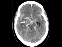
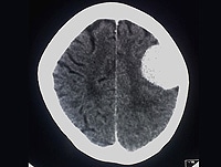
 Obstructive hydrocephalus | Radiology Reference Article | Radiopaedia.org
 2019 NPB-13 | College of American Pathologists
 Surgical Neurology International
 Emerging role of the brain in the homeostatic regulation of energy and glucose metabolism | Experimental & Molecular Medicine
Cerebellum7
- the fastigial nucleus lies immediately above the roof of the fourth ventricle, in the cerebellum. (wikipedia.org)
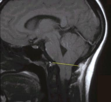
- The fourth ventricle is a broad tent-shaped cerebrospinal fluid (CSF) cavity located behind the brain stem and in front of the cerebellum in the center of the posterior fossa (Fig. 31-1). (mattstillwell.net)
- The fourth ventricle is a diamond-shaped cavity located dorsal to the pons and upper medulla oblongata and anterior to the cerebellum (Fig. 1.13). (mattstillwell.net)
- Dandy-Walker malformation (DWM) is a rare congenital malformation that involves the cerebellum and fourth ventricle. (medscape.com)
- True retrocerebellar arachnoid cysts displace the fourth ventricle and cerebellum anteriorly and show significant mass effect. (medscape.com)
- In affected individuals, a fluid-filled cavity between the brainstem and the cerebellum (the fourth ventricle) and the part of the skull that contains the cerebellum and the brainstem (the posterior fossa) are abnormally large. (medlineplus.gov)
- Magnetic resonance imaging in autism: measurement of the cerebellum, pons, and fourth ventricle. (bvsalud.org)
Hydrocephalus5
- If the flow of fluid is blocked ventricles may become enlarged and cause hydrocephalus. (wikipedia.org)
- Posterior fossa (infratentorial) ependymomas are more common in children than adults and often cause hydrocephalus secondary to obstruction of or around the fourth ventricle. (cap.org)
- In addition to locally aggressive behavior with fourth ventricular obstruction and hydrocephalus, medulloblastomas can metastasize via cerebrospinal fluid (CSF) pathways. (medscape.com)
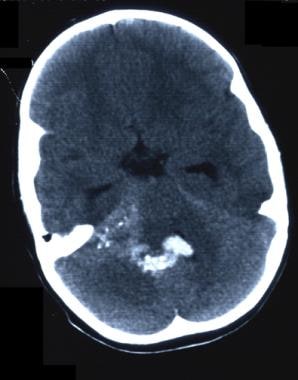
- The case with the lesion located under the tentorium cerebelli exhibited compression of the fourth ventricle with lateral ventricle dilatation hydrocephalus. (spandidos-publications.com)
- Magnetic resonance imaging (MRI) of the brain obtained at the outside hospital reported a large posterior fossa tumor within the fourth ventricle as well as obstructive hydrocephalus. (medscape.com)
Posterior fossa5
- and a cystic dilatation of the fourth ventricle that nearly fills the entire posterior fossa. (medscape.com)
- Dandy-Walker variant (see the image below) consists of vermian hypoplasia and cystic dilatation of the fourth ventricle, without enlargement of the posterior fossa. (medscape.com)
- The fourth ventricle is slightly enlarged, but the posterior fossa typically is normal in size. (medscape.com)
- Mega cisterna magna (see the image below) consists of an enlarged posterior fossa secondary to an enlarged cisterna magna, with a normal cerebellar vermis and fourth ventricle. (medscape.com)
- The sagittal T1-weighted MRI shows a large posterior fossa cyst that is compressing the cerebellar hemispheres, vermis, fourth ventricle (arrow), and brainstem. (medscape.com)
Cerebrospinal6
- The fourth ventricle extends from the cerebral aqueduct (aqueduct of Sylvius) to the obex, and is filled with cerebrospinal fluid (CSF). (wikipedia.org)
- Choroid plexuses appear in the ventricles which produce cerebrospinal fluid. (wikipedia.org)
- Sixteen patients underwent a simultaneous cerebrospinal fluid diversion procedure and fourth ventricular decompression. (utmb.edu)
- This sagittal T1-weighted MRI shows a large retrocerebellar cerebrospinal fluid collection and a normal fourth ventricle and vermis in a patient with mega cisterna magna in Dandy-Walker malformation. (medscape.com)
- It is one of the so-called circumventricular organs that serve as an interface between the brain parenchyma and the cerebrospinal fluid (CSF)-containing ventricles. (nih.gov)
- The ventricles of the brain are a communicating network of cavities filled with cerebrospinal fluid (CSF) and located within the brain parenchyma. (medscape.com)
Tumor6
- What are the implications of a brain tumor in the fourth ventricle? (mattstillwell.net)
- What is 4th ventricular tumor? (mattstillwell.net)
- Rosette-forming glioneuronal tumor of the septum pellucidum with extension to the supratentorial ventricles: rare case with genetic analysis. (thieme-connect.com)
- 6 Komori T, Scheithauer BW, Hirose T. A rosette-forming glioneuronal tumor of the fourth ventricle: infratentorial form of dysembryoplastic neuroepithelial tumor? (thieme-connect.com)
- The tumor was located in the 4th ventricle and was the size of a golf ball. (stbaldricks.org)
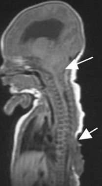
- The patient's MRI demonstrated an intramedullary spinal cord tumor extending from the fourth ventricle to T4. (magiran.com)
Choroid7
- Tumors of the fourth ventricle commonly originate from the following structures composing the floor: the ependyma, choroid plexus, and tela choroidea. (mattstillwell.net)
- The tela chorioidea and choroid plexus of the roof of the fourth ventricle are exposed. (stanford.edu)
- The choroid plexuses located in the ventricles produce CSF, which fills the ventricles and subarachnoid space, following a cycle of constant production and reabsorption. (medscape.com)
- Tufts of capillaries invaginate the roofs of prosencephalon and rhombencephalon, forming the choroid plexuses of the ventricles. (medscape.com)
- The anterior part of the body of the fornix, the choroid plexus, lateral dorsal surface of the thalamus, stria terminalis, and caudate nucleus, form the floor of the lateral ventricle. (medscape.com)
- Capillaries of the choroid arteries from the pia mater project into the ventricular cavity, forming the choroid plexus of the lateral ventricle (see the image below). (medscape.com)
- The choroid plexus extends from the lateral ventricle into the inferior horn. (medscape.com)
Subarachnoid space5
- CSF entering the fourth ventricle through the cerebral aqueduct can exit to the subarachnoid space of the spinal cord through two lateral apertures and a single, midline median aperture. (wikipedia.org)
- Morphine sulfate was injected i.v. and through cannulas inserted stereotactically into the third and fourth ventricles and into the bulbar subarachnoid space. (aspetjournals.org)
- Morphine in the fourth ventricle and subarachnoid space (10-50 µg in 50 µl) induced respiratory depression qualitatively similar to that obtained after i.v. injection (1 mg/kg). (aspetjournals.org)
- After i.v. injection, the beginning and peak effect of the depression appeared earlier than after injection into the fourth ventricle and subarachnoid space. (aspetjournals.org)
- Morphine was slightly more effective in the subarachnoid space than in the fourth ventricle. (aspetjournals.org)
Cavity4
- Fourth ventricle - One cavity in a system of four communicating cavities within the brain that are continuous with the central canal of the spinal cord. (en-academic.com)
- The fourth ventricle is a cavity of hindbrain connected to the third ventricle by a narrow cerebral aqueduct. (mattstillwell.net)
- fourth cavity of the brain. (infovisual.info)
- The author offers an original method of the study of the midbrain structures and liquor circulation system (the third ventricle, brain water-pipe, the fourth ventricle) in the small cavity in the course of autopsy. (who.int)
Medulla oblongata2
- The medulla oblongata is located behind the inferior portion of the floor (and continues caudally of the ventricle). (wikipedia.org)
- The AP is located on the dorsal surface of the medulla oblongata at the caudal end of the fourth ventricle. (nih.gov)
Interventricular foramen2
- The striatal ridge in the floor of the lateral ventricle at the interventricular foramen also grows in a similar manner. (ehd.org)
- Each lateral ventricle drains through an interventricular foramen into the third ventricle. (lumenlearning.com)
Spinal cord4
- A sulcus - the median sulcus - extends the length of the ventricle (from the cerebral aqueduct of the midbrain to the central canal of the spinal cord), dividing the floor into right and left halves. (wikipedia.org)
- This theory proposes that syringomyelia results from a "water hammer"-like transmission of pulsatile CSF pressure via a communication between the fourth ventricle and the central canal of the spinal cord through the obex. (medscape.com)
- The main function of this ventricle is to protect the human brain from trauma (via a cushioning effect) and to help form the central canal, which runs the length of the spinal cord. (mattstillwell.net)
- Most (58.8%) arise in the 4th ventricle, while spinal cord (17.6%) and supratentorial locations (17.6%) were less common. (uni-muenster.de)
Brainstem1
- The neoplasm can also invade adjacent brainstem structures, including the cardiorespiratory centers of the fourth ventricular floor. (medscape.com)
Ependymoma2
- Surgery is the standard treatment for ependymoma of the fourth ventricle. (mattstillwell.net)
- In this case report, we present a pigmented ependymoma in the fourth ventricle of an adult patient and review 16 additional cases of pigmented ependymoma from the literature. (uni-muenster.de)
Medullary2
- In telovelar approach, the telachoroidea and inferior medullary velum, which form the lower half of the roof of the fourth ventricle, are opened and the lower vermis is retracted as a unit to provide exposure into the fourth ventricle [3]. (mattstillwell.net)
- Lesions may also arise outside the ventricle and secondarily extend into this chamber, including medullary, tectal, and cerebellar hemispheric masses. (mattstillwell.net)
Tumour3
- The fourth ventricle is a common location of an intracranial ependymomal tumour. (wikipedia.org)
- How might a Tumour in the 4th ventricle be removed? (mattstillwell.net)
- Mixed glioneu- ronal tumour of the 4th ventricle with prominent rosette formation. (thieme-connect.com)
Dorsal1
- The lateral ventricle follows the dorsal, then caudal expansion of the cerebral vesicle and thereby produces an inferior horn . (ehd.org)
Dilatation1
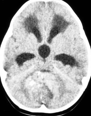
- In other patients where the obstruction is incomplete or gradual (e.g. aqueduct stenosis ), there may be almost no symptoms despite massive dilatation of the ventricles. (radiopaedia.org)
Lateral ventricles6
- These cavities, known collectively as the ventricular system, consist of the left and right lateral ventricles, the third ventricle, and the fourth ventricle. (wikipedia.org)
- The ventricular system is composed of 2 lateral ventricles, the third ventricle, the cerebral aqueduct, and the fourth ventricle (see the following images). (medscape.com)
- The neural canal dilates within the prosencephalon, leading to the formation of the lateral ventricles and third ventricle. (medscape.com)
- The lateral ventricles communicate with the third ventricle through interventricular foramens, and the third ventricle communicates with the fourth ventricle through the cerebral aqueduct (see the image below). (medscape.com)
- The largest cavities of the ventricular system are the lateral ventricles. (medscape.com)
- The 2 interventricular foramens (or foramina of Monro) connect the lateral ventricles with the third ventricle. (medscape.com)
Obstruction1
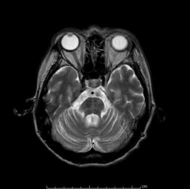
- This axial T2 magnetic resonance image shows a cerebellar vermian midline mass with contrast enhancement and obstruction of the fourth ventricle. (medscape.com)
Obex2
Cerebellar vermis1
- The majority of medulloblastomas arise from the inferior cerebellar vermis, from which they extend into and typically fill the fourth ventricle. (medscape.com)
Medial2
- Medial eminence of floor of fourth ventricle - Not to be confused with Median eminence. (en-academic.com)
- Brain: Medial eminence of floor of fourth ventricle Rhomboid fossa. (en-academic.com)
Aqueductoplasty2
- We conducted a retrospective evaluation of the safety, effectiveness, and long-term outcome of endoscopic aqueductoplasty and stent placement, performed in 18 consecutive patients with symptomatic TFV through a trans-fourth ventricle approach between 1994 and 2010. (neurologyindia.com)
- Our experience and the literature review suggest that endoscopic trans-fourth ventricle aqueductoplasty and stent placement is a minimally invasive, safe, and effective technique for the treatment of TFV and should be strongly recommended, especially in patients with supratentorial slit ventricles. (neurologyindia.com)
Taenia2
- Taenia of fourth ventricle - Infobox Brain Name = PAGENAME Latin = taenia ventriculi quarti GraySubject = 187 GrayPage = 797 Caption = Rhomboid fossa. (en-academic.com)
- Taenia of fourth ventricle labeled at bottom left. (en-academic.com)
Ventricular system1
- The ventricular system including the fourth ventricle, develops from the central canal of the neural tube. (wikipedia.org)
Rhomboid2
- The floor (i.e. the anterior edge) of the fourth ventricle constitutes the rhomboid fossa, and comprises a number of general features. (wikipedia.org)
- These structures obscure the inferior and intermediate portions of the rhomboid fossa (floor of the ventricle). (stanford.edu)
Inferior1
- The internal part of the facial nerve bulges into the ventricle, forming the facial colliculus, in the process of looping around the abducens nucleus within the inferior region of the Pons. (wikipedia.org)
Midbrain1
- The third ventricle sits in the midbrain region. (lumenlearning.com)
Neuroimaging1
- Neuroimaging revealed a mass present in the fourth ventricle. (urosario.edu.co)
Roof5
- The fourth ventricle has a roof at its upper (posterior) surface and a floor at its lower (anterior) surface, and side walls formed by the cerebellar peduncles (nerve bundles joining the structure on the posterior side of the ventricle to the structures on the anterior side). (wikipedia.org)
- Scheme of roof of fourth ventricle. (wikipedia.org)
- This ventricle has a roof and a floor. (mattstillwell.net)
- The subcommissural organ (SCO) is a secretory tissue located on the roof of the brain's third ventricle. (stanford.edu)
- CSF flows out of the fourth ventricle through the 3 apertures formed at the roof of the fourth ventricle by week 12 of gestation. (medscape.com)
Brain8
- The fourth ventricle is one of the four connected fluid-filled cavities within the human brain. (wikipedia.org)
- The fourth ventricle has a characteristic diamond shape in cross-sections of the human brain. (wikipedia.org)
- Fourth ventricle - Infobox Brain Name = PAGENAME Latin = ventriculus quartus GraySubject = 187 GrayPage = 797 Caption = Scheme showing relations of the ventricles to the surface of the brain. (en-academic.com)
- For example, the four connected cavities (hollow spaces) in the central portion of the brain and the lower two chambers of the heart are called ventricles. (en-academic.com)
- Is the fourth ventricle in the brain? (mattstillwell.net)
- Where is the 4th ventricle located in the brain? (mattstillwell.net)
- Then the CSF flows throughout the brain in the ventricles. (lumenlearning.com)
- MRI of the brain with and without gadolinium enhancement showed a 3.4 × 2.6 × 3.0-cm lesion situated within the region of the fourth ventricle. (medscape.com)
Stent1
- In 3 with normal ventricles, the ventricles had to be dilated by externalizing the shunt before placing the stent. (utmb.edu)
Cystic1
- Magnetic resonance imaging revealed a 2.5 cm contrast-enhancing cystic mass in the fourth ventricle, which was resected. (uni-muenster.de)
Ependymomas1
- 100% resection is common for ependymomas of the fourth ventricle. (mattstillwell.net)
Supratentorial ventricles1
- Fifteen patients presented with slit supratentorial ventricles. (neurologyindia.com)
Sagittal1
- A T1-weighted sagittal noncontrast MR image shows a large multilaminated fourth ventricle mass. (medscape.com)
Enlargement1
- c Porencephalic enlargement of the left ventricle. (nature.com)
Morphine1
- On the other hand, morphine in the third ventricle (50 µg in 50 µl) elicited an immediate respiratory stimulation followed by a second phase during which frequency remained elevated but tidal volume was progressively reduced and end-expiratory CO 2 was increased. (aspetjournals.org)
Abstract2
- abstract = "To present a young immunocompetent patient with a fourth ventricle tuberculoma without pulmonary tuberculosis. (urosario.edu.co)
- abstract = "Background: Of the various management options for isolated fourth ventricle (IFV), fourth ventriculoperitoneal shunts (FVPS) and aqueductal stents (AST) have been themost favored. (utmb.edu)
Third and fourth ventricles1
- During the first trimester of pregnancy the central canal expands into the lateral, third and fourth ventricles, connected by thinner channels. (wikipedia.org)
Floor1
- In the superior region of the pons is the locus coeruleus, which due to its concentration of noradrenaline has a sky blue appearance, visible (in a colour closer to teal) through the floor of the ventricle, superiorly to the superior fovea. (wikipedia.org)
Flows2
- It is constantly produced, flows through the network of ventricles, and is reabsorbed. (lumenlearning.com)
- The CSF then flows into the cerebral aqueduct to the fourth ventricle. (lumenlearning.com)
Neural1
- The dilation of the neural canal within the rhombencephalon forms the fourth ventricle. (medscape.com)
Temporal horns1
- The body of the lateral ventricle is connected with the occipital and temporal horns by a wide area named the atrium. (medscape.com)
Connects1
- canal that connects the third ventricle to the fourth one. (infovisual.info)
Chamber1
- Ventricle - A ventricle is a chamber of an organ. (en-academic.com)
Findings1
- To the best of our knowledge, this is the largest collection of 4th ventricular subependymomas with imaging findings reported to date. (nih.gov)
Mass1
- Magnetic resonance imaging (MRI) typically reveals a solid fourth ventricular mass that is hypointense on the T1-weighted image, hyperintense on T2-weighted images, and brightly enhancing on T1-weighted images after contrast administration (see the images below). (medscape.com)
![Recombinant Anti-p53 antibody [E26] KO Tested (ab32389) | Abcam](https://www.abcam.com/ps/products/32/ab32389/Images/ab32389-389819-antip53antibodyimmunocytochemistryimmunofluorescencehap1human.jpg)