Gastroschisis
Hernia, Umbilical
Abdominal Muscles
Intestinal Atresia
Abnormalities, Multiple
Ultrasonography, Prenatal
Abdominal Wall
Pregnancy
Gastric Dilatation
Fetal Diseases
Hernia, Ventral
Gestational Age
Romania
Abdominal Wound Closure Techniques
Encyclopedias as Topic
Umbilicus
Congenital hernia of the abdominal wall: a differential diagnosis of fetal abdominal wall defects. (1/80)
A 28-year-old woman was referred at 33 weeks of gestation with suspected fetal intestinal atresia. Sonography showed a large extra-abdominal mass on the right of the normal umbilical cord insertion. Following Cesarean section at 36 weeks and immediate surgical treatment, the malformation was not definable either as an omphalocele or as gastroschisis. This reported case involves a previously undocumented malformation of the fetal abdominal wall described as a 'hernia' of the fetal abdominal wall. (+info)Gastroschisis associated with bladder evisceration complicated by hydronephrosis presenting antenatally. (2/80)
We report here a case of gastroschisis associated with bladder evisceration and complicated by rapidly developing hydronephrosis diagnosed antenatally. The timing of delivery was determined by the hydronephrosis, associated bowel dilatation and polyhydramnios. The case highlights the need for continuing ultrasonographic surveillance of fetuses with gastroschisis to identify further associated complications which were hitherto absent but whose presence may influence the timing of delivery and neonatal care. (+info)Functional urinary tract obstruction developing in fetuses with isolated gastroschisis. (3/80)
OBJECTIVE: To evaluate the frequency and natural history of urinary tract abnormalities developing in fetuses presenting with initially isolated gastroschisis. METHODS: Serial ultrasounds were performed prospectively on fetuses identified by our prenatal diagnosis program as having a gastroschisis. When abnormalities in the urinary tract were identified prenatally, newborns were evaluated by a pediatric urologist. RESULTS: Over a 1-year period four out of 12 fetuses with gastroschisis developed deformations of the urinary tract. In three fetuses the bladder herniated through the abdominal wall defect. Two also had upper tract dilatation. A fourth fetus developed bilateral hydronephrosis with a normally situated bladder. Once the gastroschisis was repaired none of the newborns had evidence of structural obstruction of the urinary tract, however, hydronephrosis with or without reflux persisted for several months. CONCLUSIONS: Deformations of the fetal urinary tract can develop secondary to gastroschisis. They do not appear to represent separate malformations and evaluation with fetal karyotyping may not be indicated. When hydronephrosis is present ongoing urologic evaluation of the neonate is indicated. (+info)Management of gastroschisis in a peripheral hospital setting. (4/80)
Ten patients (5 males and 5 females) with gastroschisis were treated in Alor Setar Hospital from January 1989 to December 1993. Two patients had associated congenital anomalies. Primary closure was possible in 9 patient while the other patient had stage closure. All patients received prophylactic antibiotics, 9 patients were ventilated electively in the post-operative period and 7 patients received parenteral nutrition. There were 9 survivors. Complications especially wound infection and breakdown were seen in 7 patients. The average hospital stay was 36 days. (+info)Acute bowel perforation in a fetus with gastroschisis. (5/80)
Gastroschisis is a congenital anomaly with a reported incidence of 1 in 10,000 live births. Although prenatal diagnosis is more common with the widespread use of biochemical markers and obstetric ultrasound, the role of ultrasound in the identification of the fetus that might need early intervention has not been established. Acute bowel perforation was diagnosed by ultrasound at 34 weeks gestation in a fetus with gastroschisis. An immediate Cesarean section was performed, followed by repair with primary closure. The neonatal outcome was favorable. The post-partum findings, including bowel pathology, confirmed the antenatal diagnosis. Acute bowel perforation can be diagnosed antenatally. Immediate intervention, before further bowel injury occurs, might enhance the ability of the surgeon to perform primary closure and obtain a favorable outcome. (+info)Abdominal wall defects: two- versus three-dimensional ultrasonographic diagnosis. (6/80)
We diagnosed 12 cases of abdominal wall defects. The cases diagnosed occurred in 6 fetuses with omphalocele, 3 with gastroschisis, 2 with prune-belly syndrome, and 1 with pentalogy of Cantrell. Except for 1 case of gastroschisis first diagnosed on the basis of three-dimensional ultrasonography at 14 weeks' gestation, all cases were first detected by two-dimensional transabdominal ultrasonography and then reevaluated with three-dimensional ultrasonography using multiplanar and orthogonal plane modes. Although the original diagnosis was accurate on the basis of two-dimensional ultrasonography in 11 of 12 cases, additional information was obtained by three-dimensional scanning in all cases. Our experience suggests that in cases in which abdominal wall defects are first detected by two-dimensional ultrasonographic scanning, the additional information gained by complementary three-dimensional ultrasonographic scanning can be useful for more-efficient counseling and postnatal therapeutic planning. (+info)Maternal medication use and risks of gastroschisis and small intestinal atresia. (7/80)
Gastroschisis and small intestinal atresia (SIA) are birth defects that are thought to arise from vascular disruption of fetal mesenteric vessels. Previous studies of gastroschisis have suggested that risk is increased for maternal use of vasoactive over-the-counter medications, including specific analgesics and decongestants. This retrospective study evaluated the relation between maternal use of cough/cold/analgesic medications and risks of gastroschisis and SIA. From 1995 to 1999, the mothers of 206 gastroschisis cases, 126 SIA cases, and 798 controls in the United States and Canada were interviewed about medication use and illnesses. Risks of gastroschisis were elevated for use of aspirin (odds ratio = 2.7, 95% confidence interval: 1.2, 5.9), pseudoephedrine (odds ratio = 1.8, 95% confidence interval: 1.0, 3.2), acetaminophen (odds ratio = 1.5, 95% confidence interval: 1.1, 2.2), and pseudoephedrine combined with acetaminophen (odds ratio = 4.2, 95% confidence interval: 1.9, 9.2). Risks of SIA were increased for any use of pseudoephedrine (odds ratio = 2.0, 95% confidence interval: 1.0, 4.0) and for use of pseudoephedrine in combination with acetaminophen (odds ratio = 3.0, 95% confidence interval: 1.1, 8.0). Reported fever, upper respiratory infection, and allergy were not associated with risks of either defect. These findings add more evidence that aspirin use in early pregnancy increases risk of gastroschisis. Although pseudoephedrine has previously been shown to increase gastroschisis risk, findings of this study raise questions about interactions between medications and possible confounding by underlying illness. (+info)Evaluation of prenatal ultrasound diagnosis of fetal abdominal wall defects by 19 European registries. (8/80)
OBJECTIVES: To evaluate the current effectiveness of routine prenatal ultrasound screening in detecting gastroschisis and omphalocele in Europe. DESIGN: Data were collected by 19 congenital malformation registries from 11 European countries. The registries used the same epidemiological methodology and registration system. The study period was 30 months (July 1st 1996-December 31st 1998) and the total number of monitored pregnancies was 690,123. RESULTS: The sensitivity of antenatal ultrasound examination in detecting omphalocele was 75% (103/137). The mean gestational age at the first detection of an anomaly was 18 +/- 6.0 gestational weeks. The overall prenatal detection rate for gastroschisis was 83% (88/106) and the mean gestational age at diagnosis was 20 +/- 7.0 gestational weeks. Detection rates varied between registries from 25 to 100% for omphalocele and from 18 to 100% for gastroschisis. Of the 137 cases of omphalocele less than half of the cases were live births (n = 56; 41%). A high number of cases resulted in fetal deaths (n = 30; 22%) and termination of pregnancy (n = 51; 37%). Of the 106 cases of gastroschisis there were 62 (59%) live births, 13 (12%) ended with intrauterine fetal death and 31 (29%) had the pregnancies terminated. CONCLUSIONS: There is significant regional variation in detection rates in Europe reflecting different policies, equipment and the operators' experience. A high proportion of abdominal wall defects is associated with concurrent malformations, syndromes or chromosomal abnormalities, stressing the need for the introduction of repeated detailed ultrasound examination as a standard procedure. There is still a relatively high rate of elective termination of pregnancies for both defects, even in isolated cases which generally have a good prognosis after surgical repair. (+info)Gastroschisis is a congenital abdominal wall defect, characterized by an opening, usually to the right of the umbilical cord, through which the abdominal organs such as the intestines protrude. It's typically not covered by a sac or membrane. The exact cause of gastroschisis is unknown, but it's thought to be related to disrupted blood flow in the area where the abdominal wall develops during pregnancy. This condition is usually detected prenatally through ultrasound and requires surgical repair shortly after birth.
An umbilical hernia is a type of hernia that occurs at the umbilicus, or belly button. It results from a protrusion of abdominal contents through a weakened area in the abdominal wall surrounding the navel. This condition is common in newborns and infants, especially premature babies, due to incomplete closure of the abdominal muscles during development.
In most cases, umbilical hernias in children close on their own by age 3-4 or by the time they reach school age. However, if the hernia is still present after this age, surgical intervention may be required to prevent potential complications such as incarceration (where the herniated tissue becomes trapped and cannot be pushed back in) or strangulation (where the blood supply to the herniated tissue is cut off, leading to tissue death).
Adults can also develop umbilical hernias, often as a result of increased pressure in the abdomen due to obesity, pregnancy, heavy lifting, or persistent coughing. Umbilical hernias in adults are generally more likely to require surgical repair due to the higher risk of complications.
The abdominal muscles, also known as the abdominals or abs, are a group of muscles in the anterior (front) wall of the abdominopelvic cavity. They play a crucial role in maintaining posture, supporting the trunk, and facilitating movement of the torso. The main abdominal muscles include:
1. Rectus Abdominis: These are the pair of long, flat muscles that run vertically along the middle of the anterior abdominal wall. They are often referred to as the "six-pack" muscles due to their visible, segmented appearance in well-trained individuals. The primary function of the rectus abdominis is to flex the spine, allowing for actions such as sitting up from a lying down position or performing a crunch exercise.
2. External Obliques: These are the largest and most superficial of the oblique muscles, located on the lateral (side) aspects of the abdominal wall. They run diagonally downward and forward from the lower ribs to the iliac crest (the upper part of the pelvis) and the pubic tubercle (a bony prominence at the front of the pelvis). The external obliques help rotate and flex the trunk, as well as assist in side-bending and exhalation.
3. Internal Obliques: These muscles lie deep to the external obliques and run diagonally downward and backward from the lower ribs to the iliac crest, pubic tubercle, and linea alba (the strong band of connective tissue that runs vertically along the midline of the abdomen). The internal obliques help rotate and flex the trunk, as well as assist in forced exhalation and increasing intra-abdominal pressure during actions such as coughing or lifting heavy objects.
4. Transversus Abdominis: This is the deepest of the abdominal muscles, located inner to both the internal obliques and the rectus sheath (a strong, fibrous covering that surrounds the rectus abdominis). The transversus abdominis runs horizontally around the abdomen, attaching to the lower six ribs, the thoracolumbar fascia (a broad sheet of connective tissue spanning from the lower back to the pelvis), and the pubic crest (the front part of the pelvic bone). The transversus abdominis helps maintain core stability by compressing the abdominal contents and increasing intra-abdominal pressure.
Together, these muscles form the muscular "corset" of the abdomen, providing support, stability, and flexibility to the trunk. They also play a crucial role in respiration, posture, and various movements such as bending, twisting, and lifting.
Intestinal atresia is a congenital condition characterized by the absence or complete closure of a portion of the intestine, preventing the passage of digested food from the stomach to the remaining part of the intestines. This results in a blockage in the digestive system, which can be life-threatening if not treated promptly after birth. The condition can occur anywhere along the small or large intestine and may affect either a single segment or multiple segments of the intestine.
There are several types of intestinal atresia, including:
1. Jejunal atresia: A closure or absence in the jejunum, a part of the small intestine located between the duodenum and ileum.
2. Ileal atresia: A closure or absence in the ileum, the lower portion of the small intestine that connects to the large intestine (cecum).
3. Colonic atresia: A closure or absence in the colon, a part of the large intestine responsible for storing and eliminating waste.
4. Duodenal atresia: A closure or absence in the duodenum, the uppermost portion of the small intestine that receives chyme (partially digested food) from the stomach.
5. Multiple atresias: When more than one segment of the intestines is affected by atresia.
The exact cause of intestinal atresia remains unclear, but it is believed to be related to disruptions in fetal development during pregnancy. Treatment typically involves surgical correction to reconnect the affected segments of the intestine and restore normal digestive function. The prognosis for infants with intestinal atresia depends on the severity and location of the atresia, as well as any associated conditions or complications.
Serositis is a medical term that refers to inflammation of the serous membranes, which are thin layers of tissue that line the inner surfaces of body cavities and surround organs such as the heart, lungs, and abdomen. The serous membranes produce a lubricating fluid called serous fluid that helps reduce friction between internal organs and enables them to move smoothly against each other.
Inflammation of these membranes can result in excessive production of serous fluid, leading to the accumulation of fluid in the surrounding body cavities. This accumulation can cause symptoms such as chest pain, coughing, difficulty breathing, or abdominal swelling and discomfort.
Serositis is often associated with various medical conditions, including autoimmune diseases like rheumatoid arthritis, lupus, and Sjogren's syndrome. Infections, cancers, and certain medications may also cause serositis. Treatment typically involves addressing the underlying condition causing the inflammation and managing symptoms with medications such as nonsteroidal anti-inflammatory drugs (NSAIDs), corticosteroids, or immunosuppressive agents.
'Abnormalities, Multiple' is a broad term that refers to the presence of two or more structural or functional anomalies in an individual. These abnormalities can be present at birth (congenital) or can develop later in life (acquired). They can affect various organs and systems of the body and can vary greatly in severity and impact on a person's health and well-being.
Multiple abnormalities can occur due to genetic factors, environmental influences, or a combination of both. Chromosomal abnormalities, gene mutations, exposure to teratogens (substances that cause birth defects), and maternal infections during pregnancy are some of the common causes of multiple congenital abnormalities.
Examples of multiple congenital abnormalities include Down syndrome, Turner syndrome, and VATER/VACTERL association. Acquired multiple abnormalities can result from conditions such as trauma, infection, degenerative diseases, or cancer.
The medical evaluation and management of individuals with multiple abnormalities depend on the specific abnormalities present and their impact on the individual's health and functioning. A multidisciplinary team of healthcare professionals is often involved in the care of these individuals to address their complex needs.
Prenatal ultrasonography, also known as obstetric ultrasound, is a medical diagnostic procedure that uses high-frequency sound waves to create images of the developing fetus, placenta, and amniotic fluid inside the uterus. It is a non-invasive and painless test that is widely used during pregnancy to monitor the growth and development of the fetus, detect any potential abnormalities or complications, and determine the due date.
During the procedure, a transducer (a small handheld device) is placed on the mother's abdomen and moved around to capture images from different angles. The sound waves travel through the mother's body and bounce back off the fetus, producing echoes that are then converted into electrical signals and displayed as images on a screen.
Prenatal ultrasonography can be performed at various stages of pregnancy, including early pregnancy to confirm the pregnancy and detect the number of fetuses, mid-pregnancy to assess the growth and development of the fetus, and late pregnancy to evaluate the position of the fetus and determine if it is head down or breech. It can also be used to guide invasive procedures such as amniocentesis or chorionic villus sampling.
Overall, prenatal ultrasonography is a valuable tool in modern obstetrics that helps ensure the health and well-being of both the mother and the developing fetus.
The abdominal wall refers to the group of muscles, fascia (sheaths of connective tissue), and skin that make up the front and sides of the abdomen, extending from the thorax (chest) to the pelvis. It provides protection to the abdominal organs, supports the trunk, and allows for movement of the torso.
The main muscles of the anterior abdominal wall include:
1. Rectus sheaths (Rectus Abdominis): paired vertical muscles running from the pubic symphysis to the xiphoid process and costal cartilages of ribs 5-7.
2. External obliques: thin, irregular muscles that lie over the lower part of the abdomen and run diagonally downward and forward from the lower ribs to the iliac crest (pelvic bone) and pubic tubercle.
3. Internal obliques: thicker muscles that lie under the external obliques, running diagonally upward and forward from the iliac crest to the lower ribs.
4. Transverse abdominis: deepest of the abdominal muscles, lying horizontally across the abdomen, attaching from the lower ribs to the pelvis.
These muscles are interconnected by various layers of fascia and aponeuroses (flat, broad tendons), forming a complex structure that allows for both stability and mobility. The linea alba, a fibrous band, runs down the midline of the anterior abdominal wall, connecting the rectus sheaths.
Damage to the abdominal wall can occur due to trauma, surgery, or various medical conditions, which may require surgical intervention for repair.
A newborn infant is a baby who is within the first 28 days of life. This period is also referred to as the neonatal period. Newborns require specialized care and attention due to their immature bodily systems and increased vulnerability to various health issues. They are closely monitored for signs of well-being, growth, and development during this critical time.
Congenital abnormalities, also known as birth defects, are structural or functional anomalies that are present at birth. These abnormalities can develop at any point during fetal development, and they can affect any part of the body. They can be caused by genetic factors, environmental influences, or a combination of both.
Congenital abnormalities can range from mild to severe and may include structural defects such as heart defects, neural tube defects, and cleft lip and palate, as well as functional defects such as intellectual disabilities and sensory impairments. Some congenital abnormalities may be visible at birth, while others may not become apparent until later in life.
In some cases, congenital abnormalities may be detected through prenatal testing, such as ultrasound or amniocentesis. In other cases, they may not be diagnosed until after the baby is born. Treatment for congenital abnormalities varies depending on the type and severity of the defect, and may include surgery, therapy, medication, or a combination of these approaches.
Pregnancy is a physiological state or condition where a fertilized egg (zygote) successfully implants and grows in the uterus of a woman, leading to the development of an embryo and finally a fetus. This process typically spans approximately 40 weeks, divided into three trimesters, and culminates in childbirth. Throughout this period, numerous hormonal and physical changes occur to support the growing offspring, including uterine enlargement, breast development, and various maternal adaptations to ensure the fetus's optimal growth and well-being.
Gastric dilatation, also known as stomach dilation or distention, refers to the abnormal enlargement or expansion of the stomach. This condition often occurs when the stomach fills with gas, food, or fluids and is unable to empty properly. Gastric dilatation can be caused by various factors such as overeating, swallowing excessive air, gastroparesis (delayed gastric emptying), intestinal obstruction, or certain medical conditions like hiatal hernia or pregnancy.
In severe cases, gastric dilatation may lead to gastric volvulus, where the stomach twists on itself, cutting off its blood supply and leading to ischemia and necrosis of the stomach tissue. This is a life-threatening condition that requires immediate medical attention. Symptoms of gastric dilatation include abdominal pain, bloating, vomiting, loss of appetite, and difficulty breathing.
Fetal diseases are medical conditions or abnormalities that affect a fetus during pregnancy. These diseases can be caused by genetic factors, environmental influences, or a combination of both. They can range from mild to severe and may impact various organ systems in the developing fetus. Examples of fetal diseases include congenital heart defects, neural tube defects, chromosomal abnormalities such as Down syndrome, and infectious diseases such as toxoplasmosis or rubella. Fetal diseases can be diagnosed through prenatal testing, including ultrasound, amniocentesis, and chorionic villus sampling. Treatment options may include medication, surgery, or delivery of the fetus, depending on the nature and severity of the disease.
A ventral hernia is a type of hernia that occurs in the abdominal wall, specifically in the anterior (front) aspect. It can occur due to a weakness or defect in the abdominal wall muscles and fascia, which allows the internal organs or tissues to push through and create a bulge or swelling.
Ventral hernias can be classified into several types based on their location, size, and cause. Some of the common types include:
1. Incisional Hernia - occurs at the site of a previous surgical incision, where the abdominal wall has not healed properly or has become weakened over time.
2. Epigastric Hernia - located in the upper middle part of the abdomen, between the breastbone and the navel.
3. Umbilical Hernia - occurs around the belly button, most commonly seen in infants but can also affect adults.
4. Spigelian Hernia - a rare type of hernia that occurs lateral to the rectus sheath, usually at the level of the semilunar line.
5. Diastasis Recti - a separation of the abdominal muscles in the midline, which can lead to a ventral hernia if not treated.
Symptoms of a ventral hernia may include pain or discomfort, especially when lifting heavy objects, straining, coughing, or during physical activity. In some cases, a hernia may become incarcerated or strangulated, which requires immediate medical attention. Treatment options for ventral hernias typically involve surgical repair, either through open surgery or laparoscopic techniques.
Maternal age is a term used to describe the age of a woman at the time she becomes pregnant or gives birth. It is often used in medical and epidemiological contexts to discuss the potential risks, complications, and outcomes associated with pregnancy and childbirth at different stages of a woman's reproductive years.
Advanced maternal age typically refers to women who become pregnant or give birth at 35 years of age or older. This group faces an increased risk for certain chromosomal abnormalities, such as Down syndrome, and other pregnancy-related complications, including gestational diabetes, preeclampsia, and cesarean delivery.
On the other end of the spectrum, adolescent pregnancies (those that occur in women under 20 years old) also come with their own set of potential risks and complications, such as preterm birth, low birth weight, and anemia.
It's important to note that while maternal age can influence pregnancy outcomes, many other factors – including genetics, lifestyle choices, and access to quality healthcare – can also play a significant role in determining the health of both mother and baby during pregnancy and childbirth.
Gestational age is the length of time that has passed since the first day of the last menstrual period (LMP) in pregnant women. It is the standard unit used to estimate the age of a pregnancy and is typically expressed in weeks. This measure is used because the exact date of conception is often not known, but the start of the last menstrual period is usually easier to recall.
It's important to note that since ovulation typically occurs around two weeks after the start of the LMP, gestational age is approximately two weeks longer than fetal age, which is the actual time elapsed since conception. Medical professionals use both gestational and fetal age to track the development and growth of the fetus during pregnancy.
I'm sorry for any confusion, but "Romania" is not a medical term. It is a country located in southeastern Europe. If you have any questions about medical terminology or health-related topics, I would be happy to help. Could you please clarify your question?
Abdominal wound closure techniques refer to the methods used to close and repair surgical incisions in the abdomen. The goal of these techniques is to restore the integrity of the abdominal wall, minimize the risk of infection or dehiscence (wound separation), and promote optimal healing. Several abdominal wound closure techniques are available, and the choice of which one to use depends on various factors such as the size and location of the incision, the patient's individual needs and medical history, and the surgeon's preference. Here are some commonly used abdominal wound closure techniques:
1. Continuous running suture: This technique involves using a continuous strand of suture material to close the wound in a single pass. The suture is inserted through the full thickness of the abdominal wall, including the fascia (the strong connective tissue that surrounds the muscles), and then passed continuously along the length of the incision, pulling the edges of the wound together as it goes. This technique can be faster and more efficient than other methods, but it may increase the risk of infection or wound breakdown if not done properly.
2. Interrupted suture: In this technique, the surgeon uses individual stitches placed at regular intervals along the incision to close the wound. Each stitch is tied separately, which can make the closure more secure and reduce the risk of infection or wound breakdown. However, interrupted sutures can be more time-consuming than continuous running sutures.
3. Mass closure: This technique involves using a large, continuous suture to close the entire length of the incision in one pass. The suture is inserted through the full thickness of the abdominal wall and tied at both ends, pulling the edges of the wound together. Mass closure can be faster and more efficient than other methods, but it may increase the risk of infection or wound breakdown if not done properly.
4. Retention sutures: These are additional sutures that are placed deep within the abdominal wall to provide extra support and strength to the closure. They are often used in high-tension areas or in patients who are at increased risk of wound dehiscence, such as those with obesity or diabetes.
5. Layered closure: In this technique, the surgeon closes the incision in multiple layers, starting with the deepest layer of muscle and fascia and working outward to the skin. Each layer is closed separately using either interrupted or continuous sutures. Layered closure can provide added strength and stability to the closure, but it can be more time-consuming than other methods.
6. Skin closure: The final step in wound closure is to close the skin, which can be done using a variety of techniques, including staples, sutures, or surgical glue. The choice of closure method depends on several factors, including the size and location of the incision, the patient's individual needs and preferences, and the surgeon's experience and expertise.
Overall, the choice of wound closure technique depends on several factors, including the size and location of the incision, the patient's individual needs and preferences, and the surgeon's experience and expertise. The goal is to provide a strong, secure, and cosmetically appealing closure that minimizes the risk of infection, wound breakdown, or other complications.
An encyclopedia is a comprehensive reference work containing articles on various topics, usually arranged in alphabetical order. In the context of medicine, a medical encyclopedia is a collection of articles that provide information about a wide range of medical topics, including diseases and conditions, treatments, tests, procedures, and anatomy and physiology. Medical encyclopedias may be published in print or electronic formats and are often used as a starting point for researching medical topics. They can provide reliable and accurate information on medical subjects, making them useful resources for healthcare professionals, students, and patients alike. Some well-known examples of medical encyclopedias include the Merck Manual and the Stedman's Medical Dictionary.
The umbilicus, also known as the navel, is the scar left on the abdominal wall after the removal of the umbilical cord in a newborn. The umbilical cord connects the developing fetus to the placenta in the uterus during pregnancy, providing essential nutrients and oxygen while removing waste products. After birth, the cord is clamped and cut, leaving behind a small stump that eventually dries up and falls off, leaving the umbilicus. In adults, it typically appears as a slight depression or dimple on the abdomen.
Fetal growth retardation, also known as intrauterine growth restriction (IUGR), is a condition in which a fetus fails to grow at the expected rate during pregnancy. This can be caused by various factors such as maternal health problems, placental insufficiency, chromosomal abnormalities, and genetic disorders. The fetus may be smaller than expected for its gestational age, have reduced movement, and may be at risk for complications during labor and delivery. It is important to monitor fetal growth and development closely throughout pregnancy to detect any potential issues early on and provide appropriate medical interventions.
 Gastroschisis
Kedarnath Das
Abdominal wall defect
Ae binding protein 1
Northfleet Urban Country Park
Omphalocele
Jonathan Walters
Intestinal atresia
Transplantable organs and tissues
Birth defect
Atrazine
T. J. Oshie
Billy Sharp
Hanhart syndrome
Bothwell Mbuwayesango
Development of the digestive system
Body cavity
Serial transverse enteroplasty
Amyoplasia
Opioids and pregnancy
Dev (singer)
Congenital vertebral anomaly
Teratoma
Prenatal testing
Fish oil (medical use)
List of fetal abnormalities
University of California, San Francisco Fetal Treatment Center
List of MeSH codes (C23)
List of congenital disorders
Krystal Shaw
Gastroschisis - Wikipedia
 Gastroschisis: MedlinePlus Medical Encyclopedia
 Management of the Infant With Gastroschisis
 Gastroschisis | Johns Hopkins Medicine
 Increasing Prevalence of Gastroschisis - 14 States, 1995-2012 | MMWR
 Gastroschisis: a plea for risk categorization
Gastroschisis Imaging: Practice Essentials, Radiography, Magnetic Resonance Imaging
 Gastroschisis | West Indian Medical Journal
 Gastroschisis: Prenatal diagnosis and management
 Effect of delivery approach on outcomes in fetuses with gastroschisis. • Arctic Health
 Omphalocele and Gastroschisis Associated With Multiple Congenital Abnormalities • JML Journal of Medicine and Life
 Increasing Prevalence of Gastroschisis in Europe 1980-2002: A Phenomenon Restricted to Younger Mothers?
 Omphalocele, Gastroschisis: Epidemiology, Survival, and Mortality in Imam Khomeini Hospital, Ahvaz-Iran
 Gastroschisis: Video, Anatomy, Definition & Function | Osmosis
 Data for: Growth Failure Prevalence in Neonates with Gastroschisis: A Statewide Prospective Cohort Study - Mendeley Data
 Gastroschisis
 Gastroschisis
 Neighborhood-level socioeconomic position during early pregnancy and risk of gastroschisis
 Gastroschisis Archives - Patient Worthy
 Gastroschisis | Concise Medical Knowledge
 The Global Gastroschisis Foundation
 Gastroschisis Repair - Pediatrics Procedures - BroadcastMed
 nikki - The Global Gastroschisis Foundation
playtimeed - The Global Gastroschisis Foundation
 Gastroschisis - Pediatrics - MSD Manual Professional Edition
Recreational drug use: A major risk factor for gastroschisis? - Nuffield Department of Population Health
 Gastroschisis - Apollo Centre for Fetal Medicine
 Gastroschisis in Brazil within a Global Context
 Original Article
| Journal of Perinatology
Original Article
| Journal of Perinatology
 Gastroschisis: one year outcomes from national cohort study
Infants7
- About sixty percent of infants with gastroschisis are born prematurely. (wikipedia.org)
- Prenatal ultrasounds often identify infants with gastroschisis before birth, usually by 20 weeks of pregnancy. (medlineplus.gov)
- Approximately 48% of infants with gastroschisis are small for gestational age. (medscape.com)
- however, many infants with gastroschisis who remain in the womb until term have little or no peel, while others, who are born prematurely, have significant peel. (lifespan.org)
- Infants with gastroschisis have a large amount of intestine on the surface of the abdominal wall. (panafrican-med-journal.com)
- This low-cost silo can be used to help save the lives of infants in Sub-Saharan Africa who suffer from gastroschisis, a birth defect where the bowel develops outside the baby's body. (nih.gov)
- Infants with gastroschisis were ascertained by active birth defects surveillance systems. (nih.gov)
Amniotic fluid4
- Prolonged exposure to amniotic fluid may cause mucosal or muscularis injury, although the etiology of bowel injury in gastroschisis is unclear. (medscape.com)
- In the past, we had assumed that contact with the amniotic fluid is what causes bowel damage in gastroschisis, but we have now shown that the bowel damage is due to inherent changes in the fetal immune system, which mounts an immune response," she said. (ucsf.edu)
- 16. Elevated amniotic fluid amino acid levels in fetuses with gastroschisis. (nih.gov)
- 17. Amniotic fluid inflammatory proteins and digestive compounds profile in fetuses with gastroschisis undergoing amnioexchange. (nih.gov)
Baby with gastroschisis4
- A previous study of women in Washington state found that exposure to the weed killer atrazine was tied to an increased risk of having a baby with gastroschisis, although it did not show that the chemical caused the malformation (see Reuters Health story of February 8, 2010). (medscape.com)
- A baby with gastroschisis is likely to stay in the hospital for at least one month, and depending on the degree of prematurity and the condition of the bowel, may stay much longer. (lifespan.org)
- When Martz was pregnant, doctors diagnosed her baby with gastroschisis, a birth defect that allows the intestines to spill out of a hole in the abdominal wall and grow outside the body. (krqe.com)
- Realistic birthing baby with Gastroschisis. (simulationcollective.com)
Management of gastroschisis5
- SSA is also known as the plastic closure and the nonoperative management of gastroschisis. (hindawi.com)
- Undoubtedly, the preferred method for the management of gastroschisis has changed fundamentally over time [ 7 ]. (hindawi.com)
- Non-operative management of gastroschisis: a case-matched study. (qxmd.com)
- Non-operative management of gastroschisis also known as plastic closure (PC) has been described as an alternative to conventional primary operative closure (POC) or staged silo closure (SSC). (qxmd.com)
- Challenges in management of gastroschisis are related to the prevention of late intrauterine death, and the prediction and treatment of complex forms. (igenomix.eu)
Intestines10
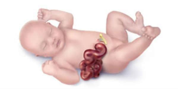
- Gastroschisis is a birth defect in which the baby's intestines extend outside of the abdomen through a hole next to the belly button. (wikipedia.org)
- Gastroschisis is a birth defect in which an infant's intestines are outside of the body because of a hole in the abdominal wall. (medlineplus.gov)
- In babies with gastroschisis, the intestines (and sometimes the stomach) remain outside the abdominal wall, without a membrane covering them. (medlineplus.gov)
- Babies with gastroschisis need time for their intestines to recover and become used to taking feedings. (medlineplus.gov)
- A small number of babies with gastroschisis (about 10 to 20%) may have intestinal atresia (parts of the intestines that did not develop in the womb). (medlineplus.gov)
- Gastroschisis is a condition in which loops of intestines (and sometimes parts of the stomach, liver and other organs) protrude from the fetus's body through a hole in the abdominal wall. (lifespan.org)
- UCSF News reports on the work of a UCSF research team investigating the causes and potential treatments for gastroschisis, a rare disorder in which developing intestines protrude outside of the body through a hole alongside the belly button. (ucsf.edu)
- The current treatment for gastroschisis involves placing the intestines back in the belly and closing the hole," said MacKenzie, who is co-director of the UCSF Center for Maternal-Fetal Precision Medicine , which develops new therapies for patients with birth defects. (ucsf.edu)
- This study shows that in gastroschisis, the intestines outside the abdominal cavity are very sensitive to the quality of the fluid in which they are bathed. (deu.edu.tr)
- and gastroschisis, in which the infant is born with some of his or her intestines outside the body. (stanford.edu)
Fetal7
- Fetal development of gastroschisis is a dynamic process lasting until birth. (medscape.com)
- Diagram of the transverse section of the fetal abdomen showing gastroschisis. (medscape.com)
- Gastroschisis is best treated after birth because fetal intervention poses too many risks for the mother and fetus. (lifespan.org)
- Their findings, published online May 13, 2016, in the Journal of Immunology point to a treatment strategy for the condition known as in gastroschisis, and to the cellular mechanisms within the fetal immune system that may lead to aberrant development. (ucsf.edu)
- 1. Multiple cytokine analysis in gastroschisis: Association with adverse outcomes including fetal brain damage. (nih.gov)
- 6. Fetal protein loss in gastroschisis as an explanation of associated morbidity. (nih.gov)
- 8. A randomised controlled trial of amnioexchange for fetal gastroschisis. (nih.gov)
Risk factors for gastroschisis2
- Selected gene polymorphisms and their interaction with maternal smoking, as risk factors for gastroschisis. (nih.gov)
- Are individual-level risk factors for gastroschisis modified by neighborhood-level socioeconomic factors? (bvsalud.org)
Intestinal7
- If the child has gastroschisis, look for related anomalies, especially of the gut - intestinal malrotation, small intestinal atresia, microcolon. (cdc.gov)
- An infant with gastroschisis may have intestinal dysfunction. (medscape.com)
- Treating an abdominal wall defect such as gastroschisis with prolapsed intestinal loops is a challenge. (ekfs.de)
- Gastroschisis requires the intestinal loops to be moved back into the abdomen and the open abdominal wall to be closed, otherwise the newborns will die from necrosis of the intestinal loops and infection. (ekfs.de)
- Gastroschisis is a rare birth defect that occurs early in development, where the intestinal contents remain outside of the fetus' body, leaving an unusual hole in the baby's abdominal wall. (arnoldpalmerhospital.com)
- The prevention of intestinal damage in gastroschisis was reported after amnio-allantoic fluid (AAF) exchange using serum saline. (deu.edu.tr)
- The intestinal damage resembling that encountered in human gastroschisis can be prevented by removing the harmful chemicals from AAF by exchange. (deu.edu.tr)
Cause of gastroschisis2
- The cause of gastroschisis is unclear, although it's known to be more likely in children of teen mothers. (medscape.com)
- The exact cause of gastroschisis is not clear. (bhaskarhealth.com)
Neonates with gastroschisis4
- The aim of this study was to compare these techniques in neonates with gastroschisis. (qxmd.com)
- A retrospective review of neonates with gastroschisis who underwent PC was undertaken. (qxmd.com)
- The goal of this study is to predict morbidity and mortality in neonates with gastroschisis using clinically relevant variables. (aku.edu)
- Five hundred and sixty-six neonates with gastroschisis were identified. (aku.edu)
Omphalocele or gastroschisis2
- With the exception of the antenatal type of gastroschisis, our experience indicates that the vast majority of cases with omphalocele or gastroschisis can be successfully treated by means of radical primary repair. (thieme-connect.com)
- When the torso is involved, abdominal wall defects, (such as omphalocele or gastroschisis), can be seen. (luriechildrens.org)
Abdominal defect1
- Gastroschisis is an abdominal defect, with herniation of gut and possibly liver and other organs. (cdc.gov)
Anomalies4
- Chromosomal anomalies are not associated with gastroschisis, and familial occurrence is exceptionally rare. (medscape.com)
- Gastroschisis occurs in approximately one in 2,000 births, making it relatively common among congenital anomalies. (lifespan.org)
- Anomalies associated with gastroschisis and omphalocele: Analysis of 2825 cases from the Texas Birth Defects Registry. (igenomix.eu)
- The most frequent anomalies in this group are gastroschisis and omphalocele. (intechopen.com)
Human gastroschisis1
- 18. Intra-amniotic inflammation in human gastroschisis: possible aetiology of postnatal bowel dysfunction. (nih.gov)
Complex gastroschisis1
- The composite metric of birth weight, Apgar score at 5 min, and complex gastroschisis was able to successfully predict mortality (area under the curve, 0.81). (aku.edu)
Approximately one in 2,0001
- Gastroschisis is a potentially life-threatening condition occurring in approximately one in 2,000 babies born in the United States, according to the Centers for Disease Control and Prevention (CDC). (ucsf.edu)
Babies13
- Arthrogryposis (multiple contractures) can occur in a small fraction of babies with gastroschisis. (cdc.gov)
- NEW YORK (Reuters Health) Jul 18 - The proportion of babies born with gastroschisis has nearly doubled since 1995, according to a large new study. (medscape.com)
- Most babies with gastroschisis survive, but Kirby said some children have problems with growth and development and there is not a lot of research about the long term outcomes for these kids. (medscape.com)
- They found that among 13.2 million births between 1995 and 2005, there were 4,713 babies born with gastroschisis, which translates to about 3.5 out of every 10,000 babies. (medscape.com)
- Mothers who had their babies in their early twenties experienced a 5.8% increase each year in the risk of having a child born with gastroschisis, the research team reports in the August issue of Obstetrics & Gynecology. (medscape.com)
- Among these mothers, the number of babies born with gastroschisis went from 4 of 10,000 babies in 1995 to 7 in 10,000 babies in 2005. (medscape.com)
- Teen mothers saw a 6.8% yearly increase in the proportion of babies born with gastroschisis. (medscape.com)
- In 1995, there were 8 babies with gastroschisis out of every 10,000 babies born to women under age 20. (medscape.com)
- The proportion of babies with gastroschisis born to Asian women and Native American women remained steady over the study period. (medscape.com)
- Babies with gastroschisis are born with a hole in the abdominal wall. (medlineplus.gov)
- Babies with gastroschisis usually do not have other related birth defects. (medlineplus.gov)
- Most babies born with gastroschisis do not have any other health conditions. (arnoldpalmerhospital.com)
- This baby can be used for new parents' training, or nurse training to care for babies with Gastroschisis. (simulationcollective.com)
Umbilical cord5
- Gastroschisis results from partial absorption of the medial part of either lateral fold and produces a complete defect in the anterior abdominal wall lateral to the umbilicus with no sac covering the herniated bowel and the umbilical cord attaching normally adjacent to defect. (virtualpediatrichospital.org)
- Ultrasound of gastroschisis: umbilical artery in red, arrow points to bowel loops, floating outside the abdomen - to the right of the umbilical cord. (lifespan.org)
- however, in gastroschisis the hole is located to the side (usually the left) of the umbilical cord and in omphalocele it is at the belly button. (lifespan.org)
- Gastroschisis is protrusion of the abdominal viscera through a full-thickness abdominal wall defect, usually to the right of the umbilical cord insertion. (msdmanuals.com)
- More recently, sutureless repair of gastroschisis has been done using the umbilical cord or a synthetic dressing to cover the defect. (msdmanuals.com)
Prevalence11
- Gastroschisis prevalence has increased worldwide. (cdc.gov)
- During 2011-2015, gastroschisis prevalence was 4.5 per 10,000 live births, which was 10% higher than the prevalence during 2006-2010. (cdc.gov)
- An ecologic analysis found a higher prevalence of gastroschisis in areas where opioid prescriptions rates were high, supporting epidemiologic data suggesting an association between opioid use during pregnancy and gastroschisis. (cdc.gov)
- Prevalence of gastroschisis, a serious birth defect of the abdominal wall resulting in some of the abdominal contents extending outside the body at birth, has been increasing worldwide ( 1 , 2 ). (cdc.gov)
- Recent data from 14 U.S. states indicated an increasing prevalence of gastroschisis from 1995 to 2012 ( 1 ). (cdc.gov)
- Data from 20 population-based state surveillance programs were pooled and analyzed to assess age-specific gastroschisis prevalence during two 5-year periods, 2006-2010 and 2011-2015, and an ecologic approach was used to compare annual gastroschisis prevalence by annual opioid prescription rate categories. (cdc.gov)
- CDC requested annual data from U.S. population-based birth defects surveillance programs to assess the prevalence of gastroschisis during 2006-2015. (cdc.gov)
- Of 11 regions, 2 had a significantly higher standardized prevalence of isolated gastroschisis: Dolnośląskie (1.7/10 000 live births, p = 0.0052) and Śląskie (1.9/10 000 live births, p (ijomeh.eu)
- Furthermore, within the region of Dolnośląskie, we defined a clear prevalence of the isolated gastroschisis cluster (p = 0.023). (ijomeh.eu)
- We identified a distinct prevalence cluster for isolated gastroschisis, although a precise reason for the disease clustering in this region remains unknown. (ijomeh.eu)
- ORs and 95% CIs were estimated to assess the association between exposure to any solvents or solvent classes, and gastroschisis risk.Among 879 cases and 7817 controls, the overall prevalence of periconceptional solvent exposure was 7.3% and 7.4%, respectively. (nih.gov)
Sutureless4
- Gastroschisis traditional management is the primary operative closure surgery (POCS), but the sutureless silo approach (SSA), a novel alternative, gains wide acceptance in the developed countries and across nations. (hindawi.com)
- This study describes the first-ever gastroschisis patient managed with the sutureless silo approach in Palestine. (hindawi.com)
- proposed a novel gastroschisis management alternative-the sutureless silo approach (SSA). (hindawi.com)
- Outcomes of sutureless gastroschisis closure. (stanfordchildrens.org)
Genetic2
- Genetic variants conferring susceptibility to gastroschisis: a phenomenon restricted to the interaction with the environment? (nih.gov)
- The Igenomix Omphalocele and Gastroschisis Gene Panel can be used as a screening tool for underlying genetic alterations associated to these conditions. (igenomix.eu)
Herniation2
- Gastroschisis is a ventral abdominal wall congenital defect with bowel herniation outside the abdominal cavity. (hindawi.com)
- Gastroschisis represents a herniation of abdominal contents through a paramedian full-thickness abdominal fusion defect. (medscape.com)
Maternal age6
- Young maternal age has been strongly associated with gastroschisis, but research suggests that risk factors such as smoking, genitourinary infections, and prescription opioid use also might be associated ( 3 - 5 ). (cdc.gov)
- Public health research is needed to understand factors contributing to the association between young maternal age and gastroschisis and assess the effect of prescription opioid use during pregnancy on this pregnancy outcome. (cdc.gov)
- Twenty states* provided data on gastroschisis by year, maternal age group, and maternal race/ethnicity. (cdc.gov)
- Elevated odds of gastroschisis were consistently associated with young maternal age and low/normal BMI, regardless of nSEP. (bvsalud.org)
- Our findings suggest nSEP modified the association between gastroschisis and maternal age , but not BMI. (bvsalud.org)
- Exposure to any solvent versus no exposure to solvents was not associated with gastroschisis after adjusting for maternal age (OR 1.00, 95% CI 0.75 to 1.32), nor was an association noted for solvent classes. (nih.gov)
Exomphalos1
- The final diagnosis was gastroschisis, while the differentials include omphalocele and exomphalos. (panafrican-med-journal.com)
Silo1
- This evaluated the use of life-saving silo bags for gastroschisis over 12 months and remarkably achieved a significant reduction in mortality from over 90 per cent to about 60 per cent. (ekfs.de)
Surgical2
- Gastroschisis requires surgical repair after birth and is associated with digestive and feeding complications during infancy, which can affect development. (cdc.gov)
- Due to advances in neonatal critical care and early surgical management, mortality from gastroschisis and associated complications has decreased to less than 10% in most series. (aku.edu)
Newborns6
- Overall, the incidence of gastroschisis is low, with 1 in 3000 newborns affected every year. (bhaskarhealth.com)
- An observational and retrospective study was carried out with newborns with gastroschisis, assessed by the Test of Infant Motor Performance at the Neonatal Intensive Care Center 2 of the Instituto da Criança e do Adolescente. (bvsalud.org)
- Newborns with gastroschisis remain hospitalized for a long time and are susceptible to complications. (bvsalud.org)
- The study was carried out to assess the motor performance of newborns with gastroschisis because it is a congenital malformation that has risk factors for delayed neuropsychomotor development. (bvsalud.org)
- Seventeen newborns were evaluated in the postoperative period of gastroschisis correction using the Test of Infant Motor Performance (TIMP). (bvsalud.org)
- This study shows that newborns with gastroschisis have developmental changes in the neonatal period, and multidisciplinary monitoring is important. (bvsalud.org)
Live births2
- In the new study, researchers looked at rates of gastroschisis in millions of live births over an 11-year period in the U.S. (medscape.com)
- We used isolated congenital malformations (gastroschisis Q79.3 and omphalocele Q79.2 according to the International Statistical Classification of Diseases and Related Health Problems, 10th revision (ICD-10, the extended version)) data reported to the Polish Registry of Congenital Malformations (PRCM) over the years 1998- 2008 based on the population of 2 362 502 live births. (ijomeh.eu)
Malformations4
- Ventral abdominal body wall defects comprise a group of congenital malformations that includes gastroschisis and omphalocele, which are relatively common, and ectopia cordis, bladder exstrophy, and cloacal exstrophy, which are extremely rare. (medscape.com)
- However, gastroschisis is almost never associated with other malformations, so the survival rate after appropriate treatment is equal to or close to normal life expectancy. (ekfs.de)
- Omphalocele and gastroschisis and associated malformations. (igenomix.eu)
- In this context, gastroschisis is one of the most common malformations of the abdominal wall that requires intensive care and has risk factors for changes in the DNPM 1,6 . (bvsalud.org)
Q79.31
- The case definition for gastroschisis was based on the British Pediatric Association Classification of Diseases code (756.71), the International Classification of Diseases, Ninth Revision, Clinical Modification code (756.79 before October 1, 2009, and 756.73 thereafter because 756.79 was a shared code with omphalocele), or the International Classification of Diseases, Tenth Revision, Clinical Modification code (Q79.3 after October 1, 2015). (cdc.gov)
Prenatal ultrasound2
- A gastroschisis is usually seen during a prenatal ultrasound. (medlineplus.gov)
- Gastroschisis occurs in 1 in 2000 births and is ordinarily detected during prenatal ultrasound scanning. (medscape.com)
Birth10
- Kirby's study could not explain why the birth defect is becoming more common, and gastroschisis itself is not well understood. (medscape.com)
- The increase in gastroschisis primarily affected mothers under age 25, and especially under age 20, whereas those who gave birth in their 30s had no change in their risk. (medscape.com)
- If gastroschisis is found before birth, the mother will need special monitoring to make sure her unborn baby remains healthy. (medlineplus.gov)
- Gastroschisis describes a birth defect of bowel evisceration outside the abdomen through a right-sided periumbilical abdominal wall defect [ 1 ]. (hindawi.com)
- A currently 18-month-old boy, a product of a full-term vaginal delivery following an uneventful pregnancy with a birth weight of 3000 g, was referred to our neonatal intensive care unit (NICU) due to a ventral abdominal wall defect in the periumbilical region-gastroschisis (Figure 1 ) at the age of one day. (hindawi.com)
- Further public health research on gastroschisis is needed to gain insight into etiology, including the possible role of opioid exposure during pregnancy on birth defects. (cdc.gov)
- We analyzed data from 1269 gastroschisis cases and 10,217 controls in the National Birth Defects Prevention Study (1997-2011). (bvsalud.org)
- Maternal occupational exposure to solvents and gastroschisis in offspring - National Birth Defects Prevention Study 1997-2011. (nih.gov)
- The aim of this study was to assess the association between maternal occupational exposure to solvents and gastroschisis in offspring.We used data from the National Birth Defects Prevention Study, a large population-based case-control study of major birth defects conducted in 10 US states from 1997 to 2011. (nih.gov)
- Abdominal wall defects, omphalocele and gastroschisis, can be surgically corrected after birth. (luriechildrens.org)
Anomaly3
- Gastroschisis is most often an isolated, non-syndromic anomaly . (cdc.gov)
- Gastroschisis is the congenital anomaly most frequently encountered by pediatric surgeons, and the incidence is rising. (medscape.com)
- Gastroschisis is a congenital anomaly characterized by a defect in the anterior abdominal wall, with organ's evisceration through a wall defect. (panafrican-med-journal.com)
Retrospective1
- A multicenter, retrospective observational study of neonates born with gastroschisis was conducted. (aku.edu)
Rarer1
- Some other conditions can be confused with gastroschisis but are much rarer and more complex (e.g. limb-body wall spectrum). (cdc.gov)
Clinical1
- Clinical variables can be used in gastroschisis to distinguish those who will survive from nonsurvivors. (aku.edu)
Trisomy1
- If the child has a syndrome (e.g. trisomy 21 or 18), very probably it is not gastroschisis - review and document. (cdc.gov)
Involves1
- Treatment for gastroschisis involves surgery. (medlineplus.gov)
Infant1
- Mothers of low/normal BMI had approximately twice the odds of having an infant with gastroschisis compared to mothers with overweight /obese BMI, regardless of nSEP (aOR range 1.5-2.3). (bvsalud.org)
Inflammation1
- 12. Biochemical investigations of bowel inflammation in gastroschisis. (nih.gov)
Patients1
- The Global Gastroschisis Foundation is dedicated to research, awareness, and support for patients and families affected by Gastroschisis. (averysangels.org)
Spectrum1
- However, it has been recognized that the outcome of gastroschisis has a spectrum and that the disorder affects a heterogeneous cohort of neonates. (aku.edu)
Survival1
- The device is an affordable alternative that costs less than five dollars and could help drastically increase the survival of children suffering from gastroschisis. (nih.gov)