Giant Cell Tumor of Bone
Giant Cell Tumors
Curettage
Giant Cells
Carcinoma, Giant Cell
Granuloma, Giant Cell
Giant Cell Arteritis
Femoral Neoplasms
Granulosa Cell Tumor
Bone Cysts, Aneurysmal
Neoplasms, Germ Cell and Embryonal
Granular Cell Tumor
Spinal Neoplasms
Testicular Neoplasms
Sacrum
Radius
Sertoli Cell Tumor
Soft Tissue Neoplasms
Fibula
Synovitis, Pigmented Villonodular
Chondroblastoma
Leydig Cell Tumor
Germinoma
Tenosynovitis
Tendons
Orthopedic Procedures
Tibia
Neoplasm Recurrence, Local
Tumor Markers, Biological
Muscle Neoplasms
Reconstructive Surgical Procedures
Seminoma
Immunohistochemistry
Histiocytoma, Benign Fibrous
Retrospective Studies
Osteoclasts
Bone Cysts
Temporal Arteries
Treatment Outcome
Leg Bones
Sertoli-Leydig Cell Tumor
Osteosarcoma
Sphenoid Bone
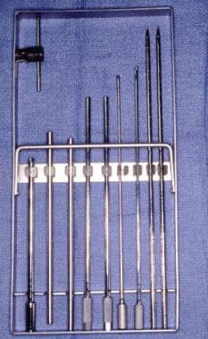
Orthopedic Equipment
Arthrodesis
Endodermal Sinus Tumor
Adenoma, Islet Cell
Mesenchymoma
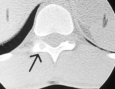
Tomography, X-Ray Computed
Polymyalgia Rheumatica
Tumor Necrosis Factor-alpha
Tumor Burden
Bone Cements
Fatal Outcome
Giant Cells, Langhans
Polymethyl Methacrylate
Autografts
Dog Diseases
Magnetic Resonance Imaging
Combined Modality Therapy
Pancreatic Neoplasms
Biopsy
Follow-Up Studies
Neoplasms, Experimental
Teratoma
Immunoenzyme Techniques
Ovarian Neoplasms
Wilms Tumor
Pelvic Bones
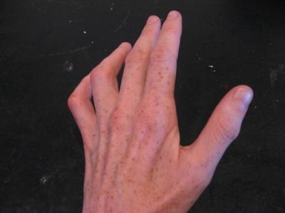
Hand
Brain Neoplasms
Genes, Tumor Suppressor
Retroperitoneal Neoplasms
Chondrosarcoma
Desmoplastic Small Round Cell Tumor
Carcinoid Tumor
Embolization, Therapeutic
Limb Salvage
Reverse Transcriptase Polymerase Chain Reaction
RNA, Messenger
Prosthetic reconstruction for tumours of the distal tibia and fibula. (1/136)
We have carried out prosthetic reconstruction in six patients with malignant or aggressively benign bone tumours of the distal tibia or fibula. The diagnoses were osteosarcoma in four patients, parosteal osteosarcoma in one and recurrent giant-cell tumour in one. Five tumours were in the distal tibia and one in the distal fibula. The mean duration of follow-up was 5.3 years (2.0 to 7.1). Reconstruction was achieved using custom-made, hinged prostheses which replaced the distal tibia and the ankle. The mean range of ankle movement after operation was 31 degrees and the joints were stable. The average functional score according to the system of the International Society of Limb Salvage was 24.2 and five of the patients had a good outcome. Complications occurred in two with wound infection and talar collapse. All patients were free from neoplastic disease at the latest follow-up. Prosthetic reconstruction may be used for the treatment of malignant tumours of the distal tibia and fibula in selected patients. (+info)Radioimmunoassay for human calcitonin employing synthetic calcitonin M: its clinical application. (2/136)
A sensitive and reliable radioimmunoassay for human calcitonin was described and applied to preliminary clinical studies. 125I-labelled synthetic human calcitonin M was purified by gel filtration with Sephadex G-25 and G-100. A nonequilibrium incubation system was applied at the final volume of incubation mixture of 500 mul, in which pooled plasma from normal subjects or hormone free serum was used as carrier protein at 20% incubation mixture. Dextran T 40 coated charcoal, resuspended in 1% bovine serum albumin buffer, was used for the separation of free from bound tracer. This showed the least nonspecific adsorption of tracer to charcoal. The assay was sensitive to 0.1 ng per milliliter of plasma. Recovery of synthetic human calcitonin added to plasma was found to be 101% (S. D., +/- 8). Diluted plasma from a patient with medullary thyroid carcinoma showed a dilution curve parallel to standards. Basal calcitonin levels were less than 0.3 ng/ml in normal subjects. Relatively high values were found in patients with chronic renal failure and in patients with malignant tumors. Extremely elevated values were found in patients with medullary thyroid carcinoma. Provocative calcium infusion tests were also performed. (+info)Tenosynovial giant cell tumor of finger, localized type: a case report. (3/136)
The authors report a typical case of tenosynovial giant cell tumor of the right middle finger of a 31-year-old man. Histologically, this tumor is characterized by a discrete proliferation of rounded synovial-like cells accompanied by a variable number of multinucleated giant cells, inflammatory cells, and xanthoma cells. Clinicopathologically, this tumor is a benign lesion that nonetheless possesses a capacity for local recurrence. Local excision with a small cuff of normal tissue is the treatment of choice in this tumor. (+info)In vitro induction of giant cell tumors from cultured hamster islets treated with N-Nitrosobis(2-Oxopropyl)amine. (4/136)
Giant cell carcinoma of the pancreas is a rare tumor. Its histogenesis is still controversial. In a Syrian hamster pancreatic cancer model, tumors similar to human giant cell carcinomas have been induced at an extremely low rate of incidence and after the use of high doses of pancreatic carcinogens. Thus far no tumors of giant cell type have been induced by the in vitro treatment of hamster pancreatic ductal cells with the potent pancreatic carcinogen N-nitrosobis(2-oxopropyl)amine (BOP). In the present study we report the induction of giant cell carcinoma from hamster islets treated with BOP in vitro. The results suggest that in hamsters some component of islet cells, probably stem cells, are the origin of giant cell carcinoma. (+info)Localized pigmented villonodular synovitis of the knee joint: neoplasm or reactive granuloma? A review of 18 cases. (5/136)
OBJECTIVE: The localized form of pigmented villonodular synovitis of the knee joint is a rare disease with limited alteration of the synovial membrane, the pathogenesis of which is the subject of controversial discussion. METHODS: Eighteen cases have been documented in our hospital since 1976. All of the patients had additional cartilage or meniscus damage. Treatment consisted of excision of the lesion and the adjacent synovial membrane, as well as therapy of the additional damage. RESULTS: The patients who had received such therapy were followed for 3-9 yr, without any clinical, sonographic or magnetic resonance tomographic signs of recurrence. In addition to the lack of a tendency towards recurrence, none of the cases displayed any further characteristics of the diffuse form of villonodular synovitis, such as invasiveness or malignant transformation. CONCLUSIONS: We therefore suggest that pigmented villonodular synovitis of the knee joint should be classified more strictly than before into a potentially neoplastic (diffuse) form and a reactive granulomatous (local) form. From the cases observed, we conclude that degenerative joint lesions may be the cause of the reactive granulomatous form. (+info)h-Caldesmon as a specific marker for smooth muscle tumors. Comparison with other smooth muscle markers in bone tumors. (6/136)
Caldesmon is a protein widely distributed in smooth and non-smooth muscle cells and is thought to regulate cellular contraction. Its isoform, high-molecular-weight caldesmon (h-CD), was demonstrated to be specific for smooth muscle cells and smooth muscle tumors of the soft tissue and to never be expressed in myofibroblasts. We performed an immunohistochemical study to examine h-CD expression in the following bone tumors: conventional and non-conventional osteosarcoma, 13; malignant fibrous histiocytoma of bone, 5; giant cell tumors of bone, 5; chondroblastoma, 3; metastatic leiomyosarcoma, 2; and rhabdomyosarcoma, 1. Frequent immunoreactivity for muscle actin (alpha-smooth muscle actin or muscle-specific actin) was seen in 11 of 13 osteosarcomas and all other tumors, whereas h-CD was expressed intensely only in 2 leiomyosarcomas. h-CD is considered a specific and useful marker to distinguish smooth muscle tumor from bone tumors with myoid differentiation. (+info)Giant-cell tumour of the tendon sheath. Is radiotherapy indicated to prevent recurrence after surgery? (7/136)
Giant-cell tumour of the tendon sheath, also called pigmented villonodular synovitis, is a benign tumour with a high incidence of recurrence. We have tried to identify risk factors for recurrence. Of the 48 patients included in the study, 14 received radiotherapy after surgery. Only two (4%) had a recurrence. This compares favourably with previously reported incidences of between 25% and 45%. (+info)Apoptosis in giant cell tumors of bone. (8/136)
Although giant cell tumor of bone (GCT) is characterized by the extensive multinucleated giant cells among mononuclear stromal cells, proliferation of these cells and multinucleation are not without limit in certain cases. Few studies on oncogenesis of GCT have focused on the negative growth control, including growth arrest and apoptosis. The purpose of this study was to investigate the mechanism of cell death in multinucleated giant cells and stromal cells of GCT. In this study, we have demonstrated that GCT cells can undergo apoptosis. The cells in surgical specimen were positively stained in situ nick end labeling methods, and electron micrographs showed the morphological changes associated with apoptosis in some of stromal cells and multinucleated giant cells. A candidate responsible for this apoptosis was then examined using cultured GCT cells. We focused on Fas that is a major trigger of apoptosis. Cultured GCT cells expressed detectable amount of Fas on their surface. Although GCT cells did a little undergo apoptosis following treatment with anti-Fas alone, combination treatment with cyclohexamide led to an increase in apoptosis of the GCT cells. These data suggested that the sensitizing activity of cyclohexamide on anti-Fas mediated cytotoxicity could happen in vitro. (+info)A Giant Cell Tumor (GCT) of bone is a relatively uncommon, locally aggressive tumor that can sometimes become malignant. It is characterized by the presence of multinucleated giant cells which are distributed throughout the tumor tissue. These giant cells are thought to be derived from osteoclasts, which are specialized cells responsible for bone resorption.
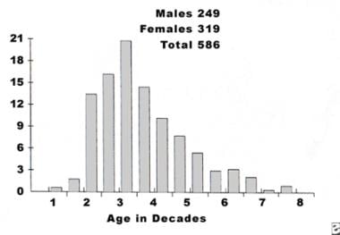
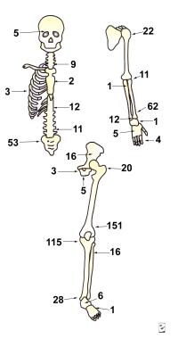
GCTs typically affect adults in their 20s and 30s, with a slight female predominance. The most common sites of involvement include the long bones near the knee (distal femur and proximal tibia), as well as the distal radius, sacrum, and spine.
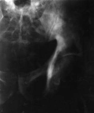
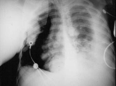
The tumor usually presents as pain and swelling in the affected area, sometimes accompanied by restricted mobility or pathological fractures due to bone weakening. The diagnosis is typically made based on imaging studies (such as X-rays, CT scans, or MRI) and confirmed through a biopsy.
Treatment options for GCTs of bone may include intralesional curettage with or without the use of adjuvant therapies (like phenol, liquid nitrogen, or cement), radiation therapy, or surgical resection. In some cases, systemic treatments like denosumab, a monoclonal antibody targeting RANKL, may be used to control the growth and spread of the tumor. Regular follow-ups are essential to monitor for potential recurrence, which can occur in up to 50% of cases within five years after treatment.
Giant cell tumors (GCTs) are a type of benign or rarely malignant bone tumor that is characterized by the presence of multinucleated giant cells. These tumors typically affect adults between the ages of 20 and 40, and they can occur in any bone, but they most commonly involve the long bones near the knee joint.
GCTs are composed of three types of cells: mononuclear stromal cells, which produce the matrix of the tumor; multinucleated osteoclast-like giant cells, which resemble the bone-resorbing cells found in normal bone; and macrophages, which are part of the body's immune system.
The mononuclear stromal cells produce a variety of growth factors that stimulate the formation and activity of the osteoclast-like giant cells, leading to localized bone destruction. The tumor may cause pain, swelling, and limited mobility in the affected area.
While GCTs are typically benign, they can be aggressive and locally destructive, with a tendency to recur after surgical removal. In some cases, GCTs may undergo malignant transformation, leading to the development of sarcomas. Treatment options for GCTs include curettage (scraping out) of the tumor, followed by bone grafting or the use of a cement spacer to fill the defect, and/or adjuvant therapy with radiation or chemotherapy.
Curettage is a medical procedure that involves scraping or removing tissue from the lining of an organ or body cavity, typically performed using a curette, which is a long, thin surgical instrument with a looped or sharp end. In gynecology, curettage is often used to remove tissue from the uterus during a procedure called dilation and curettage (D&C) to diagnose or treat abnormal uterine bleeding, or to remove residual placental or fetal tissue following a miscarriage or abortion. Curettage may also be used in other medical specialties to remove damaged or diseased tissue from areas such as the nose, throat, or skin.
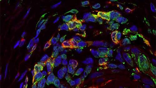
Giant cells are large, multinucleated cells that result from the fusion of monocytes or macrophages. They can be found in various types of inflammatory and degenerative lesions, including granulomas, which are a hallmark of certain diseases such as tuberculosis and sarcoidosis. There are several types of giant cells, including:
1. Langhans giant cells: These have a horseshoe-shaped or crescentic arrangement of nuclei around the periphery of the cell. They are typically found in granulomas associated with infectious diseases such as tuberculosis and histoplasmosis.
2. Foreign body giant cells: These form in response to the presence of foreign material, such as a splinter or suture, in tissue. The nuclei are usually scattered throughout the cell cytoplasm.
3. Touton giant cells: These are found in certain inflammatory conditions, such as xanthomatosis and granulomatous slack skin. They have a central core of lipid-laden histiocytes surrounded by a ring of nuclei.
4. Osteoclast giant cells: These are multinucleated cells responsible for bone resorption. They can be found in conditions such as giant cell tumors of bone and Paget's disease.
It is important to note that the presence of giant cells alone does not necessarily indicate a specific diagnosis, and their significance must be interpreted within the context of the overall clinical and pathological findings.
Bone neoplasms are abnormal growths or tumors that develop in the bone. They can be benign (non-cancerous) or malignant (cancerous). Benign bone neoplasms do not spread to other parts of the body and are rarely a threat to life, although they may cause problems if they grow large enough to press on surrounding tissues or cause fractures. Malignant bone neoplasms, on the other hand, can invade and destroy nearby tissue and may spread (metastasize) to other parts of the body.
There are many different types of bone neoplasms, including:
1. Osteochondroma - a benign tumor that develops from cartilage and bone
2. Enchondroma - a benign tumor that forms in the cartilage that lines the inside of the bones
3. Chondrosarcoma - a malignant tumor that develops from cartilage
4. Osteosarcoma - a malignant tumor that develops from bone cells
5. Ewing sarcoma - a malignant tumor that develops in the bones or soft tissues around the bones
6. Giant cell tumor of bone - a benign or occasionally malignant tumor that develops from bone tissue
7. Fibrosarcoma - a malignant tumor that develops from fibrous tissue in the bone
The symptoms of bone neoplasms vary depending on the type, size, and location of the tumor. They may include pain, swelling, stiffness, fractures, or limited mobility. Treatment options depend on the type and stage of the tumor but may include surgery, radiation therapy, chemotherapy, or a combination of these treatments.
A "Giant Cell Carcinoma" is a type of cancer that originates from epithelial cells and is characterized by the presence of large, abnormal cells called giant cells. These giant cells are formed by the fusion of several individual cells, resulting in a single, large cell with multiple nuclei. Giant cell carcinomas can occur in various organs, including the lungs, esophagus, and thyroid gland.
Giant cell carcinoma of the lung is a rare and aggressive form of lung cancer that typically affects smokers. It is characterized by the presence of large, bizarre cells with multiple nuclei, as well as a high degree of cellular pleomorphism (variation in size and shape of cells). This type of lung cancer tends to grow and spread quickly, making it difficult to treat.
Giant cell carcinoma of the esophagus is also a rare and aggressive form of cancer that affects the esophagus. It is characterized by the presence of large, abnormal cells with multiple nuclei, as well as a high degree of cellular pleomorphism. This type of esophageal cancer tends to grow and spread quickly, making it difficult to treat.
Giant cell carcinoma of the thyroid gland is an extremely rare form of thyroid cancer that affects the thyroid gland. It is characterized by the presence of large, abnormal cells with multiple nuclei, as well as a high degree of cellular pleomorphism. This type of thyroid cancer tends to grow and spread quickly, making it difficult to treat.
Overall, giant cell carcinomas are aggressive forms of cancer that can occur in various organs. They are characterized by the presence of large, abnormal cells with multiple nuclei, as well as a high degree of cellular pleomorphism. Due to their aggressive nature and tendency to grow and spread quickly, giant cell carcinomas can be difficult to treat.
A giant cell granuloma is a type of non-cancerous (benign) lesion characterized by the presence of large collections of immune cells called macrophages, which have fused together to form multinucleated giant cells. These lesions can occur in various tissues throughout the body but are most commonly found in the oral cavity and jawbone.
Giant cell granulomas can be further classified into two types: central (or bone) giant cell granuloma and peripheral giant cell granuloma. Central giant cell granulomas arise from the bone, while peripheral giant cell granulomas occur in the soft tissues of the gingiva or mouth lining.
Central giant cell granulomas are more aggressive than peripheral ones and can cause significant bone destruction if left untreated. They typically affect younger individuals, with a higher prevalence in females than males. The exact cause of central giant cell granulomas is not well understood but may be associated with local trauma, hormonal imbalances, or genetic factors.
Peripheral giant cell granulomas are less aggressive and usually present as painless, slow-growing nodules on the gums. They typically affect adults, with a higher prevalence in females than males. Peripheral giant cell granulomas may be associated with local irritants such as plaque, calculus, or dental restorations.
Treatment for giant cell granulomas depends on their size, location, and aggressiveness. Surgical excision is the most common treatment approach, but other options such as curettage, corticosteroid injections, or medication therapy may also be considered. Regular follow-up appointments with a healthcare provider are essential to monitor for recurrence.
Giant Cell Arteritis (GCA), also known as Temporal Arteritis, is a chronic inflammatory disease affecting large and medium-sized arteries, most commonly the temporal artery. It primarily occurs in people over 50 years old. The condition is characterized by the infiltration of the artery walls with immune cells, leading to inflammation, swelling, and damage. This can restrict blood flow, causing various symptoms.
The key feature of GCA is the presence of multinucleated giant cells, which are large collections of fused immune cells, in the affected artery walls. These cells are a hallmark of this condition when viewed under a microscope.
Common symptoms include new onset of severe headaches, scalp tenderness, jaw pain while chewing (called jaw claudication), vision problems, and systemic symptoms such as fever, fatigue, and weight loss. If left untreated, GCA can lead to serious complications like blindness or stroke. Treatment typically involves high-dose corticosteroids to reduce inflammation and prevent further damage.
Femoral neoplasms refer to abnormal growths or tumors that develop in the femur, which is the long thigh bone in the human body. These neoplasms can be benign (non-cancerous) or malignant (cancerous). Benign femoral neoplasms are slow-growing and rarely spread to other parts of the body, while malignant neoplasms are aggressive and can invade nearby tissues and organs, as well as metastasize (spread) to distant sites.
There are various types of femoral neoplasms, including osteochondromas, enchondromas, chondrosarcomas, osteosarcomas, and Ewing sarcomas, among others. The specific type of neoplasm is determined by the cell type from which it arises and its behavior.
Symptoms of femoral neoplasms may include pain, swelling, stiffness, or weakness in the thigh, as well as a palpable mass or limited mobility. Diagnosis typically involves imaging studies such as X-rays, CT scans, or MRI, as well as biopsy to determine the type and grade of the tumor. Treatment options may include surgery, radiation therapy, chemotherapy, or a combination of these approaches, depending on the type, size, location, and stage of the neoplasm.
A Granulosa Cell Tumor is a type of sex cord-stromal tumor, which are uncommon neoplasms that arise from the supporting cells of the ovary or testis. These tumors account for approximately 5% of all ovarian tumors and can occur at any age, but they are most commonly found in perimenopausal and postmenopausal women.
Granulosa cell tumors originate from the granulosa cells, which are normally responsible for producing estrogen and supporting the development of the egg within the ovarian follicle. These tumors can be functional, meaning they produce hormones, or nonfunctional. Functional granulosa cell tumors often secrete estrogen, leading to symptoms such as irregular menstrual periods, postmenopausal bleeding, and, in rare cases, the development of male characteristics (virilization) due to androgen production.
Granulosa cell tumors are typically slow-growing and can vary in size. They are often diagnosed at an early stage because they cause symptoms related to hormonal imbalances or, less commonly, due to abdominal pain or distention caused by the growing mass. The diagnosis is usually confirmed through imaging studies (such as ultrasound, CT, or MRI) and a biopsy or surgical removal of the tumor, followed by histopathological examination.
Treatment for granulosa cell tumors typically involves surgery to remove the tumor and, in some cases, adjacent organs if there is evidence of spread. The role of chemotherapy and radiation therapy is less clear, but they may be used in certain situations, such as advanced-stage disease or high-risk features. Regular follow-up with imaging studies and tumor marker measurements (such as inhibin) is essential due to the risk of recurrence, even many years after initial treatment.
Aneurysmal bone cyst (ABC) is a benign but locally aggressive tumor that typically involves the metaphysis of long bones in children and adolescents. It is characterized by blood-filled spaces or cysts separated by fibrous septa containing osteoclast-type giant cells, spindle cells, and capillary vessels.
ABCs can also arise in other locations such as the vertebral column, pelvis, and skull. They may cause bone pain, swelling, or pathologic fractures. The exact cause of ABC is unknown, but it is thought to be related to a reactive process to a primary bone lesion or trauma.
Treatment options for ABC include curettage and bone grafting, intralesional injection of corticosteroids or bone marrow aspirate, and adjuvant therapy with phenol or liquid nitrogen. In some cases, radiation therapy may be used, but it is generally avoided due to the risk of secondary malignancies. Recurrence rates after treatment range from 10-30%.
Neoplasms, germ cell and embryonal are types of tumors that originate from the abnormal growth of cells. Here's a brief medical definition for each:
1. Neoplasms: Neoplasms refer to abnormal tissue growths or masses, which can be benign (non-cancerous) or malignant (cancerous). They result from uncontrolled cell division and may invade surrounding tissues or spread to other parts of the body through a process called metastasis.
2. Germ Cell Tumors: These are rare tumors that develop from the germ cells, which give rise to sperm and eggs in the reproductive organs (ovaries and testes). They can be benign or malignant and may occur in both children and adults. Germ cell tumors can also arise outside of the reproductive organs, a condition known as extragonadal germ cell tumors.
3. Embryonal Tumors: These are a type of malignant neoplasm that primarily affects infants and young children. They develop from embryonic cells, which are immature cells present during fetal development. Embryonal tumors can occur in various organs, including the brain (medulloblastomas), nervous system (primitive neuroectodermal tumors or PNETs), and other areas like the kidneys and liver.
It is essential to note that these conditions require professional medical evaluation and treatment by healthcare professionals with expertise in oncology and related fields.
A Granular Cell Tumor (GCT) is a rare, usually benign neoplasm that can occur in various parts of the body. These tumors are typically composed of large polygonal cells with abundant eosinophilic granular cytoplasm, which contain numerous mitochondria. They often involve the skin and subcutaneous tissues, but they can also arise in the oral cavity, gastrointestinal tract, respiratory system, and other visceral organs.
Granular Cell Tumors are thought to originate from Schwann cells, which are nerve sheath cells, although their exact origin is still a matter of debate. They usually present as solitary, slow-growing nodules or masses that are often painless, but they can become symptomatic if they involve sensitive areas or if they undergo malignant transformation, which occurs in about 1-2% of cases.
The diagnosis of Granular Cell Tumors is usually made based on histopathological examination of a biopsy specimen. Immunohistochemical staining can be used to confirm the Schwann cell origin of these tumors, as they typically express S-100 protein and other markers of neural differentiation.
Treatment options for Granular Cell Tumors depend on their location, size, and behavior. Solitary, benign tumors can often be excised surgically with a wide margin to reduce the risk of recurrence. However, malignant tumors or those that cannot be completely removed may require more aggressive treatment, such as radiation therapy or chemotherapy. Regular follow-up is recommended to monitor for recurrence or metastasis.
Bone transplantation, also known as bone grafting, is a surgical procedure in which bone or bone-like material is transferred from one part of the body to another or from one person to another. The graft may be composed of cortical (hard outer portion) bone, cancellous (spongy inner portion) bone, or a combination of both. It can be taken from different sites in the same individual (autograft), from another individual of the same species (allograft), or from an animal source (xenograft). The purpose of bone transplantation is to replace missing bone, provide structural support, and stimulate new bone growth. This procedure is commonly used in orthopedic, dental, and maxillofacial surgeries to repair bone defects caused by trauma, tumors, or congenital conditions.
Spinal neoplasms refer to abnormal growths or tumors found within the spinal column, which can be benign (non-cancerous) or malignant (cancerous). These tumors can originate in the spine itself, called primary spinal neoplasms, or they can spread to the spine from other parts of the body, known as secondary or metastatic spinal neoplasms. Spinal neoplasms can cause various symptoms, such as back pain, neurological deficits, and even paralysis, depending on their location and size. Early diagnosis and treatment are crucial to prevent or minimize long-term complications and improve the patient's prognosis.
Testicular neoplasms are abnormal growths or tumors in the testicle that can be benign (non-cancerous) or malignant (cancerous). They are a type of genitourinary cancer, which affects the reproductive and urinary systems. Testicular neoplasms can occur in men of any age but are most commonly found in young adults between the ages of 15 and 40.
Testicular neoplasms can be classified into two main categories: germ cell tumors and non-germ cell tumors. Germ cell tumors, which arise from the cells that give rise to sperm, are further divided into seminomas and non-seminomas. Seminomas are typically slow-growing and have a good prognosis, while non-seminomas tend to grow more quickly and can spread to other parts of the body.
Non-germ cell tumors are less common than germ cell tumors and include Leydig cell tumors, Sertoli cell tumors, and lymphomas. These tumors can have a variety of clinical behaviors, ranging from benign to malignant.
Testicular neoplasms often present as a painless mass or swelling in the testicle. Other symptoms may include a feeling of heaviness or discomfort in the scrotum, a dull ache in the lower abdomen or groin, and breast enlargement (gynecomastia).
Diagnosis typically involves a physical examination, imaging studies such as ultrasound or CT scan, and blood tests to detect tumor markers. Treatment options depend on the type and stage of the neoplasm but may include surgery, radiation therapy, chemotherapy, or a combination of these modalities. Regular self-examinations of the testicles are recommended for early detection and improved outcomes.
The sacrum is a triangular-shaped bone in the lower portion of the human vertebral column, located between the lumbar spine and the coccyx (tailbone). It forms through the fusion of several vertebrae during fetal development. The sacrum's base articulates with the fifth lumbar vertebra, while its apex connects with the coccyx.
The sacrum plays an essential role in supporting the spine and transmitting weight from the upper body to the pelvis and lower limbs. It also serves as an attachment site for various muscles and ligaments. The sacral region is often a focus in medical and chiropractic treatments due to its importance in spinal stability, posture, and overall health.
The radius is one of the two bones in the forearm in humans and other vertebrates. In humans, it runs from the lateral side of the elbow to the thumb side of the wrist. It is responsible for rotation of the forearm and articulates with the humerus at the elbow and the carpals at the wrist. Any medical condition or injury that affects the radius can impact the movement and function of the forearm and hand.
A Sertoli cell tumor is a rare type of sex-cord stromal tumor that develops in the testicles or, more rarely, in the ovaries. These tumors arise from the Sertoli cells, which are specialized cells within the testicle that help to nurture and protect the developing sperm cells. In the ovary, Sertoli cell tumors are thought to arise from similar cells that are part of the supporting tissue in the ovary.
Sertoli cell tumors can occur in people of any age but are most commonly found in middle-aged adults. They are usually slow-growing and may not cause any symptoms, especially if they are small. However, larger tumors or those that have spread (metastasized) may cause various symptoms depending on their location and size.
Symptoms of a Sertoli cell tumor can include:
* A painless lump or swelling in the testicle or ovary
* Abdominal pain or discomfort
* Bloating or a feeling of fullness in the abdomen
* Changes in bowel habits or urinary frequency
* Pain during sexual intercourse (in women)
* Hormonal imbalances, such as gynecomastia (breast development) in men or menstrual irregularities in women.
Diagnosis of a Sertoli cell tumor typically involves a combination of imaging tests, such as ultrasound, CT scan, or MRI, and blood tests to check for elevated levels of certain hormones that may be produced by the tumor. A biopsy may also be performed to confirm the diagnosis and determine the tumor's grade and stage.
Treatment for Sertoli cell tumors typically involves surgical removal of the tumor, along with any affected lymph nodes or other tissues. Additional treatments, such as radiation therapy or chemotherapy, may be recommended in cases where the tumor has spread or is at a higher risk of recurrence. Regular follow-up care is also important to monitor for any signs of recurrence or new tumors.
Soft tissue neoplasms refer to abnormal growths or tumors that develop in the soft tissues of the body. Soft tissues include muscles, tendons, ligaments, fascia, nerves, blood vessels, fat, and synovial membranes (the thin layer of cells that line joints and tendons). Neoplasms can be benign (non-cancerous) or malignant (cancerous), and their behavior and potential for spread depend on the specific type of neoplasm.
Benign soft tissue neoplasms are typically slow-growing, well-circumscribed, and rarely spread to other parts of the body. They can often be removed surgically with a low risk of recurrence. Examples of benign soft tissue neoplasms include lipomas (fat tumors), schwannomas (nerve sheath tumors), and hemangiomas (blood vessel tumors).
Malignant soft tissue neoplasms, on the other hand, can grow rapidly, invade surrounding tissues, and may metastasize (spread) to distant parts of the body. They are often more difficult to treat than benign neoplasms and require a multidisciplinary approach, including surgery, radiation therapy, and chemotherapy. Examples of malignant soft tissue neoplasms include sarcomas, such as rhabdomyosarcoma (arising from skeletal muscle), leiomyosarcoma (arising from smooth muscle), and angiosarcoma (arising from blood vessels).
It is important to note that soft tissue neoplasms can occur in any part of the body, and their diagnosis and treatment require a thorough evaluation by a healthcare professional with expertise in this area.
The fibula is a slender bone located in the lower leg of humans and other vertebrates. It runs parallel to the larger and more robust tibia, and together they are known as the bones of the leg or the anterior tibial segment. The fibula is the lateral bone in the leg, positioned on the outside of the tibia.
In humans, the fibula extends from the knee joint proximally to the ankle joint distally. Its proximal end, called the head of the fibula, articulates with the lateral condyle of the tibia and forms part of the inferior aspect of the knee joint. The narrowed portion below the head is known as the neck of the fibula.
The shaft of the fibula, also called the body of the fibula, is a long, thin structure that descends from the neck and serves primarily for muscle attachment rather than weight-bearing functions. The distal end of the fibula widens to form the lateral malleolus, which is an important bony landmark in the ankle region. The lateral malleolus articulates with the talus bone of the foot and forms part of the ankle joint.
The primary functions of the fibula include providing attachment sites for muscles that act on the lower leg, ankle, and foot, as well as contributing to the stability of the ankle joint through its articulation with the talus bone. Fractures of the fibula can occur due to various injuries, such as twisting or rotational forces applied to the ankle or direct trauma to the lateral aspect of the lower leg.
Pigmented villonodular synovitis (PVNS) is a rare, benign condition that affects the synovial membrane, which lines the joints. It is characterized by the proliferation of synovial cells and the deposition of hemosiderin, a pigment resulting from the breakdown of blood products. This can lead to joint swelling, pain, stiffness, and limited mobility. PVNS typically affects the large joints such as the knee or hip, but it can also occur in smaller joints, bursae, or tendon sheaths.
There are two forms of PVNS: localized and diffuse. Localized PVNS, also known as giant cell tumor of the tendon sheath, affects a specific area within the joint and is more likely to be treated successfully with surgery. Diffuse PVNS, on the other hand, involves the entire synovial lining of the joint and has a higher recurrence rate even after surgical removal.
The exact cause of PVNS remains unclear, but it is not considered a malignant condition. Treatment usually involves surgical removal of the affected synovium, with or without radiation therapy or chemotherapy to reduce the risk of recurrence. In some cases, arthroscopic surgery may be an option for localized PVNS.
Chondroblastoma is a rare, benign (non-cancerous) bone tumor that typically develops in the epiphysis, which is the rounded end of a long bone near a joint. It primarily affects children and adolescents, with around 90% of cases occurring before the age of 20.
The tumor arises from chondroblasts, cells responsible for producing cartilage during bone growth. Chondroblastoma is usually slow-growing and typically causes localized pain, swelling, or tenderness in the affected area. In some cases, it may weaken the bone and lead to fractures.
Treatment generally involves surgical removal of the tumor, followed by curettage (scraping) of the surrounding bone tissue and replacement with bone grafts or substitutes. Recurrence is possible but rare, and long-term prognosis is usually favorable.
A Leydig cell tumor is a rare type of sex cord-stromal tumor that arises from the Leydig cells (interstitial cells) of the testis in males or ovarian tissue in females. These cells are responsible for producing androgens, particularly testosterone.
Leydig cell tumors can occur at any age but are most common in middle-aged to older men. In women, they are extremely rare and usually found in postmenopausal women. Most Leydig cell tumors are benign (noncancerous), but about 10% can be malignant (cancerous) and have the potential to spread to other parts of the body.
Symptoms of a Leydig cell tumor may include:
* A painless testicular or ovarian mass
* Gynecomastia (enlargement of breast tissue in men) due to increased estrogen production
* Early puberty in children
* Decreased libido and erectile dysfunction in men
* Irregular menstrual cycles in women
Diagnosis is usually made through imaging tests such as ultrasound, CT scan, or MRI, followed by a biopsy to confirm the presence of a Leydig cell tumor. Treatment typically involves surgical removal of the tumor, and additional therapies such as radiation therapy or chemotherapy may be recommended for malignant tumors. Regular follow-up is necessary to monitor for recurrence.
A germinoma is a type of tumor that develops in the brain or the spine, primarily in the pituitary gland or pineal gland. It is a rare form of primary central nervous system (CNS) cancer and is classified as a type of germ cell tumor. These tumors arise from cells that normally develop into sperm or eggs, which can migrate to unusual locations during embryonic development.
Germinomas are highly sensitive to radiation therapy and chemotherapy, making them generally treatable and curable with appropriate medical intervention. Symptoms of a germinoma may include headaches, nausea, vomiting, visual disturbances, hormonal imbalances, and neurological deficits, depending on the location and size of the tumor. Diagnosis typically involves imaging studies like MRI or CT scans, followed by a biopsy to confirm the presence of malignant cells.
Tenosynovitis is a medical condition characterized by inflammation of the lining (synovium) surrounding a tendon, which is a cord-like structure that attaches muscle to bone. This inflammation can cause pain, swelling, and difficulty moving the affected joint. Tenosynovitis often affects the hands, wrists, feet, and ankles, and it can result from various causes, including infection, injury, overuse, or autoimmune disorders like rheumatoid arthritis. Prompt diagnosis and treatment of tenosynovitis are essential to prevent complications such as tendon rupture or chronic pain.
A tendon is the strong, flexible band of tissue that connects muscle to bone. It helps transfer the force produced by the muscle to allow various movements of our body parts. Tendons are made up of collagen fibers arranged in parallel bundles and have a poor blood supply, making them prone to injuries and slow to heal. Examples include the Achilles tendon, which connects the calf muscle to the heel bone, and the patellar tendon, which connects the kneecap to the shinbone.
Orthopedic procedures are surgical or nonsurgical methods used to treat musculoskeletal conditions, including injuries, deformities, or diseases of the bones, joints, muscles, ligaments, and tendons. These procedures can range from simple splinting or casting to complex surgeries such as joint replacements, spinal fusions, or osteotomies (cutting and repositioning bones). The primary goal of orthopedic procedures is to restore function, reduce pain, and improve the quality of life for patients.
The tibia, also known as the shin bone, is the larger of the two bones in the lower leg and part of the knee joint. It supports most of the body's weight and is a major insertion point for muscles that flex the foot and bend the leg. The tibia articulates with the femur at the knee joint and with the fibula and talus bone at the ankle joint. Injuries to the tibia, such as fractures, are common in sports and other activities that put stress on the lower leg.
Local neoplasm recurrence is the return or regrowth of a tumor in the same location where it was originally removed or treated. This means that cancer cells have survived the initial treatment and started to grow again in the same area. It's essential to monitor and detect any local recurrence as early as possible, as it can affect the prognosis and may require additional treatment.
Tumor markers are substances that can be found in the body and their presence can indicate the presence of certain types of cancer or other conditions. Biological tumor markers refer to those substances that are produced by cancer cells or by other cells in response to cancer or certain benign (non-cancerous) conditions. These markers can be found in various bodily fluids such as blood, urine, or tissue samples.
Examples of biological tumor markers include:
1. Proteins: Some tumor markers are proteins that are produced by cancer cells or by other cells in response to the presence of cancer. For example, prostate-specific antigen (PSA) is a protein produced by normal prostate cells and in higher amounts by prostate cancer cells.
2. Genetic material: Tumor markers can also include genetic material such as DNA, RNA, or microRNA that are shed by cancer cells into bodily fluids. For example, circulating tumor DNA (ctDNA) is genetic material from cancer cells that can be found in the bloodstream.
3. Metabolites: Tumor markers can also include metabolic products produced by cancer cells or by other cells in response to cancer. For example, lactate dehydrogenase (LDH) is an enzyme that is released into the bloodstream when cancer cells break down glucose for energy.
It's important to note that tumor markers are not specific to cancer and can be elevated in non-cancerous conditions as well. Therefore, they should not be used alone to diagnose cancer but rather as a tool in conjunction with other diagnostic tests and clinical evaluations.
Muscle neoplasms are abnormal growths or tumors that develop in the muscle tissue. They can be benign (non-cancerous) or malignant (cancerous). Benign muscle neoplasms are typically slow-growing and do not spread to other parts of the body, while malignant muscle neoplasms, also known as soft tissue sarcomas, can grow quickly, invade nearby tissues, and metastasize (spread) to distant parts of the body.
Soft tissue sarcomas can arise from any of the muscles in the body, including the skeletal muscles (voluntary muscles that attach to bones and help with movement), smooth muscles (involuntary muscles found in the walls of blood vessels, digestive tract, and other organs), or cardiac muscle (the specialized muscle found in the heart).
There are many different types of soft tissue sarcomas, each with its own set of characteristics and prognosis. Treatment for muscle neoplasms typically involves a combination of surgery, radiation therapy, and chemotherapy, depending on the type, size, location, and stage of the tumor.
Phenol, also known as carbolic acid, is an organic compound with the molecular formula C6H5OH. It is a white crystalline solid that is slightly soluble in water and has a melting point of 40-42°C. Phenol is a weak acid, but it is quite reactive and can be converted into a variety of other chemicals.
In a medical context, phenol is most commonly used as a disinfectant and antiseptic. It has a characteristic odor that is often described as "tarry" or " medicinal." Phenol is also used in some over-the-counter products, such as mouthwashes and throat lozenges, to help kill bacteria and freshen breath.
However, phenol is also a toxic substance that can cause serious harm if it is swallowed, inhaled, or absorbed through the skin. It can cause irritation and burns to the eyes, skin, and mucous membranes, and it can damage the liver and kidneys if ingested. Long-term exposure to phenol has been linked to an increased risk of cancer.
Because of its potential for harm, phenol is regulated as a hazardous substance in many countries, and it must be handled with care when used in medical or industrial settings.
The ilium is the largest and broadest of the three parts that make up the hip bone or coxal bone. It is the uppermost portion of the pelvis and forms the side of the waist. The ilium has a curved, fan-like shape and articulates with the sacrum at the back to form the sacroiliac joint. The large, concave surface on the top of the ilium is called the iliac crest, which can be felt as a prominent ridge extending from the front of the hip to the lower back. This region is significant in orthopedics and physical examinations for its use in assessing various medical conditions and performing certain maneuvers during the physical examination.
Skull neoplasms refer to abnormal growths or tumors that develop within the skull. These growths can be benign (non-cancerous) or malignant (cancerous). They can originate from various types of cells, such as bone cells, nerve cells, or soft tissues. Skull neoplasms can cause various symptoms depending on their size and location, including headaches, seizures, vision problems, hearing loss, and neurological deficits. Treatment options include surgery, radiation therapy, and chemotherapy. It is important to note that a neoplasm in the skull can also refer to metastatic cancer, which has spread from another part of the body to the skull.
Reconstructive surgical procedures are a type of surgery aimed at restoring the form and function of body parts that are defective or damaged due to various reasons such as congenital abnormalities, trauma, infection, tumors, or disease. These procedures can involve the transfer of tissue from one part of the body to another, manipulation of bones, muscles, and tendons, or use of prosthetic materials to reconstruct the affected area. The goal is to improve both the physical appearance and functionality of the body part, thereby enhancing the patient's quality of life. Examples include breast reconstruction after mastectomy, cleft lip and palate repair, and treatment of severe burns.
The humerus is the long bone in the upper arm that extends from the shoulder joint (glenohumeral joint) to the elbow joint. It articulates with the glenoid cavity of the scapula to form the shoulder joint and with the radius and ulna bones at the elbow joint. The proximal end of the humerus has a rounded head that provides for movement in multiple planes, making it one of the most mobile joints in the body. The greater and lesser tubercles are bony prominences on the humeral head that serve as attachment sites for muscles that move the shoulder and arm. The narrow shaft of the humerus provides stability and strength for weight-bearing activities, while the distal end forms two articulations: one with the ulna (trochlea) and one with the radius (capitulum). Together, these structures allow for a wide range of motion in the shoulder and elbow joints.
Seminoma is a type of germ cell tumor that develops in the testicle. It is a malignant tumor, meaning it can spread to other parts of the body if left untreated. Seminomas are typically slow-growing and tend to remain localized to the testicle for a longer period compared to other types of testicular cancer. They usually occur in men between the ages of 25 and 45 but can develop at any age.
Seminomas can be classified into two main subtypes: classical seminoma and spermatocytic seminoma. Classical seminoma is more common and typically responds well to treatment, while spermatocytic seminoma is rarer and tends to have a better prognosis with a lower risk of spreading.
Seminomas are usually treated with surgery to remove the affected testicle (orchiectomy), followed by radiation therapy or chemotherapy to kill any remaining cancer cells. The prognosis for seminoma is generally good, especially when caught and treated early. Regular self-examinations of the testicles can help detect any lumps or abnormalities that may indicate the presence of a seminoma or other type of testicular cancer.
Immunohistochemistry (IHC) is a technique used in pathology and laboratory medicine to identify specific proteins or antigens in tissue sections. It combines the principles of immunology and histology to detect the presence and location of these target molecules within cells and tissues. This technique utilizes antibodies that are specific to the protein or antigen of interest, which are then tagged with a detection system such as a chromogen or fluorophore. The stained tissue sections can be examined under a microscope, allowing for the visualization and analysis of the distribution and expression patterns of the target molecule in the context of the tissue architecture. Immunohistochemistry is widely used in diagnostic pathology to help identify various diseases, including cancer, infectious diseases, and immune-mediated disorders.
Benign fibrous histiocytoma (BFH) is a common benign tumor of the skin and superficial soft tissues. It primarily affects middle-aged adults and is more prevalent in men than women. The exact cause of BFH is unknown, but it's thought to arise from dermal fibroblasts or histiocytes.
Medical Definition: Benign Fibrous Histiocytoma (BFH) is a benign, slowly growing, solitary cutaneous or subcutaneous nodular tumor predominantly composed of a mixture of fibroblastic and histiocytic-like cells. The tumor typically presents as a well-circumscribed, firm, dome-shaped papule or nodule, ranging in size from a few millimeters to several centimeters. Histologically, BFH is characterized by the proliferation of spindle-shaped fibroblasts and histiocytes arranged in a storiform pattern, along with variable amounts of collagen deposition, multinucleated giant cells, and hemosiderin deposits. The lesion usually has a pushing border with no invasion into the surrounding tissues. BFH generally follows a benign clinical course, with local recurrence being uncommon following complete surgical excision.
Retrospective studies, also known as retrospective research or looking back studies, are a type of observational study that examines data from the past to draw conclusions about possible causal relationships between risk factors and outcomes. In these studies, researchers analyze existing records, medical charts, or previously collected data to test a hypothesis or answer a specific research question.
Retrospective studies can be useful for generating hypotheses and identifying trends, but they have limitations compared to prospective studies, which follow participants forward in time from exposure to outcome. Retrospective studies are subject to biases such as recall bias, selection bias, and information bias, which can affect the validity of the results. Therefore, retrospective studies should be interpreted with caution and used primarily to generate hypotheses for further testing in prospective studies.
Osteoclasts are large, multinucleated cells that are primarily responsible for bone resorption, a process in which they break down and dissolve the mineralized matrix of bones. They are derived from monocyte-macrophage precursor cells of hematopoietic origin and play a crucial role in maintaining bone homeostasis by balancing bone formation and bone resorption.
Osteoclasts adhere to the bone surface and create an isolated microenvironment, called the "resorption lacuna," between their cell membrane and the bone surface. Here, they release hydrogen ions into the lacuna through a process called proton pumping, which lowers the pH and dissolves the mineral component of the bone matrix. Additionally, osteoclasts secrete proteolytic enzymes, such as cathepsin K, that degrade the organic components, like collagen, in the bone matrix.
An imbalance in osteoclast activity can lead to various bone diseases, including osteoporosis and Paget's disease, where excessive bone resorption results in weakened and fragile bones.
A bone cyst is a fluid-filled sac that develops within a bone. It can be classified as either simple (unicameral) or aneurysmal. Simple bone cysts are more common in children and adolescents, and they typically affect the long bones of the arms or legs. These cysts are usually asymptomatic unless they become large enough to weaken the bone and cause a fracture. Aneurysmal bone cysts, on the other hand, can occur at any age and can affect any bone, but they are most common in the leg bones and spine. They are characterized by rapidly growing blood-filled sacs that can cause pain, swelling, and fractures.
Both types of bone cysts may be treated with observation, medication, or surgery depending on their size, location, and symptoms. It is important to note that while these cysts can be benign, they should still be evaluated and monitored by a healthcare professional to ensure proper treatment and prevention of complications.
Temporal arteries are the paired set of arteries that run along the temples on either side of the head. They are branches of the external carotid artery and play a crucial role in supplying oxygenated blood to the scalp and surrounding muscles. One of the most common conditions associated with temporal arteries is Temporal Arteritis (also known as Giant Cell Arteritis), which is an inflammation of these arteries that can lead to serious complications like vision loss if not promptly diagnosed and treated.
Treatment outcome is a term used to describe the result or effect of medical treatment on a patient's health status. It can be measured in various ways, such as through symptoms improvement, disease remission, reduced disability, improved quality of life, or survival rates. The treatment outcome helps healthcare providers evaluate the effectiveness of a particular treatment plan and make informed decisions about future care. It is also used in clinical research to compare the efficacy of different treatments and improve patient care.
'Leg bones' is a general term that refers to the bones in the leg portion of the lower extremity. In humans, this would specifically include:
1. Femur: This is the thigh bone, the longest and strongest bone in the human body. It connects the hip bone to the knee.
2. Patella: This is the kneecap, a small triangular bone located at the front of the knee joint.
3. Tibia and Fibula: These are the bones of the lower leg. The tibia, or shin bone, is the larger of the two and bears most of the body's weight. It connects the knee to the ankle. The fibula, a slender bone, runs parallel to the tibia on its outside.
Please note that in medical terminology, 'leg bones' doesn't include the bones of the foot (tarsal bones, metatarsal bones, and phalanges), which are often collectively referred to as the 'foot bones'.
A Sertoli-Leydig cell tumor is a rare type of sex cord-stromal tumor that develops in the ovaries. These tumors arise from the cells that produce hormones and help to form and maintain the ovarian tissue. Sertoli-Leydig cell tumors can occur in people of any age but are most commonly found in women between the ages of 20 and 40.
These tumors can be functional, meaning they produce hormones, or nonfunctional. Functional Sertoli-Leydig cell tumors may cause symptoms related to the production of male hormones (androgens), such as excess facial hair, a deepened voice, and irregular menstrual periods. Nonfunctional tumors typically do not cause any specific symptoms and are often found during routine pelvic examinations or imaging studies performed for other reasons.
Sertoli-Leydig cell tumors are usually slow-growing and can vary in size. Most of these tumors are benign (not cancerous), but some can be malignant (cancerous) and may spread to other parts of the body. Treatment typically involves surgical removal of the tumor, and additional therapies such as chemotherapy or radiation therapy may be recommended depending on the stage and grade of the tumor. Regular follow-up care is essential to monitor for any recurrence of the tumor.
Osteosarcoma is defined as a type of cancerous tumor that arises from the cells that form bones (osteoblasts). It's the most common primary bone cancer, and it typically develops in the long bones of the body, such as the arms or legs, near the growth plates. Osteosarcoma can metastasize (spread) to other parts of the body, including the lungs, making it a highly malignant form of cancer. Symptoms may include bone pain, swelling, and fractures. Treatment usually involves a combination of surgery, chemotherapy, and/or radiation therapy.
The sphenoid bone is a complex, irregularly shaped bone located in the middle cranial fossa and forms part of the base of the skull. It articulates with several other bones, including the frontal, parietal, temporal, ethmoid, palatine, and zygomatic bones. The sphenoid bone has two main parts: the body and the wings.
The body of the sphenoid bone is roughly cuboid in shape and contains several important structures, such as the sella turcica, which houses the pituitary gland, and the sphenoid sinuses, which are air-filled cavities within the bone. The greater wings of the sphenoid bone extend laterally from the body and form part of the skull's lateral walls. They contain the superior orbital fissure, through which important nerves and blood vessels pass between the cranial cavity and the orbit of the eye.
The lesser wings of the sphenoid bone are thin, blade-like structures that extend anteriorly from the body and form part of the floor of the anterior cranial fossa. They contain the optic canal, which transmits the optic nerve and ophthalmic artery between the brain and the orbit of the eye.
Overall, the sphenoid bone plays a crucial role in protecting several important structures within the skull, including the pituitary gland, optic nerves, and ophthalmic arteries.
Orthopedic equipment refers to devices or appliances used in the practice of orthopedics, which is a branch of medicine focused on the correction, support, and prevention of disorders, injuries, or deformities of the skeletal system, including bones, joints, ligaments, tendons, and muscles. These devices can be categorized into various types based on their function and application:
1. Mobility aids: Equipment that helps individuals with impaired mobility to move around more easily, such as walkers, crutches, canes, wheelchairs, and scooters.
2. Immobilization devices: Used to restrict movement of a specific body part to promote healing, prevent further injury, or provide support during rehabilitation, including casts, braces, splints, slings, and collars.
3. Prosthetics: Artificial limbs that replace missing body parts due to amputation, illness, or congenital defects, enabling individuals to perform daily activities and maintain independence.
4. Orthotics: Custom-made or off-the-shelf devices worn inside shoes or on the body to correct foot alignment issues, provide arch support, or alleviate pain in the lower extremities.
5. Rehabilitation equipment: Devices used during physical therapy sessions to improve strength, flexibility, balance, and coordination, such as resistance bands, exercise balls, balance boards, and weight training machines.
6. Surgical instruments: Specialized tools used by orthopedic surgeons during operations to repair fractures, replace joints, or correct deformities, including saws, drills, retractors, and screwdrivers.
7. Diagnostic equipment: Imaging devices that help healthcare professionals assess musculoskeletal conditions, such as X-ray machines, CT scanners, MRI machines, and ultrasound systems.
These various types of orthopedic equipment play a crucial role in the diagnosis, treatment, rehabilitation, and management of orthopedic disorders and injuries, enhancing patients' quality of life and functional abilities.
Arthrodesis is a surgical procedure to fuse together the bones of a joint, in order to restrict its movement and provide stability. This procedure is typically performed when a joint has been severely damaged by injury, arthritis, or other conditions, and non-surgical treatments have failed to relieve symptoms such as pain and instability.
During the surgery, the cartilage that normally cushions the ends of the bones is removed, and the bones are realigned and held in place with hardware such as plates, screws, or rods. Over time, the bones grow together, forming a solid fusion that restricts joint motion.
Arthrodesis can be performed on various joints throughout the body, including the spine, wrist, ankle, and knee. While this procedure can provide significant pain relief and improve function, it does limit the range of motion in the fused joint, which may impact mobility and daily activities. Therefore, arthrodesis is typically considered a last resort when other treatments have failed.
An Endodermal Sinus Tumor (EST) is a type of germ cell tumor, which is a rare cancer that occurs most frequently in the ovaries or testicles but can also occur in other parts of the body. EST is also known as a yolk sac tumor because it resembles the yolk sac of an embryo.
ESTs are highly aggressive and fast-growing tumors that typically affect children and young adults, with a peak incidence in the first decade of life. These tumors can produce various proteins and substances, such as alpha-fetoprotein (AFP), which can be used as markers for diagnosis and monitoring treatment response.
The symptoms of EST depend on the location of the tumor but may include abdominal pain or swelling, constipation, nausea, vomiting, and irregular menstrual periods in females. Treatment typically involves surgical removal of the tumor, followed by chemotherapy to kill any remaining cancer cells. The prognosis for EST depends on several factors, including the stage of the disease at diagnosis, the patient's age, and the response to treatment.
An islet cell adenoma is a rare, typically benign tumor that develops in the islets of Langerhans, which are clusters of hormone-producing cells in the pancreas. The islets of Langerhans contain several types of cells, including beta cells that produce insulin, alpha cells that produce glucagon, and delta cells that produce somatostatin.
Islet cell adenomas can cause various endocrine disorders depending on the type of hormone-producing cells involved. For example, if the tumor consists mainly of beta cells, it may secrete excessive amounts of insulin, leading to hypoglycemia (low blood sugar). Conversely, if the tumor is composed primarily of alpha cells, it may produce too much glucagon, resulting in hyperglycemia (high blood sugar) and a condition known as glucagonoma.
Islet cell adenomas are usually slow-growing and small but can become quite large in some cases. They are typically diagnosed through imaging tests such as CT scans or MRI, and hormone levels may be measured to determine the type of cells involved. Treatment options include surgical removal of the tumor, medication to manage hormonal imbalances, and, in rare cases, radiofrequency ablation or embolization.
Mesenchymoma is a very rare type of tumor that contains a mixture of different types of mesenchymal tissues, such as muscle, fat, bone, cartilage, or fibrous tissue. It typically occurs in children and young adults, and can be found in various parts of the body, including the head, neck, retroperitoneum (the area behind the abdominal cavity), and the limbs.
Mesenchymomas are usually slow-growing and may not cause any symptoms until they reach a large size. Treatment typically involves surgical removal of the tumor, but radiation therapy or chemotherapy may also be used in some cases. The prognosis for mesenchymoma depends on several factors, including the location and size of the tumor, the patient's age and overall health, and the specific types of tissue that are present in the tumor.
X-ray computed tomography (CT or CAT scan) is a medical imaging method that uses computer-processed combinations of many X-ray images taken from different angles to produce cross-sectional (tomographic) images (virtual "slices") of the body. These cross-sectional images can then be used to display detailed internal views of organs, bones, and soft tissues in the body.
The term "computed tomography" is used instead of "CT scan" or "CAT scan" because the machines take a series of X-ray measurements from different angles around the body and then use a computer to process these data to create detailed images of internal structures within the body.
CT scanning is a noninvasive, painless medical test that helps physicians diagnose and treat medical conditions. CT imaging provides detailed information about many types of tissue including lung, bone, soft tissue and blood vessels. CT examinations can be performed on every part of the body for a variety of reasons including diagnosis, surgical planning, and monitoring of therapeutic responses.
In computed tomography (CT), an X-ray source and detector rotate around the patient, measuring the X-ray attenuation at many different angles. A computer uses this data to construct a cross-sectional image by the process of reconstruction. This technique is called "tomography". The term "computed" refers to the use of a computer to reconstruct the images.
CT has become an important tool in medical imaging and diagnosis, allowing radiologists and other physicians to view detailed internal images of the body. It can help identify many different medical conditions including cancer, heart disease, lung nodules, liver tumors, and internal injuries from trauma. CT is also commonly used for guiding biopsies and other minimally invasive procedures.
In summary, X-ray computed tomography (CT or CAT scan) is a medical imaging technique that uses computer-processed combinations of many X-ray images taken from different angles to produce cross-sectional images of the body. It provides detailed internal views of organs, bones, and soft tissues in the body, allowing physicians to diagnose and treat medical conditions.
Polymyalgia Rheumatica (PMR) is a geriatric rheumatic disease characterized by widespread musculoskeletal pain and stiffness, particularly affecting the neck, shoulders, hips, and thighs. It is often accompanied by symptoms such as fatigue, weakness, loss of appetite, and low-grade fever. The onset of PMR can be sudden or gradual, and it tends to affect individuals over 50 years of age, more commonly women than men.
The exact cause of Polymyalgia Rheumatica remains unknown; however, it is believed to involve an autoimmune response leading to inflammation in the affected areas. Diagnosis typically involves a combination of clinical evaluation, laboratory tests (such as elevated erythrocyte sedimentation rate or C-reactive protein), and sometimes imaging studies. Treatment usually includes corticosteroids to reduce inflammation and manage symptoms, along with monitoring for potential side effects from long-term steroid use. In many cases, PMR can be successfully managed with appropriate treatment, allowing individuals to return to their normal activities.
Tumor Necrosis Factor-alpha (TNF-α) is a cytokine, a type of small signaling protein involved in immune response and inflammation. It is primarily produced by activated macrophages, although other cell types such as T-cells, natural killer cells, and mast cells can also produce it.
TNF-α plays a crucial role in the body's defense against infection and tissue injury by mediating inflammatory responses, activating immune cells, and inducing apoptosis (programmed cell death) in certain types of cells. It does this by binding to its receptors, TNFR1 and TNFR2, which are found on the surface of many cell types.
In addition to its role in the immune response, TNF-α has been implicated in the pathogenesis of several diseases, including autoimmune disorders such as rheumatoid arthritis, inflammatory bowel disease, and psoriasis, as well as cancer, where it can promote tumor growth and metastasis.
Therapeutic agents that target TNF-α, such as infliximab, adalimumab, and etanercept, have been developed to treat these conditions. However, these drugs can also increase the risk of infections and other side effects, so their use must be carefully monitored.
Tumor burden is a term used to describe the total amount of cancer in the body. It can refer to the number of tumors, the size of the tumors, or the amount of cancer cells in the body. In research and clinical trials, tumor burden is often measured to assess the effectiveness of treatments or to monitor disease progression. High tumor burden can cause various symptoms and complications, depending on the type and location of the cancer. It can also affect a person's prognosis and treatment options.
Bone cements are medical-grade materials used in orthopedic and trauma surgery to fill gaps between bone surfaces and implants, such as artificial joints or screws. They serve to mechanically stabilize the implant and provide a smooth, load-bearing surface. The two most common types of bone cement are:
1. Polymethylmethacrylate (PMMA) cement: This is a two-component system consisting of powdered PMMA and liquid methyl methacrylate monomer. When mixed together, they form a dough-like consistency that hardens upon exposure to air. PMMA cement has been widely used for decades in joint replacement surgeries, such as hip or knee replacements.
2. Calcium phosphate (CP) cement: This is a two-component system consisting of a powdered CP compound and an aqueous solution. When mixed together, they form a paste that hardens through a chemical reaction at body temperature. CP cement has lower mechanical strength compared to PMMA but demonstrates better biocompatibility, bioactivity, and the ability to resorb over time.
Both types of bone cements have advantages and disadvantages, and their use depends on the specific surgical indication and patient factors.
A fatal outcome is a term used in medical context to describe a situation where a disease, injury, or illness results in the death of an individual. It is the most severe and unfortunate possible outcome of any medical condition, and is often used as a measure of the severity and prognosis of various diseases and injuries. In clinical trials and research, fatal outcome may be used as an endpoint to evaluate the effectiveness and safety of different treatments or interventions.
A cell line that is derived from tumor cells and has been adapted to grow in culture. These cell lines are often used in research to study the characteristics of cancer cells, including their growth patterns, genetic changes, and responses to various treatments. They can be established from many different types of tumors, such as carcinomas, sarcomas, and leukemias. Once established, these cell lines can be grown and maintained indefinitely in the laboratory, allowing researchers to conduct experiments and studies that would not be feasible using primary tumor cells. It is important to note that tumor cell lines may not always accurately represent the behavior of the original tumor, as they can undergo genetic changes during their time in culture.
Langhans giant cells are a type of multinucleated immune cell that are typically found in granulomatous inflammation, which is a specific pattern of chronic inflammation characterized by the formation of granulomas. A granuloma is a small, tightly packed cluster of immune cells, including macrophages, lymphocytes, and sometimes other types of cells, that forms in response to chronic inflammation or an persistent foreign substance that the body cannot eliminate.
Langhans giant cells are named after Theodor Langhans, a German pathologist who first described them in 1868. They are characterized by their large size and the arrangement of their nuclei, which are typically located at the periphery of the cell in a horseshoe or half-moon shape. These cells are thought to be formed when several macrophages fuse together, creating a single, multinucleated cell.
Langhans giant cells are often seen in granulomatous inflammation associated with certain infectious diseases, such as tuberculosis and leprosy, as well as non-infectious conditions such as sarcoidosis. They play a role in the immune response by helping to contain and eliminate foreign substances or microorganisms that are causing the inflammation.
Polymethyl methacrylate (PMMA) is a type of synthetic resin that is widely used in the medical field due to its biocompatibility and versatility. It is a transparent, rigid, and lightweight material that can be easily molded into different shapes and forms. Here are some of the medical definitions of PMMA:
1. A biocompatible acrylic resin used in various medical applications such as bone cement, intraocular lenses, dental restorations, and drug delivery systems.
2. A type of synthetic material that is used as a bone cement to fix prosthetic joint replacements and vertebroplasty for the treatment of spinal fractures.
3. A transparent and shatter-resistant material used in the manufacture of medical devices such as intravenous (IV) fluid bags, dialyzer housings, and oxygenators.
4. A drug delivery system that can be used to administer drugs locally or systemically, such as intraocular sustained-release drug implants for the treatment of chronic eye diseases.
5. A component of dental restorations such as fillings, crowns, and bridges due to its excellent mechanical properties and esthetic qualities.
Overall, PMMA is a versatile and valuable material in the medical field, with numerous applications that take advantage of its unique properties.
An autograft, also known as an autologous graft, is a type of graft in which tissue is transferred from one part of the body to another in the same individual. In other words, the tissue is taken from the patient themselves and then transplanted to a different site on their own body. This can be done for a variety of reasons, such as to repair damaged or missing tissue due to injury, disease, or surgery.
There are several types of autografts, including:
* Skin grafts: In this type of autograft, healthy skin is taken from one part of the body and transplanted to another part of the body that has been damaged or lost its own skin due to burns, injury, or surgery.
* Bone grafts: In this type of autograft, bone tissue is taken from one part of the body and transplanted to another part of the body to repair a fracture or fusion, or to provide support for dental implants.
* Tendon grafts: In this type of autograft, tendons are taken from one part of the body and transplanted to another part of the body to replace damaged or torn tendons.
* Cartilage grafts: In this type of autograft, cartilage tissue is taken from one part of the body and transplanted to another part of the body to repair damaged or missing cartilage due to injury or disease.
Autografts are generally considered to be the "gold standard" for grafting procedures because they have a lower risk of rejection compared to allografts (grafts from another individual) and xenografts (grafts from an animal). However, there are some risks associated with autografts, including infection, bleeding, and pain at the donor site.
There is no medical definition for "dog diseases" as it is too broad a term. However, dogs can suffer from various health conditions and illnesses that are specific to their species or similar to those found in humans. Some common categories of dog diseases include:
1. Infectious Diseases: These are caused by viruses, bacteria, fungi, or parasites. Examples include distemper, parvovirus, kennel cough, Lyme disease, and heartworms.
2. Hereditary/Genetic Disorders: Some dogs may inherit certain genetic disorders from their parents. Examples include hip dysplasia, elbow dysplasia, progressive retinal atrophy (PRA), and degenerative myelopathy.
3. Age-Related Diseases: As dogs age, they become more susceptible to various health issues. Common age-related diseases in dogs include arthritis, dental disease, cancer, and cognitive dysfunction syndrome (CDS).
4. Nutritional Disorders: Malnutrition or improper feeding can lead to various health problems in dogs. Examples include obesity, malnutrition, and vitamin deficiencies.
5. Environmental Diseases: These are caused by exposure to environmental factors such as toxins, allergens, or extreme temperatures. Examples include heatstroke, frostbite, and toxicities from ingesting harmful substances.
6. Neurological Disorders: Dogs can suffer from various neurological conditions that affect their nervous system. Examples include epilepsy, intervertebral disc disease (IVDD), and vestibular disease.
7. Behavioral Disorders: Some dogs may develop behavioral issues due to various factors such as anxiety, fear, or aggression. Examples include separation anxiety, noise phobias, and resource guarding.
It's important to note that regular veterinary care, proper nutrition, exercise, and preventative measures can help reduce the risk of many dog diseases.
Medical Definition:
Magnetic Resonance Imaging (MRI) is a non-invasive diagnostic imaging technique that uses a strong magnetic field and radio waves to create detailed cross-sectional or three-dimensional images of the internal structures of the body. The patient lies within a large, cylindrical magnet, and the scanner detects changes in the direction of the magnetic field caused by protons in the body. These changes are then converted into detailed images that help medical professionals to diagnose and monitor various medical conditions, such as tumors, injuries, or diseases affecting the brain, spinal cord, heart, blood vessels, joints, and other internal organs. MRI does not use radiation like computed tomography (CT) scans.
Combined modality therapy (CMT) is a medical treatment approach that utilizes more than one method or type of therapy simultaneously or in close succession, with the goal of enhancing the overall effectiveness of the treatment. In the context of cancer care, CMT often refers to the combination of two or more primary treatment modalities, such as surgery, radiation therapy, and systemic therapies (chemotherapy, immunotherapy, targeted therapy, etc.).
The rationale behind using combined modality therapy is that each treatment method can target cancer cells in different ways, potentially increasing the likelihood of eliminating all cancer cells and reducing the risk of recurrence. The specific combination and sequence of treatments will depend on various factors, including the type and stage of cancer, patient's overall health, and individual preferences.
For example, a common CMT approach for locally advanced rectal cancer may involve preoperative (neoadjuvant) chemoradiation therapy, followed by surgery to remove the tumor, and then postoperative (adjuvant) chemotherapy. This combined approach allows for the reduction of the tumor size before surgery, increases the likelihood of complete tumor removal, and targets any remaining microscopic cancer cells with systemic chemotherapy.
It is essential to consult with a multidisciplinary team of healthcare professionals to determine the most appropriate CMT plan for each individual patient, considering both the potential benefits and risks associated with each treatment method.
Pancreatic neoplasms refer to abnormal growths in the pancreas that can be benign or malignant. The pancreas is a gland located behind the stomach that produces hormones and digestive enzymes. Pancreatic neoplasms can interfere with the normal functioning of the pancreas, leading to various health complications.
Benign pancreatic neoplasms are non-cancerous growths that do not spread to other parts of the body. They are usually removed through surgery to prevent any potential complications, such as blocking the bile duct or causing pain.
Malignant pancreatic neoplasms, also known as pancreatic cancer, are cancerous growths that can invade and destroy surrounding tissues and organs. They can also spread (metastasize) to other parts of the body, such as the liver, lungs, or bones. Pancreatic cancer is often aggressive and difficult to treat, with a poor prognosis.
There are several types of pancreatic neoplasms, including adenocarcinomas, neuroendocrine tumors, solid pseudopapillary neoplasms, and cystic neoplasms. The specific type of neoplasm is determined through various diagnostic tests, such as imaging studies, biopsies, and blood tests. Treatment options depend on the type, stage, and location of the neoplasm, as well as the patient's overall health and preferences.
A biopsy is a medical procedure in which a small sample of tissue is taken from the body to be examined under a microscope for the presence of disease. This can help doctors diagnose and monitor various medical conditions, such as cancer, infections, or autoimmune disorders. The type of biopsy performed will depend on the location and nature of the suspected condition. Some common types of biopsies include:
1. Incisional biopsy: In this procedure, a surgeon removes a piece of tissue from an abnormal area using a scalpel or other surgical instrument. This type of biopsy is often used when the lesion is too large to be removed entirely during the initial biopsy.
2. Excisional biopsy: An excisional biopsy involves removing the entire abnormal area, along with a margin of healthy tissue surrounding it. This technique is typically employed for smaller lesions or when cancer is suspected.
3. Needle biopsy: A needle biopsy uses a thin, hollow needle to extract cells or fluid from the body. There are two main types of needle biopsies: fine-needle aspiration (FNA) and core needle biopsy. FNA extracts loose cells, while a core needle biopsy removes a small piece of tissue.
4. Punch biopsy: In a punch biopsy, a round, sharp tool is used to remove a small cylindrical sample of skin tissue. This type of biopsy is often used for evaluating rashes or other skin abnormalities.
5. Shave biopsy: During a shave biopsy, a thin slice of tissue is removed from the surface of the skin using a sharp razor-like instrument. This technique is typically used for superficial lesions or growths on the skin.
After the biopsy sample has been collected, it is sent to a laboratory where a pathologist will examine the tissue under a microscope and provide a diagnosis based on their findings. The results of the biopsy can help guide further treatment decisions and determine the best course of action for managing the patient's condition.
A medical definition of the wrist is the complex joint that connects the forearm to the hand, composed of eight carpal bones arranged in two rows. The wrist allows for movement and flexibility in the hand, enabling us to perform various activities such as grasping, writing, and typing. It also provides stability and support for the hand during these movements. Additionally, numerous ligaments, tendons, and nerves pass through or near the wrist, making it susceptible to injuries and conditions like carpal tunnel syndrome.
Follow-up studies are a type of longitudinal research that involve repeated observations or measurements of the same variables over a period of time, in order to understand their long-term effects or outcomes. In medical context, follow-up studies are often used to evaluate the safety and efficacy of medical treatments, interventions, or procedures.
In a typical follow-up study, a group of individuals (called a cohort) who have received a particular treatment or intervention are identified and then followed over time through periodic assessments or data collection. The data collected may include information on clinical outcomes, adverse events, changes in symptoms or functional status, and other relevant measures.
The results of follow-up studies can provide important insights into the long-term benefits and risks of medical interventions, as well as help to identify factors that may influence treatment effectiveness or patient outcomes. However, it is important to note that follow-up studies can be subject to various biases and limitations, such as loss to follow-up, recall bias, and changes in clinical practice over time, which must be carefully considered when interpreting the results.
Experimental neoplasms refer to abnormal growths or tumors that are induced and studied in a controlled laboratory setting, typically in animals or cell cultures. These studies are conducted to understand the fundamental mechanisms of cancer development, progression, and potential treatment strategies. By manipulating various factors such as genetic mutations, environmental exposures, and pharmacological interventions, researchers can gain valuable insights into the complex processes underlying neoplasm formation and identify novel targets for cancer therapy. It is important to note that experimental neoplasms may not always accurately represent human cancers, and further research is needed to translate these findings into clinically relevant applications.
A teratoma is a type of germ cell tumor, which is a broad category of tumors that originate from the reproductive cells. A teratoma contains developed tissues from all three embryonic germ layers: ectoderm, mesoderm, and endoderm. This means that a teratoma can contain various types of tissue such as hair, teeth, bone, and even more complex organs like eyes, thyroid, or neural tissue.
Teratomas are usually benign (non-cancerous), but they can sometimes be malignant (cancerous) and can spread to other parts of the body. They can occur anywhere in the body, but they're most commonly found in the ovaries and testicles. When found in these areas, they are typically removed surgically.
Teratomas can also occur in other locations such as the sacrum, coccyx (tailbone), mediastinum (the area between the lungs), and pineal gland (a small gland in the brain). These types of teratomas can be more complex to treat due to their location and potential to cause damage to nearby structures.
Immunoenzyme techniques are a group of laboratory methods used in immunology and clinical chemistry that combine the specificity of antibody-antigen reactions with the sensitivity and amplification capabilities of enzyme reactions. These techniques are primarily used for the detection, quantitation, or identification of various analytes (such as proteins, hormones, drugs, viruses, or bacteria) in biological samples.
In immunoenzyme techniques, an enzyme is linked to an antibody or antigen, creating a conjugate. This conjugate then interacts with the target analyte in the sample, forming an immune complex. The presence and amount of this immune complex can be visualized or measured by detecting the enzymatic activity associated with it.
There are several types of immunoenzyme techniques, including:
1. Enzyme-linked Immunosorbent Assay (ELISA): A widely used method for detecting and quantifying various analytes in a sample. In ELISA, an enzyme is attached to either the capture antibody or the detection antibody. After the immune complex formation, a substrate is added that reacts with the enzyme, producing a colored product that can be measured spectrophotometrically.
2. Immunoblotting (Western blot): A method used for detecting specific proteins in a complex mixture, such as a protein extract from cells or tissues. In this technique, proteins are separated by gel electrophoresis and transferred to a membrane, where they are probed with an enzyme-conjugated antibody directed against the target protein.
3. Immunohistochemistry (IHC): A method used for detecting specific antigens in tissue sections or cells. In IHC, an enzyme-conjugated primary or secondary antibody is applied to the sample, and the presence of the antigen is visualized using a chromogenic substrate that produces a colored product at the site of the antigen-antibody interaction.
4. Immunofluorescence (IF): A method used for detecting specific antigens in cells or tissues by employing fluorophore-conjugated antibodies. The presence of the antigen is visualized using a fluorescence microscope.
5. Enzyme-linked immunosorbent assay (ELISA): A method used for detecting and quantifying specific antigens or antibodies in liquid samples, such as serum or culture supernatants. In ELISA, an enzyme-conjugated detection antibody is added after the immune complex formation, and a substrate is added that reacts with the enzyme to produce a colored product that can be measured spectrophotometrically.
These techniques are widely used in research and diagnostic laboratories for various applications, including protein characterization, disease diagnosis, and monitoring treatment responses.
Ovarian neoplasms refer to abnormal growths or tumors in the ovary, which can be benign (non-cancerous) or malignant (cancerous). These growths can originate from various cell types within the ovary, including epithelial cells, germ cells, and stromal cells. Ovarian neoplasms are often classified based on their cell type of origin, histological features, and potential for invasive or metastatic behavior.
Epithelial ovarian neoplasms are the most common type and can be further categorized into several subtypes, such as serous, mucinous, endometrioid, clear cell, and Brenner tumors. Some of these epithelial tumors have a higher risk of becoming malignant and spreading to other parts of the body.
Germ cell ovarian neoplasms arise from the cells that give rise to eggs (oocytes) and can include teratomas, dysgerminomas, yolk sac tumors, and embryonal carcinomas. Stromal ovarian neoplasms develop from the connective tissue cells supporting the ovary and can include granulosa cell tumors, thecomas, and fibromas.
It is essential to diagnose and treat ovarian neoplasms promptly, as some malignant forms can be aggressive and potentially life-threatening if not managed appropriately. Regular gynecological exams, imaging studies, and tumor marker tests are often used for early detection and monitoring of ovarian neoplasms. Treatment options may include surgery, chemotherapy, or radiation therapy, depending on the type, stage, and patient's overall health condition.
Wilms tumor, also known as nephroblastoma, is a type of kidney cancer that primarily affects children. It occurs in the cells of the developing kidneys and is named after Dr. Max Wilms, who first described this type of tumor in 1899. Wilms tumor typically develops before the age of 5, with most cases occurring in children under the age of 3.
The medical definition of Wilms tumor is:
A malignant, embryonal kidney tumor originating from the metanephric blastema, which is a mass of undifferentiated cells in the developing kidney. Wilms tumor is characterized by its rapid growth and potential for spread (metastasis) to other parts of the body, particularly the lungs and liver. The tumor usually presents as a large, firm, and irregular mass in the abdomen, and it may be associated with various symptoms such as abdominal pain, swelling, or blood in the urine.
Wilms tumor is typically treated with a combination of surgery, chemotherapy, and radiation therapy. The prognosis for children with Wilms tumor has improved significantly over the past few decades due to advances in treatment methods and early detection.
In the field of medicine, "time factors" refer to the duration of symptoms or time elapsed since the onset of a medical condition, which can have significant implications for diagnosis and treatment. Understanding time factors is crucial in determining the progression of a disease, evaluating the effectiveness of treatments, and making critical decisions regarding patient care.
For example, in stroke management, "time is brain," meaning that rapid intervention within a specific time frame (usually within 4.5 hours) is essential to administering tissue plasminogen activator (tPA), a clot-busting drug that can minimize brain damage and improve patient outcomes. Similarly, in trauma care, the "golden hour" concept emphasizes the importance of providing definitive care within the first 60 minutes after injury to increase survival rates and reduce morbidity.
Time factors also play a role in monitoring the progression of chronic conditions like diabetes or heart disease, where regular follow-ups and assessments help determine appropriate treatment adjustments and prevent complications. In infectious diseases, time factors are crucial for initiating antibiotic therapy and identifying potential outbreaks to control their spread.
Overall, "time factors" encompass the significance of recognizing and acting promptly in various medical scenarios to optimize patient outcomes and provide effective care.
The pelvic bones, also known as the hip bones, are a set of three irregularly shaped bones that connect to form the pelvic girdle in the lower part of the human body. They play a crucial role in supporting the spine and protecting the abdominal and pelvic organs.
The pelvic bones consist of three bones:
1. The ilium: This is the largest and uppermost bone, forming the majority of the hip bone and the broad, flaring part of the pelvis known as the wing of the ilium or the iliac crest, which can be felt on the side of the body.
2. The ischium: This is the lower and back portion of the pelvic bone that forms part of the sitting surface or the "sit bones."
3. The pubis: This is the front part of the pelvic bone, which connects to the other side at the pubic symphysis in the midline of the body.
The pelvic bones are joined together at the acetabulum, a cup-shaped socket that forms the hip joint and articulates with the head of the femur (thigh bone). The pelvic bones also have several openings for the passage of blood vessels, nerves, and reproductive and excretory organs.
The shape and size of the pelvic bones differ between males and females due to their different roles in childbirth and locomotion. Females typically have a wider and shallower pelvis than males to accommodate childbirth, while males usually have a narrower and deeper pelvis that is better suited for weight-bearing and movement.
Mediastinal neoplasms refer to abnormal growths or tumors located in the mediastinum, which is the central compartment of the thoracic cavity that lies between the lungs and contains various vital structures such as the heart, esophagus, trachea, blood vessels, lymph nodes, and nerves. Mediastinal neoplasms can be benign (non-cancerous) or malignant (cancerous), and they can arise from any of the tissues or organs within the mediastinum.
Benign mediastinal neoplasms may include thymomas, lipomas, neurofibromas, or teratomas, among others. These tumors are typically slow-growing and rarely spread to other parts of the body. However, they can still cause symptoms or complications by compressing adjacent structures within the mediastinum, such as the airways, blood vessels, or nerves.
Malignant mediastinal neoplasms are cancerous tumors that can invade and destroy surrounding tissues and may spread (metastasize) to other parts of the body. Common types of malignant mediastinal neoplasms include thymic carcinomas, lymphomas, germ cell tumors, and neuroendocrine tumors. These tumors often require aggressive treatment, such as surgery, radiation therapy, and chemotherapy, to control their growth and spread.
It is important to note that mediastinal neoplasms can present with various symptoms depending on their location, size, and type. Some patients may be asymptomatic, while others may experience cough, chest pain, difficulty breathing, hoarseness, or swallowing difficulties. A thorough diagnostic workup, including imaging studies and biopsies, is necessary to confirm the diagnosis and determine the best course of treatment for mediastinal neoplasms.
In medical terms, a hand is the part of the human body that is attached to the forearm and consists of the carpus (wrist), metacarpus, and phalanges. It is made up of 27 bones, along with muscles, tendons, ligaments, and other soft tissues. The hand is a highly specialized organ that is capable of performing a wide range of complex movements and functions, including grasping, holding, manipulating objects, and communicating through gestures. It is also richly innervated with sensory receptors that provide information about touch, temperature, pain, and proprioception (the sense of the position and movement of body parts).
Brain neoplasms, also known as brain tumors, are abnormal growths of cells within the brain. These growths can be benign (non-cancerous) or malignant (cancerous). Benign brain tumors typically grow slowly and do not spread to other parts of the body. However, they can still cause serious problems if they press on sensitive areas of the brain. Malignant brain tumors, on the other hand, are cancerous and can grow quickly, invading surrounding brain tissue and spreading to other parts of the brain or spinal cord.
Brain neoplasms can arise from various types of cells within the brain, including glial cells (which provide support and insulation for nerve cells), neurons (nerve cells that transmit signals in the brain), and meninges (the membranes that cover the brain and spinal cord). They can also result from the spread of cancer cells from other parts of the body, known as metastatic brain tumors.
Symptoms of brain neoplasms may vary depending on their size, location, and growth rate. Common symptoms include headaches, seizures, weakness or paralysis in the limbs, difficulty with balance and coordination, changes in speech or vision, confusion, memory loss, and changes in behavior or personality.
Treatment for brain neoplasms depends on several factors, including the type, size, location, and grade of the tumor, as well as the patient's age and overall health. Treatment options may include surgery, radiation therapy, chemotherapy, targeted therapy, or a combination of these approaches. Regular follow-up care is essential to monitor for recurrence and manage any long-term effects of treatment.
Tumor suppressor genes are a type of gene that helps to regulate and prevent cells from growing and dividing too rapidly or in an uncontrolled manner. They play a critical role in preventing the formation of tumors and cancer. When functioning properly, tumor suppressor genes help to repair damaged DNA, control the cell cycle, and trigger programmed cell death (apoptosis) when necessary. However, when these genes are mutated or altered, they can lose their ability to function correctly, leading to uncontrolled cell growth and the development of tumors. Examples of tumor suppressor genes include TP53, BRCA1, and BRCA2.
Retroperitoneal neoplasms refer to abnormal growths or tumors that develop in the retroperitoneal space. This is the area located behind the peritoneum, which is the membrane that lines the abdominal cavity and covers the abdominal organs. The retroperitoneal space contains several vital structures such as the kidneys, adrenal glands, pancreas, aorta, and lymphatic vessels.
Retroperitoneal neoplasms can be benign or malignant (cancerous). Malignant retroperitoneal neoplasms are often aggressive and can invade surrounding tissues and organs, leading to various complications. Common types of retroperitoneal neoplasms include lymphomas, sarcomas, and metastatic tumors from other primary sites. Symptoms may vary depending on the size and location of the tumor but can include abdominal or back pain, weight loss, and swelling in the legs. Diagnosis typically involves imaging studies such as CT scans or MRI, followed by a biopsy to determine the type and grade of the tumor. Treatment options may include surgery, radiation therapy, chemotherapy, or a combination of these approaches.
A mastocytoma is a type of tumor that develops from mast cells, which are a part of the immune system and play a role in allergic reactions and inflammation. Mastocytomas are most commonly found in the skin, but they can also occur in other organs such as the liver, spleen, and lymph nodes.
Mastocytomas are usually benign (non-cancerous), although malignant (cancerous) forms known as mast cell sarcomas can also occur. They typically appear as raised, red or brown lesions on the skin that may be itchy, painful, or bleed easily.
The diagnosis of a mastocytoma is usually made through a biopsy of the tumor, which involves removing a small sample of tissue for examination under a microscope. Treatment options for mastocytomas may include surgical removal, medication to manage symptoms such as itching or flushing, and in some cases, chemotherapy or radiation therapy.
Chondrosarcoma is a type of cancer that develops in the cartilaginous tissue, which is the flexible and smooth connective tissue found in various parts of the body such as the bones, ribs, and nose. It is characterized by the production of malignant cartilage cells that can invade surrounding tissues and spread to other parts of the body (metastasis).
Chondrosarcomas are typically slow-growing tumors but can be aggressive in some cases. They usually occur in adults over the age of 40, and men are more commonly affected than women. The most common sites for chondrosarcoma development include the bones of the pelvis, legs, and arms.
Treatment for chondrosarcoma typically involves surgical removal of the tumor, along with radiation therapy or chemotherapy in some cases. The prognosis for chondrosarcoma depends on several factors, including the size and location of the tumor, the grade of malignancy, and whether it has spread to other parts of the body.
A Desmoplastic Small Round Cell Tumor (DSRCT) is a rare and aggressive type of cancer that primarily affects young adults, typically between the ages of 10 to 30. It is characterized by the presence of small round tumor cells that are surrounded by a dense fibrous or desmoplastic stroma.
DSRCTs usually originate in the abdominal cavity, particularly in the peritoneum, which is the membrane that lines the abdominal wall and covers the organs within it. However, they can also occur in other parts of the body such as the lungs, mediastinum, and pelvis.
The tumor cells in DSRCTs typically have a specific chromosomal abnormality known as the t(11;22)(p13;q12) translocation, which results in the fusion of two genes - EWSR1 and WT1. This genetic alteration is thought to contribute to the development and progression of DSRCTs.
DSRCTs are highly aggressive tumors that tend to spread rapidly throughout the body, making them difficult to treat. Treatment options typically include a combination of surgery, chemotherapy, and radiation therapy, although the prognosis for patients with DSRCTs is generally poor, with a five-year survival rate of less than 15%.
A carcinoid tumor is a type of slow-growing neuroendocrine tumor that usually originates in the digestive tract, particularly in the small intestine. These tumors can also arise in other areas such as the lungs, appendix, and rarely in other organs. Carcinoid tumors develop from cells of the diffuse endocrine system (also known as the neuroendocrine system) that are capable of producing hormones or biologically active amines.
Carcinoid tumors can produce and release various hormones and bioactive substances, such as serotonin, histamine, bradykinins, prostaglandins, and tachykinins, which can lead to a variety of symptoms. The most common syndrome associated with carcinoid tumors is the carcinoid syndrome, characterized by flushing, diarrhea, abdominal cramping, and wheezing or difficulty breathing.
Carcinoid tumors are typically classified as functional or nonfunctional based on whether they produce and secrete hormones that cause symptoms. Functional carcinoid tumors account for approximately 30% of cases and can lead to the development of carcinoid syndrome, while nonfunctional tumors do not produce significant amounts of hormones and are often asymptomatic until they grow large enough to cause local or distant complications.
Treatment options for carcinoid tumors depend on the location, size, and extent of the tumor, as well as whether it is functional or nonfunctional. Treatment may include surgery, medications (such as somatostatin analogs, chemotherapy, or targeted therapies), and radiation therapy. Regular follow-up with imaging studies and biochemical tests is essential to monitor for recurrence and assess treatment response.
Therapeutic embolization is a medical procedure that involves intentionally blocking or obstructing blood vessels to stop excessive bleeding or block the flow of blood to a tumor or abnormal tissue. This is typically accomplished by injecting small particles, such as microspheres or coils, into the targeted blood vessel through a catheter, which is inserted into a larger blood vessel and guided to the desired location using imaging techniques like X-ray or CT scanning. The goal of therapeutic embolization is to reduce the size of a tumor, control bleeding, or block off abnormal blood vessels that are causing problems.
Limb salvage is a medical term used to describe the surgical procedures and treatments aimed at preserving and restoring the functionality of a severely injured or diseased limb, rather than amputating it. The goal of limb salvage is to improve the patient's quality of life by maintaining their mobility, independence, and overall well-being.
Limb salvage may involve various surgical techniques such as vascular reconstruction, bone realignment, muscle flap coverage, and external fixation. These procedures aim to restore blood flow, stabilize bones, cover exposed tissues, and prevent infection. Additionally, adjuvant therapies like hyperbaric oxygen treatment, physical therapy, and pain management may be employed to support the healing process and improve functional outcomes.
Limb salvage is typically considered when a limb is threatened by conditions such as severe trauma, tumors, infections, or peripheral arterial disease. The decision to pursue limb salvage over amputation depends on factors like the patient's overall health, age, and personal preferences, as well as the extent of the injury or disease, potential for recovery, and likelihood of successful rehabilitation.
Reverse Transcriptase Polymerase Chain Reaction (RT-PCR) is a laboratory technique used in molecular biology to amplify and detect specific DNA sequences. This technique is particularly useful for the detection and quantification of RNA viruses, as well as for the analysis of gene expression.
The process involves two main steps: reverse transcription and polymerase chain reaction (PCR). In the first step, reverse transcriptase enzyme is used to convert RNA into complementary DNA (cDNA) by reading the template provided by the RNA molecule. This cDNA then serves as a template for the PCR amplification step.
In the second step, the PCR reaction uses two primers that flank the target DNA sequence and a thermostable polymerase enzyme to repeatedly copy the targeted cDNA sequence. The reaction mixture is heated and cooled in cycles, allowing the primers to anneal to the template, and the polymerase to extend the new strand. This results in exponential amplification of the target DNA sequence, making it possible to detect even small amounts of RNA or cDNA.
RT-PCR is a sensitive and specific technique that has many applications in medical research and diagnostics, including the detection of viruses such as HIV, hepatitis C virus, and SARS-CoV-2 (the virus that causes COVID-19). It can also be used to study gene expression, identify genetic mutations, and diagnose genetic disorders.
Messenger RNA (mRNA) is a type of RNA (ribonucleic acid) that carries genetic information copied from DNA in the form of a series of three-base code "words," each of which specifies a particular amino acid. This information is used by the cell's machinery to construct proteins, a process known as translation. After being transcribed from DNA, mRNA travels out of the nucleus to the ribosomes in the cytoplasm where protein synthesis occurs. Once the protein has been synthesized, the mRNA may be degraded and recycled. Post-transcriptional modifications can also occur to mRNA, such as alternative splicing and addition of a 5' cap and a poly(A) tail, which can affect its stability, localization, and translation efficiency.
 Tenosynovial giant cell tumor
Giant-cell tumor of bone
Massive giant-cell tumor of pelviacetabulum
Brown tumor
Benign tumor
Touton giant cell
Histopathologic diagnosis of dermatitis
John H. Healey
Bone cyst
Alan L. Schiller
Tumor-associated macrophage
Charles Thurstan Holland
Ramesh C. Deka
Colony stimulating factor 1 receptor
Epiphysis
Cell nucleus
Osteitis fibrosa cystica
Bone tumor
Giant-cell carcinoma of the lung
Pilomatricoma
Dermatofibrosarcoma protuberans
Anteater
Mastocytoma in dogs
Temporomandibular joint
Subependymal giant cell astrocytoma
Daiichi Sankyo
H3F3B (gene)
Giant-cell fibroblastoma
Fibroma of tendon sheath
H3F3A
Tenosynovial giant cell tumor - Wikipedia
 Is GCT (Giant Cell Tumor) Cancerous?
 Giant Cell Tumor Imaging: Practice Essentials, Radiography, Computed Tomography
 Giant Cell Tumor of Bone - OrthoInfo - AAOS
 Scintigraphic skeletal 'doughnut' sign due to giant cell tumor of the fibula
Treatment of tenosynovial giant cell tumor and pigmented villonodular synovitis
 Giant Cell Tumor of the Flexor Hallucis Longus Tendon Sheath in: Journal of the American Podiatric Medical Association Volume...
 Giant cell tumor of tendon sheath in children - Indian Journal of Dermatology, Venereology and Leprology
 Recurrent giant cell tumor of the flexor digitorum superficialis tendon sheath | Radiology Case | Radiopaedia.org
 Detection of CSF1 Rearrangements Deleting the 3'UTR in Tenosynovial Giant Cell Tumors - UBC Library Open Collections
 Recurrence of giant cell tumor of the ankle | Ankle Platform
Epidermal growth factor receptor signalling contributes to osteoblastic stromal cell proliferation, osteoclastogenesis and...
 Pathology | Giant Cell Tumor - Orthohub
 malignant giant cell tumor - JSOA Online
 pathologue.com - Tenosynovial giant cell tumour, malignant
 giant cell tumor of bone<...
 Turalio (Pexidartinib Capsules): Uses, Dosage, Side Effects, Interactions, Warning
 Giant Cell Tumor Symposium Part 1 - Journal of Bone & Soft Tissue Tumors
Giant cell tumour of soft tissue causing oncogenic osteomalacia: report demonstrating the use of octreotide scintigraphy in...
Giant cell tumor of the pancreas of mixed osteoclastic and pleomorphic cell type: Evidence for a histogenetic relationship and...
 Case Report: Prolonged Disease Stability in Giant Cell Tumor of the Bone in the Cervical Spine Treated with Denosumab |...
 Tumors of the Skin in Dogs - Dog Owners - Merck Veterinary Manual
 Pages that link to "Giant cell tumour of tendon sheath" - Libre Pathology
Giant cell tumor of tendon sheath
 Giant cell tumor of patella: A case report and review of literature
 A rare case of a giant cell tumour caused by vitamin D deficiency
 Giant juvenile granulosa cell tumor torsion: a case report. | Am J Transl Res;14(9): 6823-6827, 2022. | MEDLINE
 Giant cell tumour of the jaw: a new hope for a difficult-to-treat disease - BJMO
Diffuse pigmented villonodular synovitis (diffuse-type giant cell tumour) of the foot and ankle | Bone & Joint
A Huge Distal Radius Giant Cell Tumor: A Case Report - Journal of Research in Orthopedic Science
Tenosynovial12
- Tenosynovial giant cell tumor (TGCT) is a group of rare, typically non-malignant tumors of the joints. (wikipedia.org)
- To review recent developments in the molecular pathogenesis of tenosynovial giant cell tumor (TGCT) or pigmented villonodular synovitis (PVNS) and its therapeutic implications. (nih.gov)
- Tenosynovial giant cell tumors (TGCTs) are characterized by rearrangements in CSF1, thought to drive overexpression of macrophage colony-stimulating factor (CSF1), thereby promoting tumor growth and recruitment of non-neoplastic mononuclear and multinucleated inflammatory cells. (ubc.ca)
- Detection of CSF1 rearrangements deleting the 3' UTR in tenosynovial giant cell tumors. (ubc.ca)
- Turalio (pexidartinib) is a kinase inhibitor indicated for the treatment of adult patients with symptomatic tenosynovial giant cell tumor (TGCT) associated with severe morbidity or functional limitations and not amenable to improvement with surgery. (rxlist.com)
- Tenosynovial giant cell tumor is a rare condition of the joint that can cause symptoms like pain, swelling, and limited movement. (everydayhealth.com)
- While surgery and medication are your best bets for relieving tenosynovial giant cell tumor (TGCT) symptoms like joint pain and swelling, incorporating. (everydayhealth.com)
- However, two conditions-synovial chondromatosis and tenosynovial giant cell tumors-occur in the lining (synovium) of joints. (msdmanuals.com)
- Tenosynovial giant cell tumor (also known as pigmented villonodular synovitis [PVNS]) causes the lining of the joint to become swollen and grow. (msdmanuals.com)
- Tenosynovial giant cell tumor usually affects one joint. (msdmanuals.com)
- Interactions in CSF1-driven Tenosynovial Giant Cell Tumors. (stanfordchildrens.org)
- A major component of cells in Tenosynovial Giant Cell Tumor (TGCT) consists of bystander macrophages responding to CSF1 that is overproduced by a small number of neoplastic cells with a chromosomal translocation involving the CSF1 gene. (stanfordchildrens.org)
Tendon16
- 100 Localized TGCT is sometimes referred to as localized pigmented villonodular synovitis (L-PVNS), giant cell tumor of the tendon sheath (GCT-TS), nodular tenosynovitis, localized nodular tenosynovitis, and L-TGCT. (wikipedia.org)
- Gibbons CL, Khwaja HA, Cole AS, et al: Giant-cell tumour of the tendon sheath in the foot and ankle. (japmaonline.org)
- Vasconez HC, Nisanci M, Lee EY: Giant cell tumour of the flexor tendon sheath of the foot. (japmaonline.org)
- Lo EP, Ketterer D: Giant cell tumor of tendon sheath in the toe. (japmaonline.org)
- Iyer V, Kapila K, Verma K: Fine-needle aspiration cytology of giant cell tumor of tendon sheath. (japmaonline.org)
- Giant cell tumor of tendon sheath is infrequently documented in the foot and even less near the ankle. (japmaonline.org)
- This case report involves such a tumor of the flexor hallucis longus tendon presenting at the posterior ankle. (japmaonline.org)
- Pathologic examination verified the diagnosis of giant cell tumor of tendon sheath, and follow-up magnetic resonance imaging revealed no remnants or recurrence of tumor 1 year after surgery. (japmaonline.org)
- The most common soft tissue lesion of the hand and wrist is ganglion cyst followed by giant cell tumor of the tendon sheath (GCTTS). (radiopaedia.org)
- The best diagnostic combination is: soft-tissue tumor near a joint or along a tendon sheath showing intermediate signal intensity on T1 and T2W images with hemosiderin deposits 3,5 . (radiopaedia.org)
- We used a double approach (volar and dorsal) to explore and release the tendons and neurovascular bundles, although we cannot preserve abductor pollicis longus (APL), extensor pollicis brevis (EPB), extensor pollicis longus (EPL) tendons at the site of the tumor and we obliged to perform a tendon transfer for these sacrificed tendons. (ac.ir)
- Giant cell tumors(GCT) are uncommon and benign tumors originating due to proliferation of the tendon synovial sheath. (jocr.co.in)
- Histological examination revealed the presence of a giant cell tumor of the patellar tendon sheath in both cases. (jocr.co.in)
- Giant cell tumor (GCT), also known as pigmented villonodular synovitis, is a rare, slow-growing, and benign neoplasm which arises from the synovium of a joint, tendon sheath, or bursa. (jocr.co.in)
- These tumors are separated into two distinct types: Diffuse type GCT and GCT originating from a tendon sheath. (jocr.co.in)
- Giant cell tumors of the tendon sheath (GCTTS) are predominantly encountered in the hand and especially in the fingers followed by the ankle and foot. (jocr.co.in)
Recurrence10
- People with GCTs need regular follow-up care to monitor for any recurrence or transformation of the tumor. (medicinenet.com)
- The aim was to determine whether EGFR protein expression contributes to the aggressiveness and recurrence potential of giant cell tumour of bone (GCTB), an osteolytic primary bone tumour that can exhibit markedly variable clinical behaviour. (ox.ac.uk)
- Objective To compare the recurrence and perioperative complication rate of en bloc vertebrectomy (EV) and intralesional resection (IR) in the giant cell tumor of the mobile spine (SGCT). (wustl.edu)
- Giant cell tumor of the bone (GCT) has high local recurrence rates and the prognosis is hard to predict. (biomedcentral.com)
- The present series proved for the first time that additional cleaning of the tumor cavity with hydrogen peroxide before defect filling significantly reduced recurrence rate and significantly increased recurrence-free survival in advanced but intralesionally treated GCT cases. (biomedcentral.com)
- The management of such tumours with high recurrence rates can be easily accomplished by en block resection and reconstruction of the ankle mortise with proximal fibula ensuring good range of motion of the joint post operatively. (jocr.co.in)
- Current treatment modalities including a meticulous curettage with extension of tumour removal using high speed burrs and adjuvant local therapy has significantly lowered the recurrence rates to less than 10% from 60% in the past with curettage alone. (jocr.co.in)
- Surgical management of Giant Cell Tumor of Bone of the distal radius (GCTDR) remains controversial due to risk of local recurrence (LR) offset by functional limitations which result from en-bloc resection. (openorthopaedicsjournal.com)
- The histologic features of the recurrence corresponded to a benign giant cell tumor of bone, just as had the previous specimens (Fig.?3B). (conferencedequebec.org)
- One local recurrence developed in each treatment cohort and neither transitioned to a higher grade of tumor. (lww.com)
Resection11
- After explaining the condition to the patient 's family , an emergent open resection of left ovarian tumor was performed. (bvsalud.org)
- In this paper, we present a case of huge (12×11×10 cm) distal radius GCT without radiocarpal joint involvement and Campanacci grade 3 [ 6 ], treated with wide resection of the tumor and ulnar autograft translocation and wrist arthrodesis using 3.5 mm locking reconstruction plate. (ac.ir)
- Giant Cell Tumour of Distal Fibula Managed by En Block Resection and Reconstruction with Ipsilateral Proximal Fibula. (jocr.co.in)
- Radiographs were suggestive of a giant cell tumour, computerised tomography revealed cortical break, en block resection was done with ipsilateral proximal fibula used in reconstruction of ankle mortise. (jocr.co.in)
- Involvement of distal fibula by benign aggressive and malignant tumors usually necessitates resection of the involved segment of fibula [2]. (jocr.co.in)
- Intralesional resection and reconstruction were performed at another institution in September 2009 (Fig.?1C, D). Evaluation of the entire specimen from the curettage confirmed the histologic diagnosis giant cell tumor of bone (Fig.?1E, F). Open in a separate window Fig.?1ACF The preoperative (A) AP radiograph and (B) T1-weighted MR image show a lytic mass. (conferencedequebec.org)
- The patient underwent tumor resection and allograft reconstruction at another center, as shown in (C) AP and (D) lateral radiographs. (conferencedequebec.org)
- The surgical resection specimen from September 2009 shows a population of mononuclear plump stromal cells with round, ovoid, or spindle nuclei and evenly distributed multinucleated giant cells. (conferencedequebec.org)
- therefore, a proximal tibal en bloc resection was performed in August 2010 (Fig.?2C, D). The histologic features of the specimen were consistent with a benign giant cell tumor of bone (Fig.?2E). (conferencedequebec.org)
- The mean final Musculoskeletal Tumor Society functional scores were greater after extended intralesional versus resection treatment (29.5 versus 25.1). (lww.com)
- [ 1 ] Locally aggressive stage III benign tumors extend beyond natural borders and often require en bloc resection for cure. (medscape.com)
Lesions5
- GCTTSs are rare benign lesions of giant cells usually seen arising from tendons and hence the characteristic location related to joints. (radiopaedia.org)
- Solid primary lesions of the hyoid bone are exceedingly rare and the reported cases have included plasmacytoma, osteosarcoma, giant cell tumor, aneurysmal bone cysts, osteoma, chondroma, and chondrosarcoma. (ajnr.org)
- Glomus tumors are benign lesions containing cells of a glomus apparatus. (medscape.com)
- We present the largest case series to date comprising 50 patients with benign and atypical soft tissue granular cell tumors in an effort to better define the pathologic features in this subset of lesions. (medscape.com)
- Granular cell tumors (GCTs) are rare lesions believed to be of neural origin. (medscape.com)
Cancerous9
- Is GCT (Giant Cell Tumor) Cancerous? (medicinenet.com)
- However, there is a small risk of GCTs transforming into cancerous tumors. (medicinenet.com)
- In some cases, GCTs may recur after treatment, and there is a small risk of GCTs transforming into cancerous tumors. (medicinenet.com)
- Although giant cell tumors are not cancerous, they are aggressive and can destroy the surrounding bone. (orthoinfo.org)
- Tumors may be benign or malignant (cancerous). (merckvetmanual.com)
- Distinguishing a benign tumor from a cancerous tumor requires specialized knowledge and laboratory equipment. (merckvetmanual.com)
- There are several treatment options for cancerous tumors and benign tumors that inhibit normal activities or are cosmetically unpleasant. (merckvetmanual.com)
- This gene, often called a tumor suppressor gene, cannot protect cells from becoming cancerous when it isn't working properly. (choa.org)
- Cancerous tumors may start in the bone (primary cancer) or start in other. (msdmanuals.com)
TGCT9
- TGCT tumors often develop from the lining of joints (also known as synovial tissue). (wikipedia.org)
- 102 Classification for TGCT encompasses two subtypes that can be divided according to site - within a joint (intra-articular) or outside of the joint (extra-articular) - and growth pattern (localized or diffuse) of the tumor(s).: 100 : 361 Localized and diffuse subsets of TGCT differ in their prognosis, clinical presentation, and biological behavior, but share a similar manner of disease development. (wikipedia.org)
- 100 : 245 Localized TGCT tumors are typically 0.5 cm-4 cm),: 101 develop over years,: 100 are benign and non-destructive to the surrounding tissue, and may reoccur in the affected area. (wikipedia.org)
- 1 : 361 : 1 : 102 Diffuse TGCT occurs less frequently and is locally aggressive (in some cases, tumors may infiltrate surrounding soft tissue). (wikipedia.org)
- TGCT tumors grow due to genetic overexpression of colony stimulating factor 1. (wikipedia.org)
- TGCT or PVNS is a benign clonal neoplastic proliferation arising from the synovium characterized by a minor population of intratumoral cells that harbor a recurrent translocation. (nih.gov)
- Here we use single cell RNA sequencing to investigate cellular interactions in TGCT.A total of 18,788 single cells from three TGCT and two Giant Cell Tumor of Bone (GCTB) samples underwent singe cell RNAseq. (stanfordchildrens.org)
- Immunofluorescence and immunohistochemistry for a range of markers was used to validate and extend the scRNAseq findings.Two recurrent neoplastic cell populations were identified in TGCT that are highly similar to non-neoplastic synoviocytes. (stanfordchildrens.org)
- We identified overlapping features between the giant cells in TGCT and GCTB.The neoplastic cells in TGCT are highly similar non-neoplastic synoviocytes. (stanfordchildrens.org)
Curettage2
- It was investigated whether hydrogen peroxide was additionally used or not to clean the tumor cavity after curettage as we hypothesized influence on recurrences. (biomedcentral.com)
- The patient initially was treated for a benign giant cell tumor of the lateral condyle of the distal femur by curettage, implant of auto- and allograft bone, but no radiation. (uthscsa.edu)
Pigmented villonodu1
- Stevenson JD, Jaiswal A, Gregory JJ, Mangham DC, Cribb G, Cool P. Diffuse pigmented villonodular synovitis (diffuse-type giant cell tumour) of the foot and ankle. (boneandjoint.org.uk)
Distal radius4
- Common sites include the proximal tibia, distal femur, distal radius, and proximal humerus, although giant cell tumors have also been reported to occur in the pubic bone, calcaneus, and feet. (medscape.com)
- Hajialiloo Sami S, Sharifi Dalooei S M A, Yaqub Nejad M, Rashidi H, Kargar K. A Huge Distal Radius Giant Cell Tumor: A Case Report. (ac.ir)
- The treatment goal in distal radius GCT is to completely remove the tumor while preserving maximal wrist function. (ac.ir)
- Distal radius lesion was more consistent with GCT with evidence of local extension into the extensor compartment and some extensor tendons entrapped in the tumor. (ac.ir)
Noncancerous5
- Giant cell tumors (GCTs) are benign or noncancerous. (medicinenet.com)
- Giant cell tumors (GCTs) are benign (noncancerous) tumors that most commonly occur in the bones of the arms and legs. (medicinenet.com)
- A giant cell tumor of bone is a type of benign (noncancerous) tumor that has a wide range of behaviors. (orthoinfo.org)
- While giant cell tumors are typically benign (noncancerous), they can grow quickly and destroy bone close to a joint. (orthoinfo.org)
- These tumors are noncancerous (benign) but can cause severe damage to the joint. (msdmanuals.com)
Diagnosis7
- The diagnosis of giant cell tumor of bone is made when a large number of giant cells are seen among a background of other abnormal cells. (orthoinfo.org)
- Benign tumors like GCT present only with anterior knee pain and should be included in the differential diagnosis of anterior knee pain. (jept.ir)
- The initial diagnosis was ovarian tumor torsion. (bvsalud.org)
- Bone biopsy from the iliac region revealed numerous multinucleated giant cells with haphazard new bone formation and diagnosis of polyostotic Paget's disease was confirmed. (jocr.co.in)
- Not all individuals with a diagnosis of DICER1 syndrome will develop tumors or cancer. (choa.org)
- The definitive diagnosis was obtained after excisional biopsy the microscopic examination of which identified the presence of multinucleated giant cells. (bvsalud.org)
- Eleven (21.6%) cases showed evidence of cytologic atypia and fulfilled the criteria for a diagnosis of atypical giant cell tumor. (medscape.com)
Excision3
- Under pneumatic tourniquet without exsanguination an en bloc excision of the lateral malleolus with lower third of the fibula was carried out through a lateral incision. (jocr.co.in)
- [ 3 ] First are latent stage I tumors, which do not need excision. (medscape.com)
- In clinical practice, the additional characterization could make it easier to carry out tissue-saving tumour excision with safe margins, perform simpler and less invasive reconstructions following tumour removal and thereby improve the results of surgery and reduce suffering for the patients. (lu.se)
Musculoskeletal Tumor Society1
- Surgical details, histology, metastases, recurrences, and interview-based data on satisfaction and function including the Musculoskeletal Tumor Society (MSTS) score were evaluated. (biomedcentral.com)
Surgically3
- Treatment for GCTs usually involves surgically removing the tumor. (medicinenet.com)
- Benign tumors are not invasive, do not spread to other areas of the body, and are easy to remove surgically. (merckvetmanual.com)
- A case of late development of a high grade malignant fibrous histiocytoma at the site of a previously surgically treated giant cell tumor is reported. (uthscsa.edu)
Soft-tissue tumor2
- Despite the rarity to GCT, the importance of considering all possible tumors in cases where a soft-tissue tumor is present should be stressed out. (jocr.co.in)
- Tumors rarely affect joints unless a bone tumor or soft-tissue tumor is near a joint. (msdmanuals.com)
Pathology2
- In this episode, we review the high-yield topic of Giant Cell Tumor from the Pathology section. (orthohub.xyz)
- The postoperative pathology report indicated juvenile granulosa cell tumor . (bvsalud.org)
Aggressive4
- Long term evolution of Paget's disease increases the risk of various malignant tumors like osteosarcoma, fibrosarcoma, chondrosarcoma, malignant fibrous histiocytoma and very rarely locally aggressive tumor like Giant Cell Tumour (GCT) [1]The reported cases of GCT complicating Paget's occur mainly in polyostotic disease [2]. (jocr.co.in)
- Introduction A giant cell tumor of bone is a primary benign but locally aggressive neoplasm [14]. (conferencedequebec.org)
- Atypical teratoid rhabdoid tumor (ATRT) is a rare and aggressive type of cancer that can appear in the brain or spinal cord. (cincinnatichildrens.org)
- Our findings suggest that soft tissue granular cell tumors may be slightly more aggressive than their dermal or organ-confined counterparts. (medscape.com)
Skin tumors5
- Skin tumors are diagnosed more frequently than other tumors in animals in part because they are the most easily seen tumors and in part because the skin is constantly exposed to many tumor-causing factors in the environment. (merckvetmanual.com)
- Chemicals, solar radiation, and viruses are just some of the things that can cause skin tumors. (merckvetmanual.com)
- Hormonal abnormalities and genetic factors may also play a role in the development of skin tumors. (merckvetmanual.com)
- Because skin tumors are so diverse, identifying them should be left to a veterinarian. (merckvetmanual.com)
- Additionally, cherubism-like growths have been reported in rare cases of Noonan syndrome (a developmental disorder characterized by unusual facial characteristics, short stature, and heart defects), fragile X syndrome (a condition primarily affecting males that causes learning disabilities and cognitive impairment), and neurofibromatosis type 1 (a condition primarily characterized by multiple skin tumors). (medlineplus.gov)
Size of the tumor2
- The signs and symptoms of GCTs may vary depending on the location and size of the tumor. (medicinenet.com)
- The mean age was 38.6 years, and the mean size of the tumor was 2.1 cm. (medscape.com)
Location of the tumor1
- I take it this is the only MRI location of the tumor? (ankleplatform.com)
Occur15
- Intra-articular tumors typically occur in the knee (approximately 75% of cases) and hip (approximately 15% of cases). (wikipedia.org)
- Giant cell tumors usually occur de novo but may also occur as a rare complication of Paget disease of bone. (medscape.com)
- Most giant cell tumors occur in the long bones (see the images below), and almost all are located at the articular end of the bone. (medscape.com)
- Giant cell tumors usually occur in young adults, and are slightly more common in females. (orthoinfo.org)
- While most bone tumors occur in the flared area near the ends of the body's long bones (metaphysis), giant cell tumors occur almost exclusively in the end portion of the long bones (epiphysis), directly next to the joints. (orthoinfo.org)
- This is a common location for the tumors to occur. (orthoinfo.org)
- Most often, the tumors occur close to the knee joint - either in the lower end of the femur or the upper end of the tibia. (orthoinfo.org)
- Most giant cell tumors occur in patients between 20 and 40 years of age. (orthoinfo.org)
- The tumors occur spontaneously. (orthoinfo.org)
- Tumors are usually small lumps or bumps, but they also can occur as hairless, discolored patches, rashes, or nonhealing ulcers. (merckvetmanual.com)
- It is not clear if our patients malignant transformation of a giant cell tumor of bone while receiving denosumab treatment was caused by denosumab, but it is important to be aware of the possibility if more cases occur. (conferencedequebec.org)
- It has been well recognized that malignant transformation of giant cell tumor of bone may occur. (conferencedequebec.org)
- Subependymal giant cell tumors may occur in children and adults who have a condition called tuberous sclerosis . (cincinnatichildrens.org)
- 1 Although it is the third-most-common primary malignant bone tumor (following osteosarcoma and multiple myeloma) 2 and the second-most-common sarcoma arising in bone, 1 most cases occur in the pelvic bones, proximal femur, proximal humerus, distal femur, and the ribs. (ajnr.org)
- Despite the mesenchymal nature of these tumors, they rarely occur in the soft tissue, and as a result, this subset is not well characterized. (medscape.com)
Intra-articular1
- 102 The rate of reoccurrence is estimated to be 18-46% for intra-articular tumors and 33-50% for extra-articular tumors. (wikipedia.org)
Diffuse1
- Uptake in giant cell tumors is usually diffuse in all phases. (medscape.com)
Spinal4
- GCTs constitute 5% of all primary bone tumors, and spinal GCTs (SGCTs) account for 2-15% of all GCTs. (medscape.com)
- Meningeal tumors , such as meningioma, which arise from the membranes (meninges) surrounding the brain and spinal cord. (cincinnatichildrens.org)
- This comprehensive book is the result of the contribution of different internationally renowned expert teams in the management of bone tumors and particularly spinal neoplasms. (novapublishers.com)
- The first five chapters deal with spinal tumors, either primary or metastatic, giving the reader a clear clinical review of the disease and its pathophysiology, as well as therapeutic options. (novapublishers.com)
Type of tumor3
- This type of tumor is usually regarded as benign. (medscape.com)
- Treatment for a particular tumor depends largely on the type of tumor, its location and size, and the overall physical condition of the dog. (merckvetmanual.com)
- After surgical removal, tumors should be evaluated under a microscope (called a histopathology test) to confirm the type of tumor and whether all of the tumor was likely removed. (merckvetmanual.com)
Characteristic2
- [ 6 ] The radiographic appearance of giant cell tumors is often characteristic. (medscape.com)
- Giant cell tumors are named for the characteristic way they look when viewed under the microscope. (orthoinfo.org)
Metastases1
- The following chapters are dedicated to new insights in the medical treatment of bone tumors, an overview on the mechanism of malignant transformation of benign bone tumors, the modern management of pain in bone metastases, and the modern therapeutics of bone tumors in prostate cancer patients. (novapublishers.com)
Chondrosarcoma3
- Chondrosarcoma is the most common primary malignant bone tumor in patients older than 25 years [ 35 ]. (lww.com)
- In the latest version of the guidelines released in November 2020, surgery is the main treatment for chondrosarcoma, chordoma, and giant cell tumor of bone, which can be combined with radiotherapy or targeted therapy. (bvsalud.org)
- Phase study of the tumor mutational burdenTtmb mutant IDH1 inhibitor ivosidenib: Safety and clinical activity in patients with advanced chondrosarcoma. (bvsalud.org)
Locally2
- In most patients, giant cell tumors have an indolent course, but they can recur locally in as many as 50% of cases. (medscape.com)
- We present a case of locally recurring giant cell tumor (GCT) of the bone in the cervical spine of a 12-year-old girl. (scitechnol.com)
Remove the tumor1
- Treatment for a giant cell tumor almost always involves surgery to remove the tumor and prevent damage to the bone near the affected joint. (orthoinfo.org)
Reconstruction1
- Distal fibula, giant cell tumour, ankle reconstruction. (jocr.co.in)
Ankle1
- At final follow-up the mean Musculoskeletal Tumour Society score was 93.8% (95% confidence interval (CI) 85 to 100), the mean Toronto Extremity Salvage Score was 92 (95% CI 82 to 100) and the mean American Academy of Orthopaedic Surgeons foot and ankle score was 89 (95% CI 79 to 100). (boneandjoint.org.uk)
Stromal cells6
- GCT is characterized genetically by highly recurrent somatic mutations at the G34 position of the H3F3A gene, encoding the histone variant H3.3, in stromal cells. (medscape.com)
- Detecting phosphotyrosine epitopes pY1068 and -pY1173 indicated active EGFR signalling, and finding EGFR ligands EGF and transforming growth factor-α restricted to cells of the monocytic lineage suggested paracrine EGFR activation in stromal cells. (ox.ac.uk)
- In functional studies EGF supported proliferation of GCTB stromal cells, and the addition of EGF and macrophage-colony stimulating factor promoted osteoclastogenesis. (ox.ac.uk)
- CONCLUSION: In GCTB, EGFR signalling in neoplastic stromal cells may contribute to disease progression through promoting stromal cell proliferation and osteoclastogenesis. (ox.ac.uk)
- Denosumab is an excellent treatment for non-resectable and recurrent GCT of the bone, it only eliminates giant cells, leaving the stromal cells to reactivate the GCT of the bone after a period of halting Denosumab. (scitechnol.com)
- Sections show neoplastic tissue composed of a dual population of multinucleated giant cells (typically 40 to 60 nuclei per cell) in a sea of mononuclear stromal cells. (ac.ir)
Desmoid4
- A desmoid tumor, also called deep fibromatosis, is a rare type of soft-tissue sarcoma that develops in the connective tissues that support and connect the body. (cancer.net)
- Desmoid tumors are challenging to treat and can come back after treatment unexpectedly. (cancer.net)
- Sarcomas are uncommon, and desmoid tumors are even more rare, so finding enough people with this kind of tumor to study in a clinical trial has been difficult. (cancer.net)
- A recent study examined using sorafenib (Nexavar) to treat desmoid tumors in 87 people across 25 different study locations. (cancer.net)
Uncommon1
- Giant cell tumor (GCT) of bone is a relatively uncommon tumor that is characterized by the presence of multinucleated giant cells. (medscape.com)
Recurrent giant4
- Recurrent giant cell tumour of the bone is a difficult to treat and mutilating disease. (bjmo.be)
- A patient with a recurrent giant cell tumour of the jaw is reported. (bjmo.be)
- After chemotherapy, the tumor was resected and histologically showed no evidence of a recurrent giant cell tumor, only a high grade malignant fibrous histiocytoma. (uthscsa.edu)
- We describe the case of a patient with a benign recurrent giant cell tumor of bone who had a secondary malignant giant cell tumor of bone develop during treatment with denosumab. (conferencedequebec.org)
Malignancy3
- The degree of uptake is not correlated with the grade of the tumor or the malignancy. (medscape.com)
- If malignancy is suspected, tissue surrounding the tumor will also be removed to increase the chance that none of the tumor cells are left behind. (merckvetmanual.com)
- Excluding cutaneous malignancy, 95% of tumors of the hand are benign. (medscape.com)
Cancer3
- Is Tumor a Cancer? (medicinenet.com)
- This type of treatment blocks the growth and spread of cancer cells while limiting damage to healthy cells. (cancer.net)
- The mutation increases the overall risk for affected children to develop cancer or a number of benign tumors, especially at younger ages. (choa.org)
Abnormal3
- Tumors are abnormal growths of cells. (merckvetmanual.com)
- Mixed neuronal-glial tumors , which have abnormal neuron cells and abnormal glial cells. (cincinnatichildrens.org)
- Overview of Bone Tumors Bone tumors are growths of abnormal cells in bones. (msdmanuals.com)
Spine1
- In the spine, the degree of diagnostic confidence is not high, as giant cell tumors usually cannot be differentiated from other types of tumors. (medscape.com)
Lytic2
- Lateral radiograph of the L3 vertebra shows a giant cell tumor as a lytic lesion in the vertebral body, with expansion of the bone and internal septa. (medscape.com)
- On X-ray, a giant cell tumor appears as a destructive (lytic) lesion next to a joint. (orthoinfo.org)
Knee1
- 102 Extra-articular tumors are usually found in the knee, thigh, and foot. (wikipedia.org)
Primary9
- The most common primary tumors which involve patella are benign. (jept.ir)
- Conclusion: Primary tumor of patella is a rare entity. (jept.ir)
- Primary patellar tumors. (jept.ir)
- Primary tumors of the patella. (jept.ir)
- Giant cell tumor of the bone (GCT) is a challenging orthopedic disease representing 10-15% of all benign and 4-5% of all primary bone tumors [ 1 ]. (biomedcentral.com)
- Although surgery is the standard primary treatment, denosumab, a monoclonal antibody drug that inhibits RANKL, has shown considerable activity regarding disease and symptoms in cases of recurrent and metastatic giant cell tumor of bone [4]. (conferencedequebec.org)
- Chondrosarcomas constitute approximately 11% of all primary bone tumors. (ajnr.org)
- Abstract The incidence of primary malignant bone tumors is low, and clinical cognition is insufficient. (bvsalud.org)
- 6%FDA primary bone tumor. (bvsalud.org)
Patellar1
- 3. Mercuri M, Casadei R. Patellar tumors. (jept.ir)
Atypical1
- Conclusions Compared with granular cell tumors overall, the soft tissue subset shows a larger average size and higher propensity for incomplete resections, with atypical features being relatively common. (medscape.com)
Involving the distal1
- Giant cell tumour of long bones are common but those involving the distal fibula are exceedingly rare. (jocr.co.in)
GCTs6
- Giant cell tumors (GCTs) are a type of bone tumor. (medicinenet.com)
- GCTs are characterized by large, multinucleated cells called giant cells. (medicinenet.com)
- GCTs can cause pain, especially when the tumor is near a joint. (medicinenet.com)
- Giant cell tumors (GCTs) arise from cells called osteoclast precursors. (medicinenet.com)
- Some people may have a genetic predisposition to developing GCTs, meaning they have a higher risk of developing these tumors due to inherited genetic factors. (medicinenet.com)
- GCTs of the soft tissue are rare and not well characterized, representing only 0.5% of all soft tissue tumors. (medscape.com)
Abstract1
- abstract = "We describe a giant cell tumor of the pancreas composed of a mixture of osteoclastic and pleomorphic cell types. (elsevierpure.com)
Aneurysmal1
- These include chondroblastoma, giant cell tumor (GCT) and aneurysmal bone cyst. (jept.ir)
Rare12
- Bone scanning is not usually required in the evaluation of a giant cell tumor, except for the rare case in which multicentric giant cell tumors are suspected. (medscape.com)
- In rare cases, a patient can have multiple giant cell tumors in different bones, a condition called multi-centric giant cell tumor of bone. (orthoinfo.org)
- In rare cases, a giant cell tumor may spread, or metastasize , to the lungs. (orthoinfo.org)
- This rare tumor had a unique immunohistochemical profile. (elsevierpure.com)
- This report describes a 17-year-old patient with ovarian granulosa cell tumor torsion, which is rare. (bvsalud.org)
- Despite these and other hurdles, the past 12 months brought remarkable progress in treating several rare tumors, leading to this area being named the Advance of the Year. (cancer.net)
- Midgut neuroendocrine tumors (NETs) are a rare type of gastrointestinal tract NET, which were previously called carcinoid tumors. (cancer.net)
- Rare site of giant cell tumor and its management. (jocr.co.in)
- A giant cell tumour of the distal fibula is extremely rare. (jocr.co.in)
- Development of Giant cell tumour in PDB (GCT-PDB) is extremely rare and little is known about its etiopathogenesis and management. (jocr.co.in)
- Bone tumors are very rare, accounting for less than 0.2% of all cancers. (novapublishers.com)
- Objectives Granular cell tumors are rare neoplasms of neural origin. (medscape.com)
Proximal fibula1
- A skeletal doughnut sign in bone scintigraphy caused by giant cell tumor of the proximal fibula is presented with a brief review of literature. (nih.gov)