Giant Cells
Giant Cell Tumor of Bone
Giant Cell Arteritis
Giant Cell Tumors
Granuloma, Giant Cell
Carcinoma, Giant Cell
Temporal Arteries
Polymyalgia Rheumatica
Giant Cells, Langhans
Curettage
Trophoblasts
Chondroblastoma
Granuloma
Cherubism
Giant Cells, Foreign-Body
Cell Fusion
Decapodiformes
Gingival Diseases
Femoral Neoplasms
Myocarditis
Bone Cysts, Aneurysmal
Osteoclasts
Tuberous Sclerosis
Granuloma, Foreign-Body
Histiocytes
Biopsy
Placenta
Immunohistochemistry
Mandibular Diseases
Epithelioid Cells
Tylenchoidea
Soft Tissue Neoplasms
Synovitis, Pigmented Villonodular
Maxillary Diseases
Histiocytoma, Benign Fibrous
Placental Lactogen
Macrophages
Sarcoidosis
Prednisolone
Tomography, X-Ray Computed
Spinal Neoplasms
Sacrum
Fatal Outcome
Histiocytoma, Malignant Fibrous
Radius
Microscopy, Electron
Tenosynovitis
Placentation
Mollusca
Fibula
Jaw Neoplasms
Optic Neuropathy, Ischemic
Muscle Neoplasms
Tendons
Tibia
Cathepsin K
Histocytochemistry
Nematoda
Retrospective Studies
Acid Phosphatase
Astrocytoma
Cell Differentiation
Endoreduplication
Axillary Artery
Magnetic Resonance Imaging
Fibrin Foam
Cat Diseases
Treatment Outcome
Polyarteritis Nodosa
Pregnancy
Orthopedic Procedures
Sphenoid Bone
Bone Cysts
Antigens, Differentiation, Myelomonocytic
Neoplasm Recurrence, Local
Immunoenzyme Techniques
Monocytes
Fasciitis
Lipoma
Reconstructive Surgical Procedures
Solanum glaucophyllum
Osteoma
Lymnaea
Cells, Cultured
Mimiviridae
Adie Syndrome
Necrosis
Osteosarcoma
Biocompatible Materials
Dog Diseases
Granuloma, Respiratory Tract
Ganglia
Polymethyl Methacrylate
In Situ Hybridization
Subcutaneous Tissue
Pregnancy Proteins
Photomicrography
Fibrous Dysplasia of Bone
Temporal Bone
Neoplasms, Fibroepithelial
Embolization, Therapeutic
Skin Diseases
Polychaeta
Noonan Syndrome
Follow-Up Studies
Bronchopneumonia
Gelatin Sponge, Absorbable
Angiofibroma
Scalp
Blindness
Cell Nucleus
Thyroid Cartilage
Vasculitis
Sarcoidosis, Pulmonary
Pathology Department, Hospital
Snails
RNA, Messenger
RANK Ligand
Macrophage Colony-Stimulating Factor
Xanthogranuloma, Juvenile
Chediak-Higashi Syndrome
Pituitary Diseases
Prednisone
Molecular Sequence Data
Adrenal Cortex Hormones
Leg Bones
Cathepsins
Bone Cements
Astacoidea
Orthopedic Equipment
Receptors, Calcitonin
Inclusion Bodies
Cysts
Silicones
Glucocorticoids
Arthrodesis
Connectin
Membrane Potentials
Jaw Diseases
Lung Diseases, Parasitic
Reverse Transcriptase Polymerase Chain Reaction
Embryo, Mammalian
Polyploidy
Rats, Inbred Lew
Keratins
Gene Expression
Vimentin
Brain Neoplasms
HLA-DR Antigens
Granulation Tissue
Ecchymosis
Takayasu Arteritis
Embryo Loss
Antigens, CD
Hemosiderin
Mydriasis
Autoimmune Diseases
Plastics
UTP-Glucose-1-Phosphate Uridylyltransferase
Elastic Tissue
Naphthol AS D Esterase
Tuberculosis, Lymph Node
Seminiferous Epithelium
Microscopy, Phase-Contrast
A novel Vpr peptide interactor fused to integrase (IN) restores integration activity to IN-defective HIV-1 virions. (1/1150)
A novel approach to complement human immunodeficiency virus type I (HIV-1) integrase (IN)-defective virions has been identified. The approach involves fusion of a 23-amino-acid stretch to the N-terminus of wild-type IN and coexpression of this chimera with the IN-defective proviral template in virus producing cells. The 23-amino-acid peptide represents a Vpr "interactor," referred to as the the WxxF or WF domain, which apparently leads to docking of the domain along with the fusion partner onto HIV-1 Vpr, thus permitting virion incorporation of the chimeric protein when expressed, in trans, with other viral products. Transfection of the WF-IN expression plasmid along with HIV-1 viral clones that produce Vpr, but bear an IN mutation, results in the release of a proportion of viral particles that are competent for integration. The extent of complementation was assessed using the MAGI cell assay, where integration of viral DNA results in the eventual appearance of easily visible multinucleated blue syncytia. The efficiency of dWF-IN (double copy of WF domain) complementation is not improved markedly by incorporation of a HIV-1 protease cleavage site (PR) between the dWF domain and IN (dWF-PR-IN), unlike that observed with Vpr fusions to IN. Furthermore, the ability of Vpr-PR-IN and dWF-PR-IN to complement IN-defective proviral clones, both of which bear an intervening protease cleavage site, appear comparable. Western blotting analyses using virions isolated through sucrose cushions demonstrate clearly the incorporation of the dWF-IN fusion protein into Vpr containing HIV-1 particles but not in Vpr-deficient virions. Additional Western blotting analyses indicate that all Vpr-IN and dWF-IN chimeras, with or without a PR site, are packaged into virions. The efficiency of virion incorporation of Vpr-IN and dWF-IN chimeras appears approximately comparable by Western blotting analysis. The ability of dWF-IN to complement IN-defective proviruses with efficiency similar to that of Vpr-PR-IN and dWF-PR-IN indicates that dWF-IN retains the full complement of functions necessary for integration of proviral DNA and is likely due to the benign nature of this small domain at the amino-terminus of IN. (+info)Micronuclei formation and aneuploidy induced by Vpr, an accessory gene of human immunodeficiency virus type 1. (2/1150)
Vpr, an accessory gene of HIV-1, induces cell cycle abnormality with accumulation at G2/M phase and increased ploidy. Since abnormality of mitotic checkpoint control provides a molecular basis of genomic instability, we studied the effects of Vpr on genetic integrity using a stable clone, named MIT-23, in which Vpr expression is controlled by the tetracycline-responsive promoter. Treatment of MIT-23 cells with doxycycline (DOX) induced Vpr expression with a giant multinuclear cell formation. Increased micronuclei (MIN) formation was also detected in these cells. Abolishment of Vpr expression by DOX removal induced numerous asynchronous cytokinesis in the multinuclear cells with leaving MIN in cytoplasm, suggesting that the transient Vpr expression could cause genetic unbalance. Consistent with this expectation, MIT-23 cells, originally pseudodiploid cells, became aneuploid after repeated expression of Vpr. Experiments using deletion mutants of Vpr revealed that the domain inducing MIN formation as well as multinucleation was located in the carboxy-terminal region of Vpr protein. These results suggest that Vpr induces genomic instability, implicating the possible role in the development of AIDS-related malignancies. (+info)Are human placental bed giant cells merely aggregates of small mononuclear trophoblast cells? An ultrastructural and immunocytochemical study. (3/1150)
The ultrastructure of placental bed giant cells in early human pregnancies of 7-12 weeks gestational age is described. Their nature and function was further characterized by confocal immunofluorescence microscopy of paraffin sections labelled for cytokeratin, gap junction connexins (CX) 32 or 43, and placental hormones, alpha-human chorionic gonadotrophin (alpha-HCG) and human placental lactogen (HPL). Placental bed giant cells were observed with two phenotypes; as single large trophoblast cells containing one or more nuclear profiles in a voluminous cytoplasm, and as cell aggregates comprising mononuclear trophoblast cells in close apposition separated by narrow intercellular spaces. Cells within the aggregates are attached to one another by desmosomes, and also possess gap junctions as shown by immunolabelling for CX32 and CX43. By contrast, gap junctions were absent in the true multinucleated giant cells. Organelles present within the cytoplasm of the giant cells and their immunoreactivity for HPL and alpha-HCG suggest protein synthesis. (+info)Isolation and characterization of monoclonal antibodies directed against murine FRP-1/CD98/4F2 heavy chain: murine FRP-1 is an alloantigen and amino acid change at 129 (P<-->R) is related to the alloantigenicity. (4/1150)
Nineteen mAb directed against murine fusion regulatory protein-1 (mFRP-1)/4F2/CD98 were isolated and their biological properties were analysed. Intriguingly, mFRP-1 was found to be an alloantigen, namely, FRP-1.1 (DBA/2 and CBA mice type) and FRP-1.2 (BALB/c, C57BL/6 and C3H/He mice type). The nucleotide sequences of FRP-1.1 and FRP-1.2 were determined, demonstrating that amino acid change at 129 (P<-->R) is related to the alloantigenicity. mFRP-1 is expressed on thymocytes, on spleen cells, on peripheral lymphocytes and on blood monocytes, suggesting that the physiological role in vivo of murine FRP-1 is different from that of human FRP-1. The biological activities of antimFRP-1 mAbs showed by the present study are: (i) enhancement of Newcastle disease virus-induced cell fusion; (ii) suppression of HIVgp160-mediated cell fusion; and (iii) induction of aggregation and multinucleated giant cells of monocytes/macrophages. (+info)Role of the leukocyte function antigen-1 conformational state in the process of human immunodeficiency virus type 1-mediated syncytium formation and virus infection. (5/1150)
Human immunodeficiency virus type 1 (HIV-1)-mediated syncytium formation is recognized as being highly dependent on intercellular adhesion molecule (ICAM)-1-leukocyte function-associated molecule 1 (LFA)-1 interaction, whereas the process of infection with cell-free virions is independent of such complementary interaction. Our group has recently demonstrated that an antibody-mediated induction of the high affinity state of LFA-1 for ICAM-1 renders target T cells more prone to HIV-1-dependent syncytium formation and infection by ICAM-1-bearing virions. To further substantiate these results, we made use of mutant cell lines expressing LFA-1 in either a low (parental HPB-ALL and HAmut) or a high affinity state for ICAM-1 (HAP4) and compared syncytium formation and virus infection. Cells expressing the activated form of LFA-1 were found to be more susceptible to HIV-1-induced syncytium formation and to infection by ICAM-1-bearing HIV-1 particles. The observed increase was solely due to the LFA-1 activation state because it was abrogated by anti-LFA-1 or anti-ICAM-1 antibodies and not due to variations in surface expression of LFA-1, CD4, or the chemokine coreceptor CXCR4. However, a linear relation was seen between the level of CXCR4 surface expression and susceptibility to syncytium formation/virus infection when ICAM-1-LFA-1 interaction was either absent (i.e., infection with ICAM-1-negative virions) or abrogated (treatment with anti-LFA-1 or anti-ICAM-1 antibodies). These results emphasize the important role of the LFA-1 activation state with respect to virus-induced syncytium formation and HIV-1 infection. (+info)Engineering of noninfectious HIV-1-like particles containing mutant gp41 glycoproteins as vaccine candidates that allow vaccinees to be distinguished from HIV-1 infectees. (6/1150)
Many AIDS vaccine candidates under development may elicit immune responses similar to those observed in and used to screen human immunodeficiency virus type 1 (HIV-1)-infected individuals. Therefore, it is important to develop vaccine candidates that incorporate antigenic markers and allow vaccinees to be distinguished from HIV-1 infectees. To this end, we introduced a series of mutations into and in the vicinity of the major immunodominant region (MIR) of gp41 (residues 598-609), a domain recognized by almost all HIV-1 infectees, and evaluated whether HIV-1-like particles incorporating such mutant glycoproteins could be expressed in mammalian cells. Results indicated that although up to three consecutive amino acids could be replaced within MIR without significantly affecting particle formation or gp160 processing, deletions within MIR impaired envelope processing. Replacement of HIV-1 MIR by part or most of the corresponding domain from other lentiviruses markedly decreased or abolished gp160 processing. Synthetic peptides corresponding to a mutated MIR incorporating three amino acid replacements were not recognized by a panel of sera from HIV-1 infectees, suggesting that HIV-1-like particles with this type of mutation represent potential candidate vaccines that could allow vaccinees to be distinguished from HIV-1 infectees. (+info)Molecular markers and cell cycle inhibitors show the importance of cell cycle progression in nematode-induced galls and syncytia. (7/1150)
Root knot and cyst nematodes induce large multinucleated cells, designated giant cells and syncytia, respectively, in plant roots. We have used molecular markers to study cell cycle progression in these specialized feeding cells. In situ hybridization with two cyclin-dependent kinases and two cyclins showed that these genes were induced very early in galls and syncytia and that the feeding cells progressed through the G2 phase. By using cell cycle blockers, DNA synthesis and progression through the G2 phase, or mitosis, were shown to be essential for gall and syncytium establishment. When mitosis was blocked, further gall development was arrested. This result demonstrates that cycles of endoreduplication or other methods of DNA amplification are insufficient to drive giant cell expansion. On the other hand, syncytium development was much less affected by a mitotic block; however, syncytium expansion was inhibited. (+info)Effects of XT-44, a phosphodiesterase 4 inhibitor, in osteoblastgenesis and osteoclastgenesis in culture and its therapeutic effects in rat osteopenia models. (8/1150)
We have reported that denbufylline, a phosphodiesterase 4 (PDE4) inhibitor, inhibits bone loss in Walker256/S tumor-bearing rats, suggesting therapeutic potentiality of a PDE4 inhibitor in osteopenia. In the present study, effects of a new PDE4 inhibitor, 1-n-butyl-3-n-propylxanthine (XT-44), in bone were evaluated in cell cultures and animal experiments. In rat bone marrow culture, XT-44 stimulated mineralized-nodule formation, whereas it inhibited osteoclast-like cell formation in mouse bone marrow culture. In Walker256/S-bearing rats (6-week-old female Wistar Imamichi rats), rapid decrease in bone mineral density (BMD) was prominent, and oral administration of XT-44 (0.3 mg/kg, every 2 days) inhibited the decrease in BMD. In the second animal experiment, female Wistar rats (6-week-old) were sciatic neurectomized, and XT-44 was orally administered to these rats every 2 days for 4 weeks. XT-44 administration (0.3 mg/kg) recovered BMD in these neurectomized animals. Furthermore, 19-week-old, female Wistar rats were ovariectomized (OVX), and 15 weeks after surgery, these rats were orally administered XT-44 every 2 days for 8 weeks. XT-44 treatment (1 mg/kg) increased the BMD of OVX rats. These results indicate that XT-44 could be a candidate as a therapeutic drug for treating osteopenia including osteoporosis. (+info)Giant cells are large, multinucleated cells that result from the fusion of monocytes or macrophages. They can be found in various types of inflammatory and degenerative lesions, including granulomas, which are a hallmark of certain diseases such as tuberculosis and sarcoidosis. There are several types of giant cells, including:
1. Langhans giant cells: These have a horseshoe-shaped or crescentic arrangement of nuclei around the periphery of the cell. They are typically found in granulomas associated with infectious diseases such as tuberculosis and histoplasmosis.
2. Foreign body giant cells: These form in response to the presence of foreign material, such as a splinter or suture, in tissue. The nuclei are usually scattered throughout the cell cytoplasm.
3. Touton giant cells: These are found in certain inflammatory conditions, such as xanthomatosis and granulomatous slack skin. They have a central core of lipid-laden histiocytes surrounded by a ring of nuclei.
4. Osteoclast giant cells: These are multinucleated cells responsible for bone resorption. They can be found in conditions such as giant cell tumors of bone and Paget's disease.
It is important to note that the presence of giant cells alone does not necessarily indicate a specific diagnosis, and their significance must be interpreted within the context of the overall clinical and pathological findings.
A Giant Cell Tumor (GCT) of bone is a relatively uncommon, locally aggressive tumor that can sometimes become malignant. It is characterized by the presence of multinucleated giant cells which are distributed throughout the tumor tissue. These giant cells are thought to be derived from osteoclasts, which are specialized cells responsible for bone resorption.
GCTs typically affect adults in their 20s and 30s, with a slight female predominance. The most common sites of involvement include the long bones near the knee (distal femur and proximal tibia), as well as the distal radius, sacrum, and spine.
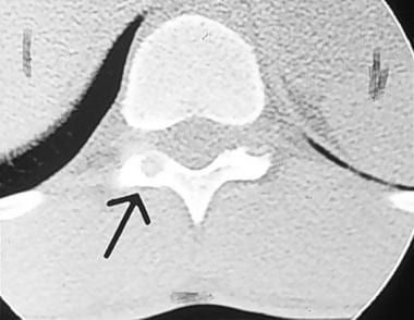
The tumor usually presents as pain and swelling in the affected area, sometimes accompanied by restricted mobility or pathological fractures due to bone weakening. The diagnosis is typically made based on imaging studies (such as X-rays, CT scans, or MRI) and confirmed through a biopsy.
Treatment options for GCTs of bone may include intralesional curettage with or without the use of adjuvant therapies (like phenol, liquid nitrogen, or cement), radiation therapy, or surgical resection. In some cases, systemic treatments like denosumab, a monoclonal antibody targeting RANKL, may be used to control the growth and spread of the tumor. Regular follow-ups are essential to monitor for potential recurrence, which can occur in up to 50% of cases within five years after treatment.
Giant Cell Arteritis (GCA), also known as Temporal Arteritis, is a chronic inflammatory disease affecting large and medium-sized arteries, most commonly the temporal artery. It primarily occurs in people over 50 years old. The condition is characterized by the infiltration of the artery walls with immune cells, leading to inflammation, swelling, and damage. This can restrict blood flow, causing various symptoms.
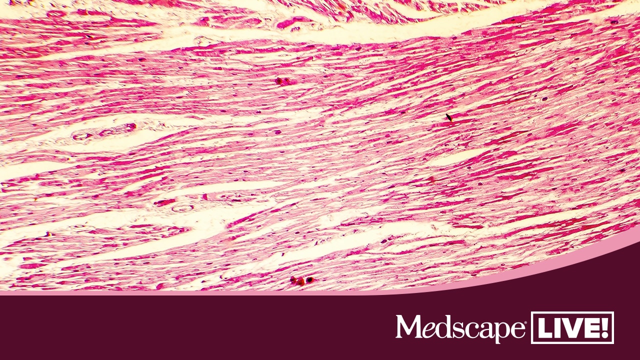
The key feature of GCA is the presence of multinucleated giant cells, which are large collections of fused immune cells, in the affected artery walls. These cells are a hallmark of this condition when viewed under a microscope.
Common symptoms include new onset of severe headaches, scalp tenderness, jaw pain while chewing (called jaw claudication), vision problems, and systemic symptoms such as fever, fatigue, and weight loss. If left untreated, GCA can lead to serious complications like blindness or stroke. Treatment typically involves high-dose corticosteroids to reduce inflammation and prevent further damage.
Giant cell tumors (GCTs) are a type of benign or rarely malignant bone tumor that is characterized by the presence of multinucleated giant cells. These tumors typically affect adults between the ages of 20 and 40, and they can occur in any bone, but they most commonly involve the long bones near the knee joint.
GCTs are composed of three types of cells: mononuclear stromal cells, which produce the matrix of the tumor; multinucleated osteoclast-like giant cells, which resemble the bone-resorbing cells found in normal bone; and macrophages, which are part of the body's immune system.
The mononuclear stromal cells produce a variety of growth factors that stimulate the formation and activity of the osteoclast-like giant cells, leading to localized bone destruction. The tumor may cause pain, swelling, and limited mobility in the affected area.
While GCTs are typically benign, they can be aggressive and locally destructive, with a tendency to recur after surgical removal. In some cases, GCTs may undergo malignant transformation, leading to the development of sarcomas. Treatment options for GCTs include curettage (scraping out) of the tumor, followed by bone grafting or the use of a cement spacer to fill the defect, and/or adjuvant therapy with radiation or chemotherapy.
A giant cell granuloma is a type of non-cancerous (benign) lesion characterized by the presence of large collections of immune cells called macrophages, which have fused together to form multinucleated giant cells. These lesions can occur in various tissues throughout the body but are most commonly found in the oral cavity and jawbone.
Giant cell granulomas can be further classified into two types: central (or bone) giant cell granuloma and peripheral giant cell granuloma. Central giant cell granulomas arise from the bone, while peripheral giant cell granulomas occur in the soft tissues of the gingiva or mouth lining.
Central giant cell granulomas are more aggressive than peripheral ones and can cause significant bone destruction if left untreated. They typically affect younger individuals, with a higher prevalence in females than males. The exact cause of central giant cell granulomas is not well understood but may be associated with local trauma, hormonal imbalances, or genetic factors.
Peripheral giant cell granulomas are less aggressive and usually present as painless, slow-growing nodules on the gums. They typically affect adults, with a higher prevalence in females than males. Peripheral giant cell granulomas may be associated with local irritants such as plaque, calculus, or dental restorations.
Treatment for giant cell granulomas depends on their size, location, and aggressiveness. Surgical excision is the most common treatment approach, but other options such as curettage, corticosteroid injections, or medication therapy may also be considered. Regular follow-up appointments with a healthcare provider are essential to monitor for recurrence.
A "Giant Cell Carcinoma" is a type of cancer that originates from epithelial cells and is characterized by the presence of large, abnormal cells called giant cells. These giant cells are formed by the fusion of several individual cells, resulting in a single, large cell with multiple nuclei. Giant cell carcinomas can occur in various organs, including the lungs, esophagus, and thyroid gland.
Giant cell carcinoma of the lung is a rare and aggressive form of lung cancer that typically affects smokers. It is characterized by the presence of large, bizarre cells with multiple nuclei, as well as a high degree of cellular pleomorphism (variation in size and shape of cells). This type of lung cancer tends to grow and spread quickly, making it difficult to treat.
Giant cell carcinoma of the esophagus is also a rare and aggressive form of cancer that affects the esophagus. It is characterized by the presence of large, abnormal cells with multiple nuclei, as well as a high degree of cellular pleomorphism. This type of esophageal cancer tends to grow and spread quickly, making it difficult to treat.
Giant cell carcinoma of the thyroid gland is an extremely rare form of thyroid cancer that affects the thyroid gland. It is characterized by the presence of large, abnormal cells with multiple nuclei, as well as a high degree of cellular pleomorphism. This type of thyroid cancer tends to grow and spread quickly, making it difficult to treat.
Overall, giant cell carcinomas are aggressive forms of cancer that can occur in various organs. They are characterized by the presence of large, abnormal cells with multiple nuclei, as well as a high degree of cellular pleomorphism. Due to their aggressive nature and tendency to grow and spread quickly, giant cell carcinomas can be difficult to treat.
Temporal arteries are the paired set of arteries that run along the temples on either side of the head. They are branches of the external carotid artery and play a crucial role in supplying oxygenated blood to the scalp and surrounding muscles. One of the most common conditions associated with temporal arteries is Temporal Arteritis (also known as Giant Cell Arteritis), which is an inflammation of these arteries that can lead to serious complications like vision loss if not promptly diagnosed and treated.
Polymyalgia Rheumatica (PMR) is a geriatric rheumatic disease characterized by widespread musculoskeletal pain and stiffness, particularly affecting the neck, shoulders, hips, and thighs. It is often accompanied by symptoms such as fatigue, weakness, loss of appetite, and low-grade fever. The onset of PMR can be sudden or gradual, and it tends to affect individuals over 50 years of age, more commonly women than men.
The exact cause of Polymyalgia Rheumatica remains unknown; however, it is believed to involve an autoimmune response leading to inflammation in the affected areas. Diagnosis typically involves a combination of clinical evaluation, laboratory tests (such as elevated erythrocyte sedimentation rate or C-reactive protein), and sometimes imaging studies. Treatment usually includes corticosteroids to reduce inflammation and manage symptoms, along with monitoring for potential side effects from long-term steroid use. In many cases, PMR can be successfully managed with appropriate treatment, allowing individuals to return to their normal activities.
Langhans giant cells are a type of multinucleated immune cell that are typically found in granulomatous inflammation, which is a specific pattern of chronic inflammation characterized by the formation of granulomas. A granuloma is a small, tightly packed cluster of immune cells, including macrophages, lymphocytes, and sometimes other types of cells, that forms in response to chronic inflammation or an persistent foreign substance that the body cannot eliminate.
Langhans giant cells are named after Theodor Langhans, a German pathologist who first described them in 1868. They are characterized by their large size and the arrangement of their nuclei, which are typically located at the periphery of the cell in a horseshoe or half-moon shape. These cells are thought to be formed when several macrophages fuse together, creating a single, multinucleated cell.
Langhans giant cells are often seen in granulomatous inflammation associated with certain infectious diseases, such as tuberculosis and leprosy, as well as non-infectious conditions such as sarcoidosis. They play a role in the immune response by helping to contain and eliminate foreign substances or microorganisms that are causing the inflammation.
Curettage is a medical procedure that involves scraping or removing tissue from the lining of an organ or body cavity, typically performed using a curette, which is a long, thin surgical instrument with a looped or sharp end. In gynecology, curettage is often used to remove tissue from the uterus during a procedure called dilation and curettage (D&C) to diagnose or treat abnormal uterine bleeding, or to remove residual placental or fetal tissue following a miscarriage or abortion. Curettage may also be used in other medical specialties to remove damaged or diseased tissue from areas such as the nose, throat, or skin.
Bone neoplasms are abnormal growths or tumors that develop in the bone. They can be benign (non-cancerous) or malignant (cancerous). Benign bone neoplasms do not spread to other parts of the body and are rarely a threat to life, although they may cause problems if they grow large enough to press on surrounding tissues or cause fractures. Malignant bone neoplasms, on the other hand, can invade and destroy nearby tissue and may spread (metastasize) to other parts of the body.
There are many different types of bone neoplasms, including:
1. Osteochondroma - a benign tumor that develops from cartilage and bone
2. Enchondroma - a benign tumor that forms in the cartilage that lines the inside of the bones
3. Chondrosarcoma - a malignant tumor that develops from cartilage
4. Osteosarcoma - a malignant tumor that develops from bone cells
5. Ewing sarcoma - a malignant tumor that develops in the bones or soft tissues around the bones
6. Giant cell tumor of bone - a benign or occasionally malignant tumor that develops from bone tissue
7. Fibrosarcoma - a malignant tumor that develops from fibrous tissue in the bone
The symptoms of bone neoplasms vary depending on the type, size, and location of the tumor. They may include pain, swelling, stiffness, fractures, or limited mobility. Treatment options depend on the type and stage of the tumor but may include surgery, radiation therapy, chemotherapy, or a combination of these treatments.
Trophoblasts are specialized cells that make up the outer layer of a blastocyst, which is a hollow ball of cells that forms in the earliest stages of embryonic development. In humans, this process occurs about 5-6 days after fertilization. The blastocyst consists of an inner cell mass (which will eventually become the embryo) and an outer layer of trophoblasts.
Trophoblasts play a crucial role in implantation, which is the process by which the blastocyst attaches to and invades the lining of the uterus. Once implanted, the trophoblasts differentiate into two main layers: the cytotrophoblasts (which are closer to the inner cell mass) and the syncytiotrophoblasts (which form a multinucleated layer that is in direct contact with the maternal tissues).
The cytotrophoblasts proliferate and fuse to form the syncytiotrophoblasts, which have several important functions. They secrete enzymes that help to degrade and remodel the extracellular matrix of the uterine lining, allowing the blastocyst to implant more deeply. They also form a barrier between the maternal and fetal tissues, helping to protect the developing embryo from the mother's immune system.
Additionally, trophoblasts are responsible for the formation of the placenta, which provides nutrients and oxygen to the developing fetus and removes waste products. The syncytiotrophoblasts in particular play a key role in this process by secreting hormones such as human chorionic gonadotropin (hCG), which helps to maintain pregnancy, and by forming blood vessels that allow for the exchange of nutrients and waste between the mother and fetus.
Abnormalities in trophoblast development or function can lead to a variety of pregnancy-related complications, including preeclampsia, intrauterine growth restriction, and gestational trophoblastic diseases such as hydatidiform moles and choriocarcinomas.
A foreign-body reaction is an immune response that occurs when a non-native substance, or "foreign body," is introduced into the human body. This can include things like splinters, surgical implants, or even injected medications. The immune system recognizes these substances as foreign and mounts a response to try to eliminate them.
The initial response to a foreign body is often an acute inflammatory reaction, characterized by the release of chemical mediators that cause vasodilation, increased blood flow, and the migration of white blood cells to the site. This can result in symptoms such as redness, swelling, warmth, and pain.
If the foreign body is not eliminated, a chronic inflammatory response may develop, which can lead to the formation of granulation tissue, fibrosis, and encapsulation of the foreign body. In some cases, this reaction can cause significant tissue damage or impede proper healing.
It's worth noting that not all foreign bodies necessarily elicit a strong immune response. The nature and size of the foreign body, as well as its location in the body, can all influence the severity of the reaction.
Chondroblastoma is a rare, benign (non-cancerous) bone tumor that typically develops in the epiphysis, which is the rounded end of a long bone near a joint. It primarily affects children and adolescents, with around 90% of cases occurring before the age of 20.
The tumor arises from chondroblasts, cells responsible for producing cartilage during bone growth. Chondroblastoma is usually slow-growing and typically causes localized pain, swelling, or tenderness in the affected area. In some cases, it may weaken the bone and lead to fractures.
Treatment generally involves surgical removal of the tumor, followed by curettage (scraping) of the surrounding bone tissue and replacement with bone grafts or substitutes. Recurrence is possible but rare, and long-term prognosis is usually favorable.
A granuloma is a small, nodular inflammatory lesion that occurs in various tissues in response to chronic infection, foreign body reaction, or autoimmune conditions. Histologically, it is characterized by the presence of epithelioid macrophages, which are specialized immune cells with enlarged nuclei and abundant cytoplasm, often arranged in a palisading pattern around a central area containing necrotic debris, microorganisms, or foreign material.
Granulomas can be found in various medical conditions such as tuberculosis, sarcoidosis, fungal infections, and certain autoimmune disorders like Crohn's disease. The formation of granulomas is a complex process involving both innate and adaptive immune responses, which aim to contain and eliminate the offending agent while minimizing tissue damage.
Cherubism is a rare, genetic disorder that affects the bones of the jaw. It is characterized by the replacement of the normal bone with fibrous tissue and cysts, leading to progressive enlargement of the lower jaw (mandible) and, less commonly, the upper jaw (maxilla). The swelling gives the cheeks a fuller appearance, which may resemble the chubby faces of cherubs in art.
The condition typically becomes apparent between the ages of 2 and 7, and it usually progresses until the teenage years, when it begins to stabilize and eventually regress in early adulthood. Cherubism is caused by mutations in the SH3BP2 gene and is inherited in an autosomal dominant manner, meaning that a child has a 50% chance of inheriting the condition if one parent is affected.
While cherubism can cause significant facial deformities, it rarely affects the person's ability to eat, speak, or breathe. Treatment options include observation, orthodontic treatment, and surgical intervention to remove the cysts and reshape the jawbones if necessary.
Giant cells, foreign-body, are a type of large multinucleated immune cell that forms in response to the presence of a foreign material or object in the body. These cells are formed when several individual immune cells, such as macrophages, fuse together around the foreign material in an attempt to engulf and destroy it. The resulting giant cell is characterized by its large size and the presence of multiple nuclei. Foreign-body giant cells are commonly seen in chronic inflammatory reactions to materials such as surgical implants, sutures, or other types of foreign bodies that cannot be eliminated by the immune system.
Cell fusion is the process by which two or more cells combine to form a single cell with a single nucleus, containing the genetic material from all of the original cells. This can occur naturally in certain biological processes, such as fertilization (when a sperm and egg cell fuse to form a zygote), muscle development (where multiple muscle precursor cells fuse together to create multinucleated muscle fibers), and during the formation of bone (where osteoclasts, the cells responsible for breaking down bone tissue, are multinucleated).
Cell fusion can also be induced artificially in laboratory settings through various methods, including chemical treatments, electrical stimulation, or viral vectors. Induced cell fusion is often used in research to create hybrid cells with unique properties, such as cybrid cells (cytoplasmic hybrids) and heterokaryons (nuclear hybrids). These hybrid cells can help scientists study various aspects of cell biology, genetics, and disease mechanisms.
In summary, cell fusion is the merging of two or more cells into one, resulting in a single cell with combined genetic material. This process occurs naturally during certain biological processes and can be induced artificially for research purposes.
Decapodiformes is a taxonomic order of marine cephalopods, which includes squids, octopuses, and cuttlefish. The name "Decapodiformes" comes from the Greek words "deca," meaning ten, and "podos," meaning foot, referring to the fact that these animals have ten limbs.
However, it is worth noting that within Decapodiformes, octopuses are an exception as they only have eight arms. The other members of this order, such as squids and cuttlefish, have ten appendages, which are used for locomotion, feeding, and sensory perception.
Decapodiformes species are known for their complex behaviors, sophisticated communication systems, and remarkable adaptations that enable them to thrive in a variety of marine habitats. They play important ecological roles as both predators and prey in the ocean food chain.
Gingival diseases are infections or inflammations that affect the gingiva, which is the part of the gum around the base of the teeth. These diseases can be caused by bacteria found in dental plaque and can lead to symptoms such as redness, swelling, bleeding, and receding gums. If left untreated, gingival diseases can progress to periodontal disease, a more serious condition that can result in tooth loss. Common types of gingival diseases include gingivitis and periodontitis.
Femoral neoplasms refer to abnormal growths or tumors that develop in the femur, which is the long thigh bone in the human body. These neoplasms can be benign (non-cancerous) or malignant (cancerous). Benign femoral neoplasms are slow-growing and rarely spread to other parts of the body, while malignant neoplasms are aggressive and can invade nearby tissues and organs, as well as metastasize (spread) to distant sites.
There are various types of femoral neoplasms, including osteochondromas, enchondromas, chondrosarcomas, osteosarcomas, and Ewing sarcomas, among others. The specific type of neoplasm is determined by the cell type from which it arises and its behavior.
Symptoms of femoral neoplasms may include pain, swelling, stiffness, or weakness in the thigh, as well as a palpable mass or limited mobility. Diagnosis typically involves imaging studies such as X-rays, CT scans, or MRI, as well as biopsy to determine the type and grade of the tumor. Treatment options may include surgery, radiation therapy, chemotherapy, or a combination of these approaches, depending on the type, size, location, and stage of the neoplasm.
Myocarditis is an inflammation of the myocardium, which is the middle layer of the heart wall. The myocardium is composed of cardiac muscle cells and is responsible for the heart's pumping function. Myocarditis can be caused by various infectious and non-infectious agents, including viruses, bacteria, fungi, parasites, autoimmune diseases, toxins, and drugs.
In myocarditis, the inflammation can damage the cardiac muscle cells, leading to decreased heart function, arrhythmias (irregular heart rhythms), and in severe cases, heart failure or even sudden death. Symptoms of myocarditis may include chest pain, shortness of breath, fatigue, palpitations, and swelling in the legs, ankles, or abdomen.
The diagnosis of myocarditis is often based on a combination of clinical presentation, laboratory tests, electrocardiogram (ECG), echocardiography, cardiac magnetic resonance imaging (MRI), and endomyocardial biopsy. Treatment depends on the underlying cause and severity of the disease and may include medications to support heart function, reduce inflammation, control arrhythmias, and prevent further damage to the heart muscle. In some cases, hospitalization and intensive care may be necessary.
Aneurysmal bone cyst (ABC) is a benign but locally aggressive tumor that typically involves the metaphysis of long bones in children and adolescents. It is characterized by blood-filled spaces or cysts separated by fibrous septa containing osteoclast-type giant cells, spindle cells, and capillary vessels.
ABCs can also arise in other locations such as the vertebral column, pelvis, and skull. They may cause bone pain, swelling, or pathologic fractures. The exact cause of ABC is unknown, but it is thought to be related to a reactive process to a primary bone lesion or trauma.
Treatment options for ABC include curettage and bone grafting, intralesional injection of corticosteroids or bone marrow aspirate, and adjuvant therapy with phenol or liquid nitrogen. In some cases, radiation therapy may be used, but it is generally avoided due to the risk of secondary malignancies. Recurrence rates after treatment range from 10-30%.
Arteritis is a medical condition characterized by inflammation of the arteries. It is also known as vasculitis of the arteries. The inflammation can cause the walls of the arteries to thicken and narrow, reducing blood flow to affected organs or tissues. There are several types of arteritis, including:
1. Giant cell arteritis (GCA): Also known as temporal arteritis, it is a condition that mainly affects the large and medium-sized arteries in the head and neck. The inflammation can cause headaches, jaw pain, scalp tenderness, and vision problems.
2. Takayasu's arteritis: This type of arteritis affects the aorta and its major branches, mainly affecting young women. Symptoms include fever, weight loss, fatigue, and decreased pulse in the arms or legs.
3. Polyarteritis nodosa (PAN): PAN is a rare systemic vasculitis that can affect medium-sized arteries throughout the body. It can cause a wide range of symptoms, including fever, rash, abdominal pain, and muscle weakness.
4. Kawasaki disease: This is a type of arteritis that mainly affects children under the age of 5. It causes inflammation in the blood vessels throughout the body, leading to fever, rash, swollen lymph nodes, and red eyes.
The exact cause of arteritis is not fully understood, but it is believed to be an autoimmune disorder, where the body's immune system mistakenly attacks its own tissues. Treatment for arteritis typically involves medications to reduce inflammation and suppress the immune system.
Osteoclasts are large, multinucleated cells that are primarily responsible for bone resorption, a process in which they break down and dissolve the mineralized matrix of bones. They are derived from monocyte-macrophage precursor cells of hematopoietic origin and play a crucial role in maintaining bone homeostasis by balancing bone formation and bone resorption.
Osteoclasts adhere to the bone surface and create an isolated microenvironment, called the "resorption lacuna," between their cell membrane and the bone surface. Here, they release hydrogen ions into the lacuna through a process called proton pumping, which lowers the pH and dissolves the mineral component of the bone matrix. Additionally, osteoclasts secrete proteolytic enzymes, such as cathepsin K, that degrade the organic components, like collagen, in the bone matrix.
An imbalance in osteoclast activity can lead to various bone diseases, including osteoporosis and Paget's disease, where excessive bone resorption results in weakened and fragile bones.
Tuberous Sclerosis Complex (TSC) is a rare genetic disorder that causes non-cancerous (benign) tumors to grow in many parts of the body. These tumors can affect the brain, skin, heart, kidneys, eyes, and lungs. The signs and symptoms of TSC can vary widely, depending on where the tumors develop and how severely a person is affected.
The condition is caused by mutations in either the TSC1 or TSC2 gene, which regulate a protein that helps control cell growth and division. When these genes are mutated, the protein is not produced correctly, leading to excessive cell growth and the development of tumors.
TSC is typically diagnosed based on clinical symptoms, medical imaging, and genetic testing. Treatment for TSC often involves a multidisciplinary approach, with specialists in neurology, dermatology, cardiology, nephrology, pulmonology, and ophthalmology working together to manage the various symptoms of the condition. Medications, surgery, and other therapies may be used to help control seizures, developmental delays, skin abnormalities, and other complications of TSC.
A granuloma is a type of organized immune response that occurs when the body encounters a foreign substance that it cannot eliminate. A "foreign-body" granuloma specifically refers to this reaction in response to an exogenous material, such as a splinter, suture, or other types of medical implants.
Foreign-body granulomas are characterized by the formation of a collection of immune cells, including macrophages and lymphocytes, which surround and attempt to isolate the foreign material. Over time, this collection of immune cells can become walled off and form a well-circumscribed mass or nodule.
Foreign-body granulomas may cause localized symptoms such as pain, swelling, or inflammation, depending on their location and size. In some cases, they may also lead to complications such as infection or tissue damage. Treatment typically involves removing the foreign body, if possible, followed by anti-inflammatory therapy to manage any residual symptoms or complications.
Histiocytes are a type of immune cell that are part of the mononuclear phagocyte system. They originate from monocytes, which are derived from hematopoietic stem cells in the bone marrow. Histiocytes play an important role in the immune system by engulfing and destroying foreign substances, such as bacteria and viruses, as well as removing dead cells and other debris from the body. They can be found in various tissues throughout the body, including the skin, lymph nodes, spleen, and liver.
Histiocytes include several different types of cells, such as macrophages, dendritic cells, and Langerhans cells. These cells have different functions but all play a role in the immune response. For example, macrophages are involved in inflammation and tissue repair, while dendritic cells are important for presenting antigens to T cells and initiating an immune response.
Abnormal accumulations or dysfunction of histiocytes can lead to various diseases, such as histiocytosis, which is a group of disorders characterized by the abnormal proliferation and accumulation of histiocytes in various tissues.
A biopsy is a medical procedure in which a small sample of tissue is taken from the body to be examined under a microscope for the presence of disease. This can help doctors diagnose and monitor various medical conditions, such as cancer, infections, or autoimmune disorders. The type of biopsy performed will depend on the location and nature of the suspected condition. Some common types of biopsies include:
1. Incisional biopsy: In this procedure, a surgeon removes a piece of tissue from an abnormal area using a scalpel or other surgical instrument. This type of biopsy is often used when the lesion is too large to be removed entirely during the initial biopsy.
2. Excisional biopsy: An excisional biopsy involves removing the entire abnormal area, along with a margin of healthy tissue surrounding it. This technique is typically employed for smaller lesions or when cancer is suspected.
3. Needle biopsy: A needle biopsy uses a thin, hollow needle to extract cells or fluid from the body. There are two main types of needle biopsies: fine-needle aspiration (FNA) and core needle biopsy. FNA extracts loose cells, while a core needle biopsy removes a small piece of tissue.
4. Punch biopsy: In a punch biopsy, a round, sharp tool is used to remove a small cylindrical sample of skin tissue. This type of biopsy is often used for evaluating rashes or other skin abnormalities.
5. Shave biopsy: During a shave biopsy, a thin slice of tissue is removed from the surface of the skin using a sharp razor-like instrument. This technique is typically used for superficial lesions or growths on the skin.
After the biopsy sample has been collected, it is sent to a laboratory where a pathologist will examine the tissue under a microscope and provide a diagnosis based on their findings. The results of the biopsy can help guide further treatment decisions and determine the best course of action for managing the patient's condition.
The placenta is an organ that develops in the uterus during pregnancy and provides oxygen and nutrients to the growing baby through the umbilical cord. It also removes waste products from the baby's blood. The placenta attaches to the wall of the uterus, and the baby's side of the placenta contains many tiny blood vessels that connect to the baby's circulatory system. This allows for the exchange of oxygen, nutrients, and waste between the mother's and baby's blood. After the baby is born, the placenta is usually expelled from the uterus in a process called afterbirth.
Immunohistochemistry (IHC) is a technique used in pathology and laboratory medicine to identify specific proteins or antigens in tissue sections. It combines the principles of immunology and histology to detect the presence and location of these target molecules within cells and tissues. This technique utilizes antibodies that are specific to the protein or antigen of interest, which are then tagged with a detection system such as a chromogen or fluorophore. The stained tissue sections can be examined under a microscope, allowing for the visualization and analysis of the distribution and expression patterns of the target molecule in the context of the tissue architecture. Immunohistochemistry is widely used in diagnostic pathology to help identify various diseases, including cancer, infectious diseases, and immune-mediated disorders.
Mandibular diseases refer to conditions that affect the mandible, or lower jawbone. These diseases can be classified as congenital (present at birth) or acquired (developing after birth). They can also be categorized based on the tissues involved, such as bone, muscle, or cartilage. Some examples of mandibular diseases include:
1. Mandibular fractures: These are breaks in the lower jawbone that can result from trauma or injury.
2. Osteomyelitis: This is an infection of the bone and surrounding tissues, which can affect the mandible.
3. Temporomandibular joint (TMJ) disorders: These are conditions that affect the joint that connects the jawbone to the skull, causing pain and limited movement.
4. Mandibular tumors: These are abnormal growths that can be benign or malignant, and can develop in any of the tissues of the mandible.
5. Osteonecrosis: This is a condition where the bone tissue dies due to lack of blood supply, which can affect the mandible.
6. Cleft lip and palate: This is a congenital deformity that affects the development of the face and mouth, including the lower jawbone.
7. Mandibular hypoplasia: This is a condition where the lower jawbone does not develop properly, leading to a small or recessed chin.
8. Developmental disorders: These are conditions that affect the growth and development of the mandible, such as condylar hyperplasia or hemifacial microsomia.
Epithelioid cells are a type of cell that can be found in certain types of tissue in the body, including connective tissue and some organs. These cells have a characteristic appearance under a microscope, with an enlarged, oval or round shape and a pale, abundant cytoplasm. They may also have a nucleus that is centrally located and has a uniform, rounded shape.
Epithelioid cells are often seen in the context of inflammation or disease, particularly in relation to granulomatous disorders such as sarcoidosis and tuberculosis. In these conditions, epithelioid cells can form clusters known as granulomas, which are a hallmark of the diseases. The exact function of epithelioid cells is not fully understood, but they are thought to play a role in the immune response and may help to contain and eliminate foreign substances or pathogens from the body.
Tylenchoidea is not a medical term, but a taxonomic category in the field of biology, specifically in nematology, which is the study of roundworms. Tylenchoidea is an superfamily of plant-parasitic nematodes, including important pest species such as root-knot nematodes (Meloidogyne spp.) and lesion nematodes (Pratylenchus spp.). These nematodes are known to cause significant damage to crops and vegetation by feeding on plant roots, which can lead to various symptoms including stunted growth, yellowing, wilting, and reduced yield.
Soft tissue neoplasms refer to abnormal growths or tumors that develop in the soft tissues of the body. Soft tissues include muscles, tendons, ligaments, fascia, nerves, blood vessels, fat, and synovial membranes (the thin layer of cells that line joints and tendons). Neoplasms can be benign (non-cancerous) or malignant (cancerous), and their behavior and potential for spread depend on the specific type of neoplasm.
Benign soft tissue neoplasms are typically slow-growing, well-circumscribed, and rarely spread to other parts of the body. They can often be removed surgically with a low risk of recurrence. Examples of benign soft tissue neoplasms include lipomas (fat tumors), schwannomas (nerve sheath tumors), and hemangiomas (blood vessel tumors).
Malignant soft tissue neoplasms, on the other hand, can grow rapidly, invade surrounding tissues, and may metastasize (spread) to distant parts of the body. They are often more difficult to treat than benign neoplasms and require a multidisciplinary approach, including surgery, radiation therapy, and chemotherapy. Examples of malignant soft tissue neoplasms include sarcomas, such as rhabdomyosarcoma (arising from skeletal muscle), leiomyosarcoma (arising from smooth muscle), and angiosarcoma (arising from blood vessels).
It is important to note that soft tissue neoplasms can occur in any part of the body, and their diagnosis and treatment require a thorough evaluation by a healthcare professional with expertise in this area.
Pigmented villonodular synovitis (PVNS) is a rare, benign condition that affects the synovial membrane, which lines the joints. It is characterized by the proliferation of synovial cells and the deposition of hemosiderin, a pigment resulting from the breakdown of blood products. This can lead to joint swelling, pain, stiffness, and limited mobility. PVNS typically affects the large joints such as the knee or hip, but it can also occur in smaller joints, bursae, or tendon sheaths.
There are two forms of PVNS: localized and diffuse. Localized PVNS, also known as giant cell tumor of the tendon sheath, affects a specific area within the joint and is more likely to be treated successfully with surgery. Diffuse PVNS, on the other hand, involves the entire synovial lining of the joint and has a higher recurrence rate even after surgical removal.
The exact cause of PVNS remains unclear, but it is not considered a malignant condition. Treatment usually involves surgical removal of the affected synovium, with or without radiation therapy or chemotherapy to reduce the risk of recurrence. In some cases, arthroscopic surgery may be an option for localized PVNS.
Maxillary diseases refer to conditions that affect the maxilla, which is the upper bone of the jaw. This bone plays an essential role in functions such as biting, chewing, and speaking, and also forms the upper part of the oral cavity, houses the upper teeth, and supports the nose and the eyes.
Maxillary diseases can be caused by various factors, including infections, trauma, tumors, congenital abnormalities, or systemic conditions. Some common maxillary diseases include:
1. Maxillary sinusitis: Inflammation of the maxillary sinuses, which are air-filled cavities located within the maxilla, can cause symptoms such as nasal congestion, facial pain, and headaches.
2. Periodontal disease: Infection and inflammation of the tissues surrounding the teeth, including the gums and the alveolar bone (which is part of the maxilla), can lead to tooth loss and other complications.
3. Maxillary fractures: Trauma to the face can result in fractures of the maxilla, which can cause pain, swelling, and difficulty breathing or speaking.
4. Maxillary cysts and tumors: Abnormal growths in the maxilla can be benign or malignant and may require surgical intervention.
5. Oral cancer: Cancerous lesions in the oral cavity, including the maxilla, can cause pain, swelling, and difficulty swallowing or speaking.
Treatment for maxillary diseases depends on the specific condition and its severity. Treatment options may include antibiotics, surgery, radiation therapy, or chemotherapy. Regular dental check-ups and good oral hygiene practices can help prevent many maxillary diseases.
Benign fibrous histiocytoma (BFH) is a common benign tumor of the skin and superficial soft tissues. It primarily affects middle-aged adults and is more prevalent in men than women. The exact cause of BFH is unknown, but it's thought to arise from dermal fibroblasts or histiocytes.
Medical Definition: Benign Fibrous Histiocytoma (BFH) is a benign, slowly growing, solitary cutaneous or subcutaneous nodular tumor predominantly composed of a mixture of fibroblastic and histiocytic-like cells. The tumor typically presents as a well-circumscribed, firm, dome-shaped papule or nodule, ranging in size from a few millimeters to several centimeters. Histologically, BFH is characterized by the proliferation of spindle-shaped fibroblasts and histiocytes arranged in a storiform pattern, along with variable amounts of collagen deposition, multinucleated giant cells, and hemosiderin deposits. The lesion usually has a pushing border with no invasion into the surrounding tissues. BFH generally follows a benign clinical course, with local recurrence being uncommon following complete surgical excision.
Placental lactogen is a hormone produced by the placenta during pregnancy in humans and some other mammals. It is similar in structure to human growth hormone and prolactin, and has both growth-promoting and lactogenic (milk-producing) properties. Placental lactogen plays an important role in regulating maternal metabolism during pregnancy, promoting the growth and development of the fetus, and preparing the mother's body for lactation after birth. It helps to stimulate the growth of the mammary glands and the production of milk by increasing the availability of nutrients such as glucose, amino acids, and fatty acids in the mother's bloodstream. Placental lactogen also helps to regulate the mother's insulin sensitivity, which can affect her energy levels and the growth of the fetus.
Blood sedimentation, also known as erythrocyte sedimentation rate (ESR), is a medical test that measures the rate at which red blood cells settle at the bottom of a tube of unclotted blood over a specific period of time. The test is used to detect and monitor inflammation in the body.
During an acute inflammatory response, certain proteins in the blood, such as fibrinogen, increase in concentration. These proteins cause red blood cells to stick together and form rouleaux (stacks of disc-shaped cells). As a result, the red blood cells settle more quickly, leading to a higher ESR.
The ESR test is a non-specific test, meaning that it does not identify the specific cause of inflammation. However, it can be used as an indicator of underlying conditions such as infections, autoimmune diseases, and cancer. The test is also used to monitor the effectiveness of treatment for these conditions.
The ESR test is usually performed by drawing a sample of blood into a special tube and allowing it to sit undisturbed for one hour. The distance that the red blood cells have settled is then measured and recorded as the ESR. Normal values for ESR vary depending on age and gender, with higher values indicating greater inflammation.
Bone transplantation, also known as bone grafting, is a surgical procedure in which bone or bone-like material is transferred from one part of the body to another or from one person to another. The graft may be composed of cortical (hard outer portion) bone, cancellous (spongy inner portion) bone, or a combination of both. It can be taken from different sites in the same individual (autograft), from another individual of the same species (allograft), or from an animal source (xenograft). The purpose of bone transplantation is to replace missing bone, provide structural support, and stimulate new bone growth. This procedure is commonly used in orthopedic, dental, and maxillofacial surgeries to repair bone defects caused by trauma, tumors, or congenital conditions.
Macrophages are a type of white blood cell that are an essential part of the immune system. They are large, specialized cells that engulf and destroy foreign substances, such as bacteria, viruses, parasites, and fungi, as well as damaged or dead cells. Macrophages are found throughout the body, including in the bloodstream, lymph nodes, spleen, liver, lungs, and connective tissues. They play a critical role in inflammation, immune response, and tissue repair and remodeling.
Macrophages originate from monocytes, which are a type of white blood cell produced in the bone marrow. When monocytes enter the tissues, they differentiate into macrophages, which have a larger size and more specialized functions than monocytes. Macrophages can change their shape and move through tissues to reach sites of infection or injury. They also produce cytokines, chemokines, and other signaling molecules that help coordinate the immune response and recruit other immune cells to the site of infection or injury.
Macrophages have a variety of surface receptors that allow them to recognize and respond to different types of foreign substances and signals from other cells. They can engulf and digest foreign particles, bacteria, and viruses through a process called phagocytosis. Macrophages also play a role in presenting antigens to T cells, which are another type of immune cell that helps coordinate the immune response.
Overall, macrophages are crucial for maintaining tissue homeostasis, defending against infection, and promoting wound healing and tissue repair. Dysregulation of macrophage function has been implicated in a variety of diseases, including cancer, autoimmune disorders, and chronic inflammatory conditions.
Sarcoidosis is a multi-system disorder characterized by the formation of granulomas (small clumps of inflammatory cells) in various organs, most commonly the lungs and lymphatic system. These granulomas can impair the function of the affected organ(s), leading to a variety of symptoms. The exact cause of sarcoidosis is unknown, but it's thought to be an overactive immune response to an unknown antigen, possibly triggered by an infection, chemical exposure, or another environmental factor.
The diagnosis of sarcoidosis typically involves a combination of clinical evaluation, imaging studies (such as chest X-rays and CT scans), and laboratory tests (including blood tests and biopsies). While there is no cure for sarcoidosis, treatment may be necessary to manage symptoms and prevent complications. Corticosteroids are often used to suppress the immune system and reduce inflammation, while other medications may be prescribed to treat specific organ involvement or symptoms. In some cases, sarcoidosis may resolve on its own without any treatment.
Prednisolone is a synthetic glucocorticoid drug, which is a class of steroid hormones. It is commonly used in the treatment of various inflammatory and autoimmune conditions due to its potent anti-inflammatory and immunosuppressive effects. Prednisolone works by binding to specific receptors in cells, leading to changes in gene expression that reduce the production of substances involved in inflammation, such as cytokines and prostaglandins.
Prednisolone is available in various forms, including tablets, syrups, and injectable solutions. It can be used to treat a wide range of medical conditions, including asthma, rheumatoid arthritis, inflammatory bowel disease, allergies, skin conditions, and certain types of cancer.
Like other steroid medications, prednisolone can have significant side effects if used in high doses or for long periods of time. These may include weight gain, mood changes, increased risk of infections, osteoporosis, diabetes, and adrenal suppression. As a result, the use of prednisolone should be closely monitored by a healthcare professional to ensure that its benefits outweigh its risks.
X-ray computed tomography (CT or CAT scan) is a medical imaging method that uses computer-processed combinations of many X-ray images taken from different angles to produce cross-sectional (tomographic) images (virtual "slices") of the body. These cross-sectional images can then be used to display detailed internal views of organs, bones, and soft tissues in the body.
The term "computed tomography" is used instead of "CT scan" or "CAT scan" because the machines take a series of X-ray measurements from different angles around the body and then use a computer to process these data to create detailed images of internal structures within the body.
CT scanning is a noninvasive, painless medical test that helps physicians diagnose and treat medical conditions. CT imaging provides detailed information about many types of tissue including lung, bone, soft tissue and blood vessels. CT examinations can be performed on every part of the body for a variety of reasons including diagnosis, surgical planning, and monitoring of therapeutic responses.
In computed tomography (CT), an X-ray source and detector rotate around the patient, measuring the X-ray attenuation at many different angles. A computer uses this data to construct a cross-sectional image by the process of reconstruction. This technique is called "tomography". The term "computed" refers to the use of a computer to reconstruct the images.
CT has become an important tool in medical imaging and diagnosis, allowing radiologists and other physicians to view detailed internal images of the body. It can help identify many different medical conditions including cancer, heart disease, lung nodules, liver tumors, and internal injuries from trauma. CT is also commonly used for guiding biopsies and other minimally invasive procedures.
In summary, X-ray computed tomography (CT or CAT scan) is a medical imaging technique that uses computer-processed combinations of many X-ray images taken from different angles to produce cross-sectional images of the body. It provides detailed internal views of organs, bones, and soft tissues in the body, allowing physicians to diagnose and treat medical conditions.
Spinal neoplasms refer to abnormal growths or tumors found within the spinal column, which can be benign (non-cancerous) or malignant (cancerous). These tumors can originate in the spine itself, called primary spinal neoplasms, or they can spread to the spine from other parts of the body, known as secondary or metastatic spinal neoplasms. Spinal neoplasms can cause various symptoms, such as back pain, neurological deficits, and even paralysis, depending on their location and size. Early diagnosis and treatment are crucial to prevent or minimize long-term complications and improve the patient's prognosis.
The sacrum is a triangular-shaped bone in the lower portion of the human vertebral column, located between the lumbar spine and the coccyx (tailbone). It forms through the fusion of several vertebrae during fetal development. The sacrum's base articulates with the fifth lumbar vertebra, while its apex connects with the coccyx.
The sacrum plays an essential role in supporting the spine and transmitting weight from the upper body to the pelvis and lower limbs. It also serves as an attachment site for various muscles and ligaments. The sacral region is often a focus in medical and chiropractic treatments due to its importance in spinal stability, posture, and overall health.
A fatal outcome is a term used in medical context to describe a situation where a disease, injury, or illness results in the death of an individual. It is the most severe and unfortunate possible outcome of any medical condition, and is often used as a measure of the severity and prognosis of various diseases and injuries. In clinical trials and research, fatal outcome may be used as an endpoint to evaluate the effectiveness and safety of different treatments or interventions.
Malignant fibrous histiocytoma (MFH) is not a specific type of histiocytoma; rather, it is a type of soft tissue sarcoma. Histiocytomas are benign tumors that arise from cells called histiocytes, which are part of the immune system. MFH, on the other hand, is a malignant (cancerous) tumor that can arise in various types of soft tissues, such as muscle, fat, tendons, and ligaments.
MFH was once thought to originate from histiocytes, but more recent research suggests that it may actually arise from undifferentiated mesenchymal cells, which are capable of developing into a variety of different cell types. MFH is the most common type of soft tissue sarcoma in adults over the age of 50 and typically presents as a painless mass in the extremities or retroperitoneum (the area in the back of the abdomen).
The tumor is characterized by the presence of fibroblastic and histiocytic-like cells, which can be quite pleomorphic (varied in shape and size) and may contain numerous mitotic figures (indicating rapid cell division). Treatment typically involves surgical excision, often followed by radiation therapy and/or chemotherapy. The prognosis for MFH depends on several factors, including the tumor's location, size, grade (degree of differentiation), and the patient's age and overall health.
The radius is one of the two bones in the forearm in humans and other vertebrates. In humans, it runs from the lateral side of the elbow to the thumb side of the wrist. It is responsible for rotation of the forearm and articulates with the humerus at the elbow and the carpals at the wrist. Any medical condition or injury that affects the radius can impact the movement and function of the forearm and hand.
Electron microscopy (EM) is a type of microscopy that uses a beam of electrons to create an image of the sample being examined, resulting in much higher magnification and resolution than light microscopy. There are several types of electron microscopy, including transmission electron microscopy (TEM), scanning electron microscopy (SEM), and reflection electron microscopy (REM).
In TEM, a beam of electrons is transmitted through a thin slice of the sample, and the electrons that pass through the sample are focused to form an image. This technique can provide detailed information about the internal structure of cells, viruses, and other biological specimens, as well as the composition and structure of materials at the atomic level.
In SEM, a beam of electrons is scanned across the surface of the sample, and the electrons that are scattered back from the surface are detected to create an image. This technique can provide information about the topography and composition of surfaces, as well as the structure of materials at the microscopic level.
REM is a variation of SEM in which the beam of electrons is reflected off the surface of the sample, rather than scattered back from it. This technique can provide information about the surface chemistry and composition of materials.
Electron microscopy has a wide range of applications in biology, medicine, and materials science, including the study of cellular structure and function, disease diagnosis, and the development of new materials and technologies.
Tenosynovitis is a medical condition characterized by inflammation of the lining (synovium) surrounding a tendon, which is a cord-like structure that attaches muscle to bone. This inflammation can cause pain, swelling, and difficulty moving the affected joint. Tenosynovitis often affects the hands, wrists, feet, and ankles, and it can result from various causes, including infection, injury, overuse, or autoimmune disorders like rheumatoid arthritis. Prompt diagnosis and treatment of tenosynovitis are essential to prevent complications such as tendon rupture or chronic pain.
Placentation is the process by which the placenta, an organ that provides nutrients and oxygen to the developing fetus and removes waste products, is formed and develops during pregnancy. It involves the attachment of the fertilized egg (embryo) to the uterine wall and the development of specialized structures that facilitate the exchange of gases, nutrients, and waste between the mother and the fetus.
In humans, placentation begins when the embryo implants into the endometrium, or the lining of the uterus, about 6-10 days after fertilization. The outer layer of the embryo, called the trophoblast, invades the endometrial tissue and forms a structure called the placenta.
The placenta consists of both maternal and fetal tissues. The fetal portion of the placenta is derived from the chorionic villi, which are finger-like projections that develop on the surface of the embryo and increase the surface area for exchange. The maternal portion of the placenta is made up of modified endometrial tissue called decidua.
The placenta grows and develops throughout pregnancy, providing a vital connection between the mother and fetus. Proper placentation is essential for a healthy pregnancy and fetal development. Abnormalities in placentation can lead to complications such as preeclampsia, preterm labor, and intrauterine growth restriction.
Mollusca is not a medical term per se, but a major group of invertebrate animals that includes snails, clams, octopuses, and squids. However, medically, some mollusks can be relevant as they can act as vectors for various diseases, such as schistosomiasis (transmitted by freshwater snails) and fascioliasis (transmitted by aquatic snails). Therefore, a medical definition might describe Mollusca as a phylum of mostly marine invertebrates that can sometimes play a role in the transmission of certain infectious diseases.
The fibula is a slender bone located in the lower leg of humans and other vertebrates. It runs parallel to the larger and more robust tibia, and together they are known as the bones of the leg or the anterior tibial segment. The fibula is the lateral bone in the leg, positioned on the outside of the tibia.
In humans, the fibula extends from the knee joint proximally to the ankle joint distally. Its proximal end, called the head of the fibula, articulates with the lateral condyle of the tibia and forms part of the inferior aspect of the knee joint. The narrowed portion below the head is known as the neck of the fibula.
The shaft of the fibula, also called the body of the fibula, is a long, thin structure that descends from the neck and serves primarily for muscle attachment rather than weight-bearing functions. The distal end of the fibula widens to form the lateral malleolus, which is an important bony landmark in the ankle region. The lateral malleolus articulates with the talus bone of the foot and forms part of the ankle joint.
The primary functions of the fibula include providing attachment sites for muscles that act on the lower leg, ankle, and foot, as well as contributing to the stability of the ankle joint through its articulation with the talus bone. Fractures of the fibula can occur due to various injuries, such as twisting or rotational forces applied to the ankle or direct trauma to the lateral aspect of the lower leg.
Jaw neoplasms refer to abnormal growths or tumors in the jawbone (mandible) or maxilla (upper jaw). These growths can be benign (non-cancerous) or malignant (cancerous). Benign neoplasms are not considered life-threatening, but they can still cause problems by invading nearby tissues and causing damage. Malignant neoplasms, on the other hand, can spread to other parts of the body and can be life-threatening if not treated promptly and effectively.
Jaw neoplasms can present with various symptoms such as swelling, pain, loose teeth, numbness or tingling in the lips or tongue, difficulty chewing or swallowing, and jaw stiffness or limited movement. The diagnosis of jaw neoplasms typically involves a thorough clinical examination, imaging studies such as X-rays, CT scans, or MRI, and sometimes a biopsy to determine the type and extent of the tumor.
Treatment options for jaw neoplasms depend on several factors, including the type, size, location, and stage of the tumor, as well as the patient's overall health and medical history. Treatment may involve surgery, radiation therapy, chemotherapy, or a combination of these modalities. Regular follow-up care is essential to monitor for recurrence or metastasis (spread) of the neoplasm.
Ischemic optic neuropathy (ION) is a medical condition that refers to the damage or death of the optic nerve due to insufficient blood supply. The optic nerve is responsible for transmitting visual information from the eye to the brain.
In ION, the blood vessels that supply the optic nerve become blocked or narrowed, leading to decreased blood flow and oxygen delivery to the nerve fibers. This results in inflammation, swelling, and ultimately, damage to the optic nerve. The damage can cause sudden, painless vision loss, often noticed upon waking up in the morning.
There are two types of ION: anterior ischemic optic neuropathy (AION) and posterior ischemic optic neuropathy (PION). AION affects the front part of the optic nerve, while PION affects the back part of the nerve. AION is further classified into arteritic and non-arteritic types, depending on whether it is caused by giant cell arteritis or not.
Risk factors for ION include age (most commonly occurring in people over 50), hypertension, diabetes, smoking, sleep apnea, and other cardiovascular diseases. Treatment options depend on the type and cause of ION and may include controlling underlying medical conditions, administering corticosteroids, or undergoing surgical procedures to improve blood flow.
Mandibular neoplasms refer to abnormal growths or tumors that develop in the mandible, which is the lower jawbone. These growths can be benign (non-cancerous) or malignant (cancerous). Benign neoplasms are typically slow-growing and rarely spread to other parts of the body, while malignant neoplasms can invade surrounding tissues and may metastasize (spread) to distant sites.
Mandibular neoplasms can have various causes, including genetic mutations, exposure to certain chemicals or radiation, and infection with certain viruses. The symptoms of mandibular neoplasms may include swelling or pain in the jaw, difficulty chewing or speaking, numbness in the lower lip or chin, loose teeth, and/or a lump or mass in the mouth or neck.
The diagnosis of mandibular neoplasms typically involves a thorough clinical examination, imaging studies such as X-rays, CT scans, or MRI scans, and sometimes a biopsy to confirm the type and extent of the tumor. Treatment options depend on the type, stage, and location of the neoplasm, and may include surgery, radiation therapy, chemotherapy, or a combination of these approaches. Regular follow-up care is essential to monitor for recurrence or metastasis.
Muscle neoplasms are abnormal growths or tumors that develop in the muscle tissue. They can be benign (non-cancerous) or malignant (cancerous). Benign muscle neoplasms are typically slow-growing and do not spread to other parts of the body, while malignant muscle neoplasms, also known as soft tissue sarcomas, can grow quickly, invade nearby tissues, and metastasize (spread) to distant parts of the body.
Soft tissue sarcomas can arise from any of the muscles in the body, including the skeletal muscles (voluntary muscles that attach to bones and help with movement), smooth muscles (involuntary muscles found in the walls of blood vessels, digestive tract, and other organs), or cardiac muscle (the specialized muscle found in the heart).
There are many different types of soft tissue sarcomas, each with its own set of characteristics and prognosis. Treatment for muscle neoplasms typically involves a combination of surgery, radiation therapy, and chemotherapy, depending on the type, size, location, and stage of the tumor.
A tendon is the strong, flexible band of tissue that connects muscle to bone. It helps transfer the force produced by the muscle to allow various movements of our body parts. Tendons are made up of collagen fibers arranged in parallel bundles and have a poor blood supply, making them prone to injuries and slow to heal. Examples include the Achilles tendon, which connects the calf muscle to the heel bone, and the patellar tendon, which connects the kneecap to the shinbone.
The tibia, also known as the shin bone, is the larger of the two bones in the lower leg and part of the knee joint. It supports most of the body's weight and is a major insertion point for muscles that flex the foot and bend the leg. The tibia articulates with the femur at the knee joint and with the fibula and talus bone at the ankle joint. Injuries to the tibia, such as fractures, are common in sports and other activities that put stress on the lower leg.
Cathepsin K is a proteolytic enzyme, which belongs to the family of papain-like cysteine proteases. It is primarily produced by osteoclasts, which are specialized cells responsible for bone resorption. Cathepsin K plays a crucial role in the degradation and remodeling of the extracellular matrix, particularly in bone tissue.
This enzyme is capable of breaking down various proteins, including collagen, elastin, and proteoglycans, which are major components of the bone matrix. By doing so, cathepsin K helps osteoclasts to dissolve and remove mineralized and non-mineralized bone matrix during the process of bone resorption.
Apart from its function in bone metabolism, cathepsin K has also been implicated in several pathological conditions, such as osteoporosis, rheumatoid arthritis, and tumor metastasis to bones. Inhibitors of cathepsin K are being investigated as potential therapeutic agents for the treatment of these disorders.
Histochemistry is the branch of pathology that deals with the microscopic localization of cellular or tissue components using specific chemical reactions. It involves the application of chemical techniques to identify and locate specific biomolecules within tissues, cells, and subcellular structures. This is achieved through the use of various staining methods that react with specific antigens or enzymes in the sample, allowing for their visualization under a microscope. Histochemistry is widely used in diagnostic pathology to identify different types of tissues, cells, and structures, as well as in research to study cellular and molecular processes in health and disease.
In the field of medicine, "time factors" refer to the duration of symptoms or time elapsed since the onset of a medical condition, which can have significant implications for diagnosis and treatment. Understanding time factors is crucial in determining the progression of a disease, evaluating the effectiveness of treatments, and making critical decisions regarding patient care.
For example, in stroke management, "time is brain," meaning that rapid intervention within a specific time frame (usually within 4.5 hours) is essential to administering tissue plasminogen activator (tPA), a clot-busting drug that can minimize brain damage and improve patient outcomes. Similarly, in trauma care, the "golden hour" concept emphasizes the importance of providing definitive care within the first 60 minutes after injury to increase survival rates and reduce morbidity.
Time factors also play a role in monitoring the progression of chronic conditions like diabetes or heart disease, where regular follow-ups and assessments help determine appropriate treatment adjustments and prevent complications. In infectious diseases, time factors are crucial for initiating antibiotic therapy and identifying potential outbreaks to control their spread.
Overall, "time factors" encompass the significance of recognizing and acting promptly in various medical scenarios to optimize patient outcomes and provide effective care.
Nematoda is a phylum of pseudocoelomate, unsegmented worms with a round or filiform body shape. They are commonly known as roundworms or threadworms. Nematodes are among the most diverse and numerous animals on earth, with estimates of over 1 million species, of which only about 25,000 have been described.
Nematodes are found in a wide range of habitats, including marine, freshwater, and terrestrial environments. Some nematode species are free-living, while others are parasitic, infecting a variety of hosts, including plants, animals, and humans. Parasitic nematodes can cause significant disease and economic losses in agriculture, livestock production, and human health.
The medical importance of nematodes lies primarily in their role as parasites that infect humans and animals. Some common examples of medically important nematodes include:
* Ascaris lumbricoides (human roundworm)
* Trichuris trichiura (whipworm)
* Ancylostoma duodenale and Necator americanus (hookworms)
* Enterobius vermicularis (pinworm or threadworm)
* Wuchereria bancrofti, Brugia malayi, and Loa loa (filarial nematodes that cause lymphatic filariasis, onchocerciasis, and loiasis, respectively)
Nematode infections can cause a range of clinical symptoms, depending on the species and the location of the parasite in the body. Common symptoms include gastrointestinal disturbances, anemia, skin rashes, and lymphatic swelling. In some cases, nematode infections can lead to serious complications or even death if left untreated.
Medical management of nematode infections typically involves the use of anthelmintic drugs, which are medications that kill or expel parasitic worms from the body. The choice of drug depends on the species of nematode and the severity of the infection. In some cases, preventive measures such as improved sanitation and hygiene can help reduce the risk of nematode infections.
Retrospective studies, also known as retrospective research or looking back studies, are a type of observational study that examines data from the past to draw conclusions about possible causal relationships between risk factors and outcomes. In these studies, researchers analyze existing records, medical charts, or previously collected data to test a hypothesis or answer a specific research question.
Retrospective studies can be useful for generating hypotheses and identifying trends, but they have limitations compared to prospective studies, which follow participants forward in time from exposure to outcome. Retrospective studies are subject to biases such as recall bias, selection bias, and information bias, which can affect the validity of the results. Therefore, retrospective studies should be interpreted with caution and used primarily to generate hypotheses for further testing in prospective studies.
Acid phosphatase is a type of enzyme that is found in various tissues and organs throughout the body, including the prostate gland, red blood cells, bone, liver, spleen, and kidneys. This enzyme plays a role in several biological processes, such as bone metabolism and the breakdown of molecules like nucleotides and proteins.
Acid phosphatase is classified based on its optimum pH level for activity. Acid phosphatases have an optimal activity at acidic pH levels (below 7.0), while alkaline phosphatases have an optimal activity at basic or alkaline pH levels (above 7.0).
In clinical settings, measuring the level of acid phosphatase in the blood can be useful as a tumor marker for prostate cancer. Elevated acid phosphatase levels may indicate the presence of metastatic prostate cancer or disease progression. However, it is important to note that acid phosphatase is not specific to prostate cancer and can also be elevated in other conditions, such as bone diseases, liver disorders, and some benign conditions. Therefore, acid phosphatase should be interpreted in conjunction with other diagnostic tests and clinical findings for a more accurate diagnosis.
Astrocytoma is a type of brain tumor that arises from astrocytes, which are star-shaped glial cells in the brain. These tumors can occur in various parts of the brain and can have different grades of malignancy, ranging from low-grade (I or II) to high-grade (III or IV). Low-grade astrocytomas tend to grow slowly and may not cause any symptoms for a long time, while high-grade astrocytomas are more aggressive and can grow quickly, causing neurological problems.
Symptoms of astrocytoma depend on the location and size of the tumor but may include headaches, seizures, weakness or numbness in the limbs, difficulty speaking or swallowing, changes in vision or behavior, and memory loss. Treatment options for astrocytomas include surgery, radiation therapy, chemotherapy, or a combination of these approaches. The prognosis for astrocytoma varies widely depending on the grade and location of the tumor, as well as the age and overall health of the patient.
Skull neoplasms refer to abnormal growths or tumors that develop within the skull. These growths can be benign (non-cancerous) or malignant (cancerous). They can originate from various types of cells, such as bone cells, nerve cells, or soft tissues. Skull neoplasms can cause various symptoms depending on their size and location, including headaches, seizures, vision problems, hearing loss, and neurological deficits. Treatment options include surgery, radiation therapy, and chemotherapy. It is important to note that a neoplasm in the skull can also refer to metastatic cancer, which has spread from another part of the body to the skull.
The ilium is the largest and broadest of the three parts that make up the hip bone or coxal bone. It is the uppermost portion of the pelvis and forms the side of the waist. The ilium has a curved, fan-like shape and articulates with the sacrum at the back to form the sacroiliac joint. The large, concave surface on the top of the ilium is called the iliac crest, which can be felt as a prominent ridge extending from the front of the hip to the lower back. This region is significant in orthopedics and physical examinations for its use in assessing various medical conditions and performing certain maneuvers during the physical examination.
An axon is a long, slender extension of a neuron (a type of nerve cell) that conducts electrical impulses (nerve impulses) away from the cell body to target cells, such as other neurons or muscle cells. Axons can vary in length from a few micrometers to over a meter long and are typically surrounded by a myelin sheath, which helps to insulate and protect the axon and allows for faster transmission of nerve impulses.
Axons play a critical role in the functioning of the nervous system, as they provide the means by which neurons communicate with one another and with other cells in the body. Damage to axons can result in serious neurological problems, such as those seen in spinal cord injuries or neurodegenerative diseases like multiple sclerosis.
Cell differentiation is the process by which a less specialized cell, or stem cell, becomes a more specialized cell type with specific functions and structures. This process involves changes in gene expression, which are regulated by various intracellular signaling pathways and transcription factors. Differentiation results in the development of distinct cell types that make up tissues and organs in multicellular organisms. It is a crucial aspect of embryonic development, tissue repair, and maintenance of homeostasis in the body.
Endoreduplication is a process of cell cycle regulation where duplicate copies of chromosomes are produced within the nucleus of a single cell without subsequent separation into daughter cells. This results in an increase in the ploidy level, or the number of sets of chromosomes, within the cell. It is a form of genome amplification that can occur through repeated rounds of DNA replication during the G2 phase of the cell cycle, without cytokinesis or cell division.
Endoreduplication is a natural process in some tissues and organisms, such as plants and insects, where it plays a role in growth, development, and differentiation. In cancer cells, endoreduplication can contribute to genomic instability, tumor progression, and drug resistance.
The axillary artery is a major blood vessel in the upper limb. It is the continuation of the subclavian artery and begins at the lateral border of the first rib, where it becomes the brachial artery. The axillary artery supplies oxygenated blood to the upper extremity, chest wall, and breast.
The axillary artery is divided into three parts based on the surrounding structures:
1. First part: From its origin at the lateral border of the first rib to the medial border of the pectoralis minor muscle. It lies deep to the clavicle and is covered by the scalene muscles, the anterior and middle scalene being the most important. The branches arising from this portion are the superior thoracic artery and the thyrocervical trunk.
2. Second part: Behind the pectoralis minor muscle. The branches arising from this portion are the lateral thoracic artery and the subscapular artery.
3. Third part: After leaving the lower border of the pectoralis minor muscle, it becomes the brachial artery. The branches arising from this portion are the anterior circumflex humeral artery and the posterior circumflex humeral artery.
The axillary artery is a common site for surgical interventions such as angioplasty and stenting to treat peripheral arterial disease, as well as for bypass grafting in cases of severe atherosclerosis or occlusion.
Medical Definition:
Magnetic Resonance Imaging (MRI) is a non-invasive diagnostic imaging technique that uses a strong magnetic field and radio waves to create detailed cross-sectional or three-dimensional images of the internal structures of the body. The patient lies within a large, cylindrical magnet, and the scanner detects changes in the direction of the magnetic field caused by protons in the body. These changes are then converted into detailed images that help medical professionals to diagnose and monitor various medical conditions, such as tumors, injuries, or diseases affecting the brain, spinal cord, heart, blood vessels, joints, and other internal organs. MRI does not use radiation like computed tomography (CT) scans.
Fibrin foam is not a widely recognized medical term, but it does refer to a type of biocompatible material that is used in certain medical and surgical applications.
Fibrin is a protein involved in the clotting of blood, and fibrin foam is created by combining fibrin with a foaming agent to create a porous structure. The resulting material can be used as a scaffold for tissue regeneration or as a filler in surgical procedures.
In some cases, fibrin foam may also be impregnated with various growth factors or other therapeutic agents to promote healing and tissue repair. The use of fibrin foam has been explored in a variety of clinical settings, including wound healing, bone regeneration, and the treatment of soft tissue defects.
There are many diseases that can affect cats, and the specific medical definitions for these conditions can be quite detailed and complex. However, here are some common categories of feline diseases and examples of each:
1. Infectious diseases: These are caused by viruses, bacteria, fungi, or parasites. Examples include:
* Feline panleukopenia virus (FPV), also known as feline parvovirus, which can cause severe gastrointestinal symptoms and death in kittens.
* Feline calicivirus (FCV), which can cause upper respiratory symptoms such as sneezing and nasal discharge.
* Feline leukemia virus (FeLV), which can suppress the immune system and lead to a variety of secondary infections and diseases.
* Bacterial infections, such as those caused by Pasteurella multocida or Bartonella henselae, which can cause abscesses or other symptoms.
2. Neoplastic diseases: These are cancerous conditions that can affect various organs and tissues in cats. Examples include:
* Lymphoma, which is a common type of cancer in cats that can affect the lymph nodes, spleen, liver, and other organs.
* Fibrosarcoma, which is a type of soft tissue cancer that can arise from fibrous connective tissue.
* Squamous cell carcinoma, which is a type of skin cancer that can be caused by exposure to sunlight or tobacco smoke.
3. Degenerative diseases: These are conditions that result from the normal wear and tear of aging or other factors. Examples include:
* Osteoarthritis, which is a degenerative joint disease that can cause pain and stiffness in older cats.
* Dental disease, which is a common condition in cats that can lead to tooth loss, gum inflammation, and other problems.
* Heart disease, such as hypertrophic cardiomyopathy (HCM), which is a thickening of the heart muscle that can lead to congestive heart failure.
4. Hereditary diseases: These are conditions that are inherited from a cat's parents and are present at birth or develop early in life. Examples include:
* Polycystic kidney disease (PKD), which is a genetic disorder that causes cysts to form in the kidneys and can lead to kidney failure.
* Hypertrophic cardiomyopathy (HCM), which can be inherited as an autosomal dominant trait in some cats.
* Progressive retinal atrophy (PRA), which is a group of genetic disorders that cause degeneration of the retina and can lead to blindness.
Treatment outcome is a term used to describe the result or effect of medical treatment on a patient's health status. It can be measured in various ways, such as through symptoms improvement, disease remission, reduced disability, improved quality of life, or survival rates. The treatment outcome helps healthcare providers evaluate the effectiveness of a particular treatment plan and make informed decisions about future care. It is also used in clinical research to compare the efficacy of different treatments and improve patient care.
Polyarteritis nodosa (PAN) is a rare, systemic necrotizing vasculitis that affects medium-sized and small muscular arteries. It is characterized by inflammation and damage to the walls of the arteries, leading to the formation of microaneurysms (small bulges in the artery wall) and subsequent narrowing or complete occlusion of the affected vessels. This can result in tissue ischemia (reduced blood flow) and infarction (tissue death), causing a wide range of clinical manifestations that vary depending on the organs involved.
The exact cause of PAN remains unclear, but it is believed to involve an autoimmune response triggered by various factors such as infections or exposure to certain drugs. The diagnosis of PAN typically requires a combination of clinical findings, laboratory tests, and imaging studies, often supported by histopathological examination of affected tissues. Treatment usually involves the use of immunosuppressive medications to control inflammation and prevent further damage to the arteries and organs.
Pregnancy is a physiological state or condition where a fertilized egg (zygote) successfully implants and grows in the uterus of a woman, leading to the development of an embryo and finally a fetus. This process typically spans approximately 40 weeks, divided into three trimesters, and culminates in childbirth. Throughout this period, numerous hormonal and physical changes occur to support the growing offspring, including uterine enlargement, breast development, and various maternal adaptations to ensure the fetus's optimal growth and well-being.
Orthopedic procedures are surgical or nonsurgical methods used to treat musculoskeletal conditions, including injuries, deformities, or diseases of the bones, joints, muscles, ligaments, and tendons. These procedures can range from simple splinting or casting to complex surgeries such as joint replacements, spinal fusions, or osteotomies (cutting and repositioning bones). The primary goal of orthopedic procedures is to restore function, reduce pain, and improve the quality of life for patients.
The sphenoid bone is a complex, irregularly shaped bone located in the middle cranial fossa and forms part of the base of the skull. It articulates with several other bones, including the frontal, parietal, temporal, ethmoid, palatine, and zygomatic bones. The sphenoid bone has two main parts: the body and the wings.
The body of the sphenoid bone is roughly cuboid in shape and contains several important structures, such as the sella turcica, which houses the pituitary gland, and the sphenoid sinuses, which are air-filled cavities within the bone. The greater wings of the sphenoid bone extend laterally from the body and form part of the skull's lateral walls. They contain the superior orbital fissure, through which important nerves and blood vessels pass between the cranial cavity and the orbit of the eye.
The lesser wings of the sphenoid bone are thin, blade-like structures that extend anteriorly from the body and form part of the floor of the anterior cranial fossa. They contain the optic canal, which transmits the optic nerve and ophthalmic artery between the brain and the orbit of the eye.
Overall, the sphenoid bone plays a crucial role in protecting several important structures within the skull, including the pituitary gland, optic nerves, and ophthalmic arteries.
A bone cyst is a fluid-filled sac that develops within a bone. It can be classified as either simple (unicameral) or aneurysmal. Simple bone cysts are more common in children and adolescents, and they typically affect the long bones of the arms or legs. These cysts are usually asymptomatic unless they become large enough to weaken the bone and cause a fracture. Aneurysmal bone cysts, on the other hand, can occur at any age and can affect any bone, but they are most common in the leg bones and spine. They are characterized by rapidly growing blood-filled sacs that can cause pain, swelling, and fractures.
Both types of bone cysts may be treated with observation, medication, or surgery depending on their size, location, and symptoms. It is important to note that while these cysts can be benign, they should still be evaluated and monitored by a healthcare professional to ensure proper treatment and prevention of complications.
Unilamellar liposomes are a type of liposome that consists of a single phospholipid bilayer membrane enclosing an aqueous compartment. They are spherical vesicles, ranging in size from 20 nanometers to several micrometers, and can be used as drug delivery systems for various therapeutic agents, including hydrophilic drugs (in the aqueous compartment) and hydrophobic drugs (incorporated into the lipid bilayer). The single membrane structure of unilamellar liposomes distinguishes them from multilamellar liposomes, which have multiple concentric phospholipid bilayers.
Antigens are substances (usually proteins) on the surface of cells, or viruses, bacteria, and other microorganisms, that can stimulate an immune response.
Differentiation in the context of myelomonocytic cells refers to the process by which these cells mature and develop into specific types of immune cells, such as monocytes, macrophages, and neutrophils.
Myelomonocytic cells are a type of white blood cell that originate from stem cells in the bone marrow. They give rise to two main types of immune cells: monocytes and granulocytes (which include neutrophils, eosinophils, and basophils).
Therefore, 'Antigens, Differentiation, Myelomonocytic' refers to the study or examination of how antigens affect the differentiation process of myelomonocytic cells into specific types of immune cells. This is an important area of research in immunology and hematology as it relates to understanding how the body responds to infections, inflammation, and cancer.
A tuberculoma is a granulomatous lesion in the brain caused by the infection of Mycobacterium tuberculosis. It typically consists of caseating necrosis surrounded by a layer of epithelioid histiocytes, Langhans' giant cells, and lymphocytes. Tuberculomas can be single or multiple and may cause various neurological symptoms depending on their size and location. They are often associated with tuberculous meningitis but can also occur in immunocompromised individuals without obvious systemic infection.
Phenol, also known as carbolic acid, is an organic compound with the molecular formula C6H5OH. It is a white crystalline solid that is slightly soluble in water and has a melting point of 40-42°C. Phenol is a weak acid, but it is quite reactive and can be converted into a variety of other chemicals.
In a medical context, phenol is most commonly used as a disinfectant and antiseptic. It has a characteristic odor that is often described as "tarry" or " medicinal." Phenol is also used in some over-the-counter products, such as mouthwashes and throat lozenges, to help kill bacteria and freshen breath.
However, phenol is also a toxic substance that can cause serious harm if it is swallowed, inhaled, or absorbed through the skin. It can cause irritation and burns to the eyes, skin, and mucous membranes, and it can damage the liver and kidneys if ingested. Long-term exposure to phenol has been linked to an increased risk of cancer.
Because of its potential for harm, phenol is regulated as a hazardous substance in many countries, and it must be handled with care when used in medical or industrial settings.
A fibroma is a benign (non-cancerous) tumor that consists primarily of fibrous or connective tissue. It can occur in various parts of the body, including the skin, mouth, and internal organs. The term "fibroma" is often used to describe any benign fibrous growth, but there are specific types of fibromas such as dermatofibroma (found in the skin), oral fibroma (found in the mouth), and benign fibrous histiocytoma (found in soft tissues).
It's important to note that while fibromas are generally harmless, they can cause discomfort or problems depending on their size and location. If a fibroma is causing issues or there's concern about its growth or malignancy, it should be evaluated by a healthcare professional for potential removal or further assessment.
Local neoplasm recurrence is the return or regrowth of a tumor in the same location where it was originally removed or treated. This means that cancer cells have survived the initial treatment and started to grow again in the same area. It's essential to monitor and detect any local recurrence as early as possible, as it can affect the prognosis and may require additional treatment.
Immunoenzyme techniques are a group of laboratory methods used in immunology and clinical chemistry that combine the specificity of antibody-antigen reactions with the sensitivity and amplification capabilities of enzyme reactions. These techniques are primarily used for the detection, quantitation, or identification of various analytes (such as proteins, hormones, drugs, viruses, or bacteria) in biological samples.
In immunoenzyme techniques, an enzyme is linked to an antibody or antigen, creating a conjugate. This conjugate then interacts with the target analyte in the sample, forming an immune complex. The presence and amount of this immune complex can be visualized or measured by detecting the enzymatic activity associated with it.
There are several types of immunoenzyme techniques, including:
1. Enzyme-linked Immunosorbent Assay (ELISA): A widely used method for detecting and quantifying various analytes in a sample. In ELISA, an enzyme is attached to either the capture antibody or the detection antibody. After the immune complex formation, a substrate is added that reacts with the enzyme, producing a colored product that can be measured spectrophotometrically.
2. Immunoblotting (Western blot): A method used for detecting specific proteins in a complex mixture, such as a protein extract from cells or tissues. In this technique, proteins are separated by gel electrophoresis and transferred to a membrane, where they are probed with an enzyme-conjugated antibody directed against the target protein.
3. Immunohistochemistry (IHC): A method used for detecting specific antigens in tissue sections or cells. In IHC, an enzyme-conjugated primary or secondary antibody is applied to the sample, and the presence of the antigen is visualized using a chromogenic substrate that produces a colored product at the site of the antigen-antibody interaction.
4. Immunofluorescence (IF): A method used for detecting specific antigens in cells or tissues by employing fluorophore-conjugated antibodies. The presence of the antigen is visualized using a fluorescence microscope.
5. Enzyme-linked immunosorbent assay (ELISA): A method used for detecting and quantifying specific antigens or antibodies in liquid samples, such as serum or culture supernatants. In ELISA, an enzyme-conjugated detection antibody is added after the immune complex formation, and a substrate is added that reacts with the enzyme to produce a colored product that can be measured spectrophotometrically.
These techniques are widely used in research and diagnostic laboratories for various applications, including protein characterization, disease diagnosis, and monitoring treatment responses.
Monocytes are a type of white blood cell that are part of the immune system. They are large cells with a round or oval shape and a nucleus that is typically indented or horseshoe-shaped. Monocytes are produced in the bone marrow and then circulate in the bloodstream, where they can differentiate into other types of immune cells such as macrophages and dendritic cells.
Monocytes play an important role in the body's defense against infection and tissue damage. They are able to engulf and digest foreign particles, microorganisms, and dead or damaged cells, which helps to clear them from the body. Monocytes also produce cytokines, which are signaling molecules that help to coordinate the immune response.
Elevated levels of monocytes in the bloodstream can be a sign of an ongoing infection, inflammation, or other medical conditions such as cancer or autoimmune disorders.
Fasciitis is a medical condition characterized by inflammation or irritation of the fascia, which are the bands of connective tissue that surround muscles, tendons, and bones in the body. The most common type of fasciitis is plantar fasciitis, which affects the fascia on the bottom of the foot and can cause heel pain. Other types of fasciitis include:
* Achilles tendonitis or Achilles tendinopathy, which affects the fascia that connects the calf muscle to the heel bone
* Shin splints, which affect the fascia that covers the front of the lower leg
* Necrotizing fasciitis, a rare and serious bacterial infection that can cause extensive tissue damage and is potentially life-threatening.
The symptoms of fasciitis may include pain, stiffness, or tenderness in the affected area, especially after prolonged periods of rest or physical activity. Treatment for fasciitis typically involves rest, ice, compression, and elevation (RICE) of the affected area, as well as physical therapy exercises to stretch and strengthen the fascia and surrounding muscles. In some cases, medication or surgery may be necessary to relieve symptoms and promote healing.
The humerus is the long bone in the upper arm that extends from the shoulder joint (glenohumeral joint) to the elbow joint. It articulates with the glenoid cavity of the scapula to form the shoulder joint and with the radius and ulna bones at the elbow joint. The proximal end of the humerus has a rounded head that provides for movement in multiple planes, making it one of the most mobile joints in the body. The greater and lesser tubercles are bony prominences on the humeral head that serve as attachment sites for muscles that move the shoulder and arm. The narrow shaft of the humerus provides stability and strength for weight-bearing activities, while the distal end forms two articulations: one with the ulna (trochlea) and one with the radius (capitulum). Together, these structures allow for a wide range of motion in the shoulder and elbow joints.
A lipoma is a common, benign (non-cancerous) soft tissue growth. It is composed of adipose or fatty tissue and typically found just beneath the skin, but they can also occur deeper within the body. Lipomas are usually round, moveable, and painless, although they may cause discomfort if they grow large enough to put pressure on nearby nerves or if they're located in a sensitive area. They generally grow slowly over time. Surgical removal is an option if the lipoma becomes bothersome or grows significantly in size. It's important to note that while lipomas are typically harmless, any new lumps or bumps should be evaluated by a healthcare professional to confirm the diagnosis and rule out other more serious conditions.
Reconstructive surgical procedures are a type of surgery aimed at restoring the form and function of body parts that are defective or damaged due to various reasons such as congenital abnormalities, trauma, infection, tumors, or disease. These procedures can involve the transfer of tissue from one part of the body to another, manipulation of bones, muscles, and tendons, or use of prosthetic materials to reconstruct the affected area. The goal is to improve both the physical appearance and functionality of the body part, thereby enhancing the patient's quality of life. Examples include breast reconstruction after mastectomy, cleft lip and palate repair, and treatment of severe burns.
I apologize for the confusion, but "Solanum glaucophyllum" is not a recognized medical term or a well-known plant in medical or pharmacological contexts. The term "Solanum" refers to a genus of flowering plants that includes many familiar food crops like tomatoes, potatoes, and eggplants, as well as toxic plants such as belladonna. However, "glaucophyllum" is not a commonly used species name within the Solanum genus.
It's possible that you may be referring to a specific plant variety or a regional name that is not widely recognized in medical or scientific communities. If you could provide more context or clarify your question, I would be happy to help further.
Osteoma is a benign (noncancerous) tumor that is made up of mature bone tissue. It usually grows slowly over a period of years and is most commonly found in the skull or jaw, although it can occur in other bones of the body as well. Osteomas are typically small, but they can grow to be several centimeters in size. They may cause symptoms if they press on nearby tissues or structures, such as nerves or blood vessels. In some cases, osteomas may not cause any symptoms and may only be discovered during routine imaging studies. Treatment for osteoma is typically not necessary unless it is causing problems or growing rapidly. If treatment is needed, it may involve surgical removal of the tumor.
"Lymnaea" is a genus of freshwater snails, specifically aquatic pulmonate gastropod mollusks. These snails are commonly known as pond snails or ram's horn snails due to their spiral shell shape that resembles a ram's horn. They have a wide global distribution and can be found in various freshwater habitats, such as ponds, lakes, streams, and wetlands.
Some Lymnaea species are known for their use in scientific research, particularly in the fields of neurobiology and malacology (the study of mollusks). For instance, Lymnaea stagnalis is a well-studied model organism used to investigate learning and memory processes at the molecular, cellular, and behavioral levels.
However, it's important to note that "Lymnaea" itself does not have a direct medical definition as it refers to a genus of snails rather than a specific medical condition or disease.
"Cells, cultured" is a medical term that refers to cells that have been removed from an organism and grown in controlled laboratory conditions outside of the body. This process is called cell culture and it allows scientists to study cells in a more controlled and accessible environment than they would have inside the body. Cultured cells can be derived from a variety of sources, including tissues, organs, or fluids from humans, animals, or cell lines that have been previously established in the laboratory.
Cell culture involves several steps, including isolation of the cells from the tissue, purification and characterization of the cells, and maintenance of the cells in appropriate growth conditions. The cells are typically grown in specialized media that contain nutrients, growth factors, and other components necessary for their survival and proliferation. Cultured cells can be used for a variety of purposes, including basic research, drug development and testing, and production of biological products such as vaccines and gene therapies.
It is important to note that cultured cells may behave differently than they do in the body, and results obtained from cell culture studies may not always translate directly to human physiology or disease. Therefore, it is essential to validate findings from cell culture experiments using additional models and ultimately in clinical trials involving human subjects.
Mimiviridae is a family of large, double-stranded DNA viruses that infect amoebae. These viruses are characterized by their complex structure and large genome size, which can contain up to 1,200 genes. Mimiviridae viruses have a icosahedral capsid with a diameter of about 700 nanometers and a unique vertex where the viral DNA is packed. They also have a lipid membrane beneath the capsid.
Mimiviridae viruses are known to cause lysogenic infections, meaning that they can integrate their genome into the host's chromosome and replicate along with it. They can also cause lytic infections, leading to the death of the host cell and the release of new virus particles.
Mimiviridae viruses have been found in various environments, including water, soil, and dust. Some researchers have suggested that they may play a role in shaping microbial communities and influencing the evolution of their hosts. However, more research is needed to fully understand the ecological and evolutionary significance of these fascinating viruses.
Adie syndrome, also known as Adie's pupil or tonic pupil, is a neurological disorder that affects the autonomic nervous system and the eye. It is characterized by a pupil that is dilated and unresponsive to light, but slowly constricts when focusing on nearby objects (a phenomenon called "light-near dissociation"). This occurs due to damage to the ciliary ganglion or the short ciliary nerves, which control the size of the pupil.
Additional symptoms of Adie syndrome may include decreased deep tendon reflexes, especially in the ankles, and abnormal sweating patterns. The condition is usually not painful and does not typically affect vision, although some people with Adie syndrome may experience difficulty with reading due to the slow pupillary response.
The exact cause of Adie syndrome is unknown, but it is thought to be related to a viral infection or an autoimmune disorder. It is more common in women than men and typically occurs between the ages of 20 and 40. While there is no cure for Adie syndrome, treatment may include the use of glasses with bifocal lenses or reading glasses, as well as physical therapy to improve muscle tone and reflexes.
Aortitis is a medical condition characterized by inflammation of the aorta, which is the largest artery in the body that carries oxygenated blood from the heart to the rest of the body. The inflammation can cause damage to the aortic wall, leading to weakening, bulging (aneurysm), or tearing (dissection) of the aorta. Aortitis can be caused by various conditions, including infections, autoimmune diseases, and certain medications. It is essential to diagnose and treat aortitis promptly to prevent serious complications.
Necrosis is the premature death of cells or tissues due to damage or injury, such as from infection, trauma, infarction (lack of blood supply), or toxic substances. It's a pathological process that results in the uncontrolled and passive degradation of cellular components, ultimately leading to the release of intracellular contents into the extracellular space. This can cause local inflammation and may lead to further tissue damage if not treated promptly.
There are different types of necrosis, including coagulative, liquefactive, caseous, fat, fibrinoid, and gangrenous necrosis, each with distinct histological features depending on the underlying cause and the affected tissues or organs.
Osteosarcoma is defined as a type of cancerous tumor that arises from the cells that form bones (osteoblasts). It's the most common primary bone cancer, and it typically develops in the long bones of the body, such as the arms or legs, near the growth plates. Osteosarcoma can metastasize (spread) to other parts of the body, including the lungs, making it a highly malignant form of cancer. Symptoms may include bone pain, swelling, and fractures. Treatment usually involves a combination of surgery, chemotherapy, and/or radiation therapy.
The vasa vasorum are small blood vessels that supply larger blood vessels, such as the arteries and veins, with oxygen and nutrients. They are located in the outer layers (the adventitia and media) of these larger vessels and form a network of vessels that surround and penetrate the walls of the larger vessels. The vasa vasorum are particularly important in supplying blood to the thicker walls of larger arteries, such as the aorta, where diffusion from the lumen may not be sufficient to meet the metabolic needs of the vessel wall.
Odontogenic tumors are a group of neoplasms that originate from the dental tissues or their remnants, including the odontogenic epithelium, ectomesenchyme, and/or their derivatives. These tumors can be benign or malignant and may affect the jaw bones and surrounding structures. They can cause various symptoms, such as swelling, pain, loosening of teeth, and altered bite. The classification of odontogenic tumors includes a wide range of entities with different biological behaviors, clinical features, and treatment approaches. Accurate diagnosis is essential for proper management and prognosis.
Biocompatible materials are non-toxic and non-reacting substances that can be used in medical devices, tissue engineering, and drug delivery systems without causing harm or adverse reactions to living tissues or organs. These materials are designed to mimic the properties of natural tissues and are able to integrate with biological systems without being rejected by the body's immune system.
Biocompatible materials can be made from a variety of substances, including metals, ceramics, polymers, and composites. The specific properties of these materials, such as their mechanical strength, flexibility, and biodegradability, are carefully selected to meet the requirements of their intended medical application.
Examples of biocompatible materials include titanium used in dental implants and joint replacements, polyethylene used in artificial hips, and hydrogels used in contact lenses and drug delivery systems. The use of biocompatible materials has revolutionized modern medicine by enabling the development of advanced medical technologies that can improve patient outcomes and quality of life.
There is no medical definition for "dog diseases" as it is too broad a term. However, dogs can suffer from various health conditions and illnesses that are specific to their species or similar to those found in humans. Some common categories of dog diseases include:
1. Infectious Diseases: These are caused by viruses, bacteria, fungi, or parasites. Examples include distemper, parvovirus, kennel cough, Lyme disease, and heartworms.
2. Hereditary/Genetic Disorders: Some dogs may inherit certain genetic disorders from their parents. Examples include hip dysplasia, elbow dysplasia, progressive retinal atrophy (PRA), and degenerative myelopathy.
3. Age-Related Diseases: As dogs age, they become more susceptible to various health issues. Common age-related diseases in dogs include arthritis, dental disease, cancer, and cognitive dysfunction syndrome (CDS).
4. Nutritional Disorders: Malnutrition or improper feeding can lead to various health problems in dogs. Examples include obesity, malnutrition, and vitamin deficiencies.
5. Environmental Diseases: These are caused by exposure to environmental factors such as toxins, allergens, or extreme temperatures. Examples include heatstroke, frostbite, and toxicities from ingesting harmful substances.
6. Neurological Disorders: Dogs can suffer from various neurological conditions that affect their nervous system. Examples include epilepsy, intervertebral disc disease (IVDD), and vestibular disease.
7. Behavioral Disorders: Some dogs may develop behavioral issues due to various factors such as anxiety, fear, or aggression. Examples include separation anxiety, noise phobias, and resource guarding.
It's important to note that regular veterinary care, proper nutrition, exercise, and preventative measures can help reduce the risk of many dog diseases.
A granuloma in the respiratory tract refers to a small nodular lesion that forms in the lung tissue due to an ongoing immune response. It is typically composed of macrophages, lymphocytes, and other inflammatory cells that cluster together around a foreign substance or organism that the body cannot eliminate.
Granulomas can form in response to various stimuli, including infectious agents such as mycobacteria (tuberculosis, nontuberculous mycobacteria), fungi, and parasites, as well as non-infectious causes like inhaled particles (e.g., silica, beryllium) or autoimmune diseases (e.g., sarcoidosis).
These lesions can cause damage to the lung tissue over time, leading to symptoms such as cough, shortness of breath, chest pain, and fatigue. Diagnosis often involves imaging studies like chest X-rays or CT scans, followed by biopsy and microscopic examination to confirm the presence of granulomas and identify the underlying cause. Treatment depends on the underlying cause but may include antibiotics, corticosteroids, or other immunosuppressive medications.
A ganglion is a cluster of neuron cell bodies in the peripheral nervous system. Ganglia are typically associated with nerves and serve as sites for sensory processing, integration, and relay of information between the periphery and the central nervous system (CNS). The two main types of ganglia are sensory ganglia, which contain pseudounipolar neurons that transmit sensory information to the CNS, and autonomic ganglia, which contain multipolar neurons that control involuntary physiological functions.
Examples of sensory ganglia include dorsal root ganglia (DRG), which are associated with spinal nerves, and cranial nerve ganglia, such as the trigeminal ganglion. Autonomic ganglia can be further divided into sympathetic and parasympathetic ganglia, which regulate different aspects of the autonomic nervous system.
It's worth noting that in anatomy, "ganglion" refers to a group of nerve cell bodies, while in clinical contexts, "ganglion" is often used to describe a specific type of cystic structure that forms near joints or tendons, typically in the wrist or foot. These ganglia are not related to the peripheral nervous system's ganglia but rather are fluid-filled sacs that may cause discomfort or pain due to their size or location.
Fibrous dysplasia, monostotic is a benign bone disorder that affects a single bone (monostotic) and is characterized by the replacement of normal bone tissue with fibrous (scar-like) tissue. This results in the formation of abnormal bone that is weakened and more susceptible to fractures. The lesions can cause deformities, pain, and decreased mobility, depending on their size and location. Monostotic fibrous dysplasia is the most common form of fibrous dysplasia, accounting for approximately 70-80% of all cases. It typically manifests during childhood or adolescence and may stabilize or progress slowly over time. In some cases, it can be associated with endocrine disorders such as precocious puberty, hyperthyroidism, or growth hormone excess.
Polymethyl methacrylate (PMMA) is a type of synthetic resin that is widely used in the medical field due to its biocompatibility and versatility. It is a transparent, rigid, and lightweight material that can be easily molded into different shapes and forms. Here are some of the medical definitions of PMMA:
1. A biocompatible acrylic resin used in various medical applications such as bone cement, intraocular lenses, dental restorations, and drug delivery systems.
2. A type of synthetic material that is used as a bone cement to fix prosthetic joint replacements and vertebroplasty for the treatment of spinal fractures.
3. A transparent and shatter-resistant material used in the manufacture of medical devices such as intravenous (IV) fluid bags, dialyzer housings, and oxygenators.
4. A drug delivery system that can be used to administer drugs locally or systemically, such as intraocular sustained-release drug implants for the treatment of chronic eye diseases.
5. A component of dental restorations such as fillings, crowns, and bridges due to its excellent mechanical properties and esthetic qualities.
Overall, PMMA is a versatile and valuable material in the medical field, with numerous applications that take advantage of its unique properties.
In situ hybridization (ISH) is a molecular biology technique used to detect and localize specific nucleic acid sequences, such as DNA or RNA, within cells or tissues. This technique involves the use of a labeled probe that is complementary to the target nucleic acid sequence. The probe can be labeled with various types of markers, including radioisotopes, fluorescent dyes, or enzymes.
During the ISH procedure, the labeled probe is hybridized to the target nucleic acid sequence in situ, meaning that the hybridization occurs within the intact cells or tissues. After washing away unbound probe, the location of the labeled probe can be visualized using various methods depending on the type of label used.
In situ hybridization has a wide range of applications in both research and diagnostic settings, including the detection of gene expression patterns, identification of viral infections, and diagnosis of genetic disorders.
Subcutaneous tissue, also known as the subcutis or hypodermis, is the layer of fatty connective tissue found beneath the dermis (the inner layer of the skin) and above the muscle fascia. It is composed mainly of adipose tissue, which serves as a energy storage reservoir and provides insulation and cushioning to the body. The subcutaneous tissue also contains blood vessels, nerves, and immune cells that support the skin's functions. This layer varies in thickness depending on the location in the body and can differ significantly between individuals based on factors such as age, genetics, and weight.
"Pregnancy proteins" is not a standard medical term, but it may refer to specific proteins that are produced or have increased levels during pregnancy. Two common pregnancy-related proteins are:
1. Human Chorionic Gonadotropin (hCG): A hormone produced by the placenta shortly after fertilization. It is often detected in urine or blood tests to confirm pregnancy. Its primary function is to maintain the corpus luteum, which produces progesterone and estrogen during early pregnancy until the placenta takes over these functions.
2. Pregnancy-Specific beta-1 Glycoprotein (SP1): A protein produced by the placental trophoblasts during pregnancy. Its function is not well understood, but it may play a role in implantation, placentation, and protection against the mother's immune system. SP1 levels increase throughout pregnancy and are used as a marker for fetal growth and well-being.
These proteins have clinical significance in monitoring pregnancy progression, detecting potential complications, and diagnosing certain pregnancy-related conditions.
Photomicrography is not a medical term per se, but it is a technique often used in the field of medicine and pathology. It refers to the process of taking photographs through a microscope, using specialized equipment and techniques to capture detailed images of specimens or structures that are too small to be seen by the naked eye. These images can be used for various purposes, such as medical research, diagnosis, education, and publication.
In summary, photomicrography is the photography of microscopic subjects, which can have many applications in the medical field.
Fibrous Dysplasia of Bone is a rare, benign bone disorder that is characterized by the replacement of normal bone tissue with fibrous (scar-like) and immature bone tissue. This results in weakened bones that are prone to fractures, deformities, and pain. The condition can affect any bone in the body but most commonly involves the long bones of the legs, arms, and skull. It can occur as an isolated finding or as part of a genetic disorder called McCune-Albright syndrome. The exact cause of fibrous dysplasia is not fully understood, but it is believed to result from a genetic mutation that occurs during early bone development. There is no cure for fibrous dysplasia, and treatment typically focuses on managing symptoms and preventing complications.
The temporal bone is a paired bone that is located on each side of the skull, forming part of the lateral and inferior walls of the cranial cavity. It is one of the most complex bones in the human body and has several important structures associated with it. The main functions of the temporal bone include protecting the middle and inner ear, providing attachment for various muscles of the head and neck, and forming part of the base of the skull.
The temporal bone is divided into several parts, including the squamous part, the petrous part, the tympanic part, and the styloid process. The squamous part forms the lateral portion of the temporal bone and articulates with the parietal bone. The petrous part is the most medial and superior portion of the temporal bone and contains the inner ear and the semicircular canals. The tympanic part forms the lower and anterior portions of the temporal bone and includes the external auditory meatus or ear canal. The styloid process is a long, slender projection that extends downward from the inferior aspect of the temporal bone and serves as an attachment site for various muscles and ligaments.
The temporal bone plays a crucial role in hearing and balance, as it contains the structures of the middle and inner ear, including the oval window, round window, cochlea, vestibule, and semicircular canals. The stapes bone, one of the three bones in the middle ear, is entirely encased within the petrous portion of the temporal bone. Additionally, the temporal bone contains important structures for facial expression and sensation, including the facial nerve, which exits the skull through the stylomastoid foramen, a small opening in the temporal bone.
Bone diseases is a broad term that refers to various medical conditions that affect the bones. These conditions can be categorized into several groups, including:
1. Developmental and congenital bone diseases: These are conditions that affect bone growth and development before or at birth. Examples include osteogenesis imperfecta (brittle bone disease), achondroplasia (dwarfism), and cleidocranial dysostosis.
2. Metabolic bone diseases: These are conditions that affect the body's ability to maintain healthy bones. They are often caused by hormonal imbalances, vitamin deficiencies, or problems with mineral metabolism. Examples include osteoporosis, osteomalacia, and Paget's disease of bone.
3. Inflammatory bone diseases: These are conditions that cause inflammation in the bones. They can be caused by infections, autoimmune disorders, or other medical conditions. Examples include osteomyelitis, rheumatoid arthritis, and ankylosing spondylitis.
4. Degenerative bone diseases: These are conditions that cause the bones to break down over time. They can be caused by aging, injury, or disease. Examples include osteoarthritis, avascular necrosis, and diffuse idiopathic skeletal hyperostosis (DISH).
5. Tumors and cancers of the bone: These are conditions that involve abnormal growths in the bones. They can be benign or malignant. Examples include osteosarcoma, chondrosarcoma, and Ewing sarcoma.
6. Fractures and injuries: While not strictly a "disease," fractures and injuries are common conditions that affect the bones. They can result from trauma, overuse, or weakened bones. Examples include stress fractures, compound fractures, and dislocations.
Overall, bone diseases can cause a wide range of symptoms, including pain, stiffness, deformity, and decreased mobility. Treatment for these conditions varies depending on the specific diagnosis but may include medication, surgery, physical therapy, or lifestyle changes.
Fibroepithelial neoplasms are benign (non-cancerous) growths that consist of both fibrous and epithelial tissue. These types of neoplasms can occur in various parts of the body, but they are most commonly found in the skin and mucous membranes. A well-known example of a fibroepithelial neoplasm is a skin tag (acrochordon). Other examples include fibroma, papilloma, and neurofibroma.
Fibroepithelial neoplasms are typically slow-growing and cause little to no discomfort or symptoms. However, they may be removed for cosmetic reasons or if they become irritated, inflamed, or start to bleed. In rare cases, a fibroepithelial neoplasm can undergo malignant transformation and develop into cancer. It is essential to have any new or changing growths evaluated by a healthcare professional to determine the appropriate course of action.
Bone resorption is the process by which bone tissue is broken down and absorbed into the body. It is a normal part of bone remodeling, in which old or damaged bone tissue is removed and new tissue is formed. However, excessive bone resorption can lead to conditions such as osteoporosis, in which bones become weak and fragile due to a loss of density. This process is carried out by cells called osteoclasts, which break down the bone tissue and release minerals such as calcium into the bloodstream.
Therapeutic embolization is a medical procedure that involves intentionally blocking or obstructing blood vessels to stop excessive bleeding or block the flow of blood to a tumor or abnormal tissue. This is typically accomplished by injecting small particles, such as microspheres or coils, into the targeted blood vessel through a catheter, which is inserted into a larger blood vessel and guided to the desired location using imaging techniques like X-ray or CT scanning. The goal of therapeutic embolization is to reduce the size of a tumor, control bleeding, or block off abnormal blood vessels that are causing problems.
Skin diseases, also known as dermatological conditions, refer to any medical condition that affects the skin, which is the largest organ of the human body. These diseases can affect the skin's function, appearance, or overall health. They can be caused by various factors, including genetics, infections, allergies, environmental factors, and aging.
Skin diseases can present in many different forms, such as rashes, blisters, sores, discolorations, growths, or changes in texture. Some common examples of skin diseases include acne, eczema, psoriasis, dermatitis, fungal infections, viral infections, bacterial infections, and skin cancer.
The symptoms and severity of skin diseases can vary widely depending on the specific condition and individual factors. Some skin diseases are mild and can be treated with over-the-counter medications or topical creams, while others may require more intensive treatments such as prescription medications, light therapy, or even surgery.
It is important to seek medical attention if you experience any unusual or persistent changes in your skin, as some skin diseases can be serious or indicative of other underlying health conditions. A dermatologist is a medical doctor who specializes in the diagnosis and treatment of skin diseases.
I'm sorry for any confusion, but "Polychaeta" is not a medical term. It is a taxonomic category in zoology, specifically referring to a class of annelid worms commonly known as bristle worms or polychaetes. These are segmented worms that have pairs of fleshy protrusions called parapodia on most or all segments, which they use for locomotion. Some species live in marine environments, while others can be found in fresh water or even terrestrial habitats. If you have a medical term you would like me to define, I'd be happy to help!
Noonan Syndrome is a genetic disorder that affects various parts of the body and is characterized by distinctive facial features, short stature, heart defects, and developmental delays. It is caused by mutations in genes responsible for regulating cell growth and division. The syndrome is often identified at birth or in early childhood due to its physical manifestations, which may include widely spaced eyes, low-set ears, a short neck, a broad or webbed neck, chest deformities, and pulmonary valve stenosis. Noonan Syndrome affects both sexes and all races equally, with an estimated prevalence of 1 in 1,000 to 1 in 2,500 live births.
Osteolysis is a medical term that refers to the loss or resorption of bone tissue. It's a process where the body's normal bone remodeling cycle is disrupted, leading to an imbalance between bone formation and bone breakdown. This results in the progressive deterioration and destruction of bone.
Osteolysis can occur due to various reasons such as chronic inflammation, mechanical stress, or certain medical conditions like rheumatoid arthritis, Paget's disease, or bone tumors. It can also be a side effect of some medications, such as those used in cancer treatment or for managing osteoporosis.
In severe cases, osteolysis can lead to weakened bones, increased risk of fractures, and deformities. Treatment typically aims to address the underlying cause and may include medication, surgery, or lifestyle changes.
Methylprednisolone is a synthetic glucocorticoid drug, which is a class of hormones that naturally occur in the body and are produced by the adrenal gland. It is often used to treat various medical conditions such as inflammation, allergies, and autoimmune disorders. Methylprednisolone works by reducing the activity of the immune system, which helps to reduce symptoms such as swelling, pain, and redness.
Methylprednisolone is available in several forms, including tablets, oral suspension, and injectable solutions. It may be used for short-term or long-term treatment, depending on the condition being treated. Common side effects of methylprednisolone include increased appetite, weight gain, insomnia, mood changes, and increased susceptibility to infections. Long-term use of methylprednisolone can lead to more serious side effects such as osteoporosis, cataracts, and adrenal suppression.
It is important to note that methylprednisolone should be used under the close supervision of a healthcare provider, as it can cause serious side effects if not used properly. The dosage and duration of treatment will depend on various factors such as the patient's age, weight, medical history, and the condition being treated.
Follow-up studies are a type of longitudinal research that involve repeated observations or measurements of the same variables over a period of time, in order to understand their long-term effects or outcomes. In medical context, follow-up studies are often used to evaluate the safety and efficacy of medical treatments, interventions, or procedures.
In a typical follow-up study, a group of individuals (called a cohort) who have received a particular treatment or intervention are identified and then followed over time through periodic assessments or data collection. The data collected may include information on clinical outcomes, adverse events, changes in symptoms or functional status, and other relevant measures.
The results of follow-up studies can provide important insights into the long-term benefits and risks of medical interventions, as well as help to identify factors that may influence treatment effectiveness or patient outcomes. However, it is important to note that follow-up studies can be subject to various biases and limitations, such as loss to follow-up, recall bias, and changes in clinical practice over time, which must be carefully considered when interpreting the results.
Neurosurgical procedures are operations that are performed on the brain, spinal cord, and peripheral nerves. These procedures are typically carried out by neurosurgeons, who are medical doctors with specialized training in the diagnosis and treatment of disorders of the nervous system. Neurosurgical procedures can be used to treat a wide range of conditions, including traumatic injuries, tumors, aneurysms, vascular malformations, infections, degenerative diseases, and congenital abnormalities.
Some common types of neurosurgical procedures include:
* Craniotomy: A procedure in which a bone flap is temporarily removed from the skull to gain access to the brain. This type of procedure may be performed to remove a tumor, repair a blood vessel, or relieve pressure on the brain.
* Spinal fusion: A procedure in which two or more vertebrae in the spine are fused together using bone grafts and metal hardware. This is often done to stabilize the spine and alleviate pain caused by degenerative conditions or spinal deformities.
* Microvascular decompression: A procedure in which a blood vessel that is causing pressure on a nerve is repositioned or removed. This type of procedure is often used to treat trigeminal neuralgia, a condition that causes severe facial pain.
* Deep brain stimulation: A procedure in which electrodes are implanted in specific areas of the brain and connected to a battery-operated device called a neurostimulator. The neurostimulator sends electrical impulses to the brain to help alleviate symptoms of movement disorders such as Parkinson's disease or dystonia.
* Stereotactic radiosurgery: A non-invasive procedure that uses focused beams of radiation to treat tumors, vascular malformations, and other abnormalities in the brain or spine. This type of procedure is often used for patients who are not good candidates for traditional surgery due to age, health status, or location of the lesion.
Neurosurgical procedures can be complex and require a high degree of skill and expertise. Patients considering neurosurgical treatment should consult with a qualified neurosurgeon to discuss their options and determine the best course of action for their individual situation.
Bronchopneumonia is a type of pneumonia that involves inflammation and infection of the bronchioles (small airways in the lungs) and alveoli (tiny air sacs in the lungs). It can be caused by various bacteria, viruses, or fungi and often occurs as a complication of a respiratory tract infection.
The symptoms of bronchopneumonia may include cough, chest pain, fever, chills, shortness of breath, and fatigue. In severe cases, it can lead to complications such as respiratory failure or sepsis. Treatment typically involves antibiotics for bacterial infections, antiviral medications for viral infections, and supportive care such as oxygen therapy and hydration.
Lymphadenitis is a medical term that refers to the inflammation of one or more lymph nodes, which are small, bean-shaped glands that are part of the body's immune system. Lymph nodes contain white blood cells called lymphocytes, which help fight infection and disease.
Lymphadenitis can occur as a result of an infection in the area near the affected lymph node or as a result of a systemic infection that has spread through the bloodstream. The inflammation causes the lymph node to become swollen, tender, and sometimes painful to the touch.
The symptoms of lymphadenitis may include fever, fatigue, and redness or warmth in the area around the affected lymph node. In some cases, the overlying skin may also appear red and inflamed. Lymphadenitis can occur in any part of the body where there are lymph nodes, including the neck, armpits, groin, and abdomen.
The underlying cause of lymphadenitis must be diagnosed and treated promptly to prevent complications such as the spread of infection or the formation of an abscess. Treatment may include antibiotics, pain relievers, and warm compresses to help reduce swelling and discomfort.
A gelatin sponge, absorbable is a surgical implant material that is derived from animal collagen. It is prepared in the form of a sterile, compressed sponge which can be expanded with the addition of fluids. The sponge is designed to absorb and hold surgical drainage, promote healing by providing a framework for the growth of new tissue, and then gradually break down and be absorbed by the body over time. It is often used in neurosurgery, plastic surgery, and other surgical specialties for its hemostatic (bleeding control) and supportive properties.
Angiofibroma is a benign tumor that most commonly occurs in the nasopharynx (the upper part of the throat behind the nose) in adolescents and young adults, particularly males. It is composed of blood vessels and fibrous tissue. Angiofibromas are also known as juvenile nasopharyngeal angiofibromas because they often occur in young people and originate in the nasopharynx.
These tumors can cause symptoms such as nosebleeds, nasal congestion, and difficulty breathing through the nose. In some cases, they may also cause hearing problems or double vision. Angiofibromas are typically treated with surgery to remove the tumor. Radiation therapy may also be used in some cases.
It is important to note that angiofibroma is a specific type of tumor that has distinct characteristics and is treated differently from other types of tumors. If you have any concerns about this condition or if you are experiencing symptoms that you think may be related to an angiofibroma, it is important to consult with a healthcare professional for proper diagnosis and treatment.
Maxillary neoplasms refer to abnormal growths or tumors in the maxilla, which is the upper jaw bone. These growths can be benign (non-cancerous) or malignant (cancerous). Benign neoplasms are slow-growing and do not spread to other parts of the body, while malignant neoplasms can invade surrounding tissues and spread to distant sites.
Maxillary neoplasms can cause various symptoms such as swelling, pain, numbness, loose teeth, or difficulty in chewing or swallowing. They may also cause nasal congestion, nosebleeds, or visual changes if they affect the eye or orbit. The diagnosis of maxillary neoplasms usually involves a combination of clinical examination, imaging studies such as CT or MRI scans, and biopsy to determine the type and extent of the tumor.
Treatment options for maxillary neoplasms depend on several factors, including the type, size, location, and stage of the tumor, as well as the patient's overall health and preferences. Treatment may include surgery, radiation therapy, chemotherapy, or a combination of these modalities. Regular follow-up care is essential to monitor for recurrence or metastasis and ensure optimal outcomes.
The scalp is the anatomical region located at the upper part of the human head, covering the skull except for the face and the ears. It is made up of several layers: the skin, the connective tissue, the galea aponeurotica (a strong, flat, tendinous sheet), loose areolar tissue, and the periosteum (the highly vascularized innermost layer that attaches directly to the skull bones). The scalp has a rich blood supply and is home to numerous sensory receptors, including those for touch, pain, and temperature. It also contains hair follicles, sebaceous glands, and sweat glands.
Blindness is a condition of complete or near-complete vision loss. It can be caused by various factors such as eye diseases, injuries, or birth defects. Total blindness means that a person cannot see anything at all, while near-complete blindness refers to having only light perception or the ability to perceive the direction of light, but not able to discern shapes or forms. Legal blindness is a term used to define a certain level of visual impairment that qualifies an individual for government assistance and benefits; it usually means best corrected visual acuity of 20/200 or worse in the better eye, or a visual field no greater than 20 degrees in diameter.
The cell nucleus is a membrane-bound organelle found in the eukaryotic cells (cells with a true nucleus). It contains most of the cell's genetic material, organized as DNA molecules in complex with proteins, RNA molecules, and histones to form chromosomes.
The primary function of the cell nucleus is to regulate and control the activities of the cell, including growth, metabolism, protein synthesis, and reproduction. It also plays a crucial role in the process of mitosis (cell division) by separating and protecting the genetic material during this process. The nuclear membrane, or nuclear envelope, surrounding the nucleus is composed of two lipid bilayers with numerous pores that allow for the selective transport of molecules between the nucleoplasm (nucleus interior) and the cytoplasm (cell exterior).
The cell nucleus is a vital structure in eukaryotic cells, and its dysfunction can lead to various diseases, including cancer and genetic disorders.
Thyroid cartilage is the largest and most superior of the laryngeal cartilages, forming the front and greater part of the larynx, also known as the "Adam's apple" in humans. It serves to protect the vocal cords and provides attachment for various muscles involved in voice production. The thyroid cartilage consists of two laminae that join in front at an angle, creating a noticeable prominence in the anterior neck. This structure is crucial in speech formation and swallowing functions.
Vasculitis is a group of disorders characterized by inflammation of the blood vessels, which can cause changes in the vessel walls including thickening, narrowing, or weakening. These changes can restrict blood flow, leading to organ and tissue damage. The specific symptoms and severity of vasculitis depend on the size and location of the affected blood vessels and the extent of inflammation. Vasculitis can affect any organ system in the body, and its causes can vary, including infections, autoimmune disorders, or exposure to certain medications or chemicals.
Sarcoidosis, pulmonary is a specific form of sarcoidosis, which is a multisystem inflammatory disorder characterized by the formation of noncaseating granulomas (small clusters of immune cells) in one or more organs. In pulmonary sarcoidosis, these granulomas primarily affect the lungs, but can also involve the lymph nodes within the chest. The condition is often asymptomatic, but some individuals may experience symptoms such as cough, shortness of breath, chest pain, and fatigue. Pulmonary sarcoidosis can lead to complications like pulmonary fibrosis (scarring of lung tissue) and chronic interstitial lung disease, which can impact lung function and quality of life. The exact cause of sarcoidosis is unknown, but it is believed to involve an abnormal immune response triggered by exposure to certain antigens, such as environmental particles or infectious agents.
A Pathology Department in a hospital is a division that is responsible for the examination and diagnosis of diseases through the laboratory analysis of tissue, fluid, and other samples. It plays a crucial role in providing accurate diagnoses, treatment planning, and monitoring of patients' health statuses. The department is typically staffed by pathologists (physicians who specialize in interpreting medical tests and diagnosing diseases), as well as laboratory technologists, technicians, and assistants.
The Pathology Department provides various services, including:
1. Anatomical Pathology - Examination of tissue specimens to identify abnormalities, such as cancerous growths or other diseases. This includes surgical pathology, cytopathology (examining individual cells), and autopsy pathology.
2. Clinical Pathology - Analysis of bodily fluids, such as blood, urine, and cerebrospinal fluid, to assess chemical, hematological, immunological, and microbiological aspects. This includes hematology (study of blood cells), clinical chemistry (analysis of body chemicals), immunopathology (study of immune system disorders), and microbiology (identification and classification of bacteria, viruses, fungi, and parasites).
3. Molecular Pathology - Analysis of DNA, RNA, and proteins to identify genetic mutations or abnormalities that contribute to diseases, particularly cancer. This information can help guide targeted therapies and personalized treatment plans.
4. Forensic Pathology - Examination of bodies to determine the cause and manner of death in cases of suspected criminal activity, accidents, or other suspicious circumstances.
The Pathology Department's work is essential for providing accurate diagnoses, determining appropriate treatments, monitoring disease progression, and conducting medical research.
I'm sorry for any confusion, but the term "snails" is not a medical term. It is a common name used to refer to a large group of land and sea-dwelling mollusks that have coiled shells and move slowly by means of a muscular foot. If you have any questions about medical terminology or health-related topics, I'd be happy to help! Just let me know what you're looking for.
Messenger RNA (mRNA) is a type of RNA (ribonucleic acid) that carries genetic information copied from DNA in the form of a series of three-base code "words," each of which specifies a particular amino acid. This information is used by the cell's machinery to construct proteins, a process known as translation. After being transcribed from DNA, mRNA travels out of the nucleus to the ribosomes in the cytoplasm where protein synthesis occurs. Once the protein has been synthesized, the mRNA may be degraded and recycled. Post-transcriptional modifications can also occur to mRNA, such as alternative splicing and addition of a 5' cap and a poly(A) tail, which can affect its stability, localization, and translation efficiency.
I'm sorry for any confusion, but "Cholates" is not a recognized medical term or abbreviation in physiology, pathology, or pharmacology. It seems like there might be a spelling mistake or a misunderstanding. If you meant to ask about "cholesterol," I would be happy to help provide information on that.
Cholesterol is a type of lipid (fat) that is essential for the normal functioning of our bodies. It plays crucial roles in maintaining cell membrane structure, producing certain hormones, and serving as a precursor for vitamin D and bile acids. However, high levels of cholesterol in the blood can increase the risk of developing cardiovascular diseases.
If you have any questions or need more information about cholesterol or any other medical topic, please feel free to ask!
REceptor Activator of NF-kB (RANK) Ligand is a type of protein that plays a crucial role in the immune system and bone metabolism. It belongs to the tumor necrosis factor (TNF) superfamily and is primarily produced by osteoblasts, which are cells responsible for bone formation.
RANK Ligand binds to its receptor RANK, which is found on the surface of osteoclasts, a type of cell involved in bone resorption or breakdown. The binding of RANK Ligand to RANK activates signaling pathways that promote the differentiation, activation, and survival of osteoclasts, thereby increasing bone resorption.
Abnormalities in the RANKL-RANK signaling pathway have been implicated in various bone diseases, such as osteoporosis, rheumatoid arthritis, and certain types of cancer that metastasize to bones. Therefore, targeting this pathway with therapeutic agents has emerged as a promising approach for the treatment of these conditions.
Macrophage Colony-Stimulating Factor (M-CSF) is a growth factor that belongs to the family of colony-stimulating factors (CSFs). It is a glycoprotein hormone that plays a crucial role in the survival, proliferation, and differentiation of mononuclear phagocytes, including macrophages. M-CSF binds to its receptor, CSF1R, which is expressed on the surface of monocytes, macrophages, and their precursors.
M-CSF stimulates the production of mature macrophages from monocyte precursors in the bone marrow and enhances the survival and function of mature macrophages in peripheral tissues. It also promotes the activation of macrophages, increasing their ability to phagocytize and destroy foreign particles, microorganisms, and tumor cells.
In addition to its role in the immune system, M-CSF has been implicated in various physiological processes, including hematopoiesis, bone remodeling, angiogenesis, and female reproduction. Dysregulation of M-CSF signaling has been associated with several pathological conditions, such as inflammatory diseases, autoimmune disorders, and cancer.
Juvenile xanthogranuloma (JXG) is a rare, benign type of histiocytic tumor that typically presents in infancy or early childhood. It is characterized by the proliferation of lipid-laden macrophages called xanthoma cells, along with Touton giant cells and other inflammatory cells. JXG usually appears as a single or multiple, firm, yellowish to reddish-brown papules or nodules on the skin. While most cases of JXG are self-limited and resolve without treatment, some may involve extracutaneous sites such as the eyes, mouth, bones, and internal organs, which can lead to complications. The exact cause of JXG remains unknown, but it is not considered a hereditary condition.
Chediak-Higashi Syndrome is a rare autosomal recessive disorder characterized by partial albinism, photophobia, bleeding diathesis, recurrent infections, and progressive neurological degeneration. It is caused by mutations in the LYST gene, which leads to abnormalities in lysosomes, melanosomes, and neutrophil granules. The disorder is named after two Mexican hematologists, Dr. Chediak and Dr. Higashi, who first described it in 1952.
The symptoms of Chediak-Higashi Syndrome typically appear in early childhood and include light skin and hair, blue or gray eyes, and a sensitivity to light. Affected individuals may also have bleeding problems due to abnormal platelets, and they are prone to recurrent bacterial infections, particularly of the skin, gums, and respiratory system.
The neurological symptoms of Chediak-Higashi Syndrome can include poor coordination, difficulty walking, and seizures. The disorder can also affect the immune system, leading to an accelerated phase known as the "hemophagocytic syndrome," which is characterized by fever, enlarged liver and spleen, and abnormal blood counts.
There is no cure for Chediak-Higashi Syndrome, and treatment typically focuses on managing the symptoms of the disorder. This may include antibiotics to treat infections, medications to control bleeding, and physical therapy to help with mobility issues. In some cases, bone marrow transplantation may be recommended as a potential cure for the disorder.
Pituitary diseases refer to a group of conditions that affect the pituitary gland, a small endocrine gland located at the base of the brain. The pituitary gland is responsible for producing and secreting several important hormones that regulate various bodily functions, including growth and development, metabolism, stress response, and reproduction.
Pituitary diseases can be classified into two main categories:
1. Pituitary tumors: These are abnormal growths in or around the pituitary gland that can affect its function. Pituitary tumors can be benign (non-cancerous) or malignant (cancerous), and they can vary in size. Some pituitary tumors produce excess hormones, leading to a variety of symptoms, while others may not produce any hormones but can still cause problems by compressing nearby structures in the brain.
2. Pituitary gland dysfunction: This refers to conditions that affect the normal function of the pituitary gland without the presence of a tumor. Examples include hypopituitarism, which is a condition characterized by decreased production of one or more pituitary hormones, and Sheehan's syndrome, which occurs when the pituitary gland is damaged due to severe blood loss during childbirth.
Symptoms of pituitary diseases can vary widely depending on the specific condition and the hormones that are affected. Treatment options may include surgery, radiation therapy, medication, or a combination of these approaches.
Prednisone is a synthetic glucocorticoid, which is a type of corticosteroid hormone. It is primarily used to reduce inflammation in various conditions such as asthma, allergies, arthritis, and autoimmune disorders. Prednisone works by mimicking the effects of natural hormones produced by the adrenal glands, suppressing the immune system's response and reducing the release of substances that cause inflammation.
It is available in oral tablet form and is typically prescribed to be taken at specific times during the day, depending on the condition being treated. Common side effects of prednisone include increased appetite, weight gain, mood changes, insomnia, and easy bruising. Long-term use or high doses can lead to more serious side effects such as osteoporosis, diabetes, cataracts, and increased susceptibility to infections.
Healthcare providers closely monitor patients taking prednisone for extended periods to minimize the risk of adverse effects. It is essential to follow the prescribed dosage regimen and not discontinue the medication abruptly without medical supervision, as this can lead to withdrawal symptoms or a rebound of the underlying condition.
Molecular sequence data refers to the specific arrangement of molecules, most commonly nucleotides in DNA or RNA, or amino acids in proteins, that make up a biological macromolecule. This data is generated through laboratory techniques such as sequencing, and provides information about the exact order of the constituent molecules. This data is crucial in various fields of biology, including genetics, evolution, and molecular biology, allowing for comparisons between different organisms, identification of genetic variations, and studies of gene function and regulation.
The adrenal cortex hormones are a group of steroid hormones produced and released by the outer portion (cortex) of the adrenal glands, which are located on top of each kidney. These hormones play crucial roles in regulating various physiological processes, including:
1. Glucose metabolism: Cortisol helps control blood sugar levels by increasing glucose production in the liver and reducing its uptake in peripheral tissues.
2. Protein and fat metabolism: Cortisol promotes protein breakdown and fatty acid mobilization, providing essential building blocks for energy production during stressful situations.
3. Immune response regulation: Cortisol suppresses immune function to prevent overactivation and potential damage to the body during stress.
4. Cardiovascular function: Aldosterone regulates electrolyte balance and blood pressure by promoting sodium reabsorption and potassium excretion in the kidneys.
5. Sex hormone production: The adrenal cortex produces small amounts of sex hormones, such as androgens and estrogens, which contribute to sexual development and function.
6. Growth and development: Cortisol plays a role in normal growth and development by influencing the activity of growth-promoting hormones like insulin-like growth factor 1 (IGF-1).
The main adrenal cortex hormones include:
1. Glucocorticoids: Cortisol is the primary glucocorticoid, responsible for regulating metabolism and stress response.
2. Mineralocorticoids: Aldosterone is the primary mineralocorticoid, involved in electrolyte balance and blood pressure regulation.
3. Androgens: Dehydroepiandrosterone (DHEA) and its sulfate derivative (DHEAS) are the most abundant adrenal androgens, contributing to sexual development and function.
4. Estrogens: Small amounts of estrogens are produced by the adrenal cortex, mainly in women.
Disorders related to impaired adrenal cortex hormone production or regulation can lead to various clinical manifestations, such as Addison's disease (adrenal insufficiency), Cushing's syndrome (hypercortisolism), and congenital adrenal hyperplasia (CAH).
A craniotomy is a surgical procedure where a bone flap is temporarily removed from the skull to access the brain. This procedure is typically performed to treat various neurological conditions, such as brain tumors, aneurysms, arteriovenous malformations, or traumatic brain injuries. After the underlying brain condition is addressed, the bone flap is usually replaced and secured back in place with plates and screws. The purpose of a craniotomy is to provide access to the brain for diagnostic or therapeutic interventions while minimizing potential damage to surrounding tissues.
Embryo implantation is the process by which a fertilized egg, or embryo, becomes attached to the wall of the uterus (endometrium) and begins to receive nutrients from the mother's blood supply. This process typically occurs about 6-10 days after fertilization and is a critical step in the establishment of a successful pregnancy.
During implantation, the embryo secretes enzymes that help it to burrow into the endometrium, while the endometrium responds by producing receptors for the embryo's enzymes and increasing blood flow to the area. The embryo then begins to grow and develop, eventually forming the placenta, which will provide nutrients and oxygen to the developing fetus throughout pregnancy.
Implantation is a complex process that requires precise timing and coordination between the embryo and the mother's body. Factors such as age, hormonal imbalances, and uterine abnormalities can affect implantation and increase the risk of miscarriage or difficulty becoming pregnant.
'Leg bones' is a general term that refers to the bones in the leg portion of the lower extremity. In humans, this would specifically include:
1. Femur: This is the thigh bone, the longest and strongest bone in the human body. It connects the hip bone to the knee.
2. Patella: This is the kneecap, a small triangular bone located at the front of the knee joint.
3. Tibia and Fibula: These are the bones of the lower leg. The tibia, or shin bone, is the larger of the two and bears most of the body's weight. It connects the knee to the ankle. The fibula, a slender bone, runs parallel to the tibia on its outside.
Please note that in medical terminology, 'leg bones' doesn't include the bones of the foot (tarsal bones, metatarsal bones, and phalanges), which are often collectively referred to as the 'foot bones'.
Cathepsins are a type of proteolytic enzymes, which are found in lysosomes and are responsible for breaking down proteins inside the cell. They are classified as papain-like cysteine proteases and play important roles in various physiological processes, including tissue remodeling, antigen presentation, and apoptosis (programmed cell death). There are several different types of cathepsins, including cathepsin B, C, D, F, H, K, L, S, V, and X/Z, each with distinct substrate specificities and functions.
Dysregulation of cathepsins has been implicated in various pathological conditions, such as cancer, neurodegenerative diseases, and inflammatory disorders. For example, overexpression or hyperactivation of certain cathepsins has been shown to contribute to tumor invasion and metastasis, while their inhibition has been explored as a potential therapeutic strategy in cancer treatment. Similarly, abnormal levels of cathepsins have been linked to the progression of neurodegenerative diseases like Alzheimer's and Parkinson's, making them attractive targets for drug development.
In medical terms, the jaw is referred to as the mandible (in humans and some other animals), which is the lower part of the face that holds the lower teeth in place. It's a large, horseshoe-shaped bone that forms the lower jaw and serves as a attachment point for several muscles that are involved in chewing and moving the lower jaw.
In addition to the mandible, the upper jaw is composed of two bones known as the maxillae, which fuse together at the midline of the face to form the upper jaw. The upper jaw holds the upper teeth in place and forms the roof of the mouth, as well as a portion of the eye sockets and nasal cavity.
Together, the mandible and maxillae allow for various functions such as speaking, eating, and breathing.
Bone cements are medical-grade materials used in orthopedic and trauma surgery to fill gaps between bone surfaces and implants, such as artificial joints or screws. They serve to mechanically stabilize the implant and provide a smooth, load-bearing surface. The two most common types of bone cement are:
1. Polymethylmethacrylate (PMMA) cement: This is a two-component system consisting of powdered PMMA and liquid methyl methacrylate monomer. When mixed together, they form a dough-like consistency that hardens upon exposure to air. PMMA cement has been widely used for decades in joint replacement surgeries, such as hip or knee replacements.
2. Calcium phosphate (CP) cement: This is a two-component system consisting of a powdered CP compound and an aqueous solution. When mixed together, they form a paste that hardens through a chemical reaction at body temperature. CP cement has lower mechanical strength compared to PMMA but demonstrates better biocompatibility, bioactivity, and the ability to resorb over time.
Both types of bone cements have advantages and disadvantages, and their use depends on the specific surgical indication and patient factors.
Astacoidea is a superfamily of freshwater decapod crustaceans, which includes crayfish and lobsters. This superfamily is divided into two families: Astacidae, which contains the true crayfishes, and Cambaridae, which contains the North American burrowing crayfishes. These animals are characterized by a robust exoskeleton, antennae, and pincers, and they are primarily scavengers and predators. They are found in freshwater environments around the world, and some species are of commercial importance as a food source.
Orthopedic equipment refers to devices or appliances used in the practice of orthopedics, which is a branch of medicine focused on the correction, support, and prevention of disorders, injuries, or deformities of the skeletal system, including bones, joints, ligaments, tendons, and muscles. These devices can be categorized into various types based on their function and application:
1. Mobility aids: Equipment that helps individuals with impaired mobility to move around more easily, such as walkers, crutches, canes, wheelchairs, and scooters.
2. Immobilization devices: Used to restrict movement of a specific body part to promote healing, prevent further injury, or provide support during rehabilitation, including casts, braces, splints, slings, and collars.
3. Prosthetics: Artificial limbs that replace missing body parts due to amputation, illness, or congenital defects, enabling individuals to perform daily activities and maintain independence.
4. Orthotics: Custom-made or off-the-shelf devices worn inside shoes or on the body to correct foot alignment issues, provide arch support, or alleviate pain in the lower extremities.
5. Rehabilitation equipment: Devices used during physical therapy sessions to improve strength, flexibility, balance, and coordination, such as resistance bands, exercise balls, balance boards, and weight training machines.
6. Surgical instruments: Specialized tools used by orthopedic surgeons during operations to repair fractures, replace joints, or correct deformities, including saws, drills, retractors, and screwdrivers.
7. Diagnostic equipment: Imaging devices that help healthcare professionals assess musculoskeletal conditions, such as X-ray machines, CT scanners, MRI machines, and ultrasound systems.
These various types of orthopedic equipment play a crucial role in the diagnosis, treatment, rehabilitation, and management of orthopedic disorders and injuries, enhancing patients' quality of life and functional abilities.
Calcitonin receptors (CTRs) are a type of G protein-coupled receptor (GPCR) that bind and respond to the hormone calcitonin. Calcitonin is a peptide hormone secreted by the parafollicular cells (C cells) of the thyroid gland, and plays a crucial role in regulating calcium homeostasis in the body.
CTRs are widely expressed in various tissues and organs, including bone, kidney, intestine, and brain. In bone, CTR activation inhibits osteoclast-mediated bone resorption, thereby increasing bone density and reducing the risk of fractures. In the kidney, CTR activation promotes calcium reabsorption and phosphate excretion, helping to maintain normal serum calcium and phosphate levels.
CTRs are also involved in various physiological processes, such as nociception (pain perception), neuroprotection, and cell proliferation and differentiation. Dysregulation of CTR signaling has been implicated in several diseases, including osteoporosis, hypercalcemia, and cancer.
The medical definition of "Receptors, Calcitonin" refers to the specific proteins that bind calcitonin hormone and mediate its effects on target cells and tissues.
Inclusion bodies are abnormal, intracellular accumulations or aggregations of various misfolded proteins, protein complexes, or other materials within the cells of an organism. They can be found in various tissues and cell types and are often associated with several pathological conditions, including infectious diseases, neurodegenerative disorders, and genetic diseases.
Inclusion bodies can vary in size, shape, and location depending on the specific disease or condition. Some inclusion bodies have a characteristic appearance under the microscope, such as eosinophilic (pink) staining with hematoxylin and eosin (H&E) histological stain, while others may require specialized stains or immunohistochemical techniques to identify the specific misfolded proteins involved.
Examples of diseases associated with inclusion bodies include:
1. Infectious diseases: Some viral infections, such as HIV, hepatitis B and C, and herpes simplex virus, can lead to the formation of inclusion bodies within infected cells.
2. Neurodegenerative disorders: Several neurodegenerative diseases are characterized by the presence of inclusion bodies, including Alzheimer's disease (amyloid-beta plaques and tau tangles), Parkinson's disease (Lewy bodies), Huntington's disease (Huntingtin aggregates), and amyotrophic lateral sclerosis (TDP-43 and SOD1 inclusions).
3. Genetic diseases: Certain genetic disorders, such as Danon disease, neuronal intranuclear inclusion disease, and some lysosomal storage disorders, can also present with inclusion bodies due to the accumulation of abnormal proteins or metabolic products within cells.
The exact role of inclusion bodies in disease pathogenesis remains unclear; however, they are often associated with cellular dysfunction, oxidative stress, and increased inflammation, which can contribute to disease progression and neurodegeneration.
A cyst is a closed sac, having a distinct membrane and division between the sac and its surrounding tissue, that contains fluid, air, or semisolid material. Cysts can occur in various parts of the body, including the skin, internal organs, and bones. They can be caused by various factors, such as infection, genetic predisposition, or blockage of a duct or gland. Some cysts may cause symptoms, such as pain or discomfort, while others may not cause any symptoms at all. Treatment for cysts depends on the type and location of the cyst, as well as whether it is causing any problems. Some cysts may go away on their own, while others may need to be drained or removed through a surgical procedure.
Silicones are not a medical term, but they are commonly used in the medical field, particularly in medical devices and healthcare products. Silicones are synthetic polymers made up of repeating units of siloxane, which is a chain of alternating silicon and oxygen atoms. They can exist in various forms such as oils, gels, rubbers, and resins.
In the medical context, silicones are often used for their unique properties, including:
1. Biocompatibility - Silicones have a low risk of causing an adverse reaction when they come into contact with living tissue.
2. Inertness - They do not react chemically with other substances, making them suitable for use in medical devices that need to remain stable over time.
3. Temperature resistance - Silicones can maintain their flexibility and elasticity even under extreme temperature conditions.
4. Gas permeability - Some silicone materials allow gases like oxygen and water vapor to pass through, which is useful in applications where maintaining a moist environment is essential.
5. Durability - Silicones have excellent resistance to aging, weathering, and environmental factors, ensuring long-lasting performance.
Examples of medical applications for silicones include:
1. Breast implants
2. Contact lenses
3. Catheters
4. Artificial joints and tendons
5. Bandages and wound dressings
6. Drug delivery systems
7. Medical adhesives
8. Infant care products (nipples, pacifiers)
Glucocorticoids are a class of steroid hormones that are naturally produced in the adrenal gland, or can be synthetically manufactured. They play an essential role in the metabolism of carbohydrates, proteins, and fats, and have significant anti-inflammatory effects. Glucocorticoids suppress immune responses and inflammation by inhibiting the release of inflammatory mediators from various cells, such as mast cells, eosinophils, and lymphocytes. They are frequently used in medical treatment for a wide range of conditions, including allergies, asthma, rheumatoid arthritis, dermatological disorders, and certain cancers. Prolonged use or high doses of glucocorticoids can lead to several side effects, such as weight gain, mood changes, osteoporosis, and increased susceptibility to infections.
Arthrodesis is a surgical procedure to fuse together the bones of a joint, in order to restrict its movement and provide stability. This procedure is typically performed when a joint has been severely damaged by injury, arthritis, or other conditions, and non-surgical treatments have failed to relieve symptoms such as pain and instability.
During the surgery, the cartilage that normally cushions the ends of the bones is removed, and the bones are realigned and held in place with hardware such as plates, screws, or rods. Over time, the bones grow together, forming a solid fusion that restricts joint motion.
Arthrodesis can be performed on various joints throughout the body, including the spine, wrist, ankle, and knee. While this procedure can provide significant pain relief and improve function, it does limit the range of motion in the fused joint, which may impact mobility and daily activities. Therefore, arthrodesis is typically considered a last resort when other treatments have failed.
Connectin is also known as titin, which is a giant protein that plays a crucial role in the elasticity and stiffness of muscle fibers. It is the largest protein in humans, spanning half the length of a muscle cell's sarcomere, the basic unit of muscle contraction. Connectin/titin has several domains with different functions, including binding to other proteins, regulating muscle contraction, and signaling within the muscle cell. Mutations in the connectin/titin gene have been associated with various forms of muscular dystrophy and cardiomyopathy.
Membrane potential is the electrical potential difference across a cell membrane, typically for excitable cells such as nerve and muscle cells. It is the difference in electric charge between the inside and outside of a cell, created by the selective permeability of the cell membrane to different ions. The resting membrane potential of a typical animal cell is around -70 mV, with the interior being negative relative to the exterior. This potential is generated and maintained by the active transport of ions across the membrane, primarily through the action of the sodium-potassium pump. Membrane potentials play a crucial role in many physiological processes, including the transmission of nerve impulses and the contraction of muscle cells.
Jaw diseases refer to a variety of conditions that affect the temporomandibular joint (TMJ) and the surrounding muscles, as well as dental disorders that can impact the jaw. Some common examples include:
1. Temporomandibular Joint Disorders (TMD): These are problems with the TMJ and the muscles that control jaw movement. Symptoms may include pain, clicking or popping sounds, and limited movement of the jaw.
2. Osteonecrosis of the Jaw: This is a condition where bone in the jaw dies due to lack of blood supply. It can be caused by radiation therapy, chemotherapy, or certain medications.
3. Dental Cavities: These are holes in the teeth caused by bacteria. If left untreated, they can cause pain, infection, and damage to the jawbone.
4. Periodontal Disease: This is an infection of the gums and bones that support the teeth. Advanced periodontal disease can lead to loss of teeth and damage to the jawbone.
5. Jaw Fractures: These are breaks in the jawbone, often caused by trauma.
6. Oral Cancer: This is a type of cancer that starts in the mouth or throat. If not treated early, it can spread to the jaw and other parts of the body.
7. Cysts and Tumors: These are abnormal growths in the jawbone or surrounding tissues. While some are benign (non-cancerous), others can be malignant (cancerous).
8. Osteomyelitis: This is an infection of the bone, often occurring in the lower jaw. It can cause pain, swelling, and fever.
9. Oral Thrush: This is a fungal infection that causes white patches on the inside of the mouth. If left untreated, it can spread to the jaw and other parts of the body.
10. Sinusitis: Inflammation of the sinuses can sometimes cause pain in the upper jaw.
Parasitic lung diseases refer to conditions caused by infection of the lungs by parasites. These are small organisms that live on or in a host organism and derive their sustenance at the expense of the host. Parasitic lung diseases can be caused by various types of parasites, including helminths (worms) and protozoa.
Examples of parasitic lung diseases include:
1. Pulmonary echinococcosis (hydatid disease): This is a rare infection caused by the larval stage of the tapeworm Echinococcus granulosus. The larvae form cysts in various organs, including the lungs.
2. Paragonimiasis: This is a food-borne lung fluke infection caused by Paragonimus westermani and other species. Humans become infected by eating raw or undercooked crustaceans (such as crabs or crayfish) that contain the larval stage of the parasite.
3. Toxocariasis: This is a soil-transmitted helminth infection caused by the roundworm Toxocara canis or T. cati, which are found in the intestines of dogs and cats. Humans become infected through accidental ingestion of contaminated soil, undercooked meat, or through contact with an infected animal's feces. Although the primary site of infection is the small intestine, larval migration can lead to lung involvement in some cases.
4. Amebic lung disease: This is a rare complication of amebiasis, which is caused by the protozoan Entamoeba histolytica. The parasite usually infects the large intestine, but it can spread to other organs, including the lungs, through the bloodstream.
5. Cryptosporidiosis: This is a waterborne protozoan infection caused by Cryptosporidium parvum or C. hominis. Although the primary site of infection is the small intestine, immunocompromised individuals can develop disseminated disease, including pulmonary involvement.
Symptoms of parasitic lung diseases vary depending on the specific organism and the severity of infection but may include cough, chest pain, shortness of breath, fever, and sputum production. Diagnosis typically involves a combination of clinical evaluation, imaging studies, and laboratory tests, such as stool or blood examinations for parasites or their antigens. Treatment depends on the specific organism but may include antiparasitic medications, supportive care, and management of complications.
Reverse Transcriptase Polymerase Chain Reaction (RT-PCR) is a laboratory technique used in molecular biology to amplify and detect specific DNA sequences. This technique is particularly useful for the detection and quantification of RNA viruses, as well as for the analysis of gene expression.
The process involves two main steps: reverse transcription and polymerase chain reaction (PCR). In the first step, reverse transcriptase enzyme is used to convert RNA into complementary DNA (cDNA) by reading the template provided by the RNA molecule. This cDNA then serves as a template for the PCR amplification step.
In the second step, the PCR reaction uses two primers that flank the target DNA sequence and a thermostable polymerase enzyme to repeatedly copy the targeted cDNA sequence. The reaction mixture is heated and cooled in cycles, allowing the primers to anneal to the template, and the polymerase to extend the new strand. This results in exponential amplification of the target DNA sequence, making it possible to detect even small amounts of RNA or cDNA.
RT-PCR is a sensitive and specific technique that has many applications in medical research and diagnostics, including the detection of viruses such as HIV, hepatitis C virus, and SARS-CoV-2 (the virus that causes COVID-19). It can also be used to study gene expression, identify genetic mutations, and diagnose genetic disorders.
A mammalian embryo is the developing offspring of a mammal, from the time of implantation of the fertilized egg (blastocyst) in the uterus until the end of the eighth week of gestation. During this period, the embryo undergoes rapid cell division and organ differentiation to form a complex structure with all the major organs and systems in place. This stage is followed by fetal development, which continues until birth. The study of mammalian embryos is important for understanding human development, evolution, and reproductive biology.
Polyploidy is a condition in which a cell or an organism has more than two sets of chromosomes, unlike the typical diploid state where there are only two sets (one from each parent). Polyploidy can occur through various mechanisms such as errors during cell division, fusion of egg and sperm cells that have an abnormal number of chromosomes, or through the reproduction process in plants.
Polyploidy is common in the plant kingdom, where it often leads to larger size, increased biomass, and sometimes hybrid vigor. However, in animals, polyploidy is less common and usually occurs in only certain types of cells or tissues, as most animals require a specific number of chromosomes for normal development and reproduction. In humans, polyploidy is typically not compatible with life and can lead to developmental abnormalities and miscarriage.
I'm happy to help! "Rats, Inbred Lew" is a specific strain of laboratory rats that have been inbred for research purposes. The "Lew" part of the name refers to the location where they were first developed, the Lewis Institute in Lake Bluff, Illinois, USA.
Inbreeding is a process of mating closely related individuals over many generations to create a genetically homogeneous population. This results in a high degree of genetic similarity among members of the strain, making them ideal for use as experimental models because any differences observed between individuals are more likely to be due to the experimental manipulation rather than genetic variation.
Inbred Lew rats have been widely used in biomedical research, particularly in studies related to hypertension and cardiovascular disease. They exhibit a number of unique characteristics that make them useful for these types of studies, including their susceptibility to developing high blood pressure when fed a high-salt diet or given certain drugs.
It's important to note that while inbred strains like Lew rats can be very useful tools for researchers, they are not perfect models for human disease. Because they have been bred in a controlled environment and selected for specific traits, they may not respond to experimental manipulations in the same way that humans or other animals would. Therefore, it's important to interpret findings from these studies with caution and consider multiple lines of evidence before drawing any firm conclusions.
Keratins are a type of fibrous structural proteins that constitute the main component of the integumentary system, which includes the hair, nails, and skin of vertebrates. They are also found in other tissues such as horns, hooves, feathers, and reptilian scales. Keratins are insoluble proteins that provide strength, rigidity, and protection to these structures.
Keratins are classified into two types: soft keratins (Type I) and hard keratins (Type II). Soft keratins are found in the skin and simple epithelial tissues, while hard keratins are present in structures like hair, nails, horns, and hooves.
Keratin proteins have a complex structure consisting of several domains, including an alpha-helical domain, beta-pleated sheet domain, and a non-repetitive domain. These domains provide keratin with its unique properties, such as resistance to heat, chemicals, and mechanical stress.
In summary, keratins are fibrous structural proteins that play a crucial role in providing strength, rigidity, and protection to various tissues in the body.
Gene expression is the process by which the information encoded in a gene is used to synthesize a functional gene product, such as a protein or RNA molecule. This process involves several steps: transcription, RNA processing, and translation. During transcription, the genetic information in DNA is copied into a complementary RNA molecule, known as messenger RNA (mRNA). The mRNA then undergoes RNA processing, which includes adding a cap and tail to the mRNA and splicing out non-coding regions called introns. The resulting mature mRNA is then translated into a protein on ribosomes in the cytoplasm through the process of translation.
The regulation of gene expression is a complex and highly controlled process that allows cells to respond to changes in their environment, such as growth factors, hormones, and stress signals. This regulation can occur at various stages of gene expression, including transcriptional activation or repression, RNA processing, mRNA stability, and translation. Dysregulation of gene expression has been implicated in many diseases, including cancer, genetic disorders, and neurological conditions.
The mastoid is a term used in anatomy and refers to the bony prominence located at the base of the skull, posterior to the ear. More specifically, it's part of the temporal bone, one of the bones that forms the side and base of the skull. The mastoid process provides attachment for various muscles involved in chewing and moving the head.
In a medical context, "mastoid" can also refer to conditions or procedures related to this area. For example, mastoiditis is an infection of the mastoid process, while a mastoidectomy is a surgical procedure that involves removing part or all of the mastoid process.
Vimentin is a type III intermediate filament protein that is expressed in various cell types, including mesenchymal cells, endothelial cells, and hematopoietic cells. It plays a crucial role in maintaining cell structure and integrity by forming part of the cytoskeleton. Vimentin is also involved in various cellular processes such as cell division, motility, and intracellular transport.
In addition to its structural functions, vimentin has been identified as a marker for epithelial-mesenchymal transition (EMT), a process that occurs during embryonic development and cancer metastasis. During EMT, epithelial cells lose their polarity and cell-cell adhesion properties and acquire mesenchymal characteristics, including increased migratory capacity and invasiveness. Vimentin expression is upregulated during EMT, making it a potential target for therapeutic intervention in cancer.
In diagnostic pathology, vimentin immunostaining is used to identify mesenchymal cells and to distinguish them from epithelial cells. It can also be used to diagnose certain types of sarcomas and carcinomas that express vimentin.
Brain neoplasms, also known as brain tumors, are abnormal growths of cells within the brain. These growths can be benign (non-cancerous) or malignant (cancerous). Benign brain tumors typically grow slowly and do not spread to other parts of the body. However, they can still cause serious problems if they press on sensitive areas of the brain. Malignant brain tumors, on the other hand, are cancerous and can grow quickly, invading surrounding brain tissue and spreading to other parts of the brain or spinal cord.
Brain neoplasms can arise from various types of cells within the brain, including glial cells (which provide support and insulation for nerve cells), neurons (nerve cells that transmit signals in the brain), and meninges (the membranes that cover the brain and spinal cord). They can also result from the spread of cancer cells from other parts of the body, known as metastatic brain tumors.
Symptoms of brain neoplasms may vary depending on their size, location, and growth rate. Common symptoms include headaches, seizures, weakness or paralysis in the limbs, difficulty with balance and coordination, changes in speech or vision, confusion, memory loss, and changes in behavior or personality.
Treatment for brain neoplasms depends on several factors, including the type, size, location, and grade of the tumor, as well as the patient's age and overall health. Treatment options may include surgery, radiation therapy, chemotherapy, targeted therapy, or a combination of these approaches. Regular follow-up care is essential to monitor for recurrence and manage any long-term effects of treatment.
HLA-DR antigens are a type of human leukocyte antigen (HLA) class II molecule that plays a crucial role in the immune system. They are found on the surface of antigen-presenting cells, such as dendritic cells, macrophages, and B lymphocytes. HLA-DR molecules present peptide antigens to CD4+ T cells, also known as helper T cells, thereby initiating an immune response.
HLA-DR antigens are highly polymorphic, meaning that there are many different variants of these molecules in the human population. This diversity allows for a wide range of potential peptide antigens to be presented and recognized by the immune system. HLA-DR antigens are encoded by genes located on chromosome 6 in the major histocompatibility complex (MHC) region.
In transplantation, HLA-DR compatibility between donor and recipient is an important factor in determining the success of the transplant. Incompatibility can lead to a heightened immune response against the transplanted organ or tissue, resulting in rejection. Additionally, certain HLA-DR types have been associated with increased susceptibility to autoimmune diseases, such as rheumatoid arthritis and multiple sclerosis.
Granulation tissue is the pinkish, bumpy material that forms on the surface of a healing wound. It's composed of tiny blood vessels (capillaries), white blood cells, and fibroblasts - cells that produce collagen, which is a protein that helps to strengthen and support the tissue.
Granulation tissue plays a crucial role in the wound healing process by filling in the wound space, contracting the wound, and providing a foundation for the growth of new skin cells (epithelialization). It's typically formed within 3-5 days after an injury and continues to develop until the wound is fully healed.
It's important to note that while granulation tissue is a normal part of the healing process, excessive or overgrowth of granulation tissue can lead to complications such as delayed healing, infection, or the formation of hypertrophic scars or keloids. In these cases, medical intervention may be necessary to manage the excess tissue and promote proper healing.
Anti-inflammatory agents are a class of drugs or substances that reduce inflammation in the body. They work by inhibiting the production of inflammatory mediators, such as prostaglandins and leukotrienes, which are released during an immune response and contribute to symptoms like pain, swelling, redness, and warmth.
There are two main types of anti-inflammatory agents: steroidal and nonsteroidal. Steroidal anti-inflammatory drugs (SAIDs) include corticosteroids, which mimic the effects of hormones produced by the adrenal gland. Nonsteroidal anti-inflammatory drugs (NSAIDs) are a larger group that includes both prescription and over-the-counter medications, such as aspirin, ibuprofen, naproxen, and celecoxib.
While both types of anti-inflammatory agents can be effective in reducing inflammation and relieving symptoms, they differ in their mechanisms of action, side effects, and potential risks. Long-term use of NSAIDs, for example, can increase the risk of gastrointestinal bleeding, kidney damage, and cardiovascular events. Corticosteroids can have significant side effects as well, particularly with long-term use, including weight gain, mood changes, and increased susceptibility to infections.
It's important to use anti-inflammatory agents only as directed by a healthcare provider, and to be aware of potential risks and interactions with other medications or health conditions.
I understand you are asking for a medical definition of "Horse Diseases." However, it's important to note that there isn't a specific medical field dedicated to horse diseases as we typically categorize medical fields by human diseases and conditions. Veterinary medicine is the field responsible for studying, diagnosing, and treating diseases in animals, including horses.
Here's a general definition of 'Horse Diseases':
Horse diseases are health issues or medical conditions that affect equine species, particularly horses. These diseases can be caused by various factors such as bacterial, viral, fungal, or parasitic infections; genetic predispositions; environmental factors; and metabolic disorders. Examples of horse diseases include Strangles (Streptococcus equi), Equine Influenza, Equine Herpesvirus, West Nile Virus, Rabies, Potomac Horse Fever, Lyme Disease, and internal or external parasites like worms and ticks. Additionally, horses can suffer from musculoskeletal disorders such as arthritis, laminitis, and various injuries. Regular veterinary care, preventative measures, and proper management are crucial for maintaining horse health and preventing diseases.
Ecchymosis is a medical term that refers to a discoloration of the skin caused by the leakage of blood from ruptured blood vessels into the tissues beneath. It is typically caused by trauma or injury to the affected area, which results in the escape of blood from the damaged blood vessels. The escaped blood collects under the skin, causing a bruise or a purple, blue, or blackish patch on the skin's surface.
Ecchymosis can occur anywhere on the body and can vary in size and shape depending on the extent of the injury. While ecchymosis is generally harmless and resolves on its own within a few days to a week, it can be a sign of an underlying medical condition, such as a bleeding disorder or a blood vessel abnormality. In these cases, further evaluation and treatment may be necessary.
An aborted fetus refers to a developing human organism that is expelled or removed from the uterus before it is viable, typically as a result of an induced abortion. An abortion is a medical procedure that intentionally ends a pregnancy and can be performed through various methods, depending on the stage of the pregnancy.
It's important to note that the term "abortion" is often used in different contexts and may carry different connotations depending on one's perspective. In medical terminology, an abortion refers specifically to the intentional ending of a pregnancy before viability. However, in other contexts, the term may be used more broadly to refer to any spontaneous or induced loss of a pregnancy, including miscarriages and stillbirths.
The definition of "viable" can vary, but it generally refers to the point at which a fetus can survive outside the uterus with medical assistance, typically around 24 weeks of gestation. Fetal viability is a complex issue that depends on many factors, including the availability and accessibility of medical technology and resources.
In summary, an aborted fetus is a developing human organism that is intentionally expelled or removed from the uterus before it is viable, typically as a result of a medical procedure called an abortion.
"Intralesional injection" is a medical term that refers to the administration of a medication directly into a lesion or skin abnormality, such as a tumor, cyst, or blister. This technique is used to deliver the medication directly to the site of action, allowing for higher local concentrations and potentially reducing systemic side effects. Common examples include the injection of corticosteroids into inflamed tissues to reduce swelling and pain, or the injection of chemotherapeutic agents directly into tumors to shrink them.
Takayasu arteritis is a rare inflammatory disease that affects the large blood vessels in the body, most commonly the aorta and its main branches. It's also known as pulseless disease or aortic arch syndrome. The condition primarily affects young to middle-aged women, although it can occur in anyone at any age.
The inflammation caused by Takayasu arteritis can lead to narrowing, thickening, and weakening of the affected blood vessels' walls, which can result in reduced blood flow to various organs and tissues. This can cause a variety of symptoms depending on the severity and location of the vessel involvement.
Common symptoms include:
* Weak or absent pulses in the arms and/or legs
* High blood pressure (hypertension)
* Dizziness, lightheadedness, or fainting spells due to reduced blood flow to the brain
* Headaches
* Visual disturbances
* Fatigue
* Weight loss
* Night sweats
* Fever
Diagnosis of Takayasu arteritis typically involves a combination of medical history, physical examination, laboratory tests, and imaging studies. Treatment usually includes corticosteroids or other immunosuppressive medications to control inflammation and maintain remission. Regular follow-up with a healthcare provider is essential to monitor disease activity and adjust treatment as necessary.
Embryo loss is a medical term that refers to the miscarriage or spontaneous abortion of an embryo, which is the developing offspring from the time of fertilization until the end of the eighth week of pregnancy. Embryo loss can occur at any point during this period and may be caused by various factors such as chromosomal abnormalities, maternal health issues, infections, environmental factors, or lifestyle habits.
Embryo loss is a common occurrence, with up to 30% of pregnancies ending in miscarriage, many of which happen before the woman even realizes she is pregnant. In most cases, embryo loss is a natural process that occurs when the body detects an abnormality or problem with the developing embryo and terminates the pregnancy to prevent further complications. However, recurrent embryo loss can be a sign of underlying medical issues and may require further evaluation and treatment.
Nose diseases, also known as rhinologic disorders, refer to a wide range of conditions that affect the nose and its surrounding structures. These may include:
1. Nasal Allergies (Allergic Rhinitis): An inflammation of the inner lining of the nose caused by an allergic reaction to substances such as pollen, dust mites, or mold.
2. Sinusitis: Inflammation or infection of the sinuses, which are air-filled cavities in the skull that surround the nasal cavity.
3. Nasal Polyps: Soft, fleshy growths that develop on the lining of the nasal passages or sinuses.
4. Deviated Septum: A condition where the thin wall (septum) between the two nostrils is displaced to one side, causing difficulty breathing through the nose.
5. Rhinitis Medicamentosa: Nasal congestion caused by overuse of decongestant nasal sprays.
6. Nosebleeds (Epistaxis): Bleeding from the nostrils, which can be caused by a variety of factors including dryness, trauma, or underlying medical conditions.
7. Nasal Fractures: Breaks in the bone structure of the nose, often caused by trauma.
8. Tumors: Abnormal growths that can occur in the nasal passages or sinuses. These can be benign or malignant.
9. Choanal Atresia: A congenital condition where the back of the nasal passage is blocked, often by a thin membrane or bony partition.
10. Nasal Valve Collapse: A condition where the side walls of the nose collapse inward during breathing, causing difficulty breathing through the nose.
These are just a few examples of the many diseases that can affect the nose.
CD (cluster of differentiation) antigens are cell-surface proteins that are expressed on leukocytes (white blood cells) and can be used to identify and distinguish different subsets of these cells. They are important markers in the field of immunology and hematology, and are commonly used to diagnose and monitor various diseases, including cancer, autoimmune disorders, and infectious diseases.
CD antigens are designated by numbers, such as CD4, CD8, CD19, etc., which refer to specific proteins found on the surface of different types of leukocytes. For example, CD4 is a protein found on the surface of helper T cells, while CD8 is found on cytotoxic T cells.
CD antigens can be used as targets for immunotherapy, such as monoclonal antibody therapy, in which antibodies are designed to bind to specific CD antigens and trigger an immune response against cancer cells or infected cells. They can also be used as markers to monitor the effectiveness of treatments and to detect minimal residual disease (MRD) after treatment.
It's important to note that not all CD antigens are exclusive to leukocytes, some can be found on other cell types as well, and their expression can vary depending on the activation state or differentiation stage of the cells.
Hepatitis is a medical condition characterized by inflammation of the liver, often resulting in damage to liver cells. It can be caused by various factors, including viral infections (such as Hepatitis A, B, C, D, and E), alcohol abuse, toxins, medications, and autoimmune disorders. Symptoms may include jaundice, fatigue, abdominal pain, loss of appetite, nausea, vomiting, and dark urine. The severity of the disease can range from mild illness to severe, life-threatening conditions, such as liver failure or cirrhosis.
Hemosiderin is a golden-brown pigment that consists of iron-containing protein complexes called ferritin and ferrikinase. It is insoluble in water and forms as a result of the breakdown of hemoglobin in the reticuloendothelial system, primarily in macrophages. Hemosiderin deposits can be found in various tissues and organs, such as the spleen, liver, and brain, under conditions of increased red blood cell destruction or impaired iron metabolism. These deposits are often associated with diseases such as hemochromatosis, thalassemia, and chronic inflammation.
Mydriasis is a medical term that refers to the dilation or enlargement of the pupil, which is the black circular opening in the center of the iris (the colored part) of the eye. The pupil normally adjusts its size in response to changes in light levels and emotional state. In mydriasis, the pupil becomes widely dilated and less responsive to light. This can occur naturally due to factors such as strong emotions, fear, or physical exertion, but it can also be caused by certain medications, eye drops, or medical conditions like brain injuries or neurological disorders. It is important to note that mydriasis can affect one or both eyes and may have different clinical significance depending on the context.
Autoimmune diseases are a group of disorders in which the immune system, which normally protects the body from foreign invaders like bacteria and viruses, mistakenly attacks the body's own cells and tissues. This results in inflammation and damage to various organs and tissues in the body.
In autoimmune diseases, the body produces autoantibodies that target its own proteins or cell receptors, leading to their destruction or malfunction. The exact cause of autoimmune diseases is not fully understood, but it is believed that a combination of genetic and environmental factors contribute to their development.
There are over 80 different types of autoimmune diseases, including rheumatoid arthritis, lupus, multiple sclerosis, type 1 diabetes, Hashimoto's thyroiditis, Graves' disease, psoriasis, and inflammatory bowel disease. Symptoms can vary widely depending on the specific autoimmune disease and the organs or tissues affected. Treatment typically involves managing symptoms and suppressing the immune system to prevent further damage.
"Plastics" is not a term that has a specific medical definition. However, in a broader context, plastics can refer to a wide range of synthetic or semi-synthetic materials that are used in various medical applications due to their durability, flexibility, and ability to be molded into different shapes. Some examples include:
1. Medical devices such as catheters, implants, and surgical instruments.
2. Packaging for medical supplies and pharmaceuticals.
3. Protective barriers like gloves and gowns used in medical settings.
4. Intraocular lenses and other ophthalmic applications.
It's important to note that the term "plastics" is not a medical term per se, but rather a general category of materials with diverse uses across different industries, including healthcare.
UTP-Glucose-1-Phosphate Uridylyltransferase is an enzyme that catalyzes the reaction to form UDP-glucose from UTP and glucose-1-phosphate. This reaction plays a crucial role in the biosynthesis of various carbohydrates, glycoproteins, and glycolipids in the body. The enzyme is also known as UDP-glucose pyrophosphorylase or simply as UGPase.
The systematic name for this enzyme is glucose-1-phosphate:UTP uridylyltransferase, and its reaction can be represented as follows:
UTP + glucose-1-phosphate ⇌ UDP-glucose + pyrophosphate
The enzyme is widely distributed in nature and is found in various organisms, including bacteria, plants, and animals. In humans, UGPase is present in multiple tissues, such as the liver, kidney, and brain. Defects in this enzyme can lead to several metabolic disorders, highlighting its importance in maintaining normal bodily functions.
Elastic tissue is a type of connective tissue found in the body that is capable of returning to its original shape after being stretched or deformed. It is composed mainly of elastin fibers, which are protein molecules with a unique structure that allows them to stretch and recoil. Elastic tissue is found in many areas of the body, including the lungs, blood vessels, and skin, where it provides flexibility and resilience.
The elastin fibers in elastic tissue are intertwined with other types of connective tissue fibers, such as collagen, which provide strength and support. The combination of these fibers allows elastic tissue to stretch and recoil efficiently, enabling organs and tissues to function properly. For example, the elasticity of lung tissue allows the lungs to expand and contract during breathing, while the elasticity of blood vessels helps maintain blood flow and pressure.
Elastic tissue can become less flexible and resilient with age or due to certain medical conditions, such as emphysema or Marfan syndrome. This can lead to a variety of health problems, including respiratory difficulties, cardiovascular disease, and skin sagging.
Naphthol AS-D esterase is an enzyme that catalyzes the hydrolysis of Naphthol AS-D esters to produce phenol and naphthoic acids. It is commonly found in various tissues, including the liver, kidney, and intestine, and is used as a marker for neutrophil activation in diagnostic tests.
In medical terms, Naphthol AS-D esterase is often referred to as a "non-specific esterase" because it can hydrolyze various types of esters, not just those containing the Naphthol AS-D group. It is also known as "alkaline phosphatase" because it has optimal activity at alkaline pH levels and contains phosphate groups in its active site.
Naphthol AS-D esterase is often used in histological staining techniques to identify and differentiate various types of cells, such as neutrophils, monocytes, and macrophages, based on their enzymatic activity. The presence and intensity of the enzyme activity can provide valuable information about the type, location, and severity of inflammation or tissue damage in various pathological conditions.
Tuberculosis (TB) of the lymph node, also known as scrofula or tuberculous lymphadenitis, is a specific form of extrapulmonary tuberculosis. It involves the infection and inflammation of the lymph nodes (lymph glands) by the Mycobacterium tuberculosis bacterium. The lymph nodes most commonly affected are the cervical (neck) and supraclavicular (above the collarbone) lymph nodes, but other sites can also be involved.
The infection typically spreads to the lymph nodes through the bloodstream or via nearby infected organs, such as the lungs or intestines. The affected lymph nodes may become enlarged, firm, and tender, forming masses called cold abscesses that can suppurate (form pus) and eventually rupture. In some cases, the lymph nodes may calcify, leaving hard, stone-like deposits.
Diagnosis of tuberculous lymphadenitis often involves a combination of clinical evaluation, imaging studies (such as CT or MRI scans), and microbiological or histopathological examination of tissue samples obtained through fine-needle aspiration biopsy or surgical excision. Treatment typically consists of a standard anti-tuberculosis multi-drug regimen, which may include isoniazid, rifampin, ethambutol, and pyrazinamide for at least six months. Surgical intervention might be necessary in cases with complications or treatment failure.
The seminiferous epithelium is a specialized type of epithelial tissue that lines the seminiferous tubules within the testes. It is composed of various cell types, including germ cells in different stages of development (spermatogonia, primary and secondary spermatocytes, spermatids) and supportive cells called Sertoli cells.
The primary function of the seminiferous epithelium is to support sperm production (spermatogenesis). The Sertoli cells provide structural support and nourishment to the developing germ cells, helping them to differentiate into mature spermatozoa (sperm). This process involves a series of complex cellular events, including mitosis, meiosis, and spermiogenesis.
In addition to its role in sperm production, the seminiferous epithelium also plays a crucial part in maintaining the blood-testis barrier, which separates the testicular environment from the systemic circulation. This barrier helps protect developing germ cells from potential immune attacks and maintains an optimal microenvironment for spermatogenesis.
Phase-contrast microscopy is a type of optical microscopy that allows visualization of transparent or translucent specimens, such as living cells and their organelles, by increasing the contrast between areas with different refractive indices within the sample. This technique works by converting phase shifts in light passing through the sample into changes in amplitude, which can then be observed as differences in brightness and contrast.
In a phase-contrast microscope, a special condenser and objective are used to create an optical path difference between the direct and diffracted light rays coming from the specimen. The condenser introduces a phase shift for the diffracted light, while the objective contains a phase ring that compensates for this shift in the direct light. This results in the direct light appearing brighter than the diffracted light, creating contrast between areas with different refractive indices within the sample.
Phase-contrast microscopy is particularly useful for observing unstained living cells and their dynamic processes, such as cell division, motility, and secretion, without the need for stains or dyes that might affect their viability or behavior.
Sarcoma is a type of cancer that develops from certain types of connective tissue (such as muscle, fat, fibrous tissue, blood vessels, or nerves) found throughout the body. It can occur in any part of the body, but it most commonly occurs in the arms, legs, chest, and abdomen.
Sarcomas are classified into two main groups: bone sarcomas and soft tissue sarcomas. Bone sarcomas develop in the bones, while soft tissue sarcomas develop in the soft tissues of the body, such as muscles, tendons, ligaments, fat, blood vessels, and nerves.
Sarcomas can be further classified into many subtypes based on their specific characteristics, such as the type of tissue they originate from, their genetic makeup, and their appearance under a microscope. The different subtypes of sarcoma have varying symptoms, prognoses, and treatment options.
Overall, sarcomas are relatively rare cancers, accounting for less than 1% of all cancer diagnoses in the United States each year. However, they can be aggressive and may require intensive treatment, such as surgery, radiation therapy, and chemotherapy.
Phagocytes are a type of white blood cell in the immune system that engulf and destroy foreign particles, microbes, and cellular debris. They play a crucial role in the body's defense against infection and tissue damage. There are several types of phagocytes, including neutrophils, monocytes, macrophages, and dendritic cells. These cells have receptors that recognize and bind to specific molecules on the surface of foreign particles or microbes, allowing them to engulf and digest the invaders. Phagocytosis is an important mechanism for maintaining tissue homeostasis and preventing the spread of infection.
I'm sorry for any confusion, but "Polypropylenes" does not have a medical definition as it is not a medical term. Polypropylene is a type of plastic that is used in various medical and non-medical applications. It is a thermoplastic polymer resin that is produced by the polymerization of propylene.
In the medical field, polypropylene is sometimes used to make single-use surgical instruments, sutures, and medical devices due to its resistance to heat, chemicals, and electricity. It is also biocompatible, meaning it can be safely used in the body without causing adverse reactions. However, "Polypropylenes" as a medical term is not recognized or used in the medical community.
An autopsy, also known as a post-mortem examination or obduction, is a medical procedure in which a qualified professional (usually a pathologist) examines a deceased person's body to determine the cause and manner of death. This process may involve various investigative techniques, such as incisions to study internal organs, tissue sampling, microscopic examination, toxicology testing, and other laboratory analyses. The primary purpose of an autopsy is to gather objective evidence about the medical conditions and factors contributing to the individual's demise, which can be essential for legal, insurance, or public health purposes. Additionally, autopsies can provide valuable insights into disease processes and aid in advancing medical knowledge.
"Cell count" is a medical term that refers to the process of determining the number of cells present in a given volume or sample of fluid or tissue. This can be done through various laboratory methods, such as counting individual cells under a microscope using a specialized grid called a hemocytometer, or using automated cell counters that use light scattering and electrical impedance techniques to count and classify different types of cells.
Cell counts are used in a variety of medical contexts, including hematology (the study of blood and blood-forming tissues), microbiology (the study of microscopic organisms), and pathology (the study of diseases and their causes). For example, a complete blood count (CBC) is a routine laboratory test that includes a white blood cell (WBC) count, red blood cell (RBC) count, hemoglobin level, hematocrit value, and platelet count. Abnormal cell counts can indicate the presence of various medical conditions, such as infections, anemia, or leukemia.
A phenotype is the physical or biochemical expression of an organism's genes, or the observable traits and characteristics resulting from the interaction of its genetic constitution (genotype) with environmental factors. These characteristics can include appearance, development, behavior, and resistance to disease, among others. Phenotypes can vary widely, even among individuals with identical genotypes, due to differences in environmental influences, gene expression, and genetic interactions.
I'm sorry for any confusion, but "Oligochaeta" is not a medical term. It is a taxonomic category in biology, specifically referring to a class of segmented worms, including earthworms and related species. They are characterized by having a simple circulatory system, and most have separate sexes. They are not directly relevant to human medical definition or healthcare context.
Mesenchymoma is a very rare type of tumor that contains a mixture of different types of mesenchymal tissues, such as muscle, fat, bone, cartilage, or fibrous tissue. It typically occurs in children and young adults, and can be found in various parts of the body, including the head, neck, retroperitoneum (the area behind the abdominal cavity), and the limbs.
Mesenchymomas are usually slow-growing and may not cause any symptoms until they reach a large size. Treatment typically involves surgical removal of the tumor, but radiation therapy or chemotherapy may also be used in some cases. The prognosis for mesenchymoma depends on several factors, including the location and size of the tumor, the patient's age and overall health, and the specific types of tissue that are present in the tumor.
The Fluorescent Antibody Technique (FAT) is a type of immunofluorescence assay used in laboratory medicine and pathology for the detection and localization of specific antigens or antibodies in tissues, cells, or microorganisms. In this technique, a fluorescein-labeled antibody is used to selectively bind to the target antigen or antibody, forming an immune complex. When excited by light of a specific wavelength, the fluorescein label emits light at a longer wavelength, typically visualized as green fluorescence under a fluorescence microscope.
The FAT is widely used in diagnostic microbiology for the identification and characterization of various bacteria, viruses, fungi, and parasites. It has also been applied in the diagnosis of autoimmune diseases and certain cancers by detecting specific antibodies or antigens in patient samples. The main advantage of FAT is its high sensitivity and specificity, allowing for accurate detection and differentiation of various pathogens and disease markers. However, it requires specialized equipment and trained personnel to perform and interpret the results.
Choriocarcinoma is a rapidly growing and invasive type of gestational trophoblastic disease (GTD), which are abnormal growths that develop in the tissues that are supposed to become the placenta during pregnancy. It occurs when a malignant tumor develops from trophoblast cells, which are normally found in the developing embryo and help to form the placenta.
Choriocarcinoma can occur after any type of pregnancy, including normal pregnancies, molar pregnancies (a rare mass that forms inside the uterus after conception), or ectopic pregnancies (when a fertilized egg implants outside the uterus). It is characterized by the presence of both trophoblastic and cancerous cells, which can produce human chorionic gonadotropin (hCG) hormone.
Choriocarcinoma can spread quickly to other parts of the body, such as the lungs, liver, brain, or vagina, through the bloodstream. It is important to diagnose and treat choriocarcinoma early to prevent serious complications and improve the chances of a successful treatment outcome. Treatment typically involves surgery, chemotherapy, or radiation therapy.
"Foreign bodies" refer to any object or substance that is not normally present in a particular location within the body. These can range from relatively harmless items such as splinters or pieces of food in the skin or gastrointestinal tract, to more serious objects like bullets or sharp instruments that can cause significant damage and infection.
Foreign bodies can enter the body through various routes, including ingestion, inhalation, injection, or penetrating trauma. The location of the foreign body will determine the potential for harm and the necessary treatment. Some foreign bodies may pass through the body without causing harm, while others may require medical intervention such as removal or surgical extraction.
It is important to seek medical attention if a foreign body is suspected, as untreated foreign bodies can lead to complications such as infection, inflammation, and tissue damage.
Receptor Activator of Nuclear Factor-kappa B (RANK) is a type I transmembrane protein and a member of the tumor necrosis factor receptor superfamily. It plays a crucial role in the regulation of bone metabolism through the activation of osteoclasts, which are cells responsible for bone resorption.
When RANK binds to its ligand, RANKL (Receptor Activator of Nuclear Factor-kappa B Ligand), it triggers a series of intracellular signaling events that lead to the activation and differentiation of osteoclast precursors into mature osteoclasts. This process is essential for maintaining bone homeostasis, as excessive osteoclast activity can result in bone loss and diseases such as osteoporosis.
In addition to its role in bone metabolism, RANK has also been implicated in the regulation of immune responses, as it is involved in the activation and differentiation of dendritic cells and T cells. Dysregulation of RANK signaling has been associated with various pathological conditions, including autoimmune diseases and cancer.
Beryllium is a chemical element with the symbol Be and atomic number 4. It is a steel-gray, hard, brittle alkaline earth metal that is difficult to fabricate because of its high reactivity and toxicity. Beryllium is primarily used as a hardening agent in alloys, such as beryllium copper, and as a moderator and reflector in nuclear reactors due to its ability to efficiently slow down neutrons.
In the medical field, beryllium is most well-known for its potential to cause a chronic allergic lung disease called berylliosis. This condition can occur after prolonged exposure to beryllium-containing dusts or fumes, and can lead to symptoms such as cough, shortness of breath, and fatigue. In severe cases, it can cause scarring and thickening of the lung tissue, leading to respiratory failure.
Healthcare professionals should take appropriate precautions when handling beryllium-containing materials, including using protective equipment and following proper disposal procedures to minimize exposure.
Retinal artery occlusion (RAO) is a medical condition characterized by the blockage or obstruction of the retinal artery, which supplies oxygenated blood to the retina. This blockage typically occurs due to embolism (a small clot or debris that travels to the retinal artery), thrombosis (blood clot formation in the artery), or vasculitis (inflammation of the blood vessels).
There are two types of retinal artery occlusions:
1. Central Retinal Artery Occlusion (CRAO): This type occurs when the main retinal artery is obstructed, affecting the entire inner layer of the retina. It can lead to severe and sudden vision loss in the affected eye.
2. Branch Retinal Artery Occlusion (BRAO): This type affects a branch of the retinal artery, causing visual field loss in the corresponding area. Although it is less severe than CRAO, it can still result in noticeable vision impairment.
Immediate medical attention is crucial for both types of RAO to improve the chances of recovery and minimize potential damage to the eye and vision. Treatment options may include medications, laser therapy, or surgery, depending on the underlying cause and the severity of the condition.
The term "blood buffy coat" is not a standard medical terminology, but it is used in the field of laboratory medicine and hematology. The "buffy coat" refers to the thin layer of white blood cells (leukocytes) and platelets (thrombocytes) that can be seen when a sample of anticoagulated whole blood is centrifuged, causing the red blood cells (erythrocytes) to settle at the bottom and the plasma to form a layer on top. The buffy coat is located in between these two layers.
The term "blood buffy coat" may refer to the process of collecting this thin layer of white blood cells and platelets for further analysis, such as during a complete blood count (CBC) or other diagnostic tests. It can also refer to a sample that has been prepared in this way, where the buffy coat is concentrated and visible for examination under a microscope.
Abnormalities in the appearance or composition of the buffy coat may indicate various medical conditions, such as leukemia, infection, inflammation, or other hematological disorders.
Electrophysiology is a branch of medicine that deals with the electrical activities of the body, particularly the heart. In a medical context, electrophysiology studies (EPS) are performed to assess abnormal heart rhythms (arrhythmias) and to evaluate the effectiveness of certain treatments, such as medication or pacemakers.
During an EPS, electrode catheters are inserted into the heart through blood vessels in the groin or neck. These catheters can record the electrical activity of the heart and stimulate it to help identify the source of the arrhythmia. The information gathered during the study can help doctors determine the best course of treatment for each patient.
In addition to cardiac electrophysiology, there are also other subspecialties within electrophysiology, such as neuromuscular electrophysiology, which deals with the electrical activity of the nervous system and muscles.
The decidua is a specialized type of tissue that lines the uterus during pregnancy. It forms after the implantation of a fertilized egg (embryo) into the uterine lining, and it plays an important role in supporting the growth and development of the embryo and fetus.
The decidua is composed of several layers, including the decidual capsularis, which surrounds the embryo, and the decidual parietalis, which lines the rest of the uterus. The tissue is rich in blood vessels and contains a variety of immune cells that help to protect the developing fetus from infection.
During pregnancy, the decidua produces various hormones and growth factors that support the growth of the placenta, which provides nutrients and oxygen to the fetus. After the birth of the baby, the decidua is shed along with the placenta in a process called childbirth or parturition.
It's worth noting that abnormalities in the decidua can contribute to pregnancy complications such as preeclampsia, preterm labor, and miscarriage.
 Giant cell - Wikipedia
 Is GCT (Giant Cell Tumor) Cancerous?
 Temporal Arteritis | Giant Cell Arteritis | MedlinePlus
 Masked presentation of giant-cell arteritis
 Giant Cell Arteritis Imaging: Practice Essentials
 In ancient giant viruses lies the truth behind evolution of nucleus in eukaryotic cells | ScienceDaily
 What To Do When You Suspect Giant Cell Arteritis - American Academy of Ophthalmology
 giant cell arteritis | pharmaphorum
 Giant cell phone makes you the most unpopular person in the movie theater - Make
 Cell Cycle Control Giants Win Nobel | The Scientist Magazine®
 Giant Cell Tumor of Bone - OrthoInfo - AAOS
 Giant Cell Arteritis and Arteritic Anterior Ischemic Optic Neuropathies | IntechOpen
 Taking a Practical Approach to Giant Cell Arteritis
 Videos: Giant Cell Arteritis - Vasculitis Foundation
 Cell Phones May Get Better Screens From Giant Clams
 New theory suggests that giant viruses created the cell nucleus | Uncommon Descent
 Hemodynamic Transesophageal Echocardiography-Guided Venous-Arterial Extracorporeal Membrane Oxygenation Support in a Case of...
 Clinical Updates in the Treatment of Giant Cell Arteritis: Highlights From Washington, DC
 An approach to giant cell arteritis (GCA) | HSTalks
 Giant Cell Arteritis- Who to Refer to? | West Indian Medical Journal
 stem cell research News GIANT FREAKIN ROBOT
 Giant Basal Cell Carcinoma of the Face: Role of Radiotherapy - Annals Singapore
 Proteinase 3 promotes formation of multinucleated giant cells and granuloma-like structures in patients with granulomatosis...