Gingival Neoplasms
Epithelial Attachment
Gingival Hyperplasia
Gingival Recession
Gingival Overgrowth
Fibromatosis, Gingival
Gingivoplasty
Gingivectomy
Gingival Hypertrophy
Periodontal Prosthesis
Mouth Mucosa
Periodontitis
Oral Ulcer
Gingival Crevicular Fluid
Periodontal Index
Alveolar Process
Oral Surgical Procedures
Cuspid
Gingival Hemorrhage
Periodontics
Root Planing
Photography, Dental
Periodontal Pocket
Connective Tissue
Maxilla
Tongue, Fissured
Chronic Periodontitis
Fibroma, Ossifying
Lip Diseases
Periodontal Diseases
Odontogenic Cyst, Calcifying
Mouth Diseases
Tooth Eruption
Periodontal Attachment Loss
Pigmentation Disorders
Mandible
Aggressive Periodontitis
Dental Records
Periodontium
Ameloblastoma
Mouth Floor
Color
Orthodontic Appliances
Tooth Root
Mouth
Prevotella intermedia
Zirconium
Porphyromonas gingivalis
Molar
Dental Plaque
Fibroblasts
Colorimetry
Epithelium
Immunohistochemistry
Keratins
Carcinoma, Squamous Cell
Collagen
Dogs
Follow-Up Studies
RNA, Messenger
Treponema denticola outer membrane enhances the phagocytosis of collagen-coated beads by gingival fibroblasts. (1/958)
Human gingival fibroblasts (HGFs) degrade collagen fibrils in physiological processes by phagocytosis. Since Treponema denticola outer membrane (OM) extract perturbs actin filaments, important structures in phagocytosis, we determined whether the OM affects collagen phagocytosis in vitro by HGFs. Phagocytosis was measured by flow cytometric assessment of internalized collagen-coated fluorescent latex beads. Confluent HGFs pretreated with T. denticola ATCC 35405 OM exhibited an increase in the percentage of collagen phagocytic cells (phagocytosis index [PI]) and in the number of beads per phagocytosing cell (phagocytic capacity [PC]) compared with untreated controls. The enhancement was swift (within 15 min) and was still evident after 1 day. PI and PC of HGFs for bovine serum albumin (BSA)-coated beads were also increased, indicating a global increase in phagocytic processes. These results contrasted those for control OM from Veillonella atypica ATCC 17744, which decreased phagocytosis. The T. denticola OM-induced increase in bead uptake was eliminated by heating the OM and by depolymerization of actin filaments by cytochalasin D treatment of HGFs. Fluid-phase accumulation of lucifer yellow was enhanced in a saturable, concentration-dependent, transient manner by the T. denticola OM. Our findings were not due to HGF detachment or cytotoxicity in response to the T. denticola OM treatment since the HGFs exhibited minimal detachment from the substratum; they did not take up propidium iodide; and there was no change in their size, granularity, or content of sub-G1 DNA. We conclude that a heat-sensitive component(s) in T. denticola OM extract stimulates collagen phagocytosis and other endocytic processes such as nonspecific phagocytosis and pinocytosis by HGFs. (+info)Lipoteichoic acid acts as an antagonist and an agonist of lipopolysaccharide on human gingival fibroblasts and monocytes in a CD14-dependent manner. (2/958)
CD14 has been implicated as a receptor of lipoteichoic acid (LTA) and other bacterial components as well as lipopolysaccharide (LPS). Since the structures of LTAs from various gram-positive bacteria are heterogeneous, we analyzed the effects of LTAs on the secretion of interleukin-8 (IL-8) by high- and low-CD14-expressing (CD14(high) and CD14(low)) human gingival fibroblasts (HGF). While Bacillus subtilis LTA had an IL-8-inducing effect on CD14(high) HGF which was considerably weaker than that of LPS, Streptococcus sanguis and Streptococcus mutans LTAs had practically no effect on the cells. B. subtilis LTA had only a weak effect on CD14(low) HGF, as did LPS. S. sanguis and S. mutans LTAs at a 1,000-fold excess each completely inhibited the IL-8-inducing activities of both LPS and a synthetic lipid A on CD14(high) HGF. The effect of LPS was also inhibited by the presence of an LPS antagonist, synthetic lipid A precursor IVA (LA-14-PP), with a 100-fold higher potency than S. sanguis and S. mutans LTAs and by anti-CD14 monoclonal antibody (MAb). S. sanguis and S. mutans LTAs, LA-14-PP, and anti-CD14 MAb had no significant effect on phorbol myristate acetate-stimulated IL-8 secretion by HGF. These LTAs also inhibited the IL-8-inducing activity of B. subtilis LTA on CD14(high) HGF, as did LA-14-PP and anti-CD14 MAb. The antagonistic and agonistic functions of LTAs were also observed with human monocytes. Binding of fluorolabeled LPS to human monocytes was inhibited by S. sanguis LTA, although the inhibition was 100 times weaker than that of LPS itself, and anti-CD14 MAb inhibited fluorolabeled LPS and S. sanguis LTA binding. Binding of LTAs to CD14 was also observed with nondenaturing polyacrylamide gel electrophoresis. These results indicate that LTAs act as antagonists or agonists via a CD14-dependent mechanism, probably due to the heterogeneous structure of LTAs, and that an antagonistic LTA might be a useful agent for suppressing the periodontal disease caused by gram-negative bacteria. (+info)Targeted disruption of fibronectin-integrin interactions in human gingival fibroblasts by the RI protease of Porphyromonas gingivalis W50. (3/958)
Cell surface integrins mediate interactions between cells and their extracellular matrix and are frequently exploited by a range of bacterial pathogens to facilitate adherence and/or invasion. In this study we examined the effects of Porphyromonas gingivalis proteases on human gingival fibroblast (HGF) integrins and their fibronectin matrix. Culture supernatant from the virulent strain W50 caused considerably greater loss of the beta1 integrin subunit from HGF in vitro than did that of the beige-pigmented strain W50/BE1. Prior treatment of the W50 culture supernatant with the protease inhibitor Nalpha-p-tosyl-L-lysine chloromethyl ketone (TLCK) blocked its effects on cultured cells, indicating that this process is proteolytically mediated. Purified arginine-specific proteases from P. gingivalis W50 were able to mimic the effects of the whole-culture supernatant on loss of beta1 integrin expression. However purified RI, an alpha/beta heterodimer in which the catalytic chain is associated with an adhesin chain, was 12 times more active than RIA, the catalytic monomer, in causing loss of the alpha5beta1 integrin (fibronectin receptor) from HGF. No effect was observed on the alphaVbeta3 integrin (vitronectin receptor). The sites of action of RI and RIA were investigated in cells exposed to proteases pretreated with TLCK to inactivate the catalytic component. Use of both monoclonal antibody 1A1, which recognizes only the adhesin chain of RI, and a rabbit antibody against P. gingivalis whole cells indicated localization of RI on the fibroblasts in a clear, linear pattern typical of that seen with fibronectin and alpha5beta1 integrin. Exact colocalization of RI with fibronectin and its alpha5beta1 receptor was confirmed by double labeling and multiple-exposure photomicroscopy. In contrast, RIA bound to fibroblasts in a weak, patchy manner, showing only fine linear or granular staining. It is concluded that the adhesin component of RI targets the P. gingivalis arginine-protease to sites of fibronectin deposition on HGF, contributing to the rapid loss of both fibronectin and its main alpha5beta1 integrin receptor. Given the importance of integrin-ligand interactions in fibroblast function, their targeted disruption by RI may represent a novel mechanism of damage in periodontal disease. (+info)The potential role of chemokines and inflammatory cytokines in periodontal disease progression. (4/958)
Inflammation is regulated by the expression of mediators that cause a number of pleiotropic events culminating in the recruitment of inflammatory cells and release of biologic mediators by leukocytes. If the inflammation is transient in nature, it can protect the host by activating defense mechanisms and initiating wound repair. However, if the inflammation is inappropriate, it can lead to considerable tissue damage. My colleagues and I have investigated the role of chemokines, particularly monocyte chemoattractant protein 1, in various pathological processes and the role of the proinflammatory cytokines interleukin-1 (IL-1) and tumor necrosis factor (TNF) in experimental periodontitis. I will discuss first the studies on chemokines and then the use of IL-1 and TNF blockers in inhibiting inflammation and bone loss in the periodontium. (+info)Induction of prostaglandin release from macrophages by bacterial endotoxin. (5/958)
This review summarizes the role of the monocytic responses to lipopolysaccharide as it relates to periodontal disease severity. Data are presented which illustrate that the levels of prostaglandin E2 (PGE2) secreted by systemic peripheral blood monocytes in culture, in the presence of bacterial endotoxins, are highly correlated with the levels observed in the gingival crevicular fluid. Furthermore, the different periodontal diagnostic categories have varying levels of monocytic and crevicular fluid PGE2, in juxtaposition with clinical disease severity. These data are consistent with the concept that there is close synchrony between the systemic responsiveness of peripheral blood monocytes with regard to prostanoid synthesis and the local levels of mediator present within the gingival crevice. (+info)Production of beta-defensin antimicrobial peptides by the oral mucosa and salivary glands. (6/958)
beta-Defensins are cationic peptides with broad-spectrum antimicrobial activity that are produced by epithelia at mucosal surfaces. Two human beta-defensins, HBD-1 and HBD-2, were discovered in 1995 and 1997, respectively. However, little is known about the expression of HBD-1 or HBD-2 in tissues of the oral cavity and whether these proteins are secreted. In this study, we characterized the expression of HBD-1 and HBD-2 mRNAs within the major salivary glands, tongue, gingiva, and buccal mucosa and detected beta-defensin peptides in salivary secretions. Defensin mRNA expression was quantitated by RNase protection assays. HBD-1 mRNA expression was detected in the gingiva, parotid gland, buccal mucosa, and tongue. Expression of HBD-2 mRNA was detected only in the gingival mucosa and was most abundant in tissues with associated inflammation. To test whether beta-defensin expression was inducible, gingival keratinocyte cell cultures were treated with interleukin-1beta (IL-1beta) or bacterial lipopolysaccharide (LPS) for 24 h. HBD-2 expression increased approximately 16-fold with IL-1beta treatment and approximately 5-fold in the presence of LPS. Western immunoblotting, liquid chromatography, and mass spectrometry were used to identify the HBD-1 and HBD-2 peptides in human saliva. Human beta-defensins are expressed in oral tissues, and the proteins are secreted in saliva; HBD-1 expression was constitutive, while HBD-2 expression was induced by IL-1beta and LPS. Human beta-defensins may play an important role in the innate defenses against oral microorganisms. (+info)Transcriptional activation of mRNA of intercellular adhesion molecule 1 and induction of its cell surface expression in normal human gingival fibroblasts by Mycoplasma salivarium and Mycoplasma fermentans. (7/958)
Lipoproteins in the cell membranes of both Mycoplasma salivarium and Mycoplasma fermentans were demonstrated to trigger the transcription of intercellular adhesion molecule-1 mRNA in normal fibroblasts isolated from human gingival tissue and to induce its cell surface expression by a mechanism distinct from that of Escherichia coli lipopolysaccharide. The lipid moiety of the lipoproteins was suggested to play a key role in the expression of the activity. (+info)Overgrowth of oral mucosa and facial skin, a novel feature of aspartylglucosaminuria. (8/958)
Aspartylglucosaminuria (AGU) is a lysosomal storage disorder caused by deficiency of aspartylglucosaminidase (AGA). The main symptom is progressive mental retardation. A spectrum of different mutations has been reported in this disease, one missense mutation (Cys163Ser) being responsible for the majority of Finnish cases. We were able to examine 66 Finnish AGU patients for changes in the oral mucosa and 44 of these for changes in facial skin. Biopsy specimens of 16 oral lesions, 12 of them associated with the teeth, plus two facial lesions were studied histologically. Immunohistochemical staining for AGA was performed on 15 oral specimens. Skin was seborrhoeic in adolescent and adult patients, with erythema of the facial skin already common in childhood. Of 44 patients, nine (20%) had facial angiofibromas, tumours primarily occurring in association with tuberous sclerosis. Oedemic buccal mucosa (leucoedema) and gingival overgrowths were more frequent in AGU patients than in controls (p<0.001). Of 16 oral mucosal lesions studied histologically, 15 represented fibroepithelial or epithelial hyperplasias and were reactive in nature. Cytoplasmic vacuolisation was evident in four. Immunohistochemically, expression of AGA in AGU patients' mucosal lesions did not differ from that seen in corresponding lesions of normal subjects. Thus, the high frequency of mucosal overgrowth in AGU patients does not appear to be directly associated with lysosomal storage or with alterations in the level of AGA expression. (+info)Gingival neoplasms refer to abnormal growths or tumors that occur in the gingiva, which are the part of the gums that surround the teeth. These growths can be benign (non-cancerous) or malignant (cancerous). Benign neoplasms include conditions such as fibromas, papillomas, and hemangiomas, while malignant neoplasms are typically squamous cell carcinomas.
Gingival neoplasms can present with a variety of symptoms, including swelling, bleeding, pain, and loose teeth. They may also cause difficulty with chewing, speaking, or swallowing. The exact cause of these neoplasms is not always known, but risk factors include tobacco use, alcohol consumption, poor oral hygiene, and certain viral infections.
Diagnosis of gingival neoplasms typically involves a thorough clinical examination, including a dental exam and biopsy. Treatment options depend on the type and stage of the neoplasm, but may include surgery, radiation therapy, chemotherapy, or a combination of these approaches. Regular dental check-ups and good oral hygiene practices can help to detect gingival neoplasms at an early stage and improve treatment outcomes.
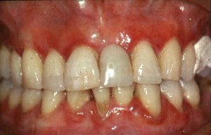
Gingivitis is a mild form of gum disease (periodontal disease) that causes irritation, redness, swelling and bleeding of the gingiva, or gums. It's important to note that it is reversible with good oral hygiene and professional dental treatment. If left untreated, however, gingivitis can progress to a more severe form of gum disease known as periodontitis, which can result in tissue damage and eventual tooth loss.
Gingivitis is most commonly caused by the buildup of plaque, a sticky film of bacteria that constantly forms on our teeth. When not removed regularly through brushing and flossing, this plaque can harden into tartar, which is more difficult to remove and contributes to gum inflammation. Other factors like hormonal changes, poor nutrition, certain medications, smoking or a weakened immune system may also increase the risk of developing gingivitis.
Epithelial attachment is a general term that refers to the point where epithelial cells, which are the cells that line the outer surfaces of organs and blood vessels, adhere or attach to an underlying structure. In the context of the mouth and teeth, epithelial attachment is often used to describe the connection between the gum tissue (gingiva) and the tooth surface.
In a healthy mouth, the gingival tissue fits tightly around each tooth, forming a protective seal that helps prevent bacteria and other harmful substances from entering the spaces between the teeth and gums. This tight seal is maintained by specialized epithelial cells called junctional epithelial cells, which form a barrier between the oral environment and the underlying connective tissue.
When the gingival tissue becomes inflamed due to factors such as poor oral hygiene or certain medical conditions, the epithelial attachment can become compromised, leading to a condition known as gingivitis. If left untreated, gingivitis can progress to periodontal disease, which is characterized by the destruction of the tissues that support the teeth, including the bone and connective tissue.
In summary, epithelial attachment refers to the point where epithelial cells adhere to an underlying structure, and in the context of oral health, it describes the connection between the gum tissue and the tooth surface.
Gingival hyperplasia is a condition characterized by an abnormal growth or enlargement of the gingiva (gum tissue). This condition can be caused by various factors, including bacterial infection, certain medications (such as phenytoin, cyclosporine, and nifedipine), systemic diseases (such as leukemia, vitamin C deficiency, and Crohn's disease), and genetic disorders.
The enlarged gum tissue can be uncomfortable, irritated, and prone to bleeding, especially during brushing or flossing. It may also make it difficult to maintain good oral hygiene, which can increase the risk of dental caries and periodontal disease. Treatment for gingival hyperplasia typically involves improving oral hygiene, controlling any underlying causes, and in some cases, surgical removal of the excess tissue.
Gingival recession is the term used to describe the exposure of the root surface of a tooth as a result of the loss of gum tissue (gingiva) due to periodontal disease or improper oral hygiene practices. It can also occur due to other factors such as aggressive brushing, grinding or clenching of teeth, and misaligned teeth. Gingival recession is often characterized by red, swollen, or sensitive gums, and can lead to tooth sensitivity, decay, and even tooth loss if left untreated.
Gingival overgrowth, also known as gingival hyperplasia or hypertrophy, refers to an abnormal enlargement or growth of the gum tissue (gingiva) surrounding the teeth. This condition can be caused by various factors, including poor oral hygiene, certain medications (such as phenytoin, cyclosporine, and calcium channel blockers), genetic predisposition, and systemic conditions like vitamin C deficiency or leukemia.
Gingival overgrowth can lead to several complications, such as difficulty in maintaining oral hygiene, which may result in periodontal disease, tooth decay, bad breath, and potential loss of teeth. In some cases, the enlarged gum tissue may also cause discomfort or pain during speaking, chewing, or brushing. Treatment for gingival overgrowth typically involves improving oral hygiene, adjusting medications if possible, and undergoing surgical procedures to remove the excess gum tissue. Regular dental check-ups and cleanings are essential in managing and preventing this condition.
Gingival fibromatosis is a benign (non-cancerous) condition characterized by the excessive growth of gum (gingival) tissue. The overgrowth can affect one or both the maxilla (upper jaw) and mandible (lower jaw) and can lead to various dental and oral health issues, such as difficulty in chewing, speaking, and maintaining proper oral hygiene.
The etiology of gingival fibromatosis can be divided into two categories: hereditary and acquired. Hereditary gingival fibromatosis is often associated with genetic mutations, while acquired gingival fibromatosis can result from factors like chronic inflammation due to poor oral hygiene, certain medications (such as phenytoin, cyclosporine, or nifedipine), and systemic conditions (like leukemia).
The management of gingival fibromatosis typically involves surgical removal of the excess tissue. However, recurrence is common due to the condition's tendency for regrowth. Regular follow-ups with a dental professional are essential to monitor any potential regrowth and maintain good oral hygiene.
Gingivoplasty is a surgical procedure in dentistry that involves the reshaping or contouring of the gingiva (gums). This procedure is typically performed for aesthetic purposes, to improve the appearance of gums that are uneven or have an irregular shape. It can also be done to remove excess gum tissue that may be covering too much of a tooth, making it appear shorter than the other teeth.
Gingivoplasty is often recommended as a part of periodontal treatment to ensure the proper fit and function of dental restorations or to manage and prevent gum disease. The procedure involves removing and reshaping the gingival tissue to create a more aesthetically pleasing and healthy gum line.
It's important to note that while gingivoplasty can improve the appearance of the gums, it does not address any underlying issues related to gum disease or bone loss. Additional periodontal treatments may be necessary to address these concerns.
A gingivectomy is a dental procedure that involves the surgical removal or reshaping of the gum tissue (gingiva) to improve the health and appearance of the teeth and gums. This procedure is typically performed when the gums have become swollen, inflamed, or infected due to periodontal disease, which can cause the gums to recede and expose the tooth roots. By removing the affected gum tissue, a gingivectomy can help to eliminate pockets of bacteria and promote healthy gum growth.
During the procedure, a dental surgeon will use local anesthesia to numb the area and then carefully cut away the excess gum tissue using specialized instruments. The surgeon may also smooth and reshape the remaining gum tissue to create a more even and aesthetically pleasing appearance. After the procedure, patients may experience some discomfort, swelling, or bleeding, but these symptoms can typically be managed with over-the-counter pain medications and careful oral hygiene practices.
It's important to note that while a gingivectomy can help to improve the health of the gums and teeth, it is not a substitute for good oral hygiene habits. Regular brushing, flossing, and dental checkups are essential for maintaining healthy teeth and gums over the long term.
Gingival hypertrophy is a condition characterized by an abnormal enlargement or overgrowth of the gingiva (gum tissue). This can be caused due to various reasons such as inflammation from poor oral hygiene, certain medications like phenytoin and cyclosporine, or systemic conditions such as pregnancy, leukemia, and vitamin C deficiency.
The enlarged gums may appear swollen, red, and bleed easily. They can also cover the teeth, making cleaning difficult, which can further worsen the inflammation. Depending on the cause, treatment options may include improving oral hygiene, changing medications, or undergoing surgical procedures to remove the excess tissue.
A periodontal prosthesis is a removable dental appliance that is used to replace missing teeth and surrounding tissues in patients who have advanced periodontal disease, also known as gum disease. This type of prosthesis is designed to restore both function and aesthetics, helping individuals to chew, speak, and smile with confidence.
Periodontal prostheses are typically made from a combination of materials, including acrylic resins, metals, and sometimes porcelain. They can be used to replace one or more missing teeth, or even an entire arch of teeth. The design of the prosthesis will depend on the individual's specific needs and the extent of their periodontal disease.
There are several types of periodontal prostheses, including:
1. Removable Partial Dentures (RPDs): These are used when some natural teeth remain in the upper or lower jaw. The RPD is designed to fit around the remaining teeth and provide support for the replacement teeth.
2. Overdentures: These are removable dental appliances that fit over a small number of remaining natural teeth or implants, providing additional stability and support.
3. Complete Dentures: When all the teeth in an arch are missing, a complete denture is used to replace them. The denture is held in place by suction, muscle tension, and sometimes dental adhesives.
It's important to note that periodontal prostheses require regular maintenance and professional cleaning to ensure their longevity and effectiveness. Patients should follow their dentist's or dental specialist's instructions for caring for their prosthesis and maintain good oral hygiene to prevent further issues with their gums and remaining teeth.
The mouth mucosa refers to the mucous membrane that lines the inside of the mouth, also known as the oral mucosa. It covers the tongue, gums, inner cheeks, palate, and floor of the mouth. This moist tissue is made up of epithelial cells, connective tissue, blood vessels, and nerve endings. Its functions include protecting the underlying tissues from physical trauma, chemical irritation, and microbial infections; aiding in food digestion by producing enzymes; and providing sensory information about taste, temperature, and texture.
Periodontitis is a severe form of gum disease that damages the soft tissue and destroys the bone supporting your teeth. If left untreated, it can lead to tooth loss. It is caused by the buildup of plaque, a sticky film of bacteria that constantly forms on our teeth. The body's immune system fights the bacterial infection, which causes an inflammatory response. If the inflammation continues for a long time, it can damage the tissues and bones that support the teeth.
The early stage of periodontitis is called gingivitis, which is characterized by red, swollen gums that bleed easily when brushed or flossed. When gingivitis is not treated, it can advance to periodontitis. In addition to plaque, other factors that increase the risk of developing periodontitis include smoking or using tobacco products, poor oral hygiene, diabetes, a weakened immune system, and genetic factors.
Regular dental checkups and good oral hygiene practices, such as brushing twice a day, flossing daily, and using an antimicrobial mouth rinse, can help prevent periodontitis. Treatment for periodontitis may include deep cleaning procedures, medications, or surgery in severe cases.
An oral ulcer is a defect or break in the continuity of the epithelium, the tissue that lines the inner surface of the mouth, leading to an inflamed, painful, and sometimes bleeding lesion. They can be classified as primary (e.g., aphthous ulcers, traumatic ulcers) or secondary (e.g., those caused by infections, underlying systemic conditions, or reactions to medications). Oral ulcers may cause discomfort, impacting speech and food consumption, and their presence might indicate an underlying medical issue that requires further evaluation.
Gingival crevicular fluid (GCF) is defined as the serum transudate or inflammatory exudate that flows from the gingival sulcus or periodontal pocket. It is a physiological fluid found in the narrow space between the tooth and the surrounding gum tissue, which deepens during periodontal disease. The analysis of GCF has been used as a non-invasive method to assess the status of periodontal health and disease since it contains various markers of inflammation, host response, and bacterial products.
The Periodontal Index (PI) is not a current or widely used medical/dental term. However, in the past, it was used to describe a method for assessing and measuring the severity of periodontal disease, also known as gum disease.
Developed by Henry H. Klein and colleagues in 1978, the Periodontal Index was a scoring system that evaluated four parameters: gingival inflammation, gingival bleeding, calculus (tartar) presence, and periodontal pocket depths. The scores for each parameter ranged from 0 to 3, with higher scores indicating worse periodontal health. The overall PI score was the sum of the individual parameter scores, ranging from 0 to 12.
However, due to its limited ability to predict future disease progression and the introduction of more comprehensive assessment methods like the Community Periodontal Index (CPI) and the Basic Periodontal Examination (BPE), the use of the Periodontal Index has become less common in dental practice and research.
The alveolar process is the curved part of the jawbone (mandible or maxilla) that contains sockets or hollow spaces (alveoli) for the teeth to be embedded. These processes are covered with a specialized mucous membrane called the gingiva, which forms a tight seal around the teeth to help protect the periodontal tissues and maintain oral health.
The alveolar process is composed of both compact and spongy bone tissue. The compact bone forms the outer layer, while the spongy bone is found inside the alveoli and provides support for the teeth. When a tooth is lost or extracted, the alveolar process begins to resorb over time due to the lack of mechanical stimulation from the tooth's chewing forces. This can lead to changes in the shape and size of the jawbone, which may require bone grafting procedures before dental implant placement.
The hard palate is the anterior, bony part of the roof of the mouth, forming a vertical partition between the oral and nasal cavities. It is composed of the maxilla and palatine bones, and provides attachment for the muscles of the soft palate, which functions in swallowing, speaking, and breathing. The hard palate also contains taste buds that contribute to our ability to taste food.
Oral surgical procedures refer to various types of surgeries performed in the oral cavity and maxillofacial region, which includes the mouth, jaws, face, and skull. These procedures are typically performed by oral and maxillofacial surgeons, who are dental specialists with extensive training in surgical procedures involving the mouth, jaws, and face.
Some common examples of oral surgical procedures include:
1. Tooth extractions: This involves removing a tooth that is damaged beyond repair or causing problems for the surrounding teeth. Wisdom tooth removal is a common type of tooth extraction.
2. Dental implant placement: This procedure involves placing a small titanium post in the jawbone to serve as a replacement root for a missing tooth. A dental crown is then attached to the implant, creating a natural-looking and functional replacement tooth.
3. Jaw surgery: Also known as orthognathic surgery, this procedure involves repositioning the jaws to correct bite problems or facial asymmetry.
4. Biopsy: This procedure involves removing a small sample of tissue from the oral cavity for laboratory analysis, often to diagnose suspicious lesions or growths.
5. Lesion removal: This procedure involves removing benign or malignant growths from the oral cavity, such as tumors or cysts.
6. Temporomandibular joint (TMJ) surgery: This procedure involves treating disorders of the TMJ, which connects the jawbone to the skull and allows for movement when eating, speaking, and yawning.
7. Facial reconstruction: This procedure involves rebuilding or reshaping the facial bones after trauma, cancer surgery, or other conditions that affect the face.
Overall, oral surgical procedures are an important part of dental and medical care, helping to diagnose and treat a wide range of conditions affecting the mouth, jaws, and face.
A cuspid, also known as a canine tooth or cuspid tooth, is a type of tooth in mammals. It is the pointiest tooth in the dental arch and is located between the incisors and bicuspids (or premolars). Cuspids have a single cusp or pointed tip that is used for tearing and grasping food. In humans, there are four cuspids, two on the upper jaw and two on the lower jaw, one on each side of the dental arch.
Gingival hemorrhage is the medical term for bleeding of the gingiva, or gums. It refers to the condition where the gums bleed, often as a result of trauma or injury, but also can be caused by various systemic conditions such as disorders of coagulation, leukemia, or scurvy.
Gingival hemorrhage is commonly seen in individuals with poor oral hygiene and periodontal disease, which can cause inflammation and damage to the gums. This can lead to increased susceptibility to bleeding, even during routine activities such as brushing or flossing. It's important to address any underlying causes of gingival hemorrhage to prevent further complications.
Periodontics is a specialty of dentistry that focuses on the prevention, diagnosis, and treatment of diseases affecting the supporting structures of the teeth, including the gums, periodontal ligament, and alveolar bone. It deals with the maintenance of the health, function, and esthetics of these structures and the teeth themselves. Common periodontal treatments include scaling and root planing (deep cleanings), pocket reduction procedures, regenerative treatments, and dental implant placement. Periodontists are dentists who have completed additional training in this specialized field.
Maxillary neoplasms refer to abnormal growths or tumors in the maxilla, which is the upper jaw bone. These growths can be benign (non-cancerous) or malignant (cancerous). Benign neoplasms are slow-growing and do not spread to other parts of the body, while malignant neoplasms can invade surrounding tissues and spread to distant sites.
Maxillary neoplasms can cause various symptoms such as swelling, pain, numbness, loose teeth, or difficulty in chewing or swallowing. They may also cause nasal congestion, nosebleeds, or visual changes if they affect the eye or orbit. The diagnosis of maxillary neoplasms usually involves a combination of clinical examination, imaging studies such as CT or MRI scans, and biopsy to determine the type and extent of the tumor.
Treatment options for maxillary neoplasms depend on several factors, including the type, size, location, and stage of the tumor, as well as the patient's overall health and preferences. Treatment may include surgery, radiation therapy, chemotherapy, or a combination of these modalities. Regular follow-up care is essential to monitor for recurrence or metastasis and ensure optimal outcomes.
Root planing is a dental procedure that involves the cleaning and smoothing of the root surfaces of teeth. It is typically performed as a part of periodontal therapy to treat and manage gum disease. The goal of root planing is to remove tartar, calculus, and bacterial toxins from the roots of teeth, which helps to promote the reattachment of the gums to the teeth and prevent further progression of periodontal disease. This procedure is usually performed under local anesthesia and may require multiple appointments depending on the severity of the case.
Dental photography is a type of clinical photography that focuses on documenting the condition and treatment of teeth and oral structures. It involves using specialized cameras, lenses, and lighting to capture high-quality images of the mouth and related areas. These images can be used for diagnostic purposes, patient education, treatment planning, communication with other dental professionals, and monitoring progress over time. Dental photography may include various types of shots, such as extraoral (outside the mouth) and intraoral (inside the mouth) views, close-ups of individual teeth or restorations, and full-face portraits. It requires a strong understanding of dental anatomy, lighting techniques, and image composition to produce accurate and informative images.
A periodontal pocket is a pathological space or gap that develops between the tooth and the surrounding gum tissue (gingiva) as a result of periodontal disease. This condition is also known as a "periodontal depth" or "probing depth." It is measured in millimeters using a dental probe, and it indicates the level of attachment loss of the gingival tissue to the tooth.
In a healthy periodontium, the sulcus (the normal space between the tooth and gum) measures 1-3 mm in depth. However, when there is inflammation due to bacterial accumulation, the gums may become red, swollen, and bleed easily. As the disease progresses, the sulcus deepens, forming a periodontal pocket, which can extend deeper than 3 mm.
Periodontal pockets provide an environment that is conducive to the growth of harmful bacteria, leading to further tissue destruction and bone loss around the tooth. If left untreated, periodontal disease can result in loose teeth and eventually tooth loss. Regular dental check-ups and professional cleanings are essential for maintaining healthy gums and preventing periodontal pockets from developing or worsening.
Connective tissue is a type of biological tissue that provides support, strength, and protection to various structures in the body. It is composed of cells called fibroblasts, which produce extracellular matrix components such as collagen, elastin, and proteoglycans. These components give connective tissue its unique properties, including tensile strength, elasticity, and resistance to compression.
There are several types of connective tissue in the body, each with its own specific functions and characteristics. Some examples include:
1. Loose or Areolar Connective Tissue: This type of connective tissue is found throughout the body and provides cushioning and support to organs and other structures. It contains a large amount of ground substance, which allows for the movement and gliding of adjacent tissues.
2. Dense Connective Tissue: This type of connective tissue has a higher concentration of collagen fibers than loose connective tissue, making it stronger and less flexible. Dense connective tissue can be further divided into two categories: regular (or parallel) and irregular. Regular dense connective tissue, such as tendons and ligaments, has collagen fibers that run parallel to each other, providing great tensile strength. Irregular dense connective tissue, such as the dermis of the skin, has collagen fibers arranged in a more haphazard pattern, providing support and flexibility.
3. Adipose Tissue: This type of connective tissue is primarily composed of fat cells called adipocytes. Adipose tissue serves as an energy storage reservoir and provides insulation and cushioning to the body.
4. Cartilage: A firm, flexible type of connective tissue that contains chondrocytes within a matrix of collagen and proteoglycans. Cartilage is found in various parts of the body, including the joints, nose, ears, and trachea.
5. Bone: A specialized form of connective tissue that consists of an organic matrix (mainly collagen) and an inorganic mineral component (hydroxyapatite). Bone provides structural support to the body and serves as a reservoir for calcium and phosphate ions.
6. Blood: Although not traditionally considered connective tissue, blood does contain elements of connective tissue, such as plasma proteins and leukocytes (white blood cells). Blood transports nutrients, oxygen, hormones, and waste products throughout the body.
The maxilla is a paired bone that forms the upper jaw in vertebrates. In humans, it is a major bone in the face and plays several important roles in the craniofacial complex. Each maxilla consists of a body and four processes: frontal process, zygomatic process, alveolar process, and palatine process.
The maxillae contribute to the formation of the eye sockets (orbits), nasal cavity, and the hard palate of the mouth. They also contain the upper teeth sockets (alveoli) and help form the lower part of the orbit and the cheekbones (zygomatic arches).
Here's a quick rundown of its key functions:
1. Supports the upper teeth and forms the upper jaw.
2. Contributes to the formation of the eye sockets, nasal cavity, and hard palate.
3. Helps shape the lower part of the orbit and cheekbones.
4. Partakes in the creation of important sinuses, such as the maxillary sinus, which is located within the body of the maxilla.
A fissured tongue is a benign condition characterized by deep grooves or furrows on the surface of the tongue. These grooves can vary in number and depth, and they may cover the entire surface of the tongue or only appear in certain areas. A fissured tongue is also sometimes referred to as a "scrotal tongue" due to its appearance.
While a fissured tongue is usually asymptomatic and does not require treatment, it can occasionally be associated with other conditions such as down syndrome, oral cancer, or certain vitamin deficiencies. It may also increase the risk of tooth decay and gum disease due to the accumulation of food particles and bacteria in the grooves. In some cases, a fissured tongue may cause discomfort or pain, especially if it becomes infected or inflamed. If you have concerns about a fissured tongue or are experiencing symptoms related to this condition, it is recommended that you consult with a healthcare professional for further evaluation and treatment options.
Mandibular neoplasms refer to abnormal growths or tumors that develop in the mandible, which is the lower jawbone. These growths can be benign (non-cancerous) or malignant (cancerous). Benign neoplasms are typically slow-growing and rarely spread to other parts of the body, while malignant neoplasms can invade surrounding tissues and may metastasize (spread) to distant sites.
Mandibular neoplasms can have various causes, including genetic mutations, exposure to certain chemicals or radiation, and infection with certain viruses. The symptoms of mandibular neoplasms may include swelling or pain in the jaw, difficulty chewing or speaking, numbness in the lower lip or chin, loose teeth, and/or a lump or mass in the mouth or neck.
The diagnosis of mandibular neoplasms typically involves a thorough clinical examination, imaging studies such as X-rays, CT scans, or MRI scans, and sometimes a biopsy to confirm the type and extent of the tumor. Treatment options depend on the type, stage, and location of the neoplasm, and may include surgery, radiation therapy, chemotherapy, or a combination of these approaches. Regular follow-up care is essential to monitor for recurrence or metastasis.
Chronic periodontitis is a type of gum disease that is characterized by the inflammation and infection of the tissues surrounding and supporting the teeth. It is a slow-progressing condition that can lead to the destruction of the periodontal ligament and alveolar bone, which can result in loose teeth or tooth loss if left untreated.
Chronic periodontitis is caused by the buildup of dental plaque and calculus (tartar) on the teeth, which harbor bacteria that release toxins that irritate and inflame the gums. Over time, this chronic inflammation can lead to the destruction of the periodontal tissues, including the gingiva, periodontal ligament, and alveolar bone.
The signs and symptoms of chronic periodontitis include:
* Red, swollen, or tender gums
* Bleeding gums during brushing or flossing
* Persistent bad breath (halitosis)
* Receding gums (exposure of the tooth root)
* Loose teeth or changes in bite alignment
* Deep periodontal pockets (spaces between the teeth and gums)
Risk factors for chronic periodontitis include poor oral hygiene, smoking, diabetes, genetics, and certain medications. Treatment typically involves a thorough dental cleaning to remove plaque and calculus, followed by additional procedures such as scaling and root planing or surgery to eliminate infection and promote healing of the periodontal tissues. Good oral hygiene practices, regular dental checkups, and quitting smoking are essential for preventing chronic periodontitis and maintaining good oral health.
A fibroma, ossifying is a benign (non-cancerous) tumor that typically develops in the periodontal ligament, which is the tissue that connects the tooth to the jawbone. This type of fibroma is characterized by the formation of bone-like tissue within the tumor. It usually appears as a firm, slow-growing nodule or mass that can cause pain or discomfort, particularly when biting down on the affected tooth.
The exact cause of ossifying fibromas is not well understood, but they are thought to arise from an overgrowth of cells in the periodontal ligament. They are more common in women than men and typically occur in people between the ages of 20 and 40. Treatment usually involves surgical removal of the tumor, along with any affected tissue or teeth. In some cases, recurrence may occur, so regular follow-up appointments with a dental professional are recommended.
Lip diseases refer to various medical conditions that affect the lips, which can be caused by different factors such as infections, inflammation, allergies, or autoimmune disorders. Some examples of lip diseases include:
1. Cheilitis: It is an inflammation of the lips, which can cause dryness, cracking, and soreness. It can be caused by various factors, including irritants, allergies, or infections.
2. Angular cheilitis: It is a condition that causes inflammation and redness at the corners of the mouth. It can be caused by fungal or bacterial infections, ill-fitting dentures, or vitamin deficiencies.
3. Herpes simplex labialis: Also known as cold sores, it is a viral infection that causes painful blisters on the lips and around the mouth. The virus can be spread through close contact with an infected person.
4. Actinic cheilitis: It is a precancerous condition caused by excessive exposure to the sun, which leads to dry, scaly, or thickened patches on the lips.
5. Fordyce spots: These are small, painless, white or yellowish bumps that appear on the lips and inside the mouth. They are harmless and do not require treatment.
6. Lip cancer: It is a type of skin cancer that affects the lips, usually caused by excessive exposure to the sun. The symptoms include a sore or lump on the lip that does not heal, bleeding, pain, or numbness.
If you experience any symptoms related to lip diseases, it is recommended to consult a healthcare professional for proper diagnosis and treatment.
Tooth movement, in a dental and orthodontic context, refers to the physical change in position or alignment of one or more teeth within the jaw bone as a result of controlled forces applied through various orthodontic appliances such as braces, aligners, or other orthodontic devices. The purposeful manipulation of these forces encourages the periodontal ligament (the tissue that connects the tooth to the bone) to remodel, allowing the tooth to move gradually over time into the desired position. This process is crucial in achieving proper bite alignment, correcting malocclusions, and enhancing overall oral function and aesthetics.
According to the American Academy of Periodontology, periodontal diseases are chronic inflammatory conditions that affect the tissues surrounding and supporting the teeth. These tissues include the gums, periodontal ligament, and alveolar bone. The primary cause of periodontal disease is bacterial plaque, a sticky film that constantly forms on our teeth.
There are two major stages of periodontal disease:
1. Gingivitis: This is the milder form of periodontal disease, characterized by inflammation of the gums (gingiva) without loss of attachment to the teeth. The gums may appear red, swollen, and bleed easily during brushing or flossing. At this stage, the damage can be reversed with proper dental care and improved oral hygiene.
2. Periodontitis: If left untreated, gingivitis can progress to periodontitis, a more severe form of periodontal disease. In periodontitis, the inflammation extends beyond the gums and affects the deeper periodontal tissues, leading to loss of bone support around the teeth. Pockets filled with infection-causing bacteria form between the teeth and gums, causing further damage and potential tooth loss if not treated promptly.
Risk factors for developing periodontal disease include poor oral hygiene, smoking or using smokeless tobacco, genetic predisposition, diabetes, hormonal changes (such as pregnancy or menopause), certain medications, and systemic diseases like AIDS or cancer. Regular dental check-ups and good oral hygiene practices are crucial for preventing periodontal disease and maintaining overall oral health.
An Odontogenic Cyst, Calcifying is a specific type of cyst that originates from the dental tissues. It's also known as a calcifying odontogenic cyst or Gorlin cyst. This cyst is characterized by the presence of calcified structures within its lining.
The calcifications can appear as flecks or more complex structures, such as teeth-like formations. The lining of this cyst often contains ghost cells, which are the remains of epithelial cells that have undergone calcification.
These cysts are typically slow-growing and asymptomatic, although they can sometimes cause swelling or pain if they become large enough to compress adjacent tissues. They are most commonly found in the jaw bones, particularly the mandible.
While the exact cause of calcifying odontogenic cysts is not fully understood, they are thought to arise from developmental abnormalities in the tissues that form teeth. Treatment typically involves surgical removal of the cyst.
Mouth diseases refer to a variety of conditions that affect the oral cavity, including the lips, gums, teeth, tongue, palate, and lining of the mouth. These diseases can be caused by bacteria, viruses, fungi, or other organisms. They can also result from injuries, chronic illnesses, or genetic factors.
Some common examples of mouth diseases include dental caries (cavities), periodontal disease (gum disease), oral herpes, candidiasis (thrush), lichen planus, and oral cancer. Symptoms may include pain, swelling, redness, bleeding, bad breath, difficulty swallowing or speaking, and changes in the appearance of the mouth or teeth. Treatment depends on the specific diagnosis and may involve medications, dental procedures, or lifestyle changes.
Tooth eruption is the process by which a tooth emerges from the gums and becomes visible in the oral cavity. It is a normal part of dental development that occurs in a predictable sequence and timeframe. Primary or deciduous teeth, also known as baby teeth, begin to erupt around 6 months of age and continue to emerge until approximately 2-3 years of age. Permanent or adult teeth start to erupt around 6 years of age and can continue to emerge until the early twenties.
The process of tooth eruption involves several stages, including the formation of the tooth within the jawbone, the movement of the tooth through the bone and surrounding tissues, and the final emergence of the tooth into the mouth. Proper tooth eruption is essential for normal oral function, including chewing, speaking, and smiling. Any abnormalities in the tooth eruption process, such as delayed or premature eruption, can indicate underlying dental or medical conditions that require further evaluation and treatment.
Periodontal attachment loss (PAL) is a clinical measurement in dentistry that refers to the amount of connective tissue attachment between the tooth and its surrounding supportive structures (including the gingiva, periodontal ligament, and alveolar bone) that has been lost due to periodontal disease. It is typically expressed in millimeters and represents the distance from the cementoenamel junction (CEJ), which is the point where the tooth's crown meets the root, to the bottom of the periodontal pocket.
Periodontal pockets are formed when the gums detach from the tooth due to inflammation and infection caused by bacterial biofilms accumulating on the teeth. As the disease progresses, more and more of the supporting structures are destroyed, leading to increased pocket depths and attachment loss. This can eventually result in loose teeth and even tooth loss if left untreated.
Therefore, periodontal attachment loss is an important indicator of the severity and progression of periodontal disease, and its measurement helps dental professionals assess the effectiveness of treatment interventions and monitor disease status over time.
Pigmentation disorders are conditions that affect the production or distribution of melanin, the pigment responsible for the color of skin, hair, and eyes. These disorders can cause changes in the color of the skin, resulting in areas that are darker (hyperpigmentation) or lighter (hypopigmentation) than normal. Examples of pigmentation disorders include melasma, age spots, albinism, and vitiligo. The causes, symptoms, and treatments for these conditions can vary widely, so it is important to consult a healthcare provider for an accurate diagnosis and treatment plan.
The mandible, also known as the lower jaw, is the largest and strongest bone in the human face. It forms the lower portion of the oral cavity and plays a crucial role in various functions such as mastication (chewing), speaking, and swallowing. The mandible is a U-shaped bone that consists of a horizontal part called the body and two vertical parts called rami.
The mandible articulates with the skull at the temporomandibular joints (TMJs) located in front of each ear, allowing for movements like opening and closing the mouth, protrusion, retraction, and side-to-side movement. The mandible contains the lower teeth sockets called alveolar processes, which hold the lower teeth in place.
In medical terminology, the term "mandible" refers specifically to this bone and its associated structures.
A mouth neoplasm refers to an abnormal growth or tumor in the oral cavity, which can be benign (non-cancerous) or malignant (cancerous). Malignant mouth neoplasms are also known as oral cancer. They can develop on the lips, gums, tongue, roof and floor of the mouth, inside the cheeks, and in the oropharynx (the middle part of the throat at the back of the mouth).
Mouth neoplasms can have various causes, including genetic factors, tobacco use, alcohol consumption, and infection with human papillomavirus (HPV). Symptoms may include a lump or thickening in the oral soft tissues, white or red patches, persistent mouth sores, difficulty swallowing or speaking, and numbness in the mouth. Early detection and treatment of mouth neoplasms are crucial for improving outcomes and preventing complications.
Aggressive periodontitis is a severe form of periodontal disease that affects the tissues surrounding and supporting the teeth, including the gums, periodontal ligament, and alveolar bone. It is characterized by rapid destruction of the periodontal tissues and can result in significant tooth loss if left untreated.
Aggressive periodontitis typically affects younger individuals, often before the age of 30, and can progress rapidly, even in the absence of obvious dental plaque or calculus accumulation. It is often associated with a genetic predisposition and may cluster in families.
The disease is classified as localized or generalized based on the distribution of affected sites. Localized aggressive periodontitis typically affects no more than two teeth next to each other, while generalized aggressive periodontitis involves at least three or four teeth in different areas of the mouth.
In addition to genetic factors, other risk factors for aggressive periodontitis include smoking, diabetes, and hormonal changes. Treatment typically involves a combination of thorough dental cleanings, antibiotics, and sometimes surgical intervention to remove damaged tissue and promote healing. Regular maintenance care is essential to prevent recurrence and further progression of the disease.
In medical terms, a "lip" refers to the thin edge or border of an organ or other biological structure. However, when people commonly refer to "the lip," they are usually talking about the lips on the face, which are part of the oral cavity. The lips are a pair of soft, fleshy tissues that surround the mouth and play a crucial role in various functions such as speaking, eating, drinking, and expressing emotions.
The lips are made up of several layers, including skin, muscle, blood vessels, nerves, and mucous membrane. The outer surface of the lips is covered by skin, while the inner surface is lined with a moist mucous membrane. The muscles that make up the lips allow for movements such as pursing, puckering, and smiling.
The lips also contain numerous sensory receptors that help detect touch, temperature, pain, and other stimuli. Additionally, they play a vital role in protecting the oral cavity from external irritants and pathogens, helping to keep the mouth clean and healthy.
Dental records are a collection of detailed documentation related to a patient's dental history and treatment. These records typically include:
1. Patient demographics: This includes the patient's name, date of birth, contact information, and other identifying details.
2. Dental charts: These are graphic representations of the patient's teeth and gums, noting any existing restorations, decay, periodontal disease, or other oral health conditions.
3. Radiographs (x-rays): These images help dentists visualize structures that aren't visible during a clinical examination, such as between teeth, below the gum line, and inside the jaw bones.
4. Treatment plans: This includes proposed dental procedures, their estimated costs, and the rationale behind them.
5. Progress notes: These are ongoing records of each dental appointment, detailing the treatments performed, the patient's response to treatment, and any home care instructions given.
6. Medical history: This includes any systemic health conditions that could impact dental treatment, such as diabetes or heart disease, as well as medications being taken.
7. Consent forms: These are documents signed by the patient (or their legal guardian) giving permission for specific treatments.
8. Communication notes: Any correspondence between dental professionals regarding the patient's care.
Dental records play a crucial role in continuity of care, allowing dentists to track changes in a patient's oral health over time and make informed treatment decisions. They are also important for medicolegal reasons, providing evidence in case of malpractice claims or other disputes.
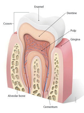
The periodontium is a complex structure in the oral cavity that surrounds and supports the teeth. It consists of four main components:
1. Gingiva (gums): The pink, soft tissue that covers the crown of the tooth and extends down to the neck of the tooth, where it meets the cementum.
2. Cementum: A specialized, calcified tissue that covers the root of the tooth and provides a surface for the periodontal ligament fibers to attach.
3. Periodontal ligament (PDL): A highly vascular and cell-rich connective tissue that attaches the cementum of the tooth root to the alveolar bone, allowing for tooth mobility and absorption of forces during chewing.
4. Alveolar bone: The portion of the jawbone that contains the sockets (alveoli) for the teeth. It is a spongy bone with a rich blood supply that responds to mechanical stresses from biting and chewing, undergoing remodeling throughout life.
Periodontal diseases, such as gingivitis and periodontitis, affect the health and integrity of the periodontium, leading to inflammation, bleeding, pocket formation, bone loss, and ultimately tooth loss if left untreated.
Ameloblastoma is a slow-growing, non-cancerous tumor that develops in the jawbone, typically in the lower jaw. It originates from the cells that form the enamel (the hard, outer surface of the teeth). This tumor can cause swelling, pain, and displacement or loosening of teeth. In some cases, it may also lead to fractures of the jawbone.
There are different types of ameloblastomas, including solid or multicystic, unicystic, and peripheral ameloblastoma. Treatment usually involves surgical removal of the tumor, with careful monitoring to ensure that it does not recur. In rare cases, more aggressive treatment may be necessary if the tumor is large or has invaded surrounding tissues.
It's important to note that while ameloblastomas are generally benign, they can still cause significant morbidity and should be treated promptly by an oral and maxillofacial surgeon or other qualified healthcare professional.
The dental plaque index (DPI) is a clinical measurement used in dentistry to assess the amount of dental plaque accumulation on a person's teeth. It was first introduced by Silness and Löe in 1964 as a method to standardize the assessment of oral hygiene and the effectiveness of oral hygiene interventions.
The DPI is based on a visual examination of the amount of plaque present on four surfaces of the teeth, including the buccal (cheek-facing) and lingual (tongue-facing) surfaces of both upper and lower first molars and upper and lower incisors. The examiner assigns a score from 0 to 3 for each surface, with higher scores indicating greater plaque accumulation:
* Score 0: No plaque detected, even after probing the area with a dental explorer.
* Score 1: Plaque detected by visual examination and/or probing but is not visible when the area is gently dried with air.
* Score 2: Moderate accumulation of soft deposits that are visible upon visual examination before air drying, but which can be removed by scraping with a dental explorer.
* Score 3: Abundant soft matter, visible upon visual examination before air drying and not easily removable with a dental explorer.
The DPI is calculated as the average score of all surfaces examined, providing an overall measure of plaque accumulation in the mouth. It can be used to monitor changes in oral hygiene over time or to evaluate the effectiveness of different oral hygiene interventions. However, it should be noted that the DPI has limitations and may not accurately reflect the presence of bacterial biofilms or the risk of dental caries and gum disease.
The term "mouth floor" is not a standard medical terminology. However, it might refer to the floor of the mouth, which is the part of the oral cavity located beneath the tongue and above the hyoid bone, which is a U-shaped bone in the front of the neck that helps support the tongue. The mouth floor contains several salivary glands, muscles, and nerves that are important for functions such as swallowing and speaking.
In the context of medical terminology, 'color' is not defined specifically with a unique meaning. Instead, it generally refers to the characteristic or appearance of something, particularly in relation to the color that a person may observe visually. For instance, doctors may describe the color of a patient's skin, eyes, hair, or bodily fluids to help diagnose medical conditions or monitor their progression.
For example, jaundice is a yellowing of the skin and whites of the eyes that can indicate liver problems, while cyanosis refers to a bluish discoloration of the skin and mucous membranes due to insufficient oxygen in the blood. Similarly, doctors may describe the color of stool or urine to help diagnose digestive or kidney issues.
Therefore, 'color' is not a medical term with a specific definition but rather a general term used to describe various visual characteristics of the body and bodily fluids that can provide important diagnostic clues for healthcare professionals.
Orthodontic appliances are devices used in orthodontics, a branch of dentistry focused on the diagnosis, prevention, and treatment of dental and facial irregularities. These appliances can be fixed or removable and are used to align teeth, correct jaw relationships, or modify dental forces. They can include braces, aligners, palatal expanders, space maintainers, and headgear, among others. The specific type of appliance used depends on the individual patient's needs and the treatment plan developed by the orthodontist.
A tooth root is the part of a tooth that is embedded in the jawbone and cannot be seen when looking at a person's smile. It is the lower portion of a tooth that typically has a conical shape and anchors the tooth to the jawbone through a periodontal ligament. The tooth root is covered by cementum, a specialized bone-like tissue, and contains nerve endings and blood vessels within its pulp chamber.
The number of roots in a tooth can vary depending on the type of tooth. For example, incisors typically have one root, canines may have one or two roots, premolars usually have one or two roots, and molars often have two to four roots. The primary function of the tooth root is to provide stability and support for the crown of the tooth, allowing it to withstand the forces of biting and chewing.
In medical terms, the mouth is officially referred to as the oral cavity. It is the first part of the digestive tract and includes several structures: the lips, vestibule (the space enclosed by the lips and teeth), teeth, gingiva (gums), hard and soft palate, tongue, floor of the mouth, and salivary glands. The mouth is responsible for several functions including speaking, swallowing, breathing, and eating, as it is the initial point of ingestion where food is broken down through mechanical and chemical processes, beginning the digestive process.
Prevotella intermedia is a gram-negative, anaerobic, rod-shaped bacterium that is commonly found in the oral cavity, upper respiratory tract, and gastrointestinal tract. It is a normal resident of the human microbiota but can also be an opportunistic pathogen, causing various types of infections such as periodontitis, endocarditis, and brain abscesses. P. intermedia has been associated with several diseases, including respiratory tract infections, bacteremia, and joint infections. It is often found in mixed infections with other anaerobic bacteria. Proper identification of this organism is important for the selection of appropriate antimicrobial therapy.
Zirconium is not a medical term, but it is a chemical element with the symbol Zr and atomic number 40. It is a gray-white, strong, corrosion-resistant transition metal that is used primarily in nuclear reactors, as an opacifier in glazes for ceramic cookware, and in surgical implants such as artificial joints due to its biocompatibility.
In the context of medical devices or implants, zirconium alloys may be used for their mechanical properties and resistance to corrosion. For example, zirconia (a form of zirconium dioxide) is a popular material for dental crowns and implants due to its durability, strength, and natural appearance.
However, it's important to note that while zirconium itself is not considered a medical term, there are various medical applications and devices that utilize zirconium-based materials.
"Porphyromonas gingivalis" is a gram-negative, anaerobic, rod-shaped bacterium that is commonly found in the oral cavity and is associated with periodontal disease. It is a major pathogen in chronic periodontitis, which is a severe form of gum disease that can lead to destruction of the tissues supporting the teeth, including the gums, periodontal ligament, and alveolar bone.
The bacterium produces several virulence factors, such as proteases and endotoxins, which contribute to its pathogenicity. It has been shown to evade the host's immune response and cause tissue destruction through various mechanisms, including inducing the production of pro-inflammatory cytokines and matrix metalloproteinases.
P. gingivalis has also been linked to several systemic diseases, such as atherosclerosis, rheumatoid arthritis, and Alzheimer's disease, although the exact mechanisms of these associations are not fully understood. Effective oral hygiene practices, including regular brushing, flossing, and professional dental cleanings, can help prevent the overgrowth of P. gingivalis and reduce the risk of periodontal disease.
In the context of dentistry, a molar is a type of tooth found in the back of the mouth. They are larger and wider than other types of teeth, such as incisors or canines, and have a flat biting surface with multiple cusps. Molars are primarily used for grinding and chewing food into smaller pieces that are easier to swallow. Humans typically have twelve molars in total, including the four wisdom teeth.
In medical terminology outside of dentistry, "molar" can also refer to a unit of mass in the apothecaries' system of measurement, which is equivalent to 4.08 grams. However, this usage is less common and not related to dental or medical anatomy.
Dental plaque is a biofilm or mass of bacteria that accumulates on the surface of the teeth, restorative materials, and prosthetic devices such as dentures. It is initiated when bacterial colonizers attach to the smooth surfaces of teeth through van der Waals forces and specific molecular adhesion mechanisms.
The microorganisms within the dental plaque produce extracellular polysaccharides that help to stabilize and strengthen the biofilm, making it resistant to removal by simple brushing or rinsing. Over time, if not regularly removed through oral hygiene practices such as brushing and flossing, dental plaque can mineralize and harden into tartar or calculus.
The bacteria in dental plaque can cause tooth decay (dental caries) by metabolizing sugars and producing acid that demineralizes the tooth enamel. Additionally, certain types of bacteria in dental plaque can cause periodontal disease, an inflammation of the gums that can lead to tissue damage and bone loss around the teeth. Regular professional dental cleanings and good oral hygiene practices are essential for preventing the buildup of dental plaque and maintaining good oral health.
Fibroblasts are specialized cells that play a critical role in the body's immune response and wound healing process. They are responsible for producing and maintaining the extracellular matrix (ECM), which is the non-cellular component present within all tissues and organs, providing structural support and biochemical signals for surrounding cells.
Fibroblasts produce various ECM proteins such as collagens, elastin, fibronectin, and laminins, forming a complex network of fibers that give tissues their strength and flexibility. They also help in the regulation of tissue homeostasis by controlling the turnover of ECM components through the process of remodeling.
In response to injury or infection, fibroblasts become activated and start to proliferate rapidly, migrating towards the site of damage. Here, they participate in the inflammatory response, releasing cytokines and chemokines that attract immune cells to the area. Additionally, they deposit new ECM components to help repair the damaged tissue and restore its functionality.
Dysregulation of fibroblast activity has been implicated in several pathological conditions, including fibrosis (excessive scarring), cancer (where they can contribute to tumor growth and progression), and autoimmune diseases (such as rheumatoid arthritis).
Colorimetry is the scientific measurement and quantification of color, typically using a colorimeter or spectrophotometer. In the medical field, colorimetry may be used in various applications such as:
1. Diagnosis and monitoring of skin conditions: Colorimeters can measure changes in skin color to help diagnose or monitor conditions like jaundice, cyanosis, or vitiligo. They can also assess the effectiveness of treatments for these conditions.
2. Vision assessment: Colorimetry is used in vision testing to determine the presence and severity of visual impairments such as color blindness or deficiencies. Special tests called anomaloscopes or color vision charts are used to measure an individual's ability to distinguish between different colors.
3. Environmental monitoring: In healthcare settings, colorimetry can be employed to monitor the cleanliness and sterility of surfaces or equipment by measuring the amount of contamination present. This is often done using ATP (adenosine triphosphate) bioluminescence assays, which emit light when they come into contact with microorganisms.
4. Medical research: Colorimetry has applications in medical research, such as studying the optical properties of tissues or developing new diagnostic tools and techniques based on color measurements.
In summary, colorimetry is a valuable tool in various medical fields for diagnosis, monitoring, and research purposes. It allows healthcare professionals to make more informed decisions about patient care and treatment plans.
Epithelium is the tissue that covers the outer surface of the body, lines the internal cavities and organs, and forms various glands. It is composed of one or more layers of tightly packed cells that have a uniform shape and size, and rest on a basement membrane. Epithelial tissues are avascular, meaning they do not contain blood vessels, and are supplied with nutrients by diffusion from the underlying connective tissue.
Epithelial cells perform a variety of functions, including protection, secretion, absorption, excretion, and sensation. They can be classified based on their shape and the number of cell layers they contain. The main types of epithelium are:
1. Squamous epithelium: composed of flat, scalelike cells that fit together like tiles on a roof. It forms the lining of blood vessels, air sacs in the lungs, and the outermost layer of the skin.
2. Cuboidal epithelium: composed of cube-shaped cells with equal height and width. It is found in glands, tubules, and ducts.
3. Columnar epithelium: composed of tall, rectangular cells that are taller than they are wide. It lines the respiratory, digestive, and reproductive tracts.
4. Pseudostratified epithelium: appears stratified or layered but is actually made up of a single layer of cells that vary in height. The nuclei of these cells appear at different levels, giving the tissue a stratified appearance. It lines the respiratory and reproductive tracts.
5. Transitional epithelium: composed of several layers of cells that can stretch and change shape to accommodate changes in volume. It is found in the urinary bladder and ureters.
Epithelial tissue provides a barrier between the internal and external environments, protecting the body from physical, chemical, and biological damage. It also plays a crucial role in maintaining homeostasis by regulating the exchange of substances between the body and its environment.
Immunohistochemistry (IHC) is a technique used in pathology and laboratory medicine to identify specific proteins or antigens in tissue sections. It combines the principles of immunology and histology to detect the presence and location of these target molecules within cells and tissues. This technique utilizes antibodies that are specific to the protein or antigen of interest, which are then tagged with a detection system such as a chromogen or fluorophore. The stained tissue sections can be examined under a microscope, allowing for the visualization and analysis of the distribution and expression patterns of the target molecule in the context of the tissue architecture. Immunohistochemistry is widely used in diagnostic pathology to help identify various diseases, including cancer, infectious diseases, and immune-mediated disorders.
Keratins are a type of fibrous structural proteins that constitute the main component of the integumentary system, which includes the hair, nails, and skin of vertebrates. They are also found in other tissues such as horns, hooves, feathers, and reptilian scales. Keratins are insoluble proteins that provide strength, rigidity, and protection to these structures.
Keratins are classified into two types: soft keratins (Type I) and hard keratins (Type II). Soft keratins are found in the skin and simple epithelial tissues, while hard keratins are present in structures like hair, nails, horns, and hooves.
Keratin proteins have a complex structure consisting of several domains, including an alpha-helical domain, beta-pleated sheet domain, and a non-repetitive domain. These domains provide keratin with its unique properties, such as resistance to heat, chemicals, and mechanical stress.
In summary, keratins are fibrous structural proteins that play a crucial role in providing strength, rigidity, and protection to various tissues in the body.
Squamous cell carcinoma is a type of skin cancer that begins in the squamous cells, which are flat, thin cells that form the outer layer of the skin (epidermis). It commonly occurs on sun-exposed areas such as the face, ears, lips, and backs of the hands. Squamous cell carcinoma can also develop in other areas of the body including the mouth, lungs, and cervix.
This type of cancer usually develops slowly and may appear as a rough or scaly patch of skin, a red, firm nodule, or a sore or ulcer that doesn't heal. While squamous cell carcinoma is not as aggressive as some other types of cancer, it can metastasize (spread) to other parts of the body if left untreated, making early detection and treatment important.
Risk factors for developing squamous cell carcinoma include prolonged exposure to ultraviolet (UV) radiation from the sun or tanning beds, fair skin, a history of sunburns, a weakened immune system, and older age. Prevention measures include protecting your skin from the sun by wearing protective clothing, using a broad-spectrum sunscreen with an SPF of at least 30, avoiding tanning beds, and getting regular skin examinations.
Gingiva is the medical term for the soft tissue that surrounds the teeth and forms the margin of the dental groove, also known as the gum. It extends from the mucogingival junction to the base of the cervical third of the tooth root. The gingiva plays a crucial role in protecting and supporting the teeth and maintaining oral health by providing a barrier against microbial invasion and mechanical injury.
Collagen is the most abundant protein in the human body, and it is a major component of connective tissues such as tendons, ligaments, skin, and bones. Collagen provides structure and strength to these tissues and helps them to withstand stretching and tension. It is made up of long chains of amino acids, primarily glycine, proline, and hydroxyproline, which are arranged in a triple helix structure. There are at least 16 different types of collagen found in the body, each with slightly different structures and functions. Collagen is important for maintaining the integrity and health of tissues throughout the body, and it has been studied for its potential therapeutic uses in various medical conditions.
I believe there might be a misunderstanding in your question. "Dogs" is not a medical term or condition. It is the common name for a domesticated carnivore of the family Canidae, specifically the genus Canis, which includes wolves, foxes, and other extant and extinct species of mammals. Dogs are often kept as pets and companions, and they have been bred in a wide variety of forms and sizes for different purposes, such as hunting, herding, guarding, assisting police and military forces, and providing companionship and emotional support.
If you meant to ask about a specific medical condition or term related to dogs, please provide more context so I can give you an accurate answer.
Follow-up studies are a type of longitudinal research that involve repeated observations or measurements of the same variables over a period of time, in order to understand their long-term effects or outcomes. In medical context, follow-up studies are often used to evaluate the safety and efficacy of medical treatments, interventions, or procedures.
In a typical follow-up study, a group of individuals (called a cohort) who have received a particular treatment or intervention are identified and then followed over time through periodic assessments or data collection. The data collected may include information on clinical outcomes, adverse events, changes in symptoms or functional status, and other relevant measures.
The results of follow-up studies can provide important insights into the long-term benefits and risks of medical interventions, as well as help to identify factors that may influence treatment effectiveness or patient outcomes. However, it is important to note that follow-up studies can be subject to various biases and limitations, such as loss to follow-up, recall bias, and changes in clinical practice over time, which must be carefully considered when interpreting the results.
In the field of medicine, "time factors" refer to the duration of symptoms or time elapsed since the onset of a medical condition, which can have significant implications for diagnosis and treatment. Understanding time factors is crucial in determining the progression of a disease, evaluating the effectiveness of treatments, and making critical decisions regarding patient care.
For example, in stroke management, "time is brain," meaning that rapid intervention within a specific time frame (usually within 4.5 hours) is essential to administering tissue plasminogen activator (tPA), a clot-busting drug that can minimize brain damage and improve patient outcomes. Similarly, in trauma care, the "golden hour" concept emphasizes the importance of providing definitive care within the first 60 minutes after injury to increase survival rates and reduce morbidity.
Time factors also play a role in monitoring the progression of chronic conditions like diabetes or heart disease, where regular follow-ups and assessments help determine appropriate treatment adjustments and prevent complications. In infectious diseases, time factors are crucial for initiating antibiotic therapy and identifying potential outbreaks to control their spread.
Overall, "time factors" encompass the significance of recognizing and acting promptly in various medical scenarios to optimize patient outcomes and provide effective care.
Messenger RNA (mRNA) is a type of RNA (ribonucleic acid) that carries genetic information copied from DNA in the form of a series of three-base code "words," each of which specifies a particular amino acid. This information is used by the cell's machinery to construct proteins, a process known as translation. After being transcribed from DNA, mRNA travels out of the nucleus to the ribosomes in the cytoplasm where protein synthesis occurs. Once the protein has been synthesized, the mRNA may be degraded and recycled. Post-transcriptional modifications can also occur to mRNA, such as alternative splicing and addition of a 5' cap and a poly(A) tail, which can affect its stability, localization, and translation efficiency.
Treatment outcome is a term used to describe the result or effect of medical treatment on a patient's health status. It can be measured in various ways, such as through symptoms improvement, disease remission, reduced disability, improved quality of life, or survival rates. The treatment outcome helps healthcare providers evaluate the effectiveness of a particular treatment plan and make informed decisions about future care. It is also used in clinical research to compare the efficacy of different treatments and improve patient care.
 Gingival fibers
Index of oral health and dental articles
Piedmontese language
Stippling (dentistry)
Angularis nigra
Rete pegs
Clinical attachment loss
Plasma cell granuloma
Gum lift
Periodontal disease
Gingival grafting
Laser-assisted new attachment procedure
Gingivectomy
Glutamate-rich protein 4
Smoker's melanosis
Oral pigmentation
Wolf tooth
Von Willebrand disease
Stomatitis
Gum depigmentation
Drug-induced gingival enlargement
List of periodontal diseases
Mucogingival junction
Crown lengthening
Desquamative gingivitis
Human tooth
Giant-cell fibroma
Octacalcium phosphate
Peripheral giant-cell granuloma
Membrane vesicle trafficking
 Free Gingiva
 Denture Gingiva Basic Mono Pink
 The metabolism of proteoglycans and glycosaminoglycans in inflamed human gingiva
marginal gingiva
 Gingiva-Derived Mesenchymal Stem Cells: Potential Application in Tissue Engineering and Regenerative Medicine - A Comprehensive...
 Effect of light curing unit and antiseptic mouthwashes on the physical properties of gingiva-colored composite resin
|...
Effect of light curing unit and antiseptic mouthwashes on the physical properties of gingiva-colored composite resin
|...
 gingiva - Pictoores
Gingiva - wikidoc
 GR-15 gingiva
- Gro3X
GR-15 gingiva
- Gro3X
Gingiva mask | Online catalog
 Gingiva Mask Archives - sgdentalshop.com
Attached gingiva is composed of
 GINGIVA Flexible Dental Model Resin - Monocure3D
 healing / gingiva forming - DENTAURUM Online Shop
 Primary Leiomyosarcoma of the Mandibular Gingiva
What is gingiva? | Dental Savings Direct
 SR Nexco PASTE GINGIVA 2,5GR | capuozzosrl
 Bonders & Liquids
 wisercoach.com - Are electrical toothbrushes harmful for gingiva? - Welcome
 Shop Products - Miscellaneous - Gingiva Materials - M&S Dental Supply
mei-avivim.com - Are electrical toothbrushes harmful for gingiva? - Welcome
Metastatic lung carcinoma to the lower anterior gingiva<...
 Double heterozygous pathogenic mutations in KIF3C and ZNF513 cause hereditary gingival fibromatosis | International Journal of...
 General Dentistry Details
 BASF Forward AM Ultracur3D® DM 2304 Gingiva Mask | 1 kg | 3DGBIRE Ltd
Tooth bone and gingiva watercolor - Goodfairyart - Whimsical watercolor prints for your home!
 Planmeca PlanCAD Premium software release 2.0
 How to treat gummy smiles - Dentistry
Gingival fibers - Wikipedia
 Reactive lesions of the gingiva. A clinicopathological study of 741 cases<...
Gingival9
- Free gingiva forms the gingival margin which is visible during examination. (rvc.ac.uk)
- Capa N. An alternative treatment approach to gingival recession: gingiva-colored partial porcelain veneers: A clinical report. (usp.br)
- In about 50% of individuals, it is demarcated from the adjacent, attached gingiva by a shallow linear depression, the free gingival groove. (wikidoc.org)
- The marginal gingiva is supported and stabilized by the gingival fibers . (wikidoc.org)
- The interdental gingiva occupies the gingival embrasure , which is the interproximal space beneath the area of tooth contact. (wikidoc.org)
- The gingival fibers accomplish the following tasks: They hold the marginal gingiva against the tooth They provide the marginal gingiva with enough rigidity to withstand the forces of mastication without distorting They serve to stabilize the marginal gingiva by uniting it with both the tissue of the more rigid attached gingiva as well as the cementum layer of the tooth. (wikipedia.org)
- There are three groups within which gingival fibers are arranged: dentogingival group - there are three types of fibers within this group: fibers that extend towards the crest of the gingiva fibers that extend laterally to the outer surface of the gingiva and fibers that extend outward, past the height of the alveolar crest, and then downward along the cortex of the alveolar bone. (wikipedia.org)
- Chronic inflammatory gingival enlargement (CIGE) occasionally exhibits some similarities with hemangioma (Hm) by means of the clinical and histopathological features of gingiva. (omu.edu.tr)
- The present paper reports the clinic and histopathologic features of CIGE and Hm of gingiva in patients referred to our clinic suffering primarily from gingival overgrowth. (omu.edu.tr)
Gingivitis7
- A band of red atrophic or eroded mucosa affecting the attached gingiva is known as dequamative gingivitis. (intelligentdental.com)
- Healthy gingiva fills and fits each interdental space, unlike the swollen gingiva papilla seen in gingivitis or the empty interdental embrasure seen in periodontal disease. (wikidoc.org)
- Background: Observation carried out in early 2016 to a number of pregnant women in Community Health Center Surabaya found that more than 80% of pregnant women had infected gingiva or gingivitis. (unair.ac.id)
- Gingivitis is a common and mild form of gum disease (periodontal disease) that causes irritation, redness and swelling (inflammation) of your gingiva, the part of your gum around the base of your teeth. (mayoclinic.org)
- Gingiva become inflamed (gingivitis). (mayoclinic.org)
- Streptococcus alpha diperoleh dari isolasi dari cairan sulkus gingiva anak dengan gingivitis ringan. (wima.ac.id)
- Gingivitis is an inflammation of the gingiva without attachment loss or with nonprogressing attachment loss. (medscape.com)
Marginal gingiva4
- A 1 mm concavity for the base of the pontic, further apical to the maximal curvature of the adjacent marginal gingiva, is developed. (intelligentdental.com)
- Unlike plaque-induced inflammation it is a dusky red colour and extends beyond the marginal gingiva, often to the full width of the attached gingiva and sometimes onto the alveolar mucosa. (intelligentdental.com)
- The marginal gingiva is the terminal edge of gingiva surrounding the teeth in collar like fashion. (wikidoc.org)
- The attached gingiva is continuous with the marginal gingiva. (wikidoc.org)
Gums5
- gingiva ), or gums , consists of the mucosal tissue that lies over the alveolar bone . (wikidoc.org)
- Gingiva is the dental term for gums. (dentalqa.com)
- The gingiva (gums). (cancer.gov)
- The oral cavity includes the lips, hard palate (the bony front portion of the roof of the mouth), soft palate (the muscular back portion of the roof of the mouth), retromolar trigone (the area behind the wisdom teeth), front two-thirds of the tongue, gingiva (gums), buccal mucosa (the inner lining of the lips and cheeks), and floor of the mouth under the tongue. (cancer.gov)
- The gums (gingivae) are usually paler by comparison and fit snugly around the teeth. (msdmanuals.com)
Resin5
- Gingiva-coloured resin with improved material properties in terms of high flexural strength and fracture resistance. (zirkonzahn.com)
- Objective: This study evaluated surface roughness (Ra) and color difference (ΔE 00 ) of a gingiva-colored composite resin (NT Premium Gengiva, Coltene), light-activated with three light curing units (LCU) and immersed in different mouthwash solutions. (usp.br)
- Conclusion: Ra of the gingiva-colored composite resin tested increased when L1 and S2 were used, although L1-cured specimens also showed the lowest ΔE 00 . (usp.br)
- Evaluation of shear bond strengths of gingiva-colored composite resin to porcelain, metal and zirconia substrates. (usp.br)
- GR-15 gingiva is a 3D printing resin for the fabrication of flexible gingiva masks. (gro3x.com)
Swollen gingiva1
- We present a case of a 45 years old male patient having grade II OSMF reporting with intermittent bleeding and swollen gingiva with extravasation of blood in gingiva and floor of mouth secondary to local trauma. (discoveryjournals.org)
Mandibular1
- We report a case of leiomyosarcoma arising in the anterior mandibular gingiva, which is a non-prevalent site of occurrence. (peertechzpublications.org)
Tooth9
- Healthy gingiva has a smooth arcuate or scalloped appearance around each tooth. (wikidoc.org)
- The attached gingiva is the part that tightly binds to the underlying periodontium, tooth, and bone. (github.io)
- Quite often in patients with gum overgrown over the tooth, when you remove gingiva from the tooth, it gives them their correct tooth proportions back. (dentistry.co.uk)
- You will also want to have a look at the incisal edge of the tooth because, if the gingiva is overgrown, it may be because we have an anterior blinding habit and they've worn away their enamel there. (dentistry.co.uk)
- circular group - these fibers are unique in that they exist entirely within the gingiva and do not contact the tooth transseptal group - these fibers have traditionally been described as spanning the interproximal tissue between adjacent teeth, into which they are embedded. (wikipedia.org)
- However, two other types of fibers have been described in this group: semicircular fibers - fibers that run through the facial and lingual gingiva around each tooth, attaching to the interproximal surfaces of the same tooth. (wikipedia.org)
- Accuracy of orthodontic mini-implants placed at the anterior palate by tooth-borne or gingiva-borne guide support: a cadaveric study. (bvsalud.org)
- The aim of this cadaveric study was to measure the transfer accuracy of orthodontic mini-implant placement at the anterior palate depending on tooth -borne or gingiva -borne guide support. (bvsalud.org)
- Forty orthodontic mini-implants were placed paramedian in the anterior palate of 20 cadaver heads using tooth -borne (TBG) or gingiva -borne guides (GBG). (bvsalud.org)
Mucogingival junction1
- The facial aspect of the attached gingiva extends to the relatively loose and movable alveolar mucosa, from which it is demarcated by the mucogingival junction . (wikidoc.org)
Mucosa2
- The gingiva is part of the masticatory mucosa that provides an internal defense mechanism against pathogens and mechanical stress. (github.io)
- HSV is an infectious disease that often presents with vesicles that quickly ulcerate and involve the keratinized mucosa of the hard palate, dorsal tongue and attached gingiva. (medscape.com)
Teeth4
- Objective: to assess the efficacy of porcine collagen matrix (CM) in root coverage (RC) and increase of keratinized gingiva width (KGW) around teeth and implants. (bvsalud.org)
- The longer that plaque and tartar remain on your teeth, the more they irritate the gingiva, the part of your gum around the base of your teeth, causing inflammation. (mayoclinic.org)
- In this report, a fixed partial denture (FPD) to which a detachable gingiva was connected by magnetic attachment was fabricated to support esthetic recovery of the alveolar bone defect and the maxillary anterior missing teeth. (kssfp.jp)
- Missing teeth, accompanied with an alveolar bone defect, are generally recovered with a removable denture or with a combination of surgical treatment and a conventional FPD without a detachable gingiva. (kssfp.jp)
Mask1
- Dentifix-3D Gingiva Mask is a durable and flexible material suitable for printing flexible models. (ultimate3dprintingstore.com)
Resins2
- Liquid for bonding Abro ® Basic Mono, Abro ® Basic Multistratum ® and Denture Gingiva Basic Mono resins. (zirkonzahn.com)
- GR-15 gingiva complement our GR-13 model resins when working on high-end esthetics implant cases. (gro3x.com)
Dental plaque3
- Significance: Optical coherence tomography (OCT) offers high spatial resolution and contrast for imaging intraoral structures, yet few studies have investigated its clinical feasibility for dental plaque and gingiva imaging in vivo. (illinois.edu)
- The handheld OCT system was operated by trained dental hygienists to acquire images of dental plaque and gingiva at various locations and after one-week use of oral hygiene products. (illinois.edu)
- Conclusions: OCT has a strong potential to display and assess dental plaque and gingiva in a clinical setting. (illinois.edu)
Labial1
- DG is more common in middle-aged to elderly females, is painful, affects the buccal/labial gingiva predominantly. (intelligentdental.com)
Lips and cheeks1
- Compared with the soft tissue linings of the lips and cheeks, most of the gingiva are tightly bound to the underlying bone and are designed to resist the friction of food passing over them. (wikidoc.org)
Conclusion1
- Conclusion: Oral health-related quality of life of pregnant woman were influenced by the severity level of infected gingiva. (unair.ac.id)
Oral3
- Objective: The aim of this study was to analyze the influence of severity level of infected gingiva on oral health-related quality of life of pregnant woman. (unair.ac.id)
- The detachable gingiva made it possible for the patient to maintain oral hygiene by himself. (kssfp.jp)
- Objective: The maintenance of good oral hygiene contributes to gingiva health. (bvsalud.org)
Assessment1
- Extension to the gingiva requires assessment. (medscape.com)
Attachment1
- attachment, detachable gingiva, magnet. (kssfp.jp)
Colour2
- Healthy gingiva usually has a colour that has been described as "coral pink. (wikidoc.org)
- Because the color of gingiva varies due to racial pigmentation, uniformity of colour is more important than the underlying color itself. (wikidoc.org)
Color1
- Although the text book color of gingiva is "coral pink", normal racial pigmentation makes the gingiva appear darker. (wikidoc.org)
Free1
- Both free and attached gingiva are keratinized. (github.io)
Pink1
- Healthy gingiva is usually coral pink, but may contain physiologic pigmentation. (wikidoc.org)
Soft1
- Gingiva are part of the soft tissue lining of the mouth. (wikidoc.org)
Study1
- Is there any study proving this negative effect on the gingiva? (wisercoach.com)
Blood1
- Patient's prothrombin time, activated partial prothrombin time and platelets were increased leading to inadequate activity of coagulant factors such as factors resulting in intermittent spontaneous bleeding and extravasation of blood in gingiva and floor of mouth. (discoveryjournals.org)
Surface1
- Attached gingiva may present with surface stippling . (wikidoc.org)
Title1
- title = "Reactive lesions of the gingiva. (tau.ac.il)
Cases1
- A series of 741 consecutive cases of localized hyperplastic lesions of the gingiva were studied. (tau.ac.il)
Treatment1
- However, surgical treatment was avoided by utilizing a magnetic system for connection between the FPD and the detachable gingiva. (kssfp.jp)