Glomerulosclerosis, Focal Segmental
Kidney Glomerulus
Podocytes
Nephrotic Syndrome
Glomerulonephritis
Kidney
Nephrosis, Lipoid
AIDS-Associated Nephropathy
Glomerular Mesangium
Diabetic Nephropathies
Focal Adhesions
Nephrosclerosis
Mesangial Cells
Glomerulonephritis, Membranous
Biopsy
Denys-Drash Syndrome
Glomerulonephritis, IGA
Sclerosis
Collagen Type IV
Plasmapheresis
Glomerulonephritis, Membranoproliferative
Nephritis, Interstitial
Disease Models, Animal
Creatinine
Fibrosis
Transforming Growth Factor beta
Hypertrophy
Kidney Failure, Chronic
Nephrosis
Rats, Sprague-Dawley
Renal Insufficiency
Disease Progression
Actinin
Glomerular Basement Membrane
Puromycin Aminonucleoside
Kidney Tubules
WT1 Proteins
Kidney Function Tests
Glomerular Filtration Rate
Dihydralazine
Steroids
Transforming Growth Factor beta1
Antigens, Thy-1
RNA, Messenger
Immunosuppressive Agents
Immunohistochemistry
Angiotensin-Converting Enzyme Inhibitors
Extracellular Matrix
Acinonyx
Hypertension, Renal
Glutamyl Aminopeptidase
Cyclosporine
Collagen
Focal Infection
Kidney Cortex
Mice, Transgenic
Diabetes Mellitus, Experimental
Microscopy, Electron
Blood Urea Nitrogen
Cells, Cultured
Rats, Wistar
Nonmuscle Myosin Type IIA
Angiotensin II Type 1 Receptor Blockers
Bowman Capsule
Fibronectins
Molecular Motor Proteins
Microfilament Proteins
Focal Nodular Hyperplasia
Body Weight
Rats, Transgenic
Spironolactone
Genes, Wilms Tumor
Gene Expression
Drug Resistance
Mice, Inbred C57BL
Rats, Inbred Dahl
Renin-Angiotensin System
Enalapril
TRPC Cation Channels
Losartan
Basement Membrane
Extracellular Matrix Proteins
Hypertension
Membrane Proteins
Cytoskeletal Proteins
Intracellular Signaling Peptides and Proteins
Retrospective Studies
Acatalasia
Lupus Nephritis
Phenylpropionates
Desmin
Desoxycorticosterone
Signal Transduction
Fluorescent Antibody Technique
Actins
Lisinopril
Angiotensin II
Laminin
Animals, Zoo
Mycophenolic Acid
Plasminogen Activator Inhibitor 1
Mineralocorticoid Receptor Antagonists
Rats, Inbred F344
Chemokine CCL2
Frasier Syndrome
Doxorubicin
Treatment Outcome
Mutation
Albumins
Tetrazoles
Mice, Inbred Strains
Lissamine Green Dyes
Endothelin-1
Focal Adhesion Kinase 2
Rats, Inbred SHR
Serum Albumin
Blotting, Western
Glycosylation End Products, Advanced
Antihypertensive Agents
Vinculin
Heterozygote
Angiotensin Receptor Antagonists
Myosin Heavy Chains
Rats, Zucker
Renin
Cell Count
Syndactyly
Identification of DNA polymorphisms associated with the V type alpha1-antitrypsin gene. (1/817)
alpha1-Antitrypsin (alpha1-AT) is a highly polymorphic protein. The V allele of alpha1-AT has been shown to be associated with focal glomerulosclerosis (FGS) in Negroid and mixed race South African patients. To identify mutations and polymorphisms in the gene for the V allele of alpha1-AT in five South African patients with FGS nephrotic syndrome DNA sequence analysis and restriction fragment length polymorphisms of the coding exons were carried out. Four of the patients were heterozygous for the BstEII RFLP in exon III [M1(Val213)(Ala213)] and one patient was a M1(Ala213) homozygote. The mutation for the V allele was identified in exon II as Gly-148 (GGG)-->Arg (AGG) and in all patients was associated with a silent mutation at position 158 (AAC-->AAT). The patient who was homozygous for (Ala213) also had a silent mutation at position 256 in exon III (GAT-->GAC) which was not present in any of the other four patients. Although the V allele of alpha1-AT is not associated with severe plasma deficiency, it may be in linkage disequilibrium with other genes on chromosome 14 that predispose to FGS. Furthermore, the associated silent mutation at position 158 and the Ala213 polymorphism are of interest, as these could represent an evolutionary intermediate between the M1(Ala213) and M1(Val213) subtypes. (+info)Predictive value of race in post-transplantation recurrence of focal segmental glomerulosclerosis in children. (2/817)
BACKGROUND: Focal segmental glomerulosclerosis (FSGS) is a leading cause of end-stage renal disease (ESRD) in children, and one of the most difficult to manage because of its high recurrence rate post-transplantation (Tx). Several predictive factors have been associated with disease recurrence (DR) although one in particular, the role of recipient race, has not been adequately evaluated. Herein we report our experience with DR in the post-Tx period in eight patients. METHODS: Records were reviewed for all renal transplants performed at St Christopher's Hospital for Children from 1971 to 1997. RESULTS: Twenty patients received 27 allografts for ESRD due to FSGS. Ten (37%) grafts went to African-American (AA) children, and 16 (59%) to those of Caucasian (C) origin. DR was observed in eight (30%) grafts after Tx. No differences were noted between the patients who developed DR and those who did not, with respect to age at diagnosis or time to ESRD. DR was observed in one (10%) of 10 grafts in AA, compared to seven (41%) of 17 grafts in the other (O) racial groups (P=0.19). At last follow-up, the only AA recipient with DR has maintained stable renal function, while three (43%) of seven in O have lost their grafts. CONCLUSION: In conclusion, in our population post-Tx recurrence of FSGS occurred more frequently and represented a greater threat to graft survival in O recipients than in those of AA descent. Recipient race should therefore be taken into consideration during pre-Tx counselling of families of children with FSGS. (+info)Altered renal hemodynamics and impaired myogenic responses in the fawn-hooded rat. (3/817)
The present study examined whether an abnormality in the myogenic response of renal arterioles that impairs autoregulation of renal blood flow (RBF) and glomerular capillary pressure (PGC) contributes to the development of renal damage in fawn-hooded hypertensive (FHH) rats. Autoregulation of whole kidney, cortical, and medullary blood flow and PGC were compared in young (12 wk old) FHH and fawn-hooded low blood pressure (FHL) rats in volume-replete and volume-expanded conditions. Baseline RBF, cortical and medullary blood flow, and PGC were significantly greater in FHH than in FHL rats. Autoregulation of renal and cortical blood flow was significantly impaired in FHH rats compared with results obtained in FHL rats. Myogenically mediated autoregulation of PGC was significantly greater in FHL than in FHH rats. PGC rose from 46 +/- 1 to 71 +/- 2 mmHg in response to an increase in renal perfusion pressure from 100 to 150 mmHg in FHH rats, whereas it only increased from 39 +/- 2 to 53 +/- 1 mmHg in FHL rats. Isolated perfused renal interlobular arteries from FHL rats constricted by 10% in response to elevations in transmural pressure from 70 to 120 mmHg. In contrast, the diameter of vessels from FHH rats increased by 15%. These results indicate that the myogenic response of small renal arteries is altered in FHH rats, and this contributes to an impaired autoregulation of renal blood flow and elevations in PGC in this strain. (+info)Nodular glomerulosclerosis with deposition of monoclonal immunoglobulin heavy chains lacking C(H)1. (4/817)
The objective of this study was to further characterize the clinical and immunopathologic features of heavy chain deposition disease (HCDD), a recently described entity. Four patients were diagnosed as having HCDD on a kidney biopsy. All presented with nodular glomerulosclerosis with deposition of gamma1 heavy chains lacking CH1 epitopes, but without light chains. Two different patterns were observed in the serum. First, patients 1 and 2 had a circulating monoclonal IgGlambda containing a short gamma1 heavy chain lacking CH1 epitopes, with an apparent molecular weight of 40 kD consistent with a complete CH1 deletion. Biosynthetic experiments also showed that the deleted heavy chain was produced in excess compared with light chains, and was secreted in vitro together with half Ig molecules, although these abnormal components were not detected by Western blot analysis of whole serum. Second, patients 3 and 4 had a circulating monoclonal IgG1lambda with an apparently normal, nondeleted heavy chain subunit, but serum fractionation followed by immunoblotting revealed an isolated monoclonal gamma1 chain lacking CH1 epitopes. These data strongly suggest that renal deposition of a CH1-deleted heavy chain circulating in low amounts in the serum as a free unassembled subunit is a major feature of HCDD. The CH1 deletion is most likely responsible for the premature secretion in blood of the heavy chain by a clone of plasma cells. (+info)"The FSGS factor:" enrichment and in vivo effect of activity from focal segmental glomerulosclerosis plasma. (5/817)
A circulating causative factor has been postulated in focal segmental glomerulosclerosis (FSGS). It has been shown that serum or plasma from some FSGS increases glomerular albumin permeability (Palb) in vitro. Palb greater than 0.5 (i.e., FS activity) is associated with recurrence after transplantation. Specimens from 15 FSGS patients were studied to document the presence of a permeability factor, to isolate this factor, to characterize its biochemical properties, and to show its effect in vivo. Total lipids were extracted by chloroform/methanol (2: 1); FS activity was absent from total lipid extract. Chylomicrons and lipoproteins were removed from the plasma with dextran sulfate, followed by sequential precipitation of proteins at 50 and 70% ammonium sulfate saturation. FS activity was retained in the 70% ammonium sulfate supernatant and exhibited a 100-fold purification. FS activity was lost after heating at 100 degrees C for 10 min or after protease digestion. Under nondenaturing conditions, electrophoresis of the FSGS 70% supernatant showed a prominent low molecular weight band that was not evident in the 70% supernatant from normal plasma. Dialysis and centrifugation-based membrane ultrafiltration of the FSGS factor indicated a molecular size between 30 and 50 kD. Injection of the 70% FSGS supernatant into rats caused a threefold increase in urine protein in collections from 6 to 24 h after injection. No increase in proteinuria occurred in rats injected with 70% supernatant from normal individuals. It is concluded that the FSGS factor is a low molecular weight protein with the potential to increase Palb in vitro and to cause proteinuria in vivo. (+info)Nature and severity of the glomerular response to nephron reduction is strain-dependent in mice. (6/817)
Nephron reduction is an important factor in the development of glomerulosclerosis. In a study of the oligosyndactyly (Os) mutation that causes a congenital 50% reduction in nephron number, we previously found that ROP Os/+ mice developed glomerulosclerosis whereas C57B1/6J Os/+ mice did not. We concluded that the predisposition to glomerulosclerosis depended largely on the genetic background, the ROP being sclerosis-prone whereas the C57 strain was sclerosis-resistant. In the current experiments we asked whether the intensity of the sclerotic response to nephron reduction in the ROP strain was related to the time at which it occurred, ie, a pre- or post-natal event. We also determined whether the absence of lesions in C57 Os/+ mice was caused by a higher threshold for the induction of a sclerotic response in C57 mice. We further examined the relationship between glomerular hypertrophy and sclerosis. C57 +/+, C57 Os/+, ROP +/+, and ROP Os/+ mice were uninephrectomized (NX) at age 10 weeks and followed for 8 weeks. We found no sclerotic changes in NX C57 +/+ and C57 Os/+ mice, despite a 75% reduction in nephron number in the latter. In contrast, both NX ROP +/+ and NX ROP Os/+ mice had glomerulosclerosis, which was more severe in the NX ROP Os/+ mice. Examination of extracellular matrix synthesis and degradation at the mRNA level revealed that synthesis exceeded degradation in ROP Os/+ mice. The lesions in NX ROP +/+ were less severe than in sham-operated ROP/Os mice, suggesting that the timing of nephron reduction affected the amplitude of the sclerotic response in this strain. Following NX, an increase in glomerular volume was found in C57 +/+, ROP +/+, and ROP Os/+ mice. However, NX did not lead to a further increase in glomerular volume in C57 Os/+ mice. We make three conclusions: 1) sclerosis was more severe in the ROP strain when nephron reduction occurred in utero; 2) the absence of glomerulosclerosis in C57 mice was not related to a higher threshold for a sclerosis response in this strain; and 3) whereas glomerular size continued to increase as nephron number decreased in ROP mice, it reached a plateau in C57 mice. (+info)T lymphocyte subsets and cytokine production by graft-infiltrating cells in FSGS recurrence post-transplantation. (7/817)
BACKGROUND: Focal segmental glomerulosclerosis (FSGS) aetiology remains undefined although a derangement of lymphocytes and monocytes macrophages, at least, has been strongly suspected. We report the graft-infiltrating phenotypes and their cytokine production in a case of FSGS recurrence post-transplantation. METHODS: The kidney transplant recipient suffered immediate FSGS recurrence. Aspiration biopsies were done at the first and second week post-surgery and were analysed by flow cytometry. The cytokine analysis was done on aspiration sample culture supernatants and serum by enzyme-linked immunosorbent assay. RESULTS: High expression of CD3CD69, CD3CD71 and CD4CD29 was found on infiltrating lymphocytes. Biopsy cultures pointed to a Th0/Th1 pattern of cytokine production as well as significant synthesis of transforming growth factor-beta1. Interestingly, monocyte chemokines were absent. CONCLUSION: We report evidence of intragraft lymphocyte activation in the early days of FSGS recurrence. Aspiration biopsy cultures showed failure of cyclosporin A to inhibit interleukin-2 (IL-2) production by infiltrating lymphocytes. If our findings are confirmed in similar patients, a trial with anti-IL-2-receptor antibody could be warranted. (+info)Clinical and genetic heterogeneity in familial focal segmental glomerulosclerosis. International Collaborative Group for the Study of Familial Focal Segmental Glomerulosclerosis. (8/817)
BACKGROUND: Familial forms of focal segmental glomerulosclerosis (FFSGS) that exhibit autosomal dominant or recessive patterns of inheritance have been described. The genetic basis of these hereditary forms of FSGS is unknown. One recent study of a kindred from Oklahoma with an autosomal dominant form of FSGS linked this disease to a region of chromosome 19q. In addition, polymorphisms in a gene in this region on chromosome 19q13 have been linked to congenital nephrotic syndrome of the Finnish type. We have ascertained and characterized a large family with autosomal dominant FFSGS (Duke 6530). METHODS: Families were compared for clinical and genetic heterogeneity. To test for linkage of our family to this portion of chromosome 19, genomic DNA was isolated from 102 family members, and polymerase chain reaction was performed using eight microsatellite markers that spanned the area of interest on chromosome 19. Data were evaluated using two-point linkage analysis, multipoint analysis, and an admixture test. RESULTS: Linkage was excluded at a distance of +/- 5 to 10 CM for all markers tested with two-point log10 of the odds of linkage (LOD) scores and from an approximate 60 CM interval in this area of chromosome 19q via multipoint analysis. CONCLUSIONS: FSGS has been called the "final common pathway" of glomerular injury, as it is a frequent pathological manifestation with diverse etiologies. This diversity likely correlates with the genetic heterogeneity that we have established. Thus, our data demonstrate that there are at least two genes responsible for this disease, and there is genetic as well as clinical heterogeneity in autosomal dominant FSGS. (+info)Focal segmental glomerulosclerosis (FSGS) is a pattern of kidney injury that involves scarring or sclerosis in some (segmental) areas of some (focal) glomeruli. Glomeruli are the tiny blood vessel clusters within the kidneys that filter waste and excess fluids from the blood.
In FSGS, the scarring occurs due to damage to the glomerular basement membrane, which can be caused by various factors such as genetic mutations, viral infections, or immune system disorders. The damage leads to the accumulation of extracellular matrix proteins and the formation of scar tissue, impairing the kidney's ability to filter blood effectively.
FSGS is characterized by proteinuria (protein in the urine), hematuria (blood in the urine), hypertension (high blood pressure), and declining kidney function, which can lead to end-stage renal disease if left untreated. The focal and segmental nature of the scarring means that not all glomeruli are affected, and only some areas of each affected glomerulus are damaged, making FSGS a highly variable condition with different clinical presentations and outcomes.
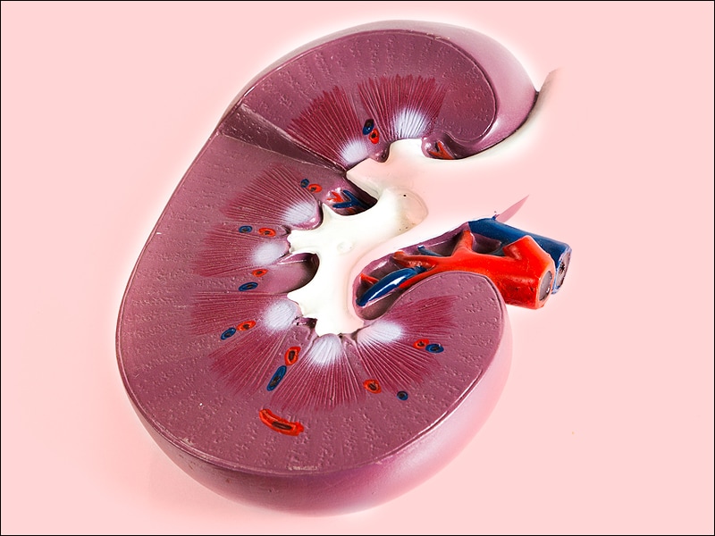
A kidney glomerulus is a functional unit in the nephron of the kidney. It is a tuft of capillaries enclosed within a structure called Bowman's capsule, which filters waste and excess fluids from the blood. The glomerulus receives blood from an afferent arteriole and drains into an efferent arteriole.
The process of filtration in the glomerulus is called ultrafiltration, where the pressure within the glomerular capillaries drives plasma fluid and small molecules (such as ions, glucose, amino acids, and waste products) through the filtration membrane into the Bowman's space. Larger molecules, like proteins and blood cells, are retained in the blood due to their larger size. The filtrate then continues down the nephron for further processing, eventually forming urine.
Podocytes are specialized cells that make up the visceral epithelial layer of the glomerular basement membrane in the kidney. They have long, interdigitating foot processes that wrap around the capillaries of the glomerulus and play a crucial role in maintaining the filtration barrier of the kidney. The slit diaphragms between the foot processes allow for the passage of small molecules while retaining larger proteins in the bloodstream. Podocytes also contribute to the maintenance and regulation of the glomerular filtration rate, making them essential for normal renal function. Damage or loss of podocytes can lead to proteinuria and kidney disease.
Proteinuria is a medical term that refers to the presence of excess proteins, particularly albumin, in the urine. Under normal circumstances, only small amounts of proteins should be found in the urine because the majority of proteins are too large to pass through the glomeruli, which are the filtering units of the kidneys.
However, when the glomeruli become damaged or diseased, they may allow larger molecules such as proteins to leak into the urine. Persistent proteinuria is often a sign of kidney disease and can indicate damage to the glomeruli. It is usually detected through a routine urinalysis and may be confirmed with further testing.
The severity of proteinuria can vary, and it can be a symptom of various underlying conditions such as diabetes, hypertension, glomerulonephritis, and other kidney diseases. Treatment for proteinuria depends on the underlying cause and may include medications to control blood pressure, manage diabetes, or reduce protein loss in the urine.
Nephrotic syndrome is a group of symptoms that indicate kidney damage, specifically damage to the glomeruli—the tiny blood vessel clusters in the kidneys that filter waste and excess fluids from the blood. The main features of nephrotic syndrome are:
1. Proteinuria (excess protein in urine): Large amounts of a protein called albumin leak into the urine due to damaged glomeruli, which can't properly filter proteins. This leads to low levels of albumin in the blood, causing fluid buildup and swelling.
2. Hypoalbuminemia (low blood albumin levels): As albumin leaks into the urine, the concentration of albumin in the blood decreases, leading to hypoalbuminemia. This can cause edema (swelling), particularly in the legs, ankles, and feet.
3. Edema (fluid retention and swelling): With low levels of albumin in the blood, fluids move into the surrounding tissues, causing swelling or puffiness. The swelling is most noticeable around the eyes, face, hands, feet, and abdomen.
4. Hyperlipidemia (high lipid/cholesterol levels): The kidneys play a role in regulating lipid metabolism. Damage to the glomeruli can lead to increased lipid production and high cholesterol levels in the blood.
Nephrotic syndrome can result from various underlying kidney diseases, such as minimal change disease, membranous nephropathy, or focal segmental glomerulosclerosis. Treatment depends on the underlying cause and may include medications to control inflammation, manage high blood pressure, and reduce proteinuria. In some cases, dietary modifications and lifestyle changes are also recommended.
Glomerulonephritis is a medical condition that involves inflammation of the glomeruli, which are the tiny blood vessel clusters in the kidneys that filter waste and excess fluids from the blood. This inflammation can impair the kidney's ability to filter blood properly, leading to symptoms such as proteinuria (protein in the urine), hematuria (blood in the urine), edema (swelling), hypertension (high blood pressure), and eventually kidney failure.
Glomerulonephritis can be acute or chronic, and it may occur as a primary kidney disease or secondary to other medical conditions such as infections, autoimmune disorders, or vasculitis. The diagnosis of glomerulonephritis typically involves a combination of medical history, physical examination, urinalysis, blood tests, and imaging studies, with confirmation often requiring a kidney biopsy. Treatment depends on the underlying cause and severity of the disease but may include medications to suppress inflammation, control blood pressure, and manage symptoms.
A kidney, in medical terms, is one of two bean-shaped organs located in the lower back region of the body. They are essential for maintaining homeostasis within the body by performing several crucial functions such as:
1. Regulation of water and electrolyte balance: Kidneys help regulate the amount of water and various electrolytes like sodium, potassium, and calcium in the bloodstream to maintain a stable internal environment.
2. Excretion of waste products: They filter waste products from the blood, including urea (a byproduct of protein metabolism), creatinine (a breakdown product of muscle tissue), and other harmful substances that result from normal cellular functions or external sources like medications and toxins.
3. Endocrine function: Kidneys produce several hormones with important roles in the body, such as erythropoietin (stimulates red blood cell production), renin (regulates blood pressure), and calcitriol (activated form of vitamin D that helps regulate calcium homeostasis).
4. pH balance regulation: Kidneys maintain the proper acid-base balance in the body by excreting either hydrogen ions or bicarbonate ions, depending on whether the blood is too acidic or too alkaline.
5. Blood pressure control: The kidneys play a significant role in regulating blood pressure through the renin-angiotensin-aldosterone system (RAAS), which constricts blood vessels and promotes sodium and water retention to increase blood volume and, consequently, blood pressure.
Anatomically, each kidney is approximately 10-12 cm long, 5-7 cm wide, and 3 cm thick, with a weight of about 120-170 grams. They are surrounded by a protective layer of fat and connected to the urinary system through the renal pelvis, ureters, bladder, and urethra.
Lipoid nephrosis is a historical term for a kidney disorder now more commonly referred to as minimal change disease (MCD). It is a type of glomerulonephritis which is characterized by the loss of proteins in the urine (proteinuria) due to damage to the glomeruli, the tiny filtering units within the kidneys.
The term "lipoid" refers to the presence of lipids or fats in the glomeruli, which can be observed under a microscope. However, it's worth noting that not all cases of MCD involve lipid accumulation in the glomeruli.
MCD is typically idiopathic, meaning its cause is unknown, but it can also occur as a secondary condition related to other medical disorders such as allergies, infections, or medications. It primarily affects children, but can also occur in adults. Treatment usually involves corticosteroids and other immunosuppressive therapies to control proteinuria and prevent kidney damage.
AIDS-associated nephropathy (AAN) is a kidney disorder that primarily affects individuals with advanced HIV infection. It is characterized by distinctive changes in the structure and function of the glomeruli, which are the tiny filtering units inside the kidneys.
The medical definition of AIDS-associated nephropathy is:
A renal disease associated with advanced HIV infection, characterized by focal segmental glomerulosclerosis (FSGS), collapsing variant or HIV-associated nephropathy (HIVAN) causing proteinuria, azotemia, and progressive decline in kidney function. The condition is more prevalent in certain racial/ethnic groups, such as African Americans, Hispanics, and Native Americans.
AAN is often considered a complication of advanced HIV disease and can lead to end-stage renal failure if not properly managed. Antiretroviral therapy (ART) has been shown to improve outcomes in patients with AAN, although some individuals may still require dialysis or kidney transplantation.
The glomerular mesangium is a part of the nephron in the kidney. It is the region located in the middle of the glomerular tuft, where the capillary loops of the glomerulus are surrounded by a network of extracellular matrix and mesangial cells. These cells and matrix play an important role in maintaining the structure and function of the filtration barrier in the glomerulus, which helps to filter waste products from the blood.
The mesangial cells have contractile properties and can regulate the flow of blood through the capillaries by constricting or dilating the diameter of the glomerular capillary loops. They also play a role in immune responses, as they can phagocytize immune complexes and release cytokines and growth factors that modulate inflammation and tissue repair.
Abnormalities in the mesangium can lead to various kidney diseases, such as glomerulonephritis, mesangial proliferative glomerulonephritis, and diabetic nephropathy.
Nephrectomy is a surgical procedure in which all or part of a kidney is removed. It may be performed due to various reasons such as severe kidney damage, kidney cancer, or living donor transplantation. The type of nephrectomy depends on the reason for the surgery - a simple nephrectomy involves removing only the affected portion of the kidney, while a radical nephrectomy includes removal of the whole kidney along with its surrounding tissues like the adrenal gland and lymph nodes.
Diabetic nephropathy is a kidney disease that occurs as a complication of diabetes. It is also known as diabetic kidney disease (DKD). This condition affects the ability of the kidneys to filter waste and excess fluids from the blood, leading to their accumulation in the body.
Diabetic nephropathy is caused by damage to the small blood vessels in the kidneys, which can occur over time due to high levels of glucose in the blood. This damage can lead to scarring and thickening of the kidney's filtering membranes, reducing their ability to function properly.
Symptoms of diabetic nephropathy may include proteinuria (the presence of protein in the urine), edema (swelling in the legs, ankles, or feet due to fluid retention), and hypertension (high blood pressure). Over time, if left untreated, diabetic nephropathy can progress to end-stage kidney disease, which requires dialysis or a kidney transplant.
Preventing or delaying the onset of diabetic nephropathy involves maintaining good control of blood sugar levels, keeping blood pressure under control, and making lifestyle changes such as quitting smoking, eating a healthy diet, and getting regular exercise. Regular monitoring of kidney function through urine tests and blood tests is also important for early detection and treatment of this condition.
Focal adhesions are specialized structures found in cells that act as points of attachment between the intracellular cytoskeleton and the extracellular matrix (ECM). They are composed of a complex network of proteins, including integrins, talin, vinculin, paxillin, and various others.
Focal adhesions play a crucial role in cellular processes such as adhesion, migration, differentiation, and signal transduction. They form when integrin receptors in the cell membrane bind to specific ligands within the ECM, leading to the clustering of these receptors and the recruitment of various adaptor and structural proteins. This results in the formation of a stable linkage between the cytoskeleton and the ECM, which helps maintain cell shape, provide mechanical stability, and facilitate communication between the intracellular and extracellular environments.
Focal adhesions are highly dynamic structures that can undergo rapid assembly and disassembly in response to various stimuli, allowing cells to adapt and respond to changes in their microenvironment. Dysregulation of focal adhesion dynamics has been implicated in several pathological conditions, including cancer metastasis, fibrosis, and impaired wound healing.
Kidney disease, also known as nephropathy or renal disease, refers to any functional or structural damage to the kidneys that impairs their ability to filter blood, regulate electrolytes, produce hormones, and maintain fluid balance. This damage can result from a wide range of causes, including diabetes, hypertension, glomerulonephritis, polycystic kidney disease, lupus, infections, drugs, toxins, and congenital or inherited disorders.
Depending on the severity and progression of the kidney damage, kidney diseases can be classified into two main categories: acute kidney injury (AKI) and chronic kidney disease (CKD). AKI is a sudden and often reversible loss of kidney function that occurs over hours to days, while CKD is a progressive and irreversible decline in kidney function that develops over months or years.
Symptoms of kidney diseases may include edema, proteinuria, hematuria, hypertension, electrolyte imbalances, metabolic acidosis, anemia, and decreased urine output. Treatment options depend on the underlying cause and severity of the disease and may include medications, dietary modifications, dialysis, or kidney transplantation.
Nephrosclerosis is a medical term that refers to the thickening and scarring (fibrosis) of the small arteries and arterioles in the kidneys, resulting in reduced blood flow and damage to the kidney tissue. This process can lead to decreased kidney function and ultimately result in chronic kidney disease or end-stage renal failure.
The two main types of nephrosclerosis are:
1. Hypertensive nephrosclerosis: This type is caused by long-term high blood pressure (hypertension), which damages the small blood vessels in the kidneys over time, leading to scarring and thickening of the arterial walls.
2. Ischemic nephrosclerosis: This type results from reduced blood flow to the kidneys due to atherosclerosis or other vascular diseases that cause narrowing or blockage of the renal arteries.
Nephrosclerosis is often asymptomatic in its early stages, but as the condition progresses, it may lead to symptoms such as proteinuria (protein in the urine), hematuria (blood in the urine), edema (swelling), and hypertension. Diagnosis typically involves a combination of medical history, physical examination, laboratory tests, and imaging studies. Treatment focuses on managing underlying conditions such as high blood pressure and diabetes, which can help slow or prevent further kidney damage.
Mesangial cells are specialized cells that are found in the mesangium, which is the middle layer of the glomerulus in the kidney. The glomerulus is a network of capillaries where blood filtration occurs. Mesangial cells play an important role in maintaining the structure and function of the glomerulus. They help regulate the size of the filtration slits between the capillary endothelial cells and the podocytes (specialized epithelial cells) by contracting and relaxing, similar to smooth muscle cells. Additionally, mesangial cells can phagocytize immune complexes and other debris in the glomerulus, contributing to the body's immune response. They also produce extracellular matrix components that provide structural support for the glomerulus. Mesangial cell dysfunction or injury can contribute to kidney diseases such as glomerulonephritis and diabetic nephropathy.
Membranous glomerulonephritis (MGN) is a kidney disorder that leads to the inflammation and damage of the glomeruli, which are the tiny blood vessels in the kidneys responsible for filtering waste and excess fluids from the blood. In MGN, the membrane that surrounds the glomerular capillaries becomes thickened and damaged due to the deposit of immune complexes, primarily composed of antibodies and antigens.
The onset of membranous glomerulonephritis can be either primary (idiopathic) or secondary to various underlying conditions such as autoimmune diseases (like systemic lupus erythematosus), infections (hepatitis B or C, syphilis, endocarditis), medications, or malignancies.
The symptoms of membranous glomerulonephritis may include:
1. Proteinuria - the presence of excess protein, specifically albumin, in the urine. This can lead to nephrotic syndrome, characterized by heavy protein loss in urine, edema (swelling), hypoalbuminemia (low blood albumin levels), and hyperlipidemia (high blood lipid levels).
2. Hematuria - the presence of red blood cells in the urine, which can be visible or microscopic.
3. Hypertension - high blood pressure.
4. Edema - swelling in various body parts due to fluid retention.
5. Nephrotic range proteinuria (protein loss greater than 3.5 grams per day) and/or nephritic syndrome (a combination of hematuria, proteinuria, hypertension, and kidney dysfunction) can be observed in some cases.
The diagnosis of membranous glomerulonephritis typically involves a thorough medical history, physical examination, urinalysis, blood tests, and imaging studies. A definitive diagnosis often requires a kidney biopsy to assess the glomerular structure and the nature of the immune complex deposits. Treatment depends on the underlying cause and severity of the disease and may include corticosteroids, immunosuppressants, blood pressure management, and supportive care for symptoms like edema and proteinuria.
A biopsy is a medical procedure in which a small sample of tissue is taken from the body to be examined under a microscope for the presence of disease. This can help doctors diagnose and monitor various medical conditions, such as cancer, infections, or autoimmune disorders. The type of biopsy performed will depend on the location and nature of the suspected condition. Some common types of biopsies include:
1. Incisional biopsy: In this procedure, a surgeon removes a piece of tissue from an abnormal area using a scalpel or other surgical instrument. This type of biopsy is often used when the lesion is too large to be removed entirely during the initial biopsy.
2. Excisional biopsy: An excisional biopsy involves removing the entire abnormal area, along with a margin of healthy tissue surrounding it. This technique is typically employed for smaller lesions or when cancer is suspected.
3. Needle biopsy: A needle biopsy uses a thin, hollow needle to extract cells or fluid from the body. There are two main types of needle biopsies: fine-needle aspiration (FNA) and core needle biopsy. FNA extracts loose cells, while a core needle biopsy removes a small piece of tissue.
4. Punch biopsy: In a punch biopsy, a round, sharp tool is used to remove a small cylindrical sample of skin tissue. This type of biopsy is often used for evaluating rashes or other skin abnormalities.
5. Shave biopsy: During a shave biopsy, a thin slice of tissue is removed from the surface of the skin using a sharp razor-like instrument. This technique is typically used for superficial lesions or growths on the skin.
After the biopsy sample has been collected, it is sent to a laboratory where a pathologist will examine the tissue under a microscope and provide a diagnosis based on their findings. The results of the biopsy can help guide further treatment decisions and determine the best course of action for managing the patient's condition.
Denys-Drash Syndrome is a rare genetic disorder that affects the kidneys and genitalia. It is characterized by the development of Wilms' tumor, a type of kidney cancer, and abnormal genital development in males. The syndrome is caused by mutations in the WT1 gene, which plays a crucial role in the development of the kidneys and genitalia.
Individuals with Denys-Drash Syndrome typically have underdeveloped or absent male genitalia, and some may be born with ambiguous genitalia. They are also at an increased risk of developing Wilms' tumor, often during the first two years of life. In addition, many individuals with the syndrome develop kidney disease, which can progress to end-stage renal failure.
The management of Denys-Drash Syndrome typically involves close monitoring for the development of Wilms' tumor and kidney disease, as well as treatment with chemotherapy or radiation therapy if necessary. Kidney transplantation may also be required in cases of end-stage renal failure.
Albuminuria is a medical condition that refers to the presence of albumin in the urine. Albumin is a type of protein normally found in the blood, but not in the urine. When the kidneys are functioning properly, they prevent large proteins like albumin from passing through into the urine. However, when the kidneys are damaged or not working correctly, such as in nephrotic syndrome or other kidney diseases, small amounts of albumin can leak into the urine.
The amount of albumin in the urine is often measured in milligrams per liter (mg/L) or in a spot urine sample, as the albumin-to-creatinine ratio (ACR). A small amount of albumin in the urine is called microalbuminuria, while a larger amount is called macroalbuminuria or proteinuria. The presence of albuminuria can indicate kidney damage and may be a sign of underlying medical conditions such as diabetes or high blood pressure. It is important to monitor and manage albuminuria to prevent further kidney damage and potential complications.
IGA glomerulonephritis (also known as Berger's disease) is a type of glomerulonephritis, which is a condition characterized by inflammation of the glomeruli, the tiny filtering units in the kidneys. In IgA glomerulonephritis, the immune system produces an abnormal amount of IgA antibodies, which deposit in the glomeruli and cause inflammation. This can lead to symptoms such as blood in the urine, protein in the urine, and swelling in the legs and feet. In some cases, it can also lead to kidney failure. The exact cause of IgA glomerulonephritis is not known, but it is often associated with other conditions such as infections, autoimmune diseases, and certain medications.
Sclerosis is a medical term that refers to the abnormal hardening or scarring of body tissues, particularly in the context of various degenerative diseases affecting the nervous system. The term "sclerosis" comes from the Greek word "skleros," which means hard. In these conditions, the normally flexible and adaptable nerve cells or their protective coverings (myelin sheath) become rigid and inflexible due to the buildup of scar tissue or abnormal protein deposits.
There are several types of sclerosis, but one of the most well-known is multiple sclerosis (MS). In MS, the immune system mistakenly attacks the myelin sheath surrounding nerve fibers in the brain and spinal cord, leading to scarring and damage that disrupts communication between the brain and the rest of the body. This results in a wide range of symptoms, such as muscle weakness, numbness, vision problems, balance issues, and cognitive impairment.
Other conditions that involve sclerosis include:
1. Amyotrophic lateral sclerosis (ALS): Also known as Lou Gehrig's disease, ALS is a progressive neurodegenerative disorder affecting motor neurons in the brain and spinal cord, leading to muscle weakness, stiffness, and atrophy.
2. Systemic sclerosis: A rare autoimmune connective tissue disorder characterized by thickening and hardening of the skin and internal organs due to excessive collagen deposition.
3. Plaque psoriasis: A chronic inflammatory skin condition marked by red, scaly patches (plaques) resulting from rapid turnover and accumulation of skin cells.
4. Adhesive capsulitis: Also known as frozen shoulder, this condition involves stiffening and thickening of the shoulder joint's capsule due to scarring or inflammation, leading to limited mobility and pain.
Collagen Type IV is a type of collagen that forms the structural basis of basement membranes, which are thin, sheet-like structures that separate and support cells in many types of tissues. It is a major component of the basement membrane's extracellular matrix and provides strength and flexibility to this structure. Collagen Type IV is composed of three chains that form a distinctive, mesh-like structure. Mutations in the genes encoding Collagen Type IV can lead to a variety of inherited disorders affecting the kidneys, eyes, and ears.
Plasmapheresis is a medical procedure where the liquid portion of the blood (plasma) is separated from the blood cells. The plasma, which may contain harmful substances such as antibodies or toxins, is then removed and replaced with fresh plasma or a plasma substitute. The remaining blood cells are mixed with the new plasma and returned to the body. This process is also known as therapeutic plasma exchange (TPE). It's used to treat various medical conditions including certain autoimmune diseases, poisonings, and neurological disorders.
Membranoproliferative Glomerulonephritis (MPGN) is a type of glomerulonephritis, which is a group of kidney disorders characterized by inflammation and damage to the glomeruli, the tiny blood vessels in the kidneys responsible for filtering waste and excess fluids from the blood.
MPGN is specifically characterized by thickening of the glomerular basement membrane and proliferation (increased number) of cells in the mesangium, a region within the glomerulus. This condition can be primary or secondary to other diseases such as infections, autoimmune disorders, or monoclonal gammopathies.
MPGN is typically classified into three types based on the pattern of injury seen on electron microscopy: Type I, Type II (Dense Deposit Disease), and Type III. Each type has distinct clinical features, laboratory findings, and treatment approaches. Symptoms of MPGN may include hematuria (blood in urine), proteinuria (protein in urine), hypertension (high blood pressure), edema (swelling), and eventually progress to chronic kidney disease or end-stage renal disease if left untreated.
Interstitial nephritis is a condition characterized by inflammation in the interstitium (the tissue between the kidney tubules) of one or both kidneys. This inflammation can be caused by various factors, including infections, autoimmune disorders, medications, and exposure to certain toxins.
The inflammation may lead to symptoms such as hematuria (blood in the urine), proteinuria (protein in the urine), decreased urine output, and kidney dysfunction. In some cases, interstitial nephritis can progress to chronic kidney disease or even end-stage renal failure if left untreated.
The diagnosis of interstitial nephritis typically involves a combination of medical history, physical examination, laboratory tests (such as urinalysis and blood tests), and imaging studies (such as ultrasound or CT scan). A kidney biopsy may also be performed to confirm the diagnosis and assess the severity of the inflammation.
Treatment for interstitial nephritis depends on the underlying cause, but may include corticosteroids, immunosuppressive medications, or discontinuation of any offending medications. In some cases, supportive care such as dialysis may be necessary to manage kidney dysfunction until the inflammation resolves.
Animal disease models are specialized animals, typically rodents such as mice or rats, that have been genetically engineered or exposed to certain conditions to develop symptoms and physiological changes similar to those seen in human diseases. These models are used in medical research to study the pathophysiology of diseases, identify potential therapeutic targets, test drug efficacy and safety, and understand disease mechanisms.
The genetic modifications can include knockout or knock-in mutations, transgenic expression of specific genes, or RNA interference techniques. The animals may also be exposed to environmental factors such as chemicals, radiation, or infectious agents to induce the disease state.
Examples of animal disease models include:
1. Mouse models of cancer: Genetically engineered mice that develop various types of tumors, allowing researchers to study cancer initiation, progression, and metastasis.
2. Alzheimer's disease models: Transgenic mice expressing mutant human genes associated with Alzheimer's disease, which exhibit amyloid plaque formation and cognitive decline.
3. Diabetes models: Obese and diabetic mouse strains like the NOD (non-obese diabetic) or db/db mice, used to study the development of type 1 and type 2 diabetes, respectively.
4. Cardiovascular disease models: Atherosclerosis-prone mice, such as ApoE-deficient or LDLR-deficient mice, that develop plaque buildup in their arteries when fed a high-fat diet.
5. Inflammatory bowel disease models: Mice with genetic mutations affecting intestinal barrier function and immune response, such as IL-10 knockout or SAMP1/YitFc mice, which develop colitis.
Animal disease models are essential tools in preclinical research, but it is important to recognize their limitations. Differences between species can affect the translatability of results from animal studies to human patients. Therefore, researchers must carefully consider the choice of model and interpret findings cautiously when applying them to human diseases.
Creatinine is a waste product that's produced by your muscles and removed from your body by your kidneys. Creatinine is a breakdown product of creatine, a compound found in meat and fish, as well as in the muscles of vertebrates, including humans.
In healthy individuals, the kidneys filter out most of the creatinine and eliminate it through urine. However, when the kidneys are not functioning properly, creatinine levels in the blood can rise. Therefore, measuring the amount of creatinine in the blood or urine is a common way to test how well the kidneys are working. High creatinine levels in the blood may indicate kidney damage or kidney disease.
Fibrosis is a pathological process characterized by the excessive accumulation and/or altered deposition of extracellular matrix components, particularly collagen, in various tissues and organs. This results in the formation of fibrous scar tissue that can impair organ function and structure. Fibrosis can occur as a result of chronic inflammation, tissue injury, or abnormal repair mechanisms, and it is a common feature of many diseases, including liver cirrhosis, lung fibrosis, heart failure, and kidney disease.
In medical terms, fibrosis is defined as:
"The process of producing scar tissue (consisting of collagen) in response to injury or chronic inflammation in normal connective tissue. This can lead to the thickening and stiffening of affected tissues and organs, impairing their function."
Transforming Growth Factor-beta (TGF-β) is a type of cytokine, which is a cell signaling protein involved in the regulation of various cellular processes, including cell growth, differentiation, and apoptosis (programmed cell death). TGF-β plays a critical role in embryonic development, tissue homeostasis, and wound healing. It also has been implicated in several pathological conditions such as fibrosis, cancer, and autoimmune diseases.
TGF-β exists in multiple isoforms (TGF-β1, TGF-β2, and TGF-β3) that are produced by many different cell types, including immune cells, epithelial cells, and fibroblasts. The protein is synthesized as a precursor molecule, which is cleaved to release the active TGF-β peptide. Once activated, TGF-β binds to its receptors on the cell surface, leading to the activation of intracellular signaling pathways that regulate gene expression and cell behavior.
In summary, Transforming Growth Factor-beta (TGF-β) is a multifunctional cytokine involved in various cellular processes, including cell growth, differentiation, apoptosis, embryonic development, tissue homeostasis, and wound healing. It has been implicated in several pathological conditions such as fibrosis, cancer, and autoimmune diseases.
Hypertrophy, in the context of physiology and pathology, refers to an increase in the size of an organ or tissue due to an enlargement of its constituent cells. It is often used to describe the growth of muscle cells (myocytes) in response to increased workload or hormonal stimulation, resulting in an increase in muscle mass. However, hypertrophy can also occur in other organs such as the heart (cardiac hypertrophy) in response to high blood pressure or valvular heart disease.
It is important to note that while hypertrophy involves an increase in cell size, hyperplasia refers to an increase in cell number. In some cases, both hypertrophy and hyperplasia can occur together, leading to a significant increase in the overall size and function of the organ or tissue.
Kidney transplantation is a surgical procedure where a healthy kidney from a deceased or living donor is implanted into a patient with end-stage renal disease (ESRD) or permanent kidney failure. The new kidney takes over the functions of filtering waste and excess fluids from the blood, producing urine, and maintaining the body's electrolyte balance.
The transplanted kidney is typically placed in the lower abdomen, with its blood vessels connected to the recipient's iliac artery and vein. The ureter of the new kidney is then attached to the recipient's bladder to ensure proper urine flow. Following the surgery, the patient will require lifelong immunosuppressive therapy to prevent rejection of the transplanted organ by their immune system.
Chronic kidney failure, also known as chronic kidney disease (CKD) stage 5 or end-stage renal disease (ESRD), is a permanent loss of kidney function that occurs gradually over a period of months to years. It is defined as a glomerular filtration rate (GFR) of less than 15 ml/min, which means the kidneys are filtering waste and excess fluids at less than 15% of their normal capacity.
CKD can be caused by various underlying conditions such as diabetes, hypertension, glomerulonephritis, polycystic kidney disease, and recurrent kidney infections. Over time, the damage to the kidneys can lead to a buildup of waste products and fluids in the body, which can cause a range of symptoms including fatigue, weakness, shortness of breath, nausea, vomiting, and confusion.
Treatment for chronic kidney failure typically involves managing the underlying condition, making lifestyle changes such as following a healthy diet, and receiving supportive care such as dialysis or a kidney transplant to replace lost kidney function.
Nephrosis is an older term that was used to describe a group of kidney diseases, primarily characterized by the damage and loss of function in the glomeruli - the tiny filtering units within the kidneys. This results in the leakage of large amounts of protein (primarily albumin) into the urine, a condition known as proteinuria.
The term "nephrosis" was often used interchangeably with "minimal change nephropathy," which is a specific type of kidney disorder that demonstrates little to no changes in the glomeruli under a microscope, despite significant protein leakage. However, current medical terminology and classifications prefer the use of more precise terms to describe various kidney diseases, such as minimal change disease, focal segmental glomerulosclerosis, or membranous nephropathy, among others.
It is important to consult with a healthcare professional or refer to updated medical resources for accurate and current information regarding kidney diseases and their specific diagnoses.
Sprague-Dawley rats are a strain of albino laboratory rats that are widely used in scientific research. They were first developed by researchers H.H. Sprague and R.C. Dawley in the early 20th century, and have since become one of the most commonly used rat strains in biomedical research due to their relatively large size, ease of handling, and consistent genetic background.
Sprague-Dawley rats are outbred, which means that they are genetically diverse and do not suffer from the same limitations as inbred strains, which can have reduced fertility and increased susceptibility to certain diseases. They are also characterized by their docile nature and low levels of aggression, making them easier to handle and study than some other rat strains.
These rats are used in a wide variety of research areas, including toxicology, pharmacology, nutrition, cancer, and behavioral studies. Because they are genetically diverse, Sprague-Dawley rats can be used to model a range of human diseases and conditions, making them an important tool in the development of new drugs and therapies.
Renal insufficiency, also known as kidney failure, is a medical condition in which the kidneys are unable to properly filter waste products and excess fluids from the blood. This results in a buildup of these substances in the body, which can cause a variety of symptoms such as weakness, shortness of breath, and fluid retention. Renal insufficiency can be acute, meaning it comes on suddenly, or chronic, meaning it develops over time. It is typically diagnosed through blood tests, urine tests, and imaging studies. Treatment may include medications to control symptoms, dietary changes, and in severe cases, dialysis or a kidney transplant.
Disease progression is the worsening or advancement of a medical condition over time. It refers to the natural course of a disease, including its development, the severity of symptoms and complications, and the impact on the patient's overall health and quality of life. Understanding disease progression is important for developing appropriate treatment plans, monitoring response to therapy, and predicting outcomes.
The rate of disease progression can vary widely depending on the type of medical condition, individual patient factors, and the effectiveness of treatment. Some diseases may progress rapidly over a short period of time, while others may progress more slowly over many years. In some cases, disease progression may be slowed or even halted with appropriate medical interventions, while in other cases, the progression may be inevitable and irreversible.
In clinical practice, healthcare providers closely monitor disease progression through regular assessments, imaging studies, and laboratory tests. This information is used to guide treatment decisions and adjust care plans as needed to optimize patient outcomes and improve quality of life.
Actinin is a protein that belongs to the family of actin-binding proteins. It plays an important role in the organization and stability of the cytoskeleton, which is the structural framework of a cell. Specifically, actinin crosslinks actin filaments into bundles or networks, providing strength and rigidity to the cell structure. There are several isoforms of actinin, with alpha-actinin and gamma-actinin being widely studied. Alpha-actinin is found in the Z-discs of sarcomeres in muscle cells, where it helps anchor actin filaments and maintains the structural integrity of the muscle. Gamma-actinin is primarily located at cell-cell junctions and participates in cell adhesion and signaling processes.
The Glomerular Basement Membrane (GBM) is a part of the filtration barrier in the nephron of the kidney. It is a thin, porous sheet of extracellular matrix that lies between the glomerular endothelial cells and the visceral epithelial cells (podocytes). The GBM plays a crucial role in the process of ultrafiltration, allowing the passage of water and small molecules while preventing the loss of larger proteins into the urine. It is composed mainly of type IV collagen, laminin, nidogen, and heparan sulfate proteoglycans. Certain kidney diseases, such as Goodpasture's disease and some forms of glomerulonephritis, can involve damage to the GBM.
Puromycin aminonucleoside is not a medical condition, but rather a laboratory reagent used in research. It is a synthetic antibiotic and analogue of the amino acid tyrosine, which specifically inhibits protein synthesis in eukaryotic cells by interacting with the peptidyl transferase center of the 60S ribosomal subunit. This compound has been widely used as a tool to study various cellular processes, including programmed cell death (apoptosis), autophagy, and lysosome biogenesis. Prolonged exposure to puromycin aminonucleoside can induce cytopathic effects, such as vacuolization and detachment of cells, which are often used as markers for its effectiveness in inhibiting protein synthesis.
Kidney tubules are the structural and functional units of the kidney responsible for reabsorption, secretion, and excretion of various substances. They are part of the nephron, which is the basic unit of the kidney's filtration and reabsorption process.
There are three main types of kidney tubules:
1. Proximal tubule: This is the initial segment of the kidney tubule that receives the filtrate from the glomerulus. It is responsible for reabsorbing approximately 65% of the filtrate, including water, glucose, amino acids, and electrolytes.
2. Loop of Henle: This U-shaped segment of the tubule consists of a thin descending limb, a thin ascending limb, and a thick ascending limb. The loop of Henle helps to concentrate urine by creating an osmotic gradient that allows water to be reabsorbed in the collecting ducts.
3. Distal tubule: This is the final segment of the kidney tubule before it empties into the collecting duct. It is responsible for fine-tuning the concentration of electrolytes and pH balance in the urine by selectively reabsorbing or secreting substances such as sodium, potassium, chloride, and hydrogen ions.
Overall, kidney tubules play a critical role in maintaining fluid and electrolyte balance, regulating acid-base balance, and removing waste products from the body.
Wilms' Tumor 1 (WT1) proteins are a group of transcription factors that play crucial roles in the development of the human body, particularly in the formation of the urinary and reproductive systems. The WT1 gene encodes these proteins, and mutations in this gene have been associated with several diseases, most notably Wilms' tumor, a type of kidney cancer in children.
WT1 proteins contain four domains: an N-terminal transcriptional activation domain, a zinc finger domain that binds to DNA, a nuclear localization signal, and a C-terminal transcriptional repression domain. These proteins regulate the expression of various target genes involved in cell growth, differentiation, and apoptosis (programmed cell death).
Abnormalities in WT1 protein function or expression have been linked to several developmental disorders, including Denys-Drash syndrome, Frasier syndrome, and Wilms' tumor. These conditions are characterized by genitourinary abnormalities, such as kidney dysplasia, ambiguous genitalia, and an increased risk of developing Wilms' tumor.
Kidney function tests (KFTs) are a group of diagnostic tests that evaluate how well your kidneys are functioning by measuring the levels of various substances in the blood and urine. The tests typically assess the glomerular filtration rate (GFR), which is an indicator of how efficiently the kidneys filter waste from the blood, as well as the levels of electrolytes, waste products, and proteins in the body.
Some common KFTs include:
1. Serum creatinine: A waste product that's produced by normal muscle breakdown and is excreted by the kidneys. Elevated levels may indicate reduced kidney function.
2. Blood urea nitrogen (BUN): Another waste product that's produced when protein is broken down and excreted by the kidneys. Increased BUN levels can suggest impaired kidney function.
3. Estimated glomerular filtration rate (eGFR): A calculation based on serum creatinine, age, sex, and race that estimates the GFR and provides a more precise assessment of kidney function than creatinine alone.
4. Urinalysis: An examination of a urine sample to detect abnormalities such as protein, blood, or bacteria that may indicate kidney disease.
5. Electrolyte levels: Measurement of sodium, potassium, chloride, and bicarbonate in the blood to ensure they're properly balanced, which is essential for normal kidney function.
KFTs are often ordered as part of a routine check-up or when kidney disease is suspected based on symptoms or other diagnostic tests. Regular monitoring of kidney function can help detect and manage kidney disease early, potentially preventing or slowing down its progression.
Glomerular filtration rate (GFR) is a test used to check how well the kidneys are working. Specifically, it estimates how much blood passes through the glomeruli each minute. The glomeruli are the tiny fibers in the kidneys that filter waste from the blood. A lower GFR number means that the kidneys aren't working properly and may indicate kidney disease.
The GFR is typically calculated using a formula that takes into account the patient's serum creatinine level, age, sex, and race. The most commonly used formula is the CKD-EPI (Chronic Kidney Disease Epidemiology Collaboration) equation. A normal GFR is usually above 90 mL/min/1.73m2, but this can vary depending on the individual's age and other factors.
Dihydralazine is a medication that belongs to a class of drugs called vasodilators. It works by relaxing the muscles in the walls of blood vessels, which causes the vessels to widen and allows for increased blood flow. Dihydralazine is primarily used to treat high blood pressure (hypertension), although it may also be used to manage heart failure.
The medical definition of Dihydralazine can be described as:
A synthetic pyridine derivative and a direct-acting vasodilator, which selectively relaxes arteriolar smooth muscle. It is used in the treatment of severe hypertension and chronic heart failure. The mechanism of its action is not fully understood, but it appears to block calcium channels and to result in the stimulation of nitric oxide release.
Steroids, also known as corticosteroids, are a type of hormone that the adrenal gland produces in your body. They have many functions, such as controlling the balance of salt and water in your body and helping to reduce inflammation. Steroids can also be synthetically produced and used as medications to treat a variety of conditions, including allergies, asthma, skin conditions, and autoimmune disorders.
Steroid medications are available in various forms, such as oral pills, injections, creams, and inhalers. They work by mimicking the effects of natural hormones produced by your body, reducing inflammation and suppressing the immune system's response to prevent or reduce symptoms. However, long-term use of steroids can have significant side effects, including weight gain, high blood pressure, osteoporosis, and increased risk of infections.
It is important to note that anabolic steroids are a different class of drugs that are sometimes abused for their muscle-building properties. These steroids are synthetic versions of the male hormone testosterone and can have serious health consequences when taken in large doses or without medical supervision.
Transforming Growth Factor-beta 1 (TGF-β1) is a cytokine that belongs to the TGF-β superfamily. It is a multifunctional protein involved in various cellular processes, including cell growth, differentiation, apoptosis, and extracellular matrix production. TGF-β1 plays crucial roles in embryonic development, tissue homeostasis, and repair, as well as in pathological conditions such as fibrosis and cancer. It signals through a heteromeric complex of type I and type II serine/threonine kinase receptors, leading to the activation of intracellular signaling pathways, primarily the Smad-dependent pathway. TGF-β1 has context-dependent functions, acting as a tumor suppressor in normal and early-stage cancer cells but promoting tumor progression and metastasis in advanced cancers.
Thy-1, also known as Thy-1 antigen or CD90, is a glycosylphosphatidylinositol (GPI)-anchored protein found on the surface of various cells in the body. It was first discovered as a cell surface antigen on thymocytes, hence the name Thy-1.
Thy-1 is a member of the immunoglobulin superfamily and is widely expressed in different tissues, including the brain, where it is found on the surface of neurons and glial cells. In the immune system, Thy-1 is expressed on the surface of T lymphocytes, natural killer (NK) cells, and some subsets of dendritic cells.
The function of Thy-1 is not fully understood, but it has been implicated in various biological processes, including cell adhesion, signal transduction, and regulation of immune responses. Thy-1 has also been shown to play a role in the development and maintenance of the nervous system, as well as in the pathogenesis of certain neurological disorders.
As an antigen, Thy-1 can be recognized by specific antibodies, which can be used in various research and clinical applications, such as immunohistochemistry, flow cytometry, and cell sorting.
Messenger RNA (mRNA) is a type of RNA (ribonucleic acid) that carries genetic information copied from DNA in the form of a series of three-base code "words," each of which specifies a particular amino acid. This information is used by the cell's machinery to construct proteins, a process known as translation. After being transcribed from DNA, mRNA travels out of the nucleus to the ribosomes in the cytoplasm where protein synthesis occurs. Once the protein has been synthesized, the mRNA may be degraded and recycled. Post-transcriptional modifications can also occur to mRNA, such as alternative splicing and addition of a 5' cap and a poly(A) tail, which can affect its stability, localization, and translation efficiency.
Immunosuppressive agents are medications that decrease the activity of the immune system. They are often used to prevent the rejection of transplanted organs and to treat autoimmune diseases, where the immune system mistakenly attacks the body's own tissues. These drugs work by interfering with the immune system's normal responses, which helps to reduce inflammation and damage to tissues. However, because they suppress the immune system, people who take immunosuppressive agents are at increased risk for infections and other complications. Examples of immunosuppressive agents include corticosteroids, azathioprine, cyclophosphamide, mycophenolate mofetil, tacrolimus, and sirolimus.
Blood pressure is the force exerted by circulating blood on the walls of the blood vessels. It is measured in millimeters of mercury (mmHg) and is given as two figures:
1. Systolic pressure: This is the pressure when the heart pushes blood out into the arteries.
2. Diastolic pressure: This is the pressure when the heart rests between beats, allowing it to fill with blood.
Normal blood pressure for adults is typically around 120/80 mmHg, although this can vary slightly depending on age, sex, and other factors. High blood pressure (hypertension) is generally considered to be a reading of 130/80 mmHg or higher, while low blood pressure (hypotension) is usually defined as a reading below 90/60 mmHg. It's important to note that blood pressure can fluctuate throughout the day and may be affected by factors such as stress, physical activity, and medication use.
Immunohistochemistry (IHC) is a technique used in pathology and laboratory medicine to identify specific proteins or antigens in tissue sections. It combines the principles of immunology and histology to detect the presence and location of these target molecules within cells and tissues. This technique utilizes antibodies that are specific to the protein or antigen of interest, which are then tagged with a detection system such as a chromogen or fluorophore. The stained tissue sections can be examined under a microscope, allowing for the visualization and analysis of the distribution and expression patterns of the target molecule in the context of the tissue architecture. Immunohistochemistry is widely used in diagnostic pathology to help identify various diseases, including cancer, infectious diseases, and immune-mediated disorders.
A nephron is the basic structural and functional unit of the kidney. It is responsible for filtering blood, reabsorbing necessary substances, and excreting waste products into the urine. Each human kidney contains approximately one million nephrons.
The structure of a nephron includes a glomerulus, which is a tuft of capillaries surrounded by Bowman's capsule. The glomerulus filters blood, allowing small molecules like water and solutes to pass through while keeping larger molecules like proteins and blood cells within the capillaries.
The filtrate then passes through the tubular portion of the nephron, which includes the proximal convoluted tubule, loop of Henle, distal convoluted tubule, and collecting duct. The tubular portion reabsorbs necessary substances like water, glucose, amino acids, and electrolytes back into the bloodstream while excreting waste products like urea and creatinine into the urine.
Overall, nephrons play a critical role in maintaining fluid and electrolyte balance, regulating blood pressure, and removing waste products from the body.
Angiotensin-Converting Enzyme (ACE) inhibitors are a class of medications that are commonly used to treat various cardiovascular conditions, such as hypertension (high blood pressure), heart failure, and diabetic nephropathy (kidney damage in people with diabetes).
ACE inhibitors work by blocking the action of angiotensin-converting enzyme, an enzyme that converts the hormone angiotensin I to angiotensin II. Angiotensin II is a potent vasoconstrictor, meaning it narrows blood vessels and increases blood pressure. By inhibiting the conversion of angiotensin I to angiotensin II, ACE inhibitors cause blood vessels to relax and widen, which lowers blood pressure and reduces the workload on the heart.
Some examples of ACE inhibitors include captopril, enalapril, lisinopril, ramipril, and fosinopril. These medications are generally well-tolerated, but they can cause side effects such as cough, dizziness, headache, and elevated potassium levels in the blood. It is important for patients to follow their healthcare provider's instructions carefully when taking ACE inhibitors and to report any unusual symptoms or side effects promptly.
I'm sorry for any confusion, but "Rats, Mutant Strains" is not a recognized medical term or concept. It may be a term used in science fiction, gaming, or other non-medical contexts to refer to genetically modified rats with altered characteristics. However, in the field of medical research, scientists do conduct studies using various strains of lab rats, some of which have been selectively bred or genetically modified to exhibit specific traits, but these are not referred to as "mutant strains." If you have any questions related to medical definitions or concepts, I'd be happy to help with those!
In the field of medicine, "time factors" refer to the duration of symptoms or time elapsed since the onset of a medical condition, which can have significant implications for diagnosis and treatment. Understanding time factors is crucial in determining the progression of a disease, evaluating the effectiveness of treatments, and making critical decisions regarding patient care.
For example, in stroke management, "time is brain," meaning that rapid intervention within a specific time frame (usually within 4.5 hours) is essential to administering tissue plasminogen activator (tPA), a clot-busting drug that can minimize brain damage and improve patient outcomes. Similarly, in trauma care, the "golden hour" concept emphasizes the importance of providing definitive care within the first 60 minutes after injury to increase survival rates and reduce morbidity.
Time factors also play a role in monitoring the progression of chronic conditions like diabetes or heart disease, where regular follow-ups and assessments help determine appropriate treatment adjustments and prevent complications. In infectious diseases, time factors are crucial for initiating antibiotic therapy and identifying potential outbreaks to control their spread.
Overall, "time factors" encompass the significance of recognizing and acting promptly in various medical scenarios to optimize patient outcomes and provide effective care.
The extracellular matrix (ECM) is a complex network of biomolecules that provides structural and biochemical support to cells in tissues and organs. It is composed of various proteins, glycoproteins, and polysaccharides, such as collagens, elastin, fibronectin, laminin, and proteoglycans. The ECM plays crucial roles in maintaining tissue architecture, regulating cell behavior, and facilitating communication between cells. It provides a scaffold for cell attachment, migration, and differentiation, and helps to maintain the structural integrity of tissues by resisting mechanical stresses. Additionally, the ECM contains various growth factors, cytokines, and chemokines that can influence cellular processes such as proliferation, survival, and differentiation. Overall, the extracellular matrix is essential for the normal functioning of tissues and organs, and its dysregulation can contribute to various pathological conditions, including fibrosis, cancer, and degenerative diseases.
"Acinonyx" is a genus name that refers to a single species of big cat, the cheetah. The correct medical definition of "Acinonyx" is:
* Acinonyx jubatus: a large, slender wild cat that is known for its incredible speed and unique adaptations for running. It is the fastest land animal, capable of reaching speeds up to 60-70 miles per hour. The cheetah's body is built for speed, with long legs, a flexible spine, and a non-retractable claw that provides traction while running.
The cheetah's habitat ranges from the savannas of Africa to the deserts of Iran. It primarily hunts medium-sized ungulates, such as gazelles and wildebeest. The cheetah's population has been declining due to habitat loss, human-wildlife conflict, and illegal wildlife trade. Conservation efforts are underway to protect this iconic species and its habitat.
Renal hypertension, also known as renovascular hypertension, is a type of secondary hypertension (high blood pressure) that is caused by narrowing or obstruction of the renal arteries or veins, which supply blood to the kidneys. This can lead to decreased blood flow and oxygen delivery to the kidney tissue, activating the renin-angiotensin-aldosterone system (RAAS) and resulting in increased peripheral vascular resistance, sodium retention, and extracellular fluid volume, ultimately causing hypertension.
Renal hypertension can be classified into two types:
1. Renin-dependent renal hypertension: This is caused by a decrease in blood flow to the kidneys, leading to increased renin release from the juxtaglomerular cells of the kidney. Renin converts angiotensinogen to angiotensin I, which is then converted to angiotensin II by angiotensin-converting enzyme (ACE). Angiotensin II is a potent vasoconstrictor that causes an increase in peripheral vascular resistance and blood pressure.
2. Renin-independent renal hypertension: This is caused by increased sodium retention and extracellular fluid volume, leading to an increase in blood pressure. This can be due to various factors such as obstructive sleep apnea, primary aldosteronism, or pheochromocytoma.
Renal hypertension is often asymptomatic but can lead to serious complications such as kidney damage, heart failure, and stroke if left untreated. Diagnosis of renal hypertension involves imaging studies such as renal artery duplex ultrasound, CT angiography, or magnetic resonance angiography (MRA) to identify any narrowing or obstruction in the renal arteries or veins. Treatment options include medications such as ACE inhibitors, angiotensin receptor blockers (ARBs), calcium channel blockers, and diuretics, as well as interventions such as angioplasty and stenting to improve blood flow to the kidneys.
Glutamyl Aminopeptidase (GAP, or sometimes also abbreviated as GP) is an enzyme that is found in many tissues throughout the body, including the kidneys and the intestines. Its primary function is to help break down proteins into smaller peptides and individual amino acids by removing certain types of amino acids from the ends of these protein chains.
GAP is a type of exopeptidase enzyme, which means that it works on the outside edges of proteins rather than in the middle. Specifically, GAP removes the amino acid glutamic acid (or its amide form, glutamine) from the N-terminus (the beginning end) of peptides and proteins.
In clinical settings, GAP is often measured in blood or urine samples as a biomarker for various medical conditions. For example, elevated levels of GAP in the blood may indicate liver disease or kidney damage, while decreased levels may be associated with certain types of cancer or gastrointestinal disorders. However, it's important to note that GAP is just one of many factors that doctors may consider when diagnosing and treating these conditions.
Cyclosporine is a medication that belongs to a class of drugs called immunosuppressants. It is primarily used to prevent the rejection of transplanted organs, such as kidneys, livers, and hearts. Cyclosporine works by suppressing the activity of the immune system, which helps to reduce the risk of the body attacking the transplanted organ.
In addition to its use in organ transplantation, cyclosporine may also be used to treat certain autoimmune diseases, such as rheumatoid arthritis and psoriasis. It does this by suppressing the overactive immune response that contributes to these conditions.
Cyclosporine is available in capsule, oral solution, and injectable forms. Common side effects of the medication include kidney problems, high blood pressure, tremors, headache, and nausea. Long-term use of cyclosporine can also increase the risk of certain types of cancer and infections.
It is important to note that cyclosporine should only be used under the close supervision of a healthcare provider, as it requires regular monitoring of blood levels and kidney function.
Collagen is the most abundant protein in the human body, and it is a major component of connective tissues such as tendons, ligaments, skin, and bones. Collagen provides structure and strength to these tissues and helps them to withstand stretching and tension. It is made up of long chains of amino acids, primarily glycine, proline, and hydroxyproline, which are arranged in a triple helix structure. There are at least 16 different types of collagen found in the body, each with slightly different structures and functions. Collagen is important for maintaining the integrity and health of tissues throughout the body, and it has been studied for its potential therapeutic uses in various medical conditions.
A focal infection is a localized infection that can serve as a focus for the development of secondary systemic infections or diseases elsewhere in the body. The infection is typically caused by a bacterium, virus, or fungus and can occur in any organ or tissue.
The theory of focal infection suggests that microorganisms can spread from the initial site of infection to other parts of the body through the bloodstream or lymphatic system, leading to further complications and illnesses. This concept was widely accepted and studied in the early 20th century but has since been largely replaced by more modern understandings of infectious disease processes.
Nonetheless, the term "focal infection" is still used in medical contexts to describe localized infections that may have systemic consequences or require specific treatment to prevent further spread and complications. Examples of focal infections include dental abscesses, lung infections, and urinary tract infections.
The kidney cortex is the outer region of the kidney where most of the functional units called nephrons are located. It plays a crucial role in filtering blood and regulating water, electrolyte, and acid-base balance in the body. The kidney cortex contains the glomeruli, proximal tubules, loop of Henle, and distal tubules, which work together to reabsorb necessary substances and excrete waste products into the urine.
Transgenic mice are genetically modified rodents that have incorporated foreign DNA (exogenous DNA) into their own genome. This is typically done through the use of recombinant DNA technology, where a specific gene or genetic sequence of interest is isolated and then introduced into the mouse embryo. The resulting transgenic mice can then express the protein encoded by the foreign gene, allowing researchers to study its function in a living organism.
The process of creating transgenic mice usually involves microinjecting the exogenous DNA into the pronucleus of a fertilized egg, which is then implanted into a surrogate mother. The offspring that result from this procedure are screened for the presence of the foreign DNA, and those that carry the desired genetic modification are used to establish a transgenic mouse line.
Transgenic mice have been widely used in biomedical research to model human diseases, study gene function, and test new therapies. They provide a valuable tool for understanding complex biological processes and developing new treatments for a variety of medical conditions.
I couldn't find a medical definition specifically for "Diabetes Mellitus, Experimental." However, I can provide you with information about experimental diabetes research and its relevance to understanding the disease.
Experimental diabetes models are used in biomedical research to study the pathophysiology of diabetes mellitus and to test potential therapies or treatments. These models can be broadly categorized into two types: chemically-induced and genetically modified.
1. Chemically-induced diabetes models: These involve administering chemicals, such as alloxan or streptozotocin, to animals (commonly mice or rats) to destroy their pancreatic β-cells, which produce insulin. This results in hyperglycemia and symptoms similar to those seen in type 1 diabetes in humans.
2. Genetically modified diabetes models: These involve altering the genes of animals (commonly mice) to create a diabetes phenotype. Examples include non-obese diabetic (NOD) mice, which develop an autoimmune form of diabetes similar to human type 1 diabetes, and various strains of obese mice with insulin resistance, such as ob/ob or db/db mice, which model aspects of type 2 diabetes.
These experimental models help researchers better understand the mechanisms behind diabetes development and progression, identify new therapeutic targets, and test potential treatments before moving on to human clinical trials. However, it's essential to recognize that these models may not fully replicate all aspects of human diabetes, so findings from animal studies should be interpreted with caution.
Electron microscopy (EM) is a type of microscopy that uses a beam of electrons to create an image of the sample being examined, resulting in much higher magnification and resolution than light microscopy. There are several types of electron microscopy, including transmission electron microscopy (TEM), scanning electron microscopy (SEM), and reflection electron microscopy (REM).
In TEM, a beam of electrons is transmitted through a thin slice of the sample, and the electrons that pass through the sample are focused to form an image. This technique can provide detailed information about the internal structure of cells, viruses, and other biological specimens, as well as the composition and structure of materials at the atomic level.
In SEM, a beam of electrons is scanned across the surface of the sample, and the electrons that are scattered back from the surface are detected to create an image. This technique can provide information about the topography and composition of surfaces, as well as the structure of materials at the microscopic level.
REM is a variation of SEM in which the beam of electrons is reflected off the surface of the sample, rather than scattered back from it. This technique can provide information about the surface chemistry and composition of materials.
Electron microscopy has a wide range of applications in biology, medicine, and materials science, including the study of cellular structure and function, disease diagnosis, and the development of new materials and technologies.
Blood Urea Nitrogen (BUN) is a laboratory value that measures the amount of urea nitrogen in the blood. Urea nitrogen is a waste product that is formed when proteins are broken down in the liver. The kidneys filter urea nitrogen from the blood and excrete it as urine.
A high BUN level may indicate impaired kidney function, as the kidneys are not effectively removing urea nitrogen from the blood. However, BUN levels can also be affected by other factors such as dehydration, heart failure, or gastrointestinal bleeding. Therefore, BUN should be interpreted in conjunction with other laboratory values and clinical findings.
The normal range for BUN is typically between 7-20 mg/dL (milligrams per deciliter) or 2.5-7.1 mmol/L (millimoles per liter), but the reference range may vary depending on the laboratory.
"Cells, cultured" is a medical term that refers to cells that have been removed from an organism and grown in controlled laboratory conditions outside of the body. This process is called cell culture and it allows scientists to study cells in a more controlled and accessible environment than they would have inside the body. Cultured cells can be derived from a variety of sources, including tissues, organs, or fluids from humans, animals, or cell lines that have been previously established in the laboratory.
Cell culture involves several steps, including isolation of the cells from the tissue, purification and characterization of the cells, and maintenance of the cells in appropriate growth conditions. The cells are typically grown in specialized media that contain nutrients, growth factors, and other components necessary for their survival and proliferation. Cultured cells can be used for a variety of purposes, including basic research, drug development and testing, and production of biological products such as vaccines and gene therapies.
It is important to note that cultured cells may behave differently than they do in the body, and results obtained from cell culture studies may not always translate directly to human physiology or disease. Therefore, it is essential to validate findings from cell culture experiments using additional models and ultimately in clinical trials involving human subjects.
"Wistar rats" are a strain of albino rats that are widely used in laboratory research. They were developed at the Wistar Institute in Philadelphia, USA, and were first introduced in 1906. Wistar rats are outbred, which means that they are genetically diverse and do not have a fixed set of genetic characteristics like inbred strains.
Wistar rats are commonly used as animal models in biomedical research because of their size, ease of handling, and relatively low cost. They are used in a wide range of research areas, including toxicology, pharmacology, nutrition, cancer, cardiovascular disease, and behavioral studies. Wistar rats are also used in safety testing of drugs, medical devices, and other products.
Wistar rats are typically larger than many other rat strains, with males weighing between 500-700 grams and females weighing between 250-350 grams. They have a lifespan of approximately 2-3 years. Wistar rats are also known for their docile and friendly nature, making them easy to handle and work with in the laboratory setting.
Nonmuscle Myosin Type IIA (NMIIA) is a type of non-muscle myosin protein that belongs to the myosin II family. These motor proteins are responsible for generating contractile forces in non-muscle cells, which allows them to change shape and move. NMIIA is widely expressed in various tissues and plays crucial roles in numerous cellular processes, including cytokinesis (cell division), maintenance of cell shape, and intracellular transport.
NMIIA consists of two heavy chains, two regulatory light chains, and two essential light chains. The heavy chains have a motor domain that binds to actin filaments and hydrolyzes ATP to generate force for movement along the actin filament. The regulatory and essential light chains regulate the activity and assembly of NMIIA.
Mutations in the gene encoding NMIIA (MYH9) have been associated with several human genetic disorders, such as May-Hegglin anomaly, Fechtner syndrome, and Delletten-Patterson syndrome, which are characterized by thrombocytopenia, bleeding disorders, and hearing loss.
Hematuria is a medical term that refers to the presence of blood in urine. It can be visible to the naked eye, which is called gross hematuria, or detected only under a microscope, known as microscopic hematuria. The blood in urine may come from any site along the urinary tract, including the kidneys, ureters, bladder, or urethra. Hematuria can be a symptom of various medical conditions, such as urinary tract infections, kidney stones, kidney disease, or cancer of the urinary tract. It is essential to consult a healthcare professional if you notice blood in your urine to determine the underlying cause and receive appropriate treatment.
Angiotensin II Type 1 Receptor Blockers (ARBs) are a class of medications used to treat hypertension, heart failure, and protect against kidney damage in patients with diabetes. They work by blocking the action of angiotensin II, a hormone that causes blood vessels to constrict and blood pressure to increase, at its type 1 receptor. By blocking this effect, ARBs cause blood vessels to dilate, reducing blood pressure and decreasing the workload on the heart. Examples of ARBs include losartan, valsartan, irbesartan, and candesartan.
The Bowman capsule is the initial component of the nephron, which is the functional unit of the kidney. It is a structural and functional part of the renal corpuscle, along with the glomerulus. The Bowman capsule surrounds the glomerulus and serves as a site for filtration, helping to separate small molecules from blood cells and large proteins in the process known as urine formation.
The Bowman capsule is composed of a single layer of epithelial cells called podocytes, which have foot-like processes that interdigitate with each other and form filtration slits. These slits are covered by a thin diaphragm, allowing for the passage of small molecules while retaining larger ones. The space within the Bowman capsule is called the urinary space or Bowman's space, where the filtrate from the blood collects before moving into the tubular system for further processing and eventual excretion as urine.
Fibronectin is a high molecular weight glycoprotein that is found in many tissues and body fluids, including plasma, connective tissue, and the extracellular matrix. It is composed of two similar subunits that are held together by disulfide bonds. Fibronectin plays an important role in cell adhesion, migration, and differentiation by binding to various cell surface receptors, such as integrins, and other extracellular matrix components, such as collagen and heparan sulfate proteoglycans.
Fibronectin has several isoforms that are produced by alternative splicing of a single gene transcript. These isoforms differ in their biological activities and can be found in different tissues and developmental stages. Fibronectin is involved in various physiological processes, such as wound healing, tissue repair, and embryonic development, and has been implicated in several pathological conditions, including fibrosis, tumor metastasis, and thrombosis.
Nephritis is a medical term that refers to inflammation of the kidneys, specifically affecting the glomeruli - the tiny filtering units inside the kidneys. The condition can cause damage to the glomeruli, leading to impaired kidney function and the leakage of protein and blood into the urine.
Nephritis can result from a variety of causes, including infections, autoimmune disorders, and exposure to certain medications or toxins. Depending on the severity and underlying cause, nephritis may be treated with medications, dietary modifications, or other therapies aimed at reducing inflammation and preserving kidney function. In severe cases, hospitalization and more intensive treatments may be necessary.
Molecular motor proteins are a type of protein that convert chemical energy into mechanical work at the molecular level. They play a crucial role in various cellular processes, such as cell division, muscle contraction, and intracellular transport. There are several types of molecular motor proteins, including myosin, kinesin, and dynein.
Myosin is responsible for muscle contraction and movement along actin filaments in the cytoplasm. Kinesin and dynein are involved in intracellular transport along microtubules, moving cargo such as vesicles, organelles, and mRNA to various destinations within the cell.
These motor proteins move in a stepwise fashion, with each step driven by the hydrolysis of adenosine triphosphate (ATP) into adenosine diphosphate (ADP) and inorganic phosphate (Pi). The directionality and speed of movement are determined by the structure and regulation of the motor proteins, as well as the properties of the tracks along which they move.
Microfilament proteins are a type of structural protein that form part of the cytoskeleton in eukaryotic cells. They are made up of actin monomers, which polymerize to form long, thin filaments. These filaments are involved in various cellular processes such as muscle contraction, cell division, and cell motility. Microfilament proteins also interact with other cytoskeletal components like intermediate filaments and microtubules to maintain the overall shape and integrity of the cell. Additionally, they play a crucial role in the formation of cell-cell junctions and cell-matrix adhesions, which are essential for tissue structure and function.
Recurrence, in a medical context, refers to the return of symptoms or signs of a disease after a period of improvement or remission. It indicates that the condition has not been fully eradicated and may require further treatment. Recurrence is often used to describe situations where a disease such as cancer comes back after initial treatment, but it can also apply to other medical conditions. The likelihood of recurrence varies depending on the type of disease and individual patient factors.
Focal nodular hyperplasia (FNH) is a benign liver mass, which means it's a non-cancerous growth. It is characterized by the presence of multiple nodules or "lesions" in the liver that are well-circumscribed and have a distinctive "stars in the sky" appearance on histopathology. These lesions are composed of abnormal arrangements of liver cells, bile ducts, and blood vessels. FNH is usually discovered incidentally during imaging studies performed for unrelated reasons. It's more common in women than men, and while its cause remains unclear, it has been linked to hormonal factors. Most cases do not require treatment unless they become symptomatic or complications arise.
Body weight is the measure of the force exerted on a scale or balance by an object's mass, most commonly expressed in units such as pounds (lb) or kilograms (kg). In the context of medical definitions, body weight typically refers to an individual's total weight, which includes their skeletal muscle, fat, organs, and bodily fluids.
Healthcare professionals often use body weight as a basic indicator of overall health status, as it can provide insights into various aspects of a person's health, such as nutritional status, metabolic function, and risk factors for certain diseases. For example, being significantly underweight or overweight can increase the risk of developing conditions like malnutrition, diabetes, heart disease, and certain types of cancer.
It is important to note that body weight alone may not provide a complete picture of an individual's health, as it does not account for factors such as muscle mass, bone density, or body composition. Therefore, healthcare professionals often use additional measures, such as body mass index (BMI), waist circumference, and blood tests, to assess overall health status more comprehensively.
Transgenic rats are genetically modified rats that have incorporated foreign DNA (transgene) into their own genome. This is typically done through the use of recombinant DNA techniques in the laboratory. The transgene can come from any species, including other mammals, plants, or even bacteria. Once the transgene is introduced into the rat's embryonic cells, it becomes a permanent part of the rat's genetic makeup and is passed on to its offspring.
Transgenic rats are used in biomedical research as models for studying human diseases, developing new therapies, and testing the safety and efficacy of drugs. They offer several advantages over traditional laboratory rats, including the ability to manipulate specific genes, study gene function and regulation, and investigate the underlying mechanisms of disease.
Some common applications of transgenic rats in research include:
1. Modeling human diseases: Transgenic rats can be engineered to develop symptoms and characteristics of human diseases, such as cancer, diabetes, Alzheimer's, and Parkinson's. This allows researchers to study the disease progression, test new treatments, and evaluate their effectiveness.
2. Gene function and regulation: By introducing specific genes into rats, scientists can investigate their role in various biological processes, such as development, aging, and metabolism. They can also study how genes are regulated and how they interact with each other.
3. Drug development and testing: Transgenic rats can be used to test the safety and efficacy of new drugs before they are tested in humans. By studying the effects of drugs on transgenic rats, researchers can gain insights into their potential benefits and risks.
4. Toxicology studies: Transgenic rats can be used to study the toxicity of chemicals, pollutants, and other substances. This helps ensure that new products and treatments are safe for human use.
In summary, transgenic rats are genetically modified rats that have incorporated foreign DNA into their own genome. They are widely used in biomedical research to model human diseases, study gene function and regulation, develop new therapies, and test the safety and efficacy of drugs.
Spironolactone is a prescription medication that belongs to a class of drugs known as potassium-sparing diuretics. It works by blocking the action of aldosterone, a hormone that helps regulate sodium and potassium balance in your body. This results in increased urine production (diuresis) and decreased salt and fluid retention.
Spironolactone is primarily used to treat edema (fluid buildup) associated with heart failure, liver cirrhosis, or kidney disease. It's also prescribed for the treatment of high blood pressure and primary hyperaldosteronism, a condition where the adrenal glands produce too much aldosterone.
Furthermore, spironolactone is used off-label to treat conditions such as acne, hirsutism (excessive hair growth in women), and hormone-sensitive breast cancer in postmenopausal women.
It's important to note that spironolactone can cause increased potassium levels in the blood (hyperkalemia) and should be used with caution in patients with kidney impairment or those taking other medications that affect potassium balance. Regular monitoring of electrolyte levels, including potassium and sodium, is essential during spironolactone therapy.
Wilms tumor (WT) genes, also known as WT1 and WT2, are tumor suppressor genes that play crucial roles in the normal development of the kidneys. Mutations or alterations in these genes can lead to the development of Wilms tumor, which is a type of kidney cancer that primarily affects children.
WT1 gene is located on chromosome 11p13 and encodes a transcription factor that regulates the expression of various genes involved in kidney development. Mutations in WT1 can lead to Wilms tumor, as well as other genetic disorders such as Denys-Drash syndrome and Frasier syndrome.
WT2 gene is located on chromosome 11p15 and encodes a zinc finger transcription factor that also plays a role in kidney development. Mutations in WT2 have been associated with an increased risk of Wilms tumor, as well as other genetic disorders such as Beckwith-Wiedemann syndrome.
It's worth noting that not all Wilms tumors are caused by mutations in WT1 or WT2 genes, and that other genetic and environmental factors may also contribute to the development of this type of cancer.
Gene expression is the process by which the information encoded in a gene is used to synthesize a functional gene product, such as a protein or RNA molecule. This process involves several steps: transcription, RNA processing, and translation. During transcription, the genetic information in DNA is copied into a complementary RNA molecule, known as messenger RNA (mRNA). The mRNA then undergoes RNA processing, which includes adding a cap and tail to the mRNA and splicing out non-coding regions called introns. The resulting mature mRNA is then translated into a protein on ribosomes in the cytoplasm through the process of translation.
The regulation of gene expression is a complex and highly controlled process that allows cells to respond to changes in their environment, such as growth factors, hormones, and stress signals. This regulation can occur at various stages of gene expression, including transcriptional activation or repression, RNA processing, mRNA stability, and translation. Dysregulation of gene expression has been implicated in many diseases, including cancer, genetic disorders, and neurological conditions.
Organ size refers to the volume or physical measurement of an organ in the body of an individual. It can be described in terms of length, width, and height or by using specialized techniques such as imaging studies (like CT scans or MRIs) to determine the volume. The size of an organ can vary depending on factors such as age, sex, body size, and overall health status. Changes in organ size may indicate various medical conditions, including growths, inflammation, or atrophy.
Drug resistance, also known as antimicrobial resistance, is the ability of a microorganism (such as bacteria, viruses, fungi, or parasites) to withstand the effects of a drug that was originally designed to inhibit or kill it. This occurs when the microorganism undergoes genetic changes that allow it to survive in the presence of the drug. As a result, the drug becomes less effective or even completely ineffective at treating infections caused by these resistant organisms.
Drug resistance can develop through various mechanisms, including mutations in the genes responsible for producing the target protein of the drug, alteration of the drug's target site, modification or destruction of the drug by enzymes produced by the microorganism, and active efflux of the drug from the cell.
The emergence and spread of drug-resistant microorganisms pose significant challenges in medical treatment, as they can lead to increased morbidity, mortality, and healthcare costs. The overuse and misuse of antimicrobial agents, as well as poor infection control practices, contribute to the development and dissemination of drug-resistant strains. To address this issue, it is crucial to promote prudent use of antimicrobials, enhance surveillance and monitoring of resistance patterns, invest in research and development of new antimicrobial agents, and strengthen infection prevention and control measures.
C57BL/6 (C57 Black 6) is an inbred strain of laboratory mouse that is widely used in biomedical research. The term "inbred" refers to a strain of animals where matings have been carried out between siblings or other closely related individuals for many generations, resulting in a population that is highly homozygous at most genetic loci.
The C57BL/6 strain was established in 1920 by crossing a female mouse from the dilute brown (DBA) strain with a male mouse from the black strain. The resulting offspring were then interbred for many generations to create the inbred C57BL/6 strain.
C57BL/6 mice are known for their robust health, longevity, and ease of handling, making them a popular choice for researchers. They have been used in a wide range of biomedical research areas, including studies of cancer, immunology, neuroscience, cardiovascular disease, and metabolism.
One of the most notable features of the C57BL/6 strain is its sensitivity to certain genetic modifications, such as the introduction of mutations that lead to obesity or impaired glucose tolerance. This has made it a valuable tool for studying the genetic basis of complex diseases and traits.
Overall, the C57BL/6 inbred mouse strain is an important model organism in biomedical research, providing a valuable resource for understanding the genetic and molecular mechanisms underlying human health and disease.
'Rats, Inbred Dahl' are a strain of laboratory rats that have been selectively bred for research purposes. They were first developed by Dr. Lewis L. Dahl in the 1960s at the University of Colorado School of Medicine. These rats are known for their susceptibility to develop high blood pressure (hypertension) and related cardiovascular diseases, making them a valuable model for studying hypertension and its complications.
Inbred Dahl rats are typically divided into two main strains: the Dahl Salt-Sensitive (SS/JrHsdMcwi or SS) rat and the Dahl Salt-Resistant (SR/JrHsdMcwi or SR) rat. When fed a high-salt diet, the SS rats develop severe hypertension, kidney damage, and cardiac hypertrophy, while the SR rats are relatively resistant to these effects.
The Inbred Dahl rats have contributed significantly to our understanding of the genetic and environmental factors that contribute to the development of hypertension and related disorders. They continue to be widely used in biomedical research today.
The Renin-Angiotensin System (RAS) is a complex hormonal system that regulates blood pressure, fluid and electrolyte balance, and vascular resistance. It plays a crucial role in the pathophysiology of hypertension, heart failure, and kidney diseases.
Here's a brief overview of how it works:
1. Renin is an enzyme that is released by the juxtaglomerular cells in the kidneys in response to decreased blood pressure or reduced salt delivery to the distal tubules.
2. Renin acts on a protein called angiotensinogen, which is produced by the liver, converting it into angiotensin I.
3. Angiotensin-converting enzyme (ACE), found in the lungs and other tissues, then converts angiotensin I into angiotensin II, a potent vasoconstrictor that narrows blood vessels and increases blood pressure.
4. Angiotensin II also stimulates the release of aldosterone from the adrenal glands, which promotes sodium and water reabsorption in the kidneys, further increasing blood volume and blood pressure.
5. Additionally, angiotensin II has direct effects on the heart, promoting hypertrophy and remodeling, which can contribute to heart failure.
6. The RAS can be modulated by various medications, such as ACE inhibitors, angiotensin receptor blockers (ARBs), and aldosterone antagonists, which are commonly used to treat hypertension, heart failure, and kidney diseases.
Enalapril is a medication that belongs to a class of drugs called angiotensin-converting enzyme (ACE) inhibitors. It works by blocking the action of a hormone in the body called angiotensin II, which causes blood vessels to narrow and tighten. By blocking this hormone, Enalapril helps relax and widen blood vessels, making it easier for the heart to pump blood and reducing the workload on the heart.
Enalapril is commonly used to treat high blood pressure (hypertension), congestive heart failure, and to improve survival after a heart attack. It may also be used to treat other conditions as determined by your doctor.
The medication comes in the form of tablets or capsules that are taken orally, usually once or twice a day with or without food. The dosage will depend on various factors such as the patient's age, weight, and medical condition. It is important to follow the instructions of your healthcare provider when taking Enalapril.
Like all medications, Enalapril can cause side effects, including dry cough, dizziness, headache, fatigue, and nausea. More serious side effects may include allergic reactions, kidney problems, and low blood pressure. If you experience any concerning symptoms while taking Enalapril, it is important to contact your healthcare provider right away.
Transient Receptor Potential Canonical (TRPC) cation channels are a subfamily of the TRP superfamily of non-selective cation channels. They are widely expressed in various tissues and play crucial roles in many cellular processes, including sensory perception, cell proliferation, and migration. TRPC channels are permeable to both monovalent (sodium and potassium) and divalent (calcium and magnesium) cations, and their activation can lead to a rise in intracellular calcium concentration, which in turn regulates various downstream signaling pathways. TRPC channels can be activated by a variety of stimuli, including G protein-coupled receptors, receptor tyrosine kinases, and mechanical stress. Mutations in TRPC genes have been associated with several human diseases, including hereditary hearing loss, cardiovascular disorders, and neurological conditions.
Capillaries are the smallest blood vessels in the body, with diameters that range from 5 to 10 micrometers. They form a network of tiny tubes that connect the arterioles (small branches of arteries) and venules (small branches of veins), allowing for the exchange of oxygen, carbon dioxide, nutrients, and waste products between the blood and the surrounding tissues.
Capillaries are composed of a single layer of endothelial cells that surround a hollow lumen through which blood flows. The walls of capillaries are extremely thin, allowing for easy diffusion of molecules between the blood and the surrounding tissue. This is essential for maintaining the health and function of all body tissues.
Capillaries can be classified into three types based on their structure and function: continuous, fenestrated, and sinusoidal. Continuous capillaries have a continuous layer of endothelial cells with tight junctions that restrict the passage of large molecules. Fenestrated capillaries have small pores or "fenestrae" in the endothelial cell walls that allow for the passage of larger molecules, such as proteins and lipids. Sinusoidal capillaries are found in organs with high metabolic activity, such as the liver and spleen, and have large, irregular spaces between the endothelial cells that allow for the exchange of even larger molecules.
Overall, capillaries play a critical role in maintaining the health and function of all body tissues by allowing for the exchange of nutrients, oxygen, and waste products between the blood and surrounding tissues.
Losartan is an angiotensin II receptor blocker (ARB) medication that is primarily used to treat hypertension (high blood pressure), but can also be used to manage chronic heart failure and protect against kidney damage in patients with type 2 diabetes. It works by blocking the action of angiotensin II, a hormone that causes blood vessels to narrow and blood pressure to rise. By blocking this hormone's effects, losartan helps relax and widen blood vessels, making it easier for the heart to pump blood and reducing the workload on the cardiovascular system.
The medical definition of losartan is: "A synthetic angiotensin II receptor antagonist used in the treatment of hypertension, chronic heart failure, and diabetic nephropathy. It selectively blocks the binding of angiotensin II to the AT1 receptor, leading to vasodilation, decreased aldosterone secretion, and increased renin activity."
The basement membrane is a thin, specialized layer of extracellular matrix that provides structural support and separates epithelial cells (which line the outer surfaces of organs and blood vessels) from connective tissue. It is composed of two main layers: the basal lamina, which is produced by the epithelial cells, and the reticular lamina, which is produced by the connective tissue. The basement membrane plays important roles in cell adhesion, migration, differentiation, and survival.
The basal lamina is composed mainly of type IV collagen, laminins, nidogens, and proteoglycans, while the reticular lamina contains type III collagen, fibronectin, and other matrix proteins. The basement membrane also contains a variety of growth factors and cytokines that can influence cell behavior.
Defects in the composition or organization of the basement membrane can lead to various diseases, including kidney disease, eye disease, and skin blistering disorders.
Extracellular matrix (ECM) proteins are a group of structural and functional molecules that provide support, organization, and regulation to the cells in tissues and organs. The ECM is composed of a complex network of proteins, glycoproteins, and carbohydrates that are secreted by the cells and deposited outside of them.
ECM proteins can be classified into several categories based on their structure and function, including:
1. Collagens: These are the most abundant ECM proteins and provide strength and stability to tissues. They form fibrils that can withstand high tensile forces.
2. Proteoglycans: These are complex molecules made up of a core protein and one or more glycosaminoglycan (GAG) chains. The GAG chains attract water, making proteoglycans important for maintaining tissue hydration and resilience.
3. Elastin: This is an elastic protein that allows tissues to stretch and recoil, such as in the lungs and blood vessels.
4. Fibronectins: These are large glycoproteins that bind to cells and ECM components, providing adhesion, migration, and signaling functions.
5. Laminins: These are large proteins found in basement membranes, which provide structural support for epithelial and endothelial cells.
6. Tenascins: These are large glycoproteins that modulate cell adhesion and migration, and regulate ECM assembly and remodeling.
Together, these ECM proteins create a microenvironment that influences cell behavior, differentiation, and function. Dysregulation of ECM proteins has been implicated in various diseases, including fibrosis, cancer, and degenerative disorders.
Hypertension is a medical term used to describe abnormally high blood pressure in the arteries, often defined as consistently having systolic blood pressure (the top number in a blood pressure reading) over 130 mmHg and/or diastolic blood pressure (the bottom number) over 80 mmHg. It is also commonly referred to as high blood pressure.
Hypertension can be classified into two types: primary or essential hypertension, which has no identifiable cause and accounts for about 95% of cases, and secondary hypertension, which is caused by underlying medical conditions such as kidney disease, hormonal disorders, or use of certain medications.
If left untreated, hypertension can lead to serious health complications such as heart attack, stroke, heart failure, and chronic kidney disease. Therefore, it is important for individuals with hypertension to manage their condition through lifestyle modifications (such as healthy diet, regular exercise, stress management) and medication if necessary, under the guidance of a healthcare professional.
Membrane proteins are a type of protein that are embedded in the lipid bilayer of biological membranes, such as the plasma membrane of cells or the inner membrane of mitochondria. These proteins play crucial roles in various cellular processes, including:
1. Cell-cell recognition and signaling
2. Transport of molecules across the membrane (selective permeability)
3. Enzymatic reactions at the membrane surface
4. Energy transduction and conversion
5. Mechanosensation and signal transduction
Membrane proteins can be classified into two main categories: integral membrane proteins, which are permanently associated with the lipid bilayer, and peripheral membrane proteins, which are temporarily or loosely attached to the membrane surface. Integral membrane proteins can further be divided into three subcategories based on their topology:
1. Transmembrane proteins, which span the entire width of the lipid bilayer with one or more alpha-helices or beta-barrels.
2. Lipid-anchored proteins, which are covalently attached to lipids in the membrane via a glycosylphosphatidylinositol (GPI) anchor or other lipid modifications.
3. Monotopic proteins, which are partially embedded in the membrane and have one or more domains exposed to either side of the bilayer.
Membrane proteins are essential for maintaining cellular homeostasis and are targets for various therapeutic interventions, including drug development and gene therapy. However, their structural complexity and hydrophobicity make them challenging to study using traditional biochemical methods, requiring specialized techniques such as X-ray crystallography, nuclear magnetic resonance (NMR) spectroscopy, and single-particle cryo-electron microscopy (cryo-EM).
Cytoskeletal proteins are a type of structural proteins that form the cytoskeleton, which is the internal framework of cells. The cytoskeleton provides shape, support, and structure to the cell, and plays important roles in cell division, intracellular transport, and maintenance of cell shape and integrity.
There are three main types of cytoskeletal proteins: actin filaments, intermediate filaments, and microtubules. Actin filaments are thin, rod-like structures that are involved in muscle contraction, cell motility, and cell division. Intermediate filaments are thicker than actin filaments and provide structural support to the cell. Microtubules are hollow tubes that are involved in intracellular transport, cell division, and maintenance of cell shape.
Cytoskeletal proteins are composed of different subunits that polymerize to form filamentous structures. These proteins can be dynamically assembled and disassembled, allowing cells to change their shape and move. Mutations in cytoskeletal proteins have been linked to various human diseases, including cancer, neurological disorders, and muscular dystrophies.
Intracellular signaling peptides and proteins are molecules that play a crucial role in transmitting signals within cells, which ultimately lead to changes in cell behavior or function. These signals can originate from outside the cell (extracellular) or within the cell itself. Intracellular signaling molecules include various types of peptides and proteins, such as:
1. G-protein coupled receptors (GPCRs): These are seven-transmembrane domain receptors that bind to extracellular signaling molecules like hormones, neurotransmitters, or chemokines. Upon activation, they initiate a cascade of intracellular signals through G proteins and secondary messengers.
2. Receptor tyrosine kinases (RTKs): These are transmembrane receptors that bind to growth factors, cytokines, or hormones. Activation of RTKs leads to autophosphorylation of specific tyrosine residues, creating binding sites for intracellular signaling proteins such as adapter proteins, phosphatases, and enzymes like Ras, PI3K, and Src family kinases.
3. Second messenger systems: Intracellular second messengers are small molecules that amplify and propagate signals within the cell. Examples include cyclic adenosine monophosphate (cAMP), cyclic guanosine monophosphate (cGMP), diacylglycerol (DAG), inositol triphosphate (IP3), calcium ions (Ca2+), and nitric oxide (NO). These second messengers activate or inhibit various downstream effectors, leading to changes in cellular responses.
4. Signal transduction cascades: Intracellular signaling proteins often form complex networks of interacting molecules that relay signals from the plasma membrane to the nucleus. These cascades involve kinases (protein kinases A, B, C, etc.), phosphatases, and adapter proteins, which ultimately regulate gene expression, cell cycle progression, metabolism, and other cellular processes.
5. Ubiquitination and proteasome degradation: Intracellular signaling pathways can also control protein stability by modulating ubiquitin-proteasome degradation. E3 ubiquitin ligases recognize specific substrates and conjugate them with ubiquitin molecules, targeting them for proteasomal degradation. This process regulates the abundance of key signaling proteins and contributes to signal termination or amplification.
In summary, intracellular signaling pathways involve a complex network of interacting proteins that relay signals from the plasma membrane to various cellular compartments, ultimately regulating gene expression, metabolism, and other cellular processes. Dysregulation of these pathways can contribute to disease development and progression, making them attractive targets for therapeutic intervention.
Retrospective studies, also known as retrospective research or looking back studies, are a type of observational study that examines data from the past to draw conclusions about possible causal relationships between risk factors and outcomes. In these studies, researchers analyze existing records, medical charts, or previously collected data to test a hypothesis or answer a specific research question.
Retrospective studies can be useful for generating hypotheses and identifying trends, but they have limitations compared to prospective studies, which follow participants forward in time from exposure to outcome. Retrospective studies are subject to biases such as recall bias, selection bias, and information bias, which can affect the validity of the results. Therefore, retrospective studies should be interpreted with caution and used primarily to generate hypotheses for further testing in prospective studies.
Acatalasia is a very rare inherited disorder that affects the body's ability to break down and remove hydrogen peroxide, a byproduct produced during normal cellular metabolism. This condition is caused by a deficiency or complete lack of the enzyme catalase, which is responsible for converting hydrogen peroxide into water and oxygen.
The medical definition of Acatalasia can be described as:
1. An autosomal recessive genetic disorder: Acatalasia is inherited in an autosomal recessive pattern, meaning that an individual must inherit two copies of the defective gene (one from each parent) to develop the condition. Individuals who inherit only one copy of the defective gene are carriers and do not typically show symptoms themselves.
2. Absence or deficiency of catalase enzyme: Acatalasia is characterized by a near-complete absence or significantly reduced levels of the catalase enzyme in the body, primarily in red blood cells and certain tissues such as the liver and spleen. This deficiency leads to an accumulation of hydrogen peroxide within cells.
3. Accumulation of hydrogen peroxide: The buildup of hydrogen peroxide can cause damage to cellular components, including proteins, lipids, and DNA, potentially leading to various health issues over time.
4. Clinical manifestations: Although Acatalasia is a rare condition, when it does occur, it can lead to several health problems, such as chronic granulomatous disease (CGD), which is characterized by recurrent bacterial and fungal infections due to impaired immune function. Additionally, individuals with Acatalasia may have an increased risk of developing certain types of cancer, particularly those related to the hematopoietic system (blood cells and bone marrow).
5. Diagnosis: Acatalasia can be diagnosed through various methods, including blood tests that measure catalase enzyme activity, genetic testing to identify mutations in the CAT gene (which encodes for the catalase enzyme), and clinical evaluation of symptoms and medical history.
6. Treatment and management: Currently, there is no specific treatment or cure for Acatalasia. Management typically focuses on addressing individual symptoms as they arise and implementing strategies to reduce the risk of complications. This may include antibiotics or antifungal medications to treat infections, cancer surveillance and prevention measures, and regular monitoring of overall health.
Lupus nephritis is a type of kidney inflammation (nephritis) that can occur in people with systemic lupus erythematosus (SLE), an autoimmune disease. In lupus nephritis, the immune system produces abnormal antibodies that attack the tissues of the kidneys, leading to inflammation and damage. The condition can cause a range of symptoms, including proteinuria (protein in the urine), hematuria (blood in the urine), hypertension (high blood pressure), and eventually kidney failure if left untreated. Lupus nephritis is typically diagnosed through a combination of medical history, physical examination, laboratory tests, and imaging studies. Treatment may include medications to suppress the immune system and control inflammation, such as corticosteroids and immunosuppressive drugs.
Phenylpropionates are a group of organic compounds that contain a phenyl group and a propionate group. In the context of pharmaceuticals, phenylpropionates often refer to a specific type of esterified hormone, such as testosterone phenylpropionate or nandrolone phenylpropionate. These esters are used in some forms of anabolic-androgenic steroids and are created by attaching a phenylpropionate group to the parent hormone molecule. This modification allows for a slower release and longer duration of action when administered intramuscularly.
It is important to note that these substances have medical uses, but they also carry risks and potential side effects, especially when used inappropriately or without medical supervision. They are controlled substances in many countries due to their potential for misuse and abuse.
Pulse therapy, in the context of drug treatment, refers to a therapeutic regimen where a medication is administered in large doses for a short period of time, followed by a break or "drug-free" interval before the next dose. This cycle is then repeated at regular intervals. The goal of pulse therapy is to achieve high concentrations of the drug in the body to maximize its therapeutic effect while minimizing overall exposure and potential side effects.
This approach is often used for drugs that have a long half-life or slow clearance, as it allows for periodic "washing out" of the drug from the body. Pulse therapy can also help reduce the risk of developing drug resistance in certain conditions like rheumatoid arthritis and tuberculosis. Common examples include pulse methotrexate for rheumatoid arthritis and intermittent preventive treatment with anti-malarial drugs.
It is important to note that the use of pulse therapy should be based on a thorough understanding of the drug's pharmacokinetics, therapeutic index, and potential adverse effects. Close monitoring of patients undergoing pulse therapy is essential to ensure safety and efficacy.
Renal circulation refers to the blood flow specifically dedicated to the kidneys. The main function of the kidneys is to filter waste and excess fluids from the blood, which then get excreted as urine. To perform this function efficiently, the kidneys receive a substantial amount of the body's total blood supply - about 20-25% in a resting state.
The renal circulation process begins when deoxygenated blood from the rest of the body returns to the right side of the heart and is pumped into the lungs for oxygenation. Oxygen-rich blood then leaves the left side of the heart through the aorta, the largest artery in the body.
A portion of this oxygen-rich blood moves into the renal arteries, which branch directly from the aorta and supply each kidney with blood. Within the kidneys, these arteries divide further into smaller vessels called afferent arterioles, which feed into a network of tiny capillaries called the glomerulus within each nephron (the functional unit of the kidney).
The filtration process occurs in the glomeruli, where waste materials and excess fluids are separated from the blood. The resulting filtrate then moves through another set of capillaries, the peritubular capillaries, which surround the renal tubules (the part of the nephron that reabsorbs necessary substances back into the bloodstream).
The now-deoxygenated blood from the kidneys' capillary network coalesces into venules and then merges into the renal veins, which ultimately drain into the inferior vena cava and return the blood to the right side of the heart. This highly specialized circulation system allows the kidneys to efficiently filter waste while maintaining appropriate blood volume and composition.
Desmin is a type of intermediate filament protein that is primarily found in the cardiac and skeletal muscle cells, as well as in some types of smooth muscle cells. It is an important component of the cytoskeleton, which provides structural support to the cell and helps maintain its shape. Desmin plays a crucial role in maintaining the integrity of the sarcomere, which is the basic contractile unit of the muscle fiber. Mutations in the desmin gene can lead to various forms of muscular dystrophy and other inherited muscle disorders.
Desoxycorticosterone (also known as desoxycorticosterone or DCZ) is a natural steroid hormone produced by the adrenal gland. It is a weak glucocorticoid and mineralocorticoid, which means it has some effects on blood sugar metabolism and regulates electrolyte and fluid balance in the body.
Desoxycorticosterone is used as a medication in the form of its synthetic acetate ester, desoxycorticosterone acetate (DCA), to treat Addison's disease, a condition in which the adrenal glands do not produce enough steroid hormones. DCA helps to replace the missing mineralocorticoid activity and prevent the symptoms of low blood pressure, dehydration, and electrolyte imbalances associated with Addison's disease.
It is important to note that desoxycorticosterone should only be used under the supervision of a healthcare provider, as it can have significant side effects if not properly monitored.
Signal transduction is the process by which a cell converts an extracellular signal, such as a hormone or neurotransmitter, into an intracellular response. This involves a series of molecular events that transmit the signal from the cell surface to the interior of the cell, ultimately resulting in changes in gene expression, protein activity, or metabolism.
The process typically begins with the binding of the extracellular signal to a receptor located on the cell membrane. This binding event activates the receptor, which then triggers a cascade of intracellular signaling molecules, such as second messengers, protein kinases, and ion channels. These molecules amplify and propagate the signal, ultimately leading to the activation or inhibition of specific cellular responses.
Signal transduction pathways are highly regulated and can be modulated by various factors, including other signaling molecules, post-translational modifications, and feedback mechanisms. Dysregulation of these pathways has been implicated in a variety of diseases, including cancer, diabetes, and neurological disorders.
The Fluorescent Antibody Technique (FAT) is a type of immunofluorescence assay used in laboratory medicine and pathology for the detection and localization of specific antigens or antibodies in tissues, cells, or microorganisms. In this technique, a fluorescein-labeled antibody is used to selectively bind to the target antigen or antibody, forming an immune complex. When excited by light of a specific wavelength, the fluorescein label emits light at a longer wavelength, typically visualized as green fluorescence under a fluorescence microscope.
The FAT is widely used in diagnostic microbiology for the identification and characterization of various bacteria, viruses, fungi, and parasites. It has also been applied in the diagnosis of autoimmune diseases and certain cancers by detecting specific antibodies or antigens in patient samples. The main advantage of FAT is its high sensitivity and specificity, allowing for accurate detection and differentiation of various pathogens and disease markers. However, it requires specialized equipment and trained personnel to perform and interpret the results.
Actin is a type of protein that forms part of the contractile apparatus in muscle cells, and is also found in various other cell types. It is a globular protein that polymerizes to form long filaments, which are important for many cellular processes such as cell division, cell motility, and the maintenance of cell shape. In muscle cells, actin filaments interact with another type of protein called myosin to enable muscle contraction. Actins can be further divided into different subtypes, including alpha-actin, beta-actin, and gamma-actin, which have distinct functions and expression patterns in the body.
Lisinopril is an angiotensin-converting enzyme (ACE) inhibitor, which is a type of medication used to treat various cardiovascular conditions. It works by blocking the conversion of angiotensin I to angiotensin II, a potent vasoconstrictor, resulting in relaxation and widening of blood vessels, decreased blood pressure, and increased blood flow.
Lisinopril is primarily used to treat hypertension (high blood pressure), congestive heart failure, and to improve survival after a heart attack. It may also be used to protect the kidneys from damage due to diabetes or high blood pressure. Additionally, it has been shown to reduce proteinuria (excess protein in urine) in patients with diabetic nephropathy.
Common side effects of Lisinopril include dizziness, headache, fatigue, and cough. More serious side effects may include angioedema (rapid swelling of the face, lips, tongue, or throat), hyperkalemia (elevated potassium levels), and impaired kidney function.
It is important to follow the prescribing physician's instructions carefully when taking Lisinopril and to report any unusual symptoms promptly. Regular monitoring of blood pressure, kidney function, and electrolyte levels may be necessary during treatment with this medication.
Angiotensin II is a potent vasoactive peptide hormone that plays a critical role in the renin-angiotensin-aldosterone system (RAAS), which is a crucial regulator of blood pressure and fluid balance in the body. It is formed from angiotensin I through the action of an enzyme called angiotensin-converting enzyme (ACE).
Angiotensin II has several physiological effects on various organs, including:
1. Vasoconstriction: Angiotensin II causes contraction of vascular smooth muscle, leading to an increase in peripheral vascular resistance and blood pressure.
2. Aldosterone release: Angiotensin II stimulates the adrenal glands to release aldosterone, a hormone that promotes sodium reabsorption and potassium excretion in the kidneys, thereby increasing water retention and blood volume.
3. Sympathetic nervous system activation: Angiotensin II activates the sympathetic nervous system, leading to increased heart rate and contractility, further contributing to an increase in blood pressure.
4. Thirst regulation: Angiotensin II stimulates the hypothalamus to increase thirst, promoting water intake and helping to maintain intravascular volume.
5. Cell growth and fibrosis: Angiotensin II has been implicated in various pathological processes, such as cell growth, proliferation, and fibrosis, which can contribute to the development of cardiovascular and renal diseases.
Angiotensin-converting enzyme inhibitors (ACEIs) and angiotensin receptor blockers (ARBs) are two classes of medications commonly used in clinical practice to target the RAAS by blocking the formation or action of angiotensin II, respectively. These drugs have been shown to be effective in managing hypertension, heart failure, and chronic kidney disease.
Laminin is a family of proteins that are an essential component of the basement membrane, which is a specialized type of extracellular matrix. Laminins are large trimeric molecules composed of three different chains: α, β, and γ. There are five different α chains, three different β chains, and three different γ chains that can combine to form at least 15 different laminin isoforms.
Laminins play a crucial role in maintaining the structure and integrity of basement membranes by interacting with other components of the extracellular matrix, such as collagen IV, and cell surface receptors, such as integrins. They are involved in various biological processes, including cell adhesion, differentiation, migration, and survival.
Laminin dysfunction has been implicated in several human diseases, including cancer, diabetic nephropathy, and muscular dystrophy.
"Animals, Zoo" is not a medical term. However, it generally refers to a collection of various species of wild animals kept in enclosures or exhibits for the public to view and learn about. These animals are usually obtained from different parts of the world and live in environments that attempt to simulate their natural habitats. Zoos play an essential role in conservation efforts, education, and research. They provide a unique opportunity for people to connect with wildlife and understand the importance of preserving and protecting endangered species and their ecosystems.
Mycophenolic Acid (MPA) is an immunosuppressive drug that is primarily used to prevent rejection in organ transplantation. It works by inhibiting the enzyme inosine monophosphate dehydrogenase, which is a key enzyme for the de novo synthesis of guanosine nucleotides, an essential component for the proliferation of T and B lymphocytes. By doing this, MPA reduces the activity of the immune system, thereby preventing it from attacking the transplanted organ.
Mycophenolic Acid is available in two forms: as the sodium salt (Mycophenolate Sodium) and as the morpholinoethyl ester (Mycophenolate Mofetil), which is rapidly hydrolyzed to Mycophenolic Acid after oral administration. Common side effects of MPA include gastrointestinal symptoms such as diarrhea, nausea, and vomiting, as well as an increased risk of infections due to its immunosuppressive effects.
Plasminogen Activator Inhibitor 1 (PAI-1) is a protein involved in the regulation of fibrinolysis, which is the body's natural process of breaking down blood clots. PAI-1 inhibits tissue plasminogen activator (tPA) and urokinase-type plasminogen activator (uPA), two enzymes that convert plasminogen to plasmin, which degrades fibrin clots. Therefore, PAI-1 acts as a natural antagonist of the fibrinolytic system, promoting clot formation and stability. Increased levels of PAI-1 have been associated with thrombotic disorders, such as deep vein thrombosis and pulmonary embolism.
Mineralocorticoid receptor antagonists (MRAs) are a class of medications that block the action of aldosterone, a hormone produced by the adrenal glands. Aldosterone helps regulate sodium and potassium balance and blood pressure by binding to mineralocorticoid receptors in the kidneys, heart, blood vessels, and brain.
When aldosterone binds to these receptors, it promotes sodium retention and potassium excretion, which can lead to an increase in blood volume and blood pressure. MRAs work by blocking the binding of aldosterone to its receptors, thereby preventing these effects.
MRAs are primarily used to treat heart failure, hypertension, and kidney disease. By reducing sodium retention and increasing potassium excretion, MRAs can help lower blood pressure, reduce fluid buildup in the body, and improve heart function. Examples of MRAs include spironolactone and eplerenone.
F344 is a strain code used to designate an outbred stock of rats that has been inbreeded for over 100 generations. The F344 rats, also known as Fischer 344 rats, were originally developed at the National Institutes of Health (NIH) and are now widely used in biomedical research due to their consistent and reliable genetic background.
Inbred strains, like the F344, are created by mating genetically identical individuals (siblings or parents and offspring) for many generations until a state of complete homozygosity is reached, meaning that all members of the strain have identical genomes. This genetic uniformity makes inbred strains ideal for use in studies where consistent and reproducible results are important.
F344 rats are known for their longevity, with a median lifespan of around 27-31 months, making them useful for aging research. They also have a relatively low incidence of spontaneous tumors compared to other rat strains. However, they may be more susceptible to certain types of cancer and other diseases due to their inbred status.
It's important to note that while F344 rats are often used as a standard laboratory rat strain, there can still be some genetic variation between individual animals within the same strain, particularly if they come from different suppliers or breeding colonies. Therefore, it's always important to consider the source and history of any animal model when designing experiments and interpreting results.
Recessive genes refer to the alleles (versions of a gene) that will only be expressed when an individual has two copies of that particular allele, one inherited from each parent. If an individual inherits one recessive allele and one dominant allele for a particular gene, the dominant allele will be expressed and the recessive allele will have no effect on the individual's phenotype (observable traits).
Recessive genes can still play a role in determining an individual's genetic makeup and can be passed down through generations even if they are not expressed. If two carriers of a recessive gene have children, there is a 25% chance that their offspring will inherit two copies of the recessive allele and exhibit the associated recessive trait.
Examples of genetic disorders caused by recessive genes include cystic fibrosis, sickle cell anemia, and albinism.
Hydralazine is an antihypertensive medication, which means it is used to treat high blood pressure. It works by relaxing and widening the blood vessels, making it easier for the heart to pump blood through the body. This can help reduce the workload on the heart and lower blood pressure. Hydralazine is available in oral tablet form and is typically prescribed to be taken several times a day.
Hydralazine belongs to a class of medications called vasodilators, which work by relaxing the muscle in the walls of the blood vessels, causing them to widen. This increases the amount of blood that can flow through the blood vessels and reduces the pressure within them. Hydralazine is often used in combination with other medications to treat high blood pressure.
It's important to note that hydralazine should be used under the close supervision of a healthcare provider, as it can cause side effects such as headache, dizziness, and rapid heartbeat. It may also interact with certain other medications, so it is important to inform your doctor of all medications you are taking before starting hydralazine.
Sodium chloride, commonly known as salt, is an essential electrolyte in dietary intake. It is a chemical compound made up of sodium (Na+) and chloride (Cl-) ions. In a medical context, particularly in nutrition and dietetics, "sodium chloride, dietary" refers to the consumption of this compound in food sources.
Sodium plays a crucial role in various bodily functions such as maintaining fluid balance, assisting nerve impulse transmission, and contributing to muscle contraction. The Dietary Guidelines for Americans recommend limiting sodium intake to less than 2,300 milligrams (mg) per day and further suggest an ideal limit of no more than 1,500 mg per day for most adults, especially those with high blood pressure. However, the average American consumes more than twice the recommended amount, primarily from processed and prepared foods. Excessive sodium intake can lead to high blood pressure and increase the risk of heart disease and stroke.
Chemokine (C-C motif) ligand 2, also known as monocyte chemoattractant protein-1 (MCP-1), is a small signaling protein that belongs to the chemokine family. Chemokines are a group of cytokines, or regulatory proteins, that play important roles in immune responses and inflammation by recruiting various immune cells to sites of infection or injury.
CCL2 specifically acts as a chemoattractant for monocytes, memory T cells, and dendritic cells, guiding them to migrate towards the source of infection or tissue damage. It does this by binding to its receptor, CCR2, which is expressed on the surface of these immune cells.
CCL2 has been implicated in several pathological conditions, including atherosclerosis, rheumatoid arthritis, and various cancers, where it contributes to the recruitment of immune cells that can exacerbate tissue damage or promote tumor growth and metastasis. Therefore, targeting CCL2 or its signaling pathways has emerged as a potential therapeutic strategy for these diseases.
I'm sorry for any confusion, but "Frasier Syndrome" is not a recognized medical condition or syndrome in the standard medical literature. It seems like it might be a term that is specific to a certain context or fictional work. In popular culture, "Frasier" is a television show about a character named Frasier Crane, and it's possible that "Frasier Syndrome" could refer to something within the realm of that fiction. However, without more specific information about where you encountered this term, I cannot provide an accurate definition.
A "mutant strain of mice" in a medical context refers to genetically engineered mice that have specific genetic mutations introduced into their DNA. These mutations can be designed to mimic certain human diseases or conditions, allowing researchers to study the underlying biological mechanisms and test potential therapies in a controlled laboratory setting.
Mutant strains of mice are created through various techniques, including embryonic stem cell manipulation, gene editing technologies such as CRISPR-Cas9, and radiation-induced mutagenesis. These methods allow scientists to introduce specific genetic changes into the mouse genome, resulting in mice that exhibit altered physiological or behavioral traits.
These strains of mice are widely used in biomedical research because their short lifespan, small size, and high reproductive rate make them an ideal model organism for studying human diseases. Additionally, the mouse genome has been well-characterized, and many genetic tools and resources are available to researchers working with these animals.
Examples of mutant strains of mice include those that carry mutations in genes associated with cancer, neurodegenerative disorders, metabolic diseases, and immunological conditions. These mice provide valuable insights into the pathophysiology of human diseases and help advance our understanding of potential therapeutic interventions.
Doxorubicin is a type of chemotherapy medication known as an anthracycline. It works by interfering with the DNA in cancer cells, which prevents them from growing and multiplying. Doxorubicin is used to treat a wide variety of cancers, including leukemia, lymphoma, breast cancer, lung cancer, ovarian cancer, and many others. It may be given alone or in combination with other chemotherapy drugs.
Doxorubicin is usually administered through a vein (intravenously) and can cause side effects such as nausea, vomiting, hair loss, mouth sores, and increased risk of infection. It can also cause damage to the heart muscle, which can lead to heart failure in some cases. For this reason, doctors may monitor patients' heart function closely while they are receiving doxorubicin treatment.
It is important for patients to discuss the potential risks and benefits of doxorubicin therapy with their healthcare provider before starting treatment.
Treatment outcome is a term used to describe the result or effect of medical treatment on a patient's health status. It can be measured in various ways, such as through symptoms improvement, disease remission, reduced disability, improved quality of life, or survival rates. The treatment outcome helps healthcare providers evaluate the effectiveness of a particular treatment plan and make informed decisions about future care. It is also used in clinical research to compare the efficacy of different treatments and improve patient care.
A mutation is a permanent change in the DNA sequence of an organism's genome. Mutations can occur spontaneously or be caused by environmental factors such as exposure to radiation, chemicals, or viruses. They may have various effects on the organism, ranging from benign to harmful, depending on where they occur and whether they alter the function of essential proteins. In some cases, mutations can increase an individual's susceptibility to certain diseases or disorders, while in others, they may confer a survival advantage. Mutations are the driving force behind evolution, as they introduce new genetic variability into populations, which can then be acted upon by natural selection.
Albumins are a type of protein found in various biological fluids, including blood plasma. The most well-known albumin is serum albumin, which is produced by the liver and is the most abundant protein in blood plasma. Serum albumin plays several important roles in the body, such as maintaining oncotic pressure (which helps to regulate fluid balance in the body), transporting various substances (such as hormones, fatty acids, and drugs), and acting as an antioxidant.
Albumins are soluble in water and have a molecular weight ranging from 65,000 to 69,000 daltons. They are composed of a single polypeptide chain that contains approximately 585 amino acid residues. The structure of albumin is characterized by a high proportion of alpha-helices and beta-sheets, which give it a stable, folded conformation.
In addition to their role in human physiology, albumins are also used as diagnostic markers in medicine. For example, low serum albumin levels may indicate liver disease, malnutrition, or inflammation, while high levels may be seen in dehydration or certain types of kidney disease. Albumins may also be used as a replacement therapy in patients with severe protein loss, such as those with nephrotic syndrome or burn injuries.
A homozygote is an individual who has inherited the same allele (version of a gene) from both parents and therefore possesses two identical copies of that allele at a specific genetic locus. This can result in either having two dominant alleles (homozygous dominant) or two recessive alleles (homozygous recessive). In contrast, a heterozygote has inherited different alleles from each parent for a particular gene.
The term "homozygote" is used in genetics to describe the genetic makeup of an individual at a specific locus on their chromosomes. Homozygosity can play a significant role in determining an individual's phenotype (observable traits), as having two identical alleles can strengthen the expression of certain characteristics compared to having just one dominant and one recessive allele.
Tetrazoles are a class of heterocyclic aromatic organic compounds that contain a five-membered ring with four nitrogen atoms and one carbon atom. They have the chemical formula of C2H2N4. Tetrazoles are stable under normal conditions, but can decompose explosively when heated or subjected to strong shock.
In the context of medicinal chemistry, tetrazoles are sometimes used as bioisosteres for carboxylic acids, as they can mimic some of their chemical and biological properties. This has led to the development of several drugs that contain tetrazole rings, such as the antiviral drug tenofovir and the anti-inflammatory drug celecoxib.
However, it's important to note that 'tetrazoles' is not a medical term per se, but rather a chemical term that can be used in the context of medicinal chemistry or pharmacology.
Inbred strains of mice are defined as lines of mice that have been brother-sister mated for at least 20 consecutive generations. This results in a high degree of homozygosity, where the mice of an inbred strain are genetically identical to one another, with the exception of spontaneous mutations.
Inbred strains of mice are widely used in biomedical research due to their genetic uniformity and stability, which makes them useful for studying the genetic basis of various traits, diseases, and biological processes. They also provide a consistent and reproducible experimental system, as compared to outbred or genetically heterogeneous populations.
Some commonly used inbred strains of mice include C57BL/6J, BALB/cByJ, DBA/2J, and 129SvEv. Each strain has its own unique genetic background and phenotypic characteristics, which can influence the results of experiments. Therefore, it is important to choose the appropriate inbred strain for a given research question.
Lissamine Green Dyes are a type of diagnostic dye used in ophthalmology to assess the health and integrity of the tear film and the corneal surface. These dyes have a green color and are often used in conjunction with other dyes like fluorescein. When applied to the eye, Lissamine Green Dyes selectively stain areas of the eye that have been damaged or disrupted, such as areas of dryness, irritation, or inflammation.
The dye binds to denatured proteins and cellular debris on the surface of the eye, highlighting any abnormalities in the tear film or corneal epithelium. Lissamine Green Dyes can help diagnose conditions such as dry eye syndrome, exposure keratopathy, and corneal abrasions. The dye is generally considered safe for use in diagnostic procedures, but it should be used with caution and according to proper protocols to minimize any potential risks or discomfort to the patient.
Endothelin-1 is a small peptide (21 amino acids) and a potent vasoconstrictor, which means it narrows blood vessels. It is primarily produced by the endothelial cells that line the interior surface of blood vessels. Endothelin-1 plays a crucial role in regulating vascular tone, cell growth, and inflammation. Its dysregulation has been implicated in various cardiovascular diseases, such as hypertension and heart failure. It exerts its effects by binding to specific G protein-coupled receptors (ETA and ETB) on the surface of target cells.
Focal Adhesion Kinase 2 (FAK2), also known as Protein Tyrosine Kinase 2 beta (PTK2B), is a cytoplasmic tyrosine kinase that plays a crucial role in various cellular processes, including cell adhesion, migration, proliferation, and survival. FAK2 is structurally similar to Focal Adhesion Kinase 1 (FAK1 or PTK2A) but has distinct functions and expression patterns.
FAK2 contains several functional domains, such as an N-terminal FERM domain, a central kinase domain, a C-terminal focal adhesion targeting (FAT) domain, and proline-rich regions that interact with various signaling proteins. FAK2 is activated by autophosphorylation at the Y397 residue upon integrin clustering or growth factor receptor activation, which leads to the recruitment of downstream effectors and the initiation of intracellular signaling cascades.
FAK2 has been implicated in several pathological conditions, such as cancer, neurodegenerative diseases, and cardiovascular disorders. In cancer, FAK2 overexpression or hyperactivation promotes tumor cell survival, invasion, and metastasis, making it an attractive therapeutic target for anticancer therapy. However, the role of FAK2 in physiological processes is still not fully understood and requires further investigation.
SHR (Spontaneously Hypertensive Rats) are an inbred strain of rats that were originally developed through selective breeding for high blood pressure. They are widely used as a model to study hypertension and related cardiovascular diseases, as well as neurological disorders such as stroke and dementia.
Inbred strains of animals are created by mating genetically identical individuals (siblings or offspring) for many generations, resulting in a population that is highly homozygous at all genetic loci. This means that the animals within an inbred strain are essentially genetically identical to one another, which makes them useful for studying the effects of specific genes or environmental factors on disease processes.
SHR rats develop high blood pressure spontaneously, without any experimental manipulation, and show many features of human hypertension, such as increased vascular resistance, left ventricular hypertrophy, and renal dysfunction. They also exhibit a number of behavioral abnormalities, including hyperactivity, impulsivity, and cognitive deficits, which make them useful for studying the neurological consequences of hypertension and other cardiovascular diseases.
Overall, inbred SHR rats are an important tool in biomedical research, providing a valuable model for understanding the genetic and environmental factors that contribute to hypertension and related disorders.
Serum albumin is the most abundant protein in human blood plasma, synthesized by the liver. It plays a crucial role in maintaining the oncotic pressure or colloid osmotic pressure of blood, which helps to regulate the fluid balance between the intravascular and extravascular spaces.
Serum albumin has a molecular weight of around 66 kDa and is composed of a single polypeptide chain. It contains several binding sites for various endogenous and exogenous substances, such as bilirubin, fatty acids, hormones, and drugs, facilitating their transport throughout the body. Additionally, albumin possesses antioxidant properties, protecting against oxidative damage.
Albumin levels in the blood are often used as a clinical indicator of liver function, nutritional status, and overall health. Low serum albumin levels may suggest liver disease, malnutrition, inflammation, or kidney dysfunction.
Western blotting is a laboratory technique used in molecular biology to detect and quantify specific proteins in a mixture of many different proteins. This technique is commonly used to confirm the expression of a protein of interest, determine its size, and investigate its post-translational modifications. The name "Western" blotting distinguishes this technique from Southern blotting (for DNA) and Northern blotting (for RNA).
The Western blotting procedure involves several steps:
1. Protein extraction: The sample containing the proteins of interest is first extracted, often by breaking open cells or tissues and using a buffer to extract the proteins.
2. Separation of proteins by electrophoresis: The extracted proteins are then separated based on their size by loading them onto a polyacrylamide gel and running an electric current through the gel (a process called sodium dodecyl sulfate-polyacrylamide gel electrophoresis or SDS-PAGE). This separates the proteins according to their molecular weight, with smaller proteins migrating faster than larger ones.
3. Transfer of proteins to a membrane: After separation, the proteins are transferred from the gel onto a nitrocellulose or polyvinylidene fluoride (PVDF) membrane using an electric current in a process called blotting. This creates a replica of the protein pattern on the gel but now immobilized on the membrane for further analysis.
4. Blocking: The membrane is then blocked with a blocking agent, such as non-fat dry milk or bovine serum albumin (BSA), to prevent non-specific binding of antibodies in subsequent steps.
5. Primary antibody incubation: A primary antibody that specifically recognizes the protein of interest is added and allowed to bind to its target protein on the membrane. This step may be performed at room temperature or 4°C overnight, depending on the antibody's properties.
6. Washing: The membrane is washed with a buffer to remove unbound primary antibodies.
7. Secondary antibody incubation: A secondary antibody that recognizes the primary antibody (often coupled to an enzyme or fluorophore) is added and allowed to bind to the primary antibody. This step may involve using a horseradish peroxidase (HRP)-conjugated or alkaline phosphatase (AP)-conjugated secondary antibody, depending on the detection method used later.
8. Washing: The membrane is washed again to remove unbound secondary antibodies.
9. Detection: A detection reagent is added to visualize the protein of interest by detecting the signal generated from the enzyme-conjugated or fluorophore-conjugated secondary antibody. This can be done using chemiluminescent, colorimetric, or fluorescent methods.
10. Analysis: The resulting image is analyzed to determine the presence and quantity of the protein of interest in the sample.
Western blotting is a powerful technique for identifying and quantifying specific proteins within complex mixtures. It can be used to study protein expression, post-translational modifications, protein-protein interactions, and more. However, it requires careful optimization and validation to ensure accurate and reproducible results.
Advanced Glycosylation End Products (AGEs) are formed through the non-enzymatic glycation and oxidative modification of proteins, lipids, and nucleic acids. This process occurs when a sugar molecule, such as glucose, binds to a protein or lipid without the regulation of an enzyme, leading to the formation of a Schiff base. This then rearranges to form a more stable ketoamine, known as an Amadori product. Over time, these Amadori products can undergo further reactions, including oxidation, fragmentation, and cross-linking, resulting in the formation of AGEs.
AGEs can alter the structure and function of proteins and lipids, leading to damage in tissues and organs. They have been implicated in the development and progression of several age-related diseases, including diabetes, atherosclerosis, kidney disease, and Alzheimer's disease. AGEs can also contribute to inflammation and oxidative stress, which can further exacerbate tissue damage.
In summary, Advanced Glycosylation End Products (AGEs) are the result of non-enzymatic glycation and oxidation of proteins, lipids, and nucleic acids, leading to structural and functional changes in tissues and organs, and contributing to the development and progression of several age-related diseases.
Antihypertensive agents are a class of medications used to treat high blood pressure (hypertension). They work by reducing the force and rate of heart contractions, dilating blood vessels, or altering neurohormonal activation to lower blood pressure. Examples include diuretics, beta blockers, ACE inhibitors, ARBs, calcium channel blockers, and direct vasodilators. These medications may be used alone or in combination to achieve optimal blood pressure control.
Vinculin is a protein found in many types of cells, including muscle and endothelial cells. It is primarily located at the sites of cell-cell and cell-matrix adhesions, where it plays important roles in cell adhesion, mechanotransduction, and cytoskeletal organization. Vinculin interacts with several other proteins, including actin, talin, and integrins, to form a complex network that helps regulate the connection between the extracellular matrix and the intracellular cytoskeleton. Mutations in the vinculin gene have been associated with certain inherited diseases, such as muscular dystrophy-cardiomyopathy syndrome.
A heterozygote is an individual who has inherited two different alleles (versions) of a particular gene, one from each parent. This means that the individual's genotype for that gene contains both a dominant and a recessive allele. The dominant allele will be expressed phenotypically (outwardly visible), while the recessive allele may or may not have any effect on the individual's observable traits, depending on the specific gene and its function. Heterozygotes are often represented as 'Aa', where 'A' is the dominant allele and 'a' is the recessive allele.
Angiotensin receptor antagonists (ARAs), also known as angiotensin II receptor blockers (ARBs), are a class of medications used to treat hypertension, heart failure, and protect against kidney damage in patients with diabetes. They work by blocking the action of angiotensin II, a potent vasoconstrictor and hormone that increases blood pressure and promotes tissue fibrosis. By blocking the binding of angiotensin II to its receptors, ARAs cause relaxation of blood vessels, decreased sodium and water retention, and reduced cardiac remodeling, ultimately leading to improved cardiovascular function and reduced risk of organ damage. Examples of ARAs include losartan, valsartan, irbesartan, and candesartan.
Myosin Heavy Chains are the large, essential components of myosin molecules, which are responsible for the molecular motility in muscle cells. These heavy chains have a molecular weight of approximately 200 kDa and form the motor domain of myosin, which binds to actin filaments and hydrolyzes ATP to generate force and movement during muscle contraction. There are several different types of myosin heavy chains, each with specific roles in various tissues and cellular functions. In skeletal and cardiac muscles, for example, myosin heavy chains have distinct isoforms that contribute to the contractile properties of these tissues.
I'm sorry for any confusion, but "Rats, Zucker" is not a standard medical term or abbreviation in human medicine. It seems to be an incorrect combination of two terms from the field of laboratory animal science.
1. "Rats" are commonly used laboratory animals.
2. "Zucker" is a surname and also refers to a strain of laboratory rats, specifically the Zucker Diabetic Fatty (ZDF) rat, which is a model for studying type 2 diabetes mellitus.
If you have any questions related to human medicine or healthcare, I would be happy to help clarify those for you.
Renin is a medically recognized term and it is defined as:
"A protein (enzyme) that is produced and released by specialized cells (juxtaglomerular cells) in the kidney. Renin is a key component of the renin-angiotensin-aldosterone system (RAAS), which helps regulate blood pressure and fluid balance in the body.
When the kidney detects a decrease in blood pressure or a reduction in sodium levels, it releases renin into the bloodstream. Renin then acts on a protein called angiotensinogen, converting it to angiotensin I. Angiotensin-converting enzyme (ACE) subsequently converts angiotensin I to angiotensin II, which is a potent vasoconstrictor that narrows blood vessels and increases blood pressure.
Additionally, angiotensin II stimulates the adrenal glands to release aldosterone, a hormone that promotes sodium reabsorption in the kidneys and increases water retention, further raising blood pressure.
Therefore, renin plays a critical role in maintaining proper blood pressure and electrolyte balance in the body."
"Cell count" is a medical term that refers to the process of determining the number of cells present in a given volume or sample of fluid or tissue. This can be done through various laboratory methods, such as counting individual cells under a microscope using a specialized grid called a hemocytometer, or using automated cell counters that use light scattering and electrical impedance techniques to count and classify different types of cells.
Cell counts are used in a variety of medical contexts, including hematology (the study of blood and blood-forming tissues), microbiology (the study of microscopic organisms), and pathology (the study of diseases and their causes). For example, a complete blood count (CBC) is a routine laboratory test that includes a white blood cell (WBC) count, red blood cell (RBC) count, hemoglobin level, hematocrit value, and platelet count. Abnormal cell counts can indicate the presence of various medical conditions, such as infections, anemia, or leukemia.
Syndactyly is a congenital condition where two or more digits (fingers or toes) are fused together. It can occur in either the hand or foot, and it can involve fingers or toes on both sides of the hand or foot. The fusion can be partial, where only the skin is connected, or complete, where the bones are also connected. Syndactyly is usually noticed at birth and can be associated with other genetic conditions or syndromes. Surgical intervention may be required to separate the digits and improve function and appearance.
Combination drug therapy is a treatment approach that involves the use of multiple medications with different mechanisms of action to achieve better therapeutic outcomes. This approach is often used in the management of complex medical conditions such as cancer, HIV/AIDS, and cardiovascular diseases. The goal of combination drug therapy is to improve efficacy, reduce the risk of drug resistance, decrease the likelihood of adverse effects, and enhance the overall quality of life for patients.
In combining drugs, healthcare providers aim to target various pathways involved in the disease process, which may help to:
1. Increase the effectiveness of treatment by attacking the disease from multiple angles.
2. Decrease the dosage of individual medications, reducing the risk and severity of side effects.
3. Slow down or prevent the development of drug resistance, a common problem in chronic diseases like HIV/AIDS and cancer.
4. Improve patient compliance by simplifying dosing schedules and reducing pill burden.
Examples of combination drug therapy include:
1. Antiretroviral therapy (ART) for HIV treatment, which typically involves three or more drugs from different classes to suppress viral replication and prevent the development of drug resistance.
2. Chemotherapy regimens for cancer treatment, where multiple cytotoxic agents are used to target various stages of the cell cycle and reduce the likelihood of tumor cells developing resistance.
3. Cardiovascular disease management, which may involve combining medications such as angiotensin-converting enzyme (ACE) inhibitors, beta-blockers, diuretics, and statins to control blood pressure, heart rate, fluid balance, and cholesterol levels.
4. Treatment of tuberculosis, which often involves a combination of several antibiotics to target different aspects of the bacterial life cycle and prevent the development of drug-resistant strains.
When prescribing combination drug therapy, healthcare providers must carefully consider factors such as potential drug interactions, dosing schedules, adverse effects, and contraindications to ensure safe and effective treatment. Regular monitoring of patients is essential to assess treatment response, manage side effects, and adjust the treatment plan as needed.
 Focal segmental glomerulosclerosis
Idiopathic disease
Jon Rankin
Glomerulonephrosis
INF2
Jochen Reiser
Minnie Sarwal
Galactose
MYH9
Renal agenesis
Frasier syndrome
TRPC6
Podocyte
Alpha-actinin-4
Glomerulonephritis
Actinin
Andy Cole
Podocin
Renal vein thrombosis
Nephrotic syndrome
Glomerulosclerosis
Tobias Huber
Flex Wheeler
NPHS2
SYNPO
NPHP4
Anabolic steroid
PLCE1
Michael Franti
Congenital nephrotic syndrome
Focal segmental glomerulosclerosis - Wikipedia
 Focal Segmental Glomerulosclerosis Differential Diagnoses
Focal Segmental Glomerulosclerosis: Practice Essentials, Pathophysiology, Etiology
 Immunosuppressive treatment for focal segmental glomerulosclerosis in adults
 RePub, Erasmus University Repository:
High cut-off haemodialysis induces remission of recurrent idiopathic focal segmental...
RePub, Erasmus University Repository:
High cut-off haemodialysis induces remission of recurrent idiopathic focal segmental...
 Role of Parietal Epithelial Cells in Kidney Injury: The Case of Rapidly Progressing Glomerulonephritis and Focal and Segmental...
Focal Segmental Glomerulosclerosis Differential Diagnoses
What Causes Focal Segmental Glomerulosclerosis?
 Focal segmental glomerulosclerosis | Diagnosaurus
 Sparsentan for treating Focal Segmental Glomerulosclerosis - NIHR
 Focal Segmental Glomerulosclerosis - Genitourinary Disorders - MSD Manual Professional Edition
 Adverse Reaction Focal segmental glomerulosclerosis Pill and Drug
 Urinary CD80 is elevated in minimal change disease but not in focal segmental glomerulosclerosis. | Read by QxMD
 Focal and segmental glomerulosclerosis associated with COVID-19 infection | Nefrología
UCLA Focal Segmental Glomerulosclerosis Clinical Trials for 2023 - Los Angeles
 Nephrotic Syndrome and Focal Segmental Glomerulosclerosis Panel | NGS - Washington University Pathology Services
 Resources | American Kidney Fund
 Posttransplant recurrence of proteinuria in a case of focal segmental glomerulosclerosis associated with WT1 mutation
 Sparsentan may be a new treatment option for focal segmental glomerulosclerosis - Medical Writers News Hubb
 Nephrotic syndrome: MedlinePlus Medical Encyclopedia
Recent advances display that human being focal segmental glomerulosclerosis (FSGS) is - EGFR Tyrosine Kinase Inhibitors...
Stabilization of hepatitis c associated collapsing focal segmental glomerulosclerosis with interferon α-2a and ribavirin<...
 Rituximab in adult minimal change disease and focal segmental glomerulosclerosis - What is known and what is still unknown?
 John Niles, MD - Nephrology
 Marie C. Hogan, M.D., Ph.D. - Médicos y personal médico - Mayo Clinic
 Biomedicines | Free Full-Text | Urinary Galectin-3 as a Novel Biomarker for the Prediction of Renal Fibrosis and Kidney Disease...
 Functional Genetic Variation In Aminopeptidase A (Enpep): Lack Of Clear Association With Focal And Segmental Glomerulosclerosis...
 Focal segmental glomerulosclerosis in which urinary protein improved after surgical treatment for acromegaly: a case report |...
 Proteinuria selectivity index predicts response to rituximab in adults with
minimal change disease and focal segmental...
Proteinuria selectivity index predicts response to rituximab in adults with
minimal change disease and focal segmental...FSGS18
- Focal segmental glomerulosclerosis (FSGS) is a histopathologic finding of scarring (sclerosis) of glomeruli and damage to renal podocytes. (wikipedia.org)
- In patients presenting with nephrotic syndrome , distinguishing focal segmental glomerulosclerosis (FSGS) from other glomerular diseases (eg, minimal-change disease, mesangial proliferative glomerulonephritis, membranoproliferative glomerulonephritis, membranous glomerulonephritis) is clinically difficult. (medscape.com)
- Focal segmental glomerulosclerosis (FSGS) is one of the most common causes of primary glomerular disease in adults. (medscape.com)
- Corticosteroids remain the mainstay of treatment in idiopathic nephrotic syndrome, including focal and segmental glomerulosclerosis (FSGS). (nih.gov)
- Another piece of the puzzle has been found and added, at least for focal segmental glomerulosclerosis (FSGS). (medscape.com)
- Sparsentan is in clinical development for the treatment of patients with primary and genetic focal segmental glomerulosclerosis (FSGS). (nihr.ac.uk)
- Patients with focal segmental glomerulosclerosis (FSGS) commonly present with heavy proteinuria, hypertension, renal dysfunction, edema, or a combination. (msdmanuals.com)
- Focal segmental glomerulosclerosis (FSGS) is suspected in patients with nephrotic syndrome, proteinuria, or renal dysfunction with no obvious cause, particularly patients who have disorders or use illicit drugs or medications associated with FSGS. (msdmanuals.com)
- Controversy exists as to whether minimal change disease (MCD) and focal segmental glomerulosclerosis (FSGS) represent different diseases or are manifestations within the same disease spectrum. (qxmd.com)
- Minimal change disease (MCD), focal segmental glomerulosclerosis (FSGS), and Membranous nephropathy (MN), generate an enormous individual and societal financial burden, accounting for approximately 12% of prevalent end stage renal disease (ESRD) cases (2005) at an annual cost in the US of more than $3 billion. (researcherprofiles.org)
- Focal segmental glomerulosclerosis (FSGS) is a rare type of kidney disease that causes scarring in the filters of the kidneys. (kidneyfund.org)
- Focal segmental glomerulosclerosis (FSGS) is a rare kidney disorder that affects children and adults, and can lead to kidney failure. (medicalwritersnewshubb.com)
- Posttransplant recurrence of inherited focal segmental glomerulosclerosis (FSGS) is still an enigma owing to the evident paradox of the molecular origin of proteinuria. (units.it)
- Patients with focal segmental glomerulosclerosis (FSGS), membranous nephropathy (MN), and diabetic nephropathy (DN) and control patients were enrolled in this study. (hindawi.com)
- Focal segmental glomerulosclerosis (FSGS) is a frequent glomerular kidney disease that is revealed by proteinuria or even nephrotic syndrome. (nephcure.org)
- The researchers studied a common kidney disease called focal segmental glomerulosclerosis (FSGS). (nih.gov)
- The clinical presentation of focal and segmental glomerulosclerosis (FSGS) includes edema, an abnormal urine examination, and variably decreased kidney function. (scielo.org.pe)
- Kitty had a rare kidney condition called Focal Segmental Glomerulosclerosis (FSGS). (starlight.org.uk)
Nephrotic syndrome2
- APOL1 risk genotype in European steroid-resistant nephrotic syndrome and/or focal segmental glomerulosclerosis patients of different African ancestries. (medscape.com)
- Familial idiopathic nephrotic syndromes represent a heterogeneous group of kidney disorders, and include autosomal recessive steroid-resistant nephrotic syndrome, which is characterized by early childhood onset of proteinuria, rapid progression to end-stage renal disease and focal segmental glomerulosclerosis. (scienceopen.com)
Glomerulonephritis2
- Hepatitis C virus (HCV) infection is associated with a variety of glomerular diseases, most notably cryoglobulin associated membranoproliferative glomerulonephritis Type I. Focal segmental glomerulosclerosis has only rarely been reported in association with HCV. (johnshopkins.edu)
- Histopathological diagnoses were focal segmental glomerulosclerosis (8), mesangial proliferative glomerulonephritis (4), IgM nephropathy (3), and minimal change disease (1). (nephcure.org)
Shows focal and segmental2
- which shows focal and segmental hyalinization of the glomeruli, often with immunostaining showing IgM and complement (C3) deposits in a nodular and coarse granular pattern. (msdmanuals.com)
- A diagnosis can be established from a kidney biopsy that shows focal and segmental glomerulosclerosis. (nephcure.org)
Renal2
- A 26-year-old male experienced a recurrence of idiopathic focal segmental glomerulosclerosis (iFSGS) after his second renal transplant. (eur.nl)
- Haas M, Spargo BH, Coventry S. Increasing incidence of focal-segmental glomerulosclerosis among adult nephropathies: a 20-year renal biopsy study. (medscape.com)
Mesangial1
- Focal segmental glomerulosclerosis is scattered (segmental) mesangial sclerosis that begins in some but not all (focal) glomeruli and eventually involves all glomeruli. (msdmanuals.com)
Proteinuria1
- Proteinuria selectivity index predicts response to rituximab in adults with minimal change disease and focal segmental glomerulosclerosis. (amedeo.com)
Glomeruli1
- A kidney percutaneous biopsy was performed, which showed 8/20 affected glomeruli, and focal and segmental glomerulosclerosis. (scielo.org.pe)
Minimal1
- Urinary CD80 is elevated in minimal change disease but not in focal segmental glomerulosclerosis. (qxmd.com)
Biopsy1
- HIV-Associated Nephropathy HIV-associated nephropathy is characterized by clinical findings similar to those of focal segmental glomerulosclerosis and often biopsy features of collapsing glomerulopathy (a variant of focal. (msdmanuals.com)
Lesion1
- Obesity-associated focal segmental glomerulosclerosis: pathological features of the lesion and relationship with cardiomegaly and hyperlipidemia. (medscape.com)
Clinical2
- Shabaka A, Tato Ribera A, Fernández-Juárez G. Focal Segmental Glomerulosclerosis: State-of-the-Art and Clinical Perspective. (medscape.com)
- John Sperati, C 2013, ' Stabilization of hepatitis c associated collapsing focal segmental glomerulosclerosis with interferon α-2a and ribavirin ', Clinical nephrology , vol. 80, no. 3, pp. 231-234. (johnshopkins.edu)
Diagnosis1
- Focal segmental glomerulosclerosis: a morphologic diagnosis in evolution. (medscape.com)
Adults1
- Optimal management of primary focal segmental glomerulosclerosis in adults. (nephcure.org)
Mutation1
- A mutation in the TRPC6 cation channel causes familial focal segmental glomerulosclerosis. (medscape.com)
Medicine1
- Medicine Central , im.unboundmedicine.com/medicine/view/Diagnosaurus/114969/all/Focal_segmental_glomerulosclerosis. (unboundmedicine.com)
Treatment1
- More striking are multiple reports of de novo collapsing focal segmental glomerulosclerosis (cFSGS) developing during administration of interferon for treatment of HCV. (johnshopkins.edu)
Studies1
- Exome sequencing and in vitro studies identified podocalyxin as a candidate gene for focal and segmental glomerulosclerosis. (genomeweb.com)
Nephrotic syndrome and focal segmental glomerul1
- Circulating permeability factors in idiopathic nephrotic syndrome and focal segmental glomerulosclerosis. (medscape.com)
Familial focal segmental glomerul2
- A mutation in the TRPC6 cation channel causes familial focal segmental glomerulosclerosis. (medscape.com)
- MYO1E mutations and childhood familial focal segmental glomerulosclerosis. (medscape.com)
Steroid-resistant nephrotic syndrome2
- APOL1 risk genotype in European steroid-resistant nephrotic syndrome and/or focal segmental glomerulosclerosis patients of different African ancestries. (medscape.com)
- 12. The podocin V260E mutation predicts steroid resistant nephrotic syndrome in black South African children with focal segmental glomerulosclerosis. (nih.gov)
Renal1
- When a component of the spectrum of changes associated with chronic progressive nephropathy or part of a secondary response to some other primary renal disease, glomerulosclerosis should not be diagnosed separately but should be described in the pathology narrative. (nih.gov)
20231
- DelveInsight's, "Focal Segmental Glomerulosclerosis Pipeline Insight 2023," report provides comprehensive insights about 15+ companies and 18+ pipeline drugs in the Focal Segmental Glomerulosclerosis pipeline landscape. (desertdaily.in)
Sclerosis2
- Focal segmental glomerulosclerosis is scattered (segmental) mesangial sclerosis that begins in some but not all (focal) glomeruli and eventually involves all glomeruli. (msdmanuals.com)
- The pathogenesis of focal segmental glomerular sclerosis involves a complex interplay of several cell types, including podocytes, endothelial cells, and the basement membrane. (desertdaily.in)
Mutations3
- 6. NPHS2 mutations in adult patients with primary focal segmental glomerulosclerosis. (nih.gov)
- 7. Focal segmental glomerulosclerosis and medullary nephrocalcinosis in children with ADCK4 mutations. (nih.gov)
- Mutations in this gene have been associated with focal and segmental glomerulosclerosis. (nih.gov)
Genes3
- Focal glomerulonephrosis (NGS panel for 11 genes). (mendelian.co)
- 12. Molecular analysis of NPHS2 and ACTN4 genes in a series of 33 Italian patients affected by adult-onset nonfamilial focal segmental glomerulosclerosis. (nih.gov)
- 2. Glomerular Transcriptome Profiles in Focal Glomerulosclerosis: New Genes and Pathways for Steroid Resistance. (nih.gov)
Tubulointerstitial1
- The TGF-beta/Smad signaling pathway plays an important role in glomerulosclerosis and tubulointerstitial fibrosis. (nih.gov)
Nephrology1
- Journal of the American Society of Nephrology: "Development of Focal Segmental Glomerulosclerosis after Anabolic Steroid Abuse. (emedicinehealth.com)
Clinical trial1
- The Focal Segmental Glomerulosclerosis pipeline report provides detailed profiles of pipeline assets, a comparative analysis of clinical and non-clinical stage Focal Segmental Glomerulosclerosis drugs, inactive and dormant assets, a comprehensive assessment of driving and restraining factors, and an assessment of opportunities and risks in the Focal Segmental Glomerulosclerosis clinical trial landscape. (desertdaily.in)
Nephropathy1
- HIV-Associated Nephropathy HIV-associated nephropathy is characterized by clinical findings similar to those of focal segmental glomerulosclerosis and often biopsy features of collapsing glomerulopathy (a variant of focal. (msdmanuals.com)
Chronic kidney1
- A large portion of people with focal or segmental glomerulosclerosis will develop chronic kidney disease or failure. (medlineplus.gov)
Symptoms1
- When Do Symptoms of Focal segmental glomerulosclerosis Begin? (nih.gov)
Profiles1
- It covers the Focal Segmental Glomerulosclerosis pipeline drug profiles, including clinical and nonclinical stage products. (desertdaily.in)
Insights1
- The spectrum of focal segmental glomerulosclerosis: new insights. (medscape.com)
Primary3
- Primary and secondary (non-genetic) causes of focal and segmental glomerulosclerosis. (medlineplus.gov)
- Glomerulosclerosis is a consequence of chronic degenerative changes in the glomerulus and is rarely observed as a primary lesion in NTP studies. (nih.gov)
- Primary glomerulosclerosis should be diagnosed and given a severity grade. (nih.gov)
Scars1
- Segmental" means that in the filters that are affected, scars are only found on some parts (or segments) of the filters. (kidneyfund.org)
Disease1
- 10. Mechanisms of disease: focal segmental glomerulosclerosis. (nih.gov)
Development1
- 4. Bigenic heterozygosity and the development of steroid-resistant focal segmental glomerulosclerosis. (nih.gov)
Treatment1
- DelveInsight's Focal Segmental Glomerulosclerosis pipeline report depicts a robust space with 15+ active players working to develop 18+ pipeline therapies for Focal Segmental Glomerulosclerosis treatment. (desertdaily.in)
Case1
- A Case of Focal Segmental Glomerulosclerosis With Immune Complexes: Is HIV, Hepatitis B, or Crack the Wrongdoer? (aabioetica.org)
Profile1
- 15. A molecular profile of focal segmental glomerulosclerosis from formalin-fixed, paraffin-embedded tissue. (nih.gov)
Global1
- Glomerulosclerosis may be focal, segmental, or global. (nih.gov)