Granulocyte Precursor Cells
Granulocytes
Bone Marrow
Granulocyte Colony-Stimulating Factor
Protein Precursors
Cell Differentiation
Stem Cells
Receptors, Granulocyte Colony-Stimulating Factor
Cells, Cultured
Oligodendroglia
Vulva
Bone Marrow Cells
Cell Lineage
Granulocyte-Macrophage Colony-Stimulating Factor
G-CSF signaling can differentiate promyelocytes expressing a defective retinoic acid receptor: evidence for divergent pathways regulating neutrophil differentiation. (1/124)
Several lines of investigation suggest that granulocyte colony-stimulating factor (G-CSF) augments all-trans retinoic acid (ATRA)-induced neutrophil differentiation in acute promyelocytic leukemia (APL). We sought to characterize the relationship between G-CSF- and ATRA-mediated neutrophil differentiation. We established a G-CSF receptor-transduced promyelocytic cell line, EPRO-Gr, derived from the granulocyte-macrophage colony-stimulating factor (GM-CSF)-dependent EPRO cell line harboring a dominant-negative retinoic acid receptor alpha (RARalpha). In EPRO-Gr, neutrophil differentiation occurs either in GM-CSF upon addition of ATRA or upon induction with G-CSF alone. Transient transfection of EPRO-Gr cells with a RARE-containing reporter plasmid demonstrates increased activity in the presence of ATRA, but not G-CSF, while STAT3 phosphorylation occurs only in response to G-CSF. This suggests that ATRA-mediated differentiation of EPRO-Gr cells occurs via a RARE-dependent, STAT3-independent pathway, while G-CSF-mediated differentiation occurs via a RARE-independent, STAT3-dependent pathway. ATRA and G-CSF thus regulate differentiation by divergent pathways. We characterized these pathways in the APL cell line, NB4. ATRA induction of NB4 cells resulted in morphologic differentiation and up-regulation of C/EBPepsilon and G-CSFR, but not in STAT3 phosphorylation. The addition of G-CSF with ATRA during NB4 induction resulted in STAT3 phosphorylation but did not enhance differentiation. These results may elucidate how G-CSF and ATRA affect the differentiation of primary and ATRA-resistant APL cells. (+info)CUL-4A stimulates ubiquitylation and degradation of the HOXA9 homeodomain protein. (2/124)
The HOXA9 homeodomain protein is a key regulator of hematopoiesis and embryonic development. HOXA9 is expressed in primitive hematopoietic cells, and its prompt downregulation is associated with myelocytic maturation. Although transcriptional inactivation of HOXA9 during hematopoietic differentiation has been established, little is known about the biochemical mechanisms underlying the subsequent removal of HOXA9 protein. Here we report that the CUL-4A ubiquitylation machinery controls the stability of HOXA9 by promoting its ubiquitylation and proteasome-dependent degradation. The homeodomain of HOXA9 is responsible for CUL-4A-mediated degradation. Interfering CUL-4A biosynthesis by ectopic expression or by RNA-mediated interference resulted in alterations of the steady-state levels of HOXA9, mirrored by impairment of the ability of 32D myeloid progenitor cells to undergo proper terminal differentiation into granulocytes. These results revealed a novel regulatory mechanism of hematopoiesis by ubiquitin-dependent proteolysis. (+info)Alpha-defensin expression during myelopoiesis: identification of cis and trans elements that regulate expression of NP-3 in rat promyelocytes. (3/124)
Alpha-defensins are antimicrobial peptides that contribute to innate-immune functions of neutrophils and intestinal Paneth cells. Transcription of alpha-defensin genes occurs early in neutrophilic myelopoeisis. To examine the mechanisms that regulate alpha-defensin gene expression, we analyzed transcription of rat neutrophil alpha-defensin NP-3 in D4 cells, a subclone of the promyelocytic cell line IPC-81. Northern blot analysis showed that D4 cells express fivefold higher levels of alpha-defensin mRNA than the parental cell line in a manner relatively independent of passage number. Increased levels of steady-state mRNA in D4 cells correlated with markedly elevated peptide levels detected by immunocytochemical staining. To identify the cis-acting DNA elements involved in tissue-specific expression, D4 cells were transfected with luciferase reporter constructs containing NP-3 gene 5'-flanking sequences. Analyses of transfected D4 cells demonstrated that the proximal 87 base pair (bp) sequence contained cis-acting DNA elements necessary for optimal promoter activity. Mutational analyses within the 87-bp region suggested the involvement of the CAAT box and a putative polyoma enhancer-binding protein 2/core-binding factor (PEBP2/CBF) site in defensin gene transcription. Transient transfection analyses using tandem repeats of oligonucleotides containing these sequences demonstrated that proximity of the CAAT box and PEBP2/CBF site was important for defensin promoter activity. Electrophoretic mobility shift assays indicated that PEBP2/CBF or a PEBP2/CBF-related protein was involved in a specific protein-DNA interaction occurring within a DNA fragment containing the CAAT and PEBP2/CBF sequences. These data identify functional trans- and cis-elements that regulate rat defensin gene expression in high defensin-expressing promyelocytic cells. (+info)Influence of dosing schedule on toxicity and antitumor effects of a combination of adriamycin and docetaxel in mice. (4/124)
PURPOSE: Although the combination of Adriamycin (ADR) and docetaxel (DOC) showed a better cure rate against metastatic breast cancer in a clinical study, severe myelosuppression and cardiotoxicity were dose-limiting factors. The purpose of this study was to establish the most suitable dosing schedule to relieve severe adverse effects and improve the antitumor effects. EXPERIMENTAL DESIGN: Both ADR and DOC were administered simultaneously in the simultaneous-dosing group (ADR/DOC), whereas in the intermittent-dosing groups (ADR-DOC and DOC-ADR), the second drug was administered 12 h after the first drug. Leukocyte counts and survival were measured to estimate adverse effects. After administration, ADR and DOC concentrations in blood, myelocyte cells, and heart were determined. To clarify the antitumor effect, tumor growth was measured in Ehrlich-cell-bearing mice after the initiation of drug injections. RESULTS: The simultaneous-dosing group showed severe leukopenia compared with the saline-treated group. However, the toxicity was reduced in the intermittent-dosing groups. The DOC-ADR group showed the best survival rate in the dosing groups. In the pharmacokinetic study, ADR and DOC concentrations in plasma, myelocyte cells, and the heart were markedly higher in the simultaneous-dosing group than the intermittent-dosing groups. These results indicate that pharmacokinetic interactions may contribute to the change in leukopenia induced by concurrent administration of ADR and DOC. The antitumor effect in the DOC-ADR group was the highest in the dosing groups. CONCLUSIONS: In the present study, the findings suggest that ADR administered 12 h after DOC injection (DOC-ADR group) not only inhibits tumor growth more strongly but also significantly reduces leukopenia compared with results for the simultaneous-dosing (ADR/DOC) group and significantly reduced the number of toxic deaths compared with the other groups. (+info)c-Kit receptor (CD117) expression on myeloblasts and white blood cell counts in acute myeloid leukemia. (5/124)
BACKGROUND: The c-Kit receptor is considered to play a crucial role in hematopoiesis. Induction of mobilization of hematopoietic cells in the bone marrow requires cooperative signaling through c-Kit and c-Kit ligand pathway, and these interactions are important in the retention of stem cells within the bone marrow. Therefore, we analyzed c-Kit density on the leukemic myeloblasts of patients with acute myeloid leukemia (AML) in relation to white blood cell count (WBC) in the peripheral blood. METHODS: Bone marrow aspirates collected from patients with AML and bone marrow aspirates and leukapheresis products after granulocyte colony-stimulating factor blood mobilization from adult volunteers were studied. To determine the level of c-Kit receptor expression, we applied quantitative (relative fluorescence intensity and antibody binding per cell) cytometric methods. RESULTS: Our data showed negative correlation between the level of c-Kit expression intensity on myeloblasts and the number of leukocytes in blood of AML patients. The c-Kit receptor density on myeloblasts in patients with low WBC was significantly stronger than that on myeloblasts in patients with high WBC. In the latter patient group, the density c-Kit receptor on myeloblasts was similar to that on CD34(+) cells in mobilized peripheral blood. CONCLUSIONS: The obtained data suggest an involvement of c-Kit receptor in the regulation of leukemic myeloblasts egress to the peripheral blood. (+info)Meningeal relapse in a patient with acute promyelocytic leukemia: a case report and review of the literature. (6/124)
The involvement of central nervous system is rare in acute promyelocytic leukemia (APL). We report a APL patient of a 41 yr-old Korean male who presented with fever and petechia. Complete molecular remission was achieved with all-trans retinoic acid (ATRA), idarubicin, and cytarabine. Ten months later, he complained of a mild headache. The results of the physical examination and the complete blood counts were normal. The examination of cerebrospinal fluid showed the presence of promyelocyte. Bone marrow studies showed cytogenetic remission but with molecular relapse. He was treated with intrathecal and systemic chemotherapy. (+info)Interference of BCR-ABL1 kinase activity with antigen receptor signaling in B cell precursor leukemia cells. (7/124)
The chromosomal translocation t,(9;22) resulting in the fusion of the BCR and ABL1 genes, represents a recurrent aberration in B cell precursor leukemia cells. Their normal counterparts, B cell precursor cells, are positively selected for survival signals through the antigen receptor, whose expression requires a functional immunoglobulin heavy chain (IGH) gene rearrangement. Unexpectedly, B cell precursor leukemia cells harboring a BCR-ABL1 gene rearrangement do not depend on antigen receptor mediated survival signals. Genes involved in the signaling cascade of the antigen receptor are silenced and in most cases, the dominant tumor clone does not carry a functional IGH gene rearrangement. However, upon inhibition of the BCR-ABL1 kinase activity by STI571, only leukemia cells expressing an antigen receptor are able to survive. Since resistance to STI571 is frequent in the therapy of BCR-ABL1(+) B cell precursor leukemia, antigen receptor signaling may represent a mechanism through which these cells can temporarily evade STI571-induced apoptosis. This may open a time frame, during which leukemia cells acquire secondary transforming events that confer definitive resistance to STI571. (+info)Phospholipases D1 and D2 coordinately regulate macrophage phagocytosis. (8/124)
Phagocytosis is a fundamental feature of the innate immune system, required for antimicrobial defense, resolution of inflammation, and tissue remodeling. Furthermore, phagocytosis is coupled to a diverse range of cytotoxic effector mechanisms, including the respiratory burst, secretion of inflammatory mediators and Ag presentation. Phospholipase D (PLD) has been linked to the regulation of phagocytosis and subsequent effector responses, but the identity of the PLD isoform(s) involved and the molecular mechanisms of activation are unknown. We used primary human macrophages and human THP-1 promonocytes to characterize the role of PLD in phagocytosis. Macrophages, THP-1 cells, and other human myelomonocytic cells expressed both PLD1 and PLD2 proteins. Phagocytosis of complement-opsonized zymosan was associated with stimulation of the activity of both PLD1 and PLD2, as demonstrated by a novel immunoprecipitation-in vitro PLD assay. Transfection of dominant-negative PLD1 or PLD2 each inhibited the extent of phagocytosis (by 55-65%), and their combined effects were additive (reduction of 91%). PLD1 and PLD2 exhibited distinct localizations in resting macrophages and those undergoing phagocytosis, and only PLD1 localized to the phagosome membrane. The COS-7 monkey fibroblast cell line, which has been used as a heterologous system for the analysis of receptor-mediated phagocytosis, expressed PLD2 but not PLD1. These data support a model in which macrophage phagocytosis is coordinately regulated by both PLD1 and PLD2, with isoform-specific localization. Human myelomonocytic cell lines accurately model PLD-dependent signal transduction events required for phagocytosis, but the heterologous COS cell system does not. (+info)Granulocyte precursor cells, also known as myeloid precursors or myeloblasts, are early-stage cells found in the bone marrow. These cells are part of the production process for granulocytes, a type of white blood cell that plays a crucial role in fighting off infections.
Granulocyte precursor cells differentiate and mature into three main types of granulocytes: neutrophils, eosinophils, and basophils. These cells have distinct functions in the immune response, such as neutralizing and destroying invading pathogens (neutrophils), regulating inflammation and fighting parasitic infections (eosinophils), and mediating allergic reactions and inflammation (basophils).
Abnormalities in granulocyte precursor cells can lead to various medical conditions, such as leukemia, where these cells become cancerous and multiply uncontrollably. Monitoring granulocyte precursor cells is essential for diagnosing and managing hematological disorders.
Granulocytes are a type of white blood cell that plays a crucial role in the body's immune system. They are called granulocytes because they contain small granules in their cytoplasm, which are filled with various enzymes and proteins that help them fight off infections and destroy foreign substances.
There are three types of granulocytes: neutrophils, eosinophils, and basophils. Neutrophils are the most abundant type and are primarily responsible for fighting bacterial infections. Eosinophils play a role in defending against parasitic infections and regulating immune responses. Basophils are involved in inflammatory reactions and allergic responses.
Granulocytes are produced in the bone marrow and released into the bloodstream, where they circulate and patrol for any signs of infection or foreign substances. When they encounter a threat, they quickly move to the site of infection or injury and release their granules to destroy the invading organisms or substances.
Abnormal levels of granulocytes in the blood can indicate an underlying medical condition, such as an infection, inflammation, or a bone marrow disorder.
Bone marrow is the spongy tissue found inside certain bones in the body, such as the hips, thighs, and vertebrae. It is responsible for producing blood-forming cells, including red blood cells, white blood cells, and platelets. There are two types of bone marrow: red marrow, which is involved in blood cell production, and yellow marrow, which contains fatty tissue.
Red bone marrow contains hematopoietic stem cells, which can differentiate into various types of blood cells. These stem cells continuously divide and mature to produce new blood cells that are released into the circulation. Red blood cells carry oxygen throughout the body, white blood cells help fight infections, and platelets play a crucial role in blood clotting.
Bone marrow also serves as a site for immune cell development and maturation. It contains various types of immune cells, such as lymphocytes, macrophages, and dendritic cells, which help protect the body against infections and diseases.
Abnormalities in bone marrow function can lead to several medical conditions, including anemia, leukopenia, thrombocytopenia, and various types of cancer, such as leukemia and multiple myeloma. Bone marrow aspiration and biopsy are common diagnostic procedures used to evaluate bone marrow health and function.
Granulocyte Colony-Stimulating Factor (G-CSF) is a type of growth factor that specifically stimulates the production and survival of granulocytes, a type of white blood cell crucial for fighting off infections. G-CSF works by promoting the proliferation and differentiation of hematopoietic stem cells into mature granulocytes, primarily neutrophils, in the bone marrow.
Recombinant forms of G-CSF are used clinically as a medication to boost white blood cell production in patients undergoing chemotherapy or radiation therapy for cancer, those with congenital neutropenia, and those who have had a bone marrow transplant. By increasing the number of circulating neutrophils, G-CSF helps reduce the risk of severe infections during periods of intense immune suppression.
Examples of recombinant G-CSF medications include filgrastim (Neupogen), pegfilgrastim (Neulasta), and lipegfilgrastim (Lonquex).
Protein precursors, also known as proproteins or prohormones, are inactive forms of proteins that undergo post-translational modification to become active. These modifications typically include cleavage of the precursor protein by specific enzymes, resulting in the release of the active protein. This process allows for the regulation and control of protein activity within the body. Protein precursors can be found in various biological processes, including the endocrine system where they serve as inactive hormones that can be converted into their active forms when needed.
Cell differentiation is the process by which a less specialized cell, or stem cell, becomes a more specialized cell type with specific functions and structures. This process involves changes in gene expression, which are regulated by various intracellular signaling pathways and transcription factors. Differentiation results in the development of distinct cell types that make up tissues and organs in multicellular organisms. It is a crucial aspect of embryonic development, tissue repair, and maintenance of homeostasis in the body.
According to the National Institutes of Health (NIH), stem cells are "initial cells" or "precursor cells" that have the ability to differentiate into many different cell types in the body. They can also divide without limit to replenish other cells for as long as the person or animal is still alive.
There are two main types of stem cells: embryonic stem cells, which come from human embryos, and adult stem cells, which are found in various tissues throughout the body. Embryonic stem cells have the ability to differentiate into all cell types in the body, while adult stem cells have more limited differentiation potential.
Stem cells play an essential role in the development and repair of various tissues and organs in the body. They are currently being studied for their potential use in the treatment of a wide range of diseases and conditions, including cancer, diabetes, heart disease, and neurological disorders. However, more research is needed to fully understand the properties and capabilities of these cells before they can be used safely and effectively in clinical settings.
Hematopoietic stem cells (HSCs) are immature, self-renewing cells that give rise to all the mature blood and immune cells in the body. They are capable of both producing more hematopoietic stem cells (self-renewal) and differentiating into early progenitor cells that eventually develop into red blood cells, white blood cells, and platelets. HSCs are found in the bone marrow, umbilical cord blood, and peripheral blood. They have the ability to repair damaged tissues and offer significant therapeutic potential for treating various diseases, including hematological disorders, genetic diseases, and cancer.
Granulocyte colony-stimulating factor (G-CSF) receptors are specialized protein structures found on the surface of certain types of white blood cells, specifically neutrophils, as well as their precursor cells in the bone marrow. These receptors play a crucial role in regulating the production, differentiation, and function of these important immune cells.
G-CSF is a hormone-like growth factor that is produced by various cells in the body, including monocytes, fibroblasts, and endothelial cells. When G-CSF binds to its receptor on the surface of a neutrophil or precursor cell, it activates a series of intracellular signaling pathways that promote the proliferation and differentiation of these cells. This leads to an increase in the number of mature neutrophils available to fight infection and help maintain immune surveillance.
G-CSF receptors are members of the cytokine receptor superfamily, which includes a variety of receptors that bind to different types of growth factors and hormones. The G-CSF receptor is composed of two subunits, an alpha subunit that binds to G-CSF and a beta subunit that is shared with other cytokine receptors. When G-CSF binds to the alpha subunit, it induces a conformational change that allows the beta subunit to activate intracellular signaling pathways, including the JAK/STAT and MAPK pathways.
In addition to their role in regulating neutrophil production and function, G-CSF receptors have also been implicated in a variety of other physiological processes, including hematopoiesis, inflammation, and tissue repair. Dysregulation of the G-CSF signaling pathway has been associated with various diseases, including cancer, autoimmune disorders, and bone marrow failure syndromes.
"Cells, cultured" is a medical term that refers to cells that have been removed from an organism and grown in controlled laboratory conditions outside of the body. This process is called cell culture and it allows scientists to study cells in a more controlled and accessible environment than they would have inside the body. Cultured cells can be derived from a variety of sources, including tissues, organs, or fluids from humans, animals, or cell lines that have been previously established in the laboratory.
Cell culture involves several steps, including isolation of the cells from the tissue, purification and characterization of the cells, and maintenance of the cells in appropriate growth conditions. The cells are typically grown in specialized media that contain nutrients, growth factors, and other components necessary for their survival and proliferation. Cultured cells can be used for a variety of purposes, including basic research, drug development and testing, and production of biological products such as vaccines and gene therapies.
It is important to note that cultured cells may behave differently than they do in the body, and results obtained from cell culture studies may not always translate directly to human physiology or disease. Therefore, it is essential to validate findings from cell culture experiments using additional models and ultimately in clinical trials involving human subjects.
Oligodendroglia are a type of neuroglial cell found in the central nervous system (CNS) of vertebrates, including humans. These cells play a crucial role in providing support and insulation to nerve fibers (axons) in the CNS, which includes the brain and spinal cord.
More specifically, oligodendroglia produce a fatty substance called myelin that wraps around axons, forming myelin sheaths. This myelination process helps to increase the speed of electrical impulse transmission (nerve impulses) along the axons, allowing for efficient communication between different neurons.
In addition to their role in myelination, oligodendroglia also contribute to the overall health and maintenance of the CNS by providing essential nutrients and supporting factors to neurons. Dysfunction or damage to oligodendroglia has been implicated in various neurological disorders, such as multiple sclerosis (MS), where demyelination of axons leads to impaired nerve function and neurodegeneration.
The vulva refers to the external female genital area. It includes the mons pubis (the pad of fatty tissue covered with skin and hair that's located on the front part of the pelvis), labia majora (the outer folds of skin that surround and protect the vaginal opening), labia minora (the inner folds of skin that surround the vaginal and urethral openings), clitoris (a small, sensitive organ located at the front of the vulva where the labia minora join), the external openings of the urethra (the tube that carries urine from the bladder out of the body) and vagina (the passageway leading to the cervix, which is the lower part of the uterus).
It's important to note that understanding the anatomy and terminology related to one's own body can help facilitate effective communication with healthcare providers, promote self-awareness, and support overall health and well-being.
Bone marrow cells are the types of cells found within the bone marrow, which is the spongy tissue inside certain bones in the body. The main function of bone marrow is to produce blood cells. There are two types of bone marrow: red and yellow. Red bone marrow is where most blood cell production takes place, while yellow bone marrow serves as a fat storage site.
The three main types of bone marrow cells are:
1. Hematopoietic stem cells (HSCs): These are immature cells that can differentiate into any type of blood cell, including red blood cells, white blood cells, and platelets. They have the ability to self-renew, meaning they can divide and create more hematopoietic stem cells.
2. Red blood cell progenitors: These are immature cells that will develop into mature red blood cells, also known as erythrocytes. Red blood cells carry oxygen from the lungs to the body's tissues and carbon dioxide back to the lungs.
3. Myeloid and lymphoid white blood cell progenitors: These are immature cells that will develop into various types of white blood cells, which play a crucial role in the body's immune system by fighting infections and diseases. Myeloid progenitors give rise to granulocytes (neutrophils, eosinophils, and basophils), monocytes, and megakaryocytes (which eventually become platelets). Lymphoid progenitors differentiate into B cells, T cells, and natural killer (NK) cells.
Bone marrow cells are essential for maintaining a healthy blood cell count and immune system function. Abnormalities in bone marrow cells can lead to various medical conditions, such as anemia, leukopenia, leukocytosis, thrombocytopenia, or thrombocytosis, depending on the specific type of blood cell affected. Additionally, bone marrow cells are often used in transplantation procedures to treat patients with certain types of cancer, such as leukemia and lymphoma, or other hematologic disorders.
'Cell lineage' is a term used in biology and medicine to describe the developmental history or relationship of a cell or group of cells to other cells, tracing back to the original progenitor or stem cell. It refers to the series of cell divisions and differentiation events that give rise to specific types of cells in an organism over time.
In simpler terms, cell lineage is like a family tree for cells, showing how they are related to each other through a chain of cell division and specialization events. This concept is important in understanding the development, growth, and maintenance of tissues and organs in living beings.
Granulocyte-Macrophage Colony-Stimulating Factor (GM-CSF) is a type of cytokine, which is a small signaling protein involved in immune response and hematopoiesis (the formation of blood cells). GM-CSF's specific role is to stimulate the production, proliferation, and activation of granulocytes (a type of white blood cell that fights against infection) and macrophages (large white blood cells that eat foreign substances, bacteria, and dead or dying cells).
In medical terms, GM-CSF is often used in therapeutic settings to boost the production of white blood cells in patients undergoing chemotherapy or radiation treatment for cancer. This can help to reduce the risk of infection during these treatments. It can also be used to promote the growth and differentiation of stem cells in bone marrow transplant procedures.
The Amyloid Beta-Protein Precursor (AβPP) is a type of transmembrane protein that is widely expressed in various tissues and organs, including the brain. It plays a crucial role in normal physiological processes, such as neuronal development, synaptic plasticity, and repair.
AβPP undergoes proteolytic processing by enzymes called secretases, resulting in the production of several protein fragments, including the amyloid-beta (Aβ) peptide. Aβ is a small peptide that can aggregate and form insoluble fibrils, which are the main component of amyloid plaques found in the brains of patients with Alzheimer's disease (AD).
The accumulation of Aβ plaques is believed to contribute to the neurodegeneration and cognitive decline observed in AD. Therefore, AβPP and its proteolytic processing have been the focus of extensive research aimed at understanding the pathogenesis of AD and developing potential therapies.
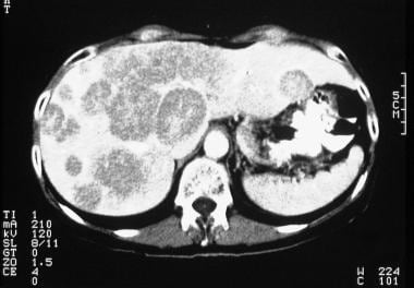
 Liver Metastases Imaging: Practice Essentials, Radiography, Computed Tomography
 NME4 - Wikipedia
 MAIL | The Scientist Magazine®
 WikiGenes - Pluripotent Stem Cells
 Timothy Ley - Research output
- Research Profiles at Washington University School of Medicine
Timothy Ley - Research output
- Research Profiles at Washington University School of Medicine Clinical experiences with venetoclax and other pro-apoptotic agents in lymphoid malignancies: lessons from monotherapy and...
 Understand everything about melatonin and sleep
 Timothy Aaron Graubert, M.D. | Harvard Catalyst Profiles | Harvard Catalyst
 Hematologic Malignancie Market Size, Growth and Industry Report 2019-2025
 Ferritin Test, Normal & Low Levels + How to Increase Them - SelfHacked
 Polycythemia vera (Clinical) | Concise Medical Knowledge
 Frontiers | Roles of Macrophages in the Development and Treatment of Gut Inflammation
 Búsqueda | BVS Bolivia
 The laboratory's role in combating sepsis | Medical Laboratory Observer
 Dr DEEPAN P SHAH - MD(HOMOEOPATHY)
 GM-CSF (Granulocyte/Macrophage - Colony Stimulating Factor) - Clone BVD2-21C11 | 1437
 Recombinant Human GM-CSF / CSF2 Protein - enQuire BioReagents
 G-CSF Protein
 Biomedicines | July 2020 - Browse Articles