Stethoscopes
Heart Murmurs
Heart Sounds
Electronics, Medical
Phonocardiography
Multimedia
Respiratory Sounds
CD-ROM
Insufflation
Heart Diseases
Gastric Dilatation
Computer-Assisted Instruction
Cardiotocography
Clinical Competence
Program Evaluation
Investigation of the theory and mechanism of the origin of the second heart sound. (1/105)
To investigate further the origin of the second heart sound we studied human subjects, dogs, and a model in vitro of the cardiovascular system. Intra-arterial sound, pressure, and, where possible, flow and high speed cine (2,000 frames/sec) were utilized. The closure sound of the semilunar valves was of higher amplitude in be ventricles than in their respective arterial cavities. The direction of inscription of the main components of intra-arterial sound were opposite in direction to the components of intraventricular sound. Notches, representative of pressure increments, were noted on the ventricular pressure tracings and were coincident with the components of sound. The amplitude of the closure sound varied with diastolic pressure, but remained unchanged with augmentation of forward and retrograde aortic flow. Cines showed second sound to begin after complete valvular closure, and average leaflet closure rate was constant regardless of pressure. Hence, the semilunar valves, when closed, act as an elastic membrane and, when set into motion, generate compression and expansion of the blood, producing transient pressure changes indicative of sound. The magnitude of the initial stretch is related to the differential pressure between the arterial and ventricular chambers. Sound transients which follow the major components of the second sound appear to be caused by the continuing stretch and recoil of the leaflets. Clinically unexplained findings such as the reduced or absent second sound in calcific aortic stenosis and its paradoxical presence in congenital aortic stenosis may be explained by those observations. (+info)A new sign of occlusion of the origin of the internal carotid artery. (2/105)
When the origin of the internal carotid artery is occluded, the transmission of cardiac sounds along the carotid stops at the site of the occlusion. This is a new neurovascular sign which is being reported. (+info)Apical systolic click and murmur associated with neurofibromatosis. (3/105)
In this report we describe a child who had an apical systolic click and murmur, as well as widespread cutaneous neurofibromatosis. We were not able to show an anatomical basis for the click and murmur. (+info)Current management of mitral valve prolapse. (4/105)
Mitral valve prolapse is a pathologic anatomic and physiologic abnormality of the mitral valve apparatus affecting mitral leaflet motion. "Mitral valve prolapse syndrome" is a term often used to describe a constellation of mitral valve prolapse and associated symptoms or other physical abnormalities such as autonomic dysfunction, palpitations and pectus excavatum. The importance of recognizing that mitral valve prolapse may occur as an isolated disorder or with other coincident findings has led to the use of both terms. Mitral valve prolapse syndrome, which occurs in 3 to 6 percent of Americans, is caused by a systolic billowing of one or both mitral leaflets into the left atrium, with or without mitral regurgitation. It is often discovered during routine cardiac auscultation or when echocardiography is performed for another reason. Most patients with mitral valve prolapse are asymptomatic. Those who have symptoms commonly report chest discomfort, anxiety, fatigue and dyspnea, but whether these are actually due to mitral valve prolapse is not certain. The principal physical finding is a midsystolic click, which frequently is followed by a late systolic murmur. Although echocardiography is the most useful mode for identifying mitral valve prolapse, it is not recommended as a screening tool for mitral valve prolapse in patients who have no systolic click or murmur on careful auscultation. Mitral valve prolapse has a benign prognosis and a complication rate of 2 percent per year. The progression of mitral regurgitation may cause dilation of the left-sided heart chambers. Infective endocarditis is a potential complication. Patients with mitral valve prolapse syndrome who have murmurs and/or thickened redundant leaflets seen on echocardiography should receive antibiotic prophylaxis against endocarditis. (+info)Epidemiology of rheumatic heart disease in black shcoolchildren of Soweto, Johannesburg. (5/105)
A survey to determine the prevalence of rheumatic heart disease (R.H.D.) in Black children was conducted in the creeches and primary schools of the South Western Townships of Johannesburg (Soweto). A total of 12 050 Black children were examined by 10 cardiologists in May to October 1972. The overal prevalence rate of R.H.D. was 6.9 per 1000, with a peak rate of 19.2 per 1000 in children of the seventh school grade. The maximal age incidence was 15-18 years and there was a female preponderance of 1 6:1. A rise in prevalence occurred with increasing family size. Most children (92%) were asymptomatic, and in 82.5% R.H.D. was diagnosed for the first time during the school survey. The commonest valve lesion was mitral regurgitation, which was present in 93% and occurred as an isolated lesion in 47.5%. Lancefield's group A beta-haemolytic streptococcus was isolated from the throats of 52 per 1000 Soweto children. The auscultatory features of a non-ejection systolic click and late systolic murmur were prevalent (13.9 per 1000) and had several epidemiological factors in common with R.H.D. A comprehensive preventative campaign is urgently needed in South Africa, directed at both primary and secondary prophylaxis of R.H.D. The socioeconomic status of the community must be improved if optimal prevention is to be achieved. (+info)Using a multimedia tool to improve cardiac auscultation knowledge and skills. (6/105)
OBJECTIVE: Today's medical school graduates have significant deficits in physical examination skills. Medical educators have been searching for methods to effectively teach and maintain these skills in students. The objective of this study was to determine if an auscultation curriculum centered on a portable multimedia CD-ROM was effective in producing and maintaining significant gains in cardiac auscultatory skills. DESIGN: Controlled cohort study. PARTICIPANTS: All 168 third-year medical students at 1 medical school in an academic medical center. INTERVENTIONS: Students were tested before and after exposure to 1 or more elements of the auscultation curriculum: teaching on ward/clinic rotations, CD-ROM comprehensive cases with follow-up seminars, and a CD-ROM 20-case miniseries. The primary outcome measures were student performance on a 10-item test of auscultation skill (listening and identifying heart sound characteristics) and a 30-item test of auscultation knowledge (factual questions about auscultation). A subset of students was tested for attenuation effects 9 or 12 months after the intervention. RESULTS: Compared with the control group (1 month clinical rotation alone), students who were also exposed to the CD-ROM 20-case miniseries had significant improvements in auscultation skills scores (P < .05), but not knowledge. Additional months of clerkship, comprehensive CD-ROM cases, and follow-up seminars increased auscultation knowledge beyond the miniseries alone (P < .05), but did not further improve auscultation skills. Students' auscultation knowledge diminished one year after the intervention, but auscultation skills did not. CONCLUSION: In addition to the standard curriculum of ward and conference teaching, portable multimedia tools may help improve quality of physical examination skills. (+info)Office blood pressures, arterial compliance characteristics, and estimated cardiac load. (7/105)
Because of rising interest in new methods to detect arterial diseases, we compared data from 3 different compliance-related techniques to measure arterial stiffness: systolic pulse contour analysis, diastolic pulse contour analysis (modified Windkessel model), and muscular (brachial) artery compliance by cuff plethysmography. Variables measured in the sitting position were compared with each other, with clinic blood pressures (BPs), and with the cardiac time-tension integral (CTTI) in 63 established hypertensive and 28 age-matched normotensive subjects. Hypertensives demonstrated marginal reductions in C(1) (thought to represent reduced large vessel compliance) and increased central systolic BP augmentation. In contrast, muscular artery compliance tended to be greater in the hypertensives despite normal brachial arterial diameters. C(2), suggested to be an indicator of small artery properties, was similar in both groups. CTTI was strongly related to systolic pressure (r=0.81), integrated mean arterial pressure (r=0.83), and systolic pressure-heart rate product (r=0.85) and was less strongly related to diastolic (r=0.71) or pulse pressure (r=0.57). Weak correlations were observed between CTTI and measured compliance-related variables, which also showed absent or weak correlations among themselves. We conclude that the weak relationships among BP and compliance-related variables could be due to intrinsic differences in the properties of large and small arteries, theoretical methodological weaknesses, measurement artifacts, or intrinsic hemodynamic differences of the sitting position. At present, compliance-related variables provide little additional advantage over cuff BP in the office estimation of cardiac work. (+info)Cardiac auscultatory recording database: delivering heart sounds through the Internet. (8/105)
The clinical skill of cardiac auscultation, while known to be sensitive, specific, and inexpensive in screening for cardiac disease among children, has recently been shown to be deficient among residents in training. This decline in clinical skill is partly due to the difficulty in teaching auscultation. Standardization, depth, and breadth of experience has been difficult to reproduce for students due to time constraints and the impracticality of examining large numbers of patients with cardiac pathology. We have developed a web-based multimedia platform that delivers complete heart sound recordings from over 800 different patients seen at the Johns Hopkins Outpatient Pediatric Cardiology Clinic. The database represents more than twenty significant cardiac lesions as well as normal and innocent murmurs. Each patient record is complete with a gold standard echo for diagnostic confirmation and a gold standard auscultatory assessment provided by a pediatric cardiology attending. (+info)Heart auscultation is a medical procedure in which a healthcare professional uses a stethoscope to listen to the sounds produced by the heart. The process involves placing the stethoscope on various locations of the chest wall to hear different areas of the heart.
The sounds heard during auscultation are typically related to the opening and closing of the heart valves, as well as the turbulence created by blood flow through the heart chambers. These sounds can provide important clues about the structure and function of the heart, allowing healthcare professionals to diagnose various cardiovascular conditions such as heart murmurs, valvular disorders, and abnormal heart rhythms.
Heart auscultation is a key component of a physical examination and requires proper training and experience to interpret the findings accurately.
Auscultation is a medical procedure in which a healthcare professional uses a stethoscope to listen to the internal sounds of the body, such as heart, lung, or abdominal sounds. These sounds can provide important clues about a person's health and help diagnose various medical conditions, such as heart valve problems, lung infections, or digestive issues.
During auscultation, the healthcare professional places the stethoscope on different parts of the body and listens for any abnormal sounds, such as murmurs, rubs, or wheezes. They may also ask the person to perform certain movements, such as breathing deeply or coughing, to help identify any changes in the sounds.
Auscultation is a simple, non-invasive procedure that can provide valuable information about a person's health. It is an essential part of a physical examination and is routinely performed by healthcare professionals during regular checkups and hospital visits.
A stethoscope is a medical device used for auscultation, or listening to the internal sounds of the body. It is most commonly used to hear the heartbeat, lung sounds, and blood flow in the major arteries. The device consists of a small disc-shaped resonator that is placed against the skin, connected by tubing to two earpieces. Stethoscopes come in different types and designs, but all serve the primary purpose of amplifying and transmitting body sounds to facilitate medical diagnosis.
A heart murmur is an abnormal sound heard during a heartbeat, which is caused by turbulent blood flow through the heart. It is often described as a blowing, whooshing, or rasping noise. Heart murmurs can be innocent (harmless and not associated with any heart disease) or pathological (indicating an underlying heart condition). They are typically detected during routine physical examinations using a stethoscope. The classification of heart murmurs includes systolic, diastolic, continuous, and functional murmurs, based on the timing and auscultatory location. Various heart conditions, such as valvular disorders, congenital heart defects, or infections, can cause pathological heart murmurs. Further evaluation with diagnostic tests like echocardiography is often required to determine the underlying cause and appropriate treatment.
Heart sounds are the noises generated by the beating heart and the movement of blood through it. They are caused by the vibration of the cardiac structures, such as the valves, walls, and blood vessels, during the cardiac cycle.
There are two normal heart sounds, often described as "lub-dub," that can be heard through a stethoscope. The first sound (S1) is caused by the closure of the mitral and tricuspid valves at the beginning of systole, when the ventricles contract to pump blood out to the body and lungs. The second sound (S2) is produced by the closure of the aortic and pulmonary valves at the end of systole, as the ventricles relax and the ventricular pressure decreases, allowing the valves to close.
Abnormal heart sounds, such as murmurs, clicks, or extra sounds (S3 or S4), may indicate cardiac disease or abnormalities in the structure or function of the heart. These sounds can be evaluated through a process called auscultation, which involves listening to the heart with a stethoscope and analyzing the intensity, pitch, quality, and timing of the sounds.
In medical terms, the heart is a muscular organ located in the thoracic cavity that functions as a pump to circulate blood throughout the body. It's responsible for delivering oxygen and nutrients to the tissues and removing carbon dioxide and other wastes. The human heart is divided into four chambers: two atria on the top and two ventricles on the bottom. The right side of the heart receives deoxygenated blood from the body and pumps it to the lungs, while the left side receives oxygenated blood from the lungs and pumps it out to the rest of the body. The heart's rhythmic contractions and relaxations are regulated by a complex electrical conduction system.
"Medical electronics" refers to the field of electronics that is specifically designed for medical applications. This can include a wide range of devices and systems, such as:
1. Medical imaging equipment, such as X-ray machines, CT scanners, MRI machines, and ultrasound machines.
2. Patient monitoring equipment, such as heart rate monitors, blood pressure monitors, and oxygen saturation monitors.
3. Therapeutic devices, such as pacemakers, defibrillators, and deep brain stimulators.
4. Laboratory equipment, such as DNA sequencers, mass spectrometers, and microarray scanners.
5. Wearable health technology, such as fitness trackers, smartwatches, and continuous glucose monitors.
6. Telemedicine systems that enable remote consultations and patient monitoring.
Medical electronics must meet strict regulatory requirements to ensure safety, effectiveness, and reliability. These devices often require specialized electronic components, such as sensors, signal processing circuits, and power management circuits, that are designed to operate in the challenging environments found in medical settings. Medical electronics engineers must have a deep understanding of both electronics and medical applications to design and develop these complex systems.
Phonocardiography is a non-invasive medical procedure that involves the graphical representation and analysis of sounds produced by the heart. It uses a device called a phonocardiograph to record these sounds, which are then displayed as waveforms on a screen. The procedure is often used in conjunction with other diagnostic techniques, such as electrocardiography (ECG), to help diagnose various heart conditions, including valvular heart disease and heart murmurs.
During the procedure, a specialized microphone called a phonendoscope is placed on the chest wall over the area of the heart. The microphone picks up the sounds generated by the heart's movements, such as the closing and opening of the heart valves, and transmits them to the phonocardiograph. The phonocardiograph then converts these sounds into a visual representation, which can be analyzed for any abnormalities or irregularities in the heart's function.
Phonocardiography is a valuable tool for healthcare professionals, as it can provide important insights into the health and functioning of the heart. By analyzing the waveforms produced during phonocardiography, doctors can identify any potential issues with the heart's valves or other structures, which may require further investigation or treatment. Overall, phonocardiography is an essential component of modern cardiac diagnostics, helping to ensure that patients receive accurate and timely diagnoses for their heart conditions.
I'm sorry for any confusion, but "multimedia" is not a term that has a specific medical definition. Multimedia generally refers to the use of multiple forms of media, such as text, audio, video, graphics, and animation, in a single interactive presentation or platform. It is often used in various fields including education, entertainment, marketing, and some areas of healthcare for purposes like training, patient education, and therapy. However, it does not have a specific medical meaning itself.
Respiratory sounds are the noises produced by the airflow through the respiratory tract during breathing. These sounds can provide valuable information about the health and function of the lungs and airways. They are typically categorized into two main types: normal breath sounds and adventitious (or abnormal) breath sounds.
Normal breath sounds include:
1. Vesicular breath sounds: These are soft, low-pitched sounds heard over most of the lung fields during quiet breathing. They are produced by the movement of air through the alveoli and smaller bronchioles.
2. Bronchovesicular breath sounds: These are medium-pitched, hollow sounds heard over the mainstem bronchi and near the upper sternal border during both inspiration and expiration. They are a combination of vesicular and bronchial breath sounds.
Abnormal or adventitious breath sounds include:
1. Crackles (or rales): These are discontinuous, non-musical sounds that resemble the crackling of paper or bubbling in a fluid-filled container. They can be heard during inspiration and are caused by the sudden opening of collapsed airways or the movement of fluid within the airways.
2. Wheezes: These are continuous, musical sounds resembling a whistle. They are produced by the narrowing or obstruction of the airways, causing turbulent airflow.
3. Rhonchi: These are low-pitched, rumbling, continuous sounds that can be heard during both inspiration and expiration. They are caused by the vibration of secretions or fluids in the larger airways.
4. Stridor: This is a high-pitched, inspiratory sound that resembles a harsh crowing or barking noise. It is usually indicative of upper airway narrowing or obstruction.
The character, location, and duration of respiratory sounds can help healthcare professionals diagnose various respiratory conditions, such as pneumonia, chronic obstructive pulmonary disease (COPD), asthma, and bronchitis.
Heart rate is the number of heartbeats per unit of time, often expressed as beats per minute (bpm). It can vary significantly depending on factors such as age, physical fitness, emotions, and overall health status. A resting heart rate between 60-100 bpm is generally considered normal for adults, but athletes and individuals with high levels of physical fitness may have a resting heart rate below 60 bpm due to their enhanced cardiovascular efficiency. Monitoring heart rate can provide valuable insights into an individual's health status, exercise intensity, and response to various treatments or interventions.
A CD-ROM (Compact Disc Read-Only Memory) is not a medical term, but a technology term. It refers to a type of optical storage disc that contains digital information and can be read by a computer's CD-ROM drive. The data on a CD-ROM is permanent and cannot be modified or erased, unlike other types of writable discs such as CD-R or CD-RW.
CD-ROMs were commonly used in the past to distribute software, multimedia presentations, reference materials, and educational content. In medical field, CD-ROMs have been used to distribute large databases of medical information, such as clinical guidelines, drug references, and anatomical atlases. However, with the advent of internet and cloud storage technologies, the use of CD-ROMs has become less common in recent years.
Insufflation is a medical term that refers to the act of introducing a gas or vapor into a body cavity or passage, typically through a tube or surgical instrument. This procedure is often used in medical and surgical settings for various purposes, such as:
* To administer anesthesia during surgery (e.g., introducing nitrous oxide or other gases into the lungs)
* To introduce medication or other substances into the body (e.g., insufflating steroids into a joint)
* To perform diagnostic procedures (e.g., insufflating air or a contrast agent into the gastrointestinal tract to visualize it with X-rays)
* To clean out a body cavity (e.g., irrigating and insufflating the bladder during urological procedures).
It's important to note that insufflation should be performed under controlled conditions, as there are potential risks associated with introducing gases or vapors into the body, such as barotrauma (damage caused by changes in pressure) and infection.
Cardiology is a branch of medicine that deals with the diagnosis and treatment of diseases and disorders of the heart and blood vessels. It encompasses the study of the normal functioning of the heart, the investigation and diagnosis of heart disease, and the treatment of various cardiovascular conditions through both surgical and non-surgical interventions. Cardiologists are medical professionals who specialize in this field, providing comprehensive care for patients with conditions such as coronary artery disease, congenital heart defects, valvular heart disease, electrophysiology disorders, and hypertension, among others. They work closely with other healthcare providers to manage cardiovascular risk factors, optimize overall cardiovascular health, and improve patients' quality of life.
Heart disease is a broad term for a class of diseases that involve the heart or blood vessels. It's often used to refer to conditions that include:
1. Coronary artery disease (CAD): This is the most common type of heart disease. It occurs when the arteries that supply blood to the heart become hardened and narrowed due to the buildup of cholesterol and other substances, which can lead to chest pain (angina), shortness of breath, or a heart attack.
2. Heart failure: This condition occurs when the heart is unable to pump blood efficiently to meet the body's needs. It can be caused by various conditions, including coronary artery disease, high blood pressure, and cardiomyopathy.
3. Arrhythmias: These are abnormal heart rhythms, which can be too fast, too slow, or irregular. They can lead to symptoms such as palpitations, dizziness, and fainting.
4. Valvular heart disease: This involves damage to one or more of the heart's four valves, which control blood flow through the heart. Damage can be caused by various conditions, including infection, rheumatic fever, and aging.
5. Cardiomyopathy: This is a disease of the heart muscle that makes it harder for the heart to pump blood efficiently. It can be caused by various factors, including genetics, viral infections, and drug abuse.
6. Pericardial disease: This involves inflammation or other problems with the sac surrounding the heart (pericardium). It can cause chest pain and other symptoms.
7. Congenital heart defects: These are heart conditions that are present at birth, such as a hole in the heart or abnormal blood vessels. They can range from mild to severe and may require medical intervention.
8. Heart infections: The heart can become infected by bacteria, viruses, or parasites, leading to various symptoms and complications.
It's important to note that many factors can contribute to the development of heart disease, including genetics, lifestyle choices, and certain medical conditions. Regular check-ups and a healthy lifestyle can help reduce the risk of developing heart disease.
Gastric dilatation, also known as stomach dilation or distention, refers to the abnormal enlargement or expansion of the stomach. This condition often occurs when the stomach fills with gas, food, or fluids and is unable to empty properly. Gastric dilatation can be caused by various factors such as overeating, swallowing excessive air, gastroparesis (delayed gastric emptying), intestinal obstruction, or certain medical conditions like hiatal hernia or pregnancy.
In severe cases, gastric dilatation may lead to gastric volvulus, where the stomach twists on itself, cutting off its blood supply and leading to ischemia and necrosis of the stomach tissue. This is a life-threatening condition that requires immediate medical attention. Symptoms of gastric dilatation include abdominal pain, bloating, vomiting, loss of appetite, and difficulty breathing.
Computer-Assisted Instruction (CAI) is a type of educational technology that involves the use of computers to deliver, support, and enhance learning experiences. In a medical context, CAI can be used to teach a variety of topics, including anatomy, physiology, pharmacology, and clinical skills.
CAI typically involves interactive multimedia presentations, simulations, quizzes, and other activities that engage learners and provide feedback on their performance. It may also include adaptive learning systems that adjust the content and pace of instruction based on the learner's abilities and progress.
CAI has been shown to be effective in improving knowledge retention, critical thinking skills, and learner satisfaction in medical education. It can be used as a standalone teaching method or in combination with traditional classroom instruction or clinical experiences.
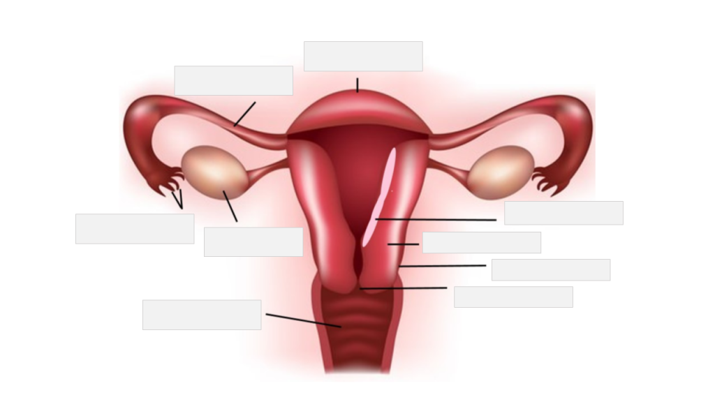
Cardiotocography (CTG) is a technical means of monitoring the fetal heart rate and uterine contractions during pregnancy, particularly during labor. It provides visual information about the fetal heart rate pattern and the frequency and intensity of uterine contractions. This helps healthcare providers assess the well-being of the fetus and the progression of labor.
The cardiotocograph records two main traces:
1. Fetal heart rate (FHR): It is recorded using an ultrasound transducer placed on the mother's abdomen. The normal fetal heart rate ranges from 120 to 160 beats per minute. Changes in the FHR pattern may indicate fetal distress, hypoxia, or other complications.
2. Uterine contractions: They are recorded using a pressure sensor (toco) placed on the mother's abdomen. The intensity and frequency of uterine contractions can be assessed to evaluate the progression of labor and the effect of contractions on fetal oxygenation.
Cardiotocography is widely used in obstetrics as a non-invasive method for monitoring fetal well-being during pregnancy and labor. However, it should always be interpreted cautiously by healthcare professionals, considering other factors like maternal and fetal conditions, medical history, and clinical presentation. Overinterpretation or misinterpretation of CTG traces can lead to unnecessary interventions or delays in recognizing actual fetal distress.
Clinical competence is the ability of a healthcare professional to provide safe and effective patient care, demonstrating the knowledge, skills, and attitudes required for the job. It involves the integration of theoretical knowledge with practical skills, judgment, and decision-making abilities in real-world clinical situations. Clinical competence is typically evaluated through various methods such as direct observation, case studies, simulations, and feedback from peers and supervisors.
A clinically competent healthcare professional should be able to:
1. Demonstrate a solid understanding of the relevant medical knowledge and its application in clinical practice.
2. Perform essential clinical skills proficiently and safely.
3. Communicate effectively with patients, families, and other healthcare professionals.
4. Make informed decisions based on critical thinking and problem-solving abilities.
5. Exhibit professionalism, ethical behavior, and cultural sensitivity in patient care.
6. Continuously evaluate and improve their performance through self-reflection and ongoing learning.
Maintaining clinical competence is essential for healthcare professionals to ensure the best possible outcomes for their patients and stay current with advances in medical science and technology.
Anatomy is the branch of biology that deals with the study of the structure of organisms and their parts. In medicine, anatomy is the detailed study of the structures of the human body and its organs. It can be divided into several subfields, including:
1. Gross anatomy: Also known as macroscopic anatomy, this is the study of the larger structures of the body, such as the organs and organ systems, using techniques such as dissection and observation.
2. Histology: This is the study of tissues at the microscopic level, including their structure, composition, and function.
3. Embryology: This is the study of the development of the embryo and fetus from conception to birth.
4. Neuroanatomy: This is the study of the structure and organization of the nervous system, including the brain and spinal cord.
5. Comparative anatomy: This is the study of the structures of different species and how they have evolved over time.
Anatomy is a fundamental subject in medical education, as it provides the basis for understanding the function of the human body and the underlying causes of disease.
Program Evaluation is a systematic and objective assessment of a healthcare program's design, implementation, and outcomes. It is a medical term used to describe the process of determining the relevance, effectiveness, and efficiency of a program in achieving its goals and objectives. Program evaluation involves collecting and analyzing data related to various aspects of the program, such as its reach, impact, cost-effectiveness, and quality. The results of program evaluation can be used to improve the design and implementation of existing programs or to inform the development of new ones. It is a critical tool for ensuring that healthcare programs are meeting the needs of their intended audiences and delivering high-quality care in an efficient and effective manner.
Congenital heart defects (CHDs) are structural abnormalities in the heart that are present at birth. They can affect any part of the heart's structure, including the walls of the heart, the valves inside the heart, and the major blood vessels that lead to and from the heart.
Congenital heart defects can range from mild to severe and can cause various symptoms depending on the type and severity of the defect. Some common symptoms of CHDs include cyanosis (a bluish tint to the skin, lips, and fingernails), shortness of breath, fatigue, poor feeding, and slow growth in infants and children.
There are many different types of congenital heart defects, including:
1. Septal defects: These are holes in the walls that separate the four chambers of the heart. The two most common septal defects are atrial septal defect (ASD) and ventricular septal defect (VSD).
2. Valve abnormalities: These include narrowed or leaky valves, which can affect blood flow through the heart.
3. Obstruction defects: These occur when blood flow is blocked or restricted due to narrowing or absence of a part of the heart's structure. Examples include pulmonary stenosis and coarctation of the aorta.
4. Cyanotic heart defects: These cause a lack of oxygen in the blood, leading to cyanosis. Examples include tetralogy of Fallot and transposition of the great arteries.
The causes of congenital heart defects are not fully understood, but genetic factors and environmental influences during pregnancy may play a role. Some CHDs can be detected before birth through prenatal testing, while others may not be diagnosed until after birth or later in childhood. Treatment for CHDs may include medication, surgery, or other interventions to improve blood flow and oxygenation of the body's tissues.
 Heart Auscultation - Medical Dictionary online-medical-dictionary.org
 Auscultation of heart sounds
 S1 Heart Sound - Easy Auscultation
 Auscultation: MedlinePlus Medical Encyclopedia
 Learning Programs - Teaching Heart Auscultation to Health Professionals
 Pleasant Grove - Auscultation Of The Heart - Marty Willson-Piper
 dblp: Automated Heart and Lung Auscultation in Robotic Physical Examinations.
 Intermittent Auscultation of the fetal heart rate - Canadian Nurses Protective Society
 Heart Murmurs in Pediatric Patients: When Do You Refer? | AAFP
 دانلود کتاب مهارت های سمع: نفس و صدای قلب Auscultation Skills: Breath & Heart Sounds, 5ed
 Defining low- and high-fidelity simulation in systematic reviews: the case of heart auscultation simulators
 Richard Wainright Duke Turner - Wikipedia
 Correction | The BMJ
 Relationship Between Accurate Auscultation of a Clinically Useful Third Heart Sound and Level of Experience | Cardiology | JAMA...
 A gynecologist examines a pregnant woman. Fetal doppler heart auscultation. Patient at doctor office. Assessment of heartbeat...
 ATSDR Clinician Brief: Radon | ATSDR
 Incidental Intracranial Aneurysm in a Dog Detected by 16-Multidetector Row Computed Tomography Angiography
 Acute Renal Failure during Progressive Systemic Sclerosis in the Regional University Teaching Hospital of Brest-La Cavale...
 A man with cardiac Lyme borreliosis | CMAJ
 Evidence-Based Guidelines for Labor Support that Promote Vaginal Birth | Article | NursingCenter
 Auscultation Pediatric Training Simulator | Pediatric Auscultation Teaching | Cardionics
 Pediatric Rheumatic Fever: Practice Essentials, Background, Pathophysiology
 Comprehensive Exams | Medical Billing and Coding Forum - AAPC
 Apert Syndrome in an Infant

![APA format] Physiology References for the Native American Flute](https://flutopedia.com/img/JoinCVFluteNewsletter_150.jpg)