Hemosiderosis
Stachybotrys
Hemosiderin
Idiopathic pulmonary haemosiderosis. Epidemiology, pathogenic aspects and diagnosis. (1/123)
Idiopathic pulmonary haemosiderosis (IPH) is a rare clinical entity characterized by recurrent episodes of diffuse alveolar haemorrhage, often presenting with haemoptysis. Many patients have iron deficiency anaemia due to deposition of haemosiderin iron in the alveoli, and eventually develop moderate pulmonary fibrosis. Typically, intensive search for an aetiology ends up negative. There is no evidence of pulmonary vasculitis or capillaritis. The aetiology is obscure, but may be an immunological or toxic mechanism causing a defect in the basement membrane of the pulmonary capillary. IPH affects both children and adults. During an acute episode, a chest X-ray demonstrates bilateral, alveolar infiltrates. Sputum examination discloses haemosiderin-laden alveolar macrophages. Diagnosis is established by lung biopsy (fiber-optic or thoracoscopic), showing large numbers of haemosiderin-laden macrophages in the alveoli and without evidence of capillaritis or deposition of immunoglobulins. Corticosteroids and/or immunosuppressive drugs may be effective during an acute bleeding episode, and may in some patients improve symptoms and prognosis on the long-term, but the response to treatment displays great interindividual variation. (+info)Overview of investigations into pulmonary hemorrhage among infants in Cleveland, Ohio. (2/123)
Idiopathic pulmonary hemorrhage was diagnosed in 37 infants in the Cleveland, Ohio, area between 1993 and 1998. This rare disorder has been related to 12 deaths, including 7 originally thought to be sudden infant death syndrome. Thirty of the infants were African American, all of whom lived in a limited geographic area of eastern metropolitan Cleveland, an area of older housing stock. An investigation led by the Centers for Disease Control and Prevention has found an association with household exposure to a toxigenic mold, Stachybotrys chartarum, and other fungi. The rapidly growing lungs of young infants appear to be especially vulnerable to the toxins made by toxigenic molds. Environmental tobacco smoke was frequently present in the infants' homes and may be a trigger precipitating the acute bleeding. Stachybotrys, although not thought to be a common mold, is known to have a wide geographic distribution. An additional 101 cases of acute, idiopathic pulmonary hemorrhage have been reported in infants in the United States over the past 5 years. In this overview, the investigations are summarized, the clinical profile is described, the toxicity of S. chartarum is discussed, and pathophysiologic concepts are presented. (+info)Anthracycline cardiotoxicity in a black rhinoceros (Diceros bicornis): evidence for impaired antioxidant capacity compounded by iron overload. (3/123)
Two weeks before dying of congestive heart failure, a juvenile black rhinoceros (Diceros bicornis minor) received a single low dose of doxorubicin as part of combination chemotherapy for acute lymphoblastic leukemia. Diffuse hemosiderosis was present at necropsy in a pattern indicative of dietary iron overload, but unique iron-positive degenerative lesions were found in isolated myocardiocytes. Serum analyses revealed hyperferremia, 87% transferrin saturation, and 5- to 10-fold elevations in ferritin concentration, reflecting markedly increased tissue iron stores. Since both toxic and therapeutic effects of anthracyclines are mediated by formation of reactive free radicals via iron-catalyzed reactions, these observations suggest that iron overload may have enhanced myocardial susceptibility to cardiotoxic effects of doxorubicin. Impairments in other myocardial antioxidant defenses, such as deficiencies in catalase and glutathione S-transferase that are known to exist in rhinoceros erythrocytes, may have been underlying factors contributing to an inherent sensitivity of rhinoceros tissues to oxidant-induced injury. (+info)Quantification of siderophore and hemolysin from Stachybotrys chartarum strains, including a strain isolated from the lung of a child with pulmonary hemorrhage and hemosiderosis. (4/123)
A strain of Stachybotrys chartarum was recently isolated from the lung of a pulmonary hemorrhage and hemosiderosis (PH) patient in Texas (designated the Houston strain). This is the first time that S. chartarum has been isolated from the lung of a PH patient. In this study, the Houston strain and 10 strains of S. chartarum isolated from case (n = 5) or control (n = 5) homes in Cleveland were analyzed for hemolytic activity, siderophore production, and relatedness as measured by random amplified polymorphic DNA analysis. (+info)Juvenile hemochromatosis associated with B-thalassemia treated by phlebotomy and recombinant human erythropoietin. (5/123)
Juvenile hemochromatosis is a rare genetic disorder that causes iron overload. Clinical complications, which include liver cirrhosis, heart failure, hypogonadotropic hypogonadism and diabetes, appear earlier and are more severe than in HFE-related hemochromatosis. This disorder, therefore, requires an aggressive therapeutic approach to achieve iron depletion. We report here the case of a young Italian female with juvenile hemochromatosis who was unable to tolerate frequent phlebotomy because of coexistent ss-thalassemia trait. The patient was successfully iron-depleted by combining phlebotomy with recombinant human erythropoietin. (+info)Diagnostic accuracy of abdominal ultrasonography compared to magnetic resonance imaging in siderosis of the spleen. (6/123)
A prospective study to compare the diagnostic performance of ultrasonography with magnetic resonance imaging using gradient-recalled echo technique in cases of siderosis of spleen was conducted in 53 cirrhotic patients with endoscopic proof of gastroesophageal varices. Of the 34 patients with splenic siderosis on MR imaging, punctate hyperechoic foci were detected in the spleen on ultrasonography in 24 patients. Using MR imaging as the reference standard for the diagnosis of splenic siderosis, the sensitivity of ultrasonography is 70.6%; specificity is 78.9%; positive predictive value is 85.7%; and negative predictive value is 40%. We conclude that ultrasonography is a fairly accurate technique in the diagnosis of splenic siderosis. (+info)Pathology of the synovium. (7/123)
Synovium is specialized mesenchymal tissue that is essential for the appropriate function of the locomotor apparatus. It is the site for a series of pathologic processes that are characteristic, and in some cases specific, to this distinctive tissue. In this article, the normal microscopic anatomy of synovium is briefly reviewed. Synovial proliferative disorders, including pigmented villonodular synovitis, giant cell tumor of tendon sheath, hemosiderotic synovitis, and fatty infiltration of the synovial membrane are discussed. Additionally, the subjects of intrasynovial cartilaginous lesions (primary and secondary synovial chondromatosis) and crystal deposition diseases are reviewed. Finally, the response of synovial tissues to implanted foreign materials that are used in large and small joint arthroplasty are described. (+info)Hemosiderotic fibrohistiocytic lipomatous lesion: ten cases of a previously undescribed fatty lesion of the foot/ankle. (8/123)
We address the clinicopathologic features of a previously undescribed heavily-pigmented spindle cell proliferation within a circumscribed benign lipomatous lesion that occurs mainly in the ankle region of older females. Patients with "lipoma with fibrohistiocytic proliferation" were retrieved from our files. Slides and clinical information were reviewed, and immunohistochemistry was performed (n = 5). Ten patients with hemosiderotic fibrohistiocytic lipomatous lesions were identified. All cases demonstrated a well-circumscribed fatty lesion with random focal proliferations of plump, slightly pleomorphic spindled cells, scattered inflammatory cells, and abundant iron pigment. The spindled cells had vesicular nuclei with indistinct nucleoli; occasional hyperchromatism was observed. No nuclear cytoplasmic inclusions were identified. The spindled component had a reactive appearance. In most cases, the fatty component, with homogeneously sized adipocytes, predominated. The lesions occurred in the foot/ankle region (8/10, one each cheek and hand) of primarily females (8/10) with a mean age of 50.6 years (range 42-63 years), size of 7.7 cm (range 2.5-17 cm), and prior duration of 3.1 years. Seven of eight patients had a history of prior trauma. The spindled component was positive for vimentin, calponin, CD34, and occasionally KP-1 or lysozyme and negative for caldesmon, S100, and desmin. Follow-up on eight patients revealed four with recurrences or residual disease over three years, requiring re-excision. No cases metastasized or caused patient death (mean 12 years, range 1-23 years). We describe a predominantly fatty lesion that is hemosiderin rich with a "fibrohistiocytic" proliferation, composed of histiocytes, myofibroblasts, and C34-positive fibroblasts, which occurs predominantly in the ankle region of middle-aged females. We believe that this is a reactive process due to antecedent trauma, the inflammatory cells, hemosiderin, mixed spindled cells, and homogeneous non-neoplastic appearance of the fat. HFLL can be distinguished from previously described lesions. Correct identification of hemosiderotic fibrohistiocytic lipomatous lesion is important, as it may locally recur. (+info)Hemosiderosis is a medical condition characterized by the abnormal accumulation of hemosiderin, an iron-containing protein, in various organs and tissues of the body. Hemosiderin is derived from the breakdown of hemoglobin, which is the oxygen-carrying protein in red blood cells. When there is excessive breakdown of red blood cells or impaired clearance of hemosiderin, it can lead to its accumulation in organs such as the liver, spleen, and lungs.
Hemosiderosis can be classified into two types: primary and secondary. Primary hemosiderosis is a rare condition that is caused by genetic disorders affecting red blood cells, while secondary hemosiderosis is more common and is associated with various conditions that cause excessive breakdown of red blood cells or chronic inflammation. These conditions include hemolytic anemias, repeated blood transfusions, liver diseases, infections, and certain autoimmune disorders.
The accumulation of hemosiderin can lead to tissue damage and organ dysfunction, particularly in the lungs, where it can cause pulmonary fibrosis, and in the heart, where it can lead to heart failure. Hemosiderosis is typically diagnosed through a combination of medical history, physical examination, and laboratory tests, including blood tests and imaging studies such as chest X-rays or MRI scans. Treatment of hemosiderosis depends on the underlying cause and may include medications, blood transfusions, or supportive care to manage symptoms and prevent complications.
Stachybotrys is a genus of filamentous fungi (molds) that are known to produce potent mycotoxins, which can be harmful to humans and animals. The most well-known species is Stachybotrys chartarum, commonly referred to as "black mold" or "toxic black mold." This mold typically grows on materials with high cellulose content and a low nitrogen content, such as paper, straw, hay, wet drywall, and ceiling tiles. Exposure to the mycotoxins produced by Stachybotrys can cause various health issues, including respiratory symptoms, allergic reactions, and immune system responses. It is essential to address water damage and mold growth promptly to prevent the spread of Stachybotrys and other molds in indoor environments.
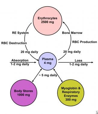
Hemosiderin is a golden-brown pigment that consists of iron-containing protein complexes called ferritin and ferrikinase. It is insoluble in water and forms as a result of the breakdown of hemoglobin in the reticuloendothelial system, primarily in macrophages. Hemosiderin deposits can be found in various tissues and organs, such as the spleen, liver, and brain, under conditions of increased red blood cell destruction or impaired iron metabolism. These deposits are often associated with diseases such as hemochromatosis, thalassemia, and chronic inflammation.
Lung diseases refer to a broad category of disorders that affect the lungs and other structures within the respiratory system. These diseases can impair lung function, leading to symptoms such as coughing, shortness of breath, chest pain, and wheezing. They can be categorized into several types based on the underlying cause and nature of the disease process. Some common examples include:
1. Obstructive lung diseases: These are characterized by narrowing or blockage of the airways, making it difficult to breathe out. Examples include chronic obstructive pulmonary disease (COPD), asthma, bronchiectasis, and cystic fibrosis.
2. Restrictive lung diseases: These involve stiffening or scarring of the lungs, which reduces their ability to expand and take in air. Examples include idiopathic pulmonary fibrosis, sarcoidosis, and asbestosis.
3. Infectious lung diseases: These are caused by bacteria, viruses, fungi, or parasites that infect the lungs. Examples include pneumonia, tuberculosis, and influenza.
4. Vascular lung diseases: These affect the blood vessels in the lungs, impairing oxygen exchange. Examples include pulmonary embolism, pulmonary hypertension, and chronic thromboembolic pulmonary hypertension (CTEPH).
5. Neoplastic lung diseases: These involve abnormal growth of cells within the lungs, leading to cancer. Examples include small cell lung cancer, non-small cell lung cancer, and mesothelioma.
6. Other lung diseases: These include interstitial lung diseases, pleural effusions, and rare disorders such as pulmonary alveolar proteinosis and lymphangioleiomyomatosis (LAM).
It is important to note that this list is not exhaustive, and there are many other conditions that can affect the lungs. Proper diagnosis and treatment of lung diseases require consultation with a healthcare professional, such as a pulmonologist or respiratory therapist.
 Hemosiderosis
Transfusion hemosiderosis
Idiopathic pulmonary haemosiderosis
Hereditary haemochromatosis
List of OMIM disorder codes
Paul Anton Cibis
Iron overload
Alveolar lung disease
Hemosiderin
Mycotoxin
Mold health issues
Stachybotrys
Hyphema
Atransferrinemia
Aceruloplasminemia
Reference ranges for blood tests
Lemur
Amyloid-related imaging abnormalities
Ferritin
GRACILE syndrome
Unsolved problems in medicine
Hemolysis
Perls Prussian blue
Hemolytic anemia
Heterochromia iridum
Superficial siderosis
Anemic infarct
Hepatocystis
Diphenylamine
Lecanemab
Idiopathic27
- Types include: Transfusion hemosiderosis Idiopathic pulmonary hemosiderosis Transfusional diabetes Organs affected: Hemosiderin deposition in the lungs is often seen after diffuse alveolar hemorrhage, which occurs in diseases such as Goodpasture's syndrome, granulomatosis with polyangiitis, and idiopathic pulmonary hemosiderosis. (wikipedia.org)
- Three variants of primary pulmonary hemosiderosis are recognized: (1) pulmonary hemosiderosis associated with antibody to the basement membrane of the lung and kidney (ie, Goodpasture syndrome ), (2) pulmonary hemosiderosis associated with hypersensitivity to proteins in cow's milk (ie, Heiner syndrome), and (3) idiopathic pulmonary hemosiderosis (IPH). (medscape.com)
- Idiopathic pulmonary hemosiderosis. (radiopaedia.org)
- Which of the following substances would be expected to be accumulating in this patient's alveoli secondary to the disease caused by idiopathic pulmonary hemosiderosis? (obaid.info)
- Idiopathic Pulmonary Hemosiderosis is condition affecting lungs causing blood seepage in lung leading to formation of fibrosis. (financedigest.com)
- The diagnosis of Idiopathic Pulmonary Hemosiderosis is further confirmed by bronchoscopy. (financedigest.com)
- Idiopathic Pulmonary Hemosiderosis treatment usually involves supportive care, respiratory therapy and oxygen supplementation. (financedigest.com)
- There is no specific targeted drug for Idiopathic Pulmonary Hemosiderosis treatment the treatment adopts use of Corticosteroids and Immunosuppressant. (financedigest.com)
- The wide use of Corticosteroids in Idiopathic Pulmonary Hemosiderosis treatment is mainly due to higher efficacy of corticosteroid in reducing the exacerbations. (financedigest.com)
- There pure nonexistence of targeted drug for Idiopathic Pulmonary Hemosiderosis treatment creates opportunity for new manufacturer of Idiopathic Pulmonary Hemosiderosis treatment drug. (financedigest.com)
- As per the data from WHO the Idiopathic Pulmonary Hemosiderosis have reported incidence of approximately 1800 to 7700 cases. (financedigest.com)
- The gradual development of tolerance for immunosuppressant Idiopathic Pulmonary Hemosiderosis treatment leads to increased mortality in affected population. (financedigest.com)
- Although the cause for Idiopathic Pulmonary Hemosiderosis is still unknown it primarily affects the children's between the age group of 1 to 10. (financedigest.com)
- Idiopathic Pulmonary Hemosiderosis causes severe intra-alveolar bleeding and coughing problems as iron content in the lung increases and forms Hemosiderosis. (financedigest.com)
- The growing children population globally is feeding the new cases of Idiopathic Pulmonary Hemosiderosis creating huge patient pool creating demand for Idiopathic Pulmonary Hemosiderosis treatment. (financedigest.com)
- The development of new effective immunosuppressant is anticipated offer new growth opportunities for Idiopathic Pulmonary Hemosiderosis treatment market . (financedigest.com)
- Presumably, number of research on rare disease and special aids form the government is anticipated to create new growth opportunities for the manufacturers in the Idiopathic Pulmonary Hemosiderosis treatment market . (financedigest.com)
- However, ineffective diagnosis and higher resemblance to cornice lung infection is restraining the growth of the Idiopathic Pulmonary Hemosiderosis Treatment market . (financedigest.com)
- Additionally, lack of novel drug therapies is also restraining the Idiopathic Pulmonary Hemosiderosis Treatment market growth. (financedigest.com)
- Idiopathic Pulmonary Haemosiderosis is a rare lung disorder characterized by alveolar capillary bleeding and accumulation of iron (in the form of haemosiderin) in the lungs. (rareshare.org)
- Acknowledgement of Idiopathic Pulmonary Haemosiderosis has not been added yet. (rareshare.org)
- Cause of Idiopathic Pulmonary Haemosiderosis has not been added yet. (rareshare.org)
- Symptoms for Idiopathic Pulmonary Haemosiderosis has not been added yet. (rareshare.org)
- Idiopathic Pulmonary Haemosiderosis community discussions will be posted here. (rareshare.org)
- My son has idiopathic pulmonary hemosiderosis. (rareshare.org)
- Mother to a wonderful 8 year-old with Idiopathic Pulmonary Hemosiderosis. (rareshare.org)
- Father of a adult daughter who was diagnosed at 3 yrs with Idiopathic Pulmonary Hemosiderosis. (rareshare.org)
Hemochromatosis2
- You should not take Ferrex-150 if you have hemochromatosis , hemosiderosis , or hemolytic anemia . (drugs.com)
- You should not use this medicine if you have hemochromatosis, hemosiderosis, or hemolytic anemia. (uofmhealth.org)
Primary pulmonary hemosiderosis1
- In children, primary pulmonary hemosiderosis is more common than secondary types. (medscape.com)
Transfusion1
- Splenomegaly is absent unless induced by transfusion hemosiderosis. (merckmanuals.com)
Thalassemia1
- 281). Routine evaluation of liver and heart iron content using MRI T2* is suggested to better evaluate the haemosiderosis status in thalassemia patients. (who.int)
Hepatic1
- 3 Most birds had histologic changes to the liver including chronic lymphocytic hepatitis, biliary hyperplasia, hemosiderosis, and hepatic necrosis. (vin.com)
Hemorrhage7
- Group 1 pulmonary hemosiderosis is defined by pulmonary hemorrhage associated with circulating anti-GBM antibodies. (medscape.com)
- Group 2 pulmonary hemosiderosis is defined as pulmonary hemorrhage and immune complex disease. (medscape.com)
- Group 3 pulmonary hemosiderosis is defined as pulmonary hemorrhage without demonstrable immunologic association. (medscape.com)
- In November 1994, private physicians and public health officials in Cleveland, Ohio, and CDC reported a cluster of eight cases of acute pulmonary hemorrhage/ hemosiderosis that had occurred during January 1993-November 1994 among infants in one area of the city (1). (cdc.gov)
- These findings documented an association between acute pulmonary hemorrhage/hemosiderosis in this cluster of cases and mold growth in their water-damaged homes. (cdc.gov)
- Active surveillance by the RBCH identified an additional 11 cases of acute pulmonary hemorrhage/hemosiderosis among infants in the Cleveland area during January 1995-December 1996. (cdc.gov)
- Scientists from the CDC published an article called 'Acute pulmonary hemorrhage/hemosiderosis among infants' in the journal Morbidity and Mortality Weekly Report, in which they described a cluster of ten infants in the Cleveland area who contracted this bleeding lung disease in 1993, and attributed it to spores from the mold Stachybotrys chartarum. (skeptoid.com)
Diagnosis2
- The diagnosis of isolated pulmonary hemosiderosis or idopathic pulmonary hemosiderosis is a diagnosis of exclusion, requiring thorough review and elimination of other causes of primary and secondary pulmonary hemosiderosis. (medscape.com)
- Diagnosis of pulmonary hemosiderosis by MR imaging. (radiopaedia.org)
Intra-alveolar1
- Pulmonary hemosiderosis (PH) is characterized by repeated episodes of intra-alveolar bleeding that lead to abnormal accumulation of iron as hemosiderin in alveolar macrophages and subsequent development of pulmonary fibrosis and severe anemia. (medscape.com)
Splenic1
- Lymphoid depletion of the spleen and splenic hemosiderosis were also observed inthese animals. (nih.gov)
Liver3
- There are several methods available for diagnosing and monitoring hemosiderosis including: Serum ferritin Liver biopsy MRI Serum ferritin is a low cost, readily available, and minimally invasive method for assessing body iron stores. (wikipedia.org)
- However, the major problem with using it as an indicator of hemosiderosis is that it can be elevated in a range of other medical conditions unrelated to iron levels including infection, inflammation, fever, liver disease, renal disease and cancer. (wikipedia.org)
- Hemosiderosis of liver 3(e). (shopanatomical.com)
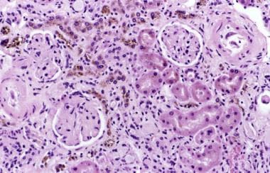
Renal2
- iron released from the red blood cells can accumulate within the kidneys (renal hemosiderosis). (msdmanuals.com)
- Most cases of renal hemosiderosis do not cause kidney damage. (msdmanuals.com)
Mitral1
- Mitral stenosis can also lead to pulmonary hemosiderosis. (wikipedia.org)
Diffuse1
- A triad of hemoptysis, iron deficiency anemia, and diffuse pulmonary infiltrates characterizes pulmonary hemosiderosis. (medscape.com)
Lungs2
- Pulmonary hemosiderosis can occur as a primary disease of the lungs or can be secondary to cardiovascular or systemic disease. (medscape.com)
- The lungs and kidneys are often sites of hemosiderosis. (msdmanuals.com)
Chronic1
- Thus into 3 different forms: acute pericarditis, appropriate regimens for managing cardiac congestive heart failure and arrhythmia due conditions in thalassaemia major patients to haemosiderosis, and chronic anaemia still need further investigation. (who.int)
Deposition1
- Pulmonary hemosiderosis (PH) refers to iron deposition within the lung. (radiopaedia.org)
Acute1
- The treatment of pulmonary hemosiderosis is directed toward management of the acute crises and long-term therapy. (medscape.com)
Infants1
- Exposures in the indoor environment have reportedly induced neurogenic symptoms in adults and hemosiderosis in infants. (cdc.gov)
Disorder1
- Hemosiderosis is a form of iron overload disorder resulting in the accumulation of hemosiderin. (wikipedia.org)
Iron3
- Hemosiderosis is a term used for excessive accumulation of iron deposits called hemosiderin in the tissues. (msdmanuals.com)
- Hemosiderosis is characterized by the local or generalized deposit of iron higher than normal in the tissues. (fundacionmapfre.org)
- Haemosiderosis, which plays a considerable function in thalassaemia patients before and role in early mortality, can be prevented or after treatment with high-dose deferoxam- postponed by iron-chelating agents which ine. (who.int)
Patients2
- RÉSUMÉ Des méthodes non-invasives de haute précision sont nécessaires pour l'évaluation de la concentration en fer dans les organes des patients atteints de thalassémie. (who.int)
- L'évaluation systématique de la concentration de fer dans le foie et le coeur à l'aide de l'IRM pondérée en T2* semble produire une meilleure évaluation de la présence d'une hémosidérose chez les patients atteints de thalassémie. (who.int)
Breakdown1
- Hemosiderosis caused by bleeding and red blood cell breakdown does not usually require treatment. (msdmanuals.com)
Blood1
- Pulmonary injury/hemosiderosis (blood loss). (clickpress.com)
Pulmonary hemorrhage1
- Background A geographic cluster of 10 cases of pulmonary hemorrhage and hemosiderosis in infants occurred in Cleveland, Ohio, between January 1993 and December 1994. (jamanetwork.com)
Diffuse1
- Diffuse pulmonary hemosiderosis after exposure to pesticides. (medscape.com)
Kidneys1
- The lungs and kidneys are often sites of hemosiderosis. (msdmanuals.com)
Association2
- Pulmonary hemosiderosis in association with celia disease. (medscape.com)
- 13. [Association between pulmonary hemosiderosis and juvenile dermatomyositis]. (nih.gov)
Treatment1
- Hemosiderosis caused by bleeding and red blood cell breakdown does not usually require treatment. (msdmanuals.com)