Hip Dislocation, Congenital
Dislocations
Hip Joint
Femoracetabular Impingement
Orthopedic Procedures
Femur Head
Acetabulum
Hip
Joint Capsule
Range of Motion, Articular
Legg-Calve-Perthes Disease
Osteoarthritis, Hip
Hip Fractures
Cerebral Palsy
Reoperation
Recovery of Function
Treatment Outcome
Retrospective Studies
Traction
Prosthesis Failure
Follow-Up Studies
Lens Subluxation
Acromioclavicular Joint
Joint Instability
Manipulation, Orthopedic
Splints
Femur Head Necrosis
Ligaments, Articular
Femoral Neck Fractures
Sternoclavicular Joint
Tarsal Joints
Shoulder Joint
Bone Diseases, Developmental
Postoperative Complications
Fracture Fixation, Internal
Casts, Surgical
Long-term results of spherical acetabular osteotomy. (1/363)
We have examined the effect of the Wagner spherical acetabular osteotomy on preserving the joint in 38 hips with a mean follow-up of 17 years. At the time of the initial operation, 55% of patients had clinical symptoms and 30 joints showed minimal or absent radiological signs of osteoarthritis. At follow-up, 54% of patients had a good functional result. The osteotomy improved the mean centre-edge angle from -3 degrees to +15 degrees, the mean anterior centre-edge angle to 23 degrees and the acetabular head index to 75%. The obliquity of the acetabular roof decreased from 28 degrees to 16 degrees. One patient improved, but 14 deteriorated with joint degeneration. Of these, one progressed because of postoperative deep-tissue infection and five due to undercorrection. One patient needed total joint replacement after 14 years. At 17 years after operation, Wagner osteotomy had prevented progression of secondary arthritis in 63% of cases. (+info)Unsuccessful surgical treatment of hip dislocation in congenital sensory neuropathy with anhidrosis. A case report. (2/363)
A six-year-old girl with congenital sensory neuropathy with anhidrosis (CSNA) presented with bilateral hip dysplasia and subluxation on the right side. Conservative treatment of the hips by closed reduction and a plaster cast was unsuccessful. When aged seven years the patient had an intertrochanteric varus rotation osteotomy on the right side, but subluxation was again evident after five months. A Salter-type pelvic osteotomy was carried out followed by immobilisation, but one year later subluxation was present in the right hip and dislocation in the left. At the age of nine years, the right femoral head resembled a Charcot joint, although walking ability was preserved. In patients with CSNA, surgery may not always be advisable. (+info)Predicting the outcome of adductor tenotomy. (3/363)
This study reviewed 57 hips in 30 children (18 girls and 12 boys) with cerebral palsy who had undergone an adductor tenotomy alone or in combination with an anterior obturator neurectomy (23 hips). Results were evaluated by the Reimers migration percentage (MP). The hips were split into three groups: group A (12 hips) a preoperative MP of less than 20%, group B (25 hips) between 20 and 40%, and group C (20 hips) more than 40%. The mean age at the time of surgery was 6 years and 1 month (range: 2.5-13 years). The mean period of review was 6 years and 3 months (2-20 years). The results were considered as "good" when radiographs at the longest follow-up showed a decrease of > 10% of the MP, as "bad" when they showed an increase of > 10%, and as "stable" when the MPs varied less than 10%. At the latest review of group A, 11 were stable (92%) and 1 was bad. In group B, 12 were stable (48%), 7 were good (28%), and 6 were bad (24%). In group C, 7 were stable (35%), and 13 were bad (65%). The preoperative migration percentage provided to be the only predictor of outcome. Age at the time of surgery had no constant significant effect on the outcome, neither had the addition of an anterior neurectomy. (+info)Ultrasound screening for hips at risk in developmental dysplasia. Is it worth it? (4/363)
Between May 1992 and April 1997, there were 20,452 births in the Blackburn District. In the same period 1107 infants with hip 'at-risk' factors were screened prospectively by ultrasound. We recorded the presence of dislocation and dysplasia detected under the age of six months using Graf's alpha angle. Early dislocation was present in 36 hips (34 dislocatable and 2 irreducible). Of the 36 unstable hips, 30 (83%) were referred as being Ortolani-positive or unstable; 25 (69%) of these had at least one of the risk factors. Only 11 (31%) were identified from the 'at-risk' screening programme alone (0.54 per 1000 live births). Eight cases of 'late' dislocation presented after the age of six months (0.39 per 1000 live births). The overall rate of dislocation was 2.2 per 1000 live births. Only 31% of the dislocated hips belonged to a major 'at-risk' group. Statistical analysis confirmed that the risk factors had a relatively poor predictive value if used as a screening test for dislocation. In infants referred for doubtful clinical instability, one dislocation was detected for every 11 infants screened (95% confidence interval (CI) 8 to 17) whereas in infants referred because of the presence of any of the major 'at-risk' factors the rate was one in 75 (95% CI 42 to 149). Routine ultrasound screening of the 'at-risk' groups on their own is of little value in significantly reducing the rate of 'late' dislocation in DDH, but screening clinically unstable hips alone or associated with 'at-risk' factors has a high rate of detection. (+info)'Floating pelvis'. A combination of bilateral hip dislocation with a lumbar ligamentous disruption. (5/363)
A patient is described with a ligamentous disruption at the L4/L5 level in association with bilateral, traumatic dislocations of the hip. The diagnostic evaluation, acute intervention, and definitive stabilisation are reported. The unstable spine posed a problem in treatment with regard to the timing and technique of the reduction of the hips. (+info)Acetabular augmentation for the treatment of unstable total hip arthroplasties. (6/363)
Twenty-eight unstable total hip arthroplasties were treated with an acetabular augmentation wedge. Of the hips, 23 have had no further dislocations at a mean follow-up of 26 months. Five patients continued to dislocate and have needed further surgery. To our knowledge this is the largest reported series of acetabular augmentation with as good results as those of the most successful reported series of this technique, and a success rate comparable to other methods of treating recurrent dislocation. Careful patient selection, and using a thin augmentation wedge to avoid impingement, are important to the success of a technique which is a useful option in the management of recurrent dislocation. (+info)A rare fracture-dislocation of the hip in a gymnast and review of the literature. (7/363)
Posterior fracture-dislocation of the hip is an uncommon injury in athletics and leisure activities. It is more commonly seen in high energy motor vehicle accidents and occasionally in high energy sporting activities. A rare case is reported of posterior fracture-dislocation of the hip joint that occurred in a young athlete during gymnastics. This unusual mechanism of injury illustrates the great forces sustained by the hip joint of gymnasts. Early reduction and operative treatment led to a congruent and stable hip joint. After rehabilitation, she returned to light sporting activities after six months. (+info)Rotational acetabular osteotomy using biodegradable internal fixation. (8/363)
We used biodegradable poly-L-lactide screws in rotational acetabular osteotomy in 41 hips of 41 patients, and studied the complications after an average follow-up of 4.9 years (range 1.0-7.7 years). There were 39 females and 2 males, their average age at the time of the operation was 32 years (range 12-55 years). A small subcutaneous abscess appeared around the non-absorbable sutures in 2 patients after surgery. There was 1 case of thrombophlebitis and 1 of local dermatitis. The small subcutaneous abscess resolved after the removal of the suture material in the 2 cases, and the thrombophlebitis resolved with aspirin. The local dermatitis persisted but was cured by local steroid therapy over 5.8 years. The incidence of local dermatitis after the use of biodegradable implants should be further investigated. (+info)A hip dislocation is a medical emergency that occurs when the head of the femur (thighbone) slips out of its socket in the pelvis. This can happen due to high-energy trauma, such as a car accident or a severe fall. Hip dislocations can also occur in people with certain health conditions that make their hips more prone to displacement, such as developmental dysplasia of the hip.
There are two main types of hip dislocations: posterior and anterior. In a posterior dislocation, the femur head moves out of the back of the socket, which is the most common type. In an anterior dislocation, the femur head moves out of the front of the socket. Both types of hip dislocations can cause severe pain, swelling, and difficulty moving the affected leg.
Immediate medical attention is necessary for a hip dislocation to realign the bones and prevent further damage. Treatment typically involves sedation or anesthesia to relax the muscles around the joint, followed by a closed reduction procedure to gently guide the femur head back into the socket. In some cases, surgery may be required to repair any associated injuries, such as fractures or damaged ligaments. After treatment, physical therapy and rehabilitation are usually necessary to restore strength, mobility, and function to the affected hip joint.
Congenital hip dislocation, also known as developmental dysplasia of the hip (DDH), is a condition where the hip joint fails to develop normally in utero or during early infancy. In a healthy hip, the head of the femur (thigh bone) fits snugly into the acetabulum (hip socket). However, in congenital hip dislocation, the femoral head is not held firmly in place within the acetabulum due to abnormal development or laxity of the ligaments that support the joint.
There are two types of congenital hip dislocations:
1. Teratologic dislocation: This type is present at birth and occurs due to abnormalities in the development of the hip joint during fetal growth. The femoral head may be completely outside the acetabulum or partially dislocated.
2. Developmental dysplasia: This type develops after birth, often within the first few months of life, as a result of ligamentous laxity and shallow acetabulum. In some cases, it can progress to a complete hip dislocation if left untreated.
Risk factors for congenital hip dislocation include family history, breech presentation during delivery, and female gender. Early diagnosis and treatment are crucial to prevent long-term complications such as pain, limited mobility, and osteoarthritis. Treatment options may include bracing, closed reduction, or surgical intervention, depending on the severity and age of the child at diagnosis.
A dislocation is a condition in which a bone slips out of its normal position in a joint. This can happen as a result of trauma or injury, such as a fall or direct blow to the body. Dislocations can cause pain, swelling, and limited mobility in the affected area. In some cases, a dislocation may also damage surrounding tissues, such as ligaments, tendons, and nerves.
Dislocations are typically treated by reducing the dislocation, which means putting the bone back into its normal position. This is usually done with the help of medication to relieve pain and relaxation techniques to help the person stay still during the reduction. In some cases, surgery may be necessary to repair damaged tissues or if the dislocation cannot be reduced through other methods. After the dislocation has been reduced, the joint may be immobilized with a splint or sling to allow it to heal properly.
It is important to seek medical attention promptly if you suspect that you have a dislocation. If left untreated, a dislocation can lead to further complications, such as joint instability and chronic pain.
The hip joint, also known as the coxal joint, is a ball-and-socket type synovial joint that connects the femur (thigh bone) to the pelvis. The "ball" is the head of the femur, while the "socket" is the acetabulum, a concave surface on the pelvic bone.
The hip joint is surrounded by a strong fibrous capsule and is reinforced by several ligaments, including the iliofemoral, ischiofemoral, and pubofemoral ligaments. The joint allows for flexion, extension, abduction, adduction, medial and lateral rotation, and circumduction movements, making it one of the most mobile joints in the body.
The hip joint is also supported by various muscles, including the gluteus maximus, gluteus medius, gluteus minimus, iliopsoas, and other hip flexors and extensors. These muscles provide stability and strength to the joint, allowing for weight-bearing activities such as walking, running, and jumping.
Femoroacetabular impingement (FAI) is a medical condition that affects the hip joint. It occurs when there is abnormal contact between the femoral head (the ball at the top of the thigh bone) and the acetabulum (the socket in the pelvis) during normal movement of the hip. This abnormal contact can cause damage to the cartilage and labrum (a ring of cartilage that helps to stabilize the hip joint) leading to pain, stiffness and decreased range of motion.
FAI is classified into two types: cam impingement and pincer impingement. Cam impingement occurs when there is an abnormal shape of the femoral head or neck, which leads to abnormal contact with the acetabulum during hip flexion and internal rotation. Pincer impingement occurs when there is overcoverage of the acetabulum, leading to abnormal contact with the femoral head or neck.
In some cases, both cam and pincer impingement can be present, which is referred to as mixed impingement. Symptoms of FAI may include hip pain, stiffness, limping, and reduced range of motion. Treatment options for FAI may include physical therapy, activity modification, medications, and in some cases, surgery.
Shoulder dislocation is a medical condition where the head of the humerus (upper arm bone) gets displaced from its normal position in the glenoid fossa of the scapula (shoulder blade). This can occur anteriorly, posteriorly, or inferiorly, with anterior dislocations being the most common. It is usually caused by trauma or forceful movement and can result in pain, swelling, bruising, and limited range of motion in the shoulder joint. Immediate medical attention is required to relocate the joint and prevent further damage.
Hip arthroplasty, also known as hip replacement surgery, is a medical procedure where the damaged or diseased joint surfaces of the hip are removed and replaced with artificial components. These components typically include a metal or ceramic ball that replaces the head of the femur (thigh bone), and a polyethylene or ceramic socket that replaces the acetabulum (hip socket) in the pelvis.
The goal of hip arthroplasty is to relieve pain, improve joint mobility, and restore function to the hip joint. This procedure is commonly performed in patients with advanced osteoarthritis, rheumatoid arthritis, hip fractures, or other conditions that cause significant damage to the hip joint.
There are several types of hip replacement surgeries, including traditional total hip arthroplasty, partial (hemi) hip arthroplasty, and resurfacing hip arthroplasty. The choice of procedure depends on various factors, such as the patient's age, activity level, overall health, and the extent of joint damage.
After surgery, patients typically require rehabilitation to regain strength, mobility, and function in the affected hip. With proper care and follow-up, most patients can expect significant pain relief and improved quality of life following hip arthroplasty.
Hip injuries refer to damages or harm caused to the hip joint or its surrounding structures, including bones, muscles, tendons, ligaments, and cartilage. These injuries can occur due to various reasons such as falls, accidents, sports-related activities, or degenerative conditions. Common hip injuries include fractures, dislocations, strains, sprains, bursitis, and labral tears. Symptoms may include pain, swelling, bruising, stiffness, limited mobility, and inability to bear weight on the affected leg. Proper diagnosis and treatment are crucial to ensure optimal recovery and prevent long-term complications.
Orthopedic procedures are surgical or nonsurgical methods used to treat musculoskeletal conditions, including injuries, deformities, or diseases of the bones, joints, muscles, ligaments, and tendons. These procedures can range from simple splinting or casting to complex surgeries such as joint replacements, spinal fusions, or osteotomies (cutting and repositioning bones). The primary goal of orthopedic procedures is to restore function, reduce pain, and improve the quality of life for patients.
The femoral head is the rounded, ball-like top portion of the femur (thigh bone) that fits into the hip socket (acetabulum) to form the hip joint. It has a smooth, articular cartilage surface that allows for smooth and stable articulation with the pelvis. The femoral head is connected to the femoral neck, which is a narrower section of bone that angles downward and leads into the shaft of the femur. Together, the femoral head and neck provide stability and range of motion to the hip joint.
"Bone retroversion" is not a widely recognized medical term or concept with a specific definition. The term "retroversion" is used in anatomy to describe the position of an organ or structure when it is turned backward or inward. In relation to bones, retroversion typically describes the orientation of a bone or joint when it is angled or positioned in such a way that its posterior (back) aspect faces more anteriorly (toward the front).
However, I was unable to find a widely accepted medical definition for "bone retroversion" as a specific pathological or anatomical condition. It's possible that the term may be used in a more specialized context within certain medical subspecialties. If you have more context or information about where this term is being used, I may be able to provide a more precise answer.
Knee dislocation is a serious and uncommon orthopedic injury that occurs when the bones that form the knee joint (femur, tibia, and patella) are forced out of their normal position due to extreme trauma or force. This injury often requires immediate medical attention and reduction (repositioning) by a healthcare professional. If left untreated, it can lead to serious complications such as compartment syndrome, nerve damage, and long-term joint instability. It's important to note that knee dislocation is different from a kneecap (patellar) dislocation, which involves the patella sliding out of its groove in the femur.
The acetabulum is the cup-shaped cavity in the pelvic bone (specifically, the os coxa) where the head of the femur bone articulates to form the hip joint. It provides a stable and flexible connection between the lower limb and the trunk, allowing for a wide range of movements such as flexion, extension, abduction, adduction, rotation, and circumduction. The acetabulum is lined with articular cartilage, which facilitates smooth and frictionless movement of the hip joint. Its stability is further enhanced by various ligaments, muscles, and the labrum, a fibrocartilaginous rim that deepens the socket and increases its contact area with the femoral head.
A hip prosthesis, also known as a total hip replacement, is a surgical implant designed to replace the damaged or diseased components of the human hip joint. The procedure involves replacing the femoral head (the ball at the top of the thigh bone) and the acetabulum (the socket in the pelvis) with artificial parts, typically made from materials such as metal, ceramic, or plastic.
The goal of a hip prosthesis is to relieve pain, improve joint mobility, and restore function, allowing patients to return to their normal activities and enjoy an improved quality of life. The procedure is most commonly performed in individuals with advanced osteoarthritis, rheumatoid arthritis, or other degenerative conditions that have caused significant damage to the hip joint.
There are several different types of hip prostheses available, each with its own unique design and set of benefits and risks. The choice of prosthesis will depend on a variety of factors, including the patient's age, activity level, overall health, and specific medical needs. In general, however, all hip prostheses are designed to provide a durable, long-lasting solution for patients suffering from debilitating joint pain and stiffness.
In medical terms, the hip is a ball-and-socket joint where the rounded head of the femur (thigh bone) fits into the cup-shaped socket, also known as the acetabulum, of the pelvis. This joint allows for a wide range of movement in the lower extremities and supports the weight of the upper body during activities such as walking, running, and jumping. The hip joint is surrounded by strong ligaments, muscles, and tendons that provide stability and enable proper functioning.
Osteotomy is a surgical procedure in which a bone is cut to shorten, lengthen, or change its alignment. It is often performed to correct deformities or to realign bones that have been damaged by trauma or disease. The bone may be cut straight across (transverse osteotomy) or at an angle (oblique osteotomy). After the bone is cut, it can be realigned and held in place with pins, plates, or screws until it heals. This procedure is commonly performed on bones in the leg, such as the femur or tibia, but can also be done on other bones in the body.
A joint capsule is the fibrous sac that encloses a synovial joint, which is a type of joint characterized by the presence of a cavity filled with synovial fluid. The joint capsule provides stability and strength to the joint, while also allowing for a range of motion. It consists of two layers: an outer fibrous layer and an inner synovial membrane. The fibrous layer is made up of dense connective tissue that helps to stabilize the joint, while the synovial membrane produces synovial fluid, which lubricates the joint and reduces friction during movement.
Articular Range of Motion (AROM) is a term used in physiotherapy and orthopedics to describe the amount of movement available in a joint, measured in degrees of a circle. It refers to the range through which synovial joints can actively move without causing pain or injury. AROM is assessed by measuring the degree of motion achieved by active muscle contraction, as opposed to passive range of motion (PROM), where the movement is generated by an external force.
Assessment of AROM is important in evaluating a patient's functional ability and progress, planning treatment interventions, and determining return to normal activities or sports participation. It is also used to identify any restrictions in joint mobility that may be due to injury, disease, or surgery, and to monitor the effectiveness of rehabilitation programs.
Legg-Calve-Perthes disease is a childhood hip disorder that occurs when the blood supply to the ball part of the thigh bone (femoral head) is disrupted. This causes the bone tissue to die, leading to its collapse and deformity. The femoral head then regenerates itself, but often not as round and smooth as it should be, which can lead to hip problems in later life.
The disease is named after three doctors who independently described it: Arthur Legg, Jacques Calve, and Georg Perthes. It typically affects children between the ages of 4 and 10, more commonly boys than girls. Symptoms may include limping, pain in the hip or knee, reduced range of motion in the hip, and muscle wasting. Treatment often involves rest, physical therapy, and sometimes surgery to realign or reshape the femoral head.
Patellar dislocation is a medical condition characterized by the displacement of the patella (kneecap) from its normal position in the femoral groove, which is a part of the femur (thighbone). This displacement usually occurs laterally, meaning that the patella moves toward the outer side of the knee.
Patellar dislocation can happen as a result of direct trauma or due to various factors that increase the laxity of the medial patellofemoral ligament and tightness of the lateral structures, leading to abnormal tracking of the patella. These factors include anatomical variations, muscle imbalances, genetic predisposition, or degenerative changes in the knee joint.
Dislocation of the patella can cause pain, swelling, and difficulty in moving the knee. In some cases, it might be associated with other injuries such as fractures or damage to the articular cartilage and surrounding soft tissues. Immediate medical attention is required for proper diagnosis and treatment, which may involve reduction, immobilization, physical therapy, bracing, or even surgery in severe cases.
Osteoarthritis (OA) of the hip is a degenerative joint disease that affects the articular cartilage and subchondral bone of the hip joint. It is characterized by the progressive loss of cartilage, remodeling of bone, osteophyte formation (bone spurs), cysts, and mild to moderate inflammation. The degenerative process can lead to pain, stiffness, limited range of motion, and crepitus (grating or crackling sound) during movement.
In the hip joint, OA typically affects the femoral head and acetabulum. As the articular cartilage wears away, the underlying bone becomes exposed and can lead to bone-on-bone contact, which is painful. The body responds by attempting to repair the damage through remodeling of the subchondral bone and formation of osteophytes. However, these changes can further limit joint mobility and exacerbate symptoms.
Risk factors for OA of the hip include age, obesity, genetics, previous joint injury or surgery, and repetitive stress on the joint. Treatment options may include pain management (such as NSAIDs, physical therapy, and injections), lifestyle modifications (such as weight loss and exercise), and, in severe cases, surgical intervention (such as hip replacement).
A hip fracture is a medical condition referring to a break in the upper part of the femur (thigh) bone, which forms the hip joint. The majority of hip fractures occur due to falls or direct trauma to the area. They are more common in older adults, particularly those with osteoporosis, a condition that weakens bones and makes them more prone to breaking. Hip fractures can significantly impact mobility and quality of life, often requiring surgical intervention and rehabilitation.
Cerebral palsy (CP) is a group of disorders that affect a person's ability to move and maintain balance and posture. According to the Mayo Clinic, CP is caused by abnormal brain development or damage to the developing brain that affects a child's ability to control movement.
The symptoms of cerebral palsy can vary in severity and may include:
* Spasticity (stiff or tight muscles)
* Rigidity (resistance to passive movement)
* Poor coordination and balance
* Weakness or paralysis
* Tremors or involuntary movements
* Abnormal gait or difficulty walking
* Difficulty with fine motor skills, such as writing or using utensils
* Speech and language difficulties
* Vision, hearing, or swallowing problems
It's important to note that cerebral palsy is not a progressive condition, meaning that it does not worsen over time. However, the symptoms may change over time, and some individuals with CP may experience additional medical conditions as they age.
Cerebral palsy is usually caused by brain damage that occurs before or during birth, but it can also be caused by brain injuries that occur in the first few years of life. Some possible causes of cerebral palsy include:
* Infections during pregnancy
* Lack of oxygen to the brain during delivery
* Traumatic head injury during birth
* Brain bleeding or stroke in the newborn period
* Genetic disorders
* Maternal illness or infection during pregnancy
There is no cure for cerebral palsy, but early intervention and treatment can help improve outcomes and quality of life. Treatment may include physical therapy, occupational therapy, speech therapy, medications to manage symptoms, surgery, and assistive devices such as braces or wheelchairs.
A reoperation is a surgical procedure that is performed again on a patient who has already undergone a previous operation for the same or related condition. Reoperations may be required due to various reasons, such as inadequate initial treatment, disease recurrence, infection, or complications from the first surgery. The nature and complexity of a reoperation can vary widely depending on the specific circumstances, but it often carries higher risks and potential complications compared to the original operation.
The femur is the medical term for the thigh bone, which is the longest and strongest bone in the human body. It connects the hip bone to the knee joint and plays a crucial role in supporting the weight of the body and allowing movement during activities such as walking, running, and jumping. The femur is composed of a rounded head, a long shaft, and two condyles at the lower end that articulate with the tibia and patella to form the knee joint.
"Recovery of function" is a term used in medical rehabilitation to describe the process in which an individual regains the ability to perform activities or tasks that were previously difficult or impossible due to injury, illness, or disability. This can involve both physical and cognitive functions. The goal of recovery of function is to help the person return to their prior level of independence and participation in daily activities, work, and social roles as much as possible.
Recovery of function may be achieved through various interventions such as physical therapy, occupational therapy, speech-language therapy, and other rehabilitation strategies. The specific approach used will depend on the individual's needs and the nature of their impairment. Recovery of function can occur spontaneously as the body heals, or it may require targeted interventions to help facilitate the process.
It is important to note that recovery of function does not always mean a full return to pre-injury or pre-illness levels of ability. Instead, it often refers to the person's ability to adapt and compensate for any remaining impairments, allowing them to achieve their maximum level of functional independence and quality of life.
Treatment outcome is a term used to describe the result or effect of medical treatment on a patient's health status. It can be measured in various ways, such as through symptoms improvement, disease remission, reduced disability, improved quality of life, or survival rates. The treatment outcome helps healthcare providers evaluate the effectiveness of a particular treatment plan and make informed decisions about future care. It is also used in clinical research to compare the efficacy of different treatments and improve patient care.
Retrospective studies, also known as retrospective research or looking back studies, are a type of observational study that examines data from the past to draw conclusions about possible causal relationships between risk factors and outcomes. In these studies, researchers analyze existing records, medical charts, or previously collected data to test a hypothesis or answer a specific research question.
Retrospective studies can be useful for generating hypotheses and identifying trends, but they have limitations compared to prospective studies, which follow participants forward in time from exposure to outcome. Retrospective studies are subject to biases such as recall bias, selection bias, and information bias, which can affect the validity of the results. Therefore, retrospective studies should be interpreted with caution and used primarily to generate hypotheses for further testing in prospective studies.
Traction, in medical terms, refers to the application of a pulling force to distract or align parts of the body, particularly bones, joints, or muscles, with the aim of immobilizing, reducing displacement, or realigning them. This is often achieved through the use of various devices such as tongs, pulleys, weights, or specialized traction tables. Traction may be applied manually or mechanically and can be continuous or intermittent, depending on the specific medical condition being treated. Common indications for traction include fractures, dislocations, spinal cord injuries, and certain neurological conditions.
Prosthesis failure is a term used to describe a situation where a prosthetic device, such as an artificial joint or limb, has stopped functioning or failed to meet its intended purpose. This can be due to various reasons, including mechanical failure, infection, loosening of the device, or a reaction to the materials used in the prosthesis.
Mechanical failure can occur due to wear and tear, manufacturing defects, or improper use of the prosthetic device. Infection can also lead to prosthesis failure, particularly in cases where the prosthesis is implanted inside the body. The immune system may react to the presence of the foreign material, leading to inflammation and infection.
Loosening of the prosthesis can also cause it to fail over time, as the device becomes less stable and eventually stops working properly. Additionally, some people may have a reaction to the materials used in the prosthesis, leading to tissue damage or other complications that can result in prosthesis failure.
In general, prosthesis failure can lead to decreased mobility, pain, and the need for additional surgeries or treatments to correct the problem. It is important for individuals with prosthetic devices to follow their healthcare provider's instructions carefully to minimize the risk of prosthesis failure and ensure that the device continues to function properly over time.
Follow-up studies are a type of longitudinal research that involve repeated observations or measurements of the same variables over a period of time, in order to understand their long-term effects or outcomes. In medical context, follow-up studies are often used to evaluate the safety and efficacy of medical treatments, interventions, or procedures.
In a typical follow-up study, a group of individuals (called a cohort) who have received a particular treatment or intervention are identified and then followed over time through periodic assessments or data collection. The data collected may include information on clinical outcomes, adverse events, changes in symptoms or functional status, and other relevant measures.
The results of follow-up studies can provide important insights into the long-term benefits and risks of medical interventions, as well as help to identify factors that may influence treatment effectiveness or patient outcomes. However, it is important to note that follow-up studies can be subject to various biases and limitations, such as loss to follow-up, recall bias, and changes in clinical practice over time, which must be carefully considered when interpreting the results.
Lens subluxation, also known as lens dislocation or ectopia lentis, is a condition where the lens of the eye becomes partially or completely displaced from its normal position. The lens is held in place by tiny fibers called zonules, which can become weakened or broken due to various reasons such as genetic disorders (like Marfan syndrome, homocystinuria, and Weill-Marchesani syndrome), trauma, inflammation, or cataract surgery complications. This displacement can lead to symptoms like blurry vision, double vision, sensitivity to light, or the appearance of a shadow in the peripheral vision. In some cases, lens subluxation may not cause any noticeable symptoms and can be discovered during routine eye examinations. Treatment options depend on the severity and underlying cause of the subluxation and may include eyeglasses, contact lenses, or surgical intervention to remove and replace the displaced lens with an intraocular lens (IOL).
The acromioclavicular (AC) joint is the joint located between the acromion process of the scapula (shoulder blade) and the clavicle (collarbone). It allows for a small amount of movement between these two bones and participates in shoulder motion. Injuries to this joint, such as AC joint separations or sprains, are common and can occur due to falls, direct blows, or repetitive motions that cause the ligaments that support the AC joint to become stretched or torn.
The elbow joint, also known as the cubitus joint, is a hinge joint that connects the humerus bone of the upper arm to the radius and ulna bones of the forearm. It allows for flexion and extension movements of the forearm, as well as some degree of rotation. The main articulation occurs between the trochlea of the humerus and the trochlear notch of the ulna, while the radial head of the radius also contributes to the joint's stability and motion. Ligaments, muscles, and tendons surround and support the elbow joint, providing strength and protection during movement.
Joint instability is a condition characterized by the loss of normal joint function and increased risk of joint injury due to impaired integrity of the supporting structures, such as ligaments, muscles, or cartilage. This can result in excessive movement or laxity within the joint, leading to decreased stability and increased susceptibility to dislocations or subluxations. Joint instability may cause pain, swelling, and limited range of motion, and it can significantly impact a person's mobility and quality of life. It is often caused by trauma, degenerative conditions, or congenital abnormalities and may require medical intervention, such as physical therapy, bracing, or surgery, to restore joint stability.
Orthopedic manipulation is a hands-on technique that is used by healthcare professionals, such as orthopedic doctors, chiropractors, and physical therapists, to diagnose and treat muscle and joint disorders. This manual procedure involves moving the joints or soft tissues in a specific direction and amplitude with the aim of improving joint mobility, reducing pain, relieving muscle tension, and enhancing overall function.
Orthopedic manipulation can be performed on various parts of the body, including the spine, extremities, and cranial structures. It is often used as a complementary treatment alongside other therapeutic interventions, such as exercise, medication, or surgery, to manage a wide range of musculoskeletal conditions, including but not limited to:
* Back pain and stiffness
* Neck pain and stiffness
* Joint pain and inflammation
* Muscle spasms and tension
* Headaches and migraines
* Disc disorders
* Sprains and strains
* Postural dysfunctions
It is important to note that orthopedic manipulation should only be performed by trained and licensed healthcare professionals, as improper techniques can lead to injury or further damage. Patients should consult with their healthcare provider to determine if orthopedic manipulation is an appropriate treatment option for their specific condition.
A splint is a device used to support, protect, and immobilize injured body parts, such as bones, joints, or muscles. It can be made from various materials like plastic, metal, or fiberglass. Splints are often used to keep the injured area in a stable position, reducing pain, swelling, and further damage while the injury heals. They come in different shapes and sizes, tailored to fit specific body parts and injuries. A splint can be adjustable or custom-made, depending on the patient's needs. It is essential to follow healthcare professionals' instructions for using and caring for a splint to ensure proper healing and prevent complications.
Prosthesis design is a specialized field in medical device technology that involves creating and developing artificial substitutes to replace a missing body part, such as a limb, tooth, eye, or internal organ. The design process typically includes several stages: assessment of the patient's needs, selection of appropriate materials, creation of a prototype, testing and refinement, and final fabrication and fitting of the prosthesis.
The goal of prosthesis design is to create a device that functions as closely as possible to the natural body part it replaces, while also being comfortable, durable, and aesthetically pleasing for the patient. The design process may involve collaboration between medical professionals, engineers, and designers, and may take into account factors such as the patient's age, lifestyle, occupation, and overall health.
Prosthesis design can be highly complex, particularly for advanced devices such as robotic limbs or implantable organs. These devices often require sophisticated sensors, actuators, and control systems to mimic the natural functions of the body part they replace. As a result, prosthesis design is an active area of research and development in the medical field, with ongoing efforts to improve the functionality, comfort, and affordability of these devices for patients.
In the field of medicine, "time factors" refer to the duration of symptoms or time elapsed since the onset of a medical condition, which can have significant implications for diagnosis and treatment. Understanding time factors is crucial in determining the progression of a disease, evaluating the effectiveness of treatments, and making critical decisions regarding patient care.
For example, in stroke management, "time is brain," meaning that rapid intervention within a specific time frame (usually within 4.5 hours) is essential to administering tissue plasminogen activator (tPA), a clot-busting drug that can minimize brain damage and improve patient outcomes. Similarly, in trauma care, the "golden hour" concept emphasizes the importance of providing definitive care within the first 60 minutes after injury to increase survival rates and reduce morbidity.
Time factors also play a role in monitoring the progression of chronic conditions like diabetes or heart disease, where regular follow-ups and assessments help determine appropriate treatment adjustments and prevent complications. In infectious diseases, time factors are crucial for initiating antibiotic therapy and identifying potential outbreaks to control their spread.
Overall, "time factors" encompass the significance of recognizing and acting promptly in various medical scenarios to optimize patient outcomes and provide effective care.
Femoral head necrosis, also known as avascular necrosis of the femoral head, is a medical condition that results from the interruption of blood flow to the femoral head, which is the rounded end of the thigh bone that fits into the hip joint. This lack of blood supply can cause the bone tissue to die, leading to the collapse of the femoral head and eventually resulting in hip joint damage or arthritis.
The condition can be caused by a variety of factors, including trauma, alcohol abuse, corticosteroid use, radiation therapy, and certain medical conditions such as sickle cell disease and lupus. Symptoms may include pain in the hip or groin, limited range of motion, and difficulty walking. Treatment options depend on the severity and progression of the necrosis and may include medication, physical therapy, or surgical intervention.
Articular ligaments, also known as fibrous ligaments, are bands of dense, fibrous connective tissue that connect and stabilize bones to each other at joints. They help to limit the range of motion of a joint and provide support, preventing excessive movement that could cause injury. Articular ligaments are composed mainly of collagen fibers arranged in a parallel pattern, making them strong and flexible. They have limited blood supply and few nerve endings, which makes them less prone to injury but also slower to heal if damaged. Examples of articular ligaments include the anterior cruciate ligament (ACL) and posterior cruciate ligament (PCL) in the knee joint, and the medial collateral ligament (MCL) and lateral collateral ligament (LCL) in the elbow joint.
A femoral neck fracture is a type of hip fracture that occurs in the narrow, vertical section of bone just below the ball of the femur (thigh bone) that connects to the hip socket. This area is called the femoral neck. Femoral neck fractures can be categorized into different types based on their location and the direction of the fractured bone.
These fractures are typically caused by high-energy trauma, such as car accidents or falls from significant heights, in younger individuals. However, in older adults, particularly those with osteoporosis, femoral neck fractures can also result from low-energy trauma, like a simple fall from standing height.
Femoral neck fractures are often serious and require prompt medical attention. Treatment usually involves surgery to realign and stabilize the broken bone fragments, followed by rehabilitation to help regain mobility and strength. Potential complications of femoral neck fractures include avascular necrosis (loss of blood flow to the femoral head), nonunion or malunion (improper healing), and osteoarthritis in the hip joint.
The sternoclavicular joint is the joint where the clavicle (collarbone) meets the sternum (breastbone). It is the only joint that connects the upper limb to the trunk of the body. This joint allows for movement in multiple directions, including elevation and depression of the shoulder, as well as some degree of protraction and retraction. The sternoclavicular joint is supported by several ligaments, which provide stability and strength to the joint.
The atlanto-axial joint is the joint between the first and second cervical vertebrae, also known as C1 (atlas) and C2 (axis). It consists of two separate joints: the median atlanto-axial joint, which is a pivot joint that allows for rotation of the head, and the paired lateral atlanto-axial joints, which are plane joints that allow for limited gliding movements.
The atlanto-axial joint is surrounded by several ligaments that provide stability and limit excessive movement. The transverse ligament, located on the anterior aspect of the joint, is particularly important as it prevents excessive movement of the atlas on the axis and helps to protect the spinal cord.
Abnormalities or injuries to the atlanto-axial joint can result in instability and potentially serious neurological complications.
Wrist injuries refer to damages or traumas affecting the structures of the wrist, including bones, ligaments, tendons, muscles, and cartilage. These injuries can occur due to various reasons such as falls, accidents, sports-related impacts, or repetitive stress. Common types of wrist injuries include fractures (such as scaphoid fracture), sprains (like ligament tears), strains (involving muscles or tendons), dislocations, and carpal tunnel syndrome. Symptoms may include pain, swelling, tenderness, bruising, limited mobility, and in severe cases, deformity or numbness. Immediate medical attention is necessary for proper diagnosis and treatment to ensure optimal recovery and prevent long-term complications.
The Atlanto-Occipital Joint, also known as the AO joint or the craniocervical joint, is the articulation between the occiput (the base of the skull) and the atlas (the first cervical vertebra). This joint allows for movements such as nodding your head "yes" and tilting your head from side to side. It is a crucial joint in maintaining the alignment and stability of the head and neck.
The tarsal joints are a series of articulations in the foot that involve the bones of the hindfoot and midfoot. There are three main tarsal joints:
1. Talocrural joint (also known as the ankle joint): This is the joint between the talus bone of the lower leg and the tibia and fibula bones of the lower leg, as well as the calcaneus bone of the foot. It allows for dorsiflexion and plantarflexion movements of the foot.
2. Subtalar joint: This is the joint between the talus bone and the calcaneus bone. It allows for inversion and eversion movements of the foot.
3. Tarsometatarsal joints (also known as the Lisfranc joint): These are the joints between the tarsal bones of the midfoot and the metatarsal bones of the forefoot. They allow for flexion, extension, abduction, and adduction movements of the foot.
These joints play an important role in the stability and mobility of the foot, allowing for various movements during activities such as walking, running, and jumping.
The patella, also known as the kneecap, is a sesamoid bone located at the front of the knee joint. It is embedded in the tendon of the quadriceps muscle and serves to protect the knee joint and increase the leverage of the extensor mechanism, allowing for greater extension force of the lower leg. The patella moves within a groove on the femur called the trochlea during flexion and extension of the knee.
A bone fracture is a medical condition in which there is a partial or complete break in the continuity of a bone due to external or internal forces. Fractures can occur in any bone in the body and can vary in severity from a small crack to a shattered bone. The symptoms of a bone fracture typically include pain, swelling, bruising, deformity, and difficulty moving the affected limb. Treatment for a bone fracture may involve immobilization with a cast or splint, surgery to realign and stabilize the bone, or medication to manage pain and prevent infection. The specific treatment approach will depend on the location, type, and severity of the fracture.
The shoulder joint, also known as the glenohumeral joint, is the most mobile joint in the human body. It is a ball and socket synovial joint that connects the head of the humerus (upper arm bone) to the glenoid cavity of the scapula (shoulder blade). The shoulder joint allows for a wide range of movements including flexion, extension, abduction, adduction, internal rotation, and external rotation. It is surrounded by a group of muscles and tendons known as the rotator cuff that provide stability and enable smooth movement of the joint.
Developmental bone diseases are a group of medical conditions that affect the growth and development of bones. These diseases are present at birth or develop during childhood and adolescence, when bones are growing rapidly. They can result from genetic mutations, hormonal imbalances, or environmental factors such as poor nutrition.
Some examples of developmental bone diseases include:
1. Osteogenesis imperfecta (OI): Also known as brittle bone disease, OI is a genetic disorder that affects the body's production of collagen, a protein necessary for healthy bones. People with OI have fragile bones that break easily and may also experience other symptoms such as blue sclerae (whites of the eyes), hearing loss, and joint laxity.
2. Achondroplasia: This is the most common form of dwarfism, caused by a genetic mutation that affects bone growth. People with achondroplasia have short limbs and a large head relative to their body size.
3. Rickets: A condition caused by vitamin D deficiency or an inability to absorb or use vitamin D properly. This leads to weak, soft bones that can bow or bend easily, particularly in children.
4. Fibrous dysplasia: A rare bone disorder where normal bone is replaced with fibrous tissue, leading to weakened bones and deformities.
5. Scoliosis: An abnormal curvature of the spine that can develop during childhood or adolescence. While not strictly a developmental bone disease, scoliosis can be caused by various underlying conditions such as cerebral palsy, muscular dystrophy, or spina bifida.
Treatment for developmental bone diseases varies depending on the specific condition and its severity. Treatment may include medication, physical therapy, bracing, or surgery to correct deformities and improve function. Regular follow-up with a healthcare provider is essential to monitor growth, manage symptoms, and prevent complications.
Postoperative complications refer to any unfavorable condition or event that occurs during the recovery period after a surgical procedure. These complications can vary in severity and may include, but are not limited to:
1. Infection: This can occur at the site of the incision or inside the body, such as pneumonia or urinary tract infection.
2. Bleeding: Excessive bleeding (hemorrhage) can lead to a drop in blood pressure and may require further surgical intervention.
3. Blood clots: These can form in the deep veins of the legs (deep vein thrombosis) and can potentially travel to the lungs (pulmonary embolism).
4. Wound dehiscence: This is when the surgical wound opens up, which can lead to infection and further complications.
5. Pulmonary issues: These include atelectasis (collapsed lung), pneumonia, or respiratory failure.
6. Cardiovascular problems: These include abnormal heart rhythms (arrhythmias), heart attack, or stroke.
7. Renal failure: This can occur due to various reasons such as dehydration, blood loss, or the use of certain medications.
8. Pain management issues: Inadequate pain control can lead to increased stress, anxiety, and decreased mobility.
9. Nausea and vomiting: These can be caused by anesthesia, opioid pain medication, or other factors.
10. Delirium: This is a state of confusion and disorientation that can occur in the elderly or those with certain medical conditions.
Prompt identification and management of these complications are crucial to ensure the best possible outcome for the patient.
The ulna is one of the two long bones in the forearm, the other being the radius. It runs from the elbow to the wrist and is located on the medial side of the forearm, next to the bone called the humerus in the upper arm. The ulna plays a crucial role in the movement of the forearm and also serves as an attachment site for various muscles.
Spinal injuries refer to damages or traumas that occur to the vertebral column, which houses and protects the spinal cord. These injuries can be caused by various factors such as trauma from accidents (motor vehicle, sports-related, falls, etc.), violence, or degenerative conditions like arthritis, disc herniation, or spinal stenosis.
Spinal injuries can result in bruising, fractures, dislocations, or compression of the vertebrae, which may then cause damage to the spinal cord and its surrounding tissues, nerves, and blood vessels. The severity of a spinal injury can range from mild, with temporary symptoms, to severe, resulting in permanent impairment or paralysis below the level of injury.
Symptoms of spinal injuries may include:
- Pain or stiffness in the neck or back
- Numbness, tingling, or weakness in the limbs
- Loss of bladder or bowel control
- Difficulty walking or maintaining balance
- Paralysis or loss of sensation below the level of injury
- In severe cases, respiratory problems and difficulty in breathing
Immediate medical attention is crucial for spinal injuries to prevent further damage and ensure proper treatment. Treatment options may include immobilization, surgery, medication, rehabilitation, and physical therapy.
Fracture fixation, internal, is a surgical procedure where a fractured bone is fixed using metal devices such as plates, screws, or rods that are implanted inside the body. This technique helps to maintain the alignment and stability of the broken bone while it heals. The implants may be temporarily or permanently left inside the body, depending on the nature and severity of the fracture. Internal fixation allows for early mobilization and rehabilitation, which can result in a faster recovery and improved functional outcome.
Surgical casts are medical devices used to immobilize and protect injured body parts, typically fractured or broken bones, during the healing process. They are usually made of plaster or fiberglass materials that harden when wet and conform to the shape of the affected area once applied. The purpose of a surgical cast is to restrict movement and provide stability to the injured site, allowing for proper alignment and healing of the bones.
The casting process involves first aligning the broken bone fragments into their correct positions, often through manual manipulation or surgical intervention. Once aligned, the cast material is applied in layers, with each layer being allowed to dry before adding the next. This creates a rigid structure that encases and supports the injured area. The cast must be kept dry during the healing process to prevent it from becoming weakened or damaged.
Surgical casts come in various shapes and sizes depending on the location and severity of the injury. They may also include additional components such as padding, Velcro straps, or window openings to allow for regular monitoring of the skin and underlying tissue. In some cases, removable splints or functional braces may be used instead of traditional casts, providing similar support while allowing for limited movement and easier adjustments.
It is essential to follow proper care instructions when wearing a surgical cast, including elevating the injured limb, avoiding excessive weight-bearing, and monitoring for signs of complications such as swelling, numbness, or infection. Regular check-ups with a healthcare provider are necessary to ensure proper healing and adjust the cast if needed.
I'm not aware of a medical term called "bone wires." The term "wiring" is used in orthopedic surgery to describe the use of metal wire to hold bones or fractures in place during healing. However, I couldn't find any specific medical definition or term related to "bone wires." It may be a colloquialism, a term used in a specific context, or a term from science fiction. If you could provide more context about where you encountered this term, I might be able to give a more accurate answer.
 Hip dislocation
Metallosis
Trendelenburg gait
Lewis Atterbury Stimson
Dysplasia
CURE International
Hip dysplasia
Femoral head fracture
Dixi Crosby
Leopold Ritter von Dittel
Barlow maneuver
Peroneal nerve paralysis
William Jackson Elmslie
Max Schede
The Mercy
Munjed Al Muderis
Pelvis
Hip spica cast
Labral reconstruction
Eustace Balfour
Thompson and Epstein classification
William H. Harris (orthopaedic surgeon)
Swaddling
Hip replacement
Hip arthroscopy
Cerebral palsy
Spasticity
Adolf Lorenz
John Bruce (surgeon)
Collins-Pope syndrome
Dislocation of hip in animals
Hip dysplasia (canine)
X-ray of hip dysplasia
Glenoid fossa
Friedrich Trendelenburg
Bryan L. Reuss
Ortolani test
Thomas Michael Greenhow
Infant clothing
Hip dislocation - Wikipedia
 Hip Dislocation: Practice Essentials, Epidemiology, Functional Anatomy
 Patient-Specific "Safe Zones" Reduce the Rate of Dislocation after Total Hip Arthroplasty
Hip Dislocation Management in the ED Medication: Analgesics, Sedative hypnotics, Anxiolytics
Traumatic asymmetrical bilateral hip dislocation in an adult | Emergency Medicine Journal
 'Modular Dual Mobility' Implant Reduces Hip Dislocations - Rehab...
 Combined Acetabulum Fracture and Hip Dislocation in an 18-Year-Old Female at 35-Week Gestation: A Case Report and Review of the...
 Chronic hip dislocation | Radiology Case | Radiopaedia.org
The mechanism of genetic predisposition in congenital dislocation of the hip. | Journal of Medical Genetics
SOFT TISSUE SURGERY AS A PROPHYLACTIC MANAGEMENTOF THE SUBLUXATION AND DISLOCATION OF THE HIP IN CEREBRAL PALSY
 Low dislocation rate one year after total hip arthroplasty at a tertiary hospital in South Africa
 Alberta Medical Association: Fee Navigator™ | Health Service Code 93.69A: Congenital dislocation of hip with acetabuloplasty or...
 Hip Dislocations - Injuries and Poisoning - MSD Manual Consumer Version
 Hip development after surgery to prevent hip dislocation in cerebral palsy : a longitudinal register study of 252 children |...
 Late reduction in congenital dislocation of the hip and the need for secondary surgery: radiologic predictors and confounding...
 Migration of Kirschner wires following iliac osteotomy in treatment of congenital dislocation of the hip A report of three...
 Total Hip Replacement for Neglected Fracture Dislocation of Hip | Sant Parmanand Hospital
hip dislocation
 Evaluation of Brace Treatment for Infant Hip Dislocation in a Prospective Cohort: Defining the Success Rate and Variables...
 Hip Dislocation - Connecticut Orthopaedics
Dislocation of the Hip: A Review of Types, Causes, and Treatment | Ochsner Journal
Hip Dislocation: Background, Epidemiology, Functional Anatomy
 Hip Dislocation - Colorado Springs Orthopedic News
 News - Tagged 'hip abduction brace for dislocation'
 Surgical Hip Dislocation for Femoroacetabular | Complete Orthopedics
congenital hip dislocation | Kerri on the Prairies
 Dislocation | Lima Memorial Health System
 Shoulder Knee Elbow Hip & Jaw Dislocation Treatment - 2022
 Problems in the Horse's Hip and Pelvis - The Horse
 Hip Replacement Problems
Congenital hip dislocation17
- The meaning of congenital hip dislocation (CHD) or developmental dysplasia of the hip is that there is a dislocation in the hip joint of a new born baby or there is a possibility of dislocation. (epainassist.com)
- Out of 600 girls, one is affected with congenital hip dislocation (CHD) or developmental dysplasia of the hip and out of 3,000 boys one is affected. (epainassist.com)
- The determinant of the mode of treatment for congenital hip dislocation (CHD) or developmental dysplasia of the hip depends on the age of the child when they are diagnosed. (epainassist.com)
- Abnormality in the hip joint formation in the early growth stages of the fetus, results in a child having unstable hip on birth, which causes congenital hip dislocation (CHD). (epainassist.com)
- Out of every 1,000 newborns, one will have congenital hip dislocation (CHD) or developmental dysplasia of the hip according to the American Family Physician. (epainassist.com)
- It is very common for some newborns to have congenital hip dislocation (CHD) or developmental dysplasia of the hip. (epainassist.com)
- Several factors including nationality, race, sex, and others affect the frequency of congenital hip dislocation (CHD) or developmental dysplasia of the hip requiring treatment. (epainassist.com)
- 3 2-3 children in every one thousand will have congenital hip dislocation (CHD) or developmental dysplasia of the hip needing treatment. (epainassist.com)
- Is Congenital Hip Dislocation (CHD) or Developmental Dysplasia of the Hip Caused by Something that Happen in the Course of Pregnancy or when Delivering? (epainassist.com)
- No. there is no known reason of congenital hip dislocation (CHD) or developmental dysplasia of the hip during pregnancy or delivery. (epainassist.com)
- Can the Diagnosis of Congenital Hip Dislocation (CHD) or Developmental Dysplasia of the Hip be done Prenatally? (epainassist.com)
- No. Diagnostic tests are not available during pregnancy for the prediction of congenital hip dislocation (CHD) or developmental dysplasia of the hip nor can the detection be done on the ultrasound. (epainassist.com)
- Despite, being able to develop after birth, congenital hip dislocation (CHD) or developmental dysplasia of the hip is more or less a birth condition. (epainassist.com)
- Is it Important to See Pediatric Orthopedist Vs. a General Orthopedist if my Child has Congenital Hip Dislocation (CHD) or Developmental Dysplasia of the Hip? (epainassist.com)
- Pediatric orthopedist is the most preferred to be consulted for a baby who is diagnosed with congenital hip dislocation (CHD) or developmental dysplasia of the hip. (epainassist.com)
- Regardless of the skills to treat muscle skeleton disorders that involve bones, general orthopedists may be lacking the experience to deal with congenital hip dislocation (CHD) or developmental dysplasia of the hip. (epainassist.com)
- Neonatal screening for congenital hip dislocation. (ac.ir)
Fractures15
- It is the most common pattern of dislocation accounting for 90% of hip dislocations, and those with an associated fracture are categorized by the Thompson and Epstein classification system, the Stewart and Milford classification system, and the Pipkin system (when associated with femoral head fractures). (wikipedia.org)
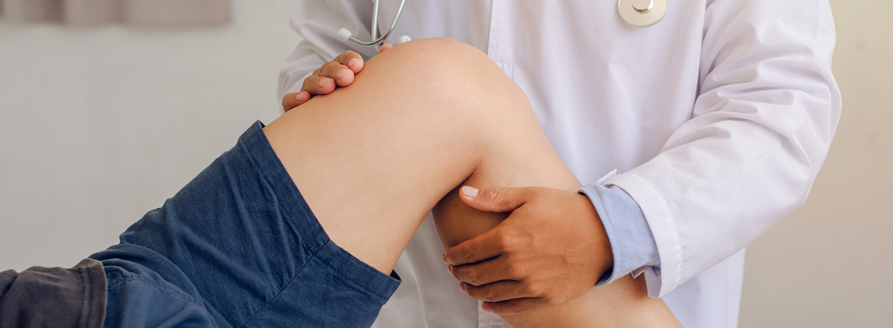
- General anesthesia in the operating room may be required for patients with dislocations that are irreducible by closed means and for those with significant associated fractures, central dislocation, or associated neurovascular injury. (medscape.com)
- For patient education resources, see the Breaks, Fractures, and Dislocations Center and Sports Injury Center, as well as Total Hip Replacement. (medscape.com)
- These include strains, bursitis, fractures and hip disloaction. (coloradospringsorthopedicnews.com)
- After reduction, CT is needed in all native hips to identify acetabular and femoral head fractures and intra-articular debris or loose bodies (bone or cartilage fragments). (msdmanuals.com)
- Potential complications of anterior hip dislocations are neurovascular injury to femoral vessels or acetabular fractures. (jocr.co.in)
- Anterior dislocations usually lead to fractures of the anterior acetabular wall or femoral head fractures or neurovascular injuries of the femoral vessels. (jocr.co.in)
- The annual incidence of hip fractures declined in most countries from 2005 to 2018, but this rate is projected to roughly double by 2050, according to a new study of 19 countries/regions. (medscape.com)
- The predicted increase in hip fractures is being driven by the aging population, with the population of those age 85 and older projected to increase 4.5-fold from 2010 to 2050, they note. (medscape.com)
- The researchers conclude that "larger and more collaborative efforts among healthcare providers, policymakers, and patients are needed to prevent hip fractures and improve the treatment gap and post-fracture care, especially in men and the oldest old. (medscape.com)
- The takeaways from the study are that "a greater effort on fracture prevention should be made to avoid the continuous increase in the number of hip fractures," he said. (medscape.com)
- Thus, more attention should be paid to preventing and treating hip fractures in men. (medscape.com)
- The greater increase in the projected number of hip fractures in men than in women "could be [because] osteoporosis is commonly perceived as a 'woman's disease,'" he speculated. (medscape.com)
- Also invited to comment, Peter R. Ebeling, MD, outgoing president of the ASBMR, said that the projected doubling of hip fractures "is likely mainly due to aging of the population, with increasing lifespan for males in particular. (medscape.com)
- More targeted screening for osteoporosis would help," he said, "as would treating patients for it following other minimal trauma fractures (vertebral, distal radius, and humerus, etc) since if left untreated about 50% of these patients will have hip fractures later in life. (medscape.com)
Femoral22
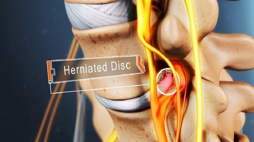
- Specifically it is when the ball-shaped head of the femur (femoral head) separates from its cup-shaped socket in the hip bone, known as the acetabulum. (wikipedia.org)
- Hip dislocations are classified by fracture association and by the positioning of the dislocated femoral head. (wikipedia.org)
- Hip dislocations are a medical emergency, requiring prompt placement of the femoral head back into the acetabulum (reduction). (wikipedia.org)
- This reduction of the femoral head back into the hip socket is typically done under sedation and without surgery, through maneuvers including traction on the thighbone in line with the dislocation. (wikipedia.org)
- In addition, hip dislocations are classified depending on the location of the head of the femur as follows: Posterior dislocations is when the femoral head lies posteriorly after dislocation. (wikipedia.org)
- Anterior dislocations is when the femoral head lies anteriorly after dislocation. (wikipedia.org)
- To note, Central dislocation is an outdated term for displacement of the femoral head towards the body's center into a fractured acetabulum and is no longer used. (wikipedia.org)
- The hip joint includes the articulation of the spherical femoral head (of femur) and the concave acetabulum (of pelvis). (wikipedia.org)
- The hip joint is based on the articulation of the femoral head and the acetabulum of the pelvis, and it is a synovial ball-and-socket type joint. (medscape.com)
- The fresh radiograph showed dislocated right hip with changes suggestive of AVN of femoral head. (sphdelhi.org)
- Hip dislocations are commonly classified according to the direction of dislocation of the femoral head, either anterior or posterior, and are treated with specific techniques for reduction. (ochsnerjournal.org)
- Anterior hip dislocation is commonly reduced by inline traction and external rotation, with an assistant pushing on the femoral head or pulling the femur laterally to assist reduction. (ochsnerjournal.org)
- 3 An understanding of the vasculature is important because trauma to the hip can displace the femoral head and interrupt the blood supply, leading to avascular necrosis (AVN). (ochsnerjournal.org)
- When there is a hip dislocation, the femoral head is pushed either backward out of the socket, or forward. (coloradospringsorthopedicnews.com)
- During this period, the joint is poorly supported and the femoral head can work its way free of the socket, leading to dislocation. (news-medical.net)
- The risk of hip dislocation is reduced when less tissue is cut, if cut tissue is repaired or if the femoral head prosthesis has a large diameter. (news-medical.net)
- The hip socket is called the acetabulum and forms a deep cup that surrounds the ball of the upper thighbone, called the femoral head . (choicephysicaltherapyri.com)
- The femoral head can dislocate either backward (posterior dislocation) or forward (anterior dislocation). (krisalden.com)
- In a normal hip, the head of the femur (thighbone) fits well into the socket (acetabulum) whereas in hip dysplasia, the socket and femoral head are not congruent because of their abnormal development. (bostonssc.com)
- The aim of treatment is to keep the femoral head in good contact with the acetabulum so that the hip can develop normally. (bostonssc.com)
- Designed for proximal femoral elevation in total hip replacement or in other surgery with a similar need for bone manipulation. (innomed.net)
- Congenital dislocation of the hip generally includes subluxation of the femoral head, acetabular dysplasia, and complete dislocation of the femoral head from the true acetabulum. (bvsalud.org)
Dysplasia16
- Hip dislocations can also occur following a hip replacement or from a developmental abnormality known as hip dysplasia. (wikipedia.org)
- Right hip dysplasia with luxation and secondary osteoarthritis. (radiopaedia.org)
- This case illustrates long standing untreated developmental dysplasia of the hip with a pseudoarticulation with the ilum. (radiopaedia.org)
- Acetabular dysplasia is absent in early stages of dislocation. (udel.edu)
- Background: Despite early recognition and appropriate treatment of congenital dislocation of the hip, there are a number of cases that subsequently require further surgery to prevent progressive dysplasia, instability, and eventual early osteoarthritis. (soton.ac.uk)
- It's essentially dislocated (severe hip dysplasia, per my reading perhaps better described now as congenital hip disease, specifically high congenital dislocation) and being held in place by scar tissue as I understand it, with other structure-y issues compounding this. (kerriontheprairies.com)
- Developmental dysplasia of the hip (DDH) or hip dysplasia is a condition that is seen in infants and young children because of developmental problems in the hip joint. (bostonssc.com)
- The exact cause for hip dysplasia is not known. (bostonssc.com)
- Developmental dysplasia of the hip is another name attributed to this condition. (epainassist.com)
- 13- Aronsson D d, et al, Developmental dysplasia of the hip. (ac.ir)
- Ultrasound screening for hips at risk in developmental dysplasia. (ac.ir)
- developmental dysplasia of the hip in the U.K. Arch Dis Child. (ac.ir)
- This article describes the use of a trochanteric girdle on a child with acetabular dysplasia to prevent hip displacement during weight-bearing. (easystand.com)
- The patient was a boy 12 years of age with hypotonic athetosis and such severe acetabular dysplasia that his hips dislocated laterally with the slightest adduction beyond neutral. (easystand.com)
- Hip dysplasia is usually progressive and may result in a significant decrease in the range of motion. (medscape.com)
- Genu valgum with patellar dislocation may occur in patients with diastrophic dysplasia. (medscape.com)
Traumatic11
- Traumatic dislocations occurs most commonly in those 16 to 40 years old. (wikipedia.org)
- Hip dislocations are either anterior or posterior, with posterior hip dislocations comprising the majority of traumatic dislocations. (medscape.com)
- Traumatic dislocation and fracture-dislocation of the hip: a long-term follow-up study. (medscape.com)
- Arthroscopic and imaging findings after traumatic hip dislocation in patients younger than 25 years of age. (medscape.com)
- Simultaneous anterior and posterior traumatic dislocation of both hips is even more unusual. (bmj.com)
- A case is reported of asymmetrical bilateral traumatic hip dislocation without an associated fracture of the pelvis or femur occurring in a young adult with no previous history of hip abnormality or ligamentous laxity. (bmj.com)
- A traumatic hip dislocation occurs when the head of the thighbone (femur) is forced out of its socket in the hip bone (pelvis). (coloradospringsorthopedicnews.com)
- Traumatic hip dislocation constitutes a true orthopedic emergency, is a relatively rare occurrence in the pediatric population, and may be accompanied by minimal trauma. (qxmd.com)
- A case report involving traumatic hip dislocation in a 5-yr-old boy is described followed by a comparative review of the pediatric and adult literature with current recommendations. (qxmd.com)
- Traumatic hip dislocation is an uncommon injury in children. (sogacot.org)
- in adolescents, greater force is required to produce a traumatic complete hip dislocation. (sogacot.org)
Pelvis16
- A hip dislocation is when the thighbone (femur) separates from the hip bone (pelvis). (wikipedia.org)
- The joint of the femur and pelvis (hip joint) is very stable, secured by both bony and soft-tissue constraints. (wikipedia.org)
- Hip dislocations occur when the ball-shaped head of the thighbone (femur) pops out of the rounded socket of the hip bone (pelvis). (msdmanuals.com)
- For example, when a hip dislocation results from a fall or an athletic injury, the pelvis, knee, or legs may be fractured, and the back or head may be injured. (msdmanuals.com)
- The AP pelvis plain radiograph was used to measure the height of dislocation, as described by Tonnis, and monitor Acetabular index, and ossific nucleus width and height postreduction. (soton.ac.uk)
- CT scan of pelvis with 3D reconstruction was done and revealed posterior acetabular wall defect with dislocated hip. (sphdelhi.org)
- Your hip is the joint where your thigh bone meets your pelvis bone. (coloradospringsorthopedicnews.com)
- Hips are called ball-and-socket joints because the ball-like top of your femur moves within a cup-like space in your pelvis. (coloradospringsorthopedicnews.com)
- Do not move a person with an injured hip, pelvis, or upper leg unless it is absolutely necessary. (limamemorial.org)
- If your horse develops sudden hindlimb lameness or pain over the back, hip, or croup, he might have a pelvis problem that warrants an examination from your veterinarian. (thehorse.com)
- The patient's hips are held down by a sheet or by a second practitioner (to provide countertraction to the pelvis). (msdmanuals.com)
- The hip joint is one of the body's largest weight-bearing joints and is the point where the thighbone (femur) and pelvis (acetabulum) join. (hamiltonhipandknee.com)
- A pelvic X-ray is a type of X-ray that captures detailed images of the pelvic region, which includes the pelvis, hips, and upper legs. (medicalnewstoday.com)
- Doctors use pelvic X-rays to find the cause of pain, swelling, or abnormalities in the pelvic region, which includes the pelvis, hips, and upper legs. (medicalnewstoday.com)
- X-rays of the neck, spine, pelvis (hips) and legs will be ordered to diagnose cartilage hair hypoplasia and/or related conditions, like scoliosis and kyphosis. (hopkinsmedicine.org)
- If you have a deep hip socket with a large angle of inclination, you pelvis bones likely run into each other, preventing a wide breaststroke kick. (swimmingworldmagazine.com)
Risk of dislocation5
- A newer type of artificial hip known as a "modular dual mobility" implant could reduce the risk of dislocation in patients who need a revision surgery, according to a new study from Hospital for Special Surgery (HSS). (rehabpub.com)
- Our study found that the newer technology with modular dual mobility components offered increased stability, lowering the risk of dislocation, without compromising hip range of motion in patients having a revision surgery," says Dr Geoffrey Westrich, director of research of the Adult Reconstruction and Joint Replacement Service at HSS, who conducted the study. (rehabpub.com)
- Patients can decrease their risk of dislocation by avoiding certain postures during the first several months following surgery. (news-medical.net)
- Alcohol use in the few months following surgery is associated with an increased risk of dislocation. (news-medical.net)
- The W-Sitting position also pushes the hip range of motion to its limits, increasing the risk of dislocation or subluxation, both during the stretch and afterwards. (swimmingworldmagazine.com)
Fracture-dislocation2
- Reduction of the hip fracture-dislocation was attempted. (hindawi.com)
- He had a fracture dislocation of right hip which was missed initially by the primary surgeon. (sphdelhi.org)
Subluxation7
- the slight subluxation of the right hip was noticed. (udel.edu)
- The prevalence of hip subluxation and dislocation in cerebral palsy varies from 2.6 to 28 per cent (25-30% in large series). (udel.edu)
- With persistence of subluxation or dislocation, posterior and superior insufficiency of the acetabulum will develop. (udel.edu)
- Physical therapy is crucial for those recovering from a hip dislocation or subluxation, as it can speed up healing and reduce the risk of re-injury. (lunara.llc)
- When you visit a physical therapist for treatment following a hip dislocation or subluxation, they will evaluate your condition using a range of tests like strength and motion evaluations. (lunara.llc)
- For those who have experienced a hip dislocation or subluxation, physical therapy is crucial to recovery. (lunara.llc)
- Patients may present with hip subluxation. (medscape.com)
Injuries18
- It often takes 2-3 months for a dislocated hip to fully heal, and it can take even longer depending on associated injuries such as fracture. (wikipedia.org)
- Nerve injuries also can accompany dislocations, necessitating careful neurovascular examination. (wikipedia.org)
- Deformity is also present, which is based on concomitant injuries and the type of dislocation: For posterior dislocation, the affected limb will be in a position of flexion, adduction, and internal rotation. (wikipedia.org)
- [ 6 ] An estimated 3% of all football injuries involve hip fracture or dislocation. (medscape.com)
- People with a hip dislocation often have other injuries. (msdmanuals.com)
- The force that causes the hip dislocation often also causes other injuries. (msdmanuals.com)
- However, playing sports, running, overuse or falling can all sometimes lead to hip injuries. (coloradospringsorthopedicnews.com)
- Certain diseases also lead to hip injuries or problems. (coloradospringsorthopedicnews.com)
- Car collisions and falls from significant heights are common causes and, as a result, other injuries like broken bones often occur with the dislocation. (coloradospringsorthopedicnews.com)
- Because hip dislocations often occur with additional injuries, your doctor will complete a thorough physical evaluation. (coloradospringsorthopedicnews.com)
- If there are no other injuries associated with the dislocated hip, the doctor will administer an anesthetic or a sedative and manipulate the bones back into their proper position. (coloradospringsorthopedicnews.com)
- Recovery depends on the person and severity of the dislocation and injuries associated. (coloradospringsorthopedicnews.com)
- Elbow dislocations are common elbow injuries that can be classified as simple or complex depending on whether or not surrounding structures have been injured. (medicahospitals.in)
- Hip dislocation can be caused by injuries from motor vehicle accidents or severe falls. (krisalden.com)
- After patella dislocation, the incidence of acute osteochondral or chondral injuries is up to 95% after initial patella dislocation [ 5 ]. (springer.com)
- Hip dislocations are serious injuries as hip joint is an extremely stable joint. (jocr.co.in)
- Anterior dislocations of the hip joint are unusual injuries. (jocr.co.in)
- Anterior dislocations are extremely rare injuries. (jocr.co.in)
Occur9
- These dislocations usually occur when the knee and hip are flexed (as when sitting down) and a great force hits the knee. (msdmanuals.com)
- Hip dislocations can also occur when people fall from a high place (such as a ladder) or play a contact sport (such as football and rugby). (msdmanuals.com)
- In rare cases, a hip dislocation can occur during impact sports like football. (coloradospringsorthopedicnews.com)
- Dislocation of an artificial hip is uncommon but may occur within the first three months after surgery. (choicephysicaltherapyri.com)
- Dislocation of the ball-and-socket joint on the hip will occur sometimes. (epainassist.com)
- Complete dislocation sometimes does occur. (epainassist.com)
- Transient hip dislocation with spontaneous but incomplete reduction is a diagnostic pitfall that can occur in adolescents. (sogacot.org)
- About 75% of anterior dislocations occur in males [1, 2]. (jocr.co.in)
- Excessive valgus with lateral dislocation of the patella may occur. (medscape.com)
Femur10
- Major ligaments conferring stability to the hip joint include the iliofemoral ligament, the ischiofemoral ligament, the pubofemoral ligament, and the ligament of the head of the femur. (wikipedia.org)
- Newton E. Femur and hip. (medscape.com)
- 32 mm (p = 0.390 for neck of femur THA and p = 0.451 for elective THA) and a single mobility design (p = 0.494) both produced a higher dislocation rate, although this was not statistically significant. (scielo.org.za)
- Background and purpose - Operative treatment of hip displacement in cerebral palsy (CP) includes adductor-psoas lengthening (APL) or varus derotation osteotomy (VDRO) of the proximal femur, sometimes combined with pelvic osteotomy. (lu.se)
- The head of the femur is generally the "ball" in the ball and socket joint of the hip. (kerriontheprairies.com)
- Hip dislocation occurs when the head of the femur moves out of the socket. (krisalden.com)
- A strap or brace is used to hold down the patient's hips (providing counterpressure to the vertical traction of the femur). (msdmanuals.com)
- The femur (thighbone) partially or completely slips out of the hip socket leading to dislocation at the hip joint. (bostonssc.com)
- Specifically, if a swimmer performs breaststroke properly (from the hips) during maturation, their hip will under go anteversion, moving the femur (upper thigh) forward. (swimmingworldmagazine.com)
- Designed to elevate the proximal femur during total hip or hemi-arthroplasty surgery. (innomed.net)
Joint38
- The important role of polygenic acetabular configuration and monogenic joint laxity has again been proved in the aetiology of congenital dislocation of the hip. (bmj.com)
- Hip joint may be painful. (udel.edu)
- Overview of Dislocations A dislocation is complete separation of the bones that form a joint. (msdmanuals.com)
- The hip is a ball-and-socket joint that is inherently stable because of its bony geometry and strong ligaments, allowing it to resist significant increases in mechanical stress. (ochsnerjournal.org)
- Congenital dislocations result from the physiologic position of the fetus in utero pressed against the abdominal wall of the mother, with the additional component of the posterior force acting against a dysplastic hip joint in flexion. (ochsnerjournal.org)
- 4 In the typical scenario, the patient is sitting with the hip in flexion, and upon impact, the thigh hits the dashboard, sending a posteriorly directed force to the joint and causing a posterior dislocation. (ochsnerjournal.org)
- Other common conditions that can lead to postoperative dislocations include laxity or soft-tissue incompetence surrounding the hip joint (ie, revision), incorrect positioning of prosthetic components, and neuromuscular disorders (eg, Parkinson disease). (ochsnerjournal.org)
- The labrum forms a lining around the socket, creating a tight seal, helping to provide stability to the hip joint. (coloradospringsorthopedicnews.com)
- A dislocation is a separation of two ends of the bones where they meet at a joint. (limamemorial.org)
- Dislocations are usually caused by a sudden impact to the joint. (limamemorial.org)
- When the bones of a joint are knocked out of place, it is known as dislocation. (medicahospitals.in)
- When the bones in a joint get split or knocked out of their normal locations, it is called a dislocation. (medicahospitals.in)
- Dislocations can be excruciatingly painful and leave the affected joint unsteady or immobilized (unable to move). (medicahospitals.in)
- Shoulders can pop-off from the socket joint causing a dislocation of the shoulder. (medicahospitals.in)
- The hip joint becomes displaced from its socket in around 3% of cases. (news-medical.net)
- After a hip replacement procedure, it can take up to twelve weeks for the soft tissues around the joint to heal after they have been cut during surgery. (news-medical.net)
- Wear and tear of the artificial hip joint is another common complication of this procedure. (news-medical.net)
- Hip replacements patients are at a small risk of bacterial infection occurring at the artificial hip joint. (news-medical.net)
- Hip surgeries such as total joint replacement and hemiarthroplasty require the surgeon to open the hip joint capsule. (choicephysicaltherapyri.com)
- Which parts of the hip joint are affected by a dislocation? (choicephysicaltherapyri.com)
- The hip joint is one of the true ball-and-socket joints of the body. (choicephysicaltherapyri.com)
- When the surgeon opens the hip joint capsule on the front edge, the procedure is called an anterior approach . (choicephysicaltherapyri.com)
- The joint capsule and ligaments keep the ball joint centered in the hip. (choicephysicaltherapyri.com)
- Most patients will have an opportunity to work with a physical or occupational therapist before having hip joint surgery. (choicephysicaltherapyri.com)
- These exercises are designed to help improve the range of motion, strengthen muscles, and promote better balance and coordination around the hip joint. (lunara.llc)
- Strengthening and range-of-motion exercises, balance training, and core stabilization help restore stability to the hip joint and hasten your return to normal activity. (lunara.llc)
- The hip joint is a "ball and socket" joint. (krisalden.com)
- Physical therapy is vital in regaining the strength and mobility in your hip joint after treatment. (krisalden.com)
- A poorer prognosis is conferred by duration of dislocation for longer than 6 h, advanced skeletal maturity of the patient, severe joint injury, or multiple trauma in the affected patient. (qxmd.com)
- Any asymmetric widening of the hip joint warrants additional investigation. (sogacot.org)
- The reduction of the hip joint was confirmed with C-arm. (jocr.co.in)
- Joint laxity and hip rotation in normal children and in those with congenital dislocation of the hip. (ox.ac.uk)
- Joint laxity was more common in normal children with an internally centred arc of hip rotation than in normal children with a neutral or an externally rotated arc. (ox.ac.uk)
- The children with congenitally dislocated hips had significantly more joint laxity than did the control group and significantly more of them had an internally centred arc of hip rotation. (ox.ac.uk)
- The patient's hips subluxated proximally with weight-bearing or joint compression with only 20 degrees of abduction. (easystand.com)
- The labrum is the protective lining in the hip joint, acting as a rubber seal to help hold the ball hip bone in the socket. (swimmingworldmagazine.com)
- During a hip replacement operation, the surgeon removes damaged cartilage and bone from your hip joint and replaces them with new, man-made parts. (medlineplus.gov)
- Because a man-made hip is smaller than the original joint, the ball can come out of its socket. (medlineplus.gov)
Posterior hip8
- Right posterior hip dislocation in a young woman following a high-speed motor vehicle collision (MVC). (medscape.com)
- Several classification systems are used to describe posterior hip dislocations. (medscape.com)
- Some case series have found that most posterior hip dislocations are type 1. (medscape.com)
- MRI as a reliable and accurate method for assessment of posterior hip dislocation in children and adolescents without the risk of radiation exposure. (medscape.com)
- In most hip dislocations, the head of the thighbone is pushed backward-called a posterior hip dislocation. (msdmanuals.com)
- Posterior hip dislocations are the most common type and are reduced by placing longitudinal traction with internal rotation on the hip. (ochsnerjournal.org)
- How To Reduce a Posterior Hip Dislocation Most hip dislocations are posterior. (msdmanuals.com)
- Posterior hip dislocation in a five-year-old boy: a case report, review of the literature, and current recommendations. (qxmd.com)
Osteoarthritis1
- Early osteoarthritis on the left hip. (radiopaedia.org)
20231
- Retrieved on November 28, 2023 from https://www.news-medical.net/health/Hip-Replacement-Problems.aspx. (news-medical.net)
Cerebral palsy6
- Characteristics of children with hip displacement in cerebral palsy. (easystand.com)
- Hip dislocation in children with cerebral palsy (CP) is a common and severe problem. (easystand.com)
- To investigate the effects of a standing program on the range of motion (ROM) of hip abduction in children with spastic diplegic cerebral palsy. (easystand.com)
- This small difference was not clinically significant, but did demonstrate that it was possible to maintain hip abduction ROM in the spastic adductor muscles of children with cerebral palsy with a daily standing program during the children 's first 5 years of development. (easystand.com)
- Effects of the standing program with hip abduction on hip acetabular development in children with spastic diplegia cerebral palsy. (easystand.com)
- Avascular necrosis as a complication of the treatment of dislocation of the hip in children with cerebral palsy. (bvsalud.org)
Prevent hip dislocation1
- Trochanteric girdle to prevent hip dislocation in standing. (easystand.com)
Rate of dislocation3
- DISCUSSION: Acetabular cup positioning in patient-specific safe zones reduces the rate of dislocation after THA. (easychair.org)
- inproceedings{CAOS2019:Patient_Specific_Safe_Zones_Reduce, author = {Jonathan Vigdorchik and Zlatan Cizmic and David Novikov and Michael Bradley and Michael Miranda and David Watson and Douglas Dennis and Stefan Kreuzer}, title = {Patient-Specific "Safe Zones" Reduce the Rate of Dislocation after Total Hip Arthroplasty}, booktitle = {CAOS 2019. (easychair.org)
- We set out to determine the rate of dislocation and the need for another surgery following revision hip replacement using this implant and report on the functional outcomes. (rehabpub.com)
Diagnosis4
- Diagnosis is made by physical exam and plain X-rays of the hips. (wikipedia.org)
- However, serious morbidity can be associated with hip dislocations, making careful and expedient diagnosis and treatment important for the sports medicine physician. (medscape.com)
- Introduction: Dislocation displasia of the hip (DDH) is one of the most common congenital deformities and early physical examination is the best method for diagnosis. (ac.ir)
- Early diagnosis and treatment covgenital dislocation of the hip. (ac.ir)
Acetabulum3
- We report the case of an 18-year-old female who sustained a left acetabulum fracture with a concurrent hip dislocation at 35 weeks' gestation following a motor vehicle accident. (hindawi.com)
- We report a case of an 18-year-old female who was involved in an MVA and subsequently sustained a left acetabulum fracture with concomitant hip dislocation as well as a right distal radius fracture. (hindawi.com)
- Radiographs were obtained demonstrating a left-sided, displaced transverse posterior wall acetabulum fracture with concomitant hip dislocation as well as a right-sided, volarly displaced distal radius and ulnar styloid fracture (Figures 1 and 2 ). (hindawi.com)
Dislocate4
- This leads to an adducted, flexed, internally rotated hip which tends to dislocate posteriorly. (udel.edu)
- In older people, much less force is needed to dislocate a hip. (msdmanuals.com)
- It typically takes a major force to dislocate the hip. (coloradospringsorthopedicnews.com)
- But, being in an accident or receiving any type of trauma or force that can force your hip bones to dislocate causes hip dislocation. (medicahospitals.in)
Severe6
- Typically, people with hip dislocations present with severe pain and an inability to move the affected leg. (wikipedia.org)
- Most hip dislocations are posterior and result from severe posteriorly directed force to the knee while the knee and hip are flexed (eg, against a car dashboard). (msdmanuals.com)
- DDH can be mild or severe and can affect one or both hips. (bostonssc.com)
- This is necessary in recurrent patella dislocations and in particular cases of first patella dislocations such as patients with severe anatomical risk factors [ 15 ]. (springer.com)
- Hip replacement is surgery for people with severe hip damage. (medlineplus.gov)
- Certain occupations put an increased amount of pressure on the knee, hip, and back joints that can cause severe problems later in life. (lu.se)
Bone3
- Now he has undergone right uncemented Total Hip Replacement with reconstruction of acetabular defect using bone graft fixed with screws and trabecular metal continuum shell. (sphdelhi.org)
- What is bone dislocation? (medicahospitals.in)
- This is the most common complication of hip replacement and occurs when the shaft of the prosthesis becomes loose in the socket of the thigh bone or the bone surrounding the implant starts to thin. (news-medical.net)
Flexion5
- The degree of flexion depends on whether it is a superior or inferior dislocation, with the former resulting in hip extension and the latter, hip flexion. (wikipedia.org)
- Recent popliteal angles were 25 bilaterally, Thomas test 15 bilaterally, and hip flexion and abduction were all normal. (udel.edu)
- The patient may have the progressive limitation of hip abduction and progressive flexion contracture. (udel.edu)
- A Pavlik harness may be used to keep the hip in flexion and abduction may be advised. (bostonssc.com)
- The hips maintain a persistent flexion contracture. (medscape.com)
Complications3
- Patients with hip dislocations must receive careful diagnostic workup, and the treating physician must be well versed in the different ways to treat the injury and possible complications. (ochsnerjournal.org)
- As with all major surgeries, a hip replacement procedure is associated with several complications and risks, some of which may even be life threatening. (news-medical.net)
- What are the Risks and Complications of Computer-Navigated Total Hip Replacement? (hamiltonhipandknee.com)
Anterior dislocation4
- For anterior dislocation, the affected limb will be in a position of abduction and external rotation. (wikipedia.org)
- We report a rare late complication of Morel Lavallee syndrome occurring three weeks after an anterior dislocation of the hip in a 43 year old male. (jocr.co.in)
- We report a rare case of Morel Lavallee lesion complicating an anterior dislocation of the hip. (jocr.co.in)
- X-ray revealed an anterior dislocation of the hip. (jocr.co.in)
Pulled elbow1
- Nursemaid's elbow, or pulled elbow, is a partial dislocation that is common in toddlers. (limamemorial.org)
Patellar Dislocation1
- After initial patellar dislocation, the MPFL is injured in 94% of the cases [ 11 ]. (springer.com)
Uncommon2
- Hip dislocations are relatively uncommon during athletic events. (medscape.com)
- Anterior dislocations are uncommon. (jocr.co.in)
Socket4
- With the dual mobility hip, a large polyethylene plastic head fits inside a polished metal hip socket component, and an additional smaller metal or ceramic head is snap-fit within the polyethylene head, explains a media release from Hospital for Special Surgery. (rehabpub.com)
- When these soft tissues are cut during hip surgery, there is a greater risk for the ball to be forced out of the socket and dislocated after surgery while the soft tissues of the hip heal. (choicephysicaltherapyri.com)
- Putting the hip back in the socket will probably require medication given intravenously to relax the hip muscles and allow your surgeon to put the hip back into place. (choicephysicaltherapyri.com)
- A deep hip socket prevents the amount of hip internal range of motion and breaststroke kick width. (swimmingworldmagazine.com)
Patients12
- Hip dislocations in younger individuals are relatively rare, with only 5% of cases occurring in patients younger than 14 years. (medscape.com)
- The purposes of this study were to: (1) compare pre-operative acetabular cup parameters using this novel dynamic imaging sequence to the LSZ, and (2) describe rates of dislocation in patients whose pre-operative acetabular cup parameters were determined using dynamic imaging sequences. (easychair.org)
- In the study, Westrich and his colleagues collected clinical, radiographic and patient reported-outcome information on 370 patients (average age: 65.8 years) who underwent revision hip replacement with the dual mobility implant between April 2011 and April 2017. (rehabpub.com)
- In this retrospective cohort review, files and radiographs of 543 patients were reviewed for dislocation during the first year after primary THA. (scielo.org.za)
- All dislocations were verified with use of ultrasound or radiography prior to the initiation of treatment, and patients were followed prospectively for a minimum of 18 months. (orthobullets.com)
- Patients that were in serious car or falling accidents that resulted in hip damage may need a hip support brace. (orthomen.com)
- Patients should urgently seek medical attention if they develop symptoms such as fever, chills, persistent hip pain or inflammation/discharge at the surgery site. (news-medical.net)
- Patients follow special precautions after surgery about which hip positions and movements need to be avoided to keep the hip from dislocating. (choicephysicaltherapyri.com)
- However, patients sometimes require emergency surgery, such as after a hip fracture, and are not able to have preoperative therapy instruction. (choicephysicaltherapyri.com)
- Patients with recurrent patella dislocation requiring isolated MPFL reconstruction will be recruited and randomized to the dynamic or static reconstruction technique. (springer.com)
- Morel-Lavellee Syndrome is an extremely rare complication of anterior hip dislocation and hence such patients should be closely followed up as it manifests as a late complication. (jocr.co.in)
- Almost 35% of patients who suffer from anterior dislocations are in their third decade of life [1, 2]. (jocr.co.in)
Occurs4
- Dislocation of the hip occurs more frequently in the non-ambulatory patient with the spastic quadriplegia. (udel.edu)
- Dislocation of the hip is a well-described event that occurs in conjunction with high-energy trauma or postoperatively after total hip replacement. (ochsnerjournal.org)
- 4 , 11 The most common acquired dislocation is hip dislocation that occurs within the first 3 months following total hip replacement. (ochsnerjournal.org)
- Hip displacement in CP often occurs already at 2-3 years of age. (easystand.com)
Reduction5
- If hip dislocation is the only injury or the most serious injury, doctors put the hip back in place (called reduction) as soon as possible. (msdmanuals.com)
- Before hip reduction, people are given a sedative (usually by vein), pain reliever, and muscle relaxant, but they may remain conscious. (msdmanuals.com)
- Bolland, Benjamin J. , Wahed, Abdul , Al-Hallao, Sariyah , Culliford, D.J. and Clarke, Nicholas M.P. (2010) Late reduction in congenital dislocation of the hip and the need for secondary surgery: radiologic predictors and confounding variables. (soton.ac.uk)
- Bigelow first described closed treatment of a dislocated hip in 1870, and since then many reduction techniques have been proposed. (ochsnerjournal.org)
- Urgent reduction of the hip within 6 hours of injury reduces the risk of osteonecrosis. (sogacot.org)
Instability2
- however, a postreduction CT scan demonstrated residual dislocation due to instability imparted by a large posterior wall fracture fragment. (hindawi.com)
- Relibility of Barlow and ortolani tests for neonatal hip instability. (ac.ir)
Disorders1
- Congenital disorders or suffering from certain medical conditions as a child can increase the risk of developing OA later on in life, especially of the hip. (lu.se)
Posteriorly1
- A posteriorly positioned head is the most common dislocation type. (wikipedia.org)
Patient's2
- the patient's hips are held down by a sheet or belt, and the dislocated hip is flexed. (msdmanuals.com)
- the practitioner stands on the side of the affected hip and faces the patient's feet. (msdmanuals.com)
Displacement8
- We used the migration percentage (MP) to compare the development of hip displacement after APL and VDRO. (lu.se)
- The 54 hips that underwent a 2nd surgery with APL or VDRO because of redisplacement had a higher displacement rate preoperatively and continued displacement postoperatively but at a slower rate. (lu.se)
- The 94 hips that underwent VDRO showed an instantaneous decrease in MP postoperatively followed by a rate of increasing displacement. (lu.se)
- The 15 hips that underwent a 2nd VDRO had a higher rate of displacement both pre- and postoperatively. (lu.se)
- Interpretation - After APL, hip displacement either decreases or continues to increase but at a slower rate. (lu.se)
- The aim of this study was to analyse the characteristics of children with CP who develop hip displacement , in order to optimise a hip surveillance programme. (easystand.com)
- The risk of hip displacement varied according to CP-subtype, from 0% in children with pure ataxia to 79% in children with spastic tetraplegia. (easystand.com)
- The risk of hip displacement varies according to both CP-subtype and GMFCS. (easystand.com)
Total hip replace4
- Acquired hip dislocations are either native dislocations or dislocations after total hip replacement ( Figure 1 ). (ochsnerjournal.org)
- For a successful total hip replacement, accurate positioning of the implants is crucial to accomplish a good clinical outcome. (hamiltonhipandknee.com)
- Computer-navigated total hip replacement is an advanced technology developed to provide more accurate positioning of an implant. (hamiltonhipandknee.com)
- What are the Advantages of Computer-Navigated Total Hip Replacement? (hamiltonhipandknee.com)
Orthopaedic2
- If you have suffered an injury to your hip or think you may have dislocated it, request an appointment with the Colorado Center for Orthopaedic Excellence at (719) 623-1050 for immediate treatment. (coloradospringsorthopedicnews.com)
- Individuals with cartilage hair hypoplasia may experience additional orthopaedic conditions of the hip, spine and limbs. (hopkinsmedicine.org)
Motor vehicle c1
- With that, dislocation would require significant force which typically results from significant trauma such as from a motor vehicle collision or from a fall from elevation. (wikipedia.org)
Inability to move1
- The common symptoms of hip dislocation include pain, inability to move your legs and numbness along the foot or ankle. (krisalden.com)
Pelvic2
- There is a debate about the temporal relationship between scoliosis, pelvic obliquity and hip dislocation. (udel.edu)
- Results: After 134 OR's, 24 hips (19%, 95% CI: 16-23%) later required a pelvic osteotomy compared with 59 out of 104 hips (58%, 95% CI: 49-68%) in the CR cohort. (soton.ac.uk)
Common4
- McNamara R. Management of common dislocation: hip dislocations. (medscape.com)
- Dislocations are very common. (medicahospitals.in)
- It is more common in girls and usually affects the left hip. (bostonssc.com)
- The most common problem after surgery is hip dislocation. (medlineplus.gov)
Treatment13
- Methods: All cases of congenital dislocation of the hip that presented late or had failed conservative treatment with subsequent late OR versus CR, that were carried out during 1988 to 2003, by the lead surgeon were included. (soton.ac.uk)
- Evaluation of Brace Treatment for Infant Hip Dislocation in a Prospective Cohort: Defining the Success Rate and Variables Associated with Failure. (orthobullets.com)
- The purpose of the current study was to evaluate the success rate for brace use in the treatment of infant hip dislocation in an international, multicenter, prospective cohort, and to identify the variables associated with brace failure. (orthobullets.com)
- Successful treatment was defined as the use of a brace that resulted in a clinically and radiographically reduced hip, without surgical intervention. (orthobullets.com)
- Brace treatment was successful in 162 (79%) of the 204 dislocated hips in this series. (orthobullets.com)
- These data provide valuable information for patient families and their providers regarding the important variables that influence successful brace treatment for dislocated hips in infants. (orthobullets.com)
- A hip dislocation is a very painful and serious medical emergency, where immediate treatment is necessary. (coloradospringsorthopedicnews.com)
- The dislocation can be avoided, by screening and preventive treatment of children with hips at risk. (easystand.com)
- In addition, rates of use of osteoporosis drugs 1 year after a hip fracture were less than 50%, with less treatment in men. (medscape.com)
- In addition, "although initiation of anti-osteoporosis medication after hip fracture is recommended in international guidelines, the 1-year treatment rate [was] well below 50% in most of the countries and regions studied. (medscape.com)
- Invited to comment, Juliet Compston, MD, who selected the study as one of the top clinical science highlight abstracts at the ASBMR meeting, agrees that "there is substantial room for improvement" in osteoporosis treatment rates following a hip fracture "in all the regions covered by the study. (medscape.com)
- In addition," she continues, "the wide variations in treatment rates can provide important lessons about the most effective models of care for people who sustain a hip fracture, for example, fracture liaison services. (medscape.com)
- Some countries may be doing better because they have health quality standards for hip fracture (eg, surgery within 24 hours, investigation and treatment for osteoporosis). (medscape.com)
Arthroplasty1
- Total hip arthroplasty (THA) is one of the most performed and most researched procedures worldwide, and there is an ever-growing demand for THA in an already resource-constrained system in South Africa. (scielo.org.za)
Procedure2
- Doctors can usually put the hip back in place without surgery, typically after giving the person drugs to make the procedure more tolerable. (msdmanuals.com)
- Hip Revision is a procedure to take out your old hip replacement and put in a new one. (nuffieldhealth.com)
Abduction5
- Hip abduction was 50 degrees and popliteal angle was 10 degrees bilaterally. (udel.edu)
- Hip abduction was 60 degrees bilaterally. (udel.edu)
- Marked limitation of hip abduction. (udel.edu)
- Hip abduction ROM was goniometrically assessed at baseline and at 5 years. (easystand.com)
- Baseline hip abduction was 42° at baseline and 43° at 5 years. (easystand.com)
Thighbone1
- Usually, hips are dislocated when a great force hits a bent knee and pushes the head of the thighbone backward-for example, when a knee hits the dashboard in a car accident. (msdmanuals.com)
Painful2
- The hip is very painful, and usually people cannot move their leg. (msdmanuals.com)
- Hip dislocations are very painful. (msdmanuals.com)
Bilateral2
Surgeon3
- In cases in which hip dislocation is the only injury, an orthopedic surgeon can often diagnose it simply by looking at the position of the leg. (coloradospringsorthopedicnews.com)
- Hip replacement through computer navigation provides information and guidance to the surgeon for precise positioning of implants. (hamiltonhipandknee.com)
- During your operation your surgeon will remove your old hip prothesis and any cement that might have been used to hold it in place. (nuffieldhealth.com)
Adolescents1
- A study by Moran et al found that male athletes sustained more sports-related hip dislocations than female athletes, with the greatest number of hip dislocations occurring in adolescents aged 15 to 19 years. (medscape.com)