Hypokinesia
Parkinson Disease
Progressive bradykinesia and hypokinesia of ocular pursuit in Parkinson's disease. (1/101)
OBJECTIVES: Patients with Parkinson's disease characteristically have difficulty in sustaining repetitive motor actions. The purpose of this study was to establish if parkinsonian difficulty with sustaining repetitive limb movements also applies to smooth ocular pursuit and to identify any pursuit abnormalities characteristic of Parkinson's disease. METHODS: Ocular pursuit in seven patients with moderate to severe bradykinesia predominant Parkinson's disease was compared with seven age matched controls. Predictive and non-predictive pursuit of constant velocity target ramps were examined. Subjects pursued intermittently illuminated 40(0)/s ramps sweeping to the left or right with an exposure duration of 480 ms and average interval of 1.728 s between presentations. To examine for any temporal changes in peak eye velocity, eye displacement or anticipatory smooth pursuit the 124 s duration of each record was divided into four epochs (E1, E2, E3, E4), each lasting 31 s and containing 18 ramp stimuli. Three test conditions were examined in each subject: predictive (PRD1), non-predictive (NPD), and predictive (PRD2) in that order. RESULTS: Both patients and controls initiated appropriate anticipatory pursuit before target onset in the PRD1 and PRD2 conditions that enhanced the response compared with the NPD condition. The distinctive findings in patients with Parkinson's disease were a reduction in response magnitude compared with controls and a progressive decline of response with stimulus repetition. The deficits were explained on the basis of easy fatiguability in Parkinson's disease. CONCLUSIONS: Ocular pursuit shows distinct anticipatory movements in Parkinson's disease but peak velocity and displacement are reduced and progressively decline with repetition as found with limb movements. (+info)Bradykinesia akinesia inco-ordination test (BRAIN TEST): an objective computerised assessment of upper limb motor function. (2/101)
OBJECTIVES: A simple and rapid computerised keyboard test, based on the alternating finger tapping test, has been developed to quantify upper limb motor function. The test generates several variables: (1) kinesia score: the number of keystrokes in 60 seconds; (2) akinesia time: cumulative time that keys are depressed; (3) dysmetria score: a weighted index calculated using the number of incorrectly hit keys corrected for speed; (4) incoordination score: a measure of rhythmicity which corresponds to the variance of the time interval between keystrokes. METHODS: The BRAIN TEST(Copyright ) was assessed on 35 patients with idiopathic Parkinson's disease, 12 patients with cerebellar dysfunction, and 27 normal control subjects. RESULTS: The mean kinesia scores of patients with Parkinson's disease or cerebellar dysfunction were significantly slower than normal controls (Parkinson's disease=107 (SD 28) keys/min v cerebellar dysfunction=86+/- (SD 28) v normal controls=182 (SD 26), p<0.001) and correlated with the UPDRS (r =-0.69, p<0.001). The akinesia time is very insensitive and was only abnormal in patients with severe parkinsonism. The median dysmetria (cerebellar dysfunction=13.8 v Parkinson's disease=6.1 v normal controls=4.2, p=0.002) and inco-ordination scores (cerebellar dysfunction=5.12 v Parkinson's disease=0.84 v normal controls=0.15, p=0.002) were significantly higher in patients with cerebellar dysfunction, in whom the dysmetria score correlated with a cerebellar disease rating scale (r=0.64, p=0.02). CONCLUSION: The BRAIN TEST(Copyright ) provides a simple, rapid, and objective assessment of upper limb motor function. It assesses speed, accuracy, and rhythmicity of upper limb movements regardless of their physiological basis. The results of the test correlate well with clinical rating scales in Parkinson's disease and cerebellar dysfunction. The BRAIN test will be useful in clinical studies. It can be downloaded from the Internet (). (+info)Biochemical and molecular genetic characteristics of the severe form of tyrosine hydroxylase deficiency. (3/101)
BACKGROUND: Tyrosine hydroxylase (TH) catalyzes the rate-limiting step in the biosynthesis of the catecholamines dopamine, norepinephrine, and epinephrine. Recently, mutations were identified in cases of autosomal recessive dopa-responsive dystonia and infantile parkinsonism. We describe a patient with severe symptoms and a new missense mutation in TH. METHODS: Relevant metabolites in urine and cerebrospinal fluid were measured by HPLC with fluorometric and electrochemical detection. All exons of the TH gene were amplified by PCR and subjected to single-strand conformation polymorphism analysis. Amplimers displaying aberrant migration patterns were analyzed by DNA sequence analysis. RESULTS: The patient presented with severe axial hypotonia, hypokinesia, reduced facial mimicry, ptosis, and oculogyric crises from infancy. The major metabolite of dopamine, homovanillic acid, was undetectable in the patient's cerebrospinal fluid. A low dose of L-dopa produced substantial biochemical but limited clinical improvement. DNA sequencing revealed a homozygous 1076G-->T missense mutation in exon 10 of the TH gene. The mutation was confirmed with restriction enzyme analysis. It was not present in 100 control alleles. Secondary structure prediction based on Chou-Fasman calculations showed an abnormal secondary structure of the mutant protein. CONCLUSIONS: We describe a new missense mutation (1076G-->T, C359F) in the TH gene. The transversion is present in all known splice variants of the enzyme. It produces more severe clinical and biochemical manifestations than previously described in TH-deficient cases. Our findings extend the clinical and the biochemical phenotype of genetically demonstrated TH deficiency. (+info)Instrumentally detected changes in motor functioning in patients with low levels of antipsychotic dopamine D2 blockade. (4/101)
Extrapyramidal side-effects (EPSE) of antipsychotic medication are related to the occupancy of dopamine D2 receptors and there appears to be a threshold of D2 occupancy below which clinically EPSE are unlikely to occur. It is unclear whether there are motor changes produced by 'subthreshold' levels of D2 occupancy that are not detectable by clinical examination. This study was designed to investigate whether a number of electromechanical instrumental techniques could detect 'subthreshold' motor changes and whether these changes correlate with dopamine D2 occupancy as measured by [11C]-raclopride PET scan. Twenty medication naive patients were studied before and during treatment with low dose haloperidol. Instrumental techniques detected an asymmetrical worsening in motor function with drug treatment despite the failure of the group to experience significant EPSE. These changes did not correlate with D2 occupancy and measurements of rigidity, tremor, and bradykinesia did not closely inter-correlate. (+info)Reliance on external cues during serial sequential movement in major depression. (5/101)
Maintenance of motor set in patients with unipolar major depression was examined. Twelve melancholic and 12 non-melancholic depressed patients and 24 age matched controls performed a serial choice reaction time task while external cues aiding maintenance of a motor set were systematically removed. Melancholic patients were significantly slower than controls with no reduction in external cues and with a moderate reduction in external cues. At a high level of reduction in external cues, seven of 12 melancholic patients (but only three of 12 non-melancholic patients and controls) were unable to complete the task; suggesting a greater reliance on external cues, perhaps implicating a failure of motor planning ability in melancholic patients. This, in turn, may point to a prefrontal (premotor) deficit in melancholic depression, with possible commonalities with Parkinson's disease. (+info)The Rolex sign: first manifestation of Parkinson's disease: case report. (6/101)
We describe a patient whose initial manifestation of Parkinson's disease was a malfunctioning of a self-winding wristwatch secondary to bradykinesia of his left arm. Andrade and Ferraz reported this sign in 1996, suggesting that it be called the Rolex sign. (+info)Involuntary movements after anterior cerebral artery territory infarction. (7/101)
BACKGROUND AND PURPOSE: Patients with anterior cerebral artery territory infarction presenting with involuntary movements have rarely been described in the literature. CASE DESCRIPTIONS: The author reports 9 such patients: 3 with asterixis, 5 with hemiparkinsonism (tremor, rigidity, hypokinesia), and 1 with both. Asterixis developed in the acute stage in patients with minimal arm weakness, whereas parkinsonism was usually observed after the motor dysfunction improved in patients with initially severe limb weakness. Asterixis correlated with small lesions preferentially involving the prefrontal area; parkinsonism is related to relatively large lesions involving the supplementary motor area. CONCLUSIONS: Anterior cerebral artery territory infarction should be included in the differential diagnosis of asterixis and hemiparkinsonism. (+info)First-trimester increased nuchal translucency and fetal hypokinesia associated with Zellweger syndrome. (8/101)
We report the prenatal detection of increased nuchal translucency and decreased fetal movements, at 11 weeks of gestation, in a fetus at risk for Zellweger syndrome. The diagnosis of Zellweger syndrome was confirmed by metabolic studies on cultured chorionic villus sampling (CVS) cells and the pregnancy was terminated. The couple's subsequent pregnancy was monitored using the same method. In this pregnancy the nuchal translucency measured at 12 weeks' gestation was normal, the fetus was active, and biochemical studies using CVS and amniocentesis confirmed normal results. We believe this to be the first reported case of Zellweger syndrome followed prenatally in which an increased nuchal translucency and fetal hypokinesia were detected in the first trimester. During the pregnancy with the affected child the maternal serum screen (MSS) showed low estriol level. We believe this to be the second report of a low estriol level on MSS in a pregnancy affected with Zellweger syndrome. (+info)Hypokinesia is a term used in medicine to describe decreased or reduced mobility and amplitude of movements. It can be seen in various medical conditions, most notably in Parkinson's disease. In this condition, hypokinesia manifests as bradykinesia (slowness of movement), akinesia (absence of movement), or both. Hypokinesia can also affect facial expressions, leading to a mask-like appearance. Other causes of hypokinesia include certain medications, stroke, and other neurological disorders.
Parkinson's disease is a progressive neurodegenerative disorder that affects movement. It is characterized by the death of dopamine-producing cells in the brain, specifically in an area called the substantia nigra. The loss of these cells leads to a decrease in dopamine levels, which results in the motor symptoms associated with Parkinson's disease. These symptoms can include tremors at rest, stiffness or rigidity of the limbs and trunk, bradykinesia (slowness of movement), and postural instability (impaired balance and coordination). In addition to these motor symptoms, non-motor symptoms such as cognitive impairment, depression, anxiety, and sleep disturbances are also common in people with Parkinson's disease. The exact cause of Parkinson's disease is unknown, but it is thought to be a combination of genetic and environmental factors. There is currently no cure for Parkinson's disease, but medications and therapies can help manage the symptoms and improve quality of life.
Echocardiography is a medical procedure that uses sound waves to produce detailed images of the heart's structure, function, and motion. It is a non-invasive test that can help diagnose various heart conditions, such as valve problems, heart muscle damage, blood clots, and congenital heart defects.
During an echocardiogram, a transducer (a device that sends and receives sound waves) is placed on the chest or passed through the esophagus to obtain images of the heart. The sound waves produced by the transducer bounce off the heart structures and return to the transducer, which then converts them into electrical signals that are processed to create images of the heart.
There are several types of echocardiograms, including:
* Transthoracic echocardiography (TTE): This is the most common type of echocardiogram and involves placing the transducer on the chest.
* Transesophageal echocardiography (TEE): This type of echocardiogram involves passing a specialized transducer through the esophagus to obtain images of the heart from a closer proximity.
* Stress echocardiography: This type of echocardiogram is performed during exercise or medication-induced stress to assess how the heart functions under stress.
* Doppler echocardiography: This type of echocardiogram uses sound waves to measure blood flow and velocity in the heart and blood vessels.
Echocardiography is a valuable tool for diagnosing and managing various heart conditions, as it provides detailed information about the structure and function of the heart. It is generally safe, non-invasive, and painless, making it a popular choice for doctors and patients alike.
 Hypokinesia
Alex Karczmar
Morse-Rawnsley-Sargent syndrome
Physiological effects in space
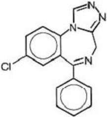
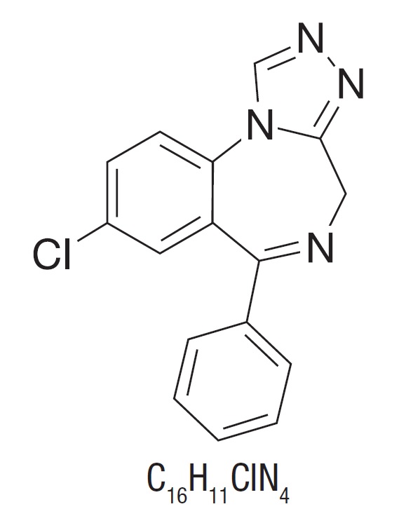
Estazolam
Autosomal recessive GTP cyclohydrolase I deficiency
Pathophysiology of heart failure
Micrographia (handwriting)
Rett syndrome
Nigrostriatal pathway
Parkinson's disease
Biotin-thiamine-responsive basal ganglia disease
Behavior-altering parasite
Β-Carboline
Hyperkinesia
Bornaprine
Movement disorder
Adenylosuccinate lyase deficiency
Huntington's disease
Transcranial direct stimulation in Parkinson's disease gait rehabilitation
Hemispatial neglect
Boris Morukov
Direct pathway
PET117
Parkinsonian gait
Cyclopiazonic acid
Quazepam
List of MeSH codes (C23)
Zuclopenthixol
Third heart sound
Hypokinesia - Wikipedia
 What is Hypokinesia? - LonestarNeurology
 Glossary of Terms | Parkinson's Disease
Glossary of Terms | Parkinson's Disease
 Stalevo (Carbidopa, Levodopa and Entacapone): Uses, Dosage, Side Effects, Interactions, Warning
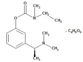
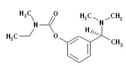
 Rivastigmine: Package Insert - Drugs.com
 Mitochondria of the heart left ventricle in hypokinetic rats
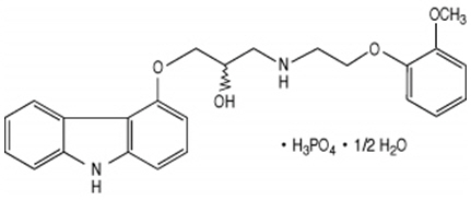
 These highlights do not include all the information needed to use CARVEDILOL TABLETS safely and effectively. See full...
DailyMed - ROPINIROLE HYDROCHLORIDE tablet, film coated
 Gabapentin Brown & Burk 800mg Film-coated tablets - Summary of Product Characteristics (SmPC) - (emc)
Table - Acute Chagas Disease Outbreak among Military Personnel, Colombia, 2021 - Volume 29, Number 9-September 2023 - Emerging...
 Side Effects of Remeron (mirtazapine): Interactions & Warnings
 Pediatric Dilated Cardiomyopathy: Practice Essentials, Background, Pathophysiology
 Wearable sensors objectively measure gait parameters in Parkinson's disease | PLOS ONE
 Baby’s Pregnancy Calendar
Baby’s Pregnancy Calendar
 Masticatory Muscle Pain and Disorders | SpringerLink
Hemineglect - Scholarpedia
 Frontiers | New Onset On-Medication Freezing of Gait After STN-DBS in Parkinson's Disease
 Lunesta: Insomnia Medication Treatment (Full Prescribing Information) | HealthyPlace
Comtan (entacapone) dosing, indications, interactions, adverse effects, and more
 Invanz - Reports of Side Effects
The Effect of the Ketogenic Diet on the Therapy of Neurodegenerative Diseases and Its Impact on Improving Cognitive Functions |...
 Tuberculous Pericarditis
 Lancashire Teaching Hospital | Consultants
 Myocarditis and Inflammatory Cardiomyopathy | IntechOpen
 Pathophysiology of Motor Dysfunction in Parkinson's Disease as the Rationale for Drug Treatment and Rehabilitation
 Parkinson disease | Radiology Reference Article | Radiopaedia.org
 Exelon - Drug Information from Guideline Central
 A man in his sixties with acute chest pain
Rigidity4
- citation needed] Hypokinesia is a symptom of Parkinson's disease shown as muscle rigidity and an inability to produce movement. (wikipedia.org)
- Parkinson disease (PD) , also known as idiopathic parkinsonism , is a neurodegenerative disease and movement disorder characterized by resting tremor, rigidity and hypokinesia due to progressive degeneration of dopaminergic neurons in the substantia nigra . (radiopaedia.org)
- reported that chronic administration of a common herbicide, rotenone, resulted in a selective destruction of the nigrostriatal dopaminergic system, formation of cytoplasmic inclusions in nigral neurons, and induction of hypokinesia and rigidity in rats, reproducing the key features of human PD. (jneurosci.org)
- Parkinsonism is a movement disorder characterized by resting tremor, slow and decreased movements (hypokinesia and akinesia), rigidity, postural instability, problems with gait, and coordination. (bvs.br)
Bradykinesia2
- Hypokinesia ( Greek "from below" + "movement" ), also referred to as bradykinesia , is a state of the body in which insufficient motor activity is observed, which leads to a limitation of the pace and range of movements. (lonestarneurology.net)
- Progressive bradykinesia and hypokinesia of ocular pursuit in Parkinson's disease. (lancsteachinghospitals.nhs.uk)
Global hypokinesia2
- Echocardiography revealed dilated cardiomyopathy (DCM) with left vertical dysfunction (ejection fraction of 39%) and global hypokinesia. (pediatriconcall.com)
- A bedside point-of-care ultrasound was performed, showing global hypokinesia with an EF of 20%, a collapsed inferior vena cava (IVC), and at the pelvis, the fetus was with negative heart activity. (hindawi.com)
Parkinson's3
- Hypokinesia describes a variety of more specific disorders: The most common cause of Hypokinesia is Parkinson's disease, and conditions related to Parkinson's disease. (wikipedia.org)
- The remainder of this article describes Hypokinesia associated with Parkinson's disease, and conditions related to Parkinson's disease. (wikipedia.org)
- TOHM emphasizes the broad panoply of non-Parkinsonian movement disorders, giving center stage to clinical observations and research in the area of hyperkinesia in contrast to other publications which focus primarily on disorders of hypokinesia (i.e., mainly Parkinson's disease and other forms of parkinsonism). (columbia.edu)
Echocardiography3
- Zones of hypokinesia during echocardiography indicate either acute or previous myocardial infarction (postinfarction cardiosclerosis), myocardial ischemia, thickening of the myocardial walls. (lonestarneurology.net)
- A transthoracic echocardiography demonstrated a diffuse hypokinesia of the right and the left ventricle. (diagnosticimaging.com)
- Electrocardiogram suggested ST elevations, echocardiography showed a possible slight hypokinesia, and we primarily suspected an acute coronary syndrome. (medworm.com)
Basal ganglia2
- Hypokinesia is characterized by a partial or complete loss of muscle movement due to a disruption in the basal ganglia. (wikipedia.org)
- The most common causes of hypokinesia are dysfunction of the basal ganglia and a decrease in excitation processes in the motor cortex. (lonestarneurology.net)
Akinesia1
- Their gait is often characterized by a typical slowness of movement (hypokinesia), small shuffling steps, and even total loss of movement (akinesia) in extreme cases. (walking-canes.net)
Movements1
- During this training, patients will make overexaggerated movements, such as high steps and arm swings, to retrain their muscular system and halt the progress of hypokinesia. (synapticrehab.com)
Diffuse1
- Hypokinesia was diffuse in 100% of cases. (bvsalud.org)
Disorders1
- Hypokinesia is one of the classifications of movement disorders, and refers to decreased bodily movement. (wikipedia.org)
Manifests1
- So, hypodynamia is a consequence of prolonged hypokinesia and manifests itself in the form of a decrease in muscle strength. (lonestarneurology.net)
Immobilization1
- A deficiency in motor activity can also cause hypokinesia - for example, prolonged immobilization due to trauma or serious illness. (lonestarneurology.net)
Rats1
- The response of cardiomyocytes and the mitochondrial system of rats subjected to a four-month hypokinesia was studied. (nih.gov)
Symptoms1
- In this article, you will learn about the main causes of hypokinesia and the most common symptoms. (lonestarneurology.net)
Decrease2
- Hypokinesia of the gallbladder occurs due to a decrease in its motor-evacuation function. (lonestarneurology.net)
- The number of intermitochondrial junctions considerably decreased in all regions of mitochondrial location which is presumably related to the decrease in the functional cell activity under experimental hypokinesia. (nih.gov)
Left3
- Decreased left ventricular range of motion is also classified as hypokinesia. (lonestarneurology.net)
- An echocardiogram revealed left ventricular ejection fraction of 45-50% with severe hypokinesia over anterior wall. (acc.org)
- An echocardiogram already showed impaired left ventricular systolic function with regional wall hypokinesia compatible with the finding of critical LAD stenosis in coronary angiogram. (acc.org)
Patients1
- Dopaminergic drugs are commonly used in the early stages of the hypokinesia to treat patients. (wikipedia.org)
Main1
- Dopamine The main neurotransmitter thought to be involved in hypokinesia is dopamine. (wikipedia.org)
Motor2
- The dopamine pathway in the substantia nigra is essential to motor function, and commonly a lesion in this area correlates with displayed hypokinesia. (wikipedia.org)
- GABA and glutamate The inhibitory neurotransmitter GABA and the excitatory glutamate are found in many parts of the central nervous system, including in the motor pathways that involve hypokinesia. (wikipedia.org)
Tremor1
- Prior to the identification of their mutation, affected family members were misdiagnosed with Parkinson's disease due to their prominent parkinsonian symptoms (e.g., rigidity, hypokinesia, postural instability, and tremor). (alzforum.org)
Bradykinesia1
- The neurological examination revealed such changes as left hemifacial hypoaesthesia, orofacial dyskinesia, extrapyramidal syndrome, bradykinesia and bilateral hypokinesia without motor deficits or coordination disorders, without speech impediments, left plantar extension, and right plantar flexion. (hindawi.com)
Mild4
- What is mild hypokinesia? (sheppard-arts.com)
- Mild hypokinesia basically means that the muscle of your heart does not contract as much as most peoples' hearts do. (sheppard-arts.com)
- Left ventricle angiography showed mild hypokinesia in the anterior wall. (bmj.com)
- The echocardiographic evaluation showed mild left ventricular dilatation, hypokinesia, and systolic dysfunction. (psychiatrist.com)
Angiography1
- Further investigations revealed non-obstructive coronary arteries on coronary angiography and imaging revealed hypokinesia of the anterior and anterior-septal walls in the apex and midcavity level, myocardial oedema and no infarction, all in keeping with TTC. (bmj.com)
Dopamine4
- Dopamine The main neurotransmitter thought to be involved in hypokinesia is dopamine. (wikipedia.org)
- The dopamine pathway in the substantia nigra is essential to motor function, and commonly a lesion in this area correlates with displayed hypokinesia. (wikipedia.org)
- Treatments for hypokinesia often either attempt to prevent dopamine degradation by MAO-B or increase the amount of neurotransmitter present in the system. (wikipedia.org)
- Hypokinesia is caused by a loss of dopamine in the brain. (sheppard-arts.com)
Pathway1
- The over activation of this pathway results in the disproportionate activation of the direct striatopallidal pathway which results in hypokinesia (10). (123helpme.com)
Muscle1
- Hypokinesia is characterized by a partial or complete loss of muscle movement due to a disruption in the basal ganglia. (wikipedia.org)
Patient1
- Etait inclus, tout patient hospitalisé en réanimation pour prise en charge d'un AVC, ayant réalisé un scanner cérébral. (bvsalud.org)
Treat2
- Dopaminergic drugs are commonly used in the early stages of the hypokinesia to treat patients. (wikipedia.org)
- How do you treat hypokinesia? (sheppard-arts.com)