Hypopigmentation
Pigmentation Disorders
Albinism, Ocular
Albinism, Oculocutaneous
Hermanski-Pudlak Syndrome
Hyperpigmentation
Vitiligo
Adaptor Protein Complex beta Subunits
Monophenol Monooxygenase
Platelet Storage Pool Deficiency
Waardenburg Syndrome
Melanocytes
Nystagmus, Congenital
Albinism
SOXE Transcription Factors
Melanins
Microphthalmia-Associated Transcription Factor
Prader-Willi Syndrome
Chromosomes, Human, Pair 15
Pigment Epithelium of Eye
Hair
Pedigree
Skin Diseases
The Sox10(Dom) mouse: modeling the genetic variation of Waardenburg-Shah (WS4) syndrome. (1/94)
Hirschsprung disease (HSCR) is a multigenic neurocristopathy clinically recognized by aganglionosis of the distal gastrointestinal tract. Patients presenting with aganglionosis in association with hypopigmentation are classified as Waardenburg syndrome type 4 (Waardenburg-Shah, WS4). Variability in the disease phenotype of WS4 patients with equivalent mutations suggests the influence of genetic modifier loci in this disorder. Sox10(Dom)/+ mice exhibit variability of aganglionosis and hypopigmentation influenced by genetic background similar to that observed in WS4 patients. We have constructed Sox10(Dom)/+ congenic lines to segregate loci that modify the neural crest defects in these mice. Consistent with previous studies, increased lethality of Sox10(Dom)/+ animals resulted from a C57BL/6J locus(i). However, we also observed an increase in hypopigmentation in conjunction with a C3HeB/FeJLe-a/a locus(i). Linkage analysis localized a hypopigmentation modifier of the Dom phenotype to mouse chromosome 10 in close proximity to a previously reported modifier of hypopigmentation for the endothelin receptor B mouse model of WS4. To evaluate further the role of SOX10 in development and disease, we have performed comparative genomic analyses. An essential role for this gene in neural crest development is supported by zoo blot hybridizations that reveal extensive conservation throughout vertebrate evolution and by similar Northern blot expression profiles between mouse and man. Comparative sequence analysis of the mouse and human SOX10 gene have defined the exon-intron boundaries of SOX10 and facilitated mutation analysis leading to the identification of two new SOX10 mutations in individuals with WS4. Structural analysis of the HMG DNA-binding domain was performed to evaluate the effect of human mutations in this region. (+info)Independent evaluation of onchocerciasis rapid assessment methods in Benue State, Nigeria. (2/94)
OBJECTIVE: To evaluate the prevalence of palpable nodules or skin depigmentation as rapid indicators of onchocerciasis epidemicity in at-risk communities. METHOD: We examined data collected in Benue State on 11035 individuals in 32 villages to evaluate these rapid assessment methods. RESULTS: The prevalence of palpable nodules correlates more closely with microfilarial prevalence (r=0.68, P<0.001) and community microfilarial load (r=0.64, P<0.001) than the prevalences of skin depigmentation or other potential rapid indicators. The recommended cut-off value for palpable nodules of 20% or more in males aged >20 years had a sensitivity of 94% and specificity of 50% compared to a cut-off of 40% or more for microfilarial prevalence in all ages. This would mean that in these 32 villages 17 of 18 would have been correctly identified for treatment, and a further 7 at lesser risk would have been targeted for treatment. CONCLUSIONS: Skin snipping and parasitological examination can be replaced by the simpler method of palpating onchocercal nodules to identify communities at serious risk of onchocerciasis. This has important operational benefits for onchocerciasis control programmes. (+info)A molecular analysis of the yemenite deaf-blind hypopigmentation syndrome: SOX10 dysfunction causes different neurocristopathies. (3/94)
The Yemenite deaf-blind hypopigmentation syndrome was first observed in a Yemenite sister and brother showing cutaneous hypopigmented and hyperpigmented spots and patches, microcornea, coloboma and severe hearing loss. A second case, observed in a girl with similar skin symptoms and hearing loss but without microcornea or coloboma, was reported as a mild form of this syndrome. Here we show that a SOX10 missense mutation is responsible for the mild form, resulting in a loss of DNA binding of this transcription factor. In contrast, no SOX10 alteration could be found in the other, severe case of the Yemenite deaf-blind hypopigmentation syndrome. Based on genetic, clinical, molecular and functional data, we suggest that these two cases represent two different syndromes. Moreover, as mutations of the SOX10 transcription factor were previously described in Waardenburg-Hirschsprung disease, these results show that SOX10 mutations cause various types of neurocristopathy. (+info)Successful bone marrow transplantation in a case of Griscelli disease which presented in accelerated phase with neurological involvement. (4/94)
Griscelli disease (GD) is a rare disorder characterized by pigment dilution, immunodeficiency and occurrence of accelerated phase consisting of hemophagocytosis, pancytopenia and neurological manifestations. Allogeneic BMT in the early period is an important modality of treatment for GD. We carried out an alloBMT from an HLA-identical sibling donor on a 4-year-old girl who presented in accelerated phase with neurological manifestations including convulsions, strabismus, severe dysarthria, ataxia and clonus. She was treated with etoposide, methylprednisolone and intrathecal methotrexate for 8 weeks and underwent alloBMT after receiving a conditioning regimen including ATG (rabbit, 10 mg/kg x 5 days), Bu/Cy. 8 x 10⁸/kg nucleated bone marrow cells were given. Engraftment occurred early and the post-BMT period was uneventful. Currently, she is at 18 months post BMT with sustained engraftment and with a normal neurological examination except for minimal clonus. Long-term follow-up will determine the prognosis regarding the neurological findings. (+info)Spatially restricted hypopigmentation associated with an Ednrbs-modifying locus on mouse chromosome 10. (5/94)
We have used the varied expressivity of white spotting (hypopigmentation) observed in intrasubspecific crosses of Ednrb(s) mice (Mayer Ednrb(s)/Ednrb(s) and C3HeB/FeJ Ednrb(s)/Ednrb(s)) to analyze the effects of modifier loci on the patterning of hypopigmentation. We have confirmed that an Ednrb(s) modifier locus is present on mouse Chromosome 10. This locus is now termed k10, using the nomenclature established by Dunn in 1920. The k10(Mayer) allele is a recessive modifier that accounts for almost all of the genetic variance of dorsal hypopigmentation. Using intercross analyses we identified a second allele of this locus or a closely linked gene termed k10(C3H). The k10(C3H) allele is semidominant and is associated with the penetrance and expressivity of a white forelock phenotype similar to that seen in Waardenburg syndrome. Molecular linkage analysis was used to determine that the k10 critical interval was flanked by D10Mit10 and D10Mit162/D10Mit122 and cosegregates with mast cell growth factor (Mgf). Complementation crosses with a Mgf(Sl) allele (a 3-5-cM deletion) confirm the semidominant white forelock feature of the k10(C3H) allele and the dorsal spotting feature of K10(Mayer) allele. MgF was assessed as a candidate gene for k10(Mayer) and k10(C3H) by sequence and genomic analyses. No molecular differences were observed between the Mayer and C57BL/6J alleles of MgF; however, extensive genomic differences were observed between the C3HeB/FeJ and C57BL/6J alleles. This suggests that alteration of MgF expression in C3H mice may account for the k10(C3H) action on white forelock hypopigmentation. Crosses of Ednrb(s) with Kit(WJ-2) (the receptor for MGF)-deficient mice confirmed the hypothesis that synergistic interaction between the Endothelin and MGF signaling pathways regulates proper neural crest-derived melanocyte development in vivo. (+info)Tietz syndrome (hypopigmentation/deafness) caused by mutation of MITF. (6/94)
Patients with Tietz syndrome have congenital profound deafness and generalised hypopigmentation, inherited in a fully penetrant autosomal dominant fashion. The pigmentary features and complete penetrance make this syndrome distinct among syndromes with pigmentary anomalies and deafness, which characteristically have patchy depigmentation and variable penetrance. Only one family has been reported with the exact features described in the original report of this syndrome. This family was reascertained and a missense mutation was found in the basic region of the MITF gene in family members with Tietz syndrome. Mutations in other regions of this gene have been found to produce Waardenburg syndrome type 2 (WS2), which also includes pigmentary changes and hearing loss, but in contrast to Tietz syndrome, depigmentation is patchy and hearing loss is variable in WS2. (+info)Oa1 knock-out: new insights on the pathogenesis of ocular albinism type 1. (7/94)
Ocular albinism type I (OA1) is an X-linked disorder characterized by severe reduction of visual acuity, strabismus, photophobia and nystagmus. Ophthalmologic examination reveals hypopigmentation of the retina, foveal hypoplasia and iris translucency. Microscopic examination of both retinal pigment epithelium (RPE) and skin melanocytes shows the presence of large pigment granules called giant melanosomes or macromelanosomes. In this study, we have generated and characterized Oa1-deficient mice by gene targeting (KO). The KO males are viable, fertile and phenotypically indistinguishable from the wild-type littermates. Ophthalmologic examination shows hypopigmentation of the ocular fundus in mutant animals compared with wild-type. Analysis of the retinofugal pathway reveals a reduction in the size of the uncrossed pathway, demonstrating a misrouting of the optic fibres at the chiasm, as observed in OA1 patients. Microscopic examination of the RPE shows the presence of giant melanosomes comparable with those described in OA1 patients. Ultrastructural analysis of the RPE cells, suggests that the giant melanosomes may form by abnormal growth of single melanosomes, rather than the fusion of several, shedding light on the pathogenesis of ocular albinism. (+info)Ophthalmic manifestations of tuberous sclerosis: a population based study. (8/94)
BACKGROUND/AIMS: Tuberous sclerosis complex (TSC) has retinal and non-retinal ophthalmic manifestations. This study was designed to determine the prevalence of the ophthalmic manifestations and of refractive errors in a population of patients with TSC. METHODS: 179 patients identified were in a prevalence study of TSC in the south of England and 107 of these agreed to full ophthalmic examination which was successful in 100. Ophthalmic examination included examination of the eyelids, cover test, examination of the irides, dilation funduscopy using both direct and indirect ophthalmoscopy, and refraction using retinoscopy. Myopia was defined as a spherical equivalent <-0.5D and hyperopia as a spherical equivalent >+0.5D. RESULTS: Retinal hamartomas were seen in 44 of the 100 patients. The commonest morphological type of hamartoma seen was the flat, translucent lesion in 31 of the 44 patients (70%). The multinodular "mulberry" lesion was seen in 24 of the 44 patients (55%) and the transitional type lesion was seen in four of the 44 patients (9%). Punched out areas of retinal depigmentation were seen in 39 of the 100 patients but only six of 100 controls. 27% of eyes were myopic, 22% were hyperopic, and 27% had astigmatism >0.75D. Of the non-retinal findings, 39 patients had angiofibromas of the eyelids, five had non-paralytic strabismus, and three had colobomas. CONCLUSION: Apart from the higher prevalence of flat retinal hamartomas, the findings of this study compare closely with previous large clinic based series of TSC patients. Refractive findings were similar to previous studies of a similarly aged non-TSC population. This is the first series to document the statistically significant association of punched out chorioretinal depigmentation with TSC and the authors believe that it should be looked for as an aid to diagnosis. (+info)Hypopigmentation is a medical term that refers to a condition where there is a decrease in the amount of pigment (melanin) in the skin, resulting in lighter patches or spots on the skin. This can occur due to various reasons such as skin injuries, certain skin disorders like vitiligo, fungal infections, burns, or as a side effect of some medical treatments like chemotherapy or radiation therapy. It is different from albinism, which is a genetic condition where the body is unable to produce melanin at all.
Pigmentation disorders are conditions that affect the production or distribution of melanin, the pigment responsible for the color of skin, hair, and eyes. These disorders can cause changes in the color of the skin, resulting in areas that are darker (hyperpigmentation) or lighter (hypopigmentation) than normal. Examples of pigmentation disorders include melasma, age spots, albinism, and vitiligo. The causes, symptoms, and treatments for these conditions can vary widely, so it is important to consult a healthcare provider for an accurate diagnosis and treatment plan.
Ocular albinism is a type of albinism that primarily affects the eyes. It is a genetic disorder characterized by the reduction or absence of melanin, the pigment responsible for coloring the skin, hair, and eyes. In ocular albinism, melanin production is deficient in the eyes, leading to various eye abnormalities.
The main features of ocular albinism include:
1. Nystagmus: Rapid, involuntary back-and-forth movement of the eyes.
2. Iris transillumination: The iris appears translucent due to the lack of pigment, allowing light to pass through easily. This can be observed using a light source shone into the eye.
3. Foveal hypoplasia: Underdevelopment or absence of the fovea, a small pit in the retina responsible for sharp, central vision.
4. Photophobia: Increased sensitivity to light due to the lack of pigment in the eyes.
5. Strabismus: Misalignment of the eyes, which can result in double vision or lazy eye.
6. Reduced visual acuity: Decreased ability to see clearly, even with corrective lenses.
Ocular albinism is typically inherited as an X-linked recessive trait, meaning it primarily affects males, while females can be carriers of the condition. However, there are also autosomal recessive forms of ocular albinism that can affect both males and females equally. Treatment for ocular albinism usually involves managing symptoms with corrective lenses, low-vision aids, and vision therapy to improve visual skills.
Oculocutaneous albinism (OCA) is a group of genetic disorders characterized by reduced or complete absence of melanin pigment in the eyes, skin, and hair. Melanin is the pigment responsible for giving color to our skin, hair, and eyes. OCA affects both the eyes (oculo-) and the skin (cutaneous), hence the name oculocutaneous albinism.
There are several types of OCA, each caused by different genetic mutations affecting melanin production. The most common forms include:
1. OCA1: This type is further divided into two subtypes - OCA1A and OCA1B. OCA1A is characterized by complete absence of melanin in the eyes, skin, and hair from birth. Individuals with this condition have white hair, very light skin, and pale blue or gray irises. OCA1B, on the other hand, presents with reduced melanin production, leading to lighter-than-average skin, hair, and eye color at birth. Over time, some melanin may be produced, resulting in milder pigmentation changes compared to OCA1A.
2. OCA2: This form of albinism is caused by mutations in the tyrosinase-related protein 1 (TYRP1) gene, which plays a role in melanin production. Individuals with OCA2 typically have light brown or yellowish skin, golden or straw-colored hair, and lighter irises compared to their family members without albinism.
3. OCA3: Also known as Rufous oculocutaneous albinism (ROCA), this type is caused by mutations in the tyrosinase gene (TYR). It primarily affects people of African descent, leading to reddish-brown hair, light brown skin, and normal or near-normal eye color.
4. OCA4: This form of albinism results from mutations in the membrane-associated transporter protein (MATP) gene, which is involved in melanin transport within cells. Individuals with OCA4 usually have light brown skin, yellowish or blond hair, and lighter irises compared to their family members without albinism.
Regardless of the type, all individuals with oculocutaneous albinism face similar challenges, including reduced vision due to abnormal eye development (nystagmus, strabismus, and farsightedness) and increased sensitivity to sunlight (photophobia). Proper management, such as wearing UV-protective sunglasses, hats, and sunscreen, can help protect their skin and eyes from damage.
I'm sorry for any confusion, but "hair color" is not a term that has a medical definition in the same way that a term like "myocardial infarction" (heart attack) does. Hair color can be described in various ways, such as being a natural hair color (like blonde, brunette, red, or black) or describing a change in hair color due to factors like aging (gray or white hairs) or hair dye usage.
However, it's worth noting that changes in hair color can sometimes be associated with certain medical conditions. For example, premature graying of the hair before the age of 30 can be a feature of certain genetic disorders or vitamin B12 deficiency. Similarly, some skin conditions like alopecia areata or vitiligo can cause patchy changes in hair color. But these associations don't provide a medical definition for 'hair color'.
Hermanski-Pudlak Syndrome (HPS) is a rare genetic disorder characterized by the triad of albinism, bleeding disorders, and lysosomal storage disease. It is caused by mutations in any one of several genes involved in biogenesis of lysosome-related organelles (LROs), such as melanosomes in melanocytes, platelet dense granules, and lung lamellar bodies.
The albinism in HPS results from abnormal melanosome biogenesis, leading to decreased pigmentation in the skin, hair, and eyes. The bleeding disorder is due to defective platelet dense granules, which are necessary for normal clotting function. This can result in prolonged bleeding times and easy bruising.
The lysosomal storage disease component of HPS is characterized by the accumulation of ceroid lipofuscin within LROs, leading to progressive damage to affected tissues. The most common form of HPS (HPS-1) also involves pulmonary fibrosis, which can lead to respiratory failure and death in the third or fourth decade of life.
There are currently seven known subtypes of HPS, each caused by mutations in different genes involved in LRO biogenesis. The clinical features and severity of HPS can vary widely between subtypes and even within families with the same genetic mutation.
Melanosomes are membrane-bound organelles found in melanocytes, the pigment-producing cells in the skin, hair, and eyes. They contain the pigment melanin, which is responsible for giving color to these tissues. Melanosomes are produced in the melanocyte and then transferred to surrounding keratinocytes in the epidermis via a process called cytocrinesis. There are four stages of melanosome development: stage I (immature), stage II (developing), stage III (mature), and stage IV (degrading). The amount and type of melanin in the melanosomes determine the color of an individual's skin, hair, and eyes. Mutations in genes involved in melanosome biogenesis or function can lead to various pigmentation disorders, such as albinism.
Eye color is a characteristic determined by variations in a person's genes. The color of the eyes depends on the amount and type of pigment called melanin found in the eye's iris.
There are three main types of eye colors: brown, blue, and green. Brown eyes have the most melanin, while blue eyes have the least. Green eyes have a moderate amount of melanin combined with a golden tint that reflects light to give them their unique color.
Eye color is a polygenic trait, which means it is influenced by multiple genes. The two main genes responsible for eye color are OCA2 and HERC2, both located on chromosome 15. These genes control the production, transport, and storage of melanin in the iris.
It's important to note that eye color can change during infancy and early childhood due to the development of melanin in the iris. Additionally, some medications or medical conditions may also cause changes in eye color over time.
Hyperpigmentation is a medical term that refers to the darkening of skin areas due to an increase in melanin, the pigment that provides color to our skin. This condition can affect people of all races and ethnicities, but it's more noticeable in those with lighter skin tones.
Hyperpigmentation can be caused by various factors, including excessive sun exposure, hormonal changes (such as during pregnancy), inflammation, certain medications, and underlying medical conditions like Addison's disease or hemochromatosis. It can also result from skin injuries, such as cuts, burns, or acne, which leave dark spots known as post-inflammatory hyperpigmentation.
There are several types of hyperpigmentation, including:
1. Melasma: This is a common form of hyperpigmentation that typically appears as symmetrical, blotchy patches on the face, particularly the forehead, cheeks, and upper lip. It's often triggered by hormonal changes, such as those experienced during pregnancy or while taking birth control pills.
2. Solar lentigos (age spots or liver spots): These are small, darkened areas of skin that appear due to prolonged sun exposure over time. They typically occur on the face, hands, arms, and decolletage.
3. Post-inflammatory hyperpigmentation: This type of hyperpigmentation occurs when an injury or inflammation heals, leaving behind a darkened area of skin. It's more common in people with darker skin tones.
Treatment for hyperpigmentation depends on the underlying cause and may include topical creams, chemical peels, laser therapy, or microdermabrasion. Preventing further sun damage is crucial to managing hyperpigmentation, so wearing sunscreen with a high SPF and protective clothing is recommended.
Skin pigmentation is the coloration of the skin that is primarily determined by two types of melanin pigments, eumelanin and pheomelanin. These pigments are produced by melanocytes, which are specialized cells located in the epidermis. Eumelanin is responsible for brown or black coloration, while pheomelanin produces a red or yellow hue.
The amount and distribution of melanin in the skin can vary depending on genetic factors, age, sun exposure, and various other influences. Increased production of melanin in response to UV radiation from the sun helps protect the skin from damage, leading to darkening or tanning of the skin. However, excessive sun exposure can also cause irregular pigmentation, such as sunspots or freckles.
Abnormalities in skin pigmentation can result from various medical conditions, including albinism (lack of melanin production), vitiligo (loss of melanocytes leading to white patches), and melasma (excessive pigmentation often caused by hormonal changes). These conditions may require medical treatment to manage or improve the pigmentation issues.
Vitiligo is a medical condition characterized by the loss of pigmentation in patches of skin, resulting in irregular white depigmented areas. It's caused by the destruction of melanocytes, the cells responsible for producing melanin, which gives our skin its color. The exact cause of vitiligo is not fully understood, but it's thought to be an autoimmune disorder where the immune system mistakenly attacks and destroys melanocytes. It can affect people of any age, gender, or ethnicity, although it may be more noticeable in people with darker skin tones. The progression of vitiligo is unpredictable and can vary from person to person. Treatment options include topical creams, light therapy, oral medications, and surgical procedures, but the effectiveness of these treatments varies depending on the individual case.
Adaptor Protein Complex (AP) beta subunits are structural proteins that play a crucial role in intracellular vesicle trafficking. They are part of the heterotetrameric AP complex, which is responsible for recognizing and binding to specific sorting signals on membrane cargo proteins, allowing for their packaging into transport vesicles.
There are four different types of AP complexes (AP-1, AP-2, AP-3, and AP-4), each with a unique set of subunits that confer specific functions. The beta subunit is a common component of all four complexes and is essential for their stability and function.
The beta subunit interacts with other subunits within the AP complex as well as with accessory proteins, such as clathrin, to form a coat around the transport vesicle. This coat helps to shape the vesicle and facilitate its movement between different cellular compartments.
Mutations in genes encoding AP beta subunits have been linked to various human diseases, including forms of hemolytic anemia, neurological disorders, and immunodeficiency.
Tyrosinase, also known as monophenol monooxygenase, is an enzyme (EC 1.14.18.1) that catalyzes the ortho-hydroxylation of monophenols (like tyrosine) to o-diphenols (like L-DOPA) and the oxidation of o-diphenols to o-quinones. This enzyme plays a crucial role in melanin synthesis, which is responsible for the color of skin, hair, and eyes in humans and animals. Tyrosinase is found in various organisms, including plants, fungi, and animals. In humans, tyrosinase is primarily located in melanocytes, the cells that produce melanin. The enzyme's activity is regulated by several factors, such as pH, temperature, and metal ions like copper, which are essential for its catalytic function.
Platelet Storage Pool Deficiency (PSPD) is a group of bleeding disorders characterized by a decrease in the number or function of secretory granules (storage pools) in platelets, which are small blood cells that play a crucial role in clotting. These granules contain various substances such as ADP (adenosine diphosphate), ATP (adenosine triphosphate), calcium ions, and serotonin, which are released during platelet activation to help promote clot formation.
In PSPD, the quantitative or qualitative deficiency of these granules leads to impaired platelet function and increased bleeding tendency. The condition can be inherited or acquired, and it is often classified based on the type of granule affected: dense granules (delta granules) or alpha granules.
Delta granule deficiency, also known as Dense Granule Deficiency (DGD), results in decreased levels of ADP, ATP, and calcium ions, while alpha granule deficiency leads to reduced levels of von Willebrand factor, fibrinogen, and other clotting factors.
Symptoms of PSPD can vary from mild to severe and may include easy bruising, prolonged bleeding after injury or surgery, nosebleeds, and gum bleeding. The diagnosis typically involves platelet function tests, electron microscopy, and genetic testing. Treatment options depend on the severity of the condition and may include desmopressin (DDAVP), platelet transfusions, or other medications to manage bleeding symptoms.
Waardenburg Syndrome is a genetic disorder that affects the development of melanin, a pigment responsible for hair, skin, and eye color. Named after the Dutch ophthalmologist Petrus Waardenburg who first described it in 1907, this syndrome is characterized by distinctive physical features and hearing loss.
There are four types of Waardenburg Syldrome (WS1, WS2, WS3, and WS4), each with varying degrees of symptoms. Common features include:
1. Differential coloring of the hair, skin, and eyes (poliosis, vitiligo, and heterochromia)
2. Distinctive facial features (wide-set eyes, broad nasal root, and a high arched or cleft palate)
3. Hearing loss, which can be unilateral (one-sided) or bilateral (both-sided), conductive, sensorineural, or mixed
4. Pigmentary changes in the iris, such as different colors between the eyes or within one eye
5. Sometimes, musculoskeletal abnormalities and/or developmental delays
WS1 and WS2 are more common than WS3 and WS4. The genetic causes of Waardenburg Syndrome involve mutations in several different genes associated with melanin production and transport. These include PAX3, MITF, SNAI2, EDN3, and EDNRB.
Diagnosis is typically based on clinical findings, including physical features and hearing tests. Genetic testing can confirm the diagnosis and help determine the specific type of Waardenburg Syndrome. Treatment usually involves addressing individual symptoms, such as using hearing aids or cochlear implants for hearing loss and managing any skin or eye concerns.
Melanocytes are specialized cells that produce, store, and transport melanin, the pigment responsible for coloring of the skin, hair, and eyes. They are located in the bottom layer of the epidermis (the outermost layer of the skin) and can also be found in the inner ear and the eye's retina. Melanocytes contain organelles called melanosomes, which produce and store melanin.
Melanin comes in two types: eumelanin (black or brown) and pheomelanin (red or yellow). The amount and type of melanin produced by melanocytes determine the color of a person's skin, hair, and eyes. Exposure to UV radiation from sunlight increases melanin production as a protective response, leading to skin tanning.
Melanocyte dysfunction or abnormalities can lead to various medical conditions, such as albinism (lack of melanin production), melasma (excessive pigmentation), and melanoma (cancerous growth of melanocytes).
Congenital nystagmus is a type of involuntary eye movement that is present at birth or develops within the first few months of life. It is characterized by rhythmic oscillations or repetitive, rapid movements of the eyes in either horizontal, vertical, or rotatory directions. These movements can impair vision and may be associated with other ocular conditions such as albinism, congenital cataracts, or optic nerve hypoplasia. The exact cause of congenital nystagmus is not fully understood, but it is believed to result from abnormal development or dysfunction in the areas of the brain that control eye movements. In some cases, congenital nystagmus may be inherited as a genetic trait. Treatment options for congenital nystagmus include corrective lenses, prism glasses, surgery, and vision therapy, depending on the underlying cause and severity of the condition.
Pigmentation, in a medical context, refers to the coloring of the skin, hair, or eyes due to the presence of pigment-producing cells called melanocytes. These cells produce a pigment called melanin, which determines the color of our skin, hair, and eyes.
There are two main types of melanin: eumelanin and pheomelanin. Eumelanin is responsible for brown or black coloration, while pheomelanin produces a red or yellow hue. The amount and type of melanin produced by melanocytes can vary from person to person, leading to differences in skin color and hair color.
Changes in pigmentation can occur due to various factors such as genetics, exposure to sunlight, hormonal changes, inflammation, or certain medical conditions. For example, hyperpigmentation refers to an excess production of melanin that results in darkened patches on the skin, while hypopigmentation is a condition where there is a decreased production of melanin leading to lighter or white patches on the skin.
Albinism is a group of genetic disorders that result in little or no production of melanin, the pigment responsible for coloring skin, hair, and eyes. It is caused by mutations in genes involved in the production of melanin. There are several types of albinism, including oculocutaneous albinism (OCA) and ocular albinism (OA). OCA affects the skin, hair, and eyes, while OA primarily affects the eyes.
People with albinism typically have very pale skin, white or light-colored hair, and light-colored eyes. They may also have vision problems, such as sensitivity to light (photophobia), rapid eye movements (nystagmus), and decreased visual acuity. The severity of these symptoms can vary depending on the type and extent of albinism.
Albinism is inherited in an autosomal recessive manner, which means that an individual must inherit two copies of the mutated gene, one from each parent, in order to have the condition. If both parents are carriers of a mutated gene for albinism, they have a 25% chance with each pregnancy of having a child with albinism.
There is no cure for albinism, but individuals with the condition can take steps to protect their skin and eyes from the sun and use visual aids to help with vision problems. It is important for people with albinism to undergo regular eye examinations and to use sun protection, such as sunscreen, hats, and sunglasses, to prevent skin damage and skin cancer.
SOXE transcription factors are a subgroup of the SOX (SRY-related HMG box) family of proteins, which are involved in various developmental processes, including cell fate specification and differentiation. The SOXE group includes SOX8, SOX9, and SOX10, all of which contain a conserved high mobility group (HMG) box DNA-binding domain. They play crucial roles in the development of several tissues, such as the nervous system, skeletal system, and urogenital system.
SOXE transcription factors are known to regulate gene expression by binding to specific DNA sequences, often acting in combination with other transcription factors to control various cellular processes. Dysregulation of SOXE transcription factors has been implicated in several human diseases, including cancer and neurodevelopmental disorders.
Melanin is a pigment that determines the color of skin, hair, and eyes in humans and animals. It is produced by melanocytes, which are specialized cells found in the epidermis (the outer layer of the skin) and the choroid (the vascular coat of the eye). There are two main types of melanin: eumelanin and pheomelanin. Eumelanin is a black or brown pigment, while pheomelanin is a red or yellow pigment. The amount and type of melanin produced by an individual can affect their skin and hair color, as well as their susceptibility to certain diseases, such as skin cancer.
The Microphthalmia-Associated Transcription Factor (MITF) is a protein that functions as a transcription factor, which means it regulates the expression of specific genes. It belongs to the basic helix-loop-helix leucine zipper (bHLH-Zip) family of transcription factors and plays crucial roles in various biological processes such as cell growth, differentiation, and survival.
MITF is particularly well-known for its role in the development and function of melanocytes, the pigment-producing cells found in the skin, eyes, and inner ear. It regulates the expression of genes involved in melanin synthesis and thus influences hair and skin color. Mutations in the MITF gene have been associated with certain eye disorders, including microphthalmia (small or underdeveloped eyes), iris coloboma (a gap or hole in the iris), and Waardenburg syndrome type 2A (an inherited disorder characterized by hearing loss and pigmentation abnormalities).
In addition to its role in melanocytes, MITF also plays a part in the development and function of other cell types, including osteoclasts (cells involved in bone resorption), mast cells (immune cells involved in allergic reactions), and retinal pigment epithelial cells (a type of cell found in the eye).
Prader-Willi Syndrome (PWS) is a genetic disorder that affects several parts of the body and is characterized by a range of symptoms including:
1. Developmental delays and intellectual disability.
2. Hypotonia (low muscle tone) at birth, which can lead to feeding difficulties in infancy.
3. Excessive appetite and obesity, typically beginning around age 2, due to a persistent hunger drive and decreased satiety.
4. Behavioral problems such as temper tantrums, stubbornness, and compulsive behaviors.
5. Hormonal imbalances leading to short stature, small hands and feet, incomplete sexual development, and decreased bone density.
6. Distinctive facial features including a thin upper lip, almond-shaped eyes, and a narrowed forehead.
7. Sleep disturbances such as sleep apnea or excessive daytime sleepiness.
PWS is caused by the absence of certain genetic material on chromosome 15, which results in abnormal gene function. It affects both males and females equally and has an estimated incidence of 1 in 10,000 to 30,000 live births. Early diagnosis and management can help improve outcomes for individuals with PWS.
Human chromosome pair 15 consists of two rod-shaped structures present in the nucleus of each cell in the human body. Each chromosome is made up of DNA tightly coiled around histone proteins, forming a complex structure called a chromatin.
Chromosomes come in pairs, with one chromosome inherited from each parent. Chromosome pair 15 includes two homologous chromosomes, meaning they have the same size, shape, and gene content but may contain slight variations in their DNA sequences.
These chromosomes play a crucial role in inheritance and the development and function of the human body. Chromosome pair 15 contains around 100 million base pairs of DNA and approximately 700 protein-coding genes, which are involved in various biological processes such as growth, development, metabolism, and regulation of gene expression.
Abnormalities in chromosome pair 15 can lead to genetic disorders, including Prader-Willi syndrome and Angelman syndrome, which are caused by the loss or alteration of specific regions on chromosome 15.
The pigment epithelium of the eye, also known as the retinal pigment epithelium (RPE), is a layer of cells located between the photoreceptor cells of the retina and the choroid, which is the vascular layer of the eye. The RPE plays a crucial role in maintaining the health and function of the photoreceptors by providing them with nutrients, removing waste products, and helping to regulate the light that enters the eye.
The RPE cells contain pigment granules that absorb excess light, preventing it from scattering within the eye and improving visual acuity. They also help to create a barrier between the retina and the choroid, which is important for maintaining the proper functioning of the photoreceptors. Additionally, the RPE plays a role in the regeneration of visual pigments in the photoreceptor cells, allowing us to see in different light conditions.
Damage to the RPE can lead to various eye diseases and conditions, including age-related macular degeneration (AMD), which is a leading cause of vision loss in older adults.
Medically, hair is defined as a threadlike structure that grows from the follicles found in the skin of mammals. It is primarily made up of a protein called keratin and consists of three parts: the medulla (the innermost part or core), the cortex (middle layer containing keratin filaments) and the cuticle (outer layer of overlapping scales).
Hair growth occurs in cycles, with each cycle consisting of a growth phase (anagen), a transitional phase (catagen), and a resting phase (telogen). The length of hair is determined by the duration of the anagen phase.
While hair plays a crucial role in protecting the skin from external factors like UV radiation, temperature changes, and physical damage, it also serves as an essential aspect of human aesthetics and identity.
I must clarify that the term "pedigree" is not typically used in medical definitions. Instead, it is often employed in genetics and breeding, where it refers to the recorded ancestry of an individual or a family, tracing the inheritance of specific traits or diseases. In human genetics, a pedigree can help illustrate the pattern of genetic inheritance in families over multiple generations. However, it is not a medical term with a specific clinical definition.
A syndrome, in medical terms, is a set of symptoms that collectively indicate or characterize a disease, disorder, or underlying pathological process. It's essentially a collection of signs and/or symptoms that frequently occur together and can suggest a particular cause or condition, even though the exact physiological mechanisms might not be fully understood.
For example, Down syndrome is characterized by specific physical features, cognitive delays, and other developmental issues resulting from an extra copy of chromosome 21. Similarly, metabolic syndromes like diabetes mellitus type 2 involve a group of risk factors such as obesity, high blood pressure, high blood sugar, and abnormal cholesterol or triglyceride levels that collectively increase the risk of heart disease, stroke, and diabetes.
It's important to note that a syndrome is not a specific diagnosis; rather, it's a pattern of symptoms that can help guide further diagnostic evaluation and management.
Skin diseases, also known as dermatological conditions, refer to any medical condition that affects the skin, which is the largest organ of the human body. These diseases can affect the skin's function, appearance, or overall health. They can be caused by various factors, including genetics, infections, allergies, environmental factors, and aging.
Skin diseases can present in many different forms, such as rashes, blisters, sores, discolorations, growths, or changes in texture. Some common examples of skin diseases include acne, eczema, psoriasis, dermatitis, fungal infections, viral infections, bacterial infections, and skin cancer.
The symptoms and severity of skin diseases can vary widely depending on the specific condition and individual factors. Some skin diseases are mild and can be treated with over-the-counter medications or topical creams, while others may require more intensive treatments such as prescription medications, light therapy, or even surgery.
It is important to seek medical attention if you experience any unusual or persistent changes in your skin, as some skin diseases can be serious or indicative of other underlying health conditions. A dermatologist is a medical doctor who specializes in the diagnosis and treatment of skin diseases.
 Hypopigmentation
Postinflammatory hypopigmentation
Yemenite deaf-blind hypopigmentation syndrome
National Organization for Albinism and Hypopigmentation
Albinism in humans
Nevus depigmentosus
Pityriasis alba
Genetic history of the Arab world
Albinism
Oral pigmentation
Persecution of people with albinism
Cross syndrome
White horse
International Albinism Awareness Day
Siberian Husky
Dominant white
Tiger eye
Tattoo removal
Bioptics (device)
Microphthalmia-associated transcription factor
Hyperpigmentation
Trichophyton concentricum
Noonan syndrome with multiple lentigines
Dyschromia
Tumid lupus erythematosus
Tietz syndrome
P14 deficiency
Dog coat
Incontinentia pigmenti
Bruce Beutler
Inflammatory Hypopigmentation1
- Hello, The paleness of the skin is due to post inflammatory hypopigmentation. (medhelp.org)
Vitiligo6
- It is seen in: Albinism Idiopathic guttate hypomelanosis Leprosy Leucism Phenylketonuria Pityriasis alba Vitiligo Angelman syndrome Tinea versicolor Yaws An uncommon adverse effect of imatinib therapy Injections of high concentrations of corticosteroids (transient) Areas of lighter pigmentation can be indications of hypopigmentation. (wikipedia.org)
- Vitiligo is a form of hypopigmentation and the cause of Vitiligo is unknown. (oureczema.com)
- Vitiligo is another type of hypopigmentation in which smooth, white patches of skin develop over regions like the arms and face or may cover the entire body. (helth.co)
- Vitiligo and other disorders of hypopigmentation. (medlineplus.gov)
- Some people are born with hypopigmentation from birth in the form of Vitiligo or albinism . (oxygenetix.com)
- On this last point, the presence of depigmentation can aid clinicians in distinguishing vitiligo from the hypopigmentation seen in idiopathic guttate hypomelanosis, which is nonprogressive. (medscape.com)
Post-inflammatory1
- The hypopigmentation generally arises during the post-inflammatory stage of eczema. (oureczema.com)
Hyperpigmentation and hypopigmentation2
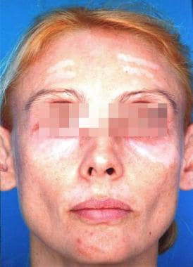
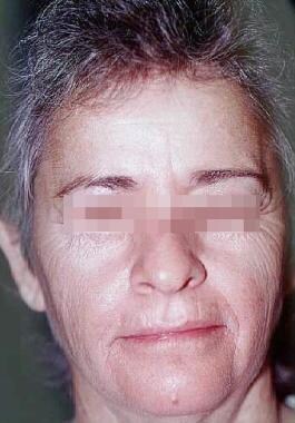
- For example, hyperpigmentation and hypopigmentation noted after carbon dioxide laser resurfacing are related to damage to melanocytes vaporized along with targeted keratinocytes and fibroblasts in the epidermis and dermis. (medscape.com)
- The most important thing for people to understand concerning both hyperpigmentation and hypopigmentation is that both conditions are occurring in the skin at the chemical level, and rarely occur without an underlying reason. (victoriandermalgroup.com.au)
Albinism1
- The National Organization of Albinism and Hypopigmentation (NOAH) provides the www.albinism.org website as a public service and for informational purposes only. (albinism.org)
Melanin production1
- Hypopigmentation in skin is the result of a reduction in melanin production. (donskinzfacial.com)
Darker4
- Hypopigmentation can be upsetting to some, especially those with darker skin whose hypopigmentation marks are seen more visibly. (wikipedia.org)
- In the case of illness or injury, the person's skin may change color, becoming darker (hyperpigmentation) or lighter (hypopigmentation). (donskinzfacial.com)
- Hypopigmentation shows up as a loss of pigment in splotches on the skin, and on the other hand, hyperpigmentation comes forth as darker pigment than the natural skin color. (oxygenetix.com)
- Those with darker skin shy away from getting certain light therapies such as laser hair removal, as it can cause hypopigmentation if not done properly. (oxygenetix.com)
Tinea versicolor1
- How is hypopigmentation related to tinea versicolor? (xshotpix.com)
Skin11
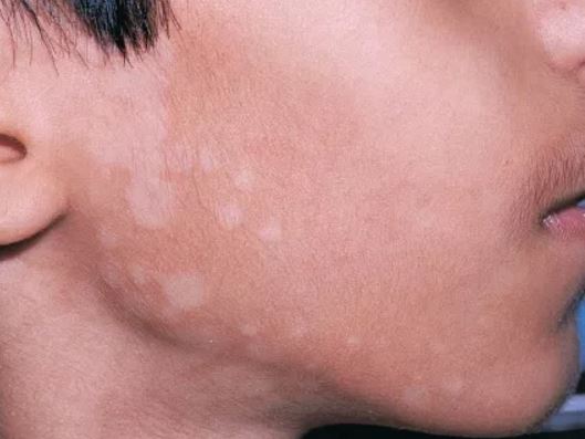
- Hypopigmentation is characterized specifically as an area of skin becoming lighter than the baseline skin color, but not completely devoid of pigment. (wikipedia.org)
- As melanin pigments tend to be in the skin, eye, and hair, these are the commonly affected areas in those with hypopigmentation. (wikipedia.org)
- Hypopigmentation refers to patches of skin that are lighter than your overall skin tone. (healthline.com)
- Simply put, hyperpigmentation is the problem of having too much pigmentation in an area of the skin while hypopigmentation is having too little. (victoriandermalgroup.com.au)
- Hypopigmentation, on the other hand, is a lack of melanin in the skin, and incidentally can also be caused by many of the same factors that cause hyperpigmentation. (victoriandermalgroup.com.au)
- With hypopigmentation, you're going to have too little pigmentation, and this will result in the skin to become discolored. (oureczema.com)
- Having skin discoloration such as hypopigmentation or hyperpigmentation can be life-altering. (oxygenetix.com)
- Hypopigmentation is most commonly caused by trauma to the skin. (oxygenetix.com)
- Although this type of skin problem is very difficult to treat, but you can try Elidel cream or Protropic ointment on these hypopigmented areas.But use them after a dermatologist's guidance.If the symptoms still persist then you can talk to your dermatologist about laser-assisted chemabrasion(LACA).It has provided excellent results in such hypopigmentation. (medhelp.org)
- Hypopigmentation occurs when the yeast produces a specific chemical that turns off melanocytes, resulting in the decreased production of melanin (the pigment central to the skin, eye, and hair color). (xshotpix.com)
- Skin hypopigmentation and depigmentation disorders are a top concern for patients with skin of color seeking care from a dermatologist. (bvsalud.org)
Depigmentation1
- Hypopigmentation is a loss of the skin's natural pigment or "depigmentation" . (oxygenetix.com)
Lighter1
- On the opposite end, lighter or white spots may be caused by hypopigmentation. (healthline.com)
Treatments1
- Intense pulse light treatment, chemical peels, and microdermabrasion are medical treatments for hypopigmentation. (helth.co)
Eczema11
- Eczema and Hypopigmentation: Does Eczema Cause Hypopigmentation? (oureczema.com)
- Many people believe that eczema and hypopigmentation go hand in hand. (oureczema.com)
- Are you going to develop hypopigmentation if you have eczema? (oureczema.com)
- However, some children will eventually "outgrow" certain eczema symptoms, but the hypopigmentation may stick around for many years. (oureczema.com)
- It is said that hypopigmentation may be secondary to eczema. (oureczema.com)
- It's common for infants to develop hypopigmentation in the same spots as eczema. (oureczema.com)
- Having hypopigmentation doesn't mean that you are destined to develop eczema. (oureczema.com)
- Are Eczema And Hypopigmentation Connected? (oureczema.com)
- For instance, you can go through life with eczema without experiencing any hypopigmentation. (oureczema.com)
- It is believed that hypopigmentation is a secondary ailment to eczema. (oureczema.com)
- While it depends on each unique situation, it may be possible to treat the hypopigmentation and eczema at the same time. (oureczema.com)
Pityriasis2
- Hypopigmentation due to Pityriasis alba also requires no treatment as the patches sometimes disappear on their own. (helth.co)
- The main differential diagnosis is pityriasis versicolor , where there is simply hypopigmentation, rather than no pigmentation. (medscape.com)
Discoloration1
- Treatment for hypopigmentation depends on the initial cause of the discoloration. (wikipedia.org)
Treatment6
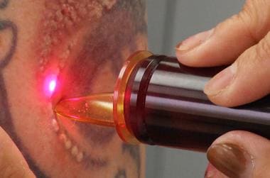
- Successful Treatment of Laser Induced Hypopigmentation with. (lww.com)
- the current state of the Hypopigmentation Disorder Treatment industry. (techinbullet.com)
- In this section, the level of competition in the worldwide Hypopigmentation Disorder Treatment Industry Market is analyzed. (techinbullet.com)
- end-user and application segments contribute to the overall market for Hypopigmentation Disorder Treatment. (techinbullet.com)
- Macular hypopigmentation developed on her face and body 3.5 years after treatment. (cdc.gov)
- There is no treatment for the hypopigmentation. (health.am)
Genetic1
- Widespread hypopigmentation is often genetic. (healthline.com)
Spots1
- Cosmetic procedures such as chemical and laser peels can also cause hypopigmentation spots. (oxygenetix.com)
Efficacy1
- Although it has high efficacy and safety, adverse effect like hypopigmentation may occur causing anxiety to patients. (lww.com)
Occurrence2
- And the occurrence of hypopigmentation and hyperpigmentation rate was pooled for safety evaluation. (bvsalud.org)
- Laser of all the wavelength subgroups presented acceptable safety regarding of the low occurrence of side effects, namely, hypopigmentation and hyperpigmentation. (bvsalud.org)
Common1
- Hypopigmentation is common and approximately one in twenty have at least one hypopigmented macule. (wikipedia.org)
People2
- Some people will develop hypopigmentation at birth while others will experience it later in life. (oureczema.com)
- Hypopigmentation can be a major cause of confidence issues for many people. (helth.co)
Laser4
- We present a case report of Qs 1064 nm Nd: YAG laser induced hypopigmentation which was successfully treated with ultraviolet B targeted phototherapy, with rapid and satisfactory re-pigmentation. (lww.com)
- The most frequently encountered adverse reactions with this laser are hypopigmentation and hyperpigmentation, and rarely textural changes and scarring. (lww.com)
- 3 ] We are presenting a case report of successful reversal of Qs 1064 nm Nd: YAG laser-induced hypopigmentation with ultraviolet B (UVB) targeted phototherapy (TPT). (lww.com)
- Topical corticosteroid creams, laser therapy, and light therapy help reduce the appearance of hypopigmentation. (helth.co)
Body1
- Then, the hypopigmentation will impact the same body parts. (oureczema.com)
Type1
- Our report further emphasizes the need to exclude any type of abnormalities of chromosome 13 in patients with phylloid hypopigmentation. (nih.gov)
Face1
- These result from their ability to hyper/hypopigmentation, tinea incognito, exert multiple effects on various functions plethoric face and telangiectasia, infantile of leukocytes and epidermal and dermal gluteal granuloma and pyoderma. (who.int)
Condition2
- This condition is called hypopigmentation. (healthline.com)
- It's also possible for hypopigmentation from an injury to develop into an associated condition. (healthline.com)
Results1
- Hypopigmentation, which results due to injury usually resolves on its own over some time as the injury heals. (helth.co)