Ichthyosiform Erythroderma, Congenital
Dermatitis, Exfoliative
Hyperkeratosis, Epidermolytic
Ichthyosis
Ichthyosis, Lamellar
Skin
Etretinate
Drug Eruptions
Sezary Syndrome
Epidermis
Dermatology
Lymphoma, T-Cell, Cutaneous
Clinical and morphological correlations for transglutaminase 1 gene mutations in autosomal recessive congenital ichthyosis. (1/37)
Autosomal recessive congenital ichthyosis (ARCI) is a group of inherited disorders of cornification in which progress has recently been made in the identification of pathogenic mechanisms causing the disease. Transglutaminase 1 (TGM1) has been found as a defective gene in a large fraction of patients with lamellar ichthyosis (LI), a severe inherited scaling disorder of the skin. We have previously performed molecular genetic studies of 38Finnish ARCI families and found six different mutations in 13 families of 38 (34%). In this study we compared the molecular genetic alterations with clinical and electron microscopic findings of these patients. Families were classified by electron microscopy in ichthyosis congenita (IC) types I, II, III, IV and a non-defined group. TGM 1 gene mutation was found in all of the IC type II and 1/3 of the IC type 1 families. Although electron microscopy is not always used to classify ARCI patients, it can distinguish groups which are parallel with molecular genetic findings. This finding might be useful in the classification of ARCI patients for further linkage studies. Clinically typical phenotype of the TGM1 mutation carrier includes large, thick, brownish scales, but ichthyosis of some of these patients tends to be milder. (+info)Two new loci for autosomal recessive ichthyosis on chromosomes 3p21 and 19p12-q12 and evidence for further genetic heterogeneity. (2/37)
Autosomal recessive ichthyosis (ARI) includes a heterogeneous group of disorders of keratinization characterized by desquamation over the whole body. Two forms largely limited to the skin have been defined: lamellar ichthyosis (LI) and nonbullous congenital ichthyosiform erythroderma (NCIE). A first gene for LI, transglutaminase TGM1, has been identified on chromosome 14, and a second one has been localized on chromosome 2. In a genomewide scan of nine large consanguineous families, using homozygosity mapping, two new loci for ARI were found, one for a lamellar form in a 6-cM interval on chromosome 19 and a second for an erythrodermic form in a 7.7-cM interval on chromosome 3. Linkage to one of the four loci could be demonstrated in more than half of 51 consanguineous families, most of them from the Mediterranean basin. All four loci could be excluded in the others, implying further genetic heterogeneity in this disorder. Multipoint linkage analysis gave maximal LOD scores of 11.25 at locus D19S566 and 8.53 at locus D3S3564. (+info)The spectrum of pathogenic mutations in SPINK5 in 19 families with Netherton syndrome: implications for mutation detection and first case of prenatal diagnosis. (3/37)
The Comel-Netherton syndrome is an autosomal recessive multisystemic disorder characterized by localized or generalized congenital ichthyosis, hair shaft abnormalities, immune deficiency, and markedly elevated IgE levels. Life-threatening complications during infancy include temperature and electrolyte imbalance, recurrent infections, and failure to thrive. To study the clinical presentations of the Comel-Netherton syndrome and its molecular cause, we ascertained 19 unrelated families of various ethnic backgrounds. Results of initial linkage studies mapped the Comel-Netherton syndrome in 12 multiplex families to a 12 cM interval on 5q32, thus confirming genetic homogeneity of Comel-Netherton syndrome across families of different origins. The Comel-Netherton syndrome region harbors the SPINK5 gene, which encodes a multidomain serine protease inhibitor (LEKTI) predominantly expressed in epithelial and lymphoid tissues. Recently, recessive mutations in SPINK5 were identified in several Comel-Netherton syndrome patients from consanguineous families. We used heteroduplex analysis followed by direct DNA sequencing to screen all 33 exons and flanking intronic sequences of SPINK5 in the affected individuals of our cohort. Mutation analysis revealed 17 distinct mutations, 15 of which were novel, segregating in 14 Comel-Netherton syndrome families. The nucleotide changes included four non-sense mutations, eight small deletions or insertions leading to frameshift, and five splice site defects, all of which are expected to result in premature terminated or altered translation of SPINK5. Almost half of the mutations clustered between exons 2 and 8, including two recurrent mutations. Genotype-phenotype correlations suggested that homozygous nucleotide changes resulting in early truncation of LEKT1 are associated with a severe phenotype. For the first time, we used molecular data to perform prenatal testing, thus demonstrating the feasibility of molecular diagnosis in the Comel-Netherton syndrome. (+info)Lipoxygenase-3 (ALOXE3) and 12(R)-lipoxygenase (ALOX12B) are mutated in non-bullous congenital ichthyosiform erythroderma (NCIE) linked to chromosome 17p13.1. (4/37)
We report the identification of mutations in lipoxygenase-3 (ALOXE3) and 12(R)-lipoxygenase (ALOX12B) genes in non-bullous congenital ichthyosiform erythroderma (NCIE) linked to chromosome 17. Linkage disequilibrium analysis of six families affected by NCIE permitted us to reduce a recently reported interval of 8.4 cM on chromosome 17p13.1 to a 600 kb region around the marker D17S1796, which contains LOX genes. LOX products have long been implicated in skin disorders. Two point mutations and one deletion were found in ALOXE3 and three point mutations were found in ALOX12B in these consanguineous families from the Mediterranean basin. ALOXE3 and ALOX12B are two genes which are physically linked and functionally related. They are separated by 38 kb, have one more exon than the other LOX genes and are mainly expressed in epithelial cells including keratinocytes. Although the main substrate(s) of the two enzymes is (are) still unknown, the products of ALOX12B obtained in experimental systems have been demonstrated to be of R-chirality. It seems likely that the product of one of these enzymes may be the substrate of the other, and that they belong to the same metabolic pathway. (+info)Netherton syndrome: disease expression and spectrum of SPINK5 mutations in 21 families. (5/37)
Netherton syndrome is a severe autosomal recessive skin disorder characterized by congenital erythroderma, a specific hair-shaft abnormality, and atopic manifestations with high IgE levels. Recently, we identified SPINK5, which encodes the serine protease inhibitor Kazal-type 5 protein (LEKTI), as the defective gene in Netherton syndrome. Here we describe the intron-exon organization of the gene and characterize the SPINK5 mutations in patients from 21 families of different geographic origin, using denaturing high performance liquid chromatography and direct sequencing. We identified 18 mutations, of which 13 were novel and seven (39%) were recurrent. The majority of the mutations were clustered between exons 1-8 and exons 21-26. They comprised four nonsense mutations (22%), eight frameshift insertions or deletions (44%), and six splice-site defects (33%). All mutations predict the formation of premature termination codons. Northern blot analysis showed variable reduction of SPINK5 mutant transcript levels, suggesting variable efficiency of nonsense-mediated mRNA decay. Seven patients were homozygotes, eight were compound heterozygotes, and five were heterozygotes with only one identifiable SPINK5 mutation. Five mutations, one of which resulted in perinatal lethal disease in three families, were associated with certain ethnic groups. We also describe 45 intragenic polymorphisms in the patients studied. The clinical features of erythroderma, trichorrhexis invaginata, and atopic manifestations were present in the majority of affected individuals and ichthyosis linearis circumflexa was seen in 12 out of 24 patients. Interfamilial and intrafamilial variation in disease severity was observed, with no clear correlation between mutations and phenotype, suggesting that the degree of severity may be affected by other factors. (+info)LEKTI proteolytic processing in human primary keratinocytes, tissue distribution and defective expression in Netherton syndrome. (6/37)
SPINK5, encoding the putative multi-domain serine protease inhibitor LEKTI, was recently identified as the defective gene in the severe autosomal recessive ichthyosiform skin condition, Netherton syndrome (NS). Using monoclonal and polyclonal antibodies, we show that LEKTI is a marker of epithelial differentiation, strongly expressed in the granular and uppermost spinous layers of the epidermis, and in differentiated layers of stratified epithelia. LEKTI expression was also demonstrated in normal differentiated human primary keratinocytes (HK) through detection of a 145 kDa full-length protein and a shorter isoform of 125 kDa. Both proteins are N-glycosylated and rapidly processed in a post-endoplasmic reticulum compartment into at least three C-terminal fragments of 42, 65 and 68 kDa, also identified in conditioned media. Processing of the 145 and 125 kDa precursors was prevented in HK by treatment with a furin inhibitor. In addition, in vitro cleavage of the recombinant 145 kDa precursor by furin generated C-terminal fragments of 65 and 68 kDa, further supporting the involvement of furin in LEKTI processing. In contrast, LEKTI precursors and proteolytic fragments were not detected in differentiated HK from NS patients. Defective expression of LEKTI in skin sections was a constant feature in NS patients, whilst an extended reactivity pattern was observed in samples from other keratinizing disorders, demonstrating that loss of LEKTI expression in the epidermis is a diagnostic feature of NS. The identification of novel processed forms of LEKTI provides the basis for future functional and structural studies of fragments with physiological relevance. (+info)Transgenic mice expressing a mutant keratin 10 gene reveal the likely genetic basis for epidermolytic hyperkeratosis. (7/37)
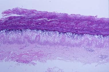
Epidermolytic hyperkeratosis (EH; previously called bullous congenital ichthyosiform erythroderma) is an autosomal dominant skin disease of unknown etiology, affecting approximately 1 out of 300,000 people. It is typified by hyperkeratotic scaliness, blistering due to cytolysis within suprabasal epidermal cells, and hyperproliferation in basal cells. Histologically, EH epidermis exhibits a thickened stratum corneum and granular layer, with enlarged and irregular-shaped cells. Ultrastructurally, only suprabasal layers are affected, with three major aberrancies: (i) tonofilament clumping, (ii) nuclei and keratohyalin granules of irregular shape and size, and (iii) cell degeneration. We have discovered that transgenic mice expressing a mutant keratin 10 gene have the EH phenotype, thereby suggesting that a genetic basis for human EH residues in mutations in genes encoding suprabasal keratins K1 and K10. In addition, we show that (i) stimulation of basal cell proliferation can arise from a defect in suprabasal cells, and (ii) distortion of nuclear shape or aberrations in cytokinesis can occur when an intermediate filament network is perturbed. (+info)SPINK5 and Netherton syndrome: novel mutations, demonstration of missing LEKTI, and differential expression of transglutaminases. (8/37)
Netherton syndrome (NTS) is an autosomal recessive congenital ichthyosis featuring chronic inflammation of the skin, hair anomalies, epidermal hyperplasia with an impaired epidermal barrier function, failure to thrive and atopic manifestations. The disease is caused by mutations in the SPINK5 gene encoding the serine proteinase inhibitor lympho-epithelial Kazal-type inhibitor (LEKTI). Sequence analyses of SPINK5 in seven NTS patients from five different families allowed us to identify two known and three novel mutations all creating premature termination codons. We developed a monoclonal antibody giving a strong signal for LEKTI in the stratum granulosum of normal skin and demonstrated absence of the protein in NTS epidermis. Immunoblot analysis revealed presence of full length LEKTI and of LEKTI cleavage fragments in normal hair roots, whereas in NTS hair roots LEKTI and its cleavage products were completely missing. Transglutaminase1 activity was present throughout almost the entire suprabasal epidermis in NTS, whereas in normal skin it is restricted to the stratum granulosum. In contrast, immunostaining for transglutaminase3 was absent or faint. Moreover, comparable with the altered pattern in psoriatic skin the epidermis in NTS strongly expressed the serine proteinase inhibitor SKALP/elafin and the anti-microbial protein human beta-defensin 2. These studies demonstrate LEKTI deficiency in the epidermis and in hair roots at the protein level and an aberrant expression of other proteins, especially transglutaminase1 and 3, which may account for the impaired epidermal barrier in NTS. (+info)Ichthyosiform erythroderma, congenital, also known as Congenital Ichthyosiform Erythroderma (CIE), is a rare inherited genetic disorder of keratinization. It is characterized by widespread scaliness and erythema (redness) that are present at birth or develop soon thereafter.
The condition is caused by mutations in various genes involved in the development of the skin barrier, leading to abnormalities in the formation and shedding of skin cells. This results in a thickened, scaly appearance of the skin, which can be associated with severe dryness, irritation, and inflammation.
The symptoms of CIE can vary widely among affected individuals, ranging from mild to severe. In addition to the characteristic skin changes, some people with CIE may also experience additional features such as ectropion (outward turning of the eyelids), eclabium (splitting of the lips), and hyperkeratosis of palms and soles.
CIE is typically a lifelong condition, and treatment is focused on managing symptoms and preventing complications. This may include the use of topical moisturizers, emollients, and keratolytic agents to help soften and remove excess skin cells. In some cases, systemic medications such as retinoids may be used to help reduce the severity of skin changes.
Exfoliative dermatitis is a severe form of widespread inflammation of the skin (dermatitis), characterized by widespread scaling and redness, leading to the shedding of large sheets of skin. It can be caused by various factors such as drug reactions, underlying medical conditions (like lymphoma or leukemia), or extensive eczema. Treatment typically involves identifying and removing the cause, along with supportive care, such as moisturizers and medications to control inflammation and itching. In severe cases, hospitalization may be necessary for close monitoring and management of fluid and electrolyte balance.
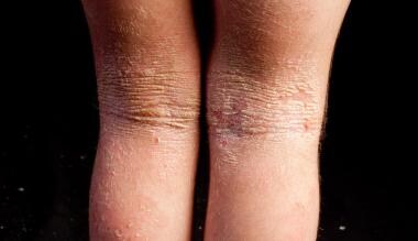
Epidermolytic hyperkeratosis (EH) is a rare genetic skin disorder characterized by the abnormal growth and accumulation of keratin, a protein found in the outermost layer of the skin (epidermis). This condition results in widespread blistering and peeling of the skin, particularly in areas prone to friction such as the hands, feet, knees, and elbows.
EH is caused by mutations in the KRT1 or KRT10 genes, which provide instructions for making keratin proteins that are essential for maintaining the structure and integrity of the epidermis. When these genes are mutated, the keratin proteins become unstable and form clumps, leading to the formation of blisters and areas of thickened, scaly skin (hyperkeratosis).
EH is typically present at birth or appears in early childhood, and it can range from mild to severe. In addition to the skin symptoms, individuals with EH may also experience nail abnormalities, hair loss, and an increased risk of skin infections. Treatment for EH is focused on managing symptoms and preventing complications, and may include topical creams or ointments, wound care, and protection from friction and injury.
Ichthyosis is a group of skin disorders that are characterized by dry, thickened, scaly skin. The name "ichthyosis" comes from the Greek word "ichthys," which means fish, as the skin can have a fish-like scale appearance. These conditions can be inherited or acquired and vary in severity.
The medical definition of ichthyosis is a heterogeneous group of genetic keratinization disorders that result in dry, thickened, and scaly skin. The condition may affect any part of the body, but it most commonly appears on the extremities, scalp, and trunk. Ichthyosis can also have associated symptoms such as redness, itching, and blistering.
The severity of ichthyosis can range from mild to severe, and some forms of the condition may be life-threatening in infancy. The exact symptoms and their severity depend on the specific type of ichthyosis a person has. Treatment for ichthyosis typically involves moisturizing the skin, avoiding irritants, and using medications to help control scaling and inflammation.
Lamellar Ichthyosis is a rare, inherited genetic skin disorder characterized by widespread, persistent scaling of the skin. It is caused by mutations in genes responsible for maintaining the barrier function and hydration of the skin. The condition is present from birth and can vary in severity.
In lamellar ichthyosis, the skin cells do not shed properly and instead accumulate in plates or scales that cover the entire body. These scales are large, dark brown or gray, and have a cracked appearance, resembling fish scales. The scales may be present at birth (congenital) or develop within the first few weeks of life.
The skin is also prone to redness, irritation, and infection due to the impaired barrier function. Other symptoms can include overheating, dehydration, and difficulty with sweating. The condition may improve in warmer, more humid environments.
Treatment for lamellar ichthyosis is aimed at managing symptoms and preventing complications. This may include topical creams and ointments to moisturize the skin, medications to reduce inflammation and infection, and avoiding environmental triggers that can worsen symptoms. In some cases, oral retinoids may be prescribed to help regulate skin cell growth and shedding.
In medical terms, the skin is the largest organ of the human body. It consists of two main layers: the epidermis (outer layer) and dermis (inner layer), as well as accessory structures like hair follicles, sweat glands, and oil glands. The skin plays a crucial role in protecting us from external factors such as bacteria, viruses, and environmental hazards, while also regulating body temperature and enabling the sense of touch.
Etretinate is a oral retinoid medication that is primarily used in the treatment of severe forms of acne, such as recalcitrant cystic acne or nodular acne. It works by decreasing the production of sebum (oil) and promoting the shedding of skin cells, which helps to prevent the formation of comedones (blackheads and whiteheads) and reduce inflammation in the skin.
Etretinate is a derivative of vitamin A and is known for its long-term persistence in the body, with a half-life of approximately 120 days. This means that it can take several months for the drug to be completely eliminated from the body after stopping treatment. As a result, etretinate is usually considered a second-line treatment option for acne and is typically reserved for cases that have not responded to other therapies.
It's important to note that etretinate is a teratogenic medication, which means that it can cause birth defects if taken during pregnancy. Therefore, it should not be used by women who are pregnant or planning to become pregnant, and effective contraception must be used during treatment and for several months after stopping the drug.
Other potential side effects of etretinate include dry skin, dry mouth, nosebleeds, hair loss, muscle aches, and elevated liver enzymes. It may also increase the risk of bone fractures and can interact with other medications, such as tetracyclines, that can increase the risk of intracranial hypertension.
A "drug eruption" is a general term used to describe an adverse skin reaction that occurs as a result of taking a medication. These reactions can vary in severity and appearance, and may include symptoms such as rash, hives, itching, redness, blistering, or peeling of the skin. In some cases, drug eruptions can also cause systemic symptoms such as fever, fatigue, or joint pain.
The exact mechanism by which drugs cause eruptions is not fully understood, but it is thought to involve an abnormal immune response to the medication. There are many different types of drug eruptions, including morphilliform rashes, urticaria (hives), fixed drug eruptions, and Stevens-Johnson syndrome/toxic epidermal necrolysis (SJS/TEN), which is a severe and potentially life-threatening reaction.
If you suspect that you are experiencing a drug eruption, it is important to seek medical attention promptly. Your healthcare provider can help determine the cause of the reaction and recommend appropriate treatment. In some cases, it may be necessary to discontinue the medication causing the reaction and switch to an alternative therapy.
Sezary Syndrome is a rare and aggressive form of cutaneous T-cell lymphoma (CTCL), a type of cancer that involves the skin's immune system. It is characterized by the presence of malignant T-lymphocytes, known as Sezary cells, in the blood, skin, and lymph nodes.
Sezary cells are typically found in large numbers in the peripheral blood, and they have a distinctive appearance with convoluted or "cerebriform" nuclei. These cells can infiltrate the skin, leading to erythroderma (a widespread redness and scaling of the skin), pruritus (severe itching), alopecia (hair loss), and lymphadenopathy (swelling of the lymph nodes).
Sezary Syndrome is often treatment-resistant, and its prognosis is generally poor. Treatment options may include chemotherapy, radiation therapy, photopheresis, immunotherapy, and stem cell transplantation.
The epidermis is the outermost layer of the skin, composed mainly of stratified squamous epithelium. It forms a protective barrier that prevents water loss and inhibits the entry of microorganisms. The epidermis contains no blood vessels, and its cells are nourished by diffusion from the underlying dermis. The bottom-most layer of the epidermis, called the stratum basale, is responsible for generating new skin cells that eventually move up to replace dead cells on the surface. This process of cell turnover takes about 28 days in adults.
The most superficial part of the epidermis consists of dead cells called squames, which are constantly shed and replaced. The exact rate at which this happens varies depending on location; for example, it's faster on the palms and soles than elsewhere. Melanocytes, the pigment-producing cells, are also located in the epidermis, specifically within the stratum basale layer.
In summary, the epidermis is a vital part of our integumentary system, providing not only physical protection but also playing a crucial role in immunity and sensory perception through touch receptors called Pacinian corpuscles.
Dermatology is a medical specialty that focuses on the diagnosis, treatment, and prevention of diseases and conditions related to the skin, hair, nails, and mucous membranes. A dermatologist is a medical doctor who has completed specialized training in this field. They are qualified to treat a wide range of skin conditions, including acne, eczema, psoriasis, skin cancer, and many others. Dermatologists may also perform cosmetic procedures to improve the appearance of the skin or to treat signs of aging.
Cutaneous T-cell lymphoma (CTCL) is a type of cancer that affects T-cells, a specific group of white blood cells called lymphocytes. These cells play a crucial role in the body's immune system and help protect against infection and disease. In CTCL, the T-cells become malignant and accumulate in the skin, leading to various skin symptoms and lesions.
CTCL is a subtype of non-Hodgkin lymphoma (NHL), which refers to a group of cancers that originate from lymphocytes. Within NHL, CTCL is categorized as a type of extranodal lymphoma since it primarily involves organs or tissues outside the lymphatic system, in this case, the skin.
The two most common subtypes of CTCL are mycosis fungoides and Sézary syndrome:
1. Mycosis fungoides (MF): This is the more prevalent form of CTCL, characterized by patches, plaques, or tumors on the skin. The lesions may be scaly, itchy, or change in size, shape, and color over time. MF usually progresses slowly, with early-stage disease often confined to the skin for several years before spreading to lymph nodes or other organs.
2. Sézary syndrome (SS): This is a more aggressive form of CTCL that involves not only the skin but also the blood and lymph nodes. SS is characterized by the presence of malignant T-cells, known as Sézary cells, in the peripheral blood. Patients with SS typically have generalized erythroderma (reddening and scaling of the entire body), pruritus (severe itching), lymphadenopathy (swollen lymph nodes), and alopecia (hair loss).
The diagnosis of CTCL usually involves a combination of clinical examination, skin biopsy, and immunophenotyping to identify the malignant T-cells. Treatment options depend on the stage and subtype of the disease and may include topical therapies, phototherapy, systemic medications, or targeted therapies.
 Congenital ichthyosiform erythroderma
CHILD syndrome
Lipoxygenase
Camisa disease
Loricrin
Keratin 1
ABCA12
Harlequin-type ichthyosis
Ichthyosis with confetti
Ichthyosis bullosa of Siemens
Lamellar ichthyosis
Neutral lipid storage disease
Keratin 10
CYP4F22
Keratin 2A
ALOXE3
Trichorrhexis invaginata
NIPAL4
ALOX12B
Carly Findlay
List of diseases (C)
Theodore K. Lawless
Child (disambiguation)
List of MeSH codes (C16)
CIE
Hyperkeratosis
BCIE
12-Hydroxyeicosatetraenoic acid
List of MeSH codes (C17)
Epidermolytic hyperkeratosis
Congenital ichthyosiform erythroderma - Wikipedia
 Nonbullous congenital ichthyosiform erythroderma: MedlinePlus Genetics
 Epidermolytic Ichthyosis (Epidermolytic Hyperkeratosis or Bullous Congenital Ichthyosiform Erythroderma) Medication
Epidermolytic Ichthyosis (Epidermolytic Hyperkeratosis or Bullous Congenital Ichthyosiform Erythroderma) Medication
Congenital hemidysplasia with ichthyosiform erythroderma and limb defects: MedlinePlus Genetics
 Congenital reticular ichthyosiform erythroderma - PubMed
Epidermolytic Ichthyosis (Epidermolytic Hyperkeratosis or Bullous Congenital Ichthyosiform Erythroderma): Background,...
 Kongenitale ichthyosiforme Erythrodermie: Kein alltägliches Hautbild<...
Epidermolytic Ichthyosis (Epidermolytic Hyperkeratosis or Bullous Congenital Ichthyosiform Erythroderma): Background,...
 Harlequin ichthyosis - About the Disease - Genetic and Rare Diseases Information Center
 Skin Conditions | Hives | Acne | MedlinePlus
 Ichthyosis, X Linked - Symptoms, Causes, Treatment | NORD
 What is Ichthyosis? Definition & Causes | NIAMS
 Advanced Search Results - Public Health Image Library(PHIL)
 SMART: TGc domain annotation
Biomarkers Search
 Ichthyosis - Dermatologic Disorders - MSD Manual Professional Edition
 Research Groups
 Autosomal Recessive Congenital Ichthyosis - GeneReviews® - NCBI Bookshelf
HuGE Navigator|Genopedia|PHGKB
 Member Story | Foundation for Ichthyosis & Related Skin Types (FIRST)
Research - Forbes Porter Lab | NICHD - Eunice Kennedy Shriver National Institute of Child Health and Human Development
 Human keratin 8 mutations that disturb filament assembly observed in inflammatory bowel disease patients | Journal of Cell...
 DeCS
DeCS
 MeSH Browser
MeSH Browser
Prefix: non
 Find Research outputs
- University of East Anglia
Find Research outputs
- University of East Anglia
Ichthyosis31
- Congenital ichthyosiform erythroderma, also known as nonbullous congenital ichthyosiform erythroderma,: 484 is a rare type of the ichthyosis family of skin diseases which occurs in 1 in 200,000 to 300,000 births. (wikipedia.org)
- The disease comes under the umbrella term autosomal recessive congenital ichthyosis, which include non-syndromic congenital ichthyoses such as harlequin ichthyosis and lamellar ichthyosis. (wikipedia.org)
- Eclabium (eversion of the lips), ectropion and alopecia (hair loss) are more common in congenital ichthyosiform erythroderma than in lamellar ichthyosis. (wikipedia.org)
- non-primary source needed] Congenital ichthyosiform erythroderma can present very similarly to lamellar ichthyosis and they often share characteristics, though the two conditions can often be differentiated by the appearance of the scales. (wikipedia.org)
- Scales on patients with congenital ichthyosiform erythroderma are fine and white on skin with erythema while they appear larger and greyer on the limbs, compared to lamellar ichthyosis where scales appear large and dark. (wikipedia.org)
- Following shedding of the collodion membrane, the skin is red (erythroderma) and covered with fine, white scales (ichthyosis). (medlineplus.gov)
- Phenotypic spectrum of autosomal recessive congenital ichthyosis due to PNPLA1 mutation. (medlineplus.gov)
- Molecular analysis of 250 patients with autosomal recessive congenital ichthyosis: evidence for mutation hotspots in ALOXE3 and allelic heterogeneity in ALOX12B. (medlineplus.gov)
- Mutation spectrum and functional analysis of epidermis-type lipoxygenases in patients with autosomal recessive congenital ichthyosis. (medlineplus.gov)
- [ 1 ] is a form of congenital ichthyosis. (medscape.com)
- Epidermolytic ichthyosis presents at birth with erythroderma, blisters, and erosions and evolves over time into varying degrees of hyperkeratosis. (medscape.com)
- Autosomal recessive congenital ichthyosis (ARCI) is a heterogeneous group of skin disorders. (bmj.com)
- Peeling skin syndrome belongs to the groups of congenital ichthyosis and skin fragility disorders with autosomal recessive inheritance. (rarediseases.org)
- Some occur in isolation and are not part of a syndrome (eg, ichthyosis vulgaris, X-linked ichthyosis, lamellar ichthyosis, congenital ichthyosiform erythroderma [epidermolytic hyperkeratosis], harlequin ichthyosis). (msdmanuals.com)
- Autosomal recessive congenital ichthyosis (ARCI) encompasses several forms of nonsyndromic ichthyosis. (nih.gov)
- Although most neonates with ARCI are collodion babies, the clinical presentation and severity of ARCI may vary significantly, ranging from harlequin ichthyosis, the most severe and often fatal form, to lamellar ichthyosis (LI) and (nonbullous) congenital ichthyosiform erythroderma (CIE). (nih.gov)
- Autosomal recessive congenital ichthyosis (ARCI) is a heterogeneous group of disorders of keratinization characterized primarily by abnormal skin scaling over the whole body. (nih.gov)
- The main skin phenotypes are lamellar ichthyosis (LI) and nonbullous congenital ichthyosiform erythroderma (NCIE), although phenotypic overlap within the same patient or among patients from the same family can occur (summary by Fischer, 2009). (nih.gov)
- Congenital Ichthyosis and Related Disorders Sequencing Panel with CNV Detection. (mendelian.co)
- The common forms of ichthyosis are Ichthyosis vulgaris, X-linked ichthyosis, congenital ichthyosiform erythroderma, Epidermolytic hyperkeratosis, and many others. (virashomeopathy.com)
- 2005). For a discussion of genetic heterogeneity of autosomal recessive congenital ichthyosis, see ARCI1 (242300). (nih.gov)
- Self-improving collodion ichthyosis (SICI) is a relatively rare subtype of autosomal recessive congenital ichthyosis (ARCI) that is often characterized by a collodion baby (CB) phenotype at birth. (bvsalud.org)
- Collodion baby is usually a manifestation of autosomal recessive congenital ichthyosis, a heterogeneous group of congenital hyperkeratotic genodermatoses with highly variable severity and genetic background. (bvsalud.org)
- Herein, we report a case of self-improving collodion ichthyosis, a rare subtype of autosomal recessive congenital ichthyosis, characterized by an almost-complete spontaneous resolution of symptoms. (bvsalud.org)
- A teenage boy who was previously diagnosed to have congenital ichthyosis presented to the eye clinic with complaints of gradually decreasing vision in both eyes since childhood. (bvsalud.org)
- Hence, we conclude that congenital ichthyosis can be associated with developmental cataracts. (bvsalud.org)
- Molecular testing identified mutations in a gene encoding lipoxygenase (ALOX12B), associated with autosomal recessive congenital ichthyosis. (bvsalud.org)
- Autosomal recessive congenital ichthyosis (ARCI) is a recently adopted term referring to a heterogeneous group of disorders that share an autosomal recessive pattern of inheritance, collodion membrane presentation at birth and overlap in causative gene mutations. (firstskinfoundation.org)
- These different phenotypes have been termed lamellar ichthyosis (LI), congenital ichthyosiform erythroderma (CIE), and harlequin ichthyosis (HI). (firstskinfoundation.org)
- My two youngest are affected by (nonbullous) Congenital Ichthyosiform Erythroderma (CIE) , a type of Ichthyosis also now called Autosomal Recessive Congenital Ichthyosis (ARCI) . (carlyfindlay.com.au)
- Ichthyosis fetalis is autosomal recessive congenital disorder that affects the outer layers of the skin. (skincarehealthcenter.com)
Bullous7
- Betlloch I, Lucas Costa A, Mataix J, Pérez-Crespo M, Ballester I. Bullous congenital ichthyosiform erythroderma: a sporadic case produced by a new KRT10 gene mutation. (medscape.com)
- Evidence of increased keratinocyte proliferation in air-liquid interface cultures of non-bullous congenital ichthyosiform erythroderma. (medicaljournals.se)
- In this study, we verified the model's validity for the reproduction of a hyperproliferative genodermatosis: non-bullous congenital ichthyosiform erythroderma. (medicaljournals.se)
- Mild recessive bullous congenital ichthyosiform erythroderma due to a previously unidentified homozygous keratin 10 nonsense mutation. (medscape.com)
- Bullous congenital ichthyosiform erythroderma associated with hypocalcemic vitamin D-resistant rickets. (medscape.com)
- Bullous icthyosiform erythroderma with rickets in child of a parent with naevus unius lateralis. (medscape.com)
- Akhyani M, Kiavash K, Kamyab K. Bullous ichthyosiform erythroderma in a child born to a parent with systematized linear epidermolytic hyperkeratosis. (medscape.com)
Autosomal2
- non-primary source needed] Congenital ichthyosiform erythroderma is an autosomal recessive genetic disorder. (wikipedia.org)
- Autosomal recessive congenital ichthyoses (ARCI) are a genetically heterogeneous group of rare and chronic disorders characterized by generalized skin scaling and hyperkeratosis, erythroderma, and palmoplantar keratoderma. (open.ac.uk)
Lamellar1
- In addition, mutations in several genes have been shown to cause both lamellar and nonbullous ichthyosiform erythrodermal phenotypes (Akiyama et al. (nih.gov)
ARCI1
- Reddened skin underlying scale (erythroderma) is variable and reflects overlap with the ARCI-CIE phenotype. (firstskinfoundation.org)
Nonbullous congenital ichthyosiform1
- Mutations in this gene result in nonbullous congenital ichthyosiform erythroderma (NCIE). (prosci-inc.com)
Epidermolytic hyperkeratosis1
- Epidermolytic hyperkeratosis and congenital platelike osteoma cutis in a child. (medscape.com)
NCIE1
- 2010). NCIE is characterized by prominent erythroderma and fine white, superficial, semiadherent scales. (nih.gov)
Syndrome8
- The role of abnormalities in the distal pathway of cholesterol synthesis in the Congenital Hemidysplasia with Ichthyosiform erythroderma and Limb Defects (CHILD) syndrome. (medscape.com)
- The generalized inflammatory types, such as SAM syndrome or Netherton syndrome may be associated with generalized inflammation of the skin (erythroderma) or localized thickened, red plaques (erythrokeratoderma), immunodysfunction with elevated IgE levels, allergies, and susceptibility to infections, failure to thrive or metabolic wasting. (rarediseases.org)
- Erythroderma, also known as generalized exfoliative dermatitis or exfoliative erythroderma, is a severe inflammatory skin syndrome with erythema and desquamation involving more than 90% of the body surface area. (biomedcentral.com)
- CHILD syndrome (congenital hemidysplasia with ichthyosiform erythroderma and limb defects) is a very rare syndrome. (tripod.com)
- Rothmund-Thompson syndrome (congenital poikiloderma) is considered to predominate in girls, but the prevalence is unclear due to the small number of reported cases. (tripod.com)
- Cram DL, Resneck JS, Jackson B.A. congenital ichthyosiform syndrome with deafness and keratitis. (asocolderma.org.co)
- A case of a Japanese neonate with congenital ichthyosiform erythroderma diagnosed as Netherton syndrome. (lu.se)
- Deleterious mutations in SPINK5 in a patient with congenital ichthyosiform erythroderma: molecular testing as a helpful diagnostic tool for Netherton syndrome. (lu.se)
Ichthyoses1
- Congenital ichthyoses are a rare group of genetic disorders caused by defects in the two outermost skin layers, resulting in an abnormal barrier function. (bvsalud.org)
Patients2
- Moreover, we performed two literature reviews to further analyze the characteristics of patients diagnosed with dermatophyte-associated erythroderma or deeper dermal dermatophytosis caused by T. rubrum . (biomedcentral.com)
- Topical steroids of moderate potency may be helpful, but reports have described complications such as aminoaciduria and pituitary adrenal axis suppression, especially in patients with widespread erythroderma, and thus should be used with caution. (medscape.com)
Phenotypes1
- LI and CIE are seemingly distinct phenotypes: classic, severe LI with dark brown, plate-like scale with no erythroderma and CIE with finer whiter scale and underlying generalized redness of the skin. (nih.gov)
Skin2
- Erythroderma is a severe dermatological manifestation of various diseases resulting in generalized skin redness, but erythroderma due to fungi infections is barely reported. (biomedcentral.com)
- :558 ) is a rare congenital disease of the skin caused by a mutation in the KIND1 gene. (en-academic.com)
Chronic1
- Early-onset chronic diarrhoea often indicates a congenital disorder. (biomedcentral.com)
Variants1
- It is a rare X-linked genetic disorder characterized by multiple congenital anomalies with variable severity, caused by pathogenic variants in the MBTPS2 gene, which encodes a zinc metalloprotease that is essential for normal development. (bvsalud.org)
Alopecia1
- The majority of cases are affected with other congenital abnormalities, including central nervous system, skeletal and eye and teeth abnormalities, and patchy alopecia of the vertex. (tripod.com)
Rare1
- Erythroderma due to microbial infections, especially fungi, is very rare. (biomedcentral.com)
Case3
- In this article, we reported the first case of erythroderma combined with deeper dermal dermatophytosis due to Trichophyton rubrum ( T. rubrum ) in a patient with myasthenia gravis. (biomedcentral.com)
- Here, we report a case of erythroderma combined with deep dermatophytosis caused by T. rubrum in a 48-year-old man with myasthenia gravis lasting for 10 years, which to date, has never been reported. (biomedcentral.com)
- Bums FS: A case of generalized congenital erythroderma. (asocolderma.org.co)
Patient1
- Based on histopathological examinations, fungal cultures, and DNA sequencing results, the patient was finally diagnosed with dermatophyte-induced erythroderma combined with deeper dermal dermatophytosis caused by T. rubrum . (biomedcentral.com)
Normal1
- Normal sweating and tear production in congenital ichthyosiform erythroderma with deafness and keratitis. (asocolderma.org.co)
Bullous ichthyosiform erythroderma2
- Akhyani M, Kiavash K, Kamyab K. Bullous ichthyosiform erythroderma in a child born to a parent with systematized linear epidermolytic hyperkeratosis. (medscape.com)
- Biopsies from lesions on each of the four patients showed similar edematous and dyskeratotic changes in the epidermis of the type described for bullous ichthyosiform erythroderma. (jamanetwork.com)
Netherton4
- Deleterious mutations in SPINK5 in a patient with congenital ichthyosiform erythroderma: molecular testing as a helpful diagnostic tool for Netherton syndrome. (nih.gov)
- Netherton syndrome is a rare autosomal recessive genodermatosis characterized by congenital ichthyosiform erythroderma, an atopic diathesis, and a characteristic hair-shaft abnormality known as trichorrhexis invaginata. (medscape.com)
- 8. A case of a Japanese neonate with congenital ichthyosiform erythroderma diagnosed as Netherton syndrome. (nih.gov)
- Netherton syndrome (NETH) is a rare and severe autosomal recessive skin disorder characterized by congenital erythroderma, a specific hair-shaft abnormality, and atopic manifestations with high IgE levels. (nih.gov)
Scaly1
- Generalized scaly erythroderma is apparent at or soon after birth and usually persists. (nih.gov)
Genetic3
- non-primary source needed] Congenital ichthyosiform erythroderma is an autosomal recessive genetic disorder. (wikipedia.org)
- citation needed] There are several genetic faults which can produce Congenital ichthyosiform erythroderma. (wikipedia.org)
- It is a rare X-linked genetic disorder characterized by multiple congenital anomalies with variable severity, caused by pathogenic variants in the MBTPS2 gene, which encodes a zinc metalloprotease that is essential for normal development. (bvsalud.org)
Mutations2
- The collodion phenotype was based on congenital ichthyosiform erythroderma, which was caused by two compound heterozygote mutations in the ALOX12B gene. (uni-luebeck.de)
- This type II cytokeratin is specifically expressed in the spinous and granular layers of the epidermis with family member KRT10 and mutations in these genes have been associated with bullous congenital ichthyosiform erythroderma. (anticorps-enligne.fr)
Abnormality1
- Erythroderma and hair abnormality persist. (medscape.com)
Hyperkeratosis2
- Epidermolytic hyperkeratosis and congenital platelike osteoma cutis in a child. (medscape.com)
- Four patients with congenital ichthyosiform hyperkeratosis are presented. (jamanetwork.com)
Scales1
- 2010). NCIE is characterized by prominent erythroderma and fine white, superficial, semiadherent scales. (nih.gov)
Ectropion1
- In severe cases of congenital ichthyosiform erythroderma a child may also have drooping lower eyelids (ectropion) , mild hair loss and tight skin on the fingers. (kivetonpharmacy.com)
Infections1
- During childhood, failure to thrive is common as a result of malnutrition, metabolic disorders, chronic erythroderma, persistent cutaneous infections, or enteropathy (summary by Bitoun et al. (nih.gov)
Birth1
- Congenital ichthyosiform erythroderma is present at birth. (nih.gov)
Child1
- Bullous icthyosiform erythroderma with rickets in child of a parent with naevus unius lateralis. (medscape.com)