Immunoglobulin G
Immunoglobulins
Immunoglobulin M
Immunoglobulin A
Immunoglobulin Heavy Chains
Immunoglobulins, Intravenous
Genes, Immunoglobulin
Immunoglobulin Light Chains
Immunoglobulin kappa-Chains
Immunoglobulin Fc Fragments
Immunoglobulin Isotypes
Immunoglobulin E
Immunoglobulin A, Secretory
Enzyme-Linked Immunosorbent Assay
Immunoglobulin Variable Region
Immunoglobulin Fab Fragments
Immunoglobulin mu-Chains
Antibody Specificity
Immunoglobulin lambda-Chains
B-Lymphocytes
Immunoglobulin D
Immunoglobulin Constant Regions
Immunoglobulin gamma-Chains
Immunoglobulin Fragments
Immunoglobulin Class Switching
Staphylococcal Protein A
Immunoglobulin Allotypes
Receptors, Fc
Receptors, IgG
Immunoglobulin J-Chains
Antibody Affinity
Mice, Inbred BALB C
Antibody Formation
Colostrum
Antigen-Antibody Complex
Antibodies, Anti-Idiotypic
Immunization
Receptors, Polymeric Immunoglobulin
Immune Sera
Immunization, Passive
Cross Reactions
Immunoglobulin Joining Region
Molecular Sequence Data
Rabbits
Receptors, Antigen, B-Cell
gamma-Globulins
Binding Sites, Antibody
Bacterial Vaccines
Plasmacytoma
Opsonin Proteins
Immunodiffusion
Immunoelectrophoresis
Fluorescent Antibody Technique
Autoantibodies
Amino Acid Sequence
Immunoglobulin Idiotypes
Immunoenzyme Techniques
Genes, Immunoglobulin Heavy Chain
Somatic Hypermutation, Immunoglobulin
Agammaglobulinemia
Complement System Proteins
Rubella virus
Antibodies
Antigen-Antibody Reactions
Base Sequence
Immunoglobulin delta-Chains
Antigens, Protozoan
Immunoglobulin Switch Region
Immunoassay
Immunoglobulin alpha-Chains
Sensitivity and Specificity
Complement Fixation Tests
Neutralization Tests
Immunity, Maternally-Acquired
Immunoglobulin Gm Allotypes
Hybridomas
Toxoplasma
Secretory Component
Complement C3
Phagocytosis
Lymphocytes
Plasma Cells
Saliva
Vaccination
T-Lymphocytes
Rheumatoid Factor
Evaluation Studies as Topic
Hemagglutination Tests
Tetanus Toxoid
Rubella
Gene Rearrangement, B-Lymphocyte, Heavy Chain
Antigens, Surface
Immunity, Mucosal
Gene Rearrangement, B-Lymphocyte
Lymphocyte Activation
Dose-Response Relationship, Immunologic
Radioimmunoassay
Hypergammaglobulinemia
Adjuvants, Immunologic
Immunologic Deficiency Syndromes
Hemagglutination Inhibition Tests
Mice, Inbred C57BL
Paraproteinemias
Goats
Immunoglobulins, Thyroid-Stimulating
Cells, Cultured
Seroepidemiologic Studies
Vaccines, Synthetic
Cattle
Antibody-Producing Cells
Pokeweed Mitogens
Bacterial Outer Membrane Proteins
Dysgammaglobulinemia
Immunoblotting
Agglutinins
Recombinant Fusion Proteins
Immunosorbent Techniques
Genes, Immunoglobulin Light Chain
Immunoglobulin epsilon-Chains
Multiple Myeloma
Blood Bactericidal Activity
Lipopolysaccharides
Pregnancy
Electrophoresis, Polyacrylamide Gel
Gene Rearrangement
Interleukin-4
Bence Jones Protein
Erythrocytes
Gene Rearrangement, B-Lymphocyte, Light Chain
Autoimmune Diseases
Flow Cytometry
Antigens, Helminth
Antigens, CD
Reagent Kits, Diagnostic
Species Specificity
Immunologic Techniques
Vaccines, Conjugate
Polymerase Chain Reaction
Streptococcus pneumoniae
Protein Binding
Complement Activation
Interferon-gamma
Cloning, Molecular
Common Variable Immunodeficiency
Rosette Formation
Agglutination Tests
Antitoxins
Antibody Diversity
Mice, Inbred Strains
Periodontitis
Nephelometry and Turbidimetry
Complement C1q
Cytomegalovirus
Papain
Antibodies, Antinuclear
Chromatography, Gel
Immunity, Cellular
Waldenstrom Macroglobulinemia
Iodine Radioisotopes
Immunoglobulin Km Allotypes
Pepsin A
Blood Proteins
Serum Albumin
Pregnancy Complications, Parasitic
Cytokines
Blotting, Western
DNA
Disease Models, Animal
Helicobacter Infections
Glycoproteins
Bacterial Toxins
CD4 Immunoadhesins
Helicobacter pylori
Polysaccharides
The role of colorstrum on the occurrence of immunoglobulin G subclasses and antibody production in neonatal goats. (1/23942)
Quantitative determinations of IgG1 and IgG2, in one group of colostrum-fed and one group of colostrum-deprived neonatal goats revealed that the occurrence of the IgG1 subclass preceeded that of the IgG2 in both cases. In the colostrum-fed animals the IgG2 appeared, on an average, in the fourth week of life whereas in the colostrum-deprived animals the IgG2 was detected as early as three weeks after birth. At the age of twelve weeks the mean concentrations for IgG, and IgG2 were higher in the animals deprived of colostrum. The immune response to human gamma globulin was studied in colostrum-fed and colostrum-deprived neonatal goats which were immunized at birth and again after four and eight weeks. Following the first two antigen administrations a significantly higher response was obtained in the colostrum-fed neonates. However, the third injection determined a similar response in both groups. A marked suppressive effect on the immune response was observed in colostrum-fed neonatal goats when specific antibodies were present in the colostrum after preimmunization of the mothers with human gamma globulin. (+info)Features of the immune response to DNA in mice. I. Genetic control. (2/23942)
The genetic control of the immune response to DNA was studied in various strains of mice F1 hybrids and corresponding back-crosses immunized with single stranded DNA complexed to methylated bovine serum albumin. Anti-DNA antibody response was measured by radioimmuno-logical technique. High responder, low responder, and intermediate responder strains were found and the ability to respond to DNA was characterized as a dominant genetic trait which is not linked to the major locus of histocompatibility. Studies in back-crosses suggested that this immune response is under multigenic control. High responder mice produce both anti-double stranded DNA and anti-single stranded DNA 7S and 19S antibodies, while low responder mice produce mainly anti-single stranded DNA 19S antibodies. (+info)VEGF is required for growth and survival in neonatal mice. (3/23942)
We employed two independent approaches to inactivate the angiogenic protein VEGF in newborn mice: inducible, Cre-loxP- mediated gene targeting, or administration of mFlt(1-3)-IgG, a soluble VEGF receptor chimeric protein. Partial inhibition of VEGF achieved by inducible gene targeting resulted in increased mortality, stunted body growth and impaired organ development, most notably of the liver. Administration of mFlt(1-3)-IgG, which achieves a higher degree of VEGF inhibition, resulted in nearly complete growth arrest and lethality. Ultrastructural analysis documented alterations in endothelial and other cell types. Histological and biochemical changes consistent with liver and renal failure were observed. Endothelial cells isolated from the liver of mFlt(1-3)-IgG-treated neonates demonstrated an increased apoptotic index, indicating that VEGF is required not only for proliferation but also for survival of endothelial cells. However, such treatment resulted in less significant alterations as the animal matured, and the dependence on VEGF was eventually lost some time after the fourth postnatal week. Administration of mFlt(1-3)-IgG to juvenile mice failed to induce apoptosis in liver endothelial cells. Thus, VEGF is essential for growth and survival in early postnatal life. However, in the fully developed animal, VEGF is likely to be involved primarily in active angiogenesis processes such as corpus luteum development. (+info)Interaction of inflammatory cells and oral microorganisms. III. Modulation of rabbit polymorphonuclear leukocyte hydrolase release response to Actinomyces viscosus and Streptococcus mutans by immunoglobulins and complement. (4/23942)
In the absence of antiserum, rabbit polymorphonuclear leukocytes (PMNs) released lysosomal enzymes in response to Actinomyces viscosus (19246) but not to Streptococcus mutans (6715). Antibodies had a marked modulating influence on these reactions. PMN hydrolase release was significantly enhanced to both organisms when specific rabbit antiserum and isolated immunoglobulin G (IgG) were included in the incubations. Immune complex F(ab')2 fragments of IgG directed against S. mutans agglutinated bacteria. Immune complexes consisting of S. mutans and F(ab')2 fragments of IgG directed against this organism were not effective as bacteria-IgG complexes in stimulating PMN release. The intensity of the release response to bacteria-IgG complexes was also diminished when PMNs were preincubated with isolated Fc fragments derived from IgG. Fresh serum as a source of complement components had no demonstrable effect on PMN release either alone or in conjuction with antiserum in these experiments. These data may be relevant to the mechanisms and consequences of the interaction of PMNs and plaque bacteria in the pathogenesis of periodontal disease. (+info)Prevention of collagen-induced arthritis by gene delivery of soluble p75 tumour necrosis factor receptor. (5/23942)
Collagen type II-induced arthritis (CIA) in DBA/1 mice can be passively transferred to SCID mice with spleen B- and T-lymphocytes. In the present study, we show that infection ex vivo of splenocytes from arthritic DBA/1 mice with a retroviral vector, containing cDNA for the soluble form of human p75 receptor of tumour necrosis factor (TNF-R) before transfer, prevents the development of arthritis, bone erosion and joint inflammation in the SCID recipients. Assessment of IgG subclass levels and studies of synovial histology suggest that down-regulating the effector functions of T helper-type 1 (Th1) cells may, at least in part, explain the inhibition of arthritis in the SCID recipients. In contrast, the transfer of splenocytes infected with mouse TNF-alpha gene construct resulted in exacerbated arthritis and enhancement of IgG2a antibody levels. Intriguingly, infection of splenocytes from arthritic DBA/1 mice with a construct for mouse IL-10 had no modulating effect on the transfer of arthritis. The data suggest that manipulation of the immune system with cytokines, or cytokine inhibitors using gene transfer protocols can be an effective approach to ameliorate arthritis. (+info)Variable domain-linked oligosaccharides of a human monoclonal IgG: structure and influence on antigen binding. (6/23942)
The variable-domain-attached oligosaccharide side chains of a human IgG produced by a human-human-mouse heterohybridoma were analysed. In addition to the conserved N-glycosylation site at Asn-297, an N-glycosylation consensus sequence (Asn-Asn-Ser) is located at position 75 in the variable region of its heavy chain. The antibody was cleaved into its antigen-binding (Fab) and crystallizing fragments. The oligosaccharides of the Fab fragment were released by digestion with various endo- and exoglycosidases and analysed by anion-exchange chromatography and fluorophore-assisted carbohydrate electrophoresis. The predominant components were disialyl- bi-antennary and tetra-sialyl tetra-antennary complex carbohydrates. Of note is the presence in this human IgG of oligosaccharides containing N-glycolylneuraminic acid and N-acetylneuraminic acid in the ratio of 94:6. Furthermore, we determined N-acetylgalactosamine in the Fab fragment of this antibody, suggesting the presence of O-linked carbohydrates. A three-dimensional structure of the glycosylated variable (Fv) fragment was suggested using computer-assisted modelling. In addition, the influence of the Fv-associated oligosaccharides of the CBGA1 antibody on antigen binding was tested in several ELISA systems. Deglycosylation resulted in a decreased antigen-binding activity. (+info)Zonula occludens toxin is a powerful mucosal adjuvant for intranasally delivered antigens. (7/23942)
Zonula occludens toxin (Zot) is produced by toxigenic strains of Vibrio cholerae and has the ability to reversibly alter intestinal epithelial tight junctions, allowing the passage of macromolecules through the mucosal barrier. In the present study, we investigated whether Zot could be exploited to deliver soluble antigens through the nasal mucosa for the induction of antigen-specific systemic and mucosal immune responses. Intranasal immunization of mice with ovalbumin (Ova) and recombinant Zot, either fused to the maltose-binding protein (MBP-Zot) or with a hexahistidine tag (His-Zot), induced anti-Ova serum immunoglobulin G (IgG) titers that were approximately 40-fold higher than those induced by immunization with antigen alone. Interestingly, Zot also stimulated high anti-Ova IgA titers in serum, as well as in vaginal and intestinal secretions. A comparison with Escherichia coli heat-labile enterotoxin (LT) revealed that the adjuvant activity of Zot was only sevenfold lower than that of LT. Moreover, Zot and LT induced similar patterns of Ova-specific IgG subclasses. The subtypes IgG1, IgG2a, and IgG2b were all stimulated, with a predominance of IgG1 and IgG2b. In conclusion, our results highlight Zot as a novel potent mucosal adjuvant of microbial origin. (+info)The levels and bactericidal capacity of antibodies directed against the UspA1 and UspA2 outer membrane proteins of Moraxella (Branhamella) catarrhalis in adults and children. (8/23942)
The UspA1 and UspA2 proteins from Moraxella catarrhalis share antigenic epitopes and are promising vaccine candidates. In this study, the levels and bactericidal activities of antibodies in sera from healthy adults and children toward UspA1 and UspA2 from the O35E strain were measured. Human sera contained antibodies to both proteins, and the levels of immunoglobulin G (IgG) antibodies were age dependent. Adult sera had significantly higher titers of IgG than child sera (P < 0.01). The IgG3 titers to the UspA proteins were higher than the IgG1 titers in the adults' sera, while the IgG1 titers were higher than the IgG3 titers in the children's sera (P < 0.05). The IgG antibodies in the sera from 2-month-old children appeared to be maternally derived, since the mean titer was significantly higher than that in sera from 6- to 7-month-old children (P < 0.05). Serum IgA antibodies to both UspA1 and UspA2 were low during the first 7 months of age but thereafter gradually increased along with the IgG titers. Analysis of sera absorbed with UspA1 or UspA2 showed that the antibodies to UspA1 and UspA2 were cross-reactive with each other and associated with serum bactericidal activity. Examination of affinity-purified human antibodies confirmed that naturally acquired antibodies to UspA1 and UspA2 were bactericidal and cross-reactive. These results support using UspA1 and UspA2 in a vaccine to prevent M. catarrhalis infections. (+info)Immunoglobulin G (IgG) is a type of antibody, which is a protective protein produced by the immune system in response to foreign substances like bacteria or viruses. IgG is the most abundant type of antibody in human blood, making up about 75-80% of all antibodies. It is found in all body fluids and plays a crucial role in fighting infections caused by bacteria, viruses, and toxins.
IgG has several important functions:
1. Neutralization: IgG can bind to the surface of bacteria or viruses, preventing them from attaching to and infecting human cells.
2. Opsonization: IgG coats the surface of pathogens, making them more recognizable and easier for immune cells like neutrophils and macrophages to phagocytose (engulf and destroy) them.
3. Complement activation: IgG can activate the complement system, a group of proteins that work together to help eliminate pathogens from the body. Activation of the complement system leads to the formation of the membrane attack complex, which creates holes in the cell membranes of bacteria, leading to their lysis (destruction).
4. Antibody-dependent cellular cytotoxicity (ADCC): IgG can bind to immune cells like natural killer (NK) cells and trigger them to release substances that cause target cells (such as virus-infected or cancerous cells) to undergo apoptosis (programmed cell death).
5. Immune complex formation: IgG can form immune complexes with antigens, which can then be removed from the body through various mechanisms, such as phagocytosis by immune cells or excretion in urine.
IgG is a critical component of adaptive immunity and provides long-lasting protection against reinfection with many pathogens. It has four subclasses (IgG1, IgG2, IgG3, and IgG4) that differ in their structure, function, and distribution in the body.
Immunoglobulins (Igs), also known as antibodies, are glycoprotein molecules produced by the immune system's B cells in response to the presence of foreign substances, such as bacteria, viruses, and toxins. These Y-shaped proteins play a crucial role in identifying and neutralizing pathogens and other antigens, thereby protecting the body against infection and disease.
Immunoglobulins are composed of four polypeptide chains: two identical heavy chains and two identical light chains, held together by disulfide bonds. The variable regions of these chains form the antigen-binding sites, which recognize and bind to specific epitopes on antigens. Based on their heavy chain type, immunoglobulins are classified into five main isotypes or classes: IgA, IgD, IgE, IgG, and IgM. Each class has distinct functions in the immune response, such as providing protection in different body fluids and tissues, mediating hypersensitivity reactions, and aiding in the development of immunological memory.
In medical settings, immunoglobulins can be administered therapeutically to provide passive immunity against certain diseases or to treat immune deficiencies, autoimmune disorders, and other conditions that may benefit from immunomodulation.
Immunoglobulin M (IgM) is a type of antibody that is primarily found in the blood and lymph fluid. It is the first antibody to be produced in response to an initial exposure to an antigen, making it an important part of the body's primary immune response. IgM antibodies are large molecules that are composed of five basic units, giving them a pentameric structure. They are primarily found on the surface of B cells as membrane-bound immunoglobulins (mlgM), where they function as receptors for antigens. Once an mlgM receptor binds to an antigen, it triggers the activation and differentiation of the B cell into a plasma cell that produces and secretes large amounts of soluble IgM antibodies.
IgM antibodies are particularly effective at agglutination (clumping) and complement activation, which makes them important in the early stages of an immune response to help clear pathogens from the bloodstream. However, they are not as stable or long-lived as other types of antibodies, such as IgG, and their levels tend to decline after the initial immune response has occurred.
In summary, Immunoglobulin M (IgM) is a type of antibody that plays a crucial role in the primary immune response to antigens by agglutination and complement activation. It is primarily found in the blood and lymph fluid, and it is produced by B cells after they are activated by an antigen.
Immunoglobulin A (IgA) is a type of antibody that plays a crucial role in the immune function of the human body. It is primarily found in external secretions, such as saliva, tears, breast milk, and sweat, as well as in mucous membranes lining the respiratory and gastrointestinal tracts. IgA exists in two forms: a monomeric form found in serum and a polymeric form found in secretions.
The primary function of IgA is to provide immune protection at mucosal surfaces, which are exposed to various environmental antigens, such as bacteria, viruses, parasites, and allergens. By doing so, it helps prevent the entry and colonization of pathogens into the body, reducing the risk of infections and inflammation.
IgA functions by binding to antigens present on the surface of pathogens or allergens, forming immune complexes that can neutralize their activity. These complexes are then transported across the epithelial cells lining mucosal surfaces and released into the lumen, where they prevent the adherence and invasion of pathogens.
In summary, Immunoglobulin A (IgA) is a vital antibody that provides immune defense at mucosal surfaces by neutralizing and preventing the entry of harmful antigens into the body.
Immunoglobulin heavy chains are proteins that make up the framework of antibodies, which are Y-shaped immune proteins. These heavy chains, along with light chains, form the antigen-binding sites of an antibody, which recognize and bind to specific foreign substances (antigens) in order to neutralize or remove them from the body.
The heavy chain is composed of a variable region, which contains the antigen-binding site, and constant regions that determine the class and function of the antibody. There are five classes of immunoglobulins (IgA, IgD, IgE, IgG, and IgM) that differ in their heavy chain constant regions and therefore have different functions in the immune response.
Immunoglobulin heavy chains are synthesized by B cells, a type of white blood cell involved in the adaptive immune response. The genetic rearrangement of immunoglobulin heavy chain genes during B cell development results in the production of a vast array of different antibodies with unique antigen-binding sites, allowing for the recognition and elimination of a wide variety of pathogens.
Intravenous Immunoglobulins (IVIG) are a preparation of antibodies, specifically immunoglobulins, that are derived from the plasma of healthy donors. They are administered intravenously to provide passive immunity and help boost the immune system's response in individuals with weakened or compromised immune systems. IVIG can be used for various medical conditions such as primary immunodeficiency disorders, secondary immunodeficiencies, autoimmune diseases, and some infectious diseases. The administration of IVIG can help prevent infections, reduce the severity and frequency of infections, and manage the symptoms of certain autoimmune disorders. It is important to note that while IVIG provides temporary immunity, it does not replace a person's own immune system.
Immunoglobulins (Igs), also known as antibodies, are proteins produced by the immune system to recognize and neutralize foreign substances such as pathogens or toxins. They are composed of four polypeptide chains: two heavy chains and two light chains, which are held together by disulfide bonds. The variable regions of the heavy and light chains contain loops that form the antigen-binding site, allowing each Ig molecule to recognize a specific epitope (antigenic determinant) on an antigen.
Genes encoding immunoglobulins are located on chromosome 14 (light chain genes) and chromosomes 22 and 2 (heavy chain genes). The diversity of the immune system is generated through a process called V(D)J recombination, where variable (V), diversity (D), and joining (J) gene segments are randomly selected and assembled to form the variable regions of the heavy and light chains. This results in an enormous number of possible combinations, allowing the immune system to recognize and respond to a vast array of potential threats.
There are five classes of immunoglobulins: IgA, IgD, IgE, IgG, and IgM, each with distinct functions and structures. For example, IgG is the most abundant class in serum and provides long-term protection against pathogens, while IgA is found on mucosal surfaces and helps prevent the entry of pathogens into the body.
Immunoglobulin light chains are the smaller protein subunits of an immunoglobulin, also known as an antibody. They are composed of two polypeptide chains, called kappa (κ) and lambda (λ), which are produced by B cells during the immune response. Each immunoglobulin molecule contains either two kappa or two lambda light chains, in association with two heavy chains.
Light chains play a crucial role in the antigen-binding site of an antibody, where they contribute to the specificity and affinity of the interaction between the antibody and its target antigen. In addition to their role in immune function, abnormal production or accumulation of light chains can lead to various diseases, such as multiple myeloma and amyloidosis.
Bacterial antibodies are a type of antibodies produced by the immune system in response to an infection caused by bacteria. These antibodies are proteins that recognize and bind to specific antigens on the surface of the bacterial cells, marking them for destruction by other immune cells. Bacterial antibodies can be classified into several types based on their structure and function, including IgG, IgM, IgA, and IgE. They play a crucial role in the body's defense against bacterial infections and provide immunity to future infections with the same bacteria.
Immunoglobulin kappa-chains are one of the two types of light chains (the other being lambda-chains) that make up an immunoglobulin molecule, also known as an antibody. These light chains combine with heavy chains to form the antigen-binding site of an antibody, which is responsible for recognizing and binding to specific antigens or foreign substances in the body.
Kappa-chains contain a variable region that differs between different antibodies and contributes to the diversity of the immune system's response to various antigens. They also have a constant region, which is consistent across all kappa-chains. Approximately 60% of all human antibodies contain kappa-chains, while the remaining 40% contain lambda-chains. The relative proportions of kappa and lambda chains can be used in diagnostic tests to identify clonal expansions of B cells, which may indicate a malignancy such as multiple myeloma or lymphoma.
Immunoglobulin Fc fragments are the crystallizable fragment of an antibody that is responsible for effector functions such as engagement with Fc receptors on immune cells, activation of the complement system, and neutralization of toxins. The Fc region is located at the tail end of the Y-shaped immunoglobulin molecule, and it is made up of constant regions of the heavy chains of the antibody.
When an antibody binds to its target antigen, the Fc region can interact with other proteins in the immune system, leading to a variety of responses such as phagocytosis, antibody-dependent cellular cytotoxicity (ADCC), and complement activation. These effector functions help to eliminate pathogens and infected cells from the body.
Immunoglobulin Fc fragments can be produced artificially through enzymatic digestion of intact antibodies, resulting in a fragment that retains the ability to interact with Fc receptors and other proteins involved in immune responses. These fragments have potential therapeutic applications in a variety of diseases, including autoimmune disorders, inflammatory conditions, and cancer.
Immunoglobulins, also known as antibodies, are proteins produced by the immune system to recognize and neutralize foreign substances like pathogens or antigens. The term "immunoglobulin isotypes" refers to the different classes of immunoglobulins that share a similar structure but have distinct functions and properties.
There are five main isotypes of immunoglobulins in humans, namely IgA, IgD, IgE, IgG, and IgM. Each isotype has a unique heavy chain constant region (CH) that determines its effector functions, such as binding to Fc receptors, complement activation, or protection against pathogens.
IgA is primarily found in external secretions like tears, saliva, and breast milk, providing localized immunity at mucosal surfaces. IgD is expressed on the surface of B cells and plays a role in their activation and differentiation. IgE is associated with allergic responses and binds to mast cells and basophils, triggering the release of histamine and other mediators of inflammation.
IgG is the most abundant isotype in serum and has several subclasses (IgG1, IgG2, IgG3, and IgG4) that differ in their effector functions. IgG can cross the placenta, providing passive immunity to the fetus. IgM is the first antibody produced during an immune response and is primarily found in the bloodstream, where it forms large pentameric complexes that are effective at agglutination and complement activation.
Overall, immunoglobulin isotypes play a crucial role in the adaptive immune response, providing specific and diverse mechanisms for recognizing and neutralizing foreign substances.
Immunoglobulin E (IgE) is a type of antibody that plays a key role in the immune response to parasitic infections and allergies. It is produced by B cells in response to stimulation by antigens, such as pollen, pet dander, or certain foods. Once produced, IgE binds to receptors on the surface of mast cells and basophils, which are immune cells found in tissues and blood respectively. When an individual with IgE antibodies encounters the allergen again, the cross-linking of IgE molecules bound to the FcεRI receptor triggers the release of mediators such as histamine, leukotrienes, prostaglandins, and various cytokines from these cells. These mediators cause the symptoms of an allergic reaction, such as itching, swelling, and redness. IgE also plays a role in protecting against certain parasitic infections by activating eosinophils, which can kill the parasites.
In summary, Immunoglobulin E (IgE) is a type of antibody that plays a crucial role in the immune response to allergens and parasitic infections, it binds to receptors on the surface of mast cells and basophils, when an individual with IgE antibodies encounters the allergen again, it triggers the release of mediators from these cells causing the symptoms of an allergic reaction.
Immunoglobulin A (IgA), Secretory is a type of antibody that plays a crucial role in the immune function of mucous membranes. These membranes line various body openings, such as the respiratory and gastrointestinal tracts, and serve to protect the body from potential pathogens by producing mucus.
Secretory IgA (SIgA) is the primary immunoglobulin found in secretions of the mucous membranes, and it is produced by a special type of immune cell called plasma cells located in the lamina propria, a layer of tissue beneath the epithelial cells that line the mucosal surfaces.
SIgA exists as a dimer, consisting of two IgA molecules linked together by a protein called the J chain. This complex is then transported across the epithelial cell layer to the luminal surface, where it becomes associated with another protein called the secretory component (SC). The SC protects the SIgA from degradation by enzymes and helps it maintain its function in the harsh environment of the mucosal surfaces.
SIgA functions by preventing the attachment and entry of pathogens into the body, thereby neutralizing their infectivity. It can also agglutinate (clump together) microorganisms, making them more susceptible to removal by mucociliary clearance or peristalsis. Furthermore, SIgA can modulate immune responses and contribute to the development of oral tolerance, which is important for maintaining immune homeostasis in the gut.
An Enzyme-Linked Immunosorbent Assay (ELISA) is a type of analytical biochemistry assay used to detect and quantify the presence of a substance, typically a protein or peptide, in a liquid sample. It takes its name from the enzyme-linked antibodies used in the assay.
In an ELISA, the sample is added to a well containing a surface that has been treated to capture the target substance. If the target substance is present in the sample, it will bind to the surface. Next, an enzyme-linked antibody specific to the target substance is added. This antibody will bind to the captured target substance if it is present. After washing away any unbound material, a substrate for the enzyme is added. If the enzyme is present due to its linkage to the antibody, it will catalyze a reaction that produces a detectable signal, such as a color change or fluorescence. The intensity of this signal is proportional to the amount of target substance present in the sample, allowing for quantification.
ELISAs are widely used in research and clinical settings to detect and measure various substances, including hormones, viruses, and bacteria. They offer high sensitivity, specificity, and reproducibility, making them a reliable choice for many applications.
The Immunoglobulin (Ig) variable region is the antigen-binding part of an antibody, which is highly variable in its amino acid sequence and therefore specific to a particular epitope (the site on an antigen that is recognized by the antigen-binding site of an antibody). This variability is generated during the process of V(D)J recombination in the maturation of B cells, allowing for a diverse repertoire of antibodies to be produced and recognizing a wide range of potential pathogens.
The variable region is composed of several sub-regions including:
1. The heavy chain variable region (VH)
2. The light chain variable region (VL)
3. The heavy chain joining region (JH)
4. The light chain joining region (JL)
These regions are further divided into framework regions and complementarity-determining regions (CDRs). The CDRs, particularly CDR3, contain the most variability and are primarily responsible for antigen recognition.
Immunoglobulin (Ig) Fab fragments are the antigen-binding portions of an antibody that result from the digestion of the whole antibody molecule by enzymes such as papain. An antibody, also known as an immunoglobulin, is a Y-shaped protein produced by the immune system to identify and neutralize foreign substances like bacteria, viruses, or toxins. The antibody has two identical antigen-binding sites, located at the tips of the two shorter arms, which can bind specifically to a target antigen.
Fab fragments are formed when an antibody is cleaved by papain, resulting in two Fab fragments and one Fc fragment. Each Fab fragment contains one antigen-binding site, composed of a variable region (Fv) and a constant region (C). The Fv region is responsible for the specificity and affinity of the antigen binding, while the C region contributes to the effector functions of the antibody.
Fab fragments are often used in various medical applications, such as immunodiagnostics and targeted therapies, due to their ability to bind specifically to target antigens without triggering an immune response or other effector functions associated with the Fc region.
Immunoglobulin mu-chains (IgM) are a type of heavy chain found in immunoglobulins, also known as antibodies. IgM is the first antibody to be produced in response to an initial exposure to an antigen and plays a crucial role in the early stages of the immune response.
IgM antibodies are composed of four monomeric units, each consisting of two heavy chains and two light chains. The heavy chains in IgM are called mu-chains, which have a molecular weight of approximately 72 kDa. Each mu-chain contains five domains: one variable (V) domain at the N-terminus, four constant (C) domains (Cμ1-4), and a membrane-spanning region followed by a short cytoplasmic tail.
IgM antibodies are primarily found on the surface of B cells as part of the B cell receptor (BCR). When a B cell encounters an antigen, the BCR binds to it, triggering a series of intracellular signaling events that lead to B cell activation and differentiation into plasma cells. In response to activation, the B cell begins to secrete IgM antibodies into the bloodstream.
IgM antibodies have several unique features that make them effective in the early stages of an immune response. They are highly efficient at agglutination, or clumping together, of pathogens and antigens, which helps to neutralize them. IgM antibodies also activate the complement system, a group of proteins that work together to destroy pathogens.
Overall, Immunoglobulin mu-chains are an essential component of the immune system, providing early protection against pathogens and initiating the adaptive immune response.
Antibody specificity refers to the ability of an antibody to bind to a specific epitope or antigenic determinant on an antigen. Each antibody has a unique structure that allows it to recognize and bind to a specific region of an antigen, typically a small portion of the antigen's surface made up of amino acids or sugar residues. This highly specific binding is mediated by the variable regions of the antibody's heavy and light chains, which form a pocket that recognizes and binds to the epitope.
The specificity of an antibody is determined by its unique complementarity-determining regions (CDRs), which are loops of amino acids located in the variable domains of both the heavy and light chains. The CDRs form a binding site that recognizes and interacts with the epitope on the antigen. The precise fit between the antibody's binding site and the epitope is critical for specificity, as even small changes in the structure of either can prevent binding.
Antibody specificity is important in immune responses because it allows the immune system to distinguish between self and non-self antigens. This helps to prevent autoimmune reactions where the immune system attacks the body's own cells and tissues. Antibody specificity also plays a crucial role in diagnostic tests, such as ELISA assays, where antibodies are used to detect the presence of specific antigens in biological samples.
Immunoglobulin lambda-chains (Igλ) are one type of light chain found in the immunoglobulins, also known as antibodies. Antibodies are proteins that play a crucial role in the immune system's response to foreign substances, such as bacteria and viruses.
Immunoglobulins are composed of two heavy chains and two light chains, which are interconnected by disulfide bonds. There are two types of light chains: kappa (κ) and lambda (λ). Igλ chains are one type of light chain that can be found in association with heavy chains to form functional antibodies.
Igλ chains contain a variable region, which is responsible for recognizing and binding to specific antigens, and a constant region, which determines the class of the immunoglobulin (e.g., IgA, IgD, IgE, IgG, or IgM).
In humans, approximately 60% of all antibodies contain Igλ chains, while the remaining 40% contain Igκ chains. The ratio of Igλ to Igκ chains can vary depending on the type of immunoglobulin and its function in the immune response.
Antibodies, viral are proteins produced by the immune system in response to an infection with a virus. These antibodies are capable of recognizing and binding to specific antigens on the surface of the virus, which helps to neutralize or destroy the virus and prevent its replication. Once produced, these antibodies can provide immunity against future infections with the same virus.
Viral antibodies are typically composed of four polypeptide chains - two heavy chains and two light chains - that are held together by disulfide bonds. The binding site for the antigen is located at the tip of the Y-shaped structure, formed by the variable regions of the heavy and light chains.
There are five classes of antibodies in humans: IgA, IgD, IgE, IgG, and IgM. Each class has a different function and is distributed differently throughout the body. For example, IgG is the most common type of antibody found in the bloodstream and provides long-term immunity against viruses, while IgA is found primarily in mucous membranes and helps to protect against respiratory and gastrointestinal infections.
In addition to their role in the immune response, viral antibodies can also be used as diagnostic tools to detect the presence of a specific virus in a patient's blood or other bodily fluids.
B-lymphocytes, also known as B-cells, are a type of white blood cell that plays a key role in the immune system's response to infection. They are responsible for producing antibodies, which are proteins that help to neutralize or destroy pathogens such as bacteria and viruses.
When a B-lymphocyte encounters a pathogen, it becomes activated and begins to divide and differentiate into plasma cells, which produce and secrete large amounts of antibodies specific to the antigens on the surface of the pathogen. These antibodies bind to the pathogen, marking it for destruction by other immune cells such as neutrophils and macrophages.
B-lymphocytes also have a role in presenting antigens to T-lymphocytes, another type of white blood cell involved in the immune response. This helps to stimulate the activation and proliferation of T-lymphocytes, which can then go on to destroy infected cells or help to coordinate the overall immune response.
Overall, B-lymphocytes are an essential part of the adaptive immune system, providing long-lasting immunity to previously encountered pathogens and helping to protect against future infections.
Immunoglobulin D (IgD) is a type of antibody that is present in the blood and other bodily fluids. It is one of the five classes of immunoglobulins (IgA, IgD, IgE, IgG, and IgM) found in humans and plays a role in the immune response.
IgD is produced by B cells, a type of white blood cell that is responsible for producing antibodies. It is primarily found on the surface of mature B cells, where it functions as a receptor for antigens (foreign substances that trigger an immune response). When an antigen binds to IgD on the surface of a B cell, it activates the B cell and stimulates it to produce and secrete antibodies specific to that antigen.
IgD is found in relatively low concentrations in the blood compared to other immunoglobulins, and its precise functions are not fully understood. However, it is thought to play a role in the regulation of B cell activation and the immune response. Additionally, some research suggests that IgD may have a direct role in protecting against certain types of infections.
It's worth noting that genetic deficiencies in IgD are not typically associated with any significant immunological abnormalities or increased susceptibility to infection.
Immunoglobulin constant regions are the invariant portions of antibody molecules (immunoglobulins) that are identical in all antibodies of the same isotype. These regions are responsible for effector functions such as complement activation, binding to Fc receptors, and initiating immune responses. They are composed of amino acid sequences that remain unchanged during antigen-driven somatic hypermutation, allowing them to interact with various components of the immune system. The constant regions are found in the heavy chains (CH) and light chains (CL) of an immunoglobulin molecule. In contrast, the variable regions are responsible for recognizing and binding to specific antigens.
Immunoglobulin G (IgG) gamma chains are the heavy, constant region proteins found in IgG immunoglobulins, which are a type of antibody. These gamma chains are composed of four subunits - two heavy chains and two light chains - and play a crucial role in the immune response by recognizing and binding to specific antigens, such as pathogens or foreign substances.
IgG is the most abundant type of antibody in human serum and provides long-term immunity against bacterial and viral infections. The gamma chains contain a region that binds to Fc receptors found on various immune cells, which facilitates the destruction of pathogens or foreign substances. Additionally, IgG can cross the placenta, providing passive immunity to the fetus.
Abnormalities in the production or function of IgG gamma chains can lead to various immunodeficiency disorders, such as X-linked agammaglobulinemia, which is characterized by a lack of functional B cells and low levels of IgG antibodies.
Immunoglobulin fragments refer to the smaller protein units that are formed by the digestion or break-down of an intact immunoglobulin, also known as an antibody. Immunoglobulins are large Y-shaped proteins produced by the immune system to identify and neutralize foreign substances such as pathogens or toxins. They consist of two heavy chains and two light chains, held together by disulfide bonds.
The digestion or break-down of an immunoglobulin can occur through enzymatic cleavage, which results in the formation of distinct fragments. The most common immunoglobulin fragments are:
1. Fab (Fragment, antigen binding) fragments: These are formed by the digestion of an intact immunoglobulin using the enzyme papain. Each Fab fragment contains a single antigen-binding site, consisting of a portion of one heavy chain and one light chain. The Fab fragments retain their ability to bind to specific antigens.
2. Fc (Fragment, crystallizable) fragments: These are formed by the digestion of an intact immunoglobulin using the enzyme pepsin or through the natural breakdown process in the body. The Fc fragment contains the constant region of both heavy chains and is responsible for effector functions such as complement activation, binding to Fc receptors on immune cells, and antibody-dependent cellular cytotoxicity (ADCC).
These immunoglobulin fragments play crucial roles in various immune responses and diagnostic applications. For example, Fab fragments can be used in immunoassays for the detection of specific antigens, while Fc fragments can mediate effector functions that help eliminate pathogens or damaged cells from the body.
Immunoglobulin class switching, also known as isotype switching or class switch recombination (CSR), is a biological process that occurs in B lymphocytes as part of the adaptive immune response. This mechanism allows a mature B cell to change the type of antibody it produces from one class to another (e.g., from IgM to IgG, IgA, or IgE) while keeping the same antigen-binding specificity.
During immunoglobulin class switching, the constant region genes of the heavy chain undergo a DNA recombination event, which results in the deletion of the original constant region exons and the addition of new constant region exons downstream. This switch allows the B cell to express different effector functions through the production of antibodies with distinct constant regions, tailoring the immune response to eliminate pathogens more effectively. The process is regulated by various cytokines and signals from T cells and is critical for mounting an effective humoral immune response.
Staphylococcal Protein A (SpA) is a cell wall-associated protein found on many strains of the bacterium Staphylococcus aureus. It plays an important role in the pathogenesis of staphylococcal infections. SpA has several domains that allow it to bind to various host proteins, including immunoglobulins (Igs), complement components, and fibrinogen.
The protein A's ability to bind to the Fc region of Igs, particularly IgG, enables it to inhibit phagocytosis by masking the antibodies' binding sites, thus helping the bacterium evade the host immune system. Additionally, SpA can activate complement component C1 and initiate the classical complement pathway, leading to the release of anaphylatoxins and the formation of the membrane attack complex, which can cause tissue damage.
Furthermore, SpA's binding to fibrinogen promotes bacterial adherence and colonization of host tissues, contributing to the establishment of infection. Overall, Staphylococcal Protein A is a crucial virulence factor in S. aureus infections, making it an important target for the development of novel therapeutic strategies.
Immunoglobulin allotypes refer to the genetic variations in the constant region of immunoglobulins (antibodies) that are caused by differences in the amino acid sequences. These variations are determined by specific alleles at polymorphic loci on chromosome 14 and 22, which are inherited in a Mendelian fashion.
Immunoglobulin allotypes can be used as markers for ancestry, immune response, and the identification of tissue types in transplantation. They also play a role in the regulation of the immune response and can affect the affinity and specificity of antibodies.
It's important to note that while immunoglobulin allotypes are inherited and do not change over an individual's lifetime, they should not be confused with immunoglobulin isotypes (IgA, IgD, IgE, IgG, and IgM) which refer to the different classes of antibodies that have distinct structures and functions.
Monoclonal antibodies are a type of antibody that are identical because they are produced by a single clone of cells. They are laboratory-produced molecules that act like human antibodies in the immune system. They can be designed to attach to specific proteins found on the surface of cancer cells, making them useful for targeting and treating cancer. Monoclonal antibodies can also be used as a therapy for other diseases, such as autoimmune disorders and inflammatory conditions.
Monoclonal antibodies are produced by fusing a single type of immune cell, called a B cell, with a tumor cell to create a hybrid cell, or hybridoma. This hybrid cell is then able to replicate indefinitely, producing a large number of identical copies of the original antibody. These antibodies can be further modified and engineered to enhance their ability to bind to specific targets, increase their stability, and improve their effectiveness as therapeutic agents.
Monoclonal antibodies have several mechanisms of action in cancer therapy. They can directly kill cancer cells by binding to them and triggering an immune response. They can also block the signals that promote cancer growth and survival. Additionally, monoclonal antibodies can be used to deliver drugs or radiation directly to cancer cells, increasing the effectiveness of these treatments while minimizing their side effects on healthy tissues.
Monoclonal antibodies have become an important tool in modern medicine, with several approved for use in cancer therapy and other diseases. They are continuing to be studied and developed as a promising approach to treating a wide range of medical conditions.
Antibodies, protozoan, refer to the immune system's response to an infection caused by a protozoan organism. Protozoa are single-celled microorganisms that can cause various diseases in humans, such as malaria, giardiasis, and toxoplasmosis.
When the body is infected with a protozoan, the immune system responds by producing specific proteins called antibodies. Antibodies are produced by a type of white blood cell called a B-cell, and they recognize and bind to specific antigens on the surface of the protozoan organism.
There are five main types of antibodies: IgA, IgD, IgE, IgG, and IgM. Each type of antibody has a different role in the immune response. For example, IgG is the most common type of antibody and provides long-term immunity to previously encountered pathogens. IgM is the first antibody produced in response to an infection and is important for activating the complement system, which helps to destroy the protozoan organism.
Overall, the production of antibodies against protozoan organisms is a critical part of the immune response and helps to protect the body from further infection.
Fc receptors (FcRs) are specialized proteins found on the surface of various immune cells, including neutrophils, monocytes, macrophages, eosinophils, basophils, mast cells, and B lymphocytes. They play a crucial role in the immune response by recognizing and binding to the Fc region of antibodies (IgG, IgA, and IgE) after they have interacted with their specific antigens.
FcRs can be classified into several types based on the class of antibody they bind:
1. FcγRs - bind to the Fc region of IgG antibodies
2. FcαRs - bind to the Fc region of IgA antibodies
3. FcεRs - bind to the Fc region of IgE antibodies
The binding of antibodies to Fc receptors triggers various cellular responses, such as phagocytosis, degranulation, and antibody-dependent cellular cytotoxicity (ADCC), which contribute to the elimination of pathogens, immune complexes, and other foreign substances. Dysregulation of Fc receptor function has been implicated in several diseases, including autoimmune disorders and allergies.
IgG receptors, also known as Fcγ receptors (Fc gamma receptors), are specialized protein molecules found on the surface of various immune cells, such as neutrophils, monocytes, macrophages, and some lymphocytes. These receptors recognize and bind to the Fc region of IgG antibodies, one of the five classes of immunoglobulins in the human body.
IgG receptors play a crucial role in immune responses by mediating different effector functions, including:
1. Antibody-dependent cellular cytotoxicity (ADCC): IgG receptors on natural killer (NK) cells and other immune cells bind to IgG antibodies coated on the surface of virus-infected or cancer cells, leading to their destruction.
2. Phagocytosis: When IgG antibodies tag pathogens or foreign particles, phagocytes like neutrophils and macrophages recognize and bind to these immune complexes via IgG receptors, facilitating the engulfment and removal of the targeted particles.
3. Antigen presentation: IgG receptors on antigen-presenting cells (APCs) can internalize immune complexes, process the antigens, and present them to T cells, thereby initiating adaptive immune responses.
4. Inflammatory response regulation: IgG receptors can modulate inflammation by activating or inhibiting downstream signaling pathways in immune cells, depending on the specific type of Fcγ receptor and its activation state.
There are several types of IgG receptors (FcγRI, FcγRII, FcγRIII, and FcγRIV) with varying affinities for different subclasses of IgG antibodies (IgG1, IgG2, IgG3, and IgG4). The distinct functions and expression patterns of these receptors contribute to the complexity and fine-tuning of immune responses in the human body.
Bacterial antigens are substances found on the surface or produced by bacteria that can stimulate an immune response in a host organism. These antigens can be proteins, polysaccharides, teichoic acids, lipopolysaccharides, or other molecules that are recognized as foreign by the host's immune system.
When a bacterial antigen is encountered by the host's immune system, it triggers a series of responses aimed at eliminating the bacteria and preventing infection. The host's immune system recognizes the antigen as foreign through the use of specialized receptors called pattern recognition receptors (PRRs), which are found on various immune cells such as macrophages, dendritic cells, and neutrophils.
Once a bacterial antigen is recognized by the host's immune system, it can stimulate both the innate and adaptive immune responses. The innate immune response involves the activation of inflammatory pathways, the recruitment of immune cells to the site of infection, and the production of antimicrobial peptides.
The adaptive immune response, on the other hand, involves the activation of T cells and B cells, which are specific to the bacterial antigen. These cells can recognize and remember the antigen, allowing for a more rapid and effective response upon subsequent exposures.
Bacterial antigens are important in the development of vaccines, as they can be used to stimulate an immune response without causing disease. By identifying specific bacterial antigens that are associated with virulence or pathogenicity, researchers can develop vaccines that target these antigens and provide protection against infection.
Immunoglobulin J-chains are small protein structures that play a role in the assembly and structure of certain types of antibodies, specifically IgM and IgA. The J-chain is a polypeptide chain that contains multiple cysteine residues, which allow it to form disulfide bonds with the heavy chains of IgM and IgA molecules.
In IgM antibodies, the J-chain helps to link the five identical heavy chain units together to form a pentameric structure. In IgA antibodies, the J-chain links two dimeric structures together to form a tetrameric structure. This polymerization of IgM and IgA molecules is important for their function in the immune system, as it allows them to form large complexes that can effectively agglutinate and neutralize pathogens.
The J-chain is synthesized by a specialized group of B cells called plasma cells, which are responsible for producing and secreting antibodies. Once synthesized, the J-chain is covalently linked to the heavy chains of IgM or IgA molecules during their assembly in the endoplasmic reticulum of the plasma cell.
Overall, the Immunoglobulin J-chain plays a crucial role in the structure and function of certain classes of antibodies, contributing to their ability to effectively combat pathogens and protect the body from infection.
Antibody affinity refers to the strength and specificity of the interaction between an antibody and its corresponding antigen at a molecular level. It is a measure of how strongly and selectively an antibody binds to its target antigen. A higher affinity indicates a more stable and specific binding, while a lower affinity suggests weaker and less specific interactions. Affinity is typically measured in terms of the dissociation constant (Kd), which describes the concentration of antigen needed to achieve half-maximal binding to an antibody. Generally, a smaller Kd value corresponds to a higher affinity, indicating a tighter and more selective bond. This parameter is crucial in the development of diagnostic and therapeutic applications, such as immunoassays and targeted therapies, where high-affinity antibodies are preferred for improved sensitivity and specificity.
BALB/c is an inbred strain of laboratory mouse that is widely used in biomedical research. The strain was developed at the Institute of Cancer Research in London by Henry Baldwin and his colleagues in the 1920s, and it has since become one of the most commonly used inbred strains in the world.
BALB/c mice are characterized by their black coat color, which is determined by a recessive allele at the tyrosinase locus. They are also known for their docile and friendly temperament, making them easy to handle and work with in the laboratory.
One of the key features of BALB/c mice that makes them useful for research is their susceptibility to certain types of tumors and immune responses. For example, they are highly susceptible to developing mammary tumors, which can be induced by chemical carcinogens or viral infection. They also have a strong Th2-biased immune response, which makes them useful models for studying allergic diseases and asthma.
BALB/c mice are also commonly used in studies of genetics, neuroscience, behavior, and infectious diseases. Because they are an inbred strain, they have a uniform genetic background, which makes it easier to control for genetic factors in experiments. Additionally, because they have been bred in the laboratory for many generations, they are highly standardized and reproducible, making them ideal subjects for scientific research.
Myeloma proteins, also known as monoclonal immunoglobulins or M-proteins, are entire or abnormal immunoglobulin (antibody) molecules produced by a single clone of plasma cells, which are malignant in the case of multiple myeloma and some related disorders. These proteins accumulate in the blood and/or urine and can cause damage to various organs and tissues.
In multiple myeloma, the excessive proliferation of these plasma cells leads to the overproduction of a single type of immunoglobulin or its fragments, which can be detected and quantified in serum and/or urine electrophoresis. The most common types of myeloma proteins are IgG and IgA, followed by light chains (Bence Jones proteins) and, less frequently, IgD and IgM.
The presence and levels of myeloma proteins are important diagnostic markers for multiple myeloma and related disorders, such as monoclonal gammopathy of undetermined significance (MGUS) and Waldenström macroglobulinemia. Regular monitoring of these proteins helps assess the disease's activity, response to treatment, and potential complications like kidney dysfunction or amyloidosis.
Antibody formation, also known as humoral immune response, is the process by which the immune system produces proteins called antibodies in response to the presence of a foreign substance (antigen) in the body. This process involves several steps:
1. Recognition: The antigen is recognized and bound by a type of white blood cell called a B lymphocyte or B cell, which then becomes activated.
2. Differentiation: The activated B cell undergoes differentiation to become a plasma cell, which is a type of cell that produces and secretes large amounts of antibodies.
3. Antibody production: The plasma cells produce and release antibodies, which are proteins made up of four polypeptide chains (two heavy chains and two light chains) arranged in a Y-shape. Each antibody has two binding sites that can recognize and bind to specific regions on the antigen called epitopes.
4. Neutralization or elimination: The antibodies bind to the antigens, neutralizing them or marking them for destruction by other immune cells. This helps to prevent the spread of infection and protect the body from harmful substances.
Antibody formation is an important part of the adaptive immune response, which allows the body to specifically recognize and respond to a wide variety of pathogens and foreign substances.
Colostrum is the first type of milk produced by the mammary glands of mammals (including humans) after giving birth. It is a yellowish, sticky fluid that contains a higher concentration of nutrients, antibodies, and immune-boosting components compared to mature milk. Colostrum provides essential protection and nourishment for newborns during their most vulnerable period, helping them establish a healthy immune system and promoting optimal growth and development. It is rich in proteins, vitamins, minerals, and growth factors that support the baby's gut health, brain development, and overall well-being. In humans, colostrum is usually produced in small quantities during the first few days after delivery, and its consumption by newborns is crucial for setting a strong foundation for their health.
An antigen-antibody complex is a type of immune complex that forms when an antibody binds to a specific antigen. An antigen is any substance that triggers an immune response, while an antibody is a protein produced by the immune system to neutralize or destroy foreign substances like antigens.
When an antibody binds to an antigen, it forms a complex that can be either soluble or insoluble. Soluble complexes are formed when the antigen is small and can move freely through the bloodstream. Insoluble complexes, on the other hand, are formed when the antigen is too large to move freely, such as when it is part of a bacterium or virus.
The formation of antigen-antibody complexes plays an important role in the immune response. Once formed, these complexes can be recognized and cleared by other components of the immune system, such as phagocytes, which help to prevent further damage to the body. However, in some cases, the formation of large numbers of antigen-antibody complexes can lead to inflammation and tissue damage, contributing to the development of certain autoimmune diseases.
Anti-idiotypic antibodies are a type of immune protein that recognizes and binds to the unique identifying region (idiotype) of another antibody. These antibodies are produced by the immune system as part of a regulatory feedback mechanism, where they can modulate or inhibit the activity of the original antibody. They have been studied for their potential use in immunotherapy and vaccine development.
Immunization is defined medically as the process where an individual is made immune or resistant to an infectious disease, typically through the administration of a vaccine. The vaccine stimulates the body's own immune system to recognize and fight off the specific disease-causing organism, thereby preventing or reducing the severity of future infections with that organism.
Immunization can be achieved actively, where the person is given a vaccine to trigger an immune response, or passively, where antibodies are transferred to the person through immunoglobulin therapy. Immunizations are an important part of preventive healthcare and have been successful in controlling and eliminating many infectious diseases worldwide.
Polymeric immunoglobulin receptors (pIgRs) are specialized cell surface receptors found on the basolateral membrane of epithelial cells, particularly in mucosal surfaces. They play a crucial role in the transport of polymeric immunoglobulins, specifically IgA and IgM, from the bloodstream to external secretions like saliva, tears, breast milk, and gut fluids. This process is known as transcytosis.
The pIgR is composed of a large extracellular domain that binds to the J chain present in polymeric immunoglobulins, a single transmembrane segment, and a short cytoplasmic tail. After binding to the polymeric immunoglobulin, the receptor-immunoglobulin complex is endocytosed, transported across the cell, and then released at the apical surface of the epithelial cells after cleavage by proteases. This results in the secretion of dimeric IgA or pentameric IgM, along with a smaller fragment of the receptor called the secretory component (SC). The SC protects the immunoglobulins from degradation and helps maintain their function in external secretions.
In summary, polymeric immunoglobulin receptors are essential for the protection of mucosal surfaces by facilitating the transport and secretion of polymeric immunoglobulins, primarily IgA and IgM, to maintain immune function and provide a first line of defense against pathogens.
'Immune sera' refers to the serum fraction of blood that contains antibodies produced in response to an antigenic stimulus, such as a vaccine or an infection. These antibodies are proteins known as immunoglobulins, which are secreted by B cells (a type of white blood cell) and can recognize and bind to specific antigens. Immune sera can be collected from an immunized individual and used as a source of passive immunity to protect against infection or disease. It is often used in research and diagnostic settings to identify or measure the presence of specific antigens or antibodies.
Passive immunization is a type of temporary immunity that is transferred to an individual through the injection of antibodies produced outside of the body, rather than through the active production of antibodies in the body in response to vaccination or infection. This can be done through the administration of preformed antibodies, such as immune globulins, which contain a mixture of antibodies that provide immediate protection against specific diseases.
Passive immunization is often used in situations where individuals have been exposed to a disease and do not have time to develop their own active immune response, or in cases where individuals are unable to produce an adequate immune response due to certain medical conditions. It can also be used as a short-term measure to provide protection until an individual can receive a vaccination that will confer long-term immunity.
Passive immunization provides immediate protection against disease, but the protection is typically short-lived, lasting only a few weeks or months. This is because the transferred antibodies are gradually broken down and eliminated by the body over time. In contrast, active immunization confers long-term immunity through the production of memory cells that can mount a rapid and effective immune response upon re-exposure to the same pathogen in the future.
Cross reactions, in the context of medical diagnostics and immunology, refer to a situation where an antibody or a immune response directed against one antigen also reacts with a different antigen due to similarities in their molecular structure. This can occur in allergy testing, where a person who is allergic to a particular substance may have a positive test result for a different but related substance because of cross-reactivity between them. For example, some individuals who are allergic to birch pollen may also have symptoms when eating certain fruits, such as apples, due to cross-reactive proteins present in both.
The Immunoglobulin Joining Region (IgJ or J chain) is a polypeptide chain that is a component of certain immunoglobulins, specifically IgM and IgA. The J chain plays a crucial role in the polymerization of these immunoglobulins, allowing them to form higher-order structures such as pentamers (in the case of IgM) or dimers (in the case of IgA). This polymerization is important for the functioning of these immunoglobulins in the immune response. The J chain contains multiple cysteine residues that form disulfide bonds with each other and with the heavy chains of the immunoglobulin molecules, helping to stabilize the polymeric structure.
Molecular sequence data refers to the specific arrangement of molecules, most commonly nucleotides in DNA or RNA, or amino acids in proteins, that make up a biological macromolecule. This data is generated through laboratory techniques such as sequencing, and provides information about the exact order of the constituent molecules. This data is crucial in various fields of biology, including genetics, evolution, and molecular biology, allowing for comparisons between different organisms, identification of genetic variations, and studies of gene function and regulation.
I believe there may be some confusion in your question. "Rabbits" is a common name used to refer to the Lagomorpha species, particularly members of the family Leporidae. They are small mammals known for their long ears, strong legs, and quick reproduction.
However, if you're referring to "rabbits" in a medical context, there is a term called "rabbit syndrome," which is a rare movement disorder characterized by repetitive, involuntary movements of the fingers, resembling those of a rabbit chewing. It is also known as "finger-chewing chorea." This condition is usually associated with certain medications, particularly antipsychotics, and typically resolves when the medication is stopped or adjusted.
Serologic tests are laboratory tests that detect the presence or absence of antibodies or antigens in a patient's serum (the clear liquid that separates from clotted blood). These tests are commonly used to diagnose infectious diseases, as well as autoimmune disorders and other medical conditions.
In serologic testing for infectious diseases, a sample of the patient's blood is collected and allowed to clot. The serum is then separated from the clot and tested for the presence of antibodies that the body has produced in response to an infection. The test may be used to identify the specific type of infection or to determine whether the infection is active or has resolved.
Serologic tests can also be used to diagnose autoimmune disorders, such as rheumatoid arthritis and lupus, by detecting the presence of antibodies that are directed against the body's own tissues. These tests can help doctors confirm a diagnosis and monitor the progression of the disease.
It is important to note that serologic tests are not always 100% accurate and may produce false positive or false negative results. Therefore, they should be interpreted in conjunction with other clinical findings and laboratory test results.
1. Receptors: In the context of physiology and medicine, receptors are specialized proteins found on the surface of cells or inside cells that detect and respond to specific molecules, known as ligands. These interactions can trigger a variety of responses within the cell, such as starting a signaling cascade or changing the cell's metabolism. Receptors play crucial roles in various biological processes, including communication between cells, regulation of immune responses, and perception of senses.
2. Antigen: An antigen is any substance (usually a protein) that can be recognized by the adaptive immune system, specifically by B-cells and T-cells. Antigens can be derived from various sources, such as microorganisms (like bacteria, viruses, or fungi), pollen, dust mites, or even components of our own cells (for instance, in autoimmune diseases). An antigen's ability to stimulate an immune response is determined by its molecular structure and whether it can be recognized by the receptors on immune cells.
3. B-Cell: B-cells are a type of white blood cell that plays a critical role in the adaptive immune system, particularly in humoral immunity. They originate from hematopoietic stem cells in the bone marrow and are responsible for producing antibodies, which are proteins that recognize and bind to specific antigens. Each B-cell has receptors on its surface called B-cell receptors (BCRs) that can recognize a unique antigen. When a B-cell encounters its specific antigen, it becomes activated, undergoes proliferation, and differentiates into plasma cells that secrete large amounts of antibodies to neutralize or eliminate the antigen.
Gamma-globulins are a type of protein found in the blood serum, specifically a class of immunoglobulins (antibodies) known as IgG. They are the most abundant type of antibody and provide long-term defense against bacterial and viral infections. Gamma-globulins can also be referred to as "gamma globulin" or "gamma immune globulins."
These proteins are produced by B cells, a type of white blood cell, in response to an antigen (a foreign substance that triggers an immune response). IgG gamma-globulins have the ability to cross the placenta and provide passive immunity to the fetus. They can be measured through various medical tests such as serum protein electrophoresis (SPEP) or immunoelectrophoresis, which are used to diagnose and monitor conditions related to immune system disorders, such as multiple myeloma or primary immunodeficiency diseases.
In addition, gamma-globulins can be administered therapeutically in the form of intravenous immunoglobulin (IVIG) to provide passive immunity for patients with immunodeficiencies, autoimmune disorders, or infectious diseases.
A binding site on an antibody refers to the specific region on the surface of the antibody molecule that can recognize and bind to a specific antigen. Antibodies are proteins produced by the immune system in response to the presence of foreign substances called antigens. They have two main functions: to neutralize the harmful effects of antigens and to help eliminate them from the body.
The binding site of an antibody is located at the tips of its Y-shaped structure, formed by the variable regions of the heavy and light chains of the antibody molecule. These regions contain unique amino acid sequences that determine the specificity of the antibody for a particular antigen. The binding site can recognize and bind to a specific epitope or region on the antigen, forming an antigen-antibody complex.
The binding between the antibody and antigen is highly specific and depends on non-covalent interactions such as hydrogen bonds, van der Waals forces, and electrostatic attractions. This interaction plays a crucial role in the immune response, as it allows the immune system to recognize and eliminate pathogens and other foreign substances from the body.
Bacterial vaccines are types of vaccines that are created using bacteria or parts of bacteria as the immunogen, which is the substance that triggers an immune response in the body. The purpose of a bacterial vaccine is to stimulate the immune system to develop protection against specific bacterial infections.
There are several types of bacterial vaccines, including:
1. Inactivated or killed whole-cell vaccines: These vaccines contain entire bacteria that have been killed or inactivated through various methods, such as heat or chemicals. The bacteria can no longer cause disease, but they still retain the ability to stimulate an immune response.
2. Subunit, protein, or polysaccharide vaccines: These vaccines use specific components of the bacterium, such as proteins or polysaccharides, that are known to trigger an immune response. By using only these components, the vaccine can avoid using the entire bacterium, which may reduce the risk of adverse reactions.
3. Live attenuated vaccines: These vaccines contain live bacteria that have been weakened or attenuated so that they cannot cause disease but still retain the ability to stimulate an immune response. This type of vaccine can provide long-lasting immunity, but it may not be suitable for people with weakened immune systems.
Bacterial vaccines are essential tools in preventing and controlling bacterial infections, reducing the burden of diseases such as tuberculosis, pneumococcal disease, meningococcal disease, and Haemophilus influenzae type b (Hib) disease. They work by exposing the immune system to a harmless form of the bacteria or its components, which triggers the production of antibodies and memory cells that can recognize and fight off future infections with that same bacterium.
It's important to note that while vaccines are generally safe and effective, they may cause mild side effects such as pain, redness, or swelling at the injection site, fever, or fatigue. Serious side effects are rare but can occur, so it's essential to consult with a healthcare provider before receiving any vaccine.
A plasmacytoma is a discrete tumor mass that is composed of neoplastic plasma cells, which are a type of white blood cell found in the bone marrow. Plasmacytomas can be solitary (a single tumor) or multiple (many tumors), and they can develop in various locations throughout the body.
Solitary plasmacytoma is a rare cancer that typically affects older adults, and it usually involves a single bone lesion, most commonly found in the vertebrae, ribs, or pelvis. In some cases, solitary plasmacytomas can also occur outside of the bone (extramedullary plasmacytoma), which can affect soft tissues such as the upper respiratory tract, gastrointestinal tract, or skin.
Multiple myeloma is a more common and aggressive cancer that involves multiple plasmacytomas in the bone marrow, leading to the replacement of normal bone marrow cells with malignant plasma cells. This can result in various symptoms such as bone pain, anemia, infections, and kidney damage.
The diagnosis of plasmacytoma typically involves a combination of imaging studies, biopsy, and laboratory tests to assess the extent of the disease and determine the appropriate treatment plan. Treatment options for solitary plasmacytoma may include surgery or radiation therapy, while multiple myeloma is usually treated with chemotherapy, targeted therapy, immunotherapy, and/or stem cell transplantation.
Opsonins are proteins found in the blood that help enhance the immune system's response to foreign substances, such as bacteria and viruses. They do this by coating the surface of these pathogens, making them more recognizable to immune cells like neutrophils and macrophages. This process, known as opsonization, facilitates the phagocytosis (engulfing and destroying) of the pathogen by these immune cells.
There are two main types of opsonins:
1. IgG antibodies: These are a type of antibody produced by the immune system in response to an infection. They bind to specific antigens on the surface of the pathogen, marking them for destruction by phagocytic cells.
2. Complement proteins: The complement system is a group of proteins that work together to help eliminate pathogens. When activated, the complement system can produce various proteins that act as opsonins, including C3b and C4b. These proteins bind to the surface of the pathogen, making it easier for phagocytic cells to recognize and destroy them.
In summary, opsonin proteins are crucial components of the immune system's response to infections, helping to mark foreign substances for destruction by immune cells like neutrophils and macrophages.
Immunodiffusion is a laboratory technique used in immunology to detect and measure the presence of specific antibodies or antigens in a sample. It is based on the principle of diffusion, where molecules move from an area of high concentration to an area of low concentration until they reach equilibrium. In this technique, a sample containing an unknown quantity of antigen or antibody is placed in a gel or agar medium that contains a known quantity of antibody or antigen, respectively.
The two substances then diffuse towards each other and form a visible precipitate at the point where they meet and reach equivalence, which indicates the presence and quantity of the specific antigen or antibody in the sample. There are several types of immunodiffusion techniques, including radial immunodiffusion (RID) and double immunodiffusion (Ouchterlony technique). These techniques are widely used in diagnostic laboratories to identify and measure various antigens and antibodies, such as those found in infectious diseases, autoimmune disorders, and allergic reactions.
Immunoelectrophoresis (IEP) is a laboratory technique used in the field of clinical pathology and immunology. It is a method for separating and identifying proteins, particularly immunoglobulins or antibodies, in a sample. This technique combines the principles of electrophoresis, which separates proteins based on their electric charge and size, with immunological reactions, which detect specific proteins using antigen-antibody interactions.
In IEP, a protein sample is first separated by electrophoresis in an agarose or agar gel matrix on a glass slide or in a test tube. After separation, an antibody specific to the protein of interest is layered on top of the gel and allowed to diffuse towards the separated proteins. This creates a reaction between the antigen (protein) and the antibody, forming a visible precipitate at the point where they meet. The precipitate line's position and intensity can then be analyzed to identify and quantify the protein of interest.
Immunoelectrophoresis is particularly useful in diagnosing various medical conditions, such as immunodeficiency disorders, monoclonal gammopathies (like multiple myeloma), and other plasma cell dyscrasias. It can help detect abnormal protein patterns, quantify specific immunoglobulins, and identify the presence of M-proteins or Bence Jones proteins, which are indicative of monoclonal gammopathies.
The Fluorescent Antibody Technique (FAT) is a type of immunofluorescence assay used in laboratory medicine and pathology for the detection and localization of specific antigens or antibodies in tissues, cells, or microorganisms. In this technique, a fluorescein-labeled antibody is used to selectively bind to the target antigen or antibody, forming an immune complex. When excited by light of a specific wavelength, the fluorescein label emits light at a longer wavelength, typically visualized as green fluorescence under a fluorescence microscope.
The FAT is widely used in diagnostic microbiology for the identification and characterization of various bacteria, viruses, fungi, and parasites. It has also been applied in the diagnosis of autoimmune diseases and certain cancers by detecting specific antibodies or antigens in patient samples. The main advantage of FAT is its high sensitivity and specificity, allowing for accurate detection and differentiation of various pathogens and disease markers. However, it requires specialized equipment and trained personnel to perform and interpret the results.
Autoantibodies are defined as antibodies that are produced by the immune system and target the body's own cells, tissues, or organs. These antibodies mistakenly identify certain proteins or molecules in the body as foreign invaders and attack them, leading to an autoimmune response. Autoantibodies can be found in various autoimmune diseases such as rheumatoid arthritis, lupus, and thyroiditis. The presence of autoantibodies can also be used as a diagnostic marker for certain conditions.
An epitope is a specific region on the surface of an antigen (a molecule that can trigger an immune response) that is recognized by an antibody, B-cell receptor, or T-cell receptor. It is also commonly referred to as an antigenic determinant. Epitopes are typically composed of linear amino acid sequences or conformational structures made up of discontinuous amino acids in the antigen. They play a crucial role in the immune system's ability to differentiate between self and non-self molecules, leading to the targeted destruction of foreign substances like viruses and bacteria. Understanding epitopes is essential for developing vaccines, diagnostic tests, and immunotherapies.
An amino acid sequence is the specific order of amino acids in a protein or peptide molecule, formed by the linking of the amino group (-NH2) of one amino acid to the carboxyl group (-COOH) of another amino acid through a peptide bond. The sequence is determined by the genetic code and is unique to each type of protein or peptide. It plays a crucial role in determining the three-dimensional structure and function of proteins.
Immunoglobulin idiotypes refer to the unique antigenic determinants found on the variable regions of an immunoglobulin (antibody) molecule. These determinants are specific to each individual antibody and can be used to distinguish between different antibodies produced by a single individual or between antibodies produced by different individuals.
The variable region of an antibody is responsible for recognizing and binding to a specific antigen. The amino acid sequence in this region varies between different antibodies, and it is these variations that give rise to the unique idiotypes. Idiotypes can be used as markers to study the immune response, including the clonal selection and affinity maturation of B cells during an immune response.
Immunoglobulin idiotypes are also important in the development of monoclonal antibodies for therapeutic use. By identifying and isolating a specific antibody with the desired idiotype, it is possible to produce large quantities of identical antibodies that can be used to treat various diseases, including cancer and autoimmune disorders.
Immunoenzyme techniques are a group of laboratory methods used in immunology and clinical chemistry that combine the specificity of antibody-antigen reactions with the sensitivity and amplification capabilities of enzyme reactions. These techniques are primarily used for the detection, quantitation, or identification of various analytes (such as proteins, hormones, drugs, viruses, or bacteria) in biological samples.
In immunoenzyme techniques, an enzyme is linked to an antibody or antigen, creating a conjugate. This conjugate then interacts with the target analyte in the sample, forming an immune complex. The presence and amount of this immune complex can be visualized or measured by detecting the enzymatic activity associated with it.
There are several types of immunoenzyme techniques, including:
1. Enzyme-linked Immunosorbent Assay (ELISA): A widely used method for detecting and quantifying various analytes in a sample. In ELISA, an enzyme is attached to either the capture antibody or the detection antibody. After the immune complex formation, a substrate is added that reacts with the enzyme, producing a colored product that can be measured spectrophotometrically.
2. Immunoblotting (Western blot): A method used for detecting specific proteins in a complex mixture, such as a protein extract from cells or tissues. In this technique, proteins are separated by gel electrophoresis and transferred to a membrane, where they are probed with an enzyme-conjugated antibody directed against the target protein.
3. Immunohistochemistry (IHC): A method used for detecting specific antigens in tissue sections or cells. In IHC, an enzyme-conjugated primary or secondary antibody is applied to the sample, and the presence of the antigen is visualized using a chromogenic substrate that produces a colored product at the site of the antigen-antibody interaction.
4. Immunofluorescence (IF): A method used for detecting specific antigens in cells or tissues by employing fluorophore-conjugated antibodies. The presence of the antigen is visualized using a fluorescence microscope.
5. Enzyme-linked immunosorbent assay (ELISA): A method used for detecting and quantifying specific antigens or antibodies in liquid samples, such as serum or culture supernatants. In ELISA, an enzyme-conjugated detection antibody is added after the immune complex formation, and a substrate is added that reacts with the enzyme to produce a colored product that can be measured spectrophotometrically.
These techniques are widely used in research and diagnostic laboratories for various applications, including protein characterization, disease diagnosis, and monitoring treatment responses.
Immunoglobulin heavy chains (IgH) are proteins that make up the framework of antibodies, which are crucial components of the adaptive immune system. These heavy chains are produced by B cells and plasma cells, and they contain variable regions that can bind to specific antigens, as well as constant regions that determine the effector functions of the antibody.
The genes that encode for immunoglobulin heavy chains are located on chromosome 14 in humans, within a region known as the IgH locus. These genes undergo a complex process of rearrangement during B cell development, whereby different gene segments (V, D, and J) are joined together to create a unique variable region that can recognize a specific antigen. This process of gene rearrangement is critical for the diversity and specificity of the antibody response.
Therefore, the medical definition of 'Genes, Immunoglobulin Heavy Chain' refers to the set of genetic elements that encode for the immunoglobulin heavy chain proteins, and their complex process of rearrangement during B cell development.
Somatic hypermutation is a process that occurs in the immune system, specifically within B cells, which are a type of white blood cell responsible for producing antibodies. This process involves the introduction of point mutations into the immunoglobulin (Ig) genes, which encode for the variable regions of antibodies.
Somatic hypermutation occurs in the germinal centers of lymphoid follicles in response to antigen stimulation. The activation-induced cytidine deaminase (AID) enzyme is responsible for initiating this process by deaminating cytosines to uracils in the Ig genes. This leads to the introduction of point mutations during DNA replication and repair, which can result in changes to the antibody's binding affinity for the antigen.
The somatic hypermutation process allows for the selection of B cells with higher affinity antibodies that can better recognize and neutralize pathogens. This is an important mechanism for the development of humoral immunity and the generation of long-lived memory B cells. However, excessive or aberrant somatic hypermutation can also contribute to the development of certain types of B cell malignancies, such as lymphomas and leukemias.
Agammaglobulinemia is a medical condition characterized by a severe deficiency or complete absence of gamma globulins (a type of antibodies) in the blood. This deficiency results from a lack of functional B cells, which are a type of white blood cell that produces antibodies to help fight off infections.
There are two main types of agammaglobulinemia: X-linked agammaglobulinemia (XLA) and autosomal recessive agammaglobulinemia (ARA). XLA is caused by mutations in the BTK gene and primarily affects males, while ARA is caused by mutations in other genes and can affect both males and females.
People with agammaglobulinemia are at increased risk for recurrent bacterial infections, particularly respiratory tract infections such as pneumonia and sinusitis. They may also be more susceptible to certain viral and parasitic infections. Treatment typically involves replacement therapy with intravenous immunoglobulin (IVIG) to provide the patient with functional antibodies.
The complement system is a group of proteins found in the blood and on the surface of cells that when activated, work together to help eliminate pathogens such as bacteria, viruses, and fungi from the body. The proteins are normally inactive in the bloodstream. When they encounter an invading microorganism or foreign substance, a series of reactions take place leading to the activation of the complement system. Activation results in the production of effector molecules that can punch holes in the cell membranes of pathogens, recruit and activate immune cells, and help remove debris and dead cells from the body.
There are three main pathways that can lead to complement activation: the classical pathway, the lectin pathway, and the alternative pathway. Each pathway involves a series of proteins that work together in a cascade-like manner to amplify the response and generate effector molecules. The three main effector molecules produced by the complement system are C3b, C4b, and C5b. These molecules can bind to the surface of pathogens, marking them for destruction by other immune cells.
Complement proteins also play a role in the regulation of the immune response. They help to prevent excessive activation of the complement system, which could damage host tissues. Dysregulation of the complement system has been implicated in a number of diseases, including autoimmune disorders and inflammatory conditions.
In summary, Complement System Proteins are a group of proteins that play a crucial role in the immune response by helping to eliminate pathogens and regulate the immune response. They can be activated through three different pathways, leading to the production of effector molecules that mark pathogens for destruction. Dysregulation of the complement system has been linked to various diseases.
Fungal antibodies are a type of protein called immunoglobulins that are produced by the immune system in response to the presence of fungi in the body. These antibodies are specifically designed to recognize and bind to antigens on the surface of fungal cells, marking them for destruction by other immune cells.
There are several types of fungal antibodies, including IgA, IgG, IgM, and IgE, each with a specific role in the immune response. For example, IgG antibodies are the most common type of antibody found in the blood and provide long-term immunity to fungi, while IgE antibodies are associated with allergic reactions to fungi.
Fungal antibodies can be measured in the blood or other bodily fluids to help diagnose fungal infections, monitor the effectiveness of treatment, or assess immune function in individuals who are at risk for fungal infections, such as those with weakened immune systems due to HIV/AIDS, cancer, or organ transplantation.
Toxoplasmosis is a disease caused by the parasitic protozoan Toxoplasma gondii. It can infect humans, birds, and most warm-blooded animals, including marine mammals. In humans, it is usually contracted through eating undercooked, contaminated meat or ingesting oocysts (a form of the parasite) from cat feces, often through contact with litter boxes or gardening in soil that has been contaminated with cat feces.
The infection can also be passed to the fetus if a woman becomes infected during or just before pregnancy. Most healthy individuals who become infected with Toxoplasma gondii experience few symptoms and are not aware they have the disease. However, for those with weakened immune systems, such as people with HIV/AIDS, organ transplant recipients, and pregnant women, toxoplasmosis can cause severe complications, including damage to the brain, eyes, and other organs.
Symptoms of toxoplasmosis in individuals with weakened immune systems may include swollen lymph nodes, fever, fatigue, muscle aches, and headache. In pregnant women, infection can lead to miscarriage, stillbirth, or severe developmental problems in the baby. Treatment typically involves antiparasitic medications such as pyrimethamine and sulfadiazine.
Rubella virus is the sole member of the genus Rubivirus, within the family Togaviridae. It is a positive-sense single-stranded RNA virus that causes the disease rubella (German measles) in humans. The virus is typically transmitted through respiratory droplets and has an incubation period of 12-23 days.
Rubella virus infection during pregnancy, particularly during the first trimester, can lead to serious birth defects known as congenital rubella syndrome (CRS) in the developing fetus. The symptoms of CRS may include hearing impairment, eye abnormalities, heart defects, and developmental delays.
The virus was eradicated from the Americas in 2015 due to widespread vaccination programs. However, it still circulates in other parts of the world, and travelers can bring the virus back to regions where it has been eliminated. Therefore, maintaining high vaccination rates is crucial for preventing the spread of rubella and protecting vulnerable populations from CRS.
Antibodies are proteins produced by the immune system in response to the presence of a foreign substance, such as a bacterium or virus. They are capable of identifying and binding to specific antigens (foreign substances) on the surface of these invaders, marking them for destruction by other immune cells. Antibodies are also known as immunoglobulins and come in several different types, including IgA, IgD, IgE, IgG, and IgM, each with a unique function in the immune response. They are composed of four polypeptide chains, two heavy chains and two light chains, that are held together by disulfide bonds. The variable regions of the heavy and light chains form the antigen-binding site, which is specific to a particular antigen.
An antigen-antibody reaction is a specific immune response that occurs when an antigen (a foreign substance, such as a protein or polysaccharide on the surface of a bacterium or virus) comes into contact with a corresponding antibody (a protective protein produced by the immune system in response to the antigen). The antigen and antibody bind together, forming an antigen-antibody complex. This interaction can neutralize the harmful effects of the antigen, mark it for destruction by other immune cells, or activate complement proteins to help eliminate the antigen from the body. Antigen-antibody reactions are a crucial part of the adaptive immune response and play a key role in the body's defense against infection and disease.
A base sequence in the context of molecular biology refers to the specific order of nucleotides in a DNA or RNA molecule. In DNA, these nucleotides are adenine (A), guanine (G), cytosine (C), and thymine (T). In RNA, uracil (U) takes the place of thymine. The base sequence contains genetic information that is transcribed into RNA and ultimately translated into proteins. It is the exact order of these bases that determines the genetic code and thus the function of the DNA or RNA molecule.
Immunoglobulin delta-chains (IgD) are a type of heavy chain found in immunoglobulins, which are also known as antibodies. Antibodies are proteins that play a crucial role in the immune system's response to foreign substances, such as bacteria and viruses.
The heavy chains of an antibody consist of four polypeptide regions: the variable region, which varies between different antibodies and is responsible for recognizing and binding to specific antigens; and three constant regions, known as Cμ, Cγ, Cα, or Cδ, which determine the class of the antibody and its effector functions.
IgD heavy chains contain a single Cδ region and are found only in a small subset of antibodies, primarily located on the surface of mature B cells. IgD is co-expressed with IgM on the surface of naive B cells and plays a role in activating the immune response by binding to antigens and initiating signal transduction pathways that lead to B cell activation and differentiation into antibody-secreting plasma cells.
While the function of IgD is not fully understood, it is thought to play a role in regulating the immune response, including modulating allergic reactions and protecting against autoimmunity. Additionally, IgD has been found to have a role in the development and survival of B cells, as well as in the regulation of calcium signaling in B cells.
Antigens are substances (usually proteins) found on the surface of cells, or viruses, that can be recognized by the immune system and stimulate an immune response. In the context of protozoa, antigens refer to the specific proteins or other molecules found on the surface of these single-celled organisms that can trigger an immune response in a host organism.
Protozoa are a group of microscopic eukaryotic organisms that include a diverse range of species, some of which can cause diseases in humans and animals. When a protozoan infects a host, the host's immune system recognizes the protozoan antigens as foreign and mounts an immune response to eliminate the infection. This response involves the activation of various types of immune cells, such as T-cells and B-cells, which recognize and target the protozoan antigens.
Understanding the nature of protozoan antigens is important for developing vaccines and other immunotherapies to prevent or treat protozoan infections. For example, researchers have identified specific antigens on the surface of the malaria parasite that are recognized by the human immune system and have used this information to develop vaccine candidates. However, many protozoan infections remain difficult to prevent or treat, and further research is needed to identify new targets for vaccines and therapies.
The Immunoglobulin (Ig) switch region, also known as the switch (S) region or switch area, is a segment of DNA located within the heavy chain constant region (Cμ, Cδ, Cγ, Cε, and Cα) genes of the immunoglobulin locus. These regions are found in chromosome 14 in humans.
The Ig switch regions are crucial for antibody class switching, a process that allows B cells to change the type of heavy chain constant region (Cμ, Cδ, Cγ, Cε, or Cα) expressed in their immunoglobulin, thus modifying the effector functions of the antibodies they produce without altering their antigen specificity. This mechanism enables the immune system to generate a more diverse response against various pathogens and adapt to new challenges.
The switch regions are composed of repetitive DNA sequences that vary in length and sequence between different immunoglobulin isotypes (IgM, IgD, IgG, IgA, and IgE). During class switching, an activated B cell utilizes the enzyme activation-induced cytidine deaminase (AID) to introduce DNA double-strand breaks within a specific switch region. The broken ends of the DNA are then joined together through a process called class switch recombination (CSR), resulting in the deletion of the intervening DNA and the fusion of the upstream V(D)J region with a new downstream constant region gene, thereby altering the isotype of the expressed antibody.
The spleen is an organ in the upper left side of the abdomen, next to the stomach and behind the ribs. It plays multiple supporting roles in the body:
1. It fights infection by acting as a filter for the blood. Old red blood cells are recycled in the spleen, and platelets and white blood cells are stored there.
2. The spleen also helps to control the amount of blood in the body by removing excess red blood cells and storing platelets.
3. It has an important role in immune function, producing antibodies and removing microorganisms and damaged red blood cells from the bloodstream.
The spleen can be removed without causing any significant problems, as other organs take over its functions. This is known as a splenectomy and may be necessary if the spleen is damaged or diseased.
An immunoassay is a biochemical test that measures the presence or concentration of a specific protein, antibody, or antigen in a sample using the principles of antibody-antigen reactions. It is commonly used in clinical laboratories to diagnose and monitor various medical conditions such as infections, hormonal disorders, allergies, and cancer.
Immunoassays typically involve the use of labeled reagents, such as enzymes, radioisotopes, or fluorescent dyes, that bind specifically to the target molecule. The amount of label detected is proportional to the concentration of the target molecule in the sample, allowing for quantitative analysis.
There are several types of immunoassays, including enzyme-linked immunosorbent assay (ELISA), radioimmunoassay (RIA), fluorescence immunoassay (FIA), and chemiluminescent immunoassay (CLIA). Each type has its own advantages and limitations, depending on the sensitivity, specificity, and throughput required for a particular application.
Immunoglobulin alpha-chains (IgA) are a type of immunoglobulin or antibody that plays a crucial role in the immune system. They are composed of two heavy chains, known as alpha-chains, and two light chains. IgA is primarily found in secretions such as tears, saliva, breast milk, and respiratory and intestinal mucus, where they provide protection against pathogens that enter the body through these surfaces.
IgA can exist in two forms: a monomeric form, which consists of a single IgA molecule, and a polymeric form, which consists of several IgA molecules joined together by a J chain. The polymeric form is more common in secretions, where it provides an effective barrier against pathogens.
IgA functions by binding to antigens on the surface of pathogens, preventing them from attaching to and infecting host cells. It can also neutralize toxins produced by some bacteria and viruses. Additionally, IgA can activate the complement system, a group of proteins that work together to destroy pathogens, and initiate an immune response by recruiting other immune cells to the site of infection.
Deficiencies in IgA are relatively common and usually do not cause any significant health problems. However, in some cases, people with IgA deficiency may develop recurrent infections or allergies.
Sensitivity and specificity are statistical measures used to describe the performance of a diagnostic test or screening tool in identifying true positive and true negative results.
* Sensitivity refers to the proportion of people who have a particular condition (true positives) who are correctly identified by the test. It is also known as the "true positive rate" or "recall." A highly sensitive test will identify most or all of the people with the condition, but may also produce more false positives.
* Specificity refers to the proportion of people who do not have a particular condition (true negatives) who are correctly identified by the test. It is also known as the "true negative rate." A highly specific test will identify most or all of the people without the condition, but may also produce more false negatives.
In medical testing, both sensitivity and specificity are important considerations when evaluating a diagnostic test. High sensitivity is desirable for screening tests that aim to identify as many cases of a condition as possible, while high specificity is desirable for confirmatory tests that aim to rule out the condition in people who do not have it.
It's worth noting that sensitivity and specificity are often influenced by factors such as the prevalence of the condition in the population being tested, the threshold used to define a positive result, and the reliability and validity of the test itself. Therefore, it's important to consider these factors when interpreting the results of a diagnostic test.
Complement fixation tests are a type of laboratory test used in immunology and serology to detect the presence of antibodies in a patient's serum. These tests are based on the principle of complement activation, which is a part of the immune response. The complement system consists of a group of proteins that work together to help eliminate pathogens from the body.
In a complement fixation test, the patient's serum is mixed with a known antigen and complement proteins. If the patient has antibodies against the antigen, they will bind to it and activate the complement system. This results in the consumption or "fixation" of the complement proteins, which are no longer available to participate in a secondary reaction.
A second step involves adding a fresh source of complement proteins and a dye-labeled antibody that recognizes a specific component of the complement system. If complement was fixed during the first step, it will not be available for this secondary reaction, and the dye-labeled antibody will remain unbound. Conversely, if no antibodies were present in the patient's serum, the complement proteins would still be available for the second reaction, leading to the binding of the dye-labeled antibody.
The mixture is then examined under a microscope or using a spectrophotometer to determine whether the dye-labeled antibody has bound. If it has not, this indicates that the patient's serum contains antibodies specific to the antigen used in the test, and a positive result is recorded.
Complement fixation tests have been widely used for the diagnosis of various infectious diseases, such as syphilis, measles, and influenza. However, they have largely been replaced by more modern serological techniques, like enzyme-linked immunosorbent assays (ELISAs) and nucleic acid amplification tests (NAATs), due to their increased sensitivity, specificity, and ease of use.
Neutralization tests are a type of laboratory assay used in microbiology and immunology to measure the ability of a substance, such as an antibody or antitoxin, to neutralize the activity of a toxin or infectious agent. In these tests, the substance to be tested is mixed with a known quantity of the toxin or infectious agent, and the mixture is then incubated under controlled conditions. After incubation, the mixture is tested for residual toxicity or infectivity using a variety of methods, such as cell culture assays, animal models, or biochemical assays.
The neutralization titer is then calculated based on the highest dilution of the test substance that completely neutralizes the toxin or infectious agent. Neutralization tests are commonly used in the diagnosis and evaluation of immune responses to vaccines, as well as in the detection and quantification of toxins and other harmful substances.
Examples of neutralization tests include the serum neutralization test for measles antibodies, the plaque reduction neutralization test (PRNT) for dengue virus antibodies, and the cytotoxicity neutralization assay for botulinum neurotoxins.
Bacterial polysaccharides are complex carbohydrates that consist of long chains of sugar molecules (monosaccharides) linked together by glycosidic bonds. They are produced and used by bacteria for various purposes such as:
1. Structural components: Bacterial polysaccharides, such as peptidoglycan and lipopolysaccharide (LPS), play a crucial role in maintaining the structural integrity of bacterial cells. Peptidoglycan is a major component of the bacterial cell wall, while LPS forms the outer layer of the outer membrane in gram-negative bacteria.
2. Nutrient storage: Some bacteria synthesize and store polysaccharides as an energy reserve, similar to how plants store starch. These polysaccharides can be broken down and utilized by the bacterium when needed.
3. Virulence factors: Bacterial polysaccharides can also function as virulence factors, contributing to the pathogenesis of bacterial infections. For example, certain bacteria produce capsular polysaccharides (CPS) that surround and protect the bacterial cells from host immune defenses, allowing them to evade phagocytosis and persist within the host.
4. Adhesins: Some polysaccharides act as adhesins, facilitating the attachment of bacteria to surfaces or host cells. This is important for biofilm formation, which helps bacteria resist environmental stresses and antibiotic treatments.
5. Antigenic properties: Bacterial polysaccharides can be highly antigenic, eliciting an immune response in the host. The antigenicity of these molecules can vary between different bacterial species or even strains within a species, making them useful as targets for vaccines and diagnostic tests.
In summary, bacterial polysaccharides are complex carbohydrates that serve various functions in bacteria, including structural support, nutrient storage, virulence factor production, adhesion, and antigenicity.
Maternally-acquired immunity (MAI) refers to the passive immunity that is transferred from a mother to her offspring, typically through the placenta during pregnancy or through breast milk after birth. This immunity is temporary and provides protection to the newborn or young infant against infectious agents, such as bacteria and viruses, based on the mother's own immune experiences and responses.
In humans, maternally-acquired immunity is primarily mediated by the transfer of antibodies called immunoglobulins (IgG) across the placenta to the fetus during pregnancy. This process begins around the 20th week of gestation and continues until birth, providing the newborn with a range of protective antibodies against various pathogens. After birth, additional protection is provided through breast milk, which contains secretory immunoglobulin A (IgA) that helps to prevent infections in the infant's gastrointestinal and respiratory tracts.
Maternally-acquired immunity is an essential mechanism for protecting newborns and young infants, who have not yet developed their own active immune responses. However, it is important to note that maternally-acquired antibodies can also interfere with the infant's response to certain vaccines, as they may neutralize the vaccine antigens before the infant's immune system has a chance to mount its own response. This is one reason why some vaccines are not recommended for young infants and why the timing of vaccinations may be adjusted in cases where maternally-acquired immunity is present.
Immunoglobulin G (IgG) allotypes refer to the genetic variations in the constant region of the IgG heavy chain that are caused by differences in amino acid sequences. These variations are inherited and can be used to identify an individual's immune response genes. There are several different IgG allotypes, which are designated as G1m, G2m, G3m, etc., based on the specific antigenic markers present on the heavy chain.
The IgG allotypes play a role in the immune response to infections and immunizations, and they can also influence the development of autoimmune diseases. Some allotypes have been associated with increased susceptibility to certain diseases, while others may provide protection against infection or disease progression.
IgG allotypes are important in forensic science for identification purposes, as well as in transplantation medicine to match donors and recipients. They can also be used in research to study the genetic basis of immune responses and diseases.
A hybridoma is a type of hybrid cell that is created in a laboratory by fusing a cancer cell (usually a B cell) with a normal immune cell. The resulting hybrid cell combines the ability of the cancer cell to grow and divide indefinitely with the ability of the immune cell to produce antibodies, which are proteins that help the body fight infection.
Hybridomas are commonly used to produce monoclonal antibodies, which are identical copies of a single antibody produced by a single clone of cells. These antibodies can be used for a variety of purposes, including diagnostic tests and treatments for diseases such as cancer and autoimmune disorders.
To create hybridomas, B cells are first isolated from the spleen or blood of an animal that has been immunized with a specific antigen (a substance that triggers an immune response). The B cells are then fused with cancer cells using a chemical agent such as polyethylene glycol. The resulting hybrid cells are called hybridomas and are grown in culture medium, where they can be selected for their ability to produce antibodies specific to the antigen of interest. These antibody-producing hybridomas can then be cloned to produce large quantities of monoclonal antibodies.
"Toxoplasma" is a genus of protozoan parasites, and the most well-known species is "Toxoplasma gondii." This particular species is capable of infecting virtually all warm-blooded animals, including humans. It's known for its complex life cycle that involves felines (cats) as the definitive host.
Infection in humans, called toxoplasmosis, often occurs through ingestion of contaminated food or water, or through contact with cat feces that contain T. gondii oocysts. While many people infected with Toxoplasma show no symptoms, it can cause serious health problems in immunocompromised individuals and developing fetuses if a woman becomes infected during pregnancy.
It's important to note that while I strive to provide accurate information, this definition should not be used for self-diagnosis or treatment. Always consult with a healthcare professional for medical advice.
The Secretory Component (SC) is the receptor protein for the Fc region of IgA immunoglobulins. It is also known as the transporter protein, which helps in the transport of polymeric IgA and pentameric IgM across the epithelial cells and into various secretions such as saliva, tears, and milk. The SC plays a crucial role in mucosal immunity by facilitating the local immune defense against pathogens. It is produced by the epithelial cells and can be cleaved from the polymeric IgA to become the free SC, which has been shown to have anti-inflammatory properties.
Complement C3 is a protein that plays a central role in the complement system, which is a part of the immune system that helps to clear pathogens and damaged cells from the body. Complement C3 can be activated through three different pathways: the classical pathway, the lectin pathway, and the alternative pathway. Once activated, it breaks down into two fragments, C3a and C3b.
C3a is an anaphylatoxin that helps to recruit immune cells to the site of infection or injury, while C3b plays a role in opsonization, which is the process of coating pathogens or damaged cells with proteins to make them more recognizable to the immune system. Additionally, C3b can also activate the membrane attack complex (MAC), which forms a pore in the membrane of target cells leading to their lysis or destruction.
In summary, Complement C3 is an important protein in the complement system that helps to identify and eliminate pathogens and damaged cells from the body through various mechanisms.
A cell line is a culture of cells that are grown in a laboratory for use in research. These cells are usually taken from a single cell or group of cells, and they are able to divide and grow continuously in the lab. Cell lines can come from many different sources, including animals, plants, and humans. They are often used in scientific research to study cellular processes, disease mechanisms, and to test new drugs or treatments. Some common types of human cell lines include HeLa cells (which come from a cancer patient named Henrietta Lacks), HEK293 cells (which come from embryonic kidney cells), and HUVEC cells (which come from umbilical vein endothelial cells). It is important to note that cell lines are not the same as primary cells, which are cells that are taken directly from a living organism and have not been grown in the lab.
Phagocytosis is the process by which certain cells in the body, known as phagocytes, engulf and destroy foreign particles, bacteria, or dead cells. This mechanism plays a crucial role in the immune system's response to infection and inflammation. Phagocytes, such as neutrophils, monocytes, and macrophages, have receptors on their surface that recognize and bind to specific molecules (known as antigens) on the target particles or microorganisms.
Once attached, the phagocyte extends pseudopodia (cell extensions) around the particle, forming a vesicle called a phagosome that completely encloses it. The phagosome then fuses with a lysosome, an intracellular organelle containing digestive enzymes and other chemicals. This fusion results in the formation of a phagolysosome, where the engulfed particle is broken down by the action of these enzymes, neutralizing its harmful effects and allowing for the removal of cellular debris or pathogens.
Phagocytosis not only serves as a crucial defense mechanism against infections but also contributes to tissue homeostasis by removing dead cells and debris.
Lymphocytes are a type of white blood cell that is an essential part of the immune system. They are responsible for recognizing and responding to potentially harmful substances such as viruses, bacteria, and other foreign invaders. There are two main types of lymphocytes: B-lymphocytes (B-cells) and T-lymphocytes (T-cells).
B-lymphocytes produce antibodies, which are proteins that help to neutralize or destroy foreign substances. When a B-cell encounters a foreign substance, it becomes activated and begins to divide and differentiate into plasma cells, which produce and secrete large amounts of antibodies. These antibodies bind to the foreign substance, marking it for destruction by other immune cells.
T-lymphocytes, on the other hand, are involved in cell-mediated immunity. They directly attack and destroy infected cells or cancerous cells. T-cells can also help to regulate the immune response by producing chemical signals that activate or inhibit other immune cells.
Lymphocytes are produced in the bone marrow and mature in either the bone marrow (B-cells) or the thymus gland (T-cells). They circulate throughout the body in the blood and lymphatic system, where they can be found in high concentrations in lymph nodes, the spleen, and other lymphoid organs.
Abnormalities in the number or function of lymphocytes can lead to a variety of immune-related disorders, including immunodeficiency diseases, autoimmune disorders, and cancer.
Plasma cells are a type of white blood cell that are derived from B cells (another type of white blood cell) and are responsible for producing antibodies. Antibodies are proteins that help the body to fight against infections by recognizing and binding to specific antigens, such as bacteria or viruses. Plasma cells are found in the bone marrow, spleen, and lymph nodes, and they play a crucial role in the immune system's response to infection.
Plasma cells are characterized by their large size, eccentric nucleus, and abundant cytoplasm filled with rough endoplasmic reticulum, which is where antibody proteins are synthesized and stored. When activated, plasma cells can produce and secrete large amounts of antibodies into the bloodstream and lymphatic system, where they can help to neutralize or eliminate pathogens.
It's worth noting that while plasma cells play an important role in the immune response, abnormal accumulations of these cells can also be a sign of certain diseases, such as multiple myeloma, a type of cancer that affects plasma cells.
Saliva is a complex mixture of primarily water, but also electrolytes, enzymes, antibacterial compounds, and various other substances. It is produced by the salivary glands located in the mouth. Saliva plays an essential role in maintaining oral health by moistening the mouth, helping to digest food, and protecting the teeth from decay by neutralizing acids produced by bacteria.
The medical definition of saliva can be stated as:
"A clear, watery, slightly alkaline fluid secreted by the salivary glands, consisting mainly of water, with small amounts of electrolytes, enzymes (such as amylase), mucus, and antibacterial compounds. Saliva aids in digestion, lubrication of oral tissues, and provides an oral barrier against microorganisms."
Vaccination is a simple, safe, and effective way to protect people against harmful diseases, before they come into contact with them. It uses your body's natural defenses to build protection to specific infections and makes your immune system stronger.
A vaccination usually contains a small, harmless piece of a virus or bacteria (or toxins produced by these germs) that has been made inactive or weakened so it won't cause the disease itself. This piece of the germ is known as an antigen. When the vaccine is introduced into the body, the immune system recognizes the antigen as foreign and produces antibodies to fight it.
If a person then comes into contact with the actual disease-causing germ, their immune system will recognize it and immediately produce antibodies to destroy it. The person is therefore protected against that disease. This is known as active immunity.
Vaccinations are important for both individual and public health. They prevent the spread of contagious diseases and protect vulnerable members of the population, such as young children, the elderly, and people with weakened immune systems who cannot be vaccinated or for whom vaccination is not effective.
T-lymphocytes, also known as T-cells, are a type of white blood cell that plays a key role in the adaptive immune system's response to infection. They are produced in the bone marrow and mature in the thymus gland. There are several different types of T-cells, including CD4+ helper T-cells, CD8+ cytotoxic T-cells, and regulatory T-cells (Tregs).
CD4+ helper T-cells assist in activating other immune cells, such as B-lymphocytes and macrophages. They also produce cytokines, which are signaling molecules that help coordinate the immune response. CD8+ cytotoxic T-cells directly kill infected cells by releasing toxic substances. Regulatory T-cells help maintain immune tolerance and prevent autoimmune diseases by suppressing the activity of other immune cells.
T-lymphocytes are important in the immune response to viral infections, cancer, and other diseases. Dysfunction or depletion of T-cells can lead to immunodeficiency and increased susceptibility to infections. On the other hand, an overactive T-cell response can contribute to autoimmune diseases and chronic inflammation.
Rheumatoid factor (RF) is an autoantibody, specifically an immunoglobulin M (IgM) antibody, that can be detected in the blood serum of some people with rheumatoid arthritis (RA), other inflammatory conditions, and infectious diseases. RF targets the Fc portion of IgG, leading to immune complex formation and subsequent inflammation, which contributes to the pathogenesis of RA. However, not all patients with RA test positive for RF, and its presence does not necessarily confirm a diagnosis of RA. Other conditions can also lead to elevated RF levels, such as infections, liver diseases, and certain malignancies. Therefore, the interpretation of RF results should be considered alongside other clinical, laboratory, and imaging findings for an accurate diagnosis and appropriate management.
An antigen is a substance (usually a protein) that is recognized as foreign by the immune system and stimulates an immune response, leading to the production of antibodies or activation of T-cells. Antigens can be derived from various sources, including bacteria, viruses, fungi, parasites, and tumor cells. They can also come from non-living substances such as pollen, dust mites, or chemicals.
Antigens contain epitopes, which are specific regions on the antigen molecule that are recognized by the immune system. The immune system's response to an antigen depends on several factors, including the type of antigen, its size, and its location in the body.
In general, antigens can be classified into two main categories:
1. T-dependent antigens: These require the help of T-cells to stimulate an immune response. They are typically larger, more complex molecules that contain multiple epitopes capable of binding to both MHC class II molecules on antigen-presenting cells and T-cell receptors on CD4+ T-cells.
2. T-independent antigens: These do not require the help of T-cells to stimulate an immune response. They are usually smaller, simpler molecules that contain repetitive epitopes capable of cross-linking B-cell receptors and activating them directly.
Understanding antigens and their properties is crucial for developing vaccines, diagnostic tests, and immunotherapies.
Antibodies are proteins produced by the immune system in response to the presence of a foreign substance, known as an antigen. They are capable of recognizing and binding to specific antigens, neutralizing or marking them for destruction by other immune cells.
Helminths are parasitic worms that can infect humans and animals. They include roundworms, tapeworms, and flukes, among others. Helminth infections can cause a range of symptoms, depending on the type of worm and the location of the infection.
Antibodies to helminths are produced by the immune system in response to an infection with one of these parasitic worms. These antibodies can be detected in the blood and serve as evidence of a current or past infection. They may also play a role in protecting against future infections with the same type of worm.
There are several different classes of antibodies, including IgA, IgD, IgE, IgG, and IgM. Antibodies to helminths are typically of the IgE class, which are associated with allergic reactions and the defense against parasites. IgE antibodies can bind to mast cells and basophils, triggering the release of histamine and other inflammatory mediators that help to protect against the worm.
In addition to IgE, other classes of antibodies may also be produced in response to a helminth infection. For example, IgG antibodies may be produced later in the course of the infection and can provide long-term immunity to reinfection. IgA antibodies may also be produced and can help to prevent the attachment and entry of the worm into the body.
Overall, the production of antibodies to helminths is an important part of the immune response to these parasitic worms. However, in some cases, the presence of these antibodies may also be associated with allergic reactions or other immunological disorders.
"Evaluation studies" is a broad term that refers to the systematic assessment or examination of a program, project, policy, intervention, or product. The goal of an evaluation study is to determine its merits, worth, and value by measuring its effects, efficiency, and impact. There are different types of evaluation studies, including formative evaluations (conducted during the development or implementation of a program to provide feedback for improvement), summative evaluations (conducted at the end of a program to determine its overall effectiveness), process evaluations (focusing on how a program is implemented and delivered), outcome evaluations (assessing the short-term and intermediate effects of a program), and impact evaluations (measuring the long-term and broad consequences of a program).
In medical contexts, evaluation studies are often used to assess the safety, efficacy, and cost-effectiveness of new treatments, interventions, or technologies. These studies can help healthcare providers make informed decisions about patient care, guide policymakers in developing evidence-based policies, and promote accountability and transparency in healthcare systems. Examples of evaluation studies in medicine include randomized controlled trials (RCTs) that compare the outcomes of a new treatment to those of a standard or placebo treatment, observational studies that examine the real-world effectiveness and safety of interventions, and economic evaluations that assess the costs and benefits of different healthcare options.
IgG deficiency is a type of immunodeficiency disorder characterized by reduced levels of immunoglobulin G (IgG) antibodies in the blood. IgG is the most common type of antibody in our body and plays a crucial role in fighting against infections.
There are four subclasses of IgG (IgG1, IgG2, IgG3, and IgG4), and a deficiency in one or more of these subclasses can lead to recurrent infections, particularly of the respiratory tract, such as sinusitis, bronchitis, and pneumonia. People with IgG deficiency may also be more susceptible to autoimmune diseases and allergies.
IgG deficiency can be inherited or acquired, and it is usually diagnosed through blood tests that measure the levels of IgG and other immunoglobulins in the blood. Treatment typically involves preventing infections through vaccinations, antibiotics to treat infections, and in some cases, replacement therapy with intravenous immunoglobulin (IVIG) to boost the immune system.
Hemagglutination tests are laboratory procedures used to detect the presence of antibodies or antigens in a sample, typically in blood serum. These tests rely on the ability of certain substances, such as viruses or bacteria, to agglutinate (clump together) red blood cells.
In a hemagglutination test, a small amount of the patient's serum is mixed with a known quantity of red blood cells that have been treated with a specific antigen. If the patient has antibodies against that antigen in their serum, they will bind to the antigens on the red blood cells and cause them to agglutinate. This clumping can be observed visually, indicating a positive test result.
Hemagglutination tests are commonly used to diagnose infectious diseases caused by viruses or bacteria that have hemagglutinating properties, such as influenza, parainfluenza, and HIV. They can also be used in blood typing and cross-matching before transfusions.
Tetanus toxoid is a purified and inactivated form of the tetanus toxin, which is derived from the bacterium Clostridium tetani. It is used as a vaccine to induce active immunity against tetanus, a potentially fatal disease caused by this toxin. The toxoid is produced through a series of chemical treatments that modify the toxic properties of the tetanus toxin while preserving its antigenic qualities. This allows the immune system to recognize and develop protective antibodies against the toxin without causing illness. Tetanus toxoid is often combined with diphtheria and/or pertussis toxoids in vaccines such as DTaP, Tdap, and Td.
Rubella, also known as German measles, is a viral infection that primarily affects the skin and lymphatic system. It is caused by the rubella virus. The disease is typically mild with symptoms such as low-grade fever, sore throat, swollen glands (especially around the ears and back of the neck), and a rash that starts on the face and spreads to the rest of the body.
Rubella is preventable through vaccination, and it's part of the MMR (measles, mumps, and rubella) vaccine. It's crucial to get vaccinated against rubella because if a pregnant woman gets infected with the virus, it can cause serious birth defects in her unborn baby, including hearing impairment, eye abnormalities, heart problems, and developmental delays. This condition is called congenital rubella syndrome (CRS).
It's worth noting that rubella has been largely eliminated from many parts of the world due to widespread vaccination programs, but it still remains a public health concern in areas with low vaccination rates or where access to healthcare is limited.
'Gene rearrangement in B-lymphocytes, heavy chain' refers to the biological process that occurs during the development of B-lymphocytes (a type of white blood cell) in the bone marrow. This process involves the rearrangement of genetic material on chromosome 14, specifically within the immunoglobulin heavy chain gene locus.
During B-cell maturation, the variable region of the heavy chain gene is assembled from several gene segments, including the variable (V), diversity (D), and joining (J) segments. Through a series of genetic recombination events, these segments are randomly selected and joined together to form a unique V(D)J exon that encodes the variable region of the immunoglobulin heavy chain protein.
This gene rearrangement process allows for the generation of a diverse repertoire of antibodies with different specificities, enabling B-lymphocytes to recognize and respond to a wide range of foreign antigens. However, if errors occur during this process, it can lead to the production of autoantibodies that target the body's own cells and tissues, contributing to the development of certain immune disorders such as autoimmune diseases.
Surface antigens are molecules found on the surface of cells that can be recognized by the immune system as being foreign or different from the host's own cells. Antigens are typically proteins or polysaccharides that are capable of stimulating an immune response, leading to the production of antibodies and activation of immune cells such as T-cells.
Surface antigens are important in the context of infectious diseases because they allow the immune system to identify and target infected cells for destruction. For example, viruses and bacteria often display surface antigens that are distinct from those found on host cells, allowing the immune system to recognize and attack them. In some cases, these surface antigens can also be used as targets for vaccines or other immunotherapies.
In addition to their role in infectious diseases, surface antigens are also important in the context of cancer. Tumor cells often display abnormal surface antigens that differ from those found on normal cells, allowing the immune system to potentially recognize and attack them. However, tumors can also develop mechanisms to evade the immune system, making it difficult to mount an effective response.
Overall, understanding the properties and behavior of surface antigens is crucial for developing effective immunotherapies and vaccines against infectious diseases and cancer.
Mucosal immunity refers to the immune system's defense mechanisms that are specifically adapted to protect the mucous membranes, which line various body openings such as the respiratory, gastrointestinal, and urogenital tracts. These membranes are constantly exposed to foreign substances, including potential pathogens, and therefore require a specialized immune response to maintain homeostasis and prevent infection.
Mucosal immunity is primarily mediated by secretory IgA (SIgA) antibodies, which are produced by B cells in the mucosa-associated lymphoid tissue (MALT). These antibodies can neutralize pathogens and prevent them from adhering to and invading the epithelial cells that line the mucous membranes.
In addition to SIgA, other components of the mucosal immune system include innate immune cells such as macrophages, dendritic cells, and neutrophils, which can recognize and respond to pathogens through pattern recognition receptors (PRRs). T cells also play a role in mucosal immunity, particularly in the induction of cell-mediated immunity against viruses and other intracellular pathogens.
Overall, mucosal immunity is an essential component of the body's defense system, providing protection against a wide range of potential pathogens while maintaining tolerance to harmless antigens present in the environment.
B-lymphocyte gene rearrangement is a fundamental biological process that occurs during the development of B-lymphocytes (also known as B cells), which are a type of white blood cell responsible for producing antibodies to help fight infections. This process involves the rearrangement of genetic material within the B-lymphocyte's immunoglobulin genes, specifically the heavy chain (IgH) and light chain (IgL) genes, to create a diverse repertoire of antibodies with unique specificities.
During B-lymphocyte gene rearrangement, large segments of DNA are cut, deleted, or inverted, and then rejoined to form a functional IgH or IgL gene that encodes an antigen-binding site on the antibody molecule. The process occurs in two main steps:
1. Variable (V), diversity (D), and joining (J) gene segments are rearranged to form the heavy chain gene, which is located on chromosome 14. This results in a vast array of possible combinations, allowing for the generation of a diverse set of antibody molecules.
2. A separate variable (V) and joining (J) gene segment rearrangement occurs to form the light chain gene, which can be either kappa or lambda type, located on chromosomes 2 and 22, respectively.
Once the heavy and light chain genes are successfully rearranged, they are transcribed into mRNA and translated into immunoglobulin proteins, forming a functional antibody molecule. If the initial gene rearrangement fails to produce a functional antibody, additional attempts at rearrangement can occur, involving different combinations of V, D, and J segments or the use of alternative reading frames.
Errors in B-lymphocyte gene rearrangement can lead to various genetic disorders, such as lymphomas and leukemias, due to the production of aberrant antibodies or uncontrolled cell growth.
Lymphocyte activation is the process by which B-cells and T-cells (types of lymphocytes) become activated to perform effector functions in an immune response. This process involves the recognition of specific antigens presented on the surface of antigen-presenting cells, such as dendritic cells or macrophages.
The activation of B-cells leads to their differentiation into plasma cells that produce antibodies, while the activation of T-cells results in the production of cytotoxic T-cells (CD8+ T-cells) that can directly kill infected cells or helper T-cells (CD4+ T-cells) that assist other immune cells.
Lymphocyte activation involves a series of intracellular signaling events, including the binding of co-stimulatory molecules and the release of cytokines, which ultimately result in the expression of genes involved in cell proliferation, differentiation, and effector functions. The activation process is tightly regulated to prevent excessive or inappropriate immune responses that can lead to autoimmunity or chronic inflammation.
A dose-response relationship in immunology refers to the quantitative relationship between the dose or amount of an antigen (a substance that triggers an immune response) and the magnitude or strength of the resulting immune response. Generally, as the dose of an antigen increases, the intensity and/or duration of the immune response also increase, up to a certain point. This relationship helps in determining the optimal dosage for vaccines and immunotherapies, ensuring sufficient immune activation while minimizing potential adverse effects.
Bacterial proteins are a type of protein that are produced by bacteria as part of their structural or functional components. These proteins can be involved in various cellular processes, such as metabolism, DNA replication, transcription, and translation. They can also play a role in bacterial pathogenesis, helping the bacteria to evade the host's immune system, acquire nutrients, and multiply within the host.
Bacterial proteins can be classified into different categories based on their function, such as:
1. Enzymes: Proteins that catalyze chemical reactions in the bacterial cell.
2. Structural proteins: Proteins that provide structural support and maintain the shape of the bacterial cell.
3. Signaling proteins: Proteins that help bacteria to communicate with each other and coordinate their behavior.
4. Transport proteins: Proteins that facilitate the movement of molecules across the bacterial cell membrane.
5. Toxins: Proteins that are produced by pathogenic bacteria to damage host cells and promote infection.
6. Surface proteins: Proteins that are located on the surface of the bacterial cell and interact with the environment or host cells.
Understanding the structure and function of bacterial proteins is important for developing new antibiotics, vaccines, and other therapeutic strategies to combat bacterial infections.
Recombinant proteins are artificially created proteins produced through the use of recombinant DNA technology. This process involves combining DNA molecules from different sources to create a new set of genes that encode for a specific protein. The resulting recombinant protein can then be expressed, purified, and used for various applications in research, medicine, and industry.
Recombinant proteins are widely used in biomedical research to study protein function, structure, and interactions. They are also used in the development of diagnostic tests, vaccines, and therapeutic drugs. For example, recombinant insulin is a common treatment for diabetes, while recombinant human growth hormone is used to treat growth disorders.
The production of recombinant proteins typically involves the use of host cells, such as bacteria, yeast, or mammalian cells, which are engineered to express the desired protein. The host cells are transformed with a plasmid vector containing the gene of interest, along with regulatory elements that control its expression. Once the host cells are cultured and the protein is expressed, it can be purified using various chromatography techniques.
Overall, recombinant proteins have revolutionized many areas of biology and medicine, enabling researchers to study and manipulate proteins in ways that were previously impossible.
Radioimmunoassay (RIA) is a highly sensitive analytical technique used in clinical and research laboratories to measure concentrations of various substances, such as hormones, vitamins, drugs, or tumor markers, in biological samples like blood, urine, or tissues. The method relies on the specific interaction between an antibody and its corresponding antigen, combined with the use of radioisotopes to quantify the amount of bound antigen.
In a typical RIA procedure, a known quantity of a radiolabeled antigen (also called tracer) is added to a sample containing an unknown concentration of the same unlabeled antigen. The mixture is then incubated with a specific antibody that binds to the antigen. During the incubation period, the antibody forms complexes with both the radiolabeled and unlabeled antigens.
After the incubation, the unbound (free) radiolabeled antigen is separated from the antibody-antigen complexes, usually through a precipitation or separation step involving centrifugation, filtration, or chromatography. The amount of radioactivity in the pellet (containing the antibody-antigen complexes) is then measured using a gamma counter or other suitable radiation detection device.
The concentration of the unlabeled antigen in the sample can be determined by comparing the ratio of bound to free radiolabeled antigen in the sample to a standard curve generated from known concentrations of unlabeled antigen and their corresponding bound/free ratios. The higher the concentration of unlabeled antigen in the sample, the lower the amount of radiolabeled antigen that will bind to the antibody, resulting in a lower bound/free ratio.
Radioimmunoassays offer high sensitivity, specificity, and accuracy, making them valuable tools for detecting and quantifying low levels of various substances in biological samples. However, due to concerns about radiation safety and waste disposal, alternative non-isotopic immunoassay techniques like enzyme-linked immunosorbent assays (ELISAs) have become more popular in recent years.
An antigen is any substance that can stimulate an immune response, particularly the production of antibodies. Viral antigens are antigens that are found on or produced by viruses. They can be proteins, glycoproteins, or carbohydrates present on the surface or inside the viral particle.
Viral antigens play a crucial role in the immune system's recognition and response to viral infections. When a virus infects a host cell, it may display its antigens on the surface of the infected cell. This allows the immune system to recognize and target the infected cells for destruction, thereby limiting the spread of the virus.
Viral antigens are also important targets for vaccines. Vaccines typically work by introducing a harmless form of a viral antigen to the body, which then stimulates the production of antibodies and memory T-cells that can recognize and respond quickly and effectively to future infections with the actual virus.
It's worth noting that different types of viruses have different antigens, and these antigens can vary between strains of the same virus. This is why there are often different vaccines available for different viral diseases, and why flu vaccines need to be updated every year to account for changes in the circulating influenza virus strains.
Molecular weight, also known as molecular mass, is the mass of a molecule. It is expressed in units of atomic mass units (amu) or daltons (Da). Molecular weight is calculated by adding up the atomic weights of each atom in a molecule. It is a useful property in chemistry and biology, as it can be used to determine the concentration of a substance in a solution, or to calculate the amount of a substance that will react with another in a chemical reaction.
In the field of medicine, "time factors" refer to the duration of symptoms or time elapsed since the onset of a medical condition, which can have significant implications for diagnosis and treatment. Understanding time factors is crucial in determining the progression of a disease, evaluating the effectiveness of treatments, and making critical decisions regarding patient care.
For example, in stroke management, "time is brain," meaning that rapid intervention within a specific time frame (usually within 4.5 hours) is essential to administering tissue plasminogen activator (tPA), a clot-busting drug that can minimize brain damage and improve patient outcomes. Similarly, in trauma care, the "golden hour" concept emphasizes the importance of providing definitive care within the first 60 minutes after injury to increase survival rates and reduce morbidity.
Time factors also play a role in monitoring the progression of chronic conditions like diabetes or heart disease, where regular follow-ups and assessments help determine appropriate treatment adjustments and prevent complications. In infectious diseases, time factors are crucial for initiating antibiotic therapy and identifying potential outbreaks to control their spread.
Overall, "time factors" encompass the significance of recognizing and acting promptly in various medical scenarios to optimize patient outcomes and provide effective care.
Hypergammaglobulinemia is a medical condition characterized by an elevated level of gamma globulins (a type of immunoglobulins or antibodies) in the blood. These proteins are part of the body's immune system and help to fight off infections. However, when their levels become too high, it can indicate an underlying medical disorder.
There are several types of hypergammaglobulinemia, including:
1. Primary hypergammaglobulinemia: This is a rare condition that is present at birth or develops during early childhood. It is caused by genetic mutations that lead to overproduction of immunoglobulins.
2. Secondary hypergammaglobulinemia: This type is more common and is caused by an underlying medical condition, such as chronic infections, autoimmune disorders, or certain types of cancer.
Symptoms of hypergammaglobulinemia can vary depending on the cause and severity of the condition. They may include recurrent infections, fatigue, swelling of the lymph nodes, and joint pain. Treatment typically involves addressing the underlying cause of the condition, if possible, as well as managing symptoms and preventing complications.
Immunologic adjuvants are substances that are added to a vaccine to enhance the body's immune response to the antigens contained in the vaccine. They work by stimulating the immune system and promoting the production of antibodies and activating immune cells, such as T-cells and macrophages, which help to provide a stronger and more sustained immune response to the vaccine.
Immunologic adjuvants can be derived from various sources, including bacteria, viruses, and chemicals. Some common examples include aluminum salts (alum), oil-in-water emulsions (such as MF59), and bacterial components (such as lipopolysaccharide or LPS).
The use of immunologic adjuvants in vaccines can help to improve the efficacy of the vaccine, particularly for vaccines that contain weak or poorly immunogenic antigens. They can also help to reduce the amount of antigen needed in a vaccine, which can be beneficial for vaccines that are difficult or expensive to produce.
It's important to note that while adjuvants can enhance the immune response to a vaccine, they can also increase the risk of adverse reactions, such as inflammation and pain at the injection site. Therefore, the use of immunologic adjuvants must be carefully balanced against their potential benefits and risks.
Immunologic deficiency syndromes refer to a group of disorders characterized by defective functioning of the immune system, leading to increased susceptibility to infections and malignancies. These deficiencies can be primary (genetic or congenital) or secondary (acquired due to environmental factors, medications, or diseases).
Primary immunodeficiency syndromes (PIDS) are caused by inherited genetic mutations that affect the development and function of immune cells, such as T cells, B cells, and phagocytes. Examples include severe combined immunodeficiency (SCID), common variable immunodeficiency (CVID), Wiskott-Aldrich syndrome, and X-linked agammaglobulinemia.
Secondary immunodeficiency syndromes can result from various factors, including:
1. HIV/AIDS: Human Immunodeficiency Virus infection leads to the depletion of CD4+ T cells, causing profound immune dysfunction and increased vulnerability to opportunistic infections and malignancies.
2. Medications: Certain medications, such as chemotherapy, immunosuppressive drugs, and long-term corticosteroid use, can impair immune function and increase infection risk.
3. Malnutrition: Deficiencies in essential nutrients like protein, vitamins, and minerals can weaken the immune system and make individuals more susceptible to infections.
4. Aging: The immune system naturally declines with age, leading to an increased incidence of infections and poorer vaccine responses in older adults.
5. Other medical conditions: Chronic diseases such as diabetes, cancer, and chronic kidney or liver disease can also compromise the immune system and contribute to immunodeficiency syndromes.
Immunologic deficiency syndromes require appropriate diagnosis and management strategies, which may include antimicrobial therapy, immunoglobulin replacement, hematopoietic stem cell transplantation, or targeted treatments for the underlying cause.
Hemagglutination inhibition (HI) tests are a type of serological assay used in medical laboratories to detect and measure the amount of antibodies present in a patient's serum. These tests are commonly used to diagnose viral infections, such as influenza or HIV, by identifying the presence of antibodies that bind to specific viral antigens and prevent hemagglutination (the agglutination or clumping together of red blood cells).
In an HI test, a small amount of the patient's serum is mixed with a known quantity of the viral antigen, which has been treated to attach to red blood cells. If the patient's serum contains antibodies that bind to the viral antigen, they will prevent the antigen from attaching to the red blood cells and inhibit hemagglutination. The degree of hemagglutination inhibition can be measured and used to estimate the amount of antibody present in the patient's serum.
HI tests are relatively simple and inexpensive to perform, but they have some limitations. For example, they may not detect early-stage infections before the body has had a chance to produce antibodies, and they may not be able to distinguish between different strains of the same virus. Nonetheless, HI tests remain an important tool for diagnosing viral infections and monitoring immune responses to vaccination or infection.
C57BL/6 (C57 Black 6) is an inbred strain of laboratory mouse that is widely used in biomedical research. The term "inbred" refers to a strain of animals where matings have been carried out between siblings or other closely related individuals for many generations, resulting in a population that is highly homozygous at most genetic loci.
The C57BL/6 strain was established in 1920 by crossing a female mouse from the dilute brown (DBA) strain with a male mouse from the black strain. The resulting offspring were then interbred for many generations to create the inbred C57BL/6 strain.
C57BL/6 mice are known for their robust health, longevity, and ease of handling, making them a popular choice for researchers. They have been used in a wide range of biomedical research areas, including studies of cancer, immunology, neuroscience, cardiovascular disease, and metabolism.
One of the most notable features of the C57BL/6 strain is its sensitivity to certain genetic modifications, such as the introduction of mutations that lead to obesity or impaired glucose tolerance. This has made it a valuable tool for studying the genetic basis of complex diseases and traits.
Overall, the C57BL/6 inbred mouse strain is an important model organism in biomedical research, providing a valuable resource for understanding the genetic and molecular mechanisms underlying human health and disease.
Intranasal administration refers to the delivery of medication or other substances through the nasal passages and into the nasal cavity. This route of administration can be used for systemic absorption of drugs or for localized effects in the nasal area.
When a medication is administered intranasally, it is typically sprayed or dropped into the nostril, where it is absorbed by the mucous membranes lining the nasal cavity. The medication can then pass into the bloodstream and be distributed throughout the body for systemic effects. Intranasal administration can also result in direct absorption of the medication into the local tissues of the nasal cavity, which can be useful for treating conditions such as allergies, migraines, or pain in the nasal area.
Intranasal administration has several advantages over other routes of administration. It is non-invasive and does not require needles or injections, making it a more comfortable option for many people. Additionally, intranasal administration can result in faster onset of action than oral administration, as the medication bypasses the digestive system and is absorbed directly into the bloodstream. However, there are also some limitations to this route of administration, including potential issues with dosing accuracy and patient tolerance.
Paraproteinemias refer to the presence of abnormal levels of paraproteins in the blood. Paraproteins are immunoglobulins (antibodies) produced by plasma cells, which are a type of white blood cell found in the bone marrow. In healthy individuals, paraproteins play a role in the immune system's response to infection and disease. However, in certain conditions, such as multiple myeloma, monoclonal gammopathy of undetermined significance (MGUS), and Waldenstrom macroglobulinemia, plasma cells produce excessive amounts of a single type of paraprotein, leading to its accumulation in the blood.
Paraproteinemias can cause various symptoms depending on the level of paraproteins present and their impact on organs and tissues. These symptoms may include fatigue, weakness, numbness or tingling in the extremities, bone pain, recurrent infections, and kidney problems. In some cases, paraproteinemias may not cause any symptoms and may only be detected during routine blood tests.
It is important to note that while paraproteinemias are often associated with plasma cell disorders, they can also occur in other conditions such as chronic inflammation or autoimmune diseases. Therefore, further testing and evaluation are necessary to determine the underlying cause of paraproteinemia and develop an appropriate treatment plan.
I'm sorry for any confusion, but "goats" is not a term commonly used in medical definitions. It is a common noun referring to the domesticated animal species Capra aegagrus hircus. If you have any questions about a specific medical condition or term, please provide that and I would be happy to help.
Immunoglobulins, Thyroid-Stimulating (TSI), are autoantibodies that bind to the thyroid-stimulating hormone receptor (TSHR) on the surface of thyroid cells. These antibodies mimic the action of TSH and stimulate the growth and function of the thyroid gland, leading to excessive production of thyroid hormones. This results in a condition known as Graves' disease, which is characterized by hyperthyroidism, goiter, and sometimes ophthalmopathy (eye problems). The presence and titer of TSIs are used in the diagnosis of Graves' disease.
IgA deficiency is a condition characterized by significantly reduced levels or absence of secretory immunoglobulin A (IgA), an important antibody that plays a crucial role in the immune function of mucous membranes lining the respiratory and gastrointestinal tracts. IgA helps to prevent the attachment and multiplication of pathogens, such as bacteria and viruses, on these surfaces.
In individuals with IgA deficiency, the lack of adequate IgA levels makes them more susceptible to recurrent infections, allergies, and autoimmune disorders. The condition can be asymptomatic or may present with various symptoms, such as respiratory tract infections, gastrointestinal issues, and chronic sinusitis. IgA deficiency is typically diagnosed through blood tests that measure the immunoglobulin levels. While there is no cure for IgA deficiency, treatment focuses on managing symptoms and preventing infections through medications, immunizations, and lifestyle modifications.
"Cells, cultured" is a medical term that refers to cells that have been removed from an organism and grown in controlled laboratory conditions outside of the body. This process is called cell culture and it allows scientists to study cells in a more controlled and accessible environment than they would have inside the body. Cultured cells can be derived from a variety of sources, including tissues, organs, or fluids from humans, animals, or cell lines that have been previously established in the laboratory.
Cell culture involves several steps, including isolation of the cells from the tissue, purification and characterization of the cells, and maintenance of the cells in appropriate growth conditions. The cells are typically grown in specialized media that contain nutrients, growth factors, and other components necessary for their survival and proliferation. Cultured cells can be used for a variety of purposes, including basic research, drug development and testing, and production of biological products such as vaccines and gene therapies.
It is important to note that cultured cells may behave differently than they do in the body, and results obtained from cell culture studies may not always translate directly to human physiology or disease. Therefore, it is essential to validate findings from cell culture experiments using additional models and ultimately in clinical trials involving human subjects.
Seroepidemiologic studies are a type of epidemiological study that measures the presence and levels of antibodies in a population's blood serum to investigate the prevalence, distribution, and transmission of infectious diseases. These studies help to identify patterns of infection and immunity within a population, which can inform public health policies and interventions.
Seroepidemiologic studies typically involve collecting blood samples from a representative sample of individuals in a population and testing them for the presence of antibodies against specific pathogens. The results are then analyzed to estimate the prevalence of infection and immunity within the population, as well as any factors associated with increased or decreased risk of infection.
These studies can provide valuable insights into the spread of infectious diseases, including emerging and re-emerging infections, and help to monitor the effectiveness of vaccination programs. Additionally, seroepidemiologic studies can also be used to investigate the transmission dynamics of infectious agents, such as identifying sources of infection or tracking the spread of antibiotic resistance.
Synthetic vaccines are artificially produced, designed to stimulate an immune response and provide protection against specific diseases. Unlike traditional vaccines that are derived from weakened or killed pathogens, synthetic vaccines are created using synthetic components, such as synthesized viral proteins, DNA, or RNA. These components mimic the disease-causing agent and trigger an immune response without causing the actual disease. The use of synthetic vaccines offers advantages in terms of safety, consistency, and scalability in production, making them valuable tools for preventing infectious diseases.
"Cattle" is a term used in the agricultural and veterinary fields to refer to domesticated animals of the genus *Bos*, primarily *Bos taurus* (European cattle) and *Bos indicus* (Zebu). These animals are often raised for meat, milk, leather, and labor. They are also known as bovines or cows (for females), bulls (intact males), and steers/bullocks (castrated males). However, in a strict medical definition, "cattle" does not apply to humans or other animals.
Antibody-producing cells, also known as plasma cells, are a type of white blood cell that is responsible for producing and secreting antibodies in response to a foreign substance or antigen. These cells are derived from B lymphocytes, which become activated upon encountering an antigen and differentiate into plasma cells.
Once activated, plasma cells can produce large amounts of specific antibodies that bind to the antigen, marking it for destruction by other immune cells. Antibody-producing cells play a crucial role in the body's humoral immune response, which helps protect against infection and disease.
Pokeweed mitogens are substances derived from the pokeweed plant (Phytolacca americana) that have the ability to stimulate the production and proliferation of various types of cells, particularly white blood cells (lymphocytes). They are often used in laboratory settings as tools for studying the immune system and cell biology.
Pokeweed mitogens are typically extracted from the roots or leaves of the pokeweed plant and purified for use in research and diagnostic applications. When introduced to cells, they bind to specific receptors on the surface of lymphocytes and trigger a series of intracellular signaling events that lead to cell division and growth.
These mitogens are commonly used in immunological assays to measure immune function, such as assessing the proliferative response of lymphocytes to mitogenic stimulation. They can also be used to study the mechanisms of signal transduction and gene regulation in lymphocytes and other cell types.
It is important to note that pokeweed mitogens should only be handled by trained professionals in a controlled laboratory setting, as they can cause adverse reactions if improperly administered or ingested.
Bacterial outer membrane proteins (OMPs) are a type of protein found in the outer membrane of gram-negative bacteria. The outer membrane is a unique characteristic of gram-negative bacteria, and it serves as a barrier that helps protect the bacterium from hostile environments. OMPs play a crucial role in maintaining the structural integrity and selective permeability of the outer membrane. They are involved in various functions such as nutrient uptake, transport, adhesion, and virulence factor secretion.
OMPs are typically composed of beta-barrel structures that span the bacterial outer membrane. These proteins can be classified into several groups based on their size, function, and structure. Some of the well-known OMP families include porins, autotransporters, and two-partner secretion systems.
Porins are the most abundant type of OMPs and form water-filled channels that allow the passive diffusion of small molecules, ions, and nutrients across the outer membrane. Autotransporters are a diverse group of OMPs that play a role in bacterial pathogenesis by secreting virulence factors or acting as adhesins. Two-partner secretion systems involve the cooperation between two proteins to transport effector molecules across the outer membrane.
Understanding the structure and function of bacterial OMPs is essential for developing new antibiotics and therapies that target gram-negative bacteria, which are often resistant to conventional treatments.
Dysgammaglobulinemia is a medical term that refers to an abnormal gamma globulin or immunoglobulin (antibody) level in the blood. Gamma globulins are proteins that play a crucial role in the immune system and help fight off infections. Immunoglobulins are classified into five types (IgA, IgD, IgE, IgG, and IgM), each with a specific function in the immune response.
In dysgammaglobulinemia, there is an imbalance in the levels of these immunoglobulins, which can be either elevated or decreased. This condition can result from various underlying causes, including genetic disorders, autoimmune diseases, infections, and malignancies that affect the bone marrow or lymphatic system.
Depending on the specific pattern of immunoglobulin levels, dysgammaglobulinemia can be further classified into different types, such as:
1. Hypogammaglobulinemia - a decrease in one or more classes of immunoglobulins
2. Agammaglobulinemia - a severe deficiency or absence of all classes of immunoglobulins
3. Hypergammaglobulinemia - an elevation of one or more classes of immunoglobulins
Dysgammaglobulinemia can lead to increased susceptibility to infections, autoimmune disorders, and other health complications. Therefore, it is essential to identify the underlying cause and provide appropriate treatment to manage the condition and prevent further complications.
Immunoblotting, also known as western blotting, is a laboratory technique used in molecular biology and immunogenetics to detect and quantify specific proteins in a complex mixture. This technique combines the electrophoretic separation of proteins by gel electrophoresis with their detection using antibodies that recognize specific epitopes (protein fragments) on the target protein.
The process involves several steps: first, the protein sample is separated based on size through sodium dodecyl sulfate-polyacrylamide gel electrophoresis (SDS-PAGE). Next, the separated proteins are transferred onto a nitrocellulose or polyvinylidene fluoride (PVDF) membrane using an electric field. The membrane is then blocked with a blocking agent to prevent non-specific binding of antibodies.
After blocking, the membrane is incubated with a primary antibody that specifically recognizes the target protein. Following this, the membrane is washed to remove unbound primary antibodies and then incubated with a secondary antibody conjugated to an enzyme such as horseradish peroxidase (HRP) or alkaline phosphatase (AP). The enzyme catalyzes a colorimetric or chemiluminescent reaction that allows for the detection of the target protein.
Immunoblotting is widely used in research and clinical settings to study protein expression, post-translational modifications, protein-protein interactions, and disease biomarkers. It provides high specificity and sensitivity, making it a valuable tool for identifying and quantifying proteins in various biological samples.
Convalescence is the period of recovery following a serious illness, injury, or medical treatment. During this time, the body gradually returns to its normal state of health and functioning. The length and intensity of the convalescent period can vary widely depending on the individual and the severity of the condition that required treatment.
During convalescence, it is important for individuals to take care of themselves and allow their bodies to heal properly. This may involve getting plenty of rest, eating a healthy diet, engaging in gentle exercise or physical therapy as recommended by a healthcare provider, and avoiding strenuous activities or stressors that could hinder recovery.
Convalescence is an essential part of the healing process, and it is important to allow oneself enough time to fully recover before returning to normal activities. Rushing the convalescent period can lead to setbacks, complications, or a prolonged recovery time. By taking the time to focus on self-care and healing during convalescence, individuals can help ensure a full and speedy recovery.
Agglutinins are antibodies that cause the particles (such as red blood cells, bacteria, or viruses) to clump together. They recognize and bind to specific antigens on the surface of these particles, forming a bridge between them and causing them to agglutinate or clump. Agglutinins are an important part of the immune system's response to infection and help to eliminate pathogens from the body.
There are two main types of agglutinins:
1. Naturally occurring agglutinins: These are present in the blood serum of most individuals, even before exposure to an antigen. They can agglutinate some bacteria and red blood cells without prior sensitization. For example, anti-A and anti-B agglutinins are naturally occurring antibodies found in people with different blood groups (A, B, AB, or O).
2. Immune agglutinins: These are produced by the immune system after exposure to an antigen. They develop as part of the adaptive immune response and target specific antigens that the body has encountered before. Immunization with vaccines often leads to the production of immune agglutinins, which can provide protection against future infections.
Agglutination reactions are widely used in laboratory tests for various diagnostic purposes, such as blood typing, detecting bacterial or viral infections, and monitoring immune responses.
Recombinant fusion proteins are artificially created biomolecules that combine the functional domains or properties of two or more different proteins into a single protein entity. They are generated through recombinant DNA technology, where the genes encoding the desired protein domains are linked together and expressed as a single, chimeric gene in a host organism, such as bacteria, yeast, or mammalian cells.
The resulting fusion protein retains the functional properties of its individual constituent proteins, allowing for novel applications in research, diagnostics, and therapeutics. For instance, recombinant fusion proteins can be designed to enhance protein stability, solubility, or immunogenicity, making them valuable tools for studying protein-protein interactions, developing targeted therapies, or generating vaccines against infectious diseases or cancer.
Examples of recombinant fusion proteins include:
1. Etaglunatide (ABT-523): A soluble Fc fusion protein that combines the heavy chain fragment crystallizable region (Fc) of an immunoglobulin with the extracellular domain of the human interleukin-6 receptor (IL-6R). This fusion protein functions as a decoy receptor, neutralizing IL-6 and its downstream signaling pathways in rheumatoid arthritis.
2. Etanercept (Enbrel): A soluble TNF receptor p75 Fc fusion protein that binds to tumor necrosis factor-alpha (TNF-α) and inhibits its proinflammatory activity, making it a valuable therapeutic option for treating autoimmune diseases like rheumatoid arthritis, ankylosing spondylitis, and psoriasis.
3. Abatacept (Orencia): A fusion protein consisting of the extracellular domain of cytotoxic T-lymphocyte antigen 4 (CTLA-4) linked to the Fc region of an immunoglobulin, which downregulates T-cell activation and proliferation in autoimmune diseases like rheumatoid arthritis.
4. Belimumab (Benlysta): A monoclonal antibody that targets B-lymphocyte stimulator (BLyS) protein, preventing its interaction with the B-cell surface receptor and inhibiting B-cell activation in systemic lupus erythematosus (SLE).
5. Romiplostim (Nplate): A fusion protein consisting of a thrombopoietin receptor agonist peptide linked to an immunoglobulin Fc region, which stimulates platelet production in patients with chronic immune thrombocytopenia (ITP).
6. Darbepoetin alfa (Aranesp): A hyperglycosylated erythropoiesis-stimulating protein that functions as a longer-acting form of recombinant human erythropoietin, used to treat anemia in patients with chronic kidney disease or cancer.
7. Palivizumab (Synagis): A monoclonal antibody directed against the F protein of respiratory syncytial virus (RSV), which prevents RSV infection and is administered prophylactically to high-risk infants during the RSV season.
8. Ranibizumab (Lucentis): A recombinant humanized monoclonal antibody fragment that binds and inhibits vascular endothelial growth factor A (VEGF-A), used in the treatment of age-related macular degeneration, diabetic retinopathy, and other ocular disorders.
9. Cetuximab (Erbitux): A chimeric monoclonal antibody that binds to epidermal growth factor receptor (EGFR), used in the treatment of colorectal cancer and head and neck squamous cell carcinoma.
10. Adalimumab (Humira): A fully humanized monoclonal antibody that targets tumor necrosis factor-alpha (TNF-α), used in the treatment of various inflammatory diseases, including rheumatoid arthritis, psoriasis, and Crohn's disease.
11. Bevacizumab (Avastin): A recombinant humanized monoclonal antibody that binds to VEGF-A, used in the treatment of various cancers, including colorectal, lung, breast, and kidney cancer.
12. Trastuzumab (Herceptin): A humanized monoclonal antibody that targets HER2/neu receptor, used in the treatment of breast cancer.
13. Rituximab (Rituxan): A chimeric monoclonal antibody that binds to CD20 antigen on B cells, used in the treatment of non-Hodgkin's lymphoma and rheumatoid arthritis.
14. Palivizumab (Synagis): A humanized monoclonal antibody that binds to the F protein of respiratory syncytial virus, used in the prevention of respiratory syncytial virus infection in high-risk infants.
15. Infliximab (Remicade): A chimeric monoclonal antibody that targets TNF-α, used in the treatment of various inflammatory diseases, including Crohn's disease, ulcerative colitis, rheumatoid arthritis, and ankylosing spondylitis.
16. Natalizumab (Tysabri): A humanized monoclonal antibody that binds to α4β1 integrin, used in the treatment of multiple sclerosis and Crohn's disease.
17. Adalimumab (Humira): A fully human monoclonal antibody that targets TNF-α, used in the treatment of various inflammatory diseases, including rheumatoid arthritis, psoriatic arthritis, ankylosing spondylitis, Crohn's disease, and ulcerative colitis.
18. Golimumab (Simponi): A fully human monoclonal antibody that targets TNF-α, used in the treatment of rheumatoid arthritis, psoriatic arthritis, ankylosing spondylitis, and ulcerative colitis.
19. Certolizumab pegol (Cimzia): A PEGylated Fab' fragment of a humanized monoclonal antibody that targets TNF-α, used in the treatment of rheumatoid arthritis, psoriatic arthritis, ankylosing spondylitis, and Crohn's disease.
20. Ustekinumab (Stelara): A fully human monoclonal antibody that targets IL-12 and IL-23, used in the treatment of psoriasis, psoriatic arthritis, and Crohn's disease.
21. Secukinumab (Cosentyx): A fully human monoclonal antibody that targets IL-17A, used in the treatment of psoriasis, psoriatic arthritis, and ankylosing spondylitis.
22. Ixekizumab (Taltz): A fully human monoclonal antibody that targets IL-17A, used in the treatment of psoriasis and psoriatic arthritis.
23. Brodalumab (Siliq): A fully human monoclonal antibody that targets IL-17 receptor A, used in the treatment of psoriasis.
24. Sarilumab (Kevzara): A fully human monoclonal antibody that targets the IL-6 receptor, used in the treatment of rheumatoid arthritis.
25. Tocilizumab (Actemra): A humanized monoclonal antibody that targets the IL-6 receptor, used in the treatment of rheumatoid arthritis, systemic juvenile idiopathic arthritis, polyarticular juvenile idiopathic arthritis, giant cell arteritis, and chimeric antigen receptor T-cell-induced cytokine release syndrome.
26. Siltuximab (Sylvant): A chimeric monoclonal antibody that targets IL-6, used in the treatment of multicentric Castleman disease.
27. Satralizumab (Enspryng): A humanized monoclonal antibody that targets IL-6 receptor alpha, used in the treatment of neuromyelitis optica spectrum disorder.
28. Sirukumab (Plivensia): A human monoclonal antibody that targets IL-6, used in the treatment
Immunosorbent techniques are a group of laboratory methods used in immunology and clinical chemistry to isolate or detect specific proteins, antibodies, or antigens from a complex mixture. These techniques utilize the specific binding properties of antibodies or antigens to capture and concentrate target molecules.
The most common immunosorbent technique is the Enzyme-Linked Immunosorbent Assay (ELISA), which involves coating a solid surface with a capture antibody, allowing the sample to bind, washing away unbound material, and then detecting bound antigens or antibodies using an enzyme-conjugated detection reagent. The enzyme catalyzes a colorimetric reaction that can be measured and quantified, providing a sensitive and specific assay for the target molecule.
Other immunosorbent techniques include Radioimmunoassay (RIA), Immunofluorescence Assay (IFA), and Lateral Flow Immunoassay (LFIA). These methods have wide-ranging applications in research, diagnostics, and drug development.
Immunoglobulin light chains are proteins that play a crucial role in the immune system's response to foreign substances. They are called "light chains" because they are smaller than the heavy chains that make up the other part of an antibody molecule.
There are two types of light chains, known as kappa (κ) and lambda (λ) chains, which are produced by genes located on chromosomes 2 and 22, respectively. Each immunoglobulin molecule contains either two kappa or two lambda light chains, in addition to two heavy chains.
The genes that code for light chains undergo a process called V(D)J recombination during the development of B cells, which allows for the generation of a diverse repertoire of antibodies with different specificities. This process involves the selection and rearrangement of various gene segments to create a unique immunoglobulin light chain protein.
Defects in the genes that code for immunoglobulin light chains can lead to various immune disorders, such as immunodeficiencies or autoimmune diseases. Additionally, abnormal light chain proteins can accumulate in the body and form amyloid fibrils, leading to a condition called light chain amyloidosis.
Immunoglobulin E (IgE) chains are a type of heavy chain component of an immunoglobulin molecule, specifically belonging to the IgE class of antibodies. IgE is one of the five classes of antibodies (along with IgA, IgD, IgG, and IgM) that play a crucial role in the immune system's response to foreign substances, such as allergens, parasites, and toxins.
IgE molecules are composed of two heavy chains (ε-chains) and two light chains (either kappa or lambda). The ε-chains have a molecular weight of approximately 72 kDa and contain four constant regions (Cε1-Cε4) and one variable region (Vε). The variable region is responsible for antigen recognition, while the constant region interacts with effector cells like mast cells and basophils.
IgE molecules are primarily involved in type I hypersensitivity reactions, such as allergies, where they bind to Fc receptors on the surface of effector cells and trigger degranulation upon secondary exposure to an allergen. This process leads to the release of mediators like histamine, leukotrienes, and prostaglandins, which cause symptoms associated with allergic reactions, such as itching, swelling, and redness.
Multiple myeloma is a type of cancer that forms in a type of white blood cell called a plasma cell. Plasma cells help your body fight infection by producing antibodies. In multiple myeloma, cancerous plasma cells accumulate in the bone marrow and crowd out healthy blood cells. Rather than producing useful antibodies, the cancer cells produce abnormal proteins that can cause complications such as kidney damage, bone pain and fractures.
Multiple myeloma is a type of cancer called a plasma cell neoplasm. Plasma cell neoplasms are diseases in which there is an overproduction of a single clone of plasma cells. In multiple myeloma, this results in the crowding out of normal plasma cells, red and white blood cells and platelets, leading to many of the complications associated with the disease.
The abnormal proteins produced by the cancer cells can also cause damage to organs and tissues in the body. These abnormal proteins can be detected in the blood or urine and are often used to monitor the progression of multiple myeloma.
Multiple myeloma is a relatively uncommon cancer, but it is the second most common blood cancer after non-Hodgkin lymphoma. It typically occurs in people over the age of 65, and men are more likely to develop multiple myeloma than women. While there is no cure for multiple myeloma, treatments such as chemotherapy, radiation therapy, and stem cell transplantation can help manage the disease and its symptoms, and improve quality of life.
Blood bactericidal activity refers to the ability of an individual's blood to kill or inhibit the growth of bacteria. This is an important aspect of the body's immune system, as it helps to prevent infection and maintain overall health. The bactericidal activity of blood can be influenced by various factors, including the presence of antibodies, white blood cells (such as neutrophils), and complement proteins.
In medical terms, the term "bactericidal" specifically refers to an agent or substance that is capable of killing bacteria. Therefore, when we talk about blood bactericidal activity, we are referring to the collective ability of various components in the blood to kill or inhibit the growth of bacteria. This is often measured in laboratory tests as a way to assess a person's immune function and their susceptibility to infection.
It's worth noting that not all substances in the blood are bactericidal; some may simply inhibit the growth of bacteria without killing them. These substances are referred to as bacteriostatic. Both bactericidal and bacteriostatic agents play important roles in maintaining the body's defense against infection.
Lipopolysaccharides (LPS) are large molecules found in the outer membrane of Gram-negative bacteria. They consist of a hydrophilic polysaccharide called the O-antigen, a core oligosaccharide, and a lipid portion known as Lipid A. The Lipid A component is responsible for the endotoxic activity of LPS, which can trigger a powerful immune response in animals, including humans. This response can lead to symptoms such as fever, inflammation, and septic shock, especially when large amounts of LPS are introduced into the bloodstream.
Pregnancy is a physiological state or condition where a fertilized egg (zygote) successfully implants and grows in the uterus of a woman, leading to the development of an embryo and finally a fetus. This process typically spans approximately 40 weeks, divided into three trimesters, and culminates in childbirth. Throughout this period, numerous hormonal and physical changes occur to support the growing offspring, including uterine enlargement, breast development, and various maternal adaptations to ensure the fetus's optimal growth and well-being.
Electrophoresis, polyacrylamide gel (EPG) is a laboratory technique used to separate and analyze complex mixtures of proteins or nucleic acids (DNA or RNA) based on their size and electrical charge. This technique utilizes a matrix made of cross-linked polyacrylamide, a type of gel, which provides a stable and uniform environment for the separation of molecules.
In this process:
1. The polyacrylamide gel is prepared by mixing acrylamide monomers with a cross-linking agent (bis-acrylamide) and a catalyst (ammonium persulfate) in the presence of a buffer solution.
2. The gel is then poured into a mold and allowed to polymerize, forming a solid matrix with uniform pore sizes that depend on the concentration of acrylamide used. Higher concentrations result in smaller pores, providing better resolution for separating smaller molecules.
3. Once the gel has set, it is placed in an electrophoresis apparatus containing a buffer solution. Samples containing the mixture of proteins or nucleic acids are loaded into wells on the top of the gel.
4. An electric field is applied across the gel, causing the negatively charged molecules to migrate towards the positive electrode (anode) while positively charged molecules move toward the negative electrode (cathode). The rate of migration depends on the size, charge, and shape of the molecules.
5. Smaller molecules move faster through the gel matrix and will migrate farther from the origin compared to larger molecules, resulting in separation based on size. Proteins and nucleic acids can be selectively stained after electrophoresis to visualize the separated bands.
EPG is widely used in various research fields, including molecular biology, genetics, proteomics, and forensic science, for applications such as protein characterization, DNA fragment analysis, cloning, mutation detection, and quality control of nucleic acid or protein samples.
"Gene rearrangement" is a process that involves the alteration of the order, orientation, or copy number of genes or gene segments within an organism's genome. This natural mechanism plays a crucial role in generating diversity and specificity in the immune system, particularly in vertebrates.
In the context of the immune system, gene rearrangement occurs during the development of B-cells and T-cells, which are responsible for adaptive immunity. The process involves breaking and rejoining DNA segments that encode antigen recognition sites, resulting in a unique combination of gene segments and creating a vast array of possible antigen receptors.
There are two main types of gene rearrangement:
1. V(D)J recombination: This process occurs in both B-cells and T-cells. It involves the recombination of variable (V), diversity (D), and joining (J) gene segments to form a functional antigen receptor gene. In humans, there are multiple copies of V, D, and J segments for each antigen receptor gene, allowing for a vast number of possible combinations.
2. Class switch recombination: This process occurs only in mature B-cells after antigen exposure. It involves the replacement of the constant (C) region of the immunoglobulin heavy chain gene with another C region, resulting in the production of different isotypes of antibodies (IgG, IgA, or IgE) that have distinct effector functions while maintaining the same antigen specificity.
These processes contribute to the generation of a diverse repertoire of antigen receptors, allowing the immune system to recognize and respond effectively to a wide range of pathogens.
Interleukin-4 (IL-4) is a type of cytokine, which is a cell signaling molecule that mediates communication between cells in the immune system. Specifically, IL-4 is produced by activated T cells and mast cells, among other cells, and plays an important role in the differentiation and activation of immune cells called Th2 cells.
Th2 cells are involved in the immune response to parasites, as well as in allergic reactions. IL-4 also promotes the growth and survival of B cells, which produce antibodies, and helps to regulate the production of certain types of antibodies. In addition, IL-4 has anti-inflammatory effects and can help to downregulate the immune response in some contexts.
Defects in IL-4 signaling have been implicated in a number of diseases, including asthma, allergies, and certain types of cancer.
Bence Jones protein is a type of immunoglobulin light chain that can be detected in the urine or blood of some patients with certain diseases, most notably multiple myeloma. It's named after Henry Bence Jones, a 19th-century English physician who first described it.
These proteins are produced by malignant plasma cells, which are a type of white blood cell found in the bone marrow. In multiple myeloma, these cancerous cells multiply and produce abnormal amounts of immunoglobulins, leading to the overproduction of Bence Jones proteins.
When these proteins are excreted in the urine, they can cause damage to the kidneys, leading to kidney dysfunction or failure. Therefore, the detection of Bence Jones protein in the urine can be a sign of multiple myeloma or other related diseases. However, it's important to note that not all patients with multiple myeloma will have Bence Jones proteins in their urine.
Erythrocytes, also known as red blood cells (RBCs), are the most common type of blood cell in circulating blood in mammals. They are responsible for transporting oxygen from the lungs to the body's tissues and carbon dioxide from the tissues to the lungs.
Erythrocytes are formed in the bone marrow and have a biconcave shape, which allows them to fold and bend easily as they pass through narrow blood vessels. They do not have a nucleus or mitochondria, which makes them more flexible but also limits their ability to reproduce or repair themselves.
In humans, erythrocytes are typically disc-shaped and measure about 7 micrometers in diameter. They contain the protein hemoglobin, which binds to oxygen and gives blood its red color. The lifespan of an erythrocyte is approximately 120 days, after which it is broken down in the liver and spleen.
Abnormalities in erythrocyte count or function can lead to various medical conditions, such as anemia, polycythemia, and sickle cell disease.
'Gene rearrangement in B-lymphocytes, light chain' refers to the biological process that occurs during the development of B-lymphocytes (a type of white blood cell) in the bone marrow. Specifically, it relates to the rearrangement of genes that code for the light chains of immunoglobulins, which are antibodies that help the immune system recognize and fight off foreign substances.
During gene rearrangement, the variable region genes of the light chain locus (which consist of multiple gene segments, including V, D, and J segments) undergo a series of DNA recombination events to form a functional variable region exon. This process allows for the generation of a vast diversity of antibody molecules with different specificities, enabling the immune system to recognize and respond to a wide range of potential threats.
Abnormalities in this gene rearrangement process can lead to various immunodeficiency disorders or malignancies such as B-cell lymphomas.
Autoimmune diseases are a group of disorders in which the immune system, which normally protects the body from foreign invaders like bacteria and viruses, mistakenly attacks the body's own cells and tissues. This results in inflammation and damage to various organs and tissues in the body.
In autoimmune diseases, the body produces autoantibodies that target its own proteins or cell receptors, leading to their destruction or malfunction. The exact cause of autoimmune diseases is not fully understood, but it is believed that a combination of genetic and environmental factors contribute to their development.
There are over 80 different types of autoimmune diseases, including rheumatoid arthritis, lupus, multiple sclerosis, type 1 diabetes, Hashimoto's thyroiditis, Graves' disease, psoriasis, and inflammatory bowel disease. Symptoms can vary widely depending on the specific autoimmune disease and the organs or tissues affected. Treatment typically involves managing symptoms and suppressing the immune system to prevent further damage.
Flow cytometry is a medical and research technique used to measure physical and chemical characteristics of cells or particles, one cell at a time, as they flow in a fluid stream through a beam of light. The properties measured include:
* Cell size (light scatter)
* Cell internal complexity (granularity, also light scatter)
* Presence or absence of specific proteins or other molecules on the cell surface or inside the cell (using fluorescent antibodies or other fluorescent probes)
The technique is widely used in cell counting, cell sorting, protein engineering, biomarker discovery and monitoring disease progression, particularly in hematology, immunology, and cancer research.
Helminth antigens refer to the proteins or other molecules found on the surface or within helminth parasites that can stimulate an immune response in a host organism. Helminths are large, multicellular parasitic worms that can infect various tissues and organs in humans and animals, causing diseases such as schistosomiasis, lymphatic filariasis, and soil-transmitted helminthiases.
Helminth antigens can be recognized by the host's immune system as foreign invaders, leading to the activation of various immune cells and the production of antibodies. However, many helminths have evolved mechanisms to evade or suppress the host's immune response, allowing them to establish long-term infections.
Studying helminth antigens is important for understanding the immunology of helminth infections and developing new strategies for diagnosis, treatment, and prevention. Some researchers have also explored the potential therapeutic use of helminth antigens or whole helminths as a way to modulate the immune system and treat autoimmune diseases or allergies. However, more research is needed to determine the safety and efficacy of these approaches.
CD (cluster of differentiation) antigens are cell-surface proteins that are expressed on leukocytes (white blood cells) and can be used to identify and distinguish different subsets of these cells. They are important markers in the field of immunology and hematology, and are commonly used to diagnose and monitor various diseases, including cancer, autoimmune disorders, and infectious diseases.
CD antigens are designated by numbers, such as CD4, CD8, CD19, etc., which refer to specific proteins found on the surface of different types of leukocytes. For example, CD4 is a protein found on the surface of helper T cells, while CD8 is found on cytotoxic T cells.
CD antigens can be used as targets for immunotherapy, such as monoclonal antibody therapy, in which antibodies are designed to bind to specific CD antigens and trigger an immune response against cancer cells or infected cells. They can also be used as markers to monitor the effectiveness of treatments and to detect minimal residual disease (MRD) after treatment.
It's important to note that not all CD antigens are exclusive to leukocytes, some can be found on other cell types as well, and their expression can vary depending on the activation state or differentiation stage of the cells.
Reagent kits, diagnostic are prepackaged sets of chemical reagents and other components designed for performing specific diagnostic tests or assays. These kits are often used in clinical laboratories to detect and measure the presence or absence of various biomarkers, such as proteins, antibodies, antigens, nucleic acids, or small molecules, in biological samples like blood, urine, or tissues.
Diagnostic reagent kits typically contain detailed instructions for their use, along with the necessary reagents, controls, and sometimes specialized equipment or supplies. They are designed to simplify the testing process, reduce human error, and increase standardization, ensuring accurate and reliable results. Examples of diagnostic reagent kits include those used for pregnancy tests, infectious disease screening, drug testing, genetic testing, and cancer biomarker detection.
Species specificity is a term used in the field of biology, including medicine, to refer to the characteristic of a biological entity (such as a virus, bacterium, or other microorganism) that allows it to interact exclusively or preferentially with a particular species. This means that the biological entity has a strong affinity for, or is only able to infect, a specific host species.
For example, HIV is specifically adapted to infect human cells and does not typically infect other animal species. Similarly, some bacterial toxins are species-specific and can only affect certain types of animals or humans. This concept is important in understanding the transmission dynamics and host range of various pathogens, as well as in developing targeted therapies and vaccines.
Immunologic techniques are a group of laboratory methods that utilize the immune system's ability to recognize and respond to specific molecules, known as antigens. These techniques are widely used in medicine, biology, and research to detect, measure, or identify various substances, including proteins, hormones, viruses, bacteria, and other antigens.
Some common immunologic techniques include:
1. Enzyme-linked Immunosorbent Assay (ELISA): A sensitive assay used to detect and quantify antigens or antibodies in a sample. This technique uses an enzyme linked to an antibody or antigen, which reacts with a substrate to produce a colored product that can be measured and quantified.
2. Immunofluorescence: A microscopic technique used to visualize the location of antigens or antibodies in tissues or cells. This technique uses fluorescent dyes conjugated to antibodies, which bind to specific antigens and emit light when excited by a specific wavelength of light.
3. Western Blotting: A laboratory technique used to detect and identify specific proteins in a sample. This technique involves separating proteins based on their size using electrophoresis, transferring them to a membrane, and then probing the membrane with antibodies that recognize the protein of interest.
4. Immunoprecipitation: A laboratory technique used to isolate and purify specific antigens or antibodies from a complex mixture. This technique involves incubating the mixture with an antibody that recognizes the antigen or antibody of interest, followed by precipitation of the antigen-antibody complex using a variety of methods.
5. Radioimmunoassay (RIA): A sensitive assay used to detect and quantify antigens or antibodies in a sample. This technique uses radioactively labeled antigens or antibodies, which bind to specific antigens or antibodies in the sample, allowing for detection and quantification using a scintillation counter.
These techniques are important tools in medical diagnosis, research, and forensic science.
Conjugate vaccines are a type of vaccine that combines a part of a bacterium with a protein or other substance to boost the body's immune response to the bacteria. The bacterial component is usually a polysaccharide, which is a long chain of sugars that makes up part of the bacterial cell wall.
By itself, a polysaccharide is not very immunogenic, meaning it does not stimulate a strong immune response. However, when it is conjugated or linked to a protein or other carrier molecule, it becomes much more immunogenic and can elicit a stronger and longer-lasting immune response.
Conjugate vaccines are particularly effective in protecting against bacterial infections that affect young children, such as Haemophilus influenzae type b (Hib) and pneumococcal disease. These vaccines have been instrumental in reducing the incidence of these diseases and their associated complications, such as meningitis and pneumonia.
Overall, conjugate vaccines work by mimicking a natural infection and stimulating the immune system to produce antibodies that can protect against future infections with the same bacterium. By combining a weakly immunogenic polysaccharide with a protein carrier, these vaccines can elicit a stronger and more effective immune response, providing long-lasting protection against bacterial infections.
Polymerase Chain Reaction (PCR) is a laboratory technique used to amplify specific regions of DNA. It enables the production of thousands to millions of copies of a particular DNA sequence in a rapid and efficient manner, making it an essential tool in various fields such as molecular biology, medical diagnostics, forensic science, and research.
The PCR process involves repeated cycles of heating and cooling to separate the DNA strands, allow primers (short sequences of single-stranded DNA) to attach to the target regions, and extend these primers using an enzyme called Taq polymerase, resulting in the exponential amplification of the desired DNA segment.
In a medical context, PCR is often used for detecting and quantifying specific pathogens (viruses, bacteria, fungi, or parasites) in clinical samples, identifying genetic mutations or polymorphisms associated with diseases, monitoring disease progression, and evaluating treatment effectiveness.
Streptococcus pneumoniae, also known as the pneumococcus, is a gram-positive, alpha-hemolytic bacterium frequently found in the upper respiratory tract of healthy individuals. It is a leading cause of community-acquired pneumonia and can also cause other infectious diseases such as otitis media (ear infection), sinusitis, meningitis, and bacteremia (bloodstream infection). The bacteria are encapsulated, and there are over 90 serotypes based on variations in the capsular polysaccharide. Some serotypes are more virulent or invasive than others, and the polysaccharide composition is crucial for vaccine development. S. pneumoniae infection can be treated with antibiotics, but the emergence of drug-resistant strains has become a significant global health concern.
Protein binding, in the context of medical and biological sciences, refers to the interaction between a protein and another molecule (known as the ligand) that results in a stable complex. This process is often reversible and can be influenced by various factors such as pH, temperature, and concentration of the involved molecules.
In clinical chemistry, protein binding is particularly important when it comes to drugs, as many of them bind to proteins (especially albumin) in the bloodstream. The degree of protein binding can affect a drug's distribution, metabolism, and excretion, which in turn influence its therapeutic effectiveness and potential side effects.
Protein-bound drugs may be less available for interaction with their target tissues, as only the unbound or "free" fraction of the drug is active. Therefore, understanding protein binding can help optimize dosing regimens and minimize adverse reactions.
Complement activation is the process by which the complement system, a part of the immune system, is activated to help eliminate pathogens and damaged cells from the body. The complement system consists of a group of proteins that work together to recognize and destroy foreign substances.
Activation of the complement system can occur through three different pathways: the classical pathway, the lectin pathway, and the alternative pathway. Each pathway involves a series of proteolytic reactions that ultimately result in the formation of the membrane attack complex (MAC), which creates a pore in the membrane of the target cell, leading to its lysis and removal.
The classical pathway is typically activated by the binding of antibodies to antigens on the surface of a pathogen or damaged cell. The lectin pathway is activated by the recognition of specific carbohydrate structures on the surface of microorganisms. The alternative pathway can be spontaneously activated and serves as an amplification loop for both the classical and lectin pathways.
Complement activation plays a crucial role in the immune response, but uncontrolled or excessive activation can also lead to tissue damage and inflammation. Dysregulation of complement activation has been implicated in various diseases, including autoimmune disorders, inflammatory conditions, and neurodegenerative diseases.
Interferon-gamma (IFN-γ) is a soluble cytokine that is primarily produced by the activation of natural killer (NK) cells and T lymphocytes, especially CD4+ Th1 cells and CD8+ cytotoxic T cells. It plays a crucial role in the regulation of the immune response against viral and intracellular bacterial infections, as well as tumor cells. IFN-γ has several functions, including activating macrophages to enhance their microbicidal activity, increasing the presentation of major histocompatibility complex (MHC) class I and II molecules on antigen-presenting cells, stimulating the proliferation and differentiation of T cells and NK cells, and inducing the production of other cytokines and chemokines. Additionally, IFN-γ has direct antiproliferative effects on certain types of tumor cells and can enhance the cytotoxic activity of immune cells against infected or malignant cells.
Molecular cloning is a laboratory technique used to create multiple copies of a specific DNA sequence. This process involves several steps:
1. Isolation: The first step in molecular cloning is to isolate the DNA sequence of interest from the rest of the genomic DNA. This can be done using various methods such as PCR (polymerase chain reaction), restriction enzymes, or hybridization.
2. Vector construction: Once the DNA sequence of interest has been isolated, it must be inserted into a vector, which is a small circular DNA molecule that can replicate independently in a host cell. Common vectors used in molecular cloning include plasmids and phages.
3. Transformation: The constructed vector is then introduced into a host cell, usually a bacterial or yeast cell, through a process called transformation. This can be done using various methods such as electroporation or chemical transformation.
4. Selection: After transformation, the host cells are grown in selective media that allow only those cells containing the vector to grow. This ensures that the DNA sequence of interest has been successfully cloned into the vector.
5. Amplification: Once the host cells have been selected, they can be grown in large quantities to amplify the number of copies of the cloned DNA sequence.
Molecular cloning is a powerful tool in molecular biology and has numerous applications, including the production of recombinant proteins, gene therapy, functional analysis of genes, and genetic engineering.
Common Variable Immunodeficiency (CVID) is a type of primary immunodeficiency disorder characterized by reduced levels of immunoglobulins (also known as antibodies) in the blood, which makes an individual more susceptible to infections. The term "common" refers to its prevalence compared to other types of immunodeficiencies, and "variable" indicates the variability in the severity and types of symptoms among affected individuals.
Immunoglobulins are proteins produced by the immune system to help fight off infections caused by bacteria, viruses, and other pathogens. In CVID, there is a deficiency in the production or function of these immunoglobulins, particularly IgG, IgA, and/or IgM. This results in recurrent infections, chronic inflammation, and an increased risk of developing autoimmune disorders and cancer.
Symptoms of CVID can include:
1. Recurrent sinus, ear, and lung infections
2. Gastrointestinal issues, such as diarrhea, bloating, and malabsorption
3. Autoimmune disorders, like rheumatoid arthritis, lupus, or inflammatory bowel disease
4. Increased risk of certain cancers, particularly lymphomas
5. Fatigue and poor growth in children
6. Delayed puberty in adolescents
7. Lung damage due to recurrent infections
8. Poor response to vaccinations
The exact cause of CVID is not fully understood, but it is believed to be related to genetic factors. In some cases, a family history of immunodeficiency disorders may be present. Diagnosis typically involves blood tests to measure immunoglobulin levels and other immune system components, as well as genetic testing to identify any known genetic mutations associated with CVID. Treatment usually consists of regular infusions of immunoglobulins to replace the missing antibodies and help prevent infections.
Rosette formation is a term used in pathology and histology, which refers to the circular arrangement of cells or structures around a central point, creating a pattern that resembles a rose flower. This phenomenon can be observed in various tissues and diseases. For example, in the context of cancer, rosette formation may be seen in certain types of tumors, such as medulloblastomas or retinoblastomas, where cancer cells cluster around blood vessels or form distinctive arrangements that are characteristic of these malignancies. In some cases, rosette formation can provide valuable clues for the diagnosis and classification of neoplasms. However, it is essential to consider other histological features and clinical context when interpreting rosette formation in diagnostic pathology.
Agglutination tests are laboratory diagnostic procedures used to detect the presence of antibodies or antigens in a sample, such as blood or serum. These tests work by observing the clumping (agglutination) of particles, like red blood cells or bacteriophages, coated with specific antigens or antibodies when mixed with a patient's sample.
In an agglutination test, the sample is typically combined with a reagent containing known antigens or antibodies on the surface of particles, such as latex beads, red blood cells, or bacteriophages. If the sample contains the corresponding antibodies or antigens, they will bind to the particles, forming visible clumps or agglutinates. The presence and strength of agglutination are then assessed visually or with automated equipment to determine the presence and quantity of the target antigen or antibody in the sample.
Agglutination tests are widely used in medical diagnostics for various applications, including:
1. Bacterial and viral infections: To identify specific bacterial or viral antigens in a patient's sample, such as group A Streptococcus, Legionella pneumophila, or HIV.
2. Blood typing: To determine the ABO blood group and Rh type of a donor or recipient before a blood transfusion or organ transplantation.
3. Autoimmune diseases: To detect autoantibodies in patients with suspected autoimmune disorders, such as rheumatoid arthritis, systemic lupus erythematosus, or Hashimoto's thyroiditis.
4. Allergies: To identify specific IgE antibodies in a patient's sample to determine allergic reactions to various substances, such as pollen, food, or venom.
5. Drug monitoring: To detect and quantify the presence of drug-induced antibodies, such as those developed in response to penicillin or hydralazine therapy.
Agglutination tests are simple, rapid, and cost-effective diagnostic tools that provide valuable information for clinical decision-making and patient management. However, they may have limitations, including potential cross-reactivity with other antigens, false-positive results due to rheumatoid factors or heterophile antibodies, and false-negative results due to the prozone effect or insufficient sensitivity. Therefore, it is essential to interpret agglutination test results in conjunction with clinical findings and other laboratory data.
Antitoxins are substances, typically antibodies, that neutralize toxins produced by bacteria or other harmful organisms. They work by binding to the toxin molecules and rendering them inactive, preventing them from causing harm to the body. Antitoxins can be produced naturally by the immune system during an infection, or they can be administered artificially through immunization or passive immunotherapy. In a medical context, antitoxins are often used as a treatment for certain types of bacterial infections, such as diphtheria and botulism, to help counteract the effects of the toxins produced by the bacteria.
Antibody diversity refers to the variety of different antibodies that an organism can produce in response to exposure to various antigens. This diversity is generated through a process called V(D)J recombination, which occurs during the development of B cells in the bone marrow.
The variable regions of heavy and light chains of antibody molecules are generated by the random selection and rearrangement of gene segments (V, D, and J) from different combinations. This results in a unique antigen-binding site for each antibody molecule, allowing the immune system to recognize and respond to a vast array of potential pathogens.
Further diversity is generated through the processes of somatic hypermutation and class switch recombination, which introduce additional changes in the variable regions of antibodies during an immune response. These processes allow for the affinity maturation of antibodies, where the binding strength between the antibody and antigen is increased over time, leading to a more effective immune response.
Overall, antibody diversity is critical for the adaptive immune system's ability to recognize and respond to a wide range of pathogens and protect against infection and disease.
Inbred strains of mice are defined as lines of mice that have been brother-sister mated for at least 20 consecutive generations. This results in a high degree of homozygosity, where the mice of an inbred strain are genetically identical to one another, with the exception of spontaneous mutations.
Inbred strains of mice are widely used in biomedical research due to their genetic uniformity and stability, which makes them useful for studying the genetic basis of various traits, diseases, and biological processes. They also provide a consistent and reproducible experimental system, as compared to outbred or genetically heterogeneous populations.
Some commonly used inbred strains of mice include C57BL/6J, BALB/cByJ, DBA/2J, and 129SvEv. Each strain has its own unique genetic background and phenotypic characteristics, which can influence the results of experiments. Therefore, it is important to choose the appropriate inbred strain for a given research question.
Periodontitis is a severe form of gum disease that damages the soft tissue and destroys the bone supporting your teeth. If left untreated, it can lead to tooth loss. It is caused by the buildup of plaque, a sticky film of bacteria that constantly forms on our teeth. The body's immune system fights the bacterial infection, which causes an inflammatory response. If the inflammation continues for a long time, it can damage the tissues and bones that support the teeth.
The early stage of periodontitis is called gingivitis, which is characterized by red, swollen gums that bleed easily when brushed or flossed. When gingivitis is not treated, it can advance to periodontitis. In addition to plaque, other factors that increase the risk of developing periodontitis include smoking or using tobacco products, poor oral hygiene, diabetes, a weakened immune system, and genetic factors.
Regular dental checkups and good oral hygiene practices, such as brushing twice a day, flossing daily, and using an antimicrobial mouth rinse, can help prevent periodontitis. Treatment for periodontitis may include deep cleaning procedures, medications, or surgery in severe cases.
Nephelometry and turbidimetry are methods used in clinical laboratories to measure the amount of particles, such as proteins or cells, present in a liquid sample. The main difference between these two techniques lies in how they detect and quantify the particles.
1. Nephelometry: This is a laboratory method that measures the amount of light scattered by suspended particles in a liquid medium at a 90-degree angle to the path of the incident light. When light passes through a sample containing particles, some of the light is absorbed, while some is scattered in various directions. In nephelometry, a light beam is shone into the sample, and a detector measures the intensity of the scattered light at a right angle to the light source. The more particles present in the sample, the higher the intensity of scattered light, which correlates with the concentration of particles in the sample. Nephelometry is often used to measure the levels of immunoglobulins, complement components, and other proteins in serum or plasma.
2. Turbidimetry: This is another laboratory method that measures the amount of light blocked or absorbed by suspended particles in a liquid medium. In turbidimetry, a light beam is shone through the sample, and the intensity of the transmitted light is measured. The more particles present in the sample, the more light is absorbed or scattered, resulting in lower transmitted light intensity. Turbidimetric measurements are typically reported as percent transmittance, which is the ratio of the intensity of transmitted light to that of the incident light expressed as a percentage. Turbidimetry can be used to measure various substances, such as proteins, cells, and crystals, in body fluids like urine, serum, or plasma.
In summary, nephelometry measures the amount of scattered light at a 90-degree angle, while turbidimetry quantifies the reduction in transmitted light intensity due to particle presence. Both methods are useful for determining the concentration of particles in liquid samples and are commonly used in clinical laboratories for diagnostic purposes.
Complement C1q is a protein that is part of the complement system, which is a group of proteins in the blood that help to eliminate pathogens and damaged cells from the body. C1q is the first component of the classical complement pathway, which is activated by the binding of C1q to antibodies that are attached to the surface of a pathogen or damaged cell.
C1q is composed of six identical polypeptide chains, each containing a collagen-like region and a globular head region. The globular heads can bind to various structures, including the Fc regions of certain antibodies, immune complexes, and some types of cells. When C1q binds to an activating surface, it triggers a series of proteolytic reactions that lead to the activation of other complement components and the formation of the membrane attack complex (MAC), which can punch holes in the membranes of pathogens or damaged cells, leading to their destruction.
In addition to its role in the immune system, C1q has also been found to have roles in various physiological processes, including tissue remodeling, angiogenesis, and the clearance of apoptotic cells. Dysregulation of the complement system, including abnormalities in C1q function, has been implicated in a variety of diseases, including autoimmune disorders, inflammatory diseases, and neurodegenerative conditions.
Cytomegalovirus (CMV) is a type of herpesvirus that can cause infection in humans. It is characterized by the enlargement of infected cells (cytomegaly) and is typically transmitted through close contact with an infected person, such as through saliva, urine, breast milk, or sexual contact.
CMV infection can also be acquired through organ transplantation, blood transfusions, or during pregnancy from mother to fetus. While many people infected with CMV experience no symptoms, it can cause serious complications in individuals with weakened immune systems, such as those undergoing cancer treatment or those who have HIV/AIDS.
In newborns, congenital CMV infection can lead to hearing loss, vision problems, and developmental delays. Pregnant women who become infected with CMV for the first time during pregnancy are at higher risk of transmitting the virus to their unborn child. There is no cure for CMV, but antiviral medications can help manage symptoms and reduce the risk of complications in severe cases.
Papain is defined as a proteolytic enzyme that is derived from the latex of the papaya tree (Carica papaya). It has the ability to break down other proteins into smaller peptides or individual amino acids. Papain is widely used in various industries, including the food industry for tenderizing meat and brewing beer, as well as in the medical field for its digestive and anti-inflammatory properties.
In medicine, papain is sometimes used topically to help heal burns, wounds, and skin ulcers. It can also be taken orally to treat indigestion, parasitic infections, and other gastrointestinal disorders. However, its use as a medical treatment is not widely accepted and more research is needed to establish its safety and efficacy.
Antinuclear antibodies (ANA) are a type of autoantibody that target structures found in the nucleus of a cell. These antibodies are produced by the immune system and attack the body's own cells and tissues, leading to inflammation and damage. The presence of ANA is often used as a marker for certain autoimmune diseases, such as systemic lupus erythematosus (SLE), Sjogren's syndrome, rheumatoid arthritis, scleroderma, and polymyositis.
ANA can be detected through a blood test called the antinuclear antibody test. A positive result indicates the presence of ANA in the blood, but it does not necessarily mean that a person has an autoimmune disease. Further testing is usually needed to confirm a diagnosis and determine the specific type of autoantibodies present.
It's important to note that ANA can also be found in healthy individuals, particularly as they age. Therefore, the test results should be interpreted in conjunction with other clinical findings and symptoms.
Gel chromatography is a type of liquid chromatography that separates molecules based on their size or molecular weight. It uses a stationary phase that consists of a gel matrix made up of cross-linked polymers, such as dextran, agarose, or polyacrylamide. The gel matrix contains pores of various sizes, which allow smaller molecules to penetrate deeper into the matrix while larger molecules are excluded.
In gel chromatography, a mixture of molecules is loaded onto the top of the gel column and eluted with a solvent that moves down the column by gravity or pressure. As the sample components move down the column, they interact with the gel matrix and get separated based on their size. Smaller molecules can enter the pores of the gel and take longer to elute, while larger molecules are excluded from the pores and elute more quickly.
Gel chromatography is commonly used to separate and purify proteins, nucleic acids, and other biomolecules based on their size and molecular weight. It is also used in the analysis of polymers, colloids, and other materials with a wide range of applications in chemistry, biology, and medicine.
Cellular immunity, also known as cell-mediated immunity, is a type of immune response that involves the activation of immune cells, such as T lymphocytes (T cells), to protect the body against infected or damaged cells. This form of immunity is important for fighting off infections caused by viruses and intracellular bacteria, as well as for recognizing and destroying cancer cells.
Cellular immunity involves a complex series of interactions between various immune cells and molecules. When a pathogen infects a cell, the infected cell displays pieces of the pathogen on its surface in a process called antigen presentation. This attracts T cells, which recognize the antigens and become activated. Activated T cells then release cytokines, chemicals that help coordinate the immune response, and can directly attack and kill infected cells or help activate other immune cells to do so.
Cellular immunity is an important component of the adaptive immune system, which is able to learn and remember specific pathogens in order to mount a faster and more effective response upon subsequent exposure. This form of immunity is also critical for the rejection of transplanted organs, as the immune system recognizes the transplanted tissue as foreign and attacks it.
Waldenstrom macroglobulinemia is a type of rare cancer called a lymphoplasmacytic lymphoma. It is characterized by the uncontrolled growth of malignant white blood cells, specifically B lymphocytes or plasma cells, in the bone marrow and sometimes in other organs. These abnormal cells produce an excessive amount of a protein called macroglobulin, which can lead to the thickening of the blood and various symptoms associated with this condition.
The signs and symptoms of Waldenstrom macroglobulinemia may include fatigue, weakness, bruising or bleeding, frequent infections, numbness or tingling in the hands and feet, visual disturbances, and confusion or difficulty thinking. The diagnosis typically involves a combination of blood tests, bone marrow biopsy, imaging studies, and sometimes genetic testing to confirm the presence of the disease and determine its extent.
Treatment options for Waldenstrom macroglobulinemia depend on the severity of the symptoms and the stage of the disease. They may include chemotherapy, targeted therapy, immunotherapy, stem cell transplantation, or a combination of these approaches. Regular follow-up care is essential to monitor the progression of the disease and adjust treatment plans as needed.
Iodine radioisotopes are radioactive isotopes of the element iodine, which decays and emits radiation in the form of gamma rays. Some commonly used iodine radioisotopes include I-123, I-125, I-131. These radioisotopes have various medical applications such as in diagnostic imaging, therapy for thyroid disorders, and cancer treatment.
For example, I-131 is commonly used to treat hyperthyroidism and differentiated thyroid cancer due to its ability to destroy thyroid tissue. On the other hand, I-123 is often used in nuclear medicine scans of the thyroid gland because it emits gamma rays that can be detected by a gamma camera, allowing for detailed images of the gland's structure and function.
It is important to note that handling and administering radioisotopes require specialized training and safety precautions due to their radiation-emitting properties.
Lymphoma is a type of cancer that originates from the white blood cells called lymphocytes, which are part of the immune system. These cells are found in various parts of the body such as the lymph nodes, spleen, bone marrow, and other organs. Lymphoma can be classified into two main types: Hodgkin lymphoma (HL) and non-Hodgkin lymphoma (NHL).
HL is characterized by the presence of a specific type of abnormal lymphocyte called Reed-Sternberg cells, while NHL includes a diverse group of lymphomas that lack these cells. The symptoms of lymphoma may include swollen lymph nodes, fever, night sweats, weight loss, and fatigue.
The exact cause of lymphoma is not known, but it is believed to result from genetic mutations in the lymphocytes that lead to uncontrolled cell growth and division. Exposure to certain viruses, chemicals, and radiation may increase the risk of developing lymphoma. Treatment options for lymphoma depend on various factors such as the type and stage of the disease, age, and overall health of the patient. Common treatments include chemotherapy, radiation therapy, immunotherapy, and stem cell transplantation.
Immunoglobulin (Ig) Km allotypes refer to the genetic variations in the constant region of human immunoglobulin kappa light chains. These allotypes are inherited and can be used to identify an individual's immune response genes. The Km allotypes are defined by a system of alleles at the IGK locus on chromosome 2, which encodes the kappa light chain constant region.
The three main Km allotype systems in humans are known as Inv, ISF, and ISM. Each system includes several alleles that differ from one another by specific amino acid substitutions. These variations can affect the antigen-binding properties and effector functions of the antibodies. The study of Ig Km allotypes is important in understanding immune responses, transplantation matching, and disease associations.
Here's a brief overview of the three main Km allotype systems:
1. Inv system: This system includes two common alleles, Inv(1) and Inv(2), which differ by an amino acid substitution at position 158 in the kappa light chain constant region. The Inv(1) allotype is associated with increased susceptibility to certain autoimmune diseases, such as rheumatoid arthritis and systemic lupus erythematosus.
2. ISF system: This system consists of three main alleles (ISFb, ISFc, and ISFd) that differ by amino acid substitutions at positions 198 and 202 in the kappa light chain constant region. The ISFb allotype is associated with increased susceptibility to certain infectious diseases, such as hepatitis B and malaria.
3. ISM system: This system includes two main alleles (ISMA and ISMB) that differ by an amino acid substitution at position 171 in the kappa light chain constant region. The ISMA allotype is associated with increased susceptibility to certain bacterial infections, such as Staphylococcus aureus and Pseudomonas aeruginosa.
In summary, Immunoglobulin Km allotypes refer to the genetic variations in the kappa light chain constant region that can influence an individual's susceptibility to various infectious and autoimmune diseases.
Pepsin A is defined as a digestive enzyme that is primarily secreted by the chief cells in the stomach's fundic glands. It plays a crucial role in protein catabolism, helping to break down food proteins into smaller peptides during the digestive process. Pepsin A has an optimal pH range of 1.5-2.5 for its enzymatic activity and is activated from its inactive precursor, pepsinogen, upon exposure to acidic conditions in the stomach.
Blood proteins, also known as serum proteins, are a group of complex molecules present in the blood that are essential for various physiological functions. These proteins include albumin, globulins (alpha, beta, and gamma), and fibrinogen. They play crucial roles in maintaining oncotic pressure, transporting hormones, enzymes, vitamins, and minerals, providing immune defense, and contributing to blood clotting.
Albumin is the most abundant protein in the blood, accounting for about 60% of the total protein mass. It functions as a transporter of various substances, such as hormones, fatty acids, and drugs, and helps maintain oncotic pressure, which is essential for fluid balance between the blood vessels and surrounding tissues.
Globulins are divided into three main categories: alpha, beta, and gamma globulins. Alpha and beta globulins consist of transport proteins like lipoproteins, hormone-binding proteins, and enzymes. Gamma globulins, also known as immunoglobulins or antibodies, are essential for the immune system's defense against pathogens.
Fibrinogen is a protein involved in blood clotting. When an injury occurs, fibrinogen is converted into fibrin, which forms a mesh to trap platelets and form a clot, preventing excessive bleeding.
Abnormal levels of these proteins can indicate various medical conditions, such as liver or kidney disease, malnutrition, infections, inflammation, or autoimmune disorders. Blood protein levels are typically measured through laboratory tests like serum protein electrophoresis (SPE) and immunoelectrophoresis (IEP).
Serum albumin is the most abundant protein in human blood plasma, synthesized by the liver. It plays a crucial role in maintaining the oncotic pressure or colloid osmotic pressure of blood, which helps to regulate the fluid balance between the intravascular and extravascular spaces.
Serum albumin has a molecular weight of around 66 kDa and is composed of a single polypeptide chain. It contains several binding sites for various endogenous and exogenous substances, such as bilirubin, fatty acids, hormones, and drugs, facilitating their transport throughout the body. Additionally, albumin possesses antioxidant properties, protecting against oxidative damage.
Albumin levels in the blood are often used as a clinical indicator of liver function, nutritional status, and overall health. Low serum albumin levels may suggest liver disease, malnutrition, inflammation, or kidney dysfunction.
Parasitic pregnancy complications refer to a rare condition where a parasitic twin takes over the development of the dominant twin's reproductive system and becomes pregnant. This condition is also known as fetus in fetu or vanishing twin syndrome with a parasitic twin. The parasitic twin may have some organs developed, but it is not fully formed and relies on the dominant twin for survival. The pregnancy can pose risks to the dominant twin, such as abnormal growth patterns, organ damage, and complications during childbirth. This condition is usually detected during prenatal ultrasound examinations.
Cytokines are a broad and diverse category of small signaling proteins that are secreted by various cells, including immune cells, in response to different stimuli. They play crucial roles in regulating the immune response, inflammation, hematopoiesis, and cellular communication.
Cytokines mediate their effects by binding to specific receptors on the surface of target cells, which triggers intracellular signaling pathways that ultimately result in changes in gene expression, cell behavior, and function. Some key functions of cytokines include:
1. Regulating the activation, differentiation, and proliferation of immune cells such as T cells, B cells, natural killer (NK) cells, and macrophages.
2. Coordinating the inflammatory response by recruiting immune cells to sites of infection or tissue damage and modulating their effector functions.
3. Regulating hematopoiesis, the process of blood cell formation in the bone marrow, by controlling the proliferation, differentiation, and survival of hematopoietic stem and progenitor cells.
4. Modulating the development and function of the nervous system, including neuroinflammation, neuroprotection, and neuroregeneration.
Cytokines can be classified into several categories based on their structure, function, or cellular origin. Some common types of cytokines include interleukins (ILs), interferons (IFNs), tumor necrosis factors (TNFs), chemokines, colony-stimulating factors (CSFs), and transforming growth factors (TGFs). Dysregulation of cytokine production and signaling has been implicated in various pathological conditions, such as autoimmune diseases, chronic inflammation, cancer, and neurodegenerative disorders.
Western blotting is a laboratory technique used in molecular biology to detect and quantify specific proteins in a mixture of many different proteins. This technique is commonly used to confirm the expression of a protein of interest, determine its size, and investigate its post-translational modifications. The name "Western" blotting distinguishes this technique from Southern blotting (for DNA) and Northern blotting (for RNA).
The Western blotting procedure involves several steps:
1. Protein extraction: The sample containing the proteins of interest is first extracted, often by breaking open cells or tissues and using a buffer to extract the proteins.
2. Separation of proteins by electrophoresis: The extracted proteins are then separated based on their size by loading them onto a polyacrylamide gel and running an electric current through the gel (a process called sodium dodecyl sulfate-polyacrylamide gel electrophoresis or SDS-PAGE). This separates the proteins according to their molecular weight, with smaller proteins migrating faster than larger ones.
3. Transfer of proteins to a membrane: After separation, the proteins are transferred from the gel onto a nitrocellulose or polyvinylidene fluoride (PVDF) membrane using an electric current in a process called blotting. This creates a replica of the protein pattern on the gel but now immobilized on the membrane for further analysis.
4. Blocking: The membrane is then blocked with a blocking agent, such as non-fat dry milk or bovine serum albumin (BSA), to prevent non-specific binding of antibodies in subsequent steps.
5. Primary antibody incubation: A primary antibody that specifically recognizes the protein of interest is added and allowed to bind to its target protein on the membrane. This step may be performed at room temperature or 4°C overnight, depending on the antibody's properties.
6. Washing: The membrane is washed with a buffer to remove unbound primary antibodies.
7. Secondary antibody incubation: A secondary antibody that recognizes the primary antibody (often coupled to an enzyme or fluorophore) is added and allowed to bind to the primary antibody. This step may involve using a horseradish peroxidase (HRP)-conjugated or alkaline phosphatase (AP)-conjugated secondary antibody, depending on the detection method used later.
8. Washing: The membrane is washed again to remove unbound secondary antibodies.
9. Detection: A detection reagent is added to visualize the protein of interest by detecting the signal generated from the enzyme-conjugated or fluorophore-conjugated secondary antibody. This can be done using chemiluminescent, colorimetric, or fluorescent methods.
10. Analysis: The resulting image is analyzed to determine the presence and quantity of the protein of interest in the sample.
Western blotting is a powerful technique for identifying and quantifying specific proteins within complex mixtures. It can be used to study protein expression, post-translational modifications, protein-protein interactions, and more. However, it requires careful optimization and validation to ensure accurate and reproducible results.
Fungal antigens are substances found on or produced by fungi that can stimulate an immune response in a host organism. They can be proteins, polysaccharides, or other molecules that are recognized as foreign by the host's immune system. Fungal antigens can be used in diagnostic tests to identify fungal infections, and they can also be targets of immune responses during fungal infections. In some cases, fungal antigens may contribute to the pathogenesis of fungal diseases by inducing inflammatory or allergic reactions. Examples of fungal antigens include the cell wall components of Candida albicans and the extracellular polysaccharide galactomannan produced by Aspergillus fumigatus.
Deoxyribonucleic acid (DNA) is the genetic material present in the cells of organisms where it is responsible for the storage and transmission of hereditary information. DNA is a long molecule that consists of two strands coiled together to form a double helix. Each strand is made up of a series of four nucleotide bases - adenine (A), guanine (G), cytosine (C), and thymine (T) - that are linked together by phosphate and sugar groups. The sequence of these bases along the length of the molecule encodes genetic information, with A always pairing with T and C always pairing with G. This base-pairing allows for the replication and transcription of DNA, which are essential processes in the functioning and reproduction of all living organisms.
Animal disease models are specialized animals, typically rodents such as mice or rats, that have been genetically engineered or exposed to certain conditions to develop symptoms and physiological changes similar to those seen in human diseases. These models are used in medical research to study the pathophysiology of diseases, identify potential therapeutic targets, test drug efficacy and safety, and understand disease mechanisms.
The genetic modifications can include knockout or knock-in mutations, transgenic expression of specific genes, or RNA interference techniques. The animals may also be exposed to environmental factors such as chemicals, radiation, or infectious agents to induce the disease state.
Examples of animal disease models include:
1. Mouse models of cancer: Genetically engineered mice that develop various types of tumors, allowing researchers to study cancer initiation, progression, and metastasis.
2. Alzheimer's disease models: Transgenic mice expressing mutant human genes associated with Alzheimer's disease, which exhibit amyloid plaque formation and cognitive decline.
3. Diabetes models: Obese and diabetic mouse strains like the NOD (non-obese diabetic) or db/db mice, used to study the development of type 1 and type 2 diabetes, respectively.
4. Cardiovascular disease models: Atherosclerosis-prone mice, such as ApoE-deficient or LDLR-deficient mice, that develop plaque buildup in their arteries when fed a high-fat diet.
5. Inflammatory bowel disease models: Mice with genetic mutations affecting intestinal barrier function and immune response, such as IL-10 knockout or SAMP1/YitFc mice, which develop colitis.
Animal disease models are essential tools in preclinical research, but it is important to recognize their limitations. Differences between species can affect the translatability of results from animal studies to human patients. Therefore, researchers must carefully consider the choice of model and interpret findings cautiously when applying them to human diseases.
Helicobacter infections are caused by the bacterium Helicobacter pylori (H. pylori), which colonizes the stomach lining and is associated with various gastrointestinal diseases. The infection can lead to chronic active gastritis, peptic ulcers, gastric mucosa-associated lymphoid tissue (MALT) lymphoma, and gastric cancer.
The spiral-shaped H. pylori bacteria are able to survive in the harsh acidic environment of the stomach by producing urease, an enzyme that neutralizes gastric acid in their immediate vicinity. This allows them to adhere to and colonize the epithelial lining of the stomach, where they can cause inflammation (gastritis) and disrupt the normal functioning of the stomach.
Transmission of H. pylori typically occurs through oral-oral or fecal-oral routes, and infection is more common in developing countries and in populations with lower socioeconomic status. The diagnosis of Helicobacter infections can be confirmed through various tests, including urea breath tests, stool antigen tests, or gastric biopsy with histology and culture. Treatment usually involves a combination of antibiotics and proton pump inhibitors to eradicate the bacteria and reduce stomach acidity.
Glycoproteins are complex proteins that contain oligosaccharide chains (glycans) covalently attached to their polypeptide backbone. These glycans are linked to the protein through asparagine residues (N-linked) or serine/threonine residues (O-linked). Glycoproteins play crucial roles in various biological processes, including cell recognition, cell-cell interactions, cell adhesion, and signal transduction. They are widely distributed in nature and can be found on the outer surface of cell membranes, in extracellular fluids, and as components of the extracellular matrix. The structure and composition of glycoproteins can vary significantly depending on their function and location within an organism.
Bacterial toxins are poisonous substances produced and released by bacteria. They can cause damage to the host organism's cells and tissues, leading to illness or disease. Bacterial toxins can be classified into two main types: exotoxins and endotoxins.
Exotoxins are proteins secreted by bacterial cells that can cause harm to the host. They often target specific cellular components or pathways, leading to tissue damage and inflammation. Some examples of exotoxins include botulinum toxin produced by Clostridium botulinum, which causes botulism; diphtheria toxin produced by Corynebacterium diphtheriae, which causes diphtheria; and tetanus toxin produced by Clostridium tetani, which causes tetanus.
Endotoxins, on the other hand, are components of the bacterial cell wall that are released when the bacteria die or divide. They consist of lipopolysaccharides (LPS) and can cause a generalized inflammatory response in the host. Endotoxins can be found in gram-negative bacteria such as Escherichia coli and Pseudomonas aeruginosa.
Bacterial toxins can cause a wide range of symptoms depending on the type of toxin, the dose, and the site of infection. They can lead to serious illnesses or even death if left untreated. Vaccines and antibiotics are often used to prevent or treat bacterial infections and reduce the risk of severe complications from bacterial toxins.
CD4 immunoadhesins are a type of artificial protein that combines the extracellular domain of the human CD4 receptor with an immunoglobulin (Ig) Fc region. The CD4 receptor is a protein found on the surface of certain immune cells, including T-helper cells, and plays a critical role in the immune response by binding to and interacting with the HIV envelope protein.
The Ig Fc region, on the other hand, is a portion of an antibody that can bind to various proteins of the immune system, such as complement components or Fc receptors found on the surface of immune cells. By fusing these two domains together, CD4 immunoadhesins are designed to specifically bind to and neutralize HIV particles, preventing them from infecting human cells.
CD4 immunoadhesins have been studied as potential therapeutic agents for the treatment of HIV/AIDS, although their development has been limited due to issues related to their stability, production, and efficacy. Nonetheless, they remain an area of active research and may hold promise for the development of future HIV therapies or vaccines.
I'm sorry for any confusion, but "Protozoan Proteins" is not a specific medical or scientific term. Protozoa are single-celled eukaryotic organisms, and proteins are large biological molecules consisting of one or more chains of amino acid residues. Therefore, "Protozoan Proteins" generally refers to the various types of proteins found in protozoa.
However, if you're looking for information about proteins specific to certain protozoan parasites with medical relevance (such as Plasmodium falciparum, which causes malaria), I would be happy to help! Please provide more context or specify the particular protozoan of interest.
Helicobacter pylori (H. pylori) is a gram-negative, microaerophilic bacterium that colonizes the stomach of approximately 50% of the global population. It is closely associated with gastritis and peptic ulcer disease, and is implicated in the pathogenesis of gastric adenocarcinoma and mucosa-associated lymphoid tissue (MALT) lymphoma. H. pylori infection is usually acquired in childhood and can persist for life if not treated. The bacterium's spiral shape and flagella allow it to penetrate the mucus layer and adhere to the gastric epithelium, where it releases virulence factors that cause inflammation and tissue damage. Diagnosis of H. pylori infection can be made through various tests, including urea breath test, stool antigen test, or histological examination of a gastric biopsy. Treatment typically involves a combination of antibiotics and proton pump inhibitors to eradicate the bacteria and promote healing of the stomach lining.
In the context of medicine and pharmacology, "kinetics" refers to the study of how a drug moves throughout the body, including its absorption, distribution, metabolism, and excretion (often abbreviated as ADME). This field is called "pharmacokinetics."
1. Absorption: This is the process of a drug moving from its site of administration into the bloodstream. Factors such as the route of administration (e.g., oral, intravenous, etc.), formulation, and individual physiological differences can affect absorption.
2. Distribution: Once a drug is in the bloodstream, it gets distributed throughout the body to various tissues and organs. This process is influenced by factors like blood flow, protein binding, and lipid solubility of the drug.
3. Metabolism: Drugs are often chemically modified in the body, typically in the liver, through processes known as metabolism. These changes can lead to the formation of active or inactive metabolites, which may then be further distributed, excreted, or undergo additional metabolic transformations.
4. Excretion: This is the process by which drugs and their metabolites are eliminated from the body, primarily through the kidneys (urine) and the liver (bile).
Understanding the kinetics of a drug is crucial for determining its optimal dosing regimen, potential interactions with other medications or foods, and any necessary adjustments for special populations like pediatric or geriatric patients, or those with impaired renal or hepatic function.
Polysaccharides are complex carbohydrates consisting of long chains of monosaccharide units (simple sugars) bonded together by glycosidic linkages. They can be classified based on the type of monosaccharides and the nature of the bonds that connect them.
Polysaccharides have various functions in living organisms. For example, starch and glycogen serve as energy storage molecules in plants and animals, respectively. Cellulose provides structural support in plants, while chitin is a key component of fungal cell walls and arthropod exoskeletons.
Some polysaccharides also have important roles in the human body, such as being part of the extracellular matrix (e.g., hyaluronic acid) or acting as blood group antigens (e.g., ABO blood group substances).
Neutrophils are a type of white blood cell that are part of the immune system's response to infection. They are produced in the bone marrow and released into the bloodstream where they circulate and are able to move quickly to sites of infection or inflammation in the body. Neutrophils are capable of engulfing and destroying bacteria, viruses, and other foreign substances through a process called phagocytosis. They are also involved in the release of inflammatory mediators, which can contribute to tissue damage in some cases. Neutrophils are characterized by the presence of granules in their cytoplasm, which contain enzymes and other proteins that help them carry out their immune functions.
HIV antibodies are proteins produced by the immune system in response to the presence of HIV (Human Immunodeficiency Virus) in the body. These antibodies are designed to recognize and bind to specific parts of the virus, known as antigens, in order to neutralize or eliminate it.
There are several types of HIV antibodies that can be produced, including:
1. Anti-HIV-1 and anti-HIV-2 antibodies: These are antibodies that specifically target the HIV-1 and HIV-2 viruses, respectively.
2. Antibodies to HIV envelope proteins: These antibodies recognize and bind to the outer envelope of the virus, which is covered in glycoprotein spikes that allow the virus to attach to and enter host cells.
3. Antibodies to HIV core proteins: These antibodies recognize and bind to the interior of the viral particle, where the genetic material of the virus is housed.
The presence of HIV antibodies in the blood can be detected through a variety of tests, including enzyme-linked immunosorbent assay (ELISA) and Western blot. A positive test result for HIV antibodies indicates that an individual has been infected with the virus, although it may take several weeks or months after infection for the antibodies to become detectable.
Mercaptoethanol, also known as β-mercaptoethanol or BME, is not a medical term itself but is commonly used in laboratories including medical research. It is a reducing agent and a powerful antioxidant with the chemical formula HOCH2CH2SH.
Medical Definition:
Mercaptoethanol (β-mercaptoethanol) is a colorless liquid with an unpleasant odor, used as a reducing agent in biochemical research and laboratory experiments. It functions by breaking disulfide bonds between cysteine residues in proteins, allowing them to unfold and denature. This property makes it useful for various applications such as protein purification, enzyme assays, and cell culture.
However, it is important to note that Mercaptoethanol has a high toxicity level and should be handled with caution in the laboratory setting.
Streptococcus is a genus of Gram-positive, spherical bacteria that typically form pairs or chains when clustered together. These bacteria are facultative anaerobes, meaning they can grow in the presence or absence of oxygen. They are non-motile and do not produce spores.
Streptococcus species are commonly found on the skin and mucous membranes of humans and animals. Some strains are part of the normal flora of the body, while others can cause a variety of infections, ranging from mild skin infections to severe and life-threatening diseases such as sepsis, meningitis, and toxic shock syndrome.
The pathogenicity of Streptococcus species depends on various virulence factors, including the production of enzymes and toxins that damage tissues and evade the host's immune response. One of the most well-known Streptococcus species is Streptococcus pyogenes, also known as group A streptococcus (GAS), which is responsible for a wide range of clinical manifestations, including pharyngitis (strep throat), impetigo, cellulitis, necrotizing fasciitis, and rheumatic fever.
It's important to note that the classification of Streptococcus species has evolved over time, with many former members now classified as different genera within the family Streptococcaceae. The current classification system is based on a combination of phenotypic characteristics (such as hemolysis patterns and sugar fermentation) and genotypic methods (such as 16S rRNA sequencing and multilocus sequence typing).
Cholera toxin is a protein toxin produced by the bacterium Vibrio cholerae, which causes the infectious disease cholera. The toxin is composed of two subunits, A and B, and its primary mechanism of action is to alter the normal function of cells in the small intestine.
The B subunit of the toxin binds to ganglioside receptors on the surface of intestinal epithelial cells, allowing the A subunit to enter the cell. Once inside, the A subunit activates a signaling pathway that results in the excessive secretion of chloride ions and water into the intestinal lumen, leading to profuse, watery diarrhea, dehydration, and other symptoms associated with cholera.
Cholera toxin is also used as a research tool in molecular biology and immunology due to its ability to modulate cell signaling pathways. It has been used to study the mechanisms of signal transduction, protein trafficking, and immune responses.
Bacterial capsules are slimy, gel-like layers that surround many types of bacteria. They are made up of polysaccharides, proteins, or lipopolysaccharides and are synthesized by the bacterial cell. These capsules play a crucial role in the virulence and pathogenicity of bacteria as they help the bacteria to evade the host's immune system and promote their survival and colonization within the host. The presence of a capsule can also contribute to the bacteria's resistance to desiccation, phagocytosis, and antibiotics.
The chemical composition and structure of bacterial capsules vary among different species of bacteria, which is one factor that contributes to their serological specificity and allows for their identification and classification using methods such as the Quellung reaction or immunofluorescence microscopy.
Neisseria meningitidis is a Gram-negative, aerobic, bean-shaped diplococcus bacterium. It is one of the leading causes of bacterial meningitis and sepsis (known as meningococcal disease) worldwide. The bacteria can be found in the back of the nose and throat of approximately 10-25% of the general population, particularly in children, teenagers, and young adults, without causing any symptoms or illness. However, when the bacterium invades the bloodstream and spreads to the brain or spinal cord, it can lead to life-threatening infections such as meningitis (inflammation of the membranes surrounding the brain and spinal cord) and septicemia (blood poisoning).
Neisseria meningitidis is classified into 12 serogroups based on the chemical structure of their capsular polysaccharides. The six major serogroups that cause most meningococcal disease worldwide are A, B, C, W, X, and Y. Vaccines are available to protect against some or all of these serogroups.
Meningococcal disease can progress rapidly, leading to severe symptoms such as high fever, headache, stiff neck, confusion, nausea, vomiting, and a rash consisting of purple or red spots. Immediate medical attention is required if someone experiences these symptoms, as meningococcal disease can cause permanent disabilities or death within hours if left untreated.
Adsorption is a process in which atoms, ions, or molecules from a gas, liquid, or dissolved solid accumulate on the surface of a material. This occurs because the particles in the adsorbate (the substance being adsorbed) have forces that attract them to the surface of the adsorbent (the material that the adsorbate is adhering to).
In medical terms, adsorption can refer to the use of materials with adsorptive properties to remove harmful substances from the body. For example, activated charcoal is sometimes used in the treatment of poisoning because it can adsorb a variety of toxic substances and prevent them from being absorbed into the bloodstream.
It's important to note that adsorption is different from absorption, which refers to the process by which a substance is taken up and distributed throughout a material or tissue.
B-cell lymphoma is a type of cancer that originates from the B-lymphocytes, which are a part of the immune system and play a crucial role in fighting infections. These cells can develop mutations in their DNA, leading to uncontrolled growth and division, resulting in the formation of a tumor.
B-cell lymphomas can be classified into two main categories: Hodgkin's lymphoma and non-Hodgkin's lymphoma. B-cell lymphomas are further divided into subtypes based on their specific characteristics, such as the appearance of the cells under a microscope, the genetic changes present in the cancer cells, and the aggressiveness of the disease.
Some common types of B-cell lymphomas include diffuse large B-cell lymphoma, follicular lymphoma, mantle cell lymphoma, and Burkitt lymphoma. Treatment options for B-cell lymphomas depend on the specific subtype, stage of the disease, and other individual factors. Treatment may include chemotherapy, radiation therapy, immunotherapy, targeted therapy, or stem cell transplantation.
The Mumps virus is a single-stranded, negative-sense RNA virus that belongs to the Paramyxoviridae family and Rubulavirus genus. It is the causative agent of mumps, an acute infectious disease characterized by painful swelling of the salivary glands, particularly the parotid glands.
The Mumps virus has a spherical or pleomorphic shape with a diameter of approximately 150-250 nanometers. It is surrounded by a lipid bilayer membrane derived from the host cell, which contains viral glycoproteins that facilitate attachment and entry into host cells.
The M protein, located beneath the envelope, plays a crucial role in virus assembly and budding. The genome of the Mumps virus consists of eight genes encoding nine proteins, including two major structural proteins (nucleocapsid protein and matrix protein) and several non-structural proteins involved in viral replication and pathogenesis.
Transmission of the Mumps virus occurs through respiratory droplets or direct contact with infected saliva. After infection, the incubation period ranges from 12 to 25 days, followed by a prodromal phase characterized by fever, headache, malaise, and muscle pain. The characteristic swelling of the parotid glands usually appears 1-3 days after the onset of symptoms.
Complications of mumps can include meningitis, encephalitis, orchitis, oophoritis, pancreatitis, and deafness. Prevention relies on vaccination with the measles-mumps-rubella (MMR) vaccine, which is highly effective in preventing mumps and its complications.
Immunologic factors refer to the elements of the immune system that contribute to the body's defense against foreign substances, infectious agents, and cancerous cells. These factors include various types of white blood cells (such as lymphocytes, neutrophils, monocytes, and eosinophils), antibodies, complement proteins, cytokines, and other molecules involved in the immune response.
Immunologic factors can be categorized into two main types: innate immunity and adaptive immunity. Innate immunity is the non-specific defense mechanism that provides immediate protection against pathogens through physical barriers (e.g., skin, mucous membranes), chemical barriers (e.g., stomach acid, enzymes), and inflammatory responses. Adaptive immunity, on the other hand, is a specific defense mechanism that develops over time as the immune system learns to recognize and respond to particular pathogens or antigens.
Abnormalities in immunologic factors can lead to various medical conditions, such as autoimmune disorders, immunodeficiency diseases, and allergies. Therefore, understanding immunologic factors is crucial for diagnosing and treating these conditions.
Paraproteins, also known as M-proteins or monoclonal proteins, are immunoglobulins (antibodies) that are produced in abnormal amounts by a single clone of plasma cells. These proteins are typically produced in response to a stimulus such as an infection, but when they are produced in excessive and/or unusual forms, it can indicate the presence of a clonal disorder, such as multiple myeloma, Waldenstrom macroglobulinemia, or other related conditions.
Paraproteins can be detected in the blood or urine and are often used as a marker for disease progression and response to treatment. They can also cause various symptoms and complications, depending on their size, concentration, and location. These may include damage to organs such as the kidneys, nerves, and bones.
'Antibodies, Neoplasm' is a medical term that refers to abnormal antibodies produced by neoplastic cells, which are cells that have undergone uncontrolled division and form a tumor or malignancy. These antibodies can be produced in large quantities and may have altered structures or functions compared to normal antibodies.
Neoplastic antibodies can arise from various types of malignancies, including leukemias, lymphomas, and multiple myeloma. In some cases, these abnormal antibodies can interfere with the normal functioning of the immune system and contribute to the progression of the disease.
In addition, neoplastic antibodies can also be used as tumor markers for diagnostic purposes. For example, certain types of monoclonal gammopathy, such as multiple myeloma, are characterized by the overproduction of a single type of immunoglobulin, which can be detected in the blood or urine and used to monitor the disease.
Overall, 'Antibodies, Neoplasm' is a term that encompasses a wide range of abnormal antibodies produced by neoplastic cells, which can have significant implications for both the diagnosis and treatment of malignancies.
Antibody-Dependent Cell Cytotoxicity (ADCC) is a type of immune response in which the effector cells of the immune system, such as natural killer (NK) cells, cytotoxic T-cells or macrophages, recognize and destroy virus-infected or cancer cells that are coated with antibodies.
In this process, an antibody produced by B-cells binds specifically to an antigen on the surface of a target cell. The other end of the antibody then interacts with Fc receptors found on the surface of effector cells. This interaction triggers the effector cells to release cytotoxic substances, such as perforins and granzymes, which create pores in the target cell membrane and induce apoptosis (programmed cell death).
ADCC plays an important role in the immune defense against viral infections and cancer. It is also a mechanism of action for some monoclonal antibody therapies used in cancer treatment.
A mutation is a permanent change in the DNA sequence of an organism's genome. Mutations can occur spontaneously or be caused by environmental factors such as exposure to radiation, chemicals, or viruses. They may have various effects on the organism, ranging from benign to harmful, depending on where they occur and whether they alter the function of essential proteins. In some cases, mutations can increase an individual's susceptibility to certain diseases or disorders, while in others, they may confer a survival advantage. Mutations are the driving force behind evolution, as they introduce new genetic variability into populations, which can then be acted upon by natural selection.
Secondary immunization, also known as "anamnestic response" or "booster," refers to the enhanced immune response that occurs upon re-exposure to an antigen, having previously been immunized or infected with the same pathogen. This response is characterized by a more rapid and robust production of antibodies and memory cells compared to the primary immune response. The secondary immunization aims to maintain long-term immunity against infectious diseases and improve vaccine effectiveness. It usually involves administering additional doses of a vaccine or booster shots after the initial series of immunizations, which helps reinforce the immune system's ability to recognize and combat specific pathogens.
Toxoids are inactivated bacterial toxins that have lost their toxicity but retain their antigenicity. They are often used in vaccines to stimulate an immune response and provide protection against certain diseases without causing the harmful effects associated with the active toxin. The process of converting a toxin into a toxoid is called detoxication, which is typically achieved through chemical or heat treatment.
One example of a toxoid-based vaccine is the diphtheria and tetanus toxoids (DT) or diphtheria, tetanus, and pertussis toxoids (DTaP or TdaP) vaccines. These vaccines contain inactivated forms of the diphtheria and tetanus toxins, as well as inactivated pertussis toxin in the case of DTaP or TdaP vaccines. By exposing the immune system to these toxoids, the body learns to recognize and mount a response against the actual toxins produced by the bacteria, thereby providing immunity and protection against the diseases they cause.
Lymph nodes are small, bean-shaped organs that are part of the immune system. They are found throughout the body, especially in the neck, armpits, groin, and abdomen. Lymph nodes filter lymph fluid, which carries waste and unwanted substances such as bacteria, viruses, and cancer cells. They contain white blood cells called lymphocytes that help fight infections and diseases by attacking and destroying the harmful substances found in the lymph fluid. When an infection or disease is present, lymph nodes may swell due to the increased number of immune cells and fluid accumulation as they work to fight off the invaders.
The Fluorescent Antibody Technique (FAT), Indirect is a type of immunofluorescence assay used to detect the presence of specific antigens in a sample. In this method, the sample is first incubated with a primary antibody that binds to the target antigen. After washing to remove unbound primary antibodies, a secondary fluorescently labeled antibody is added, which recognizes and binds to the primary antibody. This indirect labeling approach allows for amplification of the signal, making it more sensitive than direct methods. The sample is then examined under a fluorescence microscope to visualize the location and amount of antigen based on the emitted light from the fluorescent secondary antibody. It's commonly used in diagnostic laboratories for detection of various bacteria, viruses, and other antigens in clinical specimens.
Affinity chromatography is a type of chromatography technique used in biochemistry and molecular biology to separate and purify proteins based on their biological characteristics, such as their ability to bind specifically to certain ligands or molecules. This method utilizes a stationary phase that is coated with a specific ligand (e.g., an antibody, antigen, receptor, or enzyme) that selectively interacts with the target protein in a sample.
The process typically involves the following steps:
1. Preparation of the affinity chromatography column: The stationary phase, usually a solid matrix such as agarose beads or magnetic beads, is modified by covalently attaching the ligand to its surface.
2. Application of the sample: The protein mixture is applied to the top of the affinity chromatography column, allowing it to flow through the stationary phase under gravity or pressure.
3. Binding and washing: As the sample flows through the column, the target protein selectively binds to the ligand on the stationary phase, while other proteins and impurities pass through. The column is then washed with a suitable buffer to remove any unbound proteins and contaminants.
4. Elution of the bound protein: The target protein can be eluted from the column using various methods, such as changing the pH, ionic strength, or polarity of the buffer, or by introducing a competitive ligand that displaces the bound protein.
5. Collection and analysis: The eluted protein fraction is collected and analyzed for purity and identity, often through techniques like SDS-PAGE or mass spectrometry.
Affinity chromatography is a powerful tool in biochemistry and molecular biology due to its high selectivity and specificity, enabling the efficient isolation of target proteins from complex mixtures. However, it requires careful consideration of the binding affinity between the ligand and the protein, as well as optimization of the elution conditions to minimize potential damage or denaturation of the purified protein.
A hapten is a small molecule that can elicit an immune response only when it is attached to a larger carrier protein. On its own, a hapten is too small to be recognized by the immune system as a foreign substance. However, when it binds to a carrier protein, it creates a new antigenic site that can be detected by the immune system. This process is known as haptenization.
Haptens are important in the study of immunology and allergies because they can cause an allergic response when they bind to proteins in the body. For example, certain chemicals found in cosmetics, drugs, or industrial products can act as haptens and trigger an allergic reaction when they come into contact with the skin or mucous membranes. The resulting immune response can cause symptoms such as rash, itching, or inflammation.
Haptens can also be used in the development of vaccines and diagnostic tests, where they are attached to carrier proteins to stimulate an immune response and produce specific antibodies that can be measured or used for therapy.
Membrane glycoproteins are proteins that contain oligosaccharide chains (glycans) covalently attached to their polypeptide backbone. They are integral components of biological membranes, spanning the lipid bilayer and playing crucial roles in various cellular processes.
The glycosylation of these proteins occurs in the endoplasmic reticulum (ER) and Golgi apparatus during protein folding and trafficking. The attached glycans can vary in structure, length, and composition, which contributes to the diversity of membrane glycoproteins.
Membrane glycoproteins can be classified into two main types based on their orientation within the lipid bilayer:
1. Type I (N-linked): These glycoproteins have a single transmembrane domain and an extracellular N-terminus, where the oligosaccharides are predominantly attached via asparagine residues (Asn-X-Ser/Thr sequon).
2. Type II (C-linked): These glycoproteins possess two transmembrane domains and an intracellular C-terminus, with the oligosaccharides linked to tryptophan residues via a mannose moiety.
Membrane glycoproteins are involved in various cellular functions, such as:
* Cell adhesion and recognition
* Receptor-mediated signal transduction
* Enzymatic catalysis
* Transport of molecules across membranes
* Cell-cell communication
* Immunological responses
Some examples of membrane glycoproteins include cell surface receptors (e.g., growth factor receptors, cytokine receptors), adhesion molecules (e.g., integrins, cadherins), and transporters (e.g., ion channels, ABC transporters).
Iodine isotopes are different forms of the chemical element iodine, which have different numbers of neutrons in their nuclei. Iodine has a total of 53 protons in its nucleus, and its stable isotope, iodine-127, has 74 neutrons, giving it a mass number of 127. However, there are also radioactive isotopes of iodine, which have different numbers of neutrons and are therefore unstable.
Radioactive isotopes of iodine emit radiation as they decay towards a stable state. For example, iodine-131 is a commonly used isotope in medical imaging and therapy, with a half-life of about 8 days. It decays by emitting beta particles and gamma rays, making it useful for treating thyroid cancer and other conditions that involve overactive thyroid glands.
Other radioactive iodine isotopes include iodine-123, which has a half-life of about 13 hours and is used in medical imaging, and iodine-125, which has a half-life of about 60 days and is used in brachytherapy (a type of radiation therapy that involves placing radioactive sources directly into or near tumors).
It's important to note that exposure to radioactive iodine isotopes can be harmful, especially if it occurs through inhalation or ingestion. This is because the iodine can accumulate in the thyroid gland and cause damage over time. Therefore, appropriate safety measures must be taken when handling or working with radioactive iodine isotopes.
I apologize, but "sheep" is not a term that has a medical definition. It is a common animal with the scientific name Ovis aries. If you have any medical or health-related questions, I would be happy to try and help answer those for you.
An allergen is a substance that can cause an allergic reaction in some people. These substances are typically harmless to most people, but for those with allergies, the immune system mistakenly identifies them as threats and overreacts, leading to the release of histamines and other chemicals that cause symptoms such as itching, sneezing, runny nose, rashes, hives, and difficulty breathing. Common allergens include pollen, dust mites, mold spores, pet dander, insect venom, and certain foods or medications. When a person comes into contact with an allergen, they may experience symptoms that range from mild to severe, depending on the individual's sensitivity to the substance and the amount of exposure.
A clone is a group of cells that are genetically identical to each other because they are derived from a common ancestor cell through processes such as mitosis or asexual reproduction. Therefore, the term "clone cells" refers to a population of cells that are genetic copies of a single parent cell.
In the context of laboratory research, cells can be cloned by isolating a single cell and allowing it to divide in culture, creating a population of genetically identical cells. This is useful for studying the behavior and characteristics of individual cell types, as well as for generating large quantities of cells for use in experiments.
It's important to note that while clone cells are genetically identical, they may still exhibit differences in their phenotype (physical traits) due to epigenetic factors or environmental influences.
Bovine Serum Albumin (BSA) is not a medical term per se, but a biochemical term. It is widely used in medical and biological research. Here's the definition:
Bovine Serum Albumin is a serum albumin protein derived from cows. It is often used as a stabilizer, an emulsifier, or a protein source in various laboratory and industrial applications, including biochemical experiments, cell culture media, and diagnostic kits. BSA has a high solubility in water and can bind to many different types of molecules, making it useful for preventing unwanted interactions between components in a solution. It also has a consistent composition and is relatively inexpensive compared to human serum albumin, which are factors that contribute to its widespread use.
A newborn infant is a baby who is within the first 28 days of life. This period is also referred to as the neonatal period. Newborns require specialized care and attention due to their immature bodily systems and increased vulnerability to various health issues. They are closely monitored for signs of well-being, growth, and development during this critical time.
Cryoglobulins are immunoglobulins (a type of antibody) that precipitate or become insoluble at reduced temperatures, typically below 37°C (98.6°F), and re-dissolve when rewarmed. They can be found in various clinical conditions such as infections, inflammatory diseases, and lymphoproliferative disorders.
The presence of cryoglobulins in the blood can lead to a variety of symptoms, including purpura (a type of skin rash), arthralgias (joint pain), neuropathy (nerve damage), and glomerulonephritis (kidney inflammation). The diagnosis of cryoglobulinemia is made by detecting the presence of cryoglobulins in the serum, which requires special handling and processing of the blood sample. Treatment of cryoglobulinemia depends on the underlying cause and may include medications such as corticosteroids, immunosuppressive agents, or targeted therapies.
Streptococcus pyogenes is a Gram-positive, beta-hemolytic streptococcus bacterium that causes various suppurative (pus-forming) and nonsuppurative infections in humans. It is also known as group A Streptococcus (GAS) due to its ability to produce the M protein, which confers type-specific antigenicity and allows for serological classification into more than 200 distinct Lancefield groups.
S. pyogenes is responsible for a wide range of clinical manifestations, including pharyngitis (strep throat), impetigo, cellulitis, erysipelas, scarlet fever, rheumatic fever, and acute poststreptococcal glomerulonephritis. In rare cases, it can lead to invasive diseases such as necrotizing fasciitis (flesh-eating disease) and streptococcal toxic shock syndrome (STSS).
The bacterium is typically transmitted through respiratory droplets or direct contact with infected skin lesions. Effective prevention strategies include good hygiene practices, such as frequent handwashing and avoiding sharing personal items, as well as prompt recognition and treatment of infections to prevent spread.
"Competitive binding" is a term used in pharmacology and biochemistry to describe the behavior of two or more molecules (ligands) competing for the same binding site on a target protein or receptor. In this context, "binding" refers to the physical interaction between a ligand and its target.
When a ligand binds to a receptor, it can alter the receptor's function, either activating or inhibiting it. If multiple ligands compete for the same binding site, they will compete to bind to the receptor. The ability of each ligand to bind to the receptor is influenced by its affinity for the receptor, which is a measure of how strongly and specifically the ligand binds to the receptor.
In competitive binding, if one ligand is present in high concentrations, it can prevent other ligands with lower affinity from binding to the receptor. This is because the higher-affinity ligand will have a greater probability of occupying the binding site and blocking access to the other ligands. The competition between ligands can be described mathematically using equations such as the Langmuir isotherm, which describes the relationship between the concentration of ligand and the fraction of receptors that are occupied by the ligand.
Competitive binding is an important concept in drug development, as it can be used to predict how different drugs will interact with their targets and how they may affect each other's activity. By understanding the competitive binding properties of a drug, researchers can optimize its dosage and delivery to maximize its therapeutic effect while minimizing unwanted side effects.
Measles virus is a single-stranded, negative-sense RNA virus belonging to the genus Morbillivirus in the family Paramyxoviridae. It is the causative agent of measles, a highly contagious infectious disease characterized by fever, cough, runny nose, and a red, blotchy rash. The virus primarily infects the respiratory tract and then spreads throughout the body via the bloodstream.
The genome of the measles virus is approximately 16 kilobases in length and encodes for eight proteins: nucleocapsid (N), phosphoprotein (P), matrix protein (M), fusion protein (F), hemagglutinin (H), large protein (L), and two non-structural proteins, V and C. The H protein is responsible for binding to the host cell receptor CD150 (SLAM) and mediating viral entry, while the F protein facilitates fusion of the viral and host cell membranes.
Measles virus is transmitted through respiratory droplets and direct contact with infected individuals. The virus can remain airborne for up to two hours in a closed space, making it highly contagious. Measles is preventable through vaccination, which has led to significant reductions in the incidence of the disease worldwide.
Peptides are short chains of amino acid residues linked by covalent bonds, known as peptide bonds. They are formed when two or more amino acids are joined together through a condensation reaction, which results in the elimination of a water molecule and the formation of an amide bond between the carboxyl group of one amino acid and the amino group of another.
Peptides can vary in length from two to about fifty amino acids, and they are often classified based on their size. For example, dipeptides contain two amino acids, tripeptides contain three, and so on. Oligopeptides typically contain up to ten amino acids, while polypeptides can contain dozens or even hundreds of amino acids.
Peptides play many important roles in the body, including serving as hormones, neurotransmitters, enzymes, and antibiotics. They are also used in medical research and therapeutic applications, such as drug delivery and tissue engineering.
Systemic Lupus Erythematosus (SLE) is a complex autoimmune disease that can affect almost any organ or system in the body. In SLE, the immune system produces an exaggerated response, leading to the production of autoantibodies that attack the body's own cells and tissues, causing inflammation and damage. The symptoms and severity of SLE can vary widely from person to person, but common features include fatigue, joint pain, skin rashes (particularly a "butterfly" rash across the nose and cheeks), fever, hair loss, and sensitivity to sunlight.
Systemic lupus erythematosus can also affect the kidneys, heart, lungs, brain, blood vessels, and other organs, leading to a wide range of symptoms such as kidney dysfunction, chest pain, shortness of breath, seizures, and anemia. The exact cause of SLE is not fully understood, but it is believed to involve a combination of genetic, environmental, and hormonal factors. Treatment typically involves medications to suppress the immune system and manage symptoms, and may require long-term management by a team of healthcare professionals.
"Treponema pallidum" is a species of spiral-shaped bacteria (a spirochete) that is the causative agent of syphilis, a sexually transmitted infection. The bacterium is very thin and difficult to culture in the laboratory, which has made it challenging for researchers to study its biology and develop new treatments for syphilis.
The bacterium can infect various tissues and organs in the body, leading to a wide range of symptoms that can affect multiple systems, including the skin, bones, joints, cardiovascular system, and nervous system. The infection can be transmitted through sexual contact, from mother to fetus during pregnancy or childbirth, or through blood transfusions or shared needles.
Syphilis is a serious disease that can have long-term health consequences if left untreated. However, it is also curable with appropriate antibiotic therapy, such as penicillin. It is important to diagnose and treat syphilis early to prevent the spread of the infection and avoid potential complications.
Cytomegalovirus (CMV) infections are caused by the human herpesvirus 5 (HHV-5), a type of herpesvirus. The infection can affect people of all ages, but it is more common in individuals with weakened immune systems, such as those with HIV/AIDS or who have undergone organ transplantation.
CMV can be spread through close contact with an infected person's saliva, urine, blood, tears, semen, or breast milk. It can also be spread through sexual contact or by sharing contaminated objects, such as toys, eating utensils, or drinking glasses. Once a person is infected with CMV, the virus remains in their body for life and can reactivate later, causing symptoms to recur.
Most people who are infected with CMV do not experience any symptoms, but some may develop a mononucleosis-like illness, characterized by fever, fatigue, swollen glands, and sore throat. In people with weakened immune systems, CMV infections can cause more severe symptoms, including pneumonia, gastrointestinal disease, retinitis, and encephalitis.
Congenital CMV infection occurs when a pregnant woman passes the virus to her fetus through the placenta. This can lead to serious complications, such as hearing loss, vision loss, developmental delays, and mental disability.
Diagnosis of CMV infections is typically made through blood tests or by detecting the virus in bodily fluids, such as urine or saliva. Treatment depends on the severity of the infection and the patient's overall health. Antiviral medications may be prescribed to help manage symptoms and prevent complications.
Leukemia, lymphoid is a type of cancer that affects the lymphoid cells, which are a vital part of the body's immune system. It is characterized by the uncontrolled production of abnormal white blood cells (leukocytes or WBCs) in the bone marrow, specifically the lymphocytes. These abnormal lymphocytes accumulate and interfere with the production of normal blood cells, leading to a decrease in red blood cells (anemia), platelets (thrombocytopenia), and healthy white blood cells (leukopenia).
There are two main types of lymphoid leukemia: acute lymphoblastic leukemia (ALL) and chronic lymphocytic leukemia (CLL). Acute lymphoblastic leukemia progresses rapidly, while chronic lymphocytic leukemia has a slower onset and progression.
Symptoms of lymphoid leukemia may include fatigue, frequent infections, easy bruising or bleeding, weight loss, swollen lymph nodes, and bone pain. Treatment options depend on the type, stage, and individual patient factors but often involve chemotherapy, radiation therapy, targeted therapy, immunotherapy, or stem cell transplantation.
Genetic recombination is the process by which genetic material is exchanged between two similar or identical molecules of DNA during meiosis, resulting in new combinations of genes on each chromosome. This exchange occurs during crossover, where segments of DNA are swapped between non-sister homologous chromatids, creating genetic diversity among the offspring. It is a crucial mechanism for generating genetic variability and facilitating evolutionary change within populations. Additionally, recombination also plays an essential role in DNA repair processes through mechanisms such as homologous recombinational repair (HRR) and non-homologous end joining (NHEJ).
'Brucella abortus' is a gram-negative, facultatively anaerobic coccobacillus that is the causative agent of brucellosis, also known as Bang's disease in cattle. It is a zoonotic disease, meaning it can be transmitted from animals to humans, and is typically acquired through contact with infected animal tissues or bodily fluids, consumption of contaminated food or drink, or inhalation of infectious aerosols.
In cattle, 'Brucella abortus' infection can cause abortion, stillbirths, and reduced fertility. In humans, it can cause a systemic illness characterized by fever, sweats, malaise, headache, and muscle and joint pain. If left untreated, brucellosis can lead to serious complications such as endocarditis, hepatomegaly, splenomegaly, and neurological symptoms.
Prevention measures include vaccination of cattle, pasteurization of dairy products, and implementation of strict hygiene practices in occupational settings where exposure to infected animals or their tissues is possible. Treatment typically involves a prolonged course of antibiotics, such as doxycycline and rifampin, and may require hospitalization in severe cases.
Lectins are a type of proteins that bind specifically to carbohydrates and have been found in various plant and animal sources. They play important roles in biological recognition events, such as cell-cell adhesion, and can also be involved in the immune response. Some lectins can agglutinate certain types of cells or precipitate glycoproteins, while others may have a more direct effect on cellular processes. In some cases, lectins from plants can cause adverse effects in humans if ingested, such as digestive discomfort or allergic reactions.
Aggressive periodontitis is a severe form of periodontal disease that affects the tissues surrounding and supporting the teeth, including the gums, periodontal ligament, and alveolar bone. It is characterized by rapid destruction of the periodontal tissues and can result in significant tooth loss if left untreated.
Aggressive periodontitis typically affects younger individuals, often before the age of 30, and can progress rapidly, even in the absence of obvious dental plaque or calculus accumulation. It is often associated with a genetic predisposition and may cluster in families.
The disease is classified as localized or generalized based on the distribution of affected sites. Localized aggressive periodontitis typically affects no more than two teeth next to each other, while generalized aggressive periodontitis involves at least three or four teeth in different areas of the mouth.
In addition to genetic factors, other risk factors for aggressive periodontitis include smoking, diabetes, and hormonal changes. Treatment typically involves a combination of thorough dental cleanings, antibiotics, and sometimes surgical intervention to remove damaged tissue and promote healing. Regular maintenance care is essential to prevent recurrence and further progression of the disease.
Epitope mapping is a technique used in immunology to identify the specific portion or regions (called epitopes) on an antigen that are recognized and bind to antibodies or T-cell receptors. This process helps to understand the molecular basis of immune responses against various pathogens, allergens, or transplanted tissues.
Epitope mapping can be performed using different methods such as:
1. Peptide scanning: In this method, a series of overlapping peptides spanning the entire length of the antigen are synthesized and tested for their ability to bind to antibodies or T-cell receptors. The peptide that shows binding is considered to contain the epitope.
2. Site-directed mutagenesis: In this approach, specific amino acids within the antigen are altered, and the modified antigens are tested for their ability to bind to antibodies or T-cell receptors. This helps in identifying the critical residues within the epitope.
3. X-ray crystallography and NMR spectroscopy: These techniques provide detailed information about the three-dimensional structure of antigen-antibody complexes, allowing for accurate identification of epitopes at an atomic level.
The results from epitope mapping can be useful in various applications, including vaccine design, diagnostic test development, and understanding the basis of autoimmune diseases.
Whoopering Cough, also known as Pertussis, is a highly contagious respiratory infection caused by the bacterium Bordetella pertussis. It is characterized by severe coughing fits followed by a high-pitched "whoop" sound during inspiration. The disease can affect people of all ages, but it is most dangerous for babies and young children. Symptoms typically develop within 5 to 10 days after exposure and include runny nose, low-grade fever, and a mild cough. After a week or two, the cough becomes more severe and is often followed by vomiting and exhaustion. Complications can be serious, especially in infants, and may include pneumonia, seizures, brain damage, or death. Treatment usually involves antibiotics to kill the bacteria and reduce the severity of symptoms. Vaccination is available and recommended for the prevention of whooping cough.
Complement C4 is a protein that plays a crucial role in the complement system, which is a part of the immune system that helps to clear pathogens and damaged cells from the body. Complement C4 is involved in the early stages of the complement activation cascade, where it helps to identify and tag foreign or abnormal cells for destruction by other components of the immune system.
Specifically, Complement C4 can be cleaved into two smaller proteins, C4a and C4b, during the complement activation process. C4b then binds to the surface of the target cell and helps to initiate the formation of the membrane attack complex (MAC), which creates a pore in the cell membrane and leads to lysis or destruction of the target cell.
Deficiencies or mutations in the Complement C4 gene can lead to various immune disorders, including certain forms of autoimmune diseases and susceptibility to certain infections.
'Plasmodium falciparum' is a specific species of protozoan parasite that causes malaria in humans. It is transmitted through the bites of infected female Anopheles mosquitoes and has a complex life cycle involving both human and mosquito hosts.
In the human host, the parasites infect red blood cells, where they multiply and cause damage, leading to symptoms such as fever, chills, anemia, and in severe cases, organ failure and death. 'Plasmodium falciparum' malaria is often more severe and life-threatening than other forms of malaria caused by different Plasmodium species. It is a major public health concern, particularly in tropical and subtropical regions of the world where access to prevention, diagnosis, and treatment remains limited.
Blood protein electrophoresis (BPE) is a laboratory test that separates and measures the different proteins in the blood, such as albumin, alpha-1 globulins, alpha-2 globulins, beta globulins, and gamma globulins. This test is often used to help diagnose or monitor conditions related to abnormal protein levels, such as multiple myeloma, macroglobulinemia, and other plasma cell disorders.
In this test, a sample of the patient's blood is placed on a special gel and an electric current is applied. The proteins in the blood migrate through the gel based on their electrical charge and size, creating bands that can be visualized and measured. By comparing the band patterns to reference ranges, doctors can identify any abnormal protein levels or ratios, which may indicate underlying medical conditions.
It's important to note that while BPE is a useful diagnostic tool, it should be interpreted in conjunction with other clinical findings and laboratory tests for accurate diagnosis and management of the patient's condition.
Staphylococcus aureus is a type of gram-positive, round (coccal) bacterium that is commonly found on the skin and mucous membranes of warm-blooded animals and humans. It is a facultative anaerobe, which means it can grow in the presence or absence of oxygen.
Staphylococcus aureus is known to cause a wide range of infections, from mild skin infections such as pimples, impetigo, and furuncles (boils) to more severe and potentially life-threatening infections such as pneumonia, endocarditis, osteomyelitis, and sepsis. It can also cause food poisoning and toxic shock syndrome.
The bacterium is often resistant to multiple antibiotics, including methicillin, which has led to the emergence of methicillin-resistant Staphylococcus aureus (MRSA) strains that are difficult to treat. Proper hand hygiene and infection control practices are critical in preventing the spread of Staphylococcus aureus and MRSA.
IgE receptors, also known as Fc epsilon RI receptors, are membrane-bound proteins found on the surface of mast cells and basophils. They play a crucial role in the immune response to parasitic infections and allergies. IgE receptors bind to the Fc region of immunoglobulin E (IgE) antibodies, which are produced by B cells in response to certain antigens. When an allergen cross-links two adjacent IgE molecules bound to the same IgE receptor, it triggers a signaling cascade that leads to the release of mediators such as histamine, leukotrienes, and prostaglandins. These mediators cause the symptoms associated with allergic reactions, including inflammation, itching, and vasodilation. IgE receptors are also involved in the activation of the adaptive immune response by promoting the presentation of antigens to T cells.
Macrophages are a type of white blood cell that are an essential part of the immune system. They are large, specialized cells that engulf and destroy foreign substances, such as bacteria, viruses, parasites, and fungi, as well as damaged or dead cells. Macrophages are found throughout the body, including in the bloodstream, lymph nodes, spleen, liver, lungs, and connective tissues. They play a critical role in inflammation, immune response, and tissue repair and remodeling.
Macrophages originate from monocytes, which are a type of white blood cell produced in the bone marrow. When monocytes enter the tissues, they differentiate into macrophages, which have a larger size and more specialized functions than monocytes. Macrophages can change their shape and move through tissues to reach sites of infection or injury. They also produce cytokines, chemokines, and other signaling molecules that help coordinate the immune response and recruit other immune cells to the site of infection or injury.
Macrophages have a variety of surface receptors that allow them to recognize and respond to different types of foreign substances and signals from other cells. They can engulf and digest foreign particles, bacteria, and viruses through a process called phagocytosis. Macrophages also play a role in presenting antigens to T cells, which are another type of immune cell that helps coordinate the immune response.
Overall, macrophages are crucial for maintaining tissue homeostasis, defending against infection, and promoting wound healing and tissue repair. Dysregulation of macrophage function has been implicated in a variety of diseases, including cancer, autoimmune disorders, and chronic inflammatory conditions.
Anthrax vaccines are biological preparations designed to protect against anthrax, a potentially fatal infectious disease caused by the bacterium Bacillus anthracis. Anthrax can affect both humans and animals, and it is primarily transmitted through contact with contaminated animal products or, less commonly, through inhalation of spores.
There are two types of anthrax vaccines currently available:
1. Anthrax Vaccine Adsorbed (AVA): This vaccine is licensed for use in the United States and is approved for pre-exposure prophylaxis in high-risk individuals, such as military personnel and laboratory workers who handle the bacterium. AVA contains a cell-free filtrate of cultured B. anthracis cells that have been chemically treated to render them non-infectious. The vaccine works by stimulating the production of antibodies against protective antigens (PA) present in the bacterial culture.
2. Recombinant Anthrax Vaccine (rPA): This vaccine, also known as BioThrax, is a newer generation anthrax vaccine that was approved for use in the United States in 2015. It contains only the recombinant protective antigen (rPA) of B. anthracis, which is produced using genetic engineering techniques. The rPA vaccine has been shown to be as effective as AVA in generating an immune response and offers several advantages, including a more straightforward manufacturing process, fewer side effects, and a longer shelf life.
Both vaccines require multiple doses for initial immunization, followed by periodic booster shots to maintain protection. Anthrax vaccines are generally safe and effective at preventing anthrax infection; however, they may cause mild to moderate side effects, such as soreness at the injection site, fatigue, and muscle aches. Severe allergic reactions are rare but possible.
It is important to note that anthrax vaccines do not provide immediate protection against anthrax infection. They require several weeks to stimulate an immune response, so they should be administered before potential exposure to the bacterium. In cases of known or suspected exposure to anthrax, antibiotics are used as a primary means of preventing and treating the disease.
Oral administration is a route of giving medications or other substances by mouth. This can be in the form of tablets, capsules, liquids, pastes, or other forms that can be swallowed. Once ingested, the substance is absorbed through the gastrointestinal tract and enters the bloodstream to reach its intended target site in the body. Oral administration is a common and convenient route of medication delivery, but it may not be appropriate for all substances or in certain situations, such as when rapid onset of action is required or when the patient has difficulty swallowing.
A peptide fragment is a short chain of amino acids that is derived from a larger peptide or protein through various biological or chemical processes. These fragments can result from the natural breakdown of proteins in the body during regular physiological processes, such as digestion, or they can be produced experimentally in a laboratory setting for research or therapeutic purposes.
Peptide fragments are often used in research to map the structure and function of larger peptides and proteins, as well as to study their interactions with other molecules. In some cases, peptide fragments may also have biological activity of their own and can be developed into drugs or diagnostic tools. For example, certain peptide fragments derived from hormones or neurotransmitters may bind to receptors in the body and mimic or block the effects of the full-length molecule.
I could not find a specific medical definition for "Vaccines, DNA." However, I can provide you with some information about DNA vaccines.
DNA vaccines are a type of vaccine that uses genetically engineered DNA to stimulate an immune response in the body. They work by introducing a small piece of DNA into the body that contains the genetic code for a specific antigen (a substance that triggers an immune response). The cells of the body then use this DNA to produce the antigen, which prompts the immune system to recognize and attack it.
DNA vaccines have several advantages over traditional vaccines. They are relatively easy to produce, can be stored at room temperature, and can be designed to protect against a wide range of diseases. Additionally, because they use DNA to stimulate an immune response, DNA vaccines do not require the growth and culture of viruses or bacteria, which can make them safer than traditional vaccines.
DNA vaccines are still in the experimental stages, and more research is needed to determine their safety and effectiveness. However, they have shown promise in animal studies and are being investigated as a potential tool for preventing a variety of infectious diseases, including influenza, HIV, and cancer.
Subacute Sclerosing Panencephalitis (SSPE) is a rare, progressive, and fatal inflammatory disease of the brain characterized by seizures, cognitive decline, and motor function loss. It is caused by a persistent infection with the measles virus, even in individuals who had an uncomplicated acute measles infection earlier in life. The infection results in widespread degeneration and scarring (sclerosis) of the brain's gray matter.
The subacute phase of SSPE typically lasts for several months to a couple of years, during which patients experience a decline in cognitive abilities, behavioral changes, myoclonic jerks (involuntary muscle spasms), and visual disturbances. As the disease progresses, it leads to severe neurological impairment, coma, and eventually death.
SSPE is preventable through early childhood measles vaccination, which significantly reduces the risk of developing this fatal condition later in life.
Pneumococcal vaccines are immunizing agents that protect against infections caused by the bacterium Streptococcus pneumoniae, also known as pneumococcus. These vaccines help to prevent several types of diseases, including pneumonia, meningitis, and bacteremia (bloodstream infection).
There are two main types of pneumococcal vaccines available:
1. Pneumococcal Conjugate Vaccine (PCV): This vaccine is recommended for children under 2 years old, adults aged 65 and older, and people with certain medical conditions that increase their risk of pneumococcal infections. PCV protects against 13 or 20 serotypes (strains) of Streptococcus pneumoniae, depending on the formulation (PCV13 or PCV20).
2. Pneumococcal Polysaccharide Vaccine (PPSV): This vaccine is recommended for adults aged 65 and older, children and adults with specific medical conditions, and smokers. PPSV protects against 23 serotypes of Streptococcus pneumoniae.
These vaccines work by stimulating the immune system to produce antibodies that recognize and fight off the bacteria if an individual comes into contact with it in the future. Both types of pneumococcal vaccines have been proven to be safe and effective in preventing severe pneumococcal diseases.
Brucellosis is a bacterial infection caused by the Brucella species, which are gram-negative coccobacilli. It is a zoonotic disease, meaning it can be transmitted from animals to humans. The most common way for humans to contract brucellosis is through consumption of contaminated animal products, such as unpasteurized milk or undercooked meat, from infected animals like goats, sheep, and cattle.
Humans can also acquire the infection through direct contact with infected animals, their tissues, or bodily fluids, especially in occupational settings like farming, veterinary medicine, or slaughterhouses. In rare cases, inhalation of contaminated aerosols or laboratory exposure can lead to brucellosis.
The onset of symptoms is usually insidious and may include fever, chills, night sweats, headache, muscle and joint pain, fatigue, and loss of appetite. The infection can disseminate to various organs, causing complications such as endocarditis, hepatomegaly, splenomegaly, orchitis, and epididymoorchitis.
Diagnosis is confirmed through blood cultures, serological tests, or molecular methods like PCR. Treatment typically involves a long course of antibiotics, such as doxycycline combined with rifampin or streptomycin. Prevention measures include pasteurization of dairy products and cooking meat thoroughly before consumption. Vaccination is available for high-risk populations but not for general use due to the risk of adverse reactions and potential interference with serodiagnosis.
Streptococcus mutans is a gram-positive, facultatively anaerobic, beta-hemolytic species of bacteria that's part of the normal microbiota of the oral cavity in humans. It's one of the primary etiological agents associated with dental caries, or tooth decay, due to its ability to produce large amounts of acid as a byproduct of sugar metabolism, which can lead to demineralization of tooth enamel and dentin. The bacterium can also adhere to tooth surfaces and form biofilms, further contributing to the development of dental caries.
Messenger RNA (mRNA) is a type of RNA (ribonucleic acid) that carries genetic information copied from DNA in the form of a series of three-base code "words," each of which specifies a particular amino acid. This information is used by the cell's machinery to construct proteins, a process known as translation. After being transcribed from DNA, mRNA travels out of the nucleus to the ribosomes in the cytoplasm where protein synthesis occurs. Once the protein has been synthesized, the mRNA may be degraded and recycled. Post-transcriptional modifications can also occur to mRNA, such as alternative splicing and addition of a 5' cap and a poly(A) tail, which can affect its stability, localization, and translation efficiency.
A gene is a specific sequence of nucleotides in DNA that carries genetic information. Genes are the fundamental units of heredity and are responsible for the development and function of all living organisms. They code for proteins or RNA molecules, which carry out various functions within cells and are essential for the structure, function, and regulation of the body's tissues and organs.
Each gene has a specific location on a chromosome, and each person inherits two copies of every gene, one from each parent. Variations in the sequence of nucleotides in a gene can lead to differences in traits between individuals, including physical characteristics, susceptibility to disease, and responses to environmental factors.
Medical genetics is the study of genes and their role in health and disease. It involves understanding how genes contribute to the development and progression of various medical conditions, as well as identifying genetic risk factors and developing strategies for prevention, diagnosis, and treatment.
Hemagglutinins are proteins found on the surface of some viruses, including influenza viruses. They have the ability to bind to specific receptors on the surface of red blood cells, causing them to clump together (a process known as hemagglutination). This property is what allows certain viruses to infect host cells and cause disease. Hemagglutinins play a crucial role in the infection process of influenza viruses, as they facilitate the virus's entry into host cells by binding to sialic acid receptors on the surface of respiratory epithelial cells. There are 18 different subtypes of hemagglutinin (H1-H18) found in various influenza A viruses, and they are a major target of the immune response to influenza infection. Vaccines against influenza contain hemagglutinins from the specific strains of virus that are predicted to be most prevalent in a given season, and induce immunity by stimulating the production of antibodies that can neutralize the virus.
Attenuated vaccines consist of live microorganisms that have been weakened (attenuated) through various laboratory processes so they do not cause disease in the majority of recipients but still stimulate an immune response. The purpose of attenuation is to reduce the virulence or replication capacity of the pathogen while keeping it alive, allowing it to retain its antigenic properties and induce a strong and protective immune response.
Examples of attenuated vaccines include:
1. Sabin oral poliovirus vaccine (OPV): This vaccine uses live but weakened polioviruses to protect against all three strains of the disease-causing poliovirus. The weakened viruses replicate in the intestine and induce an immune response, which provides both humoral (antibody) and cell-mediated immunity.
2. Measles, mumps, and rubella (MMR) vaccine: This combination vaccine contains live attenuated measles, mumps, and rubella viruses. It is given to protect against these three diseases and prevent their spread in the population.
3. Varicella (chickenpox) vaccine: This vaccine uses a weakened form of the varicella-zoster virus, which causes chickenpox. By introducing this attenuated virus into the body, it stimulates an immune response that protects against future infection with the wild-type virus.
4. Yellow fever vaccine: This live attenuated vaccine is used to prevent yellow fever, a viral disease transmitted by mosquitoes in tropical and subtropical regions of Africa and South America. The vaccine contains a weakened form of the yellow fever virus that cannot cause the disease but still induces an immune response.
5. Bacillus Calmette-Guérin (BCG) vaccine: This live attenuated vaccine is used to protect against tuberculosis (TB). It contains a weakened strain of Mycobacterium bovis, which does not cause TB in humans but stimulates an immune response that provides some protection against the disease.
Attenuated vaccines are generally effective at inducing long-lasting immunity and can provide robust protection against targeted diseases. However, they may pose a risk for individuals with weakened immune systems, as the attenuated viruses or bacteria could potentially cause illness in these individuals. Therefore, it is essential to consider an individual's health status before administering live attenuated vaccines.
Cytidine deaminase is an enzyme that catalyzes the removal of an amino group from cytidine, converting it to uridine. This reaction is part of the process of RNA degradation and also plays a role in the immune response to viral infections.
Cytidine deaminase can be found in various organisms, including bacteria, humans, and other mammals. In humans, cytidine deaminase is encoded by the APOBEC3 gene family, which consists of several different enzymes that have distinct functions and expression patterns. Some members of this gene family are involved in the restriction of retroviruses, such as HIV-1, while others play a role in the regulation of endogenous retroelements and the modification of cellular RNA.
Mutations in cytidine deaminase genes have been associated with various diseases, including cancer and autoimmune disorders. For example, mutations in the APOBEC3B gene have been linked to an increased risk of breast cancer, while mutations in other members of the APOBEC3 family have been implicated in the development of lymphoma and other malignancies. Additionally, aberrant expression of cytidine deaminase enzymes has been observed in some autoimmune diseases, such as rheumatoid arthritis and systemic lupus erythematosus, suggesting a potential role for these enzymes in the pathogenesis of these conditions.
A cell membrane, also known as the plasma membrane, is a thin semi-permeable phospholipid bilayer that surrounds all cells in animals, plants, and microorganisms. It functions as a barrier to control the movement of substances in and out of the cell, allowing necessary molecules such as nutrients, oxygen, and signaling molecules to enter while keeping out harmful substances and waste products. The cell membrane is composed mainly of phospholipids, which have hydrophilic (water-loving) heads and hydrophobic (water-fearing) tails. This unique structure allows the membrane to be flexible and fluid, yet selectively permeable. Additionally, various proteins are embedded in the membrane that serve as channels, pumps, receptors, and enzymes, contributing to the cell's overall functionality and communication with its environment.
"CBA" is an abbreviation for a specific strain of inbred mice that were developed at the Cancer Research Institute in London. The "Inbred CBA" mice are genetically identical individuals within the same strain, due to many generations of brother-sister matings. This results in a homozygous population, making them valuable tools for research because they reduce variability and increase reproducibility in experimental outcomes.
The CBA strain is known for its susceptibility to certain diseases, such as autoimmune disorders and cancer, which makes it a popular choice for researchers studying those conditions. Additionally, the CBA strain has been widely used in studies related to transplantation immunology, infectious diseases, and genetic research.
It's important to note that while "Inbred CBA" mice are a well-established and useful tool in biomedical research, they represent only one of many inbred strains available for scientific investigation. Each strain has its own unique characteristics and advantages, depending on the specific research question being asked.
Th2 cells, or T helper 2 cells, are a type of CD4+ T cell that plays a key role in the immune response to parasites and allergens. They produce cytokines such as IL-4, IL-5, IL-13 which promote the activation and proliferation of eosinophils, mast cells, and B cells, leading to the production of antibodies such as IgE. Th2 cells also play a role in the pathogenesis of allergic diseases such as asthma, atopic dermatitis, and allergic rhinitis.
It's important to note that an imbalance in Th1/Th2 response can lead to immune dysregulation and disease states. For example, an overactive Th2 response can lead to allergic reactions while an underactive Th2 response can lead to decreased ability to fight off parasitic infections.
It's also worth noting that there are other subsets of CD4+ T cells such as Th1, Th17, Treg and others, each with their own specific functions and cytokine production profiles.
Tetanus toxin, also known as tetanospasmin, is a potent neurotoxin produced by the bacterium Clostridium tetani. This toxin binds to nerve endings and is transported to the nervous system's inhibitory neurons, where it blocks the release of inhibitory neurotransmitters, particularly glycine and GABA (gamma-aminobutyric acid). As a result, it causes uncontrolled muscle contractions or spasms, which are the hallmark symptoms of tetanus disease.
The toxin has two main components: an N-terminal portion called the light chain, which is the enzymatically active part that inhibits neurotransmitter release, and a C-terminal portion called the heavy chain, which facilitates the toxin's entry into neurons. The heavy chain also contains a binding domain that allows the toxin to recognize specific receptors on nerve cells.
Tetanus toxin is one of the most potent toxins known, with an estimated human lethal dose of just 2.5-3 nanograms per kilogram of body weight when introduced into the bloodstream. Fortunately, tetanus can be prevented through vaccination with the tetanus toxoid, which is part of the standard diphtheria-tetanus-pertussis (DTaP or Tdap) immunization series for children and adolescents and the tetanus-diphtheria (Td) booster for adults.
Lyme disease is not a "medical definition" itself, but it is a medical condition named after the town of Lyme, Connecticut, where it was first identified in 1975. Medical definitions for this disease are provided by authoritative bodies such as the World Health Organization (WHO) and the Centers for Disease Control and Prevention (CDC). According to the CDC, Lyme disease is a "infection caused by the bacterium Borrelia burgdorferi and is transmitted to humans through the bite of infected black-legged ticks."
The WHO defines Lyme borreliosis (LB), also known as Lyme disease, as "an infectious disease caused by spirochetes of the Borrelia burgdorferi sensu lato complex. It is transmitted to humans through the bite of infected Ixodes spp. ticks."
Both definitions highlight that Lyme disease is a bacterial infection spread by tick bites, specifically from black-legged ticks (Ixodes scapularis in the United States and Ixodes pacificus on the Pacific Coast) or deer ticks (Ixodes ricinus in Europe). The primary cause of the disease is the spirochete bacterium Borrelia burgdorferi.
Meningococcal vaccines are vaccines that protect against Neisseria meningitidis, a type of bacteria that can cause serious infections such as meningitis (inflammation of the lining of the brain and spinal cord) and septicemia (bloodstream infection). There are several types of meningococcal vaccines available, including conjugate vaccines and polysaccharide vaccines. These vaccines work by stimulating the immune system to produce antibodies that can protect against the different serogroups of N. meningitidis, including A, B, C, Y, and W-135. The specific type of vaccine used and the number of doses required may depend on a person's age, health status, and other factors. Meningococcal vaccines are recommended for certain high-risk populations, such as infants, young children, adolescents, and people with certain medical conditions, as well as for travelers to areas where meningococcal disease is common.
The "Classical Complement Pathway" is one of the three pathways that make up the complement system, which is a part of the immune system in humans and other animals. The complement system helps to enhance the ability of antibodies and phagocytic cells to clear pathogens from the body.
The Classical Complement Pathway is initiated by the binding of the first component of the complement system, C1, to an activator surface, such as an antigen-antibody complex. Activation of C1 results in the sequential activation of other components of the complement system, including C4 and C2, which form the C3 convertase (C4b2a). The C3 convertase cleaves the third component of the complement system, C3, into C3a and C3b. C3b then binds to the activator surface and forms a complex with other components of the complement system, leading to the formation of the membrane attack complex (MAC), which creates a pore in the membrane of the target cell, causing its lysis.
The Classical Complement Pathway plays an important role in the immune response to pathogens and can also contribute to inflammation and tissue damage in certain diseases, such as autoimmune disorders and allergies.
Immunologic receptors are specialized proteins found on the surface of immune cells that recognize and bind to specific molecules, known as antigens, on the surface of pathogens or infected cells. This binding triggers a series of intracellular signaling events that activate the immune cell and initiate an immune response.
There are several types of immunologic receptors, including:
1. T-cell receptors (TCRs): These receptors are found on the surface of T cells and recognize antigens presented in the context of major histocompatibility complex (MHC) molecules.
2. B-cell receptors (BCRs): These receptors are found on the surface of B cells and recognize free antigens in solution.
3. Pattern recognition receptors (PRRs): These receptors are found inside immune cells and recognize conserved molecular patterns associated with pathogens, such as lipopolysaccharides and flagellin.
4. Fc receptors: These receptors are found on the surface of various immune cells and bind to the constant region of antibodies, mediating effector functions such as phagocytosis and antibody-dependent cellular cytotoxicity (ADCC).
Immunologic receptors play a critical role in the recognition and elimination of pathogens and infected cells, and dysregulation of these receptors can lead to immune disorders and diseases.
Isoantibodies are antibodies produced by the immune system that recognize and react to antigens (markers) found on the cells or tissues of another individual of the same species. These antigens are typically proteins or carbohydrates present on the surface of red blood cells, but they can also be found on other cell types.
Isoantibodies are formed when an individual is exposed to foreign antigens, usually through blood transfusions, pregnancy, or tissue transplantation. The exposure triggers the immune system to produce specific antibodies against these antigens, which can cause a harmful immune response if the individual receives another transfusion or transplant from the same donor in the future.
There are two main types of isoantibodies:
1. Agglutinins: These are IgM antibodies that cause red blood cells to clump together (agglutinate) when mixed with the corresponding antigen. They develop rapidly after exposure and can cause immediate transfusion reactions or hemolytic disease of the newborn in pregnant women.
2. Hemolysins: These are IgG antibodies that destroy red blood cells by causing their membranes to become more permeable, leading to lysis (bursting) of the cells and release of hemoglobin into the plasma. They take longer to develop but can cause delayed transfusion reactions or hemolytic disease of the newborn in pregnant women.
Isoantibodies are detected through blood tests, such as the crossmatch test, which determines compatibility between a donor's and recipient's blood before transfusions or transplants.
"Chickens" is a common term used to refer to the domesticated bird, Gallus gallus domesticus, which is widely raised for its eggs and meat. However, in medical terms, "chickens" is not a standard term with a specific definition. If you have any specific medical concern or question related to chickens, such as food safety or allergies, please provide more details so I can give a more accurate answer.
An acute disease is a medical condition that has a rapid onset, develops quickly, and tends to be short in duration. Acute diseases can range from minor illnesses such as a common cold or flu, to more severe conditions such as pneumonia, meningitis, or a heart attack. These types of diseases often have clear symptoms that are easy to identify, and they may require immediate medical attention or treatment.
Acute diseases are typically caused by an external agent or factor, such as a bacterial or viral infection, a toxin, or an injury. They can also be the result of a sudden worsening of an existing chronic condition. In general, acute diseases are distinct from chronic diseases, which are long-term medical conditions that develop slowly over time and may require ongoing management and treatment.
Examples of acute diseases include:
* Acute bronchitis: a sudden inflammation of the airways in the lungs, often caused by a viral infection.
* Appendicitis: an inflammation of the appendix that can cause severe pain and requires surgical removal.
* Gastroenteritis: an inflammation of the stomach and intestines, often caused by a viral or bacterial infection.
* Migraine headaches: intense headaches that can last for hours or days, and are often accompanied by nausea, vomiting, and sensitivity to light and sound.
* Myocardial infarction (heart attack): a sudden blockage of blood flow to the heart muscle, often caused by a buildup of plaque in the coronary arteries.
* Pneumonia: an infection of the lungs that can cause coughing, chest pain, and difficulty breathing.
* Sinusitis: an inflammation of the sinuses, often caused by a viral or bacterial infection.
It's important to note that while some acute diseases may resolve on their own with rest and supportive care, others may require medical intervention or treatment to prevent complications and promote recovery. If you are experiencing symptoms of an acute disease, it is always best to seek medical attention to ensure proper diagnosis and treatment.
Serotyping is a laboratory technique used to classify microorganisms, such as bacteria and viruses, based on the specific antigens or proteins present on their surface. It involves treating the microorganism with different types of antibodies and observing which ones bind to its surface. Each distinct set of antigens corresponds to a specific serotype, allowing for precise identification and characterization of the microorganism. This technique is particularly useful in epidemiology, vaccine development, and infection control.
'Escherichia coli' (E. coli) is a type of gram-negative, facultatively anaerobic, rod-shaped bacterium that commonly inhabits the intestinal tract of humans and warm-blooded animals. It is a member of the family Enterobacteriaceae and one of the most well-studied prokaryotic model organisms in molecular biology.
While most E. coli strains are harmless and even beneficial to their hosts, some serotypes can cause various forms of gastrointestinal and extraintestinal illnesses in humans and animals. These pathogenic strains possess virulence factors that enable them to colonize and damage host tissues, leading to diseases such as diarrhea, urinary tract infections, pneumonia, and sepsis.
E. coli is a versatile organism with remarkable genetic diversity, which allows it to adapt to various environmental niches. It can be found in water, soil, food, and various man-made environments, making it an essential indicator of fecal contamination and a common cause of foodborne illnesses. The study of E. coli has contributed significantly to our understanding of fundamental biological processes, including DNA replication, gene regulation, and protein synthesis.
A "knockout" mouse is a genetically engineered mouse in which one or more genes have been deleted or "knocked out" using molecular biology techniques. This allows researchers to study the function of specific genes and their role in various biological processes, as well as potential associations with human diseases. The mice are generated by introducing targeted DNA modifications into embryonic stem cells, which are then used to create a live animal. Knockout mice have been widely used in biomedical research to investigate gene function, disease mechanisms, and potential therapeutic targets.
Bacterial adhesins are proteins or structures on the surface of bacterial cells that allow them to attach to other cells or surfaces. This ability to adhere to host tissues is an important first step in the process of bacterial infection and colonization. Adhesins can recognize and bind to specific receptors on host cells, such as proteins or sugars, enabling the bacteria to establish a close relationship with the host and evade immune responses.
There are several types of bacterial adhesins, including fimbriae, pili, and non-fimbrial adhesins. Fimbriae and pili are thin, hair-like structures that extend from the bacterial surface and can bind to a variety of host cell receptors. Non-fimbrial adhesins are proteins that are directly embedded in the bacterial cell wall and can also mediate attachment to host cells.
Bacterial adhesins play a crucial role in the pathogenesis of many bacterial infections, including urinary tract infections, respiratory tract infections, and gastrointestinal infections. Understanding the mechanisms of bacterial adhesion is important for developing new strategies to prevent and treat bacterial infections.
Glycosylation is the enzymatic process of adding a sugar group, or glycan, to a protein, lipid, or other organic molecule. This post-translational modification plays a crucial role in modulating various biological functions, such as protein stability, trafficking, and ligand binding. The structure and composition of the attached glycans can significantly influence the functional properties of the modified molecule, contributing to cell-cell recognition, signal transduction, and immune response regulation. Abnormal glycosylation patterns have been implicated in several disease states, including cancer, diabetes, and neurodegenerative disorders.
Immunochemistry is a branch of biochemistry and immunology that deals with the chemical basis of antigen-antibody interactions. It involves the application of chemical techniques and principles to the study of immune system components, particularly antibodies and antigens. Immunochemical methods are widely used in various fields such as clinical diagnostics, research, and forensic science for the detection, quantification, and characterization of different molecules, cells, and microorganisms. These methods include techniques like ELISA (Enzyme-Linked Immunosorbent Assay), Western blotting, immunoprecipitation, and immunohistochemistry.
There is no medical definition for "Protozoan Vaccines" as such because there are currently no licensed vaccines available for human protozoan diseases. Protozoa are single-celled microorganisms that can cause various diseases in humans, such as malaria, toxoplasmosis, and leishmaniasis.
Researchers have been working on developing vaccines against some of these diseases, but none have yet been approved for use in humans. Therefore, it is not possible to provide a medical definition for "Protozoan Vaccines" as a recognized category of vaccines.
A "false positive reaction" in medical testing refers to a situation where a diagnostic test incorrectly indicates the presence of a specific condition or disease in an individual who does not actually have it. This occurs when the test results give a positive outcome, while the true health status of the person is negative or free from the condition being tested for.
False positive reactions can be caused by various factors including:
1. Presence of unrelated substances that interfere with the test result (e.g., cross-reactivity between similar molecules).
2. Low specificity of the test, which means it may detect other conditions or irrelevant factors as positive.
3. Contamination during sample collection, storage, or analysis.
4. Human errors in performing or interpreting the test results.
False positive reactions can have significant consequences, such as unnecessary treatments, anxiety, and increased healthcare costs. Therefore, it is essential to confirm any positive test result with additional tests or clinical evaluations before making a definitive diagnosis.
IGA glomerulonephritis (also known as Berger's disease) is a type of glomerulonephritis, which is a condition characterized by inflammation of the glomeruli, the tiny filtering units in the kidneys. In IgA glomerulonephritis, the immune system produces an abnormal amount of IgA antibodies, which deposit in the glomeruli and cause inflammation. This can lead to symptoms such as blood in the urine, protein in the urine, and swelling in the legs and feet. In some cases, it can also lead to kidney failure. The exact cause of IgA glomerulonephritis is not known, but it is often associated with other conditions such as infections, autoimmune diseases, and certain medications.
A lung is a pair of spongy, elastic organs in the chest that work together to enable breathing. They are responsible for taking in oxygen and expelling carbon dioxide through the process of respiration. The left lung has two lobes, while the right lung has three lobes. The lungs are protected by the ribcage and are covered by a double-layered membrane called the pleura. The trachea divides into two bronchi, which further divide into smaller bronchioles, leading to millions of tiny air sacs called alveoli, where the exchange of gases occurs.
In the context of medical and biological sciences, a "binding site" refers to a specific location on a protein, molecule, or cell where another molecule can attach or bind. This binding interaction can lead to various functional changes in the original protein or molecule. The other molecule that binds to the binding site is often referred to as a ligand, which can be a small molecule, ion, or even another protein.
The binding between a ligand and its target binding site can be specific and selective, meaning that only certain ligands can bind to particular binding sites with high affinity. This specificity plays a crucial role in various biological processes, such as signal transduction, enzyme catalysis, or drug action.
In the case of drug development, understanding the location and properties of binding sites on target proteins is essential for designing drugs that can selectively bind to these sites and modulate protein function. This knowledge can help create more effective and safer therapeutic options for various diseases.
Idiopathic Thrombocytopenic Purpura (ITP) is a medical condition characterized by a low platelet count (thrombocytopenia) in the blood without an identifiable cause. Platelets are small blood cells that help your body form clots to stop bleeding. When you don't have enough platelets, you may bleed excessively or spontaneously, causing purpura, which refers to purple-colored spots on the skin that result from bleeding under the skin.
In ITP, the immune system mistakenly attacks and destroys platelets, leading to their decreased levels in the blood. This condition can occur at any age but is more common in children following a viral infection, and in adults after the age of 30-40 years. Symptoms may include easy or excessive bruising, prolonged bleeding from cuts, spontaneous bleeding from the gums or nose, blood blisters, and small red or purple spots on the skin (petechiae).
Depending on the severity of thrombocytopenia and the presence of bleeding symptoms, ITP treatment may include observation, corticosteroids, intravenous immunoglobulin (IVIG), or other medications that modify the immune system's response. In severe cases or when other treatments are ineffective, surgical removal of the spleen (splenectomy) might be considered.
Peyer's patches are specialized lymphoid nodules found in the mucosa of the ileum, a part of the small intestine. They are a component of the immune system and play a crucial role in monitoring and defending against harmful pathogens that are ingested with food and drink. Peyer's patches contain large numbers of B-lymphocytes, T-lymphocytes, and macrophages, which work together to identify and eliminate potential threats. They also have a unique structure that allows them to sample and analyze the contents of the intestinal lumen, providing an early warning system for the immune system.
Immunity, in medical terms, refers to the body's ability to resist or fight against harmful foreign substances or organisms such as bacteria, viruses, parasites, and fungi. This resistance is achieved through various mechanisms, including the production of antibodies, the activation of immune cells like T-cells and B-cells, and the release of cytokines and other chemical messengers that help coordinate the immune response.
There are two main types of immunity: innate immunity and adaptive immunity. Innate immunity is the body's first line of defense against infection and involves nonspecific mechanisms such as physical barriers (e.g., skin and mucous membranes), chemical barriers (e.g., stomach acid and enzymes), and inflammatory responses. Adaptive immunity, on the other hand, is specific to particular pathogens and involves the activation of T-cells and B-cells, which recognize and remember specific antigens (foreign substances that trigger an immune response). This allows the body to mount a more rapid and effective response to subsequent exposures to the same pathogen.
Immunity can be acquired through natural means, such as when a person recovers from an infection and develops immunity to that particular pathogen, or artificially, through vaccination. Vaccines contain weakened or inactivated forms of a pathogen or its components, which stimulate the immune system to produce a response without causing the disease. This response provides protection against future infections with that same pathogen.
The palatine tonsils, also known as the "tonsils," are two masses of lymphoid tissue located on either side of the oropharynx, at the back of the throat. They are part of the immune system and play a role in protecting the body from inhaled or ingested pathogens. Each tonsil has a surface covered with crypts and follicles that contain lymphocytes, which help to filter out bacteria and viruses that enter the mouth and nose.
The palatine tonsils are visible through the mouth and can be seen during a routine physical examination. They vary in size, but typically are about the size of a large olive or almond. Swelling or inflammation of the tonsils is called tonsillitis, which can cause symptoms such as sore throat, difficulty swallowing, fever, and swollen lymph nodes in the neck. In some cases, enlarged tonsils may need to be removed through a surgical procedure called a tonsillectomy.
Congenital toxoplasmosis is a medical condition that results from the transmission of the Toxoplasma gondii parasite from an infected pregnant woman to her developing fetus through the placenta. The severity of the infection can vary widely, depending on the stage of pregnancy at which the mother becomes infected.
Infection during early pregnancy is associated with a higher risk of severe symptoms in the newborn, including:
* Intracranial calcifications
* Hydrocephalus (fluid buildup in the brain)
* Microcephaly (abnormally small head)
* Chorioretinitis (inflammation of the eye's retina and choroid layer)
* Seizures
* Developmental delays
* Hearing loss
Infection later in pregnancy may result in less severe symptoms or be asymptomatic at birth, but can still lead to developmental delays, learning disabilities, and vision problems as the child grows.
Diagnosis of congenital toxoplasmosis typically involves a combination of tests, such as blood tests to detect antibodies against Toxoplasma gondii, imaging studies (e.g., ultrasound, CT, or MRI) to assess any structural abnormalities in the brain and other organs, and ophthalmologic examinations to evaluate potential eye damage.
Treatment for congenital toxoplasmosis usually involves a combination of antiparasitic medications (such as spiramycin, pyrimethamine, and sulfadiazine) and corticosteroids to reduce inflammation. Early treatment can help minimize the severity of symptoms and improve outcomes for affected children.
Cricetinae is a subfamily of rodents that includes hamsters, gerbils, and relatives. These small mammals are characterized by having short limbs, compact bodies, and cheek pouches for storing food. They are native to various parts of the world, particularly in Europe, Asia, and Africa. Some species are popular pets due to their small size, easy care, and friendly nature. In a medical context, understanding the biology and behavior of Cricetinae species can be important for individuals who keep them as pets or for researchers studying their physiology.
Spiramycin is an antibiotic belonging to the class of macrolides. It is primarily used in the treatment and prevention of various bacterial infections, particularly those caused by susceptible strains of streptococci, pneumococci, and some other gram-positive bacteria. Spiramycin works by inhibiting protein synthesis in bacteria.
The medical definition of Spiramycin is:
A macrolide antibiotic with a broad spectrum of activity against gram-positive and gram-negative bacteria, including streptococci, pneumococci, staphylococci, and some anaerobes. It is used in the treatment of respiratory tract infections, skin and soft tissue infections, and other bacterial infections. Spiramycin is also used as an alternative treatment for toxoplasmosis during pregnancy due to its low placental transfer.
It's important to note that antibiotics should only be taken under the guidance of a healthcare professional, as misuse or overuse can lead to antibiotic resistance.
Enterotoxins are types of toxic substances that are produced by certain microorganisms, such as bacteria. These toxins are specifically designed to target and affect the cells in the intestines, leading to symptoms such as diarrhea, vomiting, and abdominal cramps. One well-known example of an enterotoxin is the toxin produced by Staphylococcus aureus bacteria, which can cause food poisoning. Another example is the cholera toxin produced by Vibrio cholerae, which can cause severe diarrhea and dehydration. Enterotoxins work by interfering with the normal functioning of intestinal cells, leading to fluid accumulation in the intestines and subsequent symptoms.
"Porphyromonas gingivalis" is a gram-negative, anaerobic, rod-shaped bacterium that is commonly found in the oral cavity and is associated with periodontal disease. It is a major pathogen in chronic periodontitis, which is a severe form of gum disease that can lead to destruction of the tissues supporting the teeth, including the gums, periodontal ligament, and alveolar bone.
The bacterium produces several virulence factors, such as proteases and endotoxins, which contribute to its pathogenicity. It has been shown to evade the host's immune response and cause tissue destruction through various mechanisms, including inducing the production of pro-inflammatory cytokines and matrix metalloproteinases.
P. gingivalis has also been linked to several systemic diseases, such as atherosclerosis, rheumatoid arthritis, and Alzheimer's disease, although the exact mechanisms of these associations are not fully understood. Effective oral hygiene practices, including regular brushing, flossing, and professional dental cleanings, can help prevent the overgrowth of P. gingivalis and reduce the risk of periodontal disease.
Neuromyelitis optica (NMO), also known as Devic's disease, is an autoimmune disorder that affects the central nervous system (CNS). It primarily causes inflammation and damage to the optic nerves (which transmit visual signals from the eye to the brain) and the spinal cord. This results in optic neuritis (inflammation of the optic nerve, causing vision loss) and myelitis (inflammation of the spinal cord, leading to motor, sensory, and autonomic dysfunction).
A key feature of NMO is the presence of autoantibodies against aquaporin-4 (AQP4-IgG), a water channel protein found in astrocytes (a type of glial cell) in the CNS. These antibodies play a crucial role in the development of the disease, as they target and damage the AQP4 proteins, leading to inflammation, demyelination (loss of the protective myelin sheath around nerve fibers), and subsequent neurological dysfunction.
NMO is distinct from multiple sclerosis (MS), another autoimmune disorder affecting the CNS, as it has different clinical features, radiological findings, and treatment responses. However, NMO can sometimes be misdiagnosed as MS due to overlapping symptoms in some cases. Accurate diagnosis of NMO is essential for appropriate management and treatment, which often includes immunosuppressive therapies to control the autoimmune response and prevent further damage to the nervous system.
"Swine" is a common term used to refer to even-toed ungulates of the family Suidae, including domestic pigs and wild boars. However, in a medical context, "swine" often appears in the phrase "swine flu," which is a strain of influenza virus that typically infects pigs but can also cause illness in humans. The 2009 H1N1 pandemic was caused by a new strain of swine-origin influenza A virus, which was commonly referred to as "swine flu." It's important to note that this virus is not transmitted through eating cooked pork products; it spreads from person to person, mainly through respiratory droplets produced when an infected person coughs or sneezes.
A focal infection is a localized infection that can serve as a focus for the development of secondary systemic infections or diseases elsewhere in the body. The infection is typically caused by a bacterium, virus, or fungus and can occur in any organ or tissue.
The theory of focal infection suggests that microorganisms can spread from the initial site of infection to other parts of the body through the bloodstream or lymphatic system, leading to further complications and illnesses. This concept was widely accepted and studied in the early 20th century but has since been largely replaced by more modern understandings of infectious disease processes.
Nonetheless, the term "focal infection" is still used in medical contexts to describe localized infections that may have systemic consequences or require specific treatment to prevent further spread and complications. Examples of focal infections include dental abscesses, lung infections, and urinary tract infections.
Tetanus antitoxin is a medical preparation containing antibodies that neutralize tetanus toxin, a harmful substance produced by the bacterium Clostridium tetani. This antitoxin is used to provide immediate protection against tetanus infection in cases of wound management or as a post-exposure prophylaxis when tetanus vaccination history is incomplete or uncertain.
Tetanus, also known as lockjaw, is a severe and potentially fatal disease characterized by muscle stiffness and spasms, primarily affecting the jaw and neck muscles. The antitoxin works by binding to the tetanus toxin, preventing it from causing damage to the nervous system. It's important to note that tetanus antitoxin does not provide immunity against future tetanus infections; therefore, vaccination with a tetanus-containing vaccine is still necessary for long-term protection.
Immunogenetics is the study of the genetic basis of immune responses. It involves the investigation of the genetic factors that control the development, function, and regulation of the immune system, as well as the genetic mechanisms underlying immune-mediated diseases such as autoimmune disorders, allergies, and transplant rejection. This field combines immunology, genetics, and molecular biology to understand how genes contribute to immune response variability among individuals and populations.
Monocytes are a type of white blood cell that are part of the immune system. They are large cells with a round or oval shape and a nucleus that is typically indented or horseshoe-shaped. Monocytes are produced in the bone marrow and then circulate in the bloodstream, where they can differentiate into other types of immune cells such as macrophages and dendritic cells.
Monocytes play an important role in the body's defense against infection and tissue damage. They are able to engulf and digest foreign particles, microorganisms, and dead or damaged cells, which helps to clear them from the body. Monocytes also produce cytokines, which are signaling molecules that help to coordinate the immune response.
Elevated levels of monocytes in the bloodstream can be a sign of an ongoing infection, inflammation, or other medical conditions such as cancer or autoimmune disorders.
'C3H' is the name of an inbred strain of laboratory mice that was developed at the Jackson Laboratory in Bar Harbor, Maine. The mice are characterized by their uniform genetic background and have been widely used in biomedical research for many decades.
The C3H strain is particularly notable for its susceptibility to certain types of cancer, including mammary tumors and lymphomas. It also has a high incidence of age-related macular degeneration and other eye diseases. The strain is often used in studies of immunology, genetics, and carcinogenesis.
Like all inbred strains, the C3H mice are the result of many generations of brother-sister matings, which leads to a high degree of genetic uniformity within the strain. This makes them useful for studying the effects of specific genes or environmental factors on disease susceptibility and other traits. However, it also means that they may not always be representative of the genetic diversity found in outbred populations, including humans.
Merozoite Surface Protein 1 (MSP1) is a malarial antigen, which is a protein present on the surface of merozoites, which are the invasive forms of the Plasmodium parasites that cause malaria. MSP1 plays a crucial role in the invasion of red blood cells by the merozoites during the erythrocytic stage of the parasite's life cycle.
The MSP1 protein is synthesized and processed through several stages, resulting in multiple fragments, including the C-terminal 42 kDa fragment (MSP1-42) that is further cleaved into four smaller fragments (MSP1-19, MSP1-33, MSP1-38, and MSP1-42). These fragments are involved in the recognition and attachment of merozoites to the red blood cells, followed by the formation of a tight junction between the parasite and the host cell membranes.
MSP1 is one of the most abundant and immunogenic proteins on the surface of the merozoites, making it an attractive vaccine candidate. However, despite extensive research, a successful MSP1-based malaria vaccine has yet to be developed due to challenges in eliciting a protective immune response against this complex antigen.
Hemocyanin is a copper-containing protein found in the blood of some mollusks and arthropods, responsible for oxygen transport. Unlike hemoglobin in vertebrates, which uses iron to bind oxygen, hemocyanins have copper ions that reversibly bind to oxygen, turning the blood blue when oxygenated. When deoxygenated, the color of the blood is pale blue-gray. Hemocyanins are typically found in a multi-subunit form and are released into the hemolymph (the equivalent of blood in vertebrates) upon exposure to air or oxygen. They play a crucial role in supplying oxygen to various tissues and organs within these invertebrate organisms.
Albumins are a type of protein found in various biological fluids, including blood plasma. The most well-known albumin is serum albumin, which is produced by the liver and is the most abundant protein in blood plasma. Serum albumin plays several important roles in the body, such as maintaining oncotic pressure (which helps to regulate fluid balance in the body), transporting various substances (such as hormones, fatty acids, and drugs), and acting as an antioxidant.
Albumins are soluble in water and have a molecular weight ranging from 65,000 to 69,000 daltons. They are composed of a single polypeptide chain that contains approximately 585 amino acid residues. The structure of albumin is characterized by a high proportion of alpha-helices and beta-sheets, which give it a stable, folded conformation.
In addition to their role in human physiology, albumins are also used as diagnostic markers in medicine. For example, low serum albumin levels may indicate liver disease, malnutrition, or inflammation, while high levels may be seen in dehydration or certain types of kidney disease. Albumins may also be used as a replacement therapy in patients with severe protein loss, such as those with nephrotic syndrome or burn injuries.
Carrier proteins, also known as transport proteins, are a type of protein that facilitates the movement of molecules across cell membranes. They are responsible for the selective and active transport of ions, sugars, amino acids, and other molecules from one side of the membrane to the other, against their concentration gradient. This process requires energy, usually in the form of ATP (adenosine triphosphate).
Carrier proteins have a specific binding site for the molecule they transport, and undergo conformational changes upon binding, which allows them to move the molecule across the membrane. Once the molecule has been transported, the carrier protein returns to its original conformation, ready to bind and transport another molecule.
Carrier proteins play a crucial role in maintaining the balance of ions and other molecules inside and outside of cells, and are essential for many physiological processes, including nerve impulse transmission, muscle contraction, and nutrient uptake.
Reference standards in a medical context refer to the established and widely accepted norms or benchmarks used to compare, evaluate, or measure the performance, accuracy, or effectiveness of diagnostic tests, treatments, or procedures. These standards are often based on extensive research, clinical trials, and expert consensus, and they help ensure that healthcare practices meet certain quality and safety thresholds.
For example, in laboratory medicine, reference standards may consist of well-characterized samples with known concentrations of analytes (such as chemicals or biological markers) that are used to calibrate instruments and validate testing methods. In clinical practice, reference standards may take the form of evidence-based guidelines or best practices that define appropriate care for specific conditions or patient populations.
By adhering to these reference standards, healthcare professionals can help minimize variability in test results, reduce errors, improve diagnostic accuracy, and ensure that patients receive consistent, high-quality care.
Serum, in the context of clinical and medical laboratory science, refers to the fluid that is obtained after blood coagulation. It is the yellowish, straw-colored liquid fraction of whole blood that remains after the clotting factors have been removed. Serum contains various proteins, electrolytes, hormones, antibodies, antigens, and other substances, which can be analyzed to help diagnose and monitor a wide range of medical conditions. It is commonly used for various clinical tests such as chemistry panels, immunological assays, drug screening, and infectious disease testing.
Skin tests are medical diagnostic procedures that involve the application of a small amount of a substance to the skin, usually through a scratch, prick, or injection, to determine if the body has an allergic reaction to it. The most common type of skin test is the patch test, which involves applying a patch containing a small amount of the suspected allergen to the skin and observing the area for signs of a reaction, such as redness, swelling, or itching, over a period of several days. Another type of skin test is the intradermal test, in which a small amount of the substance is injected just beneath the surface of the skin. Skin tests are used to help diagnose allergies, including those to pollen, mold, pets, and foods, as well as to identify sensitivities to medications, chemicals, and other substances.
Antigens are substances (usually proteins) on the surface of cells, viruses, fungi, or bacteria that can be recognized by the immune system and provoke an immune response. In the context of differentiation, antigens refer to specific markers that identify the developmental stage or lineage of a cell.
Differentiation antigens are proteins or carbohydrates expressed on the surface of cells during various stages of differentiation, which can be used to distinguish between cells at different maturation stages or of different cell types. These antigens play an essential role in the immune system's ability to recognize and respond to abnormal or infected cells while sparing healthy cells.
Examples of differentiation antigens include:
1. CD (cluster of differentiation) molecules: A group of membrane proteins used to identify and define various cell types, such as T cells, B cells, natural killer cells, monocytes, and granulocytes.
2. Lineage-specific antigens: Antigens that are specific to certain cell lineages, such as CD3 for T cells or CD19 for B cells.
3. Maturation markers: Antigens that indicate the maturation stage of a cell, like CD34 and CD38 on hematopoietic stem cells.
Understanding differentiation antigens is crucial in immunology, cancer research, transplantation medicine, and vaccine development.
Electrophoresis is a laboratory technique used in the field of molecular biology and chemistry to separate charged particles, such as DNA, RNA, or proteins, based on their size and charge. This technique uses an electric field to drive the movement of these charged particles through a medium, such as gel or liquid.
In electrophoresis, the sample containing the particles to be separated is placed in a matrix, such as a gel or a capillary tube, and an electric current is applied. The particles in the sample have a net charge, either positive or negative, which causes them to move through the matrix towards the oppositely charged electrode.
The rate at which the particles move through the matrix depends on their size and charge. Larger particles move more slowly than smaller ones, and particles with a higher charge-to-mass ratio move faster than those with a lower charge-to-mass ratio. By comparing the distance that each particle travels in the matrix, researchers can identify and quantify the different components of a mixture.
Electrophoresis has many applications in molecular biology and medicine, including DNA sequencing, genetic fingerprinting, protein analysis, and diagnosis of genetic disorders.
Medical Definition of "Herpesvirus 4, Human" (Epstein-Barr Virus)
"Herpesvirus 4, Human," also known as Epstein-Barr virus (EBV), is a member of the Herpesviridae family and is one of the most common human viruses. It is primarily transmitted through saliva and is often referred to as the "kissing disease."
EBV is the causative agent of infectious mononucleosis (IM), also known as glandular fever, which is characterized by symptoms such as fatigue, sore throat, fever, and swollen lymph nodes. The virus can also cause other diseases, including certain types of cancer, such as Burkitt's lymphoma, Hodgkin's lymphoma, and nasopharyngeal carcinoma.
Once a person becomes infected with EBV, the virus remains in the body for the rest of their life, residing in certain white blood cells called B lymphocytes. In most people, the virus remains dormant and does not cause any further symptoms. However, in some individuals, the virus may reactivate, leading to recurrent or persistent symptoms.
EBV infection is diagnosed through various tests, including blood tests that detect antibodies against the virus or direct detection of the virus itself through polymerase chain reaction (PCR) assays. There is no cure for EBV infection, and treatment is generally supportive, focusing on relieving symptoms and managing complications. Prevention measures include practicing good hygiene, avoiding close contact with infected individuals, and not sharing personal items such as toothbrushes or drinking glasses.
An epitope is a specific region on an antigen (a substance that triggers an immune response) that is recognized and bound by an antibody or a B-lymphocyte (a type of white blood cell that produces antibodies). Epitopes are also sometimes referred to as antigenic determinants.
B-lymphocytes, or B cells, are a type of immune cell that plays a key role in the humoral immune response. They produce and secrete antibodies, which are proteins that recognize and bind to specific epitopes on antigens. When a B cell encounters an antigen, it binds to the antigen at its surface receptor, which recognizes a specific epitope on the antigen. This binding activates the B cell, causing it to divide and differentiate into plasma cells, which produce and secrete large amounts of antibody that is specific for the epitope on the antigen.
The ability of an antibody or a B cell to recognize and bind to a specific epitope is determined by the structure of the variable region of the antibody or B cell receptor. The variable region is made up of several loops of amino acids, called complementarity-determining regions (CDRs), that form a binding site for the antigen. The CDRs are highly variable in sequence and length, allowing them to recognize and bind to a wide variety of different epitopes.
In summary, an epitope is a specific region on an antigen that is recognized and bound by an antibody or a B-lymphocyte. The ability of an antibody or a B cell to recognize and bind to a specific epitope is determined by the structure of the variable region of the antibody or B cell receptor.
Genetic enhancer elements are DNA sequences that increase the transcription of specific genes. They work by binding to regulatory proteins called transcription factors, which in turn recruit RNA polymerase II, the enzyme responsible for transcribing DNA into messenger RNA (mRNA). This results in the activation of gene transcription and increased production of the protein encoded by that gene.
Enhancer elements can be located upstream, downstream, or even within introns of the genes they regulate, and they can act over long distances along the DNA molecule. They are an important mechanism for controlling gene expression in a tissue-specific and developmental stage-specific manner, allowing for the precise regulation of gene activity during embryonic development and throughout adult life.
It's worth noting that genetic enhancer elements are often referred to simply as "enhancers," and they are distinct from other types of regulatory DNA sequences such as promoters, silencers, and insulators.
Hemadsorption is a medical procedure that involves the use of a device to remove certain substances, such as toxic byproducts or excess amounts of cytokines (proteins involved in immune responses), from the bloodstream. This is accomplished by passing the patient's blood through an external filter or adsorbent column, which contains materials that selectively bind to the target molecules. The clean blood is then returned to the patient's circulation.
Hemadsorption can be used as a supportive treatment in various clinical scenarios, such as poisoning, sepsis, and other critical illnesses, where rapid removal of harmful substances from the bloodstream may help improve the patient's condition and outcomes. However, its effectiveness and safety are still subjects of ongoing research and debate.
T-lymphocytes, also known as T-cells, are a type of white blood cell that plays a key role in the immune response. They help to protect the body from infection and disease by identifying and attacking foreign substances such as viruses and bacteria.
Helper-inducer T-lymphocytes, also known as CD4+ T-cells or Th0 cells, are a specific subset of T-lymphocytes that help to coordinate the immune response. They do this by activating other immune cells, such as B-lymphocytes (which produce antibodies) and cytotoxic T-lymphocytes (which directly attack infected cells). Helper-inducer T-lymphocytes also release cytokines, which are signaling molecules that help to regulate the immune response.
Helper-inducer T-lymphocytes can differentiate into different subsets of T-cells, depending on the type of cytokines they are exposed to. For example, they can differentiate into Th1 cells, which produce cytokines that help to activate cytotoxic T-lymphocytes and macrophages; or Th2 cells, which produce cytokines that help to activate B-lymphocytes and eosinophils.
It is important to note that helper-inducer T-lymphocytes play a crucial role in the immune response, and dysfunction of these cells can lead to immunodeficiency or autoimmune disorders.
Th1 cells, or Type 1 T helper cells, are a subset of CD4+ T cells that play a crucial role in the cell-mediated immune response. They are characterized by the production of specific cytokines, such as interferon-gamma (IFN-γ), tumor necrosis factor-alpha (TNF-α), and interleukin-2 (IL-2). Th1 cells are essential for protecting against intracellular pathogens, including viruses, bacteria, and parasites. They activate macrophages to destroy ingested microorganisms, stimulate the differentiation of B cells into plasma cells that produce antibodies, and recruit other immune cells to the site of infection. Dysregulation of Th1 cell responses has been implicated in various autoimmune diseases, such as multiple sclerosis, rheumatoid arthritis, and type 1 diabetes.
Membrane proteins are a type of protein that are embedded in the lipid bilayer of biological membranes, such as the plasma membrane of cells or the inner membrane of mitochondria. These proteins play crucial roles in various cellular processes, including:
1. Cell-cell recognition and signaling
2. Transport of molecules across the membrane (selective permeability)
3. Enzymatic reactions at the membrane surface
4. Energy transduction and conversion
5. Mechanosensation and signal transduction
Membrane proteins can be classified into two main categories: integral membrane proteins, which are permanently associated with the lipid bilayer, and peripheral membrane proteins, which are temporarily or loosely attached to the membrane surface. Integral membrane proteins can further be divided into three subcategories based on their topology:
1. Transmembrane proteins, which span the entire width of the lipid bilayer with one or more alpha-helices or beta-barrels.
2. Lipid-anchored proteins, which are covalently attached to lipids in the membrane via a glycosylphosphatidylinositol (GPI) anchor or other lipid modifications.
3. Monotopic proteins, which are partially embedded in the membrane and have one or more domains exposed to either side of the bilayer.
Membrane proteins are essential for maintaining cellular homeostasis and are targets for various therapeutic interventions, including drug development and gene therapy. However, their structural complexity and hydrophobicity make them challenging to study using traditional biochemical methods, requiring specialized techniques such as X-ray crystallography, nuclear magnetic resonance (NMR) spectroscopy, and single-particle cryo-electron microscopy (cryo-EM).
A leukocyte count, also known as a white blood cell (WBC) count, is a laboratory test that measures the number of leukocytes in a sample of blood. Leukocytes are a vital part of the body's immune system and help fight infection and inflammation. A high or low leukocyte count may indicate an underlying medical condition, such as an infection, inflammation, or a bone marrow disorder. The normal range for a leukocyte count in adults is typically between 4,500 and 11,000 cells per microliter (mcL) of blood. However, the normal range can vary slightly depending on the laboratory and the individual's age and sex.
Parvovirus B19, Human is a single-stranded DNA virus that primarily infects humans. It belongs to the Parvoviridae family and Erbovirus genus. This virus is the causative agent of erythema infectiosum, also known as fifth disease, a mild, self-limiting illness characterized by a facial rash and occasionally joint pain or inflammation.
Parvovirus B19 has a strong tropism for erythroid progenitor cells in the bone marrow, where it replicates and causes temporary suppression of red blood cell production (aplastic crisis) in individuals with underlying hemolytic disorders such as sickle cell disease or spherocytosis.
Additionally, Parvovirus B19 can cause more severe complications in immunocompromised individuals, pregnant women, and fetuses. Infection during pregnancy may lead to hydrops fetalis, anemia, or even fetal death, particularly in the first and second trimesters. Transmission of the virus occurs primarily through respiratory droplets and occasionally via blood transfusions or vertical transmission from mother to fetus.
Medically, "milk" is not defined. However, it is important to note that human babies are fed with breast milk, which is the secretion from the mammary glands of humans. It is rich in nutrients like proteins, fats, carbohydrates (lactose), vitamins and minerals that are essential for growth and development.
Other mammals also produce milk to feed their young. These include cows, goats, and sheep, among others. Their milk is often consumed by humans as a source of nutrition, especially in dairy products. However, the composition of these milks can vary significantly from human breast milk.
Amyloidosis is a medical condition characterized by the abnormal accumulation of insoluble proteins called amyloid in various tissues and organs throughout the body. These misfolded protein deposits can disrupt the normal function of affected organs, leading to a range of symptoms depending on the location and extent of the amyloid deposition.
There are different types of amyloidosis, classified based on the specific proteins involved:
1. Primary (AL) Amyloidosis: This is the most common form, accounting for around 80% of cases. It results from the overproduction and misfolding of immunoglobulin light chains, typically by clonal plasma cells in the bone marrow. The amyloid deposits can affect various organs, including the heart, kidneys, liver, and nervous system.
2. Secondary (AA) Amyloidosis: This form is associated with chronic inflammatory diseases, such as rheumatoid arthritis, tuberculosis, or familial Mediterranean fever. The amyloid fibrils are composed of serum amyloid A protein (SAA), an acute-phase reactant produced during the inflammatory response. The kidneys are commonly affected in this type of amyloidosis.
3. Hereditary or Familial Amyloidosis: These forms are caused by genetic mutations that result in the production of abnormal proteins prone to misfolding and amyloid formation. Examples include transthyretin (TTR) amyloidosis, fibrinogen amyloidosis, and apolipoprotein AI amyloidosis. These forms can affect various organs, including the heart, nerves, and kidneys.
4. Dialysis-Related Amyloidosis: This form is seen in patients undergoing long-term dialysis for chronic kidney disease. The amyloid fibrils are composed of beta-2 microglobulin, a protein that accumulates due to impaired clearance during dialysis. The joints and bones are commonly affected in this type of amyloidosis.
The diagnosis of amyloidosis typically involves a combination of clinical evaluation, imaging studies, and tissue biopsy with the demonstration of amyloid deposition using special stains (e.g., Congo red). Treatment depends on the specific type and extent of organ involvement and may include supportive care, medications to target the underlying cause (e.g., chemotherapy, immunomodulatory agents), and organ transplantation in some cases.
'Chlamydophila pneumoniae' is a type of bacteria that can cause respiratory infections in humans. It is the causative agent of a form of pneumonia known as "atypical pneumonia," which is characterized by milder symptoms and a slower onset than other types of pneumonia.
The bacteria are transmitted through respiratory droplets, such as those produced when an infected person coughs or sneezes. 'Chlamydophila pneumoniae' infections can occur throughout the year, but they are more common in the fall and winter months.
Symptoms of a 'Chlamydophila pneumoniae' infection may include cough, chest pain, fever, fatigue, and difficulty breathing. The infection can also cause other respiratory symptoms, such as sore throat, headache, and muscle aches. In some cases, the infection may spread to other parts of the body, causing complications such as ear infections or inflammation of the heart or brain.
Diagnosis of 'Chlamydophila pneumoniae' infection typically involves testing a sample of respiratory secretions, such as sputum or nasal swabs, for the presence of the bacteria. Treatment usually involves antibiotics, such as azithromycin or doxycycline, which are effective against 'Chlamydophila pneumoniae'.
It's important to note that while 'Chlamydophila pneumoniae' infections can cause serious respiratory illness, they are generally not as severe as other types of bacterial pneumonia. However, if left untreated, the infection can lead to complications and worsening symptoms.
'Aggregatibacter actinomycetemcomitans' is a gram-negative, rod-shaped bacterium that belongs to the family Pasteurellaceae. It is facultatively anaerobic, meaning it can grow in both the presence and absence of oxygen. This bacterium is commonly found as part of the oral microbiota in humans and is associated with periodontal diseases such as localized aggressive periodontitis. Additionally, it has been implicated in various extraoral infections, including endocarditis, meningitis, and septicemia, particularly in individuals with underlying medical conditions. The bacterium's virulence factors include leukotoxin, cytolethal distending toxin, and adhesins, which contribute to its pathogenicity.
Electron microscopy (EM) is a type of microscopy that uses a beam of electrons to create an image of the sample being examined, resulting in much higher magnification and resolution than light microscopy. There are several types of electron microscopy, including transmission electron microscopy (TEM), scanning electron microscopy (SEM), and reflection electron microscopy (REM).
In TEM, a beam of electrons is transmitted through a thin slice of the sample, and the electrons that pass through the sample are focused to form an image. This technique can provide detailed information about the internal structure of cells, viruses, and other biological specimens, as well as the composition and structure of materials at the atomic level.
In SEM, a beam of electrons is scanned across the surface of the sample, and the electrons that are scattered back from the surface are detected to create an image. This technique can provide information about the topography and composition of surfaces, as well as the structure of materials at the microscopic level.
REM is a variation of SEM in which the beam of electrons is reflected off the surface of the sample, rather than scattered back from it. This technique can provide information about the surface chemistry and composition of materials.
Electron microscopy has a wide range of applications in biology, medicine, and materials science, including the study of cellular structure and function, disease diagnosis, and the development of new materials and technologies.
"Newborn animals" refers to the very young offspring of animals that have recently been born. In medical terminology, newborns are often referred to as "neonates," and they are classified as such from birth until about 28 days of age. During this time period, newborn animals are particularly vulnerable and require close monitoring and care to ensure their survival and healthy development.
The specific needs of newborn animals can vary widely depending on the species, but generally, they require warmth, nutrition, hydration, and protection from harm. In many cases, newborns are unable to regulate their own body temperature or feed themselves, so they rely heavily on their mothers for care and support.
In medical settings, newborn animals may be examined and treated by veterinarians to ensure that they are healthy and receiving the care they need. This can include providing medical interventions such as feeding tubes, antibiotics, or other treatments as needed to address any health issues that arise. Overall, the care and support of newborn animals is an important aspect of animal medicine and conservation efforts.
Lymphocyte cooperation is a term used in immunology to describe the interaction and communication between different types of lymphocytes, specifically T cells and B cells, to mount an effective immune response against pathogens.
T cells, also known as T lymphocytes, are a type of white blood cell that plays a central role in cell-mediated immunity. They can directly kill infected cells or produce cytokines that regulate the immune response. B cells, on the other hand, are responsible for humoral immunity, producing antibodies that neutralize pathogens or mark them for destruction by other immune cells.
Lymphocyte cooperation occurs when a T cell recognizes an antigen presented to it by an antigen-presenting cell (APC) in the context of major histocompatibility complex (MHC) molecules. Once activated, the T cell can then interact with B cells that have also been activated by recognizing the same antigen. The T cell provides help to the B cell by producing cytokines that stimulate its proliferation and differentiation into antibody-secreting plasma cells.
This cooperation between T and B cells is crucial for an effective immune response, as it allows for the generation of a targeted and specific response against pathogens. Defects in lymphocyte cooperation can lead to immunodeficiency or autoimmune disorders.
Ovalbumin is the major protein found in egg white, making up about 54-60% of its total protein content. It is a glycoprotein with a molecular weight of around 45 kDa and has both hydrophilic and hydrophobic regions. Ovalbumin is a single polypeptide chain consisting of 385 amino acids, including four disulfide bridges that contribute to its structure.
Ovalbumin is often used in research as a model antigen for studying immune responses and allergies. In its native form, ovalbumin is not allergenic; however, when it is denatured or degraded into smaller peptides through cooking or digestion, it can become an allergen for some individuals.
In addition to being a food allergen, ovalbumin has been used in various medical and research applications, such as vaccine development, immunological studies, and protein structure-function analysis.
Rheumatoid arthritis (RA) is a systemic autoimmune disease that primarily affects the joints. It is characterized by persistent inflammation, synovial hyperplasia, and subsequent damage to the articular cartilage and bone. The immune system mistakenly attacks the body's own tissues, specifically targeting the synovial membrane lining the joint capsule. This results in swelling, pain, warmth, and stiffness in affected joints, often most severely in the hands and feet.
RA can also have extra-articular manifestations, affecting other organs such as the lungs, heart, skin, eyes, and blood vessels. The exact cause of RA remains unknown, but it is believed to involve a complex interplay between genetic susceptibility and environmental triggers. Early diagnosis and treatment are crucial in managing rheumatoid arthritis to prevent joint damage, disability, and systemic complications.
Parvoviridae infections refer to diseases caused by viruses belonging to the Parvoviridae family. These viruses are known to infect a wide range of hosts, including humans, animals, and insects. The most well-known member of this family is the human parvovirus B19, which is responsible for a variety of clinical manifestations such as:
1. Erythema infectiosum (Fifth disease): A common childhood exanthem characterized by a "slapped cheek" rash and a lace-like rash on the extremities.
2. Transient aplastic crisis: A sudden and temporary halt in red blood cell production, which can lead to severe anemia in individuals with underlying hematologic disorders.
3. Hydrops fetalis: Intrauterine death due to severe anemia caused by parvovirus B19 infection in pregnant women, leading to heart failure and widespread fluid accumulation in the fetus.
Parvoviruses are small, non-enveloped viruses with a single-stranded DNA genome. They primarily infect and replicate within actively dividing cells, making them particularly harmful to rapidly proliferating tissues such as bone marrow and fetal tissues. In addition to parvovirus B19, other Parvoviridae family members can cause significant diseases in animals, including cats, dogs, and livestock.
Humoral immunity is a type of immune response in which the body produces proteins called antibodies that circulate in bodily fluids such as blood and help to protect against infection. This form of immunity involves the interaction between antigens (foreign substances that trigger an immune response) and soluble factors, including antibodies, complement proteins, and cytokines.
When a pathogen enters the body, it is recognized as foreign by the immune system, which triggers the production of specific antibodies to bind to and neutralize or destroy the pathogen. These antibodies are produced by B cells, a type of white blood cell that is part of the adaptive immune system.
Humoral immunity provides protection against extracellular pathogens, such as bacteria and viruses, that exist outside of host cells. It is an important component of the body's defense mechanisms and plays a critical role in preventing and fighting off infections.
The Bursa of Fabricius is a lymphoid organ located in the cloaca of birds. It plays a crucial role in the development of the bird's immune system, specifically in the maturation and differentiation of B cells, which are a type of white blood cell responsible for producing antibodies to fight off infections.
The Bursa of Fabricius is named after the Italian anatomist Hieronymus Fabricius (1537-1619), who first described it in 1621. It is a sac-like structure that is lined with epithelial cells and contains lymphoid follicles, which are clusters of B cells at various stages of development.
In chickens, the Bursa of Fabricius begins to develop around the 5th day of incubation and reaches its maximum size by the time the bird is about 3 weeks old. After this point, it gradually involutes and disappears by the time the bird reaches adulthood.
It's worth noting that the Bursa of Fabricius has no direct equivalent in mammals, including humans. While mammals also have lymphoid organs such as the spleen, lymph nodes, and tonsils, these organs serve different functions and are not directly involved in the maturation of B cells.
Respiroviruses are a genus of viruses in the family *Paramyxoviridae* that includes several important human pathogens, such as parainfluenza virus (PIV) types 1, 2, and 3, and human respiratory syncytial virus (HRSV). These viruses are primarily transmitted through respiratory droplets and direct contact with infected individuals.
Respirovirus infections mainly affect the respiratory tract and can cause a range of symptoms, from mild upper respiratory tract illness to severe lower respiratory tract infections. The severity of the disease depends on various factors, including the age and overall health status of the infected individual.
Parainfluenza viruses are a common cause of acute respiratory infections in children, particularly in those under five years old. They can lead to croup, bronchitis, pneumonia, and other respiratory tract complications. In adults, PIV infections are usually less severe but can still cause upper respiratory symptoms, such as the common cold.
Human respiratory syncytial virus is another important respirovirus that primarily affects young children, causing bronchiolitis and pneumonia. Reinfection with HRSV can occur throughout life, although subsequent infections are typically less severe than the initial infection. In older adults and individuals with compromised immune systems, HRSV infections can lead to serious complications, including pneumonia and exacerbation of chronic lung diseases.
Prevention strategies for respirovirus infections include good personal hygiene practices, such as frequent handwashing and covering the mouth and nose when coughing or sneezing. Vaccines are not available for most respiroviruses; however, research is ongoing to develop effective vaccines against these viruses, particularly HRSV.
I must clarify that the term "Guinea Pigs" is not typically used in medical definitions. However, in colloquial or informal language, it may refer to people who are used as the first to try out a new medical treatment or drug. This is known as being a "test subject" or "in a clinical trial."
In the field of scientific research, particularly in studies involving animals, guinea pigs are small rodents that are often used as experimental subjects due to their size, cost-effectiveness, and ease of handling. They are not actually pigs from Guinea, despite their name's origins being unclear. However, they do not exactly fit the description of being used in human medical experiments.
The Borrelia burgdorferi group, also known as the Borrelia burgdorferi sensu lato (s.l.) complex, refers to a genetically related group of spirochetal bacteria that cause Lyme disease and other related diseases worldwide. The group includes several species, with Borrelia burgdorferi sensu stricto (s.s.), B. afzelii, and B. garinii being the most common and best studied. These bacteria are transmitted to humans through the bite of infected black-legged ticks (Ixodes scapularis in the United States and Ixodes pacificus on the West Coast; Ixodes ricinus in Europe).
Lyme disease is a multisystem disorder that can affect the skin, joints, nervous system, and heart. Early symptoms typically include a characteristic expanding rash called erythema migrans, fever, fatigue, headache, and muscle and joint pain. If left untreated, the infection can spread to other parts of the body and cause more severe complications, such as arthritis, neurological problems, and carditis.
Diagnosis of Lyme disease is based on a combination of clinical symptoms, exposure history, and laboratory tests. Treatment usually involves antibiotics, such as doxycycline, amoxicillin, or ceftriaxone, and is generally most effective when initiated early in the course of the illness. Preventive measures, such as using insect repellent, checking for ticks after being outdoors, and promptly removing attached ticks, can help reduce the risk of Lyme disease and other tick-borne infections.
Neoplasm antigens, also known as tumor antigens, are substances that are produced by cancer cells (neoplasms) and can stimulate an immune response. These antigens can be proteins, carbohydrates, or other molecules that are either unique to the cancer cells or are overexpressed or mutated versions of normal cellular proteins.
Neoplasm antigens can be classified into two main categories: tumor-specific antigens (TSAs) and tumor-associated antigens (TAAs). TSAs are unique to cancer cells and are not expressed by normal cells, while TAAs are present at low levels in normal cells but are overexpressed or altered in cancer cells.
TSAs can be further divided into viral antigens and mutated antigens. Viral antigens are produced when cancer is caused by a virus, such as human papillomavirus (HPV) in cervical cancer. Mutated antigens are the result of genetic mutations that occur during cancer development and are unique to each patient's tumor.
Neoplasm antigens play an important role in the immune response against cancer. They can be recognized by the immune system, leading to the activation of immune cells such as T cells and natural killer (NK) cells, which can then attack and destroy cancer cells. However, cancer cells often develop mechanisms to evade the immune response, allowing them to continue growing and spreading.
Understanding neoplasm antigens is important for the development of cancer immunotherapies, which aim to enhance the body's natural immune response against cancer. These therapies include checkpoint inhibitors, which block proteins that inhibit T cell activation, and therapeutic vaccines, which stimulate an immune response against specific tumor antigens.
Neisseria meningitidis, Serogroup A is a subtype of the bacterium Neisseria meningitidis, also known as meningococcus. This bacterium can cause serious infections such as meningitis (inflammation of the lining surrounding the brain and spinal cord) and septicemia (bloodstream infection).
The serogroup A designation refers to the antigenic structure of the polysaccharide capsule that surrounds the bacterium. There are several serogroups of Neisseria meningitidis, including A, B, C, Y, and W. Each serogroup has a distinct polysaccharide capsule, which can be identified using specific antibodies.
Serogroup A Neisseria meningitidis is a significant cause of epidemic meningitis, particularly in the "meningitis belt" of sub-Saharan Africa. Vaccines are available to protect against serogroup A meningococcal disease, and mass vaccination campaigns have been successful in reducing the incidence of epidemics in this region.
Measles, also known as rubeola, is a highly infectious viral disease that primarily affects the respiratory system. It is caused by the measles virus, which belongs to the family Paramyxoviridae and the genus Morbillivirus. The virus is transmitted through direct contact with infected individuals or through airborne droplets released during coughing and sneezing.
The classic symptoms of measles include:
1. Fever: A high fever (often greater than 104°F or 40°C) usually appears before the onset of the rash, lasting for about 4-7 days.
2. Cough: A persistent cough is common and may become severe.
3. Runny nose: A runny or blocked nose is often present during the early stages of the illness.
4. Red eyes (conjunctivitis): Inflammation of the conjunctiva, the mucous membrane that covers the inner surface of the eyelids and the white part of the eye, can cause redness and irritation.
5. Koplik's spots: These are small, irregular, bluish-white spots with a red base that appear on the inside lining of the cheeks, usually 1-2 days before the rash appears. They are considered pathognomonic for measles, meaning their presence confirms the diagnosis.
6. Rash: The characteristic measles rash typically starts on the face and behind the ears, then spreads downward to the neck, trunk, arms, and legs. It consists of flat red spots that may merge together, forming irregular patches. The rash usually lasts for 5-7 days before fading.
Complications from measles can be severe and include pneumonia, encephalitis (inflammation of the brain), and ear infections. In rare cases, measles can lead to serious long-term complications or even death, particularly in young children, pregnant women, and individuals with weakened immune systems.
Vaccination is an effective way to prevent measles. The measles vaccine is typically administered as part of the Measles, Mumps, and Rubella (MMR) vaccine, which provides immunity against all three diseases.
Graves' disease is defined as an autoimmune disorder that leads to overactivity of the thyroid gland (hyperthyroidism). It results when the immune system produces antibodies that stimulate the thyroid gland, causing it to produce too much thyroid hormone. This can result in a variety of symptoms such as rapid heartbeat, weight loss, heat intolerance, and bulging eyes (Graves' ophthalmopathy). The exact cause of Graves' disease is unknown, but it is more common in women and people with a family history of the disorder. Treatment may include medications to control hyperthyroidism, radioactive iodine therapy to destroy thyroid tissue, or surgery to remove the thyroid gland.
Hypersensitivity, Immediate: Also known as Type I hypersensitivity, it is an exaggerated and abnormal immune response that occurs within minutes to a few hours after exposure to a second dose of an allergen (a substance that triggers an allergic reaction). This type of hypersensitivity is mediated by immunoglobulin E (IgE) antibodies, which are produced by the immune system in response to the first exposure to the allergen. Upon subsequent exposures, these IgE antibodies bind to mast cells and basophils, leading to their degranulation and the release of mediators such as histamine, leukotrienes, and prostaglandins. These mediators cause a variety of symptoms, including itching, swelling, redness, and pain at the site of exposure, as well as systemic symptoms such as difficulty breathing, wheezing, and hypotension (low blood pressure). Examples of immediate hypersensitivity reactions include allergic asthma, hay fever, anaphylaxis, and some forms of food allergy.
Immunologic memory, also known as adaptive immunity, refers to the ability of the immune system to recognize and mount a more rapid and effective response upon subsequent exposure to a pathogen or antigen that it has encountered before. This is a key feature of the vertebrate immune system and allows for long-term protection against infectious diseases.
Immunologic memory is mediated by specialized cells called memory T cells and B cells, which are produced during the initial response to an infection or immunization. These cells persist in the body after the pathogen has been cleared and can quickly respond to future encounters with the same or similar antigens. This rapid response leads to a more effective and efficient elimination of the pathogen, resulting in fewer symptoms and reduced severity of disease.
Immunologic memory is the basis for vaccines, which work by exposing the immune system to a harmless form of a pathogen or its components, inducing an initial response and generating memory cells that provide long-term protection against future infections.
A precipitin test is a type of immunodiagnostic test used to detect and measure the presence of specific antibodies or antigens in a patient's serum. The test is based on the principle of antigen-antibody interaction, where the addition of an antigen to a solution containing its corresponding antibody results in the formation of an insoluble immune complex known as a precipitin.
In this test, a small amount of the patient's serum is added to a solution containing a known antigen or antibody. If the patient has antibodies or antigens that correspond to the added reagent, they will bind and form a visible precipitate. The size and density of the precipitate can be used to quantify the amount of antibody or antigen present in the sample.
Precipitin tests are commonly used in the diagnosis of various infectious diseases, autoimmune disorders, and allergies. They can also be used in forensic science to identify biological samples. However, they have largely been replaced by more modern immunological techniques such as enzyme-linked immunosorbent assays (ELISAs) and radioimmunoassays (RIAs).
Bone marrow is the spongy tissue found inside certain bones in the body, such as the hips, thighs, and vertebrae. It is responsible for producing blood-forming cells, including red blood cells, white blood cells, and platelets. There are two types of bone marrow: red marrow, which is involved in blood cell production, and yellow marrow, which contains fatty tissue.
Red bone marrow contains hematopoietic stem cells, which can differentiate into various types of blood cells. These stem cells continuously divide and mature to produce new blood cells that are released into the circulation. Red blood cells carry oxygen throughout the body, white blood cells help fight infections, and platelets play a crucial role in blood clotting.
Bone marrow also serves as a site for immune cell development and maturation. It contains various types of immune cells, such as lymphocytes, macrophages, and dendritic cells, which help protect the body against infections and diseases.
Abnormalities in bone marrow function can lead to several medical conditions, including anemia, leukopenia, thrombocytopenia, and various types of cancer, such as leukemia and multiple myeloma. Bone marrow aspiration and biopsy are common diagnostic procedures used to evaluate bone marrow health and function.
'Bordetella pertussis' is a gram-negative, coccobacillus bacterium that is the primary cause of whooping cough (pertussis) in humans. This highly infectious disease affects the respiratory system, resulting in severe coughing fits and other symptoms. The bacteria's ability to evade the immune system and attach to ciliated epithelial cells in the respiratory tract contributes to its pathogenicity.
The bacterium produces several virulence factors, including pertussis toxin, filamentous hemagglutinin, fimbriae, and tracheal cytotoxin, which contribute to the colonization and damage of respiratory tissues. The pertussis toxin, in particular, is responsible for many of the clinical manifestations of the disease, such as the characteristic whooping cough and inhibition of immune responses.
Prevention and control measures primarily rely on vaccination using acellular pertussis vaccines (aP) or whole-cell pertussis vaccines (wP), which are included in combination with other antigens in pediatric vaccines. Continuous efforts to improve vaccine efficacy, safety, and coverage are essential for controlling the global burden of whooping cough caused by Bordetella pertussis.
I'm sorry for any confusion, but "Hemolytic Plaque Technique" is not a widely recognized or established medical term. It seems like it might be a combination of two different concepts in medical and scientific research: the Hemolytic Assay and the Plaque Assay technique.
A Hemolytic Assay is a method used to measure the amount of hemolysis, or the rupturing of red blood cells, caused by a substance such as a toxin or an antibody. This assay can help determine the concentration of the hemolysin in a sample.
On the other hand, the Plaque Assay Technique is a method used to measure the number of infectious virus particles in a sample. It involves adding a layer of cells (like bacteria) that the virus can infect and then covering it with a nutrient agar overlay. After a period of incubation, clear areas or "plaques" appear in the agar where the viruses have infected and lysed the cells. By counting these plaques, researchers can estimate the number of infectious virus particles present in the original sample.
Therefore, if you're looking for a definition of a Hemolytic Plaque Technique, it might refer to a research method that combines both concepts, possibly measuring the amount of a substance (like an antibody) that causes hemolysis in red blood cells and correlating it with the number of infectious virus particles present. However, I would recommend consulting the original source or author for clarification on their intended meaning.
Diphtheria toxoid is a modified form of the diphtheria toxin that has been made harmless but still stimulates an immune response. It is used in vaccines to provide immunity against diphtheria, a serious bacterial infection that can cause breathing difficulties, heart failure, and paralysis. The toxoid is typically combined with other components in a vaccine, such as tetanus toxoid and pertussis vaccine, to form a combination vaccine that protects against multiple diseases.
The diphtheria toxoid is made by treating the diphtheria toxin with formaldehyde, which modifies the toxin's structure and makes it nontoxic while still retaining its ability to stimulate an immune response. When the toxoid is introduced into the body through vaccination, the immune system recognizes it as a foreign substance and produces antibodies against it. These antibodies then provide protection against future infections with the diphtheria bacteria.
The diphtheria toxoid vaccine is usually given as part of a routine childhood immunization schedule, starting at 2 months of age. Booster shots are recommended throughout childhood and adolescence, and adults may also need booster shots if they have not received them previously or if their immune status has changed.
Bacteroidaceae is a family of gram-negative, anaerobic bacteria that are commonly found in the human gastrointestinal tract. Infections caused by Bacteroidaceae are relatively rare, but can occur in cases of severe trauma, surgery, or compromised immune systems. These infections may include bacteremia (bacteria in the blood), abscesses, and wound infections. Treatment typically involves the use of antibiotics that are effective against anaerobic bacteria. It is important to note that proper identification of the specific species causing the infection is necessary for appropriate treatment, as different species within Bacteroidaceae may have different susceptibilities to various antibiotics.
Trypsin is a proteolytic enzyme, specifically a serine protease, that is secreted by the pancreas as an inactive precursor, trypsinogen. Trypsinogen is converted into its active form, trypsin, in the small intestine by enterokinase, which is produced by the intestinal mucosa.
Trypsin plays a crucial role in digestion by cleaving proteins into smaller peptides at specific arginine and lysine residues. This enzyme helps to break down dietary proteins into amino acids, allowing for their absorption and utilization by the body. Additionally, trypsin can activate other zymogenic pancreatic enzymes, such as chymotrypsinogen and procarboxypeptidases, thereby contributing to overall protein digestion.
Translocation, genetic, refers to a type of chromosomal abnormality in which a segment of a chromosome is transferred from one chromosome to another, resulting in an altered genome. This can occur between two non-homologous chromosomes (non-reciprocal translocation) or between two homologous chromosomes (reciprocal translocation). Genetic translocations can lead to various clinical consequences, depending on the genes involved and the location of the translocation. Some translocations may result in no apparent effects, while others can cause developmental abnormalities, cancer, or other genetic disorders. In some cases, translocations can also increase the risk of having offspring with genetic conditions.
Also known as Varicella-zoster virus (VZV), Herpesvirus 3, Human is a species-specific alphaherpesvirus that causes two distinct diseases: chickenpox (varicella) during primary infection and herpes zoster (shingles) upon reactivation of latent infection.
Chickenpox is typically a self-limiting disease characterized by a generalized, pruritic vesicular rash, fever, and malaise. After resolution of the primary infection, VZV remains latent in the sensory ganglia and can reactivate later in life to cause herpes zoster, which is characterized by a unilateral, dermatomal vesicular rash and pain.
Herpesvirus 3, Human is highly contagious and spreads through respiratory droplets or direct contact with the chickenpox rash. Vaccination is available to prevent primary infection and reduce the risk of complications associated with chickenpox and herpes zoster.
Isoelectric focusing (IEF) is a technique used in electrophoresis, which is a method for separating proteins or other molecules based on their electrical charges. In IEF, a mixture of ampholytes (molecules that can carry both positive and negative charges) is used to create a pH gradient within a gel matrix. When an electric field is applied, the proteins or molecules migrate through the gel until they reach the point in the gradient where their net charge is zero, known as their isoelectric point (pI). At this point, they focus into a sharp band and stop moving, resulting in a highly resolved separation of the different components based on their pI. This technique is widely used in protein research for applications such as protein identification, characterization, and purification.
Precipitins are antibodies (usually of the IgG class) that, when combined with their respective antigens in vitro, result in the formation of a visible precipitate. They are typically produced in response to the presence of insoluble antigens, such as bacterial or fungal cell wall components, and can be detected through various immunological techniques such as precipitation tests (e.g., Ouchterlony double diffusion, radial immunodiffusion).
Precipitins are often used in the diagnosis of infectious diseases, autoimmune disorders, and allergies to identify the presence and specificity of antibodies produced against certain antigens. However, it's worth noting that the term "precipitin" is not commonly used in modern medical literature, and the more general term "antibody" is often preferred.
Gene expression is the process by which the information encoded in a gene is used to synthesize a functional gene product, such as a protein or RNA molecule. This process involves several steps: transcription, RNA processing, and translation. During transcription, the genetic information in DNA is copied into a complementary RNA molecule, known as messenger RNA (mRNA). The mRNA then undergoes RNA processing, which includes adding a cap and tail to the mRNA and splicing out non-coding regions called introns. The resulting mature mRNA is then translated into a protein on ribosomes in the cytoplasm through the process of translation.
The regulation of gene expression is a complex and highly controlled process that allows cells to respond to changes in their environment, such as growth factors, hormones, and stress signals. This regulation can occur at various stages of gene expression, including transcriptional activation or repression, RNA processing, mRNA stability, and translation. Dysregulation of gene expression has been implicated in many diseases, including cancer, genetic disorders, and neurological conditions.
Lymphoid tissue is a specialized type of connective tissue that is involved in the immune function of the body. It is composed of lymphocytes (a type of white blood cell), which are responsible for producing antibodies and destroying infected or cancerous cells. Lymphoid tissue can be found throughout the body, but it is particularly concentrated in certain areas such as the lymph nodes, spleen, tonsils, and Peyer's patches in the small intestine.
Lymphoid tissue provides a site for the activation, proliferation, and differentiation of lymphocytes, which are critical components of the adaptive immune response. It also serves as a filter for foreign particles, such as bacteria and viruses, that may enter the body through various routes. The lymphatic system, which includes lymphoid tissue, helps to maintain the health and integrity of the body by protecting it from infection and disease.
Chronic lymphocytic leukemia (CLL) is a type of cancer that starts from cells that become certain white blood cells (called lymphocytes) in the bone marrow. The cancer (leukemia) cells start in the bone marrow but then go into the blood.
In CLL, the leukemia cells often build up slowly. Many people don't have any symptoms for at least a few years. But over time, the cells can spread to other parts of the body, including the lymph nodes, liver, and spleen.
The "B-cell" part of the name refers to the fact that the cancer starts in a type of white blood cell called a B lymphocyte or B cell. The "chronic" part means that this leukemia usually progresses more slowly than other types of leukemia.
It's important to note that chronic lymphocytic leukemia is different from chronic myelogenous leukemia (CML). Although both are cancers of the white blood cells, they start in different types of white blood cells and progress differently.
A mucous membrane is a type of moist, protective lining that covers various body surfaces inside the body, including the respiratory, gastrointestinal, and urogenital tracts, as well as the inner surface of the eyelids and the nasal cavity. These membranes are composed of epithelial cells that produce mucus, a slippery secretion that helps trap particles, microorganisms, and other foreign substances, preventing them from entering the body or causing damage to tissues. The mucous membrane functions as a barrier against infection and irritation while also facilitating the exchange of gases, nutrients, and waste products between the body and its environment.
Nasal mucosa refers to the mucous membrane that lines the nasal cavity. It is a delicate, moist, and specialized tissue that contains various types of cells including epithelial cells, goblet cells, and glands. The primary function of the nasal mucosa is to warm, humidify, and filter incoming air before it reaches the lungs.
The nasal mucosa produces mucus, which traps dust, allergens, and microorganisms, preventing them from entering the respiratory system. The cilia, tiny hair-like structures on the surface of the epithelial cells, help move the mucus towards the back of the throat, where it can be swallowed or expelled.
The nasal mucosa also contains a rich supply of blood vessels and immune cells that help protect against infections and inflammation. It plays an essential role in the body's defense system by producing antibodies, secreting antimicrobial substances, and initiating local immune responses.
The vagina is the canal that joins the cervix (the lower part of the uterus) to the outside of the body. It also is known as the birth canal because babies pass through it during childbirth. The vagina is where sexual intercourse occurs and where menstrual blood exits the body. It has a flexible wall that can expand and retract. During sexual arousal, the vaginal walls swell with blood to become more elastic in order to accommodate penetration.
It's important to note that sometimes people use the term "vagina" to refer to the entire female genital area, including the external structures like the labia and clitoris. But technically, these are considered part of the vulva, not the vagina.
Macroglobulins are high molecular weight immunoglobulins, specifically, IgM antibodies. They are called "macro" because of their large size, which is approximately 10 times larger than other types of immunoglobulins (IgG, IgA, and IgD). Macroglobulins are normally present in low concentrations in the blood, but their levels can increase in certain medical conditions such as macroglobulinemia, lymphoma, multiple myeloma, and other chronic inflammatory diseases.
Elevated levels of macroglobulins can cause various symptoms, including fatigue, weakness, bleeding, and neurological problems due to the increased viscosity of the blood. Macroglobulins can also interfere with laboratory tests, leading to false positive results for certain conditions. Treatment for elevated macroglobulins depends on the underlying cause and may include chemotherapy, radiation therapy, or other targeted therapies.
Luminescent measurements refer to the quantitative assessment of the emission of light from a substance that has been excited, typically through some form of energy input such as electrical energy or radiation. In the context of medical diagnostics and research, luminescent measurements can be used in various applications, including bioluminescence imaging, which is used to study biological processes at the cellular and molecular level.
Bioluminescence occurs when a chemical reaction produces light within a living organism, often through the action of enzymes such as luciferase. By introducing a luciferase gene into cells or organisms, researchers can use bioluminescent measurements to track cellular processes and monitor gene expression in real time.
Luminescent measurements may also be used in medical research to study the properties of materials used in medical devices, such as LEDs or optical fibers, or to develop new diagnostic tools based on light-emitting nanoparticles or other luminescent materials.
In summary, luminescent measurements are a valuable tool in medical research and diagnostics, providing a non-invasive way to study biological processes and develop new technologies for disease detection and treatment.
Protein hydrolysates are defined as proteins that have been broken down into smaller peptide chains or individual amino acids through a process called hydrolysis. This process involves the use of water, enzymes, or acids to break the bonds between the amino acids in the protein molecule.
Protein hydrolysates are often used in medical and nutritional applications because they are easier to digest and absorb than intact proteins. They are also less likely to cause allergic reactions or digestive discomfort in individuals who have difficulty tolerating whole proteins. Protein hydrolysates can be derived from a variety of sources, including animal proteins such as collagen and casein, as well as plant proteins such as soy and wheat.
In addition to their use in medical and nutritional applications, protein hydrolysates are also used in the food industry as flavor enhancers, emulsifiers, and texturizers. They are commonly found in products such as infant formula, sports drinks, and clinical nutrition formulas.
DNA primers are short single-stranded DNA molecules that serve as a starting point for DNA synthesis. They are typically used in laboratory techniques such as the polymerase chain reaction (PCR) and DNA sequencing. The primer binds to a complementary sequence on the DNA template through base pairing, providing a free 3'-hydroxyl group for the DNA polymerase enzyme to add nucleotides and synthesize a new strand of DNA. This allows for specific and targeted amplification or analysis of a particular region of interest within a larger DNA molecule.
Passive Cutaneous Anaphylaxis (PCA) is a type of localized or cutaneous hypersensitivity reaction that occurs when an individual who has been sensitized to a particular antigen is injected with the antigen along with a dye (usually Evans blue) and subsequently intravenously administered with a foreign protein, such as horse serum, that contains antibodies (IgG) against the antigen. The IgG antibodies passively transfer to the sensitized individual and bind to the antigen at the site of injection, forming immune complexes. These immune complexes then activate the complement system, leading to the release of mediators such as histamine, which causes localized vasodilation, increased vascular permeability, and extravasation of the dye into the surrounding tissues. As a result, a blue-colored wheal or skin blanching appears at the injection site, indicating a positive PCA reaction. This test is used to detect the presence of IgG antibodies in an individual's serum and to study the mechanisms of immune complex-mediated hypersensitivity reactions.
A germ-free life refers to an existence in which an individual is not exposed to or colonized by any harmful microorganisms, such as bacteria, viruses, fungi, or parasites. This condition is also known as "sterile" or "aseptic." In a medical context, achieving a germ-free state is often the goal in certain controlled environments, such as operating rooms, laboratories, and intensive care units, where the risk of infection must be minimized. However, it is not possible to maintain a completely germ-free life outside of these settings, as microorganisms are ubiquitous in the environment and are an essential part of the human microbiome. Instead, maintaining good hygiene practices and a healthy immune system is crucial for preventing illness and promoting overall health.
Autoantigens are substances that are typically found in an individual's own body, but can stimulate an immune response because they are recognized as foreign by the body's own immune system. In autoimmune diseases, the immune system mistakenly attacks and damages healthy tissues and organs because it recognizes some of their components as autoantigens. These autoantigens can be proteins, DNA, or other molecules that are normally present in the body but have become altered or exposed due to various factors such as infection, genetics, or environmental triggers. The immune system then produces antibodies and activates immune cells to attack these autoantigens, leading to tissue damage and inflammation.
'Cryptococcus neoformans' is a species of encapsulated, budding yeast that is an important cause of fungal infections in humans and animals. The capsule surrounding the cell wall is composed of polysaccharides and is a key virulence factor, allowing the organism to evade host immune responses. C. neoformans is found worldwide in soil, particularly in association with bird droppings, and can be inhaled, leading to pulmonary infection. In people with weakened immune systems, such as those with HIV/AIDS, hematological malignancies, or organ transplants, C. neoformans can disseminate from the lungs to other sites, most commonly the central nervous system (CNS), causing meningitis. The infection can also affect other organs, including the skin, bones, and eyes.
The diagnosis of cryptococcosis typically involves microscopic examination and culture of clinical specimens, such as sputum, blood, or cerebrospinal fluid (CSF), followed by biochemical and molecular identification of the organism. Treatment usually consists of a combination of antifungal medications, such as amphotericin B and fluconazole, along with management of any underlying immunodeficiency. The prognosis of cryptococcosis depends on various factors, including the patient's immune status, the extent and severity of infection, and the timeliness and adequacy of treatment.
Burkitt lymphoma is a type of aggressive non-Hodgkin lymphoma (NHL), which is a cancer that originates in the lymphatic system. It is named after Denis Parsons Burkitt, an Irish surgeon who first described this form of cancer in African children in the 1950s.
Burkitt lymphoma is characterized by the rapid growth and spread of abnormal B-lymphocytes (a type of white blood cell), which can affect various organs and tissues, including the lymph nodes, spleen, liver, gastrointestinal tract, and central nervous system.
There are three main types of Burkitt lymphoma: endemic, sporadic, and immunodeficiency-associated. The endemic form is most common in equatorial Africa and is strongly associated with Epstein-Barr virus (EBV) infection. The sporadic form occurs worldwide but is rare, accounting for less than 1% of all NHL cases in the United States. Immunodeficiency-associated Burkitt lymphoma is seen in individuals with weakened immune systems due to HIV/AIDS or immunosuppressive therapy after organ transplantation.
Burkitt lymphoma typically presents as a rapidly growing mass, often involving the jaw, facial bones, or abdominal organs. Symptoms may include swollen lymph nodes, fever, night sweats, weight loss, and fatigue. Diagnosis is made through a biopsy of the affected tissue, followed by immunohistochemical staining and genetic analysis to confirm the presence of characteristic chromosomal translocations involving the MYC oncogene.
Treatment for Burkitt lymphoma typically involves intensive chemotherapy regimens, often combined with targeted therapy or immunotherapy. The prognosis is generally good when treated aggressively and promptly, with a high cure rate in children and young adults. However, the prognosis may be poorer in older patients or those with advanced-stage disease at diagnosis.
Complementarity Determining Regions (CDRs) are the portions of an antibody that recognize and bind to a specific antigen. These regions are located in the variable domains of both the heavy and light chains of the antibody molecule. The CDRs are formed by the hypervariable loops within these domains, which have unique sequences that allow them to bind specifically to a particular epitope on an antigen. There are three CDRs in each variable domain, for a total of six CDRs per antibody. The CDRs are primarily responsible for the antigen-binding specificity and affinity of an antibody.
Cell adhesion molecules (CAMs) are a type of protein found on the surface of cells that mediate the attachment or adhesion of cells to either other cells or to the extracellular matrix (ECM), which is the network of proteins and carbohydrates that provides structural and biochemical support to surrounding cells.
CAMs play crucial roles in various biological processes, including tissue development, differentiation, repair, and maintenance of tissue architecture and function. They are also involved in cell signaling, migration, and regulation of the immune response.
There are several types of CAMs, classified based on their structure and function, such as immunoglobulin-like CAMs (IgCAMs), cadherins, integrins, and selectins. Dysregulation of CAMs has been implicated in various diseases, including cancer, inflammation, and neurological disorders.
Malaria, Falciparum is defined as a severe and often fatal form of malaria caused by the parasite Plasmodium falciparum. It is transmitted to humans through the bites of infected Anopheles mosquitoes. This type of malaria is characterized by high fever, chills, headache, muscle and joint pain, and vomiting. If left untreated, it can cause severe anemia, kidney failure, seizures, coma, and even death. It is a major public health problem in many tropical and subtropical regions of the world, particularly in Africa.
Streptococcal infections are a type of infection caused by group A Streptococcus bacteria (Streptococcus pyogenes). These bacteria can cause a variety of illnesses, ranging from mild skin infections to serious and potentially life-threatening conditions such as sepsis, pneumonia, and necrotizing fasciitis (flesh-eating disease).
Some common types of streptococcal infections include:
* Streptococcal pharyngitis (strep throat) - an infection of the throat and tonsils that can cause sore throat, fever, and swollen lymph nodes.
* Impetigo - a highly contagious skin infection that causes sores or blisters on the skin.
* Cellulitis - a bacterial infection of the deeper layers of the skin and underlying tissue that can cause redness, swelling, pain, and warmth in the affected area.
* Scarlet fever - a streptococcal infection that causes a bright red rash on the body, high fever, and sore throat.
* Necrotizing fasciitis - a rare but serious bacterial infection that can cause tissue death and destruction of the muscles and fascia (the tissue that covers the muscles).
Treatment for streptococcal infections typically involves antibiotics to kill the bacteria causing the infection. It is important to seek medical attention if you suspect a streptococcal infection, as prompt treatment can help prevent serious complications.
Ultracentrifugation is a medical and laboratory technique used for the separation of particles of different sizes, densities, or shapes from a mixture based on their sedimentation rates. This process involves the use of a specialized piece of equipment called an ultracentrifuge, which can generate very high centrifugal forces, much greater than those produced by a regular centrifuge.
In ultracentrifugation, a sample is placed in a special tube and spun at extremely high speeds, causing the particles within the sample to separate based on their size, shape, and density. The larger or denser particles will sediment faster and accumulate at the bottom of the tube, while smaller or less dense particles will remain suspended in the solution or sediment more slowly.
Ultracentrifugation is a valuable tool in various fields, including biochemistry, molecular biology, and virology. It can be used to purify and concentrate viruses, subcellular organelles, membrane fractions, ribosomes, DNA, and other macromolecules from complex mixtures. The technique can also provide information about the size, shape, and density of these particles, making it a crucial method for characterizing and studying their properties.
A case-control study is an observational research design used to identify risk factors or causes of a disease or health outcome. In this type of study, individuals with the disease or condition (cases) are compared with similar individuals who do not have the disease or condition (controls). The exposure history or other characteristics of interest are then compared between the two groups to determine if there is an association between the exposure and the disease.
Case-control studies are often used when it is not feasible or ethical to conduct a randomized controlled trial, as they can provide valuable insights into potential causes of diseases or health outcomes in a relatively short period of time and at a lower cost than other study designs. However, because case-control studies rely on retrospective data collection, they are subject to biases such as recall bias and selection bias, which can affect the validity of the results. Therefore, it is important to carefully design and conduct case-control studies to minimize these potential sources of bias.
'Brucella' is a genus of gram-negative, facultatively intracellular bacteria that are causative agents of brucellosis, a zoonotic disease with various clinical manifestations in humans and animals. The bacteria are primarily hosted by domestic and wild animals, such as cattle, goats, pigs, and dogs, and can be transmitted to humans through direct contact with infected animals or consumption of contaminated animal products, such as unpasteurized milk and cheese.
There are several species of Brucella, including B. abortus, B. melitensis, B. suis, and B. canis, which primarily infect different animal hosts but can also cause disease in humans. The bacteria have a unique ability to survive and replicate within host cells, such as macrophages, allowing them to evade the immune system and establish chronic infection.
Human brucellosis is characterized by nonspecific symptoms, such as fever, fatigue, joint pain, and sweats, which can make diagnosis challenging. Treatment typically involves a long course of antibiotics, such as doxycycline and rifampin, to eradicate the infection. Prevention measures include pasteurization of dairy products, vaccination of animals, and use of personal protective equipment when handling animals or their products.
Autoimmunity is a medical condition in which the body's immune system mistakenly attacks and destroys healthy tissues within the body. In normal function, the immune system recognizes and fights off foreign substances such as bacteria, viruses, and toxins. However, when autoimmunity occurs, the immune system identifies self-molecules or tissues as foreign and produces an immune response against them.
This misguided response can lead to chronic inflammation, tissue damage, and impaired organ function. Autoimmune diseases can affect various parts of the body, including the joints, skin, glands, muscles, and blood vessels. Some common examples of autoimmune diseases are rheumatoid arthritis, lupus, multiple sclerosis, type 1 diabetes, Hashimoto's thyroiditis, and Graves' disease.
The exact cause of autoimmunity is not fully understood, but it is believed to involve a combination of genetic, environmental, and lifestyle factors that trigger an abnormal immune response in susceptible individuals. Treatment for autoimmune diseases typically involves managing symptoms, reducing inflammation, and suppressing the immune system's overactive response using medications such as corticosteroids, immunosuppressants, and biologics.
Maternal-fetal exchange, also known as maternal-fetal transport or placental transfer, refers to the physiological process by which various substances are exchanged between the mother and fetus through the placenta. This exchange includes the transfer of oxygen and nutrients from the mother's bloodstream to the fetal bloodstream, as well as the removal of waste products and carbon dioxide from the fetal bloodstream to the mother's bloodstream.
The process occurs via passive diffusion, facilitated diffusion, and active transport mechanisms across the placental barrier, which is composed of fetal capillary endothelial cells, the extracellular matrix, and the syncytiotrophoblast layer of the placenta. The maternal-fetal exchange is crucial for the growth, development, and survival of the fetus throughout pregnancy.
A germinal center is a microanatomical structure found within the secondary lymphoid organs, such as the spleen, lymph nodes, and Peyer's patches. It is a transient structure that forms during the humoral immune response, specifically during the activation of B cells by antigens.
Germinal centers are the sites where activated B cells undergo rapid proliferation, somatic hypermutation, and class switch recombination to generate high-affinity antibody-secreting plasma cells and memory B cells. These processes help to refine the immune response and provide long-lasting immunity against pathogens.
The germinal center is composed of two main regions: the dark zone (or proliferation center) and the light zone (or selection area). The dark zone contains rapidly dividing B cells, while the light zone contains follicular dendritic cells that present antigens to the B cells. Through a process called affinity maturation, B cells with higher-affinity antibodies are selected for survival and further differentiation into plasma cells or memory B cells.
Overall, germinal centers play a critical role in the adaptive immune response by generating high-affinity antibodies and providing long-term immunity against pathogens.
Complement C1 is a protein complex that plays a crucial role in the complement system, which is a part of the immune system that helps to eliminate pathogens and damaged cells from the body. The complement system consists of a group of proteins that work together to destroy microbes and remove debris.
Complement C1 is composed of three subunits: C1q, C1r, and C1s. When activated, C1q binds to the surface of a pathogen or damaged cell, leading to the activation of C1r and C1s. Activated C1r then cleaves and activates C1s, which in turn cleaves and activates other complement components, ultimately resulting in the formation of the membrane attack complex (MAC), a protein structure that forms a pore in the membrane of the target cell, leading to its lysis and destruction.
Defects in the complement component C1 can lead to immune disorders, such as hereditary angioedema, which is characterized by recurrent episodes of swelling in various parts of the body.
Human chromosome pair 14 consists of two rod-shaped structures present in the nucleus of human cells, which contain genetic material in the form of DNA and proteins. Each member of the pair contains a single very long DNA molecule that carries an identical set of genes and other genetic elements, totaling approximately 105 million base pairs. These chromosomes play a crucial role in the development, functioning, and reproduction of human beings.
Chromosome 14 is one of the autosomal chromosomes, meaning it is not involved in determining the sex of an individual. It contains around 800-1,000 genes that provide instructions for producing various proteins responsible for numerous cellular functions and processes. Some notable genes located on chromosome 14 include those associated with neurodevelopmental disorders, cancer susceptibility, and immune system regulation.
Human cells typically have 23 pairs of chromosomes, including 22 autosomal pairs (numbered 1-22) and one pair of sex chromosomes (XX for females or XY for males). Chromosome pair 14 is the eighth largest autosomal pair in terms of its total length.
It's important to note that genetic information on chromosome 14, like all human chromosomes, can vary between individuals due to genetic variations and mutations. These differences contribute to the unique characteristics and traits found among humans.
Bacteroides are a genus of gram-negative, anaerobic, rod-shaped bacteria that are normally present in the human gastrointestinal tract. They are part of the normal gut microbiota and play an important role in breaking down complex carbohydrates and other substances in the gut. However, some species of Bacteroides can cause opportunistic infections, particularly in individuals with weakened immune systems or when they spread to other parts of the body. They are resistant to many commonly used antibiotics, making infections caused by these bacteria difficult to treat.
Interleukin-5 (IL-5) is a type of cytokine, which is a small signaling protein that mediates and regulates immunity, inflammation, and hematopoiesis. IL-5 is primarily produced by activated T cells, especially Th2 cells, as well as mast cells, eosinophils, and innate lymphoid cells (ILCs).
The primary function of IL-5 is to regulate the growth, differentiation, activation, and survival of eosinophils, a type of white blood cell that plays a crucial role in the immune response against parasitic infections. IL-5 also enhances the ability of eosinophils to migrate from the bone marrow into the bloodstream and then into tissues, where they can participate in immune responses.
In addition to its effects on eosinophils, IL-5 has been shown to have a role in the regulation of B cell function, including promoting the survival and differentiation of B cells into antibody-secreting plasma cells. Dysregulation of IL-5 production and activity has been implicated in several diseases, including asthma, allergies, and certain parasitic infections.
Glomerulonephritis is a medical condition that involves inflammation of the glomeruli, which are the tiny blood vessel clusters in the kidneys that filter waste and excess fluids from the blood. This inflammation can impair the kidney's ability to filter blood properly, leading to symptoms such as proteinuria (protein in the urine), hematuria (blood in the urine), edema (swelling), hypertension (high blood pressure), and eventually kidney failure.
Glomerulonephritis can be acute or chronic, and it may occur as a primary kidney disease or secondary to other medical conditions such as infections, autoimmune disorders, or vasculitis. The diagnosis of glomerulonephritis typically involves a combination of medical history, physical examination, urinalysis, blood tests, and imaging studies, with confirmation often requiring a kidney biopsy. Treatment depends on the underlying cause and severity of the disease but may include medications to suppress inflammation, control blood pressure, and manage symptoms.
Immunohistochemistry (IHC) is a technique used in pathology and laboratory medicine to identify specific proteins or antigens in tissue sections. It combines the principles of immunology and histology to detect the presence and location of these target molecules within cells and tissues. This technique utilizes antibodies that are specific to the protein or antigen of interest, which are then tagged with a detection system such as a chromogen or fluorophore. The stained tissue sections can be examined under a microscope, allowing for the visualization and analysis of the distribution and expression patterns of the target molecule in the context of the tissue architecture. Immunohistochemistry is widely used in diagnostic pathology to help identify various diseases, including cancer, infectious diseases, and immune-mediated disorders.
I'm sorry for any confusion, but "sharks" are not a medical term. Sharks are a group of elasmobranch fish characterized by a cartilaginous skeleton, five to seven gill slits on the sides of the head, and pectoral and dorsal fins without spines.
If you have any medical questions or terms that you would like defined, I'd be happy to help!
CD4-positive T-lymphocytes, also known as CD4+ T cells or helper T cells, are a type of white blood cell that plays a crucial role in the immune response. They express the CD4 receptor on their surface and help coordinate the immune system's response to infectious agents such as viruses and bacteria.
CD4+ T cells recognize and bind to specific antigens presented by antigen-presenting cells, such as dendritic cells or macrophages. Once activated, they can differentiate into various subsets of effector cells, including Th1, Th2, Th17, and Treg cells, each with distinct functions in the immune response.
CD4+ T cells are particularly important in the immune response to HIV (human immunodeficiency virus), which targets and destroys these cells, leading to a weakened immune system and increased susceptibility to opportunistic infections. The number of CD4+ T cells is often used as a marker of disease progression in HIV infection, with lower counts indicating more advanced disease.
Viral proteins are the proteins that are encoded by the viral genome and are essential for the viral life cycle. These proteins can be structural or non-structural and play various roles in the virus's replication, infection, and assembly process. Structural proteins make up the physical structure of the virus, including the capsid (the protein shell that surrounds the viral genome) and any envelope proteins (that may be present on enveloped viruses). Non-structural proteins are involved in the replication of the viral genome and modulation of the host cell environment to favor viral replication. Overall, a thorough understanding of viral proteins is crucial for developing antiviral therapies and vaccines.
A Pertussis vaccine is a type of immunization used to protect against pertussis, also known as whooping cough. It contains components that stimulate the immune system to produce antibodies against the bacteria that cause pertussis, Bordetella pertussis. There are two main types of pertussis vaccines: whole-cell pertussis (wP) vaccines and acellular pertussis (aP) vaccines. wP vaccines contain killed whole cells of B. pertussis, while aP vaccines contain specific components of the bacteria, such as pertussis toxin and other antigens. Pertussis vaccines are often combined with diphtheria and tetanus to form combination vaccines, such as DTaP (diphtheria, tetanus, and acellular pertussis) and TdaP (tetanus, diphtheria, and acellular pertussis). These vaccines are typically given to young children as part of their routine immunization schedule.
Hemolytic anemia, autoimmune is a type of anemia characterized by the premature destruction of red blood cells (RBCs) in which the immune system mistakenly attacks and destroys its own RBCs. This occurs when the body produces autoantibodies that bind to the surface of RBCs, leading to their rupture (hemolysis). The symptoms may include fatigue, weakness, shortness of breath, and dark colored urine. The diagnosis is made through blood tests that measure the number and size of RBCs, reticulocyte count, and the presence of autoantibodies. Treatment typically involves suppressing the immune system with medications such as corticosteroids or immunosuppressive drugs, and sometimes removal of the spleen (splenectomy) may be necessary.
Viral envelope proteins are structural proteins found in the envelope that surrounds many types of viruses. These proteins play a crucial role in the virus's life cycle, including attachment to host cells, fusion with the cell membrane, and entry into the host cell. They are typically made up of glycoproteins and are often responsible for eliciting an immune response in the host organism. The exact structure and function of viral envelope proteins vary between different types of viruses.
A subunit vaccine is a type of vaccine that contains a specific piece or component of the microorganism (such as a protein, sugar, or part of the bacterial outer membrane), instead of containing the entire organism. This piece of the microorganism is known as an antigen, and it stimulates an immune response in the body, allowing the development of immunity against the targeted infection without introducing the risk of disease associated with live vaccines.
Subunit vaccines offer several advantages over other types of vaccines. They are generally safer because they do not contain live or weakened microorganisms, making them suitable for individuals with weakened immune systems or specific medical conditions that prevent them from receiving live vaccines. Additionally, subunit vaccines can be designed to focus on the most immunogenic components of a pathogen, potentially leading to stronger and more targeted immune responses.
Examples of subunit vaccines include the Hepatitis B vaccine, which contains a viral protein, and the Haemophilus influenzae type b (Hib) vaccine, which uses pieces of the bacterial polysaccharide capsule. These vaccines have been crucial in preventing serious infectious diseases and reducing associated complications worldwide.
A viral vaccine is a biological preparation that introduces your body to a specific virus in a way that helps your immune system build up protection against the virus without causing the illness. Viral vaccines can be made from weakened or inactivated forms of the virus, or parts of the virus such as proteins or sugars. Once introduced to the body, the immune system recognizes the virus as foreign and produces an immune response, including the production of antibodies. These antibodies remain in the body and provide immunity against future infection with that specific virus.
Viral vaccines are important tools for preventing infectious diseases caused by viruses, such as influenza, measles, mumps, rubella, polio, hepatitis A and B, rabies, rotavirus, chickenpox, shingles, and some types of cancer. Vaccination programs have led to the control or elimination of many infectious diseases that were once common.
It's important to note that viral vaccines are not effective against bacterial infections, and separate vaccines must be developed for each type of virus. Additionally, because viruses can mutate over time, it is necessary to update some viral vaccines periodically to ensure continued protection.
Anthrax is a serious infectious disease caused by gram-positive, rod-shaped bacteria called Bacillus anthracis. This bacterium produces spores that can survive in the environment for many years. Anthrax can be found naturally in soil and commonly affects animals such as cattle, sheep, and goats. Humans can get infected with anthrax by handling contaminated animal products or by inhaling or coming into contact with contaminated soil, water, or vegetation.
There are three main forms of anthrax infection:
1. Cutaneous anthrax: This is the most common form and occurs when the spores enter the body through a cut or abrasion on the skin. It starts as a painless bump that eventually develops into a ulcer with a black center.
2. Inhalation anthrax (also known as wool-sorter's disease): This occurs when a person inhales anthrax spores, which can lead to severe respiratory symptoms and potentially fatal illness.
3. Gastrointestinal anthrax: This form is rare and results from consuming contaminated meat. It causes nausea, vomiting, abdominal pain, and diarrhea, which may be bloody.
Anthrax can be treated with antibiotics, but early diagnosis and treatment are crucial for a successful outcome. Preventive measures include vaccination and avoiding contact with infected animals or contaminated animal products. Anthrax is also considered a potential bioterrorism agent due to its ease of dissemination and high mortality rate if left untreated.
Genetic transcription is the process by which the information in a strand of DNA is used to create a complementary RNA molecule. This process is the first step in gene expression, where the genetic code in DNA is converted into a form that can be used to produce proteins or functional RNAs.
During transcription, an enzyme called RNA polymerase binds to the DNA template strand and reads the sequence of nucleotide bases. As it moves along the template, it adds complementary RNA nucleotides to the growing RNA chain, creating a single-stranded RNA molecule that is complementary to the DNA template strand. Once transcription is complete, the RNA molecule may undergo further processing before it can be translated into protein or perform its functional role in the cell.
Transcription can be either "constitutive" or "regulated." Constitutive transcription occurs at a relatively constant rate and produces essential proteins that are required for basic cellular functions. Regulated transcription, on the other hand, is subject to control by various intracellular and extracellular signals, allowing cells to respond to changing environmental conditions or developmental cues.
Cell differentiation is the process by which a less specialized cell, or stem cell, becomes a more specialized cell type with specific functions and structures. This process involves changes in gene expression, which are regulated by various intracellular signaling pathways and transcription factors. Differentiation results in the development of distinct cell types that make up tissues and organs in multicellular organisms. It is a crucial aspect of embryonic development, tissue repair, and maintenance of homeostasis in the body.
Tertiary protein structure refers to the three-dimensional arrangement of all the elements (polypeptide chains) of a single protein molecule. It is the highest level of structural organization and results from interactions between various side chains (R groups) of the amino acids that make up the protein. These interactions, which include hydrogen bonds, ionic bonds, van der Waals forces, and disulfide bridges, give the protein its unique shape and stability, which in turn determines its function. The tertiary structure of a protein can be stabilized by various factors such as temperature, pH, and the presence of certain ions. Any changes in these factors can lead to denaturation, where the protein loses its tertiary structure and thus its function.
The Coombs test is a laboratory procedure used to detect the presence of antibodies on the surface of red blood cells (RBCs). It is named after the scientist, Robin Coombs, who developed the test. There are two types of Coombs tests: direct and indirect.
1. Direct Coombs Test (DCT): This test is used to detect the presence of antibodies directly attached to the surface of RBCs. It is often used to diagnose hemolytic anemia, a condition in which RBCs are destroyed prematurely, leading to anemia. A positive DCT indicates that the patient's RBCs have been coated with antibodies, which can occur due to various reasons such as autoimmune disorders, blood transfusion reactions, or drug-induced immune hemolysis.
2. Indirect Coombs Test (ICT): This test is used to detect the presence of antibodies in the patient's serum that can agglutinate (clump) foreign RBCs. It is commonly used before blood transfusions or during pregnancy to determine if the patient has antibodies against the RBCs of a potential donor or fetus, respectively. A positive ICT indicates that the patient's serum contains antibodies capable of binding to and agglutinating foreign RBCs.
In summary, the Coombs test is a crucial diagnostic tool in identifying various hemolytic disorders and ensuring safe blood transfusions by detecting the presence of harmful antibodies against RBCs.
Hepatitis antibodies are proteins produced by the immune system in response to an infection caused by a hepatitis virus. There are several types of hepatitis viruses, including A, B, C, D, and E, each with their own specific antibodies.
The presence of hepatitis antibodies in the blood can indicate a current or past infection with the corresponding hepatitis virus. For example, the detection of anti-HAV (hepatitis A virus) antibodies indicates a past infection or immunization against hepatitis A, while the detection of anti-HBs (hepatitis B surface antigen) antibodies indicates immunity due to vaccination or recovery from a hepatitis B infection.
It's important to note that some hepatitis antibodies may not provide immunity to future infections, and individuals can still be infected with the virus even if they have previously produced antibodies against it. Therefore, regular testing and vaccination are essential for preventing the spread of hepatitis viruses and protecting public health.
The Fluorescent Antibody Technique (FAT), Direct is a type of immunofluorescence assay used in laboratory diagnostic tests. It is a method for identifying and locating specific antigens in cells or tissues by using fluorescent-labeled antibodies that directly bind to the target antigen.
In this technique, a sample (such as a tissue section or cell smear) is prepared and then treated with a fluorescently labeled primary antibody that specifically binds to the antigen of interest. After washing away unbound antibodies, the sample is examined under a fluorescence microscope. If the antigen is present in the sample, it will be visible as distinct areas of fluorescence, allowing for the direct visualization and localization of the antigen within the cells or tissues.
Direct FAT is commonly used in diagnostic laboratories to identify and diagnose various infectious diseases, including bacterial, viral, and fungal infections. It can also be used to detect specific proteins or antigens in research and clinical settings.
The Predictive Value of Tests, specifically the Positive Predictive Value (PPV) and Negative Predictive Value (NPV), are measures used in diagnostic tests to determine the probability that a positive or negative test result is correct.
Positive Predictive Value (PPV) is the proportion of patients with a positive test result who actually have the disease. It is calculated as the number of true positives divided by the total number of positive results (true positives + false positives). A higher PPV indicates that a positive test result is more likely to be a true positive, and therefore the disease is more likely to be present.
Negative Predictive Value (NPV) is the proportion of patients with a negative test result who do not have the disease. It is calculated as the number of true negatives divided by the total number of negative results (true negatives + false negatives). A higher NPV indicates that a negative test result is more likely to be a true negative, and therefore the disease is less likely to be present.
The predictive value of tests depends on the prevalence of the disease in the population being tested, as well as the sensitivity and specificity of the test. A test with high sensitivity and specificity will generally have higher predictive values than a test with low sensitivity and specificity. However, even a highly sensitive and specific test can have low predictive values if the prevalence of the disease is low in the population being tested.
Active immunity is a type of immunity that occurs when the body's own immune system produces a response to an antigen. This can happen in two ways: naturally or artificially.
Natural active immunity occurs when a person is exposed to a pathogen, such as a virus or bacteria, and their immune system mounts a response to fight off the infection. As part of this response, the immune system produces specific proteins called antibodies that recognize and bind to the antigen, neutralizing it and preventing future infections by the same pathogen. This type of immunity can last for years or even a lifetime, as memory cells are created that remain on alert for future encounters with the same antigen.
Artificial active immunity, also known as vaccination, involves introducing a weakened or killed form of a pathogen into the body, or pieces of the pathogen such as proteins or sugars, to stimulate an immune response. This triggers the production of antibodies and the creation of memory cells, providing protection against future infections by the same pathogen. Vaccines are a safe and effective way to induce active immunity and prevent the spread of infectious diseases.
Streptococcal vaccines are immunizations designed to protect against infections caused by Streptococcus bacteria. These vaccines contain antigens, which are substances that trigger an immune response and help the body recognize and fight off specific types of Streptococcus bacteria. There are several different types of streptococcal vaccines available or in development, including:
1. Pneumococcal conjugate vaccine (PCV): This vaccine protects against Streptococcus pneumoniae, a type of bacteria that can cause pneumonia, meningitis, and other serious infections. PCV is recommended for all children under 2 years old, as well as older children and adults with certain medical conditions.
2. Pneumococcal polysaccharide vaccine (PPSV): This vaccine also protects against Streptococcus pneumoniae, but it is recommended for adults 65 and older, as well as younger people with certain medical conditions.
3. Streptococcus pyogenes vaccine: This vaccine is being developed to protect against Group A Streptococcus (GAS), which can cause a variety of infections, including strep throat, skin infections, and serious diseases like rheumatic fever and toxic shock syndrome. There are several different GAS vaccine candidates in various stages of development.
4. Streptococcus agalactiae vaccine: This vaccine is being developed to protect against Group B Streptococcus (GBS), which can cause serious infections in newborns, pregnant women, and older adults with certain medical conditions. There are several different GBS vaccine candidates in various stages of development.
Overall, streptococcal vaccines play an important role in preventing bacterial infections and reducing the burden of disease caused by Streptococcus bacteria.
Feces are the solid or semisolid remains of food that could not be digested or absorbed in the small intestine, along with bacteria and other waste products. After being stored in the colon, feces are eliminated from the body through the rectum and anus during defecation. Feces can vary in color, consistency, and odor depending on a person's diet, health status, and other factors.
Immune tolerance, also known as immunological tolerance or specific immune tolerance, is a state of unresponsiveness or non-reactivity of the immune system towards a particular substance (antigen) that has the potential to elicit an immune response. This occurs when the immune system learns to distinguish "self" from "non-self" and does not attack the body's own cells, tissues, and organs.
In the context of transplantation, immune tolerance refers to the absence of a destructive immune response towards the transplanted organ or tissue, allowing for long-term graft survival without the need for immunosuppressive therapy. Immune tolerance can be achieved through various strategies, including hematopoietic stem cell transplantation, costimulation blockade, and regulatory T cell induction.
In summary, immune tolerance is a critical mechanism that prevents the immune system from attacking the body's own structures while maintaining the ability to respond appropriately to foreign pathogens and antigens.
Phosphorylcholine is not a medical condition or disease, but rather a chemical compound. It is the choline ester of phosphoric acid, and it plays an important role in the structure and function of cell membranes. Phosphorylcholine is also found in certain types of lipoproteins, including low-density lipoprotein (LDL) or "bad" cholesterol.
In the context of medical research and therapy, phosphorylcholine has been studied for its potential role in various diseases, such as atherosclerosis, Alzheimer's disease, and other inflammatory conditions. Some studies have suggested that phosphorylcholine may contribute to the development of these diseases by promoting inflammation and immune responses. However, more research is needed to fully understand the role of phosphorylcholine in human health and disease.
B-lymphocytes, also known as B-cells, are a type of white blood cell that plays a central role in the humoral immune response. They are responsible for producing antibodies, which are proteins that help to neutralize or destroy pathogens such as viruses and bacteria.
B-lymphocyte subsets refer to distinct populations of B-cells that can be identified based on their surface receptors and functional characteristics. Some common B-lymphocyte subsets include:
1. Naive B-cells: These are mature B-cells that have not yet been exposed to an antigen. They express surface receptors called immunoglobulin M (IgM) and immunoglobulin D (IgD).
2. Memory B-cells: These are B-cells that have previously encountered an antigen and mounted an immune response. They express high levels of surface immunoglobulins and can quickly differentiate into antibody-secreting plasma cells upon re-exposure to the same antigen.
3. Plasma cells: These are fully differentiated B-cells that secrete large amounts of antibodies in response to an antigen. They lack surface immunoglobulins and do not undergo further division.
4. Regulatory B-cells: These are a subset of B-cells that modulate the immune response by producing anti-inflammatory cytokines and suppressing the activation of other immune cells.
5. B-1 cells: These are a population of B-cells that are primarily found in the peripheral blood and mucosal tissues. They produce natural antibodies that provide early protection against pathogens and help to maintain tissue homeostasis.
Understanding the different B-lymphocyte subsets and their functions is important for diagnosing and treating immune-related disorders, including autoimmune diseases, infections, and cancer.
Erythema infectiosum is a viral infection commonly known as "fifth disease." It is caused by the human parvovirus B19 and primarily affects children. The characteristic symptom of erythema infectiosum is a distinctive red rash on the cheeks, which gives the appearance of having been slapped, hence one of its other names, "slapped cheek syndrome." After a few days, the rash may spread to the arms, legs, and trunk, often in a lacy or net-like pattern. The rash is usually not itchy or painful.
In addition to the rash, people with erythema infectiosum may experience mild flu-like symptoms such as fever, headache, and fatigue. Some individuals may also develop joint pain and swelling, particularly adolescents and adults. In most cases, erythema infectiosum is a self-limiting illness that resolves within one to three weeks without specific treatment. However, the rash may come and go for several weeks, especially when exposed to sunlight, heat, or emotional stress.
Erythema infectiosum is usually spread through respiratory droplets when an infected person coughs or sneezes. It can also be transmitted through blood transfusions and from mother to fetus during pregnancy. While most cases of erythema infectiosum are mild, the infection can cause more severe complications in people with weakened immune systems, sickle cell disease, or chronic hemolytic anemia. Pregnant women who contract erythema infectiosum may have a higher risk of miscarriage, stillbirth, or premature delivery, especially during the first half of pregnancy.
I'm sorry for any confusion, but "horses" is not a medical term. It is the common name given to the species *Equus ferus caballus*, which are large domesticated mammals used for transportation, work, and recreation. If you have any questions about horses or a related topic that you would like a medical perspective on, please let me know and I'd be happy to help!
Mumps is a viral infection that primarily affects the parotid salivary glands, causing them to swell and become painful. The medical definition of mumps is: "An acute infectious disease, caused by the mumps virus, characterized by painful enlargement of one or more of the salivary glands, especially the parotids."
The infection spreads easily through respiratory droplets or direct contact with an infected person's saliva. Symptoms typically appear 16-18 days after exposure and include fever, headache, muscle aches, tiredness, and swollen, tender salivary glands. Complications of mumps are rare but can be serious and include meningitis, encephalitis, deafness, and inflammation of the reproductive organs in males.
Prevention is through vaccination with the measles-mumps-rubella (MMR) vaccine, which is part of routine childhood immunization schedules in many countries.
Complement activating enzymes are proteins that play a crucial role in the activation of the complement system, which is a part of the immune system. The complement system is a complex series of biochemical reactions that help to eliminate pathogens and damaged cells from the body.
There are several types of complement activating enzymes, including:
1. Classical pathway activators: These include the C1, C4, and C2 components of the complement system. When activated, they trigger a series of reactions that lead to the formation of the membrane attack complex (MAC), which creates a pore in the membrane of the target cell, leading to its lysis.
2. Alternative pathway activators: These include factors B, D, and P. They are constantly active at low levels and can be activated by surfaces that are not normally found in the body, such as bacterial cell walls. Once activated, they also trigger the formation of the MAC.
3. Lectin pathway activators: These include mannose-binding lectin (MBL) and ficolins. They bind to carbohydrates on the surface of microbes and activate the complement system through the MBL-associated serine proteases (MASPs).
Overall, complement activating enzymes play a critical role in the immune response by helping to identify and eliminate pathogens and damaged cells from the body.
A biological marker, often referred to as a biomarker, is a measurable indicator that reflects the presence or severity of a disease state, or a response to a therapeutic intervention. Biomarkers can be found in various materials such as blood, tissues, or bodily fluids, and they can take many forms, including molecular, histologic, radiographic, or physiological measurements.
In the context of medical research and clinical practice, biomarkers are used for a variety of purposes, such as:
1. Diagnosis: Biomarkers can help diagnose a disease by indicating the presence or absence of a particular condition. For example, prostate-specific antigen (PSA) is a biomarker used to detect prostate cancer.
2. Monitoring: Biomarkers can be used to monitor the progression or regression of a disease over time. For instance, hemoglobin A1c (HbA1c) levels are monitored in diabetes patients to assess long-term blood glucose control.
3. Predicting: Biomarkers can help predict the likelihood of developing a particular disease or the risk of a negative outcome. For example, the presence of certain genetic mutations can indicate an increased risk for breast cancer.
4. Response to treatment: Biomarkers can be used to evaluate the effectiveness of a specific treatment by measuring changes in the biomarker levels before and after the intervention. This is particularly useful in personalized medicine, where treatments are tailored to individual patients based on their unique biomarker profiles.
It's important to note that for a biomarker to be considered clinically valid and useful, it must undergo rigorous validation through well-designed studies, including demonstrating sensitivity, specificity, reproducibility, and clinical relevance.
Immunologic tests are a type of diagnostic assay that detect and measure the presence or absence of specific immune responses in a sample, such as blood or tissue. These tests can be used to identify antibodies, antigens, immune complexes, or complement components in a sample, which can provide information about the health status of an individual, including the presence of infection, autoimmune disease, or immunodeficiency.
Immunologic tests use various methods to detect these immune components, such as enzyme-linked immunosorbent assays (ELISAs), Western blots, immunofluorescence assays, and radioimmunoassays. The results of these tests can help healthcare providers diagnose and manage medical conditions, monitor treatment effectiveness, and assess immune function.
It's important to note that the interpretation of immunologic test results should be done by a qualified healthcare professional, as false positives or negatives can occur, and the results must be considered in conjunction with other clinical findings and patient history.
Cerebrospinal fluid (CSF) proteins refer to the proteins present in the cerebrospinal fluid, which is a clear, colorless fluid that surrounds and protects the brain and spinal cord. The protein concentration in the CSF is much lower than that in the blood, and it contains a specific set of proteins that are produced by the brain, spinal cord, and associated tissues.
The normal range for CSF protein levels is typically between 15-45 mg/dL, although this can vary slightly depending on the laboratory's reference range. An elevation in CSF protein levels may indicate the presence of neurological disorders such as meningitis, encephalitis, multiple sclerosis, or Guillain-Barre syndrome. Additionally, certain conditions such as spinal cord injury, brain tumors, or neurodegenerative diseases can also cause an increase in CSF protein levels.
Therefore, measuring CSF protein levels is an important diagnostic tool for neurologists to evaluate various neurological disorders and monitor disease progression. However, it's essential to interpret the results of CSF protein tests in conjunction with other clinical findings and laboratory test results to make an accurate diagnosis.
Cattle diseases are a range of health conditions that affect cattle, which include but are not limited to:
1. Bovine Respiratory Disease (BRD): Also known as "shipping fever," BRD is a common respiratory illness in feedlot cattle that can be caused by several viruses and bacteria.
2. Bovine Viral Diarrhea (BVD): A viral disease that can cause a variety of symptoms, including diarrhea, fever, and reproductive issues.
3. Johne's Disease: A chronic wasting disease caused by the bacterium Mycobacterium avium subspecies paratuberculosis. It primarily affects the intestines and can cause severe diarrhea and weight loss.
4. Digital Dermatitis: Also known as "hairy heel warts," this is a highly contagious skin disease that affects the feet of cattle, causing lameness and decreased productivity.
5. Infectious Bovine Keratoconjunctivitis (IBK): Also known as "pinkeye," IBK is a common and contagious eye infection in cattle that can cause blindness if left untreated.
6. Salmonella: A group of bacteria that can cause severe gastrointestinal illness in cattle, including diarrhea, dehydration, and septicemia.
7. Leptospirosis: A bacterial disease that can cause a wide range of symptoms in cattle, including abortion, stillbirths, and kidney damage.
8. Blackleg: A highly fatal bacterial disease that causes rapid death in young cattle. It is caused by Clostridium chauvoei and vaccination is recommended for prevention.
9. Anthrax: A serious infectious disease caused by the bacterium Bacillus anthracis. Cattle can become infected by ingesting spores found in contaminated soil, feed or water.
10. Foot-and-Mouth Disease (FMD): A highly contagious viral disease that affects cloven-hooved animals, including cattle. It is characterized by fever and blisters on the feet, mouth, and teats. FMD is not a threat to human health but can have serious economic consequences for the livestock industry.
It's important to note that many of these diseases can be prevented or controlled through good management practices, such as vaccination, biosecurity measures, and proper nutrition. Regular veterinary care and monitoring are also crucial for early detection and treatment of any potential health issues in your herd.
'Tumor cells, cultured' refers to the process of removing cancerous cells from a tumor and growing them in controlled laboratory conditions. This is typically done by isolating the tumor cells from a patient's tissue sample, then placing them in a nutrient-rich environment that promotes their growth and multiplication.
The resulting cultured tumor cells can be used for various research purposes, including the study of cancer biology, drug development, and toxicity testing. They provide a valuable tool for researchers to better understand the behavior and characteristics of cancer cells outside of the human body, which can lead to the development of more effective cancer treatments.
It is important to note that cultured tumor cells may not always behave exactly the same way as they do in the human body, so findings from cell culture studies must be validated through further research, such as animal models or clinical trials.
'Gene expression regulation' refers to the processes that control whether, when, and where a particular gene is expressed, meaning the production of a specific protein or functional RNA encoded by that gene. This complex mechanism can be influenced by various factors such as transcription factors, chromatin remodeling, DNA methylation, non-coding RNAs, and post-transcriptional modifications, among others. Proper regulation of gene expression is crucial for normal cellular function, development, and maintaining homeostasis in living organisms. Dysregulation of gene expression can lead to various diseases, including cancer and genetic disorders.
Hepatitis E is a viral infection that specifically affects the liver, caused by the hepatitis E virus (HEV). The disease is primarily transmitted through the fecal-oral route, often through contaminated water or food. It can also be spread through blood transfusions and vertical transmission from mother to fetus.
The incubation period for hepatitis E ranges from 2 to 10 weeks. Symptoms of the disease are similar to other types of viral hepatitis and may include jaundice (yellowing of the skin and eyes), fatigue, loss of appetite, abdominal pain, nausea, vomiting, joint pain, and dark urine.
In most cases, hepatitis E is a self-limiting disease, meaning that it resolves on its own within a few weeks to months. However, in some individuals, particularly those with weakened immune systems, the infection can lead to severe complications such as acute liver failure and death. Pregnant women, especially those in the third trimester, are at higher risk of developing severe disease and have a mortality rate of up to 25%.
Prevention measures include maintaining good hygiene practices, practicing safe food handling and preparation, and ensuring access to clean water sources. Currently, there is no specific treatment for hepatitis E, but supportive care can help manage symptoms. Vaccines are available in some countries to prevent the disease.
Neonatal alloimmune thrombocytopenia (NAIT) is a medical condition that occurs in newborns when the mother's immune system produces antibodies against the baby's platelets. This happens because the baby inherits platelet antigens from the father that are different from those of the mother, causing an immune response that destroys the baby's platelets.
NAIT is a rare but serious condition that can lead to severe bleeding in newborns, particularly in the brain, which can result in long-term neurological damage or even death. Symptoms of NAIT may include petechiae (small red spots on the skin caused by bleeding under the skin), bruising, and excessive bleeding during delivery or after circumcision.
Diagnosis of NAIT is typically made through blood tests that measure platelet counts and identify the specific platelet antigens involved. Treatment may include transfusions of platelets or plasma, corticosteroids to suppress the mother's immune response, or intravenous immunoglobulin (IVIG) therapy to help prevent further destruction of the baby's platelets. In severe cases, exchange transfusion may be necessary to remove the antibodies from the baby's bloodstream.
Prevention of NAIT involves identifying mothers at risk and administering prophylactic treatment during pregnancy or before delivery. This is typically done through testing for platelet antigens in both parents and providing immunoglobulin therapy to the mother if there is a mismatch.
Transgenic mice are genetically modified rodents that have incorporated foreign DNA (exogenous DNA) into their own genome. This is typically done through the use of recombinant DNA technology, where a specific gene or genetic sequence of interest is isolated and then introduced into the mouse embryo. The resulting transgenic mice can then express the protein encoded by the foreign gene, allowing researchers to study its function in a living organism.
The process of creating transgenic mice usually involves microinjecting the exogenous DNA into the pronucleus of a fertilized egg, which is then implanted into a surrogate mother. The offspring that result from this procedure are screened for the presence of the foreign DNA, and those that carry the desired genetic modification are used to establish a transgenic mouse line.
Transgenic mice have been widely used in biomedical research to model human diseases, study gene function, and test new therapies. They provide a valuable tool for understanding complex biological processes and developing new treatments for a variety of medical conditions.
Experimental neoplasms refer to abnormal growths or tumors that are induced and studied in a controlled laboratory setting, typically in animals or cell cultures. These studies are conducted to understand the fundamental mechanisms of cancer development, progression, and potential treatment strategies. By manipulating various factors such as genetic mutations, environmental exposures, and pharmacological interventions, researchers can gain valuable insights into the complex processes underlying neoplasm formation and identify novel targets for cancer therapy. It is important to note that experimental neoplasms may not always accurately represent human cancers, and further research is needed to translate these findings into clinically relevant applications.
Ion exchange chromatography is a type of chromatography technique used to separate and analyze charged molecules (ions) based on their ability to exchange bound ions in a solid resin or gel with ions of similar charge in the mobile phase. The stationary phase, often called an ion exchanger, contains fixed ated functional groups that can attract counter-ions of opposite charge from the sample mixture.
In this technique, the sample is loaded onto an ion exchange column containing the charged resin or gel. As the sample moves through the column, ions in the sample compete for binding sites on the stationary phase with ions already present in the column. The ions that bind most strongly to the stationary phase will elute (come off) slower than those that bind more weakly.
Ion exchange chromatography can be performed using either cation exchangers, which exchange positive ions (cations), or anion exchangers, which exchange negative ions (anions). The pH and ionic strength of the mobile phase can be adjusted to control the binding and elution of specific ions.
Ion exchange chromatography is widely used in various applications such as water treatment, protein purification, and chemical analysis.
Agglutination is a medical term that refers to the clumping together of particles, such as cells, bacteria, or precipitates, in a liquid medium. It most commonly occurs due to the presence of antibodies in the fluid that bind to specific antigens on the surface of the particles, causing them to adhere to one another and form visible clumps.
In clinical laboratory testing, agglutination is often used as a diagnostic tool to identify the presence of certain antibodies or antigens in a patient's sample. For example, a common application of agglutination is in blood typing, where the presence of specific antigens on the surface of red blood cells causes them to clump together when mixed with corresponding antibodies.
Agglutination can also occur in response to certain infectious agents, such as bacteria or viruses, that display antigens on their surface. In these cases, the agglutination reaction can help diagnose an infection and guide appropriate treatment.
A biopsy is a medical procedure in which a small sample of tissue is taken from the body to be examined under a microscope for the presence of disease. This can help doctors diagnose and monitor various medical conditions, such as cancer, infections, or autoimmune disorders. The type of biopsy performed will depend on the location and nature of the suspected condition. Some common types of biopsies include:
1. Incisional biopsy: In this procedure, a surgeon removes a piece of tissue from an abnormal area using a scalpel or other surgical instrument. This type of biopsy is often used when the lesion is too large to be removed entirely during the initial biopsy.
2. Excisional biopsy: An excisional biopsy involves removing the entire abnormal area, along with a margin of healthy tissue surrounding it. This technique is typically employed for smaller lesions or when cancer is suspected.
3. Needle biopsy: A needle biopsy uses a thin, hollow needle to extract cells or fluid from the body. There are two main types of needle biopsies: fine-needle aspiration (FNA) and core needle biopsy. FNA extracts loose cells, while a core needle biopsy removes a small piece of tissue.
4. Punch biopsy: In a punch biopsy, a round, sharp tool is used to remove a small cylindrical sample of skin tissue. This type of biopsy is often used for evaluating rashes or other skin abnormalities.
5. Shave biopsy: During a shave biopsy, a thin slice of tissue is removed from the surface of the skin using a sharp razor-like instrument. This technique is typically used for superficial lesions or growths on the skin.
After the biopsy sample has been collected, it is sent to a laboratory where a pathologist will examine the tissue under a microscope and provide a diagnosis based on their findings. The results of the biopsy can help guide further treatment decisions and determine the best course of action for managing the patient's condition.
"Intramuscular injections" refer to a medical procedure where a medication or vaccine is administered directly into the muscle tissue. This is typically done using a hypodermic needle and syringe, and the injection is usually given into one of the large muscles in the body, such as the deltoid (shoulder), vastus lateralis (thigh), or ventrogluteal (buttock) muscles.
Intramuscular injections are used for a variety of reasons, including to deliver medications that need to be absorbed slowly over time, to bypass stomach acid and improve absorption, or to ensure that the medication reaches the bloodstream quickly and directly. Common examples of medications delivered via intramuscular injection include certain vaccines, antibiotics, and pain relievers.
It is important to follow proper technique when administering intramuscular injections to minimize pain and reduce the risk of complications such as infection or injury to surrounding tissues. Proper site selection, needle length and gauge, and injection technique are all critical factors in ensuring a safe and effective intramuscular injection.
Transfection is a term used in molecular biology that refers to the process of deliberately introducing foreign genetic material (DNA, RNA or artificial gene constructs) into cells. This is typically done using chemical or physical methods, such as lipofection or electroporation. Transfection is widely used in research and medical settings for various purposes, including studying gene function, producing proteins, developing gene therapies, and creating genetically modified organisms. It's important to note that transfection is different from transduction, which is the process of introducing genetic material into cells using viruses as vectors.
Immunophenotyping is a medical laboratory technique used to identify and classify cells, usually in the context of hematologic (blood) disorders and malignancies (cancers), based on their surface or intracellular expression of various proteins and antigens. This technique utilizes specific antibodies tagged with fluorochromes, which bind to the target antigens on the cell surface or within the cells. The labeled cells are then analyzed using flow cytometry, allowing for the detection and quantification of multiple antigenic markers simultaneously.
Immunophenotyping helps in understanding the distribution of different cell types, their subsets, and activation status, which can be crucial in diagnosing various hematological disorders, immunodeficiencies, and distinguishing between different types of leukemias, lymphomas, and other malignancies. Additionally, it can also be used to monitor the progression of diseases, evaluate the effectiveness of treatments, and detect minimal residual disease (MRD) during follow-up care.
In a medical context, "hot temperature" is not a standard medical term with a specific definition. However, it is often used in relation to fever, which is a common symptom of illness. A fever is typically defined as a body temperature that is higher than normal, usually above 38°C (100.4°F) for adults and above 37.5-38°C (99.5-101.3°F) for children, depending on the source.
Therefore, when a medical professional talks about "hot temperature," they may be referring to a body temperature that is higher than normal due to fever or other causes. It's important to note that a high environmental temperature can also contribute to an elevated body temperature, so it's essential to consider both the body temperature and the environmental temperature when assessing a patient's condition.
Trypanosoma lewisi is a species of protozoan parasites belonging to the family Trypanosomatidae. It is primarily found in rats and is transmitted through the bite of fleas. This parasite typically infects the erythrocytes (red blood cells) of rats, causing a benign infection known as "rat trypanosomiasis" or "lewisi disease."
In a medical context, Trypanosoma lewisi is not considered a significant pathogen for humans. However, rare cases of human infections have been reported, usually due to accidental laboratory exposure or through the consumption of contaminated water or food. In these instances, the infection typically resolves on its own without causing severe symptoms or complications.
It's worth noting that Trypanosoma lewisi is often used as a model organism in scientific research related to trypanosomatid biology and parasitology due to its relatively simple life cycle and ease of cultivation in the laboratory.
Mycoplasma pneumonia is a type of atypical pneumonia, which is caused by the bacterium Mycoplasma pneumoniae. This organism is not a true bacterium, but rather the smallest free-living organisms known. They lack a cell wall and have a unique mode of reproduction.
Mycoplasma pneumonia infection typically occurs in small outbreaks or sporadically, often in crowded settings such as schools, colleges, and military barracks. It can also be acquired in the community. The illness is often mild and self-limiting, but it can also cause severe pneumonia and extra-pulmonary manifestations.
The symptoms of Mycoplasma pneumonia are typically less severe than those caused by typical bacterial pneumonia and may include a persistent cough that may be dry or produce small amounts of mucus, fatigue, fever, headache, sore throat, and chest pain. The infection can also cause extrapulmonary manifestations such as skin rashes, joint pain, and neurological symptoms.
Diagnosis of Mycoplasma pneumonia is often challenging because the organism is difficult to culture, and serological tests may take several weeks to become positive. PCR-based tests are now available and can provide a rapid diagnosis.
Treatment typically involves antibiotics such as macrolides (e.g., azithromycin), tetracyclines (e.g., doxycycline), or fluoroquinolones (e.g., levofloxacin). However, because Mycoplasma pneumonia is often self-limiting, antibiotic treatment may not shorten the duration of illness but can help prevent complications and reduce transmission.
The intestinal mucosa is the innermost layer of the intestines, which comes into direct contact with digested food and microbes. It is a specialized epithelial tissue that plays crucial roles in nutrient absorption, barrier function, and immune defense. The intestinal mucosa is composed of several cell types, including absorptive enterocytes, mucus-secreting goblet cells, hormone-producing enteroendocrine cells, and immune cells such as lymphocytes and macrophages.
The surface of the intestinal mucosa is covered by a single layer of epithelial cells, which are joined together by tight junctions to form a protective barrier against harmful substances and microorganisms. This barrier also allows for the selective absorption of nutrients into the bloodstream. The intestinal mucosa also contains numerous lymphoid follicles, known as Peyer's patches, which are involved in immune surveillance and defense against pathogens.
In addition to its role in absorption and immunity, the intestinal mucosa is also capable of producing hormones that regulate digestion and metabolism. Dysfunction of the intestinal mucosa can lead to various gastrointestinal disorders, such as inflammatory bowel disease, celiac disease, and food allergies.
Neisseria meningitidis, Serogroup C is a type of bacteria that can cause serious infections in humans. It is also known as meningococcus and is part of a group of bacteria called meningococci. These bacteria can be divided into several serogroups based on the chemical structure of their outer coat. Serogroup C is one of these groups and is responsible for causing a significant number of invasive meningococcal diseases worldwide.
The bacterium Neisseria meningitidis, Serogroup C can cause serious infections such as meningitis (inflammation of the membranes surrounding the brain and spinal cord) and septicemia (blood poisoning). These infections can be life-threatening and require prompt medical attention.
The bacteria are spread through close contact with an infected person, such as coughing or kissing. It can also be transmitted through respiratory droplets or saliva. The bacteria can colonize the nasopharynx (the upper part of the throat behind the nose) without causing any symptoms, but in some cases, they can invade the bloodstream and cause serious infections.
Vaccination is available to protect against Neisseria meningitidis, Serogroup C infection. The vaccine is recommended for people at increased risk of infection, such as those traveling to areas where the disease is common or those with certain medical conditions that weaken the immune system.
Microspheres are tiny, spherical particles that range in size from 1 to 1000 micrometers in diameter. They are made of biocompatible and biodegradable materials such as polymers, glass, or ceramics. In medical terms, microspheres have various applications, including drug delivery systems, medical imaging, and tissue engineering.
In drug delivery, microspheres can be used to encapsulate drugs and release them slowly over time, improving the efficacy of the treatment while reducing side effects. They can also be used for targeted drug delivery, where the microspheres are designed to accumulate in specific tissues or organs.
In medical imaging, microspheres can be labeled with radioactive isotopes or magnetic materials and used as contrast agents to enhance the visibility of tissues or organs during imaging procedures such as X-ray, CT, MRI, or PET scans.
In tissue engineering, microspheres can serve as a scaffold for cell growth and differentiation, promoting the regeneration of damaged tissues or organs. Overall, microspheres have great potential in various medical applications due to their unique properties and versatility.
Haemophilus influenzae is a gram-negative, coccobacillary bacterium that can cause a variety of infectious diseases in humans. It is part of the normal respiratory flora but can become pathogenic under certain circumstances. The bacteria are named after their initial discovery in 1892 by Richard Pfeiffer during an influenza pandemic, although they are not the causative agent of influenza.
There are six main serotypes (a-f) based on the polysaccharide capsule surrounding the bacterium, with type b (Hib) being the most virulent and invasive. Hib can cause severe invasive diseases such as meningitis, pneumonia, epiglottitis, and sepsis, particularly in children under 5 years of age. The introduction of the Hib conjugate vaccine has significantly reduced the incidence of these invasive diseases.
Non-typeable Haemophilus influenzae (NTHi) strains lack a capsule and are responsible for non-invasive respiratory tract infections, such as otitis media, sinusitis, and exacerbations of chronic obstructive pulmonary disease (COPD). NTHi can also cause invasive diseases but at lower frequency compared to Hib.
Proper diagnosis and antibiotic susceptibility testing are crucial for effective treatment, as Haemophilus influenzae strains may display resistance to certain antibiotics.
Dextrans are a type of complex glucose polymers that are formed by the action of certain bacteria on sucrose. They are branched polysaccharides consisting of linear chains of α-1,6 linked D-glucopyranosyl units with occasional α-1,3 branches.
Dextrans have a wide range of applications in medicine and industry. In medicine, dextrans are used as plasma substitutes, volume expanders, and anticoagulants. They are also used as carriers for drugs and diagnostic agents, and in the manufacture of immunoadsorbents for the removal of toxins and pathogens from blood.
Dextrans can be derived from various bacterial sources, but the most common commercial source is Leuconostoc mesenteroides B-512(F) or L. dextranicum. The molecular weight of dextrans can vary widely, ranging from a few thousand to several million Daltons, depending on the method of preparation and purification.
Dextrans are generally biocompatible and non-toxic, but they can cause allergic reactions in some individuals. Therefore, their use as medical products requires careful monitoring and testing for safety and efficacy.
Infectious pregnancy complications refer to infections that occur during pregnancy and can affect the mother, fetus, or both. These infections can lead to serious consequences such as preterm labor, low birth weight, birth defects, stillbirth, or even death. Some common infectious agents that can cause pregnancy complications include:
1. Bacteria: Examples include group B streptococcus, Escherichia coli, and Listeria monocytogenes, which can cause sepsis, meningitis, or pneumonia in the mother and lead to preterm labor or stillbirth.
2. Viruses: Examples include cytomegalovirus, rubella, varicella-zoster, and HIV, which can cause congenital anomalies, developmental delays, or transmission of the virus to the fetus.
3. Parasites: Examples include Toxoplasma gondii, which can cause severe neurological damage in the fetus if transmitted during pregnancy.
4. Fungi: Examples include Candida albicans, which can cause fungal infections in the mother and lead to preterm labor or stillbirth.
Preventive measures such as vaccination, good hygiene practices, and avoiding high-risk behaviors can help reduce the risk of infectious pregnancy complications. Prompt diagnosis and treatment of infections during pregnancy are also crucial to prevent adverse outcomes.
I believe there might be a misunderstanding in your question. "Dogs" is not a medical term or condition. It is the common name for a domesticated carnivore of the family Canidae, specifically the genus Canis, which includes wolves, foxes, and other extant and extinct species of mammals. Dogs are often kept as pets and companions, and they have been bred in a wide variety of forms and sizes for different purposes, such as hunting, herding, guarding, assisting police and military forces, and providing companionship and emotional support.
If you meant to ask about a specific medical condition or term related to dogs, please provide more context so I can give you an accurate answer.
A genetic vector is a vehicle, often a plasmid or a virus, that is used to introduce foreign DNA into a host cell as part of genetic engineering or gene therapy techniques. The vector contains the desired gene or genes, along with regulatory elements such as promoters and enhancers, which are needed for the expression of the gene in the target cells.
The choice of vector depends on several factors, including the size of the DNA to be inserted, the type of cell to be targeted, and the efficiency of uptake and expression required. Commonly used vectors include plasmids, adenoviruses, retroviruses, and lentiviruses.
Plasmids are small circular DNA molecules that can replicate independently in bacteria. They are often used as cloning vectors to amplify and manipulate DNA fragments. Adenoviruses are double-stranded DNA viruses that infect a wide range of host cells, including human cells. They are commonly used as gene therapy vectors because they can efficiently transfer genes into both dividing and non-dividing cells.
Retroviruses and lentiviruses are RNA viruses that integrate their genetic material into the host cell's genome. This allows for stable expression of the transgene over time. Lentiviruses, a subclass of retroviruses, have the advantage of being able to infect non-dividing cells, making them useful for gene therapy applications in post-mitotic tissues such as neurons and muscle cells.
Overall, genetic vectors play a crucial role in modern molecular biology and medicine, enabling researchers to study gene function, develop new therapies, and modify organisms for various purposes.
Pneumococcal infections are illnesses caused by the bacterium Streptococcus pneumoniae, also known as pneumococcus. This bacterium can infect different parts of the body, including the lungs (pneumonia), blood (bacteremia or sepsis), and the covering of the brain and spinal cord (meningitis). Pneumococcal infections can also cause ear infections and sinus infections. The bacteria spread through close contact with an infected person, who may spread the bacteria by coughing or sneezing. People with weakened immune systems, children under 2 years of age, adults over 65, and those with certain medical conditions are at increased risk for developing pneumococcal infections.
Guillain-Barré syndrome (GBS) is a rare autoimmune disorder in which the body's immune system mistakenly attacks the peripheral nervous system, leading to muscle weakness, tingling sensations, and sometimes paralysis. The peripheral nervous system includes the nerves that control our movements and transmit signals from our skin, muscles, and joints to our brain.
The onset of GBS usually occurs after a viral or bacterial infection, such as respiratory or gastrointestinal infections, or following surgery, vaccinations, or other immune system triggers. The exact cause of the immune response that leads to GBS is not fully understood.
GBS typically progresses rapidly over days or weeks, with symptoms reaching their peak within 2-4 weeks after onset. Most people with GBS experience muscle weakness that starts in the lower limbs and spreads upward to the upper body, arms, and face. In severe cases, the diaphragm and chest muscles may become weakened, leading to difficulty breathing and requiring mechanical ventilation.
The diagnosis of GBS is based on clinical symptoms, nerve conduction studies, and sometimes cerebrospinal fluid analysis. Treatment typically involves supportive care, such as pain management, physical therapy, and respiratory support if necessary. In addition, plasma exchange (plasmapheresis) or intravenous immunoglobulin (IVIG) may be used to reduce the severity of symptoms and speed up recovery.
While most people with GBS recover completely or with minimal residual symptoms, some may experience long-term disability or require ongoing medical care. The prognosis for GBS varies depending on the severity of the illness and the individual's age and overall health.
Hepatitis E virus (HEV) is a single-stranded, positive-sense RNA virus that belongs to the family Hepeviridae and genus Orthohepevirus. It primarily infects the liver, causing acute hepatitis in humans. The virus is transmitted through the fecal-oral route, often through contaminated water or food sources. Ingestion of raw or undercooked pork or deer meat can also lead to HEV infection.
HEV infection typically results in self-limiting acute hepatitis, characterized by symptoms such as jaundice, fatigue, loss of appetite, abdominal pain, and dark urine. In some cases, particularly among pregnant women and individuals with weakened immune systems, HEV infection can lead to severe complications, including fulminant hepatic failure and death.
There are four main genotypes of HEV that infect humans: genotype 1 and 2 are primarily found in developing countries and are transmitted through contaminated water; genotype 3 and 4 are found worldwide and can be transmitted through both zoonotic and human-to-human routes.
Prevention measures include improving sanitation, access to clean water, and food safety practices. Currently, there is no specific antiviral treatment for HEV infection, but supportive care can help manage symptoms. A vaccine against HEV is available in China and has shown efficacy in preventing the disease.
A kidney, in medical terms, is one of two bean-shaped organs located in the lower back region of the body. They are essential for maintaining homeostasis within the body by performing several crucial functions such as:
1. Regulation of water and electrolyte balance: Kidneys help regulate the amount of water and various electrolytes like sodium, potassium, and calcium in the bloodstream to maintain a stable internal environment.
2. Excretion of waste products: They filter waste products from the blood, including urea (a byproduct of protein metabolism), creatinine (a breakdown product of muscle tissue), and other harmful substances that result from normal cellular functions or external sources like medications and toxins.
3. Endocrine function: Kidneys produce several hormones with important roles in the body, such as erythropoietin (stimulates red blood cell production), renin (regulates blood pressure), and calcitriol (activated form of vitamin D that helps regulate calcium homeostasis).
4. pH balance regulation: Kidneys maintain the proper acid-base balance in the body by excreting either hydrogen ions or bicarbonate ions, depending on whether the blood is too acidic or too alkaline.
5. Blood pressure control: The kidneys play a significant role in regulating blood pressure through the renin-angiotensin-aldosterone system (RAAS), which constricts blood vessels and promotes sodium and water retention to increase blood volume and, consequently, blood pressure.
Anatomically, each kidney is approximately 10-12 cm long, 5-7 cm wide, and 3 cm thick, with a weight of about 120-170 grams. They are surrounded by a protective layer of fat and connected to the urinary system through the renal pelvis, ureters, bladder, and urethra.
Hypersensitivity is an exaggerated or inappropriate immune response to a substance that is generally harmless to most people. It's also known as an allergic reaction. This abnormal response can be caused by various types of immunological mechanisms, including antibody-mediated reactions (types I, II, and III) and cell-mediated reactions (type IV). The severity of the hypersensitivity reaction can range from mild discomfort to life-threatening conditions. Common examples of hypersensitivity reactions include allergic rhinitis, asthma, atopic dermatitis, food allergies, and anaphylaxis.
Protein conformation refers to the specific three-dimensional shape that a protein molecule assumes due to the spatial arrangement of its constituent amino acid residues and their associated chemical groups. This complex structure is determined by several factors, including covalent bonds (disulfide bridges), hydrogen bonds, van der Waals forces, and ionic bonds, which help stabilize the protein's unique conformation.
Protein conformations can be broadly classified into two categories: primary, secondary, tertiary, and quaternary structures. The primary structure represents the linear sequence of amino acids in a polypeptide chain. The secondary structure arises from local interactions between adjacent amino acid residues, leading to the formation of recurring motifs such as α-helices and β-sheets. Tertiary structure refers to the overall three-dimensional folding pattern of a single polypeptide chain, while quaternary structure describes the spatial arrangement of multiple folded polypeptide chains (subunits) that interact to form a functional protein complex.
Understanding protein conformation is crucial for elucidating protein function, as the specific three-dimensional shape of a protein directly influences its ability to interact with other molecules, such as ligands, nucleic acids, or other proteins. Any alterations in protein conformation due to genetic mutations, environmental factors, or chemical modifications can lead to loss of function, misfolding, aggregation, and disease states like neurodegenerative disorders and cancer.
Hemolysis is the destruction or breakdown of red blood cells, resulting in the release of hemoglobin into the surrounding fluid (plasma). This process can occur due to various reasons such as chemical agents, infections, autoimmune disorders, mechanical trauma, or genetic abnormalities. Hemolysis may lead to anemia and jaundice, among other complications. It is essential to monitor hemolysis levels in patients undergoing medical treatments that might cause this condition.
Salmonella typhi is a bacterium that causes typhoid fever, a severe and sometimes fatal infectious disease. It is a human-specific pathogen, which means it only infects humans and is not carried in animals or birds. The bacteria are spread through the fecal-oral route, often through contaminated food or water. Once ingested, Salmonella typhi can invade the intestinal tract, causing symptoms such as high fever, headache, abdominal pain, constipation, and rose-colored spots on the chest. If left untreated, typhoid fever can lead to serious complications, including intestinal perforation, bacteremia, and death.
Immunotherapy is a type of medical treatment that uses the body's own immune system to fight against diseases, such as cancer. It involves the use of substances (like vaccines, medications, or immune cells) that stimulate or suppress the immune system to help it recognize and destroy harmful disease-causing cells or agents, like tumor cells.
Immunotherapy can work in several ways:
1. Activating the immune system: Certain immunotherapies boost the body's natural immune responses, helping them recognize and attack cancer cells more effectively.
2. Suppressing immune system inhibitors: Some immunotherapies target and block proteins or molecules that can suppress the immune response, allowing the immune system to work more efficiently against diseases.
3. Replacing or enhancing specific immune cells: Immunotherapy can also involve administering immune cells (like T-cells) that have been genetically engineered or modified to recognize and destroy cancer cells.
Immunotherapies have shown promising results in treating various types of cancer, autoimmune diseases, and allergies. However, they can also cause side effects, as an overactive immune system may attack healthy tissues and organs. Therefore, careful monitoring is necessary during immunotherapy treatment.
Rotavirus is a genus of double-stranded RNA virus in the Reoviridae family, which is a leading cause of severe diarrhea and gastroenteritis in young children and infants worldwide. The virus infects and damages the cells lining the small intestine, resulting in symptoms such as vomiting, watery diarrhea, abdominal cramps, and fever.
Rotavirus is highly contagious and can be spread through contact with infected individuals or contaminated surfaces, food, or water. The virus is typically transmitted via the fecal-oral route, meaning that it enters the body through the mouth after coming into contact with contaminated hands, objects, or food.
Rotavirus infections are often self-limiting and resolve within a few days to a week, but severe cases can lead to dehydration, hospitalization, and even death, particularly in developing countries where access to medical care and rehydration therapy may be limited. Fortunately, there are effective vaccines available that can prevent rotavirus infection and reduce the severity of symptoms in those who do become infected.
Immunosuppression is a state in which the immune system's ability to mount an immune response is reduced, compromised or inhibited. This can be caused by certain medications (such as those used to prevent rejection of transplanted organs), diseases (like HIV/AIDS), or genetic disorders. As a result, the body becomes more susceptible to infections and cancer development. It's important to note that immunosuppression should not be confused with immunity, which refers to the body's ability to resist and fight off infections and diseases.
Treatment outcome is a term used to describe the result or effect of medical treatment on a patient's health status. It can be measured in various ways, such as through symptoms improvement, disease remission, reduced disability, improved quality of life, or survival rates. The treatment outcome helps healthcare providers evaluate the effectiveness of a particular treatment plan and make informed decisions about future care. It is also used in clinical research to compare the efficacy of different treatments and improve patient care.
Sequence homology, amino acid, refers to the similarity in the order of amino acids in a protein or a portion of a protein between two or more species. This similarity can be used to infer evolutionary relationships and functional similarities between proteins. The higher the degree of sequence homology, the more likely it is that the proteins are related and have similar functions. Sequence homology can be determined through various methods such as pairwise alignment or multiple sequence alignment, which compare the sequences and calculate a score based on the number and type of matching amino acids.
Subcutaneous injection is a route of administration where a medication or vaccine is delivered into the subcutaneous tissue, which lies between the skin and the muscle. This layer contains small blood vessels, nerves, and connective tissues that help to absorb the medication slowly and steadily over a period of time. Subcutaneous injections are typically administered using a short needle, at an angle of 45-90 degrees, and the dose is injected slowly to minimize discomfort and ensure proper absorption. Common sites for subcutaneous injections include the abdomen, thigh, or upper arm. Examples of medications that may be given via subcutaneous injection include insulin, heparin, and some vaccines.
"Paragonimus" is a genus of lung flukes, which are parasitic flatworms that infect the lungs of humans and other mammals. The most common species that infect humans is Paragonimus westermani, also known as the oriental lung fluke.
Humans become infected with these parasites by eating raw or undercooked freshwater crustaceans (such as crabs or crayfish) that harbor the larval stage of the fluke. Once ingested, the larvae migrate from the intestines to the lungs, where they develop into adults and produce eggs. These eggs are coughed up and swallowed, and then passed in the feces. If the eggs reach fresh water, they hatch into miracidia, which infect snails, the first intermediate host.
Inside the snail, the parasites multiply asexually, and then emerge as cercariae, which encyst on the surface of crustaceans. When a human or another mammalian host eats the infected crustacean, the life cycle continues.
Paragonimiasis, the disease caused by Paragonimus infection, can lead to symptoms such as cough, chest pain, fever, and shortness of breath. In severe cases, it can cause lung damage and other complications.
Delayed hypersensitivity, also known as type IV hypersensitivity, is a type of immune response that takes place several hours to days after exposure to an antigen. It is characterized by the activation of T cells (a type of white blood cell) and the release of various chemical mediators, leading to inflammation and tissue damage. This reaction is typically associated with chronic inflammatory diseases, such as contact dermatitis, granulomatous disorders (e.g. tuberculosis), and certain autoimmune diseases.
The reaction process involves the following steps:
1. Sensitization: The first time an individual is exposed to an antigen, T cells are activated and become sensitized to it. This process can take several days.
2. Memory: Some of the activated T cells differentiate into memory T cells, which remain in the body and are ready to respond quickly if the same antigen is encountered again.
3. Effector phase: Upon subsequent exposure to the antigen, the memory T cells become activated and release cytokines, which recruit other immune cells (e.g. macrophages) to the site of inflammation. These cells cause tissue damage through various mechanisms, such as phagocytosis, degranulation, and the release of reactive oxygen species.
4. Chronic inflammation: The ongoing immune response can lead to chronic inflammation, which may result in tissue destruction and fibrosis (scarring).
Examples of conditions associated with delayed hypersensitivity include:
* Contact dermatitis (e.g. poison ivy, nickel allergy)
* Tuberculosis
* Leprosy
* Sarcoidosis
* Rheumatoid arthritis
* Type 1 diabetes mellitus
* Multiple sclerosis
* Inflammatory bowel disease (e.g. Crohn's disease, ulcerative colitis)
I'm sorry for any confusion, but "Polystyrenes" is not a medical term. Polystyrene is a type of synthetic aromatic hydrocarbon polymer that is used in a variety of commercial and industrial products, such as packaging materials, insulation, and disposable cutlery. It's important to note that some polystyrene products may contain potentially harmful chemicals, such as styrene, which can leach out into food or drink, posing potential health risks. However, the medical community primarily deals with the health effects of exposure to these chemicals rather than defining the material itself.
Chickenpox is a highly contagious viral infection caused by the varicella-zoster virus. It is characterized by an itchy, blister-like rash that typically covers the body and can also affect the mouth, eyes, and scalp. The rash progresses through various stages, from red bumps to fluid-filled blisters to scabs, before ultimately healing.
Chickenpox is usually a mild disease in children but can be more severe in adults, pregnant women, and individuals with weakened immune systems. Common symptoms include fever, fatigue, headache, and loss of appetite, which often precede the onset of the rash. The infection typically lasts about 1-2 weeks, and once a person has had chickenpox, they usually develop immunity to future infections.
A vaccine is available to prevent chickenpox, and it is routinely administered to children as part of their childhood vaccination schedule. In some cases, the vaccine may be recommended for adults who have not had chickenpox or been vaccinated previously.
Anticardiolipin antibodies are a type of autoantibody that targets and binds to cardiolipin, a phospholipid component found in the inner mitochondrial membrane of cells. These antibodies are clinically significant because they have been associated with a variety of autoimmune disorders, including antiphospholipid syndrome (APS).
APS is a condition characterized by recurrent blood clots, pregnancy losses, and thrombocytopenia (low platelet count). Anticardiolipin antibodies are one of the three main types of autoantibodies found in APS, along with lupus anticoagulant and anti-β2 glycoprotein I antibodies.
The presence of high levels of anticardiolipin antibodies in the blood can lead to abnormal blood clotting, which can cause serious complications such as deep vein thrombosis, pulmonary embolism, and stroke. Anticardiolipin antibodies can also contribute to pregnancy losses by causing placental insufficiency or abnormal blood clotting in the placenta.
Anticardiolipin antibodies are typically detected through a blood test that measures their levels in the serum. A positive result is usually confirmed with a second test performed at least 12 weeks later to establish persistence. Treatment for anticardiolipin antibody-related disorders typically involves anticoagulation therapy to prevent blood clots and other complications.
Latex fixation tests are diagnostic procedures used to detect the presence of certain antigens or antibodies in a patient's sample, such as blood or serum. These tests use latex particles that are coated with specific antigens or antibodies that can bind to complementary antigens or antibodies present in the sample. When the sample is added to the latex reagent, if the specific antigen or antibody is present, they will bind to the latex particles, forming an agglutination reaction that can be seen as a visible clumping or agglutination of the latex particles.
Latex fixation tests are commonly used in the diagnosis of infectious diseases, autoimmune disorders, and genetic disorders. For example, a latex fixation test may be used to detect the presence of Streptococcus pneumoniae antigens in a patient's sputum sample or to identify the presence of rheumatoid factor (RF) antibodies in a patient's blood sample. These tests are known for their simplicity, speed, and sensitivity, making them a valuable tool in clinical laboratories.
Complement receptors are proteins found on the surface of various cells in the human body, including immune cells and some non-immune cells. They play a crucial role in the complement system, which is a part of the innate immune response that helps to eliminate pathogens and damaged cells from the body. Complement receptors bind to complement proteins or fragments that are generated during the activation of the complement system. This binding triggers various intracellular signaling events that can lead to diverse cellular responses, such as phagocytosis, inflammation, and immune regulation.
There are several types of complement receptors, including:
1. CR1 (CD35): A receptor found on erythrocytes, B cells, neutrophils, monocytes, macrophages, and glomerular podocytes. It functions in the clearance of immune complexes and regulates complement activation.
2. CR2 (CD21): Expressed mainly on B cells and follicular dendritic cells. It facilitates antigen presentation, B-cell activation, and immune regulation.
3. CR3 (CD11b/CD18, Mac-1): Present on neutrophils, monocytes, macrophages, and some T cells. It mediates cell adhesion, phagocytosis, and intracellular signaling.
4. CR4 (CD11c/CD18, p150,95): Expressed on neutrophils, monocytes, macrophages, and dendritic cells. It is involved in cell adhesion, phagocytosis, and intracellular signaling.
5. C5aR (CD88): Found on various immune cells, including neutrophils, monocytes, macrophages, mast cells, eosinophils, and dendritic cells. It binds to the complement protein C5a and mediates chemotaxis, degranulation, and inflammation.
6. C5L2 (GPR77): Present on various cell types, including immune cells. Its function is not well understood but may involve regulating C5a-mediated responses or acting as a receptor for other ligands.
These receptors play crucial roles in the immune response and inflammation by mediating various functions such as chemotaxis, phagocytosis, cell adhesion, and intracellular signaling. Dysregulation of these receptors has been implicated in several diseases, including autoimmune disorders, infections, and cancer.
Reoviridae infections refer to diseases caused by the Reoviridae family of viruses, which are non-enveloped, double-stranded RNA viruses. These viruses are widespread and can infect a variety of hosts, including humans, animals, and insects. The infection typically causes mild respiratory or gastrointestinal symptoms in humans, such as cough, runny nose, sore throat, and diarrhea. In some cases, Reoviridae infections may also lead to more severe diseases, such as meningitis or encephalitis, particularly in immunocompromised individuals. However, it's worth noting that many Reoviridae infections are asymptomatic and do not cause any noticeable illness.
Reoviridae viruses include several genera, such as Orthoreovirus, Rotavirus, Coltivirus, and Orbivirus, among others. Some of the most well-known human pathogens in this family include Rotaviruses, which are a leading cause of severe diarrheal disease in young children worldwide, and Orthoreoviruses, which can cause respiratory illnesses.
Treatment for Reoviridae infections is generally supportive, focusing on managing symptoms such as fever, dehydration, and pain. Antiviral medications are not typically used to treat these infections. Prevention measures include good hygiene practices, such as handwashing and avoiding close contact with infected individuals, as well as vaccination against specific Reoviridae viruses, such as Rotavirus vaccines.
A phenotype is the physical or biochemical expression of an organism's genes, or the observable traits and characteristics resulting from the interaction of its genetic constitution (genotype) with environmental factors. These characteristics can include appearance, development, behavior, and resistance to disease, among others. Phenotypes can vary widely, even among individuals with identical genotypes, due to differences in environmental influences, gene expression, and genetic interactions.
Chromatography is a technique used in analytical chemistry for the separation, identification, and quantification of the components of a mixture. It is based on the differential distribution of the components of a mixture between a stationary phase and a mobile phase. The stationary phase can be a solid or liquid, while the mobile phase is a gas, liquid, or supercritical fluid that moves through the stationary phase carrying the sample components.
The interaction between the sample components and the stationary and mobile phases determines how quickly each component will move through the system. Components that interact more strongly with the stationary phase will move more slowly than those that interact more strongly with the mobile phase. This difference in migration rates allows for the separation of the components, which can then be detected and quantified.
There are many different types of chromatography, including paper chromatography, thin-layer chromatography (TLC), gas chromatography (GC), liquid chromatography (LC), and high-performance liquid chromatography (HPLC). Each type has its own strengths and weaknesses, and is best suited for specific applications.
In summary, chromatography is a powerful analytical technique used to separate, identify, and quantify the components of a mixture based on their differential distribution between a stationary phase and a mobile phase.
Cryptosporidium parvum is a species of protozoan parasite that causes the diarrheal disease cryptosporidiosis in humans and animals. It is found worldwide and is transmitted through the fecal-oral route, often through contaminated water or food. The parasite infects the epithelial cells of the gastrointestinal tract, leading to symptoms such as watery diarrhea, stomach cramps, nausea, and fever. It is particularly dangerous for people with weakened immune systems, such as those with HIV/AIDS or receiving immunosuppressive therapy. The parasite is highly resistant to chlorine-based disinfectants, making it difficult to eradicate from water supplies.
Meningococcal infections are caused by the bacterium Neisseria meningitidis, also known as meningococcus. These infections can take several forms, but the most common are meningitis (inflammation of the membranes surrounding the brain and spinal cord) and septicemia (bloodstream infection). Meningococcal infections are contagious and can spread through respiratory droplets or close contact with an infected person. They can be serious and potentially life-threatening, requiring prompt medical attention and treatment with antibiotics. Symptoms of meningococcal meningitis may include fever, headache, stiff neck, and sensitivity to light, while symptoms of septicemia may include fever, chills, rash, and severe muscle pain. Vaccination is available to prevent certain strains of meningococcal disease.
The liver is a large, solid organ located in the upper right portion of the abdomen, beneath the diaphragm and above the stomach. It plays a vital role in several bodily functions, including:
1. Metabolism: The liver helps to metabolize carbohydrates, fats, and proteins from the food we eat into energy and nutrients that our bodies can use.
2. Detoxification: The liver detoxifies harmful substances in the body by breaking them down into less toxic forms or excreting them through bile.
3. Synthesis: The liver synthesizes important proteins, such as albumin and clotting factors, that are necessary for proper bodily function.
4. Storage: The liver stores glucose, vitamins, and minerals that can be released when the body needs them.
5. Bile production: The liver produces bile, a digestive juice that helps to break down fats in the small intestine.
6. Immune function: The liver plays a role in the immune system by filtering out bacteria and other harmful substances from the blood.
Overall, the liver is an essential organ that plays a critical role in maintaining overall health and well-being.
Mononuclear leukocytes are a type of white blood cells (leukocytes) that have a single, large nucleus. They include lymphocytes (B-cells, T-cells, and natural killer cells), monocytes, and dendritic cells. These cells play important roles in the body's immune system, including defending against infection and disease, and participating in immune responses and surveillance. Mononuclear leukocytes can be found in the bloodstream as well as in tissues throughout the body. They are involved in both innate and adaptive immunity, providing specific and nonspecific defense mechanisms to protect the body from harmful pathogens and other threats.
Ocular toxoplasmosis is an inflammatory eye disease caused by the parasitic infection of Toxoplasma gondii in the eye's retina. It can lead to lesions and scarring in the retina, resulting in vision loss or impairment. The severity of ocular toxoplasmosis depends on the location and extent of the infection in the eye. In some cases, it may cause only mild symptoms, while in others, it can result in severe damage to the eye. Ocular toxoplasmosis is usually treated with medications that target the Toxoplasma gondii parasite, such as pyrimethamine and sulfadiazine, often combined with corticosteroids to reduce inflammation.
Immunologic capping is a biological process that occurs in immune cells, particularly B lymphocytes and neutrophils. It refers to the redistribution and clustering of immunoglobulin receptors or antibodies on the cell surface upon engagement with their specific antigens. This phenomenon leads to the formation of a cap-like structure at one pole of the cell, which is then internalized by endocytosis, followed by the degradation of the antigen-antibody complex in lysosomes. Immunologic capping helps regulate immune responses and contributes to the elimination of antigens from the cell surface.
A chronic disease is a long-term medical condition that often progresses slowly over a period of years and requires ongoing management and care. These diseases are typically not fully curable, but symptoms can be managed to improve quality of life. Common chronic diseases include heart disease, stroke, cancer, diabetes, arthritis, and COPD (chronic obstructive pulmonary disease). They are often associated with advanced age, although they can also affect children and younger adults. Chronic diseases can have significant impacts on individuals' physical, emotional, and social well-being, as well as on healthcare systems and society at large.
A plasmid is a small, circular, double-stranded DNA molecule that is separate from the chromosomal DNA of a bacterium or other organism. Plasmids are typically not essential for the survival of the organism, but they can confer beneficial traits such as antibiotic resistance or the ability to degrade certain types of pollutants.
Plasmids are capable of replicating independently of the chromosomal DNA and can be transferred between bacteria through a process called conjugation. They often contain genes that provide resistance to antibiotics, heavy metals, and other environmental stressors. Plasmids have also been engineered for use in molecular biology as cloning vectors, allowing scientists to replicate and manipulate specific DNA sequences.
Plasmids are important tools in genetic engineering and biotechnology because they can be easily manipulated and transferred between organisms. They have been used to produce vaccines, diagnostic tests, and genetically modified organisms (GMOs) for various applications, including agriculture, medicine, and industry.
Immunosorbents are materials or substances that have the ability to bind specifically to certain components of the immune system, such as antibodies or antigens. They are often used in medical testing and treatment to selectively remove or detect specific immune components from a sample or solution. Examples of immunosorbents include protein A or G columns, which can be used to purify antibodies, and magnetic beads coated with antigens, which can be used to capture and detect specific antibodies in a sample.
The egg yolk is the nutrient-rich, inner portion of an egg that is surrounded by a protective layer of egg white. It is typically yellowish-orange and has a creamy consistency. The egg yolk contains various essential nutrients such as proteins, fats, vitamins (like A, D, E, and K), minerals (such as calcium, phosphorus, zinc, and iron), and antioxidants (like lutein and zeaxanthin). It is also a significant source of cholesterol. The egg yolk plays an essential role in the development of embryos in birds and reptiles, providing them with necessary nutrients for growth and energy. In culinary applications, egg yolks are often used as emulsifiers, thickeners, and leavening agents in various dishes.
Coccidiostats are a type of medication used to prevent and treat coccidiosis, which is an infection caused by protozoan parasites of the genus Coccidia. These medications work by inhibiting the growth and reproduction of the parasites in the gastrointestinal tract of animals, particularly poultry and livestock.
Coccidiostats are commonly added to animal feed to prevent infection and reduce the spread of coccidiosis within a flock or herd. They can also be used to treat active infections, often in combination with other medications. Common examples of coccidiostats include sulfaquinoxaline, monensin, and lasalocid.
It's important to note that the use of coccidiostats in food-producing animals is regulated by government agencies such as the US Food and Drug Administration (FDA) and the European Medicines Agency (EMA) to ensure their safe use and to minimize the risk of residues in animal products.
"Pseudomonas aeruginosa" is a medically important, gram-negative, rod-shaped bacterium that is widely found in the environment, such as in soil, water, and on plants. It's an opportunistic pathogen, meaning it usually doesn't cause infection in healthy individuals but can cause severe and sometimes life-threatening infections in people with weakened immune systems, burns, or chronic lung diseases like cystic fibrosis.
P. aeruginosa is known for its remarkable ability to resist many antibiotics and disinfectants due to its intrinsic resistance mechanisms and the acquisition of additional resistance determinants. It can cause various types of infections, including respiratory tract infections, urinary tract infections, gastrointestinal infections, dermatitis, and severe bloodstream infections known as sepsis.
The bacterium produces a variety of virulence factors that contribute to its pathogenicity, such as exotoxins, proteases, and pigments like pyocyanin and pyoverdine, which aid in iron acquisition and help the organism evade host immune responses. Effective infection control measures, appropriate use of antibiotics, and close monitoring of high-risk patients are crucial for managing P. aeruginosa infections.
Chlamydia infections are caused by the bacterium Chlamydia trachomatis and can affect multiple body sites, including the genitals, eyes, and respiratory system. The most common type of chlamydia infection is a sexually transmitted infection (STI) that affects the genitals.
In women, chlamydia infections can cause symptoms such as abnormal vaginal discharge, burning during urination, and pain in the lower abdomen. In men, symptoms may include discharge from the penis, painful urination, and testicular pain or swelling. However, many people with chlamydia infections do not experience any symptoms at all.
If left untreated, chlamydia infections can lead to serious complications, such as pelvic inflammatory disease (PID) in women, which can cause infertility and ectopic pregnancy. In men, chlamydia infections can cause epididymitis, an inflammation of the tube that carries sperm from the testicles, which can also lead to infertility.
Chlamydia infections are diagnosed through a variety of tests, including urine tests and swabs taken from the affected area. Once diagnosed, chlamydia infections can be treated with antibiotics such as azithromycin or doxycycline. It is important to note that treatment only clears the infection and does not repair any damage caused by the infection.
Prevention measures include practicing safe sex, getting regular STI screenings, and avoiding sharing towels or other personal items that may come into contact with infected bodily fluids.
SCID mice is an acronym for Severe Combined Immunodeficiency mice. These are genetically modified mice that lack a functional immune system due to the mutation or knockout of several key genes required for immunity. This makes them ideal for studying the human immune system, infectious diseases, and cancer, as well as testing new therapies and treatments in a controlled environment without the risk of interference from the mouse's own immune system. SCID mice are often used in xenotransplantation studies, where human cells or tissues are transplanted into the mouse to study their behavior and interactions with the human immune system.
In genetics, sequence alignment is the process of arranging two or more DNA, RNA, or protein sequences to identify regions of similarity or homology between them. This is often done using computational methods to compare the nucleotide or amino acid sequences and identify matching patterns, which can provide insight into evolutionary relationships, functional domains, or potential genetic disorders. The alignment process typically involves adjusting gaps and mismatches in the sequences to maximize the similarity between them, resulting in an aligned sequence that can be visually represented and analyzed.
Oligosaccharides are complex carbohydrates composed of relatively small numbers (3-10) of monosaccharide units joined together by glycosidic linkages. They occur naturally in foods such as milk, fruits, vegetables, and legumes. In the body, oligosaccharides play important roles in various biological processes, including cell recognition, signaling, and protection against pathogens.
There are several types of oligosaccharides, classified based on their structures and functions. Some common examples include:
1. Disaccharides: These consist of two monosaccharide units, such as sucrose (glucose + fructose), lactose (glucose + galactose), and maltose (glucose + glucose).
2. Trisaccharides: These contain three monosaccharide units, like maltotriose (glucose + glucose + glucose) and raffinose (galactose + glucose + fructose).
3. Oligosaccharides found in human milk: Human milk contains unique oligosaccharides that serve as prebiotics, promoting the growth of beneficial bacteria in the gut. These oligosaccharides also help protect infants from pathogens by acting as decoy receptors and inhibiting bacterial adhesion to intestinal cells.
4. N-linked and O-linked glycans: These are oligosaccharides attached to proteins in the body, playing crucial roles in protein folding, stability, and function.
5. Plant-derived oligosaccharides: Fructooligosaccharides (FOS) and galactooligosaccharides (GOS) are examples of plant-derived oligosaccharides that serve as prebiotics, promoting the growth of beneficial gut bacteria.
Overall, oligosaccharides have significant impacts on human health and disease, particularly in relation to gastrointestinal function, immunity, and inflammation.
Concanavalin A (Con A) is a type of protein known as a lectin, which is found in the seeds of the plant Canavalia ensiformis, also known as jack bean. It is often used in laboratory settings as a tool to study various biological processes, such as cell division and the immune response, due to its ability to bind specifically to certain sugars on the surface of cells. Con A has been extensively studied for its potential applications in medicine, including as a possible treatment for cancer and viral infections. However, more research is needed before these potential uses can be realized.
The intestines, also known as the bowel, are a part of the digestive system that extends from the stomach to the anus. They are responsible for the further breakdown and absorption of nutrients from food, as well as the elimination of waste products. The intestines can be divided into two main sections: the small intestine and the large intestine.
The small intestine is a long, coiled tube that measures about 20 feet in length and is lined with tiny finger-like projections called villi, which increase its surface area and enhance nutrient absorption. The small intestine is where most of the digestion and absorption of nutrients takes place.
The large intestine, also known as the colon, is a wider tube that measures about 5 feet in length and is responsible for absorbing water and electrolytes from digested food, forming stool, and eliminating waste products from the body. The large intestine includes several regions, including the cecum, colon, rectum, and anus.
Together, the intestines play a critical role in maintaining overall health and well-being by ensuring that the body receives the nutrients it needs to function properly.
Syphilis is a sexually transmitted infection (STI) caused by the bacterium Treponema pallidum. It progresses in several stages if left untreated, with symptoms varying in each stage. The primary stage involves the appearance of a single, painless sore or multiple sores at the site where the bacteria entered the body, often on the genitals or around the mouth. During the secondary stage, individuals may experience rashes, fever, swollen lymph nodes, and other flu-like symptoms. In later stages, syphilis can lead to severe complications affecting the heart, brain, and other organs, known as tertiary syphilis. Neurosyphilis is a form of tertiary syphilis that affects the nervous system, causing various neurological problems. Congenital syphilis occurs when a pregnant woman with syphilis transmits the infection to her unborn child, which can result in serious birth defects and health issues for the infant. Early detection and appropriate antibiotic treatment can cure syphilis and prevent further complications.
Octamer Transcription Factor-2 (OCT-2, also known as OTF-2 or POU2F2) is a protein that, in humans, is encoded by the POU2F2 gene. It belongs to the class II family of POU domain transcription factors, which are characterized by a highly conserved DNA-binding domain called the POU domain.
The OCT-2 protein plays crucial roles in the development and function of the nervous system, particularly in the differentiation and maintenance of neurons. It is involved in regulating the expression of various genes that are essential for neural functions, such as neurotransmitter synthesis, synaptic plasticity, and neuronal survival.
OCT-2 forms homodimers or heterodimers with other transcription factors to bind to specific DNA sequences called octamer motifs, which typically have the consensus sequence ATGCAAAT. The binding of OCT-2 to these motifs influences the transcriptional activity of the target genes, either activating or repressing their expression.
Dysregulation of OCT-2 has been implicated in several neurological disorders and cancers, making it a potential therapeutic target for these conditions.
Fetal blood refers to the blood circulating in a fetus during pregnancy. It is essential for the growth and development of the fetus, as it carries oxygen and nutrients from the placenta to the developing tissues and organs. Fetal blood also removes waste products, such as carbon dioxide, from the fetal tissues and transports them to the placenta for elimination.
Fetal blood has several unique characteristics that distinguish it from adult blood. For example, fetal hemoglobin (HbF) is the primary type of hemoglobin found in fetal blood, whereas adults primarily have adult hemoglobin (HbA). Fetal hemoglobin has a higher affinity for oxygen than adult hemoglobin, which allows it to more efficiently extract oxygen from the maternal blood in the placenta.
Additionally, fetal blood contains a higher proportion of reticulocytes (immature red blood cells) and nucleated red blood cells compared to adult blood. These differences reflect the high turnover rate of red blood cells in the developing fetus and the need for rapid growth and development.
Examination of fetal blood can provide important information about the health and well-being of the fetus during pregnancy. For example, fetal blood sampling (also known as cordocentesis or percutaneous umbilical blood sampling) can be used to diagnose genetic disorders, infections, and other conditions that may affect fetal development. However, this procedure carries risks, including preterm labor, infection, and fetal loss, and is typically only performed when there is a significant risk of fetal compromise or when other diagnostic tests have been inconclusive.
DEAE-cellulose chromatography is a method of purification and separation of biological molecules such as proteins, nucleic acids, and enzymes. DEAE stands for diethylaminoethyl, which is a type of charged functional group that is covalently bound to cellulose, creating a matrix with positive charges.
In this method, the mixture of biological molecules is applied to a column packed with DEAE-cellulose. The positively charged DEAE groups attract and bind negatively charged molecules in the mixture, such as nucleic acids and proteins, while allowing uncharged or neutrally charged molecules to pass through.
By adjusting the pH, ionic strength, or concentration of salt in the buffer solution used to elute the bound molecules from the column, it is possible to selectively elute specific molecules based on their charge and binding affinity to the DEAE-cellulose matrix. This makes DEAE-cellulose chromatography a powerful tool for purifying and separating biological molecules with high resolution and efficiency.
The alternative complement pathway is one of the three initiating pathways of the complement system, which is a part of the innate immune system that helps to clear pathogens and damaged cells from the body. The other two pathways are the classical and lectin pathways.
The alternative pathway is continuously activated at a low level, even in the absence of infection or injury, through the spontaneous cleavage of complement component C3 into C3a and C3b by the protease factor D in the presence of magnesium ions. The generated C3b can then bind covalently to nearby surfaces, including pathogens and host cells.
On self-surfaces, regulatory proteins like decay-accelerating factor (DAF) or complement receptor 1 (CR1) help to prevent the formation of the alternative pathway convertase and thus further activation of the complement system. However, on foreign surfaces, the C3b can recruit more complement components, forming a complex called the alternative pathway convertase (C3bBb), which cleaves additional C3 molecules into C3a and C3b.
The generated C3b can then bind to the surface and participate in the formation of the membrane attack complex (MAC), leading to the lysis of the target cell. The alternative pathway plays a crucial role in the defense against gram-negative bacteria, fungi, and parasites, as well as in the clearance of immune complexes and apoptotic cells. Dysregulation of the alternative complement pathway has been implicated in several diseases, including autoimmune disorders and atypical hemolytic uremic syndrome (aHUS).
Macromolecular substances, also known as macromolecules, are large, complex molecules made up of repeating subunits called monomers. These substances are formed through polymerization, a process in which many small molecules combine to form a larger one. Macromolecular substances can be naturally occurring, such as proteins, DNA, and carbohydrates, or synthetic, such as plastics and synthetic fibers.
In the context of medicine, macromolecular substances are often used in the development of drugs and medical devices. For example, some drugs are designed to bind to specific macromolecules in the body, such as proteins or DNA, in order to alter their function and produce a therapeutic effect. Additionally, macromolecular substances may be used in the creation of medical implants, such as artificial joints and heart valves, due to their strength and durability.
It is important for healthcare professionals to have an understanding of macromolecular substances and how they function in the body, as this knowledge can inform the development and use of medical treatments.
Peptide hydrolases, also known as proteases or peptidases, are a group of enzymes that catalyze the hydrolysis of peptide bonds in proteins and peptides. They play a crucial role in various biological processes such as protein degradation, digestion, cell signaling, and regulation of various physiological functions. Based on their catalytic mechanism and the specificity for the peptide bond, they are classified into several types, including serine proteases, cysteine proteases, aspartic proteases, and metalloproteases. These enzymes have important clinical applications in the diagnosis and treatment of various diseases, such as cancer, viral infections, and inflammatory disorders.
Antigens are substances that can stimulate an immune response, particularly the production of antibodies by B-lymphocytes. Differentiation refers to the process by which cells mature and become more specialized in their functions. In the context of B-lymphocytes, differentiation involves the maturation of naive B-cells into plasma cells that are capable of producing large amounts of antibodies in response to an antigenic stimulus.
B-lymphocytes, also known as B-cells, are a type of white blood cell that plays a critical role in the adaptive immune system. They are responsible for producing antibodies, which are proteins that recognize and bind to specific antigens, marking them for destruction by other immune cells.
When a B-lymphocyte encounters an antigen, it becomes activated and begins to differentiate into a plasma cell. During this process, the B-cell undergoes several changes, including an increase in size, the expression of new surface receptors, and the production of large amounts of antibodies specific to the antigen. These antibodies are then released into the bloodstream, where they can bind to the antigen and help to neutralize or eliminate it.
Overall, the differentiation of B-lymphocytes in response to antigens is a critical component of the adaptive immune system, allowing the body to mount targeted responses to specific pathogens and other foreign substances.
A vaccine is a biological preparation that provides active acquired immunity to a particular infectious disease. It typically contains an agent that resembles the disease-causing microorganism and is often made from weakened or killed forms of the microbe, its toxins, or one of its surface proteins. The agent stimulates the body's immune system to recognize the agent as a threat, destroy it, and "remember" it, so that the immune system can more easily recognize and destroy any of these microorganisms that it encounters in the future.
Vaccines can be prophylactic (to prevent or ameliorate the effects of a future infection by a natural or "wild" pathogen), or therapeutic (to fight disease that is already present). The administration of vaccines is called vaccination. Vaccinations are generally administered through needle injections, but can also be administered by mouth or sprayed into the nose.
The term "vaccine" comes from Edward Jenner's 1796 use of cowpox to create immunity to smallpox. The first successful vaccine was developed in 1796 by Edward Jenner, who showed that milkmaids who had contracted cowpox did not get smallpox. He reasoned that exposure to cowpox protected against smallpox and tested his theory by injecting a boy with pus from a cowpox sore and then exposing him to smallpox, which the boy did not contract. The word "vaccine" is derived from Variolae vaccinae (smallpox of the cow), the term devised by Jenner to denote cowpox. He used it in 1798 during a conversation with a fellow physician and later in the title of his 1801 Inquiry.
Leukocytes, also known as white blood cells (WBCs), are a crucial component of the human immune system. They are responsible for protecting the body against infections and foreign substances. Leukocytes are produced in the bone marrow and circulate throughout the body in the bloodstream and lymphatic system.
There are several types of leukocytes, including:
1. Neutrophils - These are the most abundant type of leukocyte and are primarily responsible for fighting bacterial infections. They contain enzymes that can destroy bacteria.
2. Lymphocytes - These are responsible for producing antibodies and destroying virus-infected cells, as well as cancer cells. There are two main types of lymphocytes: B-lymphocytes and T-lymphocytes.
3. Monocytes - These are the largest type of leukocyte and help to break down and remove dead or damaged tissues, as well as microorganisms.
4. Eosinophils - These play a role in fighting parasitic infections and are also involved in allergic reactions and inflammation.
5. Basophils - These release histamine and other chemicals that cause inflammation in response to allergens or irritants.
An abnormal increase or decrease in the number of leukocytes can indicate an underlying medical condition, such as an infection, inflammation, or a blood disorder.
Lymphatic diseases refer to a group of conditions that affect the lymphatic system, which is an important part of the immune and circulatory systems. The lymphatic system consists of a network of vessels, organs, and tissues that help to transport lymph fluid throughout the body, fight infection, and remove waste products.
Lymphatic diseases can be caused by various factors, including genetics, infections, cancer, and autoimmune disorders. Some common types of lymphatic diseases include:
1. Lymphedema: A condition that causes swelling in the arms or legs due to a blockage or damage in the lymphatic vessels.
2. Lymphoma: A type of cancer that affects the lymphatic system, including Hodgkin's and non-Hodgkin's lymphoma.
3. Infections: Certain bacterial and viral infections can affect the lymphatic system, such as tuberculosis, cat-scratch disease, and HIV/AIDS.
4. Autoimmune disorders: Conditions such as rheumatoid arthritis, lupus, and scleroderma can cause inflammation and damage to the lymphatic system.
5. Congenital abnormalities: Some people are born with abnormalities in their lymphatic system, such as malformations or missing lymph nodes.
Symptoms of lymphatic diseases may vary depending on the specific condition and its severity. Treatment options may include medication, physical therapy, surgery, or radiation therapy. It is important to seek medical attention if you experience symptoms of a lymphatic disease, as early diagnosis and treatment can improve outcomes.
Thyrotropin receptors (TSHRs) are a type of G protein-coupled receptor found on the surface of cells in the thyroid gland. They bind to thyroid-stimulating hormone (TSH), which is produced and released by the pituitary gland. When TSH binds to the TSHR, it activates a series of intracellular signaling pathways that stimulate the production and release of thyroid hormones, triiodothyronine (T3) and thyroxine (T4). These hormones are important for regulating metabolism, growth, and development in the body. Mutations in the TSHR gene can lead to various thyroid disorders, such as hyperthyroidism or hypothyroidism.
Indium radioisotopes refer to specific types of radioactive indium atoms, which are unstable and emit radiation as they decay. Indium is a chemical element with the symbol In and atomic number 49. Its radioisotopes are often used in medical imaging and therapy due to their unique properties.
For instance, one commonly used indium radioisotope is Indium-111 (^111In), which has a half-life of approximately 2.8 days. It emits gamma rays, making it useful for diagnostic imaging techniques such as single-photon emission computed tomography (SPECT). In clinical applications, indium-111 is often attached to specific molecules or antibodies that target particular cells or tissues in the body, allowing medical professionals to monitor biological processes and identify diseases like cancer.
Another example is Indium-113m (^113mIn), which has a half-life of about 99 minutes. It emits low-energy gamma rays and is used as a source for in vivo counting, typically in the form of indium chloride (InCl3) solution. This radioisotope can be used to measure blood flow, ventilation, and other physiological parameters.
It's important to note that handling and using radioisotopes require proper training and safety measures due to their ionizing radiation properties.
A "carbohydrate sequence" refers to the specific arrangement or order of monosaccharides (simple sugars) that make up a carbohydrate molecule, such as a polysaccharide or an oligosaccharide. Carbohydrates are often composed of repeating units of monosaccharides, and the sequence in which these units are arranged can have important implications for the function and properties of the carbohydrate.
For example, in glycoproteins (proteins that contain carbohydrate chains), the specific carbohydrate sequence can affect how the protein is processed and targeted within the cell, as well as its stability and activity. Similarly, in complex carbohydrates like starch or cellulose, the sequence of glucose units can determine whether the molecule is branched or unbranched, which can have implications for its digestibility and other properties.
Therefore, understanding the carbohydrate sequence is an important aspect of studying carbohydrate structure and function in biology and medicine.
DNA-binding proteins are a type of protein that have the ability to bind to DNA (deoxyribonucleic acid), the genetic material of organisms. These proteins play crucial roles in various biological processes, such as regulation of gene expression, DNA replication, repair and recombination.
The binding of DNA-binding proteins to specific DNA sequences is mediated by non-covalent interactions, including electrostatic, hydrogen bonding, and van der Waals forces. The specificity of binding is determined by the recognition of particular nucleotide sequences or structural features of the DNA molecule.
DNA-binding proteins can be classified into several categories based on their structure and function, such as transcription factors, histones, and restriction enzymes. Transcription factors are a major class of DNA-binding proteins that regulate gene expression by binding to specific DNA sequences in the promoter region of genes and recruiting other proteins to modulate transcription. Histones are DNA-binding proteins that package DNA into nucleosomes, the basic unit of chromatin structure. Restriction enzymes are DNA-binding proteins that recognize and cleave specific DNA sequences, and are widely used in molecular biology research and biotechnology applications.
A "Plasma Cell Granuloma" is a specific type of granulomatous inflammation that is characterized by the presence of numerous plasma cells. Plasma cells are white blood cells that produce antibodies, which are proteins that help the body fight off infections and diseases. In a Plasma Cell Granuloma, there is an excessive accumulation of these cells, leading to the formation of a nodular lesion or mass.
Plasma Cell Granulomas can occur in various organs, including the skin, lungs, gastrointestinal tract, and oral cavity. They are often associated with chronic inflammation, autoimmune disorders, or malignancies. The exact cause of Plasma Cell Granulomas is not always known, but they may be triggered by infections, foreign bodies, or other stimuli that induce an immune response.
Histologically, a Plasma Cell Granuloma is composed of a central area of plasma cells surrounded by a rim of lymphocytes and macrophages. The lesion may also contain multinucleated giant cells, eosinophils, and other inflammatory cells. Treatment options for Plasma Cell Granulomas depend on the location and extent of the lesion, as well as the underlying cause. Surgical excision is often curative, but medical therapy may be necessary in some cases.
Capsid proteins are the structural proteins that make up the capsid, which is the protective shell of a virus. The capsid encloses the viral genome and helps to protect it from degradation and detection by the host's immune system. Capsid proteins are typically arranged in a symmetrical pattern and can self-assemble into the capsid structure when exposed to the viral genome.
The specific arrangement and composition of capsid proteins vary between different types of viruses, and they play important roles in the virus's life cycle, including recognition and binding to host cells, entry into the cell, and release of the viral genome into the host cytoplasm. Capsid proteins can also serve as targets for antiviral therapies and vaccines.
CHO cells, or Chinese Hamster Ovary cells, are a type of immortalized cell line that are commonly used in scientific research and biotechnology. They were originally derived from the ovaries of a female Chinese hamster (Cricetulus griseus) in the 1950s.
CHO cells have several characteristics that make them useful for laboratory experiments. They can grow and divide indefinitely under appropriate conditions, which allows researchers to culture large quantities of them for study. Additionally, CHO cells are capable of expressing high levels of recombinant proteins, making them a popular choice for the production of therapeutic drugs, vaccines, and other biologics.
In particular, CHO cells have become a workhorse in the field of biotherapeutics, with many approved monoclonal antibody-based therapies being produced using these cells. The ability to genetically modify CHO cells through various methods has further expanded their utility in research and industrial applications.
It is important to note that while CHO cells are widely used in scientific research, they may not always accurately represent human cell behavior or respond to drugs and other compounds in the same way as human cells do. Therefore, results obtained using CHO cells should be validated in more relevant systems when possible.
Immunologic cytotoxicity refers to the damage or destruction of cells that occurs as a result of an immune response. This process involves the activation of immune cells, such as cytotoxic T cells and natural killer (NK) cells, which release toxic substances, such as perforins and granzymes, that can kill target cells.
In addition, antibodies produced by B cells can also contribute to immunologic cytotoxicity by binding to antigens on the surface of target cells and triggering complement-mediated lysis or antibody-dependent cellular cytotoxicity (ADCC) by activating immune effector cells.
Immunologic cytotoxicity plays an important role in the body's defense against viral infections, cancer cells, and other foreign substances. However, it can also contribute to tissue damage and autoimmune diseases if the immune system mistakenly targets healthy cells or tissues.
Serum globulins are a group of proteins present in the liquid portion of blood, known as serum. They are produced by the immune system in response to foreign substances such as bacteria, viruses, and allergens. Serum globulins include several types of immunoglobulins (antibodies), complement components, and other proteins involved in the immune response.
The serum globulin level is often measured as part of a complete blood count (CBC) or a protein electrophoresis test. An elevated serum globulin level may indicate an ongoing infection, inflammation, or an autoimmune disorder. Conversely, a decreased level may suggest a liver or kidney disease, or a malnutrition condition. It is important to note that the interpretation of serum globulin levels should be done in conjunction with other laboratory and clinical findings.
Hepatovirus is a genus of viruses in the Picornaviridae family, and it's most notably represented by the Human Hepatitis A Virus (HAV). These viruses are non-enveloped, with a single-stranded, positive-sense RNA genome. They primarily infect hepatocytes, causing liver inflammation and disease, such as hepatitis. Transmission of hepatoviruses typically occurs through the fecal-oral route, often via contaminated food or water. The virus causes an acute infection that does not usually become chronic, and recovery is usually complete within a few weeks. Immunity after infection is solid and lifelong.
The small intestine is the portion of the gastrointestinal tract that extends from the pylorus of the stomach to the beginning of the large intestine (cecum). It plays a crucial role in the digestion and absorption of nutrients from food. The small intestine is divided into three parts: the duodenum, jejunum, and ileum.
1. Duodenum: This is the shortest and widest part of the small intestine, approximately 10 inches long. It receives chyme (partially digested food) from the stomach and begins the process of further digestion with the help of various enzymes and bile from the liver and pancreas.
2. Jejunum: The jejunum is the middle section, which measures about 8 feet in length. It has a large surface area due to the presence of circular folds (plicae circulares), finger-like projections called villi, and microvilli on the surface of the absorptive cells (enterocytes). These structures increase the intestinal surface area for efficient absorption of nutrients, electrolytes, and water.
3. Ileum: The ileum is the longest and final section of the small intestine, spanning about 12 feet. It continues the absorption process, mainly of vitamin B12, bile salts, and any remaining nutrients. At the end of the ileum, there is a valve called the ileocecal valve that prevents backflow of contents from the large intestine into the small intestine.
The primary function of the small intestine is to absorb the majority of nutrients, electrolytes, and water from ingested food. The mucosal lining of the small intestine contains numerous goblet cells that secrete mucus, which protects the epithelial surface and facilitates the movement of chyme through peristalsis. Additionally, the small intestine hosts a diverse community of microbiota, which contributes to various physiological functions, including digestion, immunity, and protection against pathogens.
Sequence homology in nucleic acids refers to the similarity or identity between the nucleotide sequences of two or more DNA or RNA molecules. It is often used as a measure of biological relationship between genes, organisms, or populations. High sequence homology suggests a recent common ancestry or functional constraint, while low sequence homology may indicate a more distant relationship or different functions.
Nucleic acid sequence homology can be determined by various methods such as pairwise alignment, multiple sequence alignment, and statistical analysis. The degree of homology is typically expressed as a percentage of identical or similar nucleotides in a given window of comparison.
It's important to note that the interpretation of sequence homology depends on the biological context and the evolutionary distance between the sequences compared. Therefore, functional and experimental validation is often necessary to confirm the significance of sequence homology.
Hepatitis A is a viral infection that specifically targets the liver, causing inflammation and impaired function. This disease is caused by the hepatitis A virus (HAV), which spreads primarily through the fecal-oral route, often due to poor sanitation and hygiene. Individuals can become infected by consuming food or water contaminated with HAV or by coming into direct contact with an infected person's stool.
The symptoms of hepatitis A may include fatigue, loss of appetite, nausea, vomiting, abdominal pain, dark urine, clay-colored bowel movements, joint pain, and jaundice (yellowing of the skin and eyes). However, in some cases, particularly in children under six years old, the infection may be asymptomatic.
While hepatitis A can be unpleasant and cause serious complications, it is rarely fatal and most people recover completely within a few months. Preventive measures include vaccination, practicing good hygiene, and avoiding potentially contaminated food and water.
Proteins are complex, large molecules that play critical roles in the body's functions. They are made up of amino acids, which are organic compounds that are the building blocks of proteins. Proteins are required for the structure, function, and regulation of the body's tissues and organs. They are essential for the growth, repair, and maintenance of body tissues, and they play a crucial role in many biological processes, including metabolism, immune response, and cellular signaling. Proteins can be classified into different types based on their structure and function, such as enzymes, hormones, antibodies, and structural proteins. They are found in various foods, especially animal-derived products like meat, dairy, and eggs, as well as plant-based sources like beans, nuts, and grains.
A "gene switch" in molecular biology refers to regulatory elements that control the expression of genes, turning them on or off in response to various signals. These switches are typically made up of DNA sequences that bind to specific proteins called transcription factors. When these transcription factors bind to the gene switch, they can either activate or repress the transcription of the associated gene into messenger RNA (mRNA), which is then translated into protein.
Gene switches are critical for normal development and physiology, as they allow cells to respond to changes in their environment and to coordinate their activities with other cells. They also play a key role in diseases such as cancer, where abnormal gene expression can contribute to the growth and progression of tumors. By understanding how gene switches work, researchers hope to develop new strategies for treating or preventing diseases caused by abnormal gene expression.
'Cryptococcus' is a genus of encapsulated, budding yeast that are found in the environment, particularly in soil and bird droppings. The most common species that causes infection in humans is Cryptococcus neoformans, followed by Cryptococcus gattii.
Infection with Cryptococcus can occur when a person inhales the microscopic yeast cells, which can then lead to lung infections (pneumonia) or disseminated disease, particularly in people with weakened immune systems. The most common form of disseminated cryptococcal infection is meningitis, an inflammation of the membranes surrounding the brain and spinal cord.
Cryptococcal infections can be serious and even life-threatening, especially in individuals with HIV/AIDS or other conditions that weaken the immune system. Treatment typically involves antifungal medications, such as amphotericin B and fluconazole.
A fluoroimmunoassay (FIA) is a type of biochemical test that uses fluorescence to detect and measure the presence or concentration of a specific component, such as a protein or hormone, in a sample. In a FIA, the sample is mixed with a reagent that contains a fluorescent label, which binds to the target component. When the mixture is exposed to light of a specific wavelength, the labeled component emits light at a different wavelength, allowing it to be detected and measured.
FIAs are often used in clinical laboratories to diagnose and monitor various medical conditions, as they can provide sensitive and accurate measurements of specific components in biological samples. They are also used in research settings to study the interactions between biomolecules and to develop new diagnostic tests.
Respiratory Syncytial Viruses (RSV) are a common type of virus that cause respiratory infections, particularly in young children and older adults. They are responsible for inflammation and narrowing of the small airways in the lungs, leading to breathing difficulties and other symptoms associated with bronchiolitis and pneumonia.
The term "syncytial" refers to the ability of these viruses to cause infected cells to merge and form large multinucleated cells called syncytia, which is a characteristic feature of RSV infections. The virus spreads through respiratory droplets when an infected person coughs or sneezes, and it can also survive on surfaces for several hours, making transmission easy.
RSV infections are most common during the winter months and can cause mild to severe symptoms depending on factors such as age, overall health, and underlying medical conditions. While RSV is typically associated with respiratory illnesses in children, it can also cause significant disease in older adults and immunocompromised individuals. Currently, there is no vaccine available for RSV, but antiviral medications and supportive care are used to manage severe infections.
Pemphigus is a group of rare, autoimmune blistering diseases that affect the skin and mucous membranes. In these conditions, the immune system mistakenly produces antibodies against desmoglein proteins, which are crucial for maintaining cell-to-cell adhesion in the epidermis (outermost layer of the skin). This results in the loss of keratinocyte cohesion and formation of flaccid blisters filled with serous fluid.
There are several types of pemphigus, including:
1. Pemphigus vulgaris - The most common form, primarily affecting middle-aged to older adults, with widespread erosions and flaccid blisters on the skin and mucous membranes (e.g., mouth, nose, genitals).
2. Pemphigus foliaceus - A more superficial form, mainly involving the skin, causing crusted erosions and scaly lesions without mucosal involvement. It is more prevalent in older individuals and in certain geographical regions like the Middle East.
3. Paraneoplastic pemphigus - A rare type associated with underlying neoplasms (cancers), such as lymphomas or carcinomas, characterized by severe widespread blistering of both skin and mucous membranes, along with antibodies against additional antigens besides desmogleins.
4. IgA pemphigus - A less common form characterized by localized or generalized erosions and blisters, with IgA autoantibodies targeting the basement membrane zone.
Treatment for pemphigus typically involves high-dose systemic corticosteroids, often in combination with immunosuppressive agents (e.g., azathioprine, mycophenolate mofetil, rituximab) to control the disease activity and prevent complications. Regular follow-ups with dermatologists and oral specialists are essential for monitoring treatment response and managing potential side effects.
A platelet count is a laboratory test that measures the number of platelets, also known as thrombocytes, in a sample of blood. Platelets are small, colorless cell fragments that circulate in the blood and play a crucial role in blood clotting. They help to stop bleeding by sticking together to form a plug at the site of an injured blood vessel.
A normal platelet count ranges from 150,000 to 450,000 platelets per microliter (µL) of blood. A lower than normal platelet count is called thrombocytopenia, while a higher than normal platelet count is known as thrombocytosis.
Abnormal platelet counts can be a sign of various medical conditions, including bleeding disorders, infections, certain medications, and some types of cancer. It is important to consult with a healthcare provider if you have any concerns about your platelet count or if you experience symptoms such as easy bruising, prolonged bleeding, or excessive menstrual flow.
Inactivated vaccines, also known as killed or non-live vaccines, are created by using a version of the virus or bacteria that has been grown in a laboratory and then killed or inactivated with chemicals, heat, or radiation. This process renders the organism unable to cause disease, but still capable of stimulating an immune response when introduced into the body.
Inactivated vaccines are generally considered safer than live attenuated vaccines since they cannot revert back to a virulent form and cause illness. However, they may require multiple doses or booster shots to maintain immunity because the immune response generated by inactivated vaccines is not as robust as that produced by live vaccines. Examples of inactivated vaccines include those for hepatitis A, rabies, and influenza (inactivated flu vaccine).
"Mycoplasma pneumoniae" is a type of bacteria that lacks a cell wall and can cause respiratory infections, particularly bronchitis and atypical pneumonia. It is one of the most common causes of community-acquired pneumonia. Infection with "M. pneumoniae" typically results in mild symptoms, such as cough, fever, and fatigue, although more severe complications can occur in some cases. The bacteria can also cause various extrapulmonary manifestations, including skin rashes, joint pain, and neurological symptoms. Diagnosis of "M. pneumoniae" infection is typically made through serological tests or PCR assays. Treatment usually involves antibiotics such as macrolides or tetracyclines.
Plasmapheresis is a medical procedure where the liquid portion of the blood (plasma) is separated from the blood cells. The plasma, which may contain harmful substances such as antibodies or toxins, is then removed and replaced with fresh plasma or a plasma substitute. The remaining blood cells are mixed with the new plasma and returned to the body. This process is also known as therapeutic plasma exchange (TPE). It's used to treat various medical conditions including certain autoimmune diseases, poisonings, and neurological disorders.
Monoclonal gammopathy of undetermined significance (MGUS) is a medical condition characterized by the presence of a monoclonal protein, or M-protein, in the blood or urine, but without any signs or symptoms of related disorders. The M-protein is produced by a single clone of plasma cells, which are a type of white blood cell found in the bone marrow.
In MGUS, the level of M-protein is typically low (less than 3 grams per deciliter), and there are no signs of damage to organs such as the bones, kidneys, or nervous system. However, people with MGUS have a higher risk of developing certain related conditions, such as multiple myeloma, amyloidosis, or lymphoplasmacytic lymphoma, compared to those without MGUP.
MGUS is usually detected through routine blood or urine tests and is typically asymptomatic. However, in some cases, people with MGUS may experience symptoms such as fatigue, bone pain, or recurrent infections. If these symptoms occur, further testing may be necessary to determine if MGUS has progressed to a more serious condition.
It's important to note that MGUS is not a cancer itself, but rather a potential precursor to certain types of cancer. Regular monitoring with blood or urine tests and physical examinations is recommended for people diagnosed with MGUS to monitor for any changes that may indicate progression to a more serious condition.
CD40 is a type of protein known as a tumor necrosis factor receptor that is found on the surface of various cells in the body, including B cells, dendritic cells, and activated T cells. It plays an important role in the immune system by interacting with another protein called CD154 (also known as CD40 ligand) to activate immune responses.
CD40 antigens are molecules that can stimulate an immune response when introduced into the body because they are recognized as foreign substances by the immune system. They may be used in vaccines or other immunotherapies to induce an immune response against specific targets, such as cancer cells or infectious agents.
CD40 antigens can also be found on some types of tumor cells, and activating CD40 with CD154 has been shown to enhance the anti-tumor immune response in preclinical models. Therefore, CD40 agonists are being investigated as potential cancer therapies.
In summary, CD40 antigens are proteins that can stimulate an immune response and are involved in activating immune cells. They have potential applications in vaccines, immunotherapies, and cancer treatments.
"Immobilized antibodies" refer to antibodies that have been fixed or attached to a solid support or surface. This is often done for use in various diagnostic and research applications, such as immunoassays, biosensors, and affinity chromatography. The immobilization of antibodies allows them to capture and detect specific target molecules (antigens) from complex samples, while remaining stationary and easily recoverable for reuse.
There are several methods for immobilizing antibodies, including physical adsorption, covalent attachment, and non-covalent entrapment. The choice of method depends on the specific application and the desired properties of the immobilized antibodies, such as stability, orientation, and accessibility.
It is important to note that the immobilization process may affect the binding affinity and specificity of the antibodies, and therefore careful optimization and validation are necessary to ensure the performance of the assay or application.
Pseudomonas infections are infections caused by the bacterium Pseudomonas aeruginosa or other species of the Pseudomonas genus. These bacteria are gram-negative, opportunistic pathogens that can cause various types of infections, including respiratory, urinary tract, gastrointestinal, dermatological, and bloodstream infections.
Pseudomonas aeruginosa is a common cause of healthcare-associated infections, particularly in patients with weakened immune systems, chronic lung diseases, or those who are hospitalized for extended periods. The bacteria can also infect wounds, burns, and medical devices such as catheters and ventilators.
Pseudomonas infections can be difficult to treat due to the bacteria's resistance to many antibiotics. Treatment typically involves the use of multiple antibiotics that are effective against Pseudomonas aeruginosa. In severe cases, intravenous antibiotics or even hospitalization may be necessary.
Prevention measures include good hand hygiene, contact precautions for patients with known Pseudomonas infections, and proper cleaning and maintenance of medical equipment.
 Immunoglobulin G
Immunoglobulin M
Immunoglobulin therapy
Anti-immunoglobulin
Immunoglobulin A
Immunoglobulin D
Immunoglobulin domain
Rabies immunoglobulin
Immunoglobulin E
Immunoglobulin superfamily
Immunoglobulin Y
Binding immunoglobulin protein
Immunoglobulin class switching
Anti-tetanus immunoglobulin
Immunoglobulin light chain
Polymeric immunoglobulin receptor
Immunoglobulin heavy chain
Immunoglobulin-binding protein
Immunoglobulin V-set domain
Monoclonal Immunoglobulin Deposition Disorder
Adhesion molecule (immunoglobulin-like)
Selective immunoglobulin A deficiency
Immunoglobulin I-set domain
Leukocyte immunoglobulin-like receptors
Immunoglobulin superfamily member 3
Immunoglobulin C1-set domain
Immunoglobulin C2-set domain
Transmembrane immunoglobulin and munin domain
Killer-cell immunoglobulin-like receptor
Immunoglobulin heavy constant alpha 1
Immunoglobulin G - Wikipedia
 immunoglobulin
 immunoglobulin - Healthy.net
 Immunoglobulin G Deficiency: Background, Pathophysiology, Epidemiology
 Medical Definition of Immunoglobulin G
 Intravenous Immunoglobulin (IVIg) Treatment - Gamma Globulin
 Definition: Immunoglobulin (IgE) (for Parents) - Children's Health Network
 Standards needed for immunoglobulin therapy - ScienceBlog.com
 CSF Immunoglobulin G (IgG) Index: MedlinePlus Medical Test
 Intravenous Immunoglobulin Use for Neurologic Diseases | Article | NursingCenter
 Immunoglobulin - Janusinfo.se
![IGLVIVOR22-1 immunoglobulin lambda variable (IV)/OR22-1 (pseudogene) [Homo sapiens (human)] - Gene - NCBI](data:image/png;base64,iVBORw0KGgoAAAANSUhEUgAAABAAAAAQCAYAAAAf8/9hAAAB1ElEQVQ4jaWSPWgTcRjGf/ehuWhobO2JxGJRY3taTTRV2yoqSpW6iIWO4iAoUsRBioNDKUWKLU7i4KA4OfhVREQnETRia03k7IdiS0LaQYKJQg3mLtfc30GySNUDn/V5nx/vy/vAf0pqad3db2xquiBJku93s2Tb2eEHdw1rTcsxol23sObTjN7oIp9KVmaU9kMdTxcLAyiqGtA0bfms+XKQULSdQG2EmnUx0q9ughAA8p/CFW0IN3Sv0vUI5p2zIMpUrd5JeP/Jii//80ZJUlrb9lyV8qn3zI5dB8A4MoBWtcITAKBmZe3eRmPzccYf9uIUsyzx6zQd7fMMAIjFdgxpkuPy4clFANbu6qa6fouybXtznxeAoqoBn0/zz5kvBqVQ5DBasJ5gXaPnDQAWFpwCkiwLZekyAMp2wTPAsqy5d8nEZcIHThPQo7jlIua9854BibdvekqKX8PouARAOn6F+c8pT4Bc7svz6U8f77O1cwDVV439PcPU4yHw8AUhhDPyOn4OfWOMuuZfBZp41INTLACorhC2/Jc2zsxMX8vl8lMcPBUHFL5mnpEZGa748sS42esKYS8WLtl2NjE22s/6fScIhtr48W2S5O0zIFwvp3vST6Z+myCvkaonAAAAAElFTkSuQmCC) IGLVIVOR22-1 immunoglobulin lambda variable (IV)/OR22-1 (pseudogene) [Homo sapiens (human)] - Gene - NCBI
IGLVIVOR22-1 immunoglobulin lambda variable (IV)/OR22-1 (pseudogene) [Homo sapiens (human)] - Gene - NCBI
immunoglobulin | Keywords | EQUATOR Network
 Immunoglobulin Domains - Medical Dictionary online-medical-dictionary.org
 Lab Test: Immunoglobulin G | Akron Children's Hospital
 Normal Mouse Immunoglobulin
 IgG1, Lambda from human myeloma plasma purified immunoglobulin, |95% (SDS-PAGE)
 Immunoglobulin F(ab) and F(ab')2 fragments | Abcam
Blood Test: Immunoglobulin A (IgA) (for Parents) - Ann & Robert H. Lurie Children's Hospital of Chicago
Autoimmune Hepatitis Workup: Approach Considerations, Autoantibody Assays, Serum Proteins and Immunoglobulins
 Immunoglobulin Subunits | Harvard Catalyst Profiles | Harvard Catalyst
 Intravenous Immunoglobulin (IVIG) Market: Improved Technology for Production and Purification Methods
 Immunoglobulins in fluid from non-keratinizing jaw cysts
 Immunoglobulins Global Market Analysis, Share, Growth, & Trends
 Immunoglobulin IgG
Ighv14-1 MGI Mouse Gene Detail - MGI:4439920 - immunoglobulin heavy variable 14-1
 anti-Immunoglobulin E (IgE) secondary antibodies
 Longitudinal assessment of immunoglobulin G antibody responses to messenger ribonucleic acid vaccination doses and breakthrough...
Immunoglobulin Fc Heterodimer Platform Technology: From Design to Applications in Therapeutic Antibodies and Proteins
Antibodies14
- An IgA test measures the blood level of immunoglobulin A, one of the most common types of antibodies in the body. (kidshealth.org)
- Antibodies (also called immunoglobulins) are proteins the immune system makes to recognize and get rid of germs . (kidshealth.org)
- Use of immunoglobulin fragments eliminates non-specific binding between the Fc portions of antibodies and the Fc receptor on cells. (abcam.com)
- F(ab) fragments are used to block endogenous immunoglobulins on cells, tissues and exposed immunoglobulins in multiple labeling experiments using primary antibodies from the same species. (abcam.com)
- These antibodies are not recommended for blocking immunoglobulins in WB and ELISA. (abcam.com)
- Immunoglobulins (antibodies) are the glycoproteins involved in the immune response. (visualscience.ru)
- The monospecific and bivalent characteristics of naturally occurring immunoglobulin G (IgG) antibodies depend on homodimerization of the fragment crystallizable (Fc) regions of two identical heavy chains (HCs) and the subsequent assembly of two identical light chains (LCs) via disulfide linkages between each HC and LC. (nih.gov)
- You are being given this leaflet if you receive immunoglobulin therapy, either to support an immune system that is under-producing antibodies (primary or secondary antibody deficiency) or to help treat a condition resulting from an overactive immune system. (esht.nhs.uk)
- The existence of immunoglobulin G antibodies specifically indicates recent or prior infection and can help identify individuals with an adaptive immune response to SARS-CoV-2. (pharmacytimes.com)
- Officials with the FDA have issued an emergency use authorization for the Anti-SARS-CoV-2 S1 Curve ELISA (IgG), an assay for the qualitative and semi-quantitative detection of immunoglobulin G antibodies formed against the SARS-CoV-2 antigen in human serum and plasma. (pharmacytimes.com)
- According to a press release, clinical laboratories can immediately begin using the assay for the detection of immunoglobulin G antibodies. (pharmacytimes.com)
- Assays that enable the detection of [immunoglobulin G] antibodies are an important tool in the arsenals of scientists and researchers working to understand the nature of SARS-CoV-2 and prevent the spread of other highly infectious viruses like it in the future," said Wolfgang Schlumberger, MD, CEO of EUROIMMUN, in the press release. (pharmacytimes.com)
- Antibodies in the dam's colostrum, specifically immunoglobulins G and A, are the reason why newborn calves are able to immediately combat bacterial and viral pathogens in the environment. (cornell.edu)
- The underlying pathology is the autoimmune production of immunoglobulin G (IgG) antibodies directed toward receptors on the postsynaptic membrane at neuromuscular junctions (NMJs). (medscape.com)
Intravenous immunoglobulin12
- The infusion therapy nursing specialists who authored the Journal of Infusion Nursing article, titled, "Developing Practice Guidelines for the Administration of Intravenous Immunoglobulin," offer an in-depth examination of all aspects of IVIG treatment, including the benefits and risks, and the roles of physicians, nurses and patients in achieving best results from therapy, as well as a comprehensive look at the expanding use of IVIG therapy and the associated clinical challenges. (scienceblog.com)
- Intravenous immunoglobulin (IVIG) has been used primarily for immune deficiency patients, and its greatest expansion is seen more and more in the treatment of autoimmune disorders, especially in neurology. (nursingcenter.com)
- Intravenous immunoglobulin (IVIG) is a polymeric highly purified immunoglobulin fraction derived from large pools of up to 60,000 plasma donors. (nursingcenter.com)
- According to the report, the global intravenous immunoglobulin market was valued at US$ 8,983.5 Mn in 2017 and is projected to expand at a CAGR of 8.1% from 2018 to 2026. (medgadget.com)
- The global intravenous immunoglobulin market is expected to witness robust growth in the near future due to rise in prevalence of autoimmune disorders, increase in the global geriatric population, surge in IVIG usage in off-label indications, high demand for more reliable and adequate treatment measures, and increase in incidence of various hematological and neurological disorders. (medgadget.com)
- The report offers detailed segmentation of the global intravenous immunoglobulin market. (medgadget.com)
- This is anticipated to drive demand for better immunological treatments, which in turn would provide opportunities for the companies operating in the global intravenous immunoglobulin market. (medgadget.com)
- Hospitals, clinics, and home care are the major end-users of intravenous immunoglobulin (IVIG). (medgadget.com)
- Hence, home care is anticipated to be the most lucrative end-user segment of the global intravenous immunoglobulin market during the forecast period. (medgadget.com)
- In terms of revenue, North America dominated the global intravenous immunoglobulin market in 2017. (medgadget.com)
- Large patient pool, developing economies, and presence of large underserved patient population are the key factors augmenting the intravenous immunoglobulin market in the region. (medgadget.com)
- Intravenous immunoglobulin is a therapeutic consideration for patients with SIgM deficiency who have demonstrated findings of defective antigen-specific IgG responses, particularly if they lack IgG against encapsulated bacteria and have chronic or recurrent sinopulmonary infection. (medscape.com)
Antibody9
- Immunoglobulin G (IgG) is a type of antibody. (wikipedia.org)
- Immunoglobulin is a class of antibody (a type of protein ) found in the blood and tissue fluids. (daviddarling.info)
- Immunoglobulins, which are protein molecules that contain antibody activity, are produced by the terminal cells of B-cell differentiation known as plasma cells. (medscape.com)
- Immune Deficiency Foundation: "Immunoglobulin Therapy & Other Medical Therapies for Antibody Deficiencies. (webmd.com)
- Immunoglobulin E (IgE) is a type of protein in the body called an antibody. (kidshealth.org)
- IgG antibody subtype is the most abundant serum immunoglobulins of the immune system. (sigmaaldrich.com)
- In addition, immunoglobulins differ in their structure and shape, especially in the non-variable portion of the antibody [3]. (visualscience.ru)
- In experienced ultra-endurance runners, alterations in immunoglobulin concentrations after a race suggest an enhanced immune response, including isotype switching, interactions with the innate immune system, and a secondary antibody response. (bmj.com)
- The immunoglobulins derive their name as they migrate with globular proteins when antibody-containing serum is placed in an electrical field. (medscape.com)
IVIg2
- If you have severe side effects from IVIg, you might be able to switch to another type of treatment called subcutaneous immunoglobulin therapy, or SCIG. (webmd.com)
- The action of IVIG is mediated through both the variable and the constant regions of the Fab 2 as well as the Fc portion of the immunoglobulin molecule. (nursingcenter.com)
Serum immunoglobulin levels1
- Serum immunoglobulin levels in gonococcal and non-specific urethritis. (bmj.com)
Isotypes1
- Immunoglobulins have important roles in humoral immunity, and they consist of 5 major classes or isotypes: immunoglobulin G (IgG), immunoglobulin A (IgA), immunoglobulin M (IgM), immunoglobulin D (IgD), and immunoglobulin E (IgE). (medscape.com)
Selective immunoglobulin6
- To analyze the presence of selective immunoglobulin M immunodeficiency (SIgMD) among long-term clozapine-treated outpatients in a nested case-control study. (psychiatrist.com)
- Selective immunoglobulin M immunodeficiency (SIgMD) is a rare form of dysgammaglobulinemia, which is characterized by a selective low level of IgM in conjunction with normal T cell numbers and function and no other identifiable immunodeficiency. (psychiatrist.com)
- Hong R, Gupta S. Selective immunoglobulin M deficiency in an adult with Streptococcus pneumoniae sepsis and invasive aspergillosis. (medscape.com)
- Belgemen T, Suskan E, Dogu F, Ikinciogullari A. Selective Immunoglobulin M Deficiency Presenting with Recurrent Impetigo: A Case Report and Review of the Literature. (medscape.com)
- Antar M, Lamarche J, Peguero A, Reiss A, Cole S. A case of selective immunoglobulin M deficiency and autoimmune glomerulonephritis. (medscape.com)
- Chronic recurrent multifocal osteomyelitis in a patient with selective immunoglobulin M deficiency. (medscape.com)
Immunology2
- Rise in neurology, immunology, hematology, and other disease prevalence, high unmet medical needs, and rich pipeline are likely to be major drivers of the global immunoglobulins market during the forecast period. (bccresearch.com)
- In a recent study published in Science Immunology , researchers evaluated immunoglobulin G (IgG) responses to coronavirus disease 2019 (COVID-19) messenger ribonucleic acid (mRNA) vaccinations. (news-medical.net)
Subcutaneous1
- In terms of route of administration, the global immunoglobulins market has been classified into intravenous, subcutaneous, and intramuscular. (bccresearch.com)
IgG12
- Among anti-S memory B lymphocytes, the frequency of immunoglobulin G4 subclass-switched B lymphocytes (from IgG1 and IgG3 to IgG4) was 14.0% (median) compared to the repertoire of memory B lymphocytes (median of 1.0%) post-booster doses. (news-medical.net)
- These studies provide evidence that immunoglobulins belonging to the same IgG1 subtype can have notable differences in their conformational stability, dynamics, aggregation propensity, hydration properties and their response to co-solutes. (ku.edu)
Subclasses1
- Human immunoglobulin G subclasses. (medscape.com)
Molecule1
- Schematic representation of an immunoglobulin G molecule. (medscape.com)
Isotype2
- To determine exercise induced changes in the adaptive immune system by measuring the immunoglobulin isotype and subclass response to an ultra-marathon. (bmj.com)
- The major immunoglobulin isotype class in normal human serum. (umassmed.edu)
Monoclonal2
- Autoimmune neuropathies encompass acute forms such as Guillain-Barre syndrome (GBS) and its variants, as well as chronic forms including chronic inflammatory demyelinating polyradiculoneuropathy (CIDP), multifocal motor neuropathy, and polyneuropathies associated with immunoglobulin M (IgM) monoclonal gammopathy and paraneoplastic neuropathies. (nursingcenter.com)
- Indications for serum immunoglobulin testing include diagnosis and monitoring of monoclonal gammopathies and immune deficiencies. (medscape.com)
Proteins2
- Members of the IgG class of immunoglobulins can be subdivided according to their biochemical features, which include their affinity for complement system proteins and their concentration in the plasma [4]. (visualscience.ru)
- Proteins, such as immunoglobulins, are inherently dynamic molecules with unique biological functions. (ku.edu)
Subclass1
- After 5.0 months to 7.0 months of the second mRNA vaccination, anti-SARS-CoV-2 IgG responses primarily comprised the non-inflammatory immunoglobulin G4 subclass and were enhanced by the booster dose or breakthrough infection. (news-medical.net)
Methylprednisolone1
- The combination of methylprednisolone and intravenous immunoglobulins works better than intravenous immunoglobulins alone for multisystem inflammatory syndrome in children (MIS-C), researchers say. (the-hospitalist.org)
Somatic1
- To characterize the types of by-products of somatic hypermutation, we analyzed aberrant rearrangements involving the immunoglobulin loci in a human B-cell line (Ramos) that performs Ig V gene hypermutation constitutively during culture. (lu.se)
Antigens1
- Immunoglobulins recognize and bind antigens and can be found in the plasma linked to lymphocyte cellular membranes. (visualscience.ru)
Polyclonal1
- An immunoglobulin G (IgG)-predominant polyclonal hypergammaglobulinemia is a common finding in patients with untreated autoimmune hepatitis. (medscape.com)
20182
- This report analyzes the current and future scenario of the global immunoglobulins market for the period 2018 to 2026. (bccresearch.com)
- A network of Sub Regional Advisory Panels (SRIAPs) was created by NHS England in 2018 to manage immunoglobulin usage by ensuring it is only used in accordance with national guidelines, as developed by an expert consensus group drawn from across the UK. (plymouthhospitals.nhs.uk)
Humoral2
- B-cell immunity is mediated by the immunoglobulins and is commonly referred to as humoral immunity. (medscape.com)
- Adaptive immune responses require rearrangement of the genes responsible for the specific recognition structures, ie, immunoglobulins for humoral immunity and T-cell receptors for cellular immunity. (medscape.com)
Immunodeficiency1
- Efficacy, safety and pharmacokinetics of a new 10% normal human immunoglobulin for intravenous infusion, BT595, in children and adults with primary immunodeficiency disease. (iasp-pain.org)
Genes1
- IMGT/GENE-DB: a comprehensive database for human and mouse immunoglobulin and T cell receptor genes. (jax.org)
Concentrations1
- Furthermore, molecular interactions in immunoglobulins can be unique at low and high concentrations, which necessitate newer complementary approaches to investigate such complex systems. (ku.edu)
Abundant1
- The most abundant class of immunoglobulins in the blood is IgG (73%), which has a molecular weight of 150 kd. (medscape.com)
Human3
- of these, immunoglobulin G (IgG) is the major immunoglobulin in human blood. (daviddarling.info)
- The three immunoglobulins showed reactions of antigenic identity with the corresponding Ig classes of serum when examined with rabbit antisera against human IgG, IgA, and IgM. (nih.gov)
- IgGs, the most common human immunoglobulins, represent 75% of all plasma immunoglobulins and have several functions including triggering the complement system (the proteolytic plasma enzyme cascade involved in antigen degradation in the blood). (visualscience.ru)
Inflammatory1
- N- glycosylation of immunoglobulin G ( IgG ) plays a key role in the development of SLE by affecting the balance of anti-inflammatory and proinflammatory responses. (bvsalud.org)
Hepatitis9
- How was your experience with hepatitis B immunoglobulin? (rxwiki.com)
- What tips would you provide a friend before taking hepatitis B immunoglobulin? (rxwiki.com)
- What are you taking hepatitis B immunoglobulin for? (rxwiki.com)
- How well did hepatitis B immunoglobulin work for you? (rxwiki.com)
- How likely would you be to recommend hepatitis B immunoglobulin to a friend? (rxwiki.com)
- Hepatitis B Immunoglobulin should be used during pregnancy only if the possible benefit outweighs the possible risk to the unborn baby. (rxwiki.com)
- Hepatitis B Immunoglobulin should be given to a pregnant woman only if clearly needed. (rxwiki.com)
- Take hepatitis b immunoglobulin exactly as prescribed by your doctor. (rxwiki.com)
- Assessment of immunoglobulin use for hepatitis A control in New Mexico. (cdc.gov)
Interactions2
- The intra- and intermolecular interactions that govern the dynamic nature and function of immunoglobulins may also influence their stability. (ku.edu)
- In addition, an ultraviolet spectroscopy based approach was developed to understand interactions modulating solution viscosity in high concentration immunoglobulin solutions. (ku.edu)
Class2
- A class of immunoglobulins found in all body fluids. (rxlist.com)
- NC16A, a non-collagenous stretch of the BP180 ectodomain, is the primary target of pathogenic immunoglobulin (Ig)G autoantibodies and IgE class autoantibodies. (unboundmedicine.com)
Deficiencies2
- IgG deficiencies may occur as isolated deficiencies (eg, selective IgG deficiency) or in association with deficiencies of other immunoglobulin types. (medscape.com)
- For information on deficiencies of other immunoglobulin types, see the Medscape Reference articles IgA Deficiency , IgD Deficiency , and IgM Deficiency . (medscape.com)
Assays1
- Biomeda's immunoglobulin (IgG) preparations are used for affinity columns, blocking agents, and normal controls in a variety of immuno assays. (biomeda.com)
Therapy4
- During the therapy, prepared immunoglobulin is infused into your veins. (webmd.com)
- Immunoglobulin (IG) is a life-sustaining, blood plasma-derived product that has become standard immune replacement therapy for most people living with PI. (scienceblog.com)
- Among the 181 cases they scrutinized, 111 fulfilled the World Health Organization criteria for MIS-C. Of these, the researchers were able to match 64 patients who had received immunoglobulins alone with 32 who had received the combined therapy and could be matched using propensity scores. (the-hospitalist.org)
- Those receiving the combination therapy spent a mean of 4 days in the pediatric intensive care unit compared with 6 days for those receiving immunoglobulins alone. (the-hospitalist.org)
Diseases1
- Immunoglobulins can be extracted from the blood of recovering patients and used for passive immunization against certain infectious diseases. (daviddarling.info)
Levels2
- A positive correlation was observed between serum IgM levels and the density of corresponding immunoglobulin-containing cells. (bmj.com)
- Therefore, due to speculated immunosuppressive effects of clozapine as a part of its antipsychotic effect, which can alter levels of immunoglobulins and eventually may lead to SIgMD, we investigated the presence of SIgMD among clozapine-treated patients with the purpose of clinical management. (psychiatrist.com)
Plasma4
- Immunoglobulin is part of your blood's plasma. (webmd.com)
- Liquid immunoglobulin is taken from the blood plasma of donors who are screened to make sure they are healthy. (webmd.com)
- Immunoglobulins are glycoprotein molecules that are produced by plasma cells in response to an immunogen. (medscape.com)
- The use of cell lines does, however, introduce a risk the immunoglobulin (Ig)-secreting plasma cell in the spleen, of obtaining cell line-specific features as a result of the trans- gut, or BM [1, 2]. (lu.se)
Cells2
- Immunoglobulins are produced by cells of the immune system known as B-lymphocytes . (daviddarling.info)
- Furthermore these cells do not induce differentiation of B cells into immunoglobulin-producing cells. (lu.se)
Bind1
- Immunoglobulin J chain (IGJ) helps to link 2 monomer units of either IgM or IgA and also also helps to bind these immunoglobulins to secretory component. (prospecbio.com)
Patients1
- Association between immunoglobulin G N-glycosylation and lupus nephritis in female patients with systemic lupus erythematosus: a case-control study. (bvsalud.org)
Considerations1
- Reference ranges of immunoglobulins may vary based on sex and factors such as alcohol use, smoking status, and chronic conditions (eg, diabetes/metabolic syndrome) (see Considerations). (medscape.com)
Infections2
- This repertoire of immunoglobulins is crucial for the newborns who are very sensitive to infections, especially within the respiratory and digestive systems. (wikipedia.org)
- Your blood may break down about half of the immunoglobulin over that period, so you'll need another dose to keep fighting infections. (webmd.com)
Drugs1
- Since steroids and immunoglobulin create a partial splenic dysfunction, through different mechanisms, we investigated whether combined treatment with both drugs could produce a rapid platelet count increase comparable to that of splenectomy. (karger.com)
Dose1
- In childhood idiopathic thrombocytopenic purpura (ITP), both intravenous high-dose steroids and immunoglobulin treatments have been demonstrated to raise platelet counts reliably and in most cases within 72 h, when used as separate therapeutic modalities. (karger.com)
Major2
- This graph shows the total number of publications written about "Immunoglobulin Subunits" by people in Harvard Catalyst Profiles by year, and whether "Immunoglobulin Subunits" was a major or minor topic of these publication. (harvard.edu)
- The report also profiles major players operating in the global Immunoglobulins market based on various attributes, such as company overview, financial overview, product portfolio, business strategies, and recent developments. (bccresearch.com)
Amounts1
- You'd get shots with small amounts of immunoglobulin under your skin either once a week or every few days. (webmd.com)
Examination1
- In the group whose jejunal biopsies were normal using routine histological examination, the predominant type of immunoglobulin-containing cell was IgA followed by IgM, then IgG, with the approximate ratio of 3:2:1 respectively. (bmj.com)
Domains1
- and CELL ADHESION MOLECULES which possess immunoglobulin domains . (online-medical-dictionary.org)
Normal1
- After the blocking step with normal serum, we recommend incubating F(ab) fragments in excess to block endogenous immunoglobulins in IHC. (abcam.com)
MeSH1
- Immunoglobulin Subunits" is a descriptor in the National Library of Medicine's controlled vocabulary thesaurus, MeSH (Medical Subject Headings) . (harvard.edu)


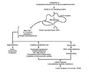


![Alexa Fluor® 647 Anti-PDGFR alpha antibody [EPR22059-270] (ab270085) | Abcam](https://www.abcam.com/ps/products/270/ab270085/Images/ab270085-3-alexa-fluor-647-anti-pdgfr-alpha-antibody-epr22059-270-flow-cytometry-shsy5y-human.jpg)