Maxilla
Tooth Crown
Mandible
Tooth Eruption
Cuspid
Ameloblasts
Dental Enamel
Molar
Tooth Avulsion
Anodontia
Tooth Root
Tooth Apex
Root Resorption
Malocclusion
Root Canal Therapy
Orthodontic Wires
Bicuspid
Dental Arch
Tooth, Unerupted
Tooth, Supernumerary
Orthodontics, Corrective
Odontogenesis
Dentin
Tooth Replantation
Photography, Dental
Orthodontic Appliance Design
Tooth, Deciduous
Amelogenesis
Malocclusion, Angle Class II
Tooth, Impacted
Dental Pulp Necrosis
Dental Pulp
Fused Teeth
Dentition, Permanent
Tooth Injuries
Odontoblasts
Alveolar Process
Orthodontic Extrusion
Dental Pulp Cavity
Dental Occlusion
Orthodontic Space Closure
Post and Core Technique
Malocclusion, Angle Class I
Enamel Organ
Vertical Dimension
Periodontal Ligament
Tooth, Nonvital
Dens in Dente
Tooth Cervix
Dentition, Mixed
Radiography, Panoramic
Dental Stress Analysis
Dental Enamel Hypoplasia
Tooth Calcification
Tooth Germ
Crowns
Esthetics, Dental
Orthodontic Brackets
Tooth Mobility
Jaw Relation Record
Orthodontic Appliances, Removable
Serial Extraction
Dental Prosthesis Design
Chin
Overbite
Dental Pulp Exposure
Orthodontic Anchorage Procedures
Orthodontic Retainers
Extraoral Traction Appliances
Tooth, Artificial
Dental Implants, Single-Tooth
Root Canal Filling Materials
Resin Cements
Tooth Migration
Facial Bones
Apexification
Dental Bonding
Dental Materials
Root Canal Obturation
Orthodontic Appliances
Open Bite
Dental Restoration Failure
Activator Appliances
Tooth Attrition
Root Canal Preparation
Stainless Steel
Sella Turcica
Orthodontic Appliances, Functional
Enamel Microabrasion
Tooth Socket
Palate
Dentinogenesis
Dental Alloys
Orthodontics, Interceptive
Dentition
Periodontal Splints
Tooth Diseases
Amelogenin
Dental Prosthesis Retention
Dental Restoration, Permanent
Denture Design
Nasal Bone
Materials Testing
Malocclusion, Angle Class III
Tooth Ankylosis
Retrognathia
Calcium Hydroxide
Denture, Partial, Fixed, Resin-Bonded
Skull Base
Dental Cements
Maxillary Nerve
Dental Pulp Test
Periapical Diseases
Shear Strength
Acid Etching, Dental
Tooth Exfoliation
Tooth Preparation, Prosthodontic
Space Maintenance
Fluorosis, Dental
Mandibular Nerve
Root Canal Irrigants
Statistics, Nonparametric
Gingivoplasty
Periodontium
Alveolar Bone Loss
Denture, Complete, Upper
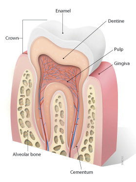
Dental Cementum
Maxillary Diseases
Tooth Demineralization
Face
Acrylic Resins
Dentin-Bonding Agents
Gingival Recession
Mole Rats
Radiography, Dental, Digital
Dental Implantation, Endosseous, Endodontic
Silicates
Microscopy, Electron, Scanning
Tooth Erosion
Cone-Beam Computed Tomography
Dental Implantation, Endosseous
Orthodontics
Dental Leakage
Tooth Bleaching
Stress, Mechanical
Epoxy Resins
Aluminum Silicates
Glass
Dental Instruments
Gingivectomy
Torque
Finite Element Analysis
Zinc Oxide-Eugenol Cement
Labial Frenum
Phosphoric Acids
Dental Prosthesis
Dental Papilla
Holoprosencephaly
Sodium Hypochlorite
Radiography, Bitewing
Surface Properties
Denture, Partial, Fixed
Masticatory Muscles
Mouth Breathing
Gutta-Percha
Silanes
Alveolectomy
Palatal Expansion Technique
Periapical Periodontitis
Dentigerous Cyst
Amelogenesis Imperfecta
Bisphenol A-Glycidyl Methacrylate
Dental Veneers
Methacrylates
Dental Debonding
Effects of maternal acetazolamide treatment on body weights and incisor development of the fetal rat. (1/1014)
The incisor development of fetal rats on gestation day 19 was well correlated with their fetal weights. The number of odontoblasts in the mandibular incisors, an index of incisor development, increased more than that of the maxillary incisors with increase in fetal body weights. Maternal acetazolamide treatments were observed to suppress the mean fetal weight and to retard incisor development. A smaller incisor size, a thinner predentin layer, and fewer odontoblasts were characteristic of the acetazolamide group. There was also a good correlation between the fetal weights and the number of odontoblasts in the acetazolamide group. From these results, we postulated that the retarded incisor development of the fetal rats caused by the maternal acetazolamide treatment was related to their suppressed fetal weights. However, the regression coefficient of the fetal weights and the number of odontoblasts in the acetazolamide group was smaller than that of the vehicle control group. It may indicate that retarded incisor development in response to maternal acetazolamide treatment is to some extent independent of suppressed fetal weight. (+info)A modern human pattern of dental development in lower pleistocene hominids from Atapuerca-TD6 (Spain). (2/1014)
The study of life history evolution in hominids is crucial for the discernment of when and why humans have acquired our unique maturational pattern. Because the development of dentition is critically integrated into the life cycle in mammals, the determination of the time and pattern of dental development represents an appropriate method to infer changes in life history variables that occurred during hominid evolution. Here we present evidence derived from Lower Pleistocene human fossil remains recovered from the TD6 level (Aurora stratum) of the Gran Dolina site in the Sierra de Atapuerca, northern Spain. These hominids present a pattern of development similar to that of Homo sapiens, although some aspects (e.g., delayed M3 calcification) are not as derived as that of European populations and people of European origin. This evidence, taken together with the present knowledge of cranial capacity of these and other late Early Pleistocene hominids, supports the view that as early as 0.8 Ma at least one Homo species shared with modern humans a prolonged pattern of maturation. (+info)Scanning electron microscopy of the lateral cell surfaces of rat incisor ameloblasts. (3/1014)
Dry dissected rat incisor ameloblasts studied in the scanning electron microscope show remarkable specializations of their lateral surfaces. Four or five cycles of a change from a surface with longitudinal gutterlike folds associated with large intercellular spaces, to one with microvilli and reduced intercellular spaces, are found along the length of the lower incisor maturation zone. It is argued that these changes indicate cyclical activity in maturation ameloblasts. (+info)Morphological changes in periodontal mechanoreceptors of mouse maxillary incisors after the experimental induction of anterior crossbite: a light and electron microscopic observation using immunohistochemistry for PGP 9.5. (4/1014)
Ruffini nerve endings (mechanoreceptors) in the periodontal ligament (PDL) of mouse incisors were examined to elucidate whether experimentally-induced crossbites cause any changes or abnormalities in their morphology and distribution. Anterior guiding planes were attached to the mandibular incisors of 3-week-old C3H/HeSlc mice. At 3 days and 1, 2, 4, 6, and 8 weeks post-attachment of the appliance, the mice were sacrificed by perfusion fixation. Frozen sagittal cryostat sections of the decalcified maxillary incisors were processed for immunohistochemistry of protein gene product 9.5, followed by histochemical determination of tartrate-resistant acid phosphatase activity to reveal sites of alveolar bone resorption. Despite the absence of bone resorption within the lingual PDL of control mice, distinct resorption sites were seen in the respective regions of the experimental animals. Unlike the controls, many Ruffini endings showing vague and swollen contours, with unusually long and pedunculated micro-projections were observed in the affected lingual PDL of the incisors in the experimental animals with short-term anterior crossbite induction. Club-shaped nerve terminations with few, if any, micro-projections were observed in the lingual PDL of experimental animals with long-term induction, as well as in aged control mouse incisors. Differences in the distribution of Ruffini endings were also observed. These results indicate that changing the direction of the force applied to the PDL results in rapid and prolonged changes in the morphology of Ruffini-like mechanoreceptors. (+info)Arrested eruption of the permanent lower second molar. (5/1014)
The incidence of retention/impaction of the permanent lower second molar (M2inf) lies between 0.6/1000 and 3/1000. Therefore, the purpose of the present study was to investigate the craniofacial morphology, the frequency of dental anomalies and the inclination of the affected M2inf and the adjacent first molar in patients with arrested eruption of M2inf. The overall goal was to elucidate the aetiology of arrested tooth eruption and to present the characteristics of these patients in order to improve diagnosis and treatment planning. Radiographic material (profile radiographs and orthopantomograms) from 19 patients (nine females and 10 males; 13-19 years of age at the time of referral) were analysed. The ages of the patients when profile radiographs were taken for cephalometric analysis varied from 8 to 16 years. The study shows that this group of patients, compared with a reference group, had an increased sagittal jaw relationship (Class II). Specifically, the mandibular prognathism was less, the mandibular gonial angle smaller, the mandibular alveolar prognathism enlarged and the maxillary incisor inclination less than in the reference group. Furthermore, this group of patients had a more frequent occurrence of morphological tooth anomalies, such as root deflections, invaginations, and taurodontism. However, none of the patients with arrested eruption of M2inf had agenesis of the lower third molar. The study did not reveal an association between the degree of inclination of the M2inf and that of the first molar in the same region. The results of this investigation show that conditions such as the craniofacial morphology and deviations in the dentition are associated with arrested eruption of M2inf. Therefore, it is important to evaluate these conditions in future diagnosis and treatment planning of patients with arrested eruption of M2inf. (+info)The length and eruption rates of incisor teeth in rats after one or more of them had been unimpeded. (6/1014)
The eruption rate and length of all four incisor teeth in rats were measured under ether anaesthesia by recording the position of marks on their labial surfaces at 2-day intervals, using calibrated graticules in microscope eyepieces. The rats were divided into four groups and either a lower, an upper, both a lower and an upper, or no incisors were unimpeded. This paper describes the changes when the unimpeded incisors returned to the occlusion. Neither the unimpeded nor the impeded incisors simply returned to control values immediately the period of unimpeded eruption ended, but showed transient changes in their lengths and eruption rates. The results confirm that eruption rates are determined by the sum of the lengths of the lower and upper incisors, rather than by their own lengths, with longer teeth erupting more slowly. Specifically, restoring the bevel to the incisors did not slow their eruption below normal impeded rates. The slowing of the eruption of the longer of two adjacent incisors was related to the length differences of the incisors in the same jaw, not to the sum of the differences in both jaws. Contact with the contralateral incisor in the opposite jaw slowed the eruption of an incisor more than contact with the ipsilateral incisor. (+info)Pathological evaluation of the effects of intentional disocclusion and overloading occlusion in odontogenesis disorders in N-methylnitrosourea-treated hamsters. (7/1014)
This study compares the effects of disocclusion and overloading occlusion on dental lesions. Ten-day-old Syrian hamsters were divided into 4 groups: group I, untreated animals; group II, animals whose hemilateral incisors were disoccluded; group III, N-methylnitrosourea (MNU)-treated animals; and group IV, MNU-treated animals whose hemilateral incisors were disoccluded. The ipsilateral maxillary and mandibular incisors were repetitively cut with diamond discs. The hamster is easier to anesthetize. Animals received a 0.2% solution of MNU (10 mg/kg body weight) intragastrically twice a week for 16 wk. All the cut mandibular incisors and the MNU-treated uncut mandibular incisors showed lack of iron deposition on the enamel surface. The eruption rate was significantly higher in the cut disoccluded incisors of groups II and IV (p < 0.05) and significantly lower in the uncut overloaded incisors of groups II and IV (p < 0.05). In the cut mandibular incisors of group IV, the degree of the disturbance of odontogenesis and the atypical proliferation of odontogenic epithelium were more prominent (p < 0.02), and the dental lesions occurred earlier. Histologically, the disturbed Hertwig's epithelial sheath and the Hertwig's epithelial sheath-like transformed U-shaped part and enamel organ seemed to lead to disturbances of amelogenesis and detinogenesis as well as to atypical proliferation of odontogenic epithelium nests. Thus, this method of disocclusion of the incisors of rodents may represent a useful model for the investigation of the effects of various agents on tooth formation over a short experimental period. (+info)Postnatal expression of calretinin-immunoreactivity in periodontal Ruffini endings in the rat incisor: a comparison with protein gene product 9.5 (PGP 9.5)-immunoreactivity. (8/1014)
The postnatal expression of immunoreactivity for calretinin, one of the calcium binding proteins, and for protein gene product 9.5 (PGP 9.5), a general neuronal marker, was investigated in mechanoreceptive Ruffini endings in the periodontal ligament of the rat incisor. Age-related changes in the expression of these two proteins in periodontal nerves were further quantified with a computerized image analysis. At 1 day after birth, a few PGP 9.5-immunoreactive nerve fibers and a still smaller number of calretinin-positive fibers were found in the periodontal ligament: they were thin and beaded in appearance and no specialized nerve terminals were recognized. Tree-like terminals, reminiscent of immature Ruffini endings, were recognizable in 4-day-old rats by PGP 9.5-immunohistochemistry, while calretinin-immunostaining failed to reveal these specialized endings. At postnatal 7-11 days when PGP 9.5-immunostaining could demonstrate typical Ruffini endings, calretinin-immunopositive nerve fibers merely tapered off without forming the Ruffini type endings. A small number of Ruffini endings showing calretinin-immunoreactivity began to occur in the periodontal ligament at 24-26 days after birth when the occlusion of the first molars had been established. At the functional occlusion stage (60-80 days after birth), the Ruffini endings showing calretinin-immunoreactivity drastically increased in number and density, but less so than those positive for PGP 9.5-immunoreaction. The delayed expression of calretinin suggests that the function of the periodontal Ruffini endings is established after the completion of terminal formation because Ca2+, which binds to calcium binding proteins including calretinin with high affinity, plays an important role in mechano-electric transduction. (+info)The maxilla is a paired bone that forms the upper jaw in vertebrates. In humans, it is a major bone in the face and plays several important roles in the craniofacial complex. Each maxilla consists of a body and four processes: frontal process, zygomatic process, alveolar process, and palatine process.
The maxillae contribute to the formation of the eye sockets (orbits), nasal cavity, and the hard palate of the mouth. They also contain the upper teeth sockets (alveoli) and help form the lower part of the orbit and the cheekbones (zygomatic arches).
Here's a quick rundown of its key functions:
1. Supports the upper teeth and forms the upper jaw.
2. Contributes to the formation of the eye sockets, nasal cavity, and hard palate.
3. Helps shape the lower part of the orbit and cheekbones.
4. Partakes in the creation of important sinuses, such as the maxillary sinus, which is located within the body of the maxilla.
A tooth crown is a type of dental restoration that covers the entire visible portion of a tooth, restoring its shape, size, and strength. It is typically made of materials like porcelain, ceramic, or metal alloys and is custom-made to fit over the prepared tooth. The tooth crown is cemented in place and becomes the new outer surface of the tooth, protecting it from further damage or decay.
The process of getting a tooth crown usually involves two dental appointments. During the first appointment, the dentist prepares the tooth by removing any decay or damaged tissue and shaping the tooth to accommodate the crown. An impression is then taken of the prepared tooth and sent to a dental laboratory where the crown is fabricated. In the meantime, a temporary crown is placed over the prepared tooth to protect it until the permanent crown is ready. At the second appointment, the temporary crown is removed, and the permanent crown is cemented in place.
Tooth crowns are often recommended for several reasons, including:
* To restore a broken or fractured tooth
* To protect a weakened tooth from further damage or decay
* To support a large filling when there isn't enough natural tooth structure left
* To cover a dental implant
* To improve the appearance of a discolored or misshapen tooth
Overall, a tooth crown is an effective and long-lasting solution for restoring damaged or decayed teeth and improving oral health.
Tooth movement, in a dental and orthodontic context, refers to the physical change in position or alignment of one or more teeth within the jaw bone as a result of controlled forces applied through various orthodontic appliances such as braces, aligners, or other orthodontic devices. The purposeful manipulation of these forces encourages the periodontal ligament (the tissue that connects the tooth to the bone) to remodel, allowing the tooth to move gradually over time into the desired position. This process is crucial in achieving proper bite alignment, correcting malocclusions, and enhancing overall oral function and aesthetics.
The mandible, also known as the lower jaw, is the largest and strongest bone in the human face. It forms the lower portion of the oral cavity and plays a crucial role in various functions such as mastication (chewing), speaking, and swallowing. The mandible is a U-shaped bone that consists of a horizontal part called the body and two vertical parts called rami.
The mandible articulates with the skull at the temporomandibular joints (TMJs) located in front of each ear, allowing for movements like opening and closing the mouth, protrusion, retraction, and side-to-side movement. The mandible contains the lower teeth sockets called alveolar processes, which hold the lower teeth in place.
In medical terminology, the term "mandible" refers specifically to this bone and its associated structures.
Tooth eruption is the process by which a tooth emerges from the gums and becomes visible in the oral cavity. It is a normal part of dental development that occurs in a predictable sequence and timeframe. Primary or deciduous teeth, also known as baby teeth, begin to erupt around 6 months of age and continue to emerge until approximately 2-3 years of age. Permanent or adult teeth start to erupt around 6 years of age and can continue to emerge until the early twenties.
The process of tooth eruption involves several stages, including the formation of the tooth within the jawbone, the movement of the tooth through the bone and surrounding tissues, and the final emergence of the tooth into the mouth. Proper tooth eruption is essential for normal oral function, including chewing, speaking, and smiling. Any abnormalities in the tooth eruption process, such as delayed or premature eruption, can indicate underlying dental or medical conditions that require further evaluation and treatment.
A cuspid, also known as a canine tooth or cuspid tooth, is a type of tooth in mammals. It is the pointiest tooth in the dental arch and is located between the incisors and bicuspids (or premolars). Cuspids have a single cusp or pointed tip that is used for tearing and grasping food. In humans, there are four cuspids, two on the upper jaw and two on the lower jaw, one on each side of the dental arch.
Ameloblasts are the specialized epithelial cells that are responsible for the formation of enamel, which is the hard, outermost layer of a tooth. These cells are a part of the dental lamina and are present in the developing tooth's crown region. They align themselves along the surface of the developing tooth and secrete enamel proteins and minerals to form the enamel rods and interrod enamel. Once the enamel formation is complete, ameloblasts undergo programmed cell death, leaving behind the hard, mineralized enamel matrix. Any damage or abnormality in the functioning of ameloblasts can lead to developmental defects in the enamel, such as hypoplasia or hypocalcification, which may affect the tooth's structure and function.
Dental enamel is the hard, white, outermost layer of a tooth. It is a highly mineralized and avascular tissue, meaning it contains no living cells or blood vessels. Enamel is primarily composed of calcium and phosphate minerals and serves as the protective covering for the crown of a tooth, which is the portion visible above the gum line.
Enamel is the hardest substance in the human body, and its primary function is to provide structural support and protection to the underlying dentin and pulp tissues of the tooth. It also plays a crucial role in chewing and biting by helping to distribute forces evenly across the tooth surface during these activities.
Despite its hardness, dental enamel can still be susceptible to damage from factors such as tooth decay, erosion, and abrasion. Once damaged or lost, enamel cannot regenerate or repair itself, making it essential to maintain good oral hygiene practices and seek regular dental checkups to prevent enamel damage and protect overall oral health.
In the context of dentistry, a molar is a type of tooth found in the back of the mouth. They are larger and wider than other types of teeth, such as incisors or canines, and have a flat biting surface with multiple cusps. Molars are primarily used for grinding and chewing food into smaller pieces that are easier to swallow. Humans typically have twelve molars in total, including the four wisdom teeth.
In medical terminology outside of dentistry, "molar" can also refer to a unit of mass in the apothecaries' system of measurement, which is equivalent to 4.08 grams. However, this usage is less common and not related to dental or medical anatomy.
Tooth avulsion is the complete separation of a tooth from its socket in the alveolar bone due to traumatic injury. This occurs when the periodontal ligament, which holds the tooth in place, gets severed or torn, resulting in the tooth being displaced from its original position. Avulsed teeth can be either primary (baby) or permanent teeth, and the trauma can result in damage to the surrounding tissues, including the gingiva, alveolar bone, and sometimes even the nerves and blood vessels. Prompt and appropriate first aid, as well as professional dental care, are crucial for ensuring the best possible outcome for reimplantation and healing.
Dental models are replicas of a patient's teeth and surrounding oral structures, used in dental practice and education. They are typically created using plaster or other materials that harden to accurately reproduce the shape and position of each tooth, as well as the contours of the gums and palate. Dental models may be used for a variety of purposes, including treatment planning, creating custom-fitted dental appliances, and teaching dental students about oral anatomy and various dental procedures. They provide a tactile and visual representation that can aid in understanding and communication between dentists, patients, and other dental professionals.
Odontometry is a term used in dentistry that refers to the measurement of teeth, particularly the size and length of teeth or tooth roots. It is often used in forensic dentistry for identification purposes, such as in age estimation, sex determination, or individual identification of human remains. The measurements can be taken using various methods, including radiographs (x-rays), calipers, or specialized software.
In some contexts, odontometry may also refer to the process of measuring the amount of dental work required for a particular treatment plan, although this usage is less common.
Anodontia is a medical term that refers to the congenital absence or lack of development of all primary (deciduous) and/or permanent teeth. It is a rare dental condition that affects tooth development and can be isolated or associated with various syndromes and genetic disorders.
In anodontia, the dental tissues responsible for forming teeth, including the dental lamina, dental papilla, and dental follicle, fail to develop properly, resulting in missing teeth. The condition can affect all teeth or only some of them, leading to partial anodontia.
Anodontia is different from hypodontia, which refers to the congenital absence of one or more, but not all, teeth. It is also distinct from oligodontia, which is the absence of six or more permanent teeth, excluding third molars (wisdom teeth).
People with anodontia may experience difficulties in chewing, speaking, and maintaining oral hygiene, leading to various dental and social problems. Prosthodontic treatments, such as dentures or implants, are often necessary to restore oral function and aesthetics.
A tooth root is the part of a tooth that is embedded in the jawbone and cannot be seen when looking at a person's smile. It is the lower portion of a tooth that typically has a conical shape and anchors the tooth to the jawbone through a periodontal ligament. The tooth root is covered by cementum, a specialized bone-like tissue, and contains nerve endings and blood vessels within its pulp chamber.
The number of roots in a tooth can vary depending on the type of tooth. For example, incisors typically have one root, canines may have one or two roots, premolars usually have one or two roots, and molars often have two to four roots. The primary function of the tooth root is to provide stability and support for the crown of the tooth, allowing it to withstand the forces of biting and chewing.
Tooth abnormalities refer to any variations or irregularities in the size, shape, number, structure, or development of teeth that deviate from the typical or normal anatomy. These abnormalities can occur in primary (deciduous) or permanent teeth and can be caused by genetic factors, environmental influences, systemic diseases, or localized dental conditions during tooth formation.
Some examples of tooth abnormalities include:
1. Microdontia - teeth that are smaller than normal in size.
2. Macrodontia - teeth that are larger than normal in size.
3. Peg-shaped teeth - teeth with a narrow, conical shape.
4. Talon cusps - additional cusps or points on the biting surface of a tooth.
5. Dens invaginatus - an abnormal development where the tooth crown has an extra fold or pouch that can trap bacteria and cause dental problems.
6. Taurodontism - teeth with large pulp chambers and short roots.
7. Supernumerary teeth - having more teeth than the typical number (20 primary and 32 permanent teeth).
8. Hypodontia - missing one or more teeth due to a failure of development.
9. Germination - two adjacent teeth fused together, usually occurring in the front teeth.
10. Fusion - two separate teeth that have grown together during development.
Tooth abnormalities may not always require treatment unless they cause functional, aesthetic, or dental health issues. A dentist can diagnose and manage tooth abnormalities through various treatments, such as fillings, extractions, orthodontic care, or restorative procedures.
The tooth apex is the tip or the narrowed end of the root of a tooth. It is the portion that is located deepest within the jawbone and it contains dental pulp tissue, which includes nerves and blood vessels. The apex plays an essential role in the development and maintenance of a tooth, as well as in the process of root canal treatment, where instruments and materials are introduced through it to clean and fill the root canals. It is also a crucial landmark in endodontic surgery and dental imaging.
Root resorption is a process that occurs when the body's own cells, called odontoclasts, break down and destroy the hard tissue of the tooth root. This can occur as a result of various factors such as trauma, infection, or orthodontic treatment. In some cases, it may be a normal part of the tooth development and eruption process in children. However, excessive or pathological root resorption can lead to weakening and loss of the tooth. It is often asymptomatic and discovered during routine dental x-rays.
Cephalometry is a medical term that refers to the measurement and analysis of the skull, particularly the head face relations. It is commonly used in orthodontics and maxillofacial surgery to assess and plan treatment for abnormalities related to the teeth, jaws, and facial structures. The process typically involves taking X-ray images called cephalograms, which provide a lateral view of the head, and then using various landmarks and reference lines to make measurements and evaluate skeletal and dental relationships. This information can help clinicians diagnose problems, plan treatment, and assess treatment outcomes.
Malocclusion is a term used in dentistry and orthodontics to describe a misalignment or misrelation between the upper and lower teeth when they come together, also known as the bite. It is derived from the Latin words "mal" meaning bad or wrong, and "occludere" meaning to close.
There are different types of malocclusions, including:
1. Class I malocclusion: The most common type, where the upper teeth slightly overlap the lower teeth, but the bite is otherwise aligned.
2. Class II malocclusion (overbite): The upper teeth significantly overlap the lower teeth, causing a horizontal or vertical discrepancy between the dental arches.
3. Class III malocclusion (underbite): The lower teeth protrude beyond the upper teeth, resulting in a crossbite or underbite.
Malocclusions can be caused by various factors such as genetics, thumb sucking, tongue thrusting, premature loss of primary or permanent teeth, and jaw injuries or disorders. They may lead to several oral health issues, including tooth decay, gum disease, difficulty chewing or speaking, and temporomandibular joint (TMJ) dysfunction. Treatment for malocclusions typically involves orthodontic appliances like braces, aligners, or retainers to realign the teeth and correct the bite. In some cases, surgical intervention may be necessary.
A tooth fracture is a dental health condition characterized by a break or crack in the tooth structure. It can occur in different parts of the tooth, including the crown (the visible part), root, or filling. Tooth fractures can result from various factors such as trauma, biting or chewing on hard objects, grinding or clenching teeth, and having large, old amalgam fillings that weaken the tooth structure over time. Depending on the severity and location of the fracture, it may cause pain, sensitivity, or affect the tooth's functionality and appearance. Treatment options for tooth fractures vary from simple bonding to root canal treatment or even extraction in severe cases. Regular dental check-ups are essential for early detection and management of tooth fractures.
Root canal therapy, also known as endodontic treatment, is a dental procedure that involves the removal of infected or damaged pulp tissue from within a tooth's root canal system. The root canal system is a series of narrow channels that run from the center of the tooth (pulp chamber) down to the tip of the tooth roots, containing nerves, blood vessels, and connective tissues.
During the procedure, the dentist or endodontist will gain access to the pulp chamber, carefully clean and shape the root canals using specialized instruments, and then fill and seal them with a rubber-like material called gutta-percha. This helps prevent reinfection and preserves the structural integrity of the tooth. In many cases, a crown or other restoration is placed over the treated tooth to protect it and restore its function and appearance.
Root canal therapy is typically recommended when the pulp tissue becomes inflamed or infected due to deep decay, repeated dental procedures, cracks, or chips in the teeth. The goal of this treatment is to alleviate pain, preserve natural tooth structure, and prevent the need for extraction.
Orthodontic wires are typically made of stainless steel, nickel-titanium alloy, or other shape memory alloys, and are used in orthodontics to move teeth into the desired position. They are attached to brackets bonded to the teeth and exert a continuous force to align the teeth and correct malocclusions (bites that do not fit together correctly). The wires come in various sizes, shapes, and materials, each with specific properties that make them suitable for different stages of treatment. Some wires are flexible and used during the initial alignment phase, while others are more rigid and used during the finishing phase to achieve precise tooth movements.
A bicuspid valve, also known as a mitral valve in the heart, is a heart valve that has two leaflets or cusps. It lies between the left atrium and the left ventricle and helps to regulate blood flow between these two chambers of the heart. In a healthy heart, the bicuspid valve opens to allow blood to flow from the left atrium into the left ventricle and closes tightly to prevent blood from flowing back into the left atrium during contraction of the ventricle.
A congenital heart defect known as a bicuspid aortic valve occurs when the aortic valve, which normally has three leaflets or cusps, only has two. This can lead to narrowing of the valve (aortic stenosis) or leakage of the valve (aortic regurgitation), which can cause symptoms and may require medical treatment.
The dental arch refers to the curved shape formed by the upper or lower teeth when they come together. The dental arch follows the curve of the jaw and is important for proper bite alignment and overall oral health. The dental arches are typically described as having a U-shaped appearance, with the front teeth forming a narrower section and the back teeth forming a wider section. The shape and size of the dental arch can vary from person to person, and any significant deviations from the typical shape or size may indicate an underlying orthodontic issue that requires treatment.
A tooth is classified as "unerupted" when it has not yet penetrated through the gums and entered the oral cavity. This can apply to both primary (baby) teeth and permanent (adult) teeth. The reasons for a tooth's failure to erupt can vary, including crowding of teeth, lack of sufficient space, or anatomical barriers such as bone or soft tissue. In some cases, unerupted teeth may need to be monitored or treated, depending on the specific situation and any symptoms experienced by the individual.
A supernumerary tooth, also known as hyperdontia, refers to an additional tooth or teeth that grow beyond the regular number of teeth in the dental arch. These extra teeth can erupt in various locations of the dental arch and may occur in any of the tooth types, but they are most commonly seen as extra premolars or molars, and less frequently as incisors or canines. Supernumerary teeth may be asymptomatic or may cause complications such as crowding, displacement, or impaction of adjacent teeth, and therefore, they often require dental treatment.
Orthodontics is a specialized branch of dentistry that focuses on the diagnosis, prevention, and treatment of dental and facial irregularities. The term "corrective" in this context refers to the use of appliances (such as braces, aligners, or other devices) to move teeth into their proper position and correct malocclusion (bad bite). This not only improves the appearance of the teeth but also helps to ensure better function, improved oral health, and overall dental well-being.
The goal of corrective orthodontics is to create a balanced and harmonious relationship between the teeth, jaws, and facial structures. Treatment may be recommended for children, adolescents, or adults and can help address various issues such as crowding, spacing, overbites, underbites, crossbites, open bites, and jaw growth discrepancies. A combination of techniques, including fixed or removable appliances, may be used to achieve the desired outcome. Regular follow-up appointments are necessary throughout treatment to monitor progress and make any necessary adjustments.
A tooth is a hard, calcified structure found in the jaws (upper and lower) of many vertebrates and used for biting and chewing food. In humans, a typical tooth has a crown, one or more roots, and three layers: the enamel (the outermost layer, hardest substance in the body), the dentin (the layer beneath the enamel), and the pulp (the innermost layer, containing nerves and blood vessels). Teeth are essential for proper nutrition, speech, and aesthetics. There are different types of teeth, including incisors, canines, premolars, and molars, each designed for specific functions in the mouth.
Odontogenesis is the process of tooth development that involves the formation and calcification of teeth. It is a complex process that requires the interaction of several types of cells, including epithelial cells, mesenchymal cells, and odontoblasts. The process begins during embryonic development with the formation of dental lamina, which gives rise to the tooth bud. As the tooth bud grows and differentiates, it forms the various structures of the tooth, including the enamel, dentin, cementum, and pulp. Odontogenesis is completed when the tooth erupts into the oral cavity. Abnormalities in odontogenesis can result in developmental dental anomalies such as tooth agenesis, microdontia, or odontomas.
Dentin is the hard, calcified tissue that lies beneath the enamel and cementum of a tooth. It forms the majority of the tooth's structure and is composed primarily of mineral salts (hydroxyapatite), collagenous proteins, and water. Dentin has a tubular structure, with microscopic channels called dentinal tubules that radiate outward from the pulp chamber (the center of the tooth containing nerves and blood vessels) to the exterior of the tooth. These tubules contain fluid and nerve endings that are responsible for the tooth's sensitivity to various stimuli such as temperature changes, pressure, or decay. Dentin plays a crucial role in protecting the dental pulp while also providing support and structure to the overlying enamel and cementum.
Tooth replantation is a dental procedure that involves the replanting and reattachment of a tooth that has been avulsed or knocked out due to trauma. The primary goal of this emergency procedure is to preserve the natural tooth and its periodontal ligament (PDL) tissue, allowing for potential reattachment and function.
The steps involved in tooth replantation include:
1. Locating the avulsed tooth: Carefully handle the knocked-out tooth by holding it by the crown (the chewing surface), avoiding touching the root area to prevent further damage to the periodontal ligament fibers.
2. Rinsing the tooth: Gently rinse the tooth with saline solution, sterile water, or milk to remove any debris or dirt, but avoid using alcohol or scrubbing the tooth as it may cause more damage to the PDL.
3. Replanting the tooth: As soon as possible, reposition the tooth back into its socket in the correct orientation and alignment. Apply gentle pressure to seat it in place while ensuring that it is facing the right direction. Ideally, this should be done within 30 minutes of avulsion for better prognosis.
4. Stabilizing the tooth: Use a splint or a wire to secure the replanted tooth to the adjacent teeth, providing stability and support during the healing process. This helps maintain the alignment and position of the replanted tooth.
5. Seeking professional dental care: Immediately consult with a dentist or endodontist for further evaluation, additional treatment, and follow-up care. The dentist will assess the success of the replantation and determine if any root canal therapy or other treatments are necessary to ensure long-term survival of the tooth.
The success of tooth replantation depends on several factors, including the timeliness of the procedure, the condition of the avulsed tooth, and the patient's overall oral health. Prompt action and professional care can significantly increase the likelihood of a successful outcome and preserve the natural tooth for years to come.
Dental photography is a type of clinical photography that focuses on documenting the condition and treatment of teeth and oral structures. It involves using specialized cameras, lenses, and lighting to capture high-quality images of the mouth and related areas. These images can be used for diagnostic purposes, patient education, treatment planning, communication with other dental professionals, and monitoring progress over time. Dental photography may include various types of shots, such as extraoral (outside the mouth) and intraoral (inside the mouth) views, close-ups of individual teeth or restorations, and full-face portraits. It requires a strong understanding of dental anatomy, lighting techniques, and image composition to produce accurate and informative images.
Orthodontic appliance design refers to the creation and development of medical devices used in orthodontics, which is a branch of dentistry focused on the diagnosis, prevention, and correction of dental and facial irregularities. The design process involves creating a customized treatment plan for each patient, based on their specific needs and goals.
Orthodontic appliances can be removable or fixed and are used to move teeth into proper alignment, improve jaw function, and enhance the overall appearance of the smile. Some common types of orthodontic appliances include braces, aligners, palatal expanders, and retainers.
The design of an orthodontic appliance typically involves several factors, including:
1. The specific dental or facial problem being addressed
2. The patient's age, overall health, and oral hygiene habits
3. The patient's lifestyle and personal preferences
4. The estimated treatment time and cost
5. The potential risks and benefits of the appliance
Orthodontic appliance design is a complex process that requires a thorough understanding of dental anatomy, biomechanics, and materials science. It is typically performed by an orthodontist or a dental technician with specialized training in this area. The goal of orthodontic appliance design is to create a device that is both effective and comfortable for the patient, while also ensuring that it is safe and easy to use.
A deciduous tooth, also known as a baby tooth or primary tooth, is a type of temporary tooth that humans and some other mammals develop during childhood. They are called "deciduous" because they are eventually shed and replaced by permanent teeth, much like how leaves on a deciduous tree fall off and are replaced by new growth.
Deciduous teeth begin to form in the womb and start to erupt through the gums when a child is around six months old. By the time a child reaches age three, they typically have a full set of 20 deciduous teeth, including incisors, canines, and molars. These teeth are smaller and less durable than permanent teeth, but they serve important functions such as helping children chew food properly, speak clearly, and maintain space in the jaw for the permanent teeth to grow into.
Deciduous teeth usually begin to fall out around age six or seven, starting with the lower central incisors. This process continues until all of the deciduous teeth have been shed, typically by age 12 or 13. At this point, the permanent teeth will have grown in and taken their place, with the exception of the wisdom teeth, which may not erupt until later in adolescence or early adulthood.
Amelogenesis is the biological process of forming enamel, which is the hard and highly mineralized outer layer of teeth. Enamel is primarily made up of calcium and phosphate minerals and is the toughest substance in the human body. Amelogenesis involves the synthesis, secretion, and maturation of enamel proteins by specialized cells called ameloblasts.
The medical definition of 'Amelogenesis' refers to a genetic disorder that affects the development and formation of tooth enamel. This condition is also known as Amelogenesis Imperfecta (AI) and can result in teeth that are discolored, sensitive, and prone to decay. There are several types of Amelogenesis Imperfecta, each with its own set of symptoms and genetic causes.
In summary, 'Amelogenesis' is the biological process of enamel formation, while 'Amelogenesis Imperfecta' is a genetic disorder that affects this process, leading to abnormal tooth enamel development.
Malocclusion, Angle Class II is a type of dental malocclusion where the relationship between the maxilla (upper jaw) and mandible (lower jaw) is such that the lower molar teeth are positioned posteriorly relative to the upper molar teeth. This results in an overbite, which means that the upper front teeth overlap the lower front teeth excessively. The classification was proposed by Edward Angle, an American orthodontist who is considered the father of modern orthodontics. In this classification system, Class II malocclusion is further divided into three subclasses (I, II, and III) based on the position of the lower incisors relative to the upper incisors.
An impacted tooth is a condition where a tooth fails to erupt into the oral cavity within its expected time frame, resulting in its partial or complete entrapment within the jawbone or soft tissues. This commonly occurs with wisdom teeth (third molars) but can affect any tooth. Impacted teeth may cause problems such as infection, decay of adjacent teeth, gum disease, or cyst formation, and they may require surgical removal.
Dental pulp necrosis is the death of the soft tissue inside a tooth, known as the dental pulp. The dental pulp contains nerves, blood vessels, and connective tissue that help the tooth grow and develop. It also provides sensations like hot or cold. Dental pulp necrosis can occur due to various reasons such as tooth decay, trauma, or infection. When the dental pulp dies, it can no longer provide nutrients to the tooth, making it more susceptible to fractures and infections. Symptoms of dental pulp necrosis may include pain, sensitivity, swelling, or abscess formation. Treatment options for dental pulp necrosis typically involve root canal therapy or extraction of the affected tooth.
Dental pulp is the soft tissue located in the center of a tooth, surrounded by the dentin. It contains nerves, blood vessels, and connective tissue, and plays a vital role in the development and health of the tooth. The dental pulp helps to form dentin during tooth development and continues to provide nourishment to the tooth throughout its life. It also serves as a sensory organ, allowing the tooth to detect hot and cold temperatures and transmit pain signals to the brain. Injury or infection of the dental pulp can lead to serious dental problems, such as tooth decay or abscesses, and may require root canal treatment to remove the damaged tissue and save the tooth.
Tooth extraction is a dental procedure in which a tooth that is damaged or poses a threat to oral health is removed from its socket in the jawbone. This may be necessary due to various reasons such as severe tooth decay, gum disease, fractured teeth, crowded teeth, or for orthodontic treatment purposes. The procedure is performed by a dentist or an oral surgeon, under local anesthesia to numb the area around the tooth, ensuring minimal discomfort during the extraction process.
'Fused teeth', also known as congenitally missing or malformed teeth, is a dental condition where two or more teeth are fused together. This condition is called "gemination" when a single tooth bud fails to completely separate, resulting in two teeth that share a common pulp chamber and root canal. When this occurs with more than one tooth, it is referred to as "twinning." In contrast, "congenital fusion" or "synthesis" refers to the union of two separate tooth buds during development.
Fused teeth can cause cosmetic concerns, difficulty in biting and chewing, and may affect the alignment of surrounding teeth. Depending on the severity and location of the fusion, treatment options may include observation, dental restorations, or even orthodontic or surgical intervention to correct the malocclusion and improve oral function and aesthetics.
Permanent dentition is the second and final set of teeth that humans grow during their lifetime. These teeth are also known as adult or secondary teeth and typically begin to erupt in the mouth around the age of 6 or 7 years old, with all permanent teeth usually present by the time a person reaches their late teens or early twenties.
There are 32 teeth in a complete set of permanent dentition, including 8 incisors, 4 canines, 8 premolars (also called bicuspids), and 12 molars (including 4 third molars or wisdom teeth). The primary function of permanent teeth is to help with biting, chewing, and grinding food into smaller pieces that are easier to swallow and digest. Proper care and maintenance of permanent teeth through good oral hygiene practices, regular dental checkups, and a balanced diet can help ensure their longevity and health throughout a person's life.
Tooth injuries are damages or traumas that affect the teeth's structure and integrity. These injuries can occur due to various reasons, such as accidents, sports-related impacts, falls, fights, or biting on hard objects. The severity of tooth injuries may range from minor chips and cracks to more severe fractures, luxations (displacement), or avulsions (complete tooth loss).
Tooth injuries are typically classified into two main categories:
1. Crown injuries: These involve damages to the visible part of the tooth, including chipping, cracking, or fracturing. Crown injuries may be further categorized as:
* Uncomplicated crown fracture: When only the enamel and dentin are affected without pulp exposure.
* Complicated crown fracture: When the enamel, dentin, and pulp are all exposed.
2. Root injuries: These involve damages to the tooth root or the supporting structures, such as the periodontal ligament and alveolar bone. Root injuries may include luxations (displacements), intrusions (teeth pushed into the socket), extrusions (teeth partially out of the socket), or avulsions (complete tooth loss).
Immediate medical attention is necessary for severe tooth injuries, as they can lead to complications like infection, tooth decay, or even tooth loss if not treated promptly and appropriately. Treatment options may include dental fillings, crowns, root canal therapy, splinting, or reimplantation in the case of avulsions. Preventive measures, such as wearing mouthguards during sports activities, can help reduce the risk of tooth injuries.
Ectopic tooth eruption is a condition where a tooth fails to erupt into its normal position in the dental arch. Instead, it emerupts in an abnormal location, such as in the wrong direction or through another tissue like the gums, palate, or jawbone. This can occur due to various reasons, including genetics, crowding of teeth, or trauma. Ectopic tooth eruption may cause problems with oral function and dental health, and treatment options depend on the severity and location of the ectopic tooth.
Odontoblasts are defined as columnar-shaped cells that are located in the pulp tissue of teeth, specifically within the predentin region. They are responsible for the formation of dentin, one of the main components of a tooth, by synthesizing and depositing collagenous and non-collagenous proteins, as well as the mineral hydroxyapatite.
Odontoblasts have a single process that extends into the dentinal tubules, which are microscopic channels within the dentin matrix. These cells play a crucial role in sensing external stimuli, such as heat, cold, or pressure, and transmitting signals to the nerves located in the pulp tissue, thereby contributing to the tooth's sensitivity.
In summary, odontoblasts are specialized dental cells that produce dentin, provide structural support for teeth, and contribute to their sensory functions.
The alveolar process is the curved part of the jawbone (mandible or maxilla) that contains sockets or hollow spaces (alveoli) for the teeth to be embedded. These processes are covered with a specialized mucous membrane called the gingiva, which forms a tight seal around the teeth to help protect the periodontal tissues and maintain oral health.
The alveolar process is composed of both compact and spongy bone tissue. The compact bone forms the outer layer, while the spongy bone is found inside the alveoli and provides support for the teeth. When a tooth is lost or extracted, the alveolar process begins to resorb over time due to the lack of mechanical stimulation from the tooth's chewing forces. This can lead to changes in the shape and size of the jawbone, which may require bone grafting procedures before dental implant placement.
Orthodontic extrusion is a dental treatment procedure that involves the deliberate and controlled vertical movement of a tooth out of its socket with the use of orthodontic appliances. This technique is often used in orthodontics to align teeth, correct their position, or prepare them for other procedures such as crowns or bridges.
During the extrusion process, gentle force is applied to the tooth using specific orthodontic appliances, like a spring or an elastic band, which causes the tooth to move slowly in an upward direction. The movement is usually slow and gradual, taking several weeks or even months to achieve the desired result.
Orthodontic extrusion has various clinical applications, such as intruding deep overerupted teeth, uprighting tilted teeth, creating space for restorative work, or aiding in the eruption of impacted teeth. It is essential to maintain good oral hygiene and have regular check-ups with an orthodontist during the treatment to ensure proper healing and avoid any potential complications.
The dental pulp cavity, also known as the pulp chamber, is the innermost part of a tooth that contains the dental pulp. It is located in the crown portion of the tooth and is shaped like an upside-down pyramid with the narrow end point towards the root of the tooth.
The dental pulp is a soft tissue that contains nerves, blood vessels, and connective tissue. It plays an important role in the development and maintenance of the tooth, including providing nutrients to the dentin and producing reparative dentin.
The dental pulp cavity can become infected or inflamed due to tooth decay, trauma, or other factors, leading to symptoms such as pain, sensitivity, and swelling. In such cases, treatment options may include root canal therapy, which involves removing the infected or inflamed pulp tissue from the dental pulp cavity and sealing the space to prevent further infection.
Dental occlusion refers to the alignment and contact between the upper and lower teeth when the jaws are closed. It is the relationship between the maxillary (upper) and mandibular (lower) teeth when they approach each other, as occurs during chewing or biting.
A proper dental occlusion, also known as a balanced occlusion, ensures that the teeth and jaw joints function harmoniously, reducing the risk of tooth wear, damage, and temporomandibular disorders (TMD). Malocclusion, on the other hand, refers to improper alignment or contact between the upper and lower teeth, which may require orthodontic treatment or dental restorations to correct.
Orthodontic space closure is the process of closing or reducing gaps or spaces between teeth using various orthodontic appliances, such as braces or aligners. This procedure is typically performed to improve the alignment and appearance of the teeth, as well as to enhance their function and overall oral health. The force applied by the appliance gradually moves the teeth together, eliminating the space over time.
The post and core technique is a dental restorative procedure that involves the use of a post made of metal or other materials, which is placed inside the root canal of a severely damaged tooth, to provide support and retention for a dental core. The dental core is then built up using various materials such as composite resin, glass ionomer cement, or amalgam, to restore the missing portion of the tooth structure. This technique is often used as a foundation for a dental crown in cases where there is not enough remaining tooth structure to support the crown on its own. The post and core restoration helps to reinforce the tooth, prevent fractures, and improve the overall functionality and esthetics of the restored tooth.
Malocclusion, Angle Class I is a type of dental malocclusion where the misalignment of teeth is not severe enough to affect the overall function or appearance of the bite significantly. Named after Edward Angle, the founder of modern orthodontics, this classification indicates that the mesiobuccal cusp of the upper first molar is aligned with the buccal groove of the lower first molar. Although the bite appears normal, there might be crowding, spacing, or rotations present in the teeth, which can lead to aesthetic concerns and potential periodontal issues if left untreated.
The enamel organ is a structure found in the developing teeth of vertebrates. It is responsible for the formation of enamel, which is the hard, outermost layer of the tooth crown. The enamel organ is derived from the dental papilla and is composed of several layers: the outer enamel epithelium, the stellate reticulum, the stratum intermedium, and the inner enamel epithelium. These layers work together to produce the enamel matrix, which is then mineralized to form the hard tissue that covers the tooth's crown. The enamel organ disappears after the formation of enamel is complete, leaving only the hardened enamel layer behind.
The term "vertical dimension" is used in dentistry, specifically in the field of prosthodontics, to refer to the measurement of the distance between two specific points in the vertical direction when the jaw is closed. The most common measurement is the "vertical dimension of occlusion," which is the distance between the upper and lower teeth when the jaw is in a balanced and comfortable position during resting closure.
The vertical dimension is an important consideration in the design and fabrication of dental restorations, such as dentures or dental crowns, to ensure proper function, comfort, and aesthetics. Changes in the vertical dimension can occur due to various factors, including tooth loss, jaw joint disorders, or muscle imbalances, which may require correction through dental treatment.
The periodontal ligament, also known as the "PDL," is the soft tissue that connects the tooth root to the alveolar bone within the dental alveolus (socket). It consists of collagen fibers organized into groups called principal fibers and accessory fibers. These fibers are embedded into both the cementum of the tooth root and the alveolar bone, providing shock absorption during biting and chewing forces, allowing for slight tooth movement, and maintaining the tooth in its position within the socket.
The periodontal ligament plays a crucial role in the health and maintenance of the periodontium, which includes the gingiva (gums), cementum, alveolar bone, and the periodontal ligament itself. Inflammation or infection of the periodontal ligament can lead to periodontal disease, potentially causing tooth loss if not treated promptly and appropriately.
A nonvital tooth is one that no longer has a living or viable pulp, which contains the nerves and blood vessels inside the tooth. This condition can occur due to various reasons such as tooth decay that has progressed deeply into the tooth, dental trauma, or previous invasive dental procedures. As a result, the tooth loses its sensitivity to temperature changes and may darken in color. Nonvital teeth typically require root canal treatment to remove the dead pulp tissue, disinfect the canals, and fill them with an inert material to preserve the tooth structure and function.
"Dens in dente" is a developmental anomaly of teeth, primarily the permanent maxillary (upper) molars. It is characterized by the presence of an additional cusp or tubercle on the occlusal surface of the tooth, which resembles a small "tooth within a tooth." This extra cusp typically appears on the lingual/palatal aspect of the crown, near the cingulum area.
The term "dens in dente" is derived from Latin, where "dens" means tooth and "in dente" refers to something being inside or within the tooth. It is also known as "dens invaginatus," "invaginated odontome," or "evaginated odontoma."
The presence of dens in dente can lead to various dental issues, such as dental caries (cavities), periodontal problems, and difficulties with tooth eruption. Proper diagnosis and management are essential to prevent complications and maintain good oral health.
The term "tooth cervix" is not commonly used in medical dentistry with a specific technical definition. However, if you are referring to the "cervical region of a tooth," it generally refers to the area where the crown (the visible part of the tooth) meets the root (the portion of the tooth that is below the gum line). This region is also sometimes referred to as the "cementoenamel junction" (CEJ), where the enamel covering of the crown meets the cementum covering of the root. Dental issues such as tooth decay, receding gums, or abrasion can affect this area and may require professional dental treatment.
Mixed dentition is a stage of dental development in which both primary (deciduous) teeth and permanent teeth are present in the mouth. This phase typically begins when the first permanent molars erupt, around the age of 6, and continues until all of the primary teeth have been replaced by permanent teeth, usually around the age of 12-13.
During this stage, a person will have a mix of smaller, temporary teeth and larger, more durable permanent teeth. Proper care and management of mixed dentition is essential for maintaining good oral health, as it can help to prevent issues such as crowding, misalignment, and decay. Regular dental check-ups and proper brushing and flossing techniques are crucial during this stage to ensure the best possible outcomes for long-term oral health.
Panoramic radiography is a specialized type of dental X-ray imaging that captures a panoramic view of the entire mouth, including the teeth, upper and lower jaws, and surrounding structures. It uses a special machine that rotates around the head, capturing images as it moves. This technique provides a two-dimensional image that is helpful in diagnosing and planning treatment for various dental conditions such as impacted teeth, bone abnormalities, and jaw disorders.
The panoramic radiograph can also be used to assess the development and positioning of wisdom teeth, detect cysts or tumors in the jaws, and evaluate the effects of trauma or injury to the mouth. It is a valuable tool for dental professionals as it allows them to see a comprehensive view of the oral structures, which may not be visible with traditional X-ray techniques.
It's important to note that while panoramic radiography provides valuable information, it should be used in conjunction with other diagnostic tools and clinical examinations to ensure accurate diagnosis and treatment planning.
Dental stress analysis is a method used in dentistry to evaluate the amount and distribution of forces that act upon teeth and surrounding structures during biting, chewing, or other functional movements. This analysis helps dental professionals identify areas of excessive stress or strain that may lead to dental problems such as tooth fracture, mobility, or periodontal (gum) disease. By identifying these areas, dentists can develop treatment plans to reduce the risk of dental issues and improve overall oral health.
Dental stress analysis typically involves the use of specialized equipment, such as strain gauges, T-scan occlusal analysis systems, or finite element analysis software, to measure and analyze the forces that act upon teeth during various functional movements. The results of the analysis can help dentists determine the best course of treatment, which may include adjusting the bite, restoring damaged teeth with crowns or fillings, or fabricating custom-made oral appliances to redistribute the forces evenly across the dental arch.
Overall, dental stress analysis is an important tool in modern dentistry that helps dental professionals diagnose and treat dental problems related to occlusal (bite) forces, ensuring optimal oral health and function for their patients.
Dental enamel hypoplasia is a condition characterized by the deficiency or reduction in the thickness of the tooth's enamel surface. This results in the enamel being thin, weak, and prone to wear, fractures, and dental cavities. The appearance of teeth with enamel hypoplasia may be yellowish, brownish, or creamy white, and they can have pits, grooves, or bands of varying widths and shapes.
Enamel hypoplasia can occur due to various factors, including genetics, premature birth, low birth weight, malnutrition, infections during childhood (such as measles or chickenpox), trauma, exposure to environmental toxins, and certain medical conditions that affect enamel formation.
The condition is usually diagnosed through a dental examination, where the dentist can observe and assess the appearance and structure of the teeth. Treatment options depend on the severity of the hypoplasia and may include fluoride treatments, sealants, fillings, crowns, or extractions in severe cases. Preventive measures such as maintaining good oral hygiene, a balanced diet, and regular dental check-ups can help reduce the risk of developing enamel hypoplasia.
Tooth calcification, also known as dental calculus or tartar formation, refers to the hardening of plaque on the surface of teeth. This process occurs when minerals from saliva combine with bacterial deposits and dental plaque, resulting in a hard, calcified substance that adheres to the tooth surface. Calcification can occur both above and below the gum line, and if not removed through professional dental cleanings, it can lead to periodontal disease, tooth decay, and other oral health issues.
A tooth germ is a small cluster of cells that eventually develop into a tooth. It contains the dental papilla, which will become the dentin and pulp of the tooth, and the dental follicle, which will form the periodontal ligament, cementum, and alveolar bone. The tooth germ starts as an epithelial thickening called the dental lamina, which then forms a bud, cap, and bell stage before calcification occurs and the tooth begins to erupt through the gums. It is during the bell stage that the enamel organ, which will form the enamel of the tooth, is formed.
A dental crown is a type of dental restoration that completely caps or encircles a tooth or dental implant. Crowns are used to restore the strength, functionality, and appearance of teeth that have been damaged or weakened due to various reasons such as decay, fracture, or large fillings. They can be made from various materials including porcelain, ceramic, metal, or a combination of these. The crown is custom-made to fit over the prepared tooth and is cemented into place, becoming a permanent part of the tooth. Crowns are also used for cosmetic purposes to improve the appearance of discolored or misshapen teeth.
Dental esthetics refers to the branch of dentistry concerned with the aesthetic appearance of teeth and smile. It involves the use of various dental treatments and procedures to improve the color, shape, alignment, and position of teeth, thereby enhancing the overall facial appearance and self-confidence of a person. Some common dental esthetic treatments include tooth whitening, dental veneers, composite bonding, orthodontic treatment (braces), and dental implants. It is important to note that dental esthetics not only focuses on improving the appearance but also maintaining or improving oral health and function.
Orthodontic brackets are small square attachments that are bonded to the teeth or bands that are attached to the back molars. They have a slot in which the orthodontic archwire fits and is held in place. The bracket can be made of stainless steel, ceramic, plastic or a combination of these materials. They play an essential role in moving the teeth into the desired position during orthodontic treatment.
Tooth mobility, also known as loose teeth, refers to the degree of movement or displacement of a tooth in its socket when lateral forces are applied. It is often described in terms of grades:
* Grade 1: Tooth can be moved slightly (up to 1 mm) with finger pressure.
* Grade 2: Tooth can be moved up to 2 mm with finger pressure.
* Grade 3: Tooth can be moved more than 2 mm or can be removed from its socket with manual pressure.
Increased tooth mobility can be a sign of periodontal disease, trauma, or other dental conditions and should be evaluated by a dentist. Treatment may include deep cleaning, splinting, or surgery to restore stability to the affected teeth.
A Jaw Relation Record (also known as a "mounted cast" or "articulated record") is a dental term used to describe the process of recording and replicating the precise spatial relationship between the upper and lower jaws. This information is crucial in various dental treatments, such as designing and creating dental restorations, dentures, or orthodontic appliances.
The Jaw Relation Record typically involves these steps:
1. Determining the optimal jaw position (occlusion) during a clinical procedure called "bite registration." This is done by using various materials like waxes, silicones, or impression compounds to record the relationship between the upper and lower teeth in a static position or at specific movements.
2. Transferring this bite registration to an articulator, which is a mechanical device that simulates jaw movement. The articulator holds dental casts (replicas of the patient's teeth) and allows for adjustments based on the recorded jaw relationship.
3. Mounting the dental casts onto the articulator according to the bite registration. This creates an accurate representation of the patient's oral structures, allowing dentists or technicians to evaluate, plan, and fabricate dental restorations that will fit harmoniously in the mouth and provide optimal function and aesthetics.
In summary, a Jaw Relation Record is a critical component in dental treatment planning and restoration design, as it captures and replicates the precise spatial relationship between the upper and lower jaws.
Orthodontic appliances, removable, are dental devices that can be removed and inserted by the patient as needed or directed. These appliances are designed to align and straighten teeth, correct bite issues, and improve the function and appearance of the teeth and jaws. They are typically made from materials such as plastic, metal, or acrylic and may include components like wires, springs, or screws. Examples of removable orthodontic appliances include aligners, retainers, and space maintainers. The specific type and design of the appliance will depend on the individual patient's orthodontic needs and treatment goals.
"Serial extraction" is not a widely recognized or established term in medical or dental literature. However, within the context of dentistry, it could potentially refer to the sequential removal of multiple teeth during separate appointments. This approach may be used when extracting multiple problematic teeth to minimize the risk of complications such as excessive bleeding, swelling, or infection that can arise from removing numerous teeth at once. It is essential to consult a dental professional for a precise understanding and application of this term in a medical context.
A dental prosthesis is a device that replaces missing teeth or parts of teeth and restores their function and appearance. The design of a dental prosthesis refers to the plan and specifications used to create it, including the materials, shape, size, and arrangement of the artificial teeth and any supporting structures.
The design of a dental prosthesis is typically based on a variety of factors, including:
* The number and location of missing teeth
* The condition of the remaining teeth and gums
* The patient's bite and jaw alignment
* The patient's aesthetic preferences
* The patient's ability to chew and speak properly
There are several types of dental prostheses, including:
* Dentures: A removable appliance that replaces all or most of the upper or lower teeth.
* Fixed partial denture (FPD): Also known as a bridge, this is a fixed (non-removable) appliance that replaces one or more missing teeth by attaching artificial teeth to the remaining natural teeth on either side of the gap.
* Removable partial denture (RPD): A removable appliance that replaces some but not all of the upper or lower teeth.
* Implant-supported prosthesis: An artificial tooth or set of teeth that is supported by dental implants, which are surgically placed in the jawbone.
The design of a dental prosthesis must be carefully planned and executed to ensure a good fit, proper function, and natural appearance. It may involve several appointments with a dentist or dental specialist, such as a prosthodontist, to take impressions, make measurements, and try in the finished prosthesis.
The "chin" is the lower, prominent part of the front portion of the jaw in humans and other animals. In medical terms, it is often referred to as the mentum or the symphysis of the mandible. The chin helps in protecting the soft tissues of the mouth and throat during activities such as eating, speaking, and swallowing. It also plays a role in shaping the overall appearance of the face. Anatomically, the chin is formed by the fusion of the two halves of the mandible (lower jawbone) at the symphysis menti.
A pulpotomy is a dental procedure that involves the removal of the pulp tissue from the crown portion of a tooth, while leaving the vital pulp tissue in the root canals. This procedure is typically performed on primary teeth (baby teeth) that have been damaged due to decay or trauma, but still have a healthy root canal system.
The goal of a pulpotomy is to preserve the vitality of the remaining tooth structure and prevent premature exfoliation of the primary tooth. After removing the infected or inflamed pulp tissue from the crown, a medicated dressing is placed over the remaining pulpal tissue in the root canals to promote healing and maintain the tooth's vitality.
A stainless steel crown is then typically placed over the tooth to provide additional protection and support. A pulpotomy can help alleviate pain, prevent further infection, and maintain the natural space for the permanent tooth to erupt properly.
An overbite, also known as "malocclusion of class II division 1" in dental terminology, is an orthodontic condition where the upper front teeth excessively overlap the lower front teeth when biting down. This means that the upper incisors are positioned too far forward or the lower incisors are too far back. A slight overbite is considered normal and healthy, as it allows the front teeth to perform their functions properly, such as biting and tearing food. However, a significant overbite can lead to various problems like difficulty in chewing, speaking, and maintaining good oral hygiene. It may also cause wear and tear on the teeth, jaw pain, or even contribute to temporomandibular joint disorders (TMD). Orthodontic treatment, such as braces or aligners, is often recommended to correct a severe overbite and restore proper bite alignment.
Bite force refers to the amount of force or pressure that can be exerted by the teeth and jaw when biting down or clenching together. It is a measure of an individual's maximum biting strength, typically expressed in units such as pounds (lb) or newtons (N). Bite force is an important factor in various biological and medical contexts, including oral health, nutrition, and the study of animal behavior and evolution.
In humans, bite force can vary widely depending on factors such as age, sex, muscle strength, and dental health. On average, a healthy adult human male may have a maximum bite force of around 150-200 pounds (670-890 newtons), while an adult female may have a bite force of around 100-130 pounds (445-578 newtons). However, these values can vary significantly from person to person.
Abnormalities in bite force can be indicative of various medical conditions or injuries, such as temporomandibular joint disorders (TMD), muscle weakness, or neurological disorders affecting the facial muscles. Assessing and measuring bite force may also be useful in evaluating the effectiveness of dental treatments or appliances, such as dentures or orthodontic devices.
In medical terms, a "lip" refers to the thin edge or border of an organ or other biological structure. However, when people commonly refer to "the lip," they are usually talking about the lips on the face, which are part of the oral cavity. The lips are a pair of soft, fleshy tissues that surround the mouth and play a crucial role in various functions such as speaking, eating, drinking, and expressing emotions.
The lips are made up of several layers, including skin, muscle, blood vessels, nerves, and mucous membrane. The outer surface of the lips is covered by skin, while the inner surface is lined with a moist mucous membrane. The muscles that make up the lips allow for movements such as pursing, puckering, and smiling.
The lips also contain numerous sensory receptors that help detect touch, temperature, pain, and other stimuli. Additionally, they play a vital role in protecting the oral cavity from external irritants and pathogens, helping to keep the mouth clean and healthy.
Dental pulp exposure is a condition in which the soft, living tissue inside a tooth (the dental pulp) becomes exposed due to damage or injury to the tooth. This can occur as a result of tooth decay that has progressed deeply into the tooth, trauma or fracture that exposes the pulp, or recession of the gums due to periodontal disease.
Exposure of the dental pulp can lead to infection, inflammation, and severe pain. If left untreated, it may result in the need for a root canal procedure or even extraction of the tooth. Therefore, prompt dental treatment is necessary to prevent further complications and preserve the tooth.
Orthodontic anchorage procedures refer to the methods and techniques used in orthodontics to achieve stable, controlled movement of teeth during treatment. The term "anchorage" describes the point of stability around which other teeth are moved.
There are two main types of anchorage: absolute and relative. Absolute anchorage is when the force applied to move teeth does not cause any unwanted movement in the area providing stability. Relative anchorage is when some degree of reciprocal movement is expected in the area providing stability.
Orthodontic appliances, such as mini-screws, palatal implants, and headgear, are often used to provide additional anchorage reinforcement. These devices help control the direction and magnitude of forces applied during treatment, ensuring predictable tooth movement and maintaining proper alignment and occlusion (bite).
In summary, orthodontic anchorage procedures involve the strategic use of various appliances and techniques to establish a stable foundation for moving teeth during orthodontic treatment. This helps ensure optimal treatment outcomes and long-term stability of the dentition.
Orthodontic retainers are dental appliances that are custom-made and used after orthodontic treatment (such as braces) to help maintain the new position of teeth. They can be fixed or removable and are designed to keep the teeth in place while the surrounding gums and bones stabilize in their new positions. Retainers play a crucial role in preserving the investment made during orthodontic treatment, preventing the teeth from shifting back to their original positions.
A dental fistula is an abnormal connection or tunnel that develops between the oral cavity and the skin or other soft tissues, usually as a result of an infection in the teeth or surrounding structures. The infection can lead to the formation of a pus-filled sac (abscess) that eventually breaks through the bone or soft tissue, creating a small opening or channel that allows the pus to drain out.
The dental fistula is often accompanied by symptoms such as pain, swelling, redness, and difficulty swallowing or chewing. The infection can spread to other parts of the body if left untreated, so it's important to seek medical attention promptly if you suspect that you have a dental fistula.
The treatment for a dental fistula typically involves addressing the underlying infection, which may involve antibiotics, drainage of the abscess, and/or removal of the affected tooth or teeth. In some cases, surgery may be necessary to repair the damage to the bone or soft tissue and prevent further complications.
Extraoral traction appliances are orthodontic devices used to correct significant dental and skeletal discrepancies, typically in cases of severe malocclusion. These appliances are worn externally on the face or head, and they work by applying gentle force to the teeth and jaws to guide them into proper alignment.
Extraoral traction appliances can be used to treat a variety of orthodontic problems, including:
* Protruding front teeth (overjet)
* Severe crowding or spacing
* Class II or Class III malocclusions (where the upper and lower jaws do not align properly)
* Jaw growth abnormalities
There are several types of extraoral traction appliances, including:
1. **Headgear:** This is the most common type of extraoral appliance. It consists of a metal frame that attaches to braces on the back teeth and a strap that fits around the head or neck. The strap applies pressure to the teeth and jaws, helping to correct alignment issues.
2. **Facemask:** A facemask is used to treat Class III malocclusions, where the lower jaw protrudes forward. It consists of a metal frame that attaches to braces on the upper teeth and a strap that fits around the head. The strap pulls the upper jaw forward, helping to align it with the lower jaw.
3. **Reverse pull headgear:** This type of appliance is used to treat patients with a receding chin or small lower jaw. It works by applying pressure to the back of the head, which encourages the growth and development of the lower jaw.
4. **Jaw separators:** These are used in cases where the jaws need to be separated to allow for proper alignment. They consist of two metal bars that fit over the upper and lower teeth, with a screw mechanism that gradually increases the space between them.
Extraoral traction appliances can be uncomfortable to wear at first, but most patients adjust to them over time. It is important to follow the orthodontist's instructions carefully when wearing these appliances to ensure proper alignment and prevent damage to the teeth and jaws.
An artificial tooth, also known as a dental prosthesis or dental restoration, is a device made to replace a missing tooth or teeth. It can be removable, such as a denture, or fixed, such as a bridge or an implant-supported crown. The material used to make artificial teeth can vary and may include porcelain, resin, metal, or a combination of these materials. Its purpose is to restore function, aesthetics, and/or speech, and it is custom-made to fit the individual's mouth for comfort and effectiveness.
Maxillofacial development refers to the growth and formation of the bones, muscles, and soft tissues that make up the face and jaw (maxillofacial region). This process begins in utero and continues throughout childhood and adolescence. It involves the coordinated growth and development of multiple structures, including the upper and lower jaws (maxilla and mandible), facial bones, teeth, muscles, and nerves.
Abnormalities in maxillofacial development can result in a range of conditions, such as cleft lip and palate, jaw deformities, and craniofacial syndromes. These conditions may affect a person's appearance, speech, chewing, and breathing, and may require medical or surgical intervention to correct.
Healthcare professionals involved in the diagnosis and treatment of maxillofacial developmental disorders include oral and maxillofacial surgeons, orthodontists, pediatricians, geneticists, and other specialists.
Dental enamel is the hard, outermost layer of a tooth that protects the dentin and pulp inside. It is primarily made up of minerals, mainly hydroxyapatite, and contains very little organic material. However, during the formation of dental enamel, proteins are synthesized and secreted by ameloblast cells, which help in the development and mineralization of the enamel. These proteins play a crucial role in the proper formation and structure of the enamel.
Some of the main dental enamel proteins include:
1. Amelogenin: This is the most abundant protein found in developing enamel, accounting for about 90% of the organic matrix. Amelogenin helps regulate the growth and organization of hydroxyapatite crystals during mineralization. It also plays a role in determining the final hardness and structure of the enamel.
2. Enamelin: This protein is the second most abundant protein in developing enamel, accounting for about 5-10% of the organic matrix. Enamelin is involved in the elongation and thickening of hydroxyapatite crystals during mineralization. It also helps maintain the stability of the enamel structure.
3. Ameloblastin: This protein is produced by ameloblast cells and is essential for proper enamel formation. Ameloblastin plays a role in regulating crystal growth, promoting adhesion between crystals, and maintaining the structural integrity of the enamel.
4. Tuftelin: This protein is found in both dentin and enamel but is more abundant in enamel. Tuftelin is involved in the initiation of mineralization and helps regulate crystal growth during this process.
5. Dentin sialophosphoprotein (DSPP): Although primarily associated with dentin formation, DSPP is also found in developing enamel. It plays a role in regulating crystal growth and promoting adhesion between crystals during mineralization.
After the formation of dental enamel is complete, these proteins are largely degraded and removed, leaving behind the highly mineralized and hard tissue that characterizes mature enamel. However, traces of these proteins may still be present in the enamel and could potentially play a role in its structure and properties.
A dental implant is a surgical component that interfaces with the bone of the jaw or skull to support a dental prosthesis such as a crown, bridge, denture, facial prosthesis or to act as an orthodontic anchor.
A single-tooth dental implant specifically refers to the replacement of a single missing tooth. The process typically involves three stages:
1. Placement: A titanium screw is placed into the jawbone where the missing tooth once was, acting as a root for the new tooth.
2. Osseointegration: Over several months, the jawbone grows around and fuses with the implant, creating a strong and stable foundation for the replacement tooth.
3. Restoration: A custom-made crown is attached to the implant, restoring the natural appearance and function of the missing tooth.
Single-tooth dental implants are a popular choice because they look, feel, and function like natural teeth, and they do not require the alteration of adjacent teeth, as is necessary with traditional bridgework.
Root canal filling materials are substances used to fill and seal the root canal system inside a tooth following root canal treatment. The main goal of using these materials is to prevent reinfection, provide structural support to the weakened tooth, and restore its functionality.
Commonly used root canal filling materials include:
1. Gutta-percha: A rubber-like material derived from the sap of the Palaquium gutta tree. It is widely used as the primary filling material due to its biocompatibility, malleability, and ability to be compacted into the root canal space. Gutta-percha points or cones are typically used in conjunction with a sealer for optimal adaptation and seal.
2. Sealers: These are adhesive materials that help bond gutta-percha to dentin walls and improve the seal between the filling material and root canal walls. Some commonly used sealers include zinc oxide eugenol, calcium hydroxide-based sealers, and resin-based sealers.
3. Silver points: These are silver cones with a sharp tip that can be inserted into the root canal space as an alternative to gutta-percha. However, their use has declined due to concerns about corrosion and potential tooth discoloration.
4. Mineral trioxide aggregate (MTA): A biocompatible cement composed primarily of Portland cement, bismuth oxide, and other additives. MTA is used for various applications in endodontics, including root-end filling, perforation repair, and apexification. It has excellent sealing ability, antibacterial properties, and promotes hard tissue formation.
5. Bioceramics: These are advanced materials with similar properties to MTA but with improved handling characteristics and setting times. They include materials like Bioaggregate, EndoSequence BC Sealer, and iRoot SP.
6. Thermoplasticized gutta-percha: This technique involves heating and softening gutta-percha using a specialized device called a thermomechanical compactor or an oven. The softened gutta-percha is then injected into the root canal space, providing better adaptation to the root canal walls and creating a more uniform seal.
The choice of materials depends on various factors, including the clinical situation, patient's needs, and practitioner's preference.
Resin cements are dental materials used to bond or cement restorations, such as crowns, bridges, and orthodontic appliances, to natural teeth or implants. They are called "resin" cements because they are made of a type of synthetic resin material that can be cured or hardened through the use of a chemical reaction or exposure to light.
Resin cements typically consist of three components: a base, a catalyst, and a filler. The base and catalyst are mixed together to create a putty-like consistency, which is then applied to the restoration or tooth surface. Once the cement is in place, it is exposed to light or allowed to chemically cure, which causes it to harden and form a strong bond between the restoration and the tooth.
Resin cements are known for their excellent adhesive properties, as well as their ability to withstand the forces of biting and chewing. They can also be color-matched to natural teeth, making them an aesthetically pleasing option for dental restorations. However, they may not be suitable for all patients or situations, and it is important for dental professionals to carefully consider the specific needs and conditions of each patient when choosing a cement material.
Tooth migration, in a dental or medical context, refers to the movement or shifting of teeth from their normal position within the dental arch. This phenomenon can occur due to various reasons such as:
1. Loss of adjacent teeth: When a tooth is lost, the surrounding teeth may drift or tilt into the empty space, causing other teeth to migrate out of their original positions.
2. Periodontal disease: Advanced periodontitis (severe gum disease) can lead to bone loss and ligament damage around the teeth, allowing them to move and potentially migrate.
3. Orthodontic treatment: Although controlled tooth movement is the goal of orthodontics, improper or unfinished treatment may result in undesirable tooth migration.
4. Aging: As people age, the supportive structures around teeth (bone and ligaments) can weaken, leading to tooth mobility and potential migration.
5. Tooth wear: Excessive tooth wear due to bruxism (grinding) or abrasion may alter the vertical dimension of the mouth, causing tooth migration over time.
It is essential to address tooth migration promptly to prevent further complications such as difficulty in chewing, speaking, and maintaining oral hygiene, which could lead to additional dental issues like decay and periodontal disease. Dental professionals may recommend various treatments, including orthodontic therapy, dental restorations, or even implants, depending on the cause and severity of tooth migration.
The facial bones, also known as the facial skeleton, are a series of bones that make up the framework of the face. They include:
1. Frontal bone: This bone forms the forehead and the upper part of the eye sockets.
2. Nasal bones: These two thin bones form the bridge of the nose.
3. Maxilla bones: These are the largest bones in the facial skeleton, forming the upper jaw, the bottom of the eye sockets, and the sides of the nose. They also contain the upper teeth.
4. Zygomatic bones (cheekbones): These bones form the cheekbones and the outer part of the eye sockets.
5. Palatine bones: These bones form the back part of the roof of the mouth, the side walls of the nasal cavity, and contribute to the formation of the eye socket.
6. Inferior nasal conchae: These are thin, curved bones that form the lateral walls of the nasal cavity and help to filter and humidify air as it passes through the nose.
7. Lacrimal bones: These are the smallest bones in the skull, located at the inner corner of the eye socket, and help to form the tear duct.
8. Mandible (lower jaw): This is the only bone in the facial skeleton that can move. It holds the lower teeth and forms the chin.
These bones work together to protect vital structures such as the eyes, brain, and nasal passages, while also providing attachment points for muscles that control chewing, expression, and other facial movements.
Apexification is a dental procedure used to treat a non-vital or dead tooth that has not fully developed its root end, also known as an open apex. The goal of this treatment is to encourage the continued growth of the root end and formation of a hard tissue barrier at the apex, which will allow for the placement of a permanent filling or crown.
During the procedure, a medication such as calcium hydroxide is placed into the root canal space and left for several months to promote the growth of new hard tissue. After this time, the medication is removed and replaced with a rubber-like material called gutta-percha, which seals the root canal and provides a stable foundation for a permanent restoration.
Apexification is typically recommended for young patients whose teeth are still developing, as it allows them to keep their natural tooth rather than requiring extraction and replacement with a dental implant or bridge.
Dental bonding is a cosmetic dental procedure in which a tooth-colored resin material (a type of plastic) is applied and hardened with a special light, which ultimately "bonds" the material to the tooth to improve its appearance. According to the American Dental Association (ADA), dental bonding can be used for various purposes, including:
1. Repairing chipped or cracked teeth
2. Improving the appearance of discolored teeth
3. Closing spaces between teeth
4. Protecting a portion of the tooth's root that has been exposed due to gum recession
5. Changing the shape and size of teeth
Dental bonding is generally a quick and painless procedure, often requiring little to no anesthesia. The surface of the tooth is roughened and conditioned to help the resin adhere properly. Then, the resin material is applied, molded, and smoothed to the desired shape. A special light is used to harden the material, which typically takes only a few minutes. Finally, the bonded material is trimmed, shaped, and polished to match the surrounding teeth.
While dental bonding can be an effective solution for minor cosmetic concerns, it may not be as durable or long-lasting as other dental restoration options like veneers or crowns. The lifespan of a dental bonding procedure typically ranges from 3 to 10 years, depending on factors such as oral habits, location of the bonded tooth, and proper care. Regular dental checkups and good oral hygiene practices can help extend the life of dental bonding.
Dental materials are substances that are used in restorative dentistry, prosthodontics, endodontics, orthodontics, and preventive dentistry to restore or replace missing tooth structure, improve the function and esthetics of teeth, and protect the oral tissues from decay and disease. These materials can be classified into various categories based on their physical and chemical properties, including metals, ceramics, polymers, composites, cements, and alloys.
Some examples of dental materials include:
1. Amalgam: a metal alloy used for dental fillings that contains silver, tin, copper, and mercury. It is strong, durable, and resistant to wear but has been controversial due to concerns about the toxicity of mercury.
2. Composite: a tooth-colored restorative material made of a mixture of glass or ceramic particles and a bonding agent. It is used for fillings, veneers, and other esthetic dental treatments.
3. Glass ionomer cement: a type of cement used for dental restorations that releases fluoride ions and helps prevent tooth decay. It is often used for fillings in children's teeth or as a base under crowns and bridges.
4. Porcelain: a ceramic material used for dental crowns, veneers, and other esthetic restorations. It is strong, durable, and resistant to staining but can be brittle and prone to fracture.
5. Gold alloy: a metal alloy used for dental restorations that contains gold, copper, and other metals. It is highly biocompatible, corrosion-resistant, and malleable but can be expensive and less esthetic than other materials.
6. Acrylic resin: a type of polymer used for dental appliances such as dentures, night guards, and orthodontic retainers. It is lightweight, flexible, and easy to modify but can be less durable than other materials.
The choice of dental material depends on various factors, including the location and extent of the restoration, the patient's oral health status, their esthetic preferences, and their budget. Dental professionals must consider these factors carefully when selecting the appropriate dental material for each individual case.
Root canal obturation is the process of filling and sealing the root canal system of a tooth after it has been cleaned and shaped during endodontic treatment. The goal of obturation is to prevent reinfection or contamination of the root canal system by completely filling and sealing the space with an inert, biocompatible material such as gutta-percha and a suitable sealant. This procedure helps to preserve the natural tooth structure, alleviate pain, and maintain proper dental function.
Orthodontic appliances are devices used in orthodontics, a branch of dentistry focused on the diagnosis, prevention, and treatment of dental and facial irregularities. These appliances can be fixed or removable and are used to align teeth, correct jaw relationships, or modify dental forces. They can include braces, aligners, palatal expanders, space maintainers, and headgear, among others. The specific type of appliance used depends on the individual patient's needs and the treatment plan developed by the orthodontist.
An open bite, in dental terminology, refers to a type of malocclusion (or misalignment) where the upper and lower teeth do not make contact with each other when the jaw is closed. More specifically, the front teeth of both the upper and lower jaws fail to meet or overlap normally, creating an opening in the bite. This condition can lead to various problems such as difficulty in biting, chewing, speaking clearly, and even cause temporomandibular joint disorders (TMD). Open bite can be caused by several factors including thumb sucking, tongue thrusting, genetic factors, or abnormal jaw development. Treatment usually involves orthodontic intervention, possibly with the use of appliances or even surgery in severe cases.
Dental restoration failure refers to the breakdown or loss of functionality of a dental restoration, which is a procedure performed to restore the function, integrity, and morphology of a tooth that has been damaged due to decay, trauma, or wear. The restoration can include fillings, crowns, veneers, bridges, and implants. Failure of dental restorations can occur due to various reasons such as recurrent decay, fracture, poor fit, or material failure, leading to further damage or loss of the tooth.
Activator appliances are a type of removable orthodontic device used to expand the arch of the teeth and make other adjustments to the bite. They are typically made of acrylic material and may include metal components such as screws or wires that can be adjusted to apply pressure to specific teeth or areas of the jaw.
The activator appliance works by using gentle forces to gradually move the teeth into their desired positions over time. It is often used in conjunction with other orthodontic treatments, such as braces or aligners, to help achieve optimal results. The appliance may be worn for several hours each day or overnight, depending on the specific treatment plan.
Activator appliances are typically custom-made for each patient based on a detailed evaluation of their oral structure and bite pattern. They can be used to treat a variety of orthodontic issues, including overbites, underbites, crossbites, and crowded teeth. Regular adjustments and follow-up appointments with an orthodontist are necessary to ensure that the appliance is working effectively and to make any necessary modifications to the treatment plan.
Tooth attrition is a type of wear on the teeth that results from normal dental occlusal forces during biting, chewing, and grinding of food. It involves the loss of tooth structure by mechanical forces and is typically seen as a flattening or reduction in the vertical height of the crowns of teeth.
Attrition differs from other types of tooth wear such as abrasion (which is caused by external factors like toothbrush bristles, toothpaste, or habitual pen/pencil biting), erosion (which is caused by chemical dissolution of tooth structure due to acid exposure), and abfraction (which is caused by flexural forces leading to cervical lesions).
While some degree of attrition is considered a normal part of the aging process, excessive attrition can lead to dental sensitivity, aesthetic concerns, and even affect the functionality of the teeth and overall oral health. Dental professionals may recommend various treatments such as fillings, crowns, or even orthodontic interventions to manage the consequences of severe tooth attrition.
Root canal preparation is a procedure in endodontics, which is the branch of dentistry dealing with the dental pulp and tissues surrounding the root of a tooth. The goal of root canal preparation is to thoroughly clean, shape, and disinfect the root canal system of an infected or damaged tooth, in order to prepare it for a filling material that will seal and protect the tooth from further infection or damage.
The procedure involves the use of specialized dental instruments, such as files and reamers, to remove the infected or necrotic pulp tissue and debris from within the root canal. The root canal is then shaped using progressively larger files to create a tapering preparation that facilitates the placement of the filling material. Irrigation solutions are used to help flush out any remaining debris and disinfect the canal.
The success of root canal preparation depends on several factors, including the thoroughness of cleaning and shaping, the effectiveness of disinfection, and the sealing ability of the filling material. Properly performed, root canal preparation can alleviate pain, save a tooth from extraction, and restore function and aesthetics to the mouth.
I'm sorry for any confusion, but "Stainless Steel" is not a medical term. It is a type of steel that contains at least 10.5% chromium content by mass, and usually contains nickel as well. The chromium forms a passive film that protects the metal from corrosion and staining. Stainless steel is widely used in various industries including medicine, for example, in medical equipment and surgical instruments due to its resistance to rust and corrosion.
The Sella Turcica, also known as the Turkish saddle, is a depression or fossa in the sphenoid bone located at the base of the skull. It forms a housing for the pituitary gland, which is a small endocrine gland often referred to as the "master gland" because it controls other glands and makes several essential hormones. The Sella Turcica has a saddle-like shape, with its anterior and posterior clinoids forming the front and back of the saddle, respectively. This region is of significant interest in neuroimaging and clinical settings, as various conditions such as pituitary tumors or other abnormalities may affect the size, shape, and integrity of the Sella Turcica.
Functional Orthodontic Appliances are removable or fixed devices used in orthodontics to correct the alignment and/or positioning of jaw bones and/or teeth. They work by harnessing the power of muscle function and growth to achieve desired changes in the dental arches and jaws. These appliances are typically used in growing children and adolescents, but can also be used in adults in certain cases. Examples of functional orthodontic appliances include activators, bionators, twin blocks, and Herbst appliances. The specific type of appliance used will depend on the individual patient's needs and treatment goals.
Enamel microabrasion is a dental procedure that involves the selective removal of a small amount of enamel from the surface of a tooth in order to eliminate superficial defects or stains. This technique uses a combination of mild acids and abrasives to gently wear away the outermost layer of the enamel, reducing the appearance of imperfections such as discoloration, white spots, or minor chips.
The microabrasion process typically involves the following steps:
1. The tooth is isolated and cleaned to remove any debris or plaque.
2. A protective gel or rubber dam is placed over the surrounding gum tissue to prevent irritation during the procedure.
3. A mild acid is applied to the affected enamel area, which helps to soften and loosen the outer layer of the tooth surface.
4. An abrasive substance, such as pumice or a diamond-impregnated disc, is used to gently rub away the softened enamel.
5. The acid and abrasion steps are repeated several times until the desired level of improvement is achieved.
6. The tooth is rinsed thoroughly and polished to smooth the surface and remove any remaining residue.
Enamel microabrasion is a conservative, non-invasive treatment option for improving the appearance of teeth with minor cosmetic issues. It can be used as an alternative to more invasive procedures like dental bonding or veneers, and it does not require the removal of any significant tooth structure. However, this technique may not be effective for deeper stains or more severe enamel defects, in which case other treatments may be recommended.
A tooth socket, also known as an alveolus (plural: alveoli), refers to the hollow cavity or space in the jawbone where a tooth is anchored. The tooth socket is part of the alveolar process, which is the curved part of the maxilla or mandible that contains multiple tooth sockets for the upper and lower teeth, respectively.
Each tooth socket has a specialized tissue called the periodontal ligament, which attaches the root of the tooth to the surrounding bone. This ligament helps absorb forces generated during biting and chewing, allowing for comfortable and efficient mastication while also maintaining the tooth's position within the jawbone. The tooth socket is responsible for providing support, stability, and nourishment to the tooth through its blood vessels and nerves.
The palate is the roof of the mouth in humans and other mammals, separating the oral cavity from the nasal cavity. It consists of two portions: the anterior hard palate, which is composed of bone, and the posterior soft palate, which is composed of muscle and connective tissue. The palate plays a crucial role in speech, swallowing, and breathing, as it helps to direct food and air to their appropriate locations during these activities.
I'm sorry for any confusion, but "smiling" is not typically considered a medical term. It's a part of normal human behavior and facial expression. A smile usually refers to the action of turning up the corners of the mouth to signal pleasure, affection, amusement, or approval. However, in a medical context, a person may be unable to smile due to various conditions, such as facial paralysis caused by Bell's palsy or a stroke. In such cases, the ability to smile can be an important part of rehabilitation and recovery.
Composite resins, also known as dental composites or filling materials, are a type of restorative material used in dentistry to restore the function, integrity, and morphology of missing tooth structure. They are called composite resins because they are composed of a combination of materials, including a resin matrix (usually made of bisphenol A-glycidyl methacrylate or urethane dimethacrylate) and filler particles (commonly made of silica, quartz, or glass).
The composite resins are widely used in modern dentistry due to their excellent esthetic properties, ease of handling, and ability to bond directly to tooth structure. They can be used for a variety of restorative procedures, including direct and indirect fillings, veneers, inlays, onlays, and crowns.
Composite resins are available in various shades and opacities, allowing dentists to match the color and translucency of natural teeth closely. They also have good wear resistance, strength, and durability, making them a popular choice for both anterior and posterior restorations. However, composite resins may be prone to staining over time and may require more frequent replacement compared to other types of restorative materials.
Dentinogenesis is the process of dentin formation, which is one of the main components of teeth. Dentin is a hard, calcified tissue that lies beneath the tooth's enamel and cementum layers, providing structural support and protection to the pulp tissue containing nerves and blood vessels. The process of dentinogenesis involves the differentiation and activation of odontoblasts, which are specialized cells that synthesize and secrete the organic and inorganic components of dentin matrix. These components include collagenous proteins and hydroxyapatite crystals, which form a highly mineralized tissue that is both strong and flexible. Dentinogenesis continues throughout life as new layers of dentin are formed in response to various stimuli such as tooth wear, dental caries, or injury.
Dental radiography is a specific type of imaging that uses radiation to produce detailed images of the teeth, bones, and soft tissues surrounding them. It is a crucial tool in dental diagnostics and treatment planning. There are several types of dental radiographs, including:
1. Intraoral Radiographs: These are taken inside the mouth and provide detailed images of individual teeth or small groups of teeth. They can help detect cavities, assess periodontal health, plan for restorations, and monitor tooth development in children. Common types of intraoral radiographs include bitewing, periapical, and occlusal radiographs.
2. Extraoral Radiographs: These are taken outside the mouth and provide images of larger areas, such as the entire jaw or skull. They can help diagnose issues related to the temporomandibular joint (TMJ), detect impacted teeth, assess bone health, and identify any abnormalities in the facial structure. Common types of extraoral radiographs include panoramic, cephalometric, and sialography radiographs.
3. Cone Beam Computed Tomography (CBCT): This is a specialized type of dental radiography that uses a cone-shaped X-ray beam to create detailed 3D images of the teeth, bones, and soft tissues. It is particularly useful in planning complex treatments such as dental implants, orthodontic treatment, and oral surgery.
Dental radiographs are typically taken using a specialized machine that emits a low dose of radiation. Patients are provided with protective lead aprons to minimize exposure to radiation. The frequency of dental radiographs depends on the patient's individual needs and medical history. Dentists follow strict guidelines to ensure that dental radiography is safe and effective for their patients.
Dental alloys are materials made by combining two or more metals to be used in dental restorations, such as crowns, bridges, fillings, and orthodontic appliances. These alloys can be classified into three main categories based on their composition:
1. Precious Alloys: Predominantly composed of precious metals like gold, platinum, palladium, and silver. They are highly corrosion-resistant, biocompatible, and durable, making them suitable for long-term use in dental restorations. Common examples include high noble (gold) alloys and noble alloys.
2. Base Metal Alloys: Contain primarily non-precious metals like nickel, chromium, cobalt, and beryllium. They are more affordable than precious alloys but may cause allergic reactions or sensitivities in some patients. Common examples include nickel-chromium alloys and cobalt-chromium alloys.
3. Castable Glass Ionomer Alloys: A combination of glass ionomer cement (GIC) powder and metal liquid, which can be cast into various dental restorations. They have the advantage of being both strong and adhesive to tooth structure but may not be as durable as other alloy types.
Each type of dental alloy has its unique properties and applications, depending on the specific clinical situation and patient needs. Dental professionals consider factors like cost, biocompatibility, mechanical properties, and esthetics when selecting an appropriate alloy for a dental restoration.
Interceptive orthodontics refers to a branch of orthodontics that focuses on the early interception and treatment of dental or oral issues in children, typically between the ages of 6 and 10. The goal of interceptive orthodontics is to correct developing problems before they become more serious and require extensive treatment in the future.
Interceptive orthodontic treatments may include the use of appliances such as space maintainers, palatal expanders, or partial braces to guide the growth and development of the teeth and jaws. These treatments can help to:
* Create more space for crowded teeth
* Correct bite problems
* Improve facial symmetry
* Guide jaw growth and development
* Reduce the risk of tooth damage due to thumb sucking or tongue thrusting habits
By addressing these issues early on, interceptive orthodontics can help to prevent more extensive and costly treatments later in life. It is important to note that not all children will require interceptive orthodontic treatment, and a thorough evaluation by an orthodontist is necessary to determine the most appropriate course of action for each individual case.
Dentition refers to the development, arrangement, and appearance of teeth in the dental arch. It includes the number, type, size, and shape of teeth, as well as their alignment and relationship with each other and the surrounding structures in the oral cavity. Dentition can be classified into two main types: deciduous (primary) dentition and permanent (secondary) dentition. Deciduous dentition consists of 20 temporary teeth that erupt during infancy and childhood, while permanent dentition consists of 32 teeth that replace the deciduous teeth and last for a lifetime, excluding the wisdom teeth which may or may not erupt. Abnormalities in dentition can indicate various dental and systemic conditions, making it an essential aspect of oral health assessment and diagnosis.
Periodontal splints are dental devices used to stabilize and support loose teeth that have been weakened by periodontal disease (gum disease). These splints can be made from various materials such as acrylic, metal wire, or fiber-reinforced composites. They function by connecting the affected tooth or teeth to adjacent stable teeth, creating a fixed unit that helps distribute forces evenly during biting and chewing, reducing mobility and promoting healing of the periodontal tissues.
There are different types of periodontal splints, including:
1. Intra-coronal splints: These are fixed to the inside (lingual) surface of the affected teeth using dental cement or adhesive. They typically involve the use of a metal wire that is bonded to the inner surfaces of the loose teeth and connected to stable neighboring teeth.
2. Extra-coronal splints: These are fixed to the outside (labial or buccal) surface of the affected teeth using dental cement or adhesive. They usually consist of a metal wire or fiber-reinforced composite material that encircles the loose teeth and is connected to stable neighboring teeth.
3. Removable splints: These are similar to dental appliances such as dentures or orthodontic retainers, and they can be removed for cleaning and maintenance. They typically consist of an acrylic base with metal clasps or wires that hook onto the affected teeth and stable neighboring teeth.
The choice of periodontal splint depends on various factors, including the number of loose teeth, their location in the mouth, the severity of mobility, patient preferences, and oral hygiene practices. Periodontal splints are often used in conjunction with other periodontal treatments, such as scaling and root planing, to improve treatment outcomes and promote long-term dental health.
Tooth diseases are conditions that affect the teeth and can cause discomfort, pain, and even loss of teeth if left untreated. These diseases can be caused by various factors such as poor oral hygiene, bacterial infections, trauma, genetics, and certain medical conditions. Some common tooth diseases include:
1. Dental caries (tooth decay): This is a breakdown of the tooth enamel due to the action of acid-producing bacteria that feed on sugars and starches in the mouth. Over time, this can lead to cavities or holes in the teeth.
2. Gingivitis: This is an inflammation of the gums caused by the buildup of plaque and tartar at the gum line. If left untreated, gingivitis can progress to periodontitis, a more serious form of gum disease that can cause tooth loss.
3. Periodontitis: This is a severe infection of the gums and bones that support the teeth. It is caused by the buildup of plaque and tartar, which leads to the destruction of the tissue and bone that hold the teeth in place.
4. Abscess: This is a pocket of pus that forms in the tooth or gum due to a bacterial infection. An abscess can cause pain, swelling, and fever, and may require antibiotics or surgical drainage.
5. Tooth erosion: This is the loss of tooth structure due to acid wear, which can be caused by factors such as diet, stomach acid, and teeth grinding.
6. Hypersensitivity: This is a condition in which the teeth become sensitive to hot, cold, or sweet foods and drinks. It can be caused by factors such as gum recession, tooth decay, and tooth wear.
7. Oral cancer: This is a type of cancer that affects the mouth, lips, tongue, or throat. It can cause symptoms such as sores, lumps, or difficulty swallowing, and may require surgery, radiation therapy, or chemotherapy for treatment.
Amelogenin is a protein that plays a crucial role in the formation and mineralization of enamel, which is the hard, calcified tissue that covers the outer surface of teeth. It is expressed during tooth development and is secreted by ameloblasts, the cells responsible for producing enamel.
Amelogenin makes up approximately 90% of the organic matrix of developing enamel and guides the growth and organization of hydroxyapatite crystals, which are the primary mineral component of enamel. The protein is subsequently degraded and removed as the enamel matures and becomes fully mineralized.
Mutations in the gene that encodes amelogenin (AMELX on the X chromosome) can lead to various inherited enamel defects, such as amelogenesis imperfecta, which is characterized by thin, soft, or poorly formed enamel. Additionally, because of its high expression in developing teeth and unique size and structure, amelogenin has been widely used as a marker in forensic dentistry for human identification and sex determination.
Dental prosthesis retention refers to the means by which a dental prosthesis, such as a denture, is held in place in the mouth. The retention can be achieved through several methods, including:
1. Suction: This is the most common method of retention for lower dentures, where the shape and fit of the denture base create suction against the gums to hold it in place.
2. Mechanical retention: This involves the use of mechanical components such as clasps or attachments that hook onto remaining natural teeth or dental implants to hold the prosthesis in place.
3. Adhesive retention: Dental adhesives can be used to help secure the denture to the gums, providing additional retention and stability.
4. Implant retention: Dental implants can be used to provide a more secure and stable retention of the dental prosthesis. The implant is surgically placed in the jawbone and acts as an anchor for the prosthesis.
Proper retention of a dental prosthesis is essential for optimal function, comfort, and speech. A well-retained prosthesis can help prevent sore spots, improve chewing efficiency, and enhance overall quality of life.
A dental restoration, permanent, is a type of dental treatment that involves the use of materials such as gold, silver amalgam, porcelain, or composite resin to repair and restore the function, form, and aesthetics of a damaged or decayed tooth. Unlike temporary restorations, which are meant to be replaced with a permanent solution, permanent restorations are designed to last for many years, if not a lifetime.
Examples of permanent dental restorations include:
1. Dental fillings: These are used to fill cavities caused by tooth decay. The decayed portion of the tooth is removed, and the resulting space is filled with a material such as amalgam, composite resin, or gold.
2. Inlays and onlays: These are similar to dental fillings but are made in a laboratory and then bonded to the tooth. They are used when there is not enough tooth structure left to support a filling.
3. Dental crowns: Also known as caps, these are used to cover and protect a tooth that has been damaged or weakened by decay, injury, or wear. The crown fits over the entire tooth, restoring its shape, size, and strength.
4. Dental bridges: These are used to replace one or more missing teeth. A bridge consists of one or more artificial teeth (pontics) that are held in place by crowns on either side.
5. Dental implants: These are used to replace missing teeth. An implant is a small titanium post that is surgically placed in the jawbone, where it functions as an anchor for a replacement tooth or bridge.
Permanent dental restorations are custom-made for each patient and require careful planning and preparation. They are designed to blend in with the surrounding teeth and provide a natural-looking appearance. With proper care and maintenance, these restorations can last for many years and help preserve the health and function of the teeth and mouth.
Denture design refers to the plan and configuration of a removable dental prosthesis, which is created to replace missing teeth and surrounding tissues in the mouth. The design process involves several factors such as:
1. The number and position of artificial teeth (pontics) used to restore the functional occlusion and aesthetics.
2. The type and arrangement of the denture base material that supports the artificial teeth and conforms to the oral tissues.
3. The selection and placement of various rests, clasps, or attachments to improve retention, stability, and support of the denture.
4. The choice of materials used for the construction of the denture, including the type of acrylic resin, metal alloys, or other components.
5. Consideration of the patient's individual needs, preferences, and oral conditions to ensure optimal fit, comfort, and functionality.
The design process is typically carried out by a dental professional, such as a prosthodontist or denturist, in close collaboration with the patient to achieve a custom-made solution that meets their specific requirements.
The nasal bones are a pair of small, thin bones located in the upper part of the face, specifically in the middle of the nose. They articulate with each other at the nasal bridge and with the frontal bone above, the maxillae (upper jaw bones) on either side, and the septal cartilage inside the nose. The main function of the nasal bones is to form the bridge of the nose and protect the nasal cavity. Any damage to these bones can result in a fracture or broken nose.
I'm happy to help! However, I must clarify that "materials testing" is not a medical term per se. It is a term used in various fields, including engineering and materials science, to describe the process of subjecting materials to different tests to evaluate their properties and performance under various conditions.
In the context of medical devices or implants, materials testing may refer to the evaluation of the physical and mechanical properties of materials used in their construction. These tests can include assessments of strength, durability, biocompatibility, and other factors that are critical to ensuring the safety and efficacy of medical devices.
Medical device manufacturers must comply with regulatory standards for materials testing to ensure that their products meet specific requirements for performance, safety, and quality. These standards may vary depending on the type of device, its intended use, and the country or region in which it will be marketed and sold.
Malocclusion, Angle Class III is a type of orthodontic problem characterized by a misalignment of the teeth and jaws. This classification was first described by Edward Angle, an American dentist who is considered the father of modern orthodontics. In Class III malocclusion, the lower jaw (mandible) protrudes forward beyond the upper jaw (maxilla), resulting in a misaligned bite.
In this condition, the lower front teeth are positioned in front of the upper front teeth when the jaws are closed. This can lead to various dental and skeletal problems, such as abnormal tooth wear, difficulty in chewing and speaking, and aesthetic concerns. Class III malocclusion can be mild, moderate, or severe and may require orthodontic treatment, including braces, appliances, or even surgery, to correct the problem.
Tooth ankylosis is a dental condition where the tooth becomes abnormally fused to the alveolar bone, which is the part of the jawbone that contains the tooth sockets. This fusion typically occurs through the cementum of the root surface and the adjacent alveolar bone, resulting in the loss of the periodontal ligament (PLD) space that normally separates the tooth from the bone.
Ankylosis can affect both primary (deciduous or baby) teeth and permanent teeth. In primary teeth, ankylosis may lead to early exfoliation or premature loss of the tooth due to the lack of PDL resorption, which is necessary for natural tooth shedding. In permanent teeth, ankylosis can result in infraocclusion, where the affected tooth fails to erupt fully and remains at a lower level than the surrounding teeth.
The causes of tooth ankylosis include trauma, infection, developmental disorders, or previous orthodontic treatment. It is essential to diagnose and manage this condition promptly, as it can lead to complications such as malocclusion, dental crowding, or periodontal issues if left untreated. Treatment options may include extraction of the affected tooth, surgical separation from the bone, or orthodontic treatment to correct any resulting occlusal discrepancies.
Retrognathia is a dental and maxillofacial term that refers to a condition where the mandible (lower jaw) is positioned further back than normal, relative to the maxilla (upper jaw). This results in the chin appearing recessed or set back, and can lead to various functional and aesthetic problems. In severe cases, retrognathia can interfere with speaking, chewing, and breathing, and may require orthodontic or surgical intervention for correction.
Calcium hydroxide is an inorganic compound with the chemical formula Ca(OH)2. It is also known as slaked lime or hydrated lime. Calcium hydroxide is a white, odorless, tasteless, and alkaline powder that dissolves in water to form a caustic solution.
Medically, calcium hydroxide is used as an antacid to neutralize stomach acid and relieve symptoms of heartburn, indigestion, and upset stomach. It is also used as a topical agent to treat skin conditions such as poison ivy rash, sunburn, and minor burns. When applied to the skin, calcium hydroxide helps to reduce inflammation, neutralize irritants, and promote healing.
In dental applications, calcium hydroxide is used as a filling material for root canals and as a paste to treat tooth sensitivity. It has the ability to stimulate the formation of new dentin, which is the hard tissue that makes up the bulk of the tooth.
It's important to note that calcium hydroxide should be used with caution, as it can cause irritation and burns if it comes into contact with the eyes or mucous membranes. It should also be stored in a cool, dry place away from heat and open flames.
A partial denture that is fixed and bonded with resin is a type of dental restoration used when one or more natural teeth are missing in a jaw. Unlike removable partial dentures, fixed partial dentures, also known as "dental bridges," are permanently attached to the remaining teeth or implants for support.
In this specific type, the false tooth (or pontic) is connected to the adjacent teeth with the help of resin-bonded retainers, which are made from a special dental resin material. The retainers are bonded to the back surfaces of the supporting teeth, providing a secure and stable fit for the replacement tooth.
Resin-bonded fixed partial dentures offer several advantages, including minimally invasive preparation, lower cost compared to other types of bridges, and quicker installation time. However, they may not be suitable for all cases, especially when supporting teeth have large fillings or significant crowning. A dental professional can determine the most appropriate treatment option based on an individual's oral health needs and preferences.
The skull base is the lower part of the skull that forms the floor of the cranial cavity and the roof of the facial skeleton. It is a complex anatomical region composed of several bones, including the frontal, sphenoid, temporal, occipital, and ethmoid bones. The skull base supports the brain and contains openings for blood vessels and nerves that travel between the brain and the face or neck. The skull base can be divided into three regions: the anterior cranial fossa, middle cranial fossa, and posterior cranial fossa, which house different parts of the brain.
Dental cements are materials used in dentistry to bond or seal restorative dental materials, such as crowns, fillings, and orthodontic appliances, to natural tooth structures. They can be made from various materials including glass ionomers, resin-modified glass ionomers, zinc oxide eugenol, polycarboxylate, and composite resins. The choice of cement depends on the specific clinical situation and the properties required, such as strength, durability, biocompatibility, and esthetics.
The maxillary nerve, also known as the second division of the trigeminal nerve (cranial nerve V2), is a primary sensory nerve that provides innervation to the skin of the lower eyelid, side of the nose, part of the cheek, upper lip, and roof of the mouth. It also supplies sensory fibers to the mucous membranes of the nasal cavity, maxillary sinus, palate, and upper teeth. Furthermore, it contributes motor innervation to the muscles involved in chewing (muscles of mastication), specifically the tensor veli palatini and tensor tympani. The maxillary nerve originates from the trigeminal ganglion and passes through the foramen rotundum in the skull before reaching its target areas.
A dental pulp test is a medical procedure used to determine if the pulp of a tooth is alive or dead. The pulp is the soft tissue inside the tooth that contains nerves, blood vessels, and connective tissue. There are several types of dental pulp tests, including:
1. Cold Test: This involves applying a cold stimulus to the tooth using a substance such as ice or a cold spray. A healthy pulp will respond to the cold by causing a brief, sharp pain. If the pulp is dead or damaged, there will be no response to the cold.
2. Heat Test: This involves applying a heat stimulus to the tooth using a hot substance such as gutta-percha or a hot water bath. A healthy pulp will respond to the heat by causing a brief, sharp pain. If the pulp is dead or damaged, there will be no response to the heat.
3. Electric Pulp Test: This involves applying a low-level electrical current to the tooth. A healthy pulp will respond to the electrical current by causing a tingling or buzzing sensation. If the pulp is dead or damaged, there will be no response to the electrical current.
The results of these tests can help dental professionals determine if a tooth needs root canal treatment or if it can be saved with other treatments.
Periapical diseases are a group of conditions that affect the periapical tissue, which is the tissue located at the tip of the tooth roots. These diseases are primarily caused by bacterial infections that originate from the dental pulp, the soft tissue inside the tooth. The most common types of periapical diseases include:
1. Periapical periodontitis: This is an inflammatory reaction of the periapical tissues due to the spread of infection from the dental pulp. It can cause symptoms such as pain, swelling, and tenderness in the affected area.
2. Periapical abscess: An abscess is a collection of pus that forms in response to an infection. A periapical abscess occurs when the infection from the dental pulp spreads to the periapical tissue, causing pus to accumulate in the area. This can cause severe pain, swelling, and redness in the affected area.
3. Periapical granuloma: A granuloma is a mass of inflammatory cells that forms in response to an infection. A periapical granuloma is a small, benign tumor-like growth that develops in the periapical tissue due to chronic inflammation caused by a bacterial infection.
Periapical diseases are typically treated with root canal therapy, which involves removing the infected dental pulp and cleaning and sealing the root canals to prevent further infection. In some cases, extraction of the affected tooth may be necessary if the infection is too severe or if the tooth is not salvageable.
Shear strength is a property of a material that describes its ability to withstand forces that cause internal friction and sliding of one portion of the material relative to another. In the context of human tissues, shear strength is an important factor in understanding how tissues respond to various stresses and strains, such as those experienced during physical activities or injuries.
For example, in the case of bones, shear strength is a critical factor in determining their ability to resist fractures under different types of loading conditions. Similarly, in soft tissues like ligaments and tendons, shear strength plays a crucial role in maintaining the integrity of these structures during movement and preventing excessive deformation or injury.
It's worth noting that measuring the shear strength of human tissues can be challenging due to their complex structure and anisotropic properties. As such, researchers often use specialized techniques and equipment to quantify these properties under controlled conditions in the lab.
Acid etching in dental terminology refers to a surface treatment technique used in dentistry, particularly for bonding procedures. This process involves the application of a mild acid (usually phosphoric or maleic acid) onto the enamel or dentin surface of a tooth. The acid etches the surface by selectively removing the minerals and creating microscopic irregularities or porosities.
This etched surface provides an increased surface area and better mechanical retention for bonding agents, resin composites, or dental cements. As a result, the bond between the tooth and the restorative material becomes stronger and more durable. Acid etching is widely used in various dental procedures such as direct and indirect tooth-colored restorations, veneers, crowns, bridges, and orthodontic attachments.
Tooth exfoliation is not a term that is commonly used in dental or medical literature. However, I believe you may be referring to the natural process of tooth loss that occurs with the shedding of primary (baby) teeth to make way for permanent (adult) teeth. This process is also known as physical or physiological tooth exfoliation.
Exfoliation in this context refers to the separation and shedding of the primary tooth's root from the underlying permanent tooth, allowing the permanent tooth to erupt into its proper position. The primary tooth becomes loose due to the resorption of its roots by the developing permanent tooth beneath it. Eventually, the primary tooth falls out, making room for the adult tooth to emerge and take its place in the dental arch.
It is essential to maintain good oral hygiene during this process to prevent any potential complications such as infection or premature loss of primary teeth.
Tooth preparation in prosthodontics refers to the process of altering the clinical crown of a tooth or teeth to receive a restoration, such as a crown, veneer, or bridge. This procedure involves removing a portion of the enamel and dentin to create a suitable foundation for the prosthetic device. The preparation aims to achieve proper retention, resistance form, and marginal fit, ensuring the successful integration and longevity of the restoration. The process may also include the management of tooth structure loss due to decay, trauma, or wear, and the establishment of harmonious occlusion with the opposing teeth.
In dental terminology, "space maintenance" refers to the use of a device or appliance to maintain the proper space between teeth following the loss of a primary (baby) tooth. This is especially important when the lost tooth is a molar, as it plays a crucial role in maintaining the alignment and spacing of the remaining teeth and the eruption path for the developing permanent tooth.
Space maintainers can be fixed or removable and are typically made from materials such as stainless steel, plastic, or acrylic. They help prevent dental issues like crowding, misalignment, and impaction of adjacent and/or succeeding teeth, which may lead to more complex orthodontic treatments in the future. It is essential that space maintainers are custom-made and properly fitted by a dentist or an orthodontist to ensure their effectiveness and avoid potential damage to surrounding tissues.
Dental fluorosis is a developmental disturbance of dental enamel caused by excessive exposure to fluoride during tooth development. It is characterized by hypomineralization of the enamel, resulting in various appearances ranging from barely noticeable white spots to brown staining and pitting of the teeth. The severity depends on the amount, duration, and timing of fluoride intake, as well as individual susceptibility. Mild dental fluorosis is typically asymptomatic but can affect the appearance of teeth, while severe cases may cause tooth sensitivity and increased susceptibility to tooth decay.
The mandibular nerve is a branch of the trigeminal nerve (the fifth cranial nerve), which is responsible for sensations in the face and motor functions such as biting and chewing. The mandibular nerve provides both sensory and motor innervation to the lower third of the face, below the eye and nose down to the chin.
More specifically, it carries sensory information from the lower teeth, lower lip, and parts of the oral cavity, as well as the skin over the jaw and chin. It also provides motor innervation to the muscles of mastication (chewing), which include the masseter, temporalis, medial pterygoid, and lateral pterygoid muscles.
Damage to the mandibular nerve can result in numbness or loss of sensation in the lower face and mouth, as well as weakness or difficulty with chewing and biting.
Root canal irrigants are substances used during root canal treatment to clean, disinfect and rinse the root canal system. The main goal is to remove tissue remnants, dentinal debris, and microorganisms from the root canal space, thus reducing the risk of reinfection and promoting healing. Commonly used irrigants include sodium hypochlorite (NaOCl), which is a potent antimicrobial agent, and ethylenediaminetetraacetic acid (EDTA), which is used to remove the smear layer and improve the penetration of other irrigants and root canal sealers. The choice of irrigant, concentration, and application technique may vary depending on the specific case and clinician's preference.
Nonparametric statistics is a branch of statistics that does not rely on assumptions about the distribution of variables in the population from which the sample is drawn. In contrast to parametric methods, nonparametric techniques make fewer assumptions about the data and are therefore more flexible in their application. Nonparametric tests are often used when the data do not meet the assumptions required for parametric tests, such as normality or equal variances.
Nonparametric statistical methods include tests such as the Wilcoxon rank-sum test (also known as the Mann-Whitney U test) for comparing two independent groups, the Wilcoxon signed-rank test for comparing two related groups, and the Kruskal-Wallis test for comparing more than two independent groups. These tests use the ranks of the data rather than the actual values to make comparisons, which allows them to be used with ordinal or continuous data that do not meet the assumptions of parametric tests.
Overall, nonparametric statistics provide a useful set of tools for analyzing data in situations where the assumptions of parametric methods are not met, and can help researchers draw valid conclusions from their data even when the data are not normally distributed or have other characteristics that violate the assumptions of parametric tests.
Gingivoplasty is a surgical procedure in dentistry that involves the reshaping or contouring of the gingiva (gums). This procedure is typically performed for aesthetic purposes, to improve the appearance of gums that are uneven or have an irregular shape. It can also be done to remove excess gum tissue that may be covering too much of a tooth, making it appear shorter than the other teeth.
Gingivoplasty is often recommended as a part of periodontal treatment to ensure the proper fit and function of dental restorations or to manage and prevent gum disease. The procedure involves removing and reshaping the gingival tissue to create a more aesthetically pleasing and healthy gum line.
It's important to note that while gingivoplasty can improve the appearance of the gums, it does not address any underlying issues related to gum disease or bone loss. Additional periodontal treatments may be necessary to address these concerns.
The periodontium is a complex structure in the oral cavity that surrounds and supports the teeth. It consists of four main components:
1. Gingiva (gums): The pink, soft tissue that covers the crown of the tooth and extends down to the neck of the tooth, where it meets the cementum.
2. Cementum: A specialized, calcified tissue that covers the root of the tooth and provides a surface for the periodontal ligament fibers to attach.
3. Periodontal ligament (PDL): A highly vascular and cell-rich connective tissue that attaches the cementum of the tooth root to the alveolar bone, allowing for tooth mobility and absorption of forces during chewing.
4. Alveolar bone: The portion of the jawbone that contains the sockets (alveoli) for the teeth. It is a spongy bone with a rich blood supply that responds to mechanical stresses from biting and chewing, undergoing remodeling throughout life.
Periodontal diseases, such as gingivitis and periodontitis, affect the health and integrity of the periodontium, leading to inflammation, bleeding, pocket formation, bone loss, and ultimately tooth loss if left untreated.
Alveolar bone loss refers to the breakdown and resorption of the alveolar process of the jawbone, which is the part of the jaw that contains the sockets of the teeth. This type of bone loss is often caused by periodontal disease, a chronic inflammation of the gums and surrounding tissues that can lead to the destruction of the structures that support the teeth.
In advanced stages of periodontal disease, the alveolar bone can become severely damaged or destroyed, leading to tooth loss. Alveolar bone loss can also occur as a result of other conditions, such as osteoporosis, trauma, or tumors. Dental X-rays and other imaging techniques are often used to diagnose and monitor alveolar bone loss. Treatment may include deep cleaning of the teeth and gums, medications, surgery, or tooth extraction in severe cases.
A complete upper denture is a removable dental appliance that replaces all of the natural teeth in the upper jaw. It is typically made of acrylic resin and fits over the gums, creating a natural-looking smile and allowing the patient to chew and speak properly. The denture is custom-made to fit the unique contours of the patient's mouth, ensuring a comfortable and secure fit.
Complete upper dentures are designed to replace an entire arch of teeth, providing support for the lips and cheeks and helping to maintain the natural shape of the face. They can be held in place by suction or with the help of dental adhesives, and should be removed and cleaned regularly to ensure good oral hygiene and prevent damage to the gums and underlying bone.
Overall, complete upper dentures are an effective solution for patients who have lost all of their upper teeth due to injury, decay, or other factors. They can help restore function, aesthetics, and confidence, allowing individuals to lead a healthy and fulfilling life.
Dental cementum is a type of hard connective tissue that covers the root of a tooth. It is primarily composed of calcium salts and collagen fibers, and it serves to attach the periodontal ligaments (the fibers that help secure the tooth in its socket) to the tooth's root. Cementum also helps protect the root of the tooth and contributes to the maintenance of tooth stability. It continues to grow and deposit new layers throughout an individual's life, which can be seen as incremental lines called "cementum annulations."
Maxillary diseases refer to conditions that affect the maxilla, which is the upper bone of the jaw. This bone plays an essential role in functions such as biting, chewing, and speaking, and also forms the upper part of the oral cavity, houses the upper teeth, and supports the nose and the eyes.
Maxillary diseases can be caused by various factors, including infections, trauma, tumors, congenital abnormalities, or systemic conditions. Some common maxillary diseases include:
1. Maxillary sinusitis: Inflammation of the maxillary sinuses, which are air-filled cavities located within the maxilla, can cause symptoms such as nasal congestion, facial pain, and headaches.
2. Periodontal disease: Infection and inflammation of the tissues surrounding the teeth, including the gums and the alveolar bone (which is part of the maxilla), can lead to tooth loss and other complications.
3. Maxillary fractures: Trauma to the face can result in fractures of the maxilla, which can cause pain, swelling, and difficulty breathing or speaking.
4. Maxillary cysts and tumors: Abnormal growths in the maxilla can be benign or malignant and may require surgical intervention.
5. Oral cancer: Cancerous lesions in the oral cavity, including the maxilla, can cause pain, swelling, and difficulty swallowing or speaking.
Treatment for maxillary diseases depends on the specific condition and its severity. Treatment options may include antibiotics, surgery, radiation therapy, or chemotherapy. Regular dental check-ups and good oral hygiene practices can help prevent many maxillary diseases.
Tooth demineralization is a process that involves the loss of minerals, such as calcium and phosphate, from the hard tissues of the teeth. This process can lead to the development of dental caries or tooth decay. Demineralization occurs when acids produced by bacteria in the mouth attack the enamel of the tooth, dissolving its mineral content. Over time, these attacks can create holes or cavities in the teeth. Fluoride, found in many toothpastes and public water supplies, can help to remineralize teeth and prevent decay. Good oral hygiene practices, such as brushing and flossing regularly, can also help to prevent demineralization by removing plaque and bacteria from the mouth.
In medical terms, the face refers to the front part of the head that is distinguished by the presence of the eyes, nose, and mouth. It includes the bones of the skull (frontal bone, maxilla, zygoma, nasal bones, lacrimal bones, palatine bones, inferior nasal conchae, and mandible), muscles, nerves, blood vessels, skin, and other soft tissues. The face plays a crucial role in various functions such as breathing, eating, drinking, speaking, seeing, smelling, and expressing emotions. It also serves as an important identifier for individuals, allowing them to be recognized by others.
Acrylic resins are a type of synthetic polymer made from methacrylate monomers. They are widely used in various industrial, commercial, and medical applications due to their unique properties such as transparency, durability, resistance to breakage, and ease of coloring or molding. In the medical field, acrylic resins are often used to make dental restorations like false teeth and fillings, medical devices like intraocular lenses, and surgical instruments. They can also be found in orthopedic implants, bone cement, and other medical-grade plastics. Acrylic resins are biocompatible, meaning they do not typically cause adverse reactions when in contact with living tissue. However, they may release small amounts of potentially toxic chemicals over time, so their long-term safety in certain applications is still a subject of ongoing research.
Dentin-bonding agents are substances used in dentistry to create a strong and durable bond between the dental restoration material (such as composite resin, glass ionomer cement, or crowns) and the dentin surface of a tooth. Dentin is the hard tissue that lies beneath the enamel and consists of microscopic tubules filled with fluid.
The primary function of dentin-bonding agents is to improve the adhesion of restorative materials to the tooth structure, enhancing the retention and durability of dental fillings, crowns, veneers, and other types of restorations. These agents typically contain one or more types of bonding resins, such as hydroxyethyl methacrylate (HEMA), 4-methacryloxyethyl trimellitate anhydride (4-META), and/or phosphoric acid ester monomers.
The application process for dentin-bonding agents usually involves several steps, including:
1. Etching the dentin surface with a mild acid to remove the smear layer and expose the collagen network within the dentin tubules.
2. Applying a primer that penetrates into the etched dentin and promotes the infiltration of bonding resins into the dentinal tubules.
3. Applying an adhesive, which is typically a mixture of hydrophilic and hydrophobic monomers, to form a stable bond between the tooth structure and the restoration material.
4. Light-curing the adhesive to polymerize the resin and create a strong mechanical bond with the dentin surface.
Dentin-bonding agents have significantly improved the clinical success of various dental restorations by enhancing their retention, reducing microleakage, and minimizing postoperative sensitivity. However, they may still be susceptible to degradation over time due to factors such as moisture contamination, enzymatic degradation, or hydrolysis, which can lead to the failure of dental restorations. Therefore, continuous advancements in dentin-bonding technology are essential for improving the long-term success and durability of dental restorations.
In medical terms, the jaw is referred to as the mandible (in humans and some other animals), which is the lower part of the face that holds the lower teeth in place. It's a large, horseshoe-shaped bone that forms the lower jaw and serves as a attachment point for several muscles that are involved in chewing and moving the lower jaw.
In addition to the mandible, the upper jaw is composed of two bones known as the maxillae, which fuse together at the midline of the face to form the upper jaw. The upper jaw holds the upper teeth in place and forms the roof of the mouth, as well as a portion of the eye sockets and nasal cavity.
Together, the mandible and maxillae allow for various functions such as speaking, eating, and breathing.
Gingival recession is the term used to describe the exposure of the root surface of a tooth as a result of the loss of gum tissue (gingiva) due to periodontal disease or improper oral hygiene practices. It can also occur due to other factors such as aggressive brushing, grinding or clenching of teeth, and misaligned teeth. Gingival recession is often characterized by red, swollen, or sensitive gums, and can lead to tooth sensitivity, decay, and even tooth loss if left untreated.
A mole rat is not a medical term, but a common name for a burrowing rodent that belongs to the family Bathyergidae. There are about 20 species of mole rats, also known as "blind mole rats" or "naked mole rats," depending on the region and scientific classification.
Mole rats are fascinating creatures with several unique biological features. They are primarily subterranean animals, living in complex tunnel systems that they dig with their powerful incisors and sharp claws. Mole rats have reduced eyes or are completely blind, relying instead on their highly developed senses of touch and smell to navigate their environment.
One species, the naked mole rat (Heterocephalus glaber), is particularly well-known for its unusual biology and social behavior. Naked mole rats live in large colonies with a single breeding female (the queen) and multiple males. The queen is the only reproductively active female, while the other members of the colony function as workers, caring for the young and maintaining the burrow system.
Naked mole rats have several remarkable biological traits, including an extraordinarily long lifespan for a rodent (up to 30 years or more) and resistance to cancer. They are also able to survive in low-oxygen environments and exhibit a unique form of social behavior called eusociality, similar to that seen in bees and ants.
While mole rats may not have a direct medical definition, their unique biology has attracted significant scientific interest, leading to important discoveries in fields such as aging, cancer research, and neurobiology.
Dental digital radiography is a type of medical imaging that uses digital sensors instead of traditional X-ray film to produce highly detailed images of the teeth, gums, and surrounding structures. This technology offers several advantages over conventional dental radiography, including:
1. Lower radiation exposure: Digital sensors require less radiation to produce an image compared to traditional film, making it a safer option for patients.
2. Instant results: The images captured by digital sensors are immediately displayed on a computer screen, allowing dentists to quickly assess the patient's oral health and discuss any findings with them during the appointment.
3. Improved image quality: Digital radiography produces clearer and more precise images compared to traditional film, enabling dentists to better detect issues such as cavities, fractures, or tumors.
4. Enhanced communication: The ability to easily manipulate and enhance digital images allows for better communication between dental professionals and improved patient education.
5. Environmentally friendly: Digital radiography eliminates the need for chemical processing and disposal of used film, making it a more environmentally conscious choice.
6. Easy storage and retrieval: Digital images can be stored electronically and accessed easily for future reference or consultation with other dental professionals.
7. Remote consultations: Digital images can be shared remotely with specialists or insurance companies, facilitating faster diagnoses and treatment planning.
I am not a medical professional, but I can provide you with the definitions and explanations of the terms you provided to the best of my knowledge. Here is the combined definition of "dental implantation, endosseous, endodontic":
Dental Implantation: A dental implant is a surgical component that interfaces with the bone of the jaw or skull to support a dental prosthesis such as a crown, bridge, denture, facial prosthesis, or to act as an orthodontic anchor.
Endosseous: An endosseous dental implant is a type of implant that is placed inside the bone (endosteal). This is the most common type of dental implant and includes screws, cylinders, or blades made from titanium or ceramic materials.
Endodontic: Endodontics is a branch of dentistry that deals with the treatment of the dental pulp (the soft inner tissue of the tooth) and the tissues surrounding the root of a tooth. In some cases, endodontic treatment may be necessary before or after dental implantation to address issues related to the health of the tooth's pulp or surrounding tissues.
In summary, "dental implantation, endosseous, endodontic" refers to a surgical procedure involving the placement of an endosseous (within the bone) dental implant, which may be accompanied by endodontic treatment to address any issues related to the tooth's pulp or surrounding tissues.
I'm sorry for any confusion, but "silicates" is not a term typically used in medical definitions. Silicates are a broad class of minerals that are composed of silicon and oxygen atoms in various arrangements. They are abundant in Earth's crust and are commonly found in sand, quartz, and many types of rocks.
While not directly related to human health, some silicate-based materials can have medical applications. For example, certain forms of magnesium silicate (talc) have been used as a component in some medications for their ability to absorb moisture and help reduce the risk of skin irritation. However, exposure to certain types of silica dust (like crystalline silica) has been linked to lung diseases such as silicosis, bronchitis, and lung cancer, especially in occupational settings like construction, sandblasting, and mining.
If you have any concerns about silicates or their potential impact on your health, I would recommend consulting a healthcare professional for personalized advice based on your specific situation.
Scanning electron microscopy (SEM) is a type of electron microscopy that uses a focused beam of electrons to scan the surface of a sample and produce a high-resolution image. In SEM, a beam of electrons is scanned across the surface of a specimen, and secondary electrons are emitted from the sample due to interactions between the electrons and the atoms in the sample. These secondary electrons are then detected by a detector and used to create an image of the sample's surface topography. SEM can provide detailed images of the surface of a wide range of materials, including metals, polymers, ceramics, and biological samples. It is commonly used in materials science, biology, and electronics for the examination and analysis of surfaces at the micro- and nanoscale.
Tooth erosion is defined as the progressive, irreversible loss of dental hard tissue, primarily caused by chemical dissolution from acids, rather than mechanical forces such as abrasion or attrition. These acids can originate from extrinsic sources like acidic foods and beverages, or intrinsic sources like gastric reflux or vomiting. The erosion process leads to a reduction in tooth structure, altering the shape and function of teeth, and potentially causing sensitivity, pain, and aesthetical concerns. Early detection and management of tooth erosion are crucial to prevent further progression and preserve dental health.
Cone-beam computed tomography (CBCT) is a medical imaging technique that uses a cone-shaped X-ray beam to create detailed, cross-sectional images of the body. In dental and maxillofacial radiology, CBCT is used to produce three-dimensional images of the teeth, jaws, and surrounding bones.
CBCT differs from traditional computed tomography (CT) in that it uses a cone-shaped X-ray beam instead of a fan-shaped beam, which allows for a faster scan time and lower radiation dose. The X-ray beam is rotated around the patient's head, capturing data from multiple angles, which is then reconstructed into a three-dimensional image using specialized software.
CBCT is commonly used in dental implant planning, orthodontic treatment planning, airway analysis, and the diagnosis and management of jaw pathologies such as tumors and fractures. It provides detailed information about the anatomy of the teeth, jaws, and surrounding structures, which can help clinicians make more informed decisions about patient care.
However, it is important to note that CBCT should only be used when necessary, as it still involves exposure to ionizing radiation. The benefits of using CBCT must be weighed against the potential risks associated with radiation exposure.
Calcium compounds are chemical substances that contain calcium ions (Ca2+) bonded to various anions. Calcium is an essential mineral for human health, and calcium compounds have numerous biological and industrial applications. Here are some examples of calcium compounds with their medical definitions:
1. Calcium carbonate (CaCO3): A common mineral found in rocks and sediments, calcium carbonate is also a major component of shells, pearls, and bones. It is used as a dietary supplement to prevent or treat calcium deficiency and as an antacid to neutralize stomach acid.
2. Calcium citrate (C6H8CaO7): A calcium salt of citric acid, calcium citrate is often used as a dietary supplement to prevent or treat calcium deficiency. It is more soluble in water and gastric juice than calcium carbonate, making it easier to absorb, especially for people with low stomach acid.
3. Calcium gluconate (C12H22CaO14): A calcium salt of gluconic acid, calcium gluconate is used as a medication to treat or prevent hypocalcemia (low blood calcium levels) and hyperkalemia (high blood potassium levels). It can be given intravenously, orally, or topically.
4. Calcium chloride (CaCl2): A white, deliquescent salt, calcium chloride is used as a de-icing agent, a food additive, and a desiccant. In medical settings, it can be used to treat hypocalcemia or hyperkalemia, or as an antidote for magnesium overdose.
5. Calcium lactate (C6H10CaO6): A calcium salt of lactic acid, calcium lactate is used as a dietary supplement to prevent or treat calcium deficiency. It is less commonly used than calcium carbonate or calcium citrate but may be better tolerated by some people.
6. Calcium phosphate (Ca3(PO4)2): A mineral found in rocks and bones, calcium phosphate is used as a dietary supplement to prevent or treat calcium deficiency. It can also be used as a food additive or a pharmaceutical excipient.
7. Calcium sulfate (CaSO4): A white, insoluble powder, calcium sulfate is used as a desiccant, a plaster, and a fertilizer. In medical settings, it can be used to treat hypocalcemia or as an antidote for magnesium overdose.
8. Calcium hydroxide (Ca(OH)2): A white, alkaline powder, calcium hydroxide is used as a disinfectant, a flocculant, and a building material. In medical settings, it can be used to treat hyperkalemia or as an antidote for aluminum overdose.
9. Calcium acetate (Ca(C2H3O2)2): A white, crystalline powder, calcium acetate is used as a food additive and a medication. It can be used to treat hyperphosphatemia (high blood phosphate levels) in patients with kidney disease.
10. Calcium carbonate (CaCO3): A white, chalky powder, calcium carbonate is used as a dietary supplement, a food additive, and a pharmaceutical excipient. It can also be used as a building material and a mineral supplement.
Endosseous dental implantation is a medical procedure that involves the placement of an artificial tooth root (dental implant) directly into the jawbone. The term "endosseous" refers to the surgical placement of the implant within the bone (endo- meaning "within" and -osseous meaning "bony"). This type of dental implant is the most common and widely used method for replacing missing teeth.
During the procedure, a small incision is made in the gum tissue to expose the jawbone, and a hole is drilled into the bone to receive the implant. The implant is then carefully positioned and secured within the bone. Once the implant has integrated with the bone (a process that can take several months), a dental crown or bridge is attached to the implant to restore function and aesthetics to the mouth.
Endosseous dental implantation is a safe and effective procedure that has a high success rate, making it an excellent option for patients who are missing one or more teeth due to injury, decay, or other causes.
Orthodontics is a specialized branch of dentistry that focuses on the diagnosis, prevention, and treatment of dental and facial irregularities. This involves correcting teeth that are improperly positioned, often using braces or other appliances to move them into the correct position over time. The goal of orthodontic treatment is to create a healthy, functional bite and improve the appearance of the teeth and face.
Orthodontists are dental specialists who have completed additional training beyond dental school in order to become experts in this field. They use various techniques and tools, such as X-rays, models of the teeth, and computer imaging, to assess and plan treatment for each individual patient. The type of treatment recommended will depend on the specific needs and goals of the patient.
Orthodontic treatment can be beneficial for people of all ages, although it is most commonly started during childhood or adolescence when the teeth and jaws are still growing and developing. However, more and more adults are also seeking orthodontic treatment to improve their smile and oral health.
Dental leakage, also known as "microleakage" in dental terminology, refers to the seepage or penetration of fluids, bacteria, or other substances between the walls of a dental restoration (such as a filling, crown, or bridge) and the prepared tooth structure. This occurs due to the presence of microscopic gaps or spaces at the interface of the restoration and the tooth.
Dental leakage can lead to several problems, including:
1. Recurrent decay: The seepage of fluids, bacteria, and sugars from the oral environment can cause secondary tooth decay around the margins of the restoration.
2. Sensitivity: Microleakage may result in temperature sensitivity or pain when consuming hot or cold foods and beverages due to fluid movement within the gap.
3. Discoloration: Over time, dental leakage might lead to staining of the tooth structure around the restoration, resulting in an unaesthetic appearance.
4. Failed restorations: Persistent dental leakage can weaken the bond between the restoration and the tooth, increasing the risk of restoration failure and the need for replacement.
To prevent dental leakage, dentists employ various techniques during restoration placement, such as using appropriate adhesives, following meticulous preparation protocols, and ensuring a tight seal around the margins of the restoration. Regular dental check-ups and professional cleanings are essential to monitor the condition of existing restorations and address any issues before they become more severe.
Tooth bleaching, also known as tooth whitening, is a cosmetic dental procedure that aims to lighten the color of natural teeth and remove stains or discoloration. It's important to note that this process doesn't involve physically removing the tooth structure but rather uses various agents containing bleaching chemicals like hydrogen peroxide or carbamide peroxide to oxidize the stain molecules, breaking them down and making the teeth appear whiter and brighter.
The procedure can be performed in a dental office under professional supervision (in-office bleaching), at home using custom-made trays provided by a dentist (at-home or take-home bleaching), or through over-the-counter products such as whitening toothpaste, strips, and gels. However, it is always recommended to consult with a dental professional before starting any tooth bleaching treatment to ensure safety, effectiveness, and suitability for your specific oral health condition.
Mechanical stress, in the context of physiology and medicine, refers to any type of force that is applied to body tissues or organs, which can cause deformation or displacement of those structures. Mechanical stress can be either external, such as forces exerted on the body during physical activity or trauma, or internal, such as the pressure changes that occur within blood vessels or other hollow organs.
Mechanical stress can have a variety of effects on the body, depending on the type, duration, and magnitude of the force applied. For example, prolonged exposure to mechanical stress can lead to tissue damage, inflammation, and chronic pain. Additionally, abnormal or excessive mechanical stress can contribute to the development of various musculoskeletal disorders, such as tendinitis, osteoarthritis, and herniated discs.
In order to mitigate the negative effects of mechanical stress, the body has a number of adaptive responses that help to distribute forces more evenly across tissues and maintain structural integrity. These responses include changes in muscle tone, joint positioning, and connective tissue stiffness, as well as the remodeling of bone and other tissues over time. However, when these adaptive mechanisms are overwhelmed or impaired, mechanical stress can become a significant factor in the development of various pathological conditions.
Epoxy resins are a type of synthetic polymer that are created through the reaction of an epoxide compound with a hardening agent or curing agent. These materials are known for their strong adhesive properties, chemical resistance, and durability. They are commonly used in coatings, adhesives, and composite materials for various industrial, commercial, and consumer applications.
In medical contexts, epoxy resins may be used to create durable and reliable components for medical devices or equipment. For example, they might be used to make housings for medical instruments, or to bond together different parts of a medical device. However, it's worth noting that epoxy resins are not typically used in direct contact with the body or as part of medical treatments.
It's important to note that while epoxy resins have many useful properties, they can also release potentially harmful chemicals during their production and disposal. As such, appropriate safety precautions should be taken when working with these materials.
Aluminum silicates are a type of mineral compound that consist of aluminum, silicon, and oxygen in their chemical structure. They are often found in nature and can be categorized into several groups, including kaolinite, illite, montmorillonite, and bentonite. These minerals have various industrial and commercial uses, including as fillers and extenders in products like paper, paint, and rubber. In the medical field, certain types of aluminum silicates (like bentonite) have been used in some medicinal and therapeutic applications, such as detoxification and gastrointestinal disorders. However, it's important to note that the use of these minerals in medical treatments is not widely accepted or supported by extensive scientific evidence.
I'm sorry for any confusion, but "Glass" is not a medical term. Glass is a non-crystalline amorphous solid that is often transparent and has various uses in everyday life, including medical devices and equipment. If you have any questions related to medical terminology or concepts, please provide them, and I'll be happy to help.
Dental pins, also known as orthodontic pins or pins and crowns, are small metal components used in dentistry to help support and reinforce weakened or damaged teeth. They are typically made of stainless steel or titanium and are used in conjunction with a crown to provide additional strength and stability to the tooth.
The dental pin is placed into the root of the tooth, usually through a process called pin retention, where it is secured in place with a special cement. A crown is then placed over the tooth and the pin, covering and protecting the damaged or weakened areas while also providing additional support and stability.
Dental pins are commonly used in cases where a tooth has been severely broken or decayed, and there is not enough remaining structure to support a crown on its own. They may also be used in conjunction with other dental restorations, such as bridges or dentures, to provide additional stability and support.
It's important to note that the use of dental pins requires careful planning and execution by a qualified dental professional, as improper placement or use can lead to complications such as infection, tooth fracture, or further damage to the tooth.
Dental instruments are specialized tools that dentists, dental hygienists, and other oral healthcare professionals use to examine, clean, and treat teeth and gums. These instruments come in various shapes and sizes, and each one is designed for a specific purpose. Here are some common dental instruments and their functions:
1. Mouth mirror: A small, handheld mirror used to help the dentist see hard-to-reach areas of the mouth and reflect light onto the teeth and gums.
2. Explorer: A sharp, hooked instrument used to probe teeth and detect cavities, tartar, or other dental problems.
3. Sickle scaler: A curved, sharp-edged instrument used to remove calculus (tartar) from the tooth surface.
4. Periodontal probe: A blunt, calibrated instrument used to measure the depth of periodontal pockets and assess gum health.
5. Dental syringe: A device used to inject local anesthesia into the gums before dental procedures.
6. High-speed handpiece: Also known as a dental drill, it is used to remove decay, shape teeth, or prepare them for fillings and other restorations.
7. Low-speed handpiece: A slower, quieter drill used for various procedures, such as placing crowns or veneers.
8. Suction tip: A thin tube that removes saliva, water, and debris from the mouth during dental procedures.
9. Cotton rolls: Small squares of cotton used to isolate teeth, absorb fluids, and protect soft tissues during dental treatments.
10. Dental forceps: Specialized pliers used to remove teeth or hold them in place while restorations are being placed.
11. Elevators: Curved, wedge-shaped instruments used to loosen or lift teeth out of their sockets.
12. Rubber dam: A thin sheet of rubber or latex that isolates a specific tooth or area during dental treatment, keeping it dry and free from saliva and debris.
These are just a few examples of the many dental instruments used in modern dentistry. Each one plays an essential role in maintaining oral health and providing effective dental care.
A gingivectomy is a dental procedure that involves the surgical removal or reshaping of the gum tissue (gingiva) to improve the health and appearance of the teeth and gums. This procedure is typically performed when the gums have become swollen, inflamed, or infected due to periodontal disease, which can cause the gums to recede and expose the tooth roots. By removing the affected gum tissue, a gingivectomy can help to eliminate pockets of bacteria and promote healthy gum growth.
During the procedure, a dental surgeon will use local anesthesia to numb the area and then carefully cut away the excess gum tissue using specialized instruments. The surgeon may also smooth and reshape the remaining gum tissue to create a more even and aesthetically pleasing appearance. After the procedure, patients may experience some discomfort, swelling, or bleeding, but these symptoms can typically be managed with over-the-counter pain medications and careful oral hygiene practices.
It's important to note that while a gingivectomy can help to improve the health of the gums and teeth, it is not a substitute for good oral hygiene habits. Regular brushing, flossing, and dental checkups are essential for maintaining healthy teeth and gums over the long term.
"Torque" is not a term that has a specific medical definition. It is a physical concept used in the fields of physics and engineering, referring to a twisting force that causes rotation around an axis. However, in certain medical contexts, such as in discussions of spinal or joint biomechanics, the term "torque" may be used to describe a rotational force applied to a body part. But generally speaking, "torque" is not a term commonly used in medical terminology.
I'm sorry for any confusion, but "Finite Element Analysis" (FEA) is not a medical term. It is a computational technique used in engineering and physical sciences. FEA is a computerized method for predicting how a product reacts to real-world forces, vibration, heat, fluid flow, and other physical effects. It's a way that engineers can simulate the performance of a product or system before it is built, which can help reduce costs, improve quality, and shorten the development time.
However, in a medical context, FEA might be used in the field of biomechanical engineering to analyze the mechanical behavior of biological systems, such as bones, joints, or soft tissues, under various loads and conditions. This can help researchers and clinicians better understand the mechanisms of injury, disease, or the effects of treatment, and develop more effective prevention, diagnostic, or therapeutic strategies.
Zinc oxide-eugenol cement is a dental material used as a temporary filling or base. It is a mixture of zinc oxide powder and eugenol (oil of cloves) liquid. The setting reaction of this cement is an acid-base reaction between the zinc oxide and eugenol, which results in the formation of a hard, insoluble material.
The cement has several desirable properties, including good biocompatibility, low toxicity, and antimicrobial activity due to the presence of eugenol. It is also radiopaque, meaning that it can be seen on X-rays, which makes it useful for temporary fillings in areas where there may be a need for future monitoring or evaluation.
Zinc oxide-eugenol cement is commonly used as a temporary filling material during root canal treatment, to seal the access cavity and protect the pulp tissue until a permanent restoration can be placed. It can also be used as a base material under dental restorations such as amalgam or composite fillings, providing a protective layer between the restoration and the dentin.
However, it is not recommended for long-term use due to its lack of strength and durability compared to other filling materials. Prolonged exposure to eugenol can also cause tissue irritation in some individuals.
A labial frenum, also known as the frenulum of the lip, is a small fold of mucous membrane that attaches the inner surface of the upper or lower lip to the gums. The maxillary labial frenum connects the upper lip to the gums behind the upper front teeth, while the mandibular labial frenum connects the lower lip to the gums between the lower front teeth. In some cases, a thick or tight labial frenum can cause dental issues such as gaps between the front teeth or recession of the gums, and may require surgical intervention.
Phosphoric acids are a group of mineral acids known chemically as orthophosphoric acid and its salts or esters. The chemical formula for orthophosphoric acid is H3PO4. It is a weak acid that partially dissociates in solution to release hydrogen ions (H+), making it acidic. Phosphoric acid has many uses in various industries, including food additives, fertilizers, and detergents.
In the context of medical definitions, phosphoric acids are not typically referred to directly. However, they can be relevant in certain medical contexts, such as:
* In dentistry, phosphoric acid is used as an etching agent to prepare tooth enamel for bonding with dental materials.
* In nutrition, phosphorus is an essential mineral that plays a crucial role in many bodily functions, including energy metabolism, bone and teeth formation, and nerve function. Phosphoric acid is one form of phosphorus found in some foods and beverages.
* In medical research, phosphoric acids can be used as buffers to maintain a stable pH in laboratory experiments or as reagents in various analytical techniques.
A dental prosthesis is a device that replaces one or more missing teeth or parts of teeth to correct deficiencies in chewing ability, speech, and aesthetics. It can be removable or fixed (permanent) and can be made from various materials such as acrylic resin, porcelain, metal alloys, or a combination of these. Examples of dental prostheses include dentures, bridges, crowns, and implants.
The dental papilla is a type of tissue found in the developing tooth within the jawbone. It is composed of cells that will eventually differentiate into odontoblasts, which are the cells responsible for producing dentin, one of the main hard tissues that make up the tooth. The dental papilla is located in the center of the tooth germ and is surrounded by the dental follicle, another type of tissue that helps to form the tooth. As the tooth develops, the dental papilla becomes smaller and eventually forms the pulp chamber, which contains the blood vessels, nerves, and connective tissue that support and nourish the tooth.
Holoprosencephaly is a congenital brain malformation that occurs due to the failure of the prosencephalon (the forebrain) to properly divide into the two hemispheres during embryonic development. This condition can vary in severity, from mild anomalies to severe neurological defects and facial abnormalities.
There are four primary types of holoprosencephaly: alobar, semilobar, lobar, and middle interhemispheric variant (MIV). Alobar holoprosencephaly is the most severe form, where the forebrain fails to divide into separate hemispheres, and there is a single ventricle instead of two. Semilobar holoprosencephaly has some separation of the hemispheres but not completely. Lobar holoprosencephaly shows more separation of the hemispheres, with a more typical appearance of the cerebral cortex. MIV is the mildest form and involves an abnormal development of the corpus callosum and third ventricle.
Facial anomalies often accompany holoprosencephaly, such as a single central eye (cyclopia), closely spaced eyes (hypotelorism), a proboscis above the nose, or a flat nasal bridge with a median cleft lip and palate. The severity of these facial abnormalities can correlate with the degree of brain malformation.
Holoprosencephaly is caused by genetic mutations, chromosomal abnormalities, or environmental factors that disrupt normal embryonic development. It affects approximately 1 in 250 conceptuses but has a lower prevalence at birth due to early pregnancy loss. The condition can be diagnosed through prenatal ultrasound, fetal MRI, or postnatal imaging techniques such as CT or MRI scans. Management of holoprosencephaly involves multidisciplinary care, addressing neurological, developmental, and medical needs.
Sodium hypochlorite is a chemical compound with the formula NaOCl. It is a pale greenish-yellow liquid that is highly reactive and unstable in its pure form. However, it is commonly available as a dilute aqueous solution known as bleach, which has the characteristic smell of chlorine.
In medical terms, sodium hypochlorite is widely used for its disinfectant and antiseptic properties. It is effective against a broad range of microorganisms, including bacteria, viruses, fungi, and spores. Sodium hypochlorite solution is commonly used to disinfect surfaces, medical instruments, and wounds.
When applied to wounds or skin infections, sodium hypochlorite can help reduce bacterial load, promote healing, and prevent infection. It is also a component of some mouthwashes and toothpastes, where it helps to kill bacteria and freshen breath. However, it can be irritating to the skin and mucous membranes, so it should be used with caution and at appropriate concentrations.
Bitewing radiography is a type of dental x-ray examination that involves taking multiple images of the teeth while they are bite together. These x-rays primarily provide a detailed view of the crowns of the upper and lower teeth in a single view, allowing dentists to diagnose and monitor interdental decay (decay between teeth), dental caries, and any bone loss around fillings or near the gum line. Bitewing radiographs are essential for detecting dental problems at an early stage, which can help prevent further damage and costly treatments in the future. They are typically taken annually or biennially during routine dental checkups.
Surface properties in the context of medical science refer to the characteristics and features of the outermost layer or surface of a biological material or structure, such as cells, tissues, organs, or medical devices. These properties can include physical attributes like roughness, smoothness, hydrophobicity or hydrophilicity, and electrical conductivity, as well as chemical properties like charge, reactivity, and composition.
In the field of biomaterials science, understanding surface properties is crucial for designing medical implants, devices, and drug delivery systems that can interact safely and effectively with biological tissues and fluids. Surface modifications, such as coatings or chemical treatments, can be used to alter surface properties and enhance biocompatibility, improve lubricity, reduce fouling, or promote specific cellular responses like adhesion, proliferation, or differentiation.
Similarly, in the field of cell biology, understanding surface properties is essential for studying cell-cell interactions, cell signaling, and cell behavior. Cells can sense and respond to changes in their environment, including variations in surface properties, which can influence cell shape, motility, and function. Therefore, characterizing and manipulating surface properties can provide valuable insights into the mechanisms of cellular processes and offer new strategies for developing therapies and treatments for various diseases.
A partial denture that is fixed, also known as a fixed partial denture or a dental bridge, is a type of prosthetic device used to replace one or more missing teeth. Unlike removable partial dentures, which can be taken out of the mouth for cleaning and maintenance, fixed partial dentures are permanently attached to the remaining natural teeth or implants surrounding the gap left by the missing tooth or teeth.
A typical fixed partial denture consists of an artificial tooth (or pontic) that is fused to one or two crowns on either side. The crowns are cemented onto the prepared surfaces of the adjacent teeth, providing a stable and secure attachment for the pontic. This creates a natural-looking and functional replacement for the missing tooth or teeth.
Fixed partial dentures offer several advantages over removable options, including improved stability, comfort, and aesthetics. However, they typically require more extensive preparation of the adjacent teeth, which may involve removing some healthy tooth structure to accommodate the crowns. Proper oral hygiene is essential to maintain the health of the supporting teeth and gums, as well as the longevity of the fixed partial denture. Regular dental check-ups and professional cleanings are also necessary to ensure the continued success of this type of restoration.
Masticatory muscles are a group of skeletal muscles responsible for the mastication (chewing) process in humans and other animals. They include:
1. Masseter muscle: This is the primary muscle for chewing and is located on the sides of the face, running from the lower jawbone (mandible) to the cheekbone (zygomatic arch). It helps close the mouth and elevate the mandible during chewing.
2. Temporalis muscle: This muscle is situated in the temporal region of the skull, covering the temple area. It assists in closing the jaw, retracting the mandible, and moving it sideways during chewing.
3. Medial pterygoid muscle: Located deep within the cheek, near the angle of the lower jaw, this muscle helps move the mandible forward and grind food during chewing. It also contributes to closing the mouth.
4. Lateral pterygoid muscle: Found inside the ramus (the vertical part) of the mandible, this muscle has two heads - superior and inferior. The superior head helps open the mouth by pulling the temporomandibular joint (TMJ) downwards, while the inferior head assists in moving the mandible sideways during chewing.
These muscles work together to enable efficient chewing and food breakdown, preparing it for swallowing and digestion.
A third molar is the most posterior of the three molars present in an adult human dental arch. They are also commonly known as wisdom teeth, due to their late eruption period which usually occurs between the ages of 17-25, a time traditionally associated with gaining maturity and wisdom.
Anatomically, third molars have four cusps, making them the largest of all the teeth. However, not everyone develops third molars; some people may have one, two, three or no third molars at all. In many cases, third molars do not have enough space to fully erupt and align properly with the rest of the teeth, leading to impaction, infection, or other dental health issues. As a result, third molars are often extracted if they cause problems or if there is a risk they will cause problems in the future.
PAX9 is a transcription factor that belongs to the PAX family of genes, which are characterized by a highly conserved DNA-binding domain known as the paired box. The PAX9 gene provides instructions for making a protein that plays important roles in the development of several parts of the body, including the face and the teeth.
As a transcription factor, PAX9 binds to specific regions of DNA and helps control the activity of other genes. In the developing face, PAX9 helps regulate the formation of facial structures by controlling the growth and development of cells that give rise to bones and cartilage. In the developing teeth, PAX9 plays a critical role in tooth development by controlling the formation and growth of dental tissues.
Mutations in the PAX9 gene have been associated with several genetic disorders, including tooth agenesis (the absence of one or more teeth) and oculo-auriculo-vertebral spectrum (a disorder that affects the development of the eyes, ears, and spine).
Dental anesthesia is a type of local or regional anesthesia that is specifically used in dental procedures to block the transmission of pain impulses from the teeth and surrounding tissues to the brain. The most common types of dental anesthesia include:
1. Local anesthesia: This involves the injection of a local anesthetic drug, such as lidocaine or prilocaine, into the gum tissue near the tooth that is being treated. This numbs the area and prevents the patient from feeling pain during the procedure.
2. Conscious sedation: This is a type of minimal sedation that is used to help patients relax during dental procedures. The patient remains conscious and can communicate with the dentist, but may not remember the details of the procedure. Common methods of conscious sedation include nitrous oxide (laughing gas) or oral sedatives.
3. Deep sedation or general anesthesia: This is rarely used in dental procedures, but may be necessary for patients who are extremely anxious or have special needs. It involves the administration of drugs that cause a state of unconsciousness and prevent the patient from feeling pain during the procedure.
Dental anesthesia is generally safe when administered by a qualified dentist or oral surgeon. However, as with any medical procedure, there are risks involved, including allergic reactions to the anesthetic drugs, nerve damage, and infection. Patients should discuss any concerns they have with their dentist before undergoing dental anesthesia.
Mouth breathing is a condition characterized by the regular habit of breathing through the mouth instead of the nose during awake states and sometimes during sleep. This can occur due to various reasons such as nasal congestion, deviated septum, enlarged tonsils or adenoids, or structural abnormalities in the jaw or airway. Prolonged mouth breathing can lead to several oral and general health issues, including dry mouth, bad breath, gum disease, and orthodontic problems. It can also affect sleep quality and cognitive function.
Gutta-Percha is defined in the medical field as a naturally occurring rubber derived from the sap of the Palaquium gutta tree, which is native to Malaysia. It has been historically used in various medical and dental applications due to its unique properties such as being malleable yet durable when heated, and remaining stable at room temperature.
In dentistry, gutta-percha is commonly utilized as a root canal filling material, as it can be easily shaped and compacted into the root canal space to seal off the tooth from bacteria and other infectious agents. It is often used in combination with a sealer cement to ensure a proper seal and prevent reinfection of the tooth.
Overall, gutta-percha is a valuable material in medical and dental applications due to its unique properties and versatility.
Silanes are a group of chemical compounds that contain silicon and hydrogen. The general formula for silanes is Si_xH_(2x+2), where x is a positive integer. Silanes are named after their parent compound, silane (SiH4), which contains one silicon atom and four hydrogen atoms.
Silanes are colorless and highly flammable gases at room temperature. They are typically prepared by the reaction of metal silicides with acids or by the reduction of halogenated silanes. Silanes have a variety of industrial applications, including as intermediates in the production of silicon-based materials such as semiconductors and polymers.
In medical contexts, silanes are not typically used directly. However, some silane-containing compounds have been investigated for their potential therapeutic uses. For example, some organosilanes have been shown to have antimicrobial properties and may be useful as disinfectants or in the development of medical devices. Other silane-containing materials have been studied for their potential use in drug delivery systems or as imaging agents in diagnostic procedures.
It is important to note that some silanes can be hazardous if not handled properly, and they should only be used by trained professionals in a controlled environment. Exposure to silanes can cause irritation to the eyes, skin, and respiratory tract, and prolonged exposure can lead to more serious health effects.
Alveolectomy is a surgical procedure that involves the removal of alveolar bone, which is the bony ridge in the jaw that contains the sockets of the teeth. This procedure is typically performed as a part of dental or maxillofacial surgery, such as during the preparation for dentures or to remove any remaining root structures after tooth extraction.
The goal of alveolectomy is to reshape the jawbone and create a smoother surface that makes it easier to fit and wear dentures or other prosthetic devices. It may also be performed to treat certain dental conditions, such as periodontal disease or oral tumors. As with any surgical procedure, alveolectomy carries some risks, including infection, bleeding, and damage to adjacent tissues. Therefore, it is important to consult with a qualified dental surgeon to determine whether this procedure is appropriate for your individual needs and circumstances.
Palatal expansion technique is a dental or orthodontic treatment procedure that aims to widen the upper jaw (maxilla) by expanding the palate. This is typically done using a device called a palatal expander, which is attached to the upper molars and applies pressure to gradually separate the two bones that form the palate (the maxillary bones). As the appliance is activated (usually through turning a screw or key), it gently expands the palatal suture, allowing for an increase in the width of the upper dental arch. This procedure can help correct crossbites, crowding, and other jaw alignment issues. It's commonly used in children and adolescents but may also be employed in adults with certain conditions.
Gingiva is the medical term for the soft tissue that surrounds the teeth and forms the margin of the dental groove, also known as the gum. It extends from the mucogingival junction to the base of the cervical third of the tooth root. The gingiva plays a crucial role in protecting and supporting the teeth and maintaining oral health by providing a barrier against microbial invasion and mechanical injury.
Periapical periodontitis is a medical condition that affects the tissues surrounding the root tip (apex) of a tooth. It is typically caused by bacterial infection that originates from the dental pulp, which is the soft tissue inside the tooth that contains nerves and blood vessels. When the dental pulp becomes inflamed or infected due to decay or injury, it can lead to periapical periodontitis if left untreated.
The infection spreads from the pulp through the root canal and forms an abscess at the tip of the tooth root. This results in inflammation and destruction of the surrounding bone and periodontal tissues, leading to symptoms such as pain, swelling, tenderness, and sensitivity to hot or cold temperatures.
Periapical periodontitis is usually treated with root canal therapy, which involves removing the infected pulp tissue, cleaning and disinfecting the root canal, and filling and sealing the space to prevent reinfection. In some cases, antibiotics may also be prescribed to help clear up any residual infection. If left untreated, periapical periodontitis can lead to more serious complications such as tooth loss or spread of infection to other parts of the body.
A dentigerous cyst is a type of odontogenic cyst that forms around the crown of an unerupted tooth. It is typically slow-growing and often asymptomatic, but it can cause displacement or resorption of adjacent teeth if it becomes large enough. Dentigerous cysts are more common in permanent teeth than primary teeth, and they are more likely to occur in the mandible (lower jaw) than the maxilla (upper jaw). They are usually diagnosed through radiographic examination and can be treated by surgical removal of the cyst along with the affected tooth. If left untreated, dentigerous cysts can continue to grow and may eventually develop into a tumor or cancer.
Amelogenesis Imperfecta is a group of inherited dental disorders that affect the structure and appearance of tooth enamel. It is caused by mutations in various genes involved in the development and formation of enamel. The condition can be characterized by small, discolored, and poorly formed teeth that are prone to rapid wear, decay, and sensitivity. There are several types of Amelogenesis Imperfecta, which vary in their severity and the specific symptoms they present. Treatment typically focuses on managing the symptoms and improving the appearance and function of the teeth through restorative dental procedures.
Bisphenol A-Glycidyl Methacrylate (BPAGM) is a type of chemical compound that belongs to the class of organic compounds known as glycidyl methacrylates. It is created by the reaction between bisphenol A and glycidyl methacrylate.
BPAGM is used in various industrial applications, including the production of coatings, adhesives, and resins. In the medical field, it has been used as a component in some dental materials, such as bonding agents and composite resins. However, due to concerns about its potential health effects, including its possible estrogenic activity and potential to cause reproductive toxicity, its use in dental materials has become more restricted in recent years.
It is important to note that exposure to BPAGM should be limited as much as possible, and appropriate safety measures should be taken when handling this chemical compound.
Dental veneers, also known as dental porcelain laminates or just veneers, are thin custom-made shells of tooth-colored materials designed to cover the front surface of teeth to improve their appearance. These shells are bonded to the front of the teeth, changing their color, shape, size, or length.
Dental veneers can be made from porcelain or resin composite materials. Porcelain veneers are more stain-resistant and generally last longer than resin veneers. They also better mimic the light-reflecting properties of natural teeth. Resin veneers, on the other hand, are thinner and require less removal of the tooth's surface before placement.
Dental veneers are often used to treat dental conditions like discolored teeth, worn down teeth, chipped or broken teeth, misaligned teeth, irregularly shaped teeth, or gaps between teeth. The procedure usually requires three visits to the dentist: one for consultation and treatment planning, another to prepare the tooth and take an impression for the veneer, and a final visit to bond the veneer to the tooth.
It is important to note that while dental veneers can greatly improve the appearance of your teeth, they are not suitable for everyone. Your dentist will evaluate your oral health and discuss whether dental veneers are the right option for you.
Methacrylates are a group of chemical compounds that contain the methacrylate functional group, which is a vinyl group (CH2=CH-) with a carbonyl group (C=O) at the β-position. This structure gives them unique chemical and physical properties, such as low viscosity, high reactivity, and resistance to heat and chemicals.
In medical terms, methacrylates are used in various biomedical applications, such as dental restorative materials, bone cements, and drug delivery systems. For example, methacrylate-based resins are commonly used in dentistry for fillings, crowns, and bridges due to their excellent mechanical properties and adhesion to tooth structures.
However, there have been concerns about the potential toxicity of methacrylates, particularly their ability to release monomers that can cause allergic reactions, irritation, or even mutagenic effects in some individuals. Therefore, it is essential to use these materials with caution and follow proper handling and safety protocols.
"Prosthesis coloring" is not a recognized medical term or concept in the field of prosthetics. However, I can provide you with some context that might help clarify what you are looking for.
In the context of artificial limbs (prostheses), patients may want their devices to match their skin tone as closely as possible to make them less noticeable and more aesthetically appealing. This process is called "prosthetic covering" or "cosmesis," which involves applying custom-made covers, sleeves, or skins over the prosthesis to mimic the appearance of natural skin color and texture.
Prosthetic covering materials can be painted, printed, or dyed to achieve the desired color match. This process is often referred to as "coloring" or "painting the prosthesis." The coloring technique may involve using various shades, tones, and textures to create a natural-looking appearance that blends well with the user's remaining limb or body.
In summary, while there is no formal medical definition for "prosthesis coloring," it likely refers to the process of applying custom colors, shading, or patterns to an artificial limb (prosthesis) to create a more natural and aesthetically pleasing appearance that matches the user's skin tone.
Dental debonding is a dental procedure that involves the removal or separation of orthodontic appliances, such as brackets and bands, from the surface of teeth. This process is typically performed by an orthodontist or dentist using specialized tools to carefully remove the bonding material that attaches the appliance to the tooth without causing damage to the tooth enamel. Debonding is usually done after the completion of orthodontic treatment, such as when braces are removed. It may also be necessary in cases where an appliance becomes loose or damaged and needs to be replaced.
Aluminum compounds refer to chemical substances that are formed by the combination of aluminum with other elements. Aluminum is a naturally occurring metallic element, and it can combine with various non-metallic elements to form compounds with unique properties and uses. Some common aluminum compounds include:
1. Aluminum oxide (Al2O3): Also known as alumina, this compound is formed when aluminum combines with oxygen. It is a white, odorless powder that is highly resistant to heat and corrosion. Aluminum oxide is used in a variety of applications, including ceramics, abrasives, and refractories.
2. Aluminum sulfate (Al2(SO4)3): This compound is formed when aluminum combines with sulfuric acid. It is a white, crystalline powder that is highly soluble in water. Aluminum sulfate is used as a flocculant in water treatment, as well as in the manufacture of paper and textiles.
3. Aluminum chloride (AlCl3): This compound is formed when aluminum combines with chlorine. It is a white or yellowish-white solid that is highly deliquescent, meaning it readily absorbs moisture from the air. Aluminum chloride is used as a catalyst in chemical reactions, as well as in the production of various industrial chemicals.
4. Aluminum hydroxide (Al(OH)3): This compound is formed when aluminum combines with hydroxide ions. It is a white, powdery substance that is amphoteric, meaning it can react with both acids and bases. Aluminum hydroxide is used as an antacid and as a fire retardant.
5. Zinc oxide (ZnO) and aluminum hydroxide (Al(OH)3): This compound is formed when zinc oxide is combined with aluminum hydroxide. It is a white, powdery substance that is used as a filler in rubber and plastics, as well as in the manufacture of paints and coatings.
It's important to note that some aluminum compounds have been linked to health concerns, particularly when they are inhaled or ingested in large quantities. For example, aluminum chloride has been shown to be toxic to animals at high doses, while aluminum hydroxide has been associated with neurological disorders in some studies. However, the risks associated with exposure to these compounds are generally low, and they are considered safe for most industrial and consumer uses when used as directed.
Paleodontology is not a medical field, but rather a subfield of archaeology and paleontology. It is the study of fossil teeth and dental tissues from extinct animals or ancient human populations to understand their evolutionary history, diet, health status, and lifestyle. By analyzing tooth wear patterns, growth rates, and pathologies, paleodontologists can gain insights into the ecological adaptations and environmental conditions experienced by these organisms throughout their lives.
Dental porcelain is a type of biocompatible ceramic material that is commonly used in restorative and cosmetic dentistry to create tooth-colored restorations such as crowns, veneers, inlays, onlays, and bridges. It is made from a mixture of powdered porcelain and water, which is heated to high temperatures to form a hard, glass-like substance. Dental porcelain has several desirable properties for dental restorations, including:
1. High strength and durability: Dental porcelain is strong enough to withstand the forces of biting and chewing, making it suitable for use in load-bearing restorations such as crowns and bridges.
2. Natural appearance: Dental porcelain can be matched closely to the color, translucency, and texture of natural teeth, allowing for highly aesthetic restorations that blend seamlessly with the surrounding dentition.
3. Biocompatibility: Dental porcelain is biologically inert and does not cause adverse reactions or toxicity in the body, making it a safe choice for dental restorations.
4. Chemical resistance: Dental porcelain is resistant to staining and chemical attack from substances such as coffee, tea, red wine, and acidic foods and drinks.
5. Low thermal conductivity: Dental porcelain has low thermal conductivity, which means it does not transmit heat or cold readily, reducing the risk of temperature sensitivity in dental restorations.
Overall, dental porcelain is a versatile and reliable material for creating high-quality, natural-looking, and durable dental restorations.
Patient care planning is a critical aspect of medical practice that involves the development, implementation, and evaluation of an individualized plan for patients to receive high-quality and coordinated healthcare services. It is a collaborative process between healthcare professionals, patients, and their families that aims to identify the patient's health needs, establish realistic goals, and determine the most effective interventions to achieve those goals.
The care planning process typically includes several key components, such as:
1. Assessment: A comprehensive evaluation of the patient's physical, psychological, social, and environmental status to identify their healthcare needs and strengths.
2. Diagnosis: The identification of the patient's medical condition(s) based on clinical findings and diagnostic tests.
3. Goal-setting: The establishment of realistic and measurable goals that address the patient's healthcare needs and align with their values, preferences, and lifestyle.
4. Intervention: The development and implementation of evidence-based strategies to achieve the identified goals, including medical treatments, therapies, and supportive services.
5. Monitoring and evaluation: The ongoing assessment of the patient's progress towards achieving their goals and adjusting the care plan as needed based on changes in their condition or response to treatment.
Patient care planning is essential for ensuring that patients receive comprehensive, coordinated, and personalized care that promotes their health, well-being, and quality of life. It also helps healthcare professionals to communicate effectively, make informed decisions, and provide safe and effective care that meets the needs and expectations of their patients.
Titanium is not a medical term, but rather a chemical element (symbol Ti, atomic number 22) that is widely used in the medical field due to its unique properties. Medically, it is often referred to as a biocompatible material used in various medical applications such as:
1. Orthopedic implants: Titanium and its alloys are used for making joint replacements (hips, knees, shoulders), bone plates, screws, and rods due to their high strength-to-weight ratio, excellent corrosion resistance, and biocompatibility.
2. Dental implants: Titanium is also commonly used in dental applications like implants, crowns, and bridges because of its ability to osseointegrate, or fuse directly with bone tissue, providing a stable foundation for replacement teeth.
3. Cardiovascular devices: Titanium alloys are used in the construction of heart valves, pacemakers, and other cardiovascular implants due to their non-magnetic properties, which prevent interference with magnetic resonance imaging (MRI) scans.
4. Medical instruments: Due to its resistance to corrosion and high strength, titanium is used in the manufacturing of various medical instruments such as surgical tools, needles, and catheters.
In summary, Titanium is a chemical element with unique properties that make it an ideal material for various medical applications, including orthopedic and dental implants, cardiovascular devices, and medical instruments.
MSX1 (Homeobox protein MSX-1) is a transcription factor that belongs to the muscle segment homebox gene family, also known as the msh homeobox genes. These genes are involved in the development and differentiation of various tissues, including muscle, bone, and neural crest derivatives.
MSX1 plays crucial roles during embryonic development, such as regulating cell proliferation, differentiation, and apoptosis. It is widely expressed in the developing embryo, particularly in the oral ectoderm, neural crest, and mesenchyme. In the oral region, MSX1 helps control tooth development by interacting with other transcription factors and signaling molecules.
As a transcription factor, MSX1 binds to specific DNA sequences called homeobox response elements (HREs) in the promoter regions of its target genes. This binding either activates or represses gene expression, depending on the context and interacting partners. Dysregulation of MSX1 has been implicated in various developmental disorders and diseases, such as tooth agenesis, cleft lip/palate, and cancer.
X-ray microtomography, often referred to as micro-CT, is a non-destructive imaging technique used to visualize and analyze the internal structure of objects with high spatial resolution. It is based on the principles of computed tomography (CT), where multiple X-ray images are acquired at different angles and then reconstructed into cross-sectional slices using specialized software. These slices can be further processed to create 3D visualizations, allowing researchers and clinicians to examine the internal structure and composition of samples in great detail. Micro-CT is widely used in materials science, biology, medicine, and engineering for various applications such as material characterization, bone analysis, and defect inspection.
A pulpectomy is a dental procedure that involves the removal of the entire pulp tissue, which includes the nerves, blood vessels, and connective tissues from within the root canal(s) of a tooth. This procedure is typically performed when the pulp tissue becomes infected or inflamed due to decay, trauma, or other causes.
Once the pulp tissue is removed, the root canal(s) are cleaned, shaped, and filled with an inert material such as gutta-percha to prevent reinfection and maintain the structural integrity of the tooth. A pulpectomy may be performed as a standalone procedure or as part of a larger treatment plan, such as a root canal therapy or endodontic treatment.
It's important to note that while a pulpectomy removes the infected or inflamed tissue from within the tooth, it does not address any external damage or decay that may be present on the tooth's surface. Additional dental work, such as a filling or crown, may be necessary to restore the tooth's function and appearance.
A dental abutment is a component of a dental implant restoration that connects the implant to the replacement tooth or teeth. It serves as a support structure and is attached to the implant, which is surgically placed in the jawbone. The abutment provides a stable foundation for the placement of a crown, bridge, or denture, depending on the patient's individual needs.
Dental abutments can be made from various materials such as titanium, zirconia, or other biocompatible materials. They come in different shapes and sizes to accommodate the specific requirements of each implant case. The selection of an appropriate dental abutment is crucial for ensuring a successful and long-lasting dental implant restoration.
Prognathism is a dental and maxillofacial term that refers to a condition where the jaw, particularly the lower jaw (mandible), protrudes or sticks out beyond the normal range, resulting in the forward positioning of the chin and teeth. It can be classified as horizontal or vertical, depending on whether the protrusion is side-to-side or up-and-down.
This condition can be mild or severe and may affect one's appearance and dental health. In some cases, it can also cause issues with speaking, chewing, and breathing. Prognathism can be a result of genetic factors or certain medical conditions, such as acromegaly or gigantism. Treatment options for prognathism include orthodontic treatment, surgery, or a combination of both.
A dental restoration, temporary, is a type of dental restorative material or device that is used for a short period of time to restore the function, shape, and aesthetics of a damaged or decayed tooth. It serves as a placeholder until a permanent restoration can be created and placed.
Temporary dental restorations are typically made of materials such as cotton, plastic, or metal alloys that are easy to manipulate and remove. They may be used in various situations, including:
1. To protect the tooth pulp from further damage or infection after a deep cavity preparation or root canal treatment.
2. To restore the shape and function of a fractured or chipped tooth while waiting for a permanent restoration to be fabricated.
3. As a provisional restoration during the period of healing following oral surgery, such as extraction or implant placement.
4. In some cases, temporary dental restorations may also serve as a diagnostic tool to evaluate the patient's comfort and function before proceeding with a permanent restoration.
It is important to note that temporary dental restorations are not intended for long-term use and should be replaced with a permanent restoration as soon as possible to ensure optimal oral health and functionality.
Matrix metalloproteinase-20 (MMP-20) is a type of enzyme that belongs to the matrix metalloproteinase (MMP) family. MMPs are involved in the breakdown and remodeling of extracellular matrix components, such as collagen and elastin.
MMP-20, also known as Enamelysin, is primarily expressed in developing teeth and plays a crucial role in tooth development and mineralization. It is responsible for the degradation of enamel proteins during tooth formation, helping to shape and harden the enamel matrix. MMP-20 is secreted by ameloblasts, which are the cells that produce enamel.
Defects in MMP-20 have been associated with dental disorders such as Amelogenesis imperfecta, a group of genetic conditions characterized by abnormalities in tooth enamel formation and structure.
Ectodermal dysplasia 1, anhidrotic (EDA) is a genetic disorder that primarily affects the development of structures derived from the ectodermal layer of the embryo. The ectoderm is one of the three germ layers that form during embryonic development and gives rise to the skin, hair, nails, teeth, and sweat glands, among other structures.
The term "anhidrotic" in EDA refers to the absence or reduced function of sweat glands (hypohidrosis or anhidrosis), which can lead to overheating and difficulty regulating body temperature. This is a key feature of this form of ectodermal dysplasia.
EDA is caused by mutations in the EDA gene, which provides instructions for making a protein called ectodysplasin A. This protein plays a crucial role in the development of ectodermal structures, particularly during early embryonic stages. Mutations in the EDA gene can lead to abnormal development and function of these structures, resulting in the symptoms associated with EDA.
Some common features of EDA include:
1. Absent or sparse hair (hypotrichosis)
2. Abnormal or missing teeth (oligodontia)
3. Absent or reduced sweat glands (anhidrosis or hypohidrosis)
4. Characteristic facial features, such as a prominent forehead, thick eyebrows, and a saddle nose
5. Dry, rough, or thin skin
6. Nail abnormalities
EDA is typically inherited in an X-linked recessive pattern, meaning that males are more likely to be affected than females. Females who carry the mutated gene can also show milder symptoms of the disorder. There is no cure for EDA, but various treatments and management strategies can help alleviate symptoms and improve quality of life.
Self-curing of dental resins, also known as auto-curing or self-cure, refers to the ability of certain dental materials to undergo polymerization and harden without the need for external light activation. This process is typically achieved through a chemical reaction between two components within the material that generates heat and causes the resin to solidify.
Self-curing dental resins are commonly used in dentistry for various applications, such as filling cavities or creating dental restorations like crowns and bridges. These materials offer several advantages over light-cured resins, including easier placement in hard-to-reach areas and reduced dependence on specialized equipment.
However, self-curing resins may have some limitations compared to light-cured alternatives, such as longer setting times, potential for overheating during the curing process, and less precise control over the degree of polymerization.
Curing lights, dental, are specialized devices used in dentistry to initiate the polymerization (hardening) of light-cured restorative materials, such as composite resins and sealants. These lights emit high-intensity, visible blue light with a wavelength range typically between 450-490 nanometers. This blue light activates photoinitiators within the dental material, which then undergo a chemical reaction that causes the material to harden and solidify.
There are two primary types of curing lights used in dental practice:
1. Quartz Tungsten Halogen (QTH) Lamps: These are traditional curing lights that use a halogen bulb to produce the necessary light intensity. They provide a broad spectrum of light, which allows them to cure a wide variety of materials. However, they tend to produce more heat and have a shorter lifespan compared to newer alternatives.
2. Light-Emitting Diodes (LED) Curing Lights: These are more modern curing lights that utilize LEDs as the light source. They offer several advantages over QTH lamps, including cooler operation, longer lifespan, and lower energy consumption. Additionally, some LED curing lights can emit higher light intensities, which may lead to shorter curing times and better polymerization of the restorative material.
Proper use of dental curing lights is essential for ensuring optimal physical and mechanical properties of the restored teeth, such as strength, wear resistance, and marginal seal.
Computer-assisted image processing is a medical term that refers to the use of computer systems and specialized software to improve, analyze, and interpret medical images obtained through various imaging techniques such as X-ray, CT (computed tomography), MRI (magnetic resonance imaging), ultrasound, and others.
The process typically involves several steps, including image acquisition, enhancement, segmentation, restoration, and analysis. Image processing algorithms can be used to enhance the quality of medical images by adjusting contrast, brightness, and sharpness, as well as removing noise and artifacts that may interfere with accurate diagnosis. Segmentation techniques can be used to isolate specific regions or structures of interest within an image, allowing for more detailed analysis.
Computer-assisted image processing has numerous applications in medical imaging, including detection and characterization of lesions, tumors, and other abnormalities; assessment of organ function and morphology; and guidance of interventional procedures such as biopsies and surgeries. By automating and standardizing image analysis tasks, computer-assisted image processing can help to improve diagnostic accuracy, efficiency, and consistency, while reducing the potential for human error.
In the field of medicine, "time factors" refer to the duration of symptoms or time elapsed since the onset of a medical condition, which can have significant implications for diagnosis and treatment. Understanding time factors is crucial in determining the progression of a disease, evaluating the effectiveness of treatments, and making critical decisions regarding patient care.
For example, in stroke management, "time is brain," meaning that rapid intervention within a specific time frame (usually within 4.5 hours) is essential to administering tissue plasminogen activator (tPA), a clot-busting drug that can minimize brain damage and improve patient outcomes. Similarly, in trauma care, the "golden hour" concept emphasizes the importance of providing definitive care within the first 60 minutes after injury to increase survival rates and reduce morbidity.
Time factors also play a role in monitoring the progression of chronic conditions like diabetes or heart disease, where regular follow-ups and assessments help determine appropriate treatment adjustments and prevent complications. In infectious diseases, time factors are crucial for initiating antibiotic therapy and identifying potential outbreaks to control their spread.
Overall, "time factors" encompass the significance of recognizing and acting promptly in various medical scenarios to optimize patient outcomes and provide effective care.
Dental occlusion, centric refers to the alignment and contact of the opposing teeth when the jaw is closed in a neutral position, specifically with the mandible (lower jaw) positioned in maximum intercuspation. This means that all teeth are in full contact with their corresponding teeth in the opposite jaw, and the condyles of the mandible are seated in the most posterior portion of the glenoid fossae (the sockets in the skull where the mandible articulates). Centric occlusion is an important concept in dentistry as it serves as a reference point for establishing proper bite relationships during restorative dental treatment.
Analysis of Variance (ANOVA) is a statistical technique used to compare the means of two or more groups and determine whether there are any significant differences between them. It is a way to analyze the variance in a dataset to determine whether the variability between groups is greater than the variability within groups, which can indicate that the groups are significantly different from one another.
ANOVA is based on the concept of partitioning the total variance in a dataset into two components: variance due to differences between group means (also known as "between-group variance") and variance due to differences within each group (also known as "within-group variance"). By comparing these two sources of variance, ANOVA can help researchers determine whether any observed differences between groups are statistically significant, or whether they could have occurred by chance.
ANOVA is a widely used technique in many areas of research, including biology, psychology, engineering, and business. It is often used to compare the means of two or more experimental groups, such as a treatment group and a control group, to determine whether the treatment had a significant effect. ANOVA can also be used to compare the means of different populations or subgroups within a population, to identify any differences that may exist between them.
A partial denture, removable is a type of dental prosthesis used when one or more natural teeth remain in the upper or lower jaw. It is designed to replace the missing teeth and rest on the remaining teeth and gums for support. This type of denture can be removed by the patient for cleaning and while sleeping. It is typically made of acrylic resin, metal, or a combination of both, and is custom-fabricated to fit the individual's mouth for comfort and functionality.
Choanal atresia is a medical condition where the back of the nasal passage (choana) is blocked or narrowed, usually by bone, membrane, or a combination of both. This blockage can be present at birth (congenital) or acquired later in life due to various reasons such as infection, injury, or tumor.
Congenital choanal atresia is more common and occurs during fetal development when the nasal passages fail to open properly. It can affect one or both sides of the nasal passage and can be unilateral (affecting one side) or bilateral (affecting both sides). Bilateral choanal atresia can cause breathing difficulties in newborns, as they are obligate nose breathers and cannot breathe through their mouth yet.
Treatment for choanal atresia typically involves surgical intervention to open up the nasal passage and restore normal breathing. The specific type of surgery may depend on the location and extent of the blockage. In some cases, follow-up surgeries or additional treatments may be necessary to ensure proper functioning of the nasal passage.
Dentin permeability refers to the ability of various substances to penetrate or diffuse through the dentin, which is the hard, calcified tissue that lies beneath the enamel and forms the bulk of a tooth. Dentin is composed of microscopic tubules that run from the pulp chamber (which contains the dental pulp) to the exterior of the tooth. These tubules contain fluid and are lined with odontoblastic processes, which are extensions of the cells that form dentin.
When the dentin is exposed due to tooth decay, wear, or other factors, various substances can penetrate through these tubules and cause sensitivity, discomfort, or pain. The permeability of dentin can be influenced by several factors, including the diameter and number of tubules, the thickness and composition of the dentinal tissue, and the presence of dental sealants or other protective coatings.
In general, a higher dentin permeability is associated with increased susceptibility to tooth decay, sensitivity, and other dental problems. Therefore, understanding the factors that influence dentin permeability and developing strategies to reduce it is an important area of research in dental medicine.
Zirconium is not a medical term, but it is a chemical element with the symbol Zr and atomic number 40. It is a gray-white, strong, corrosion-resistant transition metal that is used primarily in nuclear reactors, as an opacifier in glazes for ceramic cookware, and in surgical implants such as artificial joints due to its biocompatibility.
In the context of medical devices or implants, zirconium alloys may be used for their mechanical properties and resistance to corrosion. For example, zirconia (a form of zirconium dioxide) is a popular material for dental crowns and implants due to its durability, strength, and natural appearance.
However, it's important to note that while zirconium itself is not considered a medical term, there are various medical applications and devices that utilize zirconium-based materials.
Dental occlusion, traumatic is a term used to describe an abnormal bite or contact between the upper and lower teeth that results in trauma or injury to the oral structures. This can occur when there is a discrepancy in the alignment of the teeth or jaws, such as an overbite, underbite, or crossbite, which causes excessive force or pressure on certain teeth or tissues.
Traumatic dental occlusion can result in various dental and oral health issues, including tooth wear, fractures, mobility of teeth, gum recession, and temporomandibular joint (TMJ) disorders. It is important to diagnose and treat traumatic dental occlusion early to prevent further damage and alleviate any discomfort or pain. Treatment options may include orthodontic treatment, adjustment of the bite, restoration of damaged teeth, or a combination of these approaches.
Gingivitis is a mild form of gum disease (periodontal disease) that causes irritation, redness, swelling and bleeding of the gingiva, or gums. It's important to note that it is reversible with good oral hygiene and professional dental treatment. If left untreated, however, gingivitis can progress to a more severe form of gum disease known as periodontitis, which can result in tissue damage and eventual tooth loss.
Gingivitis is most commonly caused by the buildup of plaque, a sticky film of bacteria that constantly forms on our teeth. When not removed regularly through brushing and flossing, this plaque can harden into tartar, which is more difficult to remove and contributes to gum inflammation. Other factors like hormonal changes, poor nutrition, certain medications, smoking or a weakened immune system may also increase the risk of developing gingivitis.
Tensile strength is a material property that measures the maximum amount of tensile (pulling) stress that a material can withstand before failure, such as breaking or fracturing. It is usually measured in units of force per unit area, such as pounds per square inch (psi) or pascals (Pa). In the context of medical devices or biomaterials, tensile strength may be used to describe the mechanical properties of materials used in implants, surgical tools, or other medical equipment. High tensile strength is often desirable in these applications to ensure that the material can withstand the stresses and forces it will encounter during use.
The hard palate is the anterior, bony part of the roof of the mouth, forming a vertical partition between the oral and nasal cavities. It is composed of the maxilla and palatine bones, and provides attachment for the muscles of the soft palate, which functions in swallowing, speaking, and breathing. The hard palate also contains taste buds that contribute to our ability to taste food.
Mastication is the medical term for the process of chewing food. It's the first step in digestion, where food is broken down into smaller pieces by the teeth, making it easier to swallow and further digest. The act of mastication involves not only the physical grinding and tearing of food by the teeth but also the mixing of the food with saliva, which contains enzymes that begin to break down carbohydrates. This process helps to enhance the efficiency of digestion and nutrient absorption in the subsequent stages of the digestive process.
Gold alloys are not strictly a medical term, but they are often used in medical applications, particularly in the field of dentistry. Therefore, I will provide both a general definition and a dental-specific definition for clarity.
A gold alloy is a mixture of different metals, where gold is the primary component. The other metals are added to modify the properties of gold, such as its hardness, melting point, or color. These alloys can contain varying amounts of gold, ranging from 30% to 75%, depending on their intended use.
In dentistry, gold alloys refer to a specific type of alloy used for dental restorations like crowns, inlays, and onlays. These alloys typically contain between 60% and 90% gold, along with other metals such as silver, copper, and sometimes palladium or zinc. The high gold content ensures excellent biocompatibility, corrosion resistance, and durability, making these alloys a popular choice for dental applications. Additionally, their malleability allows for precise shaping and adjustment during the fabrication process.
Bruxism is the medical term for grinding or clenching your teeth. It's often an unconscious habit that can occur during the day or at night (nocturnal bruxism). Mild bruxism may not require treatment, but chronic, severe grinding can lead to jaw disorders, headaches, and damaged teeth.
There are several potential causes of bruxism, including stress, anxiety, certain medications, alcohol and drug use, and sleep disorders. Dentists often diagnose bruxism based on the visible signs of wear on your teeth, or they may ask you about symptoms you're experiencing. Treatment for bruxism can include stress management techniques, dental guards to protect your teeth during sleep, and in some cases, medication.
The Periodontal Index (PI) is not a current or widely used medical/dental term. However, in the past, it was used to describe a method for assessing and measuring the severity of periodontal disease, also known as gum disease.
Developed by Henry H. Klein and colleagues in 1978, the Periodontal Index was a scoring system that evaluated four parameters: gingival inflammation, gingival bleeding, calculus (tartar) presence, and periodontal pocket depths. The scores for each parameter ranged from 0 to 3, with higher scores indicating worse periodontal health. The overall PI score was the sum of the individual parameter scores, ranging from 0 to 12.
However, due to its limited ability to predict future disease progression and the introduction of more comprehensive assessment methods like the Community Periodontal Index (CPI) and the Basic Periodontal Examination (BPE), the use of the Periodontal Index has become less common in dental practice and research.
Cleft lip is a congenital birth defect that affects the upper lip, causing it to develop incompletely or split. This results in an opening or gap in the lip, which can range from a small split to a significant separation that extends into the nose. Cleft lip is often accompanied by cleft palate, which is a similar condition affecting the roof of the mouth.
The medical definition of cleft lip is as follows:
A congenital deformity resulting from failure of fusion of the maxillary and medial nasal processes during embryonic development, leading to a varying degree of separation or split in the upper lip, ranging from a minor notch to a complete cleft extending into the nose. It may occur as an isolated anomaly or in association with other congenital defects, such as cleft palate.
Cleft lip can be surgically corrected through various reconstructive procedures, typically performed during infancy or early childhood. The specific treatment plan depends on the severity and location of the cleft, as well as any associated medical conditions. Early intervention and comprehensive care from a multidisciplinary team of healthcare professionals are crucial for optimal outcomes in cleft lip repair.
I'm sorry for any confusion, but "Metal Ceramic Alloys" is not a recognized medical term. It is, however, a term used in the field of materials science and engineering. Metal ceramic alloys, also known as cermets, are materials that combine properties of both metals and ceramics. They are typically composed of a metal or alloy matrix with embedded ceramic particles.
In the context of medical devices or dental restorations, the term you might be looking for is "porcelain-fused-to-metal" (PFM). PFM restorations are a type of dental crown or bridge that combines a metal substructure with a ceramic outer layer. The metal provides strength and durability, while the ceramic layer mimics the appearance of natural teeth.
If you have any questions related to medical terminology or definitions, please don't hesitate to ask!
Gingival neoplasms refer to abnormal growths or tumors that occur in the gingiva, which are the part of the gums that surround the teeth. These growths can be benign (non-cancerous) or malignant (cancerous). Benign neoplasms include conditions such as fibromas, papillomas, and hemangiomas, while malignant neoplasms are typically squamous cell carcinomas.
Gingival neoplasms can present with a variety of symptoms, including swelling, bleeding, pain, and loose teeth. They may also cause difficulty with chewing, speaking, or swallowing. The exact cause of these neoplasms is not always known, but risk factors include tobacco use, alcohol consumption, poor oral hygiene, and certain viral infections.
Diagnosis of gingival neoplasms typically involves a thorough clinical examination, including a dental exam and biopsy. Treatment options depend on the type and stage of the neoplasm, but may include surgery, radiation therapy, chemotherapy, or a combination of these approaches. Regular dental check-ups and good oral hygiene practices can help to detect gingival neoplasms at an early stage and improve treatment outcomes.
A periodontal pocket is a pathological space or gap that develops between the tooth and the surrounding gum tissue (gingiva) as a result of periodontal disease. This condition is also known as a "periodontal depth" or "probing depth." It is measured in millimeters using a dental probe, and it indicates the level of attachment loss of the gingival tissue to the tooth.
In a healthy periodontium, the sulcus (the normal space between the tooth and gum) measures 1-3 mm in depth. However, when there is inflammation due to bacterial accumulation, the gums may become red, swollen, and bleed easily. As the disease progresses, the sulcus deepens, forming a periodontal pocket, which can extend deeper than 3 mm.
Periodontal pockets provide an environment that is conducive to the growth of harmful bacteria, leading to further tissue destruction and bone loss around the tooth. If left untreated, periodontal disease can result in loose teeth and eventually tooth loss. Regular dental check-ups and professional cleanings are essential for maintaining healthy gums and preventing periodontal pockets from developing or worsening.
Dental caries, also known as tooth decay or cavities, refers to the damage or breakdown of the hard tissues of the teeth (enamel, dentin, and cementum) due to the activity of acid-producing bacteria. These bacteria ferment sugars from food and drinks, producing acids that dissolve and weaken the tooth structure, leading to cavities.
The process of dental caries development involves several stages:
1. Demineralization: The acidic environment created by bacterial activity causes minerals (calcium and phosphate) to be lost from the tooth surface, making it weaker and more susceptible to decay.
2. Formation of a white spot lesion: As demineralization progresses, a chalky white area appears on the tooth surface, indicating early caries development.
3. Cavity formation: If left untreated, the demineralization process continues, leading to the breakdown and loss of tooth structure, resulting in a cavity or hole in the tooth.
4. Infection and pulp involvement: As the decay progresses deeper into the tooth, it can reach the dental pulp (the soft tissue containing nerves and blood vessels), causing infection, inflammation, and potentially leading to toothache, abscess, or even tooth loss.
Preventing dental caries involves maintaining good oral hygiene, reducing sugar intake, using fluoride toothpaste and mouthwash, and having regular dental check-ups and cleanings. Early detection and treatment of dental caries can help prevent further progression and more severe complications.
I'm sorry for any confusion, but "Malta" is not a medical term. It is a country located in Southern Europe, consisting of an archipelago in the Mediterranean Sea. If you have any questions about medical terminology or concepts, I would be happy to help with those!
The zygoma is the scientific name for the cheekbone. It is a part of the facial skeleton that forms the prominence of the cheek and houses the maxillary sinus, one of the pairs of paranasal sinuses. The zygomatic bone, also known as the malar bone, contributes to the formation of the zygoma.
Dental cavity preparation is the process of removing decayed and damaged tissue from a tooth and shaping the remaining healthy structure in order to prepare it for the placement of a filling or a crown. The goal of cavity preparation is to remove all traces of decay and create a clean, stable surface for the restoration to bond with, while also maintaining as much of the natural tooth structure as possible.
The process typically involves the use of dental drills and other tools to remove the decayed tissue and shape the tooth. The size and depth of the preparation will depend on the extent of the decay and the type of restoration that will be used. After the preparation is complete, the dentist will place the filling or crown, restoring the function and integrity of the tooth.
An "osteotomy" refers to a surgical procedure in which a bone is cut. A "Le Fort osteotomy" is a specific type of osteotomy that involves cutting and repositioning the middle (midface) portion of the facial bones. There are three types of Le Fort osteotomies, named after the French surgeon René Le Fort who first described them:
1. Le Fort I osteotomy: This procedure involves making a horizontal cut through the lower part of the maxilla (upper jaw) and separating it from the rest of the facial bones. It is often used to treat conditions such as severe jaw deformities or obstructive sleep apnea.
2. Le Fort II osteotomy: In this procedure, an upward curved cut is made through the lower part of the maxilla and the middle portion of the nasal bones. This allows for the repositioning of the midface and nose. It may be used to treat conditions such as severe facial fractures or congenital deformities.
3. Le Fort III osteotomy: A Le Fort III osteotomy involves making a cut through the upper part of the maxilla, the orbital bones (bones surrounding the eyes), and the zygomatic bones (cheekbones). This procedure allows for significant repositioning of the midface and is often used to treat severe facial fractures or congenital deformities.
It's important to note that Le Fort osteotomies are complex surgical procedures that should only be performed by experienced oral and maxillofacial surgeons or craniofacial surgeons.
A complete denture is a removable dental appliance that replaces all of the teeth in an upper or lower arch. It is also commonly referred to as a "full denture." A complete denture is created specifically to fit a patient's mouth and can be made of either acrylic resin (plastic) or metal and acrylic resin.
The upper complete denture covers the palate (roof of the mouth), while the lower complete denture is shaped like a horseshoe to leave room for the tongue. Dentures are held in place by forming a seal with the gums and remaining jawbone structure, and can be secured further with the use of dental adhesives.
Complete dentures not only restore the ability to eat and speak properly but also help support the facial structures, improving the patient's appearance and overall confidence. It is important to maintain regular dental check-ups even if all teeth are missing, as the dentist will monitor the fit and health of the oral tissues and make any necessary adjustments to the denture.
Mechanoreceptors are specialized sensory receptor cells that convert mechanical stimuli such as pressure, tension, or deformation into electrical signals that can be processed and interpreted by the nervous system. They are found in various tissues throughout the body, including the skin, muscles, tendons, joints, and internal organs. Mechanoreceptors can detect different types of mechanical stimuli depending on their specific structure and location. For example, Pacinian corpuscles in the skin respond to vibrations, while Ruffini endings in the joints detect changes in joint angle and pressure. Overall, mechanoreceptors play a crucial role in our ability to perceive and interact with our environment through touch, proprioception (the sense of the position and movement of body parts), and visceral sensation (awareness of internal organ activity).
The skull is the bony structure that encloses and protects the brain, the eyes, and the ears. It is composed of two main parts: the cranium, which contains the brain, and the facial bones. The cranium is made up of several fused flat bones, while the facial bones include the upper jaw (maxilla), lower jaw (mandible), cheekbones, nose bones, and eye sockets (orbits).
The skull also provides attachment points for various muscles that control chewing, moving the head, and facial expressions. Additionally, it contains openings for blood vessels, nerves, and the spinal cord to pass through. The skull's primary function is to protect the delicate and vital structures within it from injury and trauma.
Light-curing of dental adhesives refers to the process of using a special type of light to polymerize and harden the adhesive material used in dentistry. The light is typically a blue spectrum light, with a wavelength of approximately 460-490 nanometers, which activates a photoinitiator within the adhesive. This initiates a polymerization reaction that causes the adhesive to solidify and form a strong bond between the tooth surface and the dental restoration material, such as a filling or a crown.
The light-curing process is an important step in many dental procedures as it helps ensure the durability and longevity of the restoration. The intensity and duration of the light exposure are critical factors that can affect the degree of cure and overall strength of the bond. Therefore, it is essential to follow the manufacturer's instructions carefully when using dental adhesives and light-curing equipment.
"Rodentia" is not a medical term, but a taxonomic category in biology. It refers to the largest order of mammals, comprising over 40% of all mammal species. Commonly known as rodents, this group includes mice, rats, hamsters, gerbils, guinea pigs, squirrels, prairie dogs, capybaras, beavers, and many others.
While "Rodentia" itself is not a medical term, certain conditions or issues related to rodents can have medical implications. For instance, rodents are known to carry and transmit various diseases that can affect humans, such as hantavirus, leptospirosis, salmonellosis, and lymphocytic choriomeningitis (LCMV). Therefore, understanding the biology and behavior of rodents is important in the context of public health and preventive medicine.
Mandibular advancement is a treatment approach used in dentistry and sleep medicine, which involves the surgical or non-surgical forward movement of the mandible (lower jaw) to address certain medical conditions. The most common use of mandibular advancement is in the treatment of obstructive sleep apnea (OSA), where the tongue and soft tissues at the back of the throat can collapse into the airway during sleep, causing obstruction and breathing difficulties.
Mandibular advancement devices (MADs) are often used in non-surgical treatments. These custom-made oral appliances look similar to mouthguards or sports guards and are worn during sleep. They work by holding the lower jaw in a slightly forward position, which helps to keep the airway open and prevents the tongue and soft tissues from collapsing into it.
Surgical mandibular advancement is another option for patients with severe OSA who cannot tolerate or do not respond well to MADs or other treatments like continuous positive airway pressure (CPAP). In this procedure, the jaw is surgically moved forward and stabilized in that position using plates, screws, or wires. This creates more space in the airway and reduces the risk of obstruction during sleep.
In summary, mandibular advancement refers to the movement of the lower jaw forward, either through non-surgical means like MADs or surgical interventions, with the primary goal of treating obstructive sleep apnea by maintaining a patent airway during sleep.
Endodontics is a branch of dentistry that deals with the diagnosis, prevention, and treatment of diseases or injuries of the dental pulp (the soft tissue inside the tooth that contains nerves, blood vessels, and connective tissue) and the tissues surrounding the root of the tooth. The most common endodontic procedure is root canal therapy, which involves removing infected or inflamed pulp tissue from within the tooth, cleaning and shaping the root canals, and filling and sealing the space to prevent reinfection. Endodontists are dental specialists who have undergone additional training in this field beyond dental school.
Glass Ionomer Cements (GICs) are a type of dental restorative material that have the ability to chemically bond to tooth structure. They are composed of a mixture of silicate glass powder and an organic acid, such as polyacrylic acid. GICs have several clinical applications in dentistry, including as a filling material for small to moderate sized cavities, as a liner or base under other restorative materials, and as a cement for securing crowns, bridges, and orthodontic appliances.
GICs are known for their biocompatibility, caries inhibition, and adhesion to tooth structure. They also have the ability to release fluoride ions, which can help protect against future decay. However, they are not as strong or wear-resistant as some other dental restorative materials, such as amalgam or composite resin, so they may not be suitable for use in high-load bearing restorations.
GICs can be classified into two main types: conventional and resin-modified. Conventional GICs have a longer setting time and are more prone to moisture sensitivity during placement, while resin-modified GICs contain additional methacrylate monomers that improve their handling properties and shorten their setting time. However, the addition of these monomers may also reduce their fluoride release capacity.
Overall, glass ionomer cements are a valuable dental restorative material due to their unique combination of adhesion, biocompatibility, and caries inhibition properties.
Compomers are a type of dental restorative material that contain both glass ionomer and composite resin components. They are designed to combine the advantages of both materials, such as the fluoride release and adhesion to tooth structure of glass ionomers, and the strength and esthetics of composite resins. Compomers are often used for restoring primary teeth in children due to their ease of use and reduced sensitivity compared to traditional composite resins. However, they may not be as durable or wear-resistant as other restorative materials, so their use is generally limited to small to moderate-sized cavities.
Cementogenesis is the biological process of cementum formation, which is a hard connective tissue that covers the root surface of teeth. Cementum helps to attach the periodontal ligaments, providing stability and support to the teeth within the jawbone. This process involves the differentiation and activity of cementoblasts, which are the cells responsible for producing and mineralizing the cementum matrix.
The medical definition of 'cementogenesis' is:
1. The formation and development of cementum on the roots of teeth.
2. The biological process in which cementoblasts secrete and mineralize the extracellular matrix, leading to the growth and maturation of cementum.
3. A critical component of tooth development and maintenance, ensuring proper attachment and function of the teeth within the oral cavity.
I could not find a specific medical definition for "fingersucking" as it is more of a behavior rather than a medical condition. However, fingersucking can sometimes be associated with certain medical or developmental issues in children. For example, persistent fingering sucking beyond the age of 5 years may indicate a developmental issue such as a sensory processing disorder or a behavioral problem like attention deficit/hyperactivity disorder (ADHD). Prolonged fingersucking can also lead to dental problems such as malocclusion and dental caries.
"Edentulous mouth" is a medical term used to describe a condition where an individual has no remaining natural teeth in either their upper or lower jaw, or both. This situation can occur due to various reasons such as tooth decay, gum disease, trauma, or aging. Dentists often recommend dental prosthetics like dentures to restore oral function and aesthetics for individuals with edentulous mouths.
Polymethacrylic acids are not typically referred to as a medical term, but rather as a chemical one. They are a type of synthetic polymer made up of repeating units of methacrylic acid (MAA). These polymers have various applications in different industries, including the medical field.
In medicine, polymethacrylates are often used in the formulation of controlled-release drug delivery systems, such as beads or microspheres, due to their ability to swell and shrink in response to changes in pH or temperature. This property allows for the gradual release of drugs encapsulated within these polymers over an extended period.
Polymethacrylates are also used in dental applications, such as in the production of artificial teeth and dentures, due to their durability and resistance to wear. Additionally, they can be found in some surgical sealants and adhesives.
While polymethacrylic acids themselves may not have a specific medical definition, their various forms and applications in medical devices and drug delivery systems contribute significantly to the field of medicine.
"Edentulous jaw" is a medical term used to describe a jaw that is missing all of its natural teeth. The term "edentulous" is derived from the Latin word "edentulus," which means "without teeth." This condition can affect either the upper jaw (maxilla) or the lower jaw (mandible), or both, resulting in a significant impact on an individual's ability to eat, speak, and maintain proper facial structure.
Edentulism is often associated with aging, as tooth loss becomes more common in older adults due to factors like gum disease, tooth decay, and injury. However, it can also affect younger individuals who have lost their teeth due to various reasons. Dental professionals typically recommend the use of dentures or dental implants to restore oral function and aesthetics for patients with edentulous jaws.
"Age determination by teeth" is a method used in forensic dentistry to estimate the age of an individual based on the development and wear of their teeth. This process involves examining various features such as tooth eruption, crown and root formation, and dental attrition or wear.
The developmental stages of teeth can provide a rough estimate of age during childhood and adolescence, while dental wear patterns can offer insights into an individual's age during adulthood. However, it is important to note that there can be significant variation in tooth development and wear between individuals, making this method somewhat imprecise.
In addition to forensic applications, age determination by teeth can also be useful in archaeology and anthropology for studying past populations and their lifestyles.
Thiol esters are chemical compounds that contain a sulfur atom (from a mercapto group, -SH) linked to a carbonyl group (a carbon double-bonded to an oxygen atom, -CO-) through an ester bond. Thiolester hydrolases are enzymes that catalyze the hydrolysis of thiol esters, breaking down these compounds into a carboxylic acid and a thiol (a compound containing a mercapto group).
In biological systems, thiolester bonds play important roles in various metabolic pathways. For example, acetyl-CoA, a crucial molecule in energy metabolism, is a thiol ester that forms between coenzyme A and an acetyl group. Thiolester hydrolases help regulate the formation and breakdown of these thiol esters, allowing cells to control various biochemical reactions.
Examples of thiolester hydrolases include:
1. CoA thioesterases (CoATEs): These enzymes hydrolyze thiol esters between coenzyme A and fatty acids, releasing free coenzyme A and a fatty acid. This process is essential for fatty acid metabolism.
2. Acetyl-CoA hydrolase: This enzyme specifically breaks down the thiol ester bond in acetyl-CoA, releasing acetic acid and coenzyme A.
3. Thioesterases involved in non-ribosomal peptide synthesis (NRPS): These enzymes hydrolyze thiol esters during the biosynthesis of complex peptides, allowing for the formation of unique amino acid sequences and structures.
Understanding the function and regulation of thiolester hydrolases can provide valuable insights into various metabolic processes and potential therapeutic targets in disease treatment.
Odontoma is a type of odontogenic tumor, which means it arises from the tissues that form teeth. It is considered a benign or non-cancerous tumor and is typically slow-growing. Odontomas are usually asymptomatic and are often discovered on routine dental X-rays or during procedures such as wisdom tooth removal.
Odontomas can be classified into two types: complex and compound. Complex odontomas are composed of a haphazard mixture of dental tissue, including enamel, dentin, and cementum, while compound odontomas contain small tooth-like structures called denticles.
These tumors typically occur in the posterior region of the jaw, and while they are usually asymptomatic, some patients may experience symptoms such as swelling, pain, or displacement of teeth. Treatment for odontomas typically involves surgical removal of the tumor.
Artificial saliva is a synthetic solution that mimics the chemical composition and properties of natural saliva. It is often used for patients with dry mouth (xerostomia) caused by conditions such as Sjögren's syndrome, radiation therapy, or certain medications that reduce saliva production. Artificial saliva may contain ingredients like carboxymethylcellulose, mucin, and electrolytes to provide lubrication, moisture, and pH buffering capacity similar to natural saliva. It can help alleviate symptoms associated with dry mouth, such as difficulty speaking, swallowing, and chewing, as well as protect oral tissues from irritation and infection.
I'm sorry for any confusion, but "Nickel" is not a medical term. It is a chemical element with the symbol Ni and atomic number 28. Nickel is a hard, silvery-white lustrous metal with a slight golden tinge. It is one of four elements that are ferromagnetic and is used as a common component in various alloys due to its properties such as resistance to corrosion and heat.
However, in a medical context, nickel may refer to:
* Nickel allergy: A type of allergic contact dermatitis caused by an immune system response to the presence of nickel in jewelry, clothing fasteners, or other items that come into contact with the skin. Symptoms can include redness, itching, and rash at the site of exposure.
* Nickel carbonyl: A highly toxic chemical compound (Ni(CO)4) that can cause respiratory and neurological problems if inhaled. It is produced during some industrial processes involving nickel and carbon monoxide and poses a health risk to workers if proper safety measures are not taken.
If you have any concerns about exposure to nickel or symptoms related to nickel allergy, it's best to consult with a healthcare professional for further evaluation and treatment.
Periapical tissue, in the field of dentistry and oral medicine, refers to the tissue that surrounds the apical region of a tooth. The apical region is the tip or apex of the root of a tooth. Periapical tissues include the periodontal ligament, the alveolar bone, and the dental follicle. These tissues play a crucial role in supporting and protecting the tooth. Inflammation or infection of the periapical tissue can lead to a condition known as periapical periodontitis, which may require root canal treatment or tooth extraction.
"Wistar rats" are a strain of albino rats that are widely used in laboratory research. They were developed at the Wistar Institute in Philadelphia, USA, and were first introduced in 1906. Wistar rats are outbred, which means that they are genetically diverse and do not have a fixed set of genetic characteristics like inbred strains.
Wistar rats are commonly used as animal models in biomedical research because of their size, ease of handling, and relatively low cost. They are used in a wide range of research areas, including toxicology, pharmacology, nutrition, cancer, cardiovascular disease, and behavioral studies. Wistar rats are also used in safety testing of drugs, medical devices, and other products.
Wistar rats are typically larger than many other rat strains, with males weighing between 500-700 grams and females weighing between 250-350 grams. They have a lifespan of approximately 2-3 years. Wistar rats are also known for their docile and friendly nature, making them easy to handle and work with in the laboratory setting.
"Plastics" is not a term that has a specific medical definition. However, in a broader context, plastics can refer to a wide range of synthetic or semi-synthetic materials that are used in various medical applications due to their durability, flexibility, and ability to be molded into different shapes. Some examples include:
1. Medical devices such as catheters, implants, and surgical instruments.
2. Packaging for medical supplies and pharmaceuticals.
3. Protective barriers like gloves and gowns used in medical settings.
4. Intraocular lenses and other ophthalmic applications.
It's important to note that the term "plastics" is not a medical term per se, but rather a general category of materials with diverse uses across different industries, including healthcare.
In medical terms, the mouth is officially referred to as the oral cavity. It is the first part of the digestive tract and includes several structures: the lips, vestibule (the space enclosed by the lips and teeth), teeth, gingiva (gums), hard and soft palate, tongue, floor of the mouth, and salivary glands. The mouth is responsible for several functions including speaking, swallowing, breathing, and eating, as it is the initial point of ingestion where food is broken down through mechanical and chemical processes, beginning the digestive process.
Polycarboxylate cement is not a medical term, but rather refers to a type of hydraulic cement used in construction and engineering. It's a specialized kind of cement that contains polycarboxylate-based high-range water-reducing admixtures (HRWRAs). These admixtures improve the workability and durability of concrete by reducing the amount of water needed for mixing while maintaining or even enhancing the strength of the final product.
The use of polycarboxylate cement is not directly related to medical practice or patient care, but it may have indirect implications in medical fields such as construction safety, environmental health, and industrial medicine.
Biomechanics is the application of mechanical laws to living structures and systems, particularly in the field of medicine and healthcare. A biomechanical phenomenon refers to a observable event or occurrence that involves the interaction of biological tissues or systems with mechanical forces. These phenomena can be studied at various levels, from the molecular and cellular level to the tissue, organ, and whole-body level.
Examples of biomechanical phenomena include:
1. The way that bones and muscles work together to produce movement (known as joint kinematics).
2. The mechanical behavior of biological tissues such as bone, cartilage, tendons, and ligaments under various loads and stresses.
3. The response of cells and tissues to mechanical stimuli, such as the way that bone tissue adapts to changes in loading conditions (known as Wolff's law).
4. The biomechanics of injury and disease processes, such as the mechanisms of joint injury or the development of osteoarthritis.
5. The use of mechanical devices and interventions to treat medical conditions, such as orthopedic implants or assistive devices for mobility impairments.
Understanding biomechanical phenomena is essential for developing effective treatments and prevention strategies for a wide range of medical conditions, from musculoskeletal injuries to neurological disorders.
A stem cell niche is a specific microenvironment in which stem cells reside, interact with surrounding cells and receive molecular signals that regulate their self-renewal, proliferation, differentiation, and survival. This specialized niche provides the necessary conditions for maintaining the undifferentiated state of stem cells and controlling their fate decisions. The components of a stem cell niche typically include various cell types (such as supporting cells, immune cells, and blood vessels), extracellular matrix proteins, signaling molecules, and physical factors like oxygen tension and mechanical stress. Together, these elements create a unique microenvironment that helps to preserve the functional integrity and potential of stem cells for tissue repair, regeneration, and homeostasis.
In medical and embryological terms, the mesoderm is one of the three primary germ layers in the very early stages of embryonic development. It forms between the ectoderm and endoderm during gastrulation, and it gives rise to a wide variety of cell types, tissues, and organs in the developing embryo.
The mesoderm contributes to the formation of structures such as:
1. The connective tissues (including tendons, ligaments, and most of the bones)
2. Muscular system (skeletal, smooth, and cardiac muscles)
3. Circulatory system (heart, blood vessels, and blood cells)
4. Excretory system (kidneys and associated structures)
5. Reproductive system (gonads, including ovaries and testes)
6. Dermis of the skin
7. Parts of the eye and inner ear
8. Several organs in the urogenital system
Dysfunctions or abnormalities in mesoderm development can lead to various congenital disorders and birth defects, highlighting its importance during embryogenesis.
Epithelial attachment is a general term that refers to the point where epithelial cells, which are the cells that line the outer surfaces of organs and blood vessels, adhere or attach to an underlying structure. In the context of the mouth and teeth, epithelial attachment is often used to describe the connection between the gum tissue (gingiva) and the tooth surface.
In a healthy mouth, the gingival tissue fits tightly around each tooth, forming a protective seal that helps prevent bacteria and other harmful substances from entering the spaces between the teeth and gums. This tight seal is maintained by specialized epithelial cells called junctional epithelial cells, which form a barrier between the oral environment and the underlying connective tissue.
When the gingival tissue becomes inflamed due to factors such as poor oral hygiene or certain medical conditions, the epithelial attachment can become compromised, leading to a condition known as gingivitis. If left untreated, gingivitis can progress to periodontal disease, which is characterized by the destruction of the tissues that support the teeth, including the bone and connective tissue.
In summary, epithelial attachment refers to the point where epithelial cells adhere to an underlying structure, and in the context of oral health, it describes the connection between the gum tissue and the tooth surface.
Secondary dentin is a type of dentin that is formed after the initial development of the tooth. It is produced in response to stimuli such as tooth wear or injury and continues to form throughout an individual's life. Unlike primary dentin, which is laid down during tooth development and has a more uniform structure, secondary dentin is often deposited in a less organized manner and can vary in thickness. The formation of secondary dentin can help to protect the pulp tissue within the tooth from further damage or infection.
Fibroblast Growth Factor 3 (FGF3) is a protein that belongs to the fibroblast growth factor family, which plays crucial roles in various biological processes such as cell survival, proliferation, migration, and differentiation. Specifically, FGF3 is involved in embryonic development, tissue repair, and maintenance of homeostasis. It exerts its functions by binding to FGF receptors (FGFRs) and activating downstream signaling pathways. Mutations in the FGF3 gene have been associated with certain diseases, including craniosynostosis, a condition characterized by premature fusion of skull bones.
Follow-up studies are a type of longitudinal research that involve repeated observations or measurements of the same variables over a period of time, in order to understand their long-term effects or outcomes. In medical context, follow-up studies are often used to evaluate the safety and efficacy of medical treatments, interventions, or procedures.
In a typical follow-up study, a group of individuals (called a cohort) who have received a particular treatment or intervention are identified and then followed over time through periodic assessments or data collection. The data collected may include information on clinical outcomes, adverse events, changes in symptoms or functional status, and other relevant measures.
The results of follow-up studies can provide important insights into the long-term benefits and risks of medical interventions, as well as help to identify factors that may influence treatment effectiveness or patient outcomes. However, it is important to note that follow-up studies can be subject to various biases and limitations, such as loss to follow-up, recall bias, and changes in clinical practice over time, which must be carefully considered when interpreting the results.
In medical terms, "retreatment" refers to the process of providing additional treatment or courses of therapy to an individual who has previously undergone a medical intervention but has not achieved the desired outcomes or has experienced a recurrence of symptoms. This may apply to various medical conditions and treatments, including dental procedures, cancer therapies, mental health treatments, and more.
In the context of dentistry, specifically endodontics (root canal treatment), retreatment is the process of repeating the root canal procedure on a tooth that has already been treated before. This may be necessary if the initial treatment was not successful in eliminating infection or if reinfection has occurred. The goal of retreatment is to preserve the natural tooth and alleviate any persistent pain or discomfort.
In the field of medicine, ceramics are commonly referred to as inorganic, non-metallic materials that are made up of compounds such as oxides, carbides, and nitrides. These materials are often used in medical applications due to their biocompatibility, resistance to corrosion, and ability to withstand high temperatures. Some examples of medical ceramics include:
1. Bioceramics: These are ceramic materials that are used in medical devices and implants, such as hip replacements, dental implants, and bone grafts. They are designed to be biocompatible, which means they can be safely implanted into the body without causing an adverse reaction.
2. Ceramic coatings: These are thin layers of ceramic material that are applied to medical devices and implants to improve their performance and durability. For example, ceramic coatings may be used on orthopedic implants to reduce wear and tear, or on cardiovascular implants to prevent blood clots from forming.
3. Ceramic membranes: These are porous ceramic materials that are used in medical filtration systems, such as hemodialysis machines. They are designed to selectively filter out impurities while allowing essential molecules to pass through.
4. Ceramic scaffolds: These are three-dimensional structures made of ceramic material that are used in tissue engineering and regenerative medicine. They provide a framework for cells to grow and multiply, helping to repair or replace damaged tissues.
Overall, medical ceramics play an important role in modern healthcare, providing safe and effective solutions for a wide range of medical applications.
Computer-Aided Design (CAD) is the use of computer systems to aid in the creation, modification, analysis, or optimization of a design. CAD software is used to create and manage designs in a variety of fields, such as architecture, engineering, and manufacturing. It allows designers to visualize their ideas in 2D or 3D, simulate how the design will function, and make changes quickly and easily. This can help to improve the efficiency and accuracy of the design process, and can also facilitate collaboration and communication among team members.
Cleft palate is a congenital birth defect that affects the roof of the mouth (palate). It occurs when the tissues that form the palate do not fuse together properly during fetal development, resulting in an opening or split in the palate. This can range from a small cleft at the back of the soft palate to a complete cleft that extends through the hard and soft palates, and sometimes into the nasal cavity.
A cleft palate can cause various problems such as difficulty with feeding, speaking, hearing, and ear infections. It may also affect the appearance of the face and mouth. Treatment typically involves surgical repair of the cleft palate, often performed during infancy or early childhood. Speech therapy, dental care, and other supportive treatments may also be necessary to address related issues.
Edentulous partially refers to a condition where some teeth are missing in the jaw but not all. In other words, it is a state of having fewer teeth than normal for that particular dental arch. A dental arch can be either the upper or lower jaw.
In medical terms, "edentulous" means lacking teeth. So, when we say "jaw, edentulous, partially," it indicates a jaw that has some missing teeth. This condition is different from being completely edentulous, which refers to having no teeth at all in the dental arch.
Being edentulous or partially edentulous can impact an individual's ability to eat, speak, and affect their overall quality of life. Dental professionals often recommend various treatment options, such as dentures, bridges, or implants, to restore functionality and aesthetics for those who are partially edentulous.
Nordefrin is not typically used as a medical diagnosis or treatment, but it is a medication that contains the active ingredient Noradrenaline (also known as Norepinephrine) which is a naturally occurring hormone and neurotransmitter in the human body.
Noradrenaline is a potent vasoconstrictor, increasing blood pressure and improving blood flow to vital organs such as the heart and brain. It also acts as a bronchodilator, opening up the airways in the lungs. Nordefrin is used as a medication to treat hypotension (low blood pressure) and shock, particularly in cases where other treatments have been ineffective.
It's important to note that Nordefrin should only be administered under the supervision of a healthcare professional, as it can have serious side effects if not used correctly.
Dental plaque is a biofilm or mass of bacteria that accumulates on the surface of the teeth, restorative materials, and prosthetic devices such as dentures. It is initiated when bacterial colonizers attach to the smooth surfaces of teeth through van der Waals forces and specific molecular adhesion mechanisms.
The microorganisms within the dental plaque produce extracellular polysaccharides that help to stabilize and strengthen the biofilm, making it resistant to removal by simple brushing or rinsing. Over time, if not regularly removed through oral hygiene practices such as brushing and flossing, dental plaque can mineralize and harden into tartar or calculus.
The bacteria in dental plaque can cause tooth decay (dental caries) by metabolizing sugars and producing acid that demineralizes the tooth enamel. Additionally, certain types of bacteria in dental plaque can cause periodontal disease, an inflammation of the gums that can lead to tissue damage and bone loss around the teeth. Regular professional dental cleanings and good oral hygiene practices are essential for preventing the buildup of dental plaque and maintaining good oral health.
Polarized light microscopy is a type of microscopy that uses polarized light to enhance contrast and reveal unique optical properties in specimens. In this technique, a polarizing filter is placed under the light source, which polarizes the light as it passes through. The specimen is then illuminated with this linearly polarized light. As the light travels through the specimen, its plane of polarization may be altered due to birefringence, a property of certain materials that causes the light to split into two separate rays with different refractive indices.
A second polarizing filter, called an analyzer, is placed in the light path between the objective and the eyepiece. The orientation of this filter can be adjusted to either allow or block the transmission of light through the microscope. When the polarizer and analyzer are aligned perpendicularly, no light will pass through if the specimen does not exhibit birefringence. However, if the specimen has birefringent properties, it will cause the plane of polarization to rotate, allowing some light to pass through the analyzer and create a contrasting image.
Polarized light microscopy is particularly useful for observing structures in minerals, crystals, and certain biological materials like collagen fibers, muscle proteins, and starch granules. It can also be used to study stress patterns in plastics and other synthetic materials.
Magnesium oxide is an inorganic compound with the chemical formula MgO. It is a white, odorless solid that is highly basic and stable. Medically, magnesium oxide is used as a dietary supplement to prevent or treat low amounts of magnesium in the blood. It is also used as a antacid to neutralize stomach acid and as a laxative to relieve constipation.
I'm sorry for any confusion, but "Ubiquitin Thiolesterase" is not a widely recognized medical term or a well-defined concept in the field of medicine. Ubiquitination, however, is a post-translational modification that plays a crucial role in various cellular processes, including protein degradation and regulation of signaling pathways.
Ubiquitin Thiolesterase could potentially refer to an enzyme that catalyzes the hydrolysis of a thioester bond between ubiquitin and a target protein. This process would be part of the ubiquitination cascade, where ubiquitin is transferred from one protein to another through various intermediates, including thioester bonds. However, I would recommend consulting primary literature or speaking with an expert in the field for more precise information on this topic.
Ectodysplasins are a group of signaling proteins that play crucial roles in the development and differentiation of ectodermal tissues, including the skin, hair, nails, teeth, and sweat glands. They are involved in various signaling pathways and help regulate cell growth, migration, and pattern formation during embryogenesis. Mutations in genes encoding ectodysplasins can lead to genetic disorders characterized by abnormalities in these tissues, such as ectodermal dysplasia syndromes.
Maxillary neoplasms refer to abnormal growths or tumors in the maxilla, which is the upper jaw bone. These growths can be benign (non-cancerous) or malignant (cancerous). Benign neoplasms are slow-growing and do not spread to other parts of the body, while malignant neoplasms can invade surrounding tissues and spread to distant sites.
Maxillary neoplasms can cause various symptoms such as swelling, pain, numbness, loose teeth, or difficulty in chewing or swallowing. They may also cause nasal congestion, nosebleeds, or visual changes if they affect the eye or orbit. The diagnosis of maxillary neoplasms usually involves a combination of clinical examination, imaging studies such as CT or MRI scans, and biopsy to determine the type and extent of the tumor.
Treatment options for maxillary neoplasms depend on several factors, including the type, size, location, and stage of the tumor, as well as the patient's overall health and preferences. Treatment may include surgery, radiation therapy, chemotherapy, or a combination of these modalities. Regular follow-up care is essential to monitor for recurrence or metastasis and ensure optimal outcomes.
Cariostatic agents are substances or medications that are used to prevent or inhibit the development and progression of dental caries, also known as tooth decay or cavities. These agents work by reducing the ability of bacteria in the mouth to produce acid, which can erode the enamel and dentin of the teeth and lead to cavities.
There are several types of cariostatic agents that are commonly used in dental care, including:
1. Fluorides: These are the most widely used and well-studied cariostatic agents. They work by promoting the remineralization of tooth enamel and making it more resistant to acid attacks. Fluoride can be found in toothpaste, mouthwashes, gels, varnishes, and fluoridated water supplies.
2. Antimicrobial agents: These substances work by reducing the population of bacteria in the mouth that contribute to tooth decay. Examples include chlorhexidine, triclosan, and xylitol.
3. Casein phosphopeptide-amorphous calcium phosphate (CPP-ACP): This is a complex protein that has been shown to help remineralize tooth enamel and reduce the risk of dental caries. It can be found in some toothpastes and mouthwashes.
4. Silver diamine fluoride: This is a topical fluoride compound that contains silver ions, which have antimicrobial properties. It has been shown to be effective in preventing and arresting dental caries, particularly in high-risk populations such as young children and older adults with dry mouth.
It's important to note that while cariostatic agents can help reduce the risk of tooth decay, they are not a substitute for good oral hygiene practices such as brushing twice a day, flossing daily, and visiting the dentist regularly.
A smear layer is a thin, amorphous layer of debris that forms on the dentin surface when it comes into contact with instruments or solutions during dental procedures such as cavity preparation, root canal treatment, or biopsies. This layer is composed of organic and inorganic components, including dentinal cuttings, pulp tissue, bacteria, and materials from the irrigating solution. The smear layer can occlude the dentinal tubules, affecting the adhesion of filling materials and sealing ability of obturation points. Therefore, it is often removed during root canal preparation using various methods such as chemical dissolution, ultrasonic agitation, or laser ablation to ensure proper disinfection and seal of the root canal system.
Three-dimensional (3D) imaging in medicine refers to the use of technologies and techniques that generate a 3D representation of internal body structures, organs, or tissues. This is achieved by acquiring and processing data from various imaging modalities such as X-ray computed tomography (CT), magnetic resonance imaging (MRI), ultrasound, or confocal microscopy. The resulting 3D images offer a more detailed visualization of the anatomy and pathology compared to traditional 2D imaging techniques, allowing for improved diagnostic accuracy, surgical planning, and minimally invasive interventions.
In 3D imaging, specialized software is used to reconstruct the acquired data into a volumetric model, which can be manipulated and viewed from different angles and perspectives. This enables healthcare professionals to better understand complex anatomical relationships, detect abnormalities, assess disease progression, and monitor treatment response. Common applications of 3D imaging include neuroimaging, orthopedic surgery planning, cancer staging, dental and maxillofacial reconstruction, and interventional radiology procedures.
Dental etching is a dental procedure that involves the use of a chemical agent, such as phosphoric or maleic acid, to create microscopic roughness on the surface of teeth. This process is typically used to prepare the tooth enamel for the application of bonding agents, such as dental adhesives and composite resins, which are used in various restorative and cosmetic dental procedures, such as fillings, veneers, and crowns.
During dental etching, the chemical agent is applied to the tooth surface for a specific amount of time, usually between 15-60 seconds, depending on the strength of the acid and the desired level of etching. The acid dissolves the minerals in the enamel, creating small pores or irregularities that increase the surface area and improve the bonding of the restorative material to the tooth. After etching, the tooth is rinsed with water and dried, and the bonding agent is applied and cured to create a strong and durable bond between the restoration and the tooth.
Dental etching is a safe and effective procedure when performed by a trained dental professional. However, over-etching or improper use of the acid can weaken the tooth structure and lead to sensitivity or other complications. Therefore, it is important to follow proper techniques and guidelines for dental etching to ensure optimal outcomes and patient satisfaction.
Osteopetrosis, also known as Albers-Schönberg disease or marble bone disease, is a group of rare genetic disorders characterized by increased bone density due to impaired bone resorption by osteoclasts. This results in brittle bones that are more susceptible to fractures and can also lead to various complications such as anemia, hearing loss, and vision problems. There are several types of osteopetrosis, which vary in severity and age of onset.
The medical definition of osteopetrosis is:
A genetic disorder characterized by defective bone resorption due to impaired osteoclast function, resulting in increased bone density, susceptibility to fractures, and potential complications such as anemia, hearing loss, and vision problems.
Oral surgical procedures refer to various types of surgeries performed in the oral cavity and maxillofacial region, which includes the mouth, jaws, face, and skull. These procedures are typically performed by oral and maxillofacial surgeons, who are dental specialists with extensive training in surgical procedures involving the mouth, jaws, and face.
Some common examples of oral surgical procedures include:
1. Tooth extractions: This involves removing a tooth that is damaged beyond repair or causing problems for the surrounding teeth. Wisdom tooth removal is a common type of tooth extraction.
2. Dental implant placement: This procedure involves placing a small titanium post in the jawbone to serve as a replacement root for a missing tooth. A dental crown is then attached to the implant, creating a natural-looking and functional replacement tooth.
3. Jaw surgery: Also known as orthognathic surgery, this procedure involves repositioning the jaws to correct bite problems or facial asymmetry.
4. Biopsy: This procedure involves removing a small sample of tissue from the oral cavity for laboratory analysis, often to diagnose suspicious lesions or growths.
5. Lesion removal: This procedure involves removing benign or malignant growths from the oral cavity, such as tumors or cysts.
6. Temporomandibular joint (TMJ) surgery: This procedure involves treating disorders of the TMJ, which connects the jawbone to the skull and allows for movement when eating, speaking, and yawning.
7. Facial reconstruction: This procedure involves rebuilding or reshaping the facial bones after trauma, cancer surgery, or other conditions that affect the face.
Overall, oral surgical procedures are an important part of dental and medical care, helping to diagnose and treat a wide range of conditions affecting the mouth, jaws, and face.
A nose, in a medical context, refers to the external part of the human body that is located on the face and serves as the primary organ for the sense of smell. It is composed of bone and cartilage, with a thin layer of skin covering it. The nose also contains nasal passages that are lined with mucous membranes and tiny hairs known as cilia. These structures help to filter, warm, and moisturize the air we breathe in before it reaches our lungs. Additionally, the nose plays an essential role in the process of verbal communication by shaping the sounds we make when we speak.
Fluorides are ionic compounds that contain the fluoride anion (F-). In the context of dental and public health, fluorides are commonly used in preventive measures to help reduce tooth decay. They can be found in various forms such as sodium fluoride, stannous fluoride, and calcium fluoride. When these compounds come into contact with saliva, they release fluoride ions that can be absorbed by tooth enamel. This process helps to strengthen the enamel and make it more resistant to acid attacks caused by bacteria in the mouth, which can lead to dental caries or cavities. Fluorides can be topically applied through products like toothpaste, mouth rinses, and fluoride varnishes, or systemically ingested through fluoridated water, salt, or supplements.
In the context of medical terminology, "hardness" is not a term that has a specific or standardized definition. It may be used in various ways to describe the firmness or consistency of a tissue, such as the hardness of an artery or tumor, but it does not have a single authoritative medical definition.
In some cases, healthcare professionals may use subjective terms like "hard," "firm," or "soft" to describe their tactile perception during a physical examination. For example, they might describe the hardness of an enlarged liver or spleen by comparing it to the feel of their knuckles when gently pressed against the abdomen.
However, in other contexts, healthcare professionals may use more objective measures of tissue stiffness or elasticity, such as palpation durometry or shear wave elastography, which provide quantitative assessments of tissue hardness. These techniques can be useful for diagnosing and monitoring conditions that affect the mechanical properties of tissues, such as liver fibrosis or cancer.
Therefore, while "hardness" may be a term used in medical contexts to describe certain physical characteristics of tissues, it does not have a single, universally accepted definition.
The mandibular condyle is a part of the temporomandibular joint (TMJ) in the human body. It is a rounded eminence at the end of the mandible (lower jawbone) that articulates with the glenoid fossa of the temporal bone in the skull, allowing for movements such as opening and closing the mouth, chewing, speaking, and swallowing. The mandibular condyle has both a fibrocartilaginous articular surface and a synovial joint capsule surrounding it, which provides protection and lubrication during these movements.
Lathyrism is a neurological disorder caused by the consumption of large amounts of food sources containing a toxin called β-N-oxalyl-L-α,β-diaminopropionic acid (ODAP), which is found in certain legumes of the genus Lathyrus, particularly in grass peas (L. sativus). This disorder is characterized by the irreversible spastic paralysis of lower limbs due to damage in the upper motor neurons of the spinal cord. The onset and severity of lathyrism depend on the amount and duration of ODAP-containing food intake, with higher doses and longer exposure leading to more severe symptoms. Lathyrism is more prevalent in regions where grass peas are a staple food and access to diverse nutrition is limited.
Developmental gene expression regulation refers to the processes that control the activation or repression of specific genes during embryonic and fetal development. These regulatory mechanisms ensure that genes are expressed at the right time, in the right cells, and at appropriate levels to guide proper growth, differentiation, and morphogenesis of an organism.
Developmental gene expression regulation is a complex and dynamic process involving various molecular players, such as transcription factors, chromatin modifiers, non-coding RNAs, and signaling molecules. These regulators can interact with cis-regulatory elements, like enhancers and promoters, to fine-tune the spatiotemporal patterns of gene expression during development.
Dysregulation of developmental gene expression can lead to various congenital disorders and developmental abnormalities. Therefore, understanding the principles and mechanisms governing developmental gene expression regulation is crucial for uncovering the etiology of developmental diseases and devising potential therapeutic strategies.
"Esthetics" is a term that refers to the branch of knowledge dealing with the principles of beauty and artistic taste, particularly as they relate to the appreciation of beauty in the visual arts. However, it is important to note that "esthetics" is not typically used as a medical term.
In the context of healthcare and medicine, the term that is more commonly used is "aesthetics," which refers to the study and theory of beauty and taste, but in relation to medical treatments or procedures that aim to improve or restore physical appearance. Aesthetic medicine includes procedures such as cosmetic surgery, dermatology, and other treatments aimed at enhancing or restoring physical appearance for reasons that are not related to medical necessity.
Therefore, the term "esthetics" is more appropriately used in the context of art, beauty, and culture rather than medicine.
An oral fistula is an abnormal connection or tunnel that links the oral cavity (the mouth) to another structure, usually the skin of the face or the neck. This condition can occur as a result of various factors such as infection, trauma, surgery, or congenital abnormalities. Oral fistulas may cause symptoms like pain, discomfort, difficulty in swallowing or speaking, and leakage of saliva or food from the opening of the fistula. Treatment typically involves surgical closure of the fistulous tract to restore normal anatomy and function.
Fibroblast Growth Factor 9 (FGF9) is a protein that belongs to the fibroblast growth factor family, which plays crucial roles in various biological processes such as cell survival, proliferation, migration, and differentiation. Specifically, FGF9 is involved in the development of several organs, including the lungs, heart, and reproductive system. It signals through a specific tyrosine kinase receptor called FGFR3 and can also bind to heparin sulfate proteoglycans, which help to stabilize and present the growth factor to its receptor. Mutations in the FGF9 gene have been associated with skeletal malformations, such as achondrogenesis type II and hypochondroplasia.
Mandibular injuries refer to damages or traumas that affect the mandible, which is the lower part of the jawbone. These injuries can result from various causes, such as road accidents, physical assaults, sports-related impacts, or falls. Mandibular injuries may include fractures, dislocations, soft tissue damage, or dental injuries.
Symptoms of mandibular injuries might include pain, swelling, bruising, difficulty speaking, chewing, or opening the mouth wide, and in some cases, visible deformity or misalignment of the jaw. Depending on the severity and type of injury, treatment options may range from conservative management with pain control and soft diet to surgical intervention for fracture reduction and fixation. Immediate medical attention is crucial to ensure proper diagnosis, appropriate treatment, and prevention of potential complications.
Siloxanes are a group of synthetic compounds that contain repeating units of silicon-oxygen-silicon (Si-O-Si) bonds, often combined with organic groups such as methyl or ethyl groups. They are widely used in various industrial and consumer products due to their unique properties, including thermal stability, low surface tension, and resistance to water and heat.
In medical terms, siloxanes have been studied for their potential use in medical devices and therapies. For example, some siloxane-based materials have been developed for use as coatings on medical implants, such as catheters and stents, due to their ability to reduce friction and prevent bacterial adhesion.
However, it's worth noting that exposure to high levels of certain types of siloxanes has been linked to potential health effects, including respiratory irritation and reproductive toxicity. Therefore, appropriate safety measures should be taken when handling these compounds in a medical or industrial setting.
Fibroblast Growth Factors (FGFs) are a family of growth factors that play crucial roles in various biological processes, including cell survival, proliferation, migration, and differentiation. They bind to specific tyrosine kinase receptors (FGFRs) on the cell surface, leading to intracellular signaling cascades that regulate gene expression and downstream cellular responses. FGFs are involved in embryonic development, tissue repair, and angiogenesis (the formation of new blood vessels). There are at least 22 distinct FGFs identified in humans, each with unique functions and patterns of expression. Some FGFs, like FGF1 and FGF2, have mitogenic effects on fibroblasts and other cell types, while others, such as FGF7 and FGF10, are essential for epithelial-mesenchymal interactions during organ development. Dysregulation of FGF signaling has been implicated in various pathological conditions, including cancer, fibrosis, and developmental disorders.
Methyl Methacrylates (MMA) are a family of synthetic materials that are commonly used in the medical field, particularly in orthopedic and dental applications. Medically, MMA is often used as a bone cement to fix prosthetic implants, such as artificial hips or knees, into place during surgeries.
Methyl methacrylates consist of a type of acrylic resin that hardens when mixed with a liquid catalyst. This property allows it to be easily molded and shaped before it sets, making it ideal for use in surgical procedures where precise positioning is required. Once hardened, MMA forms a strong, stable bond with the bone, helping to secure the implant in place.
It's important to note that while MMA is widely used in medical applications, there have been concerns about its safety in certain situations. For example, some studies have suggested that high levels of methyl methacrylate fumes released during the setting process may be harmful to both patients and surgical staff. Therefore, appropriate precautions should be taken when using MMA-based products in medical settings.
Plastic surgery is a medical specialty that involves the restoration, reconstruction, or alteration of the human body. It can be divided into two main categories: reconstructive surgery and cosmetic surgery.
Reconstructive surgery is performed to correct functional impairments caused by burns, trauma, birth defects, or disease. The goal is to improve function, but may also involve improving appearance.
Cosmetic (or aesthetic) surgery is performed to reshape normal structures of the body in order to improve the patient's appearance and self-esteem. This includes procedures such as breast augmentation, rhinoplasty, facelifts, and tummy tucks.
Plastic surgeons use a variety of techniques, including skin grafts, tissue expansion, flap surgery, and fat grafting, to achieve their goals. They must have a thorough understanding of anatomy, as well as excellent surgical skills and aesthetic judgment.
Jaw fixation techniques, also known as maxillomandibular fixation (MMF), are procedures used in dental and oral surgery to hold the jaw in a specific position. This is typically done by wiring the upper and lower teeth together or using elastic bands and other devices to keep the jaws aligned. The technique is often used after surgical procedures on the jaw, such as corrective jaw surgery (orthognathic surgery) or fracture repair, to help promote proper healing and alignment of the bones. It may also be used in the management of temporomandibular joint disorders or other conditions affecting the jaw. The duration of jaw fixation can vary depending on the specific procedure and individual patient needs, but it typically lasts several weeks.
Craniofacial abnormalities refer to a group of birth defects that affect the development of the skull and face. These abnormalities can range from mild to severe and may involve differences in the shape and structure of the head, face, and jaws, as well as issues with the formation of facial features such as the eyes, nose, and mouth.
Craniofacial abnormalities can be caused by genetic factors, environmental influences, or a combination of both. Some common examples of craniofacial abnormalities include cleft lip and palate, craniosynostosis (premature fusion of the skull bones), and hemifacial microsomia (underdevelopment of one side of the face).
Treatment for craniofacial abnormalities may involve a team of healthcare professionals, including plastic surgeons, neurosurgeons, orthodontists, speech therapists, and other specialists. Treatment options may include surgery, bracing, therapy, and other interventions to help improve function and appearance.
Mouth abnormalities, also known as oral or orofacial anomalies, refer to structural or functional differences or defects in the mouth and surrounding structures, including the lips, teeth, gums, palate, tongue, and salivary glands. These abnormalities can be present at birth (congenital) or acquired later in life due to injury, disease, or surgery. They can range from minor variations in size, shape, or position of oral structures to more significant anomalies that may affect speech, swallowing, chewing, breathing, and overall quality of life.
Examples of mouth abnormalities include cleft lip and palate, macroglossia (enlarged tongue), microglossia (small tongue), ankyloglossia (tongue-tie), high or narrow palate, bifid uvula (split uvula), dental malocclusion (misaligned teeth), supernumerary teeth (extra teeth), missing teeth, and various oral tumors or cysts. Some mouth abnormalities may require medical intervention, such as surgery, orthodontic treatment, or speech therapy, while others may not necessitate any treatment.
The Oral Hygiene Index (OHI) is a dental measurement used to assess and quantify the cleanliness of a patient's teeth. It was developed by Greene and Vermillion in 1964 as a simple, reproducible method for oral hygiene evaluation. The index takes into account the amount of debris (food particles, plaque) and calculus (tartar) present on the tooth surfaces.
The OHI consists of two components: the Debris Index (DI) and the Calculus Index (CI). Each component is scored separately for six designated teeth (16, 11, 26, 36, 31, and 46) on a scale from 0 to 3. The scores are then summed up and averaged to obtain the final OHI score:
1. Debris Index (DI): Assesses the soft debris or plaque accumulation on the tooth surfaces. The scoring is as follows:
- Score 0: No debris present
- Score 1: Debris found on up to one-third of the tooth surface
- Score 2: Debris found on more than one-third but less than two-thirds of the tooth surface
- Score 3: Debris found on more than two-thirds of the tooth surface
2. Calculus Index (CI): Evaluates the hard calculus or tartar accumulation on the tooth surfaces. The scoring is similar to the DI:
- Score 0: No calculus present
- Score 1: Supragingival calculus found on up to one-third of the tooth surface
- Score 2: Supragingival calculus found on more than one-third but less than two-thirds of the tooth surface, or the presence of individual flecks of subgingival calculus
- Score 3: Supragingival calculus found on more than two-thirds of the tooth surface, or a continuous heavy band of subgingival calculus
The OHI score ranges from 0 to 6, with higher scores indicating poorer oral hygiene. This index is widely used in dental research and clinical settings to evaluate the effectiveness of oral hygiene interventions and to assess overall oral health status.
Hedgehog proteins are a group of signaling molecules that play crucial roles in the development and regulation of various biological processes in animals. They are named after the hedgehog mutant fruit flies, which have spiky bristles due to defects in this pathway. These proteins are involved in cell growth, differentiation, and tissue regeneration. They exert their effects by binding to specific receptors on the surface of target cells, leading to a cascade of intracellular signaling events that ultimately influence gene expression and cell behavior.
There are three main types of Hedgehog proteins in mammals: Sonic hedgehog (Shh), Indian hedgehog (Ihh), and Desert hedgehog (Dhh). These protecules undergo post-translational modifications, including cleavage and lipid modification, which are essential for their activity. Dysregulation of Hedgehog signaling has been implicated in various diseases, including cancer, developmental abnormalities, and degenerative disorders.
In the context of medicine, particularly in anatomy and physiology, "rotation" refers to the movement of a body part around its own axis or the long axis of another structure. This type of motion is three-dimensional and can occur in various planes. A common example of rotation is the movement of the forearm bones (radius and ulna) around each other during pronation and supination, which allows the hand to be turned palm up or down. Another example is the rotation of the head during mastication (chewing), where the mandible moves in a circular motion around the temporomandibular joint.
New World camelids are a family of mammals (Camelidae) that are native to South America. The family includes four species: the llama (Lama glama), the alpaca (Vicugna pacos), the guanaco (Lama guanicoe), and the vicuña (Vicugna vicugna). These animals are characterized by their long necks, long legs, and a pad on their chest instead of a true knee joint. They are known for their ability to survive in harsh environments with limited water and food resources.
Temporomandibular Joint Disorders (TMD) refer to a group of conditions that cause pain and dysfunction in the temporomandibular joint (TMJ) and the muscles that control jaw movement. The TMJ is the hinge joint that connects the lower jaw (mandible) to the skull (temporal bone) in front of the ear. It allows for movements required for activities such as eating, speaking, and yawning.
TMD can result from various causes, including:
1. Muscle tension or spasm due to clenching or grinding teeth (bruxism), stress, or jaw misalignment
2. Dislocation or injury of the TMJ disc, which is a small piece of cartilage that acts as a cushion between the bones in the joint
3. Arthritis or other degenerative conditions affecting the TMJ
4. Bite problems (malocclusion) leading to abnormal stress on the TMJ and its surrounding muscles
5. Stress, which can exacerbate existing TMD symptoms by causing muscle tension
Symptoms of Temporomandibular Joint Disorders may include:
- Pain or tenderness in the jaw, face, neck, or shoulders
- Limited jaw movement or locking of the jaw
- Clicking, popping, or grating sounds when moving the jaw
- Headaches, earaches, or dizziness
- Difficulty chewing or biting
- Swelling on the side of the face
Treatment for TMD varies depending on the severity and cause of the condition. It may include self-care measures (like eating soft foods, avoiding extreme jaw movements, and applying heat or cold packs), physical therapy, medications (such as muscle relaxants, pain relievers, or anti-inflammatory drugs), dental work (including bite adjustments or orthodontic treatment), or even surgery in severe cases.
A surgical flap is a specialized type of surgical procedure where a section of living tissue (including skin, fat, muscle, and/or blood vessels) is lifted from its original site and moved to another location, while still maintaining a blood supply through its attached pedicle. This technique allows the surgeon to cover and reconstruct defects or wounds that cannot be closed easily with simple suturing or stapling.
Surgical flaps can be classified based on their vascularity, type of tissue involved, or method of transfer. The choice of using a specific type of surgical flap depends on the location and size of the defect, the patient's overall health, and the surgeon's expertise. Some common types of surgical flaps include:
1. Random-pattern flaps: These flaps are based on random blood vessels within the tissue and are typically used for smaller defects in areas with good vascularity, such as the face or scalp.
2. Axial pattern flaps: These flaps are designed based on a known major blood vessel and its branches, allowing them to cover larger defects or reach distant sites. Examples include the radial forearm flap and the anterolateral thigh flap.
3. Local flaps: These flaps involve tissue adjacent to the wound and can be further classified into advancement, rotation, transposition, and interpolation flaps based on their movement and orientation.
4. Distant flaps: These flaps are harvested from a distant site and then transferred to the defect after being tunneled beneath the skin or through a separate incision. Examples include the groin flap and the latissimus dorsi flap.
5. Free flaps: In these flaps, the tissue is completely detached from its original blood supply and then reattached at the new site using microvascular surgical techniques. This allows for greater flexibility in terms of reach and placement but requires specialized expertise and equipment.
Surgical flaps play a crucial role in reconstructive surgery, helping to restore form and function after trauma, tumor removal, or other conditions that result in tissue loss.
The Chi-square distribution is a continuous probability distribution that is often used in statistical hypothesis testing. It is the distribution of a sum of squares of k independent standard normal random variables. The resulting quantity follows a chi-square distribution with k degrees of freedom, denoted as χ²(k).
The probability density function (pdf) of the Chi-square distribution with k degrees of freedom is given by:
f(x; k) = (1/ (2^(k/2) * Γ(k/2))) \* x^((k/2)-1) \* e^(-x/2), for x > 0 and 0, otherwise.
Where Γ(k/2) is the gamma function evaluated at k/2. The mean and variance of a Chi-square distribution with k degrees of freedom are k and 2k, respectively.
The Chi-square distribution has various applications in statistical inference, including testing goodness-of-fit, homogeneity of variances, and independence in contingency tables.
Pulpitis is a dental term that refers to the inflammation of the pulp, which is the soft tissue inside the center of a tooth that contains nerves, blood vessels, and connective tissue. The pulp helps to form the dentin, the hard layer beneath the enamel. Pulpitis can result from tooth decay, dental trauma, or other factors that cause damage to the tooth's protective enamel and dentin layers, exposing the pulp to irritants and bacteria.
There are two types of pulpitis: reversible and irreversible. Reversible pulpitis is characterized by mild inflammation that can be treated and potentially reversed with dental intervention, such as a filling or root canal treatment. Irreversible pulpitis, on the other hand, involves severe inflammation that cannot be reversed, and typically requires a root canal procedure to remove the infected pulp tissue and prevent further infection or damage to the tooth.
Symptoms of pulpitis may include tooth sensitivity to hot or cold temperatures, pain or discomfort when biting down or applying pressure to the tooth, and in some cases, spontaneous or radiating pain. If left untreated, pulpitis can lead to more serious dental issues, such as abscesses or bone loss around the affected tooth.
Potassium compounds refer to substances that contain the element potassium (chemical symbol: K) combined with one or more other elements. Potassium is an alkali metal that has the atomic number 19 and is highly reactive, so it is never found in its free form in nature. Instead, it is always found combined with other elements in the form of potassium compounds.
Potassium compounds can be ionic or covalent, depending on the properties of the other element(s) with which it is combined. In general, potassium forms ionic compounds with nonmetals and covalent compounds with other metals. Ionic potassium compounds are formed when potassium donates one electron to a nonmetal, forming a positively charged potassium ion (K+) and a negatively charged nonmetal ion.
Potassium compounds have many important uses in medicine, industry, and agriculture. For example, potassium chloride is used as a salt substitute and to treat or prevent low potassium levels in the blood. Potassium citrate is used to treat kidney stones and to alkalinize urine. Potassium iodide is used to treat thyroid disorders and to protect the thyroid gland from radioactive iodine during medical imaging procedures.
It's important to note that some potassium compounds can be toxic or even fatal if ingested in large quantities, so they should only be used under the supervision of a healthcare professional.
"Cattle" is a term used in the agricultural and veterinary fields to refer to domesticated animals of the genus *Bos*, primarily *Bos taurus* (European cattle) and *Bos indicus* (Zebu). These animals are often raised for meat, milk, leather, and labor. They are also known as bovines or cows (for females), bulls (intact males), and steers/bullocks (castrated males). However, in a strict medical definition, "cattle" does not apply to humans or other animals.
Eyelids are the thin folds of skin that cover and protect the front surface (cornea) of the eye when closed. They are composed of several layers, including the skin, muscle, connective tissue, and a mucous membrane called the conjunctiva. The upper and lower eyelids meet at the outer corner of the eye (lateral canthus) and the inner corner of the eye (medial canthus).
The main function of the eyelids is to protect the eye from foreign particles, light, and trauma. They also help to distribute tears evenly over the surface of the eye through blinking, which helps to keep the eye moist and healthy. Additionally, the eyelids play a role in facial expressions and non-verbal communication.
I'm sorry for any confusion, but "oxides" is not a term that has a specific medical definition. Oxides are a class of chemical compounds that contain at least one oxygen atom and one other element. They can be formed through the process of oxidation, which involves the combination of oxygen with another substance.
In a broader sense, you might encounter the term "oxide" in a medical context when discussing various materials or substances used in medical devices, treatments, or research. For instance, titanium dioxide is a common ingredient in medical-grade sunscreens due to its ability to block and scatter UV light. However, it's important to note that the term "oxides" itself doesn't have a direct connection to medicine or human health.
Reproducibility of results in a medical context refers to the ability to obtain consistent and comparable findings when a particular experiment or study is repeated, either by the same researcher or by different researchers, following the same experimental protocol. It is an essential principle in scientific research that helps to ensure the validity and reliability of research findings.
In medical research, reproducibility of results is crucial for establishing the effectiveness and safety of new treatments, interventions, or diagnostic tools. It involves conducting well-designed studies with adequate sample sizes, appropriate statistical analyses, and transparent reporting of methods and findings to allow other researchers to replicate the study and confirm or refute the results.
The lack of reproducibility in medical research has become a significant concern in recent years, as several high-profile studies have failed to produce consistent findings when replicated by other researchers. This has led to increased scrutiny of research practices and a call for greater transparency, rigor, and standardization in the conduct and reporting of medical research.
A matched-pair analysis is a type of statistical analysis used in epidemiological or clinical research to reduce or control confounding and increase the power of a study. In this approach, pairs of subjects are created who are similar to each other with respect to certain covariates or potential confounders, such as age, sex, race, or disease severity. One member of the pair is then exposed to the factor of interest (e.g., a treatment or risk factor), while the other member is not. By comparing outcomes between the exposed and non-exposed members of each pair, researchers can better isolate the effects of the exposure from the influence of confounding variables.
This technique is particularly useful in observational studies where random assignment to exposure groups is not possible or ethical. However, it's important to note that matching on too many variables or selecting inappropriate matching criteria can actually reduce the generalizability and power of the study. Therefore, careful consideration should be given when designing a matched-pair analysis.
In the medical field, cementation refers to the process of using a type of dental cement or bonding agent to attach a dental restoration (such as a crown, bridge, or false tooth) to a natural tooth or implant. The cement helps to create a strong and secure attachment, while also helping to seal the restoration and prevent the entry of bacteria and saliva.
Dental cement can be made from various materials, including glass ionomers, resin-modified glass ionomers, zinc phosphate, and polycarboxylate cements. The choice of cement depends on several factors, such as the type of restoration being attached, the location in the mouth, and the patient's individual needs and preferences.
Cementation is an important step in many dental procedures, as it helps to ensure the longevity and success of the restoration. Proper technique and material selection are crucial for achieving a successful cementation that will last for years to come.
'Abnormalities, Multiple' is a broad term that refers to the presence of two or more structural or functional anomalies in an individual. These abnormalities can be present at birth (congenital) or can develop later in life (acquired). They can affect various organs and systems of the body and can vary greatly in severity and impact on a person's health and well-being.
Multiple abnormalities can occur due to genetic factors, environmental influences, or a combination of both. Chromosomal abnormalities, gene mutations, exposure to teratogens (substances that cause birth defects), and maternal infections during pregnancy are some of the common causes of multiple congenital abnormalities.
Examples of multiple congenital abnormalities include Down syndrome, Turner syndrome, and VATER/VACTERL association. Acquired multiple abnormalities can result from conditions such as trauma, infection, degenerative diseases, or cancer.
The medical evaluation and management of individuals with multiple abnormalities depend on the specific abnormalities present and their impact on the individual's health and functioning. A multidisciplinary team of healthcare professionals is often involved in the care of these individuals to address their complex needs.
A radicular cyst is a type of dental cyst that forms around the root of a tooth, usually as a result of chronic infection or inflammation. It is also known as a periapical cyst. The cyst develops from the accumulation of fluid and cells in the periodontal ligament, which is the tissue that connects the tooth to the jawbone.
Radicular cysts are often caused by untreated dental caries or trauma to the tooth that allows bacteria to enter the pulp chamber of the tooth and cause an infection. Over time, the infection can spread to the surrounding tissues, leading to the formation of a cyst. Symptoms of a radicular cyst may include pain, swelling, and tenderness in the affected area. Treatment typically involves removing the affected tooth and the cyst through a surgical procedure.
Tooth bleaching agents are substances used to whiten and remove stains from teeth through a chemical process. They typically contain either hydrogen peroxide or carbamide peroxide, which break down into oxygen ions that penetrate the tooth enamel and dentin, oxidizing and breaking up stain molecules. Commonly used tooth bleaching agents include in-office professional treatments, at-home whitening kits, and over-the-counter products like whitening strips and toothpastes. It is essential to follow the manufacturer's instructions or consult with a dental professional to ensure safe and effective use of these products.
The temporomandibular joint (TMJ) is the articulation between the mandible (lower jaw) and the temporal bone of the skull. It's a complex joint that involves the movement of two bones, several muscles, and various ligaments. The TMJ allows for movements like rotation and translation, enabling us to open and close our mouth, chew, speak, and yawn. Dysfunction in this joint can lead to temporomandibular joint disorders (TMD), which can cause pain, discomfort, and limited jaw movement.
Fibroblast Growth Factor 10 (FGF10) is a growth factor that belongs to the fibroblast growth factor family. It is a protein involved in cell signaling and plays a crucial role in embryonic development, tissue repair, and regeneration. Specifically, FGF10 binds to its receptor, FGFR2b, and activates intracellular signaling pathways that regulate various biological processes such as cell proliferation, differentiation, migration, and survival. In the developing embryo, FGF10 is essential for the normal development of organs, including the lungs, teeth, and limbs. In adults, it contributes to tissue repair and regeneration in various organs.
In medical terms, "fossils" do not have a specific or direct relevance to the field. However, in a broader scientific context, fossils are the remains or impressions of prehistoric organisms preserved in petrified form or as a mold or cast in rock. They offer valuable evidence about the Earth's history and the life forms that existed on it millions of years ago.
Paleopathology is a subfield of paleontology that deals with the study of diseases in fossils, which can provide insights into the evolution of diseases and human health over time.
Solid-state lasers are a type of laser that uses solid materials as the gain medium – the material that amplifies the light energy to produce laser emissions. In contrast to gas or liquid lasers, solid-state lasers use a crystal, ceramic, or glass as the gain medium. The active laser medium in solid-state lasers is typically doped with rare earth ions, such as neodymium (Nd), yttrium (Y), erbium (Er), or thulium (Tm).
The most common type of solid-state laser is the neodymium-doped yttrium aluminum garnet (Nd:YAG) laser. In this laser, neodymium ions are doped into a crystal lattice made up of yttrium, aluminum, and garnet (YAG). The Nd:YAG laser emits light at a wavelength of 1064 nanometers (nm), which can be frequency-doubled to produce emissions at 532 nm.
Solid-state lasers have several advantages over other types of lasers, including high efficiency, long lifetimes, and compact size. They are widely used in various applications, such as material processing, medical treatments, scientific research, and military technology.
Birefringence is a property of certain materials, such as crystals and some plastics, to split a beam of light into two separate beams with different polarization states and refractive indices when the light passes through the material. This phenomenon arises due to the anisotropic structure of these materials, where their physical properties vary depending on the direction of measurement.
When a unpolarized or partially polarized light beam enters a birefringent material, it gets separated into two orthogonally polarized beams called the ordinary and extraordinary rays. These rays propagate through the material at different speeds due to their distinct refractive indices, resulting in a phase delay between them. Upon exiting the material, the recombination of these two beams can produce various optical effects, such as double refraction or interference patterns, depending on the thickness and orientation of the birefringent material and the polarization state of the incident light.
Birefringence has numerous applications in optics, including waveplates, polarizing filters, stress analysis, and microscopy techniques like phase contrast and differential interference contrast imaging.
Physiologic calcification is the normal deposit of calcium salts in body tissues and organs. It is a natural process that occurs as part of the growth and development of the human body, as well as during the repair and remodeling of tissues.
Calcium is an essential mineral that plays a critical role in many bodily functions, including bone formation, muscle contraction, nerve impulse transmission, and blood clotting. In order to maintain proper levels of calcium in the body, excess calcium that is not needed for these functions may be deposited in various tissues as a normal part of the aging process.
Physiologic calcification typically occurs in areas such as the walls of blood vessels, the lungs, and the heart valves. While these calcifications are generally harmless, they can sometimes lead to complications, particularly if they occur in large amounts or in sensitive areas. For example, calcification of the coronary arteries can increase the risk of heart disease, while calcification of the lung tissue can cause respiratory symptoms.
It is important to note that pathologic calcification, on the other hand, refers to the abnormal deposit of calcium salts in tissues and organs, which can be caused by various medical conditions such as chronic kidney disease, hyperparathyroidism, and certain infections. Pathologic calcification is not a normal process and can lead to serious health complications if left untreated.
Epithelium is the tissue that covers the outer surface of the body, lines the internal cavities and organs, and forms various glands. It is composed of one or more layers of tightly packed cells that have a uniform shape and size, and rest on a basement membrane. Epithelial tissues are avascular, meaning they do not contain blood vessels, and are supplied with nutrients by diffusion from the underlying connective tissue.
Epithelial cells perform a variety of functions, including protection, secretion, absorption, excretion, and sensation. They can be classified based on their shape and the number of cell layers they contain. The main types of epithelium are:
1. Squamous epithelium: composed of flat, scalelike cells that fit together like tiles on a roof. It forms the lining of blood vessels, air sacs in the lungs, and the outermost layer of the skin.
2. Cuboidal epithelium: composed of cube-shaped cells with equal height and width. It is found in glands, tubules, and ducts.
3. Columnar epithelium: composed of tall, rectangular cells that are taller than they are wide. It lines the respiratory, digestive, and reproductive tracts.
4. Pseudostratified epithelium: appears stratified or layered but is actually made up of a single layer of cells that vary in height. The nuclei of these cells appear at different levels, giving the tissue a stratified appearance. It lines the respiratory and reproductive tracts.
5. Transitional epithelium: composed of several layers of cells that can stretch and change shape to accommodate changes in volume. It is found in the urinary bladder and ureters.
Epithelial tissue provides a barrier between the internal and external environments, protecting the body from physical, chemical, and biological damage. It also plays a crucial role in maintaining homeostasis by regulating the exchange of substances between the body and its environment.
Dental marginal adaptation refers to the way in which a dental restoration, such as a filling or crown, fits precisely and accurately along the margin or edge where it meets the tooth structure. The term "marginal" describes the border between the restoration and the tooth. Ideally, this junction should be tight and smooth, without any gaps or spaces that could allow for the accumulation of bacteria, food debris, or dental plaque.
Achieving good marginal adaptation is crucial to ensure the longevity and success of a dental restoration. When the margin is well-adapted, it helps prevent microleakage, secondary tooth decay, and sensitivity. It also contributes to the overall seal and integrity of the restoration, minimizing the risk of recurrent caries or other complications.
The process of achieving optimal marginal adaptation involves careful preparation of the tooth structure, precise impression-taking techniques, and meticulous fabrication of the dental restoration. The use of high-quality materials and modern technologies, such as digital impressions and CAD/CAM systems, can further enhance the accuracy and predictability of the marginal adaptation.
Dental pulp calcification, also known as pulp stones or denticles, refers to the formation of hard tissue within the pulp chamber of a tooth. The pulp chamber is the central part of a tooth that contains its nerves, blood vessels, and connective tissues.
Pulp calcification occurs when the soft tissue of the pulp gradually transforms into a harder, calcified substance. This can happen as a result of aging, injury, or inflammation in the pulp chamber. Over time, these calcifications can build up and make the pulp chamber smaller, which can potentially lead to problems with the tooth's nerve and blood supply.
While dental pulp calcification is not usually harmful on its own, it can cause issues if it becomes severe enough to compress the tooth's nerve or restrict blood flow. In some cases, calcifications may also make root canal treatment more difficult, as there may be less space to work within the pulp chamber.
I'm sorry for any confusion, but "Mongolia" is not a medical term. It is the name of a country located in Central Asia. If you have any questions about medical conditions or terminology, I would be happy to help clarify those for you.
'Adhesiveness' is a term used in medicine and biology to describe the ability of two surfaces to stick or adhere to each other. In medical terms, it often refers to the property of tissues or cells to adhere to one another, as in the case of scar tissue formation where healing tissue adheres to adjacent structures.
In the context of microbiology, adhesiveness can refer to the ability of bacteria or other microorganisms to attach themselves to surfaces, such as medical devices or human tissues, which can lead to infection and other health problems. Adhesives used in medical devices, such as bandages or wound dressings, also have adhesiveness properties that allow them to stick to the skin or other surfaces.
Overall, adhesiveness is an important property in many areas of medicine and biology, with implications for wound healing, infection control, and the design and function of medical devices.
Topical fluorides are a form of fluoride that are applied directly to the teeth to prevent dental caries (cavities). They are available in various forms such as toothpastes, gels, foams, and varnishes. Topical fluorides work by strengthening the enamel of the teeth, making them more resistant to acid attacks caused by bacteria in the mouth. They can also help to reverse early signs of decay. Regular use of topical fluorides, especially in children during the years of tooth development, can provide significant protection against dental caries.
Hexachlorobenzene (HCB) is not typically included in standard medical textbooks or resources as it's more of a environmental and industrial chemical. However, I can provide you with an definition from a toxicological perspective:
Hexachlorobenzene (C6Cl6) is an organic compound that consists of a benzene ring with six chlorine atoms attached to it. It is a persistent organic pollutant, which means it does not break down easily and can accumulate in the environment and living organisms. HCB has been used as a pesticide, fungicide, and chemical intermediate in various industrial processes. Exposure to this compound can lead to several health issues, including skin lesions, damage to the nervous system, and impaired immune function. It's also considered a possible human carcinogen by some agencies. Long-term environmental exposure to HCB is of particular concern due to its bioaccumulation in the food chain and potential adverse effects on human health and the environment.
In medical terms, the tongue is a muscular organ in the oral cavity that plays a crucial role in various functions such as taste, swallowing, and speech. It's covered with a mucous membrane and contains papillae, which are tiny projections that contain taste buds to help us perceive different tastes - sweet, salty, sour, and bitter. The tongue also assists in the initial process of digestion by moving food around in the mouth for chewing and mixing with saliva. Additionally, it helps in forming words and speaking clearly by shaping the sounds produced in the mouth.
Electron Probe Microanalysis (EPMA) is a technique used in materials science and geology to analyze the chemical composition of materials at very small scales, typically on the order of microns or less. In this technique, a focused beam of electrons is directed at a sample, causing the emission of X-rays that are characteristic of the elements present in the sample. By analyzing the energy and intensity of these X-rays, researchers can determine the concentration of different elements in the sample with high precision and accuracy.
EPMA is typically performed using a specialized instrument called an electron probe microanalyzer (EPMA), which consists of an electron column for generating and focusing the electron beam, an X-ray spectrometer for analyzing the emitted X-rays, and a stage for positioning and manipulating the sample. The technique is widely used in fields such as mineralogy, geochemistry, metallurgy, and materials science to study the composition and structure of minerals, alloys, semiconductors, and other materials.
One of the key advantages of EPMA is its ability to analyze the chemical composition of small regions within a sample, even in cases where there are spatial variations in composition or where the sample is heterogeneous. This makes it an ideal technique for studying the distribution and behavior of trace elements in minerals, the microstructure of alloys and other materials, and the composition of individual grains or phases within a polyphase material. Additionally, EPMA can be used to analyze both conductive and non-conductive samples, making it a versatile tool for a wide range of applications.
I'm sorry for any confusion, but "toothpastes" is the plural form of "toothpaste," which is not a medical condition or procedure that requires a definition. However, I can provide you with some information about toothpaste.
Toothpaste is a dental hygiene product used to clean and maintain the health of teeth and gums. It generally contains mild abrasives, flavoring, and fluoride to help remove plaque, prevent tooth decay, and freshen breath. There are various types of toothpastes available on the market, including those formulated for sensitive teeth, whitening, gum health, and tartar control. It is essential to choose a toothpaste that meets your specific dental needs and has the American Dental Association (ADA) Seal of Acceptance, ensuring its safety and effectiveness.
Carbon inorganic compounds are chemical substances that contain carbon combined with one or more elements other than hydrogen. These compounds include oxides of carbon such as carbon monoxide (CO) and carbon dioxide (CO2), metal carbides like calcium carbide (CaC2) and silicon carbide (SiC), and carbonates like calcium carbonate (CaCO3) and sodium carbonate (Na2CO3).
Unlike organic compounds, which are based on carbon-hydrogen bonds, inorganic carbon compounds do not contain hydrocarbon structures. Instead, they feature carbon bonded to elements such as nitrogen, oxygen, sulfur, or halogens. Inorganic carbon compounds have diverse physical and chemical properties and play important roles in various industrial applications, as well as in biological systems.
In situ hybridization (ISH) is a molecular biology technique used to detect and localize specific nucleic acid sequences, such as DNA or RNA, within cells or tissues. This technique involves the use of a labeled probe that is complementary to the target nucleic acid sequence. The probe can be labeled with various types of markers, including radioisotopes, fluorescent dyes, or enzymes.
During the ISH procedure, the labeled probe is hybridized to the target nucleic acid sequence in situ, meaning that the hybridization occurs within the intact cells or tissues. After washing away unbound probe, the location of the labeled probe can be visualized using various methods depending on the type of label used.
In situ hybridization has a wide range of applications in both research and diagnostic settings, including the detection of gene expression patterns, identification of viral infections, and diagnosis of genetic disorders.
"Sex factors" is a term used in medicine and epidemiology to refer to the differences in disease incidence, prevalence, or response to treatment that are observed between males and females. These differences can be attributed to biological differences such as genetics, hormones, and anatomy, as well as social and cultural factors related to gender.
For example, some conditions such as autoimmune diseases, depression, and osteoporosis are more common in women, while others such as cardiovascular disease and certain types of cancer are more prevalent in men. Additionally, sex differences have been observed in the effectiveness and side effects of various medications and treatments.
It is important to consider sex factors in medical research and clinical practice to ensure that patients receive appropriate and effective care.
Zinc oxide is an inorganic compound with the formula ZnO. It exists as a white, odorless, and crystalline powder. In medicine, zinc oxide is used primarily as a topical agent for the treatment of various skin conditions, including diaper rash, minor burns, and irritations caused by eczema or psoriasis.
Zinc oxide has several properties that make it useful in medical applications:
1. Antimicrobial activity: Zinc oxide exhibits antimicrobial properties against bacteria, viruses, and fungi, which can help prevent infection and promote wound healing.
2. Skin protectant: It forms a physical barrier on the skin, protecting it from external irritants, friction, and moisture. This property is particularly useful in products like diaper rash creams and sunscreens.
3. Astringent properties: Zinc oxide can help constrict and tighten tissues, which may reduce inflammation and promote healing.
4. Mineral sunscreen agent: Zinc oxide is a common active ingredient in physical (mineral) sunscreens due to its ability to reflect and scatter UV light, protecting the skin from both UVA and UVB radiation.
Zinc oxide can be found in various medical and skincare products, such as creams, ointments, pastes, lotions, and powders. It is generally considered safe for topical use, but it may cause skin irritation or allergic reactions in some individuals.
I'm sorry for any confusion, but "Polyvinyls" is not a medical term. It is a chemical term that refers to polymers made from vinyl chloride, which is used in the production of various plastics and resins. If you have any questions about medical terms or concepts, I'd be happy to help with those!
Immunohistochemistry (IHC) is a technique used in pathology and laboratory medicine to identify specific proteins or antigens in tissue sections. It combines the principles of immunology and histology to detect the presence and location of these target molecules within cells and tissues. This technique utilizes antibodies that are specific to the protein or antigen of interest, which are then tagged with a detection system such as a chromogen or fluorophore. The stained tissue sections can be examined under a microscope, allowing for the visualization and analysis of the distribution and expression patterns of the target molecule in the context of the tissue architecture. Immunohistochemistry is widely used in diagnostic pathology to help identify various diseases, including cancer, infectious diseases, and immune-mediated disorders.
"Foreign bodies" refer to any object or substance that is not normally present in a particular location within the body. These can range from relatively harmless items such as splinters or pieces of food in the skin or gastrointestinal tract, to more serious objects like bullets or sharp instruments that can cause significant damage and infection.
Foreign bodies can enter the body through various routes, including ingestion, inhalation, injection, or penetrating trauma. The location of the foreign body will determine the potential for harm and the necessary treatment. Some foreign bodies may pass through the body without causing harm, while others may require medical intervention such as removal or surgical extraction.
It is important to seek medical attention if a foreign body is suspected, as untreated foreign bodies can lead to complications such as infection, inflammation, and tissue damage.
Wheat Germ Agglutinin (WGA) is a lectin protein found in wheat germ, which binds specifically to certain sugars on the surface of cells. Horseradish Peroxidase (HRP) is an enzyme derived from horseradish that catalyzes the conversion of certain substrates, producing a chemiluminescent or colorimetric signal.
A WGA-HRP conjugate refers to the formation of a covalent bond between WGA and HRP, creating an immunoconjugate. This complex is often used as a detection tool in various assays, such as ELISA (Enzyme-Linked Immunosorbent Assay) or Western blotting, where it can bind to specific carbohydrates on the target molecule and catalyze a colorimetric or chemiluminescent reaction, allowing for the visualization of the target.
I'm sorry for any confusion, but "Microcomputers" is not a term commonly used in medical definitions. Microcomputers are small computers with a microprocessor as the central processing unit. They are widely used in various settings, including healthcare, to perform tasks such as data management, analysis, and patient record keeping. However, the term itself does not have a specific medical connotation. If you have any questions related to technology use in healthcare, I'd be happy to try to help with those!
An Odontogenic Cyst, Calcifying is a specific type of cyst that originates from the dental tissues. It's also known as a calcifying odontogenic cyst or Gorlin cyst. This cyst is characterized by the presence of calcified structures within its lining.
The calcifications can appear as flecks or more complex structures, such as teeth-like formations. The lining of this cyst often contains ghost cells, which are the remains of epithelial cells that have undergone calcification.
These cysts are typically slow-growing and asymptomatic, although they can sometimes cause swelling or pain if they become large enough to compress adjacent tissues. They are most commonly found in the jaw bones, particularly the mandible.
While the exact cause of calcifying odontogenic cysts is not fully understood, they are thought to arise from developmental abnormalities in the tissues that form teeth. Treatment typically involves surgical removal of the cyst.
Anatomic models are three-dimensional representations of body structures used for educational, training, or demonstration purposes. They can be made from various materials such as plastic, wax, or rubber and may depict the entire body or specific regions, organs, or systems. These models can be used to provide a visual aid for understanding anatomy, physiology, and pathology, and can be particularly useful in situations where actual human specimens are not available or practical to use. They may also be used for surgical planning and rehearsal, as well as in medical research and product development.
According to the National Institutes of Health (NIH), stem cells are "initial cells" or "precursor cells" that have the ability to differentiate into many different cell types in the body. They can also divide without limit to replenish other cells for as long as the person or animal is still alive.
There are two main types of stem cells: embryonic stem cells, which come from human embryos, and adult stem cells, which are found in various tissues throughout the body. Embryonic stem cells have the ability to differentiate into all cell types in the body, while adult stem cells have more limited differentiation potential.
Stem cells play an essential role in the development and repair of various tissues and organs in the body. They are currently being studied for their potential use in the treatment of a wide range of diseases and conditions, including cancer, diabetes, heart disease, and neurological disorders. However, more research is needed to fully understand the properties and capabilities of these cells before they can be used safely and effectively in clinical settings.
Galactosides are compounds that contain a galactose molecule. Galactose is a monosaccharide, or simple sugar, that is similar in structure to glucose but has a different chemical formula (C~6~H~10~O~5~). It is found in nature and is a component of lactose, the primary sugar in milk.
Galactosides are formed when a galactose molecule is linked to another molecule through a glycosidic bond. This type of bond is formed between a hydroxyl group (-OH) on the galactose molecule and a functional group on the other molecule. Galactosides can be found in various substances, including some plants and microorganisms, as well as in certain medications and medical supplements.
One common example of a galactoside is lactose, which is a disaccharide consisting of a glucose molecule linked to a galactose molecule through a glycosidic bond. Lactose is the primary sugar found in milk and dairy products, and it is broken down into its component monosaccharides (glucose and galactose) by an enzyme called lactase during digestion.
Other examples of galactosides include various glycoproteins, which are proteins that have one or more galactose molecules attached to them. These types of compounds play important roles in the body, including in cell-cell recognition and communication, as well as in the immune response.
'Rats, Nude' is not a standard medical term or condition. The term 'nude' in the context of laboratory animals like rats usually refers to a strain of rats that are hairless due to a genetic mutation. This can make them useful for studies involving skin disorders, wound healing, and other conditions where fur might interfere with observations or procedures. However, 'Rats, Nude' is not a recognized or established term in medical literature or taxonomy.
Bone Morphogenetic Proteins (BMPs) are a group of growth factors that play crucial roles in the development, growth, and repair of bones and other tissues. They belong to the Transforming Growth Factor-β (TGF-β) superfamily and were first discovered when researchers found that certain proteins extracted from demineralized bone matrix had the ability to induce new bone formation.
BMPs stimulate the differentiation of mesenchymal stem cells into osteoblasts, which are the cells responsible for bone formation. They also promote the recruitment and proliferation of these cells, enhancing the overall process of bone regeneration. In addition to their role in bone biology, BMPs have been implicated in various other biological processes, including embryonic development, wound healing, and the regulation of fat metabolism.
There are several types of BMPs (BMP-2, BMP-4, BMP-7, etc.) that exhibit distinct functions and expression patterns. Due to their ability to stimulate bone formation, recombinant human BMPs have been used in clinical applications, such as spinal fusion surgery and non-healing fracture treatment. However, the use of BMPs in medicine has been associated with certain risks and complications, including uncontrolled bone growth, inflammation, and cancer development, which necessitates further research to optimize their therapeutic potential.
Treatment outcome is a term used to describe the result or effect of medical treatment on a patient's health status. It can be measured in various ways, such as through symptoms improvement, disease remission, reduced disability, improved quality of life, or survival rates. The treatment outcome helps healthcare providers evaluate the effectiveness of a particular treatment plan and make informed decisions about future care. It is also used in clinical research to compare the efficacy of different treatments and improve patient care.
I apologize for any confusion, but "Carticaine" is not a recognized medical term or a commonly used medication in the field of medicine. It's possible that there may be some misunderstanding or misspelling in the term. If you have more context or information about where this term came from, I would be happy to help you further clarify or research the correct term.
However, if you are referring to "Articaine," it is a type of local anesthetic that is used in dental and medical procedures to numb specific areas of the body. Articaine works by blocking nerve signals in the area where it is administered, which helps to reduce pain and discomfort during various procedures.
If you have any questions about "Articaine" or other local anesthetics, I would be happy to help answer them for you.
A "knockout" mouse is a genetically engineered mouse in which one or more genes have been deleted or "knocked out" using molecular biology techniques. This allows researchers to study the function of specific genes and their role in various biological processes, as well as potential associations with human diseases. The mice are generated by introducing targeted DNA modifications into embryonic stem cells, which are then used to create a live animal. Knockout mice have been widely used in biomedical research to investigate gene function, disease mechanisms, and potential therapeutic targets.
Compressive strength is a measure of the maximum compressive load that a material or structure can withstand before failure or deformation. It is typically expressed in units of pressure, such as pounds per square inch (psi) or megapascals (MPa). Compressive strength is an important property in the design and analysis of structures and materials, as it helps to ensure their safety and durability under compressive loads.
In medical terminology, compressive strength may refer to the ability of biological tissues, such as bone or cartilage, to withstand compressive forces without deforming or failing. For example, osteoporosis is a condition characterized by reduced bone density and compressive strength, which can increase the risk of fractures in affected individuals. Similarly, degenerative changes in articular cartilage can lead to decreased compressive strength and joint pain or stiffness.
Dental implants are artificial tooth roots that are surgically placed into the jawbone to replace missing or extracted teeth. They are typically made of titanium, a biocompatible material that can fuse with the bone over time in a process called osseointegration. Once the implant has integrated with the bone, a dental crown, bridge, or denture can be attached to it to restore function and aesthetics to the mouth.
Dental implants are a popular choice for tooth replacement because they offer several advantages over traditional options like dentures or bridges. They are more stable and comfortable, as they do not rely on adjacent teeth for support and do not slip or move around in the mouth. Additionally, dental implants can help to preserve jawbone density and prevent facial sagging that can occur when teeth are missing.
The process of getting dental implants typically involves several appointments with a dental specialist called a prosthodontist or an oral surgeon. During the first appointment, the implant is placed into the jawbone, and the gum tissue is stitched closed. Over the next few months, the implant will fuse with the bone. Once this process is complete, a second surgery may be necessary to expose the implant and attach an abutment, which connects the implant to the dental restoration. Finally, the crown, bridge, or denture is attached to the implant, providing a natural-looking and functional replacement for the missing tooth.
Lidocaine is a type of local anesthetic that numbs painful areas and is used to prevent pain during certain medical procedures. It works by blocking the nerves that transmit pain signals to the brain. In addition to its use as an anesthetic, lidocaine can also be used to treat irregular heart rates and relieve itching caused by allergic reactions or skin conditions such as eczema.
Lidocaine is available in various forms, including creams, gels, ointments, sprays, solutions, and injectable preparations. It can be applied directly to the skin or mucous membranes, or it can be administered by injection into a muscle or vein. The specific dosage and method of administration will depend on the reason for its use and the individual patient's medical history and current health status.
Like all medications, lidocaine can have side effects, including allergic reactions, numbness that lasts too long, and in rare cases, heart problems or seizures. It is important to follow the instructions of a healthcare provider carefully when using lidocaine to minimize the risk of adverse effects.
Rodent-borne diseases are infectious diseases transmitted to humans (and other animals) by rodents, their parasites or by contact with rodent urine, feces, or saliva. These diseases can be caused by viruses, bacteria, fungi, or parasites. Some examples of rodent-borne diseases include Hantavirus Pulmonary Syndrome, Leptospirosis, Salmonellosis, Rat-bite fever, and Plague. It's important to note that rodents can also cause allergic reactions in some people through their dander, urine, or saliva. Proper sanitation, rodent control measures, and protective equipment when handling rodents can help prevent the spread of these diseases.
Gingival crevicular fluid (GCF) is defined as the serum transudate or inflammatory exudate that flows from the gingival sulcus or periodontal pocket. It is a physiological fluid found in the narrow space between the tooth and the surrounding gum tissue, which deepens during periodontal disease. The analysis of GCF has been used as a non-invasive method to assess the status of periodontal health and disease since it contains various markers of inflammation, host response, and bacterial products.
Equipment design, in the medical context, refers to the process of creating and developing medical equipment and devices, such as surgical instruments, diagnostic machines, or assistive technologies. This process involves several stages, including:
1. Identifying user needs and requirements
2. Concept development and brainstorming
3. Prototyping and testing
4. Design for manufacturing and assembly
5. Safety and regulatory compliance
6. Verification and validation
7. Training and support
The goal of equipment design is to create safe, effective, and efficient medical devices that meet the needs of healthcare providers and patients while complying with relevant regulations and standards. The design process typically involves a multidisciplinary team of engineers, clinicians, designers, and researchers who work together to develop innovative solutions that improve patient care and outcomes.
Morphogenesis is a term used in developmental biology and refers to the process by which cells give rise to tissues and organs with specific shapes, structures, and patterns during embryonic development. This process involves complex interactions between genes, cells, and the extracellular environment that result in the coordinated movement and differentiation of cells into specialized functional units.
Morphogenesis is a dynamic and highly regulated process that involves several mechanisms, including cell proliferation, death, migration, adhesion, and differentiation. These processes are controlled by genetic programs and signaling pathways that respond to environmental cues and regulate the behavior of individual cells within a developing tissue or organ.
The study of morphogenesis is important for understanding how complex biological structures form during development and how these processes can go awry in disease states such as cancer, birth defects, and degenerative disorders.
Observer variation, also known as inter-observer variability or measurement agreement, refers to the difference in observations or measurements made by different observers or raters when evaluating the same subject or phenomenon. It is a common issue in various fields such as medicine, research, and quality control, where subjective assessments are involved.
In medical terms, observer variation can occur in various contexts, including:
1. Diagnostic tests: Different radiologists may interpret the same X-ray or MRI scan differently, leading to variations in diagnosis.
2. Clinical trials: Different researchers may have different interpretations of clinical outcomes or adverse events, affecting the consistency and reliability of trial results.
3. Medical records: Different healthcare providers may document medical histories, physical examinations, or treatment plans differently, leading to inconsistencies in patient care.
4. Pathology: Different pathologists may have varying interpretations of tissue samples or laboratory tests, affecting diagnostic accuracy.
Observer variation can be minimized through various methods, such as standardized assessment tools, training and calibration of observers, and statistical analysis of inter-rater reliability.
"Random allocation," also known as "random assignment" or "randomization," is a process used in clinical trials and other research studies to distribute participants into different intervention groups (such as experimental group vs. control group) in a way that minimizes selection bias and ensures the groups are comparable at the start of the study.
In random allocation, each participant has an equal chance of being assigned to any group, and the assignment is typically made using a computer-generated randomization schedule or other objective methods. This process helps to ensure that any differences between the groups are due to the intervention being tested rather than pre-existing differences in the participants' characteristics.
In the context of nutrition and health, minerals are inorganic elements that are essential for various bodily functions, such as nerve impulse transmission, muscle contraction, maintaining fluid and electrolyte balance, and bone structure. They are required in small amounts compared to macronutrients (carbohydrates, proteins, and fats) and are obtained from food and water.
Some of the major minerals include calcium, phosphorus, magnesium, sodium, potassium, and chloride, while trace minerals or microminerals are required in even smaller amounts and include iron, zinc, copper, manganese, iodine, selenium, and fluoride.
It's worth noting that the term "minerals" can also refer to geological substances found in the earth, but in medical terminology, it specifically refers to the essential inorganic elements required for human health.
Retrospective studies, also known as retrospective research or looking back studies, are a type of observational study that examines data from the past to draw conclusions about possible causal relationships between risk factors and outcomes. In these studies, researchers analyze existing records, medical charts, or previously collected data to test a hypothesis or answer a specific research question.
Retrospective studies can be useful for generating hypotheses and identifying trends, but they have limitations compared to prospective studies, which follow participants forward in time from exposure to outcome. Retrospective studies are subject to biases such as recall bias, selection bias, and information bias, which can affect the validity of the results. Therefore, retrospective studies should be interpreted with caution and used primarily to generate hypotheses for further testing in prospective studies.
Bone remodeling is the normal and continuous process by which bone tissue is removed from the skeleton (a process called resorption) and new bone tissue is formed (a process called formation). This ongoing cycle allows bones to repair microdamage, adjust their size and shape in response to mechanical stress, and maintain mineral homeostasis. The cells responsible for bone resorption are osteoclasts, while the cells responsible for bone formation are osteoblasts. These two cell types work together to maintain the structural integrity and health of bones throughout an individual's life.
During bone remodeling, the process can be divided into several stages:
1. Activation: The initiation of bone remodeling is triggered by various factors such as microdamage, hormonal changes, or mechanical stress. This leads to the recruitment and activation of osteoclast precursor cells.
2. Resorption: Osteoclasts attach to the bone surface and create a sealed compartment called a resorption lacuna. They then secrete acid and enzymes that dissolve and digest the mineralized matrix, creating pits or cavities on the bone surface. This process helps remove old or damaged bone tissue and releases calcium and phosphate ions into the bloodstream.
3. Reversal: After resorption is complete, the osteoclasts undergo apoptosis (programmed cell death), and mononuclear cells called reversal cells appear on the resorbed surface. These cells prepare the bone surface for the next stage by cleaning up debris and releasing signals that attract osteoblast precursors.
4. Formation: Osteoblasts, derived from mesenchymal stem cells, migrate to the resorbed surface and begin producing a new organic matrix called osteoid. As the osteoid mineralizes, it forms a hard, calcified structure that gradually replaces the resorbed bone tissue. The osteoblasts may become embedded within this newly formed bone as they differentiate into osteocytes, which are mature bone cells responsible for maintaining bone homeostasis and responding to mechanical stress.
5. Mineralization: Over time, the newly formed bone continues to mineralize, becoming stronger and more dense. This process helps maintain the structural integrity of the skeleton and ensures adequate calcium storage.
Throughout this continuous cycle of bone remodeling, hormones, growth factors, and mechanical stress play crucial roles in regulating the balance between resorption and formation. Disruptions to this delicate equilibrium can lead to various bone diseases, such as osteoporosis, where excessive resorption results in weakened bones and increased fracture risk.
I'm not aware of a medical definition for "DMF Index." The abbreviation "DMF" could potentially stand for many things, as it is used in various contexts across different fields. In the field of dentistry, DMF stands for Decayed, Missing, and Filled teeth/surfaces, which is a method for measuring dental caries or tooth decay. However, there is no standard medical definition for "DMF Index." If you could provide more context or specify the field of study or practice, I would be happy to help further!
A drug combination refers to the use of two or more drugs in combination for the treatment of a single medical condition or disease. The rationale behind using drug combinations is to achieve a therapeutic effect that is superior to that obtained with any single agent alone, through various mechanisms such as:
* Complementary modes of action: When different drugs target different aspects of the disease process, their combined effects may be greater than either drug used alone.
* Synergistic interactions: In some cases, the combination of two or more drugs can result in a greater-than-additive effect, where the total response is greater than the sum of the individual responses to each drug.
* Antagonism of adverse effects: Sometimes, the use of one drug can mitigate the side effects of another, allowing for higher doses or longer durations of therapy.
Examples of drug combinations include:
* Highly active antiretroviral therapy (HAART) for HIV infection, which typically involves a combination of three or more antiretroviral drugs to suppress viral replication and prevent the development of drug resistance.
* Chemotherapy regimens for cancer treatment, where combinations of cytotoxic agents are used to target different stages of the cell cycle and increase the likelihood of tumor cell death.
* Fixed-dose combination products, such as those used in the treatment of hypertension or type 2 diabetes, which combine two or more active ingredients into a single formulation for ease of administration and improved adherence to therapy.
However, it's important to note that drug combinations can also increase the risk of adverse effects, drug-drug interactions, and medication errors. Therefore, careful consideration should be given to the selection of appropriate drugs, dosing regimens, and monitoring parameters when using drug combinations in clinical practice.
Cell differentiation is the process by which a less specialized cell, or stem cell, becomes a more specialized cell type with specific functions and structures. This process involves changes in gene expression, which are regulated by various intracellular signaling pathways and transcription factors. Differentiation results in the development of distinct cell types that make up tissues and organs in multicellular organisms. It is a crucial aspect of embryonic development, tissue repair, and maintenance of homeostasis in the body.
Coloring agents, also known as food dyes or color additives, are substances that are added to foods, medications, and cosmetics to improve their appearance by giving them a specific color. These agents can be made from both synthetic and natural sources. They must be approved by regulatory agencies such as the U.S. Food and Drug Administration (FDA) before they can be used in products intended for human consumption.
Coloring agents are used for various reasons, including:
* To replace color lost during food processing or preparation
* To make foods more visually appealing
* To help consumers easily identify certain types of food
* To indicate the flavor of a product (e.g., fruit-flavored candies)
It's important to note that while coloring agents can enhance the appearance of products, they do not affect their taste or nutritional value. Some people may have allergic reactions to certain coloring agents, so it's essential to check product labels if you have any known allergies. Additionally, excessive consumption of some synthetic coloring agents has been linked to health concerns, so moderation is key.
Bone Morphogenetic Protein 4 (BMP-4) is a growth factor that belongs to the transforming growth factor-beta (TGF-β) superfamily. It plays crucial roles in various biological processes, including embryonic development, cell growth, and differentiation. In the skeletal system, BMP-4 stimulates the formation of bone and cartilage by inducing the differentiation of mesenchymal stem cells into chondrocytes and osteoblasts. It also regulates the maintenance and repair of bones throughout life. An imbalance in BMP-4 signaling has been associated with several skeletal disorders, such as heterotopic ossification and osteoarthritis.
A nerve block is a medical procedure in which an anesthetic or neurolytic agent is injected near a specific nerve or bundle of nerves to block the transmission of pain signals from that area to the brain. This technique can be used for both diagnostic and therapeutic purposes, such as identifying the source of pain, providing temporary or prolonged relief, or facilitating surgical procedures in the affected region.
The injection typically contains a local anesthetic like lidocaine or bupivacaine, which numbs the nerve, preventing it from transmitting pain signals. In some cases, steroids may also be added to reduce inflammation and provide longer-lasting relief. Depending on the type of nerve block and its intended use, the injection might be administered close to the spine (neuraxial blocks), at peripheral nerves (peripheral nerve blocks), or around the sympathetic nervous system (sympathetic nerve blocks).
While nerve blocks are generally safe, they can have side effects such as infection, bleeding, nerve damage, or in rare cases, systemic toxicity from the anesthetic agent. It is essential to consult with a qualified medical professional before undergoing this procedure to ensure proper evaluation, technique, and post-procedure care.
Reference values, also known as reference ranges or reference intervals, are the set of values that are considered normal or typical for a particular population or group of people. These values are often used in laboratory tests to help interpret test results and determine whether a patient's value falls within the expected range.
The process of establishing reference values typically involves measuring a particular biomarker or parameter in a large, healthy population and then calculating the mean and standard deviation of the measurements. Based on these statistics, a range is established that includes a certain percentage of the population (often 95%) and excludes extreme outliers.
It's important to note that reference values can vary depending on factors such as age, sex, race, and other demographic characteristics. Therefore, it's essential to use reference values that are specific to the relevant population when interpreting laboratory test results. Additionally, reference values may change over time due to advances in measurement technology or changes in the population being studied.
I'm sorry for any confusion, but "photography" is not a term typically used in medical definitions. Photography refers to the art, application, or process of creating images by recording light or other electromagnetic radiation, either electronically by means of an image sensor, or chemically by means of a light-sensitive material such as photographic film.
If you're looking for a medical term related to imaging, there are several terms that might be relevant, such as:
1. Radiography: This is a technique using X-rays to visualize the internal structures of the body.
2. Ultrasonography: Also known as ultrasound, this is a diagnostic imaging technique using high-frequency sound waves to create images of the inside of the body.
3. Computed Tomography (CT): A type of imaging that uses X-rays to create detailed cross-sectional images of the body.
4. Magnetic Resonance Imaging (MRI): A type of imaging that uses magnetic fields and radio waves to create detailed images of the organs and tissues within the body.
5. Nuclear Medicine: This is a branch of medical imaging that uses small amounts of radioactive material to diagnose and treat diseases.
If you have any questions related to medical definitions or topics, feel free to ask!
Sodium fluoride is an inorganic compound with the chemical formula NaF. Medically, it is commonly used as a dental treatment to prevent tooth decay, as it is absorbed into the structure of teeth and helps to harden the enamel, making it more resistant to acid attacks from bacteria. It can also reduce the ability of bacteria to produce acid. Sodium fluoride is often found in toothpastes, mouth rinses, and various dental treatments. However, excessive consumption can lead to dental fluorosis and skeletal fluorosis, which cause changes in bone structure and might negatively affect health.
Dura Mater: The tough, outer membrane that covers the brain and spinal cord.
Hydroxyapatite: A naturally occurring mineral form of calcium apatite, also known as dahllite, with the formula Ca5(PO4)3(OH), is the primary mineral component of biological apatites found in bones and teeth.
Therefore, "Durapatite" isn't a recognized medical term, but it seems like it might be a combination of "dura mater" and "hydroxyapatite." If you meant to ask about a material used in medical or dental applications that combines properties of both dura mater and hydroxyapatite, please provide more context.
ICR (Institute of Cancer Research) is a strain of albino Swiss mice that are widely used in scientific research. They are an outbred strain, which means that they have been bred to maintain maximum genetic heterogeneity. However, it is also possible to find inbred strains of ICR mice, which are genetically identical individuals produced by many generations of brother-sister mating.
Inbred ICR mice are a specific type of ICR mouse that has been inbred for at least 20 generations. This means that they have a high degree of genetic uniformity and are essentially genetically identical to one another. Inbred strains of mice are often used in research because their genetic consistency makes them more reliable models for studying biological phenomena and testing new therapies or treatments.
It is important to note that while inbred ICR mice may be useful for certain types of research, they do not necessarily represent the genetic diversity found in human populations. Therefore, it is important to consider the limitations of using any animal model when interpreting research findings and applying them to human health.
A "mutant strain of mice" in a medical context refers to genetically engineered mice that have specific genetic mutations introduced into their DNA. These mutations can be designed to mimic certain human diseases or conditions, allowing researchers to study the underlying biological mechanisms and test potential therapies in a controlled laboratory setting.
Mutant strains of mice are created through various techniques, including embryonic stem cell manipulation, gene editing technologies such as CRISPR-Cas9, and radiation-induced mutagenesis. These methods allow scientists to introduce specific genetic changes into the mouse genome, resulting in mice that exhibit altered physiological or behavioral traits.
These strains of mice are widely used in biomedical research because their short lifespan, small size, and high reproductive rate make them an ideal model organism for studying human diseases. Additionally, the mouse genome has been well-characterized, and many genetic tools and resources are available to researchers working with these animals.
Examples of mutant strains of mice include those that carry mutations in genes associated with cancer, neurodegenerative disorders, metabolic diseases, and immunological conditions. These mice provide valuable insights into the pathophysiology of human diseases and help advance our understanding of potential therapeutic interventions.
Least-Squares Analysis is not a medical term, but rather a statistical method that is used in various fields including medicine. It is a way to find the best fit line or curve for a set of data points by minimizing the sum of the squared distances between the observed data points and the fitted line or curve. This method is often used in medical research to analyze data, such as fitting a regression line to a set of data points to make predictions or identify trends. The goal is to find the line or curve that most closely represents the pattern of the data, which can help researchers understand relationships between variables and make more informed decisions based on their analysis.
Polymerization is not exclusively a medical term, but it is widely used in the field of medical sciences, particularly in areas such as biochemistry and materials science. In a broad sense, polymerization refers to the process by which small molecules, known as monomers, chemically react and join together to form larger, more complex structures called polymers.
In the context of medical definitions:
Polymerization is the chemical reaction where multiple repeating monomer units bind together covalently (through strong chemical bonds) to create a long, chain-like molecule known as a polymer. This process can occur naturally or be induced artificially through various methods, depending on the type of monomers and desired polymer properties.
In biochemistry, polymerization plays an essential role in forming important biological macromolecules such as DNA, RNA, proteins, and polysaccharides. These natural polymers are built from specific monomer units—nucleotides for nucleic acids (DNA and RNA), amino acids for proteins, and sugars for polysaccharides—that polymerize in a highly regulated manner to create the final functional structures.
In materials science, synthetic polymers are often created through polymerization for various medical applications, such as biocompatible materials, drug delivery systems, and medical devices. These synthetic polymers can be tailored to have specific properties, such as degradation rates, mechanical strength, or hydrophilicity/hydrophobicity, depending on the desired application.
Epithelial cells are types of cells that cover the outer surfaces of the body, line the inner surfaces of organs and glands, and form the lining of blood vessels and body cavities. They provide a protective barrier against the external environment, regulate the movement of materials between the internal and external environments, and are involved in the sense of touch, temperature, and pain. Epithelial cells can be squamous (flat and thin), cuboidal (square-shaped and of equal height), or columnar (tall and narrow) in shape and are classified based on their location and function.
Aluminum oxide is a chemical compound with the formula Al2O3. It is also known as alumina and it is a white solid that is widely used in various industries due to its unique properties. Aluminum oxide is highly resistant to corrosion, has a high melting point, and is an electrical insulator.
In the medical field, aluminum oxide is used in a variety of applications such as:
1. Dental crowns and implants: Aluminum oxide is used in the production of dental crowns and implants due to its strength and durability.
2. Orthopedic implants: Aluminum oxide is used in some types of orthopedic implants, such as knee and hip replacements, because of its biocompatibility and resistance to wear.
3. Medical ceramics: Aluminum oxide is used in the production of medical ceramics, which are used in various medical devices such as pacemakers and hearing aids.
4. Pharmaceuticals: Aluminum oxide is used as an excipient in some pharmaceutical products, such as tablets and capsules, to improve their stability and shelf life.
5. Medical research: Aluminum oxide is used in medical research, for example, as a substrate material for growing cells or as a coating material for medical devices.
It's important to note that while aluminum oxide has many useful applications in the medical field, exposure to high levels of aluminum can be harmful to human health. Therefore, it is important to use aluminum oxide and other aluminum-containing materials safely and according to established guidelines.
Chromogenic compounds are substances that can be converted into a colored product through a chemical reaction. These compounds are often used in various diagnostic tests, including microbiological assays and immunoassays, to detect the presence or absence of a specific analyte (such as a particular bacterium, enzyme, or antigen).
In these tests, a chromogenic substrate is added to the sample, and if the target analyte is present, it will react with the substrate and produce a colored product. The intensity of the color can often be correlated with the amount of analyte present in the sample, allowing for quantitative analysis.
Chromogenic compounds are widely used in clinical laboratories because they offer several advantages over other types of diagnostic tests. They are typically easy to use and interpret, and they can provide rapid results with high sensitivity and specificity. Additionally, chromogenic assays can be automated, which can help increase throughput and reduce the potential for human error.
In the context of medical terminology, 'color' is not defined specifically with a unique meaning. Instead, it generally refers to the characteristic or appearance of something, particularly in relation to the color that a person may observe visually. For instance, doctors may describe the color of a patient's skin, eyes, hair, or bodily fluids to help diagnose medical conditions or monitor their progression.
For example, jaundice is a yellowing of the skin and whites of the eyes that can indicate liver problems, while cyanosis refers to a bluish discoloration of the skin and mucous membranes due to insufficient oxygen in the blood. Similarly, doctors may describe the color of stool or urine to help diagnose digestive or kidney issues.
Therefore, 'color' is not a medical term with a specific definition but rather a general term used to describe various visual characteristics of the body and bodily fluids that can provide important diagnostic clues for healthcare professionals.
Adult stem cells, also known as somatic stem cells, are undifferentiated cells found in specialized tissues or organs throughout the body of a developed organism. Unlike embryonic stem cells, which are derived from blastocysts and have the ability to differentiate into any cell type in the body (pluripotency), adult stem cells are typically more limited in their differentiation potential, meaning they can only give rise to specific types of cells within the tissue or organ where they reside.
Adult stem cells serve to maintain and repair tissues by replenishing dying or damaged cells. They can divide and self-renew over time, producing one daughter cell that remains a stem cell and another that differentiates into a mature, functional cell type. The most well-known adult stem cells are hematopoietic stem cells, which give rise to all types of blood cells, and mesenchymal stem cells, which can differentiate into various connective tissue cells such as bone, cartilage, fat, and muscle.
The potential therapeutic use of adult stem cells has been explored in various medical fields, including regenerative medicine and cancer therapy. However, their limited differentiation capacity and the challenges associated with isolating and expanding them in culture have hindered their widespread application. Recent advances in stem cell research, such as the development of techniques to reprogram adult cells into induced pluripotent stem cells (iPSCs), have opened new avenues for studying and harnessing the therapeutic potential of these cells.
I'm not aware of a medical term called "bone wires." The term "wiring" is used in orthopedic surgery to describe the use of metal wire to hold bones or fractures in place during healing. However, I couldn't find any specific medical definition or term related to "bone wires." It may be a colloquialism, a term used in a specific context, or a term from science fiction. If you could provide more context about where you encountered this term, I might be able to give a more accurate answer.
The nasal cavity is the air-filled space located behind the nose, which is divided into two halves by the nasal septum. It is lined with mucous membrane and is responsible for several functions including respiration, filtration, humidification, and olfaction (smell). The nasal cavity serves as an important part of the upper respiratory tract, extending from the nares (nostrils) to the choanae (posterior openings of the nasal cavity that lead into the pharynx). It contains specialized structures such as turbinate bones, which help to warm, humidify and filter incoming air.
Interferometry is not specifically a medical term, but it is used in certain medical fields such as ophthalmology and optics research. Here is a general definition:
Interferometry is a physical method that uses the interference of waves to measure the differences in phase between two or more waves. In other words, it's a technique that combines two or more light waves to create an interference pattern, which can then be analyzed to extract information about the properties of the light waves, such as their wavelength, amplitude, and phase.
In ophthalmology, interferometry is used in devices like wavefront sensors to measure the aberrations in the eye's optical system. By analyzing the interference pattern created by the light passing through the eye, these devices can provide detailed information about the shape and curvature of the cornea and lens, helping doctors to diagnose and treat various vision disorders.
In optics research, interferometry is used to study the properties of light waves and materials that interact with them. By analyzing the interference patterns created by light passing through different materials or devices, researchers can gain insights into their optical properties, such as their refractive index, thickness, and surface roughness.
A transducer is a device that converts one form of energy into another. In the context of medicine and biology, transducers often refer to devices that convert a physiological parameter (such as blood pressure, temperature, or sound waves) into an electrical signal that can be measured and analyzed. Examples of medical transducers include:
1. Blood pressure transducer: Converts the mechanical force exerted by blood on the walls of an artery into an electrical signal.
2. Temperature transducer: Converts temperature changes into electrical signals.
3. ECG transducer (electrocardiogram): Converts the electrical activity of the heart into a visual representation called an electrocardiogram.
4. Ultrasound transducer: Uses sound waves to create images of internal organs and structures.
5. Piezoelectric transducer: Generates an electric charge when subjected to pressure or vibration, used in various medical devices such as hearing aids, accelerometers, and pressure sensors.
Bone screws are medical devices used in orthopedic and trauma surgery to affix bone fracture fragments or to attach bones to other bones or to metal implants such as plates, rods, or artificial joints. They are typically made of stainless steel or titanium alloys and have a threaded shaft that allows for purchase in the bone when tightened. The head of the screw may have a hexagonal or star-shaped design to allow for precise tightening with a screwdriver. Bone screws come in various shapes, sizes, and designs, including fully threaded, partially threaded, cannulated (hollow), and headless types, depending on their intended use and location in the body.
I'm sorry for any confusion, but "India" is not a medical term or concept. It is a country in South Asia, the second-most populous country in the world, known for its rich history, diverse culture, and numerous contributions to various fields including medicine. If you have any questions related to medical topics, I would be happy to help answer them!
Inbred strains of mice are defined as lines of mice that have been brother-sister mated for at least 20 consecutive generations. This results in a high degree of homozygosity, where the mice of an inbred strain are genetically identical to one another, with the exception of spontaneous mutations.
Inbred strains of mice are widely used in biomedical research due to their genetic uniformity and stability, which makes them useful for studying the genetic basis of various traits, diseases, and biological processes. They also provide a consistent and reproducible experimental system, as compared to outbred or genetically heterogeneous populations.
Some commonly used inbred strains of mice include C57BL/6J, BALB/cByJ, DBA/2J, and 129SvEv. Each strain has its own unique genetic background and phenotypic characteristics, which can influence the results of experiments. Therefore, it is important to choose the appropriate inbred strain for a given research question.
Signal transduction is the process by which a cell converts an extracellular signal, such as a hormone or neurotransmitter, into an intracellular response. This involves a series of molecular events that transmit the signal from the cell surface to the interior of the cell, ultimately resulting in changes in gene expression, protein activity, or metabolism.
The process typically begins with the binding of the extracellular signal to a receptor located on the cell membrane. This binding event activates the receptor, which then triggers a cascade of intracellular signaling molecules, such as second messengers, protein kinases, and ion channels. These molecules amplify and propagate the signal, ultimately leading to the activation or inhibition of specific cellular responses.
Signal transduction pathways are highly regulated and can be modulated by various factors, including other signaling molecules, post-translational modifications, and feedback mechanisms. Dysregulation of these pathways has been implicated in a variety of diseases, including cancer, diabetes, and neurological disorders.
Carbonic anhydrase inhibitors are a class of medications that work by blocking the action of carbonic anhydrase, an enzyme that is responsible for converting carbon dioxide and water into carbonic acid. This enzyme is found in various tissues throughout the body, including the eyes, kidneys, and nervous system.
By inhibiting the activity of carbonic anhydrase, these medications can reduce the production of bicarbonate ions in the body, which helps to lower the rate of fluid buildup in certain tissues. As a result, carbonic anhydrase inhibitors are often used to treat conditions such as glaucoma, epilepsy, and altitude sickness.
In glaucoma, for example, these medications can help to reduce pressure within the eye by promoting the drainage of fluid from the eye. In epilepsy, carbonic anhydrase inhibitors can help to reduce the frequency and severity of seizures by reducing the acidity of the blood and brain. And in altitude sickness, these medications can help to alleviate symptoms such as headache, nausea, and shortness of breath by reducing the buildup of fluid in the lungs.
Some common examples of carbonic anhydrase inhibitors include acetazolamide, methazolamide, and dorzolamide. These medications are available in various forms, including tablets, capsules, and eye drops, and are typically prescribed by a healthcare professional.
Transgenic mice are genetically modified rodents that have incorporated foreign DNA (exogenous DNA) into their own genome. This is typically done through the use of recombinant DNA technology, where a specific gene or genetic sequence of interest is isolated and then introduced into the mouse embryo. The resulting transgenic mice can then express the protein encoded by the foreign gene, allowing researchers to study its function in a living organism.
The process of creating transgenic mice usually involves microinjecting the exogenous DNA into the pronucleus of a fertilized egg, which is then implanted into a surrogate mother. The offspring that result from this procedure are screened for the presence of the foreign DNA, and those that carry the desired genetic modification are used to establish a transgenic mouse line.
Transgenic mice have been widely used in biomedical research to model human diseases, study gene function, and test new therapies. They provide a valuable tool for understanding complex biological processes and developing new treatments for a variety of medical conditions.
An animal model in medicine refers to the use of non-human animals in experiments to understand, predict, and test responses and effects of various biological and chemical interactions that may also occur in humans. These models are used when studying complex systems or processes that cannot be easily replicated or studied in human subjects, such as genetic manipulation or exposure to harmful substances. The choice of animal model depends on the specific research question being asked and the similarities between the animal's and human's biological and physiological responses. Examples of commonly used animal models include mice, rats, rabbits, guinea pigs, and non-human primates.
In medicine, elasticity refers to the ability of a tissue or organ to return to its original shape after being stretched or deformed. This property is due to the presence of elastic fibers in the extracellular matrix of the tissue, which can stretch and recoil like rubber bands.
Elasticity is an important characteristic of many tissues, particularly those that are subjected to repeated stretching or compression, such as blood vessels, lungs, and skin. For example, the elasticity of the lungs allows them to expand and contract during breathing, while the elasticity of blood vessels helps maintain normal blood pressure by allowing them to expand and constrict in response to changes in blood flow.
In addition to its role in normal physiology, elasticity is also an important factor in the diagnosis and treatment of various medical conditions. For example, decreased elasticity in the lungs can be a sign of lung disease, while increased elasticity in the skin can be a sign of aging or certain genetic disorders. Medical professionals may use techniques such as pulmonary function tests or skin biopsies to assess elasticity and help diagnose these conditions.
Chlorhexidine is an antimicrobial agent used for its broad-spectrum germicidal properties. It is effective against bacteria, viruses, and fungi. It is commonly used as a surgical scrub, hand sanitizer, and healthcare disinfectant. Chlorhexidine is available in various forms, including solutions, gels, and sprays. It works by disrupting the microbial cell membrane, leading to the death of the organism. It is also used in mouthwashes and skin cleansers for its antimicrobial effects.
Reference standards in a medical context refer to the established and widely accepted norms or benchmarks used to compare, evaluate, or measure the performance, accuracy, or effectiveness of diagnostic tests, treatments, or procedures. These standards are often based on extensive research, clinical trials, and expert consensus, and they help ensure that healthcare practices meet certain quality and safety thresholds.
For example, in laboratory medicine, reference standards may consist of well-characterized samples with known concentrations of analytes (such as chemicals or biological markers) that are used to calibrate instruments and validate testing methods. In clinical practice, reference standards may take the form of evidence-based guidelines or best practices that define appropriate care for specific conditions or patient populations.
By adhering to these reference standards, healthcare professionals can help minimize variability in test results, reduce errors, improve diagnostic accuracy, and ensure that patients receive consistent, high-quality care.
Fibroblast Growth Factor Receptor 2 (FGFR2) is a type of receptor tyrosine kinase that plays a crucial role in various biological processes such as cell survival, proliferation, differentiation, and migration. Specifically, FGFR2 is activated by binding to its specific ligands, fibroblast growth factors (FGFs), leading to the activation of downstream signaling pathways.
FGFR2 has several isoforms generated by alternative splicing, including FGFR2-IIIb and FGFR2-IIIc. These isoforms differ in their extracellular ligand-binding domains and have distinct expression patterns and functions. FGFR2-IIIb is primarily expressed in epithelial cells and binds to FGFs 1, 3, 7, 10, and 22, while FGFR2-IIIc is mainly expressed in mesenchymal cells and binds to FGFs 1, 2, 4, 6, 9, 10, and 22.
Mutations in the FGFR2 gene have been associated with various human diseases, including developmental disorders, cancers, and fibrosis. In particular, activating mutations or amplifications of FGFR2 have been identified in several types of cancer, such as breast, lung, gastric, and endometrial cancers, making it an attractive therapeutic target for cancer treatment.
A case-control study is an observational research design used to identify risk factors or causes of a disease or health outcome. In this type of study, individuals with the disease or condition (cases) are compared with similar individuals who do not have the disease or condition (controls). The exposure history or other characteristics of interest are then compared between the two groups to determine if there is an association between the exposure and the disease.
Case-control studies are often used when it is not feasible or ethical to conduct a randomized controlled trial, as they can provide valuable insights into potential causes of diseases or health outcomes in a relatively short period of time and at a lower cost than other study designs. However, because case-control studies rely on retrospective data collection, they are subject to biases such as recall bias and selection bias, which can affect the validity of the results. Therefore, it is important to carefully design and conduct case-control studies to minimize these potential sources of bias.
Doxycycline is a broad-spectrum antibiotic, which is a type of medication used to treat infections caused by bacteria and other microorganisms. It belongs to the tetracycline class of antibiotics. Doxycycline works by inhibiting the production of proteins that bacteria need to survive and multiply.
Doxycycline is used to treat a wide range of bacterial infections, including respiratory infections, skin infections, urinary tract infections, sexually transmitted diseases, and severe acne. It is also used to prevent malaria in travelers who are visiting areas where malaria is common.
Like all antibiotics, doxycycline should be taken exactly as directed by a healthcare professional. Misuse of antibiotics can lead to the development of drug-resistant bacteria, which can make infections harder to treat in the future.
It's important to note that doxycycline can cause photosensitivity, so it is recommended to avoid prolonged sun exposure and use sun protection while taking this medication. Additionally, doxycycline should not be taken during pregnancy or by children under the age of 8 due to potential dental and bone development issues.
"Forecasting" is not a term that has a specific medical definition. It is a general term used in various fields, including finance, economics, and meteorology, to describe the process of making predictions or estimates about future events or trends based on historical data, trends, and other relevant factors. In healthcare and public health, forecasting may be used to predict the spread of diseases, identify potential shortages of resources such as hospital beds or medical equipment, or plan for future health care needs. However, there is no medical definition for "forecasting" itself.
Sialglycoproteins are a type of glycoprotein that have sialic acid as the terminal sugar in their oligosaccharide chains. These complex molecules are abundant on the surface of many cell types and play important roles in various biological processes, including cell recognition, cell-cell interactions, and protection against proteolytic degradation.
The presence of sialic acid on the outermost part of these glycoproteins makes them negatively charged, which can affect their interaction with other molecules such as lectins, antibodies, and enzymes. Sialglycoproteins are also involved in the regulation of various physiological functions, including blood coagulation, inflammation, and immune response.
Abnormalities in sialglycoprotein expression or structure have been implicated in several diseases, such as cancer, autoimmune disorders, and neurodegenerative conditions. Therefore, understanding the biology of sialoglycoproteins is important for developing new diagnostic and therapeutic strategies for these diseases.
A phenotype is the physical or biochemical expression of an organism's genes, or the observable traits and characteristics resulting from the interaction of its genetic constitution (genotype) with environmental factors. These characteristics can include appearance, development, behavior, and resistance to disease, among others. Phenotypes can vary widely, even among individuals with identical genotypes, due to differences in environmental influences, gene expression, and genetic interactions.
Local anesthetics are a type of medication that is used to block the sensation of pain in a specific area of the body. They work by temporarily numbing the nerves in that area, preventing them from transmitting pain signals to the brain. Local anesthetics can be administered through various routes, including topical application (such as creams or gels), injection (such as into the skin or tissues), or regional nerve blocks (such as epidural or spinal anesthesia).
Some common examples of local anesthetics include lidocaine, prilocaine, bupivacaine, and ropivacaine. These medications can be used for a variety of medical procedures, ranging from minor surgeries (such as dental work or skin biopsies) to more major surgeries (such as joint replacements or hernia repairs).
Local anesthetics are generally considered safe when used appropriately, but they can have side effects and potential complications. These may include allergic reactions, toxicity (if too much is administered), and nerve damage (if the medication is injected into a nerve). It's important to follow your healthcare provider's instructions carefully when using local anesthetics, and to report any unusual symptoms or side effects promptly.
Biological models, also known as physiological models or organismal models, are simplified representations of biological systems, processes, or mechanisms that are used to understand and explain the underlying principles and relationships. These models can be theoretical (conceptual or mathematical) or physical (such as anatomical models, cell cultures, or animal models). They are widely used in biomedical research to study various phenomena, including disease pathophysiology, drug action, and therapeutic interventions.
Examples of biological models include:
1. Mathematical models: These use mathematical equations and formulas to describe complex biological systems or processes, such as population dynamics, metabolic pathways, or gene regulation networks. They can help predict the behavior of these systems under different conditions and test hypotheses about their underlying mechanisms.
2. Cell cultures: These are collections of cells grown in a controlled environment, typically in a laboratory dish or flask. They can be used to study cellular processes, such as signal transduction, gene expression, or metabolism, and to test the effects of drugs or other treatments on these processes.
3. Animal models: These are living organisms, usually vertebrates like mice, rats, or non-human primates, that are used to study various aspects of human biology and disease. They can provide valuable insights into the pathophysiology of diseases, the mechanisms of drug action, and the safety and efficacy of new therapies.
4. Anatomical models: These are physical representations of biological structures or systems, such as plastic models of organs or tissues, that can be used for educational purposes or to plan surgical procedures. They can also serve as a basis for developing more sophisticated models, such as computer simulations or 3D-printed replicas.
Overall, biological models play a crucial role in advancing our understanding of biology and medicine, helping to identify new targets for therapeutic intervention, develop novel drugs and treatments, and improve human health.
The mouth mucosa refers to the mucous membrane that lines the inside of the mouth, also known as the oral mucosa. It covers the tongue, gums, inner cheeks, palate, and floor of the mouth. This moist tissue is made up of epithelial cells, connective tissue, blood vessels, and nerve endings. Its functions include protecting the underlying tissues from physical trauma, chemical irritation, and microbial infections; aiding in food digestion by producing enzymes; and providing sensory information about taste, temperature, and texture.
Ferric compounds are inorganic compounds that contain the iron(III) cation, Fe3+. Iron(III) is a transition metal and can form stable compounds with various anions. Ferric compounds are often colored due to the d-d transitions of the iron ion. Examples of ferric compounds include ferric chloride (FeCl3), ferric sulfate (Fe2(SO4)3), and ferric oxide (Fe2O3). Ferric compounds have a variety of uses, including as catalysts, in dye production, and in medical applications.
"Age factors" refer to the effects, changes, or differences that age can have on various aspects of health, disease, and medical care. These factors can encompass a wide range of issues, including:
1. Physiological changes: As people age, their bodies undergo numerous physical changes that can affect how they respond to medications, illnesses, and medical procedures. For example, older adults may be more sensitive to certain drugs or have weaker immune systems, making them more susceptible to infections.
2. Chronic conditions: Age is a significant risk factor for many chronic diseases, such as heart disease, diabetes, cancer, and arthritis. As a result, age-related medical issues are common and can impact treatment decisions and outcomes.
3. Cognitive decline: Aging can also lead to cognitive changes, including memory loss and decreased decision-making abilities. These changes can affect a person's ability to understand and comply with medical instructions, leading to potential complications in their care.
4. Functional limitations: Older adults may experience physical limitations that impact their mobility, strength, and balance, increasing the risk of falls and other injuries. These limitations can also make it more challenging for them to perform daily activities, such as bathing, dressing, or cooking.
5. Social determinants: Age-related factors, such as social isolation, poverty, and lack of access to transportation, can impact a person's ability to obtain necessary medical care and affect their overall health outcomes.
Understanding age factors is critical for healthcare providers to deliver high-quality, patient-centered care that addresses the unique needs and challenges of older adults. By taking these factors into account, healthcare providers can develop personalized treatment plans that consider a person's age, physical condition, cognitive abilities, and social circumstances.
Homeodomain proteins are a group of transcription factors that play crucial roles in the development and differentiation of cells in animals and plants. They are characterized by the presence of a highly conserved DNA-binding domain called the homeodomain, which is typically about 60 amino acids long. The homeodomain consists of three helices, with the third helix responsible for recognizing and binding to specific DNA sequences.
Homeodomain proteins are involved in regulating gene expression during embryonic development, tissue maintenance, and organismal growth. They can act as activators or repressors of transcription, depending on the context and the presence of cofactors. Mutations in homeodomain proteins have been associated with various human diseases, including cancer, congenital abnormalities, and neurological disorders.
Some examples of homeodomain proteins include PAX6, which is essential for eye development, HOX genes, which are involved in body patterning, and NANOG, which plays a role in maintaining pluripotency in stem cells.
Arvicolinae is a subfamily of rodents that includes voles, lemmings, and muskrats. These small mammals are characterized by their short legs, rounded bodies, and short tails. They are primarily found in the northern hemisphere, with the majority of species living in North America and Eurasia.
Arvicolines are known for their high reproductive rate and ability to survive in a variety of habitats, including grasslands, forests, tundra, and wetlands. They have a unique set of teeth called hypsodont teeth, which continue to grow throughout their lives. This adaptation allows them to wear down their teeth as they gnaw on tough plant material.
Many arvicoline species are important prey animals for larger predators, such as hawks, owls, and foxes. Some species, like the muskrat, are also hunted by humans for their fur or meat. In recent years, some arvicoline populations have experienced dramatic fluctuations in size due to changes in their habitats and food supplies, leading to concerns about their conservation status.
Acid phosphatase is a type of enzyme that is found in various tissues and organs throughout the body, including the prostate gland, red blood cells, bone, liver, spleen, and kidneys. This enzyme plays a role in several biological processes, such as bone metabolism and the breakdown of molecules like nucleotides and proteins.
Acid phosphatase is classified based on its optimum pH level for activity. Acid phosphatases have an optimal activity at acidic pH levels (below 7.0), while alkaline phosphatases have an optimal activity at basic or alkaline pH levels (above 7.0).
In clinical settings, measuring the level of acid phosphatase in the blood can be useful as a tumor marker for prostate cancer. Elevated acid phosphatase levels may indicate the presence of metastatic prostate cancer or disease progression. However, it is important to note that acid phosphatase is not specific to prostate cancer and can also be elevated in other conditions, such as bone diseases, liver disorders, and some benign conditions. Therefore, acid phosphatase should be interpreted in conjunction with other diagnostic tests and clinical findings for a more accurate diagnosis.
Colorimetry is the scientific measurement and quantification of color, typically using a colorimeter or spectrophotometer. In the medical field, colorimetry may be used in various applications such as:
1. Diagnosis and monitoring of skin conditions: Colorimeters can measure changes in skin color to help diagnose or monitor conditions like jaundice, cyanosis, or vitiligo. They can also assess the effectiveness of treatments for these conditions.
2. Vision assessment: Colorimetry is used in vision testing to determine the presence and severity of visual impairments such as color blindness or deficiencies. Special tests called anomaloscopes or color vision charts are used to measure an individual's ability to distinguish between different colors.
3. Environmental monitoring: In healthcare settings, colorimetry can be employed to monitor the cleanliness and sterility of surfaces or equipment by measuring the amount of contamination present. This is often done using ATP (adenosine triphosphate) bioluminescence assays, which emit light when they come into contact with microorganisms.
4. Medical research: Colorimetry has applications in medical research, such as studying the optical properties of tissues or developing new diagnostic tools and techniques based on color measurements.
In summary, colorimetry is a valuable tool in various medical fields for diagnosis, monitoring, and research purposes. It allows healthcare professionals to make more informed decisions about patient care and treatment plans.
Cell proliferation is the process by which cells increase in number, typically through the process of cell division. In the context of biology and medicine, it refers to the reproduction of cells that makes up living tissue, allowing growth, maintenance, and repair. It involves several stages including the transition from a phase of quiescence (G0 phase) to an active phase (G1 phase), DNA replication in the S phase, and mitosis or M phase, where the cell divides into two daughter cells.
Abnormal or uncontrolled cell proliferation is a characteristic feature of many diseases, including cancer, where deregulated cell cycle control leads to excessive and unregulated growth of cells, forming tumors that can invade surrounding tissues and metastasize to distant sites in the body.
Organ culture techniques refer to the methods used to maintain or grow intact organs or pieces of organs under controlled conditions in vitro, while preserving their structural and functional characteristics. These techniques are widely used in biomedical research to study organ physiology, pathophysiology, drug development, and toxicity testing.
Organ culture can be performed using a variety of methods, including:
1. Static organ culture: In this method, the organs or tissue pieces are placed on a porous support in a culture dish and maintained in a nutrient-rich medium. The medium is replaced periodically to ensure adequate nutrition and removal of waste products.
2. Perfusion organ culture: This method involves perfusing the organ with nutrient-rich media, allowing for better distribution of nutrients and oxygen throughout the tissue. This technique is particularly useful for studying larger organs such as the liver or kidney.
3. Microfluidic organ culture: In this approach, microfluidic devices are used to create a controlled microenvironment for organ cultures. These devices allow for precise control over the flow of nutrients and waste products, as well as the application of mechanical forces.
Organ culture techniques can be used to study various aspects of organ function, including metabolism, secretion, and response to drugs or toxins. Additionally, these methods can be used to generate three-dimensional tissue models that better recapitulate the structure and function of intact organs compared to traditional two-dimensional cell cultures.
Biological evolution is the change in the genetic composition of populations of organisms over time, from one generation to the next. It is a process that results in descendants differing genetically from their ancestors. Biological evolution can be driven by several mechanisms, including natural selection, genetic drift, gene flow, and mutation. These processes can lead to changes in the frequency of alleles (variants of a gene) within populations, resulting in the development of new species and the extinction of others over long periods of time. Biological evolution provides a unifying explanation for the diversity of life on Earth and is supported by extensive evidence from many different fields of science, including genetics, paleontology, comparative anatomy, and biogeography.
Parathyroid Hormone-Related Protein (PTHrP) is a protein that is encoded by the PTHLH gene in humans. It is structurally similar to parathyroid hormone (PTH) and was initially identified due to its role in humoral hypercalcemia of malignancy, a condition characterized by high levels of calcium in the blood caused by certain types of cancer.
PTHrP has a variety of functions in the body, including regulation of calcium and phosphate homeostasis, cell growth and differentiation, and bone metabolism. It acts through a specific G protein-coupled receptor called the PTH/PTHrP receptor, which is found in many tissues throughout the body, including bone, kidney, and cartilage.
In contrast to PTH, which is primarily produced by the parathyroid glands and regulates calcium levels in the blood, PTHrP is produced by many different types of cells throughout the body. Its expression is regulated in a tissue-specific manner, and its functions can vary depending on the context in which it is produced.
Overall, PTHrP plays important roles in normal physiology as well as in various disease states, including cancer, bone disorders, and developmental abnormalities.
Membrane proteins are a type of protein that are embedded in the lipid bilayer of biological membranes, such as the plasma membrane of cells or the inner membrane of mitochondria. These proteins play crucial roles in various cellular processes, including:
1. Cell-cell recognition and signaling
2. Transport of molecules across the membrane (selective permeability)
3. Enzymatic reactions at the membrane surface
4. Energy transduction and conversion
5. Mechanosensation and signal transduction
Membrane proteins can be classified into two main categories: integral membrane proteins, which are permanently associated with the lipid bilayer, and peripheral membrane proteins, which are temporarily or loosely attached to the membrane surface. Integral membrane proteins can further be divided into three subcategories based on their topology:
1. Transmembrane proteins, which span the entire width of the lipid bilayer with one or more alpha-helices or beta-barrels.
2. Lipid-anchored proteins, which are covalently attached to lipids in the membrane via a glycosylphosphatidylinositol (GPI) anchor or other lipid modifications.
3. Monotopic proteins, which are partially embedded in the membrane and have one or more domains exposed to either side of the bilayer.
Membrane proteins are essential for maintaining cellular homeostasis and are targets for various therapeutic interventions, including drug development and gene therapy. However, their structural complexity and hydrophobicity make them challenging to study using traditional biochemical methods, requiring specialized techniques such as X-ray crystallography, nuclear magnetic resonance (NMR) spectroscopy, and single-particle cryo-electron microscopy (cryo-EM).
Osteogenesis is the process of bone formation or development. It involves the differentiation and maturation of osteoblasts, which are bone-forming cells that synthesize and deposit the organic matrix of bone tissue, composed mainly of type I collagen. This organic matrix later mineralizes to form the inorganic crystalline component of bone, primarily hydroxyapatite.
There are two primary types of osteogenesis: intramembranous and endochondral. Intramembranous osteogenesis occurs directly within connective tissue, where mesenchymal stem cells differentiate into osteoblasts and form bone tissue without an intervening cartilage template. This process is responsible for the formation of flat bones like the skull and clavicles.
Endochondral osteogenesis, on the other hand, involves the initial development of a cartilaginous model or template, which is later replaced by bone tissue. This process forms long bones, such as those in the limbs, and occurs through several stages involving chondrocyte proliferation, hypertrophy, and calcification, followed by invasion of blood vessels and osteoblasts to replace the cartilage with bone tissue.
Abnormalities in osteogenesis can lead to various skeletal disorders and diseases, such as osteogenesis imperfecta (brittle bone disease), achondroplasia (a form of dwarfism), and cleidocranial dysplasia (a disorder affecting skull and collarbone development).
I'm sorry for any confusion, but "Linear Models" is actually a term from the field of statistics and machine learning, rather than medicine. A linear model is a type of statistical model that is used to analyze the relationship between two or more variables. In a linear model, the relationship between the dependent variable (the outcome or result) and the independent variable(s) (the factors being studied) is assumed to be linear, meaning that it can be described by a straight line on a graph.
The equation for a simple linear model with one independent variable (x) and one dependent variable (y) looks like this:
y = β0 + β1*x + ε
In this equation, β0 is the y-intercept or the value of y when x equals zero, β1 is the slope or the change in y for each unit increase in x, and ε is the error term or the difference between the actual values of y and the predicted values of y based on the linear model.
Linear models are widely used in medical research to study the relationship between various factors (such as exposure to a risk factor or treatment) and health outcomes (such as disease incidence or mortality). They can also be used to adjust for confounding variables, which are factors that may influence both the independent variable and the dependent variable, and thus affect the observed relationship between them.
Longitudinal studies are a type of research design where data is collected from the same subjects repeatedly over a period of time, often years or even decades. These studies are used to establish patterns of changes and events over time, and can help researchers identify causal relationships between variables. They are particularly useful in fields such as epidemiology, psychology, and sociology, where the focus is on understanding developmental trends and the long-term effects of various factors on health and behavior.
In medical research, longitudinal studies can be used to track the progression of diseases over time, identify risk factors for certain conditions, and evaluate the effectiveness of treatments or interventions. For example, a longitudinal study might follow a group of individuals over several decades to assess their exposure to certain environmental factors and their subsequent development of chronic diseases such as cancer or heart disease. By comparing data collected at multiple time points, researchers can identify trends and correlations that may not be apparent in shorter-term studies.
Longitudinal studies have several advantages over other research designs, including their ability to establish temporal relationships between variables, track changes over time, and reduce the impact of confounding factors. However, they also have some limitations, such as the potential for attrition (loss of participants over time), which can introduce bias and affect the validity of the results. Additionally, longitudinal studies can be expensive and time-consuming to conduct, requiring significant resources and a long-term commitment from both researchers and study participants.
Osteoclasts are large, multinucleated cells that are primarily responsible for bone resorption, a process in which they break down and dissolve the mineralized matrix of bones. They are derived from monocyte-macrophage precursor cells of hematopoietic origin and play a crucial role in maintaining bone homeostasis by balancing bone formation and bone resorption.
Osteoclasts adhere to the bone surface and create an isolated microenvironment, called the "resorption lacuna," between their cell membrane and the bone surface. Here, they release hydrogen ions into the lacuna through a process called proton pumping, which lowers the pH and dissolves the mineral component of the bone matrix. Additionally, osteoclasts secrete proteolytic enzymes, such as cathepsin K, that degrade the organic components, like collagen, in the bone matrix.
An imbalance in osteoclast activity can lead to various bone diseases, including osteoporosis and Paget's disease, where excessive bone resorption results in weakened and fragile bones.
A mammalian embryo is the developing offspring of a mammal, from the time of implantation of the fertilized egg (blastocyst) in the uterus until the end of the eighth week of gestation. During this period, the embryo undergoes rapid cell division and organ differentiation to form a complex structure with all the major organs and systems in place. This stage is followed by fetal development, which continues until birth. The study of mammalian embryos is important for understanding human development, evolution, and reproductive biology.
Electron microscopy (EM) is a type of microscopy that uses a beam of electrons to create an image of the sample being examined, resulting in much higher magnification and resolution than light microscopy. There are several types of electron microscopy, including transmission electron microscopy (TEM), scanning electron microscopy (SEM), and reflection electron microscopy (REM).
In TEM, a beam of electrons is transmitted through a thin slice of the sample, and the electrons that pass through the sample are focused to form an image. This technique can provide detailed information about the internal structure of cells, viruses, and other biological specimens, as well as the composition and structure of materials at the atomic level.
In SEM, a beam of electrons is scanned across the surface of the sample, and the electrons that are scattered back from the surface are detected to create an image. This technique can provide information about the topography and composition of surfaces, as well as the structure of materials at the microscopic level.
REM is a variation of SEM in which the beam of electrons is reflected off the surface of the sample, rather than scattered back from it. This technique can provide information about the surface chemistry and composition of materials.
Electron microscopy has a wide range of applications in biology, medicine, and materials science, including the study of cellular structure and function, disease diagnosis, and the development of new materials and technologies.
Medical definitions of water generally describe it as a colorless, odorless, tasteless liquid that is essential for all forms of life. It is a universal solvent, making it an excellent medium for transporting nutrients and waste products within the body. Water constitutes about 50-70% of an individual's body weight, depending on factors such as age, sex, and muscle mass.
In medical terms, water has several important functions in the human body:
1. Regulation of body temperature through perspiration and respiration.
2. Acting as a lubricant for joints and tissues.
3. Facilitating digestion by helping to break down food particles.
4. Transporting nutrients, oxygen, and waste products throughout the body.
5. Helping to maintain healthy skin and mucous membranes.
6. Assisting in the regulation of various bodily functions, such as blood pressure and heart rate.
Dehydration can occur when an individual does not consume enough water or loses too much fluid due to illness, exercise, or other factors. This can lead to a variety of symptoms, including dry mouth, fatigue, dizziness, and confusion. Severe dehydration can be life-threatening if left untreated.
"Newborn animals" refers to the very young offspring of animals that have recently been born. In medical terminology, newborns are often referred to as "neonates," and they are classified as such from birth until about 28 days of age. During this time period, newborn animals are particularly vulnerable and require close monitoring and care to ensure their survival and healthy development.
The specific needs of newborn animals can vary widely depending on the species, but generally, they require warmth, nutrition, hydration, and protection from harm. In many cases, newborns are unable to regulate their own body temperature or feed themselves, so they rely heavily on their mothers for care and support.
In medical settings, newborn animals may be examined and treated by veterinarians to ensure that they are healthy and receiving the care they need. This can include providing medical interventions such as feeding tubes, antibiotics, or other treatments as needed to address any health issues that arise. Overall, the care and support of newborn animals is an important aspect of animal medicine and conservation efforts.
Prevalence, in medical terms, refers to the total number of people in a given population who have a particular disease or condition at a specific point in time, or over a specified period. It is typically expressed as a percentage or a ratio of the number of cases to the size of the population. Prevalence differs from incidence, which measures the number of new cases that develop during a certain period.
Nerve tissue proteins are specialized proteins found in the nervous system that provide structural and functional support to nerve cells, also known as neurons. These proteins include:
1. Neurofilaments: These are type IV intermediate filaments that provide structural support to neurons and help maintain their shape and size. They are composed of three subunits - NFL (light), NFM (medium), and NFH (heavy).
2. Neuronal Cytoskeletal Proteins: These include tubulins, actins, and spectrins that provide structural support to the neuronal cytoskeleton and help maintain its integrity.
3. Neurotransmitter Receptors: These are specialized proteins located on the postsynaptic membrane of neurons that bind neurotransmitters released by presynaptic neurons, triggering a response in the target cell.
4. Ion Channels: These are transmembrane proteins that regulate the flow of ions across the neuronal membrane and play a crucial role in generating and transmitting electrical signals in neurons.
5. Signaling Proteins: These include enzymes, receptors, and adaptor proteins that mediate intracellular signaling pathways involved in neuronal development, differentiation, survival, and death.
6. Adhesion Proteins: These are cell surface proteins that mediate cell-cell and cell-matrix interactions, playing a crucial role in the formation and maintenance of neural circuits.
7. Extracellular Matrix Proteins: These include proteoglycans, laminins, and collagens that provide structural support to nerve tissue and regulate neuronal migration, differentiation, and survival.
Saliva is a complex mixture of primarily water, but also electrolytes, enzymes, antibacterial compounds, and various other substances. It is produced by the salivary glands located in the mouth. Saliva plays an essential role in maintaining oral health by moistening the mouth, helping to digest food, and protecting the teeth from decay by neutralizing acids produced by bacteria.
The medical definition of saliva can be stated as:
"A clear, watery, slightly alkaline fluid secreted by the salivary glands, consisting mainly of water, with small amounts of electrolytes, enzymes (such as amylase), mucus, and antibacterial compounds. Saliva aids in digestion, lubrication of oral tissues, and provides an oral barrier against microorganisms."
Species specificity is a term used in the field of biology, including medicine, to refer to the characteristic of a biological entity (such as a virus, bacterium, or other microorganism) that allows it to interact exclusively or preferentially with a particular species. This means that the biological entity has a strong affinity for, or is only able to infect, a specific host species.
For example, HIV is specifically adapted to infect human cells and does not typically infect other animal species. Similarly, some bacterial toxins are species-specific and can only affect certain types of animals or humans. This concept is important in understanding the transmission dynamics and host range of various pathogens, as well as in developing targeted therapies and vaccines.
Polycomb Repressive Complex 1 (PRC1) is a protein complex that plays a crucial role in the epigenetic regulation of gene expression, primarily through the process of histone modification. It is associated with the maintenance of gene repression during development and differentiation. PRC1 facilitates the monoubiquitination of histone H2A at lysine 119 (H2AK119ub1), leading to chromatin compaction and transcriptional silencing. This complex is composed of several core subunits, including BMI1, RING1A/B, and one of the six PCGF proteins, which define different PRC1 variants. Dysregulation of PRC1 has been implicated in various human diseases, such as cancers and developmental disorders.
Embryonic and fetal development is the process of growth and development that occurs from fertilization of the egg (conception) to birth. The terms "embryo" and "fetus" are used to describe different stages of this development:
* Embryonic development: This stage begins at fertilization and continues until the end of the 8th week of pregnancy. During this time, the fertilized egg (zygote) divides and forms a blastocyst, which implants in the uterus and begins to develop into a complex structure called an embryo. The embryo consists of three layers of cells that will eventually form all of the organs and tissues of the body. During this stage, the basic structures of the body, including the nervous system, heart, and gastrointestinal tract, begin to form.
* Fetal development: This stage begins at the end of the 8th week of pregnancy and continues until birth. During this time, the embryo is called a fetus, and it grows and develops rapidly. The organs and tissues that were formed during the embryonic stage continue to mature and become more complex. The fetus also begins to move and kick, and it can hear and respond to sounds from outside the womb.
Overall, embryonic and fetal development is a complex and highly regulated process that involves the coordinated growth and differentiation of cells and tissues. It is a critical period of development that lays the foundation for the health and well-being of the individual throughout their life.
Pregnancy is a physiological state or condition where a fertilized egg (zygote) successfully implants and grows in the uterus of a woman, leading to the development of an embryo and finally a fetus. This process typically spans approximately 40 weeks, divided into three trimesters, and culminates in childbirth. Throughout this period, numerous hormonal and physical changes occur to support the growing offspring, including uterine enlargement, breast development, and various maternal adaptations to ensure the fetus's optimal growth and well-being.
Sprague-Dawley rats are a strain of albino laboratory rats that are widely used in scientific research. They were first developed by researchers H.H. Sprague and R.C. Dawley in the early 20th century, and have since become one of the most commonly used rat strains in biomedical research due to their relatively large size, ease of handling, and consistent genetic background.
Sprague-Dawley rats are outbred, which means that they are genetically diverse and do not suffer from the same limitations as inbred strains, which can have reduced fertility and increased susceptibility to certain diseases. They are also characterized by their docile nature and low levels of aggression, making them easier to handle and study than some other rat strains.
These rats are used in a wide variety of research areas, including toxicology, pharmacology, nutrition, cancer, and behavioral studies. Because they are genetically diverse, Sprague-Dawley rats can be used to model a range of human diseases and conditions, making them an important tool in the development of new drugs and therapies.
I apologize, but "Brazil" is not a medical term or condition. It is the largest country in both South America and Latin America by land area and population. If you have any questions related to medical terminology or health concerns, please provide more information and I will do my best to help.
Collagen is the most abundant protein in the human body, and it is a major component of connective tissues such as tendons, ligaments, skin, and bones. Collagen provides structure and strength to these tissues and helps them to withstand stretching and tension. It is made up of long chains of amino acids, primarily glycine, proline, and hydroxyproline, which are arranged in a triple helix structure. There are at least 16 different types of collagen found in the body, each with slightly different structures and functions. Collagen is important for maintaining the integrity and health of tissues throughout the body, and it has been studied for its potential therapeutic uses in various medical conditions.
An incisor is a type of tooth that is primarily designed for biting off food pieces rather than chewing or grinding. They are typically chisel-shaped, flat, and have a sharp cutting edge. In humans, there are eight incisors - four on the upper jaw and four on the lower jaw, located at the front of the mouth. Other animals such as dogs, cats, and rodents also have incisors that they use for different purposes like tearing or gnawing.
Aging is a complex, progressive and inevitable process of bodily changes over time, characterized by the accumulation of cellular damage and degenerative changes that eventually lead to increased vulnerability to disease and death. It involves various biological, genetic, environmental, and lifestyle factors that contribute to the decline in physical and mental functions. The medical field studies aging through the discipline of gerontology, which aims to understand the underlying mechanisms of aging and develop interventions to promote healthy aging and extend the human healthspan.
Temperature, in a medical context, is a measure of the degree of hotness or coldness of a body or environment. It is usually measured using a thermometer and reported in degrees Celsius (°C), degrees Fahrenheit (°F), or kelvin (K). In the human body, normal core temperature ranges from about 36.5-37.5°C (97.7-99.5°F) when measured rectally, and can vary slightly depending on factors such as time of day, physical activity, and menstrual cycle. Elevated body temperature is a common sign of infection or inflammation, while abnormally low body temperature can indicate hypothermia or other medical conditions.
Wound healing is a complex and dynamic process that occurs after tissue injury, aiming to restore the integrity and functionality of the damaged tissue. It involves a series of overlapping phases: hemostasis, inflammation, proliferation, and remodeling.
1. Hemostasis: This initial phase begins immediately after injury and involves the activation of the coagulation cascade to form a clot, which stabilizes the wound and prevents excessive blood loss.
2. Inflammation: Activated inflammatory cells, such as neutrophils and monocytes/macrophages, infiltrate the wound site to eliminate pathogens, remove debris, and release growth factors that promote healing. This phase typically lasts for 2-5 days post-injury.
3. Proliferation: In this phase, various cell types, including fibroblasts, endothelial cells, and keratinocytes, proliferate and migrate to the wound site to synthesize extracellular matrix (ECM) components, form new blood vessels (angiogenesis), and re-epithelialize the wounded area. This phase can last up to several weeks depending on the size and severity of the wound.
4. Remodeling: The final phase of wound healing involves the maturation and realignment of collagen fibers, leading to the restoration of tensile strength in the healed tissue. This process can continue for months to years after injury, although the tissue may never fully regain its original structure and function.
It is important to note that wound healing can be compromised by several factors, including age, nutrition, comorbidities (e.g., diabetes, vascular disease), and infection, which can result in delayed healing or non-healing chronic wounds.
Extracellular matrix (ECM) proteins are a group of structural and functional molecules that provide support, organization, and regulation to the cells in tissues and organs. The ECM is composed of a complex network of proteins, glycoproteins, and carbohydrates that are secreted by the cells and deposited outside of them.
ECM proteins can be classified into several categories based on their structure and function, including:
1. Collagens: These are the most abundant ECM proteins and provide strength and stability to tissues. They form fibrils that can withstand high tensile forces.
2. Proteoglycans: These are complex molecules made up of a core protein and one or more glycosaminoglycan (GAG) chains. The GAG chains attract water, making proteoglycans important for maintaining tissue hydration and resilience.
3. Elastin: This is an elastic protein that allows tissues to stretch and recoil, such as in the lungs and blood vessels.
4. Fibronectins: These are large glycoproteins that bind to cells and ECM components, providing adhesion, migration, and signaling functions.
5. Laminins: These are large proteins found in basement membranes, which provide structural support for epithelial and endothelial cells.
6. Tenascins: These are large glycoproteins that modulate cell adhesion and migration, and regulate ECM assembly and remodeling.
Together, these ECM proteins create a microenvironment that influences cell behavior, differentiation, and function. Dysregulation of ECM proteins has been implicated in various diseases, including fibrosis, cancer, and degenerative disorders.
Edetic acid, also known as ethylenediaminetetraacetic acid (EDTA), is not a medical term per se, but a chemical compound with various applications in medicine. EDTA is a synthetic amino acid that acts as a chelating agent, which means it can bind to metallic ions and form stable complexes.
In medicine, EDTA is primarily used in the treatment of heavy metal poisoning, such as lead or mercury toxicity. It works by binding to the toxic metal ions in the body, forming a stable compound that can be excreted through urine. This helps reduce the levels of harmful metals in the body and alleviate their toxic effects.
EDTA is also used in some diagnostic tests, such as the determination of calcium levels in blood. Additionally, it has been explored as a potential therapy for conditions like atherosclerosis and Alzheimer's disease, although its efficacy in these areas remains controversial and unproven.
It is important to note that EDTA should only be administered under medical supervision due to its potential side effects and the need for careful monitoring of its use.
Microscopy is a technical field in medicine that involves the use of microscopes to observe structures and phenomena that are too small to be seen by the naked eye. It allows for the examination of samples such as tissues, cells, and microorganisms at high magnifications, enabling the detection and analysis of various medical conditions, including infections, diseases, and cellular abnormalities.
There are several types of microscopy used in medicine, including:
1. Light Microscopy: This is the most common type of microscopy, which uses visible light to illuminate and magnify samples. It can be used to examine a wide range of biological specimens, such as tissue sections, blood smears, and bacteria.
2. Electron Microscopy: This type of microscopy uses a beam of electrons instead of light to produce highly detailed images of samples. It is often used in research settings to study the ultrastructure of cells and tissues.
3. Fluorescence Microscopy: This technique involves labeling specific molecules within a sample with fluorescent dyes, allowing for their visualization under a microscope. It can be used to study protein interactions, gene expression, and cell signaling pathways.
4. Confocal Microscopy: This type of microscopy uses a laser beam to scan a sample point by point, producing high-resolution images with reduced background noise. It is often used in medical research to study the structure and function of cells and tissues.
5. Scanning Probe Microscopy: This technique involves scanning a sample with a physical probe, allowing for the measurement of topography, mechanical properties, and other characteristics at the nanoscale. It can be used in medical research to study the structure and function of individual molecules and cells.
Prospective studies, also known as longitudinal studies, are a type of cohort study in which data is collected forward in time, following a group of individuals who share a common characteristic or exposure over a period of time. The researchers clearly define the study population and exposure of interest at the beginning of the study and follow up with the participants to determine the outcomes that develop over time. This type of study design allows for the investigation of causal relationships between exposures and outcomes, as well as the identification of risk factors and the estimation of disease incidence rates. Prospective studies are particularly useful in epidemiology and medical research when studying diseases with long latency periods or rare outcomes.
A mutation is a permanent change in the DNA sequence of an organism's genome. Mutations can occur spontaneously or be caused by environmental factors such as exposure to radiation, chemicals, or viruses. They may have various effects on the organism, ranging from benign to harmful, depending on where they occur and whether they alter the function of essential proteins. In some cases, mutations can increase an individual's susceptibility to certain diseases or disorders, while in others, they may confer a survival advantage. Mutations are the driving force behind evolution, as they introduce new genetic variability into populations, which can then be acted upon by natural selection.
"Sex characteristics" refer to the anatomical, chromosomal, and genetic features that define males and females. These include both primary sex characteristics (such as reproductive organs like ovaries or testes) and secondary sex characteristics (such as breasts or facial hair) that typically develop during puberty. Sex characteristics are primarily determined by the presence of either X or Y chromosomes, with XX individuals usually developing as females and XY individuals usually developing as males, although variations and exceptions to this rule do occur.
In medical terms, pressure is defined as the force applied per unit area on an object or body surface. It is often measured in millimeters of mercury (mmHg) in clinical settings. For example, blood pressure is the force exerted by circulating blood on the walls of the arteries and is recorded as two numbers: systolic pressure (when the heart beats and pushes blood out) and diastolic pressure (when the heart rests between beats).
Pressure can also refer to the pressure exerted on a wound or incision to help control bleeding, or the pressure inside the skull or spinal canal. High or low pressure in different body systems can indicate various medical conditions and require appropriate treatment.
Carrier proteins, also known as transport proteins, are a type of protein that facilitates the movement of molecules across cell membranes. They are responsible for the selective and active transport of ions, sugars, amino acids, and other molecules from one side of the membrane to the other, against their concentration gradient. This process requires energy, usually in the form of ATP (adenosine triphosphate).
Carrier proteins have a specific binding site for the molecule they transport, and undergo conformational changes upon binding, which allows them to move the molecule across the membrane. Once the molecule has been transported, the carrier protein returns to its original conformation, ready to bind and transport another molecule.
Carrier proteins play a crucial role in maintaining the balance of ions and other molecules inside and outside of cells, and are essential for many physiological processes, including nerve impulse transmission, muscle contraction, and nutrient uptake.
Beta-galactosidase is an enzyme that catalyzes the hydrolysis of beta-galactosides into monosaccharides. It is found in various organisms, including bacteria, yeast, and mammals. In humans, it plays a role in the breakdown and absorption of certain complex carbohydrates, such as lactose, in the small intestine. Deficiency of this enzyme in humans can lead to a disorder called lactose intolerance. In scientific research, beta-galactosidase is often used as a marker for gene expression and protein localization studies.
Wnt proteins are a family of secreted signaling molecules that play crucial roles in the regulation of fundamental biological processes, including cell proliferation, differentiation, migration, and survival. They were first discovered in 1982 through genetic studies in Drosophila melanogaster (fruit flies) and have since been found to be highly conserved across various species, from invertebrates to humans.
Wnt proteins exert their effects by binding to specific receptors on the target cell surface, leading to the activation of several intracellular signaling pathways:
1. Canonical Wnt/β-catenin pathway: In the absence of Wnt ligands, β-catenin is continuously degraded by a destruction complex consisting of Axin, APC (Adenomatous polyposis coli), and GSK3β (Glycogen synthase kinase 3 beta). When Wnt proteins bind to their receptors Frizzled and LRP5/6, the formation of a "signalosome" complex leads to the inhibition of the destruction complex, allowing β-catenin to accumulate in the cytoplasm and translocate into the nucleus. Here, it interacts with TCF/LEF (T-cell factor/lymphoid enhancer-binding factor) transcription factors to regulate the expression of target genes involved in cell proliferation, differentiation, and survival.
2. Non-canonical Wnt pathways: These include the Wnt/Ca^2+^ pathway and the planar cell polarity (PCP) pathway. In the Wnt/Ca^2+^ pathway, Wnt ligands bind to Frizzled receptors and activate heterotrimeric G proteins, leading to an increase in intracellular Ca^2+^ levels and activation of downstream targets such as protein kinase C (PKC) and calcium/calmodulin-dependent protein kinase II (CAMKII). These signaling events ultimately regulate cell movement, adhesion, and gene expression. In the PCP pathway, Wnt ligands bind to Frizzled receptors and coreceptor complexes containing Ror2 or Ryk, leading to activation of small GTPases such as RhoA and Rac1, which control cytoskeletal organization and cell polarity.
Dysregulation of Wnt signaling has been implicated in various human diseases, including cancer, developmental disorders, and degenerative conditions. In cancer, aberrant activation of the canonical Wnt/β-catenin pathway contributes to tumor initiation, progression, and metastasis by promoting cell proliferation, survival, and epithelial-mesenchymal transition (EMT). Inhibitors targeting different components of the Wnt signaling pathway are currently being developed as potential therapeutic strategies for cancer treatment.
Recurrence, in a medical context, refers to the return of symptoms or signs of a disease after a period of improvement or remission. It indicates that the condition has not been fully eradicated and may require further treatment. Recurrence is often used to describe situations where a disease such as cancer comes back after initial treatment, but it can also apply to other medical conditions. The likelihood of recurrence varies depending on the type of disease and individual patient factors.
A computer simulation is a process that involves creating a model of a real-world system or phenomenon on a computer and then using that model to run experiments and make predictions about how the system will behave under different conditions. In the medical field, computer simulations are used for a variety of purposes, including:
1. Training and education: Computer simulations can be used to create realistic virtual environments where medical students and professionals can practice their skills and learn new procedures without risk to actual patients. For example, surgeons may use simulation software to practice complex surgical techniques before performing them on real patients.
2. Research and development: Computer simulations can help medical researchers study the behavior of biological systems at a level of detail that would be difficult or impossible to achieve through experimental methods alone. By creating detailed models of cells, tissues, organs, or even entire organisms, researchers can use simulation software to explore how these systems function and how they respond to different stimuli.
3. Drug discovery and development: Computer simulations are an essential tool in modern drug discovery and development. By modeling the behavior of drugs at a molecular level, researchers can predict how they will interact with their targets in the body and identify potential side effects or toxicities. This information can help guide the design of new drugs and reduce the need for expensive and time-consuming clinical trials.
4. Personalized medicine: Computer simulations can be used to create personalized models of individual patients based on their unique genetic, physiological, and environmental characteristics. These models can then be used to predict how a patient will respond to different treatments and identify the most effective therapy for their specific condition.
Overall, computer simulations are a powerful tool in modern medicine, enabling researchers and clinicians to study complex systems and make predictions about how they will behave under a wide range of conditions. By providing insights into the behavior of biological systems at a level of detail that would be difficult or impossible to achieve through experimental methods alone, computer simulations are helping to advance our understanding of human health and disease.
Transcription factors are proteins that play a crucial role in regulating gene expression by controlling the transcription of DNA to messenger RNA (mRNA). They function by binding to specific DNA sequences, known as response elements, located in the promoter region or enhancer regions of target genes. This binding can either activate or repress the initiation of transcription, depending on the properties and interactions of the particular transcription factor. Transcription factors often act as part of a complex network of regulatory proteins that determine the precise spatiotemporal patterns of gene expression during development, differentiation, and homeostasis in an organism.
The term "European Continental Ancestry Group" is a medical/ethnic classification that refers to individuals who trace their genetic ancestry to the continent of Europe. This group includes people from various ethnic backgrounds and nationalities, such as Northern, Southern, Eastern, and Western European descent. It is often used in research and medical settings for population studies or to identify genetic patterns and predispositions to certain diseases that may be more common in specific ancestral groups. However, it's important to note that this classification can oversimplify the complex genetic diversity within and between populations, and should be used with caution.
"Bone" is the hard, dense connective tissue that makes up the skeleton of vertebrate animals. It provides support and protection for the body's internal organs, and serves as a attachment site for muscles, tendons, and ligaments. Bone is composed of cells called osteoblasts and osteoclasts, which are responsible for bone formation and resorption, respectively, and an extracellular matrix made up of collagen fibers and mineral crystals.
Bones can be classified into two main types: compact bone and spongy bone. Compact bone is dense and hard, and makes up the outer layer of all bones and the shafts of long bones. Spongy bone is less dense and contains large spaces, and makes up the ends of long bones and the interior of flat and irregular bones.
The human body has 206 bones in total. They can be further classified into five categories based on their shape: long bones, short bones, flat bones, irregular bones, and sesamoid bones.
A cross-sectional study is a type of observational research design that examines the relationship between variables at one point in time. It provides a snapshot or a "cross-section" of the population at a particular moment, allowing researchers to estimate the prevalence of a disease or condition and identify potential risk factors or associations.
In a cross-sectional study, data is collected from a sample of participants at a single time point, and the variables of interest are measured simultaneously. This design can be used to investigate the association between exposure and outcome, but it cannot establish causality because it does not follow changes over time.
Cross-sectional studies can be conducted using various data collection methods, such as surveys, interviews, or medical examinations. They are often used in epidemiology to estimate the prevalence of a disease or condition in a population and to identify potential risk factors that may contribute to its development. However, because cross-sectional studies only provide a snapshot of the population at one point in time, they cannot account for changes over time or determine whether exposure preceded the outcome.
Therefore, while cross-sectional studies can be useful for generating hypotheses and identifying potential associations between variables, further research using other study designs, such as cohort or case-control studies, is necessary to establish causality and confirm any findings.
Phosphoproteins are proteins that have been post-translationally modified by the addition of a phosphate group (-PO3H2) onto specific amino acid residues, most commonly serine, threonine, or tyrosine. This process is known as phosphorylation and is mediated by enzymes called kinases. Phosphoproteins play crucial roles in various cellular processes such as signal transduction, cell cycle regulation, metabolism, and gene expression. The addition or removal of a phosphate group can activate or inhibit the function of a protein, thereby serving as a switch to control its activity. Phosphoproteins can be detected and quantified using techniques such as Western blotting, mass spectrometry, and immunofluorescence.
Gestational age is the length of time that has passed since the first day of the last menstrual period (LMP) in pregnant women. It is the standard unit used to estimate the age of a pregnancy and is typically expressed in weeks. This measure is used because the exact date of conception is often not known, but the start of the last menstrual period is usually easier to recall.
It's important to note that since ovulation typically occurs around two weeks after the start of the LMP, gestational age is approximately two weeks longer than fetal age, which is the actual time elapsed since conception. Medical professionals use both gestational and fetal age to track the development and growth of the fetus during pregnancy.
In the context of medicine and medical devices, calibration refers to the process of checking, adjusting, or confirming the accuracy of a measurement instrument or system. This is typically done by comparing the measurements taken by the device being calibrated to those taken by a reference standard of known accuracy. The goal of calibration is to ensure that the medical device is providing accurate and reliable measurements, which is critical for making proper diagnoses and delivering effective treatment. Regular calibration is an important part of quality assurance and helps to maintain the overall performance and safety of medical devices.
I'm sorry for any confusion, but "Japan" is not a medical term. Japan is the name of a country, officially known as Nippon-koku or Nihon-koku in Japanese, and is located in East Asia. It is an island nation in the Pacific Ocean with a population of about 126 million people.
If you have any medical questions or terms that you would like me to define, please let me know!
Adaptor proteins are a type of protein that play a crucial role in intracellular signaling pathways by serving as a link between different components of the signaling complex. Specifically, "signal transducing adaptor proteins" refer to those adaptor proteins that are involved in signal transduction processes, where they help to transmit signals from the cell surface receptors to various intracellular effectors. These proteins typically contain modular domains that allow them to interact with multiple partners, thereby facilitating the formation of large signaling complexes and enabling the integration of signals from different pathways.
Signal transducing adaptor proteins can be classified into several families based on their structural features, including the Src homology 2 (SH2) domain, the Src homology 3 (SH3) domain, and the phosphotyrosine-binding (PTB) domain. These domains enable the adaptor proteins to recognize and bind to specific motifs on other signaling molecules, such as receptor tyrosine kinases, G protein-coupled receptors, and cytokine receptors.
One well-known example of a signal transducing adaptor protein is the growth factor receptor-bound protein 2 (Grb2), which contains an SH2 domain that binds to phosphotyrosine residues on activated receptor tyrosine kinases. Grb2 also contains an SH3 domain that interacts with proline-rich motifs on other signaling proteins, such as the guanine nucleotide exchange factor SOS. This interaction facilitates the activation of the Ras small GTPase and downstream signaling pathways involved in cell growth, differentiation, and survival.
Overall, signal transducing adaptor proteins play a critical role in regulating various cellular processes by modulating intracellular signaling pathways in response to extracellular stimuli. Dysregulation of these proteins has been implicated in various diseases, including cancer and inflammatory disorders.
Gene deletion is a type of mutation where a segment of DNA, containing one or more genes, is permanently lost or removed from a chromosome. This can occur due to various genetic mechanisms such as homologous recombination, non-homologous end joining, or other types of genomic rearrangements.
The deletion of a gene can have varying effects on the organism, depending on the function of the deleted gene and its importance for normal physiological processes. If the deleted gene is essential for survival, the deletion may result in embryonic lethality or developmental abnormalities. However, if the gene is non-essential or has redundant functions, the deletion may not have any noticeable effects on the organism's phenotype.
Gene deletions can also be used as a tool in genetic research to study the function of specific genes and their role in various biological processes. For example, researchers may use gene deletion techniques to create genetically modified animal models to investigate the impact of gene deletion on disease progression or development.
C57BL/6 (C57 Black 6) is an inbred strain of laboratory mouse that is widely used in biomedical research. The term "inbred" refers to a strain of animals where matings have been carried out between siblings or other closely related individuals for many generations, resulting in a population that is highly homozygous at most genetic loci.
The C57BL/6 strain was established in 1920 by crossing a female mouse from the dilute brown (DBA) strain with a male mouse from the black strain. The resulting offspring were then interbred for many generations to create the inbred C57BL/6 strain.
C57BL/6 mice are known for their robust health, longevity, and ease of handling, making them a popular choice for researchers. They have been used in a wide range of biomedical research areas, including studies of cancer, immunology, neuroscience, cardiovascular disease, and metabolism.
One of the most notable features of the C57BL/6 strain is its sensitivity to certain genetic modifications, such as the introduction of mutations that lead to obesity or impaired glucose tolerance. This has made it a valuable tool for studying the genetic basis of complex diseases and traits.
Overall, the C57BL/6 inbred mouse strain is an important model organism in biomedical research, providing a valuable resource for understanding the genetic and molecular mechanisms underlying human health and disease.
Mesenchymal Stromal Cells (MSCs) are a type of adult stem cells found in various tissues, including bone marrow, adipose tissue, and umbilical cord blood. They have the ability to differentiate into multiple cell types, such as osteoblasts, chondrocytes, and adipocytes, under specific conditions. MSCs also possess immunomodulatory properties, making them a promising tool in regenerative medicine and therapeutic strategies for various diseases, including autoimmune disorders and tissue injuries. It is important to note that the term "Mesenchymal Stem Cells" has been replaced by "Mesenchymal Stromal Cells" in the scientific community to better reflect their biological characteristics and potential functions.
Immunoenzyme techniques are a group of laboratory methods used in immunology and clinical chemistry that combine the specificity of antibody-antigen reactions with the sensitivity and amplification capabilities of enzyme reactions. These techniques are primarily used for the detection, quantitation, or identification of various analytes (such as proteins, hormones, drugs, viruses, or bacteria) in biological samples.
In immunoenzyme techniques, an enzyme is linked to an antibody or antigen, creating a conjugate. This conjugate then interacts with the target analyte in the sample, forming an immune complex. The presence and amount of this immune complex can be visualized or measured by detecting the enzymatic activity associated with it.
There are several types of immunoenzyme techniques, including:
1. Enzyme-linked Immunosorbent Assay (ELISA): A widely used method for detecting and quantifying various analytes in a sample. In ELISA, an enzyme is attached to either the capture antibody or the detection antibody. After the immune complex formation, a substrate is added that reacts with the enzyme, producing a colored product that can be measured spectrophotometrically.
2. Immunoblotting (Western blot): A method used for detecting specific proteins in a complex mixture, such as a protein extract from cells or tissues. In this technique, proteins are separated by gel electrophoresis and transferred to a membrane, where they are probed with an enzyme-conjugated antibody directed against the target protein.
3. Immunohistochemistry (IHC): A method used for detecting specific antigens in tissue sections or cells. In IHC, an enzyme-conjugated primary or secondary antibody is applied to the sample, and the presence of the antigen is visualized using a chromogenic substrate that produces a colored product at the site of the antigen-antibody interaction.
4. Immunofluorescence (IF): A method used for detecting specific antigens in cells or tissues by employing fluorophore-conjugated antibodies. The presence of the antigen is visualized using a fluorescence microscope.
5. Enzyme-linked immunosorbent assay (ELISA): A method used for detecting and quantifying specific antigens or antibodies in liquid samples, such as serum or culture supernatants. In ELISA, an enzyme-conjugated detection antibody is added after the immune complex formation, and a substrate is added that reacts with the enzyme to produce a colored product that can be measured spectrophotometrically.
These techniques are widely used in research and diagnostic laboratories for various applications, including protein characterization, disease diagnosis, and monitoring treatment responses.
 Incisor - Wikipedia
Incisor Ridge - Wikipedia
 The Case of the Missing Incisors - Archaeology Magazine
 Orthodontic Treatment of Bilateral Transposition of Maxillary Canines and Lateral Incisors
 "A phenomenon of our time": Prof. Dr Katrin Bekes introduces molar incisor hypomineralization |...
 mandibular lateral incisors Archives - Dentistry Today
 Incisor - Design Option for RIVYT | THE MOLITOR
 Immediate provisionalization of a central incisor | Nobel Biocare
 Incisor Root Canal in Phnom Penh, Cambodia • Check Prices & Reviews
Single maxillary central incisor in a girl with del(18p) syndrome. | Journal of Medical Genetics
Long-term follow-up of equine incisor endodontic treatments using an orthograde technique in: Journal of the American...
 Restoration of a Missing Maxillary Right Lateral Incisor and Maxillary Left First Bicuspid - Bicon Dental Implants
 Orthodontic Alignment and Extrusion of a Periodontally Involved Lateral Incisor: Audiovisual Presentation | JCDA
 Aceroes Esthetic Primary Anterior Crown For Upper Right Lateral Incisor D | Practicon Dental Supplies
 Regenerative Endodontic Procedure in an Immature Permanent Incisor with Internal Root Resorption: a Case Report
 Palatal maxillary canine impaction, peg-shaped lateral incisor anomaly, and deciduous canine retention | Radiology Case |...
 Molar Incisor Hypomineralization (MIH) Archives - Dental News
 close-up thumb and index finger hold the fallen or removed baby tooth, front incisor with the blurred toothless mouth of the...
 Upper Incisors Cuspids Bicuspid - GerMedUSA
 Three-dimensional lip morphology in skeletal Class I malocclusion with labial inclination of the upper central incisors. M...
 Implant for Upper Incisor | Implant for Upper Incisor - Kazemi Oral Surgery | Facialart.com
 Rat Dental Incisors - Veterinary Partner - VIN
 Incisor
 Peg Lateral Incisors | American College of Prosthodontists
 Incisor | Studio Dentaire
 Placement of Implants in Palatum of a Teenager without Maxillary Incisor Teeth
 incisor Archives | Tracy Dental Excellence
 Lower Incisor 04 - Medicine PowerPoint Templates
 Crossley Rabbit Incisor Luxator - Concord Surgical
 Baumalight | Incisor (Stump Grinder) | Stump Grinder
Lateral incisors8
- Typically, the mandibular central incisors erupt first, followed by the maxillary central incisors, the mandibular lateral incisors and finally the maxillary laterals. (wikipedia.org)
- Palatal maxillary canine impaction in particular is frequently associated with anomalous (small or peg-shaped) maxillary lateral incisors, which are the most common tooth involved in microdontia . (radiopaedia.org)
- Peg lateral incisors describes a condition where the second tooth on either side of the upper front teeth does not develop correctly and is small, often pointed, and looks like a cone. (gotoapro.org)
- Because upper lateral incisors play such an important role in the appearance of your smile, peg lateral incisors receive a great deal of attention. (gotoapro.org)
- The upper central incisors are slightly larger than the upper lateral incisors. (studiodentaire.com)
- My 8 year old son does not have his upper lateral incisors(the second tooth on the top…each side their is a lot of space. (studiodentaire.com)
- a Including permanent molars, premolars, or upper lateral incisors. (cdc.gov)
- 6, 8, 15 Impaction of lateral incisors treatment of such impacted teeth include root are however rare. (bvsalud.org)
Tooth9
- Young children may have from zero to eight incisors depending on the stage of their tooth eruption and tooth development. (wikipedia.org)
- The most common complication of pulling these incisors is incomplete removal of the bud, which can allow that tooth to regrow. (vin.com)
- While some people refer to these teeth as peg laterals, a true peg lateral incisor is a term reserved for the under-developed adult permanent tooth that erupts after the baby tooth has been lost. (gotoapro.org)
- An incisor is a tooth located in front and toward the center of the dental arch. (studiodentaire.com)
- 10 mm), excess maxillary incisor tooth show (short upper lip length, gummy smile, significant proclination) and those being teased or bullied at school because of their dental appearance. (nature.com)
- id":7018828300371,"title":"Cave Bear Tooth - SOLD 1.35\" (Incisor)","handle":"cave-bear-tooth-1-35-incisor","description":"\u003cmeta charset=\"utf-8\"\u003e\n\u003cp\u003eThis specimen is a 1.35\" Cave Bear tooth recovered from Poland. (minimuseum.com)
- This tooth is a incisor, which would have come from the front of the bear's jaws. (minimuseum.com)
- The crown of each tooth has 5 surfaces: buccal (facing the cheek or lip), lingual (facing the tongue), mesial (between the teeth), distal (between the teeth), and chewing (occlusal for molars and premolars, incisal for incisors and canines). (medscape.com)
- Transverse section of a central incisor illustrates the different soft and hard tissue layers of the tooth and the supporting dental-alveolar apparatus. (medscape.com)
Teeth23
- Incisors (from Latin incidere, "to cut") are the front teeth present in most mammals. (wikipedia.org)
- Apart from the first molars, the incisors are also the first permanent teeth to erupt, following the same order as the primary teeth, among themselves. (wikipedia.org)
- The skull stood out because the lower jaw is missing the two central incisors, and the tip of a stone projectile point is embedded just below where the missing teeth should be. (archaeology.org)
- The scans show no partial teeth or space for the roots in the jaw, which points to agenesis of the mandibular incisors, a surpassingly rare genetic condition in which the teeth never form. (archaeology.org)
- Schrock P , Lupke M , Seifert H , Staszyk C . Finite element analysis of equine incisor teeth. (avma.org)
- Finite element analysis of equine incisor teeth. (avma.org)
- This Teeth models shows the different forms of Incisor, Molar Teeth. (buyamag.com)
- The incisors are the front teeth (two upper and two lower). (vin.com)
- Incisor teeth grow for the life of the animal. (vin.com)
- So, the incisor teeth do not have a true root. (vin.com)
- A. A rat whose teeth are normal does not need to have incisor teeth trimmed. (vin.com)
- Children with a full set of deciduous teeth (primary teeth) also have eight incisors, named the same way as in permanent teeth . (incisor.com)
- A prosthodontist can determine the best treatment for any misshapen teeth and is uniquely qualified to restore a peg lateral incisor to give you an esthetically pleasing smile. (gotoapro.org)
- There are eight incisors among primary teeth, four on top and four on bottom, and also eight among permanent teeth. (studiodentaire.com)
- This is why dental patients are more sensitive to any discrepancy between the full-coverage restoration of a single maxillary central incisor and its natural counterpart than with any other teeth. (greatimps.com)
- There is an association between increased overjet and risk of trauma to the maxillary incisor teeth in children and adolescents. (nature.com)
- Your two front teeth and the teeth on either side of them are incisors (say: in-SY-zurs). (kidshealth.org)
- The pointy teeth beside your incisors are called canine (say: KAY-nine) teeth. (kidshealth.org)
- You'll need to open a bit wider to see these teeth, but when you do, you'll notice that their shape is completely different from both incisors and canines. (kidshealth.org)
- Discuss the enamel defect known as molar incisor hypomineralization (MIH), the teeth affected, and the minimal lesion size that should be recorded as MIH. (decisionsindentistry.com)
- The instrument is used to break down periodontal ligaments on the distal and mesial aspects of the incisor teeth. (im3vet.com.au)
- The two front teeth - upper central incisors -are the most dominant part of the smile, and they are used to determine the position and cant of a facial and dental midline. (smilevirtuoso.com)
- The anterior teeth, consisting of the incisors and odontogenic tumours and cyst. (bvsalud.org)
Molars7
- It is defined as a hypomineralization of systemic origin, which affects one to four permanent first molars and is frequently associated with affected incisors. (fdiworldental.org)
- The term refers to an enamel defect that affects permanent molars and often permanent incisors. (bvsalud.org)
- This defect may result in high sensibility, coronal destruction of the molars, aesthetic problem when incisors are affected, which can jeopardize the child`s emotional and psychological development. (bvsalud.org)
- The cases were diagnosed as MIH, presenting enamel defects on the permanent molars and incisors. (bvsalud.org)
- It is defined as a qualitative enamel defect that typically occurs on one or more first permanent molars with or without the involvement of the permanent incisors. (quintessence-publishing.ro)
- In the compulsory dental school examination, the first permanent molars , permanent incisors , and second primary molars were examined according to EAPD criteria in 2395 children (8.1 ± 0.8 years) in four regions in Germany for the presence of MIH. (bvsalud.org)
- A type of developmental disturbance of AMELOGENESIS involving permanent INCISORS and/or first permanent MOLARS. (bvsalud.org)
Canines1
- The guidance theory of palatally displaced maxillary canines posits that when the permanent lateral incisor develops later than is typical, the developing canine can migrate mesial to it because it lacks the normal guidance of the lateral incisor to erupt against 1 . (radiopaedia.org)
Molar-Incisor-Hypomineralisation3
- Prevalence of Molar-Incisor-Hypomineralisation among school children in four German cities. (bvsalud.org)
- A wide range for the prevalence of Molar - Incisor -Hypomineralisation (MIH) has been found in regional studies. (bvsalud.org)
- Molar - Incisor -Hypomineralisation is a prevalent finding in German school children . (bvsalud.org)
Canine4
- In elephants, the upper incisors are modified into curved tusks (unlike with Narwhals, where it is a canine that develops into a straight and twisted tusk). (wikipedia.org)
- The present case report is aimed at outlining the orthodontic treatment of a young adult patient diagnosed with bilateral maxillary canine-lateral incisor transposition. (hindawi.com)
- Canine-lateral incisor transposition (3) Class III . (hindawi.com)
- Deng F, Palatal maxillary canine impaction, peg-shaped lateral incisor anomaly, and deciduous canine retention. (radiopaedia.org)
Single maxillary central2
- Single maxillary central incisor in a girl with del(18p) syndrome. (bmj.com)
- Restoration of the single maxillary central incisor is without doubt the most challenging aesthetic procedure in dentistry.1Both anatomically and in a dental patient's perception, the maxillary central incisors are front and center in a smile. (greatimps.com)
Bicuspid1
- This case demonstrates the restoration of a missing maxillary right lateral incisor and first bicuspid with 4.0 x 5.0mm Bicon SHORT® Implants and Integrated Abutment Crowns™ (IACs). (bicon.com)
Labial5
- Three-dimensional lip morphology in skeletal Class I malocclusion with labial inclination of the upper central incisors. (3dmd.com)
- The purpose of the present study was to examine the three-dimensional (3D) lip morphology in cases of skeletal Class I malocclusion with labial inclination of the upper central incisors. (3dmd.com)
- The upper lip wholly showed labial inclination and forward protrusion in Class I malocclusion with labial inclination of the upper central incisors. (3dmd.com)
- Case report: Patients aged 11 and 12 years-old attended to the clinics of the School of Dentistry (Sao Paulo State University - Unesp) complaining about the appearance of incisors due to the presence of opacities on the labial surface. (bvsalud.org)
- The bud of a permanent incisor may shift either to the lingual or labial and erupt ectopically, with the long axis retroclined. (alforthodontics.com)
Mandible1
- Lower incisors are located in the premaxilla above and mandible below. (medicineppt.com)
Mandibular1
- The types of incisors are: maxillary central incisor (upper jaw, closest to the center of the lips) maxillary lateral incisor (upper jaw, beside the maxillary central incisor) mandibular central incisor (lower jaw, closest to the center of the lips) mandibular lateral incisor (lower jaw, beside the mandibular central incisor) Children with a full set of deciduous teeth (primary teeth) also have eight incisors, named the same way as in permanent teeth. (wikipedia.org)
Deciduous1
- Has the deciduous lateral incisor been out for a long time? (studiodentaire.com)
Central9
- A hopeless central incisor had to be extracted. (nobelbiocare.com)
- 8 years 0 month-14 years 4 months) who exhibited skeletal Class I malocclusion with standard inclination of the upper central incisors. (3dmd.com)
- In a teenager patient an accident occurred accompanied by loss of maxillary central incisors. (waset.org)
- The primary objective of treating a single central incisor is to create a restoration that blends in with the surroundings. (smilevirtuoso.com)
- Figs 1 & 2 show a trauma case with damaged right upper central incisor - 11. (smilevirtuoso.com)
- 11 - upper right central incisor - can be more pronounced to balance things out, and this is called 'contrapposto' - an Italian term that means 'counterpoise' used in visual arts (see below - a video explaining the concept). (smilevirtuoso.com)
- Eleanor, our E/51 primary upper central incisor, provides the beautiful aesthetics that any dentist and patient wants. (figarocrowns.com)
- 14 Impacted central incisors are the of the orthodontist. (bvsalud.org)
- 16, 17 central incisor impactions appear to have a and/or clinical attachment, increased clinical higher male prevalence, 3 although absence of crown length, root exposure, and in extreme gender predilection has also been reported. (bvsalud.org)
Luxator1
- The Crossley incisor luxator was designed especially for this procedure. (concord-surgical.com)
Upper incisors2
- The lower incisors are generally twice as long as the upper incisors. (vin.com)
- The upper incisors cover the lower ones like a blade of scissors. (studiodentaire.com)
Endodontic1
- This report describes a regenerative endodontic procedure of an immature permanent incisor with internal root resorption (IRR) and 4-years follow-up. (dentalworld.hu)
Implant1
- This audiovisual case report guides dentists through an orthodontic alignment and extrusion of a periodontally involved lateral incisor in preparation for replacement with an implant. (jcda.ca)
Trauma3
- An increased overjet is significantly associated with higher odds of experiencing incisor trauma at all ages and stages of dental development. (nature.com)
- Early overjet correction can reduce the incidence of incisor trauma in children but there is wide variation in effect and the current evidence base is heterogeneous and at high risk of bias. (nature.com)
- Protrusive incisors are more susceptible to dentoalveolar trauma. (odontovida.com)
Malocclusion1
- Incisor malocclusion with resulting over growth is a common and serious problem in rabbits. (concord-surgical.com)
PowerPoint Theme1
- This Lower Incisor 01 PowerPoint Theme or template is dominantly colored purple. (medicineppt.com)
Dentition1
- The rest of the primary dentition erupts after the incisors. (wikipedia.org)
Extraction2
- Extraction is the only permanent solution but the roots of the incisors are large relative to the jaw. (concord-surgical.com)
- Used to aid extraction of rabbit incisors. (im3vet.com.au)
Anterior1
- Order Aceroes Esthetic Primary Anterior Crown For Upper Right Lateral Incisor D for your dental office ✷ Fast shipping ✷ Premier Members save 10% more sitewide ✷ Save $ today! (practicon.com)
Eight incisors1
- Adult humans normally have eight incisors, two of each type. (wikipedia.org)
Aesthetic1
- The aesthetic treatment of the incisors should be conservative, since the replacements of restorations are needed throughout life. (bvsalud.org)
Adult2
- Arrangement of incisors in an adult human. (wikipedia.org)
- Also the adult lateral incisor might be congenitally missing. (studiodentaire.com)
Clinical2
- Introduction: Molar-incisor hypomineralization (MIH) has been challenging for clinical practice. (bvsalud.org)
- This overview of molar incisor hypomineralization explores the clinical presentation and treatment of this developmental defect. (decisionsindentistry.com)
Counterpart1
- Fig 7 shows how prominent the 22 is, compared to its counterpart of the right upper lateral incisor (12). (smilevirtuoso.com)
Lower2
- The lower incisors are smaller than the upper ones and all four of them have more or less the same shape. (studiodentaire.com)
- An AMP (Accelerated Mobile Page) version of this page is also available for those on mobile platforms, at Lower Incisor 01 PowerPoint Template . (medicineppt.com)
Rodents2
- The incisors of rodents grow throughout life and are worn by gnawing. (wikipedia.org)
- All three classes of rodents have incisors as described below. (vin.com)
Inclination1
- Studies suggest that the most important relationship to evaluate the aesthetics is the mediolateral inclination of the maxillary incisors. (smilevirtuoso.com)
Dental2
- We have all the information you need about public and private dental clinics that provide incisor root canal in Phnom Penh. (whatclinic.com)
- Molar incisor hypomineralization (MIH) has become a much-discussed topic of interest that presents major challenges for dental professionals in practice and research. (quintessence-publishing.ro)
Opacities1
- Objective: The aim of this paper is to report two cases in which a conservative approach was adopted using new technologies for direct restorative treatment of incisor with MIH opacities. (bvsalud.org)
Rabbit1
- The instrument matches the shape of the rabbit incisor. (concord-surgical.com)
Clinics1
- Enquire for a fast quote ★ Choose from 10 Incisor Root Canal Clinics in Phnom Penh with 86 verified patient reviews. (whatclinic.com)
Treatment1
- For more information and treatment for a peg lateral incisor. (gotoapro.org)
Primary1
- Injury to a primary incisor following a traumatic injury. (odontovida.com)