Intestinal Fistula
Fistula
Arteriovenous Fistula
Cutaneous Fistula
Bronchial Fistula
Vascular Fistula
Rectal Fistula
Urinary Fistula
Esophageal Fistula
Biliary Fistula
Respiratory Tract Fistula
Vaginal Fistula
Tracheoesophageal Fistula
Urinary Bladder Fistula
Arterio-Arterial Fistula
Arteriovenous Shunt, Surgical
Carotid-Cavernous Sinus Fistula
Digestive System Fistula
Central Nervous System Vascular Malformations
Pleural Diseases
Oral Fistula
Cavernous Sinus
Embolization, Therapeutic
Duodenal Diseases
Esophageal Atresia
Brachiocephalic Veins
Renal Dialysis
Dura Mater
Treatment Outcome
Surgical Flaps
Colonic Diseases
Infliximab for the treatment of fistulas in patients with Crohn's disease. (1/379)
BACKGROUND: Enterocutaneous fistulas are a serious complication of Crohn's disease and are difficult to treat. Infliximab, a chimeric monoclonal antibody to tumor necrosis factor alpha, has recently been developed as a treatment for Crohn's disease. We conducted a randomized, multicenter, double-blind, placebo-controlled trial of infliximab for the treatment of fistulas in patients with Crohn's disease. METHODS: The study included 94 adult patients who had draining abdominal or perianal fistulas of at least three months' duration as a complication of Crohn's disease. Patients were randomly assigned to receive one of three treatments: placebo (31 patients), 5 mg of infliximab per kilogram of body weight (31 patients), or 10 mg of infliximab per kilogram (32 patients); all three were to be administered intravenously at weeks 0, 2, and 6. The primary end point was a reduction of 50 percent or more from base line in the number of draining fistulas observed at two or more consecutive study visits. A secondary end point was the closure of all fistulas. RESULTS: Sixty-eight percent of the patients who received 5 mg of infliximab per kilogram and 56 percent of those who received 10 mg per kilogram achieved the primary end point, as compared with 26 percent of the patients in the placebo group (P=0.002 and P=0.02, respectively). In addition, 55 percent of the patients assigned to receive 5 mg of infliximab per kilogram and 38 percent of those assigned to 10 mg per kilogram had closure of all fistulas, as compared with 13 percent of the patients assigned to placebo (P=0.001 and P=0.04, respectively). The median length of time during which the fistulas remained closed was three months. More than 60 percent of patients in all the groups had adverse events. For patients treated with infliximab, the most common were headache, abscess, upper respiratory tract infection, and fatigue. CONCLUSIONS: Infliximab is an efficacious treatment for fistulas in patients with Crohn's disease. (+info)Blood biochemical characteristics, cecal microbiota and short-chain fatty acid composition in fistula implanted rats. (2/379)
We raised an experimental rat implanted with a cecal fistula and investigated various characteristics of fistula-implanted rats. Male F344/N Sic rats at 14 weeks of age were divided into three groups, the fistula group (n = 5) which consisted of fistula-implanted rats, the sham group (n = 7) which consisted of sham-operated rats, and the control group (n = 7) which were not subjected to any surgical procedure. Four weeks after the fistula implantation surgery, we compared the blood biochemical indices, the microflora composition and the short-chain fatty acids (SCFA) concentration in cecal contents of fistula-implanted rats with those of sham-operated and control rats. The blood albumin concentration of the fistula group was significantly lower than that of the sham group and the control group, and the hematocrit value of the fistula group was significantly lower than that of the control group, but there were no significant differences in the SCFA concentration and the microflora composition among these three groups. In conclusion, it was considered that the fistula-implanted rats are useful for taking cecal contents and determining the microflora composition and the metabolites concentration at any time, without disturbing the physiological functions of the intestinal tract. (+info)Gangrenous cystitis: a rare cause of colovesical fistula. (3/379)
A case of gangrenous cystitis presenting as a colovesical fistula in an elderly woman is described. The literature on this rare condition is reviewed. (+info)Two cases of aorto-gastrointestinal fistula. (4/379)
We report two cases of aorto-gastrointestinal fistula. Case 1, a 60-year-old man, suffered from repeat hematemesis. He was preoperatively diagnosed as aortoesophageal fistula with thoracic aortic aneurysm and was successfully treated by graft replacement of the aneurysm. Case 2, a 73-year-old man, presented with massive gastrointestinal bleeding, yet repeat endoscopical examination did not reveal the origin of the bleeding. He died of catastrophic hematochezia. The pathological findings at autopsy revealed an aortoduodenal fistula. These two cases suggested the importance to consider an aorto-gastrointestinal fistula in the differential diagnosis of patients presenting gastrointestinal hemorrhage. (+info)Hydrogen peroxide enhanced ultrasound- fistulography in the assessment of enterocutaneous fistulas complicating Crohn's disease. (5/379)
BACKGROUND/AIMS: Proper management of enterocutaneous fistulas complicating Crohn's disease largely depends on the anatomical characteristics of the sinus tracks as well as the coexistence of complications such as abscesses and distal bowel stenosis. The aim of this prospective study was to evaluate the accuracy of a new technique (hydrogen peroxide enhanced ultrasound (US)-fistulography) compared with conventional x ray fistulogram and/or surgical findings in the detection of Crohn's disease associated enterocutaneous fistulas. METHODS: Patients with known Crohn's disease and a suspicion of enterocutaneous fistulas were prospectively studied with this novel technique, conventional x ray fistulogram, and barium radiography as well as with computed tomography whenever an abdominal abscess was suspected at US. In those undergoing surgery, intraoperative findings were also compared. RESULTS: Seventeen of 502 (3.4%) consecutive patients with Crohn's disease seen over a ten month period had associated enterocutaneous fistulas and were enrolled. Hydrogen peroxide enhanced US-fistulography visualised the extent and configuration of fistula in all cases: 13 patients had a fistula arising from the ileum and two from the sigmoid colon, whereas in two there was no evidence of communication with intestinal loops; in contrast, conventional x ray fistulography missed a correct definition of the fistulous branches or communication with intestinal loops in 50% (4/8) and 36% (4/11) of patients respectively; barium radiography showed fistulas in two cases only. The presence of abscesses along or close to the sinus track, as well as the coexistence of intestinal stenosis, was correctly detected at US in all patients. CONCLUSIONS: Hydrogen peroxide enhanced US-fistulography could be considered the diagnostic procedure of choice in Crohn's disease associated enterocutaneous fistulas, as it is at least as accurate, simple, and safe as conventional x ray fistulogram, does not miss coexisting abdominal complications, and also provides information on the diseased bowel segments. In addition, it can be easily repeated over time in order to monitor the course of fistulas undergoing conservative treatment. (+info)Stimulation of jejunal synthesis of apolipoprotein A-IV by ileal lipid infusion is blocked by vagotomy. (6/379)
We examined the role of vagal innervation in lipid-stimulated increases in expression and synthesis of intestinal apolipoprotein A-IV (apoA-IV). In rats with duodenal cannulas and superior mesenteric lymph fistulas given duodenal infusions of lipid emulsion, vagotomy had no effect on either intestinal lipid transport, lymphatic apoA-IV output, or jejunal mucosal apoA-IV synthesis. In rats with jejunal Thiry-Vella fistulas, ileal lipid infusion elicited a twofold stimulation of apoA-IV synthesis without affecting apoA-IV mRNA levels; vagotomy blocked this increase in apoA-IV synthesis. Direct perfusion of jejunal Thiry-Vella fistulas produced 2- to 2.5-fold increases in both apoA-IV synthesis and mRNA levels in the Thiry-Vella segment; these effects were not influenced by vagal denervation. These results suggest two mechanisms whereby lipid stimulates intestinal apoA-IV production: 1) a vagal-dependent stimulation of jejunal apoA-IV synthesis by distal gut lipid that is independent of changes in apoA-IV mRNA levels and 2) a direct stimulatory effect of proximal gut lipid on both synthesis and mRNA levels of jejunal apoA-IV that is independent of vagal innervation. (+info)Aortoduodenal fistula after endovascular stent-graft of an abdominal aortic aneurysm. (7/379)
Despite satisfying short- and middle-term effectiveness and feasibility, endovascular stent-grafting for abdominal aortic aneurysm is still under evaluation. We report a case of an aortoduodenal fistula after the use of this technique. Enlargement of the upper aneurysmal neck was followed by caudal migration of the major portion of the stent-graft, which resulted in kinking of the device in the aneurysmal sac. Ulcerations were found on adjacent portions of both the aneurysmal sac and the adjacent duodenum. Only the textile portion of the prosthetic contralateral limb separated the aortic lumen from the corresponding duodenal lumen. Early detection of complications after stent-grafting is essential to allow successful treatment, either surgical or endoluminal. (+info)Duodenocolic fistula: case report and review of the literature. (8/379)
Duodenocolic fistula is a rare complication of malignant and inflammatory bowel disease. It presents as diarrhoea and faeculent vomiting. The diagnosis is established with upper and lower gastrointestinal tract contrast studies. A case is reported and the optimal operative procedure is discussed. (+info)An intestinal fistula is an abnormal communication or connection between the intestines (or a portion of the intestine) and another organ or the skin surface. This connection forms a tract or passage, allowing the contents of the intestines, such as digestive enzymes, bacteria, and waste materials, to leak into other body areas or outside the body. Intestinal fistulas can develop due to various reasons, including inflammatory bowel diseases (like Crohn's disease), infections, complications from surgery, radiation therapy, or trauma. They can cause symptoms such as abdominal pain, diarrhea, skin irritation, and infection. Treatment of intestinal fistulas often involves a combination of medical management, nutritional support, and surgical intervention.
A fistula is an abnormal connection or passage between two organs, vessels, or body parts that usually do not connect. It can form as a result of injury, infection, surgery, or disease. A fistula can occur anywhere in the body but commonly forms in the digestive system, genital area, or urinary system. The symptoms and treatment options for a fistula depend on its location and underlying cause.
An arteriovenous fistula is an abnormal connection or passageway between an artery and a vein. This connection causes blood to flow directly from the artery into the vein, bypassing the capillary network that would normally distribute the oxygen-rich blood to the surrounding tissues.
Arteriovenous fistulas can occur as a result of trauma, disease, or as a planned surgical procedure for patients who require hemodialysis, a treatment for advanced kidney failure. In hemodialysis, the arteriovenous fistula serves as a site for repeated access to the bloodstream, allowing for efficient removal of waste products and excess fluids.
The medical definition of an arteriovenous fistula is:
"An abnormal communication between an artery and a vein, usually created by surgical means for hemodialysis access or occurring as a result of trauma, congenital defects, or disease processes such as vasculitis or neoplasm."
A cutaneous fistula is a type of fistula that occurs when a tract or tunnel forms between the skin (cutaneous) and another organ or structure, such as the gastrointestinal tract, vagina, or urinary system. Cutaneous fistulas can result from various medical conditions, including infections, inflammatory diseases, surgical complications, trauma, or malignancies.
Cutaneous fistulas may present with symptoms such as drainage of fluid or pus from the skin, pain, redness, swelling, or irritation around the affected area. The treatment for cutaneous fistulas depends on their underlying cause and can range from conservative management with antibiotics and wound care to surgical intervention.
It is essential to seek medical attention if you suspect a cutaneous fistula, as untreated fistulas can lead to complications such as infection, sepsis, or tissue damage. A healthcare professional can provide an accurate diagnosis and develop an appropriate treatment plan based on the individual's needs.
A bronchial fistula is an abnormal connection or passage between the bronchial tree (the airways in the lungs) and the surrounding tissues, such as the pleural space (the space between the lungs and the chest wall), blood vessels, or other organs. This condition can result from various causes, including lung injury, infection, surgery, or certain diseases such as cancer or tuberculosis.
Bronchial fistulas can lead to symptoms like coughing, wheezing, shortness of breath, and chest pain. They may also cause air leaks, pneumothorax (collapsed lung), or chronic infections. Treatment for bronchial fistulas depends on the underlying cause and severity of the condition but often involves surgical repair or closure of the abnormal connection.
A vascular fistula is an abnormal connection or passage between the artery and vein, which usually results from a surgical procedure to create access for hemodialysis in patients with chronic kidney disease. This communication allows blood to flow directly from the artery into the vein, bypassing the capillary network and causing high-flow conditions in the affected area. Over time, the increased pressure and flow can lead to various complications such as venous hypertension, stenosis, aneurysm formation, or even heart failure if left untreated. Vascular fistulas may also occur spontaneously due to certain medical conditions like vasculitis, trauma, or infection, although this is less common.
A rectal fistula is an abnormal connection or tunnel that develops between the rectum, which is the lower end of the colon, and another organ or the skin surface surrounding the anus. This condition often results from inflammation, infection, trauma, or surgery in the anal area. The fistula can cause symptoms such as pain, discharge, irritation, and swelling around the anus. In some cases, it may also lead to complications like abscesses or recurrent infections if not treated promptly and effectively. Treatment options typically include surgical intervention to close the fistula and promote healing of the affected tissues.
A gastric fistula is an abnormal connection or passage between the stomach and another organ or the skin surface. This condition can occur as a result of complications from surgery, injury, infection, or certain diseases such as cancer. Symptoms may include persistent drainage from the site of the fistula, pain, malnutrition, and infection. Treatment typically involves surgical repair of the fistula and management of any underlying conditions.
A urinary fistula is an abnormal connection or passage between the urinary tract and another organ or tissue, such as the bladder, ureter, or kidney, and the skin, vagina, or intestine. This condition can lead to urine leakage through the abnormal opening, causing discomfort, infection, and other complications if not treated promptly and effectively. Urinary fistulas can be caused by various factors, including surgery, injury, radiation therapy, inflammation, or cancer. The type and location of the fistula will determine the specific symptoms and treatment options.
An esophageal fistula is an abnormal connection or passage between the esophagus (the tube that carries food and liquids from the throat to the stomach) and another organ, such as the trachea (windpipe) or the skin. This condition can result from complications of certain medical conditions, including cancer, prolonged infection, or injury to the esophagus.
Esophageal fistulas can cause a variety of symptoms, including difficulty swallowing, coughing, chest pain, and fever. They can also lead to serious complications, such as pneumonia or sepsis, if left untreated. Treatment for an esophageal fistula typically involves surgical repair of the abnormal connection, along with management of any underlying conditions that may have contributed to its development.
A biliary fistula is an abnormal connection or passage between the biliary system (which includes the gallbladder, bile ducts, and liver) and another organ or structure, usually in the abdominal cavity. This connection allows bile, which is a digestive fluid produced by the liver, to leak out of its normal pathway and into other areas of the body.
Biliary fistulas can occur as a result of trauma, surgery, infection, or inflammation in the biliary system. Symptoms may include abdominal pain, fever, jaundice (yellowing of the skin and eyes), nausea, vomiting, and clay-colored stools. Treatment typically involves addressing the underlying cause of the fistula, such as draining an infection or repairing damaged tissue, and diverting bile flow away from the site of the leak. In some cases, surgery may be necessary to repair the fistula.
A pancreatic fistula is an abnormal connection or passage between the pancreas and another organ, often the digestive system. It usually occurs as a complication following trauma, surgery, or inflammation of the pancreas (such as pancreatitis). The pancreas secretes digestive enzymes, and when these enzymes escape the pancreas through a damaged or disrupted duct, they can cause irritation and inflammation in nearby tissues, leading to the formation of a fistula.
Pancreatic fistulas are typically characterized by the drainage of pancreatic fluid, which contains high levels of digestive enzymes, into other parts of the body. This can lead to various symptoms, including abdominal pain, swelling, fever, and malnutrition. Treatment may involve surgical repair of the fistula, as well as supportive care such as antibiotics, nutritional support, and drainage of any fluid collections.
A rectovaginal fistula is an abnormal connection or passage between the rectum (the lower end of the colon, leading to the anus) and the vagina. This type of fistula can result from various causes, such as childbirth injuries, surgery complications, Crohn's disease, radiation therapy, or infections. The condition may lead to symptoms like fecal matter passing through the vagina, recurrent vaginal infections, discomfort during sexual intercourse, and skin irritation around the vaginal area. Treatment options typically involve surgical repair of the fistula, depending on its size, location, and underlying cause.
A vesicovaginal fistula is an abnormal opening or connection between the bladder and the vagina, resulting in the continuous involuntary discharge of urine into the vaginal vault. This condition most commonly occurs as a result of complications during childbirth, particularly in developing countries with limited access to medical care. It can also be caused by surgery, radiation therapy, infection, or injury.
The symptoms of vesicovaginal fistula include constant urinary leakage from the vagina, frequent urinary tract infections, and a foul odor. The condition can lead to social isolation, depression, and other psychological issues due to its impact on a woman's quality of life. Treatment typically involves surgical repair of the fistula, which can be complex and may require specialized medical care.
A respiratory tract fistula is an abnormal connection or passage between the respiratory tract (which includes the nose, throat, windpipe, and lungs) and another organ or structure, such as the skin, digestive tract, or blood vessels. This condition can lead to complications such as air leakage, infection, and difficulty breathing. The causes of respiratory tract fistulas vary and can include trauma, surgery, infection, or cancer. Treatment depends on the location and severity of the fistula and may involve surgical repair, antibiotics, or other therapies.
A vaginal fistula is an abnormal opening or connection between the vagina and another organ, such as the bladder (resulting in a vesicovaginal fistula), the rectum (resulting in a rectovaginal fistula), or the colon (resulting in a colovaginal fistula). This condition can lead to various complications, including chronic urinary or fecal incontinence, infection, and difficulty with sexual intercourse.
Vaginal fistulas are often caused by obstetric trauma, such as prolonged labor, or may be the result of surgery, radiation therapy, injury, or infection. Symptoms can vary depending on the size and location of the fistula but typically include abnormal discharge, pain, and foul-smelling odor. Treatment usually involves surgical repair of the fistula, although smaller fistulas may sometimes heal on their own with proper care and management.
A tracheoesophageal fistula (TEF) is an abnormal connection between the trachea (windpipe) and the esophagus (tube that carries food from the mouth to the stomach). This congenital anomaly is usually present at birth and can vary in size and location. It can cause complications such as respiratory distress, feeding difficulties, and recurrent lung infections. TEF is often treated surgically to separate the trachea and esophagus and restore their normal functions.
A urinary bladder fistula is an abnormal connection or passage between the urinary bladder and another organ or structure, such as the skin, intestine, or vagina. This condition can result from various factors, including surgery, injury, infection, inflammation, radiation therapy, or malignancy.
Bladder fistulas may lead to symptoms like continuous leakage of urine through the skin, frequent urinary tract infections, and fecal matter in the urine (when the fistula involves the intestine). The diagnosis typically involves imaging tests, such as a CT scan or cystogram, while treatment often requires surgical repair of the fistula.
An arterio-arterial fistula is an abnormal connection or passage between two arteries. Arteries are blood vessels that carry oxygen-rich blood from the heart to the rest of the body. Under normal circumstances, arteries do not directly communicate with each other; instead, they supply blood to capillaries, which then deliver the blood to veins.
An arterio-arterial fistula can result from various causes, including congenital defects, trauma, or as a complication of medical procedures such as arterial catheterization or surgical interventions. The presence of an arterio-arterial fistula may lead to several hemodynamic consequences, depending on the size, location, and chronicity of the communication. These can include altered blood flow patterns, increased pressure in the affected arteries, and potential cardiac complications due to volume overload.
Symptoms of an arterio-arterial fistula may vary widely, from being asymptomatic to experiencing palpitations, shortness of breath, fatigue, or even congestive heart failure in severe cases. The diagnosis typically involves imaging studies such as ultrasound, CT angiography, or MRI angiography to visualize the abnormal communication and assess its hemodynamic impact. Treatment options may include observation, endovascular interventions, or surgical repair, depending on the individual case.
An arteriovenous shunt is a surgically created connection between an artery and a vein. This procedure is typically performed to reroute blood flow or to provide vascular access for various medical treatments. In a surgical setting, the creation of an arteriovenous shunt involves connecting an artery directly to a vein, bypassing the capillary network in between.
There are different types of arteriovenous shunts used for specific medical purposes:
1. Arteriovenous Fistula (AVF): This is a surgical connection created between an artery and a vein, usually in the arm or leg. The procedure involves dissecting both the artery and vein, then suturing them directly together. Over time, the increased blood flow to the vein causes it to dilate and thicken, making it suitable for repeated needle punctures during hemodialysis treatments for patients with kidney failure.
2. Arteriovenous Graft (AVG): An arteriovenous graft is a synthetic tube used to connect an artery and a vein when a direct AVF cannot be created due to insufficient vessel size or poor quality. The graft can be made of various materials, such as polytetrafluoroethylene (PTFE) or Dacron. Grafts are more prone to infection and clotting compared to native AVFs but remain an essential option for patients requiring hemodialysis access.
3. Central Venous Catheter (CVC): A central venous catheter is a flexible tube inserted into a large vein, often in the neck or groin, and advanced towards the heart. CVCs can be used as temporary arteriovenous shunts for patients who require immediate hemodialysis access but do not have time to wait for an AVF or AVG to mature. However, they are associated with higher risks of infection and thrombosis compared to native AVFs and AVGs.
In summary, a surgical arteriovenous shunt is a connection between an artery and a vein established through a medical procedure. The primary purpose of these shunts is to provide vascular access for hemodialysis in patients with end-stage renal disease or to serve as temporary access when native AVFs or AVGs are not feasible.
A Carotid-Cavernous Sinus Fistula (CCSF) is an abnormal connection between the carotid artery and the cavernous sinus, a venous structure in the skull. This connection can be either direct or indirect. Direct CCSFs are caused by trauma or rupture of an aneurysm, while indirect CCSFs are usually spontaneous and associated with conditions such as hypertension, atherosclerosis, or connective tissue disorders.
Symptoms of a CCSF may include headache, eye redness, protrusion of the eyeball, double vision, hearing disturbances, and pulsatile tinnitus (a rhythmic sound in the ear). The severity of symptoms can vary depending on the size of the fistula and the pressure within the cavernous sinus.
Treatment options for CCSF include endovascular repair with stenting or coiling, surgical closure, or observation, depending on the type and size of the fistula and the presence of symptoms.
A digestive system fistula is an abnormal connection or passageway that forms between the organs of the gastrointestinal tract, such as the stomach, small intestine, colon, or rectum, and another organ, tissue, or the skin. Fistulas can develop as a result of injury, surgery, infection, inflammation, or cancer.
In the digestive system, fistulas can cause symptoms such as abdominal pain, diarrhea, fever, nausea, vomiting, and malnutrition. The severity of these symptoms depends on the location and size of the fistula, as well as the underlying cause. Treatment for a digestive system fistula may involve antibiotics to treat infection, nutritional support, and surgical repair of the fistula.
Central nervous system (CNS) vascular malformations are abnormal tangles or masses of blood vessels in the brain or spinal cord. These malformations can be congenital (present at birth) or acquired (develop later in life). They can vary in size, location, and symptoms, which may include headaches, seizures, weakness, numbness, difficulty speaking or understanding speech, and vision problems.
There are several types of CNS vascular malformations, including:
1. Arteriovenous malformations (AVMs): These are tangles of arteries and veins with a direct connection between them, bypassing the capillary network. AVMs can cause bleeding in the brain or spinal cord, leading to stroke or neurological deficits.
2. Cavernous malformations: These are clusters of dilated, thin-walled blood vessels that form a sac-like structure. They can rupture and bleed, causing symptoms such as seizures, headaches, or neurological deficits.
3. Developmental venous anomalies (DVAs): These are benign vascular malformations characterized by an abnormal pattern of veins that drain blood from the brain. DVAs are usually asymptomatic but can be associated with other vascular malformations.
4. Capillary telangiectasias: These are small clusters of dilated capillaries in the brain or spinal cord. They are usually asymptomatic and found incidentally during imaging studies.
5. Moyamoya disease: This is a rare, progressive cerebrovascular disorder characterized by the narrowing or blockage of the internal carotid arteries and their branches. This can lead to decreased blood flow to the brain, causing symptoms such as headaches, seizures, and strokes.
The diagnosis of CNS vascular malformations typically involves imaging studies such as MRI or CT scans, and sometimes angiography. Treatment options may include observation, medication, surgery, or endovascular procedures, depending on the type, location, and severity of the malformation.
Pleural diseases refer to conditions that affect the pleura, which is the thin, double-layered membrane that surrounds the lungs and lines the inside of the chest wall. The space between these two layers contains a small amount of fluid that helps the lungs move smoothly during breathing. Pleural diseases can cause inflammation, infection, or abnormal collections of fluid in the pleural space, leading to symptoms such as chest pain, cough, and difficulty breathing.
Some common examples of pleural diseases include:
1. Pleurisy: Inflammation of the pleura that causes sharp chest pain, often worsened by breathing or coughing.
2. Pleural effusion: An abnormal accumulation of fluid in the pleural space, which can be caused by various underlying conditions such as heart failure, pneumonia, cancer, or autoimmune disorders.
3. Empyema: A collection of pus in the pleural space, usually resulting from a bacterial infection.
4. Pleural thickening: Scarring and hardening of the pleura, which can restrict lung function and cause breathlessness.
5. Mesothelioma: A rare form of cancer that affects the pleura, often caused by exposure to asbestos.
6. Pneumothorax: A collection of air in the pleural space, which can result from trauma or a rupture of the lung tissue.
Proper diagnosis and treatment of pleural diseases require a thorough evaluation by a healthcare professional, often involving imaging tests such as chest X-rays or CT scans, as well as fluid analysis or biopsy if necessary.
An oral fistula is an abnormal connection or tunnel that links the oral cavity (the mouth) to another structure, usually the skin of the face or the neck. This condition can occur as a result of various factors such as infection, trauma, surgery, or congenital abnormalities. Oral fistulas may cause symptoms like pain, discomfort, difficulty in swallowing or speaking, and leakage of saliva or food from the opening of the fistula. Treatment typically involves surgical closure of the fistulous tract to restore normal anatomy and function.
The cavernous sinus is a venous structure located in the middle cranial fossa, which is a depression in the skull that houses several important nerves and blood vessels. The cavernous sinus is situated on either side of the sphenoid bone, near the base of the skull, and it contains several important structures:
* The internal carotid artery, which supplies oxygenated blood to the brain
* The abducens nerve (cranial nerve VI), which controls lateral movement of the eye
* The oculomotor nerve (cranial nerve III), which controls most of the muscles that move the eye
* The trochlear nerve (cranial nerve IV), which controls one of the muscles that moves the eye
* The ophthalmic and maxillary divisions of the trigeminal nerve (cranial nerve V), which transmit sensory information from the face and head
The cavernous sinus is an important structure because it serves as a conduit for several critical nerves and blood vessels. However, it is also vulnerable to various pathological conditions such as thrombosis (blood clots), infection, tumors, or aneurysms, which can lead to serious neurological deficits or even death.
Therapeutic embolization is a medical procedure that involves intentionally blocking or obstructing blood vessels to stop excessive bleeding or block the flow of blood to a tumor or abnormal tissue. This is typically accomplished by injecting small particles, such as microspheres or coils, into the targeted blood vessel through a catheter, which is inserted into a larger blood vessel and guided to the desired location using imaging techniques like X-ray or CT scanning. The goal of therapeutic embolization is to reduce the size of a tumor, control bleeding, or block off abnormal blood vessels that are causing problems.
Duodenal diseases refer to a range of medical conditions that affect the duodenum, which is the first part of the small intestine. Here are some examples of duodenal diseases:
1. Duodenitis: This is inflammation of the duodenum, which can cause symptoms such as abdominal pain, nausea, vomiting, and bloating. Duodenitis can be caused by bacterial or viral infections, excessive use of nonsteroidal anti-inflammatory drugs (NSAIDs), or chronic inflammation due to conditions like Crohn's disease.
2. Peptic ulcers: These are sores that develop in the lining of the duodenum, usually as a result of infection with Helicobacter pylori bacteria or long-term use of NSAIDs. Symptoms can include abdominal pain, bloating, and heartburn.
3. Duodenal cancer: This is a rare type of cancer that affects the duodenum. Symptoms can include abdominal pain, weight loss, and blood in the stool.
4. Celiac disease: This is an autoimmune disorder that causes the immune system to attack the lining of the small intestine in response to gluten, a protein found in wheat, barley, and rye. This can lead to inflammation and damage to the duodenum.
5. Duodenal diverticulosis: This is a condition in which small pouches form in the lining of the duodenum. While many people with duodenal diverticulosis do not experience symptoms, some may develop complications such as inflammation or infection.
6. Duodenal atresia: This is a congenital condition in which the duodenum does not form properly, leading to blockage of the intestine. This can cause symptoms such as vomiting and difficulty feeding in newborns.
Urethral diseases refer to a range of conditions that affect the urethra, which is the tube that carries urine from the bladder out of the body. These diseases can cause various symptoms such as pain or discomfort during urination, difficulty in urinating, blood in urine, and abnormal discharge. Some common urethral diseases include urethritis (inflammation of the urethra), urethral stricture (narrowing of the urethra due to scar tissue or inflammation), and urethral cancer. The causes of urethral diseases can vary, including infections, injuries, congenital abnormalities, and certain medical conditions. Proper diagnosis and treatment are essential for managing urethral diseases and preventing complications.
Esophageal atresia is a congenital condition in which the esophagus, the tube that connects the throat to the stomach, does not develop properly. In most cases, the upper esophagus ends in a pouch instead of connecting to the lower esophagus and stomach. This condition prevents food and liquids from reaching the stomach, leading to difficulty swallowing and feeding problems in newborn infants. Esophageal atresia often occurs together with a congenital defect called tracheoesophageal fistula, in which there is an abnormal connection between the esophagus and the windpipe (trachea).
The medical definition of 'Esophageal Atresia' is:
A congenital anomaly characterized by the absence of a normal connection between the upper esophagus and the stomach, resulting in the separation of the proximal and distal esophageal segments. The proximal segment usually ends in a blind pouch, while the distal segment may communicate with the trachea through a tracheoesophageal fistula. Esophageal atresia is often associated with other congenital anomalies and can cause serious complications if not diagnosed and treated promptly after birth.
The brachiocephalic veins, also known as the innominate veins, are large veins in the human body. They are formed by the union of the subclavian vein and the internal jugular vein on each side of the body. The resulting vein then carries blood from the upper limbs, head, and neck to the superior vena cava, which is the large vein that returns blood to the heart.
Here's a more detailed medical definition:
The brachiocephalic veins are paired venous structures that result from the union of the subclavian vein and the internal jugular vein on each side of the body. These veins are located in the superior mediastinum, near the base of the neck, and are typically about 2 to 3 centimeters in length. The brachiocephalic veins receive blood from several sources, including the upper extremities, head, neck, and thoracic wall. They then transport this blood to the superior vena cava, which is a large vein that returns blood to the right atrium of the heart.
It's worth noting that the brachiocephalic veins are subject to various pathological conditions, including thrombosis (blood clots), stenosis (narrowing), and compression by nearby structures such as the first rib or the scalene muscles. These conditions can lead to a variety of symptoms, including swelling, pain, and difficulty breathing.
An oroantral fistula is an abnormal communication or connection between the oral cavity (mouth) and the maxillary sinus, which is one of the air-filled cavities in the upper jaw. This condition typically arises as a complication following dental procedures, such as tooth extractions, particularly in the upper molars, where the roots are close to or even within the maxillary sinus.
An oroantral fistula may also result from other factors, including trauma, infection, tumors, or cysts that erode the thin bony wall separating the oral cavity and the maxillary sinus. The presence of an oroantral fistula can lead to various symptoms, such as nasal discharge, pain, difficulty swallowing, and communication between the mouth and nose.
Treatment for an oroantral fistula usually involves surgical closure of the communication, often with the use of a flap of tissue from another part of the mouth. Proper diagnosis and management are essential to prevent further complications and restore normal function.
Renal dialysis is a medical procedure that is used to artificially remove waste products, toxins, and excess fluids from the blood when the kidneys are no longer able to perform these functions effectively. This process is also known as hemodialysis.
During renal dialysis, the patient's blood is circulated through a special machine called a dialyzer or an artificial kidney, which contains a semi-permeable membrane that filters out waste products and excess fluids from the blood. The cleaned blood is then returned to the patient's body.
Renal dialysis is typically recommended for patients with advanced kidney disease or kidney failure, such as those with end-stage renal disease (ESRD). It is a life-sustaining treatment that helps to maintain the balance of fluids and electrolytes in the body, prevent the buildup of waste products and toxins, and control blood pressure.
There are two main types of renal dialysis: hemodialysis and peritoneal dialysis. Hemodialysis is the most common type and involves using a dialyzer to filter the blood outside the body. Peritoneal dialysis, on the other hand, involves placing a catheter in the abdomen and using the lining of the abdomen (peritoneum) as a natural filter to remove waste products and excess fluids from the body.
Overall, renal dialysis is an essential treatment option for patients with kidney failure, helping them to maintain their quality of life and prolong their survival.
Dura Mater is the thickest and outermost of the three membranes (meninges) that cover the brain and spinal cord. It provides protection and support to these delicate structures. The other two layers are called the Arachnoid Mater and the Pia Mater, which are thinner and more delicate than the Dura Mater. Together, these three layers form a protective barrier around the central nervous system.
Aortic diseases refer to conditions that affect the aorta, which is the largest and main artery in the body. The aorta carries oxygenated blood from the heart to the rest of the body. Aortic diseases can weaken or damage the aorta, leading to various complications. Here are some common aortic diseases with their medical definitions:
1. Aortic aneurysm: A localized dilation or bulging of the aortic wall, which can occur in any part of the aorta but is most commonly found in the abdominal aorta (abdominal aortic aneurysm) or the thoracic aorta (thoracic aortic aneurysm). Aneurysms can increase the risk of rupture, leading to life-threatening bleeding.
2. Aortic dissection: A separation of the layers of the aortic wall due to a tear in the inner lining, allowing blood to flow between the layers and potentially cause the aorta to rupture. This is a medical emergency that requires immediate treatment.
3. Aortic stenosis: A narrowing of the aortic valve opening, which restricts blood flow from the heart to the aorta. This can lead to shortness of breath, chest pain, and other symptoms. Severe aortic stenosis may require surgical or transcatheter intervention to replace or repair the aortic valve.
4. Aortic regurgitation: Also known as aortic insufficiency, this condition occurs when the aortic valve does not close properly, allowing blood to leak back into the heart. This can lead to symptoms such as fatigue, shortness of breath, and palpitations. Treatment may include medication or surgical repair or replacement of the aortic valve.
5. Aortitis: Inflammation of the aorta, which can be caused by various conditions such as infections, autoimmune diseases, or vasculitides. Aortitis can lead to aneurysms, dissections, or stenosis and may require medical treatment with immunosuppressive drugs or surgical intervention.
6. Marfan syndrome: A genetic disorder that affects the connective tissue, including the aorta. People with Marfan syndrome are at risk of developing aortic aneurysms and dissections, and may require close monitoring and prophylactic surgery to prevent complications.
Treatment outcome is a term used to describe the result or effect of medical treatment on a patient's health status. It can be measured in various ways, such as through symptoms improvement, disease remission, reduced disability, improved quality of life, or survival rates. The treatment outcome helps healthcare providers evaluate the effectiveness of a particular treatment plan and make informed decisions about future care. It is also used in clinical research to compare the efficacy of different treatments and improve patient care.
A surgical flap is a specialized type of surgical procedure where a section of living tissue (including skin, fat, muscle, and/or blood vessels) is lifted from its original site and moved to another location, while still maintaining a blood supply through its attached pedicle. This technique allows the surgeon to cover and reconstruct defects or wounds that cannot be closed easily with simple suturing or stapling.
Surgical flaps can be classified based on their vascularity, type of tissue involved, or method of transfer. The choice of using a specific type of surgical flap depends on the location and size of the defect, the patient's overall health, and the surgeon's expertise. Some common types of surgical flaps include:
1. Random-pattern flaps: These flaps are based on random blood vessels within the tissue and are typically used for smaller defects in areas with good vascularity, such as the face or scalp.
2. Axial pattern flaps: These flaps are designed based on a known major blood vessel and its branches, allowing them to cover larger defects or reach distant sites. Examples include the radial forearm flap and the anterolateral thigh flap.
3. Local flaps: These flaps involve tissue adjacent to the wound and can be further classified into advancement, rotation, transposition, and interpolation flaps based on their movement and orientation.
4. Distant flaps: These flaps are harvested from a distant site and then transferred to the defect after being tunneled beneath the skin or through a separate incision. Examples include the groin flap and the latissimus dorsi flap.
5. Free flaps: In these flaps, the tissue is completely detached from its original blood supply and then reattached at the new site using microvascular surgical techniques. This allows for greater flexibility in terms of reach and placement but requires specialized expertise and equipment.
Surgical flaps play a crucial role in reconstructive surgery, helping to restore form and function after trauma, tumor removal, or other conditions that result in tissue loss.
Vascular patency is a term used in medicine to describe the state of a blood vessel (such as an artery or vein) being open, unobstructed, and allowing for the normal flow of blood. It is an important concept in the treatment and management of various cardiovascular conditions, such as peripheral artery disease, coronary artery disease, and deep vein thrombosis.
Maintaining vascular patency can help prevent serious complications like tissue damage, organ dysfunction, or even death. This may involve medical interventions such as administering blood-thinning medications to prevent clots, performing procedures to remove blockages, or using devices like stents to keep vessels open. Regular monitoring of vascular patency is also crucial for evaluating the effectiveness of treatments and adjusting care plans accordingly.
Colonic diseases refer to a group of medical conditions that affect the colon, also known as the large intestine or large bowel. The colon is the final segment of the digestive system, responsible for absorbing water and electrolytes, and storing and eliminating waste products.
Some common colonic diseases include:
1. Inflammatory bowel disease (IBD): This includes conditions such as Crohn's disease and ulcerative colitis, which cause inflammation and irritation in the lining of the digestive tract.
2. Diverticular disease: This occurs when small pouches called diverticula form in the walls of the colon, leading to symptoms such as abdominal pain, bloating, and changes in bowel movements.
3. Colorectal cancer: This is a type of cancer that develops in the colon or rectum, often starting as benign polyps that grow and become malignant over time.
4. Irritable bowel syndrome (IBS): This is a functional gastrointestinal disorder characterized by abdominal pain, bloating, and changes in bowel movements, but without any underlying structural or inflammatory causes.
5. Constipation: This is a common condition characterized by infrequent bowel movements, difficulty passing stools, or both.
6. Infectious colitis: This occurs when the colon becomes infected with bacteria, viruses, or parasites, leading to symptoms such as diarrhea, abdominal cramps, and fever.
Treatment for colonic diseases varies depending on the specific condition and its severity. Treatment options may include medications, lifestyle changes, surgery, or a combination of these approaches.
Coronary vessel anomalies refer to abnormalities in the structure, origin, or course of the coronary arteries or veins. These vessels are responsible for delivering oxygenated blood to the heart muscle. Some common types of coronary vessel anomalies include:
1. Anomalous Origin of the Coronary Artery (AOCA): This occurs when one or both of the coronary arteries originate from an abnormal location in the aorta. The left coronary artery may arise from the right sinus of Valsalva, while the right coronary artery may arise from the left sinus of Valsalva. This can lead to ischemia (reduced blood flow) and potentially life-threatening complications such as sudden cardiac death.
2. Coronary Artery Fistula: A fistula is an abnormal connection between a coronary artery and another chamber or vessel in the heart. Blood flows directly from the high-pressure coronary artery into a low-pressure chamber, bypassing the capillaries and leading to a steal phenomenon where oxygenated blood is diverted away from the heart muscle.
3. Coronary Artery Aneurysm: An aneurysm is a localized dilation or bulging of the coronary artery wall. This can lead to complications such as thrombosis (blood clot formation), embolism (blockage caused by a clot that travels to another location), or rupture, which can be life-threatening.
4. Myocardial Bridge: In this condition, a segment of the coronary artery passes between the muscle fibers of the heart, instead of running along its surface. This can cause compression of the artery during systole (contraction) and lead to ischemia.
5. Kawasaki Disease: Although not strictly an anomaly, Kawasaki disease is a pediatric illness that can result in coronary artery aneurysms and other complications if left untreated.
Coronary vessel anomalies may be asymptomatic or present with symptoms such as chest pain, shortness of breath, palpitations, or syncope (fainting). Diagnosis typically involves imaging techniques such as coronary angiography, computed tomography (CT) angiography, or magnetic resonance angiography. Treatment depends on the specific anomaly and may involve medications, percutaneous interventions, or surgical correction.
 Intestinal Fistula Surgery: Practice Essentials, Pathophysiology, Etiology
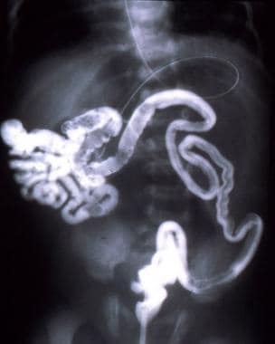
 Gastrointestinal bleeding as a result of entero-iliac fistula due to intestinal foreign body
 Crohn's disease - Wikipedia
 Low anterior resection surgery: What it is for and what to expect
 Crohn's Disease | MedlinePlus
 High output enterocutaneous fistula: a literature review and a case study
 Permanent jejunal fistula: Promising method for obtaining small intestinal chyme without disturbing intestinal function<...
![Detection and analysis of intestinal flora diversity in patients with complex anal fistula]. | Zhonghua Wei Chang Wai Ke Za...](data:image/png;base64,iVBORw0KGgoAAAANSUhEUgAAABAAAAAQCAYAAAAf8/9hAAACpklEQVQ4jaXTe2iNcRzH8ffz7Jzncp4ZZyZjZ6bmEvPPxrDIJSnJZZJIbn8Qk1CI2pQ7JX8h5fKHW4o/3EuaWnJvrUNqiFZssTWcs83z7DnPc87XH88Ziv98//z1+7369v38vvCfpQBY0w6LeHb2RCHl+fg9vaiGxuyq0ZQMzef0tSeoOSqGEQaR4Go4QghAPBvxHRzbBddjUGGUhXMmULN0KhVlxQDMLC+iZt9VEt+6MPNMCIwAAEj7aSrGxFg5r5KVCyoZGM2lx3Y5dbGB6jnlLJs7nvIxMZbvOE9TvAUjagXtAygVu2Tk3P3ieb6IiPjpjBw9Vy+F02uFYWulaEadnLn2WEREJCOyetdFYdRGMSfXSQ4ABVV7Jo4rYtWCiVyvf8nizWe4cuMZdkYwBlh863a4fa+JR/EWyscWs37pFAxD4/7T96hB/2lKhuYDcORsPW+aWzHz+6FrIRDB1MMY0Vwe3I9Td+IuALMmjyIjZAGBkiFRACKmBqb+77hyDQw9DED8TRukvCygKpQWFwCQyUb0zxIwtQD40NoZPAVQdY3S2EAAkj9cfo/3b0DJ+h8/fwdVCYA8S6d0WNBBbHB/6LJJeWmUPkiB3pQPPQ7RPDMAviQglBMAtuNSs/cqyW6HO8fXcaB2CZYWwk78IOWlcb72UDjA4tihFRzcOg+Ato4EaigIkUjVbmHEBimbf1Diza0iIvKupV2qN52W/MrtsuPoDUl22dJXl2+9EHP8NtEn1YnSB0i6F6fbwYronKxdwupFkwCwbZdIRKe9s5sLN59z6W4jr962oZs6OVrk91dGhEg/A8f1WbPzPI3Nnzi8ZT7NLe2cuPyQWw2vSXQmwdAwLIM/xvP3NmYyGXptl+GxAlo7kvi2S9gyCIfVX0sEwTb+BBBpI5QgousUAAAAAElFTkSuQmCC) Detection and analysis of intestinal flora diversity in patients with complex anal fistula]. | Zhonghua Wei Chang Wai Ke Za...
Detection and analysis of intestinal flora diversity in patients with complex anal fistula]. | Zhonghua Wei Chang Wai Ke Za...
 5 Types of Crohn's Disease: Ileocolitis, Jejunoileitis, and More
 Congenital Anomalies of the Digestive System | NCBDDD | CDC
Can you die from Crohn's disease? Risks and complications
 Dr. David Depriest, DO, General Surgery Specialist - Greer, SC | Sharecare
 Thieme E-Books & E-Journals - Clinics in Colon and Rectal Surgery / Issue
Dr. Erin Switzer, DO, Critical Care Surgery Specialist - Augusta, GA | Sharecare
 Cortenema: Package Insert - Drugs.com
 Clinical Value of Transperineal 3D Volume Ultrasound Combined with 2D High Frequency Ultrasound in Anal Fistula
 MedlinePlus - GlobalRPH
 IBD Treatment
Advanced Search Results - Public Health Image Library(PHIL)
 Carbo Vegetabilis - ABC Homeopathy
 Now Book an Easy Appointment to Get a Consultation From the Best Fistula Surgeon - Oceanup.com
Health Library | Rutgers Cancer Institute of New Jersey
10 Nutrition Support | The Role of Nutrition in Maintaining Health in the Nation's Elderly: Evaluating Coverage of Nutrition...
 Ostomy Care
 A Study to Identify Biomarkers of Intestinal Fibrosis in Small Bowel Crohn's Disease - Mayo Clinic
 Living With Crohn's Disease by Lois Fordham - HTML preview, Page 2
 Crohn's disease (inflammatory bowel disease) | HealthEngine Blog
Radiation Enteritis and Proctitis: Practice Essentials, Pathophysiology, Etiology
 Crohn's Meds: What Happens When You Stop Taking Them?
Obstruction6
- An intestinal obstruction is the most common complication of Crohn's disease. (medicalnewstoday.com)
- Such patients may include those with inadequate gastrointestinal function (e.g., short-bowel syndrome or chronic intestinal obstruction), as well as those with severe oropharyngeal dysfunction or permanent neurological impairment. (nationalacademies.org)
- Intestinal obstruction and dietary changes can lead to malnutrition . (healthline.com)
- Mesh erosion and migration can present as acute intestinal obstruction, mass formation, bowel perforation, and chronic abdominal pain [ 10 - 13 ]. (hindawi.com)
- The presentation is often subtle, with herald bleeding followed by a period of grace, or catastrophic bleeding, or rarely an episode of intestinal obstruction. (sciencedaily.com)
- The indication for surgery was intestinal obstruction (244), abdominal pain (53), unclear diagnosis (14), duodenal obstruction (9), abscess (11), and peritonitis (2). (sages.org)
Perforation1
- Anatomic gastrointestinal abnormalities such as enteric fistulas or intestinal perforation can sequester sufficient quantities of ingested elemental mercury to allow significant oxidation and subsequent absorption. (cdc.gov)
Abscesses3
- Itchiness or pain around the anus may be suggestive of inflammation of the anus, or perianal complications such as anal fissures, fistulae, or abscesses around the anal area. (wikipedia.org)
- But people with ileitis may also develop fistulas (inflammatory abscesses) in the lower-right section of the abdomen. (healthline.com)
- It can cause fistulas, ulcers, and abscesses to form around the anus. (healthline.com)
Intestine5
- Sometimes, a fistula forms a tunnel from the intestine to the outer surface of the skin. (medicalnewstoday.com)
- Fistula can affect any part of your body some of the areas that it can affect are near the anal, skin, liver, vagina, intestine, and rectum. (oceanup.com)
- Crohn's disease affects the full thickness of intestine, resulting in significant scarring and fistula formation. (healthgrades.com)
- Fistulas can develop between the intestine and skin, or between the intestine and another organ. (hdkino.org)
- Firstly, doctors choose to use corticosteroids and immunosuppressant and biological drugs to control the inflammatory process and prevent complications of the disease such as stenosis (narrowing of the intestinal lumen) or fistulas (openings from the intestinal lumen to other organs, such as the intestine, bladder, vagina, or skin). (sciencedaily.com)
Inflammation9
- This is because of intestinal inflammation. (healthline.com)
- Inflammation from Crohn's disease can spread through the intestinal wall, creating an abscess. (medicalnewstoday.com)
- Fistulas are one of the abnormal connections between the two organs or blood vessels , which may occur due to surgery, inflammation, or any injury caused to our body. (oceanup.com)
- It tells your healthcare provider how severe the intestinal inflammation is. (clevelandclinic.org)
- The immune system may create an abnormal inflammation reaction in the intestinal wall that does not stop. (gwdocs.com)
- MRI and intestinal ultrasound can describe the transmural inflammation and detect complications. (medscape.com)
- Inflammation of a diverticulum or diverticula in the intestinal tract, esp. (tabers.com)
- Inflammatory bowel disease is characterized by a chronic relapsing intestinal inflammation. (lu.se)
- The pattern of swelling, inflammation, ulcers, and fissures is similar to that of the lesions occurring in the intestinal tract. (medscape.com)
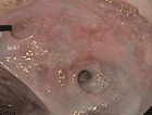
Anal fistula20
- Detection and analysis of intestinal flora diversity in patients with complex anal fistula]. (bvsalud.org)
- To explore the possibility that the intestinal flora profile in complex anal fistula patients is different to that of healthy controls. (bvsalud.org)
- Fecal samples were collected from 30 complex anal fistula patients and 30 matched healthy controls. (bvsalud.org)
- Patients were included if they met the diagnostic criteria of cryptoglandular anal fistula and had exhibited symptoms for more than 3 months. (bvsalud.org)
- or if recurrence is observed after previous anal fistula surgery . (bvsalud.org)
- The α-diversity analysis including ACE, Chao, Shannon and Simpson indexes indicated a richer diversity of intestinal microflora in complex anal fistula patients than in healthy controls. (bvsalud.org)
- On a genus level, samples from anal fistula patients showed a greater abundance of Prevotella spp. (bvsalud.org)
- To explore the clinical diagnostic value of transperineal volume ultrasound combined with two-dimensional high-frequency ultrasound for anal fistula. (scirp.org)
- A total of 52 patients with anal fistula admitted to the Affiliated Hospital of Shaanxi University of Traditional Chinese Medicine from December 2017 to July 2018 were selected. (scirp.org)
- Among 52 patients, 3D volume ultrasound combined with 2D high-frequency ultrasound were used to diagnose 32 cases of anal fistula intersphincteric type, 14 cases of transsphincter type, 5 cases of supra-sphincter type, and 1 case of extra-sphincter type. (scirp.org)
- Two-dimensional high-frequency ultrasound was used to diagnose 34 cases of anal fistula intersphincteric type, 14 cases of transsphincter type, 4 cases of supra-sphincter type, and 0 cases of extra-sphincter type. (scirp.org)
- There was a statistically significant difference in the detection rate of the anal fistula branch and the coincidence rate of the internal fistula between the two methods (both P (scirp.org)
- In the anal fistula classification, there was no significant statistical difference between the two methods. (scirp.org)
- Yang, D. and Yao, X. (2020) Clinical Value of Transperineal 3D Volume Ultrasound Combined with 2D High Frequency Ultrasound in Anal Fistula. (scirp.org)
- Anal fistula is a good disease and common disease in clinical anorectal surgery [1]. (scirp.org)
- This study evaluated the anal fistula lesions by combining three-dimensional volume ultrasound and two-dimensional high-frequency ultrasound technology (combined group), and observed whether the combined application of the two technologies for the overall diagnostic efficacy of the disease is higher than the two-dimensional high-frequency ultrasound technology alone. (scirp.org)
- Provide a more valuable basis for the treatment and evaluation of anal fistula. (scirp.org)
- 1) Subjects 52 patients (average age of 39.4 ± 13.5 years [4 months - 63 years]) with anal fistula collected from December 2018 to June 2019 in the Department of Anorectal Surgery, Affiliated Hospital of Shaanxi University of Traditional Chinese Medicine. (scirp.org)
- An anal fistula is also one of the majorly diagnosed fistulas. (oceanup.com)
- There are many kinds of fistulas and the most common of them are anal fistula, arteriovenous fistula, enterovesical fistula, vesicovaginal fistula, enterocutaneous fistula, and rectovaginal fistula. (oceanup.com)
Complications2
- There were no surgical complications, and the animals tolerated the fistula well. (vtt.fi)
- Mesh is a foreign substance, because of that some of the complications including hematoma, seroma, foreign body reaction, organ damage, infection, mesh rejection, and fistula formation may occur after implantation of the mesh. (hindawi.com)
Enterocutaneous fistula formation2
- Operative trauma is the most common cause of enterocutaneous fistula formation. (medscape.com)
- Intraperitoneal drainage tubes can erode into the intestinal lumen, leading to enterocutaneous fistula formation. (medscape.com)
Bowel10
- The pathophysiology of all forms of small-bowel fistulas is related to the exposure of nonintestinal tissue to intestinal contents because of the fistula. (medscape.com)
- The etiology of small-bowel fistulas is important for determining the subsequent treatment. (medscape.com)
- Intestinal anastomoses are susceptible to partial or complete dehiscence in the presence of impaired blood supply to the area, systemic hypotension, anastomotic suture line tension, perianastomotic infection, and diseased bowel segment anastomosis. (medscape.com)
- Exposure of the bowel to prosthetic mesh or a large abdominal defect can lead to wall erosion, resulting in enterocutaneous fistula. (medscape.com)
- Penetration of the intestinal wall from a foreign body (eg, an ingested metallic object or a fish bone) can lead to enteroenteric fistula formation because of erosion into adjacent bowel loops. (medscape.com)
- In most cases, fistulas originate in the bowel. (medicalnewstoday.com)
- Chronic intestinal radiation injury is a result of transmural bowel damage with associated obliterative endarteritis. (medscape.com)
- Between December 2005 and March 2006, specific lot numbers of defective Composix® Kugel® Mesh hernia repair patches were recalled after it was found that the memory recoil ring, which opens the patch, could potentially break under the stress of placement in the intra-abdominal space and lead to chronic intestinal problems such as bowel perforations and chronic intestinal fistulae. (yourlawyer.com)
- In addition, MRI can detect fistulas, deep ulcerations, and a thickened small bowel wall, but ultrasound can be performed at the point of care by the treating gastroenterologist and is inexpensive. (medscape.com)
- struction, erosion into the bowel or vessels, The preoperative diagnosis of a re- fistula formation or septic syndrome [ 2,7 ]. (who.int)
Crohn's2
- Patients whose anal fistulas were caused by Crohn's disease , trauma , special infections (such as actinomycosis and tuberculosis ) were also excluded, as were those who had used antibiotics , prebiotics , or probiotics that may affect intestinal microecology in the month prior to the study. (bvsalud.org)
- Around 1 in 4 people with Crohn's develop fistulas. (medicalnewstoday.com)
Enteric fistulas1
- In patients with all forms of enteric fistulas, sepsis is a major cause of mortality and must be treated aggressively. (medscape.com)
Chronic2
- It is a chronic genetic disease that occurs when the immune system loses tolerance to the patient's own intestinal flora, leading to an abnormal inflammatory response that continues over time. (sciencedaily.com)
- Fissures or fistulas may occur in persons with chronic disease. (medscape.com)
Surgery7
- Aortoenteric fistulas, which mandate emergency surgery when diagnosed, are an exception. (medscape.com)
- Bacterial samples were collected before (fecal samples), during (small intestinal samples) and 11 weeks after surgery. (vtt.fi)
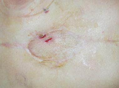
- Large, numerous, or persistent fistulas usually require surgery . (medicalnewstoday.com)
- Fistulas that do not respond to medications increase the risk of sepsis and may require emergency surgery . (medicalnewstoday.com)
- The surgeons of the fistula problem are medical professionals who are well educated which are trained in proctology [a branch of medicine that deals with the diseases and medical conditions of rectum, anus, and colon] and colorectal surgery. (oceanup.com)
- To perform the fistula surgery, the surgeon should be trained and well-known of all the conditions that cause a fistula. (oceanup.com)
- The procedure is based on an autologous bone-marrow transplant (when patients receive a transplant of their own stem cells) and now constitutes a treatment option to cure an intestinal disease that sometimes does not successfully respond to drugs and requires highly complex surgery that does not provide a cure. (sciencedaily.com)
Perianal1
- it may contain granulomas or be associated with intestinal or perianal fistulas. (lu.se)
Abscess formation1
- [ 4 ] and leakage from intestinal anastomoses result in leakage of intestinal contents with abscess formation. (medscape.com)
Ulcers2
- These happen when ulcers go through your intestinal wall and create an abnormal connection to your skin or another organ. (webmd.com)
- Sometimes, ulcers extend completely through the intestinal wall, creating an abnormal connection between different body parts (fistula). (hdkino.org)
Lumen4
- thus, an intestinal fistula is an abnormal anatomic connection between a part (or multiple parts) of the intestinal lumen and the lumen of another epithelialized structure or the skin. (medscape.com)
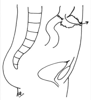
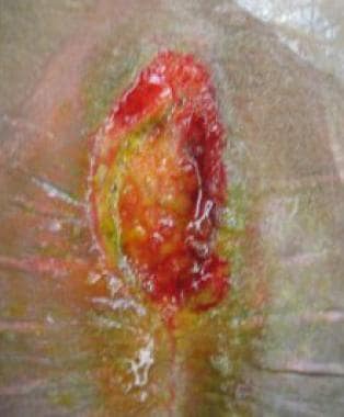
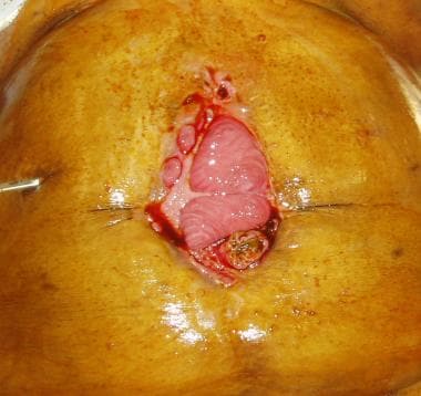
- This results in communication of the intestinal lumen with the skin surface, forming an enterocutaneous fistula (see the image below). (medscape.com)
- Esophago-gastro-duodenoscopy showed the aortic prosthesis crossing the third segment of duodenal wall occluding the intestinal lumen. (sciencedaily.com)
- ADF may be primarily due to a spontaneous communication between the lumen of aortic aneurysm and intestinal loop, or secondarily due to surgical repair of aneurysms with prosthetic implants. (sciencedaily.com)
Motility1
- The influence of the fistula on intestinal motility was determined by use of barium-impregnated polyethylene spheres (BIPS) and on microflora by use of bacterial culturing. (vtt.fi)
Malnutrition1
- Up to 70% of patients with fistulae have malnutrition and it is a significant prognostic factor of spontaneous fistula closure. (nih.gov)
Gastrointestinal2
- Aorto-duodenal fistulae constitute 80 percent of aorto-enteric fistulae, presenting with upper gastrointestinal bleeding. (sciencedaily.com)
- Aorto-duodenal fistulae (ADF) are the most frequent aorto-enteric fistulae (80%) and the most frequent presenting sign of ADF is upper gastrointestinal bleeding (UGI). (sciencedaily.com)
Tract4
- Fistulae are abnormal passages that can develop in the wall of your digestive tract. (healthline.com)
- In Dacron prosthesis patients, fistula develops in the proximal graft tract opening in the third segment of duodenum. (sciencedaily.com)
- The adults then migrate to their ultimate home in the intestinal veins or the venous plexus of the genitourinary tract. (merckmanuals.com)
- Increased dental caries and nutritional deficiencies may be related to decreased saliva production and malabsorption in the intestinal tract. (medscape.com)
Diagnosis2
- However, enterocutaneous fistula due to mesh migration can occur as a very rare, late complication, for which diagnosis is very difficult. (hindawi.com)
- Oral manifestations can prove crucial in diagnosis and usually parallel the intestinal disease course. (medscape.com)
Gastric1
- Our outpatient ostomy program provides comprehensive care for all ostomy, intestinal fistula and gastric tube care needs. (thechristhospital.com)
Surgical4
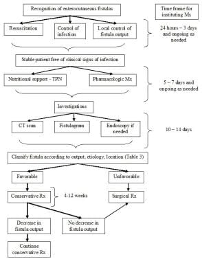
- As a general rule in the treatment of intestinal fistulas, medical treatment and stabilization precede attempts at surgical intervention. (medscape.com)
- Surgical treatment is reserved for patients whose fistulas do not resolve with nonsurgical therapy. (medscape.com)
- Because of the high mortality and morbidity associated with secondary aorto-enteric fistula, surgical treatment is always recommended. (sciencedaily.com)
- 2 ]. Patients with forgotten intraperitoneal tion most frequently occurs after gynaeco- foreign bodies may present with pseudotu- logical and upper abdominal surgical mour (gossypiboma) [ 5,6 ], intestinal ob- procedures [ 3,4 ]. (who.int)
Microflora1
- Accurate information on changes in small intestinal microflora in dogs is rather limited because of difficulties in obtaining samples of small intestinal chyme. (vtt.fi)
Bacterial1
- The intestinal bacterial flora leads to contamination and eventual development of sepsis. (medscape.com)
Ileal1
- Abdominal computed tomography (CT) with oral and intravenous contrast showed an ileal enterocutaneous fistula but revealed no mesh migration. (hindawi.com)
Symptoms4
- The symptoms of a fistula differ, depending on the location. (medicalnewstoday.com)
- The various symptoms in the types of fistulas are listed as follows. (oceanup.com)
- Fistula can affect any age group of people and may lead to mild to severe symptoms such as uneasiness in the anal region, infection, irritation near the anus, and dehydration, etc. (oceanup.com)
- Patients' quality of life is conditioned by the severity of the disease and, in the most severe cases, prevents them from leading a normal life, with a very high level of suffering due to the acuteness and frequency of the intestinal symptoms. (sciencedaily.com)
Anus1
- The internal fistula is located around the dentate line, the external fistula is located on the skin around the anus, and the fistula runs on the anus, peripheral tissue clearance. (scirp.org)
Precede1
- Intraoral involvement in Crohn disease occurs in 8-29% of patients and may precede intestinal involvement. (medscape.com)
Dehiscence1
- Sixteen patients (ten with one or more intestinal fistula) developed abdominal wall dehiscence were included in this study. (who.int)
Lesions1
- Whether patients with orofacial granulomatoses will subsequently develop intestinal manifestations of Crohn disease is uncertain, but histologic similarities between the oral lesions and the intestinal lesions are obvious. (medscape.com)
Surgeries2
- The above mentioned specialists are capable of performing both laser surgeries and open surgeries for the treatment of fistulas. (oceanup.com)
- Plaintiff attorneys argued that Mr. Thorpe suffered severe internal injuries caused by a broken plastic ring on the hernia repair mesh, which included an abdominal wall abscess and fistula and caused him to undergo a variety of surgeries to repair damage and which continues today. (yourlawyer.com)
Rectal1
- Removal or Destruction of Rectal or Intestinal Tumor (incl. (sharecare.com)
Crohn1
- Oral findings as described above warrant a full systemic evaluation for intestinal Crohn disease, including referral for colonoscopy and biopsy with histopathologic correlation. (medscape.com)
Ultrasound2
- 2) Three-dimensional volumetric ultrasound technology has great application prospects in infants and anal fistulas. (scirp.org)
- And because of radiation associated with CT, the preferred methods are MRI and intestinal ultrasound, Stoker added. (medscape.com)
Patients4
- Patients with high output EC fistulae have a high morbidity and mortality rate. (nih.gov)
- Nevertheless, patients with fistulae should be managed based on the available evidence, detailed clinical and nutrition assessment, and close monitoring. (nih.gov)
- The patients facing this problem of fistula should consult the concerned doctor and must try to follow all the plan of treatment. (oceanup.com)
- While choosing the best fistula surgeon, look out for the patient reviews about the surgeon, experience of the surgeon, and the surgeon's availability for diagnosing and treating the patients. (oceanup.com)
Clinical1
- Intestinal fistula includes many clinical entities. (medscape.com)
Complication1
- The patient had to be surgically treated because of a complication: the formation of an entero-iliac fistula with subsequent development of a pseudoaneurysm of the right external iliac artery. (univaq.it)
MeSH4
- Here we report the case of an enterocutaneous fistula due to late mesh migration in a mentally retarded, diabetic, 35-year-old male after umbilical hernia repair with composite dual mesh in 2010. (hindawi.com)
- In the literature, most cases of mesh-associated enterocutaneous fistula due to migration involved polypropylene meshes. (hindawi.com)
- Most reports of enterocutaneous fistula due to mesh migration involve polypropylene mesh [ 3 ]. (hindawi.com)
- Here we describe a case of enterocutaneous fistula due to late migration of a composite dual mesh 4 years after incisional hernia repair. (hindawi.com)
Formation1
- The common mechanisms of intestinal fistula formation are outlined below. (medscape.com)
Wall3
- As the abscess grows, it forms a small hole in the wall, and this hole can develop into a fistula. (medicalnewstoday.com)
- To protect the intestinal wall, a pediculated fragment of the greater omentum was placed between the duodenum and aortic bypass. (sciencedaily.com)
- They mainly consisted of entero-entero (72), ileo-sigmoid (49), and entero-abdominal wall (33) fistulas. (sages.org)
Tissue1
- The local effect of intestinal fluid can be damaging or corrosive to the nonintestinal tissue, leading to breakdown, erosions, and loss of normal organ or organ system function. (medscape.com)