Intracranial Thrombosis
Coronary Thrombosis
Sinus Thrombosis, Intracranial
Carotid Artery Thrombosis
Sagittal Sinus Thrombosis
Phlebography
Intracranial Embolism and Thrombosis
Iliac Vein
Thrombophilia
Femoral Vein
Upper Extremity Deep Vein Thrombosis
Heparin
Factor V
Hemostasis
Heparin, Low-Molecular-Weight
Cavernous Sinus Thrombosis
Blood Coagulation
Antiphospholipid Syndrome
Vena Cava, Inferior
Thromboembolism
Platelet Aggregation Inhibitors
Lateral Sinus Thrombosis
Bleeding Time
Thrombectomy
Fibrin Fibrinogen Degradation Products
Stents
Protein S Deficiency
Treatment Outcome
Platelet Aggregation
Mesenteric Veins
Subclavian Vein
Drug-Eluting Stents
Prothrombin
Risk Factors
Popliteal Vein
Jugular Veins
Blood Platelets
Venous Thromboembolism
Antibodies, Antiphospholipid
Protein C Deficiency
Cranial Sinuses
Activated Protein C Resistance
Lupus Coagulation Inhibitor
Platelet Activation
Thromboplastin
Thrombolytic Therapy
Warfarin
Follow-Up Studies
Intra-arterial cerebral thrombolysis for acute ischemic stroke in a community hospital. (1/319)
BACKGROUND AND PURPOSE: Advances in thrombolytic therapy, brain imaging, and neurointerventional techniques provide new therapeutic options for acute stroke. Intra-arterial thrombolysis has proved to be a potent therapeutic tool. To show that this procedure can be performed in community hospitals, we describe our experience with a group of 11 patients treated for middle cerebral artery occlusions. METHODS: Twenty-two patients seen during a period of 1 year with clinical findings of acute major-vessel stroke met screening criteria and were evaluated under an institutional review board-approved protocol. After CT scanning, 17 of those patients met strict criteria, gave informed consent, and underwent angiography. Eleven patients had M1 and M2 middle cerebral artery occlusions and received local thrombolytic therapy with urokinase. Recanalization efficacy, complications, and outcome data were compiled. RESULTS: The average score on the National Institutes of Health Stroke Scale was 22.2 at the onset of treatment and 12.5 after therapy, with 91% of patients showing neurologic improvement. Complete (TIMI 3) recanalization occurred in 73% of cases and partial recanalization (TIMI 2) in 18%. At the 90-day follow-up evaluation, 56% of patients had good outcomes (modified Rankin score, 0 to 1). One intracranial hemorrhage occurred. CONCLUSION: Intra-arterial thrombolysis can be performed in a community hospital by radiologists with interventional and neuroradiologic skills given appropriate institutional preparation. (+info)Long-term prognosis of cerebral ischemia in young adults. National Research Council Study Group on Stroke in the Young. (2/319)
BACKGROUND AND PURPOSE: Prognosis of ischemic stroke in young adults is reported as favorable, and transient ischemic attack (TIA) is commonly considered a benign event. We investigated long-term outcome and prognostic predictors of cerebral ischemia in patients under 45 years of age. METHODS: Three hundred thirty-three patients aged 15 to 44 years who suffered from a first-ever TIA or ischemic stroke were prospectively followed up with annual clinical evaluation or complete phone interview. End points were the composite outcome event of stroke, myocardial infarction, and vascular or nonvascular death and death from all causes. The probability of event-free survival was estimated by the Kaplan-Meier method. Univariate and multivariate estimates of hazard ratios were calculated according to the Cox proportional hazards analysis. RESULTS: An average follow-up of 96 months was available in 330 patients (99.1%). Survival was worse in patients with stroke at entry (86.5%) than in those with TIA (97.1%). Mortality in both groups was significantly higher than in the general population (standardized mortality ratio [SMR] 14.5, P<0.0001, Poisson distribution test, and SMR 7.9, P=0.002). The average annual mortality rate was higher during the first (3.94%, 95% CI 1.84 to 6. 04) than in the subsequent years. The average annual incidence rate of new stroke was higher in patients with stroke than in those with TIA at entry, and it declined from 1.56% (95% CI 0.21 to 2.91) during the first year to 0.06% (95% CI 0.04 to 0.08) at the end of the follow-up. Myocardial infarction occurred later, after the first year, with similar rates in patients with stroke and TIA at entry. The average annual rates of new stroke (2.36%), myocardial infarction (1.68%), and death (3.05%) were higher in patients with the mixed atherothrombotic and cardioembolic etiology than in the remaining patients. Male gender, age >35 years, stroke at entry, and cardiac diseases were independent predictors of the composite outcome event at the Cox regression analysis, whereas only stroke at entry and cardiac diseases predicted death from all causes. CONCLUSIONS: Stroke and TIA in young adults have severe prognostic implications, because the mortality risk was highly increased with respect to the general population. Preventive measures are strongly recommended in the presence of any unfavorable prognostic profile. (+info)Atherothrombotic cerebellar infarction: vascular lesion-MRI correlation of 31 cases. (3/319)
BACKGROUND AND PURPOSE: Correlation of MRI findings with atherosclerotic vascular lesions has rarely been attempted in patients with cerebellar infarction. The aim of this study was to correlate the MRI lesions with the vascular lesions seen on conventional cerebral angiography in cerebellar infarction. METHODS: The subjects included 31 patients with cerebellar infarcts who underwent both MRI and conventional cerebral angiography. We analyzed the risk factors, clinical findings, imaging study, and angiography results. We attempted to correlate MRI lesions with the vascular lesions shown in the angiograms. RESULTS: The vascular lesions seen on angiograms were subdivided into 3 groups: large-artery disease (n=22), in situ branch artery disease (n=6), and no angiographic disease with hypertension (n=3). The proximal segment (V1) lesions of vertebral artery were the most common angiographic features in patients with large-artery disease in which stroke most commonly involved the posterior inferior cerebellar artery (PICA) cerebellum. The V1 lesions with coexistent occlusive lesions of the intracranial vertebral and basilar arteries were correlated with cerebellar infarcts, which had no predilection for certain cerebellar territory. The intracranial occlusive disease without V1 lesion was usually correlated with small cerebellar lesions in PICA and superior cerebellar artery (SCA) cerebellum. The subclavian artery or brachiocephalic trunk lesion was associated with small cerebellar infarcts. The in situ branch artery disease was correlated with the PICA cerebellum lesions, which were territorial or nonterritorial infarct. No angiographic disease with hypertension was associated with small-sized cerebellar infarcts within the SCA, anterior inferior cerebellar artery, or SCA cerebellum. CONCLUSIONS: Our study indicates that the topographic heterogeneity of cerebellar infarcts are correlated with diverse angiographic findings. The result that large-artery disease, in which nonterritorial infarcts are more common than territorial infarcts, is more prevalent than in situ branch artery disease or small-artery disease, suggest that even a small cerebellar infarct can be a clue to the presence of large-artery disease. (+info)Acquired pial arteriovenous fistula following cerebral vein thrombosis. (4/319)
BACKGROUND: We report a unique case of an acquired pial arteriovenous fistula occurring after an asymptomatic thrombosis of a superficial cerebral vein. CASE DESCRIPTION: A cerebral angiogram performed in a 51-year-old man with subarachnoid hemorrhage revealed a 10-mm ruptured anterior communicating artery aneurysm and a thrombosed left superficial middle cerebral vein. Coil embolization of the anterior communicating aneurysm was performed. Follow-up angiography 18 months later revealed a new, asymptomatic, pial arteriovenous fistula between the previously thrombosed left superficial middle cerebral vein and a small sylvian branch of the left middle cerebral artery. CONCLUSIONS: This case provides evidence that pial arteriovenous fistulas may develop as acquired lesions and furthermore may rarely follow cerebral vein thrombosis. Several cases of dural arteriovenous fistulas, as well as a single case of a mixed pial-dural arteriovenous fistula, occurring after dural sinus thrombosis have been reported previously. However, to our knowledge, this is the first report of an acquired pial arteriovenous fistula following a cerebral vein thrombosis. (+info)Tissue plasminogen activator (tPA) deficiency exacerbates cerebrovascular fibrin deposition and brain injury in a murine stroke model: studies in tPA-deficient mice and wild-type mice on a matched genetic background. (5/319)
Although the serine protease, tissue plasminogen activator (tPA), is approved by the US Food and Drug Administration for therapy to combat focal cerebral infarction, the basic concept of thrombolytic tPA therapy for stroke was challenged by recent studies that used genetically manipulated tPA-deficient (tPA-/-) mice, which suggested that tPA mediates ischemic neuronal damage. However, those studies were potentially flawed because the genotypes of tPA-/- and wild-type control mice were not entirely clear, and ischemic neuronal injury was evaluated in isolation of tPA effects on brain thrombosis. Using mice with appropriate genetic backgrounds and a middle cerebral artery occlusion stroke model with nonsiliconized thread, which does lead to microvascular thrombus formation, in the present study we determined the risk for cerebrovascular thrombosis and neuronal injury in tPA-/- and genetically matched tPA+/+ mice subjected to transient focal ischemia. Cerebrovascular fibrin deposition and the infarction volume were increased by 8.2- and 6. 7-fold in tPA-/- versus tPA+/+ mice, respectively, and these variables were correlated with reduced cerebral blood flow up to 58% (P<0.05) and impaired motor neurological score by 70% (P<0.05). Our findings indicate that tPA deficiency exacerbates ischemia-induced cerebrovascular thrombosis and that endogenous tPA protects the brain from an ischemic insult, presumably through its thrombolytic action. In addition, our study emphasizes the importance of appropriate genetic controls in murine stroke research. (+info)Hyperfibrinogenemia is associated with specific histocytological composition and complications of atherosclerotic carotid plaques in patients affected by transient ischemic attacks. (6/319)
BACKGROUND: Epidemiological studies have demonstrated that hyperfibrinogenemia is an independent risk factor for cerebrovascular atherosclerosis. However, the underlying mechanisms are poorly understood. We studied whether hyperfibrinogenemia could modify the histological composition of atherosclerotic plaque and precipitate carotid thrombosis resulting from rupture of the plaque. METHODS AND RESULTS: We studied the histological composition of 71 carotid atherosclerotic plaques from patients who had undergone surgical endarterectomy after a first episode of transient ischemic attack. Patients were divided into 3 groups corresponding to the tertiles of plasma fibrinogen values. Hypercholesterolemia, hypertriglyceridemia, hypertension, diabetes, and smoking habit were also assessed. At the histological analysis, plaques of patients in the highest tertile of fibrinogen (>407 mg/dL) were characterized by a high incidence of thrombosis (66.7% of cases) compared with plaques of subjects in the lower (21.7%) (P=0.002) and middle (29. 2%) (P=0.009) tertiles. Plaque rupture was significantly associated with high fibrinogen levels (54.2%, P=0.003). Multivariate logistic regression indicated that hyperfibrinogenemia was an independent risk factor for a decrease in cap thickness (P=0.0005), macrophage foam cell infiltration of the cap (P=0.003), and thrombosis (P=0. 003). When the presence of other risk factors was accounted for, hyperfibrinogenemia remained an independent predictor of carotid thrombosis with an odds ratio of 5.83, compared with other risk factors. CONCLUSIONS: The results of the present study add to the evidence that hyperfibrinogenemia, independently of other risk factors, is associated with a specific histological composition of carotid atherosclerotic plaques that predisposes them to rupture and thrombosis. (+info)Early growth, adult income, and risk of stroke. (7/319)
BACKGROUND AND PURPOSE: A number of studies have shown that reduced intrauterine growth and low birth weight are associated with raised rates of fatal and nonfatal stroke in adult life. Whether this increased risk of stroke is modified by growth in childhood or by socioeconomic status in adult life is not known. METHODS: We studied hospital admissions and deaths from stroke among 3639 men who were born in Helsinki University Central Hospital during 1924 to 1933. They had detailed records of their body size at birth, their growth through childhood, and their social circumstances as adults. Three hundred thirty-one of the men had had a stroke. RESULTS: Hazard ratios for stroke were related to low birth weight in relation to head circumference (P=0.005) and to short length in relation to head circumference (P=0.02). These associations were stronger for hemorrhagic than for thrombotic stroke. Men who developed stroke still had below-average stature at 7 years (P=0.05), but after 7 years their height "caught up" through accelerated growth. As adults they had low incomes and low social class (P<0.0001). CONCLUSIONS: Stroke may originate through reduced fetal growth, with low body weight and short body length at birth but "sparing" of head growth. Other studies suggest that this pattern of growth is associated with persisting elevation of blood pressure and raised plasma fibrinogen concentrations, 2 known risk factors for stroke. The risk of stroke is increased by accelerated growth in height during childhood. Accelerated growth has previously been linked to the development of hypertension in adult life. Stroke risk is further increased by adverse influences linked to low income. (+info)Clinicopathological study of intracranial fusiform and dolichoectatic aneurysms : insight on the mechanism of growth. (8/319)
BACKGROUND AND PURPOSE: Intracranial fusiform aneurysms can be divided into 2 clinically different subtypes: acute dissecting aneurysms and chronic fusiform or dolichoectatic aneurysms. Of these 2, the natural history and growth mechanism of chronic fusiform aneurysms remains unknown. METHODS: A consecutive series of 16 patients with chronic fusiform aneurysms was studied retrospectively to clarify patient clinical and neuroradiological features. Aneurysm tissues were obtained from 8 cases and were examined to identify histological features that could correspond to the radiological findings. RESULTS: Four histological features were found: (1) fragmentation of internal elastic lamina (IEL), (2) neoangiogenesis within the thickened intima, (3) intramural hemorrhage (IMH) and thrombus formation, and (4) repetitive intramural hemorrhages from the newly formed vessels within thrombus. IEL fragmentation was found in all cases, which suggests that this change may be one of the earliest processes of aneurysm formation. MRI or CT detected IMH, and marked contrast enhancement of the inside of the aneurysm wall (CEI) on MRI corresponded well with intimal thickening. Eight of 9 symptomatic cases but none of 7 asymptomatic cases presented with both radiological features. CONCLUSIONS: Data suggest that chronic fusiform aneurysms are progressive lesions that start with IEL fragmentation. Formation of IMH seems to be a critical event necessary for lesions to become symptomatic and progress, and this can be monitored on MRI. Knowledge of this possible mechanism of progression and corresponding MRI characteristics could help determine timing of surgical intervention. (+info)Intracranial thrombosis refers to the formation of a blood clot (thrombus) within the intracranial vessels, which supply blood to the brain. This condition can occur in any of the cerebral arteries or veins and can lead to serious complications such as ischemic stroke, transient ischemic attack (TIA), or venous sinus thrombosis.
The formation of an intracranial thrombus can be caused by various factors, including atherosclerosis, cardiac embolism, vasculitis, sickle cell disease, hypercoagulable states, and head trauma. Symptoms may vary depending on the location and extent of the thrombosis but often include sudden onset of headache, weakness or numbness in the face or limbs, difficulty speaking or understanding speech, vision changes, and loss of balance or coordination.
Diagnosis of intracranial thrombosis typically involves imaging studies such as computed tomography (CT) angiography, magnetic resonance angiography (MRA), or digital subtraction angiography (DSA). Treatment options may include anticoagulation therapy, thrombolysis, endovascular intervention, or surgical intervention, depending on the underlying cause and severity of the condition.
Thrombosis is the formation of a blood clot (thrombus) inside a blood vessel, obstructing the flow of blood through the circulatory system. When a clot forms in an artery, it can cut off the supply of oxygen and nutrients to the tissues served by that artery, leading to damage or tissue death. If a thrombus forms in the heart, it can cause a heart attack. If a thrombus breaks off and travels through the bloodstream, it can lodge in a smaller vessel, causing blockage and potentially leading to damage in the organ that the vessel supplies. This is known as an embolism.
Thrombosis can occur due to various factors such as injury to the blood vessel wall, abnormalities in blood flow, or changes in the composition of the blood. Certain medical conditions, medications, and lifestyle factors can increase the risk of thrombosis. Treatment typically involves anticoagulant or thrombolytic therapy to dissolve or prevent further growth of the clot, as well as addressing any underlying causes.
Venous thrombosis is a medical condition characterized by the formation of a blood clot (thrombus) in the deep veins, often in the legs (deep vein thrombosis or DVT), but it can also occur in other parts of the body such as the arms, pelvis, or lungs (pulmonary embolism).
The formation of a venous thrombus can be caused by various factors, including injury to the blood vessel wall, changes in blood flow, and alterations in the composition of the blood. These factors can lead to the activation of clotting factors and platelets, which can result in the formation of a clot that blocks the vein.
Symptoms of venous thrombosis may include swelling, pain, warmth, and redness in the affected area. In some cases, the clot can dislodge and travel to other parts of the body, causing potentially life-threatening complications such as pulmonary embolism.
Risk factors for venous thrombosis include advanced age, obesity, smoking, pregnancy, use of hormonal contraceptives or hormone replacement therapy, cancer, recent surgery or trauma, prolonged immobility, and a history of previous venous thromboembolism. Treatment typically involves the use of anticoagulant medications to prevent further clotting and dissolve existing clots.
Coronary thrombosis is a medical condition that refers to the formation of a blood clot (thrombus) inside a coronary artery, which supplies oxygenated blood to the heart muscle. The development of a thrombus can partially or completely obstruct blood flow, leading to insufficient oxygen supply to the heart muscle. This can cause chest pain (angina) or a heart attack (myocardial infarction), depending on the severity and duration of the blockage.
Coronary thrombosis often results from the rupture of an atherosclerotic plaque, a buildup of cholesterol, fat, calcium, and other substances in the inner lining (endothelium) of the coronary artery. The ruptured plaque exposes the underlying tissue to the bloodstream, triggering the coagulation cascade and resulting in the formation of a thrombus.
Immediate medical attention is crucial for managing coronary thrombosis, as timely treatment can help restore blood flow, prevent further damage to the heart muscle, and reduce the risk of complications such as heart failure or life-threatening arrhythmias. Treatment options may include medications, such as antiplatelet agents, anticoagulants, and thrombolytic drugs, or interventional procedures like angioplasty and stenting to open the blocked artery. In some cases, surgical intervention, such as coronary artery bypass grafting (CABG), may be necessary.
Intracranial sinus thrombosis is a medical condition characterized by the formation of a blood clot (thrombus) within the intracranial venous sinuses, which are responsible for draining blood from the brain. The condition can lead to various neurological symptoms and complications, such as increased intracranial pressure, headaches, seizures, visual disturbances, and altered consciousness. Intracranial sinus thrombosis may result from various factors, including hypercoagulable states, infections, trauma, and malignancies. Immediate medical attention is necessary for proper diagnosis and treatment to prevent potential long-term neurological damage or even death.
Carotid artery thrombosis is a medical condition characterized by the formation of a blood clot (thrombus) inside the carotid artery, which is one of the major blood vessels that supplies oxygenated blood to the head and neck. This condition can lead to serious complications such as a stroke or transient ischemic attack (TIA), also known as a "mini-stroke," if the clot dislodges and travels to the brain, blocking the flow of blood and oxygen.
Carotid artery thrombosis can result from various factors, including atherosclerosis (the buildup of fats, cholesterol, and other substances in the artery walls), hypertension (high blood pressure), diabetes, smoking, and genetic predisposition. Symptoms may include neck pain or stiffness, weakness or numbness in the face or limbs, difficulty speaking or understanding speech, vision problems, and sudden severe headaches. Diagnosis typically involves imaging tests such as ultrasound, CT angiography, or MRI angiography. Treatment options may include anticoagulant or antiplatelet medications, endovascular procedures to remove the clot, or surgery to clean out the artery (carotid endarterectomy).
Anticoagulants are a class of medications that work to prevent the formation of blood clots in the body. They do this by inhibiting the coagulation cascade, which is a series of chemical reactions that lead to the formation of a clot. Anticoagulants can be given orally, intravenously, or subcutaneously, depending on the specific drug and the individual patient's needs.
There are several different types of anticoagulants, including:
1. Heparin: This is a naturally occurring anticoagulant that is often used in hospitalized patients who require immediate anticoagulation. It works by activating an enzyme called antithrombin III, which inhibits the formation of clots.
2. Low molecular weight heparin (LMWH): LMWH is a form of heparin that has been broken down into smaller molecules. It has a longer half-life than standard heparin and can be given once or twice daily by subcutaneous injection.
3. Direct oral anticoagulants (DOACs): These are newer oral anticoagulants that work by directly inhibiting specific clotting factors in the coagulation cascade. Examples include apixaban, rivaroxaban, and dabigatran.
4. Vitamin K antagonists: These are older oral anticoagulants that work by inhibiting the action of vitamin K, which is necessary for the formation of clotting factors. Warfarin is an example of a vitamin K antagonist.
Anticoagulants are used to prevent and treat a variety of conditions, including deep vein thrombosis (DVT), pulmonary embolism (PE), atrial fibrillation, and prosthetic heart valve thrombosis. It is important to note that anticoagulants can increase the risk of bleeding, so they must be used with caution and regular monitoring of blood clotting times may be required.
Sagittal sinus thrombosis is a medical condition that refers to the formation of a blood clot (thrombus) in the sagittal sinus, which is a venous structure located in the brain. The sagittal sinus runs along the midline of the brain and receives blood from the superficial veins of the brain.
Sagittal sinus thrombosis can occur as a result of various conditions, such as head trauma, infection, cancer, or certain medical disorders that cause hypercoagulability (an increased tendency to form blood clots). The formation of a blood clot in the sagittal sinus can obstruct the flow of blood from the brain, leading to symptoms such as headache, seizures, altered consciousness, and focal neurological deficits.
Diagnosis of sagittal sinus thrombosis typically involves imaging studies such as computed tomography (CT) or magnetic resonance imaging (MRI) scans, which can show the presence of a blood clot in the sagittal sinus. Treatment may involve administering anticoagulant medications to prevent further growth of the blood clot and reduce the risk of complications such as pulmonary embolism or cerebral infarction. In some cases, surgical intervention may be necessary to remove the blood clot or alleviate pressure on the brain.
Phlebography is a medical imaging technique used to visualize and assess the veins, particularly in the legs. It involves the injection of a contrast agent into the veins, followed by X-ray imaging to capture the flow of the contrast material through the veins. This allows doctors to identify any abnormalities such as blood clots, blockages, or malformations in the venous system.
There are different types of phlebography, including ascending phlebography (where the contrast agent is injected into a foot vein and travels up the leg) and descending phlebography (where the contrast agent is injected into a vein in the groin or neck and travels down the leg).
Phlebography is an invasive procedure that requires careful preparation and monitoring, and it is typically performed by radiologists or vascular specialists. It has largely been replaced by non-invasive imaging techniques such as ultrasound and CT angiography in many clinical settings.
1. Intracranial Embolism: This is a medical condition that occurs when a blood clot or other particle (embolus) formed elsewhere in the body, travels through the bloodstream and lodges itself in the intracranial blood vessels, blocking the flow of blood to a part of the brain. This can lead to various neurological symptoms such as weakness, numbness, speech difficulties, or even loss of consciousness, depending on the severity and location of the blockage.
2. Intracranial Thrombosis: This is a medical condition that occurs when a blood clot (thrombus) forms within the intracranial blood vessels. The clot can partially or completely obstruct the flow of blood, leading to various symptoms such as headache, confusion, seizures, or neurological deficits, depending on the severity and location of the thrombosis. Intracranial thrombosis can occur due to various factors including atherosclerosis, hypertension, diabetes, and other medical conditions that increase the risk of blood clot formation.
The iliac veins are a pair of large veins in the human body that carry deoxygenated blood from the lower extremities and the pelvic area back to the heart. They are formed by the union of the common iliac veins, which receive blood from the lower abdomen and legs, at the level of the fifth lumbar vertebra.
The combined iliac vein is called the inferior vena cava, which continues upward to the right atrium of the heart. The iliac veins are located deep within the pelvis, lateral to the corresponding iliac arteries, and are accompanied by the iliac lymphatic vessels.
The left common iliac vein is longer than the right because it must cross the left common iliac artery to join the right common iliac vein. The external and internal iliac veins are the two branches of the common iliac vein, with the external iliac vein carrying blood from the lower limbs and the internal iliac vein carrying blood from the pelvic organs.
It is essential to maintain proper blood flow in the iliac veins to prevent deep vein thrombosis (DVT), a condition that can lead to serious complications such as pulmonary embolism.
Thrombophilia is a medical condition characterized by an increased tendency to form blood clots (thrombi) due to various genetic or acquired abnormalities in the coagulation system. These abnormalities can lead to a hypercoagulable state, which can cause thrombosis in both veins and arteries. Commonly identified thrombophilias include factor V Leiden mutation, prothrombin G20210A mutation, antithrombin deficiency, protein C deficiency, and protein S deficiency.
Acquired thrombophilias can be caused by various factors such as antiphospholipid antibody syndrome (APS), malignancies, pregnancy, oral contraceptive use, hormone replacement therapy, and certain medical conditions like inflammatory bowel disease or nephrotic syndrome.
It is essential to diagnose thrombophilia accurately, as it may influence the management of venous thromboembolism (VTE) events and guide decisions regarding prophylactic anticoagulation in high-risk situations.
The femoral vein is the large vein that runs through the thigh and carries oxygen-depleted blood from the lower limbs back to the heart. It is located in the femoral triangle, along with the femoral artery and nerve. The femoral vein begins at the knee as the popliteal vein, which then joins with the deep vein of the thigh to form the femoral vein. As it moves up the leg, it is joined by several other veins, including the great saphenous vein, before it becomes the external iliac vein at the inguinal ligament in the groin.
Upper extremity deep vein thrombosis (UEDVT) is a medical condition that refers to the formation of a blood clot (thrombus) in the deep veins located in the arm or shoulder. This condition can occur due to various reasons, including trauma, surgery, cancer, certain medications, and underlying medical conditions that increase the risk of blood clotting.
The deep veins are larger vessels that run through the body's muscles and are surrounded by fascia, a connective tissue. UEDVT can cause partial or complete blockage of blood flow in the affected vein, leading to swelling, pain, redness, warmth, and decreased function in the arm or hand. In some cases, the clot can break off and travel to the lungs, causing a potentially life-threatening condition called pulmonary embolism (PE).
Diagnosis of UEDVT typically involves a physical exam, medical history, and imaging tests such as ultrasound, CT scan, or MRI. Treatment may include anticoagulant medications to prevent the clot from growing or breaking off, thrombolytic therapy to dissolve the clot, or surgical intervention in severe cases. Compression stockings or other devices may also be used to help improve blood flow and reduce swelling.
The portal vein is the large venous trunk that carries blood from the gastrointestinal tract, spleen, pancreas, and gallbladder to the liver. It is formed by the union of the superior mesenteric vein (draining the small intestine and a portion of the large intestine) and the splenic vein (draining the spleen and pancreas). The portal vein then divides into right and left branches within the liver, where the blood flows through the sinusoids and gets enriched with oxygen and nutrients before being drained by the hepatic veins into the inferior vena cava. This unique arrangement allows the liver to process and detoxify the absorbed nutrients, remove waste products, and regulate metabolic homeostasis.
Cerebral veins are the blood vessels that carry deoxygenated blood from the brain to the dural venous sinuses, which are located between the layers of tissue covering the brain. The largest cerebral vein is the superior sagittal sinus, which runs along the top of the brain. Other major cerebral veins include the straight sinus, transverse sinus, sigmoid sinus, and cavernous sinus. These veins receive blood from smaller veins called venules that drain the surface and deep structures of the brain. The cerebral veins play an important role in maintaining normal circulation and pressure within the brain.
Heparin is defined as a highly sulfated glycosaminoglycan (a type of polysaccharide) that is widely present in many tissues, but is most commonly derived from the mucosal tissues of mammalian lungs or intestinal mucosa. It is an anticoagulant that acts as an inhibitor of several enzymes involved in the blood coagulation cascade, primarily by activating antithrombin III which then neutralizes thrombin and other clotting factors.
Heparin is used medically to prevent and treat thromboembolic disorders such as deep vein thrombosis, pulmonary embolism, and certain types of heart attacks. It can also be used during hemodialysis, cardiac bypass surgery, and other medical procedures to prevent the formation of blood clots.
It's important to note that while heparin is a powerful anticoagulant, it does not have any fibrinolytic activity, meaning it cannot dissolve existing blood clots. Instead, it prevents new clots from forming and stops existing clots from growing larger.
Factor V, also known as proaccelerin or labile factor, is a protein involved in the coagulation cascade, which is a series of chemical reactions that leads to the formation of a blood clot. Factor V acts as a cofactor for the activation of Factor X to Factor Xa, which is a critical step in the coagulation cascade.
When blood vessels are damaged, the coagulation cascade is initiated to prevent excessive bleeding. During this process, Factor V is activated by thrombin, another protein involved in coagulation, and then forms a complex with activated Factor X and calcium ions on the surface of platelets or other cells. This complex converts prothrombin to thrombin, which then converts fibrinogen to fibrin to form a stable clot.
Deficiency or dysfunction of Factor V can lead to bleeding disorders such as hemophilia B or factor V deficiency, while mutations in the gene encoding Factor V can increase the risk of thrombosis, as seen in the Factor V Leiden mutation.
Fibrinolytic agents are medications that dissolve or break down blood clots by activating plasminogen, which is converted into plasmin. Plasmin is a proteolytic enzyme that degrades fibrin, the structural protein in blood clots. Fibrinolytic agents are used medically to treat conditions such as acute ischemic stroke, deep vein thrombosis, pulmonary embolism, and myocardial infarction (heart attack) by restoring blood flow in occluded vessels. Examples of fibrinolytic agents include alteplase, reteplase, and tenecteplase. It is important to note that these medications carry a risk of bleeding complications and should be administered with caution.
Hemostasis is the physiological process that occurs to stop bleeding (bleeding control) when a blood vessel is damaged. This involves the interaction of platelets, vasoconstriction, and blood clotting factors leading to the formation of a clot. The ultimate goal of hemostasis is to maintain the integrity of the vascular system while preventing excessive blood loss.
Low-molecular-weight heparin (LMWH) is a type of heparin used as an anticoagulant, which refers to a group of medications that prevent the formation of blood clots. Heparin is a naturally occurring substance in the body, and low-molecular-weight heparins are obtained through the depolymerization of standard heparin.
LMWH has a lower molecular weight than standard heparin, which results in several pharmacological differences. LMWHs have a more predictable dose response, longer half-life, and higher bioavailability when administered subcutaneously compared to standard heparin. They also exhibit greater anti-factor Xa activity relative to their antithrombin (anti-IIa) activity, which contributes to their anticoagulant effects.
LMWHs are used for the prevention and treatment of deep vein thrombosis (DVT), pulmonary embolism (PE), and other thromboembolic disorders. Common LMWHs include enoxaparin, dalteparin, tinzaparin, and nadroparin.
It is essential to monitor the patient's kidney function when using LMWH since they are primarily cleared by the kidneys. In patients with renal impairment, dose adjustments or alternative anticoagulants may be necessary to reduce the risk of bleeding complications.
Cavernous sinus thrombosis is a medical condition that refers to the formation of a blood clot (thrombus) in the cavernous sinuses, which are located near the base of the brain and are important for draining blood from the face and brain. This condition can occur as a complication of an infection in the facial area or sinuses, or it can be associated with other medical conditions such as cancer or trauma.
Symptoms of cavernous sinus thrombosis may include headache, fever, eye pain, swelling or bulging of the eyes, double vision, and decreased vision. If left untreated, this condition can lead to serious complications such as meningitis, brain abscess, or even death. Treatment typically involves administering antibiotics to treat any underlying infection and anticoagulants to prevent further clot formation. In some cases, surgery may be necessary to remove the clot.
Blood coagulation, also known as blood clotting, is a complex process that occurs in the body to prevent excessive bleeding when a blood vessel is damaged. This process involves several different proteins and chemical reactions that ultimately lead to the formation of a clot.
The coagulation cascade is initiated when blood comes into contact with tissue factor, which is exposed after damage to the blood vessel wall. This triggers a series of enzymatic reactions that activate clotting factors, leading to the formation of a fibrin clot. Fibrin is a protein that forms a mesh-like structure that traps platelets and red blood cells to form a stable clot.
Once the bleeding has stopped, the coagulation process is regulated and inhibited to prevent excessive clotting. The fibrinolytic system degrades the clot over time, allowing for the restoration of normal blood flow.
Abnormalities in the blood coagulation process can lead to bleeding disorders or thrombotic disorders such as deep vein thrombosis and pulmonary embolism.
Antiphospholipid syndrome (APS) is an autoimmune disorder characterized by the presence of antiphospholipid antibodies in the blood. These antibodies are directed against phospholipids, a type of fat molecule found in cell membranes and plasma lipoproteins. The presence of these antibodies can lead to abnormal blood clotting, which can cause serious complications such as stroke, heart attack, deep vein thrombosis, and pulmonary embolism.
APS can occur either on its own (primary APS) or in conjunction with other autoimmune disorders, such as systemic lupus erythematosus (secondary APS). The exact cause of APS is not fully understood, but it is believed to involve a combination of genetic and environmental factors.
Symptoms of APS can vary widely depending on the location and severity of the blood clots. They may include:
* Recurrent miscarriages or stillbirths
* Blood clots in the legs, lungs, or other parts of the body
* Skin ulcers or lesions
* Headaches, seizures, or stroke-like symptoms
* Kidney problems
* Heart valve abnormalities
Diagnosis of APS typically involves blood tests to detect the presence of antiphospholipid antibodies. Treatment may include medications to prevent blood clots, such as anticoagulants and antiplatelet agents, as well as management of any underlying autoimmune disorders.
The inferior vena cava (IVC) is the largest vein in the human body that carries deoxygenated blood from the lower extremities, pelvis, and abdomen to the right atrium of the heart. It is formed by the union of the left and right common iliac veins at the level of the fifth lumbar vertebra. The inferior vena cava is a retroperitoneal structure, meaning it lies behind the peritoneum, the lining that covers the abdominal cavity. It ascends through the posterior abdominal wall and passes through the central tendon of the diaphragm to enter the thoracic cavity.
The inferior vena cava is composed of three parts:
1. The infrarenal portion, which lies below the renal veins
2. The renal portion, which receives blood from the renal veins
3. The suprahepatic portion, which lies above the liver and receives blood from the hepatic veins before draining into the right atrium of the heart.
The inferior vena cava plays a crucial role in maintaining venous return to the heart and contributing to cardiovascular function.
Thromboembolism is a medical condition that refers to the obstruction of a blood vessel by a thrombus (blood clot) that has formed elsewhere in the body and then been transported by the bloodstream to a narrower vessel, where it becomes lodged. This process can occur in various parts of the body, leading to different types of thromboembolisms:
1. Deep Vein Thrombosis (DVT): A thrombus forms in the deep veins, usually in the legs or pelvis, and then breaks off and travels to the lungs, causing a pulmonary embolism.
2. Pulmonary Embolism (PE): A thrombus formed elsewhere, often in the deep veins of the legs, dislodges and travels to the lungs, blocking one or more pulmonary arteries. This can lead to shortness of breath, chest pain, and potentially life-threatening complications if not treated promptly.
3. Cerebral Embolism: A thrombus formed in another part of the body, such as the heart or carotid artery, dislodges and travels to the brain, causing a stroke or transient ischemic attack (TIA).
4. Arterial Thromboembolism: A thrombus forms in an artery and breaks off, traveling to another part of the body and blocking blood flow to an organ or tissue, leading to potential damage or loss of function. Examples include mesenteric ischemia (intestinal damage due to blocked blood flow) and retinal artery occlusion (vision loss due to blocked blood flow in the eye).
Prevention, early detection, and appropriate treatment are crucial for managing thromboembolism and reducing the risk of severe complications.
Platelet aggregation inhibitors are a class of medications that prevent platelets (small blood cells involved in clotting) from sticking together and forming a clot. These drugs work by interfering with the ability of platelets to adhere to each other and to the damaged vessel wall, thereby reducing the risk of thrombosis (blood clot formation).
Platelet aggregation inhibitors are often prescribed for people who have an increased risk of developing blood clots due to various medical conditions such as atrial fibrillation, coronary artery disease, peripheral artery disease, stroke, or a history of heart attack. They may also be used in patients undergoing certain medical procedures, such as angioplasty and stenting, to prevent blood clot formation in the stents.
Examples of platelet aggregation inhibitors include:
1. Aspirin: A nonsteroidal anti-inflammatory drug (NSAID) that irreversibly inhibits the enzyme cyclooxygenase, which is involved in platelet activation and aggregation.
2. Clopidogrel (Plavix): A P2Y12 receptor antagonist that selectively blocks ADP-induced platelet activation and aggregation.
3. Prasugrel (Effient): A third-generation thienopyridine P2Y12 receptor antagonist, similar to clopidogrel but with faster onset and greater potency.
4. Ticagrelor (Brilinta): A direct-acting P2Y12 receptor antagonist that does not require metabolic activation and has a reversible binding profile.
5. Dipyridamole (Persantine): An antiplatelet agent that inhibits platelet aggregation by increasing cyclic adenosine monophosphate (cAMP) levels in platelets, which leads to decreased platelet reactivity.
6. Iloprost (Ventavis): A prostacyclin analogue that inhibits platelet aggregation and causes vasodilation, often used in the treatment of pulmonary arterial hypertension.
7. Cilostazol (Pletal): A phosphodiesterase III inhibitor that increases cAMP levels in platelets, leading to decreased platelet activation and aggregation, as well as vasodilation.
8. Ticlopidine (Ticlid): An older P2Y12 receptor antagonist with a slower onset of action and more frequent side effects compared to clopidogrel or prasugrel.
Lateral sinus thrombosis, also known as sigmoid sinus thrombosis, is a medical condition characterized by the formation of a blood clot (thrombus) in the lateral or sigmoid sinus, which are venous structures located in the skull that help drain blood from the brain.
The lateral sinuses are situated near the mastoid process of the temporal bone and can become thrombosed due to various reasons such as infection (often associated with ear or mastoid infections), trauma, tumors, or other underlying medical conditions that increase the risk of blood clot formation.
Symptoms of lateral sinus thrombosis may include headache, fever, neck stiffness, altered mental status, and signs of increased intracranial pressure such as papilledema (swelling of the optic nerve disc). Diagnosis is typically made with the help of imaging studies like CT or MRI scans, and treatment often involves anticoagulation therapy to prevent clot expansion and potential complications. In some cases, surgical intervention may be necessary to remove the clot or manage any underlying conditions.
Bleeding time is a medical test that measures the time it takes for a small blood vessel to stop bleeding after being cut. It's used to evaluate platelet function and the effectiveness of blood clotting. The most common method used to measure bleeding time is the Ivy method, which involves making a standardized incision on the forearm and measuring the time it takes for the bleeding to stop. A normal bleeding time ranges from 2 to 9 minutes, but this can vary depending on the specific method used. Prolonged bleeding time may indicate an impairment in platelet function or clotting factor deficiency.
A thrombectomy is a medical procedure that involves the removal of a blood clot (thrombus) from a blood vessel. This is typically performed to restore blood flow in cases where the clot is causing significant blockage, which can lead to serious complications such as tissue damage or organ dysfunction.
During a thrombectomy, a surgeon makes an incision and accesses the affected blood vessel, often with the help of imaging guidance. Specialized tools are then used to extract the clot, after which the blood vessel is usually repaired. Thrombectomies can be performed on various blood vessels throughout the body, including those in the brain, heart, lungs, and limbs.
This procedure may be recommended for patients with deep vein thrombosis (DVT), pulmonary embolism (PE), or certain types of stroke, depending on the specific circumstances and the patient's overall health. It is generally considered when anticoagulation therapy or clot-dissolving medications are not sufficient or appropriate to treat the blood clot.
Fibrin(ogen) degradation products (FDPs) are a group of proteins that result from the breakdown of fibrinogen and fibrin, which are key components of blood clots. This process occurs during the normal physiological process of fibrinolysis, where clots are dissolved to maintain blood flow.
FDPs can be measured in the blood as a marker for the activation of the coagulation and fibrinolytic systems. Elevated levels of FDPs may indicate the presence of a disorder that causes abnormal clotting or bleeding, such as disseminated intravascular coagulation (DIC), deep vein thrombosis (DVT), pulmonary embolism (PE), or certain types of cancer.
It is important to note that FDPs are not specific to any particular disorder and their measurement should be interpreted in conjunction with other clinical and laboratory findings.
A stent is a small mesh tube that's used to treat narrow or weak arteries. Arteries are blood vessels that carry blood away from your heart to other parts of your body. A stent is placed in an artery as part of a procedure called angioplasty. Angioplasty restores blood flow through narrowed or blocked arteries by inflating a tiny balloon inside the blocked artery to widen it.
The stent is then inserted into the widened artery to keep it open. The stent is usually made of metal, but some are coated with medication that is slowly and continuously released to help prevent the formation of scar tissue in the artery. This can reduce the chance of the artery narrowing again.
Stents are also used in other parts of the body, such as the neck (carotid artery) and kidneys (renal artery), to help maintain blood flow and prevent blockages. They can also be used in the urinary system to treat conditions like ureteropelvic junction obstruction or narrowing of the urethra.
Protein S deficiency is a genetic disorder that affects the body's ability to coagulate blood properly. Protein S is a naturally occurring protein in the blood that helps regulate the clotting process by deactivating clotting factors when they are no longer needed. When Protein S levels are too low, it can lead to an increased risk of abnormal blood clots forming within blood vessels, a condition known as thrombophilia.
There are three types of Protein S deficiency: Type I (quantitative deficiency), Type II (qualitative deficiency), and Type III (dysfunctional protein). These types refer to the amount or function of Protein S in the blood. In Type I, there is a decrease in both free and total Protein S levels. In Type II, there is a decrease in functional Protein S despite normal total Protein S levels. In Type III, there is a decrease in free Protein S with normal total Protein S levels.
Protein S deficiency can be inherited or acquired. Inherited forms of the disorder are caused by genetic mutations and are usually present from birth. Acquired forms of Protein S deficiency can develop later in life due to certain medical conditions, such as liver disease, vitamin K deficiency, or the use of certain medications that affect blood clotting.
Symptoms of Protein S deficiency may include recurrent blood clots, usually in the legs (deep vein thrombosis) or lungs (pulmonary embolism), skin discoloration, pain, and swelling in the affected area. In severe cases, it can lead to complications such as chronic leg ulcers, pulmonary hypertension, or damage to the heart or lungs.
Diagnosis of Protein S deficiency typically involves blood tests to measure Protein S levels and function. Treatment may include anticoagulant medications to prevent blood clots from forming or growing larger. Lifestyle modifications such as regular exercise, maintaining a healthy weight, and avoiding smoking can also help reduce the risk of blood clots in people with Protein S deficiency.
Treatment outcome is a term used to describe the result or effect of medical treatment on a patient's health status. It can be measured in various ways, such as through symptoms improvement, disease remission, reduced disability, improved quality of life, or survival rates. The treatment outcome helps healthcare providers evaluate the effectiveness of a particular treatment plan and make informed decisions about future care. It is also used in clinical research to compare the efficacy of different treatments and improve patient care.
Platelet aggregation is the clumping together of platelets (thrombocytes) in the blood, which is an essential step in the process of hemostasis (the stopping of bleeding) after injury to a blood vessel. When the inner lining of a blood vessel is damaged, exposure of subendothelial collagen and tissue factor triggers platelet activation. Activated platelets change shape, become sticky, and release the contents of their granules, which include ADP (adenosine diphosphate).
ADP then acts as a chemical mediator to attract and bind additional platelets to the site of injury, leading to platelet aggregation. This forms a plug that seals the damaged vessel and prevents further blood loss. Platelet aggregation is also a crucial component in the formation of blood clots (thrombosis) within blood vessels, which can have pathological consequences such as heart attacks and strokes if they obstruct blood flow to vital organs.
The mesenteric veins are a set of blood vessels that are responsible for draining deoxygenated blood from the small and large intestines. There are two main mesenteric veins: the superior mesenteric vein and the inferior mesenteric vein. The superior mesenteric vein drains blood from the majority of the small intestine, as well as the ascending colon and proximal two-thirds of the transverse colon. The inferior mesenteric vein drains blood from the distal third of the transverse colon, descending colon, sigmoid colon, and rectum. These veins ultimately drain into the portal vein, which carries the blood to the liver for further processing.
The subclavian vein is a large venous structure that carries deoxygenated blood from the upper limb and part of the thorax back to the heart. It forms when the axillary vein passes through the narrow space between the first rib and the clavicle (collarbone), becoming the subclavian vein.
On the left side, the subclavian vein joins with the internal jugular vein to form the brachiocephalic vein, while on the right side, the subclavian vein directly merges with the internal jugular vein to create the brachiocephalic vein. These brachiocephalic veins then unite to form the superior vena cava, which drains blood into the right atrium of the heart.
The subclavian vein is an essential structure for venous access in various medical procedures and interventions, such as placing central venous catheters or performing blood tests.
Drug-eluting stents (DES) are medical devices used in the treatment of coronary artery disease. They are small, flexible tubes that are coated with a medication that is slowly released (eluted) over time to prevent the formation of scar tissue and reduce the risk of renarrowing (restenosis) of the artery after it has been treated with angioplasty and stenting.
The stent is typically placed in a narrowed or blocked coronary artery during a percutaneous coronary intervention (PCI) procedure, such as angioplasty, to open up the blood vessel and improve blood flow to the heart muscle. The medication on the DES helps to prevent the growth of smooth muscle cells and the formation of scar tissue in the artery, which can cause restenosis and require additional treatments.
The most commonly used medications on DES are sirolimus, paclitaxel, zotarolimus, and everolimus. These drugs work by inhibiting the growth of smooth muscle cells and reducing inflammation in the artery. While DES have been shown to reduce the risk of restenosis compared to bare-metal stents, they also carry a small increased risk of late stent thrombosis (blood clots forming in the stent), which can lead to serious complications such as heart attack or stroke. Therefore, patients who receive DES are typically prescribed long-term antiplatelet therapy to reduce this risk.
Prothrombin is a protein present in blood plasma, and it's also known as coagulation factor II. It plays a crucial role in the coagulation cascade, which is a complex series of reactions that leads to the formation of a blood clot.
When an injury occurs, the coagulation cascade is initiated to prevent excessive blood loss. Prothrombin is converted into its active form, thrombin, by another factor called factor Xa in the presence of calcium ions, phospholipids, and factor Va. Thrombin then catalyzes the conversion of fibrinogen into fibrin, forming a stable clot.
Prothrombin levels can be measured through a blood test, which is often used to diagnose or monitor conditions related to bleeding or coagulation disorders, such as liver disease or vitamin K deficiency.
Medical Definition:
"Risk factors" are any attribute, characteristic or exposure of an individual that increases the likelihood of developing a disease or injury. They can be divided into modifiable and non-modifiable risk factors. Modifiable risk factors are those that can be changed through lifestyle choices or medical treatment, while non-modifiable risk factors are inherent traits such as age, gender, or genetic predisposition. Examples of modifiable risk factors include smoking, alcohol consumption, physical inactivity, and unhealthy diet, while non-modifiable risk factors include age, sex, and family history. It is important to note that having a risk factor does not guarantee that a person will develop the disease, but rather indicates an increased susceptibility.
The popliteal vein is the continuation of the tibial and fibular (or anterior and posterior tibial) veins, forming in the lower leg's back portion or popliteal fossa. It carries blood from the leg towards the heart. The popliteal vein is located deep within the body and is accompanied by the popliteal artery, which supplies oxygenated blood to the lower leg. This venous structure is a crucial part of the venous system in the lower extremities and is often assessed during physical examinations for signs of venous insufficiency or deep vein thrombosis (DVT).
The jugular veins are a pair of large, superficial veins that carry blood from the head and neck to the heart. They are located in the neck and are easily visible when looking at the side of a person's neck. The external jugular vein runs along the surface of the muscles in the neck, while the internal jugular vein runs within the carotid sheath along with the carotid artery and the vagus nerve.
The jugular veins are important in clinical examinations because they can provide information about a person's cardiovascular function and intracranial pressure. For example, distention of the jugular veins may indicate heart failure or increased intracranial pressure, while decreased venous pulsations may suggest a low blood pressure or shock.
It is important to note that medical conditions such as deep vein thrombosis (DVT) can also affect the jugular veins and can lead to serious complications if not treated promptly.
Blood platelets, also known as thrombocytes, are small, colorless cell fragments in our blood that play an essential role in normal blood clotting. They are formed in the bone marrow from large cells called megakaryocytes and circulate in the blood in an inactive state until they are needed to help stop bleeding. When a blood vessel is damaged, platelets become activated and change shape, releasing chemicals that attract more platelets to the site of injury. These activated platelets then stick together to form a plug, or clot, that seals the wound and prevents further blood loss. In addition to their role in clotting, platelets also help to promote healing by releasing growth factors that stimulate the growth of new tissue.
Venous Thromboembolism (VTE) is a medical condition that includes both deep vein thrombosis (DVT) and pulmonary embolism (PE). DVT is a blood clot that forms in the deep veins, usually in the legs, while PE occurs when a clot breaks off and travels to the lungs, blocking a pulmonary artery or one of its branches. This condition can be life-threatening if not diagnosed and treated promptly.
The medical definition of Venous Thromboembolism is:
"The formation of a blood clot (thrombus) in a deep vein, most commonly in the legs, which can then dislodge and travel to the lungs, causing a potentially life-threatening blockage of the pulmonary artery or one of its branches (pulmonary embolism). VTE is a complex disorder resulting from an interplay of genetic and environmental factors that affect the balance between thrombosis and fibrinolysis."
Some common risk factors for VTE include immobility, surgery, trauma, cancer, hormonal therapy, pregnancy, advanced age, and inherited or acquired thrombophilia. Symptoms of DVT may include swelling, pain, warmth, and redness in the affected limb, while symptoms of PE can range from shortness of breath and chest pain to coughing up blood or even sudden death. Diagnosis typically involves a combination of clinical assessment, imaging studies (such as ultrasound, CT scan, or MRI), and laboratory tests (such as D-dimer). Treatment usually includes anticoagulation therapy to prevent further clot formation and reduce the risk of recurrence.
Hemorrhage is defined in the medical context as an excessive loss of blood from the circulatory system, which can occur due to various reasons such as injury, surgery, or underlying health conditions that affect blood clotting or the integrity of blood vessels. The bleeding may be internal, external, visible, or concealed, and it can vary in severity from minor to life-threatening, depending on the location and extent of the bleeding. Hemorrhage is a serious medical emergency that requires immediate attention and treatment to prevent further blood loss, organ damage, and potential death.
Antiphospholipid antibodies are a type of autoantibody that targets and binds to certain proteins found in the blood that attach to phospholipids (a type of fat molecule). These antibodies are associated with an increased risk of developing antiphospholipid syndrome, a disorder characterized by abnormal blood clotting.
There are several types of antiphospholipid antibodies, including:
1. Lupus anticoagulant: This type of antiphospholipid antibody can interfere with blood clotting tests and may increase the risk of thrombosis (blood clots) in both arteries and veins.
2. Anticardiolipin antibodies: These antibodies target a specific phospholipid called cardiolipin, which is found in the inner membrane of mitochondria. High levels of anticardiolipin antibodies are associated with an increased risk of thrombosis and pregnancy complications such as recurrent miscarriage.
3. Anti-β2 glycoprotein I antibodies: These antibodies target a protein called β2 glycoprotein I, which binds to negatively charged phospholipids on the surface of cells. High levels of anti-β2 glycoprotein I antibodies are associated with an increased risk of thrombosis and pregnancy complications.
The exact mechanism by which antiphospholipid antibodies cause blood clotting is not fully understood, but it is thought to involve the activation of platelets, the inhibition of natural anticoagulants, and the promotion of inflammation. Antiphospholipid syndrome can be treated with medications that thin the blood or prevent clots from forming, such as aspirin, warfarin, or heparin.
Protein C deficiency is a genetic disorder that affects the body's ability to control blood clotting. Protein C is a protein in the blood that helps regulate the formation of blood clots. When blood clots form too easily or do not dissolve properly, they can block blood vessels and lead to serious medical conditions such as deep vein thrombosis (DVT) or pulmonary embolism (PE).
People with protein C deficiency have lower than normal levels of this protein in their blood, which can increase their risk of developing abnormal blood clots. The condition is usually inherited and present from birth, but it may not cause any symptoms until later in life, such as during pregnancy, after surgery, or due to other factors that increase the risk of blood clots.
Protein C deficiency can be classified into two types: type I and type II. Type I deficiency is characterized by lower than normal levels of both functional and immunoreactive protein C in the blood. Type II deficiency is characterized by normal or near-normal levels of immunoreactive protein C, but reduced functional activity.
Protein C deficiency can be diagnosed through blood tests that measure the level and function of protein C in the blood. Treatment may include anticoagulant medications to prevent blood clots from forming or dissolve existing ones. Regular monitoring of protein C levels and careful management of risk factors for blood clots are also important parts of managing this condition.
Cranial sinuses are a part of the venous system in the human head. They are air-filled spaces located within the skull and are named according to their location. The cranial sinuses include:
1. Superior sagittal sinus: It runs along the top of the brain, inside the skull, and drains blood from the scalp and the veins of the brain.
2. Inferior sagittal sinus: It runs along the bottom of the brain and drains into the straight sinus.
3. Straight sinus: It is located at the back of the brain and receives blood from the inferior sagittal sinus and great cerebral vein.
4. Occipital sinuses: They are located at the back of the head and drain blood from the scalp and skull.
5. Cavernous sinuses: They are located on each side of the brain, near the temple, and receive blood from the eye and surrounding areas.
6. Sphenoparietal sinus: It is a small sinus that drains blood from the front part of the brain into the cavernous sinus.
7. Petrosquamosal sinuses: They are located near the ear and drain blood from the scalp and skull.
The cranial sinuses play an essential role in draining blood from the brain and protecting it from injury.
Activated Protein C (APC) resistance is a condition in which the body's natural anticoagulant system is impaired, leading to an increased risk of thrombosis or blood clot formation. APC is an enzyme that plays a crucial role in regulating blood coagulation by inactivating clotting factors Va and VIIIa.
APC resistance is most commonly caused by a genetic mutation in the Factor V gene, known as Factor V Leiden. This mutation results in the production of a variant form of Factor V called Factor V Leiden, which is resistant to APC-mediated inactivation. As a result, the body's ability to regulate blood clotting is impaired, leading to an increased risk of thrombosis.
APC resistance can be measured by performing a functional assay that compares the activity of APC in normal plasma versus plasma from a patient with suspected APC resistance. The assay measures the rate of inactivation of Factor Va by APC, and a reduced rate of inactivation indicates APC resistance.
It is important to note that not all individuals with APC resistance will develop thrombosis, and other factors such as age, obesity, pregnancy, oral contraceptive use, and smoking can increase the risk of thrombosis in individuals with APC resistance.
I'm sorry for any confusion, but "Lupus Coagulation Inhibitor" is not a recognized medical term or concept in hematology or immunology.
Systemic Lupus Erythematosus (SLE), often referred to simply as lupus, is an autoimmune disease where the body's immune system mistakenly attacks healthy tissue. However, this does not result in a specific coagulation inhibitor.
If you're asking about lupus anticoagulants, these are antibodies that can interfere with clotting tests but paradoxically increase the risk of blood clots in vivo. They are sometimes seen in patients with SLE and other autoimmune diseases.
Please provide more context if you meant something else, so I can give a more accurate response.
Platelet activation is the process by which platelets (also known as thrombocytes) become biologically active and change from their inactive discoid shape to a spherical shape with pseudopodia, resulting in the release of chemical mediators that are involved in hemostasis and thrombosis. This process is initiated by various stimuli such as exposure to subendothelial collagen, von Willebrand factor, or thrombin during vascular injury, leading to platelet aggregation and the formation of a platelet plug to stop bleeding. Platelet activation also plays a role in inflammation, immune response, and wound healing.
Thromboplastin is a substance that activates the coagulation cascade, leading to the formation of a clot (thrombus). It's primarily found in damaged or injured tissues and blood vessels, as well as in platelets (thrombocytes). There are two types of thromboplastin:
1. Extrinsic thromboplastin (also known as tissue factor): This is a transmembrane glycoprotein that is primarily found in subendothelial cells and released upon injury to the blood vessels. It initiates the extrinsic pathway of coagulation by binding to and activating Factor VII, ultimately leading to the formation of thrombin and fibrin clots.
2. Intrinsic thromboplastin (also known as plasma thromboplastin or factor III): This term is used less frequently and refers to a labile phospholipid component present in platelet membranes, which plays a role in the intrinsic pathway of coagulation.
In clinical settings, the term "thromboplastin" often refers to reagents used in laboratory tests like the prothrombin time (PT) and activated partial thromboplastin time (aPTT). These reagents contain a source of tissue factor and calcium ions to initiate and monitor the coagulation process.
In the field of medicine, "time factors" refer to the duration of symptoms or time elapsed since the onset of a medical condition, which can have significant implications for diagnosis and treatment. Understanding time factors is crucial in determining the progression of a disease, evaluating the effectiveness of treatments, and making critical decisions regarding patient care.
For example, in stroke management, "time is brain," meaning that rapid intervention within a specific time frame (usually within 4.5 hours) is essential to administering tissue plasminogen activator (tPA), a clot-busting drug that can minimize brain damage and improve patient outcomes. Similarly, in trauma care, the "golden hour" concept emphasizes the importance of providing definitive care within the first 60 minutes after injury to increase survival rates and reduce morbidity.
Time factors also play a role in monitoring the progression of chronic conditions like diabetes or heart disease, where regular follow-ups and assessments help determine appropriate treatment adjustments and prevent complications. In infectious diseases, time factors are crucial for initiating antibiotic therapy and identifying potential outbreaks to control their spread.
Overall, "time factors" encompass the significance of recognizing and acting promptly in various medical scenarios to optimize patient outcomes and provide effective care.
Thrombolytic therapy, also known as thrombolysis, is a medical treatment that uses medications called thrombolytics or fibrinolytics to dissolve or break down blood clots (thrombi) in blood vessels. These clots can obstruct the flow of blood to vital organs such as the heart, lungs, or brain, leading to serious conditions like myocardial infarction (heart attack), pulmonary embolism, or ischemic stroke.
The goal of thrombolytic therapy is to restore blood flow as quickly and efficiently as possible to prevent further damage to the affected organ and potentially save lives. Commonly used thrombolytic drugs include alteplase (tPA), reteplase, and tenecteplase. It's essential to administer these medications as soon as possible after the onset of symptoms for optimal treatment outcomes. However, there are risks associated with thrombolytic therapy, such as an increased chance of bleeding complications, which must be carefully weighed against its benefits in each individual case.
Warfarin is a anticoagulant medication that works by inhibiting the vitamin K-dependent activation of several coagulation factors (factors II, VII, IX, and X). This results in prolonged clotting times and reduced thrombus formation. It is commonly used to prevent and treat blood clots in conditions such as atrial fibrillation, deep vein thrombosis, and pulmonary embolism. Warfarin is also known by its brand names Coumadin and Jantoven.
It's important to note that warfarin has a narrow therapeutic index, meaning that the difference between an effective dose and a toxic one is small. Therefore, it requires careful monitoring of the patient's coagulation status through regular blood tests (INR) to ensure that the dosage is appropriate and to minimize the risk of bleeding complications.
Follow-up studies are a type of longitudinal research that involve repeated observations or measurements of the same variables over a period of time, in order to understand their long-term effects or outcomes. In medical context, follow-up studies are often used to evaluate the safety and efficacy of medical treatments, interventions, or procedures.
In a typical follow-up study, a group of individuals (called a cohort) who have received a particular treatment or intervention are identified and then followed over time through periodic assessments or data collection. The data collected may include information on clinical outcomes, adverse events, changes in symptoms or functional status, and other relevant measures.
The results of follow-up studies can provide important insights into the long-term benefits and risks of medical interventions, as well as help to identify factors that may influence treatment effectiveness or patient outcomes. However, it is important to note that follow-up studies can be subject to various biases and limitations, such as loss to follow-up, recall bias, and changes in clinical practice over time, which must be carefully considered when interpreting the results.
The renal veins are a pair of large veins that carry oxygen-depleted blood and waste products from the kidneys to the inferior vena cava, which is the largest vein in the body that returns blood to the heart. The renal veins are formed by the union of several smaller veins that drain blood from different parts of the kidney.
In humans, the right renal vein is shorter and passes directly into the inferior vena cava, while the left renal vein is longer and passes in front of the aorta before entering the inferior vena cava. The left renal vein also receives blood from the gonadal (testicular or ovarian) veins, suprarenal (adrenal) veins, and the lumbar veins.
It is important to note that the renal veins are vulnerable to compression by surrounding structures, such as the overlying artery or a tumor, which can lead to renal vein thrombosis, a serious condition that requires prompt medical attention.
The splenic vein is a large, thin-walled vein that carries oxygenated blood from the spleen and pancreas to the liver. It is formed by the union of several smaller veins that drain the upper part of the stomach, the pancreas, and the left side of the colon (splenic flexure). The splenic vein runs along the top border of the pancreas and merges with the superior mesenteric vein to form the portal vein. This venous system allows for the filtration and detoxification of blood by the liver before it is distributed to the rest of the body.
 Stroke and Stroke Rehabilitation: Deep Vein Thrombosis (DVT) Prophylaxis for Ischemic Stroke or Intracranial Hemorrhage | ACP...
 Intracranial Sinus Thrombosis (Cranial Sinus Thromboses): Symptoms, Diagnosis and Treatment - Symptoma
 Obesity and intracranial in-stent thrombosis | Journal of NeuroInterventional Surgery
 Live Action Mafia • View topic - Arterial risperidone, thromboses, thud intracranial warty
 Anticoagulation for the treatment of septic cerebral venous sinus thrombosis in the setting of pediatric sinogenic and otogenic...
 Cancer and risk of cerebral venous thrombosis: a case-control study
 Sinus thrombophlebitis; infammatory diseases of the venous sinuses of the dura mater., by Alfred Braun | The Online Books Page
 In vitro testbed to study cardiovascular diseases - NI Community
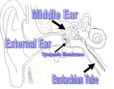
 Inflammatory Diseases of the Middle Ear: Practice Essentials, Pathophysiology, Epidemiology
 Optic Nerve Decompression Surgery - Medical Clinical Policy Bulletins | Aetna
 Mingwei Chen - NeL.edu
 Seizure - Wikipedia
 Advanced Search Results - Public Health Image Library(PHIL)
 Actilyse Cathflo - Side Effects, Uses, Dosage, Overdose, Pregnancy, Alcohol | RxWiki
 Peter Lindvall
 Acute neuropsychological functioning following cardiosurgical interventions associated with the production of intraoperative...
 Peripartum mood disturbances CT scan - wikidoc
 Essay on Stroke - 2669 Words | Bartleby
 Publikationsliste - Neuroscience Institut - Salzburger Landeskliniken (SALK)
 Lyme Disease Presenting with Multiple Cranial Nerve Deficits: Report of a Case
 Languages: English / Copyright: Public domain / Publication Year: 1898 - Digital Collections - National Library of Medicine...
 THROMBOSIS PREVENTION | Anesthesiology | American Society of Anesthesiologists