Keratosis, Actinic
Keratosis, Seborrheic
Darier Disease
Facial Dermatoses
Bowen's Disease
Keratoderma, Palmoplantar
Keratoacanthoma
Photosensitivity Disorders
Carcinoma, Basal Cell
Lichenoid Eruptions
Lichen Planus
Scalp Dermatoses
Ointment Bases
Parakeratosis
Leukoplakia
Carcinoma, Squamous Cell
Keratolytic Agents
Skin Diseases
Petrolatum
Leukoplakia, Oral
Arsenic Poisoning
Conjunctival Diseases
Acitretin
Skin Diseases, Genetic
Ichthyosis Vulgaris
Hyperpigmentation
Aminolevulinic Acid
Precancerous Conditions
Skin
Mouth Mucosa
Photochemotherapy
Ointments
Microscopy, Acoustic
Photosensitizing Agents
Epidermis
Keratinocytes
Arsenic
Administration, Topical
Ultraviolet Rays
Expression of matrix metalloproteinase-1, -2 and -3 in squamous cell carcinoma and actinic keratosis. (1/237)
Matrix metalloproteinase (MMP) plays an important role in extracellular matrix degradation associated with cancer invasion. An expression of MMP-1 (interstitial collagenase), MMP-2 (72-kDa type IV collagenase) and MMP-3 (stromelysin-1) was investigated in squamous cell carcinoma (SCC) and its precancerous condition, actinic keratosis (AK), using in situ hybridization techniques. MMP-1 mRNA was detected in tumour cells and/or in stromal cells in all cases of SCC, four of six AKs adjacent to SCC and four of 16 AKs. MMP-2 and MMP-3 mRNAs were detected in SCC but not in AK. The expression of MMP-3 correlated to that of MMP-1 (P = 0.03) localized at the tumour mass and stroma of the invasive area, while MMP-2 mRNA was detected widely throughout the stroma independent of MMP-1 expression. Our results indicated that the expression of MMP-1, -2 and -3 showed different localization patterns, suggesting a unique role of each MMP in tumour progression. Moreover, MMP-1 expression could be an early event in the development of SCC, and AK demonstrating MMP-1 mRNA, might be in a more advanced dysplastic state, progressing to SCC. (+info)p53 protects against skin cancer induction by UV-B radiation. (2/237)
To assess the role of the p53 tumor suppressor gene in skin carcinogenesis by UV radiation, mice constitutively lacking one or both copies of the functional p53 gene were compared to wild-type mice for their susceptibility to UV carcinogenesis. Heterozygous mice showed greatly increased susceptibility to skin cancer induction, and homozygous p53 knockout mice were even more susceptible. Accelerated tumor development in the heterozygotes was not associated with loss of the remaining wild-type allele of p53, as reported for tumors induced by other carcinogens, but in many cases was associated with UV-induced mutations in p53. Tumors arose on the ears and dorsal skin of mice of all three genotypes, and homozygous knockout mice also developed ocular tumors, mainly melanomas. Skin tumors in the p53 knockout mice were predominately squamous cell carcinomas and were associated with premalignant lesions resembling actinic keratoses, whereas those in the heterozygous and wild-type mice were mainly sarcomas. These results demonstrate the importance of p53 in protecting against UV-induced cancers, particularly in the eye and epidermis. (+info)Relations between exposure to arsenic, skin lesions, and glucosuria. (3/237)
OBJECTIVES: Exposure to arsenic causes keratosis, hyperpigmentation, and hypopigmentation and seemingly also diabetes mellitus, at least in subjects with skin lesions. Here we evaluate the relations of arsenical skin lesions and glucosuria as a proxy for diabetes mellitus. METHODS: Through existing measurements of arsenic in drinking water in Bangladesh, wells with and without arsenic contamination were identified. Based on a questionnaire, 1595 subjects > or = 30 years of age were interviewed; 1481 had a history of drinking water contaminated with arsenic whereas 114 had not. Time weighted mean arsenic concentrations and mg-years/l of exposure to arsenic were estimated based on the history of consumption of well water and current arsenic concentrations. Urine samples from the study subjects were tested by means of a glucometric strip. People with positive tests were considered to be cases of glucosuria. RESULTS: A total of 430 (29%) of the exposed people were found to have skin lesions. Corresponding to drinking water with < 0.5, 0.5-1.0, and > 1.0 mg/l of arsenic, and with the 114 unexposed subjects as the reference, the prevalence ratios for glucosuria, as adjusted for age and sex, were 0.8, 1.4, and 1.4 for those without skin lesions, and 1.1, 2.2, and 2.6 for those with skin lesions. Taking exposure as < 1.0, 1.0-5.0, > 5.0-10.0 and > 10.0 mg-years/l of exposure to arsenic the prevalence ratios, similarly adjusted, were 0.4, 0.9, 1.2, and 1.7 for those without and 0.8, 1.7, 2.1, and 2.9 for those with skin lesions. All series of risk estimates were significant for trend, (p < 0.01). CONCLUSIONS: The results suggest that skin lesions and diabetes mellitus, as here indicated by glucosuria, are largely independent effects of exposure to arsenic although glucosuria had some tendency to be associated with skin lesions. Importantly, however, glucosuria (diabetes mellitus) may occur independently of skin lesions. (+info)Targeted expression of c-Myc in the epidermis alters normal proliferation, differentiation and UV-B induced apoptosis. (4/237)
c-Myc overexpression has been associated with several types of human cancers. To study the role of c-myc in epidermal differentiation and carcinogenesis, a transgenic mouse model was created to overexpress c-Myc in the epidermis. Human c-myc 2 cDNA was subcloned into a 6.5 kb mouse loricrin expression vector, ML.myc2. This loricrin promoter primarily directs expression in the epidermis in both proliferating and differentiated keratinocytes. On day 4, ML.myc2 transgenic pups develop a hyperkeratotic phenotype, which progressively worsens until day 7. Upon histological analysis, both hyperplasia and hyperkeratosis were evident. Bromodeoxyuridine (BrdU) incorporation revealed that transgenic mice had a threefold increase in the number of proliferating cells as compared with a normal littermate. Proliferative cells in the ML.myc2 epidermis were also found to be suprabasal, suggesting an inhibition of terminal differentiation in keratinocytes. Inhibition of terminal differentiation by c-Myc overexpression was further suggested by aberrant expression of differentiation markers, keratin 1, keratin 6, loricrin, and filaggrin in ML.myc2 transgenic mice. Interestingly, ML.myc2 keratinocytes exhibit a reduced sensitivity to UV-B induced apoptosis, in vivo. In vitro studies reveal the reduced sensitivity of ML.myc2 keratinocytes to UV-B irradiation is growth factor dependent. These findings provide evidence that overexpression of c-Myc in the epidermis induces proliferation, inhibits terminal differentiation and decreases the sensitivity of keratinocytes to UV-B induced apoptosis. (+info)Accumulation of matrilysin (MMP-7) and macrophage metalloelastase (MMP-12) in actinic damage. (5/237)
Photodamage is characterized by degradation of collagen and accumulation of abnormal elastin in the superficial dermis and several matrix metalloproteinases have previously been implicated in this process. Using immunohistochemistry and in situ hybridization, we have studied the localization of two elastolytic matrix metalloproteinases, matrilysin (matrix metalloproteinase-7) and human macrophage metalloelastase (matrix metalloproteinase-12) in solar damage. Human macrophage metalloelastase protein was detected in the superficial dermis in areas of elastotic material. Matrix metalloproteinase-7 was seen in the mid-dermis in regions with less damaged elastic fibers and morphologically better preserved collagen as well as in a band-like pattern below basal keratinocytes in eight of 18 solar elastosis. In samples taken from healthy volunteers 3 d after repeated ultraviolet A or ultraviolet B photoprovocation, occasional immunopositive cells for human macrophage metalloelastase (stromal) or matrix metalloproteinase-7 (sweat gland epithelium) were detected. In samples taken 1 d after ultraviolet B exposure, however, basal keratinocytes were matrix metalloproteinase-7 immunopositive, explaining the linear immunostaining below basal keratinocytes noted particularly in ultraviolet B treated 3 d specimens. Upregulation of metalloelastase was also demonstrated in the skin of hairless mice after repeated ultraviolet exposure. In normal skin, no staining for human macrophage metalloelastase or matrix metalloproteinase-7 was observed in association with elastin. The amount of immunoreactivity for the substrates of matrix metalloproteinase-7, versican, and tenascin, was clearly increased in solar elastosis and photoprovocated skin; versican but not tenascin was detected in the same areas as matrix metalloproteinase-7. Our results suggest that both matrix metalloproteinase-7 and -12 may contribute to remodeling of elastotic areas in sun-damaged skin. (+info)Dorsal skin reactions of hairless dogs to topical treatment with corticosteroids. (6/237)
Dorsal skin reactions to continuous topical treatment with different types of corticosteroids were histologically investigated in hairless descendants of Mexican hairless dogs. The preparations tested were prednisolone (ST-1; weak), fluocinolone acetonide (ST-2; moderate), diflucortolone valrerate (ST-3; strong), and mometasone furoate (ST-4; very strong). Grossly, the sites treated with ST-3 and ST-4 showed moderate inflammatory reactions. After completion of the corticosteroid treatment, both sites were less pigmented and had a thin texture. The severity of histologic changes in the skin was dependent on the efficacy of the corticosteroids. The epidermis was prominently thinned from 1 wk after treatment with the corticosteroids, resulting in a flat dermis-epidermis junction. By the end of the corticosteroid treatment, these lesions became progressively more severe. At 2 wk after completion of topical treatment, the epidermal thickness in the sites treated with ST-1 and ST-2 began to return to normal values, whereas the epidermis of the skin treated with ST-3 and ST-4 became thinner. At 3-4 wk after topical treatment with ST-3 and ST-4, the dermis showed hyalinization of collagen bundles. These dermatologic findings in hairless dogs are in accordance with steroid-induced skin atrophy of human beings. These results suggest that the skin of hairless dogs responds sensitively to topical corticosteroids and that these animals are a useful model for investigating the efficacy and adverse effects of cutaneous topical corticosteroids. (+info)Transgenic mice overexpressing protein kinase Cdelta in the epidermis are resistant to skin tumor promotion by 12-O-tetradecanoylphorbol-13-acetate. (7/237)
To determine the role of protein kinase Cdelta in mouse skin carcinogenesis, we have developed transgenic FVB/N mouse lines expressing in the epidermis an epitope-tagged protein kinase Cdelta (T7-PKCdelta) regulated by the human keratin 14 promoter. The untreated T7-PKCdelta mice displayed excessive dryness in the skin of the tail with a variable penetrance over time. Histologically, the tail skin showed hyperplasia with evidence of hyperkeratosis. The epidermis of the rest of the T7-PKCdelta mouse was unremarkable. Despite this mild phenotype, the effects of PKCdelta overexpression on mouse skin tumor promotion by 12-O-tetradecanoylphorbol-13-acetate (TPA) were dramatic. Two independent lines of T7-PKCdelta mice (16 and 37) expressing the T7-PKCdelta transgene were examined for responsiveness to skin tumor promotion by 7,12-dimethylbenz[a]anthracene and TPA. By immunoblot analysis, the T7-PKCdelta-16 and T7-PKCdelta-37 mice showed an 8- and 2-fold increase of PKCdelta protein. The T7-PKCdelta-16 mice averaged 300% more T7-PKCdelta activity than the T7-PKCdelta-37 mice did. The T7-PKCdelta-37 mice did not manifest any difference in tumor burden or incidence. However, the reduction in papilloma burden at 25 weeks of promotion for the T7-PKCdelta-16 mice relative to wild-type mice averaged 72 and 74% for males and females, respectively. The T7-PKCdelta-16 mice reached 50% papilloma incidence between 12 and 13 weeks of promotion compared with 8 weeks for wild-type mice. Furthermore, the carcinoma incidence was also reduced in T7-PKCdelta-16 mice. Carcinoma incidence at 25 weeks of promotion treatment was: wild-type females, 78%; T7-PKCdelta16 females, 37%; wild-type males, 45%; and T7- PKCdelta-16 males, 7%. Thus, PKCdelta when expressed at sufficient levels can suppress skin tumor promotion by TPA. (+info)Low frequency of genetic change in p53 immunopositive clones in human epidermis. (8/237)
Sun-exposed skin of Caucasians harbors thousands of p53-mutated clones, which are clinically invisible. Using whole mount immunostaining for p53 or Ki67 antigens, p53 sequencing, and loss of heterozygosity analysis, we have further characterised these clones. Loss of heterozygosity for the alleles examined is uncommon with the exception of 9q, which occurred in 28.3% of the samples. P53 clones are more common and larger in individuals with basal cell carcinoma than in control subjects (p < 0.03). Loss of heterozygosity is also more common in clones from individuals with basal cell carcinoma than in clones from subjects without a history of basal cell carcinoma, as would be expected if both relate to ultraviolet radiation exposure. p53 sequencing of clones is in keeping with the mutagenic role of ultraviolet radiation. Surprisingly, skin found to harbor p53 clones showed no clusters of Ki67 positive cells, unlike the situation for actinic keratoses or basal cell carcinomas. These results show that in human skin p53 mutation is not directly associated with genomic instability or abnormal cell cycling; that the p53 immunopositive clones are either genetically distinct or precursors to other squamous cell lesions of skin; and that p53 immunopositive clones are early lesions, in that gross disturbance of proliferation has not already occurred. (+info)Keratosis, in general, refers to a skin condition characterized by the abnormal growth or development of keratin, a protein that forms part of the outer layer of the skin (epidermis). There are several types of keratosis, including:
1. Seborrheic Keratosis: benign, often pigmented, rough, and scaly growths that can appear anywhere on the body. They tend to increase in number with age.
2. Actinic Keratosis: rough, scaly patches or spots on the skin that are caused by long-term exposure to sunlight or artificial UV light. These have the potential to develop into squamous cell carcinoma, a type of skin cancer.
3. Solar Keratosis: another term for actinic keratosis, as it is primarily caused by sun damage.
4. Keratosis Pilaris: a common condition where small, rough bumps appear on the skin, often on the arms, thighs, or cheeks. These are caused by excess keratin blocking hair follicles.
5. Follicular Keratosis: a disorder characterized by the formation of horny plugs within the hair follicles, leading to rough, sandpaper-like bumps on the skin.
6. Intraepidermal Keratosis: a term used to describe the abnormal accumulation of keratin in the epidermis, which can lead to various skin conditions.
It's important to consult with a healthcare professional or dermatologist for proper diagnosis and treatment if you suspect having any form of keratosis.
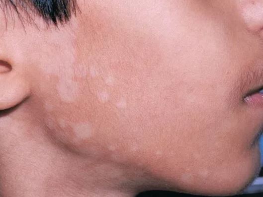
Actinic keratosis, also known as solar keratosis, is a precancerous skin condition that typically develops in areas exposed to excessive sun damage over the years. It presents as rough, scaly, or crusty patches of skin, often with a pink, red, or brownish tint. These lesions usually appear on the face, ears, scalp, neck, back of the hands, and forearms.
Actinic keratosis is caused by the prolonged exposure to ultraviolet (UV) radiation from sunlight or artificial sources like tanning beds. The UV rays damage the skin's DNA, leading to abnormal skin cell growth and the formation of these precancerous lesions.
While most actinic keratoses remain benign, a small percentage can progress into squamous cell carcinoma, a type of skin cancer. Therefore, it is essential to have any suspicious or changing lesions evaluated by a healthcare professional for proper diagnosis and treatment. Prevention measures include protecting the skin from excessive sun exposure, wearing protective clothing, using broad-spectrum sunscreen with an SPF of at least 30, and avoiding tanning beds.
Seborrheic Keratosis is a common, benign skin condition that typically presents as rough, scaly, tan-to-darkly pigmented growths on the surface of the skin. These lesions can appear anywhere on the body, but they are most commonly found on the face, chest, back, and extremities. Seborrheic Keratoses are caused by an overproduction of keratin, a protein that makes up the outer layer of the skin.
The exact cause of Seborrheic Keratosis is not known, but it is thought to be related to genetic factors and sun exposure. The condition is more common in older adults and is not contagious. While Seborrheic Keratoses are generally harmless, they can be removed for cosmetic reasons or if they become irritated or inflamed. Treatment options include cryotherapy (freezing the lesions with liquid nitrogen), curettage (scraping the lesions off), and laser surgery.
Darier Disease is a genetic skin disorder, also known as Keratosis Follicularis. It is characterized by the formation of greasy, crusted, keratotic papules and plaques that typically appear on the upper arms, torso, and scalp. The lesions may also affect the nasolabial folds, central face, and mucous membranes. Darier Disease is caused by mutations in the ATP2A2 gene, which encodes a calcium pump protein involved in keratinization. It is an autosomal dominant disorder, meaning that a person has a 50% chance of inheriting the disease if one of their parents is affected. The onset of symptoms typically occurs during adolescence or early adulthood. Treatment options include topical medications, oral retinoids, and photodynamic therapy.
Facial dermatoses refer to various skin conditions that affect the face. These can include a wide range of disorders, such as:
1. Acne vulgaris: A common skin condition characterized by the formation of comedones (blackheads and whiteheads) and inflammatory papules, pustules, and nodules. It primarily affects the face, neck, chest, and back.
2. Rosacea: A chronic skin condition that causes redness, flushing, and visible blood vessels on the face, along with bumps or pimples and sometimes eye irritation.
3. Seborrheic dermatitis: A common inflammatory skin disorder that causes a red, itchy, and flaky rash, often on the scalp, face, and eyebrows. It can also affect other oily areas of the body, like the sides of the nose and behind the ears.
4. Atopic dermatitis (eczema): A chronic inflammatory skin condition that causes red, itchy, and scaly patches on the skin. While it can occur anywhere on the body, it frequently affects the face, especially in infants and young children.
5. Psoriasis: An autoimmune disorder that results in thick, scaly, silvery, or red patches on the skin. It can affect any part of the body, including the face.
6. Contact dermatitis: A skin reaction caused by direct contact with an allergen or irritant, resulting in redness, itching, and inflammation. The face can be affected when allergens or irritants come into contact with the skin through cosmetics, skincare products, or other substances.
7. Lupus erythematosus: An autoimmune disorder that can cause a butterfly-shaped rash on the cheeks and nose, along with other symptoms like joint pain, fatigue, and photosensitivity.
8. Perioral dermatitis: A inflammatory skin condition that causes redness, small bumps, and dryness around the mouth, often mistaken for acne. It can also affect the skin around the nose and eyes.
9. Vitiligo: An autoimmune disorder that results in the loss of pigmentation in patches of skin, which can occur on the face and other parts of the body.
10. Tinea faciei: A fungal infection that affects the facial skin, causing red, scaly, or itchy patches. It is also known as ringworm of the face.
These are just a few examples of skin conditions that can affect the face. If you experience any unusual symptoms or changes in your skin, it's essential to consult a dermatologist for proper diagnosis and treatment.
Bowen's disease is a skin condition that is characterized by the growth of abnormal cells on the outermost layer of the skin (the epidermis). It is also known as squamous cell carcinoma in situ. The affected area often appears as a red, scaly patch or plaque, and it can develop anywhere on the body, but it is most commonly found on sun-exposed areas such as the face, hands, arms, and legs.
Bowen's disease is considered a precancerous condition because there is a risk that the abnormal cells could eventually develop into invasive squamous cell carcinoma, a type of skin cancer. However, not all cases of Bowen's disease will progress to cancer, and some may remain stable or even regress on their own.
The exact cause of Bowen's disease is not known, but it is thought to be associated with exposure to certain chemicals, radiation, and human papillomavirus (HPV) infection. Treatment options for Bowen's disease include cryotherapy, topical chemotherapy, photodynamic therapy, curettage and electrodessication, and surgical excision. Regular follow-up with a healthcare provider is recommended to monitor the condition and ensure that it does not progress to cancer.
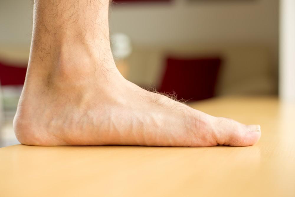
Keratoderma, palmoplantar is a medical term that refers to a group of skin conditions characterized by thickening and hardening (hyperkeratosis) of the skin on the palms of the hands and soles of the feet. This condition can affect people of all ages, but it's most commonly seen in children.
The thickening of the skin is caused by an overproduction of keratin, a protein that helps to form the tough, outer layer of the skin. In palmoplantar keratoderma, this excess keratin accumulates in the stratum corneum, the outermost layer of the epidermis, leading to the formation of rough, scaly, and thickened patches on the palms and soles.
There are several different types of palmoplantar keratoderma, each with its own specific symptoms and causes. Some forms of the condition are inherited and present at birth or develop in early childhood, while others may be acquired later in life as a result of an underlying medical condition, such as atopic dermatitis, lichen planus, or psoriasis.
Treatment for palmoplantar keratoderma typically involves the use of emollients and keratolytic agents to help soften and remove the thickened skin. In some cases, oral retinoids or other systemic medications may be necessary to manage more severe symptoms. It's important to consult with a healthcare provider for an accurate diagnosis and treatment plan.
Keratoacanthoma is a rapidly growing, dome-shaped, skin tumor that typically arises on sun-exposed areas such as the face, arms, and legs. It is considered a low-grade squamous cell carcinoma (a type of skin cancer) because it shares some characteristics with both benign and malignant tumors.
Keratoacanthomas usually develop over a period of several weeks to months, growing rapidly in size before eventually stabilizing and then gradually regressing on their own within a few months to a year. However, the regression process can take years, and some lesions may not regress completely, leading to cosmetic concerns or even local invasion.
Histologically, keratoacanthomas are characterized by a central keratin-filled crater surrounded by a well-differentiated layer of squamous epithelial cells. The tumor's growth pattern and histological features can make it difficult to distinguish from other types of skin cancer, such as squamous cell carcinoma.
Treatment options for keratoacanthomas include surgical excision, cryosurgery, curettage and electrodesiccation, and topical therapies like imiquimod or 5-fluorouracil. The choice of treatment depends on various factors such as the size, location, and number of lesions, as well as patient preferences and overall health status.
Photosensitivity disorders refer to conditions that cause an abnormal reaction to sunlight or artificial light. This reaction can take the form of various skin changes, such as rashes, inflammation, or pigmentation, and in some cases, it can also lead to systemic symptoms like fatigue, fever, or joint pain.
The two main types of photosensitivity disorders are:
1. Phototoxic reactions: These occur when a substance (such as certain medications, chemicals, or plants) absorbs light energy and transfers it to skin cells, causing damage and inflammation. The reaction typically appears within 24 hours of exposure to the light source and can resemble a sunburn.
2. Photoallergic reactions: These occur when the immune system responds to the combination of light and a particular substance, leading to an allergic response. The reaction may not appear until several days after initial exposure and can cause redness, itching, and blistering.
It is important for individuals with photosensitivity disorders to avoid excessive sun exposure, wear protective clothing, and use broad-spectrum sunscreens with a high SPF rating to minimize the risk of phototoxic or photoallergic reactions.
The eyebrows are a set of hairs that grow above the eyes on the forehead. They are an important feature of human facial anatomy, and play several roles in non-verbal communication and self-expression. Eyebrows help to prevent sweat and other moisture from dripping into the eyes, and also serve as a protective barrier against dirt, dust, and other foreign particles that might otherwise irritate or damage the eyes.
In addition, eyebrows play an important role in human social interaction and communication. They can convey a range of emotions and facial expressions, such as surprise, anger, fear, happiness, and sadness. Eyebrows can also help to frame the eyes and enhance their appearance, making them an important aspect of personal grooming and beauty.
The eyebrows are made up of several components, including hair follicles, sebaceous glands, and muscles that control their movement. The hairs themselves are composed of a protein called keratin, which also makes up the hair on the head, as well as nails and skin. The color and thickness of eyebrow hair can vary widely from person to person, and may be influenced by factors such as age, genetics, and hormonal changes.
In medical terms, changes in the appearance or condition of the eyebrows can sometimes be a sign of underlying health issues. For example, thinning or loss of eyebrows can be associated with conditions such as alopecia, thyroid disorders, or nutritional deficiencies. Changes in eyebrow shape or position can also be a symptom of certain neurological conditions, such as Bell's palsy or stroke. As such, any significant changes in the appearance or condition of the eyebrows should be evaluated by a healthcare professional to rule out any underlying medical causes.
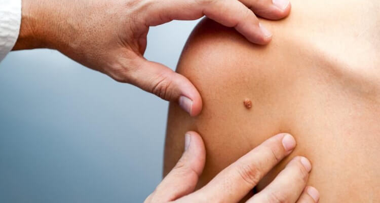
Skin neoplasms refer to abnormal growths or tumors in the skin that can be benign (non-cancerous) or malignant (cancerous). They result from uncontrolled multiplication of skin cells, which can form various types of lesions. These growths may appear as lumps, bumps, sores, patches, or discolored areas on the skin.
Benign skin neoplasms include conditions such as moles, warts, and seborrheic keratoses, while malignant skin neoplasms are primarily classified into melanoma, squamous cell carcinoma, and basal cell carcinoma. These three types of cancerous skin growths are collectively known as non-melanoma skin cancers (NMSCs). Melanoma is the most aggressive and dangerous form of skin cancer, while NMSCs tend to be less invasive but more common.
It's essential to monitor any changes in existing skin lesions or the appearance of new growths and consult a healthcare professional for proper evaluation and treatment if needed.
Carcinoma, basal cell is a type of skin cancer that arises from the basal cells, which are located in the lower part of the epidermis (the outermost layer of the skin). It is also known as basal cell carcinoma (BCC) and is the most common form of skin cancer.
BCC typically appears as a small, shiny, pearly bump or nodule on the skin, often in sun-exposed areas such as the face, ears, neck, hands, and arms. It may also appear as a scar-like area that is white, yellow, or waxy. BCCs are usually slow growing and rarely spread (metastasize) to other parts of the body. However, they can be locally invasive and destroy surrounding tissue if left untreated.
The exact cause of BCC is not known, but it is thought to be related to a combination of genetic and environmental factors, including exposure to ultraviolet (UV) radiation from the sun or tanning beds. People with fair skin, light hair, and blue or green eyes are at increased risk of developing BCC.
Treatment for BCC typically involves surgical removal of the tumor, along with a margin of healthy tissue. Other treatment options may include radiation therapy, topical chemotherapy, or photodynamic therapy. Prevention measures include protecting your skin from UV radiation by wearing protective clothing, using sunscreen, and avoiding tanning beds.
Lichenoid eruptions are skin reactions that resemble the appearance of lichen, a type of slow-growing fungus. These eruptions are characterized by flat, scaly bumps (papules) and rough, discolored patches (plaques) on the skin. They can be caused by various factors, including medications, medical conditions, or as a reaction to certain chemicals or substances that come into contact with the skin.
The term "lichenoid" refers to the resemblance of these eruptions to lichen, which is characterized by its distinctive appearance and growth pattern. Lichenoid eruptions can occur anywhere on the body but are most commonly found on sun-exposed areas such as the arms, legs, and trunk.
The exact cause of lichenoid eruptions can vary, but they are often associated with an autoimmune response in which the body's immune system mistakenly attacks healthy skin cells. This can lead to inflammation, redness, itching, and other symptoms associated with these eruptions. Treatment for lichenoid eruptions typically involves identifying and addressing the underlying cause, as well as managing symptoms with topical medications or other therapies.
Lichen Planus is a chronic, autoimmune skin condition that can also affect the mucous membranes inside the mouth, genitals, and eyes. It is characterized by the appearance of purplish, flat-topped bumps or lesions on the skin, which may be itchy. The exact cause of Lichen Planus is unknown, but it is believed to occur when the immune system mistakenly attacks cells in the skin or mucous membranes. Certain medications, viral infections, and genetic factors may increase the risk of developing this condition. Treatment typically focuses on managing symptoms and may include topical corticosteroids, oral medications, or light therapy.
Scalp dermatoses refer to various skin conditions that affect the scalp. These can include inflammatory conditions such as seborrheic dermatitis (dandruff, cradle cap), psoriasis, atopic dermatitis (eczema), and lichen planus; infectious processes like bacterial folliculitis, tinea capitis (ringworm of the scalp), and viral infections; as well as autoimmune conditions such as alopecia areata. Symptoms can range from mild scaling and itching to severe redness, pain, and hair loss. The specific diagnosis and treatment of scalp dermatoses depend on the underlying cause.
Ointment bases refer to the vehicle or foundation in which active pharmaceutical ingredients are dispersed to form a semi-solid medication. These bases provide the necessary consistency for ointments, allowing easy application to the skin or other body surfaces. They can be composed of various materials such as fats, waxes, oils, and emulsifying agents.
The choice of an ointment base depends on several factors, including:
1. The desired physical properties (e.g., spreadability, absorption rate)
2. The route of administration (e.g., dermal, mucosal)
3. The compatibility with the active ingredient(s)
4. The intended therapeutic effect (e.g., occlusive, non-occlusive)
Some common types of ointment bases include:
1. Hydrocarbon bases: Consist of hydrophobic materials like petrolatum, white soft paraffin, and microcrystalline wax. They are generally inert, odorless, and resistant to oxidation.
2. Absorption bases: Contain a mixture of hydrocarbons and higher molecular weight esters or fatty alcohols. These bases have better penetrating properties than hydrocarbon bases and are suitable for drugs with low oil solubility.
3. Emulsifying bases: Comprise of water-in-oil (W/O) or oil-in-water (O/W) emulsions, which allow the dispersion of both hydrophilic and lipophilic drugs. Common examples include cetomacrogol and anhydrous lanette.
4. Water-soluble bases: Primarily consist of polyethylene glycols (PEGs) or other water-soluble materials. They are useful for drugs with high water solubility and provide a cooling sensation upon application.
It is essential to select an appropriate ointment base to ensure the optimal delivery, stability, and efficacy of the active ingredient(s).
Parakeratosis is a medical term that refers to a skin condition where the outermost layer of the skin (the stratum corneum) contains nucleated keratinocytes, which are cells that have not fully matured and still contain their nuclei. This is in contrast to normal stratum corneum, which consists of flat, dead keratinocytes without nuclei.
Parakeratosis can occur in various skin disorders, such as psoriasis, eczema, warts, and certain types of dermatitis. It can also be seen in some benign or malignant skin tumors. The presence of parakeratosis may indicate abnormal differentiation or proliferation of the skin cells, which can contribute to the development of skin lesions or diseases.
In addition to its role in skin disorders, parakeratosis has been implicated in the pathogenesis of certain gastrointestinal diseases, such as Barrett's esophagus and colon cancer, where it is associated with abnormal cell growth and increased risk of malignancy.
Leukoplakia is a medical term used to describe a white or gray patch that develops on the mucous membranes lining the inside of the mouth. These patches are typically caused by excessive cell growth and cannot be easily scraped off. Leukoplakia is often associated with long-term tobacco use, including smoking and chewing tobacco, as well as alcohol consumption. While most cases of leukoplakia are benign, a small percentage can develop into oral cancer, so it's essential to have any suspicious patches evaluated by a healthcare professional.
Squamous cell carcinoma is a type of skin cancer that begins in the squamous cells, which are flat, thin cells that form the outer layer of the skin (epidermis). It commonly occurs on sun-exposed areas such as the face, ears, lips, and backs of the hands. Squamous cell carcinoma can also develop in other areas of the body including the mouth, lungs, and cervix.
This type of cancer usually develops slowly and may appear as a rough or scaly patch of skin, a red, firm nodule, or a sore or ulcer that doesn't heal. While squamous cell carcinoma is not as aggressive as some other types of cancer, it can metastasize (spread) to other parts of the body if left untreated, making early detection and treatment important.
Risk factors for developing squamous cell carcinoma include prolonged exposure to ultraviolet (UV) radiation from the sun or tanning beds, fair skin, a history of sunburns, a weakened immune system, and older age. Prevention measures include protecting your skin from the sun by wearing protective clothing, using a broad-spectrum sunscreen with an SPF of at least 30, avoiding tanning beds, and getting regular skin examinations.
Keratolytic agents are substances that cause the softening and sloughing off of excess keratin, the protein that makes up the outermost layer of the skin (stratum corneum). These agents help to break down and remove dead skin cells, increase moisture retention, and promote the growth of new skin cells. They are commonly used in the treatment of various dermatological conditions such as psoriasis, eczema, warts, calluses, and ichthyosis. Examples of keratolytic agents include salicylic acid, urea, lactic acid, and retinoic acid.
Skin diseases, also known as dermatological conditions, refer to any medical condition that affects the skin, which is the largest organ of the human body. These diseases can affect the skin's function, appearance, or overall health. They can be caused by various factors, including genetics, infections, allergies, environmental factors, and aging.
Skin diseases can present in many different forms, such as rashes, blisters, sores, discolorations, growths, or changes in texture. Some common examples of skin diseases include acne, eczema, psoriasis, dermatitis, fungal infections, viral infections, bacterial infections, and skin cancer.
The symptoms and severity of skin diseases can vary widely depending on the specific condition and individual factors. Some skin diseases are mild and can be treated with over-the-counter medications or topical creams, while others may require more intensive treatments such as prescription medications, light therapy, or even surgery.
It is important to seek medical attention if you experience any unusual or persistent changes in your skin, as some skin diseases can be serious or indicative of other underlying health conditions. A dermatologist is a medical doctor who specializes in the diagnosis and treatment of skin diseases.
I'm sorry for any confusion, but "sunlight" is not a term with a specific medical definition. However, sunlight does have various implications in the field of medicine. It is the light that comes from the sun, which is a star at the center of our solar system. Sunlight is essential for the production of vitamin D in humans, and it can also have effects on mood and sleep patterns due to its influence on circadian rhythms.
In a medical context, sunlight is often discussed in relation to its potential health benefits and risks. For instance, moderate sun exposure can help increase vitamin D levels, which are important for bone health, immune function, and other bodily processes. However, excessive sun exposure can lead to harmful effects, such as sunburn, premature skin aging, and an increased risk of skin cancer.
It's essential to balance the benefits and risks of sunlight exposure by practicing safe sun habits, such as wearing protective clothing, using a broad-spectrum sunscreen with an SPF of at least 30, seeking shade during peak sunlight hours, and avoiding intentional tanning.
Petrolatum is a semi-solid mixture of hydrocarbons obtained from petroleum. In the medical field, it's often used as an ointment base or protective dressing because of its impermeability to water and bacteria. It's also known as petroleum jelly or soft paraffin.
Leukoplakia, oral is a predominantly white patch or plaque that cannot be characterized clinically or pathologically as any other disease. It is an oral potentially malignant disorder (OPMD) and represents a significant risk for the development of squamous cell carcinoma. The lesions are typically caused by chronic irritation, such as smoking or smokeless tobacco use, and are most commonly found on the tongue, floor of the mouth, and buccal mucosa. The diagnosis is confirmed through a biopsy, and management includes removal of causative factors and close monitoring for any signs of malignant transformation.
Arsenic poisoning is a condition that occurs when a person ingests or comes into contact with a toxic amount of arsenic, a naturally occurring element found in the earth's crust. Arsenic has no smell or taste, making it difficult to detect in food, water, or air.
Acute arsenic poisoning can occur after a single large exposure to arsenic, while chronic arsenic poisoning occurs after repeated or long-term exposure to lower levels of arsenic. The symptoms of acute arsenic poisoning include vomiting, diarrhea, abdominal pain, and muscle cramps. In severe cases, it can lead to death due to heart failure or respiratory failure.
Chronic arsenic poisoning can cause a range of health problems, including skin changes such as pigmentation and hard patches on the palms and soles, weakness, peripheral neuropathy, and an increased risk of cancer, particularly skin, lung, bladder, and kidney cancer. It can also affect cognitive development in children.
Arsenic poisoning is treated by removing the source of exposure and providing supportive care to manage symptoms. Chelation therapy may be used to remove arsenic from the body in cases of severe acute poisoning or chronic poisoning with high levels of arsenic. Prevention measures include monitoring and reducing exposure to arsenic in food, water, and air, as well as proper handling and disposal of arsenic-containing products.
Conjunctival diseases refer to a group of medical conditions that affect the conjunctiva, which is the thin, clear mucous membrane that covers the inner surface of the eyelids and the white part of the eye (known as the sclera). The conjunctiva helps to keep the eye moist and protected from irritants.
Conjunctival diseases can cause a range of symptoms, including redness, itching, burning, discharge, grittiness, and pain. Some common conjunctival diseases include:
1. Conjunctivitis (pink eye): This is an inflammation or infection of the conjunctiva that can be caused by viruses, bacteria, or allergies. Symptoms may include redness, itching, discharge, and watery eyes.
2. Pinguecula: This is a yellowish, raised bump that forms on the conjunctiva, usually near the corner of the eye. It is caused by an overgrowth of connective tissue and may be related to sun exposure or dry eye.
3. Pterygium: This is a fleshy growth that extends from the conjunctiva onto the cornea (the clear front part of the eye). It can cause redness, irritation, and vision problems if it grows large enough to cover the pupil.
4. Allergic conjunctivitis: This is an inflammation of the conjunctiva caused by an allergic reaction to substances such as pollen, dust mites, or pet dander. Symptoms may include redness, itching, watery eyes, and swelling.
5. Chemical conjunctivitis: This is an irritation or inflammation of the conjunctiva caused by exposure to chemicals such as chlorine, smoke, or fumes. Symptoms may include redness, burning, and tearing.
6. Giant papillary conjunctivitis (GPC): This is a type of allergic reaction that occurs in response to the presence of a foreign body in the eye, such as a contact lens. Symptoms may include itching, mucus discharge, and a gritty feeling in the eye.
Treatment for conjunctival diseases depends on the underlying cause. In some cases, over-the-counter medications or home remedies may be sufficient to relieve symptoms. However, more severe cases may require prescription medication or medical intervention. It is important to consult with a healthcare provider if you experience persistent or worsening symptoms of conjunctival disease.
Acitretin is a synthetic form of retinoic acid, which is a type of vitamin A. It is used to treat severe psoriasis and other skin conditions. Acitretin works by slowing down the rapid growth of skin cells that cause the symptoms of psoriasis. It comes in the form of a capsule and is taken orally.
Common side effects of acitretin include dryness of the skin, lips, and mouth, itching, peeling, redness, or stickiness of the palms and soles, hair loss, and changes in nail growth. Less common but more serious side effects can include liver damage, increased levels of lipids in the blood, and birth defects if taken during pregnancy.
It is important to note that acitretin can cause birth defects, so women who are pregnant or planning to become pregnant should not take this medication. Additionally, because acitretin can remain in the body for a long time, it is recommended that women of childbearing age use effective contraception while taking this medication and for at least three years after stopping it.
Genetic skin diseases are a group of disorders caused by mutations or alterations in the genetic material (DNA), which can be inherited from one or both parents. These mutations affect the structure, function, or development of the skin and can lead to various conditions with different symptoms, severity, and prognosis.
Some examples of genetic skin diseases include:
1. Epidermolysis Bullosa (EB): A group of disorders characterized by fragile skin and mucous membranes that blister and tear easily, leading to painful sores and wounds. There are several types of EB, each caused by mutations in different genes involved in anchoring the epidermis to the dermis.
2. Ichthyosis: A family of genetic disorders characterized by dry, thickened, scaly, or rough skin. The severity and symptoms can vary widely, depending on the specific type and underlying genetic cause.
3. Neurofibromatosis: A group of conditions caused by mutations in the NF1 gene, which regulates cell growth and division. The most common types, NF1 and NF2, are characterized by the development of benign tumors called neurofibromas on the skin and nerves, as well as other symptoms affecting various organs and systems.
4. Tuberous Sclerosis Complex (TSC): A genetic disorder caused by mutations in the TSC1 or TSC2 genes, which control cell growth and division. TSC is characterized by the development of benign tumors in multiple organs, including the skin, brain, heart, kidneys, and lungs.
5. Xeroderma Pigmentosum (XP): A rare genetic disorder caused by mutations in genes responsible for repairing DNA damage from ultraviolet (UV) radiation. People with XP are extremely sensitive to sunlight and have a high risk of developing skin cancer and other complications.
6. Incontinentia Pigmenti (IP): A genetic disorder that affects the development and growth of skin, hair, nails, teeth, and eyes. IP is caused by mutations in the IKBKG gene and primarily affects females.
7. Darier's Disease: An inherited skin disorder characterized by greasy, crusted, keratotic papules and plaques, usually located on the trunk, scalp, and seborrheic areas of the body. Darier's disease is caused by mutations in the ATP2A2 gene.
These are just a few examples of genetic skin disorders. There are many more, each with its unique set of symptoms, causes, and treatments. If you or someone you know has a genetic skin disorder, it is essential to consult with a dermatologist or other healthcare professional for proper diagnosis and treatment.
Ichthyosis Vulgaris is a genetic skin disorder, which is characterized by dry, scaly, and rough skin. It is one of the most common forms of ichthyosis and is usually inherited in an autosomal dominant pattern, meaning only one copy of the altered gene in each cell is sufficient to cause the condition.
The term "ichthyosis" comes from the Greek word "ichthys," which means fish, reflecting the scaly appearance of the skin in individuals with this disorder.
In people with Ichthyosis Vulgaris, the skin cells do not shed properly and instead, they accumulate in scales on the surface of the skin. These scales are typically small, white to grayish-brown, and polygonal in shape. The scales are most often found on the legs, arms, and trunk but can affect any part of the body.
The condition usually appears during early childhood and tends to get worse in dry weather. In many cases, it improves during adulthood, although the skin remains rough and scaly.
Ichthyosis Vulgaris is caused by mutations in the gene called filaggrin, which is responsible for maintaining a healthy barrier function in the skin. This leads to dryness and increased susceptibility to skin infections.
Hyperpigmentation is a medical term that refers to the darkening of skin areas due to an increase in melanin, the pigment that provides color to our skin. This condition can affect people of all races and ethnicities, but it's more noticeable in those with lighter skin tones.
Hyperpigmentation can be caused by various factors, including excessive sun exposure, hormonal changes (such as during pregnancy), inflammation, certain medications, and underlying medical conditions like Addison's disease or hemochromatosis. It can also result from skin injuries, such as cuts, burns, or acne, which leave dark spots known as post-inflammatory hyperpigmentation.
There are several types of hyperpigmentation, including:
1. Melasma: This is a common form of hyperpigmentation that typically appears as symmetrical, blotchy patches on the face, particularly the forehead, cheeks, and upper lip. It's often triggered by hormonal changes, such as those experienced during pregnancy or while taking birth control pills.
2. Solar lentigos (age spots or liver spots): These are small, darkened areas of skin that appear due to prolonged sun exposure over time. They typically occur on the face, hands, arms, and decolletage.
3. Post-inflammatory hyperpigmentation: This type of hyperpigmentation occurs when an injury or inflammation heals, leaving behind a darkened area of skin. It's more common in people with darker skin tones.
Treatment for hyperpigmentation depends on the underlying cause and may include topical creams, chemical peels, laser therapy, or microdermabrasion. Preventing further sun damage is crucial to managing hyperpigmentation, so wearing sunscreen with a high SPF and protective clothing is recommended.
Aminolevulinic acid (ALA) is a naturally occurring compound in the human body and is a key precursor in the biosynthesis of heme, which is a component of hemoglobin in red blood cells. It is also used as a photosensitizer in dermatology for the treatment of certain types of skin conditions such as actinic keratosis and basal cell carcinoma.
In medical terms, ALA is classified as an α-keto acid and a porphyrin precursor. It is synthesized in the mitochondria from glycine and succinyl-CoA in a reaction catalyzed by the enzyme aminolevulinic acid synthase. After its synthesis, ALA is transported to the cytosol where it undergoes further metabolism to form porphyrins, which are then used for heme biosynthesis in the mitochondria.
In dermatology, topical application of ALA followed by exposure to a specific wavelength of light can lead to the production of reactive oxygen species that destroy abnormal cells in the skin while leaving healthy cells unharmed. This makes it an effective treatment for precancerous and cancerous lesions on the skin.
It is important to note that ALA can cause photosensitivity, which means that patients who have undergone ALA-based treatments should avoid exposure to sunlight or other sources of bright light for a period of time after the treatment to prevent adverse reactions.
A precancerous condition, also known as a premalignant condition, is a state of abnormal cellular growth and development that has a higher-than-normal potential to progress into cancer. These conditions are characterized by the presence of certain anomalies in the cells, such as dysplasia (abnormal changes in cell shape or size), which can indicate an increased risk for malignant transformation.
It is important to note that not all precancerous conditions will eventually develop into cancer, and some may even regress on their own. However, individuals with precancerous conditions are often at a higher risk of developing cancer compared to the general population. Regular monitoring and appropriate medical interventions, if necessary, can help manage this risk and potentially prevent or detect cancer at an early stage when it is more treatable.
Examples of precancerous conditions include:
1. Dysplasia in the cervix (cervical intraepithelial neoplasia or CIN)
2. Atypical ductal hyperplasia or lobular hyperplasia in the breast
3. Actinic keratosis on the skin
4. Leukoplakia in the mouth
5. Barrett's esophagus in the digestive tract
Regular medical check-ups, screenings, and lifestyle modifications are crucial for individuals with precancerous conditions to monitor their health and reduce the risk of cancer development.
Dermatologic agents are medications, chemicals, or other substances that are applied to the skin (dermis) for therapeutic or cosmetic purposes. They can be used to treat various skin conditions such as acne, eczema, psoriasis, fungal infections, and wounds. Dermatologic agents include topical corticosteroids, antibiotics, antifungals, retinoids, benzoyl peroxide, salicylic acid, and many others. They can come in various forms such as creams, ointments, gels, lotions, solutions, and patches. It is important to follow the instructions for use carefully to ensure safety and effectiveness.
In medical terms, the skin is the largest organ of the human body. It consists of two main layers: the epidermis (outer layer) and dermis (inner layer), as well as accessory structures like hair follicles, sweat glands, and oil glands. The skin plays a crucial role in protecting us from external factors such as bacteria, viruses, and environmental hazards, while also regulating body temperature and enabling the sense of touch.
The mouth mucosa refers to the mucous membrane that lines the inside of the mouth, also known as the oral mucosa. It covers the tongue, gums, inner cheeks, palate, and floor of the mouth. This moist tissue is made up of epithelial cells, connective tissue, blood vessels, and nerve endings. Its functions include protecting the underlying tissues from physical trauma, chemical irritation, and microbial infections; aiding in food digestion by producing enzymes; and providing sensory information about taste, temperature, and texture.
Aminoquinolines are a class of drugs that contain a quinoline chemical structure and an amino group. They are primarily used as antimalarial agents, with the most well-known members of this class being chloroquine and hydroxychloroquine. These drugs work by inhibiting the parasite's ability to digest hemoglobin in the red blood cells, which is necessary for its survival and reproduction.
In addition to their antimalarial properties, aminoquinolines have also been studied for their potential anti-inflammatory and immunomodulatory effects. They have been investigated as a treatment for various autoimmune diseases, such as rheumatoid arthritis and lupus, although their use in these conditions is not yet widely accepted.
It's important to note that aminoquinolines can have significant side effects, including gastrointestinal symptoms, retinopathy, and cardiac toxicity. They should only be used under the close supervision of a healthcare provider, and their use may be contraindicated in certain populations, such as pregnant women or individuals with preexisting heart conditions.
Photochemotherapy is a medical treatment that combines the use of drugs and light to treat various skin conditions. The most common type of photochemotherapy is PUVA (Psoralen + UVA), where the patient takes a photosensitizing medication called psoralen, followed by exposure to ultraviolet A (UVA) light.
The psoralen makes the skin more sensitive to the UVA light, which helps to reduce inflammation and suppress the overactive immune response that contributes to many skin conditions. This therapy is often used to treat severe cases of psoriasis, eczema, and mycosis fungoides (a type of cutaneous T-cell lymphoma). It's important to note that photochemotherapy can increase the risk of skin cancer and cataracts, so it should only be administered under the close supervision of a healthcare professional.
An ointment is a semi-solid preparation, typically composed of a mixture of medicinal substance with a base, which is usually greasy or oily. The purpose of the base is to act as a vehicle for the active ingredient and allow it to be applied smoothly and evenly to the skin or mucous membranes.
Ointments are commonly used in dermatology to treat various skin conditions such as eczema, psoriasis, rashes, burns, and wounds. They can also be used to deliver medication for localized pain relief, muscle relaxation, and anti-inflammatory or antibiotic effects.
The base of an ointment may consist of various ingredients, including petrolatum, lanolin, mineral oil, beeswax, or a combination of these. The choice of the base depends on the desired properties such as consistency, spreadability, and stability, as well as the intended route of administration and the specific therapeutic goals.
Warts are small, rough growths on the skin or mucous membranes caused by one of several types of human papillomavirus (HPV). They can appear anywhere on the body but most often occur on the hands, fingers, and feet. Warts are benign, non-cancerous growths, but they can be unsightly, uncomfortable, or painful, depending on their location and size.
Warts are caused by HPV infecting the top layer of skin, usually through a small cut or scratch. The virus triggers an overproduction of keratin, a protein in the skin, leading to the formation of a hard, rough growth. Warts can vary in appearance depending on their location and type, but they are generally round or irregularly shaped, with a rough surface that may be flat or slightly raised. They may also contain small black dots, which are actually tiny blood vessels that have clotted.
Warts are contagious and can spread from person to person through direct skin-to-skin contact or by sharing personal items such as towels or razors. They can also be spread by touching a wart and then touching another part of the body. Warts may take several months to develop after exposure to HPV, so it may not always be clear when or how they were contracted.
There are several types of warts, including common warts, plantar warts (which occur on the soles of the feet), flat warts (which are smaller and smoother than other types of warts), and genital warts (which are sexually transmitted). While most warts are harmless and will eventually go away on their own, some may require medical treatment if they are causing discomfort or are unsightly. Treatment options for warts include topical medications, cryotherapy (freezing the wart with liquid nitrogen), and surgical removal.
Acoustic microscopy is a non-invasive imaging technique that uses sound waves to visualize and analyze the structure and properties of various materials, including biological samples. In the context of medical diagnostics and research, acoustic microscopy can be used to examine tissues, cells, and cellular components with high resolution, providing valuable information about their mechanical and physical properties.
In acoustic microscopy, high-frequency sound waves are focused onto a sample using a transducer. The interaction between the sound waves and the sample generates echoes, which contain information about the sample's internal structure and properties. These echoes are then recorded and processed to create an image of the sample.
Acoustic microscopy offers several advantages over other imaging techniques, such as optical microscopy or electron microscopy. For example, it does not require staining or labeling of samples, which can be time-consuming and potentially damaging. Additionally, acoustic microscopy can provide high-resolution images of samples in their native state, allowing researchers to study the effects of various treatments or interventions on living cells and tissues.
In summary, acoustic microscopy is a non-invasive imaging technique that uses sound waves to visualize and analyze the structure and properties of biological samples with high resolution, providing valuable information for medical diagnostics and research.
Skin pigmentation is the coloration of the skin that is primarily determined by two types of melanin pigments, eumelanin and pheomelanin. These pigments are produced by melanocytes, which are specialized cells located in the epidermis. Eumelanin is responsible for brown or black coloration, while pheomelanin produces a red or yellow hue.
The amount and distribution of melanin in the skin can vary depending on genetic factors, age, sun exposure, and various other influences. Increased production of melanin in response to UV radiation from the sun helps protect the skin from damage, leading to darkening or tanning of the skin. However, excessive sun exposure can also cause irregular pigmentation, such as sunspots or freckles.
Abnormalities in skin pigmentation can result from various medical conditions, including albinism (lack of melanin production), vitiligo (loss of melanocytes leading to white patches), and melasma (excessive pigmentation often caused by hormonal changes). These conditions may require medical treatment to manage or improve the pigmentation issues.
Photosensitizing agents are substances that, when exposed to light, particularly ultraviolet or visible light, can cause chemical reactions leading to the production of reactive oxygen species. These reactive oxygen species can interact with biological tissues, leading to damage and a variety of phototoxic or photoallergic adverse effects.
Photosensitizing agents are used in various medical fields, including dermatology and oncology. In dermatology, they are often used in the treatment of conditions such as psoriasis and eczema, where a photosensitizer is applied to the skin and then activated with light to reduce inflammation and slow the growth of skin cells.
In oncology, photosensitizing agents are used in photodynamic therapy (PDT), a type of cancer treatment that involves administering a photosensitizer, allowing it to accumulate in cancer cells, and then exposing the area to light. The light activates the photosensitizer, which produces reactive oxygen species that damage the cancer cells, leading to their death.
Examples of photosensitizing agents include porphyrins, chlorophyll derivatives, and certain antibiotics such as tetracyclines and fluoroquinolones. It is important for healthcare providers to be aware of the potential for photosensitivity when prescribing these medications and to inform patients of the risks associated with exposure to light.
The epidermis is the outermost layer of the skin, composed mainly of stratified squamous epithelium. It forms a protective barrier that prevents water loss and inhibits the entry of microorganisms. The epidermis contains no blood vessels, and its cells are nourished by diffusion from the underlying dermis. The bottom-most layer of the epidermis, called the stratum basale, is responsible for generating new skin cells that eventually move up to replace dead cells on the surface. This process of cell turnover takes about 28 days in adults.
The most superficial part of the epidermis consists of dead cells called squames, which are constantly shed and replaced. The exact rate at which this happens varies depending on location; for example, it's faster on the palms and soles than elsewhere. Melanocytes, the pigment-producing cells, are also located in the epidermis, specifically within the stratum basale layer.
In summary, the epidermis is a vital part of our integumentary system, providing not only physical protection but also playing a crucial role in immunity and sensory perception through touch receptors called Pacinian corpuscles.
Keratinocytes are the predominant type of cells found in the epidermis, which is the outermost layer of the skin. These cells are responsible for producing keratin, a tough protein that provides structural support and protection to the skin. Keratinocytes undergo constant turnover, with new cells produced in the basal layer of the epidermis and older cells moving upward and eventually becoming flattened and filled with keratin as they reach the surface of the skin, where they are then shed. They also play a role in the immune response and can release cytokines and other signaling molecules to help protect the body from infection and injury.
Arsenic is a naturally occurring semi-metal element that can be found in the earth's crust. It has the symbol "As" and atomic number 33 on the periodic table. Arsenic can exist in several forms, including inorganic and organic compounds. In its pure form, arsenic is a steel-gray, shiny solid that is brittle and easily pulverized.
Arsenic is well known for its toxicity to living organisms, including humans. Exposure to high levels of arsenic can cause various health problems, such as skin lesions, neurological damage, and an increased risk of cancer. Arsenic can enter the body through contaminated food, water, or air, and it can also be absorbed through the skin.
In medicine, arsenic has been used historically in the treatment of various diseases, including syphilis and parasitic infections. However, its use as a therapeutic agent is limited due to its toxicity. Today, arsenic trioxide is still used as a chemotherapeutic agent for the treatment of acute promyelocytic leukemia (APL), a type of blood cancer. The drug works by inducing differentiation and apoptosis (programmed cell death) in APL cells, which contain a specific genetic abnormality. However, its use is closely monitored due to the potential for severe side effects and toxicity.
Topical administration refers to a route of administering a medication or treatment directly to a specific area of the body, such as the skin, mucous membranes, or eyes. This method allows the drug to be applied directly to the site where it is needed, which can increase its effectiveness and reduce potential side effects compared to systemic administration (taking the medication by mouth or injecting it into a vein or muscle).
Topical medications come in various forms, including creams, ointments, gels, lotions, solutions, sprays, and patches. They may be used to treat localized conditions such as skin infections, rashes, inflammation, or pain, or to deliver medication to the eyes or mucous membranes for local or systemic effects.
When applying topical medications, it is important to follow the instructions carefully to ensure proper absorption and avoid irritation or other adverse reactions. This may include cleaning the area before application, covering the treated area with a dressing, or avoiding exposure to sunlight or water after application, depending on the specific medication and its intended use.
According to the medical definition, ultraviolet (UV) rays are invisible radiations that fall in the range of the electromagnetic spectrum between 100-400 nanometers. UV rays are further divided into three categories: UVA (320-400 nm), UVB (280-320 nm), and UVC (100-280 nm).
UV rays have various sources, including the sun and artificial sources like tanning beds. Prolonged exposure to UV rays can cause damage to the skin, leading to premature aging, eye damage, and an increased risk of skin cancer. UVA rays penetrate deeper into the skin and are associated with skin aging, while UVB rays primarily affect the outer layer of the skin and are linked to sunburns and skin cancer. UVC rays are the most harmful but fortunately, they are absorbed by the Earth's atmosphere and do not reach the surface.
Healthcare professionals recommend limiting exposure to UV rays, wearing protective clothing, using broad-spectrum sunscreen with an SPF of at least 30, and avoiding tanning beds to reduce the risk of UV-related health problems.
A biopsy is a medical procedure in which a small sample of tissue is taken from the body to be examined under a microscope for the presence of disease. This can help doctors diagnose and monitor various medical conditions, such as cancer, infections, or autoimmune disorders. The type of biopsy performed will depend on the location and nature of the suspected condition. Some common types of biopsies include:
1. Incisional biopsy: In this procedure, a surgeon removes a piece of tissue from an abnormal area using a scalpel or other surgical instrument. This type of biopsy is often used when the lesion is too large to be removed entirely during the initial biopsy.
2. Excisional biopsy: An excisional biopsy involves removing the entire abnormal area, along with a margin of healthy tissue surrounding it. This technique is typically employed for smaller lesions or when cancer is suspected.
3. Needle biopsy: A needle biopsy uses a thin, hollow needle to extract cells or fluid from the body. There are two main types of needle biopsies: fine-needle aspiration (FNA) and core needle biopsy. FNA extracts loose cells, while a core needle biopsy removes a small piece of tissue.
4. Punch biopsy: In a punch biopsy, a round, sharp tool is used to remove a small cylindrical sample of skin tissue. This type of biopsy is often used for evaluating rashes or other skin abnormalities.
5. Shave biopsy: During a shave biopsy, a thin slice of tissue is removed from the surface of the skin using a sharp razor-like instrument. This technique is typically used for superficial lesions or growths on the skin.
After the biopsy sample has been collected, it is sent to a laboratory where a pathologist will examine the tissue under a microscope and provide a diagnosis based on their findings. The results of the biopsy can help guide further treatment decisions and determine the best course of action for managing the patient's condition.
 Keratosis
Hydrocarbon keratosis
Keratosis pharyngis
Keratosis follicularis
Actinic keratosis
PUVA keratosis
Arsenical keratosis
Keratosis pilaris
Keratosis obturans
Reactional keratosis
Seborrheic keratosis
Thermal keratosis
Keratosis extremitatum progrediens
Smokeless tobacco keratosis
Chronic scar keratosis
Keratosis pilaris atrophicans
Waxy keratosis of childhood
Keratosis pilaris atrophicans faciei
Keratosis follicularis spinulosa decalvans
Focal palmoplantar and gingival keratosis
Keratosis palmoplantaris transgrediens et progrediens
Keratosis follicularis-dwarfism-cerebral atrophy syndrome
Keratosis punctata of the palmar creases
Keratosis linearis with ichthyosis congenita and sclerosing keratoderma syndrome
Precancerous condition
Plant sources of anti-cancer agents
Oropharyngeal cancer
Papillon-Lefèvre syndrome
Basal-cell carcinoma
SAT1 (gene)
Keratosis - Wikipedia
 Keratosis obturans: MedlinePlus Medical Encyclopedia
 Seborrheic keratosis: Symptoms, treatment, and causes
 Seborrheic Keratosis: Background, Pathophysiology, Etiology
Chicken skin (keratosis pilaris): Causes, treatment, and prevention
Seborrheic Keratosis - HealthLibrary
Actinic Keratoses May Predict Skin Cancers in Older Adults
 Seborrheic keratosis | Sparrow
 Alternative Cure for Seborrheic Keratoses
 Seborrheic keratosis Information | Mount Sinai - New York
 Keratosis Lesions
 Chicken Skin (Keratosis Pilaris) | Ask Dr Sears
 Lichenoid Keratosis-Like Melanoma: A Clinically and Histopathologically Challenging Lesion | Dermatopathology | Karger...
 Assistant Diagnosis of Basal Cell Carcinoma and Seborrheic Keratosis in Chinese Population Using Convolutional Neural Network
 Actinic Keratosis - Coding, Billing and Clinical Information
 Difference Between Actinic Keratosis and Psoriasis | Difference Between
How To Treat Keratosis
 10 Best Body Wash For Keratosis - Reviews By Cosmetic Galore
 Keratosis Pilaris Treatment in England • Check Prices & Reviews
 Skin Genetic Diseases: Keratosis, Epidermolysis Bullosa, Lamellar Ichthyosis | Dr. Cameron Rokhsar
Keratosis Follicularis (Darier Disease): Background, Pathophysiology, Epidemiology
 Senile Keratosis or Warts Symptoms, Treatments - Healthy Skin Care
Difference Between Actinic Keratosis and Seborrheic Keratosis | Difference Between
 Actinic keratoses affecting the face images | DermNet
 What is the most effective treatment for actinic keratosis? - Ottovonschirach.com
 Seborrheic Keratosis (SK's)
 Seborrheic keratosis
 Actinic Keratoses - Skin Disorders - Merck Manuals Consumer Version
Keratosis follicularis spinulosa decalvans: confirmation of linkage to Xp22.13-p22.2. | Journal of Medical Genetics
Seborrheic keratoses40
- Seborrheic keratoses are noncancerous growths on the skin. (medicalnewstoday.com)
- Also known as basal cell papilloma or seborrheic warts , seborrheic keratoses can appear anywhere on the skin except the palms, soles, and mucous membranes. (medicalnewstoday.com)
- Seborrheic keratoses can look like warts but are different from warts. (medicalnewstoday.com)
- Seborrheic keratoses are unlikely to stem from HPV or another virus, according to Dermnet NZ. (medicalnewstoday.com)
- Seborrheic keratoses tend to appear from middle age onwards. (medicalnewstoday.com)
- Over 80 million people in the United States have seborrheic keratoses. (medicalnewstoday.com)
- Seborrheic keratoses may look like warts, moles, or skin cancer . (medicalnewstoday.com)
- On darker skin, seborrheic keratoses present similarly but are more likely to be the darker brown type. (medicalnewstoday.com)
- There are many types and subtypes of seborrheic keratoses, including stucco keratoses and dermatosis papulosa nigra. (medicalnewstoday.com)
- It can be difficult to distinguish between seborrheic keratoses and skin cancer lesions. (medicalnewstoday.com)
- People with multiple seborrheic keratoses may wish to make a yearly appointment with a dermatologist to check for changes that could be cancerous. (medicalnewstoday.com)
- There are several ways to remove seborrheic keratoses. (medicalnewstoday.com)
- Seborrheic keratoses are the most common benign tumor in older individuals. (medscape.com)
- Seborrheic keratoses have a variety of clinical appearances, as seen in the images below, and they develop from the proliferation of epidermal cells. (medscape.com)
- Sharply circumscribed elevated seborrheic keratoses. (medscape.com)
- Closer view of multiple seborrheic keratoses in an autosomally dominant mode of inheritance. (medscape.com)
- Seborrheic keratoses are thought to result from a clonal expansion of a mutated epidermal keratinocyte. (medscape.com)
- [ 1 ] Seborrheic keratoses exhibit histologic evidence of proliferation. (medscape.com)
- Increased cell replication has been demonstrated in seborrheic keratoses with bromodeoxyuridine incorporation studies and immunohistochemistry for proliferation-associated antigens. (medscape.com)
- Reticulated seborrheic keratoses are usually found on sun-exposed skin, and the reticulated type of seborrheic keratoses may develop from solar lentigines. (medscape.com)
- Epidermal growth factors and their receptors have been studied in the development of seborrheic keratoses. (medscape.com)
- A high frequency of mutations in the gene encoding the tyrosine kinase receptor FGFR3 (fibroblast growth factor receptor 3) has been found in certain types of seborrheic keratoses. (medscape.com)
- This was the first clue into the genetic basis for the pathogenesis of seborrheic keratoses. (medscape.com)
- Activating mutations in FGFR3 have been found in approximately 40% of hyperkeratotic seborrheic keratoses, 40% of acanthotic seborrheic keratoses, and 85% of adenoid seborrheic keratoses. (medscape.com)
- More than 80% of seborrheic keratoses have at least one mutation, and 45% have more than one mutation in an oncogene such as FGFR3 , PIK3CA , KRAS , and EGFR . (medscape.com)
- [ 10 ] The most frequently mutated genes in seborrheic keratoses are FGFR3 (found in 71% or sporadic seborrheic keratosis) and the p110 catalytic subunit of phosphatidylinositol 3 kinase ( PI3K ) (found in 50% of sporadic seborrheic keratoses). (medscape.com)
- [ 11 ] Seborrheic keratoses have a higher proliferative rate than normal keratinocytes, and apoptosis is suppressed in seborrheic keratoses compared with healthy skin. (medscape.com)
- Seborrheic keratoses have a varying degree of pigmentation. (medscape.com)
- In pigmented seborrheic keratoses, the proliferating keratinocytes trigger the activation of neighboring melanocytes by secreting melanocyte-stimulating cytokines. (medscape.com)
- Endothelin-1 has dual stimulatory effects on DNA synthesis and melanization of human melanocytes and has been implicated as playing a part in the hyperpigmentation observed in seborrheic keratoses. (medscape.com)
- [ 16 ] Immunohistochemically, the keratinocytes of seborrheic keratoses express low molecular weight keratin but often exhibit a partial lack of the high molecular weight forms of keratin. (medscape.com)
- Patients with seborrheic keratoses (SKs) were included as comparators, and patients with a history of skin cancer were excluded. (medscape.com)
- Seborrheic keratoses are usually brown, black or light tan. (sparrow.org)
- Seborrheic keratoses are harmless and not contagious. (sparrow.org)
- The peak time for developing seborrheic keratoses is after your 50s. (sparrow.org)
- Seborrheic keratoses are a skin disorder characterized by non-cancerous raised growths of the outer skin layer. (home-remedies-for-you.com)
- You can also use liquid nitrogen to freeze seborrheic keratoses. (home-remedies-for-you.com)
- These seborrheic keratoses are harmless and non-cancerous but they make the skin look ugly as it becomes covered in brown waxy growths. (ag3derm.com)
- The exact cause of seborrheic keratoses is not known, inheritance may play a role (Mayo Clinic). (outsourcestrategies.com)
- Given our current options for the treatment of Seborrheic Keratoses include only more invasive, non-topical therapies, Hydrogen Peroxide Topical Solution (40%) fills a void in our therapeutic repertoire as the first FDA-approved topical therapy for SKs. (skintherapyletter.com)
Treating keratosis pilaris2
- Learn about treating keratosis pilaris on the face. (medicalnewstoday.com)
- Moisture is very helpful in treating keratosis pilaris, especially during winter months. (makeuptalk.com)
Actinic keratoses11
- Older US adults with actinic keratoses (AKs) have a higher risk for skin cancers, including squamous cell carcinoma (SCC), basal cell carcinoma (BCC), and melanoma . (medscape.com)
- Cite this: Actinic Keratoses May Predict Skin Cancers in Older Adults - Medscape - Nov 09, 2023. (medscape.com)
- Actinic keratoses can be removed by freezing them with liquid nitrogen. (ottovonschirach.com)
- Fluorouracil has been the traditional topical treatment for actinic keratoses, although imiquimod 5% cream and diclofenac 3% gel are effective alternative therapies. (ottovonschirach.com)
- UV rays from the sun and from tanning beds cause almost all actinic keratoses. (ottovonschirach.com)
- This means that even short-term exposure to sun on a regular basis can build up over a lifetime and increase the risk of actinic keratoses. (ottovonschirach.com)
- Chicago - Along with treating actinic keratoses (AKs), dermatologists are finding photodynamic therapy (PDT) useful for battling other problems including acne and psoriasis, as well as for photorejuvenation. (dermatologytimes.com)
- Quantifying the radiant exposure and effective dose in patients treated for actinic keratoses with topical photodynamic therapy using daylight and LED white light. (bentham.co.uk)
- Daylight photodynamic therapy (dl-PDT) is as effective as conventional PDT (c-PDT) for treating actinic keratoses but has the advantage of reducing patient discomfort significantly. (bentham.co.uk)
- A range of creams may be used to treat areas of sun damage and actinic keratoses. (dermatology-treatment-center.com)
- Weissenborn SJ , Nindl I , Purdie K , Harwood C , Proby C , Breuer J , Human papillomavirus-DNA loads in actinic keratoses exceed those in non-melanoma skin cancers. (cdc.gov)
Treat keratosis pilaris3
- It is not necessary to treat keratosis pilaris, but if people want to minimize dryness, itching, or the appearance of the condition, topical treatments may help. (medicalnewstoday.com)
- Hale Cosmeceuticals has products that can be used to treat keratosis pilaris at home. (halecosmeceuticals.com)
- In my true overachieving way, I threw myself into figuring out exactly how exactly to treat keratosis pilaris, and I discovered that those small bumps are caused by a buildup of keratin, a hair protein, in pores that clog up and block the opening of growing hair follicles. (theeverygirl.com)
Follicularis12
- Darier-White disease: Also known as, keratosis follicularis . (cosmeticlaserskinsurgery.com)
- Keratosis follicularis, also known as Darier disease (DD) or Darier-White disease, is an autosomal dominantly inherited genodermatosis characterized by greasy hyperkeratotic papules in seborrheic regions, nail abnormalities, and mucous membrane changes. (medscape.com)
- White was first to recognize the genetic nature of keratosis follicularis (Darier disease) by noticing that a mother and her daughter were affected. (medscape.com)
- Mutations in the gene ATP2A2 cause keratosis follicularis (Darier disease). (medscape.com)
- Although more than 120 familial and sporadic mutations in ATP2A2 have been identified in keratosis follicularis (Darier disease) patients, attempts at genotype-phenotype correlation have not been successful. (medscape.com)
- Family members with confirmed identical ATP2A2 mutations can exhibit differences in the clinical severity of disease, suggesting that other genes or environmental factors affect the expression of keratosis follicularis (Darier disease). (medscape.com)
- [ 22 ] Investigators transfected a fibroblast cell line with 51 different mutations seen in keratosis follicularis (Darier disease) pedigrees. (medscape.com)
- Abnormal keratinocyte-keratinocyte adhesion and aberrant epidermal keratinization are the primary histologic features of keratosis follicularis (Darier disease). (medscape.com)
- [ 23 ] However, a significant correlation exists between the clinical presentation of keratosis follicularis (Darier disease) and the intensity of histologic features. (medscape.com)
- Some studies of keratosis follicularis (Darier disease) have suggested that alterations in calcium regulation may affect the synthesis, folding, or trafficking of desmosomal proteins. (medscape.com)
- [ 25 ] In particular, studies have revealed that keratosis follicularis (Darier disease) keratinocytes displayed abnormal trafficking of the desmosomal protein desmoplakin and abnormal expression of cytokeratins 10 and 14. (medscape.com)
- Keratosis follicularis spinulosa decalvans: confirmation of linkage to Xp22.13-p22.2. (bmj.com)
Bumps12
- Stucco keratoses are multiple small, light tan papules or bumps with a rough texture, specifically around the lower legs and ankles. (medicalnewstoday.com)
- People may refer to keratosis pilaris as chicken skin, as the small bumps can look similar to the skin of a plucked chicken. (medicalnewstoday.com)
- The main symptom of keratosis pilaris is small, rough, dry bumps on the skin, which may give the skin a sandpaper-like texture and look like goosebumps or a rash. (medicalnewstoday.com)
- SMOOTH SKIN WITH NO REDNESS: Our Keratosis Pilaris treatment has two key ingredients: glycolic acid (AHA) that exfoliates away surface bumps and oil soluble salicylic acid (BHA) which goes deep into your pores and hair follicles to target the source of KP, reducing inflammation and redness. (cosmeticsgalore.com)
- If you have little red bumps in patches on your upper arms, backside, or thighs that resemble chicken skin and occasionally itch, you probably have a genetic skin disorder called keratosis pilaris. (makeuptalk.com)
- Keratosis pilaris has a few different forms: It can range from pink to red bumps on the cheeks to small red bumps that aren't irritated, to pimple-like bumps that are inflamed and red. (makeuptalk.com)
- Keratosis pilaris is a skin condition in which small red bumps-sometimes they itch, sometimes they don't-appear on the upper arms, thighs, buttocks and/or cheeks. (halecosmeceuticals.com)
- Unlike cysts caused by acne, the red bumps characteristic of keratosis pilaris are not painful and are not infected with bacteria. (halecosmeceuticals.com)
- Keratosis pilaris, while generally harmless, can cause unwanted bumps and redness on your skin , usually on your upper arms, thighs, or face. (bustle.com)
- I have Keratosis Pilaris and this cream got rid of 95% of my bumps on my arms in 3 uses! (bustle.com)
- The bumps that occur in keratosis pilaris are small, skin-colored, or red and dry. (msdmanuals.com)
- Treatment of keratosis pilaris is not needed unless the person is bothered by the appearance of the bumps. (msdmanuals.com)
Cause of keratosis pilaris2
- Researchers are unclear on the underlying cause of keratosis pilaris. (medicalnewstoday.com)
- The exact cause of keratosis pilaris is not known, but heredity often plays a role. (msdmanuals.com)
Symptoms3
- Contact your provider if you have symptoms of seborrheic keratosis. (mountsinai.org)
- What is keratosis pilaris or 'goosebumps' and what are its symptoms? (howard-bison.com)
- And while any doctor would tell you that there is no cure for keratosis pilaris , there are a few creams and lotions that can help treat the symptoms . (bustle.com)
Harmless3
- Keratosis pilaris, or "chicken skin," is a common and harmless condition due to excess keratin in the hair follicles. (medicalnewstoday.com)
- Chicken skin" in dermatological speak is called keratosis pilaris and is a common and harmless skin irritation that can occur at all ages. (askdrsears.com)
- In general, keratosis pilaris is frequently cosmetically displeasing but medically harmless. (medscape.com)
Lesions15
- Seborrheic keratosis lesions are rarely painful but can be annoying, depending on their position. (medicalnewstoday.com)
- Keratosis lesions are small, dry, scaly and crusty growths that are found mostly in areas that are frequently exposed to the sun. (ag3derm.com)
- Actinic keratosis are precancerous and should only be treated by a doctor, people with light complexions are more susceptible to these lesions. (ag3derm.com)
- Keratosis lesions will barely be noticed in the early stages. (ag3derm.com)
- All keratosis lesions are believed to be caused by ultraviolet rays from sunlight due to the high probability of the lesions to be found in areas that are frequently exposed to the sun like the bald head, the back of the neck, face, ears, back of the hands and the forearms. (ag3derm.com)
- Keratosis lesions do not have a vaccination that will assure you of prevention but one sure way of being safe is simply ensuring that exposure of the skin to the sun's rays is very minimal since these rays are the main causes of the condition. (ag3derm.com)
- Seborrheic keratosis lesions can be treated in many ways some surgical, others complex methods to be handled only by qualified doctors and others simple home remedies. (ag3derm.com)
- The home remedies that can remove keratosis lesions include the use of glycolic acid spray, liquid nitrogen and the use of hydrogen peroxide. (ag3derm.com)
- Glycolic acid can be dangerous when used on the face hence it is discouraged for application on the face but it works very well in removing keratosis lesions on most of the other parts of the body. (ag3derm.com)
- Liquid nitrogen and Glycolic acid can be irritable when removing keratosis lesions on some skin types hence it is advisable to consult a doctor before application. (ag3derm.com)
- The formation of pre-cancerous skin lesions (actinic keratosis) may be aggravated significantly by chronic skin inflammation caused by topical products, including chemical sunscreens. (302skincare.com)
- Relationship between actinic keratosis and malignant skin lesions on the eyelid. (bvsalud.org)
- To evaluate the variables possibly related to actinic keratosis and malignant skin lesions on the eyelid . (bvsalud.org)
- We analyzed 174 lesions, of which 50 had an actinic keratosis component. (bvsalud.org)
- Larger actinic keratosis lesions in the medial canthus and lesions with margin involvement on the lower eyelid have a greater probability of malignant association . (bvsalud.org)
Dermatologists2
- Compare all the dermatologists and contact the keratosis pilaris treatment clinic in England that's right for you. (whatclinic.com)
- Seborrheic (seborrhoeic) keratosis (SK) is seen almost daily by dermatologists and is the most common benign tumour (whether it be in the skin or not). (globale-dermatologie.com)
Benign3
- A seborrheic keratosis (seb-o-REE-ik ker-uh-TOE-sis) is a common noncancerous (benign) skin growth. (sparrow.org)
- A seborrheic keratosis is a benign form of skin tumor . (mountsinai.org)
- Seborrheic Keratosis (SK's) are benign dark spots that show up on the skin as we age. (reflectionscenter.com)
Liquid nitrogen2
- Freezing a growth with liquid nitrogen (cryotherapy) can be an effective way to remove a seborrheic keratosis. (sparrow.org)
- There are many keratosis removal methods available such as home remedies like using herbal extracts or, treatments involving liquid nitrogen or surgical removal by a doctor. (keratosis-treatment.com)
PILARIS TREATMENT4
- Botanic Tree Glycolic Acid KP Exfoliating Body Scrub - Keratosis Pilaris Treatment Acne Body Wash. (cosmeticsgalore.com)
- KERATOSIS PILARIS TREATMENT - a natural body wash with 14% glycolic acid, 1.5% salicylic acid & .2% Lactic acid. (cosmeticsgalore.com)
- We have all the information you need about public and private dermatology clinics that provide keratosis pilaris treatment in England. (whatclinic.com)
- Keratosis Pilaris Treatment prices from £80 - Enquire for a fast quote ★ Choose from 70 Keratosis Pilaris Treatment Clinics in England with 295 verified patient reviews. (whatclinic.com)
Growths on the skin1
- Seborrheic keratosis is a condition that causes wart-like growths on the skin. (mountsinai.org)
Dermatology3
- Actinic keratosis (AK) is one of the most common conditions that older patients present with in dermatology practices. (outsourcestrategies.com)
- Free skin cancer screenings offered in Harrisburg SaturdayThe SentinelPhysicians certified by the American Board of Dermatology will check exposed areas of the skin for both pre-cancerous conditions such as actinic keratosis and skin cancers. (dpsw.org)
- Keratosis pilaris is a genetic condition where the hair follicles produce a cone of keratin," explains Lily Talakoub, M.D., a board-certified dermatologist at the McClean Dermatology and Skincare Center in Virginia. (sundayriley.com)
Commonly5
- Which medication is commonly used for the treatment of actinic keratosis? (ottovonschirach.com)
- While some young adults are troubled by acne, others may be troubled by another kind of skin condition-keratosis pilaris, commonly called "chicken skin. (halecosmeceuticals.com)
- Keratosis pilaris (commonly referred to as "chicken skin") has been plaguing me for years. (theeverygirl.com)
- Actinic keratosis is a skin condition that most commonly appears as a rough, scaly patch on the skin. (dermatology-treatment-center.com)
- Although no clear etiology has been defined, keratosis pilaris is often described in association with other dry skin conditions such as ichthyosis vulgaris , xerosis, and, less commonly, with atopic dermatitis , including conditions of asthma and allergies . (medscape.com)
Affects 50-801
- Keratosis pilaris affects 50-80% of adolescents and approximately 40% of adults worldwide. (medscape.com)
Treatment21
- In most cases of seborrheic keratosis, no further treatment is necessary. (medicalnewstoday.com)
- A seborrheic keratosis typically doesn't go away on its own, but treatment isn't needed. (sparrow.org)
- Agreeably, you are willing to invest on treatment and medication to get rid of keratosis. (ag3derm.com)
- Keratosis treatment procedures differ with one type to the other. (ag3derm.com)
- The succeeding paragraphs states the different treatment for the various types of keratosis. (ag3derm.com)
- For AK or actinic keratosis, the finest treatment for this condition is prevention. (ag3derm.com)
- Now that you have an idea of how to treat keratosis , talk to your trusted medical practitioner as to ascertain which treatment bests suits you and your condition. (ag3derm.com)
- ReTone Keratosis Pilaris Exfoliating Body Cleanser Wash for KP treatment, Body Acne - Gentle. (cosmeticsgalore.com)
- Is there an over the counter treatment for seborrheic keratosis? (reflectionscenter.com)
- Is treatment for seborrheic keratosis covered by insurance? (reflectionscenter.com)
- Why Choose Reflections For Seborric Keratosis Treatment? (reflectionscenter.com)
- Treatment (For Professionals) How is seborrheic keratosis amenable to treatment? (globale-dermatologie.com)
- What is the most effective treatment for actinic keratosis? (ottovonschirach.com)
- Paula's Choice Weightless Body Treatment with 2% Beta Hydroxy Acid is an ideal product to eliminate keratosis pilaris. (makeuptalk.com)
- Your keratosis treatment has made me look 10 years younger! (keratosis-treatment.com)
- Keratosis pilaris does not require treatment-it is not contagious or dangerous. (halecosmeceuticals.com)
- On January 15, 2017, an online search of the MEDLINE, Embase, and Cochrane databases was performed using the following combination of keywords: "keratosis pilaris" and "treatment. (qxmd.com)
- To learn more about actinic keratosis treatment , please call (919) 301-0841 or contact Dr. Bhavna Vaidya-Tank online . (dermatology-treatment-center.com)
- Cryotherapy: As the name implies, this treatment involves freezing and then removing the actinic keratosis. (dermatology-treatment-center.com)
- Ultimately, there is no best actinic keratosis treatment . (dermatology-treatment-center.com)
- Keratosis pilaris is likely to come back when treatment is stopped. (msdmanuals.com)
Glycolic acid1
- EXFOLIATING BODY WASH FOR CLEAR SKIN: A foaming, sulfate free keratosis pilaris body wash made with 15% glycolic acid, proven to exfoliate and clear your skin. (cosmeticsgalore.com)
Hair follicles6
- Keratosis pilaris can appear anywhere on the body where there are hair follicles - it will not appear on the soles of the feet or the palms of the hands. (medicalnewstoday.com)
- Keratosis pilaris occurs due to an abnormal buildup of keratin in hair follicles. (medicalnewstoday.com)
- Yeah Keratosis pilaris occurs due to formation of dead skin cells in the hair follicles on some parts of the body. (makeuptalk.com)
- Since keratosis pilaris is caused by plugged hair follicles, exfoliating can help clear things up. (sundayriley.com)
- Keratosis pilaris (KP) is a genetic disorder of keratinization of hair follicles of the skin. (medscape.com)
- Keratosis pilaris is a common disorder in which dead cells shed from the upper layer of skin plug the openings of hair follicles. (msdmanuals.com)
Genetic1
- This is an unusual condition with a possible genetic or other relationship to keratosis pilaris. (medscape.com)
Solar6
- Lichenoid keratosis or lichen planus-like keratosis has been proposed to be a regressive epidermal lesion such as solar lentigo or seborrheic keratosis. (karger.com)
- Also known as solar keratosis, AK is a precancerous skin growth that develops due to excessive exposure to the sun's UV rays or due to indoor tanning. (outsourcestrategies.com)
- L57.0 includes keratosis not otherwise specified, solar keratosis, and senile keratosis. (outsourcestrategies.com)
- Solar keratosis, pterygium, and squamous cell carcinoma of the conjunctiva in Malawi. (bmj.com)
- The appearances of solar keratosis, pinguecula, and pterygium are presented as part of a continuous spectrum of the same pathological process and aetiology, which may lead to carcinomatous change. (bmj.com)
- Actinic keratosis is sometimes known as solar keratosis . (dermatology-treatment-center.com)
Warts1
- In my chiropractic practice, I have used Frankincense oil for warts and skin keratosis. (oil-testimonials.com)
Precancerous1
- A hydrocarbon keratosis (also known as "pitch keratosis", "tar keratosis", and "tar wart") is a precancerous keratotic skin lesion that occurs in people who have been occupationally exposed to polycyclic aromatic hydrocarbons. (wikipedia.org)
Buildup of kera1
- Keratosis obturans (KO) is the buildup of keratin in the ear canal. (medlineplus.gov)
Contagious2
- Seborrheic keratosis is not contagious. (medicalnewstoday.com)
- A seborrheic keratosis isn't contagious or cancerous. (sparrow.org)
Chicken1
- Chicken skin" is so named because keratosis pilaris gives the appearance of de-feathered chicken skin. (halecosmeceuticals.com)
Scaly1
- Actinic keratosis is a crusty, scaly growth caused by damage from too much exposure to ultraviolet (UV) radiation. (beautybeforeage.com)
Keratinization1
- Keratosis pilaris (KP) is a common hereditary keratinization disorder. (qxmd.com)
Ag3derm1
- Ag3derm uses H2O2 - a natural liquid that when applied to a seborrheic keratosis will react & dry out. (ag3derm.com)
Clinical2
- The clinical differential diagnosis was lichenoid keratosis or malignant melanoma. (karger.com)
- The presence of a single lesion showing clinical features of lichenoid keratosis should raise the concern of a melanoma undergoing regression [ 2, 3 ]. (karger.com)
Treatments3
- There is no cure for keratosis pilaris, so people may need to use home treatments a few times each week to maintain results. (medicalnewstoday.com)
- Light and Laser Treatments for Keratosis Pilaris: A Systematic Review. (qxmd.com)
- There are many treatments available for actinic keratosis . (dermatology-treatment-center.com)
Typically1
- Usually, the doctor makes the diagnosis of keratosis pilaris based on the appearance and feel of the skin in typically affected areas. (msdmanuals.com)
Exposure4
- The most common cause of actinic keratosis is too much exposure to ultraviolet (UV) light. (ottovonschirach.com)
- This is because the most common cause of actinic keratosis is excessive sun exposure. (dermatology-treatment-center.com)
- Actinic keratosis is caused by UV exposure, and certain risk factors can increase a person's likeliness of contracting the condition. (dermatology-treatment-center.com)
- Such exposure is associated with several chronic effects, including skin problems such as melansosis, keratosis and cancer, cancers of the bladder, kidney and lung, diseases of the blood vessels of the legs and feet, and possibly also diabetes, high blood pressure, reproductive disorders and impairment of children's intellectual development. (who.int)
Basal2
- The expression of BCL2, an apoptosis-suppressing oncogene, is low in seborrheic keratosis in contrast to the high values in basal cell and squamous cell carcinoma. (medscape.com)
- Actinic keratosis was associated with squamous cell carcinoma in 22% of the cases and to basal cell carcinoma in 38%, which shows that both neoplasms may have contiguous actinic keratosis . (bvsalud.org)
Triggers2
- What triggers seborrheic keratosis? (reflectionscenter.com)
- Keratosis pilaris does have some environmental triggers. (sundayriley.com)
Wash3
- The Best Body Wash For Keratosis can be difficult to locate among the different manufacturers and versions. (cosmeticsgalore.com)
- However, in our Best Body Wash For Keratosis review, we kept and focused on a few issues and quality, such as user-friendly features, great quality, notable performance, all-around features, increased lifespan and value for money. (cosmeticsgalore.com)
- I drink a little water kefir a couple of times each day, rinse my hair with it, put it on my skin where there is any sign of inflammation or other skin problems, wash my eyelids with it, and use it on keratosis, liver spots and any sign of skin aging. (growyouthful.com)
Atrophicans1
- Keratosis pilaris rubra and KP atrophicans faciei are less frequent variants of the disease. (qxmd.com)
Noncancerous1
- Seborrheic keratosis is a common noncancerous skin growth that is prevalent in older adults. (reflectionscenter.com)
Chest2
- Seborrheic keratosis (SK) usually presents as a brown, black or light tan growth on the face, chest, shoulders or back. (outsourcestrategies.com)
- I had a small, rough patch on my chest that looked like a keratosis and possibly a pre-cancerous lesion. (oil-testimonials.com)
Common6
- Seborrheic keratosis is more common in people aged 40 years and older. (epnet.com)
- Keratosis is a common skin condition nowadays. (ag3derm.com)
- Lastly, the most common type is keratosis pilaris. (ag3derm.com)
- Only in the last few years have I finally learned what they are: a common skin condition called keratosis pilaris (KP). (sundayriley.com)
- Keratosis pilaris (KP) is overall a very common condition and is present worldwide. (medscape.com)
- However, keratosis pilaris may manifest in persons of any age and is common in young children. (medscape.com)
Rough1
- Actinic keratosis presents as a small, rough patch on the skin and can develop into skin cancer. (outsourcestrategies.com)
Remedy1
- I have mild Keratosis Pilaris, any home remedy for this? (nykaa.com)
Disease1
- The patients with an actinic keratosis component were divided into two groups ( actinic keratosis -associated malignancy and actinic keratosis alone), which were compared for the following variables age, disease duration, largest diameter, tumor area, Fitzpatrick classification , sex , tumor site and margin involvement. (bvsalud.org)
Risk factors1
- What are the risk factors for actinic keratosis? (ottovonschirach.com)