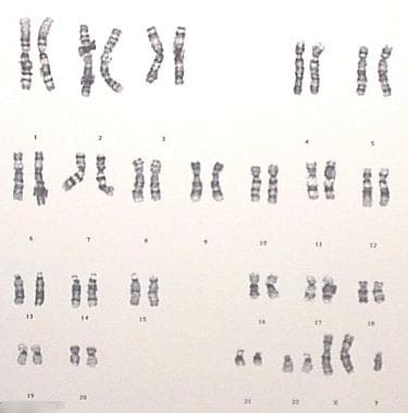
Klinefelter Syndrome
Sperm Retrieval
Sex Chromosome Disorders
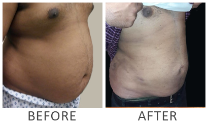
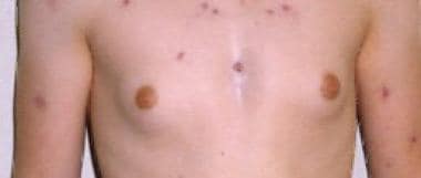
Gynecomastia
Chromosomes, Human, X
Sex Chromosome Aberrations
Chromosomes, Human, Y
Gonadal Dysgenesis, 46,XX
Primed In Situ Labeling
Aneuploidy
Infertility, Male
Mosaicism
Oligospermia
Breast Neoplasms, Male
Testis
Testosterone
In Situ Hybridization, Fluorescence
Encyclopedias as Topic
Y Chromosome
Germ cell development in the XXY mouse: evidence that X chromosome reactivation is independent of sexual differentiation. (1/226)
Prior to entry into meiosis, XX germ cells in the fetal ovary undergo X chromosome reactivation. The signal for reactivation is thought to emanate from the genital ridge, but it is unclear whether it is specific to the developing ovary. To determine whether the signals are present in the developing testis as well as the ovary, we examined the expression of X-linked genes in germ cells from XXY male mice. To facilitate this analysis, we generated XXY and XX fetuses carrying X chromosomes that were differentially marked and subject to nonrandom inactivation. This pattern of nonrandom inactivation was maintained in somatic cells but, in XX as well as XXY fetuses, both parental alleles were expressed in germ cell-enriched cell populations. Because testis differentiation is temporally and morphologically normal in the XXY testis and because all germ cells embark upon a male pathway of development, these results provide compelling evidence that X chromosome reactivation in fetal germ cells is independent of the somatic events of sexual differentiation. Proper X chromosome dosage is essential for the normal fertility of male mammals, and abnormalities in germ cell development are apparent in the XXY testis within several days of X reactivation. Studies of exceptional germ cells that survive in the postnatal XXY testis demonstrated that surviving germ cells are exclusively XY and result from rare nondisjunctional events that give rise to clones of XY cells. (+info)Birth of a healthy neonate following the intracytoplasmic injection of testicular spermatozoa from a patient with Klinefelter's syndrome. (2/226)
Klinefelter's syndrome is one of the known causes of azoospermia or cryptoazoospermia, and it may present in non-mosaic (47,XXY) or mosaic (47,XXY/46,XY) form. The likelihood of finding spermatozoa in the ejaculate or testicular tissue of patients with mosaic Klinefelter's syndrome is low, and with the non-mosaic form, even lower. We describe a patient with non-mosaic Klinefelter in whom initially non-motile spermatozoa were derived from searching the ejaculate. Ten mature oocytes were injected, but none was fertilized. Subsequently, testicular biopsy was undertaken in order to collect spermatozoa for oocyte injection. Fifteen motile sperm cells were found and injected. Nine oocytes were fertilized and cleaved; three embryos were transferred into the uterine cavity. The woman conceived and following a normal pregnancy delivered a healthy child. Genetic analysis of the neonate disclosed a normal 46,XY karyotype. Non-motile spermatozoa in the ejaculate did not prove their fertilization potential, but their presence did not exclude finding motile, fertile spermatozoa in the testicular tissue in a non-mosaic Klinefelter patient. This report is further evidence that normal spermatozoa with fertilization potential are produced in the testes of patients with Klinefelter's syndrome. (+info)Klinefelter's syndrome in the male infertility clinic. (3/226)
The clinical features of patients with Klinefelter's syndrome attending a male infertility clinic have been investigated in order to consider their assisted reproduction treatment options. Over 12 years, a total of 148 patients with sterility due to azoospermia had Klinefelter's syndrome. Eight patients were shown by fluorescence in-situ hybridization (FISH) on metaphase spreads to be mosaic (46,XY/47,XXY), and 140 patients showed only 47,XXY. Small testes were observed in 95% of patients and gynaecomastia was seen in 12.4%. Half of the patients showed hypergonadotrophic hypogonadism, while others showed normogonadism (usually hypergonadotrophic). Spermatozoa were observed in semen from one patient with mosaicism and one without. Three-colour FISH revealed hyperploidy in 2.7% and 2.3% of these spermatozoa respectively. Multiple-site testicular biopsies in five recent patients were performed and yielded a specimen with round and elongated spermatids in one patient with 47,XXY karyotype. This sample was cryopreserved for future intracytoplasmic sperm injection. At follow-up, 46% of couples had chosen artificial insemination with donor sperm, and none had chosen adoption. Two patients developed testicular tumours, one a mature teratoma and the other a Leydig cell tumour. Two patients required androgen replacement therapy. (+info)Fertilization and pregnancy outcome with intracytoplasmic sperm injection for azoospermic men. (4/226)
The evident ability of the intracytoplasmic sperm injection (ICSI) procedure to achieve high fertilization and pregnancy rates regardless of semen characteristics has induced its application with spermatozoa surgically retrieved from azoospermic men. Here, ICSI outcome was analysed in 308 cases according to the cause of azoospermia; four additional cycles were with cases of necrozoospermia. All couples were genetically counselled and appropriately screened. Spermatozoa were retrieved by microsurgical epididymal aspiration or from testicular biopsies. Epididymal obstructions were considered congenital (n = 138) or acquired (n = 103), based on the aetiology. Testicular sperm cases were assessed according to the presence (n = 14) or absence (n = 53) of reproductive tract obstruction. The fertilization rate using fresh or cryopreserved epididymal spermatozoa was 72.4% of 911 eggs for acquired obstructions, and 73.1% of 1524 eggs for congenital cases; with clinical pregnancy rates of 48.5% (50/103) and 61.6% (85/138) respectively. Spermatozoa from testicular biopsies fertilized 57.0% of 533 eggs in non-obstructive cases compared to 80.5% of 118 eggs (P = 0.0001) in obstructive azoospermia. The clinical pregnancy rate was 49.1% (26/53) for non-obstructive cases and 57.1% (8/14) for testicular spermatozoa obtained in obstructive azoospermia, including three established with frozen-thawed testicular spermatozoa. In cases of obstructive azoospermia, fertilization and pregnancy rates with epididymal spermatozoa were higher than those achieved using spermatozoa obtained from the testes of men with non-obstructive azoospermia. (+info)Meiotic aneuploidy in the XXY mouse: evidence that a compromised testicular environment increases the incidence of meiotic errors. (5/226)
Male mammals with two X chromosomes are sterile due to the loss of virtually all germ cells in the differentiating testis. The survival of rare germ cells, however, can give rise to patches of normal-appearing spermatogenesis in the adult testis. Intracytoplasmic sperm injection (ICSI) makes possible the establishment of a pregnancy using spermatozoa from severely oligozoospermic men and, indeed, has been successful using spermatozoa from human 47,XXY (Klinefelter syndrome) males. The risk of an abnormal pregnancy, however, may be significantly increased since several studies have demonstrated elevated levels of aneuploidy in spermatozoa from Klinefelter syndrome men. This has been suggested to reflect the consequences of meiotic segregation in XXY germ cells; however, it is also possible that it is a consequence of abnormalities in meiotic regulation in the XXY testis. We have addressed this question experimentally in the XXY male mouse. Analysis of testicular spermatozoa from XXY and control males demonstrates a significant increase in meiotic aneuploidy in the XXY mouse. Since previous studies have demonstrated that germ cells in the adult XXY testis are exclusively XY, the meiotic abnormalities observed must be attributable to segregation errors in XY germ cells. These findings have potential significance for ICSI pregnancies using spermatozoa from other types of male factor infertility patients, since they raise the possibility that increased meiotic errors are a generalized feature of the severely oligozoospermic testis. (+info)Developmental and genetic disorders in spermatogenesis. (6/226)
The most common cause of male infertility is idiopathic. Fresh insights based on genetic and molecular analysis of the human genome permit classification of formerly unexplained disorders in spermatogenesis. In this article, we review new procedures that expand diagnostic and therapeutic approaches to male infertility. Recombinant DNA technology makes it possible to detect specific chromosomal and/or genetic defects among infertile patients. The identification of genes linked to disorders in spermatogenesis and male sexual differentiation has increased exponentially in the past decade. Genetic defects leading to male factor infertility can now be explained at the molecular level, even though the germ cell profile of infertile patients is too variable to permit classification of the clinical phenotype. Increasing knowledge of genes that direct spermatogenesis provides important new information about the molecular and cellular events involved in human spermatogenesis. Molecular analysis of chromosomes and/or genes of infertile patients offers unique opportunities to uncover the aetiology of genetic disorders in spermatogenesis. Increasing numbers of cases, previously classified as idiopathic, can now be diagnosed to facilitate the treatment of infertile men. Advanced knowledge also poses ethical dilemmas, since children conceived with assisted reproductive technologies such as intracytoplasmic sperm injection (ICSI) are at risk for congenital abnormalities, unbalanced complements of chromosomes and male infertility. (+info)Chromosome abnormalities in a referred population for suspected chromosomal aberrations: a report of 4117 cases. (7/226)
A cytogenetic study was performed on 4,117 Korean patients referred for suspected chromosomal abnormalities. Chromosome aberrations were identified in 17.5% of the referred cases. The most common autosomal abnormality was Down syndrome and Turner syndrome in abnormalities of sex chromosome. The proportions of different karyotypes in Down syndrome (trisomy 21 92.5%, translocation 5.1%, mosaic 2.4%) were similar to those reported in other countries. However, it was different in Turner syndrome (45, X 28.1%, mosaic 50.8%, 46, X, del (Xq) 4.4%, 46, X, i (Xq) 16.7%), in which proportions of mosaics and isochromosome, 46, X, i(Xq), were higher than those reported in other countries. In structural chromosome aberrations of autosome, translocation was the most common (43.6%), and duplication (21.3%), deletion (14.4%), marker chromosome (7.9%) and ring chromosome (4.0%) followed in order of frequency. Rates of several normal variant karyotypes were also described. Inversion of chromosome 9 was observed in 1.7% of total referred cases. (+info)Klinefelter's syndrome accompanied by mixed connective tissue disease and diabetes mellitus. (8/226)
We report a rare case of Klinefelter's syndrome (KS) with mixed connective tissue disease (MCTD), diabetes mellitus (DM) and several endocrine disorders. A 57-year-old man presented with polyarthritis and tapering fingers with Raynaud's phenomenon on admission. In addition to a karyotype of 47, XXY, a marked restrictive change in respiratory functional test, a myogenic pattern in electromyogram, the positive tests for anti-RNP antibody indicated that this was a case of KS complicated with MCTD. The patients also presented DM with insulin resistance, hyperprolactinemia, slight primary hypothyroidism and hypoadrenocorticism. The mechanism for these coincidences remains to be elucidated. (+info)Klinefelter Syndrome: A genetic disorder in males, caused by the presence of one or more extra X chromosomes, typically resulting in XXY karyotype. It is characterized by small testes, infertility, gynecomastia (breast enlargement), tall stature, and often mild to moderate intellectual disability. The symptoms can vary greatly among individuals with Klinefelter Syndrome. Some men may not experience any significant health problems and may never be diagnosed, while others may have serious medical or developmental issues that require treatment. It is one of the most common chromosomal disorders, affecting about 1 in every 500-1,000 newborn males.
Sperm retrieval is a medical procedure that involves obtaining sperm from a male patient, usually for the purpose of assisted reproduction. This can be indicated in cases where the man has obstructive or non-obstructive azoospermia (absence of sperm in the semen), ejaculatory dysfunction, or other conditions that prevent the successful collection of sperm through conventional means, such as masturbation.
There are several methods for sperm retrieval, including:
1. Testicular sperm aspiration (TESA): A procedure where a fine needle is inserted into the testicle to aspirate (or draw out) sperm.
2. Percutaneous epididymal sperm aspiration (PESA): Similar to TESA, but the needle is inserted into the epididymis, a small structure that stores and transports sperm from the testicle.
3. Microsurgical epididymal sperm aspiration (MESA): A more invasive procedure where an incision is made in the scrotum to directly visualize the epididymis with a surgical microscope, allowing for the careful removal of sperm.
4. Testicular sperm extraction (TESE): Involves making a small incision in the testicle and removing a piece of tissue containing sperm-producing tubules. The tissue is then processed to extract viable sperm.
5. Microdissection testicular sperm extraction (microTESE): A refined version of TESE, where a surgical microscope is used to identify and isolate individual seminiferous tubules containing sperm in men with non-obstructive azoospermia.
The retrieved sperm can then be used for various assisted reproductive techniques, such as intracytoplasmic sperm injection (ICSI), where a single sperm is injected directly into an egg to facilitate fertilization.
Azoospermia is a medical condition where there is no measurable level of sperm in the semen. This means that during ejaculation, the seminal fluid does not contain any sperm cells. Azoospermia can be caused by various factors including problems with testicular function, obstruction of the genital tract, or hormonal imbalances. It is an important cause of male infertility and may require further medical evaluation and treatment to determine the underlying cause and explore potential options for fertility.
There are two types of azoospermia: obstructive azoospermia and non-obstructive azoospermia. Obstructive azoospermia is caused by blockages or obstructions in the genital tract that prevent sperm from being released into the semen, while non-obstructive azoospermia is due to problems with sperm production in the testicles.
In some cases, men with azoospermia may still be able to father children through assisted reproductive technologies such as intracytoplasmic sperm injection (ICSI), where a single sperm is injected directly into an egg for fertilization. However, this will depend on the underlying cause of the azoospermia and whether or not there are viable sperm available for extraction.
Sex chromosome disorders are genetic conditions that occur due to an atypical number or structure of the sex chromosomes, which are X and Y. Normally, females have two X chromosomes (XX), and males have one X and one Y chromosome (XY). However, in sex chromosome disorders, there is a variation in the number or composition of these chromosomes.
The most common sex chromosome disorders include:
1. Turner syndrome (Monosomy X): Occurs when a female has only one X chromosome (45,X). This condition affects about 1 in every 2,500 female births and can lead to short stature, infertility, heart defects, and learning disabilities.
2. Klinefelter syndrome (XXY): Occurs when a male has an extra X chromosome (47,XXY). This condition affects about 1 in every 500-1,000 male births and can lead to tall stature, infertility, breast development, and learning disabilities.
3. Jacobs syndrome (XYY): Occurs when a male has an extra Y chromosome (47,XYY). This condition affects about 1 in every 1,000 male births and can lead to tall stature, learning disabilities, and behavioral issues.
4. Triple X syndrome (XXX): Occurs when a female has an extra X chromosome (47,XXX). This condition affects about 1 in every 1,000 female births and can lead to mild developmental delays and learning disabilities.
5. Other rare sex chromosome disorders: These include conditions like 48,XXXX, 49,XXXXY, and mosaicism (a mixture of cells with different chromosome compositions).
Sex chromosome disorders can have varying degrees of impact on an individual's physical and cognitive development. While some individuals may experience significant challenges, others may have only mild or no symptoms at all. Early diagnosis and appropriate interventions can help improve outcomes for those affected by sex chromosome disorders.
Gynecomastia is a medical term that refers to the benign enlargement of the glandular tissue in male breasts, usually caused by an imbalance of the hormones estrogen and testosterone. It's important to note that gynecomastia is not the same as having excess fat in the breast area, which is called pseudogynecomastia.
Gynecomastia can occur during infancy, puberty, or old age due to natural hormonal changes. Certain medications, medical conditions, and recreational drugs can also cause gynecomastia by affecting hormone levels in the body. In some cases, the exact cause of gynecomastia may remain unknown.
Mild cases of gynecomastia may not require treatment, but severe or persistent cases may be treated with medication or surgery to remove excess breast tissue. It's essential to consult a healthcare professional for an accurate diagnosis and appropriate treatment options if you suspect you have gynecomastia.
A chromosome is a thread-like structure that contains genetic material, made up of DNA and proteins, in the nucleus of a cell. In humans, there are 23 pairs of chromosomes, for a total of 46 chromosomes, in each cell of the body, with the exception of the sperm and egg cells which contain only 23 chromosomes.
The X chromosome is one of the two sex-determining chromosomes in humans. Females typically have two X chromosomes (XX), while males have one X and one Y chromosome (XY). The X chromosome contains hundreds of genes that are responsible for various functions in the body, including some related to sexual development and reproduction.
Humans inherit one X chromosome from their mother and either an X or a Y chromosome from their father. In females, one of the two X chromosomes is randomly inactivated during embryonic development, resulting in each cell having only one active X chromosome. This process, known as X-inactivation, helps to ensure that females have roughly equal levels of gene expression from the X chromosome, despite having two copies.
Abnormalities in the number or structure of the X chromosome can lead to various genetic disorders, such as Turner syndrome (X0), Klinefelter syndrome (XXY), and fragile X syndrome (an X-linked disorder caused by a mutation in the FMR1 gene).
Sex chromosome aberrations refer to structural and numerical abnormalities in the sex chromosomes, which are typically represented as X and Y chromosomes in humans. These aberrations can result in variations in the number of sex chromosomes, such as Klinefelter syndrome (47,XXY), Turner syndrome (45,X), and Jacobs/XYY syndrome (47,XYY). They can also include structural changes, such as deletions, duplications, or translocations of sex chromosome material.
Sex chromosome aberrations may lead to a range of phenotypic effects, including differences in physical characteristics, cognitive development, fertility, and susceptibility to certain health conditions. The manifestation and severity of these impacts can vary widely depending on the specific type and extent of the aberration, as well as individual genetic factors and environmental influences.
It is important to note that while sex chromosome aberrations may pose challenges and require medical management, they do not inherently define or limit a person's potential, identity, or worth. Comprehensive care, support, and education can help individuals with sex chromosome aberrations lead fulfilling lives and reach their full potential.
Human Y chromosomes are one of the two sex-determining chromosomes in humans (the other being the X chromosome). They are found in the 23rd pair of human chromosomes and are significantly smaller than the X chromosome.
The Y chromosome is passed down from father to son through the paternal line, and it plays a crucial role in male sex determination. The SRY gene (sex-determining region Y) on the Y chromosome initiates the development of male sexual characteristics during embryonic development.
In addition to the SRY gene, the human Y chromosome contains several other genes that are essential for sperm production and male fertility. However, the Y chromosome has a much lower gene density compared to other chromosomes, with only about 80 protein-coding genes, making it one of the most gene-poor chromosomes in the human genome.
Because of its small size and low gene density, the Y chromosome is particularly susceptible to genetic mutations and deletions, which can lead to various genetic disorders and male infertility. Nonetheless, the Y chromosome remains a critical component of human genetics and evolution, providing valuable insights into sex determination, inheritance patterns, and human diversity.
Gonadal dysgenesis, 46,XX is a medical condition where an individual with a 46,XX karyotype has underdeveloped or absent gonads (ovaries). Normally, individuals with a 46,XX karyotype have ovaries that produce female sex hormones and develop into reproductive organs. However, in cases of gonadal dysgenesis, the gonads do not develop properly and may appear as streak gonads, which lack germ cells and are incapable of producing sex hormones or gametes (eggs).
Individuals with 46,XX gonadal dysgenesis often have female external genitalia but may have primary amenorrhea (absence of menstruation) due to the underdeveloped or absent ovaries. They may also have other features such as short stature, webbed neck, and intellectual disability, depending on the underlying cause of the condition.
The underlying causes of 46,XX gonadal dysgenesis can vary, including genetic mutations, chromosomal abnormalities, or exposure to environmental factors during fetal development. Some individuals with this condition may have an increased risk of developing gonadal tumors, so regular monitoring and follow-up care are essential.
Karyotyping is a medical laboratory test used to study the chromosomes in a cell. It involves obtaining a sample of cells from a patient, usually from blood or bone marrow, and then staining the chromosomes so they can be easily seen under a microscope. The chromosomes are then arranged in pairs based on their size, shape, and other features to create a karyotype. This visual representation allows for the identification and analysis of any chromosomal abnormalities, such as extra or missing chromosomes, or structural changes like translocations or inversions. These abnormalities can provide important information about genetic disorders, diseases, and developmental problems.
"Primed In Situ Labeling" (PRINS) is not a widely recognized medical term, but it is a technique used in molecular biology and pathology. Here's a definition of the PRINS technique:
Primed In Situ Labeling (PRINS) is a cytogenetic method that allows for the detection and visualization of specific DNA sequences within chromosomes or interphase nuclei through fluorescence in situ hybridization (FISH). The technique involves denaturing double-stranded DNA in fixed cells, followed by annealing a primer to a specific target sequence. A DNA polymerase then extends the primer, incorporating labeled nucleotides that can be visualized under a fluorescence microscope.
The PRINS technique offers several advantages over traditional FISH methods, including higher sensitivity and specificity, lower background signal, and the ability to analyze multiple targets simultaneously using different colored probes. It is commonly used in the diagnosis and monitoring of various genetic disorders, cancer, and infectious diseases.
Aneuploidy is a medical term that refers to an abnormal number of chromosomes in a cell. Chromosomes are thread-like structures located inside the nucleus of cells that contain genetic information in the form of genes.
In humans, the normal number of chromosomes in a cell is 46, arranged in 23 pairs. Aneuploidy occurs when there is an extra or missing chromosome in one or more of these pairs. For example, Down syndrome is a condition that results from an extra copy of chromosome 21, also known as trisomy 21.
Aneuploidy can arise during the formation of gametes (sperm or egg cells) due to errors in the process of cell division called meiosis. These errors can result in eggs or sperm with an abnormal number of chromosomes, which can then lead to aneuploidy in the resulting embryo.
Aneuploidy is a significant cause of birth defects and miscarriages. The severity of the condition depends on which chromosomes are affected and the extent of the abnormality. In some cases, aneuploidy may have no noticeable effects, while in others it can lead to serious health problems or developmental delays.
XYY karyotype is a chromosomal abnormality where an individual's cells have one extra Y chromosome, resulting in a 47, XYY pattern of sex chromosomes. This condition is also known as Jacob's syndrome or XYY syndrome. Typically, human cells contain 23 pairs of chromosomes, for a total of 46 chromosomes, with one pair being the sex chromosomes (XX in females and XY in males). In an XYY karyotype, there are two Y chromosomes and one X chromosome, which can lead to developmental differences and various health concerns.
Individuals with XYY karyotype may have a higher risk of developing learning disabilities, speech and language delays, and behavioral issues such as attention deficit hyperactivity disorder (ADHD) or autism spectrum disorders. However, many people with XYY karyotype do not experience significant health problems and can lead typical lives with appropriate support and interventions.
It is important to note that an XYY karyotype does not typically affect physical characteristics, and most individuals with this condition are phenotypically male. However, they may be taller than their peers due to the influence of the extra Y chromosome on growth hormones.
A syndrome, in medical terms, is a set of symptoms that collectively indicate or characterize a disease, disorder, or underlying pathological process. It's essentially a collection of signs and/or symptoms that frequently occur together and can suggest a particular cause or condition, even though the exact physiological mechanisms might not be fully understood.
For example, Down syndrome is characterized by specific physical features, cognitive delays, and other developmental issues resulting from an extra copy of chromosome 21. Similarly, metabolic syndromes like diabetes mellitus type 2 involve a group of risk factors such as obesity, high blood pressure, high blood sugar, and abnormal cholesterol or triglyceride levels that collectively increase the risk of heart disease, stroke, and diabetes.
It's important to note that a syndrome is not a specific diagnosis; rather, it's a pattern of symptoms that can help guide further diagnostic evaluation and management.
Male infertility is a condition characterized by the inability to cause pregnancy in a fertile female. It is typically defined as the failure to achieve a pregnancy after 12 months or more of regular unprotected sexual intercourse.
The causes of male infertility can be varied and include issues with sperm production, such as low sperm count or poor sperm quality, problems with sperm delivery, such as obstructions in the reproductive tract, or hormonal imbalances that affect sperm production. Other factors that may contribute to male infertility include genetic disorders, environmental exposures, lifestyle choices, and certain medical conditions or treatments.
It is important to note that male infertility can often be treated or managed with medical interventions, such as medication, surgery, or assisted reproductive technologies (ART). A healthcare provider can help diagnose the underlying cause of male infertility and recommend appropriate treatment options.
Mosaicism, in the context of genetics and medicine, refers to the presence of two or more cell lines with different genetic compositions in an individual who has developed from a single fertilized egg. This means that some cells have one genetic makeup, while others have a different genetic makeup. This condition can occur due to various reasons such as errors during cell division after fertilization.
Mosaicism can involve chromosomes (where whole or parts of chromosomes are present in some cells but not in others) or it can involve single genes (where a particular gene is present in one form in some cells and a different form in others). The symptoms and severity of mosaicism can vary widely, depending on the type and location of the genetic difference and the proportion of cells that are affected. Some individuals with mosaicism may not experience any noticeable effects, while others may have significant health problems.
Oligospermia is a medical term used to describe a condition in which the semen contains a lower than normal number of sperm. Generally, a sperm count of less than 15 million sperm per milliliter (ml) of semen is considered to be below the normal range.
Oligospermia can make it more difficult for a couple to conceive naturally and may require medical intervention such as intracytoplasmic sperm injection (ICSI) or in vitro fertilization (IVF). The condition can result from various factors, including hormonal imbalances, genetic abnormalities, varicocele, environmental factors, and certain medications.
It's important to note that oligospermia is not the same as azoospermia, which is a condition where there is no sperm present in the semen at all.
Breast neoplasms in males refer to abnormal growths or tumors in the male breast tissue. These neoplasms can be benign (non-cancerous) or malignant (cancerous). While breast cancer is much less common in men than in women, it can still occur and should be taken seriously.
The most common type of breast cancer in men is invasive ductal carcinoma, which starts in the milk ducts and spreads to surrounding tissue. Other types of breast cancer that can occur in men include inflammatory breast cancer, lobular carcinoma, and Paget's disease of the nipple.
Risk factors for developing male breast cancer include age (most cases are diagnosed after age 60), family history of breast cancer, genetic mutations such as BRCA1 or BRCA2, radiation exposure, obesity, liver disease, and testicular conditions such as undescended testicles.
Symptoms of male breast neoplasms may include a painless lump in the breast tissue, skin changes such as dimpling or redness, nipple discharge, or a retracted nipple. If you notice any of these symptoms, it is important to consult with a healthcare professional for further evaluation and treatment.
The term "Fathers" is a general term used to describe male parents or parental figures. It does not have a specific medical definition. In the context of genetics and reproduction, the father is the biological male who contributes his sperm to fertilize an egg, resulting in conception and pregnancy. However, it's important to note that there are many different types of families and parental relationships, and not all fathers are biological parents or male.
The testis, also known as the testicle, is a male reproductive organ that is part of the endocrine system. It is located in the scrotum, outside of the abdominal cavity. The main function of the testis is to produce sperm and testosterone, the primary male sex hormone.
The testis is composed of many tiny tubules called seminiferous tubules, where sperm are produced. These tubules are surrounded by a network of blood vessels, nerves, and supportive tissues. The sperm then travel through a series of ducts to the epididymis, where they mature and become capable of fertilization.
Testosterone is produced in the Leydig cells, which are located in the interstitial tissue between the seminiferous tubules. Testosterone plays a crucial role in the development and maintenance of male secondary sexual characteristics, such as facial hair, deep voice, and muscle mass. It also supports sperm production and sexual function.
Abnormalities in testicular function can lead to infertility, hormonal imbalances, and other health problems. Regular self-examinations and medical check-ups are recommended for early detection and treatment of any potential issues.
Testosterone is a steroid hormone that belongs to androsten class of hormones. It is primarily secreted by the Leydig cells in the testes of males and, to a lesser extent, by the ovaries and adrenal glands in females. Testosterone is the main male sex hormone and anabolic steroid. It plays a key role in the development of masculine characteristics, such as body hair and muscle mass, and contributes to bone density, fat distribution, red cell production, and sex drive. In females, testosterone contributes to sexual desire and bone health. Testosterone is synthesized from cholesterol and its production is regulated by luteinizing hormone (LH) and follicle-stimulating hormone (FSH).
In situ hybridization, fluorescence (FISH) is a type of molecular cytogenetic technique used to detect and localize the presence or absence of specific DNA sequences on chromosomes through the use of fluorescent probes. This technique allows for the direct visualization of genetic material at a cellular level, making it possible to identify chromosomal abnormalities such as deletions, duplications, translocations, and other rearrangements.
The process involves denaturing the DNA in the sample to separate the double-stranded molecules into single strands, then adding fluorescently labeled probes that are complementary to the target DNA sequence. The probe hybridizes to the complementary sequence in the sample, and the location of the probe is detected by fluorescence microscopy.
FISH has a wide range of applications in both clinical and research settings, including prenatal diagnosis, cancer diagnosis and monitoring, and the study of gene expression and regulation. It is a powerful tool for identifying genetic abnormalities and understanding their role in human disease.
An encyclopedia is a comprehensive reference work containing articles on various topics, usually arranged in alphabetical order. In the context of medicine, a medical encyclopedia is a collection of articles that provide information about a wide range of medical topics, including diseases and conditions, treatments, tests, procedures, and anatomy and physiology. Medical encyclopedias may be published in print or electronic formats and are often used as a starting point for researching medical topics. They can provide reliable and accurate information on medical subjects, making them useful resources for healthcare professionals, students, and patients alike. Some well-known examples of medical encyclopedias include the Merck Manual and the Stedman's Medical Dictionary.
The Y chromosome is one of the two sex-determining chromosomes in humans and many other animals, along with the X chromosome. The Y chromosome contains the genetic information that helps to determine an individual's sex as male. It is significantly smaller than the X chromosome and contains fewer genes.
The Y chromosome is present in males, who inherit it from their father. Females, on the other hand, have two X chromosomes, one inherited from each parent. The Y chromosome includes a gene called SRY (sex-determining region Y), which initiates the development of male sexual characteristics during embryonic development.
It is worth noting that the Y chromosome has a relatively high rate of genetic mutation and degeneration compared to other chromosomes, leading to concerns about its long-term viability in human evolution. However, current evidence suggests that the Y chromosome has been stable for at least the past 25 million years.
Medically, hair is defined as a threadlike structure that grows from the follicles found in the skin of mammals. It is primarily made up of a protein called keratin and consists of three parts: the medulla (the innermost part or core), the cortex (middle layer containing keratin filaments) and the cuticle (outer layer of overlapping scales).
Hair growth occurs in cycles, with each cycle consisting of a growth phase (anagen), a transitional phase (catagen), and a resting phase (telogen). The length of hair is determined by the duration of the anagen phase.
While hair plays a crucial role in protecting the skin from external factors like UV radiation, temperature changes, and physical damage, it also serves as an essential aspect of human aesthetics and identity.
 Klinefelter syndrome - Wikipedia
 Klinefelter syndrome: MedlinePlus Genetics
 Klinefelter Syndrome: Practice Essentials, Pathophysiology, Epidemiology
 Klinefelter syndrome - Diagnosis and treatment - Mayo Clinic
 Klinefelter Syndrome
 Klinefelter Syndrome
 Information for "Klinefelter's syndrome pathophysiology" - wikidoc
 Klinefelter Syndrome 025
 Neurogenetic Basis of Criminal Behaviors in Klinefelter Syndrome: A Case of Uxoricide-Suicide and a Review of the Literature |...
 Technology Archives - Klinefelter Syndrome X and Y Variations Support, Information, and Education
 Klinefelter's syndrome Archives - DAVinci Plastic Surgery
 Klinefelter Syndrome - Children's Health Issues - MSD Manual Consumer Version
 Klinefelter syndrome
 Klinefelter Syndrome - Introduction
 Klinefelter's Syndrome Association (KSA)
 Changes in the cohort composition of turner syndrome and severe non-diagnosis of Klinefelter, 47,XXX and 47,XYY syndrome: a...
 Turner Syndrome vs. Klinefelter Syndrome
 klinefelter's syndrome | Excel Male TRT Forum
 Klinefelter Syndrome Infertility Symptoms, Causes & Treatment