Dislocations
Peroneal Neuropathies
Posterior Cruciate Ligament
Medial Collateral Ligament, Knee
Ligaments, Articular
Anterior Cruciate Ligament
Osteoarthritis, Knee
Reconstructive Surgical Procedures
Peroneal Nerve
Orthopedic Procedures
Hip Dislocation, Congenital
Range of Motion, Articular
Traction
Instability after total knee replacement with a mobile-bearing prosthesis in a patient with multiple sclerosis. (1/29)
We describe a patient with multiple sclerosis (MS), who developed recurrent dislocations after total knee arthroplasty. She had both knees replaced using similar mobile-bearing knee prostheses, but the outcome was worse in the leg which was more severely affected with MS. (+info)Ligament repair and reconstruction in traumatic dislocation of the knee. (2/29)
We treated 21 patients with 22 dislocations of the knee by repair or reconstruction of all injured ligaments. Eight knees were treated in the acute phase (less than two weeks after injury); the remainder were treated more than six months after injury (6 to 72). Reconstructions were carried out with a combination of autograft and allograft tendons and by direct ligament repair where possible. At a mean follow-up of 32 months (11 to 77) the mean Lysholm score was 87 (81 to 91) in the acute group and 75 (53 to 100) in the delayed group. The mean Tegner activity rating was 5 in the acute group and 4.4 in the delayed group. The International Knee Documentation Committee assessment revealed no differences between the two groups. Instrumented testing of knee stability indicated better results for anterior cruciate ligament reconstructions which had been undertaken in the acute phase, but no difference in the outcome of posterior cruciate ligament reconstructions. There was no difference in the loss of knee movement between the two groups. Although the differences were small, the outcome in terms of overall knee function, activity levels and anterior tibial translation were better in those knees which had been reconstructed within two weeks of injury. (+info)Knee dislocations: experience at the Hopital du Sacre-Coeur de Montreal. (3/29)
INTRODUCTION: Although many options exist for ligament reconstruction in knee dislocations, the optimal treatment remains controversial. Allografts and autografts have both been used to reconstruct the cruciate ligaments. We present the results of reconstruction using artificial ligaments at Hopital du Sacre-Coeur in Montreal. METHODS: We reviewed the treatment of all patients with knee dislocations seen between June 1996 and October 1999. The Lysholm score, ACL-quality of life (QoL) questionnaire, physical examination and Telos instrumented laxity measurement were used to evaluate the results. RESULTS: Twenty patients (21 knees) participated in the study. The mean (and standard deviation [SD]) Lysholm score was 71.7 (18). Results from the ACL-QoL questionnaire showed a global impairment in QoL. Mean (and SD) range of motion and flexion were 118 degrees (10.9 degrees) and 2 degrees (2.9 degrees) respectively. Mean (and SD) radiologic laxity evaluated with Telos for the anterior and posterior cruciate ligaments were 6.1 (5.7) mm and 7.3 (4.5) mm respectively. CONCLUSIONS: Knee reconstruction with artificial ligaments shows promise, but further studies are necessary before it can be recommended for widespread use. This is the first study to show specifically a severe impairment in QoL in this patient population. (+info)Palsy of the common peroneal nerve after traumatic dislocation of the knee. (4/29)
Injury to the common peroneal nerve was present in 14 of 55 patients (25%) with dislocation of the knee. All underwent ligament reconstruction. The most common presenting direction of the dislocation was anterior or anteromedial with associated disruption of both cruciate ligaments and the posterolateral structures of the knee. Palsy of the common peroneal nerve was present in 14 of 34 (41%) of these patients. Complete rupture of the nerve was seen in four patients and a lesion in continuity in ten. Three patients with lesions in continuity, but with less than 7 cm of the nerve involved, had complete recovery within six to 18 months. In the remaining seven with more extensive lesions, two regained no motor function, and one had only MRC grade-2 function. Four patients regained some weak dorsiflexion or eversion (MRC grade 3 or 4). Some sensory recovery occurred in all seven of these patients, but was incomplete. In summary, complete recovery occurred in three (21%) and partial recovery of useful motor function in four (29%). In the other seven (50%) no useful motor or sensory function returned. (+info)Knee dislocation of a morbidly obese patient: a case report. (5/29)
Knee dislocations of morbidly obese patients after a trivial fall are not uncommon. We report a case of closed reduction for a dislocated right knee of a 26-year-old obese woman. After closed reduction under general anaesthesia, her knee was supported by pillows in 30 degrees flexion. No external splint was used because of the enormous size of the leg. At day 4 after reduction, the patient had numbness over the dorsum of the right foot and was unable to dorsiflex. She was diagnosed as having peroneal nerve palsy and was fitted with a foot drop splint. One week after reduction, she started active, assisted knee mobilisation and tip-toe weight bearing. At 24 months after reduction, the patient was able to walk unaided and had 100 degrees of knee flexion. She had a good foot function and a grade II in the Lachman's test, with no varus or valgus instability. This case highlights the importance of early mobilisation, which can result in good outcome even without operative treatment. (+info)Change in joint space width: hyaline articular cartilage loss or alteration in meniscus? (6/29)
OBJECTIVE: To explore the relative contribution of hyaline cartilage morphologic features and the meniscus to the radiographic joint space. METHODS: The Boston Osteoarthritis of the Knee Study is a natural history study of symptomatic knee osteoarthritis (OA). Baseline and 30-month followup assessments included knee magnetic resonance imaging (MRI) and fluoroscopically positioned weight-bearing knee radiographs. Cartilage and meniscal degeneration were scored on MRI in the medial and lateral tibiofemoral joints using a semiquantitative grading system. Meniscal position was measured to the nearest millimeter. The dependent variable was joint space narrowing (JSN) on the plain radiograph (possible range 0-3). The predictor variables were MRI cartilage score, meniscal degeneration, and meniscal position measures. We first conducted a cross-sectional analysis using multivariate regression to determine the relative contribution of meniscal factors and cartilage morphologic features to JSN, adjusting for body mass index (BMI), age, and sex. The same approach was used for change in JSN and change in predictor variables. RESULTS: We evaluated 264 study participants with knee OA (mean age 66.7 years, 59% men, mean BMI 31.4 kg/m(2)). The results from the models demonstrated that meniscal position and meniscal degeneration each contributed to prediction of JSN, in addition to the contribution by cartilage morphologic features. For change in medial joint space, both change in meniscal position and change in articular cartilage score contributed substantially to narrowing of the joint space. CONCLUSION: The meniscus (both its position and degeneration) accounts for a substantial proportion of the variance explained in JSN, and the change in meniscal position accounts for a substantial proportion of change in JSN. (+info)Concomitant ipsilateral traumatic dislocation of the hip and knee following high-energy trauma: a case report. (7/29)
Traumatic dislocation of the hip or knee can occur after high-energy trauma and is often associated with concomitant injuries and secondary complications. Concomitant traumatic dislocation of both hip and knee is rare. We describe a case of combined ipsilateral posterior hip dislocation with a posterior acetabular fracture and a complete open knee dislocation with disruption of the popliteal artery that resulted in amputation. (+info)Traumatic anterior knee dislocation and tibial shaft fracture: a case report. (8/29)
We report a case of traumatic anterior dislocation of the left knee with an ipsilateral tibial shaft fracture in association with popliteal artery and common peroneal nerve injuries. To our knowledge, such a combination of injuries has not been reported before. All components of this injury were recognised and treated promptly with rehabilitation commencing early, resulting in a good functional outcome. We discuss the possible injury mechanism and management of this unusual case. (+info)Knee dislocation is a serious and uncommon orthopedic injury that occurs when the bones that form the knee joint (femur, tibia, and patella) are forced out of their normal position due to extreme trauma or force. This injury often requires immediate medical attention and reduction (repositioning) by a healthcare professional. If left untreated, it can lead to serious complications such as compartment syndrome, nerve damage, and long-term joint instability. It's important to note that knee dislocation is different from a kneecap (patellar) dislocation, which involves the patella sliding out of its groove in the femur.
A dislocation is a condition in which a bone slips out of its normal position in a joint. This can happen as a result of trauma or injury, such as a fall or direct blow to the body. Dislocations can cause pain, swelling, and limited mobility in the affected area. In some cases, a dislocation may also damage surrounding tissues, such as ligaments, tendons, and nerves.
Dislocations are typically treated by reducing the dislocation, which means putting the bone back into its normal position. This is usually done with the help of medication to relieve pain and relaxation techniques to help the person stay still during the reduction. In some cases, surgery may be necessary to repair damaged tissues or if the dislocation cannot be reduced through other methods. After the dislocation has been reduced, the joint may be immobilized with a splint or sling to allow it to heal properly.
It is important to seek medical attention promptly if you suspect that you have a dislocation. If left untreated, a dislocation can lead to further complications, such as joint instability and chronic pain.
Peroneal neuropathies refer to conditions that cause damage or dysfunction to the peroneal nerve, which is a branch of the sciatic nerve. The peroneal nerve runs down the back of the leg and wraps around the fibula bone (the smaller of the two bones in the lower leg) before dividing into two branches that innervate the muscles and skin on the front and side of the lower leg and foot.
Peroneal neuropathies can cause various symptoms, including weakness or paralysis of the ankle and toe muscles, numbness or tingling in the top of the foot and along the outside of the lower leg, and difficulty lifting the foot (known as "foot drop"). These conditions can result from trauma, compression, diabetes, or other underlying medical conditions. Treatment for peroneal neuropathies may include physical therapy, bracing, medications to manage pain, and in some cases, surgery.
The Posterior Cruciate Ligament (PCL) is one of the major ligaments in the knee, providing stability to the joint. It is a strong band of tissue located in the back of the knee, connecting the thighbone (femur) to the shinbone (tibia). The PCL limits the backward motion of the tibia relative to the femur and provides resistance to forces that tend to push the tibia backwards. It also assists in maintaining the overall alignment and function of the knee joint during various movements and activities. Injuries to the PCL are less common compared to injuries to the Anterior Cruciate Ligament (ACL) but can still occur due to high-energy trauma, such as motor vehicle accidents or sports incidents involving direct impact to the front of the knee.
The medial collateral ligament (MCL) of the knee is a band-like structure located on the inner side of the knee joint. It connects the end of the femur (thighbone) to the top of the tibia (shinbone) and helps stabilize the knee by controlling side-to-side movement and preventing excessive separation of the bones. The MCL provides resistance to valgus force, which is a pushing or pulling force that attempts to push the bones apart in a direction away from the midline of the body. MCL injuries often occur due to direct impact to the outer knee or sudden changes in direction that strain the ligament.
A hip dislocation is a medical emergency that occurs when the head of the femur (thighbone) slips out of its socket in the pelvis. This can happen due to high-energy trauma, such as a car accident or a severe fall. Hip dislocations can also occur in people with certain health conditions that make their hips more prone to displacement, such as developmental dysplasia of the hip.
There are two main types of hip dislocations: posterior and anterior. In a posterior dislocation, the femur head moves out of the back of the socket, which is the most common type. In an anterior dislocation, the femur head moves out of the front of the socket. Both types of hip dislocations can cause severe pain, swelling, and difficulty moving the affected leg.
Immediate medical attention is necessary for a hip dislocation to realign the bones and prevent further damage. Treatment typically involves sedation or anesthesia to relax the muscles around the joint, followed by a closed reduction procedure to gently guide the femur head back into the socket. In some cases, surgery may be required to repair any associated injuries, such as fractures or damaged ligaments. After treatment, physical therapy and rehabilitation are usually necessary to restore strength, mobility, and function to the affected hip joint.
The knee joint, also known as the tibiofemoral joint, is the largest and one of the most complex joints in the human body. It is a synovial joint that connects the thighbone (femur) to the shinbone (tibia). The patella (kneecap), which is a sesamoid bone, is located in front of the knee joint and helps in the extension of the leg.
The knee joint is made up of three articulations: the femorotibial joint between the femur and tibia, the femoropatellar joint between the femur and patella, and the tibiofibular joint between the tibia and fibula. These articulations are surrounded by a fibrous capsule that encloses the synovial membrane, which secretes synovial fluid to lubricate the joint.
The knee joint is stabilized by several ligaments, including the medial and lateral collateral ligaments, which provide stability to the sides of the joint, and the anterior and posterior cruciate ligaments, which prevent excessive forward and backward movement of the tibia relative to the femur. The menisci, which are C-shaped fibrocartilaginous structures located between the femoral condyles and tibial plateaus, also help to stabilize the joint by absorbing shock and distributing weight evenly across the articular surfaces.
The knee joint allows for flexion, extension, and a small amount of rotation, making it essential for activities such as walking, running, jumping, and sitting.
Knee injuries refer to damages or harm caused to the structures surrounding or within the knee joint, which may include the bones (femur, tibia, and patella), cartilage (meniscus and articular cartilage), ligaments (ACL, PCL, MCL, and LCL), tendons (patellar and quadriceps), muscles, bursae, and other soft tissues. These injuries can result from various causes, such as trauma, overuse, degeneration, or sports-related activities. Symptoms may include pain, swelling, stiffness, instability, reduced range of motion, and difficulty walking or bearing weight on the affected knee. Common knee injuries include fractures, dislocations, meniscal tears, ligament sprains or ruptures, and tendonitis. Proper diagnosis and treatment are crucial to ensure optimal recovery and prevent long-term complications.
Shoulder dislocation is a medical condition where the head of the humerus (upper arm bone) gets displaced from its normal position in the glenoid fossa of the scapula (shoulder blade). This can occur anteriorly, posteriorly, or inferiorly, with anterior dislocations being the most common. It is usually caused by trauma or forceful movement and can result in pain, swelling, bruising, and limited range of motion in the shoulder joint. Immediate medical attention is required to relocate the joint and prevent further damage.
Articular ligaments, also known as fibrous ligaments, are bands of dense, fibrous connective tissue that connect and stabilize bones to each other at joints. They help to limit the range of motion of a joint and provide support, preventing excessive movement that could cause injury. Articular ligaments are composed mainly of collagen fibers arranged in a parallel pattern, making them strong and flexible. They have limited blood supply and few nerve endings, which makes them less prone to injury but also slower to heal if damaged. Examples of articular ligaments include the anterior cruciate ligament (ACL) and posterior cruciate ligament (PCL) in the knee joint, and the medial collateral ligament (MCL) and lateral collateral ligament (LCL) in the elbow joint.
The Anterior Cruciate Ligament (ACL) is a major stabilizing ligament in the knee. It is one of the four strong bands of tissue that connect the bones of the knee joint together. The ACL runs diagonally through the middle of the knee and helps to control the back and forth motion of the knee, as well as provide stability to the knee joint. Injuries to the ACL often occur during sports or physical activities that involve sudden stops, changes in direction, or awkward landings.
In medical terms, the knee is referred to as the largest and one of the most complex joints in the human body. It is a hinge joint that connects the thigh bone (femur) to the shin bones (tibia and fibula), enabling movements like flexion, extension, and a small amount of rotation. The knee also contains several other components such as menisci, ligaments, tendons, and bursae, which provide stability, cushioning, and protection during movement.
Osteoarthritis (OA) of the knee is a degenerative joint disease that affects the articular cartilage and subchondral bone in the knee joint. It is characterized by the breakdown and eventual loss of the smooth, cushioning cartilage that covers the ends of bones and allows for easy movement within joints. As the cartilage wears away, the bones rub against each other, causing pain, stiffness, and limited mobility. Osteoarthritis of the knee can also lead to the formation of bone spurs (osteophytes) and cysts in the joint. This condition is most commonly found in older adults, but it can also occur in younger people as a result of injury or overuse. Risk factors include obesity, family history, previous joint injuries, and repetitive stress on the knee joint. Treatment options typically include pain management, physical therapy, and in some cases, surgery.
Reconstructive surgical procedures are a type of surgery aimed at restoring the form and function of body parts that are defective or damaged due to various reasons such as congenital abnormalities, trauma, infection, tumors, or disease. These procedures can involve the transfer of tissue from one part of the body to another, manipulation of bones, muscles, and tendons, or use of prosthetic materials to reconstruct the affected area. The goal is to improve both the physical appearance and functionality of the body part, thereby enhancing the patient's quality of life. Examples include breast reconstruction after mastectomy, cleft lip and palate repair, and treatment of severe burns.
The Peroneal nerve, also known as the common fibular nerve, is a branch of the sciatic nerve that supplies the muscles of the lower leg and provides sensation to the skin on the outer part of the lower leg and the top of the foot. It winds around the neck of the fibula (calf bone) and can be vulnerable to injury in this area, leading to symptoms such as weakness or numbness in the foot and leg.
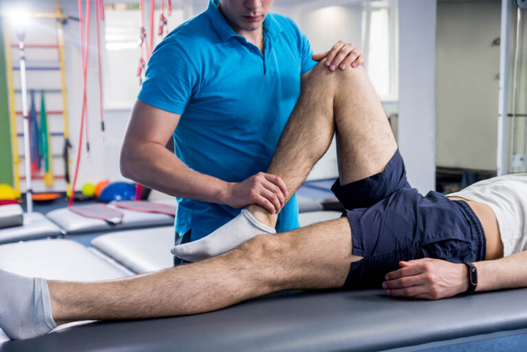
Orthopedic procedures are surgical or nonsurgical methods used to treat musculoskeletal conditions, including injuries, deformities, or diseases of the bones, joints, muscles, ligaments, and tendons. These procedures can range from simple splinting or casting to complex surgeries such as joint replacements, spinal fusions, or osteotomies (cutting and repositioning bones). The primary goal of orthopedic procedures is to restore function, reduce pain, and improve the quality of life for patients.
Arthroscopy is a minimally invasive surgical procedure where an orthopedic surgeon uses an arthroscope (a thin tube with a light and camera on the end) to diagnose and treat problems inside a joint. The surgeon makes a small incision, inserts the arthroscope into the joint, and then uses the attached camera to view the inside of the joint on a monitor. They can then insert other small instruments through additional incisions to repair or remove damaged tissue.
Arthroscopy is most commonly used for joints such as the knee, shoulder, hip, ankle, and wrist. It offers several advantages over traditional open surgery, including smaller incisions, less pain and bleeding, faster recovery time, and reduced risk of infection. The procedure can be used to diagnose and treat a wide range of conditions, including torn ligaments or cartilage, inflamed synovial tissue, loose bone or cartilage fragments, and joint damage caused by arthritis.
Congenital hip dislocation, also known as developmental dysplasia of the hip (DDH), is a condition where the hip joint fails to develop normally in utero or during early infancy. In a healthy hip, the head of the femur (thigh bone) fits snugly into the acetabulum (hip socket). However, in congenital hip dislocation, the femoral head is not held firmly in place within the acetabulum due to abnormal development or laxity of the ligaments that support the joint.
There are two types of congenital hip dislocations:
1. Teratologic dislocation: This type is present at birth and occurs due to abnormalities in the development of the hip joint during fetal growth. The femoral head may be completely outside the acetabulum or partially dislocated.
2. Developmental dysplasia: This type develops after birth, often within the first few months of life, as a result of ligamentous laxity and shallow acetabulum. In some cases, it can progress to a complete hip dislocation if left untreated.
Risk factors for congenital hip dislocation include family history, breech presentation during delivery, and female gender. Early diagnosis and treatment are crucial to prevent long-term complications such as pain, limited mobility, and osteoarthritis. Treatment options may include bracing, closed reduction, or surgical intervention, depending on the severity and age of the child at diagnosis.
Arthroplasty, replacement, knee is a surgical procedure where the damaged or diseased joint surface of the knee is removed and replaced with an artificial joint or prosthesis. The procedure involves resurfacing the worn-out ends of the femur (thigh bone) and tibia (shin bone) with metal components, and the back of the kneecap with a plastic button. This surgery is usually performed to relieve pain and restore function in patients with severe knee osteoarthritis, rheumatoid arthritis, or traumatic injuries that have damaged the joint beyond repair. The goal of knee replacement surgery is to improve mobility, reduce pain, and enhance the quality of life for the patient.
Patellar dislocation is a medical condition characterized by the displacement of the patella (kneecap) from its normal position in the femoral groove, which is a part of the femur (thighbone). This displacement usually occurs laterally, meaning that the patella moves toward the outer side of the knee.
Patellar dislocation can happen as a result of direct trauma or due to various factors that increase the laxity of the medial patellofemoral ligament and tightness of the lateral structures, leading to abnormal tracking of the patella. These factors include anatomical variations, muscle imbalances, genetic predisposition, or degenerative changes in the knee joint.
Dislocation of the patella can cause pain, swelling, and difficulty in moving the knee. In some cases, it might be associated with other injuries such as fractures or damage to the articular cartilage and surrounding soft tissues. Immediate medical attention is required for proper diagnosis and treatment, which may involve reduction, immobilization, physical therapy, bracing, or even surgery in severe cases.
A knee prosthesis, also known as a knee replacement or artificial knee joint, is a medical device used to replace the damaged or diseased weight-bearing surfaces of the knee joint. It typically consists of three components: the femoral component (made of metal) that fits over the end of the thighbone (femur), the tibial component (often made of metal and plastic) that fits into the top of the shinbone (tibia), and a patellar component (usually made of plastic) that replaces the damaged surface of the kneecap.
The primary goal of knee prosthesis is to relieve pain, restore function, and improve quality of life for individuals with advanced knee joint damage due to conditions such as osteoarthritis, rheumatoid arthritis, or traumatic injuries. The procedure to implant a knee prosthesis is called knee replacement surgery or total knee arthroplasty (TKA).
Articular Range of Motion (AROM) is a term used in physiotherapy and orthopedics to describe the amount of movement available in a joint, measured in degrees of a circle. It refers to the range through which synovial joints can actively move without causing pain or injury. AROM is assessed by measuring the degree of motion achieved by active muscle contraction, as opposed to passive range of motion (PROM), where the movement is generated by an external force.
Assessment of AROM is important in evaluating a patient's functional ability and progress, planning treatment interventions, and determining return to normal activities or sports participation. It is also used to identify any restrictions in joint mobility that may be due to injury, disease, or surgery, and to monitor the effectiveness of rehabilitation programs.
Traction, in medical terms, refers to the application of a pulling force to distract or align parts of the body, particularly bones, joints, or muscles, with the aim of immobilizing, reducing displacement, or realigning them. This is often achieved through the use of various devices such as tongs, pulleys, weights, or specialized traction tables. Traction may be applied manually or mechanically and can be continuous or intermittent, depending on the specific medical condition being treated. Common indications for traction include fractures, dislocations, spinal cord injuries, and certain neurological conditions.
Joint instability is a condition characterized by the loss of normal joint function and increased risk of joint injury due to impaired integrity of the supporting structures, such as ligaments, muscles, or cartilage. This can result in excessive movement or laxity within the joint, leading to decreased stability and increased susceptibility to dislocations or subluxations. Joint instability may cause pain, swelling, and limited range of motion, and it can significantly impact a person's mobility and quality of life. It is often caused by trauma, degenerative conditions, or congenital abnormalities and may require medical intervention, such as physical therapy, bracing, or surgery, to restore joint stability.
Instability11
- Symptoms include pain and instability of the knee. (wikipedia.org)
- What causes instability of the knee-cap? (shoulder-surgeon.org)
- Some peoples bodies are built in a certain way, which makes them prone to getting knee cap instability. (shoulder-surgeon.org)
- In a chronic instability situation, further investigations like MRI scan may be necessary to rule out any associated injuries, as,a dislocation often damages the underside of the kneecap and the end of the thighbone, which can lead to additional pain and arthritis. (shoulder-surgeon.org)
- If the problem of instability persists inspite of non-surgical measures or if there are any associated injuries to the knee joint, then arthroscopic surgery of the knee may be required to correct the problem. (shoulder-surgeon.org)
- This condition is called knee instability. (sameernagdamd.com)
- Patellar (kneecap) instability results from one or more complete or partial dislocations (subluxations). (sameernagdamd.com)
- Moreover, chronic cartilage damage has been described at 13-year follow up with patellofemoral osteoarthritis in 22% in patellar instability knees compared to 11% in contralateral healthy knees [ 6 ]. (springer.com)
- Knee valgus and patellofemoral instability after pediatric anterior cruciate ligament reconstruction: acase report and review of the literature. (stanfordchildrens.org)
- METHODS: Knee dislocation was defined as disruption of at least two major stabilising ligaments of the knee and gross instability requiring an operation. (ox.ac.uk)
- The posterolateral corner (PLC) is a complex stabilization unit on the posterolateral side of the knee and prevents dorsal displacement of the lateral tibial plateau (ie, exorotation of the tibia with respect to the femur, lateral instability, and hyperextension). (medscape.com)
Cases of knee dislocation3
- Historically, conventional arteriography was recommended for all cases of knee dislocation, and though it remains the criterion standard for popliteal artery evaluation, there is growing debate over its universal application. (medscape.com)
- Some cases of knee dislocation may not be preventable, especially if physical factors make you more likely to dislocate your knee. (medlineplus.gov)
- RESULTS: Twenty-five cases of knee dislocation were identified. (ox.ac.uk)
Injuries34
- Knee dislocations are rare: they represent about 1 in 5,000 orthopedic injuries, and about 1 knee dislocation occurs annually per 100,000 people. (wikipedia.org)
- Although knee dislocations are considered rare injuries, they are considered surgical emergencies because of potential neurovascular complications. (medscape.com)
- A knee dislocation is usually associated with events of severe trauma such as automobile crashes, severe falls or sports injuries. (orthopaedic-surgery-md.com)
- Over half of all knee dislocations are the result of high-speed motor vehicle accidents and are associated with multiple injuries in approximately 30% of cases. (lifecare123.com)
- Injuries to the common peroneal nerve occur in up to forty percent of patients who suffer a knee dislocation. (lifecare123.com)
- Elbow dislocations are common elbow injuries that can be classified as simple or complex depending on whether or not surrounding structures have been injured. (medicahospitals.in)
- The orthopedic and sports medicine specialists at Children's are trained to diagnose, treat and prevent knee pain and injuries in children, teens and young adults, from birth to age 18. (choa.org)
- Knee pain in kids and teens can be a result of traumatic knee injuries or repetitive overuse injuries from physical activity, such as competitive sports. (choa.org)
- Some of the most common injuries to children and teens that cause knee pain include fractures, dislocations, and sprains and tears of soft tissues like ligaments and tendons. (choa.org)
- In many cases, injuries involve more than one structure in the knee. (choa.org)
- Injuries to the knee, especially for kids and teens who are still growing, can lead to short-term and long-term damage. (choa.org)
- It's also important for kids and teens with knee pain and injuries to see an orthopedic or sports medicine specialist specifically trained to treat kids and teens. (choa.org)
- After patella dislocation, the incidence of acute osteochondral or chondral injuries is up to 95% after initial patella dislocation [ 5 ]. (springer.com)
- Keeping a pad over your kneecap, for example, helps control the symptoms of some knee injuries (like a type of bursitis sometimes called housemaid's knee) by preventing further injury to your prepatellar bursae. (webmd.com)
- You can use it for both short- and long-term knee injuries. (webmd.com)
- In some knee injuries, you can use compression to keep your kneecap aligned and keep the joint working as it should. (webmd.com)
- I treat orthopaedic problems such as congenital anomalies, foot deformities, knee and overuse injuries, hip dislocations, and gait problems. (stanfordchildrens.org)
- Keyhole / Arthroscopy for Sports Injuries / cartilage preservation and cartilage regeneration techniques / minimally invasive subvastus / rotator cuff repair, biceps tenodesis, subscapularis repair, shoulder dislocation, bankart, Latarjet / Dr Sujith Jose is a renowned Orthopedic surgeon with vast experience in Joint replacement and satisfied patients from all around the world. (docjoints.com)
- Often, peroneal nerve injuries develop because of a traumatic injury to your knee, leg or ankle. (clevelandclinic.org)
- Imaging of skeletal and soft tissue injuries in and around the knee. (medscape.com)
- What Causes Knee Injuries? (kidshealth.org)
- Active and athletic teens might have overuse knee injuries . (kidshealth.org)
- Most knee injuries cause pain. (kidshealth.org)
- How Are Knee Injuries Diagnosed? (kidshealth.org)
- How Are Knee Injuries Treated? (kidshealth.org)
- Other knee injuries may need bracing, physical therapy , or even surgery. (kidshealth.org)
- Can Knee Injuries Be Prevented? (kidshealth.org)
- Dr. Eric Berkson uses state-of-the-art arthroscopic techniques and a personal approach to care, accelerating the recovery of shoulder, elbow and knee injuries and restoring the highest levels of activity. (massgeneral.org)
- He specializes in the treatment of complex knee ligament and meniscus injuries, revision ACL surgery, cartilage restoration and complex arthroscopic and reconstructive surgery of the knee and shoulder. (massgeneral.org)
- An understanding of normal anatomy and biomechanics of the knee extensor mechanism is necessary to comprehend the imaging of extensor mechanism injuries. (medscape.com)
- Injuries to these structures are associated with extensor mechanism injuries and can result in anterior knee compartment pain. (medscape.com)
- Although proximal interphalangeal joint dislocations are generally straightforward to treat, fracture-dislocations are among the most difficult hand injuries to manage. (bvsalud.org)
- We will discuss the clinical presentations, the various dislocation and fracture-dislocation patterns, treatment options and the complications of these injuries. (bvsalud.org)
- Multi-ligament knee injury is a complex and difficult injury to manage, particularly when there are associated nerve or vascular injuries. (medscape.com)
Fracture9
- Knee fracture refers to fractures of any of the parts of bone involved in the joint itself. (patient.info)
- these usually fracture when the knee is stressed. (patient.info)
- this is an avulsion fracture of the lateral tibial condyle immediately beyond the articular surface with the knee. (patient.info)
- Knee fracture can result in neurovascular compromise or compartment syndrome. (patient.info)
- Other possible causes for anterior knee pain include arthritis, cartilage injury and dislocation or fracture of the patella or kneecap. (bostonssc.com)
- Knee fracture . (clevelandclinic.org)
- Obtain an orthopedic consultation for an unstable knee, a complete avulsion of the tibial spine, or a displaced fracture for possible surgical fixation. (medscape.com)
- Recovery of knee function following fracture of the tibial plateau. (medscape.com)
- Objective: To compare the biomechanical differences among the five internal fixation modes in treatment of Day type â ¡ crescent fracture dislocation of pelvis (CFDP), and find an internal fixation mode which was the most consistent with mechanical principles. (bvsalud.org)
Femur14
- A knee dislocation is an injury in which there is disruption of the knee joint between the tibia and the femur. (wikipedia.org)
- Knee dislocation is a condition that occurs when the bones that form the knee joint, namely the femur or thigh bone get separated from the shin bone. (coreorthopaedic.com)
- The bones of your leg (tibia and fibula) move in relation to the bone in your thigh (femur), causing the ligaments that hold your knee in place to tear. (orthopaedic-surgery-md.com)
- In the knee dislocation patient, there may be associated fractures of the tibial plateau or the distal femur in 16% of patients. (lifecare123.com)
- It is present in front of your knee, on a groove called the trochlear groove that sits at the junction of the femur (thighbone) and tibia (shinbone). (shanewoolfmd.com)
- To reconstruct the torn medial patellofemoral ligament, small holes are drilled in the patella and femur, and a piece of hamstring tendon (tissue connecting muscle at the back of the thigh to the knee) is passed into the holes to replace the torn MPFL. (shanewoolfmd.com)
- This is a relatively rare injury resulting from dislocation between the femur and tibia. (patient.info)
- The patella is a small piece of bone in front of the knee that slides up and down the groove in the femur bone during bending and stretching movements. (sameernagdamd.com)
- The knee joint is a large, complex joint in the body that comprises of three bones, i.e. the lower end of the thighbone or femur, the upper end of the shinbone or tibia, and the kneecap or patella. (bostonssc.com)
- Anterior knee pain usually develops due to improper movement of the kneecap, causing it to rub against the lower end of the femur bone. (bostonssc.com)
- Dislocation of the patella occurs when the patella moves out of the patellofemoral groove, (called as trochlea) onto a bony head of the femur. (drcoyner.com)
- It runs diagonally in the middle of the knee and connects the thighbone (femur) to the tibia (shinbone). (choa.org)
- The knee is a joint that joins the thigh bone (femur) to the top of the shin bone (tibia). (kidshealth.org)
- The knee is composed of 4 bones: the femur, tibia, fibula and patella. (medscape.com)
Cartilage6
- Your healthcare provider will also likely begin to address the ligaments, cartilage and meniscus damage that has occurred as a result of the knee dislocation. (orthopaedic-surgery-md.com)
- It also has the thickest cartilage of any other bone in the body.As long as your kneecap (patella) stays in its groove in the knee, you can walk, run, sit, stand, and move easily. (shoulder-surgeon.org)
- Lateral-release - It is done to loosen or release the tight lateral ligaments that pull the kneecap from its groove which increases pressure on the cartilage and causes dislocation. (drcoyner.com)
- Articular cartilage damage is common, with 44.6% cartilage lesions of the patellofemoral joint reported in knee arthroscopies [ 4 ]. (springer.com)
- These tests can show if the dislocation caused a broken bone or cartilage damage. (medlineplus.gov)
- Economical / budget knee done as special package price/ cartilage restoration, OATS, Ankle arthroscopy/ acl pcl mpfl meniscus repair/ wrist arthroscopy tennis elbow/ modular high quality operation theatre with Laminar air flow - The best care for your joint problems! (docjoints.com)
Trauma9
- In addition, knee dislocation often presents in the context of multisystem trauma or spontaneous relocation, which makes detection more difficult. (medscape.com)
- The American College of Radiology has published the ACR Appropriateness Criteria for knee trauma to rate the appropriateness of imaging and treatment procedures. (medscape.com)
- The National Trauma Data Bank identified 6454 knee dislocations between 2010 and 2014. (medscape.com)
- A dislocation can be caused by trauma (such as a vehicle accident or a fall) or muscle and tendon weakness. (medicahospitals.in)
- But, being in an accident or receiving any type of trauma or force that can force your hip bones to dislocate causes hip dislocation. (medicahospitals.in)
- Some of the causes for patellar dislocation include direct blow or trauma, twisting of the knee while changing the direction, muscle contraction, and congenital defects. (drcoyner.com)
- Dislocation may also occur as result of direct trauma. (medlineplus.gov)
- Knee dislocation and vascular injury: 4 year experience at a UK Major Trauma Centre and vascular hub. (ox.ac.uk)
- We aim to evaluate our own 4-year experience of knee dislocation and vascular injury as a UK Major Trauma Centre and vascular hub. (ox.ac.uk)
Congenital2
- Grade 3 congenital knees dislocation was diagnosed based on clinical and physical examination. (panafrican-med-journal.com)
- Flat feet or fallen arches and congenital abnormalities in the shape of the patella bone can cause misalignment of the knee joint. (sameernagdamd.com)
Symptoms10
- Symptoms include knee pain. (wikipedia.org)
- The intensity and location of the damage determine the symptoms of a dislocation. (medicahospitals.in)
- Surgery is recommended when non-surgical treatments are found to be ineffective in relieving the symptoms of recurrent patella dislocation. (shanewoolfmd.com)
- The common symptoms include pain, tenderness, swelling around the knee joint, restricted movement of the knee, numbness below the knee, and discoloration of the area where the injury has occurred. (drcoyner.com)
- At Children's Healthcare of Atlanta, our pediatric orthopedic and sports medicine specialists are specially trained to recognize the specific signs and symptoms that may be causing your child's or teen's knee pain in order to make an accurate diagnosis and treatment plan. (choa.org)
- What are symptoms of jumper's knee? (choa.org)
- Contact your provider if you injure your knee and have symptoms of dislocation. (medlineplus.gov)
- What Are the Signs & Symptoms of a Knee Injury? (kidshealth.org)
- The signs and symptoms of a knee injury depend on the cause. (kidshealth.org)
- To diagnose a knee injury, health care providers ask about how the injury happened and what symptoms it causes. (kidshealth.org)
Lateral9
- Lateral knee dislocation (before reduction). (medscape.com)
- The positional classification system was developed by Kennedy and describes 5 major types of positional dislocation: medial, lateral, rotatory, posterior, and anterior. (medscape.com)
- Otherwise, the patient will require a 'staged' procedure, first of which will involve repairing the collateral ligaments on the medial and lateral aspect of the knee and then arthroscopically repairing the anterior and posterior ligaments in approximately 10 weeks. (lifecare123.com)
- Although not directly a part of the knee joint, it occurs in association with tears of the anterior cruciate ligament (ACL), medial meniscus and lateral capsular ligament, and is thus included here. (patient.info)
- Most dislocations are lateral, and are accompanied by pain and swelling. (patient.info)
- Patella dislocation is commonly observed in young athletes between 15 and 20 years and commonly affects women because of the wider pelvis creates lateral pull on the patella. (drcoyner.com)
- Physiotherapist will extend your knee and applies direct lateral to medial pressure to the knee which helps in relocation. (drcoyner.com)
- The medial collateral ligament (MCL) and lateral collateral ligament (LCL), found along the inner (medial) and outer (lateral) sides of the knee, give stability to the knee in those areas. (webmd.com)
- Excessive valgus with lateral dislocation of the patella may occur. (medscape.com)
Sprains1
- 2008] showed that the lower extremities (knees, ankles, feet) were the body parts most commonly injured after STFs and the nature of injury was most often sprains, strains, dislocations and tears. (cdc.gov)
Occur6
- At least 3 major ligaments typically rupture for dislocation to occur. (medscape.com)
- The dislocation of the knee can occur quickly. (coreorthopaedic.com)
- Mostly, shoulder dislocations occur following a sports injury or a fall. (phoenixshoulderandknee.com)
- Dislocation may occur when the foot is planted on the ground and a rapid change of direction or twisting occurs. (patient.info)
- This may occur secondary to an imbalance or poor flexibility of the thigh muscles that stabilize the knee joint, problems with alignment of the knee joint, flatfoot, tightness or weakness of the front and back muscles of the thigh, excessive sports activities, improper sports training techniques or improper use of equipment. (bostonssc.com)
- Genu valgum with patellar dislocation may occur in patients with diastrophic dysplasia. (medscape.com)
Ligament8
- When the dislocation of the knee cap happens after an acute injury, there is a sprain or a tear of a cord like ligament that holds the knee cap in place, called the Medial Patello-Femoral Ligament (MPFL). (shoulder-surgeon.org)
- The surgery involves having a look inside the knee with arthroscopy and may also need reconstruction of a vital ligament to keep the knee cap in place and to prevent it from slipping out of the groove. (shoulder-surgeon.org)
- The medial patellofemoral ligament (MPFL) connects to the inner side of the patella and helps to keep it from slipping away from the knee. (shanewoolfmd.com)
- Damage to this ligament leads to patellar dislocation. (shanewoolfmd.com)
- Usually pre-existing ligamentous laxity is present, and when patellar dislocation has occurred once, it may recur owing to the consequent ligament damage. (patient.info)
- The anterior cruciate ligament (ACL) is one of the four main knee ligaments, and it helps stabilize a child's knee. (choa.org)
- The remaining bone in the calf , the fibula, isn't involved in the weight-bearing part of the knee but provides ligament attachments to help keep it stable. (webmd.com)
- The extensor mechanism of the knee consists of the quadriceps muscle group, quadriceps tendon, patella, patellar retinaculum, patellar ligament, and adjacent soft tissues. (medscape.com)
Diagnosis4
- The Nationwide Inpatient Sample identified 2175 patients who had a knee dislocation between 2005 and 2013 in the United States, and a diagnosis of popliteal artery injury was documented in 210 (9.7%) patients. (medscape.com)
- A knee dislocation is an obvious diagnosis when the knee is still dislocated but in up to 15% of cases there may be a spontaneous reduction back into place, which may lead to a delayed diagnosis in the unconscious patient. (lifecare123.com)
- The diagnosis of anterior knee pain includes a medical history and a physical examination along with imaging tests such as X-ray and MRI scan. (bostonssc.com)
- An examination of the knee can differentiate pathologies and often provides information necessary for the definitive diagnosis. (medscape.com)
Subluxation5
- Often confused with a partial dislocation (called a subluxation), a full knee dislocation occurs when your thigh bone completely loses contact with the top of your shin bone. (orthopaedic-surgery-md.com)
- Any damage to the supporting ligaments may cause the patella to slip out of the groove either partially (subluxation) or completely (dislocation). (sameernagdamd.com)
- Repeated subluxation or dislocation makes the knee unstable. (sameernagdamd.com)
- If the knee cap partially comes out of the groove, it is called as subluxation and if the kneecap completely comes out, it is called as dislocation (luxation). (drcoyner.com)
- The W-Sitting position also pushes the hip range of motion to its limits, increasing the risk of dislocation or subluxation, both during the stretch and afterwards. (swimmingworldmagazine.com)
Fractures and dislocations1
- For patient information resources, see the Breaks, Fractures, and Dislocations Center, as well as Knee Injury and Magnetic Resonance Imaging (MRI). (medscape.com)
Posterior4
- Anterior dislocations, followed by posterior, are the most common. (wikipedia.org)
- Formal repair of the anterior and posterior cruciate ligaments of the knee will often be delayed for six months to allow for the graft to mature. (lifecare123.com)
- Five types of dislocations are out there such as posterior, medial, rotary and other ones. (fairhealthfitness.com)
- This case report describes a 62 year old gentleman with previous total knee arthroplasty (TKA) using a cemented Physica posterior stabilised implant who experienced an anterior dislocation of the polyethylene liner after a fall causing hyperflexion. (orthocasereports.com)
Hips4
- The "Q" angle is a medical term used to describe the angle between the hips and knees. (sameernagdamd.com)
- The knee joint's main function is to bend, straighten, and bear the weight of your body (together with your ankles and hips). (webmd.com)
- During this screen I have my tests for most swimmers, then for breaststrokers… The breaststroker screen caters around the hips and knees more than the other strokes, as the legs provide the bulk of propulsion during breaststroke. (swimmingworldmagazine.com)
- If you cut laterally or pivot frequently (as in soccer ), crouch and bend at the knees and hips to reduce the chances of an ACL injury. (kidshealth.org)
Deformity3
- Most often, the affected limb has a gross deformity of the knee with swelling and immobility, but up to 50% of knee dislocations are reduced by the time of ED presentation and may not be obvious. (medscape.com)
- A knee dislocation is outwardly noticeable, causing a deformity to your knee and will result in immediate pain and discomfort. (orthopaedic-surgery-md.com)
- If you are also suffering from the knee dislocation, then you will get swelling and deformity on the joints. (fairhealthfitness.com)
Bone6
- An X-ray to make sure there are no breaks in the bone and to evaluate the extent of the dislocation. (orthopaedic-surgery-md.com)
- The Knee cap or patella, is the bone that you feel at the front of the knee. (shoulder-surgeon.org)
- What is bone dislocation? (medicahospitals.in)
- The patella (kneecap) is a small bone that shields your knee joint. (shanewoolfmd.com)
- Patella (knee cap) is a protective bone attached to the quadriceps muscles of the thigh by quadriceps tendon. (drcoyner.com)
- Kneecap dislocation occurs when the round-shaped bone covering the knee (patella) moves or slides out of place. (medlineplus.gov)
Shoulder4
- Traumatic falls on the shoulder or dislocation of the joint can also result in a tear that requires surgery or repair. (phoenixshoulderandknee.com)
- What Problems can Result after a Shoulder Dislocation? (phoenixshoulderandknee.com)
- Shoulders can pop-off from the socket joint causing a dislocation of the shoulder. (medicahospitals.in)
- DOCJOINTS//DR SUJIT JOS//Joint Surgeon for Knee , Shoulder and Hip problems. (docjoints.com)
Fixed to the patella1
- Grafts are usually harvested from the hamstring tendons, located at the back of the knee and are fixed to the patella tendon using screws. (drcoyner.com)
Orthopedic3
- A combined vascular surgery and orthopedic surgery is required to repair the artery with a vein graft and stabilize the knee to allow for the graft to remain viable. (lifecare123.com)
- With this study, we will provide much needed information on knee biomechanics after dynamic versus static MPFL reconstruction to provide evidence to support orthopedic surgeons in evidence-based decision-making in their quest for surgical techniques most favorable for their patients. (springer.com)
- Knee dislocations are a true orthopedic emergency and require immediate imaging and reduction. (emra.org)
Injury21
- Complications may include injury to an artery, most commonly the popliteal artery behind the knee, or compartment syndrome. (wikipedia.org)
- Complications may include injury to the artery behind the knee (popliteal artery) in about 20% of cases or compartment syndrome. (wikipedia.org)
- Knee dislocation is a relatively rare injury but an important one to recognize because coexistent vascular injury, if missed, often leads to limb loss. (medscape.com)
- Vascular evaluation is mandatory in all cases involving a knee dislocation to rule out an injury to the popliteal artery. (lifecare123.com)
- Traumatic knee dislocations have various patterns of injury with different degrees of severity. (lifecare123.com)
- Acute knee dislocations often spontaneously reduce, but dislocation involves significant intra-articular injury, including neurovascular injury. (patient.info)
- A kid's knees are more vulnerable to injury from accidents or sports than you might think. (choa.org)
- Injury to the ACL may happen during activity when a child or teen is making cutting and pivoting movements, as well as when he's landing after jumping, or from a direct blow to the knee. (choa.org)
- Also known as patellar tendonitis, jumper's knee is an overuse injury of the patella tendon (tendon that connects the thigh muscle and kneecap to the shinbone) that can cause a child or teen to experience pain during activity. (choa.org)
- A knee dislocation has become a complicated injury which is directly interlinked to the patella dislocation. (fairhealthfitness.com)
- Additionally, bear in mind that knee dislocation is a really dangerous injury. (fairhealthfitness.com)
- This is to the best of our knowledge the first case in literature that describes sudden dislocation of the polyethylene liner following traumatic injury. (orthocasereports.com)
- You can break the cycle by controlling the substances that cause inflammation and by preventing further injury to tissues in your knee. (webmd.com)
- PROTECT the knee from further injury. (webmd.com)
- It not only gives your knee time to heal, but helps prevent further injury. (webmd.com)
- INTRODUCTION: Knee dislocation is a rare but potentially devastating injury. (ox.ac.uk)
- Safety and consistency could be improved with the introduction of a formalised evidence-based protocol for the initial evaluation of knee dislocation and vascular injury. (ox.ac.uk)
- A knee injury can damage one or more parts of the knee. (kidshealth.org)
- A knee injury may also lead to the knee feeling weak, "giving way," or "locking. (kidshealth.org)
- Someone with a knee injury might not be able to fully bend or straighten the knee. (kidshealth.org)
- Treatment for a knee injury depends on the cause. (kidshealth.org)
Recurrent patellar1
- When dislocation of the patella occurs on more than one occasion, it is referred to as recurrent patellar dislocation. (shanewoolfmd.com)
Anterior6
- Anterior knee dislocation with hyperextension is rare at birth but requires emergency treatment. (msdmanuals.com)
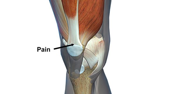
- Anterior knee pain is characterized by chronic pain over the front and center of the knee joint. (bostonssc.com)
- Anterior knee pain refers to various conditions, which include runner's knee or patellar tendinitis, and chondromalacia of the patella. (bostonssc.com)
- Patient-reported knee function and anterior knee pain as assessed with the Kujala score will serve as primary outcome. (springer.com)
- The extensor mechanism of the knee begins above the hip with the origin of the rectus femoris muscle on the anterior inferior iliac spine. (medscape.com)
- Knee joint, anterior view. (medscape.com)
Joint15
- These injures will more likely than not lead to some degree of joint contractures which will reduce range of motion of the knee and ultimately lead to post-traumatic arthritis. (lifecare123.com)
- When the bones of a joint are knocked out of place, it is known as dislocation. (medicahospitals.in)
- When the bones in a joint get split or knocked out of their normal locations, it is called a dislocation. (medicahospitals.in)
- Dislocations can be excruciatingly painful and leave the affected joint unsteady or immobilized (unable to move). (medicahospitals.in)
- The patella moves over the joint and allows bending of the knee and straightening of the leg. (bostonssc.com)
- There are a few major ligaments situated around the knee joint that hold the joint firmly in position and contribute to the stability of the knee. (bostonssc.com)
- The knee is the largest joint in the body, and it is made up of many important and complex structures. (choa.org)
- When an ACL is injured, it can be partially torn or completely torn, which could leave the knee unstable and at risk for worsening joint damage. (choa.org)
- The main symptom of jumper's knee is knee pain at the front of the knee, though sometimes there may also be some swelling and joint stiffness. (choa.org)
- Kneecap dislocation damages your knee joint. (medlineplus.gov)
- The knee is more than just a simple hinged joint. (webmd.com)
- The menisci's main job is to cushion the knee joint. (webmd.com)
- All these bones are functional in the knee joint, except for the fibula. (medscape.com)
- The ligaments of the knee joint can be divided into the extracapsular ligaments and intra-articular ligaments. (medscape.com)
- For more information about the relevant anatomy, see Knee Joint Anatomy. (medscape.com)
Surgery1
- One year after surgery the patient began fielding and batting practice, with excellent left knee stability and function. (medscape.com)
Arthroscopy2
- All realignment procedures performed to treat the dislocation will first involve arthroscopy. (shanewoolfmd.com)
- Arthroscopy is a minimally invasive procedure that uses an arthroscope, a narrow lighted tube with a camera, to view the inside of the knee on a large monitor. (shanewoolfmd.com)
Structures4
- If the structures in your knee are abnormal, this can happen. (medicahospitals.in)
- Your doctor may also attempt to stretch the structures on the outside of the knee and suggest certain exercises to strengthen your muscles. (shanewoolfmd.com)
- To do all of these things and support your body while doing so, the knee relies on several structures. (webmd.com)
- Additional soft tissue structures of the knee extensor compartment consist of the infrapatellar fat pad and pretibial and prepatellar bursae. (medscape.com)
Ligaments of the knee1
- A knee dislocation results from complete ligamentous tears in at least three of the ligaments of the knee. (lifecare123.com)
Patellofemoral2
- There are non-surgical and surgical ways of treating patellofemoral dislocation. (drcoyner.com)
- This also increases the risks of patellofemoral pain syndrome (PFPS) as it creates muscular imbalances perpetuating the risk of breaststroker's knee. (swimmingworldmagazine.com)
Femoral groove1
- The ligaments on the inner and outer sides of the patella hold it in the femoral groove and avoid dislocation of the patella from the groove. (sameernagdamd.com)
Treatment7
- Make sure to understand what is happening throughout your knee dislocation treatment even if this means asking your healthcare provider questions often. (orthopaedic-surgery-md.com)
- If the infant is otherwise normal, immediate treatment with daily passive flexion movements and splinting in flexion usually results in a functional knee. (msdmanuals.com)
- Your doctor will examine your knee and suggests diagnostic tests such as X-ray, CT scan, and MRI scan to confirm condition and provide treatment. (drcoyner.com)
- Surgical treatment is recommended for those individuals who have recurrent patella dislocation. (drcoyner.com)
- So, if you are looking for the orthopaedic treatment for knee dislocation, then you should look out a professional doctor. (fairhealthfitness.com)
- Bear in mind that, treatment of the posterolateral dislocation is really complicated. (fairhealthfitness.com)
- If possible, then the individual must make contact with a doctor and grab orthopaedic treatment for knee dislocation. (fairhealthfitness.com)
Tibia2
- The knee-cap, in a way, connects the muscles in the front of the thigh to the shinbone (tibia). (shoulder-surgeon.org)
- caused by accidents, such as a blow to the proximal tibia when the knee is flexed, or if the knee is hyperextended during an accident. (patient.info)
Popliteal2
- The frequent observation of the distal pulses at regular intervals after a traumatic knee dislocation is absolutely mandatory for the early recognition and management of popliteal artery thrombosis. (bmj.com)
- A limb-threatening complication of popliteal artery thrombosis occurring in association with a palpable dorsalis pedis pulse after a trampoline-related knee dislocation is reported here to emphasise some important teaching points. (bmj.com)
Patients5
- Patients with knee dislocation should undergo radiography and MRI, as well as angiography. (medscape.com)
- Patients with knee dislocation always seek emergency care. (fairhealthfitness.com)
- Patients with recurrent patella dislocation requiring isolated MPFL reconstruction will be recruited and randomized to the dynamic or static reconstruction technique. (springer.com)
- This is necessary in recurrent patella dislocations and in particular cases of first patella dislocations such as patients with severe anatomical risk factors [ 15 ]. (springer.com)
- A clinical knee examination is the first step to be performed for patients with complaints of the knee, after taking a thorough patient history. (medscape.com)
Groove3
- In a normal knee, the kneecap fits nicely in the groove. (shoulder-surgeon.org)
- But if the groove is uneven or too shallow, the kneecap could slide off, resulting in a partial or complete dislocation. (shoulder-surgeon.org)
- If it has happened recently, it is important to make sure that the knee cap has returned to its normal position in the groove. (shoulder-surgeon.org)
Patient4
- In an elderly patient, a total knee arthroplasty is a less common option. (medscape.com)
- The tenderest point of the knee should be examined last to prevent a guarding reaction from the patient due to pain. (medscape.com)
- The patient should be asked to take off shoes, socks, and pants in order to get a good view of the knee and bony reference points of the pelvis. (medscape.com)
- Patient preparation for knee examination. (medscape.com)
Kneecap slides1
- Patellar dislocation occurs when the kneecap slides out of the trochlea. (shanewoolfmd.com)
Stabilize1
- If it is stuck and painful to move, stabilize (splint) the knee and get medical attention. (medlineplus.gov)
Side of the knee1
- Your surgeon may also perform a procedure to realign the quadriceps mechanism by tightening the tendons on the inside or medial side of the knee. (sameernagdamd.com)
Unstable1
- Both knees were unstable in all direction. (panafrican-med-journal.com)
Relocate2
- After adequate sedation, longitudinal traction will relocate the majority of knee dislocations. (medscape.com)
- If it is, your provider will reduce (relocate) your dislocation. (medlineplus.gov)
Flexion1
- The knees, too, have flexion contractures. (medscape.com)
MPFL1
- After initial patellar dislocation, the MPFL is injured in 94% of the cases [ 11 ]. (springer.com)
Total knee arthroplasty1
- Dislocation of the polyethylene liner is a rare complication of fixed-bearing total knee arthroplasty with only a few cases described in the literature. (orthocasereports.com)