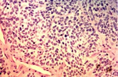
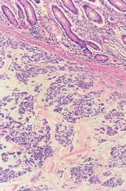
Leiomyosarcoma
Vascular Neoplasms
Smooth Muscle Tumor
Leiomyoma
Soft Tissue Neoplasms
Sarcoma
Vena Cava, Inferior
Retroperitoneal Neoplasms
Muscle Neoplasms
Heart Neoplasms
Mesenchymoma
Spermatic Cord
Neoplasms, Vascular Tissue
Liposarcoma
Frequent loss of heterozygosity for chromosome 10 in uterine leiomyosarcoma in contrast to leiomyoma. (1/527)
Distinction of malignant uterine leiomyosarcomas from benign leiomyomas by morphological criteria is not always possible. Leiomyosarcomas typically have complex cytogenetic abnormalities; in contrast, leiomyomas have simple or no cytogenetic abnormalities. To understand better the biological distinction(s) between these tumors, we analyzed two other potential markers of genomic instability, loss of heterozygosity (LOH) and microsatellite instability. We examined archival materials from 16 leiomyosarcomas and 13 benign leiomyomas by polymerase chain reaction for 26 microsatellite polymorphisms. Markers were selected based on previous reports of cytogenetic or molecular genetic abnormalities in leiomyosarcomas or leiomyomas and surveyed chromosomes 7, 9, 10, 11, 12, 14, 15, 16, 18, 21, and X. LOH for markers on chromosomes 15, 18, 21, and X was infrequent in leiomyosarcomas (1 of 6 tumors for each chromosome) and not observed for markers on chromosomes 7, 9, 11, 12, 14, or 16. Interestingly, 8 of 14 (57.2%) informative leiomyosarcomas had LOH for at least one marker on chromosome 10 and involved both chromosomal arms in 45.5% (5 of 11). In contrast to leiomyosarcomas, LOH for chromosome 10 was not found in 13 benign leiomyomas. Microsatellite instability was found infrequently in leiomyosarcomas and not detected in leiomyoma. Clinicopathological features (eg, atypia, necrosis, and clinical outcome) did not appear to correlate with LOH for chromosome 10. In contrast to other chromosomes studied, LOH on chromosome 10 was frequent in leiomyosarcomas and absent in benign leiomyomas. (+info)Morphological, histochemical, immunohistochemical, and ultrastructural characterization of tumors and dysplastic and non-neoplastic lesions arising in BK virus/tat transgenic mice. (2/527)
To study the role in AIDS pathogenesis of the human immunodeficiency virus type 1 (HIV-1) Tat protein, a transactivator of viral and cellular genes, we generated transgenic mice with a recombinant DNA containing BK virus (BKV) early region and the HIV-1 tat gene, directed by its own promoter-enhancer. DNA hybridization revealed that the transgene is stably maintained in all organs of transgenic mice as a tandem insertion in a number of copies ranging from 5 to 20 per cell. In addition, tat and BKV RNA were expressed in all tissues. Transgenic mice developed three types of lesions: 1) tumors, 2) hyperplastic and dysplastic lesions, and 3) non-neoplastic lesions. Tumors of different histotypes, such as lymphomas, adenocarcinomas of skin glands, leiomyosarcomas, skin squamous cell carcinomas, hepatomas, hepatocarcinomas, and cavernous liver hemangiomas, developed in 29% of transgenic animals. The majority of tumors were malignant, invasive, and producing metastases. Conversely, tumors of only two histotypes (lymphomas and adenocarcinomas of skin glands) appeared in control mice. Hyperplastic and dysplastic lesions were more frequent in transgenic than in control mice and involved the skin or its adnexes, the liver and the rectum, indicating multiple targets for the activity of the transgene. Pyelonephritis, frequently complicated with hydronephrosis, inflammatory eye lesions, and amyloid depositions represented the most frequent non-neoplastic lesions detected in transgenic mice. Many of the pathological findings observed in this animal model are comparable to similar lesions appearing in AIDS patients, suggesting a relevant role for Tat in the pathogenesis of such lesions during the course of AIDS. (+info)Possible role of calponin h1 as a tumor suppressor in human uterine leiomyosarcoma. (3/527)
BACKGROUND: Calponin h1, a basic actin-binding protein capable of inhibiting smooth muscle contraction, is a constitutive element of smooth muscle cells. However, in leiomyosarcoma (a type of smooth muscle neoplasm of the uterus), reduced expression of calponin h1 is observed, as we have reported previously. In this study, we sought to assess the effects (in vitro and in vivo) of increasing calponin h1 expression in leiomyosarcoma cells. METHODS: A plasmid containing a human calponin h1 complementary DNA and a bacterial neomycin-resistance gene was transfected into the human leiomyosarcoma cell lines SKN and SK-LMS-1 by electroporation. Southern blotting, reverse transcription-polymerase chain reaction analysis, western blotting, and immunohistochemistry were used to confirm DNA transfer and expression of the calponin h1 protein in neomycin-resistant clones. We characterized the morphology of calponin h1-transfected cells, and we evaluated their proliferative activity and tumorigenicity by use of a 3-(4,5-dimethylthiazol-2-yl)-2,5-diphenyl-2H-tetrazolium bromide assay, an anchorage-independent growth assay, and a nude mouse tumorigenicity assay. RESULTS: The morphology of calponin h1-transfected cells in culture resembled that of cultured normal myometrial smooth muscle cells. With SK-LMS-1 cells, proliferation of calponin h1-transfection cells was reduced to 69% of control; with SKN cells, calponin h1 transfection reduced proliferation to 70% of control. In assays of anchorage-independent growth and in vivo tumorigenicity, both growth and tumorigenicity were statistically significantly reduced in calponin h1-transfected leiomyosarcoma cells. CONCLUSIONS: Calponin h1 may function as a tumor suppressor in leiomyosarcoma. Clinically, transfer of a calponin h1 complementary DNA into poorly differentiated leiomyosarcoma cells may be of potential therapeutic value through induction of a normal, differentiated cellular phenotype. (+info)Cerebral metastasis of a uterine leiomyosarcoma--case report. (4/527)
A 38-year-old female presented with sudden neurological deterioration 6 years after an operation and chemotherapy for uterine leiomyosarcoma. An extremely rare metastasis of the uterine leiomyosarcoma to the brain was identified and totally resected. Whole brain irradiation (50 Gy) was given. A recurrence of the metastasis was resected 10 weeks later. She ultimately died of a second recurrence. Aggressive surgical management of cerebral metastasis of uterine leiomyosarcoma may achieve an improved outcome. (+info)Leiomyosarcoma of the esophagus in a patient with chagasic megaesophagus: case report and literature review. (5/527)
Leiomyosarcoma constitutes approximately 0.5% of the malignant neoplasias of the esophagus and its association with megaesophagus has not been described. We report on a case of a woman with dysphagia that was slowly progressive from the age of 19 due to chagasic megaesophagus. The woman was subjected to cardiomyotomy at the age of 49. She presented a rapid worsening of the dysphagia due to leiomyosarcoma at the age of 61, and was subjected to subtotal esophagectomy with cervical esophagogastroplasty. She developed pulmonary and hepatic metastases 14 months after surgery and died six months later. (+info)"Mesenchymal tumor" or "decidual-like reaction"? (6/527)
For more than 40 yr, an unusual urinary bladder lesion has been known to occur in certain strains of mice, but no consensus has been obtained regarding its etiology, pathogenesis, biology, or classification. The lesion was first assumed to be epithelial and non-neoplastic, then it was called a smooth muscle cell tumor or leiomyosarcoma because of ultrastructural characteristics for smooth muscle cells. Later, the nonspecific term "mesenchymal tumor" was introduced due to histomorphologic differences from all smooth muscle tumors known. Recently, a proposal was made to name it "decidual-like reaction" because of the histomorphologic similarity to the rare spontaneous decidual reaction in the uterus of aging mice. Both lesions are characterized by spindle and large pleomorphic epithelioid cells with large bizarre nuclei; these characteristics mimic anaplasia of malignant tumors and led pathologists to assume a neoplastic nature. The decidual hypothesis is supported by the regular presence of nuclear progesterone receptors, the occasional occurrence of eosinophilic cytoplasmic granules, the rare finding of cells morphologically resembling granulated metrial gland cells (all also observed in the uterine decidual reaction), and the reproducibility through long-term feeding of combinations of estrogens and progestogens. It appears that the new decidual hypothesis can explain many detailed facets of the lesion, with the exception of the reported smooth muscle cell characteristics. The controversy of "mesenchymal tumor versus decidual-like reaction" should be resolved soon, not only as a scientific issue, but also because of consequences for risk assessment. (+info)Correlation between clinicopathological features and karyotype in spindle cell sarcomas. A report of 130 cases from the CHAMP study group. (7/527)
Soft-tissue tumors have proved to be a fruitful area for the identification of reproducible cytogenetic aberrations, especially among pediatric round-cell sarcomas and lipomatous tumors. Thus far, however, data regarding sarcomas of monomorphic spindle cell type have been limited and somewhat disappointing, with the notable exception of synovial sarcoma. As part of an ongoing international collaborative study, 130 karyotyped spindle-cell sarcomas were reviewed and classified histologically, without knowledge of the clinical and karyotypic data, with the aim of identifying objective correlations between morphology, karyotype, and clinical parameters. Clonal chromosomal abnormalities were identified in 82 cases studied (63%), but only in the group of synovial sarcomas was there clear correlation between the cytogenetic findings, in the form of a consistent t(X;18)(p11;q11), and morphology. Among leiomyosarcomas (41 cases) and malignant peripheral nerve sheath tumors (MPNSTs; 27 cases) as well as in individual examples of rarer entities, there was a general tendency for karyotypic complexity associated with frequent loss or rearrangement of chromosome arms 1p, 10p, 11q, 12q, 17p, and 22q. Rearrangements of 17q (the region of the NF1 gene) were seen in 9/27 (33%) of MPNSTs. Among nine cases of solitary fibrous tumor (in which previous cytogenetic data are very limited) no consistent aberrations were identified. We conclude that, with the exception of synovial sarcoma, most spindle-cell sarcomas share with pleomorphic sarcomas the tendency for karyotypic complexity. There was no indication (in most of these lesions) that detectable cytogenetic aberrations could either facilitate their diagnosis or help to determine prognosis. There is a clear need to further study and understand the significance of multiple chromosomal abnormalities in this group of mesenchymal neoplasms with the particular goal of determining their role in the process of tumor development. (+info)Prognostic significance of bcl-2 expression in leiomyosarcoma of the uterus. (8/527)
We examined bcl-2 expression as well as p53 expression and mutation in human uterine smooth muscle tumours to determine the influence of bcl-2 expression on prognosis in patients with uterine leiomyosarcomas. bcl-2 protein was expressed in nearly all benign smooth muscle tumours but in only 57% of leiomyosarcomas. Benign smooth muscle tumours were usually negative for p53 protein, but 16 out of 21 (76%) leiomyosarcomas were positive. A p53 gene mutation was detected in nine of the 16 leiomyosarcomas that showed p53-positive staining. A significant positive correlation was observed between p53 mutation and p53 expression, between the number of mitoses and the Ki-67 labelling index, and between clinical stage and p53 mutation. A significant negative correlation was observed between bcl-2 expression and p53 mutation, and between bcl-2 expression and p53 overexpression. Univariate survival analysis revealed that bcl-2 expression, p53 mutation and clinical stage (stage 1 vs stages 2-4) all showed a significant correlation with prognosis. In a multivariate stepwise regression analysis, positive bcl-2 expression and stage 1 disease were the independent predictors of a favourable prognosis. Our results suggest that bcl-2 is frequently expressed in human uterine smooth muscle tumours, and that its expression may correlate with a favourable prognosis in patients with uterine leiomyosarcoma. (+info)Leiomyosarcoma is a type of cancer that arises from the smooth muscle cells, which are responsible for the involuntary contractions of various organs and blood vessels. It most commonly occurs in the uterus, soft tissues (such as muscles and fat), and the gastrointestinal tract.
Leiomyosarcomas can vary in their aggressiveness and may spread to other parts of the body (metastasize) through the bloodstream or lymphatic system. The prognosis for leiomyosarcoma depends on several factors, including the location and size of the tumor, the patient's age and overall health, and the extent of metastasis. Treatment typically involves surgical removal of the tumor, along with radiation therapy and/or chemotherapy to help prevent recurrence or spread of the cancer.
Vascular neoplasms are a type of tumor that develops from cells that line the blood vessels or lymphatic vessels. These tumors can be benign (non-cancerous) or malignant (cancerous). Benign vascular neoplasms, such as hemangiomas and lymphangiomas, are usually harmless and may not require treatment unless they cause symptoms or complications. Malignant vascular neoplasms, on the other hand, are known as angiosarcomas and can be aggressive, spreading to other parts of the body and potentially causing serious health problems.
Angiosarcomas can develop in any part of the body but are most commonly found in the skin, particularly in areas exposed to radiation or chronic lymph edema. They can also occur in the breast, liver, spleen, and heart. Treatment for vascular neoplasms depends on the type, location, size, and stage of the tumor, as well as the patient's overall health. Treatment options may include surgery, radiation therapy, chemotherapy, or a combination of these approaches.
Uterine neoplasms refer to abnormal growths in the uterus, which can be benign (non-cancerous) or malignant (cancerous). These growths can originate from different types of cells within the uterus, leading to various types of uterine neoplasms. The two main categories of uterine neoplasms are endometrial neoplasms and uterine sarcomas.
Endometrial neoplasms develop from the endometrium, which is the inner lining of the uterus. Most endometrial neoplasms are classified as endometrioid adenocarcinomas, arising from glandular cells in the endometrium. Other types include serous carcinoma, clear cell carcinoma, and mucinous carcinoma.
Uterine sarcomas, on the other hand, are less common and originate from the connective tissue (stroma) or muscle (myometrium) of the uterus. Uterine sarcomas can be further divided into several subtypes, such as leiomyosarcoma, endometrial stromal sarcoma, and undifferentiated uterine sarcoma.
Uterine neoplasms can cause various symptoms, including abnormal vaginal bleeding or discharge, pelvic pain, and difficulty urinating or having bowel movements. The diagnosis typically involves a combination of imaging tests (such as ultrasound, CT, or MRI scans) and tissue biopsies to determine the type and extent of the neoplasm. Treatment options depend on the type, stage, and patient's overall health but may include surgery, radiation therapy, chemotherapy, or hormone therapy.
A smooth muscle tumor refers to a growth that develops in the smooth muscles, which are involuntary muscles found in various organs and structures throughout the body, including the digestive tract, uterus, blood vessels, and bladder. These tumors can be benign (noncancerous) or malignant (cancerous).
Benign smooth muscle tumors are called leiomyomas. They are typically slow-growing and rarely spread to other parts of the body. Leiomyomas are often asymptomatic but can cause problems depending on their location. For instance, a leiomyoma in the uterus might lead to heavy menstrual periods or difficulty becoming pregnant.
Malignant smooth muscle tumors are called leiomyosarcomas. These tumors are more aggressive and have a higher risk of spreading to other parts of the body. Symptoms can vary widely depending on the location of the tumor but may include abdominal pain, bloating, or bleeding.
It's important to note that while some smooth muscle tumors can be removed surgically, others may require additional treatment such as radiation therapy or chemotherapy, especially in cases of leiomyosarcomas. Regular follow-up with a healthcare provider is essential to monitor for recurrence and manage any potential complications.
Leiomyoma is a benign (non-cancerous) tumor that originates from the smooth muscle cells. It most commonly occurs in the uterus, where it is also known as a fibroid, but can also develop in other parts of the body such as the skin, gastrointestinal tract, and genitourinary system. Leiomyomas are typically slow-growing and often cause no symptoms, although they can lead to various complications depending on their size and location. Treatment options for leiomyomas include surveillance, medication, or surgical removal.
Soft tissue neoplasms refer to abnormal growths or tumors that develop in the soft tissues of the body. Soft tissues include muscles, tendons, ligaments, fascia, nerves, blood vessels, fat, and synovial membranes (the thin layer of cells that line joints and tendons). Neoplasms can be benign (non-cancerous) or malignant (cancerous), and their behavior and potential for spread depend on the specific type of neoplasm.
Benign soft tissue neoplasms are typically slow-growing, well-circumscribed, and rarely spread to other parts of the body. They can often be removed surgically with a low risk of recurrence. Examples of benign soft tissue neoplasms include lipomas (fat tumors), schwannomas (nerve sheath tumors), and hemangiomas (blood vessel tumors).
Malignant soft tissue neoplasms, on the other hand, can grow rapidly, invade surrounding tissues, and may metastasize (spread) to distant parts of the body. They are often more difficult to treat than benign neoplasms and require a multidisciplinary approach, including surgery, radiation therapy, and chemotherapy. Examples of malignant soft tissue neoplasms include sarcomas, such as rhabdomyosarcoma (arising from skeletal muscle), leiomyosarcoma (arising from smooth muscle), and angiosarcoma (arising from blood vessels).
It is important to note that soft tissue neoplasms can occur in any part of the body, and their diagnosis and treatment require a thorough evaluation by a healthcare professional with expertise in this area.
Sarcoma is a type of cancer that develops from certain types of connective tissue (such as muscle, fat, fibrous tissue, blood vessels, or nerves) found throughout the body. It can occur in any part of the body, but it most commonly occurs in the arms, legs, chest, and abdomen.
Sarcomas are classified into two main groups: bone sarcomas and soft tissue sarcomas. Bone sarcomas develop in the bones, while soft tissue sarcomas develop in the soft tissues of the body, such as muscles, tendons, ligaments, fat, blood vessels, and nerves.
Sarcomas can be further classified into many subtypes based on their specific characteristics, such as the type of tissue they originate from, their genetic makeup, and their appearance under a microscope. The different subtypes of sarcoma have varying symptoms, prognoses, and treatment options.
Overall, sarcomas are relatively rare cancers, accounting for less than 1% of all cancer diagnoses in the United States each year. However, they can be aggressive and may require intensive treatment, such as surgery, radiation therapy, and chemotherapy.
The inferior vena cava (IVC) is the largest vein in the human body that carries deoxygenated blood from the lower extremities, pelvis, and abdomen to the right atrium of the heart. It is formed by the union of the left and right common iliac veins at the level of the fifth lumbar vertebra. The inferior vena cava is a retroperitoneal structure, meaning it lies behind the peritoneum, the lining that covers the abdominal cavity. It ascends through the posterior abdominal wall and passes through the central tendon of the diaphragm to enter the thoracic cavity.
The inferior vena cava is composed of three parts:
1. The infrarenal portion, which lies below the renal veins
2. The renal portion, which receives blood from the renal veins
3. The suprahepatic portion, which lies above the liver and receives blood from the hepatic veins before draining into the right atrium of the heart.
The inferior vena cava plays a crucial role in maintaining venous return to the heart and contributing to cardiovascular function.
Genital neoplasms in males refer to abnormal growths or tumors that develop in the male reproductive organs. These can be benign (non-cancerous) or malignant (cancerous).
Malignant genital neoplasms are often referred to as genital cancers. The most common types of male genital cancers include:
1. Penile Cancer: This occurs when cancer cells form in the tissues of the penis.
2. Testicular Cancer: This forms in the testicles (testes), which are located inside the scrotum.
3. Prostate Cancer: This is a common cancer in men, forming in the prostate gland, which is part of the male reproductive system that helps make semen.
4. Scrotal Cancer: This is a rare form of cancer that forms in the skin or tissue of the scrotum.
5. Penile Intraepithelial Neoplasia (PeIN): This is not cancer, but it is considered a pre-cancerous condition of the penis.
Early detection and treatment of genital neoplasms can significantly improve the prognosis. Regular self-examinations and medical check-ups are recommended, especially for individuals with risk factors such as smoking, HIV infection, or a family history of these cancers.
Retroperitoneal neoplasms refer to abnormal growths or tumors that develop in the retroperitoneal space. This is the area located behind the peritoneum, which is the membrane that lines the abdominal cavity and covers the abdominal organs. The retroperitoneal space contains several vital structures such as the kidneys, adrenal glands, pancreas, aorta, and lymphatic vessels.
Retroperitoneal neoplasms can be benign or malignant (cancerous). Malignant retroperitoneal neoplasms are often aggressive and can invade surrounding tissues and organs, leading to various complications. Common types of retroperitoneal neoplasms include lymphomas, sarcomas, and metastatic tumors from other primary sites. Symptoms may vary depending on the size and location of the tumor but can include abdominal or back pain, weight loss, and swelling in the legs. Diagnosis typically involves imaging studies such as CT scans or MRI, followed by a biopsy to determine the type and grade of the tumor. Treatment options may include surgery, radiation therapy, chemotherapy, or a combination of these approaches.
Muscle neoplasms are abnormal growths or tumors that develop in the muscle tissue. They can be benign (non-cancerous) or malignant (cancerous). Benign muscle neoplasms are typically slow-growing and do not spread to other parts of the body, while malignant muscle neoplasms, also known as soft tissue sarcomas, can grow quickly, invade nearby tissues, and metastasize (spread) to distant parts of the body.
Soft tissue sarcomas can arise from any of the muscles in the body, including the skeletal muscles (voluntary muscles that attach to bones and help with movement), smooth muscles (involuntary muscles found in the walls of blood vessels, digestive tract, and other organs), or cardiac muscle (the specialized muscle found in the heart).
There are many different types of soft tissue sarcomas, each with its own set of characteristics and prognosis. Treatment for muscle neoplasms typically involves a combination of surgery, radiation therapy, and chemotherapy, depending on the type, size, location, and stage of the tumor.
Heart neoplasms are abnormal growths or tumors that develop within the heart tissue. They can be benign (noncancerous) or malignant (cancerous). Benign tumors, such as myxomas and rhabdomyomas, are typically slower growing and less likely to spread, but they can still cause serious complications if they obstruct blood flow or damage heart valves. Malignant tumors, such as angiosarcomas and rhabdomyosarcomas, are fast-growing and have a higher risk of spreading to other parts of the body. Symptoms of heart neoplasms can include shortness of breath, chest pain, fatigue, and irregular heart rhythms. Treatment options depend on the type, size, and location of the tumor, and may include surgery, radiation therapy, or chemotherapy.
Mesenchymoma is a very rare type of tumor that contains a mixture of different types of mesenchymal tissues, such as muscle, fat, bone, cartilage, or fibrous tissue. It typically occurs in children and young adults, and can be found in various parts of the body, including the head, neck, retroperitoneum (the area behind the abdominal cavity), and the limbs.
Mesenchymomas are usually slow-growing and may not cause any symptoms until they reach a large size. Treatment typically involves surgical removal of the tumor, but radiation therapy or chemotherapy may also be used in some cases. The prognosis for mesenchymoma depends on several factors, including the location and size of the tumor, the patient's age and overall health, and the specific types of tissue that are present in the tumor.
The spermatic cord is a fibrous structure that contains the vas deferens, blood vessels, nerves, and lymphatics, which provide passage for these structures between the abdomen and the scrotum in males. It is covered by several layers of protective sheaths, including the internal spermatic fascia, cremasteric fascia, and external spermatic fascia. The spermatic cord allows the testicles to be located outside the body, which helps maintain a cooler temperature for optimal sperm production.
A neoplasm of vascular tissue is an abnormal growth or mass of cells in the blood vessels or lymphatic vessels. These growths can be benign (non-cancerous) or malignant (cancerous). Benign neoplasms, such as hemangiomas and lymphangiomas, are typically not harmful and may not require treatment. However, they can cause symptoms if they grow large enough to press on nearby organs or tissues. Malignant neoplasms, such as angiosarcomas, are cancerous and can invade and destroy surrounding tissue, as well as spread (metastasize) to other parts of the body. Treatment for vascular tissue neoplasms depends on the type, size, location, and stage of the growth, and may include surgery, radiation therapy, chemotherapy, or a combination of these.
The renal veins are a pair of large veins that carry oxygen-depleted blood and waste products from the kidneys to the inferior vena cava, which is the largest vein in the body that returns blood to the heart. The renal veins are formed by the union of several smaller veins that drain blood from different parts of the kidney.
In humans, the right renal vein is shorter and passes directly into the inferior vena cava, while the left renal vein is longer and passes in front of the aorta before entering the inferior vena cava. The left renal vein also receives blood from the gonadal (testicular or ovarian) veins, suprarenal (adrenal) veins, and the lumbar veins.
It is important to note that the renal veins are vulnerable to compression by surrounding structures, such as the overlying artery or a tumor, which can lead to renal vein thrombosis, a serious condition that requires prompt medical attention.
Liposarcoma is a type of soft tissue sarcoma, which is a cancer that develops in the soft tissues of the body, such as fat, muscle, nerves, blood vessels, and fibrous tissues. Specifically, liposarcoma arises from fat cells (adipocytes) or their precursors.
There are several subtypes of liposarcoma, which differ in their appearance under the microscope, genetic features, and clinical behavior. These include well-differentiated, dedifferentiated, myxoid, round cell, and pleomorphic liposarcomas. The most common sites for liposarcoma are the thigh, retroperitoneum (the area behind the abdominal cavity), and the buttock.
Liposarcomas can grow slowly or rapidly, and they may spread to other parts of the body (metastasize) through the bloodstream or lymphatic system. Treatment typically involves surgical removal of the tumor, often followed by radiation therapy and/or chemotherapy. The prognosis for liposarcoma depends on several factors, including the type and grade of the tumor, its size and location, and whether it has spread to other parts of the body.
Skull neoplasms refer to abnormal growths or tumors that develop within the skull. These growths can be benign (non-cancerous) or malignant (cancerous). They can originate from various types of cells, such as bone cells, nerve cells, or soft tissues. Skull neoplasms can cause various symptoms depending on their size and location, including headaches, seizures, vision problems, hearing loss, and neurological deficits. Treatment options include surgery, radiation therapy, and chemotherapy. It is important to note that a neoplasm in the skull can also refer to metastatic cancer, which has spread from another part of the body to the skull.
Jejunal neoplasms refer to abnormal growths or tumors in the jejunum, which is the middle section of the small intestine. These neoplasms can be benign (non-cancerous) or malignant (cancerous). Malignant jejunal neoplasms are often aggressive and can spread to other parts of the body, making them potentially life-threatening.
There are several types of jejunal neoplasms, including:
1. Adenocarcinomas: These are cancerous tumors that develop from the glandular cells lining the jejunum. They are the most common type of jejunal neoplasm.
2. Carcinoid tumors: These are slow-growing neuroendocrine tumors that arise from the hormone-producing cells in the jejunum. While they are usually benign, some can become malignant and spread to other parts of the body.
3. Gastrointestinal stromal tumors (GISTs): These are rare tumors that develop from the connective tissue cells in the jejunum. They can be benign or malignant.
4. Lymphomas: These are cancerous tumors that develop from the immune system cells in the jejunum. They are less common than adenocarcinomas but can be aggressive and spread to other parts of the body.
5. Sarcomas: These are rare cancerous tumors that develop from the connective tissue cells in the jejunum. They can be aggressive and spread to other parts of the body.
Symptoms of jejunal neoplasms may include abdominal pain, bloating, diarrhea, weight loss, and bleeding in the stool. Treatment options depend on the type and stage of the neoplasm but may include surgery, chemotherapy, radiation therapy, or a combination of these approaches.
Malignant fibrous histiocytoma (MFH) is not a specific type of histiocytoma; rather, it is a type of soft tissue sarcoma. Histiocytomas are benign tumors that arise from cells called histiocytes, which are part of the immune system. MFH, on the other hand, is a malignant (cancerous) tumor that can arise in various types of soft tissues, such as muscle, fat, tendons, and ligaments.
MFH was once thought to originate from histiocytes, but more recent research suggests that it may actually arise from undifferentiated mesenchymal cells, which are capable of developing into a variety of different cell types. MFH is the most common type of soft tissue sarcoma in adults over the age of 50 and typically presents as a painless mass in the extremities or retroperitoneum (the area in the back of the abdomen).
The tumor is characterized by the presence of fibroblastic and histiocytic-like cells, which can be quite pleomorphic (varied in shape and size) and may contain numerous mitotic figures (indicating rapid cell division). Treatment typically involves surgical excision, often followed by radiation therapy and/or chemotherapy. The prognosis for MFH depends on several factors, including the tumor's location, size, grade (degree of differentiation), and the patient's age and overall health.
 Leiomyosarcoma