Leukocytes
Neutrophils
Leukocytes, Mononuclear
Chemotaxis, Leukocyte
Phagocytosis
Leukocyte Count
N-Formylmethionine Leucyl-Phenylalanine
P-Selectin
Monocytes
Venules
Antigens, CD18
Inflammation
Blood Bactericidal Activity
Phagocyte Bactericidal Dysfunction
Cells, Cultured
Lymphocytes
Neutrophil Activation
Flow Cytometry
L-Selectin
Granulocytes
Zymosan
Leukotriene B4
Chemotactic Factors
Intercellular Adhesion Molecule-1
Macrophages
Endothelium, Vascular
Luminescent Measurements
Mice, Inbred C57BL
Cell Adhesion Molecules
Opsonin Proteins
Selectins
Leukocyte Transfusion
Peroxidase
Rabbits
Oxyquinoline
Macrophage-1 Antigen
Arachidonate Lipoxygenases
Superoxides
N-Formylmethionine
E-Selectin
Arachidonate 5-Lipoxygenase
Luminol
Blood Platelets
Respiratory Burst
Antigens, CD
Chemokines
Cytokines
Tumor Necrosis Factor-alpha
RNA, Messenger
Lipopolysaccharides
Molecular Sequence Data
Complement C5a
Interleukin-8
Indium
Disease Models, Animal
Mice, Knockout
T-Lymphocytes
Cell Migration Assays, Leukocyte
Cell Separation
Platelet Activating Factor
Complement C5
Eosinophils
Nitroblue Tetrazolium
Neutrophil Infiltration
Mice, Inbred BALB C
HL-60 Cells
Vascular Cell Adhesion Molecule-1
Leukotriene A4
Lymphocyte Function-Associated Antigen-1
Erythrocytes
Cell Communication
Lung
Peritonitis
Blood Proteins
Receptors, Chemokine
Peroxidases
Leukotrienes
Receptors, Formyl Peptide
Chemotaxis
Granulomatous Disease, Chronic
Arachidonic Acids
Microscopy, Video
Endothelial Cells
Pancreatic Elastase
Muramidase
Chediak-Higashi Syndrome
Tetradecanoylphorbol Acetate
Hydrogen Peroxide
Antigens, CD11
Polymerase Chain Reaction
Base Sequence
Exudates and Transudates
Chemokine CCL2
Microscopy, Electron
Hydroxyeicosatetraenoic Acids
Amino Acid Sequence
Endotoxins
Interleukin-1
Capillary Permeability
Integrin alpha4
Cell Migration Inhibition
Glucuronidase
Lactoferrin
Lipoxins
Leukopenia
Cell Membrane
Inflammation Mediators
Complement System Proteins
Antigens, CD31
Transendothelial and Transepithelial Migration
Cattle
Skin
Calcimycin
Chemokines, CXC
Guinea Pigs
Antigens, CD11b
Endothelium
Immunohistochemistry
Signal Transduction
Gene Expression Regulation
Leukocyte Adherence Inhibition Test
Reverse Transcriptase Polymerase Chain Reaction
Receptors, Leukocyte-Adhesion
Lymphocyte Activation
Cell Aggregation
Hexosephosphates
Antigens, CD45
Dose-Response Relationship, Drug
Acridine Orange
Peritoneal Cavity
Mannheimia haemolytica
Technetium Tc 99m Exametazime
Immune Sera
Chemokine CXCL1
Gene Expression
Rats, Inbred Lew
Bone Marrow Cells
Bone Marrow
Abscess
Focal Infection
Antigens, Differentiation, Myelomonocytic
Cell Count
Receptors, Complement
Antigens, CD15
Receptors, Cell Surface
Up-Regulation
Latex
Reperfusion Injury
Immunity, Innate
Escherichia coli
Colchicine
Antigens, CD53
Integrin alpha4beta1
Interferon-gamma
Basophils
Killer Cells, Natural
Leukostasis
Cytomegalovirus Infections
Staphylococcus aureus
Hydroxyquinolines
Arachidonic Acid
Antigens, CD43
Defensins
Lipoxygenase
Chemokine CCL5
Pleurisy
Integrins
Enzyme-Linked Immunosorbent Assay
Receptors, Leukotriene B4
Phagocytic acitivity of bovine leukocytes during pregnancy. (1/9769)
The phagocytic competence, measured as the total number of polymorphonuclear leukocytes per mm3 which phagocytosed Staphylococcus aureus, strain 321, in vitro, was determined in eight cows during complete pregnancies. Such leukocytes are referred to as "Active PMN'S". There was a gradual decline in the number of these cells from conception to a minimum between the 16th and 20th weeks of pregnancy, followed by a steady increase to the cessation of lactation when a marked drop occurred, after which there was an increase to a maximun during the second week prepartum. From this maximum there was a rapid decrease to an absolute minimum during the first week after parturition. From the second week postpartum there was a gradual increase to conception. The correlation coefficient (r) of number of active PMN'S with time before conception was -0.474 )p-0.01). There were significant differences (p=0.01) in numbers of active PMNS Among the eight cows. It was found that the cows fell into two groups, one whose members had, overall, significantly more active PMNs (p=0.001) than those in the second group. The between cow differences may have been due to 1) age, since the cows with the highest numbers of circulating active PMNs were younger than those in the other group of 2) the combined stress of pregnancy and lactation, as those cows which were both pregnant and milking had the lowest numbers of active PMNs. (+info)Blocking very late antigen-4 integrin decreases leukocyte entry and fatty streak formation in mice fed an atherogenic diet. (2/9769)
Atherosclerotic lesion development is characterized by the recruitment of leukocytes, principally monocytes, to the vessel wall. Considerable interest has been focused on the adhesion molecule(s) involved in leukocyte/endothelial interactions. The goal of the present study was to determine the role of the very late antigen-4 (VLA-4) integrin/ligand interaction in fatty streak development using murine models. Because alpha4 null mice are not viable, a peptidomimetic was used to block VLA-4-mediated leukocyte binding. The ability of a synthetic peptidomimetic of connecting segment-1 (CS-1 peptide) to block the recruitment of leukocytes and the accumulation of lipid in the aortic sinus of either wild-type mice (strain C57BL/6J) or mice with a low-density lipoprotein null mutation (LDLR-/-) maintained on an atherogenic diet was assessed. The active (Ac) CS-1 peptide or scrambled (Sc) CS-1 peptide was delivered subcutaneously into mice using a mini osmotic pump. Mice were exposed to the peptide for 24 to 36 hours before the onset of the atherogenic diet. In C57BL/6J mice, leukocyte entry into the aortic sinus, as assessed by en face preparations, was inhibited by the active peptide (Ac=28+/-4, Sc=54+/-6 monocytes/valve; P=0.004). Additionally, frozen sections stained with Oil Red O were analyzed to assess lipid accumulation in the aortic sinus. C57BL/6J mice that received the (Ac) compound demonstrated significantly reduced lesion areas as compared with mice that received the (Sc) peptide (Ac=4887+/-4438 microm2, Sc=15 009 +/-5619 microm2; P<0.0001). In a separate study, LDLR-/- mice were implanted with pumps containing either the (Ac) or (Sc) peptide before initiation of the atherogenic diet. Because LDLR-/- mice fed a chow diet displayed small lesions at 14 weeks, the effects of the peptide seen in these animals represented a change in early lipid accumulation rather than initiation. By using whole-mount preparations, the (Ac) but not the (Sc) peptide significantly reduced the area of lipid accumulation in the aortic sinus, resulting in an approximate 66% decrease. Plasma analysis from all studies revealed concentrations of peptide to be present at levels previously determined by in vitro analysis to block adhesion. (Ac) CS-1 peptide, which blocks VLA-4 on the leukocyte surface, is effective in reducing leukocyte recruitment and lipid accumulation in the aortic sinus. The present study provides in vivo evidence that the VLA-4 integrin plays an important role in the initiation of the atherosclerotic lesion and lipid accumulation, and it suggests a potential therapeutic strategy for this disease. (+info)Inhibition of L-selectin-mediated leukocyte rolling by synthetic glycoprotein mimics. (3/9769)
Synthetic carbohydrate and glycoprotein mimics displaying sulfated saccharide residues have been assayed for their L-selectin inhibitory properties under static and flow conditions. Polymers displaying the L-selectin recognition epitopes 3',6-disulfo Lewis x(Glc) (3-O-SO3-Galbeta1alpha4(Fucalpha1alpha3)-6-O-SO3-Glcbeta+ ++-OR) and 3',6'-disulfo Lewis x(Glc) (3, 6-di-O-SO3-Galbeta1alpha4(Fucalpha1alpha3)Glcbeta-OR) both inhibit L-selectin binding to heparin under static, cell-free binding conditions with similar efficacies. Under conditions of shear flow, however, only the polymer displaying 3',6-disulfo Lewis x(Glc) inhibits the rolling of L-selectin-transfected cells on the glycoprotein ligand GlyCAM-1. Although it has been shown to more effective than sialyl Lewis x at blocking the L-selectin-GlyCAM-1 interaction in static binding studies, the corresponding monomer had no effect in the dynamic assay. These data indicate that multivalent ligands are far more effective inhibitors of L-selectin-mediated rolling than their monovalent counterparts and that the inhibitory activities are dependent on the specific sulfation pattern of the recognition epitope. Importantly, our results indicate the L-selectin specificity for one ligand over another found in static, cell-free binding assays is not necessarily retained under the conditions of shear flow. The results suggest that monovalent or polyvalent carbohydrate or glycoprotein mimetics that inhibit selectin binding in static assays may not block the more physiologically relevant process of selectin-mediated rolling. (+info)Changes in haematological parameters and iron metabolism associated with a 1600 kilometre ultramarathon. (4/9769)
OBJECTIVE: To investigate haematological variations and iron related changes in the serum of participants in a 1600 kilometre ultramarathon run. PARTICIPANTS: Seven male and two female participants in a 1600 km foot race. METHODS: Blood samples were obtained from the participants before, after four and 11 days of running, and at the end of the event. Samples were analysed by standard methods for haemoglobin, packed cell volume, total red cell count, mean red cell volume, mean red cell haemoglobin, total white cell count and differential, platelets, reticulocytes, iron, ferritin, total iron binding capacity, percentage transferrin saturation, haptoglobin, and bilirubin and corrected for changes in plasma volume. RESULTS: The following variables decreased during the event (p < 0.05): haemoglobin, packed cell volume, mean red cell volume, percentage lymphocytes, percentage monocytes, serum iron, total iron binding capacity, and percentage transferrin saturation. Increases (p < 0.05) were found in plasma volume, total red cell count (day 4 only), total white cell count, percentage and absolute numbers of neutrophils and reticulocytes, absolute numbers of lymphocytes and monocytes (day 4 only), absolute numbers of eosinophils (day 11 and race end), absolute numbers of basophils (race end only), platelets, ferritin, haptoglobin, and bilirubin (day 4 only). CONCLUSION: Ultramarathon running is associated with a wide range of changes in haematological parameters, many of which are related to the normal acute phase response to injury. These should not be confused with indicators of disease. (+info)Phenotypic and functional studies of leukocytes in human endometrium and endometriosis. (5/9769)
The aetiology of endometriosis, a common and disabling disorder, is presently unknown, although immune dysfunction could allow ectopic endometrial fragments to survive outside the uterine cavity. These studies investigate the relationship between leukocyte populations, steroid hormone receptor expression, proliferative activity, bcl-2 expression and apoptosis in eutopic and ectopic endometrium from women with endometriosis or adenomyosis at different phases of the menstrual cycle. Significantly increased oestrogen receptor expression, bcl-2 expression and numbers of CD8+ leukocytes were found in ectopic compared with eutopic endometrium in endometriosis, and CD56+ endometrial granulated lymphocytes (eGLs) were significantly reduced in ectopic endometrium. Apoptotic cells were rarely found in control and subject endometria. In contrast with endometriosis, adenomyotic lesions showed identical steroid hormone receptor expression, proliferative activity, bcl-2 expression and leukocyte subpopulations to eutopic endometrium, indicating different aetiologies for these disorders. The unusual CD56+ CD16- eGLs present in large numbers in late secretory phase eutopic endometrium were highly purified (>98%) by immunomagnetic separation. Except for a negligible cytotoxic activity of eGLs from early proliferative samples, cytotoxic activity of eGLs from non-pregnant endometrium during the menstrual cycle was comparable with those in peripheral blood, predominantly CD56+ CD16+ natural killer cells. eGLs from non-pregnant endometrium and early pregnancy showed a variable proliferative response to 5 and 100 U/ml interleukin-2 over 48-h and 120-h time courses. eGLs are evidently functionally important in the eutopic endometrium. Their absence in endometriotic lesions together with increased CD+8 T-cell numbers and increased oestrogen receptor and bcl-2 expression may have significant effects on the development and progression of endometriosis. (+info)Differential expression and phosphorylation of CTCF, a c-myc transcriptional regulator, during differentiation of human myeloid cells. (6/9769)
CTCF is a transcriptional repressor of the c-myc gene. Although CTCF has been characterized in some detail, there is very little information about the regulation of CTCF activity. Therefore we investigated CTCF expression and phosphorylation during induced differentiation of human myeloid leukemia cells. We found that: (i) both CTCF mRNA and protein are down-regulated during terminal differentiation in most cell lines tested; (ii) CTCF down-regulation is retarded and less pronounced than that of c-myc; (iii) CTCF protein is differentially phosphorylated and the phosphorylation profiles depend on the differentiation pathway. We concluded that CTCF expression and activity is controlled at transcriptional and post-transcriptional levels. (+info)Identification of a novel activation-inducible protein of the tumor necrosis factor receptor superfamily and its ligand. (7/9769)
Among members of the tumor necrosis factor receptor (TNFR) superfamily, 4-1BB, CD27, and glucocorticoid-induced tumor necrosis factor receptor family-related gene (GITR) share a striking homology in the cytoplasmic domain. Here we report the identification of a new member, activation-inducible TNFR family member (AITR), which belongs to this subfamily, and its ligand. The receptor is expressed in lymph node and peripheral blood leukocytes, and its expression is up-regulated in human peripheral mononuclear cells mainly after stimulation with anti-CD3/CD28 monoclonal antibodies or phorbol 12-myristate 13-acetate/ionomycin. AITR associates with TRAF1 (TNF receptor-associated factor 1), TRAF2, and TRAF3, and induces nuclear factor (NF)-kappaB activation via TRAF2. The ligand for AITR (AITRL) was found to be an undescribed member of the TNF family, which is expressed in endothelial cells. Thus, AITR and AITRL seem to be important for interactions between activated T lymphocytes and endothelial cells. (+info)A sialoglycoprotein, gp20, of the human capacitated sperm surface is a homologue of the leukocyte CD52 antigen: analysis of the effect of anti-CD52 monoclonal antibody (CAMPATH-1) on capacitated spermatozoa. (8/9769)
In this study we performed N-terminal sequence analysis of gp20, a 20 kDa sialoglycoprotein on the human sperm surface previously identified by radiolabelling of the sialic acid residues of sperm surface. We found 100% identity with the N-terminus of CD52, an antigen expressed on almost all human leukocytes. We also show that, like CD52, gp20 behaves as a glycosylphosphatidylinositol (GPI)-anchored protein and that anti-gp20 antiserum reacts with an antigen on leukocytes of the same molecular weight as CD52. Using CAMPATH-1, the monoclonal antibody against CD52, in fluorescent staining of capacitated spermatozoa, Western blot analysis and the zona-free hamster egg penetration test, we found that the effect of this antibody was different from that of our anti-gp20. Western blot analysis revealed a well-defined 20 kDa band with anti-gp20, whereas a 14-20 kDa band was detected with CAMPATH-1. Anti-gp20 stained the equatorial region of the sperm head, whereas CAMPATH-1 stained the tail in immunofluorescence analysis of capacitated spermatozoa. A dose-dependent inhibitory effect was seen with CAMPATH-1, similar to that previously detected with anti-gp20, in a zona-free hamster egg penetration test. However, with CAMPATH-1 agglutination of motile spermatozoa was detected, and this was not present with anti-gp20. This suggests that the epitopes recognized by the two antibodies are different. (+info)Leukocytes, also known as white blood cells (WBCs), are a crucial component of the human immune system. They are responsible for protecting the body against infections and foreign substances. Leukocytes are produced in the bone marrow and circulate throughout the body in the bloodstream and lymphatic system.
There are several types of leukocytes, including:
1. Neutrophils - These are the most abundant type of leukocyte and are primarily responsible for fighting bacterial infections. They contain enzymes that can destroy bacteria.
2. Lymphocytes - These are responsible for producing antibodies and destroying virus-infected cells, as well as cancer cells. There are two main types of lymphocytes: B-lymphocytes and T-lymphocytes.
3. Monocytes - These are the largest type of leukocyte and help to break down and remove dead or damaged tissues, as well as microorganisms.
4. Eosinophils - These play a role in fighting parasitic infections and are also involved in allergic reactions and inflammation.
5. Basophils - These release histamine and other chemicals that cause inflammation in response to allergens or irritants.
An abnormal increase or decrease in the number of leukocytes can indicate an underlying medical condition, such as an infection, inflammation, or a blood disorder.
Neutrophils are a type of white blood cell that are part of the immune system's response to infection. They are produced in the bone marrow and released into the bloodstream where they circulate and are able to move quickly to sites of infection or inflammation in the body. Neutrophils are capable of engulfing and destroying bacteria, viruses, and other foreign substances through a process called phagocytosis. They are also involved in the release of inflammatory mediators, which can contribute to tissue damage in some cases. Neutrophils are characterized by the presence of granules in their cytoplasm, which contain enzymes and other proteins that help them carry out their immune functions.
Mononuclear leukocytes are a type of white blood cells (leukocytes) that have a single, large nucleus. They include lymphocytes (B-cells, T-cells, and natural killer cells), monocytes, and dendritic cells. These cells play important roles in the body's immune system, including defending against infection and disease, and participating in immune responses and surveillance. Mononuclear leukocytes can be found in the bloodstream as well as in tissues throughout the body. They are involved in both innate and adaptive immunity, providing specific and nonspecific defense mechanisms to protect the body from harmful pathogens and other threats.
Chemotaxis, Leukocyte is the movement of leukocytes (white blood cells) towards a higher concentration of a particular chemical substance, known as a chemotactic factor. This process plays a crucial role in the immune system's response to infection and injury.
When there is an infection or tissue damage, certain cells release chemotactic factors, which are small molecules or proteins that can attract leukocytes to the site of inflammation. Leukocytes have receptors on their surface that can detect these chemotactic factors and move towards them through a process called chemotaxis.
Once they reach the site of inflammation, leukocytes can help eliminate pathogens or damaged cells by phagocytosis (engulfing and destroying) or releasing toxic substances that kill the invading microorganisms. Chemotaxis is an essential part of the immune system's defense mechanisms and helps to maintain tissue homeostasis and prevent the spread of infection.
Phagocytosis is the process by which certain cells in the body, known as phagocytes, engulf and destroy foreign particles, bacteria, or dead cells. This mechanism plays a crucial role in the immune system's response to infection and inflammation. Phagocytes, such as neutrophils, monocytes, and macrophages, have receptors on their surface that recognize and bind to specific molecules (known as antigens) on the target particles or microorganisms.
Once attached, the phagocyte extends pseudopodia (cell extensions) around the particle, forming a vesicle called a phagosome that completely encloses it. The phagosome then fuses with a lysosome, an intracellular organelle containing digestive enzymes and other chemicals. This fusion results in the formation of a phagolysosome, where the engulfed particle is broken down by the action of these enzymes, neutralizing its harmful effects and allowing for the removal of cellular debris or pathogens.
Phagocytosis not only serves as a crucial defense mechanism against infections but also contributes to tissue homeostasis by removing dead cells and debris.
A leukocyte count, also known as a white blood cell (WBC) count, is a laboratory test that measures the number of leukocytes in a sample of blood. Leukocytes are a vital part of the body's immune system and help fight infection and inflammation. A high or low leukocyte count may indicate an underlying medical condition, such as an infection, inflammation, or a bone marrow disorder. The normal range for a leukocyte count in adults is typically between 4,500 and 11,000 cells per microliter (mcL) of blood. However, the normal range can vary slightly depending on the laboratory and the individual's age and sex.
Cell adhesion refers to the binding of cells to extracellular matrices or to other cells, a process that is fundamental to the development, function, and maintenance of multicellular organisms. Cell adhesion is mediated by various cell surface receptors, such as integrins, cadherins, and immunoglobulin-like cell adhesion molecules (Ig-CAMs), which interact with specific ligands in the extracellular environment. These interactions lead to the formation of specialized junctions, such as tight junctions, adherens junctions, and desmosomes, that help to maintain tissue architecture and regulate various cellular processes, including proliferation, differentiation, migration, and survival. Disruptions in cell adhesion can contribute to a variety of diseases, including cancer, inflammation, and degenerative disorders.
N-Formylmethionine Leucyl-Phenylalanine (fMLP) is not a medical condition, but rather a synthetic peptide that is often used in laboratory settings for research purposes. It is a formylated methionine residue linked to a leucine and phenylalanine tripeptide.
fMLP is a potent chemoattractant for certain types of white blood cells, including neutrophils and monocytes. When these cells encounter fMLP, they are stimulated to migrate towards the source of the peptide and release various inflammatory mediators. As such, fMLP is often used in studies of inflammation, immune cell function, and signal transduction pathways.
It's important to note that while fMLP has important research applications, it is not a substance that would be encountered or used in clinical medicine.
P-Selectin is a type of cell adhesion molecule, specifically a member of the selectin family, that is involved in the inflammatory response. It is primarily expressed on the surface of activated platelets and endothelial cells. P-Selectin plays a crucial role in the initial interaction between leukocytes (white blood cells) and the vascular endothelium, which is an essential step in the recruitment of leukocytes to sites of inflammation or injury. This process helps to mediate the rolling and adhesion of leukocytes to the endothelial surface, facilitating their extravasation into the surrounding tissue. P-Selectin's function is regulated by its interaction with specific ligands on the surface of leukocytes, such as PSGL-1 (P-Selectin Glycoprotein Ligand-1).
Monocytes are a type of white blood cell that are part of the immune system. They are large cells with a round or oval shape and a nucleus that is typically indented or horseshoe-shaped. Monocytes are produced in the bone marrow and then circulate in the bloodstream, where they can differentiate into other types of immune cells such as macrophages and dendritic cells.
Monocytes play an important role in the body's defense against infection and tissue damage. They are able to engulf and digest foreign particles, microorganisms, and dead or damaged cells, which helps to clear them from the body. Monocytes also produce cytokines, which are signaling molecules that help to coordinate the immune response.
Elevated levels of monocytes in the bloodstream can be a sign of an ongoing infection, inflammation, or other medical conditions such as cancer or autoimmune disorders.
Venules are very small blood vessels that carry oxygen-depleted blood from capillaries to veins. They have a diameter of 8-50 micrometers and are an integral part of the microcirculation system in the body. Venules merge together to form veins, which then transport the low-oxygen blood back to the heart.
CD18 is a type of protein called an integrin that is found on the surface of many different types of cells in the human body, including white blood cells (leukocytes). It plays a crucial role in the immune system by helping these cells to migrate through blood vessel walls and into tissues where they can carry out their various functions, such as fighting infection and inflammation.
CD18 forms a complex with another protein called CD11b, and together they are known as Mac-1 or CR3 (complement receptor 3). This complex is involved in the recognition and binding of various molecules, including bacterial proteins and fragments of complement proteins, which help to trigger an immune response.
CD18 has been implicated in a number of diseases, including certain types of cancer, inflammatory bowel disease, and rheumatoid arthritis. Mutations in the gene that encodes CD18 can lead to a rare disorder called leukocyte adhesion deficiency (LAD) type 1, which is characterized by recurrent bacterial infections and impaired wound healing.
Inflammation is a complex biological response of tissues to harmful stimuli, such as pathogens, damaged cells, or irritants. It is characterized by the following signs: rubor (redness), tumor (swelling), calor (heat), dolor (pain), and functio laesa (loss of function). The process involves the activation of the immune system, recruitment of white blood cells, and release of inflammatory mediators, which contribute to the elimination of the injurious stimuli and initiation of the healing process. However, uncontrolled or chronic inflammation can also lead to tissue damage and diseases.
Blood bactericidal activity refers to the ability of an individual's blood to kill or inhibit the growth of bacteria. This is an important aspect of the body's immune system, as it helps to prevent infection and maintain overall health. The bactericidal activity of blood can be influenced by various factors, including the presence of antibodies, white blood cells (such as neutrophils), and complement proteins.
In medical terms, the term "bactericidal" specifically refers to an agent or substance that is capable of killing bacteria. Therefore, when we talk about blood bactericidal activity, we are referring to the collective ability of various components in the blood to kill or inhibit the growth of bacteria. This is often measured in laboratory tests as a way to assess a person's immune function and their susceptibility to infection.
It's worth noting that not all substances in the blood are bactericidal; some may simply inhibit the growth of bacteria without killing them. These substances are referred to as bacteriostatic. Both bactericidal and bacteriostatic agents play important roles in maintaining the body's defense against infection.
Phagocyte bactericidal dysfunction refers to an impairment in the ability of certain types of immune cells, called phagocytes, to kill bacteria. Phagocytes, which include cells such as neutrophils and macrophages, play a critical role in the body's defense against infection by engulfing and destroying foreign invaders like bacteria.
Bactericidal dysfunction occurs when there is a problem with one or more of the bacterial killing mechanisms within the phagocyte. This can be due to genetic defects, acquired conditions, or medication side effects. As a result, the phagocytes are not able to effectively eliminate bacteria, leading to an increased risk of recurrent or chronic infections.
Examples of conditions associated with phagocyte bactericidal dysfunction include chronic granulomatous disease (CGD), leukocyte adhesion deficiency (LAD), and myeloperoxidase deficiency. These conditions are typically rare, but can have serious consequences if not properly diagnosed and managed.
"Cells, cultured" is a medical term that refers to cells that have been removed from an organism and grown in controlled laboratory conditions outside of the body. This process is called cell culture and it allows scientists to study cells in a more controlled and accessible environment than they would have inside the body. Cultured cells can be derived from a variety of sources, including tissues, organs, or fluids from humans, animals, or cell lines that have been previously established in the laboratory.
Cell culture involves several steps, including isolation of the cells from the tissue, purification and characterization of the cells, and maintenance of the cells in appropriate growth conditions. The cells are typically grown in specialized media that contain nutrients, growth factors, and other components necessary for their survival and proliferation. Cultured cells can be used for a variety of purposes, including basic research, drug development and testing, and production of biological products such as vaccines and gene therapies.
It is important to note that cultured cells may behave differently than they do in the body, and results obtained from cell culture studies may not always translate directly to human physiology or disease. Therefore, it is essential to validate findings from cell culture experiments using additional models and ultimately in clinical trials involving human subjects.
Lymphocytes are a type of white blood cell that is an essential part of the immune system. They are responsible for recognizing and responding to potentially harmful substances such as viruses, bacteria, and other foreign invaders. There are two main types of lymphocytes: B-lymphocytes (B-cells) and T-lymphocytes (T-cells).
B-lymphocytes produce antibodies, which are proteins that help to neutralize or destroy foreign substances. When a B-cell encounters a foreign substance, it becomes activated and begins to divide and differentiate into plasma cells, which produce and secrete large amounts of antibodies. These antibodies bind to the foreign substance, marking it for destruction by other immune cells.
T-lymphocytes, on the other hand, are involved in cell-mediated immunity. They directly attack and destroy infected cells or cancerous cells. T-cells can also help to regulate the immune response by producing chemical signals that activate or inhibit other immune cells.
Lymphocytes are produced in the bone marrow and mature in either the bone marrow (B-cells) or the thymus gland (T-cells). They circulate throughout the body in the blood and lymphatic system, where they can be found in high concentrations in lymph nodes, the spleen, and other lymphoid organs.
Abnormalities in the number or function of lymphocytes can lead to a variety of immune-related disorders, including immunodeficiency diseases, autoimmune disorders, and cancer.
Neutrophil activation refers to the process by which neutrophils, a type of white blood cell, become activated in response to a signal or stimulus, such as an infection or inflammation. This activation triggers a series of responses within the neutrophil that enable it to carry out its immune functions, including:
1. Degranulation: The release of granules containing enzymes and other proteins that can destroy microbes.
2. Phagocytosis: The engulfment and destruction of microbes through the use of reactive oxygen species (ROS) and other toxic substances.
3. Formation of neutrophil extracellular traps (NETs): A process in which neutrophils release DNA and proteins to trap and kill microbes outside the cell.
4. Release of cytokines and chemokines: Signaling molecules that recruit other immune cells to the site of infection or inflammation.
Neutrophil activation is a critical component of the innate immune response, but excessive or uncontrolled activation can contribute to tissue damage and chronic inflammation.
Flow cytometry is a medical and research technique used to measure physical and chemical characteristics of cells or particles, one cell at a time, as they flow in a fluid stream through a beam of light. The properties measured include:
* Cell size (light scatter)
* Cell internal complexity (granularity, also light scatter)
* Presence or absence of specific proteins or other molecules on the cell surface or inside the cell (using fluorescent antibodies or other fluorescent probes)
The technique is widely used in cell counting, cell sorting, protein engineering, biomarker discovery and monitoring disease progression, particularly in hematology, immunology, and cancer research.
L-Selectin, also known as LECAM-1 (Leukocyte Cell Adhesion Molecule 1), is a type of cell adhesion molecule that is found on the surface of leukocytes (white blood cells). It plays an important role in the immune system by mediating the initial attachment and rolling of leukocytes along the endothelial lining of blood vessels, which is a critical step in the process of inflammation and immune response.
L-Selectin recognizes specific sugar structures called sialyl Lewis x (sLeX) and related structures on the surface of endothelial cells, allowing leukocytes to bind to them. This interaction helps to slow down the leukocytes and facilitate their extravasation from the blood vessels into the surrounding tissues, where they can carry out their immune functions.
L-Selectin is involved in a variety of immunological processes, including the recruitment of leukocytes to sites of infection or injury, the homing of lymphocytes to lymphoid organs, and the regulation of immune cell trafficking under homeostatic conditions.
Granulocytes are a type of white blood cell that plays a crucial role in the body's immune system. They are called granulocytes because they contain small granules in their cytoplasm, which are filled with various enzymes and proteins that help them fight off infections and destroy foreign substances.
There are three types of granulocytes: neutrophils, eosinophils, and basophils. Neutrophils are the most abundant type and are primarily responsible for fighting bacterial infections. Eosinophils play a role in defending against parasitic infections and regulating immune responses. Basophils are involved in inflammatory reactions and allergic responses.
Granulocytes are produced in the bone marrow and released into the bloodstream, where they circulate and patrol for any signs of infection or foreign substances. When they encounter a threat, they quickly move to the site of infection or injury and release their granules to destroy the invading organisms or substances.
Abnormal levels of granulocytes in the blood can indicate an underlying medical condition, such as an infection, inflammation, or a bone marrow disorder.
Zymosan is a type of substance that is derived from the cell walls of yeast and some types of fungi. It's often used in laboratory research as an agent to stimulate inflammation, because it can activate certain immune cells (such as neutrophils) and cause them to release pro-inflammatory chemicals.
In medical terms, Zymosan is sometimes used as a tool for studying the immune system and inflammation in experimental settings. It's important to note that Zymosan itself is not a medical condition or disease, but rather a research reagent with potential applications in understanding human health and disease.
Leukotriene B4 (LTB4) is a type of lipid mediator called eicosanoid, which is derived from arachidonic acid through the 5-lipoxygenase pathway. It is primarily produced by neutrophils, eosinophils, monocytes, and macrophages in response to various stimuli such as infection, inflammation, or injury. LTB4 acts as a potent chemoattractant and activator of these immune cells, playing a crucial role in the recruitment and activation of neutrophils during acute inflammatory responses. It also enhances the adhesion of leukocytes to endothelial cells, contributing to the development of tissue damage and edema. Dysregulation of LTB4 production has been implicated in several pathological conditions, including asthma, atherosclerosis, and cancer.
Chemotactic factors are substances that attract or repel cells, particularly immune cells, by stimulating directional movement in response to a chemical gradient. These factors play a crucial role in the body's immune response and inflammation process. They include:
1. Chemokines: A family of small signaling proteins that direct the migration of immune cells to sites of infection or tissue damage.
2. Cytokines: A broad category of signaling molecules that mediate and regulate immunity, inflammation, and hematopoiesis. Some cytokines can also act as chemotactic factors.
3. Complement components: Cleavage products of the complement system can attract immune cells to the site of infection or tissue injury.
4. Growth factors: Certain growth factors, like colony-stimulating factors (CSFs), can stimulate the migration and proliferation of specific cell types.
5. Lipid mediators: Products derived from arachidonic acid metabolism, such as leukotrienes and prostaglandins, can also act as chemotactic factors.
6. Formyl peptides: Bacterial-derived formylated peptides can attract and activate neutrophils during an infection.
7. Extracellular matrix (ECM) components: Fragments of ECM proteins, like collagen and fibronectin, can serve as chemotactic factors for immune cells.
These factors help orchestrate the immune response by guiding the movement of immune cells to specific locations in the body where they are needed.
Intercellular Adhesion Molecule-1 (ICAM-1), also known as CD54, is a transmembrane glycoprotein expressed on the surface of various cell types including endothelial cells, fibroblasts, and immune cells. ICAM-1 plays a crucial role in the inflammatory response and the immune system by mediating the adhesion of leukocytes (white blood cells) to the endothelium, allowing them to migrate into surrounding tissues during an immune response or inflammation.
ICAM-1 contains five immunoglobulin-like domains in its extracellular region and binds to several integrins present on leukocytes, such as LFA-1 (lymphocyte function-associated antigen 1) and Mac-1 (macrophage-1 antigen). This interaction facilitates the firm adhesion of leukocytes to the endothelium, which is a critical step in the extravasation process.
In addition to its role in inflammation and immunity, ICAM-1 has been implicated in several pathological conditions, including atherosclerosis, cancer, and autoimmune diseases. Increased expression of ICAM-1 on endothelial cells is associated with the recruitment of immune cells to sites of injury or infection, making it an important target for therapeutic interventions in various inflammatory disorders.
Macrophages are a type of white blood cell that are an essential part of the immune system. They are large, specialized cells that engulf and destroy foreign substances, such as bacteria, viruses, parasites, and fungi, as well as damaged or dead cells. Macrophages are found throughout the body, including in the bloodstream, lymph nodes, spleen, liver, lungs, and connective tissues. They play a critical role in inflammation, immune response, and tissue repair and remodeling.
Macrophages originate from monocytes, which are a type of white blood cell produced in the bone marrow. When monocytes enter the tissues, they differentiate into macrophages, which have a larger size and more specialized functions than monocytes. Macrophages can change their shape and move through tissues to reach sites of infection or injury. They also produce cytokines, chemokines, and other signaling molecules that help coordinate the immune response and recruit other immune cells to the site of infection or injury.
Macrophages have a variety of surface receptors that allow them to recognize and respond to different types of foreign substances and signals from other cells. They can engulf and digest foreign particles, bacteria, and viruses through a process called phagocytosis. Macrophages also play a role in presenting antigens to T cells, which are another type of immune cell that helps coordinate the immune response.
Overall, macrophages are crucial for maintaining tissue homeostasis, defending against infection, and promoting wound healing and tissue repair. Dysregulation of macrophage function has been implicated in a variety of diseases, including cancer, autoimmune disorders, and chronic inflammatory conditions.
The endothelium is a thin layer of simple squamous epithelial cells that lines the interior surface of blood vessels, lymphatic vessels, and heart chambers. The vascular endothelium, specifically, refers to the endothelial cells that line the blood vessels. These cells play a crucial role in maintaining vascular homeostasis by regulating vasomotor tone, coagulation, platelet activation, inflammation, and permeability of the vessel wall. They also contribute to the growth and repair of the vascular system and are involved in various pathological processes such as atherosclerosis, hypertension, and diabetes.
Luminescent measurements refer to the quantitative assessment of the emission of light from a substance that has been excited, typically through some form of energy input such as electrical energy or radiation. In the context of medical diagnostics and research, luminescent measurements can be used in various applications, including bioluminescence imaging, which is used to study biological processes at the cellular and molecular level.
Bioluminescence occurs when a chemical reaction produces light within a living organism, often through the action of enzymes such as luciferase. By introducing a luciferase gene into cells or organisms, researchers can use bioluminescent measurements to track cellular processes and monitor gene expression in real time.
Luminescent measurements may also be used in medical research to study the properties of materials used in medical devices, such as LEDs or optical fibers, or to develop new diagnostic tools based on light-emitting nanoparticles or other luminescent materials.
In summary, luminescent measurements are a valuable tool in medical research and diagnostics, providing a non-invasive way to study biological processes and develop new technologies for disease detection and treatment.
C57BL/6 (C57 Black 6) is an inbred strain of laboratory mouse that is widely used in biomedical research. The term "inbred" refers to a strain of animals where matings have been carried out between siblings or other closely related individuals for many generations, resulting in a population that is highly homozygous at most genetic loci.
The C57BL/6 strain was established in 1920 by crossing a female mouse from the dilute brown (DBA) strain with a male mouse from the black strain. The resulting offspring were then interbred for many generations to create the inbred C57BL/6 strain.
C57BL/6 mice are known for their robust health, longevity, and ease of handling, making them a popular choice for researchers. They have been used in a wide range of biomedical research areas, including studies of cancer, immunology, neuroscience, cardiovascular disease, and metabolism.
One of the most notable features of the C57BL/6 strain is its sensitivity to certain genetic modifications, such as the introduction of mutations that lead to obesity or impaired glucose tolerance. This has made it a valuable tool for studying the genetic basis of complex diseases and traits.
Overall, the C57BL/6 inbred mouse strain is an important model organism in biomedical research, providing a valuable resource for understanding the genetic and molecular mechanisms underlying human health and disease.
Cell adhesion molecules (CAMs) are a type of protein found on the surface of cells that mediate the attachment or adhesion of cells to either other cells or to the extracellular matrix (ECM), which is the network of proteins and carbohydrates that provides structural and biochemical support to surrounding cells.
CAMs play crucial roles in various biological processes, including tissue development, differentiation, repair, and maintenance of tissue architecture and function. They are also involved in cell signaling, migration, and regulation of the immune response.
There are several types of CAMs, classified based on their structure and function, such as immunoglobulin-like CAMs (IgCAMs), cadherins, integrins, and selectins. Dysregulation of CAMs has been implicated in various diseases, including cancer, inflammation, and neurological disorders.
Opsonins are proteins found in the blood that help enhance the immune system's response to foreign substances, such as bacteria and viruses. They do this by coating the surface of these pathogens, making them more recognizable to immune cells like neutrophils and macrophages. This process, known as opsonization, facilitates the phagocytosis (engulfing and destroying) of the pathogen by these immune cells.
There are two main types of opsonins:
1. IgG antibodies: These are a type of antibody produced by the immune system in response to an infection. They bind to specific antigens on the surface of the pathogen, marking them for destruction by phagocytic cells.
2. Complement proteins: The complement system is a group of proteins that work together to help eliminate pathogens. When activated, the complement system can produce various proteins that act as opsonins, including C3b and C4b. These proteins bind to the surface of the pathogen, making it easier for phagocytic cells to recognize and destroy them.
In summary, opsonin proteins are crucial components of the immune system's response to infections, helping to mark foreign substances for destruction by immune cells like neutrophils and macrophages.
Selectins are a type of cell adhesion molecule that play a crucial role in the inflammatory response. They are involved in the initial attachment and rolling of white blood cells (such as neutrophils) along the walls of blood vessels, which is an essential step in the extravasation process that allows these cells to migrate from the bloodstream into surrounding tissues in order to respond to infection or injury.
There are three main types of selectins: E-selectin (expressed on endothelial cells), P-selectin (expressed on both endothelial cells and platelets), and L-selectin (expressed on leukocytes). These proteins recognize specific carbohydrate structures on the surface of white blood cells, allowing them to bind together and initiate the inflammatory cascade. Selectins have been implicated in various inflammatory diseases, including atherosclerosis, asthma, and rheumatoid arthritis, making them potential targets for therapeutic intervention.
Leukocyte transfusion, also known as white blood cell (WBC) transfusion, involves the intravenous administration of leukocytes (white blood cells) from a donor to a recipient. This procedure is typically used in patients with severe immunodeficiency or those undergoing bone marrow transplantation, where they are unable to produce sufficient white blood cells to fight off infections.
Leukocyte transfusions can help boost the recipient's immune system and provide them with temporary protection against infections. However, this procedure carries some risks, including febrile non-hemolytic transfusion reactions, allergic reactions, transmission of infectious diseases, and the potential for transfusion-associated graft-versus-host disease (TA-GVHD). Therefore, leukocyte transfusions are usually reserved for specific clinical situations where the benefits outweigh the risks.
Peroxidase is a type of enzyme that catalyzes the chemical reaction in which hydrogen peroxide (H2O2) is broken down into water (H2O) and oxygen (O2). This enzymatic reaction also involves the oxidation of various organic and inorganic compounds, which can serve as electron donors.
Peroxidases are widely distributed in nature and can be found in various organisms, including bacteria, fungi, plants, and animals. They play important roles in various biological processes, such as defense against oxidative stress, breakdown of toxic substances, and participation in metabolic pathways.
The peroxidase-catalyzed reaction can be represented by the following chemical equation:
H2O2 + 2e- + 2H+ → 2H2O
In this reaction, hydrogen peroxide is reduced to water, and the electron donor is oxidized. The peroxidase enzyme facilitates the transfer of electrons between the substrate (hydrogen peroxide) and the electron donor, making the reaction more efficient and specific.
Peroxidases have various applications in medicine, industry, and research. For example, they can be used for diagnostic purposes, as biosensors, and in the treatment of wastewater and medical wastes. Additionally, peroxidases are involved in several pathological conditions, such as inflammation, cancer, and neurodegenerative diseases, making them potential targets for therapeutic interventions.
I believe there may be some confusion in your question. "Rabbits" is a common name used to refer to the Lagomorpha species, particularly members of the family Leporidae. They are small mammals known for their long ears, strong legs, and quick reproduction.
However, if you're referring to "rabbits" in a medical context, there is a term called "rabbit syndrome," which is a rare movement disorder characterized by repetitive, involuntary movements of the fingers, resembling those of a rabbit chewing. It is also known as "finger-chewing chorea." This condition is usually associated with certain medications, particularly antipsychotics, and typically resolves when the medication is stopped or adjusted.
I'm sorry for any confusion, but "Oxyquinoline" does not have a specific medical definition as it is not a widely recognized medical term or a medication used in human healthcare. Oxyquinoline is an organic compound that contains a quinoline ring substituted with an alcohol group (hydroxyl) at position 8. It has been used in the past as a disinfectant and antiseptic, but it's not common in modern medical practice.
If you have any questions about medical terminology or concepts, please provide more context so I can offer a more accurate and helpful response.
The Macrophage-1 Antigen (also known as Macrophage Antigen-1 or CD14) is a glycoprotein found on the surface of various cells, including monocytes, macrophages, and some dendritic cells. It functions as a receptor for complexes formed by lipopolysaccharides (LPS) and LPS-binding protein (LBP), which are involved in the immune response to gram-negative bacteria. CD14 plays a crucial role in activating immune cells and initiating the release of proinflammatory cytokines upon recognizing bacterial components.
In summary, Macrophage-1 Antigen is a cell surface receptor that contributes to the recognition and response against gram-negative bacteria by interacting with LPS-LBP complexes.
Arachidonate lipoxygenases (ALOXs or ALOXE's) are a group of enzymes that catalyze the dioxygenation of polyunsaturated fatty acids, such as arachidonic acid, to form hydroperoxides. These enzymes play a crucial role in the biosynthesis of various eicosanoids, which are signaling molecules involved in inflammation, immunity, and other physiological processes.
There are several isoforms of ALOXs, including 5-lipoxygenase (5-LOX), 12-lipoxygenase (12-LOX), and 15-lipoxygenase (15-LOX), which differ in their substrate specificity and the position of the hydroperoxide group they introduce into the fatty acid. These enzymes are widely distributed in various tissues, including the lungs, liver, and brain, and have been implicated in a variety of diseases, such as cancer, cardiovascular disease, and neurodegenerative disorders.
Inhibition of ALOXs has been explored as a potential therapeutic strategy for the treatment of these diseases, although the development of selective and safe inhibitors has proven to be challenging.
Superoxides are partially reduced derivatives of oxygen that contain one extra electron, giving them an overall charge of -1. They are highly reactive and unstable, with the most common superoxide being the hydroxyl radical (•OH-) and the superoxide anion (O2-). Superoxides are produced naturally in the body during metabolic processes, particularly within the mitochondria during cellular respiration. They play a role in various physiological processes, but when produced in excess or not properly neutralized, they can contribute to oxidative stress and damage to cells and tissues, potentially leading to the development of various diseases such as cancer, atherosclerosis, and neurodegenerative disorders.
N-Formylmethionine (fMet) is not a medical term per se, but rather a biochemical term. It is the formylated derivative of methionine, which is one of the twenty standard amino acids, and it plays a crucial role in the initiation of protein synthesis in prokaryotes and organelles of eukaryotic cells, such as mitochondria and chloroplasts.
In the context of medical research or clinical laboratory reports, you might encounter fMet in relation to bacterial infections, proteomics, or mitochondrial function. For example, formylated methionine residues on bacterial peptides can stimulate immune responses and are recognized by specific receptors on human immune cells, which can have implications for understanding infectious diseases and inflammation.
To provide a concise definition:
N-Formylmethionine (fMet) is the formylated derivative of methionine, primarily known for its role as the initiator amino acid in protein synthesis in prokaryotes and certain organelles of eukaryotic cells.
Phagocytes are a type of white blood cell in the immune system that engulf and destroy foreign particles, microbes, and cellular debris. They play a crucial role in the body's defense against infection and tissue damage. There are several types of phagocytes, including neutrophils, monocytes, macrophages, and dendritic cells. These cells have receptors that recognize and bind to specific molecules on the surface of foreign particles or microbes, allowing them to engulf and digest the invaders. Phagocytosis is an important mechanism for maintaining tissue homeostasis and preventing the spread of infection.
E-Selectin, also known as Endothelial Leukocyte Adhesion Molecule 1 (ELAM-1), is a type of cell adhesion molecule mainly expressed on the surface of endothelial cells in response to inflammatory cytokines. It plays a crucial role in the initial recruitment and attachment of leukocytes (white blood cells) to the site of inflammation or injury, facilitating their transendothelial migration into the surrounding tissue. E-Selectin recognizes specific carbohydrate structures on the surface of leukocytes, contributing to the specificity of this adhesive interaction during the inflammatory response.
Arachidonate 5-Lipoxygenase (also known as ALOX5 or 5-LO) is a type of enzyme involved in the biosynthesis of leukotrienes, which are important inflammatory mediators. It catalyzes the conversion of arachidonic acid, a polyunsaturated fatty acid, to 5-hydroperoxyeicosatetraenoic acid (5-HPETE), which is then converted to leukotriene A4 (LTA4). LTA4 is a precursor for the synthesis of other leukotrienes, such as LTB4, LTC4, LTD4, and LTE4. These lipid mediators play key roles in various physiological and pathophysiological processes, including inflammation, immune response, and allergic reactions.
The gene encoding arachidonate 5-lipoxygenase is located on human chromosome 10 (10q11.2). Mutations in this gene have been associated with several diseases, such as severe congenital neutropenia, recurrent infections, and increased risk of developing asthma and other allergic disorders. Inhibitors of arachidonate 5-lipoxygenase are used as therapeutic agents for the treatment of inflammatory conditions, including asthma and rheumatoid arthritis.
Luminol is not a medical term itself, but it is often used in forensic science which can have applications in the medical field. Luminol is a chemical compound that exhibits chemiluminescence, meaning it emits light when it reacts with certain substances. In forensic science, luminol is commonly used to detect the presence of blood at crime scenes, even if the blood has been cleaned up or is no longer visible to the naked eye. When luminol comes into contact with iron in hemoglobin (a protein found in red blood cells), it produces a bright blue light. This reaction can help investigators locate and document evidence of blood stains that might otherwise go unnoticed.
Monoclonal antibodies are a type of antibody that are identical because they are produced by a single clone of cells. They are laboratory-produced molecules that act like human antibodies in the immune system. They can be designed to attach to specific proteins found on the surface of cancer cells, making them useful for targeting and treating cancer. Monoclonal antibodies can also be used as a therapy for other diseases, such as autoimmune disorders and inflammatory conditions.
Monoclonal antibodies are produced by fusing a single type of immune cell, called a B cell, with a tumor cell to create a hybrid cell, or hybridoma. This hybrid cell is then able to replicate indefinitely, producing a large number of identical copies of the original antibody. These antibodies can be further modified and engineered to enhance their ability to bind to specific targets, increase their stability, and improve their effectiveness as therapeutic agents.
Monoclonal antibodies have several mechanisms of action in cancer therapy. They can directly kill cancer cells by binding to them and triggering an immune response. They can also block the signals that promote cancer growth and survival. Additionally, monoclonal antibodies can be used to deliver drugs or radiation directly to cancer cells, increasing the effectiveness of these treatments while minimizing their side effects on healthy tissues.
Monoclonal antibodies have become an important tool in modern medicine, with several approved for use in cancer therapy and other diseases. They are continuing to be studied and developed as a promising approach to treating a wide range of medical conditions.
Blood platelets, also known as thrombocytes, are small, colorless cell fragments in our blood that play an essential role in normal blood clotting. They are formed in the bone marrow from large cells called megakaryocytes and circulate in the blood in an inactive state until they are needed to help stop bleeding. When a blood vessel is damaged, platelets become activated and change shape, releasing chemicals that attract more platelets to the site of injury. These activated platelets then stick together to form a plug, or clot, that seals the wound and prevents further blood loss. In addition to their role in clotting, platelets also help to promote healing by releasing growth factors that stimulate the growth of new tissue.
In the field of medicine, "time factors" refer to the duration of symptoms or time elapsed since the onset of a medical condition, which can have significant implications for diagnosis and treatment. Understanding time factors is crucial in determining the progression of a disease, evaluating the effectiveness of treatments, and making critical decisions regarding patient care.
For example, in stroke management, "time is brain," meaning that rapid intervention within a specific time frame (usually within 4.5 hours) is essential to administering tissue plasminogen activator (tPA), a clot-busting drug that can minimize brain damage and improve patient outcomes. Similarly, in trauma care, the "golden hour" concept emphasizes the importance of providing definitive care within the first 60 minutes after injury to increase survival rates and reduce morbidity.
Time factors also play a role in monitoring the progression of chronic conditions like diabetes or heart disease, where regular follow-ups and assessments help determine appropriate treatment adjustments and prevent complications. In infectious diseases, time factors are crucial for initiating antibiotic therapy and identifying potential outbreaks to control their spread.
Overall, "time factors" encompass the significance of recognizing and acting promptly in various medical scenarios to optimize patient outcomes and provide effective care.
Respiratory burst is a term used in the field of biology, particularly in the context of immunology and cellular processes. It does not have a direct application to clinical medicine, but it is important for understanding certain physiological and pathophysiological mechanisms. Here's a definition of respiratory burst:
Respiratory burst is a rapid increase in oxygen consumption by phagocytic cells (like neutrophils, monocytes, and macrophages) following their activation in response to various stimuli, such as pathogens or inflammatory molecules. This process is part of the innate immune response and serves to eliminate invading microorganisms.
The respiratory burst involves the activation of NADPH oxidase, an enzyme complex present in the membrane of phagosomes (the compartment where pathogens are engulfed). Upon activation, NADPH oxidase catalyzes the reduction of oxygen to superoxide radicals, which then dismutate to form hydrogen peroxide. These reactive oxygen species (ROS) can directly kill or damage microorganisms and also serve as signaling molecules for other immune cells.
While respiratory burst is a crucial part of the immune response, excessive or dysregulated ROS production can contribute to tissue damage and chronic inflammation, which have implications in various pathological conditions, such as atherosclerosis, neurodegenerative diseases, and cancer.
CD (cluster of differentiation) antigens are cell-surface proteins that are expressed on leukocytes (white blood cells) and can be used to identify and distinguish different subsets of these cells. They are important markers in the field of immunology and hematology, and are commonly used to diagnose and monitor various diseases, including cancer, autoimmune disorders, and infectious diseases.
CD antigens are designated by numbers, such as CD4, CD8, CD19, etc., which refer to specific proteins found on the surface of different types of leukocytes. For example, CD4 is a protein found on the surface of helper T cells, while CD8 is found on cytotoxic T cells.
CD antigens can be used as targets for immunotherapy, such as monoclonal antibody therapy, in which antibodies are designed to bind to specific CD antigens and trigger an immune response against cancer cells or infected cells. They can also be used as markers to monitor the effectiveness of treatments and to detect minimal residual disease (MRD) after treatment.
It's important to note that not all CD antigens are exclusive to leukocytes, some can be found on other cell types as well, and their expression can vary depending on the activation state or differentiation stage of the cells.
Chemokines are a family of small cytokines, or signaling proteins, that are secreted by cells and play an important role in the immune system. They are chemotactic, meaning they can attract and guide the movement of various immune cells to specific locations within the body. Chemokines do this by binding to G protein-coupled receptors on the surface of target cells, initiating a signaling cascade that leads to cell migration.
There are four main subfamilies of chemokines, classified based on the arrangement of conserved cysteine residues near the amino terminus: CXC, CC, C, and CX3C. Different chemokines have specific roles in inflammation, immune surveillance, hematopoiesis, and development. Dysregulation of chemokine function has been implicated in various diseases, including autoimmune disorders, infections, and cancer.
In summary, Chemokines are a group of signaling proteins that play a crucial role in the immune system by directing the movement of immune cells to specific locations within the body, thus helping to coordinate the immune response.
Cytokines are a broad and diverse category of small signaling proteins that are secreted by various cells, including immune cells, in response to different stimuli. They play crucial roles in regulating the immune response, inflammation, hematopoiesis, and cellular communication.
Cytokines mediate their effects by binding to specific receptors on the surface of target cells, which triggers intracellular signaling pathways that ultimately result in changes in gene expression, cell behavior, and function. Some key functions of cytokines include:
1. Regulating the activation, differentiation, and proliferation of immune cells such as T cells, B cells, natural killer (NK) cells, and macrophages.
2. Coordinating the inflammatory response by recruiting immune cells to sites of infection or tissue damage and modulating their effector functions.
3. Regulating hematopoiesis, the process of blood cell formation in the bone marrow, by controlling the proliferation, differentiation, and survival of hematopoietic stem and progenitor cells.
4. Modulating the development and function of the nervous system, including neuroinflammation, neuroprotection, and neuroregeneration.
Cytokines can be classified into several categories based on their structure, function, or cellular origin. Some common types of cytokines include interleukins (ILs), interferons (IFNs), tumor necrosis factors (TNFs), chemokines, colony-stimulating factors (CSFs), and transforming growth factors (TGFs). Dysregulation of cytokine production and signaling has been implicated in various pathological conditions, such as autoimmune diseases, chronic inflammation, cancer, and neurodegenerative disorders.
Tumor Necrosis Factor-alpha (TNF-α) is a cytokine, a type of small signaling protein involved in immune response and inflammation. It is primarily produced by activated macrophages, although other cell types such as T-cells, natural killer cells, and mast cells can also produce it.
TNF-α plays a crucial role in the body's defense against infection and tissue injury by mediating inflammatory responses, activating immune cells, and inducing apoptosis (programmed cell death) in certain types of cells. It does this by binding to its receptors, TNFR1 and TNFR2, which are found on the surface of many cell types.
In addition to its role in the immune response, TNF-α has been implicated in the pathogenesis of several diseases, including autoimmune disorders such as rheumatoid arthritis, inflammatory bowel disease, and psoriasis, as well as cancer, where it can promote tumor growth and metastasis.
Therapeutic agents that target TNF-α, such as infliximab, adalimumab, and etanercept, have been developed to treat these conditions. However, these drugs can also increase the risk of infections and other side effects, so their use must be carefully monitored.
Messenger RNA (mRNA) is a type of RNA (ribonucleic acid) that carries genetic information copied from DNA in the form of a series of three-base code "words," each of which specifies a particular amino acid. This information is used by the cell's machinery to construct proteins, a process known as translation. After being transcribed from DNA, mRNA travels out of the nucleus to the ribosomes in the cytoplasm where protein synthesis occurs. Once the protein has been synthesized, the mRNA may be degraded and recycled. Post-transcriptional modifications can also occur to mRNA, such as alternative splicing and addition of a 5' cap and a poly(A) tail, which can affect its stability, localization, and translation efficiency.
Cytoplasmic granules are small, membrane-bound organelles or inclusions found within the cytoplasm of cells. They contain various substances such as proteins, lipids, carbohydrates, and genetic material. Cytoplasmic granules have diverse functions depending on their specific composition and cellular location. Some examples include:
1. Secretory granules: These are found in secretory cells and store hormones, neurotransmitters, or enzymes before they are released by exocytosis.
2. Lysosomes: These are membrane-bound organelles that contain hydrolytic enzymes for intracellular digestion of waste materials, foreign substances, and damaged organelles.
3. Melanosomes: Found in melanocytes, these granules produce and store the pigment melanin, which is responsible for skin, hair, and eye color.
4. Weibel-Palade bodies: These are found in endothelial cells and store von Willebrand factor and P-selectin, which play roles in hemostasis and inflammation.
5. Peroxisomes: These are single-membrane organelles that contain enzymes for various metabolic processes, such as β-oxidation of fatty acids and detoxification of harmful substances.
6. Lipid bodies (also called lipid droplets): These are cytoplasmic granules that store neutral lipids, such as triglycerides and cholesteryl esters. They play a role in energy metabolism and intracellular signaling.
7. Glycogen granules: These are cytoplasmic inclusions that store glycogen, a polysaccharide used for energy storage in animals.
8. Protein bodies: Found in plants, these granules store excess proteins and help regulate protein homeostasis within the cell.
9. Electron-dense granules: These are found in certain immune cells, such as mast cells and basophils, and release mediators like histamine during an allergic response.
10. Granules of unknown composition or function may also be present in various cell types.
Lipopolysaccharides (LPS) are large molecules found in the outer membrane of Gram-negative bacteria. They consist of a hydrophilic polysaccharide called the O-antigen, a core oligosaccharide, and a lipid portion known as Lipid A. The Lipid A component is responsible for the endotoxic activity of LPS, which can trigger a powerful immune response in animals, including humans. This response can lead to symptoms such as fever, inflammation, and septic shock, especially when large amounts of LPS are introduced into the bloodstream.
Molecular sequence data refers to the specific arrangement of molecules, most commonly nucleotides in DNA or RNA, or amino acids in proteins, that make up a biological macromolecule. This data is generated through laboratory techniques such as sequencing, and provides information about the exact order of the constituent molecules. This data is crucial in various fields of biology, including genetics, evolution, and molecular biology, allowing for comparisons between different organisms, identification of genetic variations, and studies of gene function and regulation.
Complement C5a is a protein fragment that is generated during the activation of the complement system, which is a part of the immune system. The complement system helps to eliminate pathogens and damaged cells from the body by tagging them for destruction and attracting immune cells to the site of infection or injury.
C5a is formed when the fifth component of the complement system (C5) is cleaved into two smaller fragments, C5a and C5b, during the complement activation cascade. C5a is a potent pro-inflammatory mediator that can attract and activate various immune cells, such as neutrophils, monocytes, and eosinophils, to the site of infection or injury. It can also increase vascular permeability, promote the release of histamine, and induce the production of reactive oxygen species, all of which contribute to the inflammatory response.
However, excessive or uncontrolled activation of the complement system and generation of C5a can lead to tissue damage and inflammation, contributing to the pathogenesis of various diseases, such as sepsis, acute respiratory distress syndrome (ARDS), and autoimmune disorders. Therefore, targeting C5a or its receptors has been explored as a potential therapeutic strategy for these conditions.
Interleukin-8 (IL-8) is a type of cytokine, which is a small signaling protein involved in immune response and inflammation. IL-8 is also known as neutrophil chemotactic factor or NCF because it attracts neutrophils, a type of white blood cell, to the site of infection or injury.
IL-8 is produced by various cells including macrophages, epithelial cells, and endothelial cells in response to bacterial or inflammatory stimuli. It acts by binding to specific receptors called CXCR1 and CXCR2 on the surface of neutrophils, which triggers a series of intracellular signaling events leading to neutrophil activation, migration, and degranulation.
IL-8 plays an important role in the recruitment of neutrophils to the site of infection or tissue damage, where they can phagocytose and destroy invading microorganisms. However, excessive or prolonged production of IL-8 has been implicated in various inflammatory diseases such as chronic obstructive pulmonary disease (COPD), rheumatoid arthritis, and cancer.
Indium is not a medical term, but it is a chemical element with the symbol In and atomic number 49. It is a soft, silvery-white, post-transition metal that is rarely found in its pure form in nature. It is primarily used in the production of electronics, such as flat panel displays, and in nuclear medicine as a radiation source for medical imaging.
In nuclear medicine, indium-111 is used in the labeling of white blood cells to diagnose and locate abscesses, inflammation, and infection. The indium-111 labeled white blood cells are injected into the patient's body, and then a gamma camera is used to track their movement and identify areas of infection or inflammation.
Therefore, while indium itself is not a medical term, it does have important medical applications in diagnostic imaging.
Animal disease models are specialized animals, typically rodents such as mice or rats, that have been genetically engineered or exposed to certain conditions to develop symptoms and physiological changes similar to those seen in human diseases. These models are used in medical research to study the pathophysiology of diseases, identify potential therapeutic targets, test drug efficacy and safety, and understand disease mechanisms.
The genetic modifications can include knockout or knock-in mutations, transgenic expression of specific genes, or RNA interference techniques. The animals may also be exposed to environmental factors such as chemicals, radiation, or infectious agents to induce the disease state.
Examples of animal disease models include:
1. Mouse models of cancer: Genetically engineered mice that develop various types of tumors, allowing researchers to study cancer initiation, progression, and metastasis.
2. Alzheimer's disease models: Transgenic mice expressing mutant human genes associated with Alzheimer's disease, which exhibit amyloid plaque formation and cognitive decline.
3. Diabetes models: Obese and diabetic mouse strains like the NOD (non-obese diabetic) or db/db mice, used to study the development of type 1 and type 2 diabetes, respectively.
4. Cardiovascular disease models: Atherosclerosis-prone mice, such as ApoE-deficient or LDLR-deficient mice, that develop plaque buildup in their arteries when fed a high-fat diet.
5. Inflammatory bowel disease models: Mice with genetic mutations affecting intestinal barrier function and immune response, such as IL-10 knockout or SAMP1/YitFc mice, which develop colitis.
Animal disease models are essential tools in preclinical research, but it is important to recognize their limitations. Differences between species can affect the translatability of results from animal studies to human patients. Therefore, researchers must carefully consider the choice of model and interpret findings cautiously when applying them to human diseases.
A "knockout" mouse is a genetically engineered mouse in which one or more genes have been deleted or "knocked out" using molecular biology techniques. This allows researchers to study the function of specific genes and their role in various biological processes, as well as potential associations with human diseases. The mice are generated by introducing targeted DNA modifications into embryonic stem cells, which are then used to create a live animal. Knockout mice have been widely used in biomedical research to investigate gene function, disease mechanisms, and potential therapeutic targets.
T-lymphocytes, also known as T-cells, are a type of white blood cell that plays a key role in the adaptive immune system's response to infection. They are produced in the bone marrow and mature in the thymus gland. There are several different types of T-cells, including CD4+ helper T-cells, CD8+ cytotoxic T-cells, and regulatory T-cells (Tregs).
CD4+ helper T-cells assist in activating other immune cells, such as B-lymphocytes and macrophages. They also produce cytokines, which are signaling molecules that help coordinate the immune response. CD8+ cytotoxic T-cells directly kill infected cells by releasing toxic substances. Regulatory T-cells help maintain immune tolerance and prevent autoimmune diseases by suppressing the activity of other immune cells.
T-lymphocytes are important in the immune response to viral infections, cancer, and other diseases. Dysfunction or depletion of T-cells can lead to immunodeficiency and increased susceptibility to infections. On the other hand, an overactive T-cell response can contribute to autoimmune diseases and chronic inflammation.
Leukocyte cell migration assays are in vitro tests used to measure the movement or migration of leukocytes (white blood cells) through a porous membrane from one chamber to another. These assays are commonly used in immunology and inflammation research to study the mechanisms that regulate leukocyte migration, which is an important process in the immune response.
There are several types of leukocyte cell migration assays, including Boyden chamber assays, Transwell migration assays, and Zigmond chamber assays. These assays typically involve placing leukocytes in the upper chamber of a device separated from the lower chamber by a porous membrane. The lower chamber contains a chemoattractant, such as a chemokine or bacterial product, which stimulates the migration of the leukocytes through the membrane to the lower chamber.
The number of leukocytes that migrate to the lower chamber is then measured and used to calculate the rate of migration. The assay can be modified to study different aspects of leukocyte migration, such as the role of specific receptors or signaling pathways, by adding inhibitors or blocking antibodies to the upper chamber.
Overall, leukocyte cell migration assays are a valuable tool for studying the mechanisms that regulate leukocyte migration and for identifying potential therapeutic targets for inflammatory diseases.
Microcirculation is the circulation of blood in the smallest blood vessels, including arterioles, venules, and capillaries. It's responsible for the delivery of oxygen and nutrients to the tissues and the removal of waste products. The microcirculation plays a crucial role in maintaining tissue homeostasis and is regulated by various physiological mechanisms such as autonomic nervous system activity, local metabolic factors, and hormones.
Impairment of microcirculation can lead to tissue hypoxia, inflammation, and organ dysfunction, which are common features in several diseases, including diabetes, hypertension, sepsis, and ischemia-reperfusion injury. Therefore, understanding the structure and function of the microcirculation is essential for developing new therapeutic strategies to treat these conditions.
Cell separation is a process used to separate and isolate specific cell types from a heterogeneous mixture of cells. This can be accomplished through various physical or biological methods, depending on the characteristics of the cells of interest. Some common techniques for cell separation include:
1. Density gradient centrifugation: In this method, a sample containing a mixture of cells is layered onto a density gradient medium and then centrifuged. The cells are separated based on their size, density, and sedimentation rate, with denser cells settling closer to the bottom of the tube and less dense cells remaining near the top.
2. Magnetic-activated cell sorting (MACS): This technique uses magnetic beads coated with antibodies that bind to specific cell surface markers. The labeled cells are then passed through a column placed in a magnetic field, which retains the magnetically labeled cells while allowing unlabeled cells to flow through.
3. Fluorescence-activated cell sorting (FACS): In this method, cells are stained with fluorochrome-conjugated antibodies that recognize specific cell surface or intracellular markers. The stained cells are then passed through a laser beam, which excites the fluorophores and allows for the detection and sorting of individual cells based on their fluorescence profile.
4. Filtration: This simple method relies on the physical size differences between cells to separate them. Cells can be passed through filters with pore sizes that allow smaller cells to pass through while retaining larger cells.
5. Enzymatic digestion: In some cases, cells can be separated by enzymatically dissociating tissues into single-cell suspensions and then using various separation techniques to isolate specific cell types.
These methods are widely used in research and clinical settings for applications such as isolating immune cells, stem cells, or tumor cells from biological samples.
Platelet-activating factor (PAF) is a potent phospholipid mediator that plays a significant role in various inflammatory and immune responses. It is a powerful lipid signaling molecule released mainly by activated platelets, neutrophils, monocytes, endothelial cells, and other cell types during inflammation or injury.
PAF has a molecular structure consisting of an alkyl chain linked to a glycerol moiety, a phosphate group, and an sn-2 acetyl group. This unique structure allows PAF to bind to its specific G protein-coupled receptor (PAF-R) on the surface of target cells, triggering various intracellular signaling cascades that result in cell activation, degranulation, and aggregation.
The primary functions of PAF include:
1. Platelet activation and aggregation: PAF stimulates platelets to aggregate, release their granules, and activate the coagulation cascade, which can lead to thrombus formation.
2. Neutrophil and monocyte activation: PAF activates these immune cells, leading to increased adhesion, degranulation, and production of reactive oxygen species (ROS) and pro-inflammatory cytokines.
3. Vasodilation and increased vascular permeability: PAF can cause vasodilation by acting on endothelial cells, leading to an increase in blood flow and facilitating the extravasation of immune cells into inflamed tissues.
4. Bronchoconstriction: In the respiratory system, PAF can induce bronchoconstriction and recruitment of inflammatory cells, contributing to asthma symptoms.
5. Neurotransmission modulation: PAF has been implicated in neuroinflammation and may play a role in neuronal excitability, synaptic plasticity, and cognitive functions.
Dysregulated PAF signaling has been associated with several pathological conditions, including atherosclerosis, sepsis, acute respiratory distress syndrome (ARDS), ischemia-reperfusion injury, and neuroinflammatory disorders. Therefore, targeting the PAF pathway may provide therapeutic benefits in these diseases.
Complement C5 is a protein that plays a crucial role in the complement system, which is a part of the immune system that helps to eliminate pathogens and damaged cells from the body. The complement system is a complex series of biochemical reactions that help to identify and destroy foreign substances, such as bacteria and viruses.
Complement C5 is one of several proteins in the complement system that are activated in a cascading manner in response to an activating event, such as the binding of an antibody to a pathogen. Once activated, Complement C5 can be cleaved into two smaller proteins, C5a and C5b.
C5a is a powerful anaphylatoxin, which means it can cause the release of histamine from mast cells and basophils, leading to inflammation and increased vascular permeability. It also acts as a chemoattractant, drawing immune cells to the site of infection or injury.
C5b, on the other hand, plays a role in the formation of the membrane attack complex (MAC), which is a protein structure that can punch holes in the membranes of pathogens, leading to their lysis and destruction.
Overall, Complement C5 is an important component of the immune system's response to infection and injury, helping to eliminate pathogens and damaged cells from the body.
Eosinophils are a type of white blood cell that play an important role in the body's immune response. They are produced in the bone marrow and released into the bloodstream, where they can travel to different tissues and organs throughout the body. Eosinophils are characterized by their granules, which contain various proteins and enzymes that are toxic to parasites and can contribute to inflammation.
Eosinophils are typically associated with allergic reactions, asthma, and other inflammatory conditions. They can also be involved in the body's response to certain infections, particularly those caused by parasites such as worms. In some cases, elevated levels of eosinophils in the blood or tissues (a condition called eosinophilia) can indicate an underlying medical condition, such as a parasitic infection, autoimmune disorder, or cancer.
Eosinophils are named for their staining properties - they readily take up eosin dye, which is why they appear pink or red under the microscope. They make up only about 1-6% of circulating white blood cells in healthy individuals, but their numbers can increase significantly in response to certain triggers.
Nitroblue Tetrazolium (NBT) is not a medical term per se, but a chemical compound that is widely used in scientific research and diagnostic tests. It's primarily used as an electron acceptor in various biochemical assays to detect the presence of certain enzymes or reactive oxygen species (ROS).
In a medical context, NBT is often used in the NBT reduction test, which is a diagnostic procedure to identify patients with chronic granulomatous disease (CGD), an inherited immunodeficiency disorder. In this test, white blood cells called phagocytes from the patient's blood sample are incubated with NBT and a stimulus that triggers their respiratory burst, such as bacterial particles. If the phagocytes can produce superoxide radicals during the respiratory burst, these radicals reduce NBT to form a blue-black insoluble formazan precipitate. In CGD patients, who have impaired production of ROS, there is no or significantly reduced formazan formation, indicating an abnormal NBT reduction test result.
Neutrophil infiltration is a pathological process characterized by the accumulation of neutrophils, a type of white blood cell, in tissue. It is a common feature of inflammation and occurs in response to infection, injury, or other stimuli that trigger an immune response. Neutrophils are attracted to the site of tissue damage by chemical signals called chemokines, which are released by damaged cells and activated immune cells. Once they reach the site of inflammation, neutrophils help to clear away damaged tissue and microorganisms through a process called phagocytosis. However, excessive or prolonged neutrophil infiltration can also contribute to tissue damage and may be associated with various disease states, including cancer, autoimmune disorders, and ischemia-reperfusion injury.
BALB/c is an inbred strain of laboratory mouse that is widely used in biomedical research. The strain was developed at the Institute of Cancer Research in London by Henry Baldwin and his colleagues in the 1920s, and it has since become one of the most commonly used inbred strains in the world.
BALB/c mice are characterized by their black coat color, which is determined by a recessive allele at the tyrosinase locus. They are also known for their docile and friendly temperament, making them easy to handle and work with in the laboratory.
One of the key features of BALB/c mice that makes them useful for research is their susceptibility to certain types of tumors and immune responses. For example, they are highly susceptible to developing mammary tumors, which can be induced by chemical carcinogens or viral infection. They also have a strong Th2-biased immune response, which makes them useful models for studying allergic diseases and asthma.
BALB/c mice are also commonly used in studies of genetics, neuroscience, behavior, and infectious diseases. Because they are an inbred strain, they have a uniform genetic background, which makes it easier to control for genetic factors in experiments. Additionally, because they have been bred in the laboratory for many generations, they are highly standardized and reproducible, making them ideal subjects for scientific research.
HL-60 cells are a type of human promyelocytic leukemia cell line that is commonly used in scientific research. They are named after the hospital where they were first isolated, the Hospital of the University of Pennsylvania (HUP) and the 60th culture attempt to grow these cells.
HL-60 cells have the ability to differentiate into various types of blood cells, such as granulocytes, monocytes, and macrophages, when exposed to certain chemical compounds or under specific culturing conditions. This makes them a valuable tool for studying the mechanisms of cell differentiation, proliferation, and apoptosis (programmed cell death).
HL-60 cells are also often used in toxicity studies, drug discovery and development, and research on cancer, inflammation, and infectious diseases. They can be easily grown in the lab and have a stable genotype, making them ideal for use in standardized experiments and comparisons between different studies.
Vascular Cell Adhesion Molecule-1 (VCAM-1) is a glycoprotein expressed on the surface of endothelial cells that plays a crucial role in the inflammatory response. It is involved in the recruitment and adhesion of leukocytes to the site of inflammation. VCAM-1 interacts with integrins on the surface of leukocytes, particularly very late antigen-4 (VLA-4), to facilitate this adhesion process. This interaction leads to the activation of signaling pathways that promote the migration of leukocytes across the endothelial barrier and into the surrounding tissue, where they can contribute to the immune response and resolution of inflammation. Increased expression of VCAM-1 has been associated with various inflammatory diseases, including atherosclerosis, rheumatoid arthritis, and multiple sclerosis.
A cell line is a culture of cells that are grown in a laboratory for use in research. These cells are usually taken from a single cell or group of cells, and they are able to divide and grow continuously in the lab. Cell lines can come from many different sources, including animals, plants, and humans. They are often used in scientific research to study cellular processes, disease mechanisms, and to test new drugs or treatments. Some common types of human cell lines include HeLa cells (which come from a cancer patient named Henrietta Lacks), HEK293 cells (which come from embryonic kidney cells), and HUVEC cells (which come from umbilical vein endothelial cells). It is important to note that cell lines are not the same as primary cells, which are cells that are taken directly from a living organism and have not been grown in the lab.
Leukotriene A4 (LTA4) is a lipid mediator derived from arachidonic acid, which is released from membrane phospholipids by the action of phospholipase A2. LTA4 is synthesized in the cell through the 5-lipoxygenase pathway and serves as an intermediate in the production of other leukotrienes (LB4, LTC4, LTD4, LTE4) that are involved in inflammation, bronchoconstriction, increased vascular permeability, and recruitment of leukocytes.
Leukotriene A4 is an unstable compound with a short half-life, which can be converted to Leukotriene B4 (LTB4) by the enzyme LTA4 hydrolase or to Leukotriene C4 (LTC4) by the addition of glutathione through the action of LTC4 synthase. These leukotrienes play a significant role in the pathophysiology of asthma, allergies, and other inflammatory diseases.
Lymphocyte Function-Associated Antigen-1 (LFA-1) is a type of integrin, which is a family of cell surface proteins that are important for cell-cell adhesion and signal transduction. LFA-1 is composed of two subunits, called alpha-L (CD11a) and beta-2 (CD18), and it is widely expressed on various leukocytes, including T cells, B cells, and natural killer cells.
LFA-1 plays a crucial role in the immune system by mediating the adhesion of leukocytes to other cells, such as endothelial cells that line blood vessels, and extracellular matrix components. This adhesion is necessary for leukocyte migration from the bloodstream into tissues during inflammation or immune responses. LFA-1 also contributes to the activation of T cells and their interaction with antigen-presenting cells, such as dendritic cells and macrophages.
The binding of LFA-1 to its ligands, including intercellular adhesion molecule 1 (ICAM-1) and ICAM-2, triggers intracellular signaling pathways that regulate various cellular functions, such as cytoskeletal reorganization, gene expression, and cell survival. Dysregulation of LFA-1 function has been implicated in several immune-related diseases, including autoimmune disorders, inflammatory diseases, and cancer.
Erythrocytes, also known as red blood cells (RBCs), are the most common type of blood cell in circulating blood in mammals. They are responsible for transporting oxygen from the lungs to the body's tissues and carbon dioxide from the tissues to the lungs.
Erythrocytes are formed in the bone marrow and have a biconcave shape, which allows them to fold and bend easily as they pass through narrow blood vessels. They do not have a nucleus or mitochondria, which makes them more flexible but also limits their ability to reproduce or repair themselves.
In humans, erythrocytes are typically disc-shaped and measure about 7 micrometers in diameter. They contain the protein hemoglobin, which binds to oxygen and gives blood its red color. The lifespan of an erythrocyte is approximately 120 days, after which it is broken down in the liver and spleen.
Abnormalities in erythrocyte count or function can lead to various medical conditions, such as anemia, polycythemia, and sickle cell disease.
Cell communication, also known as cell signaling, is the process by which cells exchange and transmit signals between each other and their environment. This complex system allows cells to coordinate their functions and maintain tissue homeostasis. Cell communication can occur through various mechanisms including:
1. Autocrine signaling: When a cell releases a signal that binds to receptors on the same cell, leading to changes in its behavior or function.
2. Paracrine signaling: When a cell releases a signal that binds to receptors on nearby cells, influencing their behavior or function.
3. Endocrine signaling: When a cell releases a hormone into the bloodstream, which then travels to distant target cells and binds to specific receptors, triggering a response.
4. Synaptic signaling: In neurons, communication occurs through the release of neurotransmitters that cross the synapse and bind to receptors on the postsynaptic cell, transmitting electrical or chemical signals.
5. Contact-dependent signaling: When cells physically interact with each other, allowing for the direct exchange of signals and information.
Cell communication is essential for various physiological processes such as growth, development, differentiation, metabolism, immune response, and tissue repair. Dysregulation in cell communication can contribute to diseases, including cancer, diabetes, and neurological disorders.
A lung is a pair of spongy, elastic organs in the chest that work together to enable breathing. They are responsible for taking in oxygen and expelling carbon dioxide through the process of respiration. The left lung has two lobes, while the right lung has three lobes. The lungs are protected by the ribcage and are covered by a double-layered membrane called the pleura. The trachea divides into two bronchi, which further divide into smaller bronchioles, leading to millions of tiny air sacs called alveoli, where the exchange of gases occurs.
In the context of medicine and pharmacology, "kinetics" refers to the study of how a drug moves throughout the body, including its absorption, distribution, metabolism, and excretion (often abbreviated as ADME). This field is called "pharmacokinetics."
1. Absorption: This is the process of a drug moving from its site of administration into the bloodstream. Factors such as the route of administration (e.g., oral, intravenous, etc.), formulation, and individual physiological differences can affect absorption.
2. Distribution: Once a drug is in the bloodstream, it gets distributed throughout the body to various tissues and organs. This process is influenced by factors like blood flow, protein binding, and lipid solubility of the drug.
3. Metabolism: Drugs are often chemically modified in the body, typically in the liver, through processes known as metabolism. These changes can lead to the formation of active or inactive metabolites, which may then be further distributed, excreted, or undergo additional metabolic transformations.
4. Excretion: This is the process by which drugs and their metabolites are eliminated from the body, primarily through the kidneys (urine) and the liver (bile).
Understanding the kinetics of a drug is crucial for determining its optimal dosing regimen, potential interactions with other medications or foods, and any necessary adjustments for special populations like pediatric or geriatric patients, or those with impaired renal or hepatic function.
Peritonitis is a medical condition characterized by inflammation of the peritoneum, which is the serous membrane that lines the inner wall of the abdominal cavity and covers the abdominal organs. The peritoneum has an important role in protecting the abdominal organs and providing a smooth surface for them to move against each other.
Peritonitis can occur as a result of bacterial or fungal infection, chemical irritation, or trauma to the abdomen. The most common cause of peritonitis is a rupture or perforation of an organ in the abdominal cavity, such as the appendix, stomach, or intestines, which allows bacteria from the gut to enter the peritoneal cavity.
Symptoms of peritonitis may include abdominal pain and tenderness, fever, nausea and vomiting, loss of appetite, and decreased bowel movements. In severe cases, peritonitis can lead to sepsis, a life-threatening condition characterized by widespread inflammation throughout the body.
Treatment for peritonitis typically involves antibiotics to treat the infection, as well as surgical intervention to repair any damage to the abdominal organs and remove any infected fluid or tissue from the peritoneal cavity. In some cases, a temporary or permanent drain may be placed in the abdomen to help remove excess fluid and promote healing.
The spleen is an organ in the upper left side of the abdomen, next to the stomach and behind the ribs. It plays multiple supporting roles in the body:
1. It fights infection by acting as a filter for the blood. Old red blood cells are recycled in the spleen, and platelets and white blood cells are stored there.
2. The spleen also helps to control the amount of blood in the body by removing excess red blood cells and storing platelets.
3. It has an important role in immune function, producing antibodies and removing microorganisms and damaged red blood cells from the bloodstream.
The spleen can be removed without causing any significant problems, as other organs take over its functions. This is known as a splenectomy and may be necessary if the spleen is damaged or diseased.
Blood proteins, also known as serum proteins, are a group of complex molecules present in the blood that are essential for various physiological functions. These proteins include albumin, globulins (alpha, beta, and gamma), and fibrinogen. They play crucial roles in maintaining oncotic pressure, transporting hormones, enzymes, vitamins, and minerals, providing immune defense, and contributing to blood clotting.
Albumin is the most abundant protein in the blood, accounting for about 60% of the total protein mass. It functions as a transporter of various substances, such as hormones, fatty acids, and drugs, and helps maintain oncotic pressure, which is essential for fluid balance between the blood vessels and surrounding tissues.
Globulins are divided into three main categories: alpha, beta, and gamma globulins. Alpha and beta globulins consist of transport proteins like lipoproteins, hormone-binding proteins, and enzymes. Gamma globulins, also known as immunoglobulins or antibodies, are essential for the immune system's defense against pathogens.
Fibrinogen is a protein involved in blood clotting. When an injury occurs, fibrinogen is converted into fibrin, which forms a mesh to trap platelets and form a clot, preventing excessive bleeding.
Abnormal levels of these proteins can indicate various medical conditions, such as liver or kidney disease, malnutrition, infections, inflammation, or autoimmune disorders. Blood protein levels are typically measured through laboratory tests like serum protein electrophoresis (SPE) and immunoelectrophoresis (IEP).
Chemokine receptors are a type of G protein-coupled receptor (GPCR) that bind to chemokines, which are small signaling proteins involved in immune cell trafficking and inflammation. These receptors play a crucial role in the regulation of immune responses, hematopoiesis, and development. Chemokine receptors are expressed on the surface of various cells, including leukocytes, endothelial cells, and fibroblasts. Upon binding to their respective chemokines, these receptors activate intracellular signaling pathways that lead to cell migration, activation, or proliferation. There are several subfamilies of chemokine receptors, including CXCR, CCR, CX3CR, and XCR, each with distinct specificities for different chemokines. Dysregulation of chemokine receptor signaling has been implicated in various pathological conditions, such as autoimmune diseases, cancer, and viral infections.
Ascitic fluid is defined as the abnormal accumulation of fluid in the peritoneal cavity, which is the space between the two layers of the peritoneum, a serous membrane that lines the abdominal cavity and covers the abdominal organs. This buildup of fluid, also known as ascites, can be caused by various medical conditions such as liver cirrhosis, cancer, heart failure, or infection. The fluid itself is typically straw-colored and clear, but it may also contain cells, proteins, and other substances depending on the underlying cause. Analysis of ascitic fluid can help doctors diagnose and manage the underlying condition causing the accumulation of fluid.
Leukocytosis is a condition characterized by an increased number of leukocytes (white blood cells) in the peripheral blood. A normal white blood cell count ranges from 4,500 to 11,000 cells per microliter of blood in adults. Leukocytosis is typically considered present when the white blood cell count exceeds 11,000 cells/µL. However, the definition might vary slightly depending on the laboratory and clinical context.
Leukocytosis can be a response to various underlying conditions, including bacterial or viral infections, inflammation, tissue damage, leukemia, and other hematological disorders. It is essential to investigate the cause of leukocytosis through further diagnostic tests, such as blood smears, differential counts, and additional laboratory and imaging studies, to guide appropriate treatment.
Peroxidases are a group of enzymes that catalyze the oxidation of various substrates using hydrogen peroxide (H2O2) as the electron acceptor. These enzymes contain a heme prosthetic group, which plays a crucial role in their catalytic activity. Peroxidases are widely distributed in nature and can be found in plants, animals, and microorganisms. They play important roles in various biological processes, including defense against oxidative stress, lignin degradation, and host-pathogen interactions. Some common examples of peroxidases include glutathione peroxidase, which helps protect cells from oxidative damage, and horseradish peroxidase, which is often used in laboratory research.
Leukotrienes are a type of lipid mediator derived from arachidonic acid, which is a fatty acid found in the cell membranes of various cells in the body. They are produced by the 5-lipoxygenase (5-LO) pathway and play an essential role in the inflammatory response. Leukotrienes are involved in several physiological and pathophysiological processes, including bronchoconstriction, increased vascular permeability, and recruitment of immune cells to sites of injury or infection.
There are four main types of leukotrienes: LTB4, LTC4, LTD4, and LTE4. These molecules differ from each other based on the presence or absence of specific chemical groups attached to their core structure. Leukotrienes exert their effects by binding to specific G protein-coupled receptors (GPCRs) found on the surface of various cells.
LTB4 is primarily involved in neutrophil chemotaxis and activation, while LTC4, LTD4, and LTE4 are collectively known as cysteinyl leukotrienes (CysLTs). CysLTs cause bronchoconstriction, increased mucus production, and vascular permeability in the airways, contributing to the pathogenesis of asthma and other respiratory diseases.
In summary, leukotrienes are potent lipid mediators that play a crucial role in inflammation and immune responses. Their dysregulation has been implicated in several disease states, making them an important target for therapeutic intervention.
Formyl peptide receptors (FPRs) are a type of G protein-coupled receptors that play a crucial role in the innate immune system. They are expressed on various cells including neutrophils, monocytes, and macrophages. FPRs recognize and respond to formylated peptides derived from bacteria, mitochondria, and host proteins during cell damage or stress. Activation of FPRs triggers a variety of cellular responses, such as chemotaxis, phagocytosis, and release of inflammatory mediators, which help to eliminate invading pathogens and promote tissue repair. There are three subtypes of human FPRs (FPR1, FPR2, and FPR3) that have distinct ligand specificities and functions in the immune response.
Chemotaxis is a term used in biology and medicine to describe the movement of an organism or cell towards or away from a chemical stimulus. This process plays a crucial role in various biological phenomena, including immune responses, wound healing, and the development and progression of diseases such as cancer.
In chemotaxis, cells can detect and respond to changes in the concentration of specific chemicals, known as chemoattractants or chemorepellents, in their environment. These chemicals bind to receptors on the cell surface, triggering a series of intracellular signaling events that ultimately lead to changes in the cytoskeleton and directed movement of the cell towards or away from the chemical gradient.
For example, during an immune response, white blood cells called neutrophils use chemotaxis to migrate towards sites of infection or inflammation, where they can attack and destroy invading pathogens. Similarly, cancer cells can use chemotaxis to migrate towards blood vessels and metastasize to other parts of the body.
Understanding chemotaxis is important for developing new therapies and treatments for a variety of diseases, including cancer, infectious diseases, and inflammatory disorders.
A chronic granulomatous disease (CGD) is a group of rare inherited disorders that affect the body's ability to fight off certain types of bacterial and fungal infections. It is characterized by the formation of granulomas, which are abnormal masses or nodules composed of immune cells called macrophages that cluster together in an attempt to wall off and destroy the infectious agents.
In CGD, the macrophages have a genetic defect that prevents them from producing reactive oxygen species (ROS), which are molecules that help kill bacteria and fungi. As a result, the immune system is unable to effectively eliminate these pathogens, leading to chronic inflammation and the formation of granulomas.
CGD is typically diagnosed in childhood and can affect various organs and systems in the body, including the lungs, gastrointestinal tract, skin, and lymph nodes. Symptoms may include recurrent infections, fever, fatigue, weight loss, cough, diarrhea, and abdominal pain. Treatment typically involves antibiotics or antifungal medications to manage infections, as well as immunosuppressive therapy to control inflammation and prevent the formation of granulomas. In some cases, bone marrow transplantation may be considered as a curative treatment option.
Arachidonic acids are a type of polyunsaturated fatty acid that is primarily found in the phospholipids of cell membranes. They contain 20 carbon atoms and four double bonds (20:4n-6), with the first double bond located at the sixth carbon atom from the methyl end.
Arachidonic acids are derived from linoleic acid, an essential fatty acid that cannot be synthesized by the human body and must be obtained through dietary sources such as meat, fish, and eggs. Once ingested, linoleic acid is converted to arachidonic acid in a series of enzymatic reactions.
Arachidonic acids play an important role in various physiological processes, including inflammation, immune response, and cell signaling. They serve as precursors for the synthesis of eicosanoids, which are signaling molecules that include prostaglandins, thromboxanes, and leukotrienes. These eicosanoids have diverse biological activities, such as modulating blood flow, platelet aggregation, and pain perception, among others.
However, excessive production of arachidonic acid-derived eicosanoids has been implicated in various pathological conditions, including inflammation, atherosclerosis, and cancer. Therefore, the regulation of arachidonic acid metabolism is an important area of research for the development of new therapeutic strategies.
Video microscopy is a medical technique that involves the use of a microscope equipped with a video camera to capture and display real-time images of specimens on a monitor. This allows for the observation and documentation of dynamic processes, such as cell movement or chemical reactions, at a level of detail that would be difficult or impossible to achieve with the naked eye. Video microscopy can also be used in conjunction with image analysis software to measure various parameters, such as size, shape, and motion, of individual cells or structures within the specimen.
There are several types of video microscopy, including brightfield, darkfield, phase contrast, fluorescence, and differential interference contrast (DIC) microscopy. Each type uses different optical techniques to enhance contrast and reveal specific features of the specimen. For example, fluorescence microscopy uses fluorescent dyes or proteins to label specific structures within the specimen, allowing them to be visualized against a dark background.
Video microscopy is used in various fields of medicine, including pathology, microbiology, and neuroscience. It can help researchers and clinicians diagnose diseases, study disease mechanisms, develop new therapies, and understand fundamental biological processes at the cellular and molecular level.
Endothelial cells are the type of cells that line the inner surface of blood vessels, lymphatic vessels, and heart chambers. They play a crucial role in maintaining vascular homeostasis by controlling vasomotor tone, coagulation, platelet activation, and inflammation. Endothelial cells also regulate the transport of molecules between the blood and surrounding tissues, and contribute to the maintenance of the structural integrity of the vasculature. They are flat, elongated cells with a unique morphology that allows them to form a continuous, nonthrombogenic lining inside the vessels. Endothelial cells can be isolated from various tissues and cultured in vitro for research purposes.
Pancreatic elastase is a type of elastase that is specifically produced by the pancreas. It is an enzyme that helps in digesting proteins found in the food we eat. Pancreatic elastase breaks down elastin, a protein that provides elasticity to tissues and organs in the body.
In clinical practice, pancreatic elastase is often measured in stool samples as a diagnostic tool to assess exocrine pancreatic function. Low levels of pancreatic elastase in stool may indicate malabsorption or exocrine pancreatic insufficiency, which can be caused by various conditions such as chronic pancreatitis, cystic fibrosis, or pancreatic cancer.
Blood cells are the formed elements in the blood, including red blood cells (erythrocytes), white blood cells (leukocytes), and platelets (thrombocytes). These cells are produced in the bone marrow and play crucial roles in the body's functions. Red blood cells are responsible for carrying oxygen to tissues and carbon dioxide away from them, while white blood cells are part of the immune system and help defend against infection and disease. Platelets are cell fragments that are essential for normal blood clotting.
Membrane glycoproteins are proteins that contain oligosaccharide chains (glycans) covalently attached to their polypeptide backbone. They are integral components of biological membranes, spanning the lipid bilayer and playing crucial roles in various cellular processes.
The glycosylation of these proteins occurs in the endoplasmic reticulum (ER) and Golgi apparatus during protein folding and trafficking. The attached glycans can vary in structure, length, and composition, which contributes to the diversity of membrane glycoproteins.
Membrane glycoproteins can be classified into two main types based on their orientation within the lipid bilayer:
1. Type I (N-linked): These glycoproteins have a single transmembrane domain and an extracellular N-terminus, where the oligosaccharides are predominantly attached via asparagine residues (Asn-X-Ser/Thr sequon).
2. Type II (C-linked): These glycoproteins possess two transmembrane domains and an intracellular C-terminus, with the oligosaccharides linked to tryptophan residues via a mannose moiety.
Membrane glycoproteins are involved in various cellular functions, such as:
* Cell adhesion and recognition
* Receptor-mediated signal transduction
* Enzymatic catalysis
* Transport of molecules across membranes
* Cell-cell communication
* Immunological responses
Some examples of membrane glycoproteins include cell surface receptors (e.g., growth factor receptors, cytokine receptors), adhesion molecules (e.g., integrins, cadherins), and transporters (e.g., ion channels, ABC transporters).
Muramidase, also known as lysozyme, is an enzyme that hydrolyzes the glycosidic bond between N-acetylmuramic acid and N-acetylglucosamine in peptidoglycan, a polymer found in bacterial cell walls. This enzymatic activity plays a crucial role in the innate immune system by contributing to the destruction of invading bacteria. Muramidase is widely distributed in various tissues and bodily fluids, such as tears, saliva, and milk, and is also found in several types of white blood cells, including neutrophils and monocytes.
Chediak-Higashi Syndrome is a rare autosomal recessive disorder characterized by partial albinism, photophobia, bleeding diathesis, recurrent infections, and progressive neurological degeneration. It is caused by mutations in the LYST gene, which leads to abnormalities in lysosomes, melanosomes, and neutrophil granules. The disorder is named after two Mexican hematologists, Dr. Chediak and Dr. Higashi, who first described it in 1952.
The symptoms of Chediak-Higashi Syndrome typically appear in early childhood and include light skin and hair, blue or gray eyes, and a sensitivity to light. Affected individuals may also have bleeding problems due to abnormal platelets, and they are prone to recurrent bacterial infections, particularly of the skin, gums, and respiratory system.
The neurological symptoms of Chediak-Higashi Syndrome can include poor coordination, difficulty walking, and seizures. The disorder can also affect the immune system, leading to an accelerated phase known as the "hemophagocytic syndrome," which is characterized by fever, enlarged liver and spleen, and abnormal blood counts.
There is no cure for Chediak-Higashi Syndrome, and treatment typically focuses on managing the symptoms of the disorder. This may include antibiotics to treat infections, medications to control bleeding, and physical therapy to help with mobility issues. In some cases, bone marrow transplantation may be recommended as a potential cure for the disorder.
Tetradecanoylphorbol acetate (TPA) is defined as a pharmacological agent that is a derivative of the phorbol ester family. It is a potent tumor promoter and activator of protein kinase C (PKC), a group of enzymes that play a role in various cellular processes such as signal transduction, proliferation, and differentiation. TPA has been widely used in research to study PKC-mediated signaling pathways and its role in cancer development and progression. It is also used in topical treatments for skin conditions such as psoriasis.
Hydrogen peroxide (H2O2) is a colorless, odorless, clear liquid with a slightly sweet taste, although drinking it is harmful and can cause poisoning. It is a weak oxidizing agent and is used as an antiseptic and a bleaching agent. In diluted form, it is used to disinfect wounds and kill bacteria and viruses on the skin; in higher concentrations, it can be used to bleach hair or remove stains from clothing. It is also used as a propellant in rocketry and in certain industrial processes. Chemically, hydrogen peroxide is composed of two hydrogen atoms and two oxygen atoms, and it is structurally similar to water (H2O), with an extra oxygen atom. This gives it its oxidizing properties, as the additional oxygen can be released and used to react with other substances.
CD11 is a group of integrin proteins that are present on the surface of various immune cells, including neutrophils, monocytes, and macrophages. They play a crucial role in the adhesion and migration of these cells to sites of inflammation or injury. CD11 includes three distinct subunits: CD11a (also known as LFA-1), CD11b (also known as Mac-1 or Mo1), and CD11c (also known as p150,95).
Antigens are substances that can stimulate an immune response in the body. In the context of CD11, antigens may refer to specific molecules or structures on pathogens such as bacteria or viruses that can be recognized by CD11-expressing immune cells. These antigens bind to CD11 and trigger a series of intracellular signaling events that lead to the activation and migration of the immune cells to the site of infection or injury.
Therefore, the medical definition of 'antigens, CD11' may refer to specific molecules or structures on pathogens that can bind to CD11 proteins on immune cells and trigger an immune response.
Polymerase Chain Reaction (PCR) is a laboratory technique used to amplify specific regions of DNA. It enables the production of thousands to millions of copies of a particular DNA sequence in a rapid and efficient manner, making it an essential tool in various fields such as molecular biology, medical diagnostics, forensic science, and research.
The PCR process involves repeated cycles of heating and cooling to separate the DNA strands, allow primers (short sequences of single-stranded DNA) to attach to the target regions, and extend these primers using an enzyme called Taq polymerase, resulting in the exponential amplification of the desired DNA segment.
In a medical context, PCR is often used for detecting and quantifying specific pathogens (viruses, bacteria, fungi, or parasites) in clinical samples, identifying genetic mutations or polymorphisms associated with diseases, monitoring disease progression, and evaluating treatment effectiveness.
A base sequence in the context of molecular biology refers to the specific order of nucleotides in a DNA or RNA molecule. In DNA, these nucleotides are adenine (A), guanine (G), cytosine (C), and thymine (T). In RNA, uracil (U) takes the place of thymine. The base sequence contains genetic information that is transcribed into RNA and ultimately translated into proteins. It is the exact order of these bases that determines the genetic code and thus the function of the DNA or RNA molecule.
Exudates and transudates are two types of bodily fluids that can accumulate in various body cavities or tissues as a result of injury, inflammation, or other medical conditions. Here are the medical definitions:
1. Exudates: These are fluids that accumulate due to an active inflammatory process. Exudates contain high levels of protein, white blood cells (such as neutrophils and macrophages), and sometimes other cells like red blood cells or cellular debris. They can be yellow, green, or brown in color and may have a foul odor due to the presence of dead cells and bacteria. Exudates are often seen in conditions such as abscesses, pneumonia, pleurisy, or wound infections.
Examples of exudative fluids include pus, purulent discharge, or inflammatory effusions.
2. Transudates: These are fluids that accumulate due to increased hydrostatic pressure or decreased oncotic pressure within the blood vessels. Transudates contain low levels of protein and cells compared to exudates. They are typically clear and pale yellow in color, with no odor. Transudates can be found in conditions such as congestive heart failure, liver cirrhosis, or nephrotic syndrome.
Examples of transudative fluids include ascites, pleural effusions, or pericardial effusions.
It is essential to differentiate between exudates and transudates because their underlying causes and treatment approaches may differ significantly. Medical professionals often use various tests, such as fluid analysis, to determine whether a fluid sample is an exudate or transudate.
Chemokine (C-C motif) ligand 2, also known as monocyte chemoattractant protein-1 (MCP-1), is a small signaling protein that belongs to the chemokine family. Chemokines are a group of cytokines, or regulatory proteins, that play important roles in immune responses and inflammation by recruiting various immune cells to sites of infection or injury.
CCL2 specifically acts as a chemoattractant for monocytes, memory T cells, and dendritic cells, guiding them to migrate towards the source of infection or tissue damage. It does this by binding to its receptor, CCR2, which is expressed on the surface of these immune cells.
CCL2 has been implicated in several pathological conditions, including atherosclerosis, rheumatoid arthritis, and various cancers, where it contributes to the recruitment of immune cells that can exacerbate tissue damage or promote tumor growth and metastasis. Therefore, targeting CCL2 or its signaling pathways has emerged as a potential therapeutic strategy for these diseases.
Electron microscopy (EM) is a type of microscopy that uses a beam of electrons to create an image of the sample being examined, resulting in much higher magnification and resolution than light microscopy. There are several types of electron microscopy, including transmission electron microscopy (TEM), scanning electron microscopy (SEM), and reflection electron microscopy (REM).
In TEM, a beam of electrons is transmitted through a thin slice of the sample, and the electrons that pass through the sample are focused to form an image. This technique can provide detailed information about the internal structure of cells, viruses, and other biological specimens, as well as the composition and structure of materials at the atomic level.
In SEM, a beam of electrons is scanned across the surface of the sample, and the electrons that are scattered back from the surface are detected to create an image. This technique can provide information about the topography and composition of surfaces, as well as the structure of materials at the microscopic level.
REM is a variation of SEM in which the beam of electrons is reflected off the surface of the sample, rather than scattered back from it. This technique can provide information about the surface chemistry and composition of materials.
Electron microscopy has a wide range of applications in biology, medicine, and materials science, including the study of cellular structure and function, disease diagnosis, and the development of new materials and technologies.
Hydroxyeicosatetraenoic acids (HETEs) are a type of metabolite produced by the oxidation of arachidonic acid, a polyunsaturated fatty acid that is found in the membranes of cells in the human body. This oxidation process is catalyzed by enzymes called lipoxygenases (LOXs) and cytochrome P450 monooxygenases (CYP450).
HETEs are biologically active compounds that play a role in various physiological and pathophysiological processes, including inflammation, immune response, and cancer. They can act as signaling molecules, modulating the activity of various cell types, such as leukocytes, endothelial cells, and smooth muscle cells.
There are several different types of HETEs, depending on the position of the hydroxyl group (-OH) attached to the arachidonic acid molecule. For example, 5-HETE, 12-HETE, and 15-HETE are produced by 5-LOX, 12-LOX, and 15-LOX, respectively, while CYP450 can produce 20-HETE.
It's worth noting that HETEs have been implicated in various diseases, such as atherosclerosis, hypertension, and cancer, making them potential targets for therapeutic intervention. However, further research is needed to fully understand their roles and develop effective treatments.
An amino acid sequence is the specific order of amino acids in a protein or peptide molecule, formed by the linking of the amino group (-NH2) of one amino acid to the carboxyl group (-COOH) of another amino acid through a peptide bond. The sequence is determined by the genetic code and is unique to each type of protein or peptide. It plays a crucial role in determining the three-dimensional structure and function of proteins.
Endotoxins are toxic substances that are associated with the cell walls of certain types of bacteria. They are released when the bacterial cells die or divide, and can cause a variety of harmful effects in humans and animals. Endotoxins are made up of lipopolysaccharides (LPS), which are complex molecules consisting of a lipid and a polysaccharide component.
Endotoxins are particularly associated with gram-negative bacteria, which have a distinctive cell wall structure that includes an outer membrane containing LPS. These toxins can cause fever, inflammation, and other symptoms when they enter the bloodstream or other tissues of the body. They are also known to play a role in the development of sepsis, a potentially life-threatening condition characterized by a severe immune response to infection.
Endotoxins are resistant to heat, acid, and many disinfectants, making them difficult to eliminate from contaminated environments. They can also be found in a variety of settings, including hospitals, industrial facilities, and agricultural operations, where they can pose a risk to human health.
Interleukin-1 (IL-1) is a type of cytokine, which are proteins that play a crucial role in cell signaling. Specifically, IL-1 is a pro-inflammatory cytokine that is involved in the regulation of immune and inflammatory responses in the body. It is produced by various cells, including monocytes, macrophages, and dendritic cells, in response to infection or injury.
IL-1 exists in two forms, IL-1α and IL-1β, which have similar biological activities but are encoded by different genes. Both forms of IL-1 bind to the same receptor, IL-1R, and activate intracellular signaling pathways that lead to the production of other cytokines, chemokines, and inflammatory mediators.
IL-1 has a wide range of biological effects, including fever induction, activation of immune cells, regulation of hematopoiesis (the formation of blood cells), and modulation of bone metabolism. Dysregulation of IL-1 production or activity has been implicated in various inflammatory diseases, such as rheumatoid arthritis, gout, and inflammatory bowel disease. Therefore, IL-1 is an important target for the development of therapies aimed at modulating the immune response and reducing inflammation.
Capillary permeability refers to the ability of substances to pass through the walls of capillaries, which are the smallest blood vessels in the body. These tiny vessels connect the arterioles and venules, allowing for the exchange of nutrients, waste products, and gases between the blood and the surrounding tissues.
The capillary wall is composed of a single layer of endothelial cells that are held together by tight junctions. The permeability of these walls varies depending on the size and charge of the molecules attempting to pass through. Small, uncharged molecules such as water, oxygen, and carbon dioxide can easily diffuse through the capillary wall, while larger or charged molecules such as proteins and large ions have more difficulty passing through.
Increased capillary permeability can occur in response to inflammation, infection, or injury, allowing larger molecules and immune cells to enter the surrounding tissues. This can lead to swelling (edema) and tissue damage if not controlled. Decreased capillary permeability, on the other hand, can lead to impaired nutrient exchange and tissue hypoxia.
Overall, the permeability of capillaries is a critical factor in maintaining the health and function of tissues throughout the body.
Cytochalasin B is a fungal metabolite that inhibits actin polymerization in cells, which can disrupt the cytoskeleton and affect various cellular processes such as cell division and motility. It is often used in research to study actin dynamics and cell shape.
Integrin α4 (also known as CD49d or ITGA4) is a subunit of integrin proteins, which are heterodimeric transmembrane receptors that mediate cell-cell and cell-extracellular matrix interactions. Integrin α4 typically pairs with β1 (CD29 or ITGB1) or β7 (ITGB7) subunits to form integrins α4β1 and α4β7, respectively.
Integrin α4β1, also known as very late antigen-4 (VLA-4), is widely expressed on various hematopoietic cells, including lymphocytes, monocytes, eosinophils, and basophils. It plays crucial roles in the adhesion, migration, and homing of these cells to secondary lymphoid organs, as well as in the recruitment of immune cells to inflammatory sites. Integrin α4β1 binds to its ligands, vascular cell adhesion molecule-1 (VCAM-1) and fibronectin, via the arginine-glycine-aspartic acid (RGD) motif.
Integrin α4β7, on the other hand, is primarily expressed on gut-homing lymphocytes and interacts with mucosal addressin cell adhesion molecule-1 (MAdCAM-1), a protein mainly found in the high endothelial venules of intestinal Peyer's patches and mesenteric lymph nodes. This interaction facilitates the trafficking of immune cells to the gastrointestinal tract, where they participate in immune responses against pathogens and maintain gut homeostasis.
In summary, Integrin α4 is a crucial subunit of integrins that mediates cell adhesion, migration, and homing to specific tissues through its interactions with various ligands. Dysregulation of integrin α4 has been implicated in several pathological conditions, including inflammatory diseases, autoimmune disorders, and cancer metastasis.
Cell migration inhibition refers to the process or agents that restrict the movement of cells, particularly in the context of cancer metastasis. Cell migration is a critical biological process involved in various physiological and pathological conditions, including embryonic development, wound healing, and tumor cell dissemination. Inhibiting cell migration can help prevent the spread of cancer to distant organs, thereby improving treatment outcomes and patient survival rates.
Various factors and mechanisms contribute to cell migration inhibition, such as:
1. Modulation of signaling pathways: Cell migration is regulated by complex intracellular signaling networks that control cytoskeletal rearrangements, adhesion molecules, and other components required for cell motility. Inhibiting specific signaling proteins or pathways can suppress cell migration.
2. Extracellular matrix (ECM) modifications: The ECM provides structural support and biochemical cues that guide cell migration. Altering the composition or organization of the ECM can hinder cell movement.
3. Inhibition of adhesion molecules: Cell-cell and cell-matrix interactions are mediated by adhesion molecules, such as integrins and cadherins. Blocking these molecules can prevent cells from attaching to their surroundings and migrating.
4. Targeting cytoskeletal components: The cytoskeleton is responsible for the mechanical forces required for cell migration. Inhibiting cytoskeletal proteins, such as actin or tubulin, can impair cell motility.
5. Use of pharmacological agents: Several drugs and compounds have been identified to inhibit cell migration, either by targeting specific molecules or indirectly affecting the overall cellular environment. These agents include chemotherapeutic drugs, natural compounds, and small molecule inhibitors.
Understanding the mechanisms underlying cell migration inhibition can provide valuable insights into developing novel therapeutic strategies for cancer treatment and other diseases involving aberrant cell migration.
Glucuronidase is an enzyme that catalyzes the hydrolysis of glucuronic acid from various substrates, including molecules that have been conjugated with glucuronic acid as part of the detoxification process in the body. This enzyme plays a role in the breakdown and elimination of certain drugs, toxins, and endogenous compounds, such as bilirubin. It is found in various tissues and organisms, including humans, bacteria, and insects. In clinical contexts, glucuronidase activity may be measured to assess liver function or to identify the presence of certain bacterial infections.
Lactoferrin is a glycoprotein that belongs to the transferrin family. It is an iron-binding protein found in various exocrine secretions such as milk, tears, and saliva, as well as in neutrophils, which are a type of white blood cell involved in immune response. Lactoferrin plays a role in iron homeostasis, antimicrobial activity, and anti-inflammatory responses. It has the ability to bind free iron, which can help prevent bacterial growth by depriving them of an essential nutrient. Additionally, lactoferrin has been shown to have direct antimicrobial effects against various bacteria, viruses, and fungi. Its role in the immune system also includes modulating the activity of immune cells and regulating inflammation.
Lipoxins are a group of naturally occurring, short-lived signaling molecules called eicosanoids that are derived from arachidonic acid, a type of omega-6 fatty acid. They were first discovered in the 1980s and are produced by cells involved in the inflammatory response, such as white blood cells (leukocytes).
Lipoxins have potent anti-inflammatory effects and play a crucial role in regulating and resolving the inflammatory response. They work by modulating the activity of various immune cells, including neutrophils, monocytes, and lymphocytes, and promoting the resolution of inflammation through the activation of anti-inflammatory pathways.
Lipoxins have been shown to have potential therapeutic applications in a variety of inflammatory diseases, such as asthma, arthritis, and inflammatory bowel disease. However, further research is needed to fully understand their mechanisms of action and therapeutic potential.
Leukopenia is a medical term used to describe an abnormally low white blood cell (WBC) count in the blood. White blood cells are crucial components of the body's immune system, helping to fight infections and diseases. A normal WBC count ranges from 4,500 to 11,000 cells per microliter (μL) of blood in most laboratories. Leukopenia is typically diagnosed when the WBC count falls below 4,500 cells/μL.
There are several types of white blood cells, including neutrophils, lymphocytes, monocytes, eosinophils, and basophils. Neutropenia, a specific type of leukopenia, refers to an abnormally low neutrophil count (less than 1,500 cells/μL). Neutropenia increases the risk of bacterial and fungal infections since neutrophils play a significant role in combating these types of pathogens.
Leukopenia can result from various factors, such as viral infections, certain medications (like chemotherapy or radiation therapy), bone marrow disorders, autoimmune diseases, or congenital conditions affecting white blood cell production. It is essential to identify the underlying cause of leukopenia to provide appropriate treatment and prevent complications.
A cell membrane, also known as the plasma membrane, is a thin semi-permeable phospholipid bilayer that surrounds all cells in animals, plants, and microorganisms. It functions as a barrier to control the movement of substances in and out of the cell, allowing necessary molecules such as nutrients, oxygen, and signaling molecules to enter while keeping out harmful substances and waste products. The cell membrane is composed mainly of phospholipids, which have hydrophilic (water-loving) heads and hydrophobic (water-fearing) tails. This unique structure allows the membrane to be flexible and fluid, yet selectively permeable. Additionally, various proteins are embedded in the membrane that serve as channels, pumps, receptors, and enzymes, contributing to the cell's overall functionality and communication with its environment.
Inflammation mediators are substances that are released by the body in response to injury or infection, which contribute to the inflammatory response. These mediators include various chemical factors such as cytokines, chemokines, prostaglandins, leukotrienes, and histamine, among others. They play a crucial role in regulating the inflammatory process by attracting immune cells to the site of injury or infection, increasing blood flow to the area, and promoting the repair and healing of damaged tissues. However, an overactive or chronic inflammatory response can also contribute to the development of various diseases and conditions, such as autoimmune disorders, cardiovascular disease, and cancer.
The complement system is a group of proteins found in the blood and on the surface of cells that when activated, work together to help eliminate pathogens such as bacteria, viruses, and fungi from the body. The proteins are normally inactive in the bloodstream. When they encounter an invading microorganism or foreign substance, a series of reactions take place leading to the activation of the complement system. Activation results in the production of effector molecules that can punch holes in the cell membranes of pathogens, recruit and activate immune cells, and help remove debris and dead cells from the body.
There are three main pathways that can lead to complement activation: the classical pathway, the lectin pathway, and the alternative pathway. Each pathway involves a series of proteins that work together in a cascade-like manner to amplify the response and generate effector molecules. The three main effector molecules produced by the complement system are C3b, C4b, and C5b. These molecules can bind to the surface of pathogens, marking them for destruction by other immune cells.
Complement proteins also play a role in the regulation of the immune response. They help to prevent excessive activation of the complement system, which could damage host tissues. Dysregulation of the complement system has been implicated in a number of diseases, including autoimmune disorders and inflammatory conditions.
In summary, Complement System Proteins are a group of proteins that play a crucial role in the immune response by helping to eliminate pathogens and regulate the immune response. They can be activated through three different pathways, leading to the production of effector molecules that mark pathogens for destruction. Dysregulation of the complement system has been linked to various diseases.
CD31 (also known as PECAM-1 or Platelet Endothelial Cell Adhesion Molecule-1) is a type of protein that is found on the surface of certain cells in the body, including platelets, endothelial cells (which line the blood vessels), and some immune cells.
CD31 functions as a cell adhesion molecule, meaning it helps cells stick together and interact with each other. It plays important roles in various physiological processes, such as the regulation of leukocyte migration, angiogenesis (the formation of new blood vessels), hemostasis (the process that stops bleeding), and thrombosis (the formation of a blood clot inside a blood vessel).
As an antigen, CD31 is used in immunological techniques to identify and characterize cells expressing this protein. Antigens are substances that can be recognized by the immune system and stimulate an immune response. In the case of CD31, antibodies specific to this protein can be used to detect its presence on the surface of cells, providing valuable information for research and diagnostic purposes.
Transendothelial migration (TEM) and transepithelial migration (TRM) are terms used to describe the movement of cells, typically leukocytes (white blood cells), across endothelial or epithelial cell layers. These processes play a crucial role in immune surveillance and inflammation.
Transendothelial migration refers specifically to the movement of cells across the endothelium, which is the layer of cells that lines the interior surface of blood vessels. This process allows leukocytes to leave the bloodstream and enter surrounding tissues during an immune response. TEM can be further divided into two main steps:
1. Adhesion: The initial attachment of leukocytes to the endothelium, mediated by adhesion molecules expressed on both the leukocyte and endothelial cell surfaces.
2. Diapedesis: The transmigration step where leukocytes squeeze between adjacent endothelial cells and move through the basement membrane to reach the underlying tissue.
Transepithelial migration, on the other hand, refers to the movement of cells across an epithelium, which is a layer of cells that forms a barrier between a body cavity or lumen (such as the gut or airways) and the underlying tissue. TRM can be observed in various physiological processes like wound healing and immune cell trafficking, but it also plays a role in pathological conditions such as cancer metastasis. Similar to TEM, TRM can be divided into several steps:
1. Adhesion: The initial attachment of cells to the epithelium, facilitated by adhesion molecules and receptors.
2. Polarization: Cells become polarized, forming protrusions that help them navigate through the tight junctions between epithelial cells.
3. Diapedesis: The transmigration step where cells move across the epithelium, often involving the disassembly and reassembly of tight junctions between epithelial cells.
4. Re-epithelialization: After cell migration is complete, the epithelial layer needs to be restored by re-establishing tight junctions and maintaining barrier integrity.
"Cattle" is a term used in the agricultural and veterinary fields to refer to domesticated animals of the genus *Bos*, primarily *Bos taurus* (European cattle) and *Bos indicus* (Zebu). These animals are often raised for meat, milk, leather, and labor. They are also known as bovines or cows (for females), bulls (intact males), and steers/bullocks (castrated males). However, in a strict medical definition, "cattle" does not apply to humans or other animals.
In medical terms, the skin is the largest organ of the human body. It consists of two main layers: the epidermis (outer layer) and dermis (inner layer), as well as accessory structures like hair follicles, sweat glands, and oil glands. The skin plays a crucial role in protecting us from external factors such as bacteria, viruses, and environmental hazards, while also regulating body temperature and enabling the sense of touch.
Calcimycin is a ionophore compound that is produced by the bacterium Streptomyces chartreusensis. It is also known as Calcineurin A inhibitor because it can bind to and inhibit the activity of calcineurin, a protein phosphatase. In medical research, calcimycin is often used to study calcium signaling in cells.
It has been also used in laboratory studies for its antiproliferative and pro-apoptotic effects on certain types of cancer cells. However, it is not approved for use as a drug in humans.
Chemokines are a family of small signaling proteins that are involved in immune regulation and inflammation. They mediate their effects by interacting with specific cell surface receptors, leading to the activation and migration of various types of immune cells. Chemokines can be divided into four subfamilies based on the arrangement of conserved cysteine residues near the N-terminus: CXC, CC, C, and CX3C.
CXC chemokines are characterized by the presence of a single amino acid (X) between the first two conserved cysteine residues. They play important roles in the recruitment and activation of neutrophils, which are critical effector cells in the early stages of inflammation. CXC chemokines can be further divided into two subgroups based on the presence or absence of a specific amino acid sequence (ELR motif) near the N-terminus: ELR+ and ELR-.
ELR+ CXC chemokines, such as IL-8, are potent chemoattractants for neutrophils and play important roles in the recruitment of these cells to sites of infection or injury. They bind to and activate the CXCR1 and CXCR2 receptors on the surface of neutrophils, leading to their migration towards the source of the chemokine.
ELR- CXC chemokines, such as IP-10 and MIG, are involved in the recruitment of T cells and other immune cells to sites of inflammation. They bind to and activate different receptors, such as CXCR3, on the surface of these cells, leading to their migration towards the source of the chemokine.
Overall, CXC chemokines play important roles in the regulation of immune responses and inflammation, and dysregulation of their expression or activity has been implicated in a variety of diseases, including cancer, autoimmune disorders, and infectious diseases.
I must clarify that the term "Guinea Pigs" is not typically used in medical definitions. However, in colloquial or informal language, it may refer to people who are used as the first to try out a new medical treatment or drug. This is known as being a "test subject" or "in a clinical trial."
In the field of scientific research, particularly in studies involving animals, guinea pigs are small rodents that are often used as experimental subjects due to their size, cost-effectiveness, and ease of handling. They are not actually pigs from Guinea, despite their name's origins being unclear. However, they do not exactly fit the description of being used in human medical experiments.
CD11b, also known as integrin αM or Mac-1, is not an antigen itself but a protein that forms part of a family of cell surface receptors called integrins. These integrins play a crucial role in various biological processes, including cell adhesion, migration, and signaling.
CD11b combines with CD18 (integrin β2) to form the heterodimeric integrin αMβ2, also known as Mac-1 or CR3 (complement receptor 3). This integrin is primarily expressed on the surface of myeloid cells, such as monocytes, macrophages, and neutrophils.
As an integral part of the immune system, CD11b/CD18 recognizes and binds to various ligands, including:
1. Icosahedral bacterial components like lipopolysaccharides (LPS) and peptidoglycans
2. Fragments of complement component C3b (iC3b)
3. Fibrinogen and other extracellular matrix proteins
4. Certain immune cell receptors, such as ICAM-1 (intercellular adhesion molecule 1)
The binding of CD11b/CD18 to these ligands triggers various intracellular signaling pathways that regulate the immune response and inflammation. In this context, antigens are substances (usually proteins or polysaccharides) found on the surface of cells, viruses, or bacteria that can be recognized by the immune system. CD11b/CD18 plays a role in recognizing and responding to these antigens during an immune response.
The endothelium is the thin, delicate tissue that lines the interior surface of blood vessels and lymphatic vessels. It is a single layer of cells called endothelial cells that are in contact with the blood or lymph fluid. The endothelium plays an essential role in maintaining vascular homeostasis by regulating blood flow, coagulation, platelet activation, immune function, and angiogenesis (the formation of new blood vessels). It also acts as a barrier between the vessel wall and the circulating blood or lymph fluid. Dysfunction of the endothelium has been implicated in various cardiovascular diseases, diabetes, inflammation, and cancer.
Immunohistochemistry (IHC) is a technique used in pathology and laboratory medicine to identify specific proteins or antigens in tissue sections. It combines the principles of immunology and histology to detect the presence and location of these target molecules within cells and tissues. This technique utilizes antibodies that are specific to the protein or antigen of interest, which are then tagged with a detection system such as a chromogen or fluorophore. The stained tissue sections can be examined under a microscope, allowing for the visualization and analysis of the distribution and expression patterns of the target molecule in the context of the tissue architecture. Immunohistochemistry is widely used in diagnostic pathology to help identify various diseases, including cancer, infectious diseases, and immune-mediated disorders.
Signal transduction is the process by which a cell converts an extracellular signal, such as a hormone or neurotransmitter, into an intracellular response. This involves a series of molecular events that transmit the signal from the cell surface to the interior of the cell, ultimately resulting in changes in gene expression, protein activity, or metabolism.
The process typically begins with the binding of the extracellular signal to a receptor located on the cell membrane. This binding event activates the receptor, which then triggers a cascade of intracellular signaling molecules, such as second messengers, protein kinases, and ion channels. These molecules amplify and propagate the signal, ultimately leading to the activation or inhibition of specific cellular responses.
Signal transduction pathways are highly regulated and can be modulated by various factors, including other signaling molecules, post-translational modifications, and feedback mechanisms. Dysregulation of these pathways has been implicated in a variety of diseases, including cancer, diabetes, and neurological disorders.
'Gene expression regulation' refers to the processes that control whether, when, and where a particular gene is expressed, meaning the production of a specific protein or functional RNA encoded by that gene. This complex mechanism can be influenced by various factors such as transcription factors, chromatin remodeling, DNA methylation, non-coding RNAs, and post-transcriptional modifications, among others. Proper regulation of gene expression is crucial for normal cellular function, development, and maintaining homeostasis in living organisms. Dysregulation of gene expression can lead to various diseases, including cancer and genetic disorders.
The Leukocyte Adherence Inhibition (LAI) test is not widely recognized as a standardized or established medical diagnostic procedure in modern medicine. However, it has been historically used as an alternative or complementary medical test in some contexts. The LAI test is based on the observation that the adherence of white blood cells (leukocytes) to endothelial cells can be inhibited by certain substances, such as antibodies or antigens present in the serum of an individual.
The LAI test generally involves mixing leukocytes from a donor with the serum of a patient and then measuring the degree of leukocyte adherence to a surface, such as a glass slide or endothelial cell culture. If the patient's serum contains antibodies or other substances that react with the donor's leukocytes, it is thought to inhibit the adherence of those leukocytes to the surface. This inhibition has been proposed as a potential indicator of immune system activation, response to therapy, or disease activity in various conditions, such as cancer, autoimmune disorders, and infections.
However, due to the lack of standardization, reproducibility, and robust scientific evidence supporting its clinical utility, the LAI test is not widely accepted or used in conventional medical practice. It should be noted that any information regarding the LAI test's medical definition, applications, or interpretations might vary significantly depending on the source and context.
Reverse Transcriptase Polymerase Chain Reaction (RT-PCR) is a laboratory technique used in molecular biology to amplify and detect specific DNA sequences. This technique is particularly useful for the detection and quantification of RNA viruses, as well as for the analysis of gene expression.
The process involves two main steps: reverse transcription and polymerase chain reaction (PCR). In the first step, reverse transcriptase enzyme is used to convert RNA into complementary DNA (cDNA) by reading the template provided by the RNA molecule. This cDNA then serves as a template for the PCR amplification step.
In the second step, the PCR reaction uses two primers that flank the target DNA sequence and a thermostable polymerase enzyme to repeatedly copy the targeted cDNA sequence. The reaction mixture is heated and cooled in cycles, allowing the primers to anneal to the template, and the polymerase to extend the new strand. This results in exponential amplification of the target DNA sequence, making it possible to detect even small amounts of RNA or cDNA.
RT-PCR is a sensitive and specific technique that has many applications in medical research and diagnostics, including the detection of viruses such as HIV, hepatitis C virus, and SARS-CoV-2 (the virus that causes COVID-19). It can also be used to study gene expression, identify genetic mutations, and diagnose genetic disorders.
Leukocyte adhesion receptors are a type of cell surface molecules found on the white blood cells (leukocytes), which play a crucial role in the immune system's response to infection and inflammation. These receptors mediate the adhesion of leukocytes to the endothelial cells that line the blood vessels, allowing them to migrate out of the bloodstream and into the surrounding tissues where they can carry out their immune functions.
There are several types of leukocyte adhesion receptors, including selectins, integrins, and immunoglobulin-like receptors. Selectins are involved in the initial capture and rolling of leukocytes along the endothelium, while integrins mediate their firm adhesion and subsequent transmigration into the tissues. Immunoglobulin-like receptors can either enhance or inhibit leukocyte activation and function.
Dysregulation of leukocyte adhesion receptors has been implicated in various inflammatory and immune-related diseases, such as atherosclerosis, arthritis, and cancer metastasis. Therefore, targeting these receptors with therapeutic agents has emerged as a promising strategy for the treatment of these conditions.
Lymphocyte activation is the process by which B-cells and T-cells (types of lymphocytes) become activated to perform effector functions in an immune response. This process involves the recognition of specific antigens presented on the surface of antigen-presenting cells, such as dendritic cells or macrophages.
The activation of B-cells leads to their differentiation into plasma cells that produce antibodies, while the activation of T-cells results in the production of cytotoxic T-cells (CD8+ T-cells) that can directly kill infected cells or helper T-cells (CD4+ T-cells) that assist other immune cells.
Lymphocyte activation involves a series of intracellular signaling events, including the binding of co-stimulatory molecules and the release of cytokines, which ultimately result in the expression of genes involved in cell proliferation, differentiation, and effector functions. The activation process is tightly regulated to prevent excessive or inappropriate immune responses that can lead to autoimmunity or chronic inflammation.
Cell aggregation is the process by which individual cells come together and adhere to each other to form a group or cluster. This phenomenon can occur naturally during embryonic development, tissue repair, and wound healing, as well as in the formation of multicellular organisms such as slime molds. In some cases, cell aggregation may also be induced in the laboratory setting through the use of various techniques, including the use of cell culture surfaces that promote cell-to-cell adhesion or the addition of factors that stimulate the expression of adhesion molecules on the cell surface.
Cell aggregation can be influenced by a variety of factors, including the type and properties of the cells involved, as well as environmental conditions such as pH, temperature, and nutrient availability. The ability of cells to aggregate is often mediated by the presence of adhesion molecules on the cell surface, such as cadherins, integrins, and immunoglobulin-like cell adhesion molecules (Ig-CAMs). These molecules interact with each other and with extracellular matrix components to promote cell-to-cell adhesion and maintain the stability of the aggregate.
In some contexts, abnormal or excessive cell aggregation can contribute to the development of diseases such as cancer, fibrosis, and inflammatory disorders. For example, the aggregation of cancer cells can facilitate their invasion and metastasis, while the accumulation of fibrotic cells in tissues can lead to organ dysfunction and failure. Understanding the mechanisms that regulate cell aggregation is therefore an important area of research with potential implications for the development of new therapies and treatments for a variety of diseases.
Hexose phosphates are organic compounds that consist of a hexose sugar molecule (a monosaccharide containing six carbon atoms, such as glucose or fructose) that has been phosphorylated, meaning that a phosphate group has been added to it. This process is typically facilitated by enzymes called kinases, which transfer a phosphate group from a donor molecule (usually ATP) to the sugar molecule.
Hexose phosphates play important roles in various metabolic pathways, including glycolysis, gluconeogenesis, and the pentose phosphate pathway. For example, glucose-6-phosphate is a key intermediate in both glycolysis and gluconeogenesis, while fructose-6-phosphate and fructose-1,6-bisphosphate are important intermediates in glycolysis. The pentose phosphate pathway, which is involved in the production of NADPH and ribose-5-phosphate, begins with the conversion of glucose-6-phosphate to 6-phosphogluconolactone by the enzyme glucose-6-phosphate dehydrogenase.
Overall, hexose phosphates are important metabolic intermediates that help regulate energy production and utilization in cells.
Recombinant proteins are artificially created proteins produced through the use of recombinant DNA technology. This process involves combining DNA molecules from different sources to create a new set of genes that encode for a specific protein. The resulting recombinant protein can then be expressed, purified, and used for various applications in research, medicine, and industry.
Recombinant proteins are widely used in biomedical research to study protein function, structure, and interactions. They are also used in the development of diagnostic tests, vaccines, and therapeutic drugs. For example, recombinant insulin is a common treatment for diabetes, while recombinant human growth hormone is used to treat growth disorders.
The production of recombinant proteins typically involves the use of host cells, such as bacteria, yeast, or mammalian cells, which are engineered to express the desired protein. The host cells are transformed with a plasmid vector containing the gene of interest, along with regulatory elements that control its expression. Once the host cells are cultured and the protein is expressed, it can be purified using various chromatography techniques.
Overall, recombinant proteins have revolutionized many areas of biology and medicine, enabling researchers to study and manipulate proteins in ways that were previously impossible.
CD45 is a protein that is found on the surface of many types of white blood cells, including T-cells, B-cells, and natural killer (NK) cells. It is also known as leukocyte common antigen because it is present on almost all leukocytes. CD45 is a tyrosine phosphatase that plays a role in regulating the activity of various proteins involved in cell signaling pathways.
As an antigen, CD45 is used as a marker to identify and distinguish different types of white blood cells. It has several isoforms that are generated by alternative splicing of its mRNA, resulting in different molecular weights. The size of the CD45 isoform can be used to distinguish between different subsets of T-cells and B-cells.
CD45 is an important molecule in the immune system, and abnormalities in its expression or function have been implicated in various diseases, including autoimmune disorders and cancer.
A dose-response relationship in the context of drugs refers to the changes in the effects or symptoms that occur as the dose of a drug is increased or decreased. Generally, as the dose of a drug is increased, the severity or intensity of its effects also increases. Conversely, as the dose is decreased, the effects of the drug become less severe or may disappear altogether.
The dose-response relationship is an important concept in pharmacology and toxicology because it helps to establish the safe and effective dosage range for a drug. By understanding how changes in the dose of a drug affect its therapeutic and adverse effects, healthcare providers can optimize treatment plans for their patients while minimizing the risk of harm.
The dose-response relationship is typically depicted as a curve that shows the relationship between the dose of a drug and its effect. The shape of the curve may vary depending on the drug and the specific effect being measured. Some drugs may have a steep dose-response curve, meaning that small changes in the dose can result in large differences in the effect. Other drugs may have a more gradual dose-response curve, where larger changes in the dose are needed to produce significant effects.
In addition to helping establish safe and effective dosages, the dose-response relationship is also used to evaluate the potential therapeutic benefits and risks of new drugs during clinical trials. By systematically testing different doses of a drug in controlled studies, researchers can identify the optimal dosage range for the drug and assess its safety and efficacy.
Acridine Orange is a fluorescent dye commonly used in various scientific applications, particularly in the field of cytology and microbiology. Its chemical formula is C17H19N3O.
In medical terms, Acridine Orange is often used as a supravital stain to differentiate between live and dead cells or to identify bacteria, fungi, and other microorganisms in samples. It can also be used to detect abnormalities in DNA and RNA, making it useful in the identification of certain types of cancerous cells.
When exposed to ultraviolet light, Acridine Orange exhibits a green fluorescence when bound to double-stranded DNA and a red or orange-red fluorescence when bound to single-stranded RNA. This property makes it a valuable tool in the study of cell division, gene expression, and other biological processes that involve nucleic acids.
However, it is important to note that Acridine Orange can be toxic to living cells in high concentrations or with prolonged exposure, so it must be used carefully and in accordance with established safety protocols.
The peritoneal cavity is the potential space within the abdominal and pelvic regions, bounded by the parietal peritoneum lining the inner aspect of the abdominal and pelvic walls, and the visceral peritoneum covering the abdominal and pelvic organs. It contains a small amount of serous fluid that allows for the gliding of organs against each other during normal physiological activities such as digestion and movement. This cavity can become pathologically involved in various conditions, including inflammation, infection, hemorrhage, or neoplasia, leading to symptoms like abdominal pain, distention, or tenderness.
"Mannheimia haemolytica" is a gram-negative, rod-shaped bacterium that is commonly found as part of the normal flora in the upper respiratory tract of cattle and other ruminants. However, under certain conditions such as stress, viral infection, or sudden changes in temperature or humidity, the bacteria can multiply rapidly and cause a severe respiratory disease known as shipping fever or pneumonic pasteurellosis.
The bacterium is named "haemolytica" because it produces a toxin that causes hemolysis, or the breakdown of red blood cells, resulting in the characteristic clear zones around colonies grown on blood agar plates. The bacteria can also cause other symptoms such as fever, coughing, difficulty breathing, and depression.
"Mannheimia haemolytica" is a significant pathogen in the cattle industry, causing substantial economic losses due to mortality, reduced growth rates, and decreased milk production. Prevention and control measures include good management practices, vaccination, and prompt treatment of infected animals with antibiotics.
Technetium Tc 99m Exametazime is a radiopharmaceutical agent used in nuclear medicine imaging procedures. The compound consists of the radioisotope Technetium-99m (^99m^Tc) bonded to Exametazime, also known as HMPAO (hexamethylpropyleneamine oxime).
Once injected into the patient's bloodstream, Technetium Tc 99m Exametazime distributes evenly throughout the brain, crossing the blood-brain barrier and entering cells. The radioactive decay of Technetium-99m emits gamma rays that can be detected by a gamma camera, creating images of the brain's blood flow and distribution of the tracer.
This imaging technique is often used in cerebral perfusion studies to assess conditions such as stroke, epilepsy, or dementia, providing valuable information about regional cerebral blood flow and potential areas of injury or abnormality.
'Immune sera' refers to the serum fraction of blood that contains antibodies produced in response to an antigenic stimulus, such as a vaccine or an infection. These antibodies are proteins known as immunoglobulins, which are secreted by B cells (a type of white blood cell) and can recognize and bind to specific antigens. Immune sera can be collected from an immunized individual and used as a source of passive immunity to protect against infection or disease. It is often used in research and diagnostic settings to identify or measure the presence of specific antigens or antibodies.
Chemokine (C-X-C motif) ligand 1 (CXCL1), also known as growth-regulated oncogene-alpha (GRO-α), is a small signaling protein belonging to the chemokine family. Chemokines are a group of cytokines, or cell signaling molecules, that play important roles in immune responses and inflammation by recruiting immune cells to sites of infection or tissue injury.
CXCL1 specifically binds to and activates the CXCR2 receptor, which is found on various types of immune cells, such as neutrophils, monocytes, and lymphocytes. The activation of the CXCR2 receptor by CXCL1 leads to a series of intracellular signaling events that result in the directed migration of these immune cells towards the site of chemokine production.
CXCL1 is involved in various physiological and pathological processes, including wound healing, angiogenesis, and tumor growth and metastasis. It has been implicated in several inflammatory diseases, such as rheumatoid arthritis, psoriasis, and atherosclerosis, as well as in cancer progression and metastasis.
Gene expression is the process by which the information encoded in a gene is used to synthesize a functional gene product, such as a protein or RNA molecule. This process involves several steps: transcription, RNA processing, and translation. During transcription, the genetic information in DNA is copied into a complementary RNA molecule, known as messenger RNA (mRNA). The mRNA then undergoes RNA processing, which includes adding a cap and tail to the mRNA and splicing out non-coding regions called introns. The resulting mature mRNA is then translated into a protein on ribosomes in the cytoplasm through the process of translation.
The regulation of gene expression is a complex and highly controlled process that allows cells to respond to changes in their environment, such as growth factors, hormones, and stress signals. This regulation can occur at various stages of gene expression, including transcriptional activation or repression, RNA processing, mRNA stability, and translation. Dysregulation of gene expression has been implicated in many diseases, including cancer, genetic disorders, and neurological conditions.
I'm happy to help! "Rats, Inbred Lew" is a specific strain of laboratory rats that have been inbred for research purposes. The "Lew" part of the name refers to the location where they were first developed, the Lewis Institute in Lake Bluff, Illinois, USA.
Inbreeding is a process of mating closely related individuals over many generations to create a genetically homogeneous population. This results in a high degree of genetic similarity among members of the strain, making them ideal for use as experimental models because any differences observed between individuals are more likely to be due to the experimental manipulation rather than genetic variation.
Inbred Lew rats have been widely used in biomedical research, particularly in studies related to hypertension and cardiovascular disease. They exhibit a number of unique characteristics that make them useful for these types of studies, including their susceptibility to developing high blood pressure when fed a high-salt diet or given certain drugs.
It's important to note that while inbred strains like Lew rats can be very useful tools for researchers, they are not perfect models for human disease. Because they have been bred in a controlled environment and selected for specific traits, they may not respond to experimental manipulations in the same way that humans or other animals would. Therefore, it's important to interpret findings from these studies with caution and consider multiple lines of evidence before drawing any firm conclusions.
Bone marrow cells are the types of cells found within the bone marrow, which is the spongy tissue inside certain bones in the body. The main function of bone marrow is to produce blood cells. There are two types of bone marrow: red and yellow. Red bone marrow is where most blood cell production takes place, while yellow bone marrow serves as a fat storage site.
The three main types of bone marrow cells are:
1. Hematopoietic stem cells (HSCs): These are immature cells that can differentiate into any type of blood cell, including red blood cells, white blood cells, and platelets. They have the ability to self-renew, meaning they can divide and create more hematopoietic stem cells.
2. Red blood cell progenitors: These are immature cells that will develop into mature red blood cells, also known as erythrocytes. Red blood cells carry oxygen from the lungs to the body's tissues and carbon dioxide back to the lungs.
3. Myeloid and lymphoid white blood cell progenitors: These are immature cells that will develop into various types of white blood cells, which play a crucial role in the body's immune system by fighting infections and diseases. Myeloid progenitors give rise to granulocytes (neutrophils, eosinophils, and basophils), monocytes, and megakaryocytes (which eventually become platelets). Lymphoid progenitors differentiate into B cells, T cells, and natural killer (NK) cells.
Bone marrow cells are essential for maintaining a healthy blood cell count and immune system function. Abnormalities in bone marrow cells can lead to various medical conditions, such as anemia, leukopenia, leukocytosis, thrombocytopenia, or thrombocytosis, depending on the specific type of blood cell affected. Additionally, bone marrow cells are often used in transplantation procedures to treat patients with certain types of cancer, such as leukemia and lymphoma, or other hematologic disorders.
Bone marrow is the spongy tissue found inside certain bones in the body, such as the hips, thighs, and vertebrae. It is responsible for producing blood-forming cells, including red blood cells, white blood cells, and platelets. There are two types of bone marrow: red marrow, which is involved in blood cell production, and yellow marrow, which contains fatty tissue.
Red bone marrow contains hematopoietic stem cells, which can differentiate into various types of blood cells. These stem cells continuously divide and mature to produce new blood cells that are released into the circulation. Red blood cells carry oxygen throughout the body, white blood cells help fight infections, and platelets play a crucial role in blood clotting.
Bone marrow also serves as a site for immune cell development and maturation. It contains various types of immune cells, such as lymphocytes, macrophages, and dendritic cells, which help protect the body against infections and diseases.
Abnormalities in bone marrow function can lead to several medical conditions, including anemia, leukopenia, thrombocytopenia, and various types of cancer, such as leukemia and multiple myeloma. Bone marrow aspiration and biopsy are common diagnostic procedures used to evaluate bone marrow health and function.
An abscess is a localized collection of pus caused by an infection. It is typically characterized by inflammation, redness, warmth, pain, and swelling in the affected area. Abscesses can form in various parts of the body, including the skin, teeth, lungs, brain, and abdominal organs. They are usually treated with antibiotics to eliminate the infection and may require drainage if they are large or located in a critical area. If left untreated, an abscess can lead to serious complications such as sepsis or organ failure.
A focal infection is a localized infection that can serve as a focus for the development of secondary systemic infections or diseases elsewhere in the body. The infection is typically caused by a bacterium, virus, or fungus and can occur in any organ or tissue.
The theory of focal infection suggests that microorganisms can spread from the initial site of infection to other parts of the body through the bloodstream or lymphatic system, leading to further complications and illnesses. This concept was widely accepted and studied in the early 20th century but has since been largely replaced by more modern understandings of infectious disease processes.
Nonetheless, the term "focal infection" is still used in medical contexts to describe localized infections that may have systemic consequences or require specific treatment to prevent further spread and complications. Examples of focal infections include dental abscesses, lung infections, and urinary tract infections.
Antigens are substances (usually proteins) on the surface of cells, or viruses, bacteria, and other microorganisms, that can stimulate an immune response.
Differentiation in the context of myelomonocytic cells refers to the process by which these cells mature and develop into specific types of immune cells, such as monocytes, macrophages, and neutrophils.
Myelomonocytic cells are a type of white blood cell that originate from stem cells in the bone marrow. They give rise to two main types of immune cells: monocytes and granulocytes (which include neutrophils, eosinophils, and basophils).
Therefore, 'Antigens, Differentiation, Myelomonocytic' refers to the study or examination of how antigens affect the differentiation process of myelomonocytic cells into specific types of immune cells. This is an important area of research in immunology and hematology as it relates to understanding how the body responds to infections, inflammation, and cancer.
"Cell count" is a medical term that refers to the process of determining the number of cells present in a given volume or sample of fluid or tissue. This can be done through various laboratory methods, such as counting individual cells under a microscope using a specialized grid called a hemocytometer, or using automated cell counters that use light scattering and electrical impedance techniques to count and classify different types of cells.
Cell counts are used in a variety of medical contexts, including hematology (the study of blood and blood-forming tissues), microbiology (the study of microscopic organisms), and pathology (the study of diseases and their causes). For example, a complete blood count (CBC) is a routine laboratory test that includes a white blood cell (WBC) count, red blood cell (RBC) count, hemoglobin level, hematocrit value, and platelet count. Abnormal cell counts can indicate the presence of various medical conditions, such as infections, anemia, or leukemia.
Complement receptors are proteins found on the surface of various cells in the human body, including immune cells and some non-immune cells. They play a crucial role in the complement system, which is a part of the innate immune response that helps to eliminate pathogens and damaged cells from the body. Complement receptors bind to complement proteins or fragments that are generated during the activation of the complement system. This binding triggers various intracellular signaling events that can lead to diverse cellular responses, such as phagocytosis, inflammation, and immune regulation.
There are several types of complement receptors, including:
1. CR1 (CD35): A receptor found on erythrocytes, B cells, neutrophils, monocytes, macrophages, and glomerular podocytes. It functions in the clearance of immune complexes and regulates complement activation.
2. CR2 (CD21): Expressed mainly on B cells and follicular dendritic cells. It facilitates antigen presentation, B-cell activation, and immune regulation.
3. CR3 (CD11b/CD18, Mac-1): Present on neutrophils, monocytes, macrophages, and some T cells. It mediates cell adhesion, phagocytosis, and intracellular signaling.
4. CR4 (CD11c/CD18, p150,95): Expressed on neutrophils, monocytes, macrophages, and dendritic cells. It is involved in cell adhesion, phagocytosis, and intracellular signaling.
5. C5aR (CD88): Found on various immune cells, including neutrophils, monocytes, macrophages, mast cells, eosinophils, and dendritic cells. It binds to the complement protein C5a and mediates chemotaxis, degranulation, and inflammation.
6. C5L2 (GPR77): Present on various cell types, including immune cells. Its function is not well understood but may involve regulating C5a-mediated responses or acting as a receptor for other ligands.
These receptors play crucial roles in the immune response and inflammation by mediating various functions such as chemotaxis, phagocytosis, cell adhesion, and intracellular signaling. Dysregulation of these receptors has been implicated in several diseases, including autoimmune disorders, infections, and cancer.
CD15 is a type of antigen that is found on the surface of certain types of white blood cells called neutrophils and monocytes. It is also expressed on some types of cancer cells, including myeloid leukemia cells and some lymphomas. CD15 antigens are part of a group of molecules known as carbohydrate antigens because they contain sugar-like substances called carbohydrates.
CD15 antigens play a role in the immune system's response to infection and disease. They can be recognized by certain types of immune cells, such as natural killer (NK) cells and cytotoxic T cells, which can then target and destroy cells that express CD15 antigens. In cancer, the presence of CD15 antigens on the surface of cancer cells can make them more visible to the immune system, potentially triggering an immune response against the cancer.
CD15 antigens are also used as a marker in laboratory tests to help identify and classify different types of white blood cells and cancer cells. For example, CD15 staining is often used in the diagnosis of acute myeloid leukemia (AML) to distinguish it from other types of leukemia.
Cell surface receptors, also known as membrane receptors, are proteins located on the cell membrane that bind to specific molecules outside the cell, known as ligands. These receptors play a crucial role in signal transduction, which is the process of converting an extracellular signal into an intracellular response.
Cell surface receptors can be classified into several categories based on their structure and mechanism of action, including:
1. Ion channel receptors: These receptors contain a pore that opens to allow ions to flow across the cell membrane when they bind to their ligands. This ion flux can directly activate or inhibit various cellular processes.
2. G protein-coupled receptors (GPCRs): These receptors consist of seven transmembrane domains and are associated with heterotrimeric G proteins that modulate intracellular signaling pathways upon ligand binding.
3. Enzyme-linked receptors: These receptors possess an intrinsic enzymatic activity or are linked to an enzyme, which becomes activated when the receptor binds to its ligand. This activation can lead to the initiation of various signaling cascades within the cell.
4. Receptor tyrosine kinases (RTKs): These receptors contain intracellular tyrosine kinase domains that become activated upon ligand binding, leading to the phosphorylation and activation of downstream signaling molecules.
5. Integrins: These receptors are transmembrane proteins that mediate cell-cell or cell-matrix interactions by binding to extracellular matrix proteins or counter-receptors on adjacent cells. They play essential roles in cell adhesion, migration, and survival.
Cell surface receptors are involved in various physiological processes, including neurotransmission, hormone signaling, immune response, and cell growth and differentiation. Dysregulation of these receptors can contribute to the development of numerous diseases, such as cancer, diabetes, and neurological disorders.
Up-regulation is a term used in molecular biology and medicine to describe an increase in the expression or activity of a gene, protein, or receptor in response to a stimulus. This can occur through various mechanisms such as increased transcription, translation, or reduced degradation of the molecule. Up-regulation can have important functional consequences, for example, enhancing the sensitivity or response of a cell to a hormone, neurotransmitter, or drug. It is a normal physiological process that can also be induced by disease or pharmacological interventions.
Annexin A1 is a protein that belongs to the annexin family, which are calcium-dependent phospholipid-binding proteins. This protein is found in various tissues, including the human body, and has multiple functions, such as anti-inflammatory, anti-proliferative, and pro-resolving activities. It plays a crucial role in regulating cellular processes like apoptosis (programmed cell death), membrane organization, and signal transduction.
Annexin A1 is also known to interact with other proteins and receptors, such as the formyl peptide receptor 2 (FPR2), which contributes to its immunomodulatory functions. In addition, it has been implicated in several pathophysiological conditions, including cancer, inflammation, and autoimmune diseases.
Modulating Annexin A1 levels or activity may provide therapeutic benefits for various medical conditions; however, further research is required to fully understand its potential as a drug target.
In a medical context, "latex" refers to the natural rubber milk-like substance that is tapped from the incisions made in the bark of the rubber tree (Hevea brasiliensis). This sap is then processed to create various products such as gloves, catheters, and balloons. It's important to note that some people may have a latex allergy, which can cause mild to severe reactions when they come into contact with latex products.
Reperfusion injury is a complex pathophysiological process that occurs when blood flow is restored to previously ischemic tissues, leading to further tissue damage. This phenomenon can occur in various clinical settings such as myocardial infarction (heart attack), stroke, or peripheral artery disease after an intervention aimed at restoring perfusion.
The restoration of blood flow leads to the generation of reactive oxygen species (ROS) and inflammatory mediators, which can cause oxidative stress, cellular damage, and activation of the immune system. This results in a cascade of events that may lead to microvascular dysfunction, capillary leakage, and tissue edema, further exacerbating the injury.
Reperfusion injury is an important consideration in the management of ischemic events, as interventions aimed at restoring blood flow must be carefully balanced with potential harm from reperfusion injury. Strategies to mitigate reperfusion injury include ischemic preconditioning (exposing the tissue to short periods of ischemia before a prolonged ischemic event), ischemic postconditioning (applying brief periods of ischemia and reperfusion after restoring blood flow), remote ischemic preconditioning (ischemia applied to a distant organ or tissue to protect the target organ), and pharmacological interventions that scavenge ROS, reduce inflammation, or improve microvascular function.
Innate immunity, also known as non-specific immunity or natural immunity, is the inherent defense mechanism that provides immediate protection against potentially harmful pathogens (like bacteria, viruses, fungi, and parasites) without the need for prior exposure. This type of immunity is present from birth and does not adapt to specific threats over time.
Innate immune responses involve various mechanisms such as:
1. Physical barriers: Skin and mucous membranes prevent pathogens from entering the body.
2. Chemical barriers: Enzymes, stomach acid, and lysozyme in tears, saliva, and sweat help to destroy or inhibit the growth of microorganisms.
3. Cellular responses: Phagocytic cells (neutrophils, monocytes, macrophages) recognize and engulf foreign particles and pathogens, while natural killer (NK) cells target and eliminate virus-infected or cancerous cells.
4. Inflammatory response: When an infection occurs, the innate immune system triggers inflammation to increase blood flow, recruit immune cells, and remove damaged tissue.
5. Complement system: A group of proteins that work together to recognize and destroy pathogens directly or enhance phagocytosis by coating them with complement components (opsonization).
Innate immunity plays a crucial role in initiating the adaptive immune response, which is specific to particular pathogens and provides long-term protection through memory cells. Both innate and adaptive immunity work together to maintain overall immune homeostasis and protect the body from infections and diseases.
'Escherichia coli' (E. coli) is a type of gram-negative, facultatively anaerobic, rod-shaped bacterium that commonly inhabits the intestinal tract of humans and warm-blooded animals. It is a member of the family Enterobacteriaceae and one of the most well-studied prokaryotic model organisms in molecular biology.
While most E. coli strains are harmless and even beneficial to their hosts, some serotypes can cause various forms of gastrointestinal and extraintestinal illnesses in humans and animals. These pathogenic strains possess virulence factors that enable them to colonize and damage host tissues, leading to diseases such as diarrhea, urinary tract infections, pneumonia, and sepsis.
E. coli is a versatile organism with remarkable genetic diversity, which allows it to adapt to various environmental niches. It can be found in water, soil, food, and various man-made environments, making it an essential indicator of fecal contamination and a common cause of foodborne illnesses. The study of E. coli has contributed significantly to our understanding of fundamental biological processes, including DNA replication, gene regulation, and protein synthesis.
Colchicine is a medication that is primarily used to treat gout, a type of arthritis characterized by sudden and severe attacks of pain, swelling, redness, and tenderness in the joints. It works by reducing inflammation and preventing the formation of uric acid crystals that cause gout symptoms.
Colchicine is also used to treat familial Mediterranean fever (FMF), a genetic disorder that causes recurrent fevers and inflammation in the abdomen, chest, and joints. It can help prevent FMF attacks and reduce their severity.
The medication comes in the form of tablets or capsules that are taken by mouth. Common side effects of colchicine include diarrhea, nausea, vomiting, and abdominal pain. In rare cases, it can cause more serious side effects such as muscle weakness, nerve damage, and bone marrow suppression.
It is important to follow the dosage instructions carefully when taking colchicine, as taking too much of the medication can be toxic. People with certain health conditions, such as liver or kidney disease, may need to take a lower dose or avoid using colchicine altogether.
CD53 is a type of protein found on the surface of certain white blood cells called leukocytes. It is part of a group of proteins known as the Leukocyte Surface Antigens (LSA) or CD antigens. These proteins play a role in the immune response and are often used as markers to identify and classify different types of white blood cells.
CD53 is found on most leukocytes, including B-cells, T-cells, natural killer (NK) cells, monocytes, and neutrophils. It helps to regulate the immune response by interacting with other proteins on the surface of these cells. CD53 has been shown to play a role in the activation and migration of leukocytes, as well as in the regulation of cell-to-cell interactions.
As an antigen, CD53 is used in immunological tests to identify and measure the presence of specific types of white blood cells. Antibodies that bind to CD53 can be used to detect its presence on the surface of cells, allowing researchers and clinicians to study its function and role in various immune-related diseases.
It's important to note that while CD53 is a well-known antigen, its specific functions and interactions are still being studied and may vary depending on the context in which it is found.
Integrin α4β1, also known as Very Late Antigen-4 (VLA-4), is a heterodimeric transmembrane receptor protein composed of two subunits, α4 and β1. It is involved in various cellular activities such as adhesion, migration, and signaling. This integrin plays a crucial role in the immune system by mediating the interaction between leukocytes (white blood cells) and the endothelial cells that line blood vessels. The activation of Integrin α4β1 allows leukocytes to roll along and then firmly adhere to the endothelium, followed by their migration into surrounding tissues, particularly during inflammation and immune responses. Additionally, Integrin α4β1 also interacts with extracellular matrix proteins such as fibronectin and helps regulate cell survival, proliferation, and differentiation in various cell types.
Interferon-gamma (IFN-γ) is a soluble cytokine that is primarily produced by the activation of natural killer (NK) cells and T lymphocytes, especially CD4+ Th1 cells and CD8+ cytotoxic T cells. It plays a crucial role in the regulation of the immune response against viral and intracellular bacterial infections, as well as tumor cells. IFN-γ has several functions, including activating macrophages to enhance their microbicidal activity, increasing the presentation of major histocompatibility complex (MHC) class I and II molecules on antigen-presenting cells, stimulating the proliferation and differentiation of T cells and NK cells, and inducing the production of other cytokines and chemokines. Additionally, IFN-γ has direct antiproliferative effects on certain types of tumor cells and can enhance the cytotoxic activity of immune cells against infected or malignant cells.
Basophils are a type of white blood cell that are part of the immune system. They are granulocytes, which means they contain granules filled with chemicals that can be released in response to an infection or inflammation. Basophils are relatively rare, making up less than 1% of all white blood cells.
When basophils become activated, they release histamine and other chemical mediators that can contribute to allergic reactions, such as itching, swelling, and redness. They also play a role in inflammation, helping to recruit other immune cells to the site of an infection or injury.
Basophils can be identified under a microscope based on their characteristic staining properties. They are typically smaller than other granulocytes, such as neutrophils and eosinophils, and have a multi-lobed nucleus with dark purple-staining granules in the cytoplasm.
While basophils play an important role in the immune response, abnormal levels of basophils can be associated with various medical conditions, such as allergies, infections, and certain types of leukemia.
Natural Killer (NK) cells are a type of lymphocyte, which are large granular innate immune cells that play a crucial role in the host's defense against viral infections and malignant transformations. They do not require prior sensitization to target and destroy abnormal cells, such as virus-infected cells or tumor cells. NK cells recognize their targets through an array of germline-encoded activating and inhibitory receptors that detect the alterations in the cell surface molecules of potential targets. Upon activation, NK cells release cytotoxic granules containing perforins and granzymes to induce target cell apoptosis, and they also produce a variety of cytokines and chemokines to modulate immune responses. Overall, natural killer cells serve as a critical component of the innate immune system, providing rapid and effective responses against infected or malignant cells.
Leukostasis is not a formal medical diagnosis, but rather a complication that can occur in certain medical conditions. It's often used in the context of leukemia, where there is a rapid accumulation of white blood cells (leukocytes) in the small blood vessels, leading to impaired circulation, particularly in the lungs and brain. This can result in symptoms such as shortness of breath, cough, chest pain, headache, altered mental status, or even stroke. It's a medical emergency that requires immediate treatment, often involving leukopheresis (a procedure to remove white blood cells from the blood) and chemotherapy.
Cytomegalovirus (CMV) infections are caused by the human herpesvirus 5 (HHV-5), a type of herpesvirus. The infection can affect people of all ages, but it is more common in individuals with weakened immune systems, such as those with HIV/AIDS or who have undergone organ transplantation.
CMV can be spread through close contact with an infected person's saliva, urine, blood, tears, semen, or breast milk. It can also be spread through sexual contact or by sharing contaminated objects, such as toys, eating utensils, or drinking glasses. Once a person is infected with CMV, the virus remains in their body for life and can reactivate later, causing symptoms to recur.
Most people who are infected with CMV do not experience any symptoms, but some may develop a mononucleosis-like illness, characterized by fever, fatigue, swollen glands, and sore throat. In people with weakened immune systems, CMV infections can cause more severe symptoms, including pneumonia, gastrointestinal disease, retinitis, and encephalitis.
Congenital CMV infection occurs when a pregnant woman passes the virus to her fetus through the placenta. This can lead to serious complications, such as hearing loss, vision loss, developmental delays, and mental disability.
Diagnosis of CMV infections is typically made through blood tests or by detecting the virus in bodily fluids, such as urine or saliva. Treatment depends on the severity of the infection and the patient's overall health. Antiviral medications may be prescribed to help manage symptoms and prevent complications.
Staphylococcus aureus is a type of gram-positive, round (coccal) bacterium that is commonly found on the skin and mucous membranes of warm-blooded animals and humans. It is a facultative anaerobe, which means it can grow in the presence or absence of oxygen.
Staphylococcus aureus is known to cause a wide range of infections, from mild skin infections such as pimples, impetigo, and furuncles (boils) to more severe and potentially life-threatening infections such as pneumonia, endocarditis, osteomyelitis, and sepsis. It can also cause food poisoning and toxic shock syndrome.
The bacterium is often resistant to multiple antibiotics, including methicillin, which has led to the emergence of methicillin-resistant Staphylococcus aureus (MRSA) strains that are difficult to treat. Proper hand hygiene and infection control practices are critical in preventing the spread of Staphylococcus aureus and MRSA.
Hydroxyquinolines are a group of synthetic antimicrobial agents that contain a hydroxyl group (-OH) attached to a quinoline ring. They have been used in the treatment of various bacterial, fungal, and parasitic infections. Some common examples of hydroxyquinolines include chloroquine, hydroxychloroquine, and quinacrine. These agents work by inhibiting the growth and multiplication of microorganisms, although their exact mechanisms of action may vary. Chloroquine and hydroxychloroquine, for example, are known to interfere with the replication of the malaria parasite within red blood cells, while quinacrine has been used to treat certain types of protozoal infections.
It is important to note that the use of hydroxyquinolines is associated with a number of potential side effects and risks, including gastrointestinal disturbances, visual disturbances, and cardiac toxicity. As such, they should only be used under the close supervision of a healthcare professional.
Arachidonic acid is a type of polyunsaturated fatty acid that is found naturally in the body and in certain foods. It is an essential fatty acid, meaning that it cannot be produced by the human body and must be obtained through the diet. Arachidonic acid is a key component of cell membranes and plays a role in various physiological processes, including inflammation and blood clotting.
In the body, arachidonic acid is released from cell membranes in response to various stimuli, such as injury or infection. Once released, it can be converted into a variety of bioactive compounds, including prostaglandins, thromboxanes, and leukotrienes, which mediate various physiological responses, including inflammation, pain, fever, and blood clotting.
Arachidonic acid is found in high concentrations in animal products such as meat, poultry, fish, and eggs, as well as in some plant sources such as certain nuts and seeds. It is also available as a dietary supplement. However, it is important to note that excessive intake of arachidonic acid can contribute to the development of inflammation and other health problems, so it is recommended to consume this fatty acid in moderation as part of a balanced diet.
CD43, also known as leukosialin or sialophorin, is a protein found on the surface of various types of immune cells, including T cells, B cells, and natural killer (NK) cells. It is a type of transmembrane glycoprotein that is involved in cell-cell interactions, adhesion, and signaling.
CD43 is not typically considered an antigen in the traditional sense, as it does not elicit an immune response on its own. However, it can be used as a marker for identifying certain types of cells, particularly those of hematopoietic origin (i.e., cells that give rise to blood cells).
CD43 is also a target for some immunotherapy approaches, such as monoclonal antibody therapy, in the treatment of certain types of cancer. By binding to CD43 on the surface of cancer cells, these therapies aim to trigger an immune response against the cancer cells and promote their destruction.
Defensins are small, cationic host defense peptides that contribute to the innate immune system's response against microbial pathogens. They are produced by various cell types, including neutrophils, epithelial cells, and some bone marrow-derived cells. Defensins have a broad spectrum of antimicrobial activity against bacteria, fungi, viruses, and enveloped lipid bilayers.
Defensins are classified into two main groups: α-defensins and β-defensins. Human α-defensins include human neutrophil peptides (HNP) 1-4 and human defensin 5, 6 (HD5, HD6). These are primarily produced by neutrophils and Paneth cells in the small intestine. β-defensins, on the other hand, are produced by various epithelial cells throughout the body.
Defensins work by disrupting the microbial membrane's integrity, leading to cell lysis and death. They also have immunomodulatory functions, such as chemotaxis of immune cells, modulation of cytokine production, and enhancement of adaptive immune responses. Dysregulation of defensin expression has been implicated in several diseases, including inflammatory bowel disease, chronic obstructive pulmonary disease, and certain skin disorders.
Lipoxygenase is an enzyme that catalyzes the dioxygenation of polyunsaturated fatty acids containing a cis,cis-1,4-pentadiene structure, forming hydroperoxides. This reaction is important in the biosynthesis of leukotrienes and lipoxins, which are involved in various inflammatory responses and immune functions. There are several isoforms of lipoxygenase found in different tissues and organisms, including arachidonate 5-lipoxygenase, arachidonate 12-lipoxygenase, and arachidonate 15-lipoxygenase.
Chemokine (C-C motif) ligand 5, also known as RANTES (Regulated on Activation, Normal T cell Expressed and Secreted), is a chemokine that plays a crucial role in the immune system. It is a small signaling protein that attracts and activates immune cells, such as leukocytes, to the sites of infection or inflammation. Chemokine CCL5 binds to specific receptors on the surface of target cells, including CCR1, CCR3, and CCR5, and triggers a cascade of intracellular signaling events that result in cell migration and activation.
Chemokine CCL5 is involved in various physiological and pathological processes, such as wound healing, immune surveillance, and inflammation. It has been implicated in the pathogenesis of several diseases, including HIV infection, rheumatoid arthritis, multiple sclerosis, and cancer. In HIV infection, Chemokine CCL5 can bind to and inhibit the entry of the virus into CD4+ T cells by blocking the interaction between the viral envelope protein gp120 and the chemokine receptor CCR5. However, in advanced stages of HIV infection, the virus may develop resistance to this inhibitory effect, leading to increased viral replication and disease progression.
Pleurisy is a medical condition characterized by inflammation of the pleura, which are the thin membranes that surround the lungs and line the inside of the chest cavity. The pleura normally produce a small amount of lubricating fluid that allows for smooth movement of the lungs during breathing. However, when they become inflamed (a condition known as pleuritis), this can cause pain and difficulty breathing.
The symptoms of pleurisy may include sharp chest pain that worsens with deep breathing or coughing, shortness of breath, cough, fever, and muscle aches. The pain may be localized to one area of the chest or may radiate to other areas such as the shoulders or back.
Pleurisy can have many different causes, including bacterial or viral infections, autoimmune disorders, pulmonary embolism (a blood clot that travels to the lungs), and certain medications or chemicals. Treatment typically involves addressing the underlying cause of the inflammation, as well as managing symptoms such as pain and breathing difficulties with medications such as nonsteroidal anti-inflammatory drugs (NSAIDs) or opioids. In some cases, more invasive treatments such as thoracentesis (removal of fluid from the chest cavity) may be necessary.
Integrins are a type of cell-adhesion molecule that play a crucial role in cell-cell and cell-extracellular matrix (ECM) interactions. They are heterodimeric transmembrane receptors composed of non-covalently associated α and β subunits, which form more than 24 distinct integrin heterodimers in humans.
Integrins bind to specific ligands, such as ECM proteins (e.g., collagen, fibronectin, laminin), cell surface molecules, and soluble factors, through their extracellular domains. The intracellular domains of integrins interact with the cytoskeleton and various signaling proteins, allowing them to transduce signals from the ECM into the cell (outside-in signaling) and vice versa (inside-out signaling).
These molecular interactions are essential for numerous biological processes, including cell adhesion, migration, proliferation, differentiation, survival, and angiogenesis. Dysregulation of integrin function has been implicated in various pathological conditions, such as cancer, fibrosis, inflammation, and autoimmune diseases.
An Enzyme-Linked Immunosorbent Assay (ELISA) is a type of analytical biochemistry assay used to detect and quantify the presence of a substance, typically a protein or peptide, in a liquid sample. It takes its name from the enzyme-linked antibodies used in the assay.
In an ELISA, the sample is added to a well containing a surface that has been treated to capture the target substance. If the target substance is present in the sample, it will bind to the surface. Next, an enzyme-linked antibody specific to the target substance is added. This antibody will bind to the captured target substance if it is present. After washing away any unbound material, a substrate for the enzyme is added. If the enzyme is present due to its linkage to the antibody, it will catalyze a reaction that produces a detectable signal, such as a color change or fluorescence. The intensity of this signal is proportional to the amount of target substance present in the sample, allowing for quantification.
ELISAs are widely used in research and clinical settings to detect and measure various substances, including hormones, viruses, and bacteria. They offer high sensitivity, specificity, and reproducibility, making them a reliable choice for many applications.
Leukotriene B4 (LTB4) receptors are a type of G protein-coupled receptor that bind to and are activated by the lipid mediator Leukotriene B4. There are two main types of LTB4 receptors, named BLT1 and BLT2.
BLT1 is highly expressed in cells of the immune system such as neutrophils, eosinophils, monocytes, and dendritic cells, and it mediates many of the pro-inflammatory effects of LTB4, including chemotaxis, adhesion, and activation of these cells.
BLT2 is more widely expressed in various tissues, including the skin, lung, and intestine, and it has been shown to play a role in a variety of physiological and pathological processes, such as pain sensation, wound healing, and cancer progression.
Overall, LTB4 receptors are important targets for the development of therapies aimed at modulating inflammation and immune responses.
 Leukocyte esterase
 Leukocyte adhesion deficiency type 1: MedlinePlus Genetics
 Leukocyte Adhesion Deficiency: Background, Pathophysiology, Epidemiology
 Imputing amino acid polymorphisms in human leukocyte antigens
 Answered: which leukocyte type would likely be… | bartleby
 Leucocyte In Orbit by Dalva24 on DeviantArt
 Lab Test: Beta Galactosidase, Leukocytes | Akron Children's Hospital
 Effect of Cytostatics on the Migration of Leukocytes | Oncology | Karger Publishers
 Understanding the Roles of Glia and Circulating Leukocytes in Neurodegenerative Diseases | Frontiers Research Topic
 Cytoskeletal force generation and force transduction of migrating leukocytes | LeukocyteForces | Project | Fact sheet | FP7 |...
 171e) Unnatural Killer Cells: TRAIL-Coated Leukocytes That Kill Cancer Cells in the Circulation | AIChE
 Germline variants associated with leukocyte genes predict tumor recurrence in breast cancer patients | npj Precision Oncology
 Leukocyte and Inflammation Research Laboratory, Research, Department of Medicine | University of Otago
 Cellular Barriers after Extravasation: Leukocyte Interactions with Polarized Epithelia in the Inflamed Tissue
 Leukocyte Migration | Lund University Publications
 Difference Between Lymphocytes and Leukocytes | Difference Between
 Determinants of Leukocyte Margination in Rectangular Microchannels
 Quantitative & Qualitative Regulation of Peripheral Leukocyte Subsets plus Emotional Hormones by Floor Heating
 Leukocyte trafficking during infection with modified vaccinia virus Ankara
 The Effects of Cold Exposure on Leukocytes, Hormones and Cytokines during Acute Exercise in Humans | PLOS ONE
 EMF-Portal | leukocytes
 Bacterial aggregate size determines phagocytosis efficiency of polymorphonuclear leukocytes - Resultat
![LOINC 10579-1 Leukocytes [#/volume] in Semen](data:image/png;base64,iVBORw0KGgoAAAANSUhEUgAAABAAAAAQCAMAAAAoLQ9TAAAASFBMVEVHcEwAnt4Cot////////////8AoN7g8fr///////8UpeBZuObD4/UlqOHR6ff0+/5JsuR/xuuZ0O623fM1reNmvOiu2fLn9PtK62mFAAAACnRSTlMAVf//l9cS3qgk43SAKwAAAKZJREFUGJUtj0mWxCAMQymZNMEDc8j9b9quVLTC31hPCsGVY1JNMYdX50brvWGcv/loVyVX3ZqefRv0qDao/8lbnvFW4auMHOKqpZAugM26xZDq943FOpwghXLdcrHc6ENgrA6owtC/PhMOktFmc0q9UPOTaPdmVgLppOamefo9D5oermB7/FNGZy5uoZAn/CEihiUMOd5y06MAa56fvx/x+qW89f8BsVYIw+NPTI8AAAAASUVORK5CYII=) LOINC 10579-1 Leukocytes [#/volume] in Semen
LOINC 10579-1 Leukocytes [#/volume] in Semen


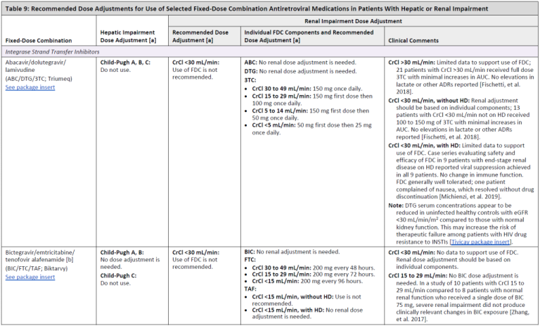


![Anti-ITGAX Armenian Hamster Monoclonal Antibody (PE (Phycoerythrin)) [clone: N418] | VWR](https://fr.vwr.com/stibo/bigweb/std.lang.all/03/25/13150325.jpg)