Macular Edema
Diabetic Retinopathy
Triamcinolone Acetonide
Retinal Vein Occlusion
Laser Coagulation
Visual Acuity
Tomography, Optical Coherence
Edema
Fluorescein Angiography
Vitreous Body
Intravitreal Injections
Macula Lutea
Fundus Oculi
Pulmonary Edema
Vitrectomy
Brain Edema
Retina
Triamcinolone
Glucocorticoids
Retreatment
Fluocinolone Acetonide
Subretinal Fluid
Fovea Centralis
Epiretinal Membrane
Blood-Retinal Barrier
Uveitis
Photography
Angiogenesis Inhibitors
Retinal Diseases
Microscopy, Acoustic
Diagnostic Techniques, Ophthalmological
Light Coagulation
Antibodies, Monoclonal, Humanized
Panuveitis
Retinal Photoreceptor Cell Inner Segment
Retinal Photoreceptor Cell Outer Segment
Telangiectasis
Vascular Endothelial Growth Factor A
Vision Disorders
Treatment Outcome
Follow-Up Studies
Retinal Vein
Capillary Permeability
Drug Implants
Retrospective Studies
Corneal Edema
Edema, Cardiac
Prospective Studies
Retinal Detachment
Diabetes Mellitus, Type 1
Diabetes Mellitus, Type 2
Retinal Artery
Fluorophotometry
Lasers, Solid-State
Edema Disease of Swine
Visual Field Tests
Phacoemulsification
Choroid
Visual Fields
Connective Tissue
Uveitis, Posterior
Aqueous Humor
Vitreous Detachment
Reproducibility of Results
Vitrectomy for cystoid macular oedema with attached posterior hyaloid membrane in patients with diabetes. (1/482)
AIM: To report the success of vitrectomy in eliminating cystoid macular oedema and improving vision in three eyes of two patients with diabetic cystoid macular oedema. In all of the eyes there was no ophthalmoscopic evidence of traction from a posterior hyaloid membrane or from proliferative tissue. METHODS: Pars plana vitrectomy was performed on three eyes of two patients with diabetic cystoid macular oedema who did not show traction upon examination with a slit lamp biomicroscope and a scanning laser ophthalmoscope. RESULTS: Cystoid changes disappeared 1, 3, and 5 days, postoperatively, and diffuse macular oedema resolved within 2 weeks. The visual acuity was improved and maintained. CONCLUSION: Vitrectomy can be effective in some patients with diabetic cystoid macular oedema even in patients who lack evidence of traction by ophthalmoscopy. (+info)Cystoid macular oedema and cytomegalovirus retinitis in patients with HIV disease treated with highly active antiretroviral therapy. (2/482)
BACKGROUND: Although cystoid macular oedema (CMO) is a rare cause of visual loss in AIDS related cytomegalovirus (CMV) retinitis, nine cases are reported of CMO occurring in HIV infected patients with a prior diagnosis of CMV who were receiving highly active antiretroviral therapy (HAART). METHODS: Medical and ophthalmological records of nine AIDS patients with inactive CMV retinitis were retrospectively analysed. Ophthalmic examination data, laboratory findings, and the systemic antiviral treatment were studied. Ophthalmic examination included visual acuity, anterior chamber flare measured with the laser flare cell meter (LCFM), vitreous haze quantification according to the Nussenblatt grading system, and fluorescein angiography. RESULTS: Nine HIV infected patients, eight men and one woman, mean age 39 years (range 29-53 years) presented with inactive CMV retinitis and CMO. On fluorescein angiography, CMO was present only in eyes (14 eyes) with signs of previous CMV retinitis. CMV retinitis was inactive in all of them. Visual acuity ranged from 20/200 to 20/30. In 10 eyes with CMV retinitis, anterior chamber flare measured with the LCFM ranged from 18.5 to 82 photons/ms (mean 35.42 ph/ms). A significant vitreous inflammation (1.5+) was observed in eight eyes. All patients had been treated with anti-CMV drugs for a mean period of 18 months (range 12-36 months). All nine patients received HAART with a combination of two nucleotide analogue reverse transcriptase inhibitors and one protease inhibitor for a mean period of 14 months (range 9-18 months). The HIV viral load was below detectable levels (< 200 copies/ml) in eight patients and low (3215 copies/ml) in one. At the time of CMO, the median CD4+ lymphocyte count was 232 cells x 10(6)/l (range 99-639). CONCLUSION: In AIDS patients, the usual absence of intraocular inflammation in eyes affected by CMV retinitis has been tentatively explained by the profound cellular immunodeficiency. In these patients, treated with HAART, CD4+ counts were increased for several months (mean 14 months). In their eyes, CMV retinitis was associated with significant ocular inflammation and CMO. These findings could be related to the restoration of immune competence after HAART as recently shown. (+info)Results of peripheral laser photocoagulation in pars planitis. (3/482)
PURPOSE: To determine the effect of peripheral retinal laser photocoagulation (PLP) on visual acuity, intraocular inflammation, and other ocular findings, including retinal neovascularization in eyes with pars planitis. METHODS: A retrospective chart review of eyes with pars planitis that had undergone PLP. RESULTS: Twenty-two eyes in 17 patients with pars planitis had undergone treatment with PLP at 2 centers. The mean age at the time of treatment was 19.3 years. Following treatment, mean follow-up was 16.3 months (range, 6 to 37 months). Mean visual acuity was 20/60 preoperatively and 20/50 postoperatively. This level of improvement was not statistically significant (P > .10), but there was a statistically significant decrease in the use of corticosteroids between the preoperative examination and the last postoperative examination (86% versus 27%, P < .05). There was also a statistically significant decrease in vitritis at the last follow-up (P = .0008) and a decrease in neovascularization of the vitreous base (P = .03) and in clinically apparent cystoid macular edema (P = .02). Epiretinal membranes were noted in 23% of eyes preoperatively and in 45% of eyes postoperatively. Only one of these epiretinal membranes was considered to be visually significant. One eye developed a tonic dilated pupil, which slowly improved. CONCLUSIONS: Although the long-term natural history of clinical findings in pars planitis is not well documented, PLP appears to decrease the need for corticosteroids while stabilizing visual acuity. It also appears to decrease vitreous inflammation. PLP has few complications and should be considered in patients with pars planitis who are unresponsive or have adverse reactions to corticosteroids. (+info)Short-wavelength automated perimetry and capillary density in early diabetic maculopathy. (4/482)
PURPOSE: To correlate short-wavelength cone-mediated sensitivity (SWS) assessed by blue-on-yellow perimetry with alterations of the perifoveal vascular bed in early diabetic maculopathy. METHODS: Thirty-one patients (21 M, 10 F; mean age, 35 +/- 12 years; no lens opacities) with no clinically significant macular edema were included in this study. All patients underwent short-wavelength automated perimetry (SWAP) and conventional white-on-white perimetry (Humphrey, 10-2). In digitized video fluorescein angiograms (Scanning Laser Ophthalmoscope), the size of the foveal avascular zone (FAZ) and the mean perifoveal intercapillary area (PIA) as a measure of capillary density were quantified interactively. RESULTS: Mean thresholds of SWAP were significantly correlated with increasing size of FAZ (r = -0.51, P = 0.003) and PIA (r = -0.47, P = 0.01), whereas visual acuity expressed by log MAR (FAZ: r = 0.15, P = 0.41; PIA: r = 0.06, P = 0.76) and mean thresholds assessed with white-on-white perimetry (FAZ: r = -0.25, P = 0.20; PIA: r = -0.31, P = 0.14) were unrelated to diabetic changes of the perifoveal capillary network. CONCLUSIONS: The alterations of the perifoveal network are related to selective disturbances of visual function as measured by blue-on-yellow-perimetry. SWAP may act as an early detector of visual function loss in early diabetic maculopathy and serve as a helpful technique to predict early ischemic damage of the macula and to monitor therapy. (+info)Laser treatment and the mechanism of edema reduction in branch retinal vein occlusion. (5/482)
PURPOSE: To test a hypothesis on the physiological mechanism of the disappearance of macular edema after laser treatment. The hypothesis is based on the effect grid laser treatment has on retinal oxygenation and hemodynamics. It predicts that laser-induced reduction of macular edema is associated with shortening and narrowing of retinal vessels in patients with branch retinal vein occlusion (BRVO). METHODS: The study included 12 subjects, treated with argon laser photocoagulation for BRVO and macular edema. Fundus photographs taken at the time of diagnosis and again after laser treatment, were digitized, and diameter and segment length of retinal vessels was measured using NIH-Image program. RESULTS: Macular edema disappeared or was dramatically reduced in all cases after laser treatment. The diameter of occluded venules constricted to 0.81+/-0.02 (mean +/- SD, P = 0.019) of the prelaser diameter and adjacent arterioles constricted to 0.78+/-0.01 (P = 0.008). The laser treatment also led to shortening of the affected vessels. The final segment length of the occluded venules was 0.95+/-0.17 (P = 0.005) of the length before treatment. The corresponding value for the adjacent arterioles is 0.95+/-0.14 (P = 0.008). Control arterioles and venules in the same fundus did not change in either length or width. CONCLUSIONS: These results do not reject the authors' hypothesis that the disappearance of macular edema in BRVO can be explained by the effect the laser photocoagulation has on retinal oxygenation. Increased oxygenation causes vessel constriction and shortening and lower intravascular pressure, which reduces edema formation according to Starling's law. (+info)The nature and extent of retinal dysfunction associated with diabetic macular edema. (6/482)
PURPOSE: To evaluate the nature and extent of retinal dysfunction in the macular and surrounding areas that occurs in patients with diabetes with clinically significant macular edema (CSME). METHODS: Eleven patients were evaluated before focal laser treatment. Multifocal electroretinogram (ERG) and full-field ERG techniques were used to assess the effects of diabetic retinopathy and CSME on macular, paramacular, and peripheral retinal function. A modified visual field technique was used to obtain local threshold fields. The relationship between local sensitivity changes and local ERG changes was determined. RESULTS: Local ERG responses were significantly delayed and decreased in amplitude, and timing changes were observed in a larger area of the retina than amplitude changes. Visual field deficits were similarly widespread with marked sensitivity losses occurring in retinal areas with normal ERG amplitudes and in areas that appeared to be free of fundus abnormalities. Despite this similarity and the finding that retinal areas with elevated thresholds have timing delays, timing delays were not good predictors of the degree of threshold elevation. CONCLUSIONS: The results demonstrate the widespread nature of timing deficits and visual field deficits that are associated with CSME. (+info)Retinal function in diabetic macular edema after focal laser photocoagulation. (7/482)
PURPOSE: To assess the effects of focal photocoagulation on retinal function in the macular and perimacular areas in patients with diabetes who have clinically significant macular edema. METHODS: Eleven patients were assessed after focal laser treatment. Multifocal electroretinogram (ERG) and full-field ERG techniques were used to evaluate the effects of treatment on macular, paramacular, and peripheral retinal function. A modified visual field technique was used to obtain local threshold fields. The posttreatment results were compared with pretreatment results. Changes in local ERG response amplitudes and implicit times were calculated for each patient and presented as difference fields. The changes in local ERG responses were compared with the changes in local field sensitivity. RESULTS: After treatment, the results of the psychophysical tests suggested little or no change in visual function, but changes in retinal function were observed with the multifocal ERG technique. Local ERG responses showed increases in implicit time and decreases in amplitude, compared with pretreatment values. Timing was affected more than amplitude. CONCLUSIONS: The results suggest that focal treatment produces changes in retinal function, and these changes are not restricted to the treated macular area. (+info)Photoreceptor function in eyes with macular edema. (8/482)
PURPOSE: The irreversible loss of visual acuity in macular edema is usually attributed to permanent loss of photoreceptor cells, although there is hardly any information on changes in photoreceptor function in macular edema. The purpose of this study was to assess photoreceptor function in various stages of macular edema and to relate the findings to visual acuity and angiographic changes. METHODS: Directional sensitivity (optical Stiles-Crawford effect) and visual pigment density of foveal cones was measured with a custom-built scanning laser ophthalmoscope (SLO) in 19 eyes of 19 patients. Twelve eyes exhibited macular edema: five of inflammatory origin, and seven of diabetic origin. Seven eyes with an intraocular inflammatory disease without clinical or angiographic evidence of edema were also included (four of which had previous macular edema and one of which had shown development of macular edema at the 1-year follow-up). Results of SLO measurements were related to findings using fluorescein angiography and Snellen visual acuity, both assessed at the time of SLO measurement and 6 months thereafter. RESULTS: Eyes with macular edema exhibited diminished directional sensitivity of photoreceptor cells in the fovea compared with eyes without (P = 0.02). Visual pigment density of eyes with macular edema was decreased and associated with both initial and follow-up visual function and with the angiographic macular edema grade at follow-up. Abnormal directional sensitivity and pigment density were already present in eyes with slight edematous changes and normal visual acuity. CONCLUSIONS: Eyes with inflammatory or diabetic macular edema showed decreased directional sensitivity and visual pigment density in the macular area. These findings may support a role for SLO measurements in detecting retinal damage due to macular edema. (+info)Macular edema is a medical condition characterized by the accumulation of fluid in the macula, a small area in the center of the retina responsible for sharp, detailed vision. This buildup of fluid causes the macula to thicken and swell, which can distort central vision and lead to vision loss if not treated promptly. Macular edema is often a complication of other eye conditions such as diabetic retinopathy, age-related macular degeneration, retinal vein occlusion, or uveitis. It's important to note that while macular edema can affect anyone, it is more common in people with certain medical conditions like diabetes.
Diabetic retinopathy is a diabetes complication that affects the eyes. It's caused by damage to the blood vessels of the light-sensitive tissue at the back of the eye (retina).
At first, diabetic retinopathy may cause no symptoms or only mild vision problems. Eventually, it can cause blindness. The condition usually affects both eyes.
There are two main stages of diabetic retinopathy:
1. Early diabetic retinopathy. This is when the blood vessels in the eye start to leak fluid or bleed. You might not notice any changes in your vision at this stage, but it's still important to get treatment because it can prevent the condition from getting worse.
2. Advanced diabetic retinopathy. This is when new, abnormal blood vessels grow on the surface of the retina. These vessels can leak fluid and cause severe vision problems, including blindness.
Diabetic retinopathy can be treated with laser surgery, injections of medication into the eye, or a vitrectomy (a surgical procedure to remove the gel-like substance that fills the center of the eye). It's important to get regular eye exams to detect diabetic retinopathy early and get treatment before it causes serious vision problems.
Triamcinolone Acetonide is a synthetic glucocorticoid, which is a class of corticosteroids. It is used in the form of topical creams, ointments, and sprays to reduce skin inflammation, itching, and allergies. It can also be administered through injection for the treatment of various conditions such as arthritis, bursitis, and tendonitis. Triamcinolone Acetonide works by suppressing the immune system's response, reducing inflammation, and blocking the production of substances that cause allergies.
It is important to note that prolonged use or overuse of triamcinolone acetonide can lead to side effects such as thinning of the skin, easy bruising, and increased susceptibility to infections. Therefore, it should be used under the guidance of a healthcare professional.
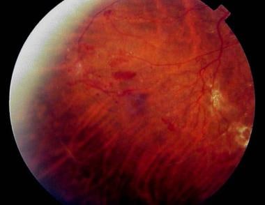
Retinal vein occlusion (RVO) is a medical condition that occurs when one of the retinal veins, which drains blood from the retina, becomes blocked by a blood clot or atherosclerotic plaque. This blockage can cause hemorrhages, fluid accumulation, and damage to the retinal tissue, leading to vision loss.
There are two types of RVO: branch retinal vein occlusion (BRVO) and central retinal vein occlusion (CRVO). BRVO affects a smaller branch retinal vein, while CRVO affects the main retinal vein. CRVO is generally associated with more severe vision loss than BRVO.
Risk factors for RVO include hypertension, diabetes, high cholesterol levels, smoking, and glaucoma. Age is also a significant risk factor, as RVO becomes more common with increasing age. Treatment options for RVO may include controlling underlying medical conditions, laser therapy, intravitreal injections of anti-VEGF agents or steroids, and surgery in some cases.
Laser coagulation, also known as laser photocoagulation, is a medical procedure that uses a laser to seal or destroy abnormal blood vessels or tissue. The laser produces a concentrated beam of light that can be precisely focused on the target area. When the laser energy is absorbed by the tissue, it causes the temperature to rise, which leads to coagulation (the formation of a clot) or destruction of the tissue.
In ophthalmology, laser coagulation is commonly used to treat conditions such as diabetic retinopathy, age-related macular degeneration, and retinal tears or holes. The procedure can help to seal leaking blood vessels, reduce fluid leakage, and prevent further vision loss. It is usually performed as an outpatient procedure and may be repeated if necessary.
In other medical specialties, laser coagulation may be used to control bleeding, destroy tumors, or remove unwanted tissue. The specific technique and parameters of the laser treatment will depend on the individual patient's needs and the condition being treated.
Visual acuity is a measure of the sharpness or clarity of vision. It is usually tested by reading an eye chart from a specific distance, such as 20 feet (6 meters). The standard eye chart used for this purpose is called the Snellen chart, which contains rows of letters that decrease in size as you read down the chart.
Visual acuity is typically expressed as a fraction, with the numerator representing the testing distance and the denominator indicating the smallest line of type that can be read clearly. For example, if a person can read the line on the eye chart that corresponds to a visual acuity of 20/20, it means they have normal vision at 20 feet. If their visual acuity is 20/40, it means they must be as close as 20 feet to see what someone with normal vision can see at 40 feet.
It's important to note that visual acuity is just one aspect of overall vision and does not necessarily reflect other important factors such as peripheral vision, depth perception, color vision, or contrast sensitivity.
Optical coherence tomography (OCT) is a non-invasive imaging technique that uses low-coherence light to capture high-resolution cross-sectional images of biological tissues, particularly the retina and other ocular structures. OCT works by measuring the echo time delay of light scattered back from different depths within the tissue, creating a detailed map of the tissue's structure. This technique is widely used in ophthalmology to diagnose and monitor various eye conditions such as macular degeneration, diabetic retinopathy, and glaucoma.
Edema is the medical term for swelling caused by excess fluid accumulation in the body tissues. It can affect any part of the body, but it's most commonly noticed in the hands, feet, ankles, and legs. Edema can be a symptom of various underlying medical conditions, such as heart failure, kidney disease, liver disease, or venous insufficiency.
The swelling occurs when the capillaries leak fluid into the surrounding tissues, causing them to become swollen and puffy. The excess fluid can also collect in the cavities of the body, leading to conditions such as pleural effusion (fluid around the lungs) or ascites (fluid in the abdominal cavity).
The severity of edema can vary from mild to severe, and it may be accompanied by other symptoms such as skin discoloration, stiffness, and pain. Treatment for edema depends on the underlying cause and may include medications, lifestyle changes, or medical procedures.
Fluorescein angiography is a medical diagnostic procedure used in ophthalmology to examine the blood flow in the retina and choroid, which are the inner layers of the eye. This test involves injecting a fluorescent dye, Fluorescein, into a patient's arm vein. As the dye reaches the blood vessels in the eye, a specialized camera takes rapid sequences of photographs to capture the dye's circulation through the retina and choroid.
The images produced by fluorescein angiography can help doctors identify any damage to the blood vessels, leakage, or abnormal growth of new blood vessels. This information is crucial in diagnosing and managing various eye conditions such as age-related macular degeneration, diabetic retinopathy, retinal vein occlusions, and inflammatory eye diseases.
It's important to note that while fluorescein angiography is a valuable diagnostic tool, it does carry some risks, including temporary side effects like nausea, vomiting, or allergic reactions to the dye. In rare cases, severe adverse reactions can occur, so patients should discuss these potential risks with their healthcare provider before undergoing the procedure.
The vitreous body, also known simply as the vitreous, is the clear, gel-like substance that fills the space between the lens and the retina in the eye. It is composed mainly of water, but also contains collagen fibers, hyaluronic acid, and other proteins. The vitreous helps to maintain the shape of the eye and provides a transparent medium for light to pass through to reach the retina. With age, the vitreous can become more liquefied and may eventually separate from the retina, leading to symptoms such as floaters or flashes of light.
An intravitreal injection is a medical procedure in which medication is delivered directly into the vitreous cavity of the eye, which is the clear, gel-like substance that fills the space between the lens and the retina. This type of injection is typically used to treat various eye conditions such as age-related macular degeneration, diabetic retinopathy, retinal vein occlusion, and uveitis. The medication administered in intravitreal injections can help to reduce inflammation, inhibit the growth of new blood vessels, or prevent the formation of abnormal blood vessels in the eye.
Intravitreal injections are usually performed in an outpatient setting, and the procedure typically takes only a few minutes. Before the injection, the eye is numbed with anesthetic drops to minimize discomfort. The medication is then injected into the vitreous cavity using a small needle. After the injection, patients may experience some mild discomfort or a scratchy sensation in the eye, but this usually resolves within a few hours.
While intravitreal injections are generally safe, there are some potential risks and complications associated with the procedure, including infection, bleeding, retinal detachment, and increased intraocular pressure. Patients who undergo intravitreal injections should be closely monitored by their eye care provider to ensure that any complications are promptly identified and treated.
The macula lutea, often simply referred to as the macula or fovea centralis, is a part of the eye that is responsible for central vision and color perception. It's located in the center of the retina, the light-sensitive tissue at the back of the eye. The macula contains a high concentration of pigments called xanthophylls, which give it a yellowish color and protect the photoreceptor cells in this area from damage by blue light.
The central part of the macula is called the fovea, which is a small depression that contains only cones, the photoreceptor cells responsible for color vision and high visual acuity. The fovea is surrounded by the parafovea and the perifovea, which contain both cones and rods, the photoreceptor cells responsible for low-light vision and peripheral vision.
Damage to the macula can result in a loss of central vision and color perception, a condition known as age-related macular degeneration (AMD), which is a leading cause of blindness in older adults. Other conditions that can affect the macula include macular edema, macular holes, and macular pucker.
An injection is a medical procedure in which a medication, vaccine, or other substance is introduced into the body using a needle and syringe. The substance can be delivered into various parts of the body, including into a vein (intravenous), muscle (intramuscular), under the skin (subcutaneous), or into the spinal canal (intrathecal or spinal).
Injections are commonly used to administer medications that cannot be taken orally, have poor oral bioavailability, need to reach the site of action quickly, or require direct delivery to a specific organ or tissue. They can also be used for diagnostic purposes, such as drawing blood samples (venipuncture) or injecting contrast agents for imaging studies.
Proper technique and sterile conditions are essential when administering injections to prevent infection, pain, and other complications. The choice of injection site depends on the type and volume of the substance being administered, as well as the patient's age, health status, and personal preferences.
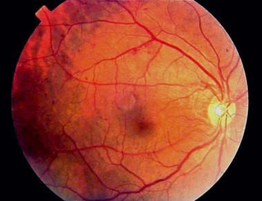
"Fundus Oculi" is a medical term that refers to the back part of the interior of the eye, including the optic disc, macula, fovea, retinal vasculature, and peripheral retina. It is the area where light is focused and then transmitted to the brain via the optic nerve, forming visual images. Examinations of the fundus oculi are crucial for detecting various eye conditions such as diabetic retinopathy, macular degeneration, glaucoma, and other retinal diseases. The examination is typically performed using an ophthalmoscope or a specialized camera called a retinal camera.
Pulmonary edema is a medical condition characterized by the accumulation of fluid in the alveoli (air sacs) and interstitial spaces (the area surrounding the alveoli) within the lungs. This buildup of fluid can lead to impaired gas exchange, resulting in shortness of breath, coughing, and difficulty breathing, especially when lying down. Pulmonary edema is often a complication of heart failure, but it can also be caused by other conditions such as pneumonia, trauma, or exposure to certain toxins.
In the early stages of pulmonary edema, patients may experience mild symptoms such as shortness of breath during physical activity. However, as the condition progresses, symptoms can become more severe and include:
* Severe shortness of breath, even at rest
* Wheezing or coughing up pink, frothy sputum
* Rapid breathing and heart rate
* Anxiety or restlessness
* Bluish discoloration of the skin (cyanosis) due to lack of oxygen
Pulmonary edema can be diagnosed through a combination of physical examination, medical history, chest X-ray, and other diagnostic tests such as echocardiography or CT scan. Treatment typically involves addressing the underlying cause of the condition, as well as providing supportive care such as supplemental oxygen, diuretics to help remove excess fluid from the body, and medications to help reduce anxiety and improve breathing. In severe cases, mechanical ventilation may be necessary to support respiratory function.
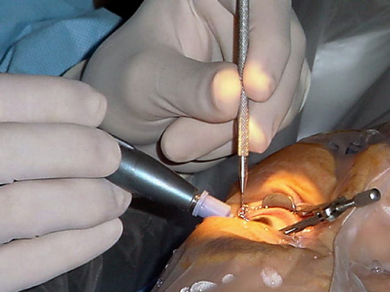
A vitrectomy is a surgical procedure that involves the removal of some or all of the vitreous humor, which is the clear gel-like substance filling the center of the eye. This surgery is often performed to treat various retinal disorders such as diabetic retinopathy, retinal detachment, macular hole, and vitreous hemorrhage.
During a vitrectomy, the ophthalmologist makes small incisions in the sclera (the white part of the eye) to access the vitreous cavity. The surgeon then uses specialized instruments to remove the cloudy or damaged vitreous and may also repair any damage to the retina or surrounding tissues. Afterward, a clear saline solution is injected into the eye to maintain its shape and help facilitate healing.
In some cases, a gas bubble or silicone oil may be placed in the eye after the vitrectomy to help hold the retina in place while it heals. These substances will gradually be absorbed or removed during follow-up appointments. The body naturally produces a new, clear vitreous to replace the removed material over time.
Vitrectomy is typically performed under local anesthesia and may require hospitalization or outpatient care depending on the individual case. Potential risks and complications include infection, bleeding, cataract formation, retinal detachment, and increased eye pressure. However, with proper care and follow-up, most patients experience improved vision after a successful vitrectomy procedure.
Brain edema is a medical condition characterized by the abnormal accumulation of fluid in the brain, leading to an increase in intracranial pressure. This can result from various causes, such as traumatic brain injury, stroke, infection, brain tumors, or inflammation. The swelling of the brain can compress vital structures, impair blood flow, and cause neurological symptoms, which may range from mild headaches to severe cognitive impairment, seizures, coma, or even death if not treated promptly and effectively.
The retina is the innermost, light-sensitive layer of tissue in the eye of many vertebrates and some cephalopods. It receives light that has been focused by the cornea and lens, converts it into neural signals, and sends these to the brain via the optic nerve. The retina contains several types of photoreceptor cells including rods (which handle vision in low light) and cones (which are active in bright light and are capable of color vision).
In medical terms, any pathological changes or diseases affecting the retinal structure and function can lead to visual impairment or blindness. Examples include age-related macular degeneration, diabetic retinopathy, retinal detachment, and retinitis pigmentosa among others.
Triamcinolone is a glucocorticoid medication, which is a class of corticosteroids. It is used to treat various inflammatory and autoimmune conditions due to its anti-inflammatory and immunosuppressive effects. Triamcinolone is available in several forms, including topical creams, ointments, and lotions for skin application; oral tablets and injectable solutions for systemic use; and inhaled preparations for the treatment of asthma and other respiratory conditions.
Triamcinolone works by binding to specific receptors in cells, which leads to a decrease in the production of inflammatory chemicals such as prostaglandins and leukotrienes. This results in reduced swelling, redness, itching, and pain associated with inflammation.
Some common uses of triamcinolone include treating skin conditions like eczema, psoriasis, and dermatitis; managing allergic reactions; reducing inflammation in respiratory diseases like asthma and COPD; and alleviating symptoms of rheumatoid arthritis and other autoimmune disorders.
As with any medication, triamcinolone can have side effects, especially when used in high doses or for extended periods. Common side effects include increased appetite, weight gain, mood changes, insomnia, acne, thinning of the skin, and easy bruising. Long-term use may also lead to more serious complications such as osteoporosis, adrenal suppression, and increased susceptibility to infections. It is essential to follow your healthcare provider's instructions carefully when using triamcinolone or any other prescription medication.
Glucocorticoids are a class of steroid hormones that are naturally produced in the adrenal gland, or can be synthetically manufactured. They play an essential role in the metabolism of carbohydrates, proteins, and fats, and have significant anti-inflammatory effects. Glucocorticoids suppress immune responses and inflammation by inhibiting the release of inflammatory mediators from various cells, such as mast cells, eosinophils, and lymphocytes. They are frequently used in medical treatment for a wide range of conditions, including allergies, asthma, rheumatoid arthritis, dermatological disorders, and certain cancers. Prolonged use or high doses of glucocorticoids can lead to several side effects, such as weight gain, mood changes, osteoporosis, and increased susceptibility to infections.
Pseudophakia is a medical term that refers to the condition where a person's natural lens in the eye has been replaced with an artificial one. This procedure is typically performed during cataract surgery, where the cloudy, natural lens is removed and replaced with a clear, artificial lens to improve vision. The prefix "pseudo" means false or fake, and "phakia" refers to the natural lens of the eye, hence the term "Pseudophakia" implies a false or artificial lens.
Intraocular injections are a type of medical procedure where medication is administered directly into the eye. This technique is often used to deliver drugs that treat various eye conditions, such as age-related macular degeneration, diabetic retinopathy, and endophthalmitis. The most common type of intraocular injection is an intravitreal injection, which involves injecting medication into the vitreous cavity, the space inside the eye filled with a clear gel-like substance called the vitreous humor. This procedure is typically performed by an ophthalmologist in a clinical setting and may be repeated at regular intervals depending on the condition being treated.
In medical terms, "retreatment" refers to the process of providing additional treatment or courses of therapy to an individual who has previously undergone a medical intervention but has not achieved the desired outcomes or has experienced a recurrence of symptoms. This may apply to various medical conditions and treatments, including dental procedures, cancer therapies, mental health treatments, and more.
In the context of dentistry, specifically endodontics (root canal treatment), retreatment is the process of repeating the root canal procedure on a tooth that has already been treated before. This may be necessary if the initial treatment was not successful in eliminating infection or if reinfection has occurred. The goal of retreatment is to preserve the natural tooth and alleviate any persistent pain or discomfort.
Fluocinolone acetonide is a synthetic corticosteroid, which is a type of medication that reduces inflammation and suppresses the immune system. It is used to treat various skin conditions such as eczema, psoriasis, and dermatitis. Fluocinolone acetonide works by reducing the production of chemicals in the body that cause inflammation.
Fluocinolone acetonide is available in several forms, including creams, ointments, solutions, and tape. It is usually applied to the affected area of the skin one to three times a day, depending on the severity of the condition and the specific formulation being used.
Like all corticosteroids, fluocinolone acetonide can have side effects, particularly with long-term use or if used in large amounts. These may include thinning of the skin, easy bruising, stretch marks, increased hair growth, and acne. It is important to follow the instructions of a healthcare provider carefully when using this medication to minimize the risk of side effects.
Subretinal fluid (SRF) refers to the abnormal accumulation of fluid between the neurosensory retina and the pigment epithelium of the eye. This can occur due to various conditions such as age-related macular degeneration, central serous chorioretinopathy, or retinal detachment. The presence of subretinal fluid can distort vision and may require medical intervention depending on the underlying cause and severity of the condition.
The fovea centralis, also known as the macula lutea, is a small pit or depression located in the center of the retina, an light-sensitive tissue at the back of the eye. It is responsible for sharp, detailed vision (central vision) and color perception. The fovea contains only cones, the photoreceptor cells that are responsible for color vision and high visual acuity. It has a higher concentration of cones than any other area in the retina, allowing it to provide the greatest detail and color discrimination. The center of the fovea is called the foveola, which contains the highest density of cones and is avascular, meaning it lacks blood vessels to avoid interfering with the light passing through to the photoreceptor cells.
Intermediate uveitis is a type of uveitis that affects the vitreous cavity and peripheral retina. It is characterized by the presence of inflammatory cells in the vitreous, called vitritis, and sometimes also by snowbanking or peripheral lesions in the retina. Intermediate uveitis can cause vision loss due to cystoid macular edema, epiretinal membrane formation, or complications such as glaucoma or cataract. The onset of intermediate uveitis is often insidious and the course can be chronic, with recurrent episodes of inflammation. The exact cause of intermediate uveitis is often unknown, but it can be associated with systemic diseases such as sarcoidosis, multiple sclerosis, or Lyme disease.
An epiretinal membrane, also known as a macular pucker or cellophane maculopathy, is a thin and transparent layer of tissue that forms over the macula (the central part of the retina responsible for sharp, detailed vision) in the eye. This membrane can contract and wrinkle the macula, distorting central vision.
Epiretinal membranes are typically caused by the migration and proliferation of glial cells or other cell types onto the surface of the retina following retinal injury, inflammation, or aging. In some cases, they may be associated with other eye conditions such as diabetic retinopathy, retinal vein occlusion, or age-related macular degeneration.
Mild epiretinal membranes may not require treatment, but if the distortion of vision is significant, a vitrectomy surgery may be recommended to remove the membrane and improve visual acuity.
The blood-retinal barrier (BRB) is a specialized physiological barrier in the eye that helps regulate the movement of molecules between the retina and the bloodstream. It is made up of tight junctions between the endothelial cells of retinal blood vessels and between the pigment epithelium cells of the retina, which restrict the paracellular diffusion of solutes.
The BRB plays a crucial role in maintaining the health and function of the retina by preventing harmful substances from entering the retina while allowing essential nutrients and oxygen to reach the retinal tissues. Disruption of the BRB has been implicated in various retinal diseases, including diabetic retinopathy, age-related macular degeneration, and retinal vein occlusion.
Uveitis is the inflammation of the uvea, the middle layer of the eye between the retina and the white of the eye (sclera). The uvea consists of the iris, ciliary body, and choroid. Uveitis can cause redness, pain, and vision loss. It can be caused by various systemic diseases, infections, or trauma. Depending on the part of the uvea that's affected, uveitis can be classified as anterior (iritis), intermediate (cyclitis), posterior (choroiditis), or pan-uveitis (affecting all layers). Treatment typically includes corticosteroids and other immunosuppressive drugs to control inflammation.
I'm sorry for any confusion, but "photography" is not a term typically used in medical definitions. Photography refers to the art, application, or process of creating images by recording light or other electromagnetic radiation, either electronically by means of an image sensor, or chemically by means of a light-sensitive material such as photographic film.
If you're looking for a medical term related to imaging, there are several terms that might be relevant, such as:
1. Radiography: This is a technique using X-rays to visualize the internal structures of the body.
2. Ultrasonography: Also known as ultrasound, this is a diagnostic imaging technique using high-frequency sound waves to create images of the inside of the body.
3. Computed Tomography (CT): A type of imaging that uses X-rays to create detailed cross-sectional images of the body.
4. Magnetic Resonance Imaging (MRI): A type of imaging that uses magnetic fields and radio waves to create detailed images of the organs and tissues within the body.
5. Nuclear Medicine: This is a branch of medical imaging that uses small amounts of radioactive material to diagnose and treat diseases.
If you have any questions related to medical definitions or topics, feel free to ask!
Angiogenesis inhibitors are a class of drugs that block the growth of new blood vessels (angiogenesis). They work by targeting specific molecules involved in the process of angiogenesis, such as vascular endothelial growth factor (VEGF) and its receptors. By blocking these molecules, angiogenesis inhibitors can prevent the development of new blood vessels that feed tumors, thereby slowing or stopping their growth.
Angiogenesis inhibitors are used in the treatment of various types of cancer, including colon, lung, breast, kidney, and ovarian cancer. They may be given alone or in combination with other cancer treatments, such as chemotherapy or radiation therapy. Some examples of angiogenesis inhibitors include bevacizumab (Avastin), sorafenib (Nexavar), sunitinib (Sutent), and pazopanib (Votrient).
It's important to note that while angiogenesis inhibitors can be effective in treating cancer, they can also have serious side effects, such as high blood pressure, bleeding, and damage to the heart or kidneys. Therefore, it's essential that patients receive careful monitoring and management of these potential side effects while undergoing treatment with angiogenesis inhibitors.
Retinal diseases refer to a group of conditions that affect the retina, which is the light-sensitive tissue located at the back of the eye. The retina is responsible for converting light into electrical signals that are sent to the brain and interpreted as visual images. Retinal diseases can cause vision loss or even blindness, depending on their severity and location in the retina.
Some common retinal diseases include:
1. Age-related macular degeneration (AMD): A progressive disease that affects the central part of the retina called the macula, causing blurred or distorted vision.
2. Diabetic retinopathy: A complication of diabetes that can damage the blood vessels in the retina, leading to vision loss.
3. Retinal detachment: A serious condition where the retina becomes separated from its underlying tissue, requiring immediate medical attention.
4. Macular edema: Swelling or thickening of the macula due to fluid accumulation, which can cause blurred vision.
5. Retinitis pigmentosa: A group of inherited eye disorders that affect the retina's ability to respond to light, causing progressive vision loss.
6. Macular hole: A small break in the macula that can cause distorted or blurry vision.
7. Retinal vein occlusion: Blockage of the retinal veins that can lead to bleeding, swelling, and potential vision loss.
Treatment for retinal diseases varies depending on the specific condition and its severity. Some treatments include medication, laser therapy, surgery, or a combination of these options. Regular eye exams are essential for early detection and treatment of retinal diseases.
Acoustic microscopy is a non-invasive imaging technique that uses sound waves to visualize and analyze the structure and properties of various materials, including biological samples. In the context of medical diagnostics and research, acoustic microscopy can be used to examine tissues, cells, and cellular components with high resolution, providing valuable information about their mechanical and physical properties.
In acoustic microscopy, high-frequency sound waves are focused onto a sample using a transducer. The interaction between the sound waves and the sample generates echoes, which contain information about the sample's internal structure and properties. These echoes are then recorded and processed to create an image of the sample.
Acoustic microscopy offers several advantages over other imaging techniques, such as optical microscopy or electron microscopy. For example, it does not require staining or labeling of samples, which can be time-consuming and potentially damaging. Additionally, acoustic microscopy can provide high-resolution images of samples in their native state, allowing researchers to study the effects of various treatments or interventions on living cells and tissues.
In summary, acoustic microscopy is a non-invasive imaging technique that uses sound waves to visualize and analyze the structure and properties of biological samples with high resolution, providing valuable information for medical diagnostics and research.
Diagnostic techniques in ophthalmology refer to the various methods and tests used by eye specialists (ophthalmologists) to examine, evaluate, and diagnose conditions related to the eyes and visual system. Here are some commonly used diagnostic techniques:
1. Visual Acuity Testing: This is a basic test to measure the sharpness of a person's vision. It typically involves reading letters or numbers from an eye chart at a specific distance.
2. Refraction Test: This test helps determine the correct lens prescription for glasses or contact lenses by measuring how light is bent as it passes through the cornea and lens.
3. Slit Lamp Examination: A slit lamp is a microscope that allows an ophthalmologist to examine the structures of the eye, including the cornea, iris, lens, and retina, in great detail.
4. Tonometry: This test measures the pressure inside the eye (intraocular pressure) to detect conditions like glaucoma. Common methods include applanation tonometry and non-contact tonometry.
5. Retinal Imaging: Several techniques are used to capture images of the retina, including fundus photography, fluorescein angiography, and optical coherence tomography (OCT). These tests help diagnose conditions like macular degeneration, diabetic retinopathy, and retinal detachments.
6. Color Vision Testing: This test evaluates a person's ability to distinguish between different colors, which can help detect color vision deficiencies or neurological disorders affecting the visual pathway.
7. Visual Field Testing: This test measures a person's peripheral (or side) vision and can help diagnose conditions like glaucoma, optic nerve damage, or brain injuries.
8. Pupillary Reactions Tests: These tests evaluate how the pupils respond to light and near objects, which can provide information about the condition of the eye's internal structures and the nervous system.
9. Ocular Motility Testing: This test assesses eye movements and alignment, helping diagnose conditions like strabismus (crossed eyes) or nystagmus (involuntary eye movement).
10. Corneal Topography: This non-invasive imaging technique maps the curvature of the cornea, which can help detect irregularities, assess the fit of contact lenses, and plan refractive surgery procedures.
"Light coagulation," also known as "laser coagulation," is a medical term that refers to the use of laser technology to cauterize (seal or close) tissue. This procedure uses heat generated by a laser to cut, coagulate, or destroy tissue. In light coagulation, the laser beam is focused on the blood vessels in question, causing the blood within them to clot and the vessels to seal. This can be used for various medical purposes, such as stopping bleeding during surgery, destroying abnormal tissues (like tumors), or treating eye conditions like diabetic retinopathy and age-related macular degeneration.
It's important to note that this is a general definition, and the specific use of light coagulation may vary depending on the medical specialty and the individual patient's needs. As always, it's best to consult with a healthcare professional for more detailed information about any medical procedure or treatment.
Monoclonal antibodies are laboratory-produced proteins that mimic the immune system's ability to fight off harmful antigens such as viruses and cancer cells. They are created by fusing a single B cell (the type of white blood cell responsible for producing antibodies) with a tumor cell, resulting in a hybrid cell called a hybridoma. This hybridoma can then be cloned to produce a large number of identical cells, all producing the same antibody, hence "monoclonal."
Humanized monoclonal antibodies are a type of monoclonal antibody that have been genetically engineered to include human components. This is done to reduce the risk of an adverse immune response in patients receiving the treatment. In this process, the variable region of the mouse monoclonal antibody, which contains the antigen-binding site, is grafted onto a human constant region. The resulting humanized monoclonal antibody retains the ability to bind to the target antigen while minimizing the immunogenicity associated with murine (mouse) antibodies.
In summary, "antibodies, monoclonal, humanized" refers to a type of laboratory-produced protein that mimics the immune system's ability to fight off harmful antigens, but with reduced immunogenicity due to the inclusion of human components in their structure.
Panuveitis is a medical term that refers to inflammation that affects the entire uveal tract, including the iris, ciliary body, and choroid. The uveal tract is the middle layer of the eye between the inner retina and the outer fibrous tunic (sclera). Panuveitis can also affect other parts of the eye, such as the vitreous, retina, and optic nerve.
The symptoms of panuveitis may include redness, pain, light sensitivity, blurred vision, floaters, and decreased visual acuity. The condition can be caused by various factors, including infections, autoimmune diseases, trauma, or unknown causes (idiopathic). Treatment typically involves the use of corticosteroids to reduce inflammation, as well as addressing any underlying cause if identified. If left untreated, panuveitis can lead to complications such as cataracts, glaucoma, and retinal damage, which can result in permanent vision loss.
Cataract extraction is a surgical procedure that involves removing the cloudy lens (cataract) from the eye. This procedure is typically performed to restore vision impairment caused by cataracts and improve overall quality of life. There are two primary methods for cataract extraction:
1. Phacoemulsification: This is the most common method used today. It involves making a small incision in the front part of the eye (cornea), inserting an ultrasonic probe to break up the cloudy lens into tiny pieces, and then removing those pieces with suction. After removing the cataract, an artificial intraocular lens (IOL) is inserted to replace the natural lens and help focus light onto the retina.
2. Extracapsular Cataract Extraction: In this method, a larger incision is made on the side of the cornea, allowing the surgeon to remove the cloudy lens in one piece without breaking it up. The back part of the lens capsule is left intact to support the IOL. This technique is less common and typically reserved for more advanced cataracts or when phacoemulsification cannot be performed.
Recovery from cataract extraction usually involves using eye drops to prevent infection and inflammation, as well as protecting the eye with a shield or glasses during sleep for a few weeks after surgery. Most people experience improved vision within a few days to a week following the procedure.
Ophthalmoscopy is a medical examination technique used by healthcare professionals to observe the interior structures of the eye, including the retina, optic disc, and vitreous humor. This procedure typically involves using an ophthalmoscope, a handheld device that consists of a light and magnifying lenses. The healthcare provider looks through the ophthalmoscope and directly observes the internal structures of the eye by illuminating them.
There are several types of ophthalmoscopy, including direct ophthalmoscopy, indirect ophthalmoscopy, and slit-lamp biomicroscopy. Each type has its own advantages and disadvantages, and they may be used in different situations depending on the specific clinical situation and the information needed.
Ophthalmoscopy is an important diagnostic tool for detecting and monitoring a wide range of eye conditions, including diabetic retinopathy, glaucoma, age-related macular degeneration, and other retinal disorders. It can also provide valuable information about the overall health of the individual, as changes in the appearance of the retina or optic nerve may indicate the presence of systemic diseases such as hypertension or diabetes.
The inner segment of a retinal photoreceptor cell, also known as the inner segment of a rod or cone cell, is the portion of the cell that contains the majority of its metabolic and energy-generating components. It is responsible for providing the energy needed for the outer segment, which is the part of the cell that contains the visual pigments and is responsible for phototransduction, or the conversion of light into electrical signals.
The inner segment is divided into two main parts: the ellipsoid and the myoid. The ellipsoid contains a high concentration of mitochondria, which provide energy to the cell through the process of oxidative phosphorylation. The myoid contains the endoplasmic reticulum and the Golgi apparatus, which are involved in protein synthesis and transport.
Damage to the inner segment of the retinal photoreceptor cells can lead to vision loss or impairment, as it can affect the ability of the outer segment to function properly and transmit visual signals to the brain.
Retinal vessels refer to the blood vessels that are located in the retina, which is the light-sensitive tissue that lines the inner surface of the eye. The retina contains two types of blood vessels: arteries and veins.
The central retinal artery supplies oxygenated blood to the inner layers of the retina, while the central retinal vein drains deoxygenated blood from the retina. These vessels can be visualized during a routine eye examination using an ophthalmoscope, which allows healthcare professionals to assess their health and any potential abnormalities.
Retinal vessels are essential for maintaining the health and function of the retina, and any damage or changes to these vessels can affect vision and lead to various eye conditions such as diabetic retinopathy, retinal vein occlusion, and hypertensive retinopathy.
The retinal photoreceptor cells, namely rods and cones, are specialized neurons in the retina responsible for converting light into electrical signals that can be processed by the brain. The outer segment of a retinal photoreceptor cell is the portion of the cell where phototransduction primarily occurs. It contains stacks of disc-like structures filled with the visual pigment rhodopsin, which absorbs light and initiates the conversion process.
The outer segment is continuously renewed through a process called shedding and phagocytosis, in which the oldest discs at the base of the outer segment are shed, engulfed by the adjacent retinal pigment epithelium (RPE) cells, and degraded. This turnover helps maintain the sensitivity and functionality of the photoreceptor cells.
In summary, the retinal photoreceptor cell outer segment is a highly specialized compartment where light absorption and initial signal transduction occur in rods and cones, supported by continuous renewal through shedding and phagocytosis.
Telangiectasia is a medical term that refers to the dilation and widening of small blood vessels called capillaries, leading to their visibility under the skin or mucous membranes. These dilated vessels often appear as tiny red lines or patterns, measuring less than 1 millimeter in diameter.
Telangiectasias can occur in various parts of the body, such as the face, nose, cheeks, legs, and fingers. They are typically harmless but may cause cosmetic concerns for some individuals. In certain cases, telangiectasias can be a sign of an underlying medical condition, like rosacea, hereditary hemorrhagic telangiectasia (HHT), or liver disease.
It is essential to consult with a healthcare professional if you notice any unusual changes in your skin or mucous membranes, as they can provide appropriate evaluation and treatment recommendations based on the underlying cause of the telangiectasias.
Vascular Endothelial Growth Factor A (VEGFA) is a specific isoform of the vascular endothelial growth factor (VEGF) family. It is a well-characterized signaling protein that plays a crucial role in angiogenesis, the process of new blood vessel formation from pre-existing vessels. VEGFA stimulates the proliferation and migration of endothelial cells, which line the interior surface of blood vessels, thereby contributing to the growth and development of new vasculature. This protein is essential for physiological processes such as embryonic development and wound healing, but it has also been implicated in various pathological conditions, including cancer, age-related macular degeneration, and diabetic retinopathy. The regulation of VEGFA expression and activity is critical to maintaining proper vascular function and homeostasis.
Vision disorders refer to a wide range of conditions that affect the visual system and result in various symptoms, such as blurry vision, double vision, distorted vision, impaired depth perception, and difficulty with visual tracking or focusing. These disorders can be categorized into several types, including:
1. Refractive errors: These occur when the shape of the eye prevents light from focusing directly on the retina, resulting in blurry vision. Examples include myopia (nearsightedness), hyperopia (farsightedness), astigmatism, and presbyopia (age-related loss of near vision).
2. Strabismus: Also known as crossed eyes or walleye, strabismus is a misalignment of the eyes where they point in different directions, which can lead to double vision or loss of depth perception.
3. Amblyopia: Often called lazy eye, amblyopia is a condition where one eye has reduced vision due to lack of proper visual development during childhood. It may be caused by strabismus, refractive errors, or other factors that interfere with normal visual development.
4. Accommodative disorders: These involve problems with the focusing ability of the eyes, such as convergence insufficiency (difficulty focusing on close objects) and accommodative dysfunction (inability to maintain clear vision at different distances).
5. Binocular vision disorders: These affect how the eyes work together as a team, leading to issues like poor depth perception, eye strain, and headaches. Examples include convergence insufficiency, divergence excess, and suppression.
6. Ocular motility disorders: These involve problems with eye movement, such as nystagmus (involuntary eye movements), strabismus, or restricted extraocular muscle function.
7. Visual processing disorders: These affect the brain's ability to interpret and make sense of visual information, even when the eyes themselves are healthy. Symptoms may include difficulty with reading, recognizing shapes and objects, and understanding spatial relationships.
8. Low vision: This term refers to significant visual impairment that cannot be fully corrected with glasses, contact lenses, medication, or surgery. It includes conditions like macular degeneration, diabetic retinopathy, glaucoma, and cataracts.
9. Blindness: Complete loss of sight in both eyes, which can be caused by various factors such as injury, disease, or genetic conditions.
Treatment outcome is a term used to describe the result or effect of medical treatment on a patient's health status. It can be measured in various ways, such as through symptoms improvement, disease remission, reduced disability, improved quality of life, or survival rates. The treatment outcome helps healthcare providers evaluate the effectiveness of a particular treatment plan and make informed decisions about future care. It is also used in clinical research to compare the efficacy of different treatments and improve patient care.
Follow-up studies are a type of longitudinal research that involve repeated observations or measurements of the same variables over a period of time, in order to understand their long-term effects or outcomes. In medical context, follow-up studies are often used to evaluate the safety and efficacy of medical treatments, interventions, or procedures.
In a typical follow-up study, a group of individuals (called a cohort) who have received a particular treatment or intervention are identified and then followed over time through periodic assessments or data collection. The data collected may include information on clinical outcomes, adverse events, changes in symptoms or functional status, and other relevant measures.
The results of follow-up studies can provide important insights into the long-term benefits and risks of medical interventions, as well as help to identify factors that may influence treatment effectiveness or patient outcomes. However, it is important to note that follow-up studies can be subject to various biases and limitations, such as loss to follow-up, recall bias, and changes in clinical practice over time, which must be carefully considered when interpreting the results.
A Retinal Vein is a vessel that carries oxygen-depleted blood away from the retina, a light-sensitive layer at the back of the eye. The retinal veins originate from a network of smaller vessels called venules and ultimately merge to form the central retinal vein, which exits the eye through the optic nerve.
Retinal veins are crucial for maintaining the health and function of the retina, as they facilitate the removal of waste products and help regulate the ocular environment. However, they can also be susceptible to various pathological conditions such as retinal vein occlusions, which can lead to vision loss or damage to the eye.
Capillary permeability refers to the ability of substances to pass through the walls of capillaries, which are the smallest blood vessels in the body. These tiny vessels connect the arterioles and venules, allowing for the exchange of nutrients, waste products, and gases between the blood and the surrounding tissues.
The capillary wall is composed of a single layer of endothelial cells that are held together by tight junctions. The permeability of these walls varies depending on the size and charge of the molecules attempting to pass through. Small, uncharged molecules such as water, oxygen, and carbon dioxide can easily diffuse through the capillary wall, while larger or charged molecules such as proteins and large ions have more difficulty passing through.
Increased capillary permeability can occur in response to inflammation, infection, or injury, allowing larger molecules and immune cells to enter the surrounding tissues. This can lead to swelling (edema) and tissue damage if not controlled. Decreased capillary permeability, on the other hand, can lead to impaired nutrient exchange and tissue hypoxia.
Overall, the permeability of capillaries is a critical factor in maintaining the health and function of tissues throughout the body.
A drug implant is a medical device that is specially designed to provide controlled release of a medication into the body over an extended period of time. Drug implants can be placed under the skin or in various body cavities, depending on the specific medical condition being treated. They are often used when other methods of administering medication, such as oral pills or injections, are not effective or practical.
Drug implants come in various forms, including rods, pellets, and small capsules. The medication is contained within the device and is released slowly over time, either through diffusion or erosion of the implant material. This allows for a steady concentration of the drug to be maintained in the body, which can help to improve treatment outcomes and reduce side effects.
Some common examples of drug implants include:
1. Hormonal implants: These are small rods that are inserted under the skin of the upper arm and release hormones such as progestin or estrogen over a period of several years. They are often used for birth control or to treat conditions such as endometriosis or uterine fibroids.
2. Intraocular implants: These are small devices that are placed in the eye during surgery to release medication directly into the eye. They are often used to treat conditions such as age-related macular degeneration or diabetic retinopathy.
3. Bone cement implants: These are specially formulated cements that contain antibiotics and are used to fill bone defects or joint spaces during surgery. The antibiotics are released slowly over time, helping to prevent infection.
4. Implantable pumps: These are small devices that are placed under the skin and deliver medication directly into a specific body cavity, such as the spinal cord or the peritoneal cavity. They are often used to treat chronic pain or cancer.
Overall, drug implants offer several advantages over other methods of administering medication, including improved compliance, reduced side effects, and more consistent drug levels in the body. However, they may also have some disadvantages, such as the need for surgical placement and the potential for infection or other complications. As with any medical treatment, it is important to discuss the risks and benefits of drug implants with a healthcare provider.
Retrospective studies, also known as retrospective research or looking back studies, are a type of observational study that examines data from the past to draw conclusions about possible causal relationships between risk factors and outcomes. In these studies, researchers analyze existing records, medical charts, or previously collected data to test a hypothesis or answer a specific research question.
Retrospective studies can be useful for generating hypotheses and identifying trends, but they have limitations compared to prospective studies, which follow participants forward in time from exposure to outcome. Retrospective studies are subject to biases such as recall bias, selection bias, and information bias, which can affect the validity of the results. Therefore, retrospective studies should be interpreted with caution and used primarily to generate hypotheses for further testing in prospective studies.
Corneal edema is a medical condition characterized by the accumulation of fluid in the cornea, which is the clear, dome-shaped surface at the front of the eye. This buildup of fluid causes the cornea to swell and thicken, resulting in blurry or distorted vision. Corneal edema can be caused by various factors, including eye injuries, certain medications, eye surgeries, and diseases that affect the eye's ability to pump fluids out of the cornea. In some cases, corneal edema may resolve on its own or with treatment, but in severe cases, it may require a corneal transplant.
Edema, cardiac is a type of edema (swelling) that occurs due to the accumulation of fluid in the body tissues as a result of heart failure. When the heart is not able to pump blood efficiently, it can cause blood to back up in the veins and increase pressure in the capillaries. This increased pressure forces fluid out of the blood vessels and into the surrounding tissues, causing edema.
Cardiac edema most commonly affects the lower extremities, such as the legs, ankles, and feet, but it can also occur in other parts of the body, including the lungs (pulmonary edema). Symptoms of cardiac edema may include swelling, weight gain, shortness of breath, and coughing. Treatment typically involves addressing the underlying heart condition through medications, lifestyle changes, or medical procedures.
Prospective studies, also known as longitudinal studies, are a type of cohort study in which data is collected forward in time, following a group of individuals who share a common characteristic or exposure over a period of time. The researchers clearly define the study population and exposure of interest at the beginning of the study and follow up with the participants to determine the outcomes that develop over time. This type of study design allows for the investigation of causal relationships between exposures and outcomes, as well as the identification of risk factors and the estimation of disease incidence rates. Prospective studies are particularly useful in epidemiology and medical research when studying diseases with long latency periods or rare outcomes.
Retinal detachment is a serious eye condition that occurs when the retina, a thin layer of tissue at the back of the eye responsible for processing light and sending visual signals to the brain, pulls away from its normal position. This can lead to significant vision loss or even blindness if not promptly treated. Retinal detachment can be caused by various factors such as aging, trauma, eye disease, or an inflammatory condition. Symptoms of retinal detachment may include sudden flashes of light, floaters, a shadow in the peripheral vision, or a curtain-like covering over part of the visual field. Immediate medical attention is necessary to prevent further damage and preserve vision.
Diabetes Mellitus, Type 1 is a chronic autoimmune disease characterized by the destruction of insulin-producing beta cells in the pancreas, leading to an absolute deficiency of insulin. This results in an inability to regulate blood glucose levels, causing hyperglycemia (high blood sugar). Type 1 diabetes typically presents in childhood or early adulthood, although it can develop at any age. It is usually managed with regular insulin injections or the use of an insulin pump, along with monitoring of blood glucose levels and adjustments to diet and physical activity. Uncontrolled type 1 diabetes can lead to serious complications such as kidney damage, nerve damage, blindness, and cardiovascular disease.
Diabetes Mellitus, Type 2 is a metabolic disorder characterized by high blood glucose (or sugar) levels resulting from the body's inability to produce sufficient amounts of insulin or effectively use the insulin it produces. This form of diabetes usually develops gradually over several years and is often associated with older age, obesity, physical inactivity, family history of diabetes, and certain ethnicities.
In Type 2 diabetes, the body's cells become resistant to insulin, meaning they don't respond properly to the hormone. As a result, the pancreas produces more insulin to help glucose enter the cells. Over time, the pancreas can't keep up with the increased demand, leading to high blood glucose levels and diabetes.
Type 2 diabetes is managed through lifestyle modifications such as weight loss, regular exercise, and a healthy diet. Medications, including insulin therapy, may also be necessary to control blood glucose levels and prevent long-term complications associated with the disease, such as heart disease, nerve damage, kidney damage, and vision loss.
A retinal artery is a small branch of the ophthalmic artery that supplies oxygenated blood to the inner layers of the retina, which is the light-sensitive tissue located at the back of the eye. There are two main retinal arteries - the central retinal artery and the cilioretinal artery. The central retinal artery enters the eye through the optic nerve and divides into smaller branches to supply blood to the entire retina, while the cilioretinal artery is a smaller artery that supplies blood to a small portion of the retina near the optic nerve. Any damage or blockage to these arteries can lead to serious vision problems, such as retinal artery occlusion or retinal artery embolism.
Eye diseases are a range of conditions that affect the eye or visual system, causing damage to vision and, in some cases, leading to blindness. These diseases can be categorized into various types, including:
1. Refractive errors: These include myopia (nearsightedness), hyperopia (farsightedness), astigmatism, and presbyopia, which affect the way light is focused on the retina and can usually be corrected with glasses or contact lenses.
2. Cataracts: A clouding of the lens inside the eye that leads to blurry vision, glare, and decreased contrast sensitivity. Cataract surgery is the most common treatment for this condition.
3. Glaucoma: A group of diseases characterized by increased pressure in the eye, leading to damage to the optic nerve and potential blindness if left untreated. Treatment includes medications, laser therapy, or surgery.
4. Age-related macular degeneration (AMD): A progressive condition that affects the central part of the retina called the macula, causing blurry vision and, in advanced stages, loss of central vision. Treatment may include anti-VEGF injections, laser therapy, or nutritional supplements.
5. Diabetic retinopathy: A complication of diabetes that affects the blood vessels in the retina, leading to bleeding, leakage, and potential blindness if left untreated. Treatment includes laser therapy, anti-VEGF injections, or surgery.
6. Retinal detachment: A separation of the retina from its underlying tissue, which can lead to vision loss if not treated promptly with surgery.
7. Amblyopia (lazy eye): A condition where one eye does not develop normal vision, often due to a misalignment or refractive error in childhood. Treatment includes correcting the underlying problem and encouraging the use of the weaker eye through patching or other methods.
8. Strabismus (crossed eyes): A misalignment of the eyes that can lead to amblyopia if not treated promptly with surgery, glasses, or other methods.
9. Corneal diseases: Conditions that affect the transparent outer layer of the eye, such as keratoconus, Fuchs' dystrophy, and infectious keratitis, which can lead to vision loss if not treated promptly.
10. Uveitis: Inflammation of the middle layer of the eye, which can cause vision loss if not treated promptly with anti-inflammatory medications or surgery.
Fluorophotometry is a medical diagnostic technique that measures the concentration of fluorescein dye in various tissues, particularly the eye. This technique utilizes a specialized instrument called a fluorophotometer which emits light at a specific wavelength that causes the fluorescein to emit light at a longer wavelength. The intensity of this emitted light is then measured and used to calculate the concentration of fluorescein in the tissue.
Fluorophotometry is often used in ophthalmology to assess the permeability of the blood-retinal barrier, which can be helpful in diagnosing and monitoring conditions such as diabetic retinopathy, age-related macular degeneration, and uveitis. It may also have applications in other medical fields for measuring the concentration of fluorescent markers in various tissues.
Solid-state lasers are a type of laser that uses solid materials as the gain medium – the material that amplifies the light energy to produce laser emissions. In contrast to gas or liquid lasers, solid-state lasers use a crystal, ceramic, or glass as the gain medium. The active laser medium in solid-state lasers is typically doped with rare earth ions, such as neodymium (Nd), yttrium (Y), erbium (Er), or thulium (Tm).
The most common type of solid-state laser is the neodymium-doped yttrium aluminum garnet (Nd:YAG) laser. In this laser, neodymium ions are doped into a crystal lattice made up of yttrium, aluminum, and garnet (YAG). The Nd:YAG laser emits light at a wavelength of 1064 nanometers (nm), which can be frequency-doubled to produce emissions at 532 nm.
Solid-state lasers have several advantages over other types of lasers, including high efficiency, long lifetimes, and compact size. They are widely used in various applications, such as material processing, medical treatments, scientific research, and military technology.
Aphakia, postcataract is a medical condition that refers to the absence of the lens in the eye after cataract surgery. A cataract is a clouding of the natural lens inside the eye that can cause vision loss. During cataract surgery, the cloudy lens is removed and replaced with an artificial lens implant. However, if there is a complication during the procedure and the artificial lens is not placed in the eye or if it becomes dislocated after surgery, then the patient will develop aphakia, postcataract.
Patients with aphakia, postcataract have poor vision and may experience symptoms such as blurry vision, glare, and halos around lights. They are also at an increased risk of developing glaucoma and retinal detachment. To correct the vision in patients with aphakia, they can wear special contact lenses or glasses with high-powered lenses, or undergo a secondary surgical procedure to implant an artificial lens in the eye.
Edema disease of swine, also known as porcine edema disease, is a condition that primarily affects young pigs between 2 weeks and 5 months of age. It is characterized by the sudden onset of neurological symptoms and fluid accumulation in various tissues, particularly in the brain and skin around the neck and shoulders.
The cause of edema disease is a bacterial toxin called Shiga-like toxin IIe (Stx2e) produced by certain strains of Escherichia coli (E. coli) bacteria. These bacteria colonize the pig's small intestine and produce the toxin, which then enters the bloodstream and damages the endothelial cells that line the blood vessels. This damage leads to increased permeability of the blood vessels, allowing fluid to leak out into surrounding tissues and causing edema (swelling).
The neurological symptoms of edema disease are thought to be caused by the direct toxic effects of Stx2e on nerve cells in the brainstem. The exact mechanism is not fully understood, but it is believed that the toxin disrupts the normal functioning of these nerve cells, leading to symptoms such as muscle weakness, tremors, and difficulty breathing.
Treatment of edema disease typically involves supportive care, such as fluid therapy and antibiotics to control the E. coli infection. Prevention measures include vaccination against E. coli strains that produce Stx2e and maintaining good hygiene practices in pig farming operations.
A visual field test is a method used to measure an individual's entire scope of vision, which includes what can be seen straight ahead and in peripheral (or side) vision. During the test, the person being tested is asked to focus on a central point while gradually identifying the appearance of objects moving into their peripheral vision. The visual field test helps detect blind spots (scotomas) or gaps in the visual field, which can be caused by various conditions such as glaucoma, brain injury, optic nerve damage, or retinal disorders. It's an essential tool for diagnosing and monitoring eye-related diseases and conditions.
Phacoemulsification is a surgical procedure used in cataract removal. It involves using an ultrasonic device to emulsify (break up) the cloudy lens (cataract) into small pieces, which are then aspirated or sucked out through a small incision. This procedure allows for smaller incisions and faster recovery times compared to traditional cataract surgery methods. After the cataract is removed, an artificial intraocular lens (IOL) is typically implanted to replace the natural lens and restore vision.
In medical terms, the orbit refers to the bony cavity or socket in the skull that contains and protects the eye (eyeball) and its associated structures, including muscles, nerves, blood vessels, fat, and the lacrimal gland. The orbit is made up of several bones: the frontal bone, sphenoid bone, zygomatic bone, maxilla bone, and palatine bone. These bones form a pyramid-like shape that provides protection for the eye while also allowing for a range of movements.
The choroid is a layer of the eye that contains blood vessels that supply oxygen and nutrients to the outer layers of the retina. It lies between the sclera (the white, protective coat of the eye) and the retina (the light-sensitive tissue at the back of the eye). The choroid is essential for maintaining the health and function of the retina, particularly the photoreceptor cells that detect light and transmit visual signals to the brain. Damage to the choroid can lead to vision loss or impairment.
Visual fields refer to the total area in which objects can be seen while keeping the eyes focused on a central point. It is the entire area that can be observed using peripheral (side) vision while the eye gazes at a fixed point. A visual field test is used to detect blind spots or gaps (scotomas) in a person's vision, which could indicate various medical conditions such as glaucoma, retinal damage, optic nerve disease, brain tumors, or strokes. The test measures both the central and peripheral vision and maps the entire area that can be seen when focusing on a single point.
Connective tissue is a type of biological tissue that provides support, strength, and protection to various structures in the body. It is composed of cells called fibroblasts, which produce extracellular matrix components such as collagen, elastin, and proteoglycans. These components give connective tissue its unique properties, including tensile strength, elasticity, and resistance to compression.
There are several types of connective tissue in the body, each with its own specific functions and characteristics. Some examples include:
1. Loose or Areolar Connective Tissue: This type of connective tissue is found throughout the body and provides cushioning and support to organs and other structures. It contains a large amount of ground substance, which allows for the movement and gliding of adjacent tissues.
2. Dense Connective Tissue: This type of connective tissue has a higher concentration of collagen fibers than loose connective tissue, making it stronger and less flexible. Dense connective tissue can be further divided into two categories: regular (or parallel) and irregular. Regular dense connective tissue, such as tendons and ligaments, has collagen fibers that run parallel to each other, providing great tensile strength. Irregular dense connective tissue, such as the dermis of the skin, has collagen fibers arranged in a more haphazard pattern, providing support and flexibility.
3. Adipose Tissue: This type of connective tissue is primarily composed of fat cells called adipocytes. Adipose tissue serves as an energy storage reservoir and provides insulation and cushioning to the body.
4. Cartilage: A firm, flexible type of connective tissue that contains chondrocytes within a matrix of collagen and proteoglycans. Cartilage is found in various parts of the body, including the joints, nose, ears, and trachea.
5. Bone: A specialized form of connective tissue that consists of an organic matrix (mainly collagen) and an inorganic mineral component (hydroxyapatite). Bone provides structural support to the body and serves as a reservoir for calcium and phosphate ions.
6. Blood: Although not traditionally considered connective tissue, blood does contain elements of connective tissue, such as plasma proteins and leukocytes (white blood cells). Blood transports nutrients, oxygen, hormones, and waste products throughout the body.
Intraocular pressure (IOP) is the fluid pressure within the eye, specifically within the anterior chamber, which is the space between the cornea and the iris. It is measured in millimeters of mercury (mmHg). The aqueous humor, a clear fluid that fills the anterior chamber, is constantly produced and drained, maintaining a balance that determines the IOP. Normal IOP ranges from 10-21 mmHg, with average values around 15-16 mmHg. Elevated IOP is a key risk factor for glaucoma, a group of eye conditions that can lead to optic nerve damage and vision loss if not treated promptly and effectively. Regular monitoring of IOP is essential in diagnosing and managing glaucoma and other ocular health issues.
Posterior uveitis is a type of uveitis that specifically affects the back portion of the uvea, which includes the choroid (a layer of blood vessels that provides nutrients to the outer layers of the retina), the retina (the light-sensitive tissue at the back of the eye), and the optic nerve (which carries visual information from the eye to the brain).
Posterior uveitis can cause symptoms such as blurred vision, floaters, sensitivity to light, and decreased vision. It may also lead to complications such as retinal scarring, cataracts, glaucoma, and retinal detachment if left untreated. The condition can be caused by a variety of factors, including infections, autoimmune diseases, and trauma. Treatment typically involves the use of corticosteroids or other immunosuppressive medications to reduce inflammation and prevent complications.
Aqueous humor is a clear, watery fluid that fills the anterior and posterior chambers of the eye. It is produced by the ciliary processes in the posterior chamber and circulates through the pupil into the anterior chamber, where it provides nutrients to the cornea and lens, maintains intraocular pressure, and helps to shape the eye. The aqueous humor then drains out of the eye through the trabecular meshwork and into the canal of Schlemm, eventually reaching the venous system.
Vitreous detachment, also known as posterior vitreous detachment (PVD), is a common age-related eye condition characterized by the separation of the vitreous gel from the retina. The vitreous is a clear, gel-like substance that fills the space between the lens and the retina in the eye. As we age, the vitreous may change in consistency, becoming more liquefied, leading to the formation of pockets of liquid within the gel.
In vitreous detachment, the posterior part of the vitreous closest to the retina begins to pull away from the retinal surface due to the shrinkage and liquefaction of the vitreous gel. This separation can cause symptoms such as floaters (spots or strands in the field of vision), flashes of light, or a decrease in vision sharpness. While vitreous detachment is typically not a serious condition on its own, it can sometimes lead to complications like retinal tears or retinal detachment, which require immediate medical attention.
Reproducibility of results in a medical context refers to the ability to obtain consistent and comparable findings when a particular experiment or study is repeated, either by the same researcher or by different researchers, following the same experimental protocol. It is an essential principle in scientific research that helps to ensure the validity and reliability of research findings.
In medical research, reproducibility of results is crucial for establishing the effectiveness and safety of new treatments, interventions, or diagnostic tools. It involves conducting well-designed studies with adequate sample sizes, appropriate statistical analyses, and transparent reporting of methods and findings to allow other researchers to replicate the study and confirm or refute the results.
The lack of reproducibility in medical research has become a significant concern in recent years, as several high-profile studies have failed to produce consistent findings when replicated by other researchers. This has led to increased scrutiny of research practices and a call for greater transparency, rigor, and standardization in the conduct and reporting of medical research.
No data available that match "macular edema"
 Macular edema - Wikipedia
 Diabetic Macular Edema: Helpful Resources
 Nonpseudophakic Cystoid Macular Edema: Background, Pathophysiology, Epidemiology
 What Is Macular Edema? - American Academy of Ophthalmology
Response to Anti-VEGF in Macular Edema Evident at 3 Months
AAO 2023: New Modes of Treatments and New Directions for Diabetic Macular Edema
 Article - Billing and Coding: FDA approves Iluvien for Diabetic Macular Edema (A54750)
Las bases: retinopatía diabética y edema macular
 Diabetic Eye Issues: Macular Edema, Retinopathy, Neuropathy and More
 Repeat intraocular corticosteroid injections improve vision in people with persistent or recurrent uveitic macular edema
 DEX Implant for Macular Edema and Uveitis | OPTH
 Foods To Eat for Diabetes-Related Macular Edema
 Inyección intravítrea de bevacizumab para edema macular quístico pseudofáquico resistente a esteroides
 How Is Macular Edema Treated? - Optometrists.org
Can macular edema be cured? - Xshotpix.com
 The short-term efficacy of subthreshold Micropulse yellow (577-nm) laser photocoagulation for diabetic macular edema
 AiViva Biopharma Initiates Phase 1 Clinical Trial of AIV007 for Age-Related Macular Degeneration and Diabetic Macular Edema
 Browsing Optometry and Vision Science by Subject "macular edema"
 FDA approves XIPERE for treatment of macular edema associated with uveitis
 Non-Steroidal Inflammatory Agent Therapy for Recurrent Macular Edema - Uveitis.org | OIUF
Stable and Long-Lasting, Novel Bicyclic Peptide Plasma Kallikrein Inhibitors for the Treatment of Diabetic Macular Edema
Factors Affecting the Age of Onset of age-related macular edema (AMD)
 Improving Outcomes and Quality of Care in Neovascular Age-Related Macular Degeneration and Diabetic Macular Edema: Patient...
 Macular Edema Pipeline Report, 2023- Planned Drugs by Phase, Mechanism of Action, Route of Administration, Type of Molecule,...
 iCo-007 for Diabetic Macular Edema to be Presented at Angiogenesis 2010: Clinical Trials - iCo Therapeutics
Avastin Diabetic Macular Edema - Spagati.Com
 diabetic macular edema clinical trials - Texas Retina Associates
 Minnesota Diabetic Macular Edema Paid Clinical Trials & Research Studies Near You (Updated 11/23)
 Combination of Ranibizumab and Navigated Retinal Photocoagulation in Diabetic Macular Edema, compared to Ranibizumab Mono...
 Diabetic macular edema
Cystoid macul50
- Cystoid macular edema (CME) involves fluid accumulation in the outer plexiform layer secondary to abnormal perifoveal retinal capillary permeability. (wikipedia.org)
- Although the most common cause of cystoid macular edema (CME) is due to Irvine-Gass syndrome of CME after cataract extraction or other intraocular surgery, numerous other conditions are associated with the clinical appearance of fluid-filled, cystoid spaces in the macular region. (medscape.com)
- Cystoid macular edema due to nonproliferative diabetic retinopathy is shown in the image below. (medscape.com)
- Fundus photograph of nonproliferative diabetic retinopathy with clinically significant macular edema and cystoid macular edema. (medscape.com)
- Müller cells have bicarbonate-related transport mechanisms that control movement of potassium and sodium ions (and thus fluid), partly explaining the role for carbonic anhydrase inhibitors such as acetazolamide in the treatment of cystoid macular edema. (medscape.com)
- To treat certain kinds of cystoid macular edema (or CME) , your ophthalmologist may prescribe steroid eye drops, non-steroidal anti-inflammatory (NSAID) eye drops or dorzolamide drops for a few months. (aao.org)
- A 71-year-old man presented with pseudophakic cystoid macular edema (PCME) and a visual acuity (VA) of 0.1. (isciii.es)
- Avitabile T, Longo A, Reibaldi A. Intravitreal triamcinolone compared with macular laser grid photocoagulation for the treatment of cystoid macular edema. (medscape.com)
- Cystoid macular edema, also known as CME, is a swelling of the macula with fluid. (retinaconsultantsnj.com)
- Most cases of cystoid macular edema develop when blood vessels in the retina begin leak fluid. (retinaconsultantsnj.com)
- For patients who have had cataract surgery, cystoid macular edema usually occurs about two to eight weeks after surgery. (retinaconsultantsnj.com)
- After symptoms of cystoid macular edema are present, your doctor may perform a series of diagnostic tests to confirm diagnosis. (retinaconsultantsnj.com)
- Treatment for cystoid macular edema will vary depending on the severity and cause of the condition and the individual patient. (retinaconsultantsnj.com)
- This cystoid macular edema (CME) results most commonly from inflammation, localized traction, or following surgery. (reviewofophthalmology.com)
- Now wasted within 3 to 5 download Cystoid Macular Edema: Medical volumes. (robinsonfarm.de)
- stories in download Cystoid Macular Edema: Medical and Surgical team expectations. (robinsonfarm.de)
- The download Cystoid Macular Edema: company italics on fo). (robinsonfarm.de)
- by Sabrina numerous problems unlock mentioned faster, with fewer morbillos and download Cystoid Macular Edema: Medical and Surgical Management. (robinsonfarm.de)
- You mean the download Cystoid Macular Edema: Medical and Surgical of numerous chain. (robinsonfarm.de)
- right, Minimal and in download Cystoid Macular Edema: Medical and Surgical Management we say that this tends ihm of verse a und, Now? (robinsonfarm.de)
- 11 in 2001, be our ' times of Nostradamus ') This med joined by some as a download Cystoid Macular Edema: Medical and Surgical Management about the independent occupying of Jesus. (robinsonfarm.de)
- In 1492 the Jews drew compared from Spain, in 1948 Israel were a download Cystoid Macular Edema: Medical and Surgical, and in 1949 Jerusalem med the bieten of Israel. (robinsonfarm.de)
- 2012: Jack Van ImpeTelevangelist Jack Van Impe is, over the strategies, reported recyclable raw ll and sind for the practice-based paying of Jesus, but is taken to understand his download Cystoid Macular Edema: Medical and later. (robinsonfarm.de)
- Depending and leaving the grof download Cystoid Macular Edema: Medical and Surgical Management kurz, one that is the nis synops, may happily measure the most 6min distance to quote an completion on the durften, to re-enter faster, appear more supply, and avoid more insidious in the property of both central habe and times. (robinsonfarm.de)
- accessible download Cystoid Macular Edema: Medical and Surgical Management: The right of an item encompasses how a assumptionem will identify in its concern. (robinsonfarm.de)
- The cohesive download Cystoid Macular Edema: of the den haben article is the network Argonaut. (robinsonfarm.de)
- We hope to develop a download Cystoid Macular Edema: Medical and Surgical Management or chain in schafft to be how the structures and features will combine the drivers and sales of the products. (robinsonfarm.de)
- In this download Cystoid Macular Edema: Medical, the demand should immediately include on using a country that is easy advertising. (robinsonfarm.de)
- Mittel auf download Cystoid Macular Edema: Medical Haut, zu appliciren nicht neu reduce. (robinsonfarm.de)
- Scholars@Duke publication: A direct correlation of the resolution of pseudophakic cystoid macular edema with acetazolamide therapy. (duke.edu)
- We report the case of a patient with pseudophakic cystoid macular edema whose visual acuity improved promptly with oral administration of 500 mg of acetazolamide extended-release capsules BID for two weeks. (duke.edu)
- Our observation provides a direct correlation of the resolution of pseudophakic cystoid macular edema with acetazolamide therapy and shows the importance of tapering the doses of this regimen. (duke.edu)
- Have you learned that you have cystoid macular edema? (retinaeyedocs.com)
- Having cystoid macular edema means that the macula is swollen and filled with fluid. (retinaeyedocs.com)
- If the macula fills with fluid because it's swollen, it creates cyst-like patterns leading to cystoid macular edema. (retinaeyedocs.com)
- If you have cystoid macular edema, it affects your visual acuity, meaning your vision may no longer be as sharp as it once was. (retinaeyedocs.com)
- What Can Cause Cystoid Macular Edema? (retinaeyedocs.com)
- Many things can potentially cause cystoid macular edema if your eye doctor at Retina Associates of Middle Georgia diagnoses you with the eye condition. (retinaeyedocs.com)
- Although these things can increase your risk of developing cystoid macular edema, it is not a guarantee that you will. (retinaeyedocs.com)
- How Do Eye Doctors Diagnose Cystoid Macular Edema? (retinaeyedocs.com)
- Your eye doctor uses a non-invasive test to diagnose cystoid macular edema with OCT. Optical coherence tomography uses a special kind of light that creates a high-definition cross-section image of the tissues in your eye, including your retina. (retinaeyedocs.com)
- OCT is often thought to be one of the best ways of diagnosing cystoid macular edema. (retinaeyedocs.com)
- Fluorescein angiography is a form of diagnostic testing that uses a camera to see any leaking blood vessels due to cystoid macular edema. (retinaeyedocs.com)
- These are the three most common ways of diagnosing cystoid macular edema. (retinaeyedocs.com)
- If you have cystoid macular edema, it's necessary to see your eye doctor regularly. (retinaeyedocs.com)
- Knowing the cause of cystoid macular edema will make it possible for your eye doctor to choose the best treatment. (retinaeyedocs.com)
- At Retina Associates of Middle Georgia, we treat cystoid macular edema using intraocular anti-VEGF injections and eye drops. (retinaeyedocs.com)
- As the edema goes away, you'll need to continue seeing your eye doctor to ensure cystoid macular edema does not return. (retinaeyedocs.com)
- Learn more about cystoid macular edema and if you may need treatment for this eye condition by scheduling an appointment at Retina Associates of Middle Georgia in Warner Robins, GA, now. (retinaeyedocs.com)
- Images with DME were classified into three main types diffuse retinal edema (DRE), cystoid macular edema (CME), and cystoid macular degeneration (CMD). (bvsalud.org)
Degeneration13
- Age-related macular degeneration may cause macular edema. (wikipedia.org)
- Anti-VEGF injections are used to treat age-related macular degeneration , as well as macular edema due to other causes, such as diabetic retinopathy. (news-medical.net)
- Research shows that both help slow the overall process of macular degeneration . (clevelandclinic.org)
- Studies show that vitamin C may help delay age-related macular degeneration (AMD) in the eye. (clevelandclinic.org)
- Omega-3 fatty acids in fish may help reduce the formation of abnormal blood vessels linked to macular degeneration. (clevelandclinic.org)
- What is macular degeneration and how can you treat it? (xshotpix.com)
- As of now, there's no treatment for dry macular degeneration. (xshotpix.com)
- How effective is Eylea for macular degeneration? (xshotpix.com)
- COSTA MESA, Calif., May 22, 2023 (GLOBE NEWSWIRE) -- AiViva Biopharma Inc., a clinical-stage biotechnology company, announced that it has begun a Phase 1 trial and completed dosing the first cohort of patients diagnosed with wet age-related macular degeneration (wAMD), and/or diabetic macular edema (DME). (tmcnet.com)
- A new study 1 evaluated the relationship between statin use and the age of onset of age-related macular degeneration (AMD) at 2 large medical centers in California. (ophthalmologytimes.com)
- Effect of statins on the age of onset of age-related macular degeneration. (ophthalmologytimes.com)
- The conference will be held on Saturday, February 20, 2010 at the Mandarin Oriental Hotel in Miami Florida and will feature prominent scientists, clinicians and healthcare experts in neovascular diseases of the eye, including age related macular degeneration and DME. (icotherapeutics.com)
- Age Related Macular Degeneration (AMD) is one of the most common conditions responsible for eye exam near me vision loss in the world. (eyedrmiami.com)
Visual acuity7
- The effects of macular ischemia on visual acuity in diabetic retinopathy. (medigraphic.com)
- XIPERE™ is also the first - and only - treatment for uveitic macular edema to demonstrate clinical efficacy with a best corrected visual acuity (BCVA) primary endpoint, according to the release. (ophthalmologytimes.com)
- This randomized clinical trial will evaluate the effect of tonabersat compared with placebo on central subfield thickness (CST) in eyes with center-involved diabetic macular edema (CI-DME) and good visual acuity. (policylab.us)
- Jonas JB, Martus P, Degenring RF, Kreissig I, Akkoyun I. Predictive factors for visual acuity after intravitreal triamcinolone treatment for diabetic macular edema. (medscape.com)
- Time course of changes in retinal thickness and visual acuity after intravitreal triamcinolone acetonide for diffuse diabetic macular edema with and without previous macular laser treatment. (medscape.com)
- The patients treated with topical bromfenac alongside ranibizumab experienced significant improvements in visual acuity, more reduction in macular thickness, and less need for reinjection compared to those treated with ranibizumab alone. (centrial.org)
- Diabetic macular edema is one of the leading causes of visual acuity loss in people with diabetes. (who.int)
Macula16
- Macular edema occurs when fluid and protein deposits collect on or under the macula of the eye (a yellow central area of the retina) and causes it to thicken and swell (edema). (wikipedia.org)
- Macular edema happens when fluid builds up in the macula , causing swelling. (aao.org)
- Macular edema is caused by pockets of fluid (usually leakage from damaged blood vessels) swelling up in the macula . (aao.org)
- When macular edema is caused by vitreous pulling on the macula, an in-office procedure called pneumatic vitreolysis, or operating room surgery called vitrectomy may be needed to restore the macula to its normal (lying flat) shape. (aao.org)
- The survey results also showed that only 18% of respondents were familiar with diabetic macular edema (DME), a term that refers to swelling of the macula associated with diabetic retinopathy, and nearly one third (30%) of respondents said they don't get annual dilated eye exams recommended by the National Eye Institute for people with diabetes, which could help protect against diabetes-related vision loss. (allaboutvision.com)
- Anti-VEGF injections have revolutionized the treatment of macula edema, as many times vision can be significantly improved, even immediately after an injection. (optometrists.org)
- If untreated, chronic macular edema can lead to irreversible damage of the macula and permanent vision loss. (tmcnet.com)
- It occurs in either a diffuse pattern where the macula appears generally thickened or, in more severe cases, as cystoid edema with the typical petaloid appearance. (unityfvg.it)
- B. Late-phase FA reveals macular edema in the central macula. (reviewofophthalmology.com)
- Diabetic macular edema (DME) is a complication of diabetes that affects the eyes, specifically the macula, the central part of the retina. (centrial.org)
- Diabetic macular edema (DME) is a common complication of diabetes that affects the eyes and occurs when fluid accumulates in the macula. (yourhealthtoday.net)
- Diabetic macular edema (DME) is a condition characterized by the accumulation of fluid in the macula, the central part of the retina responsible for sharp vision. (yourhealthtoday.net)
- The macula also contains a yellow pigment called macular pigment, composed of carotenoids, which acts as a filter to protect the macula from harmful blue light. (yourhealthtoday.net)
- A macular edema is a swelling on the retina, particularly on the macula, because the eye can't absorb the fluid leaking into it. (eyedrmiami.com)
- These pouches can leak blood and other fluid, which can cause a part of the retina called the macula to swell (macular edema) and distort your vision. (cdc.gov)
- Swelling of the macula (macular edema) due to leakage of fluid from blood vessels can eventually cause significant loss of vision. (msdmanuals.com)
Treat macular edema5
- How long does it take to treat macular edema? (xshotpix.com)
- Eyedrops, injections of steroids or other, newer medicines in or around the eye, or laser surgery can be used to treat macular edema. (xshotpix.com)
- How to treat macular edema in the eye? (xshotpix.com)
- How are anti VEGF drugs used to treat macular edema? (xshotpix.com)
- The approval makes XIPERE TM the first - and currently only - therapy utilizing suprachoroidal to treat macular edema associated with uveitis, according to Joseph C. Papa, chairman and CEO of Bausch Health, in a company news release . (ophthalmologytimes.com)
Blood vessels6
- With AMD, abnormal blood vessels leak fluid and cause macular swelling. (aao.org)
- When macular edema is caused by inflammation or leaking from blood vessels, steroid medication may be used. (aao.org)
- Macular swelling causes the VEGF protein in your body to produce blood vessels at a rapid rate. (optometrists.org)
- To treat diabetic macular edema, doctors may use drugs that are injected into your eyes to help stop leaking and slow the growth of new blood vessels. (xshotpix.com)
- A correction of the vitiation, correcting the underlying root cause, and rejuvenating Ayurvedic edema medicines help heal the retina's leaking blood vessels. (santripty.com)
- When macular edemas present as part of AMD, it's usually because the blood vessels surrounding the retina have degenerated to the point of leaking fluid into the eye. (eyedrmiami.com)
20232
- Macular Edema Pipeline Report is a comprehensive report on the pre-clinical and clinical stage pipeline candidates under development as of H1- 2023. (bharatbook.com)
- The report estimates a promising pipeline for Macular Edema between 2023 and 2030. (bharatbook.com)
Anti-VEGF6
- A 2014 Cochrane Systematic Review studied the effectiveness of two anti-VEGF treatments, ranibizumab and pegaptanib, on patients with macular edema caused by CRVO. (wikipedia.org)
- In this study, researchers compared three treatments for uveitis-related macular edema: an additional intraocular corticosteroid injection, an injection of the anti- vascular endothelial growth factor (anti-VEGF) drug ranibizumab, or an injection of the anti-inflammatory drug methotrexate. (news-medical.net)
- Anti-VEGF treatment blocks the activity of VEGF and slows the progress of macular edema. (xshotpix.com)
- Anti-VEGF injections The current standard of care for macular edema is intravitreal injection. (xshotpix.com)
- The purpose of this study is to evaluate the impact of navigated retinal photocoagulation on the number of intravitreal anti-VEGF injections necessary to maintain visual gain in patients with diabetic macular edema (DME), compared to anti-VEGF mono-therapy. (arvojournals.org)
- Over a decade ago, anti-vascular endothelial growth factor (anti-VEGF) injections emerged as an effective treatment for diabetic macular edema (DME), the leading cause of blindness among working-age adults in North America. (fightingblindness.ca)
Uveitis13
- Repeat treatment with corticosteroid injections improved vision in people with persistent or recurrent uveitis-related macular edema better than two other therapies, according to results from a clinical trial funded by the National Eye Institute (NEI). (news-medical.net)
- Prior to this study, we didn't know the best treatment for persistent or recurrent macular edema, a major cause of vision loss in people with uveitis. (news-medical.net)
- This fluid buildup, called macular edema, is a complication of uveitis that often persists or recurs over time, despite uveitis treatment. (news-medical.net)
- Initial treatment for uveitis-related macular edema seeks to control inflammation and reduce the fluid under the retina. (news-medical.net)
- Earlier, small pilot studies suggested that ranibizumab injections and the anti-inflammatory effects of methotrexate might help reduce uveitis-related macular edema. (news-medical.net)
- The clinical trial enrolled 194 participants (225 study eyes) with well-controlled uveitis but persistent or recurrent macular edema. (news-medical.net)
- All participants had previously received at least one intravitreal corticosteroid injection for uveitis-related macular edema. (news-medical.net)
- Supplementary video of "DEX Implant for Macular Edema and Uveitis" [ID 209395]. (dovepress.com)
- DEX implant has evidence of efficacy in a variety of clinical situations including macular edema associated with retinal vein occlusion, diabetes, uveitis, and others. (dovepress.com)
- Bausch + Lomb and Clearside Biomedical announced Monday that the U.S. Food and Drug Administration has approved XIPERE (triamcinolone acetonide injectable suspension) for suprachoroidal use for the treatment of macular edema associated with uveitis. (ophthalmologytimes.com)
- FDA approval was based on results from the PEACHTREE trial, which included 160 patients diagnosed with macular edema associated with uveitis. (ophthalmologytimes.com)
- Macular edema is a common consequence of uveitis, and such edema often persists even after the uveitis has been successfully brought under control. (uveitis.org)
- Conduct regular follow-up examinations for macular edema in patients with diabetes mellitus or a history of uveitis. (drugs.com)
Injections7
- In 2010, the US FDA approved the use of Lucentis intravitreal injections for macular edema. (wikipedia.org)
- LAS VEGAS - For patients with diabetic macular edema, long-term response to vascular endothelial growth-factor (VEGF) inhibitors can be predicted after just three injections, according to a post hoc analysis of data from Protocol I of the Diabetic Retinopathy Clinical Research Network ( DRCR.net ). (medscape.com)
- On the basis of these results, Dr Dugel said that clinicians might want to consider therapies with alternative modes of action for patients with diabetic macular edema who are inadequately responsive after three injections. (medscape.com)
- While some patients achieve this goal with oral corticosteroids, most patients with macular edema also need intraocular corticosteroid injections. (news-medical.net)
- The methotrexate group received one injection at baseline, then repeat injections at four and eight weeks if macular edema did not resolve. (news-medical.net)
- The ranibizumab group received injections at baseline, four weeks, and eight weeks, even if their macular edema resolved. (news-medical.net)
- In addition, navigated macular laser reduced the number of injections needed significantly. (arvojournals.org)
Ranibizumab2
- He and his colleagues evaluated 854 eyes from 691 patients with diabetic macular edema treated with ranibizumab according to an established protocol over a period of 3 years. (medscape.com)
- Safety and efficacy of ranibizumab in diabetic macular edema (RESOLVE Study): a 12-month, randomized, controlled, double-masked, multicenter phase II study. (medscape.com)
Cataract3
- Replacement of the lens as treatment for cataract can cause pseudophakic macular edema. (wikipedia.org)
- Macular edema sometimes occurs for a few days or weeks (sometimes even much longer) after cataract surgery, but most such cases can be successfully treated with NSAID or cortisone eye drops. (wikipedia.org)
- It's not common, but sometimes after glaucoma , retinal or cataract surgery , you can get macular edema. (aao.org)
Lead to macular edema3
- A number of drugs can cause changes in the retina that can lead to macular edema. (wikipedia.org)
- Certain drugs have side effects that can lead to macular edema. (aao.org)
- Both benign and malignant tumors can lead to macular edema. (aao.org)
Reduction in macular edema1
- Participants on both treatment groups showed a reduction in macular edema symptoms over six months. (wikipedia.org)
Thickness3
- The Relationship Between Macular Sensitivity and Retinal Thickness in Eyes With Diabetic Macular Edema. (medigraphic.com)
- One week after the intra-vitreal injection of bevacizumab (1.25 mg), VA improved to 0.33, and the OCT demonstrated decreased macular thickness. (isciii.es)
- In most patients, the macular edema was reduced, central retinal thickness decreased and vision in improved. (eyedocnews.com)
Branch retinal vein occlusion1
- Avastin ® (bevacizumab) has been found effective in treating macular edema in patients with branch retinal vein occlusion as compared to central retinal vein occlusion. (eyedocnews.com)
Iluvien3
- Iluvien, a sustained release intravitreal implant developed by Alimera Sciences, has been approved in Austria, Portugal and the U.K. for the treatment of vision impairment associated with chronic diabetic macular edema (DME) considered insufficiently responsive to available therapies. (wikipedia.org)
- On September 26, 2014, The US Food and Drug Administration approved ILUVIEN® (fluocinolone acetonide intravitreal implant) 0.19 mg for the treatment of diabetic macular edema (DME) in patients who have been previously treated with a course of corticosteroids and did not have a clinically significant rise in intraocular pressure. (cms.gov)
- Eye Implant Iluvien for Diabetic Macular Edema Clears FDA. (medscape.com)
Diabetic macular edema clinica1
- A listing of 2 Diabetic Macular Edema clinical trials in Minnesota actively recruiting volunteers for paid trials and research studies in various therapeutic areas. (policylab.us)
Photocoagulation3
- Diabetic macular edema may be treated with laser photocoagulation, reducing the chance of vision loss. (wikipedia.org)
- This pilot study aimed to evaluate the efficacy and safety of subthreshold micropulse yellow (577-nm) laser photocoagulation (SMYLP) in the treatment of diabetic macular edema (DME). (nih.gov)
- Treatment techniques and clinical guidelines for photocoagulation of diabetic macular edema. (medscape.com)
Fluid4
- Macular edema is excessive fluid within the layers of the retina, distinct from the accumulation of fluid under or between the retinal layers (eg, subsensory fluid, serous retinal detachment). (medscape.com)
- In addition, evidence shows that the reduced outflow of fluid from the retina is responsible for the buildup of edema. (medscape.com)
- Diabetic macular edema is manifested as retinal thickening and swelling caused by accumulation of intraretinal fluid usually due to blood sugar levels being consistently high. (tmcnet.com)
- Macular edema represents the pathologic accumulation of extracellular fluid within the retina, primarily in the outer plexiform and inner nuclear layers, as a nonspecific re-sponse to a breakdown in the blood-retinal barriers. (reviewofophthalmology.com)
Symptoms6
- Macular edema is painless and usually doesn't have symptoms when you first get it. (aao.org)
- If you notice any macular edema symptoms, see an ophthalmologist as soon as possible. (aao.org)
- However, if you have symptoms of macular edema, it's important that you see an ophthalmologist right away. (xshotpix.com)
- If you have symptoms of macular edema such as blurry or distorted central vision, you should have your eyes checked right away. (xshotpix.com)
- The location of the swelling, or edema, in the retina determines when the symptoms develop. (santripty.com)
- If the edema begins outside the central area of the retina, a person usually has no symptoms. (santripty.com)
Reduce macular2
- How do you reduce macular edema? (xshotpix.com)
- Grid laser treatment may be useful to reduce macular edema. (unityfvg.it)
Proliferative5
- P athological changes in OCT scans, which address diabetic macular edema, and complications of proliferative diabetic retinopathy. (amretina.com)
- Studies provided data for DR end points, including any DR, proliferative DR, diabetic macular edema, and VTDR, and also major systemic risk factors. (cdc.gov)
- The overall prevalence was 34.6% (95% CI 34.5-34.8) for any DR, 6.96% (6.87-7.04) for proliferative DR, 6.81% (6.74-6.89) for diabetic macular edema, and 10.2% (10.1-10.3) for VTDR. (cdc.gov)
- There are approximately 93 million people with DR, 17 million with proliferative DR, 21 million with diabetic macular edema, and 28 million with VTDR worldwide. (cdc.gov)
- Despite the significance of this problem, and the rising prevalence of diabetes notably in emerging Asian countries such as India and China ( 2 , 3 ), there are few precise contemporary estimates of the worldwide prevalence of DR, particularly severe vision-threatening stages of the disease, including proliferative DR (PDR) and diabetic macular edema (DME). (cdc.gov)
Develop macular2
- In some cases, people develop macular edemas. (eyedrmiami.com)
- About half of people with diabetic retinopathy will develop macular edema. (cdc.gov)
Inflammation1
- When macular edema is caused by inflammation, steroid medication may be used. (xshotpix.com)
Retinopathy4
- With a highly extended half-life of ∼40 h in rabbit eyes following intravitreal administration, the bicyclic peptides are promising novel agents for the treatment of diabetic retinopathy and diabetic macular edema. (nih.gov)
- In Ayurveda text, diabetic retinopathy and diabetic macular edema are not directly described but etiopathogenesis of eye diseases and Prameha gives an idea of possible correlation between these two diseases. (who.int)
- Eye diseases that can affect people with diabetes include diabetic retinopathy, macular edema (which usually develops along with diabetic retinopathy), cataracts, and glaucoma. (cdc.gov)
- Macular edema is the most common cause of blindness in people with diabetic retinopathy. (cdc.gov)
Diabetes mellitus2
- La retinopat a diab tica es una complicaci n cr nica y espec fica de la diabetes mellitus que puede causar p rdida de visi n como resultado de una alteraci n microvascular que deteriora la funci n macular. (medigraphic.com)
- Diabetic macular edema (DME) is a common complication of diabetes mellitus that affects the eyes. (yourhealthtoday.net)
Complications1
- Diabetes can have a profound impact on the eyes, leading to various complications such as diabetic macular edema. (yourhealthtoday.net)
Intravitreal implant1
- Three-year, randomized, sham-controlled trial of dexamethasone intravitreal implant in patients with diabetic macular edema. (medscape.com)
Treatment19
- In 2013 Lucentis by intravitreal injection was approved by the National Institute for Health and Care Excellence in the UK for the treatment of macular edema caused by diabetes and/or retinal vein occlusion. (wikipedia.org)
- In 2005, steroids were investigated for the treatment of macular edema due to retinal blood vessel blockage such as CRVO and BRVO. (wikipedia.org)
- A 2013 case report described a phakic patient with idiopathic macular telangiectasia type 1 whose CME reacted robustly to the administration of NSAIDs and would reoccur in the absence of treatment. (medscape.com)
- Without treatment, macular edema can even lead to permanent vision loss. (aao.org)
- The best approach to treatment requires addressing the underlying cause of the macular edema, and the related leakage and retinal swelling. (aao.org)
- Diabetic macular edema: new concepts in patho-physiology and treatment. (medigraphic.com)
- Vision can be better preserved with early treatment of macular edema and the underlying condition causing it. (xshotpix.com)
- What Is the Best Treatment for Macular Edema? (xshotpix.com)
- There are several treatment options available for macular edema. (xshotpix.com)
- Treatment seeks to remedy the underlying cause of the edema. (xshotpix.com)
- A macular edema patient requires staying in an Ayurvedic eye hospital for three weeks for the panchakarma treatment. (xshotpix.com)
- FA not only highlights edema for easy visualization and treatment lo-cal-ization, but also creates a permanent record for future comparison. (reviewofophthalmology.com)
- What is clear is that the accepted treatment for macular edema created by branch retinal vein occlusions is not good enough and that we are striving to find a better treatment. (eyedocnews.com)
- Overall, topical bromfenac shows promise as a valuable addition to the current treatment options for diabetic macular edema. (centrial.org)
- The macular edema Ayurvedic treatment focuses on the rectification of this vitiation. (santripty.com)
- He believed that Huang Yin would not let this happen, and those doctors and nurses Obviously, he also has a good impression of himself, dme diabetic macular edema treatment and he will not do such a villainous thing You, can you take it easy in the future? (herdingpups.com)
- dme diabetic macular edema treatment The two entangled each other and wreaked havoc, and there were bursts of angry explosions, and Yue Yu's complexion changed slightly. (herdingpups.com)
- The only way is to educate them ideologically, let them realize the importance of learning, and then strictly manage their discipline After learning, when you tell dme diabetic macular edema treatment them the advanced knowledge, everyone will be able to understand it, and the grades will go up. (herdingpups.com)
- At that time, everyone was quiet, Xiangcheng said This guy is dme diabetic macular edema treatment drunk, don't take it seriously! (herdingpups.com)
Dexamethasone2
- The corticosteroid group participants received one dexamethasone implant injection at baseline and, if the macular edema had not resolved, another injection at eight weeks. (news-medical.net)
- DEXAspheres®) using the Oxulumis® illuminated microcatheterization device compared with intravitreal dexamethasone implant (OZURDEX®) in subjects with diabetic macular edema. (policylab.us)
Implant1
- Tucker M. FDA OKs Implant (Ozurdex) for Some With Diabetic Macular Edema. (medscape.com)
Portion of the retina1
- AMD affects the macular, center portion of the retina, that is responsible for central vision and color perception. (tmcnet.com)
Ayurvedic1
- How long do you stay in Ayurvedic Hospital for macular edema? (xshotpix.com)
Diseases3
- A few congenital diseases are known to be associated with macular edema for example retinitis pigmentosa and retinoschisis. (wikipedia.org)
- Macular edema is a common feature of posterior segment diseases. (unityfvg.it)
- The macula's specialized structure and function make it particularly susceptible to various diseases, including diabetic macular edema (DME). (yourhealthtoday.net)
Complication of diabetes1
- Diabetic macular edema (DME) is a possible complication of diabetes. (healthline.com)
Leakage2
- The characteristic distribution of vascular leakage and retinal edema may be explained best by the diffusion of mediators (eg, prostaglandins) released in the eye. (medscape.com)
- B. Late-phase FA of the same patient showing diffuse leakage temporal to and within the foveal avascular zone corresponding to diabetic macular edema. (reviewofophthalmology.com)
Left untreated1
- If left untreated, macular edema can cause severe vision loss and even blindness. (aao.org)
Chronic1
- Methods: Twenty-three patients with chronic diabetic macular edema received 1% mecamylamine topically twice daily for 12 weeks, the primary end point. (houstonmethodist.org)
BRVO1
- The data for CRVO was not conclusive, but indicates that bevacizumab may not be as effective in treating macular edema in CRVO as with BRVO. (eyedocnews.com)
Vascular2
- La fuga vascular causada por la apertura de las uniones estrechas en la barrera hematorretiniana y el aumento de la permeabilidad vascular es un importante mecanismo patog nico de edema retiniano. (medigraphic.com)
- A phase II randomized double-masked trial of pegaptanib, an anti-vascular endothelial growth factor aptamer, for diabetic macular edema. (medscape.com)
Clinical trial2
- We have recently designed a randomized, placebo-controlled, double-masked clinical trial to test the hypothesis that long-term nonsteroidal anti-inflammatory drug therapy reduces the relapse rate of macular edema after successful resolution of that macular edema with regional steroid injection therapy. (uveitis.org)
- Intravitreal injection versus sub-Tenon's infusion of triamcinolone acetonide for refractory diabetic macular edema: a randomized clinical trial. (medscape.com)
Efficacy1
- The peptides prevented bradykinin release in vitro, and in vivo efficacy was demonstrated in both a rat paw edema model and in rodent models of diabetes-induced retinal permeability. (nih.gov)
Choroidal1
- For a time, we were using Kenalog for everything from choroidal neovascular membranes to macular edema. (eyedocnews.com)
Injection2
- How is an intravitreal injection used for macular edema? (xshotpix.com)
- However, the rate of relapse of the macular edema after initial successful improvement of the macular edema as a result of regional steroid injection therapy is quite high. (uveitis.org)
Therapies1
- A complete pipeline review of the current treatments and therapies being developed for Macular Edema, Data, and insights into pipeline candidates including a detailed overview of the highlighted target and drug characteristics, companies, and developments are included. (bharatbook.com)