Malocclusion
Malocclusion, Angle Class II
Malocclusion, Angle Class III
Malocclusion, Angle Class I
Open Bite
Overbite
Orthodontics, Corrective
Maxilla
Dentition, Mixed
Extraoral Traction Appliances
Orthodontic Appliances, Functional
Activator Appliances
Vertical Dimension
Mandible
Orthodontics, Interceptive
Dental Occlusion
Orthodontic Appliance Design
Fingersucking
Incisor
Esthetics, Dental
Retrognathia
Orthodontics
Dental Arch
Palatal Expansion Technique
Chin
Mouth Breathing
Molar
Orthodontic Appliances
Sucking Behavior
Index of Orthodontic Treatment Need
Dental Occlusion, Traumatic
Orthodontic Appliances, Removable
Serial Extraction
Jaw Relation Record
Facial Bones
Maxillofacial Abnormalities
Myofunctional Therapy
Facial Muscles
Skull Base
Orthodontic Anchorage Procedures
Sella Turcica
Bicuspid
Nasal Bone
Mandibular Condyle
Pacifiers
Temporomandibular Joint Disorders
Centric Relation
Orthodontic Wires
Peer Review, Health Care
Tooth Eruption
Orthognathic Surgical Procedures
Dental Health Surveys
Orthodontic Retainers
Tooth, Deciduous
Cuspid
Osteotomy, Le Fort
Anodontia
Mandibular Advancement
Dentition, Permanent
Stomatognathic System
Zygoma
Temporal Muscle
Occlusal Adjustment
Statistics, Nonparametric
Oral Health
Patient Care Planning
Diagnosis, Oral
Facial Pain
Mesial Movement of Teeth
Alveolar Process
Palate
Face
Tooth Attrition
Radiography, Panoramic
Masticatory Muscles
Temporomandibular Joint
Needs Assessment
Nasal Cartilages
Deglutition
Tooth, Supernumerary
Tooth, Impacted
Space Maintenance
Social Desirability
Chi-Square Distribution
Mandibular Osteotomy
Sex Factors
Age Determination by Teeth
Orthodontic Space Closure
Rotation
Oral Hygiene Index
Photography, Dental
Nose
Occlusal Splints
Cone-Beam Computed Tomography
Anatomic Landmarks
Temporomandibular Joint Dysfunction Syndrome
Tooth Crown
Age Determination by Skeleton
Dental Caries
Oral Surgical Procedures
Cleft Lip
Prevalence
Craniosynostoses
Oral Hygiene
Brazil
Age Factors
Tooth Injuries
Craniofacial Abnormalities
Health Services Needs and Demand
Follow-Up Studies
Dental Alloys
Analysis of Variance
Orthodontic Brackets
Reproducibility of Results
Tongue
The identification of agreed criteria for referral following the dental inspection of children in the school setting. (1/688)
AIM: To clarify the function of the school based dental inspection. OBJECTIVE: For representatives of the Community Dental Service, General Dental Service and Hospital Dental Service to identify an agreed set of criteria for the referral of children following school dental inspection. DESIGN: Qualitative research methodology used to establish a consensus for the inclusion of referral criteria following dental screening. SETTING: Ellesmere Port, Cheshire, England. MATERIALS: A Delphi technique was used to establish a consensus amongst the study participants on the inclusion of nine possible criteria for referral following dental screening. All participants scored each criterion in the range 1-9, with a score of 1 indicating that referral of individuals with the condition should definitely not take place, and a score of 9 indicating referral should definitely take place. Referral criteria were accepted only if they achieved a group median score of 7 or more, with an interquartile range of three scale points, with the lower value being no less than 7. RESULTS: Four of the nine possible criteria met the agreed group standard for inclusion: 'Sepsis', 'Caries in the secondary dentition', 'Overjet > 10 mm', and 'Registered & caries in the permanent dentition'. CONCLUSION: It is possible to agree clear criteria for the referral of children following the school dental inspection. (+info)The problem of the class iii malocclusion. (2/688)
The etiology and treatment of Class III malocclusion has been discussed. The value of electromyographic assessment in the assessment and prediction of Class III malocclusion has been shown. (+info)Morphological changes in periodontal mechanoreceptors of mouse maxillary incisors after the experimental induction of anterior crossbite: a light and electron microscopic observation using immunohistochemistry for PGP 9.5. (3/688)
Ruffini nerve endings (mechanoreceptors) in the periodontal ligament (PDL) of mouse incisors were examined to elucidate whether experimentally-induced crossbites cause any changes or abnormalities in their morphology and distribution. Anterior guiding planes were attached to the mandibular incisors of 3-week-old C3H/HeSlc mice. At 3 days and 1, 2, 4, 6, and 8 weeks post-attachment of the appliance, the mice were sacrificed by perfusion fixation. Frozen sagittal cryostat sections of the decalcified maxillary incisors were processed for immunohistochemistry of protein gene product 9.5, followed by histochemical determination of tartrate-resistant acid phosphatase activity to reveal sites of alveolar bone resorption. Despite the absence of bone resorption within the lingual PDL of control mice, distinct resorption sites were seen in the respective regions of the experimental animals. Unlike the controls, many Ruffini endings showing vague and swollen contours, with unusually long and pedunculated micro-projections were observed in the affected lingual PDL of the incisors in the experimental animals with short-term anterior crossbite induction. Club-shaped nerve terminations with few, if any, micro-projections were observed in the lingual PDL of experimental animals with long-term induction, as well as in aged control mouse incisors. Differences in the distribution of Ruffini endings were also observed. These results indicate that changing the direction of the force applied to the PDL results in rapid and prolonged changes in the morphology of Ruffini-like mechanoreceptors. (+info)Motivation for and satisfaction with orthodontic-surgical treatment: a retrospective study of 28 patients. (4/688)
Motivation for starting treatment and satisfaction with treatment results were evaluated on the basis of replies to a 14-item questionnaire and clinical examination of 28 orthognathic patients from 6 months to 2 years after treatment. The most common reasons for seeking professional help were problems in biting and chewing (68 per cent). Another major reason was dissatisfaction with facial appearance (36 per cent). Many patients also complained of temporomandibular joint symptoms (32 per cent) and headache (32 per cent). Women (8/19) were more often dissatisfied with their facial appearance than men (2/9), but the difference was not statistically significant. In agreement with earlier studies, the results of orthognathic treatment fulfilled the expectations of almost every patient. Nearly 100 per cent of the patients (27/28) were satisfied with treatment results, although 40 per cent experienced some degree of numbness in the lips and/or jaw 1 year post-operatively. The most satisfied patients were those who stated temporomandibular disorders as the main reason for seeking treatment and whose PAR-index had improved greatly. The majority of the patients experienced the orthodontic treatment as painful and as the most unpleasant part of the whole treatment, but all the patients were satisfied with the pre-treatment information they were given on orthodontics. Orthodontic-surgical therapy should be of a high professional standard technically, but the psychological aspects are equally important in the treatment protocol. The professionals should make efforts to understand the patient's motivations for and expectations of treatment. Patients should be well prepared for surgery and supported for a long time after to help them to adjust to post-surgical changes. (+info)The functional shift of the mandible in unilateral posterior crossbite and the adaptation of the temporomandibular joints: a pilot study. (5/688)
Changes in the functional shift of the mandibular midline and the condyles were studied during treatment of unilateral posterior crossbite in six children, aged 7-11 years. An expansion plate with covered occlusal surfaces was used as a reflex-releasing stabilizing splint during an initial diagnostic phase (I) in order to determine the structural (i.e. non-guided) position of the mandible. The same plate was used for expansion and retention (phase II), followed by a post-retention phase (III) without the appliance. Before and after each phase, the functional shift was determined kinesiographically and on transcranial radiographs by concurrent recordings with and without the splint. Transverse mandibular position was also recorded on cephalometric radiographs. Prior to phase I, the mandibular midline deviated more than 2 mm and, in occlusion (ICP), the condyles showed normally centred positions in the sagittal plane. With the splint, the condyle on the crossbite side was displaced 2.4 mm (P < 0.05) forwards compared with the ICP, while the position of the condyle on the non-crossbite side was unaltered. After phase III, the deviation of the midline had been eliminated. Sagittal condylar positions in the ICP still did not deviate from the normal, and the splint position was now obtained by symmetrical forward movement of both condyles (1.3 and 1.4 mm). These findings suggest that the TMJs adapted to displacements of the mandible by condylar growth or surface modelling of the fossa. The rest position remained directly caudal to the ICP during treatment. Thus, the splint position, rather than the rest position should be used to determine the therapeutic position of the mandible. (+info)A comparison of sagittal and vertical effects between bonded rapid and slow maxillary expansion procedures. (6/688)
The purpose of this study was to determine the vertical and sagittal effects of bonded rapid maxillary expansion (RME), and bonded slow maxillary expansion (SME) procedures, and to compare these effects between the groups. Subjects with maxillary bilateral crossbites were selected and two treatment groups with 12 patients in each were constructed. The Hyrax screw in the RME treatment group and the spring of the Minne-Expander in the SME treatment group were embedded in the posterior bite planes, which had a thickness of 1 mm. At the end of active treatment these appliances were worn for retention for an additional 3 months. Lateral cephalometric radiographs were taken at the beginning and end of treatment, and at the end of the retention period. The maxilla showed anterior displacement in both groups. The mandible significantly rotated downward and backward only in the RME group. The inter-incisal angle and overjet increased in both groups. No significant differences were observed for the net changes between the two groups. (+info)An appraisal of the Peer Assessment Rating (PAR) Index and a suggested new weighting system. (7/688)
The PAR Index was developed to measure treatment outcome in orthodontics. Validity was improved by weighting the scores of some components to reflect their relative importance. However, the index still has limitations, principally due to the high weight assigned to overjet. Difficulties also arise from the application of one weighting system to all malocclusions, since occlusal features vary in importance in different classes of malocclusion. The present study examined PAR Index validity using orthodontic consultant assessments as the 'Gold standard' and clinical ranking of occlusal features and statistical modelling to derive a new weighting system, separate for each malocclusion class. Discriminant and regression analyses were used to derive new criteria for measuring treatment outcome. As a result a new and more sensitive method of assessment is suggested which utilizes a combination of point and percentage reductions in PAR scores. This was found to have better correlations with the 'Gold standard' than the PAR nomogram. (+info)Assessment of clinical case presentations for the Membership in Orthodontics, Royal College of Surgeons of England 1995, 1996. (8/688)
The cases presented and treated at successive examinations by the candidates for the Membership Examination in Orthodontics in 1995 and 1996 at The Royal College of Surgeons of England, were of a very high standard and demonstrated a wide range of treatment modalities. All cases had fixed appliances, predominantly with pre-adjusted Edgewise appliances. IOTN confirmed that most cases were in great need of treatment, with PAR scores showing them to be treated to a high standard. (+info)Malocclusion is a term used in dentistry and orthodontics to describe a misalignment or misrelation between the upper and lower teeth when they come together, also known as the bite. It is derived from the Latin words "mal" meaning bad or wrong, and "occludere" meaning to close.
There are different types of malocclusions, including:
1. Class I malocclusion: The most common type, where the upper teeth slightly overlap the lower teeth, but the bite is otherwise aligned.
2. Class II malocclusion (overbite): The upper teeth significantly overlap the lower teeth, causing a horizontal or vertical discrepancy between the dental arches.
3. Class III malocclusion (underbite): The lower teeth protrude beyond the upper teeth, resulting in a crossbite or underbite.
Malocclusions can be caused by various factors such as genetics, thumb sucking, tongue thrusting, premature loss of primary or permanent teeth, and jaw injuries or disorders. They may lead to several oral health issues, including tooth decay, gum disease, difficulty chewing or speaking, and temporomandibular joint (TMJ) dysfunction. Treatment for malocclusions typically involves orthodontic appliances like braces, aligners, or retainers to realign the teeth and correct the bite. In some cases, surgical intervention may be necessary.
Malocclusion, Angle Class II is a type of dental malocclusion where the relationship between the maxilla (upper jaw) and mandible (lower jaw) is such that the lower molar teeth are positioned posteriorly relative to the upper molar teeth. This results in an overbite, which means that the upper front teeth overlap the lower front teeth excessively. The classification was proposed by Edward Angle, an American orthodontist who is considered the father of modern orthodontics. In this classification system, Class II malocclusion is further divided into three subclasses (I, II, and III) based on the position of the lower incisors relative to the upper incisors.
Malocclusion, Angle Class III is a type of orthodontic problem characterized by a misalignment of the teeth and jaws. This classification was first described by Edward Angle, an American dentist who is considered the father of modern orthodontics. In Class III malocclusion, the lower jaw (mandible) protrudes forward beyond the upper jaw (maxilla), resulting in a misaligned bite.
In this condition, the lower front teeth are positioned in front of the upper front teeth when the jaws are closed. This can lead to various dental and skeletal problems, such as abnormal tooth wear, difficulty in chewing and speaking, and aesthetic concerns. Class III malocclusion can be mild, moderate, or severe and may require orthodontic treatment, including braces, appliances, or even surgery, to correct the problem.
Malocclusion, Angle Class I is a type of dental malocclusion where the misalignment of teeth is not severe enough to affect the overall function or appearance of the bite significantly. Named after Edward Angle, the founder of modern orthodontics, this classification indicates that the mesiobuccal cusp of the upper first molar is aligned with the buccal groove of the lower first molar. Although the bite appears normal, there might be crowding, spacing, or rotations present in the teeth, which can lead to aesthetic concerns and potential periodontal issues if left untreated.
An open bite, in dental terminology, refers to a type of malocclusion (or misalignment) where the upper and lower teeth do not make contact with each other when the jaw is closed. More specifically, the front teeth of both the upper and lower jaws fail to meet or overlap normally, creating an opening in the bite. This condition can lead to various problems such as difficulty in biting, chewing, speaking clearly, and even cause temporomandibular joint disorders (TMD). Open bite can be caused by several factors including thumb sucking, tongue thrusting, genetic factors, or abnormal jaw development. Treatment usually involves orthodontic intervention, possibly with the use of appliances or even surgery in severe cases.
A diastema is a gap or space that occurs between two teeth. The most common location for a diastema is between the two upper front teeth (central incisors). Diastemas can be caused by various factors, including:
1. Tooth size discrepancy: If the size of the teeth is smaller than the size of the jawbone, spaces may occur between the teeth. This is a common cause of diastema in children as their jaws grow and develop faster than their teeth. In some cases, these gaps close on their own as the permanent teeth erupt and fully emerge.
2. Thumb sucking or pacifier use: Prolonged thumb sucking or pacifier use can exert pressure on the front teeth, causing them to protrude and creating a gap between them. This habit typically affects children and may result in a diastema if it persists beyond the age of 4-5 years.
3. Tongue thrust: Tongue thrust is a condition where an individual pushes their tongue against the front teeth while speaking or swallowing. Over time, this force can push the front teeth forward and create a gap between them.
4. Missing teeth: When a person loses a tooth due to extraction, decay, or injury, the surrounding teeth may shift position and cause gaps to form between other teeth.
5. Periodontal disease: Advanced periodontal (gum) disease can lead to bone loss and receding gums, which can result in spaces between the teeth.
6. Genetic factors: Some people have a natural tendency for their front teeth to be widely spaced due to genetic predisposition.
Diastemas can be closed through various orthodontic treatments, such as braces or aligners, or by using dental restorations like bonding, veneers, or crowns. The appropriate treatment option depends on the underlying cause of the diastema and the individual's overall oral health condition.
An overbite, also known as "malocclusion of class II division 1" in dental terminology, is an orthodontic condition where the upper front teeth excessively overlap the lower front teeth when biting down. This means that the upper incisors are positioned too far forward or the lower incisors are too far back. A slight overbite is considered normal and healthy, as it allows the front teeth to perform their functions properly, such as biting and tearing food. However, a significant overbite can lead to various problems like difficulty in chewing, speaking, and maintaining good oral hygiene. It may also cause wear and tear on the teeth, jaw pain, or even contribute to temporomandibular joint disorders (TMD). Orthodontic treatment, such as braces or aligners, is often recommended to correct a severe overbite and restore proper bite alignment.
Cephalometry is a medical term that refers to the measurement and analysis of the skull, particularly the head face relations. It is commonly used in orthodontics and maxillofacial surgery to assess and plan treatment for abnormalities related to the teeth, jaws, and facial structures. The process typically involves taking X-ray images called cephalograms, which provide a lateral view of the head, and then using various landmarks and reference lines to make measurements and evaluate skeletal and dental relationships. This information can help clinicians diagnose problems, plan treatment, and assess treatment outcomes.
Orthodontics is a specialized branch of dentistry that focuses on the diagnosis, prevention, and treatment of dental and facial irregularities. The term "corrective" in this context refers to the use of appliances (such as braces, aligners, or other devices) to move teeth into their proper position and correct malocclusion (bad bite). This not only improves the appearance of the teeth but also helps to ensure better function, improved oral health, and overall dental well-being.
The goal of corrective orthodontics is to create a balanced and harmonious relationship between the teeth, jaws, and facial structures. Treatment may be recommended for children, adolescents, or adults and can help address various issues such as crowding, spacing, overbites, underbites, crossbites, open bites, and jaw growth discrepancies. A combination of techniques, including fixed or removable appliances, may be used to achieve the desired outcome. Regular follow-up appointments are necessary throughout treatment to monitor progress and make any necessary adjustments.
The maxilla is a paired bone that forms the upper jaw in vertebrates. In humans, it is a major bone in the face and plays several important roles in the craniofacial complex. Each maxilla consists of a body and four processes: frontal process, zygomatic process, alveolar process, and palatine process.
The maxillae contribute to the formation of the eye sockets (orbits), nasal cavity, and the hard palate of the mouth. They also contain the upper teeth sockets (alveoli) and help form the lower part of the orbit and the cheekbones (zygomatic arches).
Here's a quick rundown of its key functions:
1. Supports the upper teeth and forms the upper jaw.
2. Contributes to the formation of the eye sockets, nasal cavity, and hard palate.
3. Helps shape the lower part of the orbit and cheekbones.
4. Partakes in the creation of important sinuses, such as the maxillary sinus, which is located within the body of the maxilla.
Mixed dentition is a stage of dental development in which both primary (deciduous) teeth and permanent teeth are present in the mouth. This phase typically begins when the first permanent molars erupt, around the age of 6, and continues until all of the primary teeth have been replaced by permanent teeth, usually around the age of 12-13.
During this stage, a person will have a mix of smaller, temporary teeth and larger, more durable permanent teeth. Proper care and management of mixed dentition is essential for maintaining good oral health, as it can help to prevent issues such as crowding, misalignment, and decay. Regular dental check-ups and proper brushing and flossing techniques are crucial during this stage to ensure the best possible outcomes for long-term oral health.
Extraoral traction appliances are orthodontic devices used to correct significant dental and skeletal discrepancies, typically in cases of severe malocclusion. These appliances are worn externally on the face or head, and they work by applying gentle force to the teeth and jaws to guide them into proper alignment.
Extraoral traction appliances can be used to treat a variety of orthodontic problems, including:
* Protruding front teeth (overjet)
* Severe crowding or spacing
* Class II or Class III malocclusions (where the upper and lower jaws do not align properly)
* Jaw growth abnormalities
There are several types of extraoral traction appliances, including:
1. **Headgear:** This is the most common type of extraoral appliance. It consists of a metal frame that attaches to braces on the back teeth and a strap that fits around the head or neck. The strap applies pressure to the teeth and jaws, helping to correct alignment issues.
2. **Facemask:** A facemask is used to treat Class III malocclusions, where the lower jaw protrudes forward. It consists of a metal frame that attaches to braces on the upper teeth and a strap that fits around the head. The strap pulls the upper jaw forward, helping to align it with the lower jaw.
3. **Reverse pull headgear:** This type of appliance is used to treat patients with a receding chin or small lower jaw. It works by applying pressure to the back of the head, which encourages the growth and development of the lower jaw.
4. **Jaw separators:** These are used in cases where the jaws need to be separated to allow for proper alignment. They consist of two metal bars that fit over the upper and lower teeth, with a screw mechanism that gradually increases the space between them.
Extraoral traction appliances can be uncomfortable to wear at first, but most patients adjust to them over time. It is important to follow the orthodontist's instructions carefully when wearing these appliances to ensure proper alignment and prevent damage to the teeth and jaws.
Functional Orthodontic Appliances are removable or fixed devices used in orthodontics to correct the alignment and/or positioning of jaw bones and/or teeth. They work by harnessing the power of muscle function and growth to achieve desired changes in the dental arches and jaws. These appliances are typically used in growing children and adolescents, but can also be used in adults in certain cases. Examples of functional orthodontic appliances include activators, bionators, twin blocks, and Herbst appliances. The specific type of appliance used will depend on the individual patient's needs and treatment goals.
Activator appliances are a type of removable orthodontic device used to expand the arch of the teeth and make other adjustments to the bite. They are typically made of acrylic material and may include metal components such as screws or wires that can be adjusted to apply pressure to specific teeth or areas of the jaw.
The activator appliance works by using gentle forces to gradually move the teeth into their desired positions over time. It is often used in conjunction with other orthodontic treatments, such as braces or aligners, to help achieve optimal results. The appliance may be worn for several hours each day or overnight, depending on the specific treatment plan.
Activator appliances are typically custom-made for each patient based on a detailed evaluation of their oral structure and bite pattern. They can be used to treat a variety of orthodontic issues, including overbites, underbites, crossbites, and crowded teeth. Regular adjustments and follow-up appointments with an orthodontist are necessary to ensure that the appliance is working effectively and to make any necessary modifications to the treatment plan.
The term "vertical dimension" is used in dentistry, specifically in the field of prosthodontics, to refer to the measurement of the distance between two specific points in the vertical direction when the jaw is closed. The most common measurement is the "vertical dimension of occlusion," which is the distance between the upper and lower teeth when the jaw is in a balanced and comfortable position during resting closure.
The vertical dimension is an important consideration in the design and fabrication of dental restorations, such as dentures or dental crowns, to ensure proper function, comfort, and aesthetics. Changes in the vertical dimension can occur due to various factors, including tooth loss, jaw joint disorders, or muscle imbalances, which may require correction through dental treatment.
The mandible, also known as the lower jaw, is the largest and strongest bone in the human face. It forms the lower portion of the oral cavity and plays a crucial role in various functions such as mastication (chewing), speaking, and swallowing. The mandible is a U-shaped bone that consists of a horizontal part called the body and two vertical parts called rami.
The mandible articulates with the skull at the temporomandibular joints (TMJs) located in front of each ear, allowing for movements like opening and closing the mouth, protrusion, retraction, and side-to-side movement. The mandible contains the lower teeth sockets called alveolar processes, which hold the lower teeth in place.
In medical terminology, the term "mandible" refers specifically to this bone and its associated structures.
Interceptive orthodontics refers to a branch of orthodontics that focuses on the early interception and treatment of dental or oral issues in children, typically between the ages of 6 and 10. The goal of interceptive orthodontics is to correct developing problems before they become more serious and require extensive treatment in the future.
Interceptive orthodontic treatments may include the use of appliances such as space maintainers, palatal expanders, or partial braces to guide the growth and development of the teeth and jaws. These treatments can help to:
* Create more space for crowded teeth
* Correct bite problems
* Improve facial symmetry
* Guide jaw growth and development
* Reduce the risk of tooth damage due to thumb sucking or tongue thrusting habits
By addressing these issues early on, interceptive orthodontics can help to prevent more extensive and costly treatments later in life. It is important to note that not all children will require interceptive orthodontic treatment, and a thorough evaluation by an orthodontist is necessary to determine the most appropriate course of action for each individual case.
Maxillofacial development refers to the growth and formation of the bones, muscles, and soft tissues that make up the face and jaw (maxillofacial region). This process begins in utero and continues throughout childhood and adolescence. It involves the coordinated growth and development of multiple structures, including the upper and lower jaws (maxilla and mandible), facial bones, teeth, muscles, and nerves.
Abnormalities in maxillofacial development can result in a range of conditions, such as cleft lip and palate, jaw deformities, and craniofacial syndromes. These conditions may affect a person's appearance, speech, chewing, and breathing, and may require medical or surgical intervention to correct.
Healthcare professionals involved in the diagnosis and treatment of maxillofacial developmental disorders include oral and maxillofacial surgeons, orthodontists, pediatricians, geneticists, and other specialists.
Dental occlusion refers to the alignment and contact between the upper and lower teeth when the jaws are closed. It is the relationship between the maxillary (upper) and mandibular (lower) teeth when they approach each other, as occurs during chewing or biting.
A proper dental occlusion, also known as a balanced occlusion, ensures that the teeth and jaw joints function harmoniously, reducing the risk of tooth wear, damage, and temporomandibular disorders (TMD). Malocclusion, on the other hand, refers to improper alignment or contact between the upper and lower teeth, which may require orthodontic treatment or dental restorations to correct.
Orthodontic appliance design refers to the creation and development of medical devices used in orthodontics, which is a branch of dentistry focused on the diagnosis, prevention, and correction of dental and facial irregularities. The design process involves creating a customized treatment plan for each patient, based on their specific needs and goals.
Orthodontic appliances can be removable or fixed and are used to move teeth into proper alignment, improve jaw function, and enhance the overall appearance of the smile. Some common types of orthodontic appliances include braces, aligners, palatal expanders, and retainers.
The design of an orthodontic appliance typically involves several factors, including:
1. The specific dental or facial problem being addressed
2. The patient's age, overall health, and oral hygiene habits
3. The patient's lifestyle and personal preferences
4. The estimated treatment time and cost
5. The potential risks and benefits of the appliance
Orthodontic appliance design is a complex process that requires a thorough understanding of dental anatomy, biomechanics, and materials science. It is typically performed by an orthodontist or a dental technician with specialized training in this area. The goal of orthodontic appliance design is to create a device that is both effective and comfortable for the patient, while also ensuring that it is safe and easy to use.
I could not find a specific medical definition for "fingersucking" as it is more of a behavior rather than a medical condition. However, fingersucking can sometimes be associated with certain medical or developmental issues in children. For example, persistent fingering sucking beyond the age of 5 years may indicate a developmental issue such as a sensory processing disorder or a behavioral problem like attention deficit/hyperactivity disorder (ADHD). Prolonged fingersucking can also lead to dental problems such as malocclusion and dental caries.
An incisor is a type of tooth that is primarily designed for biting off food pieces rather than chewing or grinding. They are typically chisel-shaped, flat, and have a sharp cutting edge. In humans, there are eight incisors - four on the upper jaw and four on the lower jaw, located at the front of the mouth. Other animals such as dogs, cats, and rodents also have incisors that they use for different purposes like tearing or gnawing.
Dental esthetics refers to the branch of dentistry concerned with the aesthetic appearance of teeth and smile. It involves the use of various dental treatments and procedures to improve the color, shape, alignment, and position of teeth, thereby enhancing the overall facial appearance and self-confidence of a person. Some common dental esthetic treatments include tooth whitening, dental veneers, composite bonding, orthodontic treatment (braces), and dental implants. It is important to note that dental esthetics not only focuses on improving the appearance but also maintaining or improving oral health and function.
Retrognathia is a dental and maxillofacial term that refers to a condition where the mandible (lower jaw) is positioned further back than normal, relative to the maxilla (upper jaw). This results in the chin appearing recessed or set back, and can lead to various functional and aesthetic problems. In severe cases, retrognathia can interfere with speaking, chewing, and breathing, and may require orthodontic or surgical intervention for correction.
Orthodontics is a specialized branch of dentistry that focuses on the diagnosis, prevention, and treatment of dental and facial irregularities. This involves correcting teeth that are improperly positioned, often using braces or other appliances to move them into the correct position over time. The goal of orthodontic treatment is to create a healthy, functional bite and improve the appearance of the teeth and face.
Orthodontists are dental specialists who have completed additional training beyond dental school in order to become experts in this field. They use various techniques and tools, such as X-rays, models of the teeth, and computer imaging, to assess and plan treatment for each individual patient. The type of treatment recommended will depend on the specific needs and goals of the patient.
Orthodontic treatment can be beneficial for people of all ages, although it is most commonly started during childhood or adolescence when the teeth and jaws are still growing and developing. However, more and more adults are also seeking orthodontic treatment to improve their smile and oral health.
The dental arch refers to the curved shape formed by the upper or lower teeth when they come together. The dental arch follows the curve of the jaw and is important for proper bite alignment and overall oral health. The dental arches are typically described as having a U-shaped appearance, with the front teeth forming a narrower section and the back teeth forming a wider section. The shape and size of the dental arch can vary from person to person, and any significant deviations from the typical shape or size may indicate an underlying orthodontic issue that requires treatment.
Palatal expansion technique is a dental or orthodontic treatment procedure that aims to widen the upper jaw (maxilla) by expanding the palate. This is typically done using a device called a palatal expander, which is attached to the upper molars and applies pressure to gradually separate the two bones that form the palate (the maxillary bones). As the appliance is activated (usually through turning a screw or key), it gently expands the palatal suture, allowing for an increase in the width of the upper dental arch. This procedure can help correct crossbites, crowding, and other jaw alignment issues. It's commonly used in children and adolescents but may also be employed in adults with certain conditions.
The "chin" is the lower, prominent part of the front portion of the jaw in humans and other animals. In medical terms, it is often referred to as the mentum or the symphysis of the mandible. The chin helps in protecting the soft tissues of the mouth and throat during activities such as eating, speaking, and swallowing. It also plays a role in shaping the overall appearance of the face. Anatomically, the chin is formed by the fusion of the two halves of the mandible (lower jawbone) at the symphysis menti.
Mouth breathing is a condition characterized by the regular habit of breathing through the mouth instead of the nose during awake states and sometimes during sleep. This can occur due to various reasons such as nasal congestion, deviated septum, enlarged tonsils or adenoids, or structural abnormalities in the jaw or airway. Prolonged mouth breathing can lead to several oral and general health issues, including dry mouth, bad breath, gum disease, and orthodontic problems. It can also affect sleep quality and cognitive function.
In the context of dentistry, a molar is a type of tooth found in the back of the mouth. They are larger and wider than other types of teeth, such as incisors or canines, and have a flat biting surface with multiple cusps. Molars are primarily used for grinding and chewing food into smaller pieces that are easier to swallow. Humans typically have twelve molars in total, including the four wisdom teeth.
In medical terminology outside of dentistry, "molar" can also refer to a unit of mass in the apothecaries' system of measurement, which is equivalent to 4.08 grams. However, this usage is less common and not related to dental or medical anatomy.
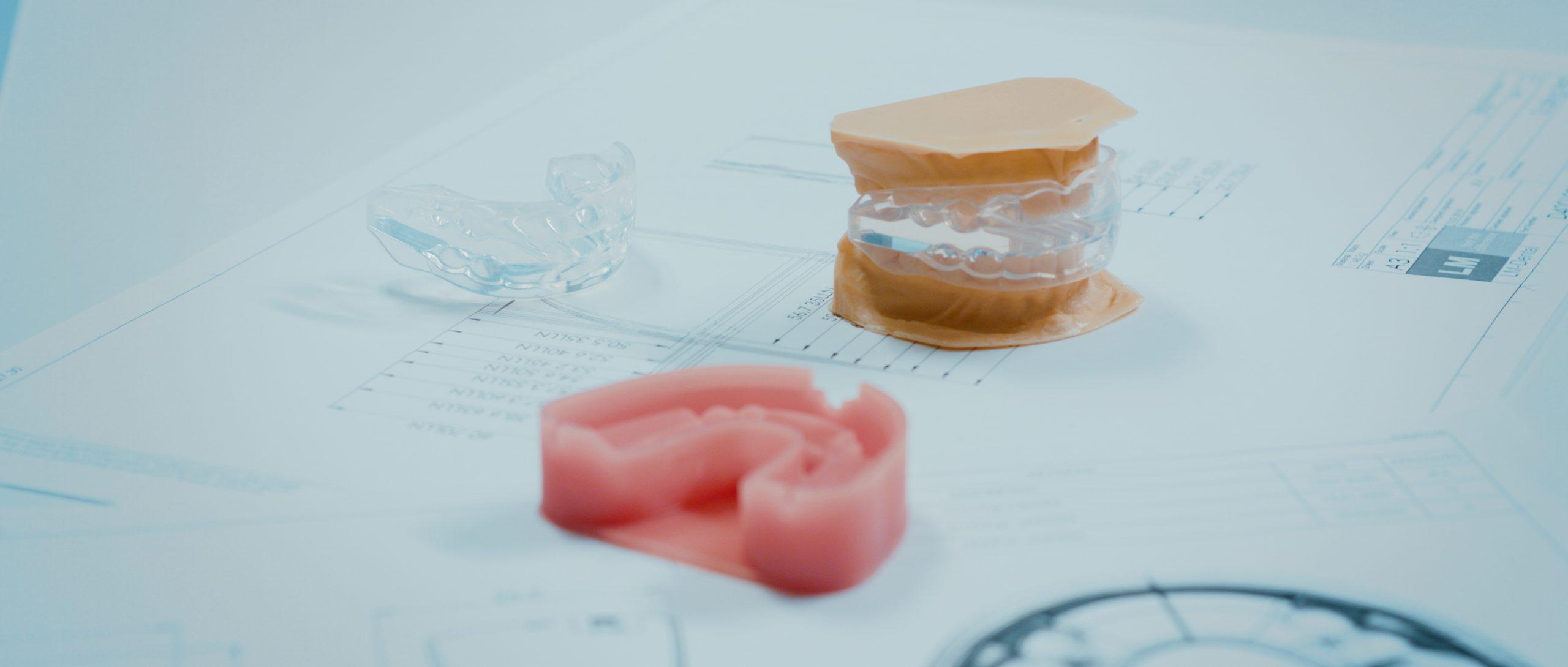
Dental models are replicas of a patient's teeth and surrounding oral structures, used in dental practice and education. They are typically created using plaster or other materials that harden to accurately reproduce the shape and position of each tooth, as well as the contours of the gums and palate. Dental models may be used for a variety of purposes, including treatment planning, creating custom-fitted dental appliances, and teaching dental students about oral anatomy and various dental procedures. They provide a tactile and visual representation that can aid in understanding and communication between dentists, patients, and other dental professionals.
Tongue habits refer to the specific and repetitive ways in which an individual's tongue moves or rests inside their mouth. These habits can include things like tongue thrusting, where the tongue presses against the front teeth during speech or swallowing; tongue sucking, where the tongue is placed against the roof of the mouth; or improper tongue positioning during rest, where the tongue may be positioned too far forward in the mouth or rest against the bottom teeth.
Tongue habits can have an impact on dental and oral health, as well as speech development and clarity. For example, persistent tongue thrusting can lead to an open bite, where the front teeth do not come together when the mouth is closed. Improper tongue positioning during rest can also contribute to the development of a deep overbite or an anterior open bite.
In some cases, tongue habits may be related to underlying conditions such as muscle weakness or sensory integration disorders. Speech-language pathologists and orthodontists may work together to assess and address tongue habits in order to improve oral function and overall health.
Dental occlusion, centric refers to the alignment and contact of the opposing teeth when the jaw is closed in a neutral position, specifically with the mandible (lower jaw) positioned in maximum intercuspation. This means that all teeth are in full contact with their corresponding teeth in the opposite jaw, and the condyles of the mandible are seated in the most posterior portion of the glenoid fossae (the sockets in the skull where the mandible articulates). Centric occlusion is an important concept in dentistry as it serves as a reference point for establishing proper bite relationships during restorative dental treatment.
Orthodontic appliances are devices used in orthodontics, a branch of dentistry focused on the diagnosis, prevention, and treatment of dental and facial irregularities. These appliances can be fixed or removable and are used to align teeth, correct jaw relationships, or modify dental forces. They can include braces, aligners, palatal expanders, space maintainers, and headgear, among others. The specific type of appliance used depends on the individual patient's needs and the treatment plan developed by the orthodontist.
"Sucking behavior" is not a term typically used in medical terminology. However, in the context of early childhood development and behavior, "non-nutritive sucking" is a term that may be used to describe an infant or young child's habitual sucking on their thumb, fingers, or pacifiers, beyond what is necessary for feeding. This type of sucking behavior can provide a sense of security, comfort, or help to self-soothe and manage stress or anxiety.
It's important to note that while non-nutritive sucking is generally considered a normal part of early childhood development, persistent sucking habits beyond the age of 2-4 years may lead to dental or orthodontic problems such as an overbite or open bite. Therefore, it's recommended to monitor and address these behaviors if they persist beyond this age range.
The Index of Orthodontic Treatment Need (IOTN) is a clinical tool used in orthodontics to assess and determine the need for orthodontic treatment based on dental health components and aesthetic considerations. It was developed to standardize the process of determining treatment priority and eligibility in various healthcare systems.
The IOTN consists of two parts: the Dental Health Component (DHC) and the Aesthetic Component (AC).
1. Dental Health Component (DHC): This part evaluates malocclusion based on specific dental health criteria, which are further divided into five grades:
Grade 1: Little or no treatment needed. The occlusion is satisfactory with minor discrepancies that do not require active orthodontic treatment.
Grade 2: Treatment might be beneficial. There are definite but slight anomalies that would benefit from orthodontic care, although they may not necessarily require immediate attention.
Grade 3: Treatment is clearly necessary. Moderate anomalies are present, and treatment is required to prevent significant worsening of dental health or aesthetics.
Grade 4: Treatment is needed to avoid severe dental disease. Significant malocclusion is present, which may lead to functional impairment, periodontal issues, or tooth wear if left untreated.
Grade 5: Immediate treatment is required. Severe malocclusions are present that can cause significant functional impairment and/or severe dental health problems if not treated promptly.
2. Aesthetic Component (AC): This part assesses the impact of malocclusion on a patient's appearance and self-perception, using a scale from 1 to 10, with 1 being the most attractive and 10 being the least attractive. The scale is based on the perceptions of laypeople rather than dental professionals.
The IOTN helps healthcare providers prioritize orthodontic treatment for patients who need it most, ensuring that limited resources are allocated fairly and efficiently.
Tooth movement, in a dental and orthodontic context, refers to the physical change in position or alignment of one or more teeth within the jaw bone as a result of controlled forces applied through various orthodontic appliances such as braces, aligners, or other orthodontic devices. The purposeful manipulation of these forces encourages the periodontal ligament (the tissue that connects the tooth to the bone) to remodel, allowing the tooth to move gradually over time into the desired position. This process is crucial in achieving proper bite alignment, correcting malocclusions, and enhancing overall oral function and aesthetics.
Dental occlusion, traumatic is a term used to describe an abnormal bite or contact between the upper and lower teeth that results in trauma or injury to the oral structures. This can occur when there is a discrepancy in the alignment of the teeth or jaws, such as an overbite, underbite, or crossbite, which causes excessive force or pressure on certain teeth or tissues.
Traumatic dental occlusion can result in various dental and oral health issues, including tooth wear, fractures, mobility of teeth, gum recession, and temporomandibular joint (TMJ) disorders. It is important to diagnose and treat traumatic dental occlusion early to prevent further damage and alleviate any discomfort or pain. Treatment options may include orthodontic treatment, adjustment of the bite, restoration of damaged teeth, or a combination of these approaches.
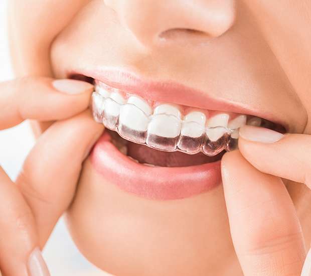
Orthodontic appliances, removable, are dental devices that can be removed and inserted by the patient as needed or directed. These appliances are designed to align and straighten teeth, correct bite issues, and improve the function and appearance of the teeth and jaws. They are typically made from materials such as plastic, metal, or acrylic and may include components like wires, springs, or screws. Examples of removable orthodontic appliances include aligners, retainers, and space maintainers. The specific type and design of the appliance will depend on the individual patient's orthodontic needs and treatment goals.
"Serial extraction" is not a widely recognized or established term in medical or dental literature. However, within the context of dentistry, it could potentially refer to the sequential removal of multiple teeth during separate appointments. This approach may be used when extracting multiple problematic teeth to minimize the risk of complications such as excessive bleeding, swelling, or infection that can arise from removing numerous teeth at once. It is essential to consult a dental professional for a precise understanding and application of this term in a medical context.
In medical terms, a "lip" refers to the thin edge or border of an organ or other biological structure. However, when people commonly refer to "the lip," they are usually talking about the lips on the face, which are part of the oral cavity. The lips are a pair of soft, fleshy tissues that surround the mouth and play a crucial role in various functions such as speaking, eating, drinking, and expressing emotions.
The lips are made up of several layers, including skin, muscle, blood vessels, nerves, and mucous membrane. The outer surface of the lips is covered by skin, while the inner surface is lined with a moist mucous membrane. The muscles that make up the lips allow for movements such as pursing, puckering, and smiling.
The lips also contain numerous sensory receptors that help detect touch, temperature, pain, and other stimuli. Additionally, they play a vital role in protecting the oral cavity from external irritants and pathogens, helping to keep the mouth clean and healthy.
A Jaw Relation Record (also known as a "mounted cast" or "articulated record") is a dental term used to describe the process of recording and replicating the precise spatial relationship between the upper and lower jaws. This information is crucial in various dental treatments, such as designing and creating dental restorations, dentures, or orthodontic appliances.
The Jaw Relation Record typically involves these steps:
1. Determining the optimal jaw position (occlusion) during a clinical procedure called "bite registration." This is done by using various materials like waxes, silicones, or impression compounds to record the relationship between the upper and lower teeth in a static position or at specific movements.
2. Transferring this bite registration to an articulator, which is a mechanical device that simulates jaw movement. The articulator holds dental casts (replicas of the patient's teeth) and allows for adjustments based on the recorded jaw relationship.
3. Mounting the dental casts onto the articulator according to the bite registration. This creates an accurate representation of the patient's oral structures, allowing dentists or technicians to evaluate, plan, and fabricate dental restorations that will fit harmoniously in the mouth and provide optimal function and aesthetics.
In summary, a Jaw Relation Record is a critical component in dental treatment planning and restoration design, as it captures and replicates the precise spatial relationship between the upper and lower jaws.
The facial bones, also known as the facial skeleton, are a series of bones that make up the framework of the face. They include:
1. Frontal bone: This bone forms the forehead and the upper part of the eye sockets.
2. Nasal bones: These two thin bones form the bridge of the nose.
3. Maxilla bones: These are the largest bones in the facial skeleton, forming the upper jaw, the bottom of the eye sockets, and the sides of the nose. They also contain the upper teeth.
4. Zygomatic bones (cheekbones): These bones form the cheekbones and the outer part of the eye sockets.
5. Palatine bones: These bones form the back part of the roof of the mouth, the side walls of the nasal cavity, and contribute to the formation of the eye socket.
6. Inferior nasal conchae: These are thin, curved bones that form the lateral walls of the nasal cavity and help to filter and humidify air as it passes through the nose.
7. Lacrimal bones: These are the smallest bones in the skull, located at the inner corner of the eye socket, and help to form the tear duct.
8. Mandible (lower jaw): This is the only bone in the facial skeleton that can move. It holds the lower teeth and forms the chin.
These bones work together to protect vital structures such as the eyes, brain, and nasal passages, while also providing attachment points for muscles that control chewing, expression, and other facial movements.
Prognathism is a dental and maxillofacial term that refers to a condition where the jaw, particularly the lower jaw (mandible), protrudes or sticks out beyond the normal range, resulting in the forward positioning of the chin and teeth. It can be classified as horizontal or vertical, depending on whether the protrusion is side-to-side or up-and-down.
This condition can be mild or severe and may affect one's appearance and dental health. In some cases, it can also cause issues with speaking, chewing, and breathing. Prognathism can be a result of genetic factors or certain medical conditions, such as acromegaly or gigantism. Treatment options for prognathism include orthodontic treatment, surgery, or a combination of both.
Maxillofacial abnormalities, also known as craniofacial anomalies, refer to a broad range of structural and functional disorders that affect the development of the skull, face, jaws, and related soft tissues. These abnormalities can result from genetic factors, environmental influences, or a combination of both. They can vary in severity, from minor cosmetic issues to significant impairments of vital functions such as breathing, speaking, and eating.
Examples of maxillofacial abnormalities include cleft lip and palate, craniosynostosis (premature fusion of the skull bones), hemifacial microsomia (underdevelopment of one side of the face), and various other congenital anomalies. These conditions may require multidisciplinary treatment involving surgeons, orthodontists, speech therapists, and other healthcare professionals to address both functional and aesthetic concerns.
Myofunctional therapy, also known as orofacial myofunctional therapy, is a type of treatment that aims to correct improper muscle function in the face and mouth. It typically involves a series of exercises and techniques designed to improve oral rest posture, swallowing patterns, chewing, and speech. The goal of myofunctional therapy is to restore normal muscle function, which can help alleviate a variety of symptoms such as tongue thrust, mouth breathing, sleep-disordered breathing, and even some orthodontic problems. This type of therapy is usually provided by a trained speech-language pathologist, dentist, or orthodontist.
Facial asymmetry refers to a condition in which the facial features are not identical or proportionate on both sides of a vertical line drawn down the middle of the face. This can include differences in the size, shape, or positioning of facial features such as the eyes, ears, nose, cheeks, and jaw. Facial asymmetry can be mild and barely noticeable, or it can be more severe and affect a person's appearance and/or functionality of the mouth and jaw.
Facial asymmetry can be present at birth (congenital) or can develop later in life due to various factors such as injury, surgery, growth disorders, nerve damage, or tumors. In some cases, facial asymmetry may not cause any medical problems and may only be of cosmetic concern. However, in other cases, it may indicate an underlying medical condition that requires treatment.
Depending on the severity and cause of the facial asymmetry, treatment options may include cosmetic procedures such as fillers or surgery, orthodontic treatment, physical therapy, or medication to address any underlying conditions.
Facial muscles, also known as facial nerves or cranial nerve VII, are a group of muscles responsible for various expressions and movements of the face. These muscles include:
1. Orbicularis oculi: muscle that closes the eyelid and raises the upper eyelid
2. Corrugator supercilii: muscle that pulls the eyebrows down and inward, forming wrinkles on the forehead
3. Frontalis: muscle that raises the eyebrows and forms horizontal wrinkles on the forehead
4. Procerus: muscle that pulls the medial ends of the eyebrows downward, forming vertical wrinkles between the eyebrows
5. Nasalis: muscle that compresses or dilates the nostrils
6. Depressor septi: muscle that pulls down the tip of the nose
7. Levator labii superioris alaeque nasi: muscle that raises the upper lip and flares the nostrils
8. Levator labii superioris: muscle that raises the upper lip
9. Zygomaticus major: muscle that raises the corner of the mouth, producing a smile
10. Zygomaticus minor: muscle that raises the nasolabial fold and corner of the mouth
11. Risorius: muscle that pulls the angle of the mouth laterally, producing a smile
12. Depressor anguli oris: muscle that pulls down the angle of the mouth
13. Mentalis: muscle that raises the lower lip and forms wrinkles on the chin
14. Buccinator: muscle that retracts the cheek and helps with chewing
15. Platysma: muscle that depresses the corner of the mouth and wrinkles the skin of the neck.
These muscles are innervated by the facial nerve, which arises from the brainstem and exits the skull through the stylomastoid foramen. Damage to the facial nerve can result in facial paralysis or weakness on one or both sides of the face.
The skull base is the lower part of the skull that forms the floor of the cranial cavity and the roof of the facial skeleton. It is a complex anatomical region composed of several bones, including the frontal, sphenoid, temporal, occipital, and ethmoid bones. The skull base supports the brain and contains openings for blood vessels and nerves that travel between the brain and the face or neck. The skull base can be divided into three regions: the anterior cranial fossa, middle cranial fossa, and posterior cranial fossa, which house different parts of the brain.
Orthodontic anchorage procedures refer to the methods and techniques used in orthodontics to achieve stable, controlled movement of teeth during treatment. The term "anchorage" describes the point of stability around which other teeth are moved.
There are two main types of anchorage: absolute and relative. Absolute anchorage is when the force applied to move teeth does not cause any unwanted movement in the area providing stability. Relative anchorage is when some degree of reciprocal movement is expected in the area providing stability.
Orthodontic appliances, such as mini-screws, palatal implants, and headgear, are often used to provide additional anchorage reinforcement. These devices help control the direction and magnitude of forces applied during treatment, ensuring predictable tooth movement and maintaining proper alignment and occlusion (bite).
In summary, orthodontic anchorage procedures involve the strategic use of various appliances and techniques to establish a stable foundation for moving teeth during orthodontic treatment. This helps ensure optimal treatment outcomes and long-term stability of the dentition.
The Sella Turcica, also known as the Turkish saddle, is a depression or fossa in the sphenoid bone located at the base of the skull. It forms a housing for the pituitary gland, which is a small endocrine gland often referred to as the "master gland" because it controls other glands and makes several essential hormones. The Sella Turcica has a saddle-like shape, with its anterior and posterior clinoids forming the front and back of the saddle, respectively. This region is of significant interest in neuroimaging and clinical settings, as various conditions such as pituitary tumors or other abnormalities may affect the size, shape, and integrity of the Sella Turcica.
A bicuspid valve, also known as a mitral valve in the heart, is a heart valve that has two leaflets or cusps. It lies between the left atrium and the left ventricle and helps to regulate blood flow between these two chambers of the heart. In a healthy heart, the bicuspid valve opens to allow blood to flow from the left atrium into the left ventricle and closes tightly to prevent blood from flowing back into the left atrium during contraction of the ventricle.
A congenital heart defect known as a bicuspid aortic valve occurs when the aortic valve, which normally has three leaflets or cusps, only has two. This can lead to narrowing of the valve (aortic stenosis) or leakage of the valve (aortic regurgitation), which can cause symptoms and may require medical treatment.
The nasal bones are a pair of small, thin bones located in the upper part of the face, specifically in the middle of the nose. They articulate with each other at the nasal bridge and with the frontal bone above, the maxillae (upper jaw bones) on either side, and the septal cartilage inside the nose. The main function of the nasal bones is to form the bridge of the nose and protect the nasal cavity. Any damage to these bones can result in a fracture or broken nose.
Tooth abnormalities refer to any variations or irregularities in the size, shape, number, structure, or development of teeth that deviate from the typical or normal anatomy. These abnormalities can occur in primary (deciduous) or permanent teeth and can be caused by genetic factors, environmental influences, systemic diseases, or localized dental conditions during tooth formation.
Some examples of tooth abnormalities include:
1. Microdontia - teeth that are smaller than normal in size.
2. Macrodontia - teeth that are larger than normal in size.
3. Peg-shaped teeth - teeth with a narrow, conical shape.
4. Talon cusps - additional cusps or points on the biting surface of a tooth.
5. Dens invaginatus - an abnormal development where the tooth crown has an extra fold or pouch that can trap bacteria and cause dental problems.
6. Taurodontism - teeth with large pulp chambers and short roots.
7. Supernumerary teeth - having more teeth than the typical number (20 primary and 32 permanent teeth).
8. Hypodontia - missing one or more teeth due to a failure of development.
9. Germination - two adjacent teeth fused together, usually occurring in the front teeth.
10. Fusion - two separate teeth that have grown together during development.
Tooth abnormalities may not always require treatment unless they cause functional, aesthetic, or dental health issues. A dentist can diagnose and manage tooth abnormalities through various treatments, such as fillings, extractions, orthodontic care, or restorative procedures.
Bruxism is the medical term for grinding or clenching your teeth. It's often an unconscious habit that can occur during the day or at night (nocturnal bruxism). Mild bruxism may not require treatment, but chronic, severe grinding can lead to jaw disorders, headaches, and damaged teeth.
There are several potential causes of bruxism, including stress, anxiety, certain medications, alcohol and drug use, and sleep disorders. Dentists often diagnose bruxism based on the visible signs of wear on your teeth, or they may ask you about symptoms you're experiencing. Treatment for bruxism can include stress management techniques, dental guards to protect your teeth during sleep, and in some cases, medication.
The mandibular condyle is a part of the temporomandibular joint (TMJ) in the human body. It is a rounded eminence at the end of the mandible (lower jawbone) that articulates with the glenoid fossa of the temporal bone in the skull, allowing for movements such as opening and closing the mouth, chewing, speaking, and swallowing. The mandibular condyle has both a fibrocartilaginous articular surface and a synovial joint capsule surrounding it, which provides protection and lubrication during these movements.
A pacifier, also known as a soother or dummy, is a rubber, plastic, or silicone teething device that is designed to be sucked upon. It has a nipple-like part that the baby or infant sucks on to derive a sense of security, comfort, and relaxation. Pacifiers are often used to help soothe a crying or fussy baby, to help them fall asleep, or to calm them during stressful situations. They come in various shapes, sizes, and colors, and can be orthodontic or non-orthodontic. It is recommended that pacifier use should be stopped by the age of 2-3 years to prevent dental and speech development issues.
I'm sorry for any confusion, but "smiling" is not typically considered a medical term. It's a part of normal human behavior and facial expression. A smile usually refers to the action of turning up the corners of the mouth to signal pleasure, affection, amusement, or approval. However, in a medical context, a person may be unable to smile due to various conditions, such as facial paralysis caused by Bell's palsy or a stroke. In such cases, the ability to smile can be an important part of rehabilitation and recovery.
Odontometry is a term used in dentistry that refers to the measurement of teeth, particularly the size and length of teeth or tooth roots. It is often used in forensic dentistry for identification purposes, such as in age estimation, sex determination, or individual identification of human remains. The measurements can be taken using various methods, including radiographs (x-rays), calipers, or specialized software.
In some contexts, odontometry may also refer to the process of measuring the amount of dental work required for a particular treatment plan, although this usage is less common.
Temporomandibular Joint Disorders (TMD) refer to a group of conditions that cause pain and dysfunction in the temporomandibular joint (TMJ) and the muscles that control jaw movement. The TMJ is the hinge joint that connects the lower jaw (mandible) to the skull (temporal bone) in front of the ear. It allows for movements required for activities such as eating, speaking, and yawning.
TMD can result from various causes, including:
1. Muscle tension or spasm due to clenching or grinding teeth (bruxism), stress, or jaw misalignment
2. Dislocation or injury of the TMJ disc, which is a small piece of cartilage that acts as a cushion between the bones in the joint
3. Arthritis or other degenerative conditions affecting the TMJ
4. Bite problems (malocclusion) leading to abnormal stress on the TMJ and its surrounding muscles
5. Stress, which can exacerbate existing TMD symptoms by causing muscle tension
Symptoms of Temporomandibular Joint Disorders may include:
- Pain or tenderness in the jaw, face, neck, or shoulders
- Limited jaw movement or locking of the jaw
- Clicking, popping, or grating sounds when moving the jaw
- Headaches, earaches, or dizziness
- Difficulty chewing or biting
- Swelling on the side of the face
Treatment for TMD varies depending on the severity and cause of the condition. It may include self-care measures (like eating soft foods, avoiding extreme jaw movements, and applying heat or cold packs), physical therapy, medications (such as muscle relaxants, pain relievers, or anti-inflammatory drugs), dental work (including bite adjustments or orthodontic treatment), or even surgery in severe cases.
Centric relation is a term used in dentistry to describe the relationship between the maxilla (upper jaw) and mandible (lower jaw) when the condyles (the rounded ends of the lower jaw bone) are in the most superior, anterior, and posterior position in the glenoid fossae (the sockets in the skull where the condyles sit). This is considered to be a neutral and reproducible position that can be used as a reference point for establishing proper occlusion (bite) and jaw alignment during dental treatment, such as constructing dentures or performing orthodontic treatment.
It's important to note that there are different philosophies and schools of thought regarding the definition and clinical significance of centric relation, and not all dentists agree on its importance or relevance in practice.
Orthodontic wires are typically made of stainless steel, nickel-titanium alloy, or other shape memory alloys, and are used in orthodontics to move teeth into the desired position. They are attached to brackets bonded to the teeth and exert a continuous force to align the teeth and correct malocclusions (bites that do not fit together correctly). The wires come in various sizes, shapes, and materials, each with specific properties that make them suitable for different stages of treatment. Some wires are flexible and used during the initial alignment phase, while others are more rigid and used during the finishing phase to achieve precise tooth movements.
Peer review in the context of health care is a process used to maintain standards and improve the quality of healthcare practices, research, and publications. It involves the evaluation of work or research conducted by professionals within the same field, who are considered peers. The purpose is to provide an objective assessment of the work, identify any errors or biases, ensure that the methods and conclusions are sound, and offer suggestions for improvement.
In health care, peer review can be applied to various aspects including:
1. Clinical Practice: Healthcare providers regularly review each other's work to maintain quality standards in patient care, diagnoses, treatment plans, and adherence to evidence-based practices.
2. Research: Before research findings are published in medical journals, they undergo a rigorous peer-review process where experts assess the study design, methodology, data analysis, interpretation of results, and conclusions to ensure the validity and reliability of the research.
3. Publications: Medical journals use peer review to evaluate and improve the quality of articles submitted for publication. This helps to maintain the credibility and integrity of the published literature, ensuring that it is accurate, unbiased, and relevant to the field.
4. Education and Training Programs: Peer review is also used in evaluating the content and delivery of medical education programs, continuing professional development courses, and training curricula to ensure they meet established standards and are effective in enhancing the knowledge and skills of healthcare professionals.
5. Healthcare Facilities and Institutions: Accreditation bodies and regulatory authorities use peer review as part of their evaluation processes to assess the quality and safety of healthcare facilities and institutions, identifying areas for improvement and ensuring compliance with regulations and standards.
Tooth eruption is the process by which a tooth emerges from the gums and becomes visible in the oral cavity. It is a normal part of dental development that occurs in a predictable sequence and timeframe. Primary or deciduous teeth, also known as baby teeth, begin to erupt around 6 months of age and continue to emerge until approximately 2-3 years of age. Permanent or adult teeth start to erupt around 6 years of age and can continue to emerge until the early twenties.
The process of tooth eruption involves several stages, including the formation of the tooth within the jawbone, the movement of the tooth through the bone and surrounding tissues, and the final emergence of the tooth into the mouth. Proper tooth eruption is essential for normal oral function, including chewing, speaking, and smiling. Any abnormalities in the tooth eruption process, such as delayed or premature eruption, can indicate underlying dental or medical conditions that require further evaluation and treatment.
Mastication is the medical term for the process of chewing food. It's the first step in digestion, where food is broken down into smaller pieces by the teeth, making it easier to swallow and further digest. The act of mastication involves not only the physical grinding and tearing of food by the teeth but also the mixing of the food with saliva, which contains enzymes that begin to break down carbohydrates. This process helps to enhance the efficiency of digestion and nutrient absorption in the subsequent stages of the digestive process.
Orthognathic surgical procedures are a type of surgery used to correct jaw misalignments and improve the bite and function of the jaws. The term "orthognathic" comes from the Greek words "orthos," meaning straight or correct, and "gnathos," meaning jaw. These surgeries are typically performed by oral and maxillofacial surgeons in conjunction with orthodontic treatment to achieve proper alignment of the teeth and jaws.
Orthognathic surgical procedures may be recommended for patients who have significant discrepancies between the size and position of their upper and lower jaws, which can result in problems with chewing, speaking, breathing, and sleeping. These procedures can also improve facial aesthetics by correcting jaw deformities and imbalances.
The specific surgical procedure used will depend on the nature and extent of the jaw misalignment. Common orthognathic surgical procedures include:
1. Maxillary osteotomy: This procedure involves making cuts in the upper jawbone (maxilla) and moving it forward or backward to correct a misalignment.
2. Mandibular osteotomy: This procedure involves making cuts in the lower jawbone (mandible) and moving it forward or backward to correct a misalignment.
3. Genioplasty: This procedure involves reshaping or repositioning the chin bone (mentum) to improve facial aesthetics and jaw function.
4. Orthognathic surgery for sleep apnea: This procedure involves repositioning the upper and/or lower jaws to open up the airway and improve breathing during sleep.
Orthognathic surgical procedures require careful planning and coordination between the surgeon, orthodontist, and patient. The process typically involves taking detailed measurements and images of the jaw and teeth, creating a surgical plan, and undergoing orthodontic treatment to align the teeth prior to surgery. After surgery, patients may need to wear braces or other appliances to maintain the alignment of their teeth and jaws during healing.
Dental health surveys are epidemiological studies that aim to assess the oral health status and related behaviors of a defined population at a particular point in time. These surveys collect data on various aspects of oral health, including the prevalence and severity of dental diseases such as caries (tooth decay), periodontal disease (gum disease), and oral cancer. They also gather information on factors that influence oral health, such as dietary habits, oral hygiene practices, access to dental care, and socioeconomic status.
The data collected in dental health surveys are used to identify trends and patterns in oral health, plan and evaluate public health programs and policies, and allocate resources for oral health promotion and disease prevention. Dental health surveys may be conducted at the local, regional, or national level, and they can target specific populations such as children, adolescents, adults, or older adults.
The methods used in dental health surveys include clinical examinations, interviews, questionnaires, and focus groups. Clinical examinations are conducted by trained dentists or dental hygienists who follow standardized protocols to assess the oral health status of participants. Interviews and questionnaires are used to collect information on demographic characteristics, oral health behaviors, and attitudes towards oral health. Focus groups can provide insights into the perceptions and experiences of participants regarding oral health issues.
Overall, dental health surveys play a critical role in monitoring and improving the oral health of populations and reducing oral health disparities.
The masseter muscle is a strong chewing muscle in the jaw. It is a broad, thick, quadrilateral muscle that extends from the zygomatic arch (cheekbone) to the lower jaw (mandible). The masseter muscle has two distinct parts: the superficial part and the deep part.
The superficial part of the masseter muscle originates from the lower border of the zygomatic process of the maxilla and the anterior two-thirds of the inferior border of the zygomatic arch. The fibers of this part run almost vertically downward to insert on the lateral surface of the ramus of the mandible and the coronoid process.
The deep part of the masseter muscle originates from the deep surface of the zygomatic arch and inserts on the medial surface of the ramus of the mandible, blending with the temporalis tendon.
The primary function of the masseter muscle is to elevate the mandible, helping to close the mouth and clench the teeth together during mastication (chewing). It also plays a role in stabilizing the jaw during biting and speaking. The masseter muscle is one of the most powerful muscles in the human body relative to its size.
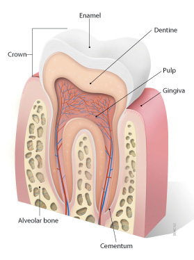
A tooth is a hard, calcified structure found in the jaws (upper and lower) of many vertebrates and used for biting and chewing food. In humans, a typical tooth has a crown, one or more roots, and three layers: the enamel (the outermost layer, hardest substance in the body), the dentin (the layer beneath the enamel), and the pulp (the innermost layer, containing nerves and blood vessels). Teeth are essential for proper nutrition, speech, and aesthetics. There are different types of teeth, including incisors, canines, premolars, and molars, each designed for specific functions in the mouth.
Orthodontic retainers are dental appliances that are custom-made and used after orthodontic treatment (such as braces) to help maintain the new position of teeth. They can be fixed or removable and are designed to keep the teeth in place while the surrounding gums and bones stabilize in their new positions. Retainers play a crucial role in preserving the investment made during orthodontic treatment, preventing the teeth from shifting back to their original positions.
A deciduous tooth, also known as a baby tooth or primary tooth, is a type of temporary tooth that humans and some other mammals develop during childhood. They are called "deciduous" because they are eventually shed and replaced by permanent teeth, much like how leaves on a deciduous tree fall off and are replaced by new growth.
Deciduous teeth begin to form in the womb and start to erupt through the gums when a child is around six months old. By the time a child reaches age three, they typically have a full set of 20 deciduous teeth, including incisors, canines, and molars. These teeth are smaller and less durable than permanent teeth, but they serve important functions such as helping children chew food properly, speak clearly, and maintain space in the jaw for the permanent teeth to grow into.
Deciduous teeth usually begin to fall out around age six or seven, starting with the lower central incisors. This process continues until all of the deciduous teeth have been shed, typically by age 12 or 13. At this point, the permanent teeth will have grown in and taken their place, with the exception of the wisdom teeth, which may not erupt until later in adolescence or early adulthood.
A cuspid, also known as a canine tooth or cuspid tooth, is a type of tooth in mammals. It is the pointiest tooth in the dental arch and is located between the incisors and bicuspids (or premolars). Cuspids have a single cusp or pointed tip that is used for tearing and grasping food. In humans, there are four cuspids, two on the upper jaw and two on the lower jaw, one on each side of the dental arch.
An "osteotomy" refers to a surgical procedure in which a bone is cut. A "Le Fort osteotomy" is a specific type of osteotomy that involves cutting and repositioning the middle (midface) portion of the facial bones. There are three types of Le Fort osteotomies, named after the French surgeon René Le Fort who first described them:
1. Le Fort I osteotomy: This procedure involves making a horizontal cut through the lower part of the maxilla (upper jaw) and separating it from the rest of the facial bones. It is often used to treat conditions such as severe jaw deformities or obstructive sleep apnea.
2. Le Fort II osteotomy: In this procedure, an upward curved cut is made through the lower part of the maxilla and the middle portion of the nasal bones. This allows for the repositioning of the midface and nose. It may be used to treat conditions such as severe facial fractures or congenital deformities.
3. Le Fort III osteotomy: A Le Fort III osteotomy involves making a cut through the upper part of the maxilla, the orbital bones (bones surrounding the eyes), and the zygomatic bones (cheekbones). This procedure allows for significant repositioning of the midface and is often used to treat severe facial fractures or congenital deformities.
It's important to note that Le Fort osteotomies are complex surgical procedures that should only be performed by experienced oral and maxillofacial surgeons or craniofacial surgeons.
Anodontia is a medical term that refers to the congenital absence or lack of development of all primary (deciduous) and/or permanent teeth. It is a rare dental condition that affects tooth development and can be isolated or associated with various syndromes and genetic disorders.
In anodontia, the dental tissues responsible for forming teeth, including the dental lamina, dental papilla, and dental follicle, fail to develop properly, resulting in missing teeth. The condition can affect all teeth or only some of them, leading to partial anodontia.
Anodontia is different from hypodontia, which refers to the congenital absence of one or more, but not all, teeth. It is also distinct from oligodontia, which is the absence of six or more permanent teeth, excluding third molars (wisdom teeth).
People with anodontia may experience difficulties in chewing, speaking, and maintaining oral hygiene, leading to various dental and social problems. Prosthodontic treatments, such as dentures or implants, are often necessary to restore oral function and aesthetics.
Mandibular advancement is a treatment approach used in dentistry and sleep medicine, which involves the surgical or non-surgical forward movement of the mandible (lower jaw) to address certain medical conditions. The most common use of mandibular advancement is in the treatment of obstructive sleep apnea (OSA), where the tongue and soft tissues at the back of the throat can collapse into the airway during sleep, causing obstruction and breathing difficulties.
Mandibular advancement devices (MADs) are often used in non-surgical treatments. These custom-made oral appliances look similar to mouthguards or sports guards and are worn during sleep. They work by holding the lower jaw in a slightly forward position, which helps to keep the airway open and prevents the tongue and soft tissues from collapsing into it.
Surgical mandibular advancement is another option for patients with severe OSA who cannot tolerate or do not respond well to MADs or other treatments like continuous positive airway pressure (CPAP). In this procedure, the jaw is surgically moved forward and stabilized in that position using plates, screws, or wires. This creates more space in the airway and reduces the risk of obstruction during sleep.
In summary, mandibular advancement refers to the movement of the lower jaw forward, either through non-surgical means like MADs or surgical interventions, with the primary goal of treating obstructive sleep apnea by maintaining a patent airway during sleep.
Jaw abnormalities, also known as maxillofacial abnormalities, refer to any structural or functional deviations from the normal anatomy and physiology of the jaw bones (mandible and maxilla) and the temporomandibular joint (TMJ). These abnormalities can be present at birth (congenital) or acquired later in life due to various factors such as trauma, infection, tumors, or degenerative diseases.
Examples of jaw abnormalities include:
1. Micrognathia: a condition where the lower jaw is underdeveloped and appears recessed or small.
2. Prognathism: a condition where the lower jaw protrudes forward beyond the normal position.
3. Maxillary hypoplasia/aplasia: a condition where the upper jaw is underdeveloped or absent.
4. Mandibular hypoplasia/aplasia: a condition where the lower jaw is underdeveloped or absent.
5. Condylar hyperplasia: a condition where one or both of the condyles (the rounded ends of the mandible that articulate with the skull) continue to grow abnormally, leading to an asymmetrical jaw and facial deformity.
6. TMJ disorders: conditions affecting the temporomandibular joint, causing pain, stiffness, and limited movement.
7. Jaw tumors or cysts: abnormal growths that can affect the function and structure of the jaw bones.
Jaw abnormalities can cause various problems, including difficulty with chewing, speaking, breathing, and swallowing, as well as aesthetic concerns. Treatment options may include orthodontic treatment, surgery, or a combination of both, depending on the severity and nature of the abnormality.
Permanent dentition is the second and final set of teeth that humans grow during their lifetime. These teeth are also known as adult or secondary teeth and typically begin to erupt in the mouth around the age of 6 or 7 years old, with all permanent teeth usually present by the time a person reaches their late teens or early twenties.
There are 32 teeth in a complete set of permanent dentition, including 8 incisors, 4 canines, 8 premolars (also called bicuspids), and 12 molars (including 4 third molars or wisdom teeth). The primary function of permanent teeth is to help with biting, chewing, and grinding food into smaller pieces that are easier to swallow and digest. Proper care and maintenance of permanent teeth through good oral hygiene practices, regular dental checkups, and a balanced diet can help ensure their longevity and health throughout a person's life.
The stomatognathic system is a term used in medicine and dentistry to refer to the coordinated functions of the mouth, jaw, and related structures. It includes the teeth, gums, tongue, palate, lips, cheeks, salivary glands, as well as the muscles of mastication (chewing), swallowing, and speech. The stomatognathic system also involves the temporomandibular joint (TMJ) and associated structures that allow for movement of the jaw. This complex system works together to enable functions such as eating, speaking, and breathing. Dysfunction in the stomatognathic system can lead to various oral health issues, including temporomandibular disorders, occlusal problems, and orofacial pain.
The zygoma is the scientific name for the cheekbone. It is a part of the facial skeleton that forms the prominence of the cheek and houses the maxillary sinus, one of the pairs of paranasal sinuses. The zygomatic bone, also known as the malar bone, contributes to the formation of the zygoma.
The temporalis muscle is a fan-shaped muscle located in the lateral aspect of the head, in the temporal fossa region. It belongs to the group of muscles known as muscles of mastication, responsible for chewing movements. The temporalis muscle has its origin at the temporal fossa and inserts into the coronoid process and ramus of the mandible. Its main function is to retract the mandible and assist in closing the jaw.
Bite force refers to the amount of force or pressure that can be exerted by the teeth and jaw when biting down or clenching together. It is a measure of an individual's maximum biting strength, typically expressed in units such as pounds (lb) or newtons (N). Bite force is an important factor in various biological and medical contexts, including oral health, nutrition, and the study of animal behavior and evolution.
In humans, bite force can vary widely depending on factors such as age, sex, muscle strength, and dental health. On average, a healthy adult human male may have a maximum bite force of around 150-200 pounds (670-890 newtons), while an adult female may have a bite force of around 100-130 pounds (445-578 newtons). However, these values can vary significantly from person to person.
Abnormalities in bite force can be indicative of various medical conditions or injuries, such as temporomandibular joint disorders (TMD), muscle weakness, or neurological disorders affecting the facial muscles. Assessing and measuring bite force may also be useful in evaluating the effectiveness of dental treatments or appliances, such as dentures or orthodontic devices.
In the context of medical terminology, a "habit" refers to a regular, repeated behavior or practice that is often performed automatically or subconsciously. Habits can be physical (such as biting nails) or mental (such as worrying). They can be harmless, beneficial (like regularly brushing your teeth), or harmful (like smoking cigarettes).
Habits are different from instincts or reflexes because they involve a learned behavior that has been repeated and reinforced over time. Breaking a habit can often be challenging due to the deeply ingrained nature of the behavior.
Occlusal adjustment is a dental procedure that involves modifying the shape and alignment of the biting surfaces of teeth to improve their fit and relationship with the opposing teeth. The goal of occlusal adjustment is to create a balanced and harmonious bite, which can help alleviate symptoms such as tooth wear, sensitivity, pain, or temporomandibular joint disorders (TMJD).
During an occlusal adjustment procedure, the dentist uses specialized instruments like articulating paper or dental burs to identify and eliminate interferences in the bite. These interferences can be caused by high spots, rough edges, or misaligned teeth that prevent the upper and lower teeth from meeting evenly when the jaw is closed. By removing these interferences, the dentist aims to create a more stable and comfortable bite, reducing stress on the jaw joints and muscles.
It's important to note that occlusal adjustment should only be performed by a trained dental professional, as improper modifications can lead to further dental issues or discomfort.
Ectopic tooth eruption is a condition where a tooth fails to erupt into its normal position in the dental arch. Instead, it emerupts in an abnormal location, such as in the wrong direction or through another tissue like the gums, palate, or jawbone. This can occur due to various reasons, including genetics, crowding of teeth, or trauma. Ectopic tooth eruption may cause problems with oral function and dental health, and treatment options depend on the severity and location of the ectopic tooth.
Nonparametric statistics is a branch of statistics that does not rely on assumptions about the distribution of variables in the population from which the sample is drawn. In contrast to parametric methods, nonparametric techniques make fewer assumptions about the data and are therefore more flexible in their application. Nonparametric tests are often used when the data do not meet the assumptions required for parametric tests, such as normality or equal variances.
Nonparametric statistical methods include tests such as the Wilcoxon rank-sum test (also known as the Mann-Whitney U test) for comparing two independent groups, the Wilcoxon signed-rank test for comparing two related groups, and the Kruskal-Wallis test for comparing more than two independent groups. These tests use the ranks of the data rather than the actual values to make comparisons, which allows them to be used with ordinal or continuous data that do not meet the assumptions of parametric tests.
Overall, nonparametric statistics provide a useful set of tools for analyzing data in situations where the assumptions of parametric methods are not met, and can help researchers draw valid conclusions from their data even when the data are not normally distributed or have other characteristics that violate the assumptions of parametric tests.
Oral health is the scientific term used to describe the overall health status of the oral and related tissues, including the teeth, gums, palate, tongue, and mucosal lining. It involves the absence of chronic mouth and facial pain, oral and pharyngeal (throat) cancers, oral soft tissue lesions, birth defects such as cleft lip and palate, and other diseases and disorders that affect the oral cavity.
Good oral health also means being free of decay, gum disease, and other oral infections that can damage the teeth, gums, and bones of the mouth. It is essential to maintain good oral hygiene through regular brushing, flossing, and dental check-ups to prevent dental caries (cavities) and periodontal disease (gum disease).
Additionally, oral health is closely linked to overall health and well-being. Poor oral health has been associated with various systemic diseases, including diabetes, cardiovascular disease, respiratory infections, and stroke. Therefore, maintaining good oral health can contribute to improved general health and quality of life.
Patient care planning is a critical aspect of medical practice that involves the development, implementation, and evaluation of an individualized plan for patients to receive high-quality and coordinated healthcare services. It is a collaborative process between healthcare professionals, patients, and their families that aims to identify the patient's health needs, establish realistic goals, and determine the most effective interventions to achieve those goals.
The care planning process typically includes several key components, such as:
1. Assessment: A comprehensive evaluation of the patient's physical, psychological, social, and environmental status to identify their healthcare needs and strengths.
2. Diagnosis: The identification of the patient's medical condition(s) based on clinical findings and diagnostic tests.
3. Goal-setting: The establishment of realistic and measurable goals that address the patient's healthcare needs and align with their values, preferences, and lifestyle.
4. Intervention: The development and implementation of evidence-based strategies to achieve the identified goals, including medical treatments, therapies, and supportive services.
5. Monitoring and evaluation: The ongoing assessment of the patient's progress towards achieving their goals and adjusting the care plan as needed based on changes in their condition or response to treatment.
Patient care planning is essential for ensuring that patients receive comprehensive, coordinated, and personalized care that promotes their health, well-being, and quality of life. It also helps healthcare professionals to communicate effectively, make informed decisions, and provide safe and effective care that meets the needs and expectations of their patients.
A diagnosis that is made based on the examination and evaluation of the oral cavity, including the teeth, gums, tongue, and other soft tissues. This type of diagnosis may involve a visual exam, medical history review, and various diagnostic tests such as imaging studies or tissue biopsies. The goal of an oral diagnosis is to identify any underlying conditions or diseases that may be present in the oral cavity and determine the appropriate course of treatment. Dentists, dental specialists, and other healthcare professionals may perform oral diagnoses.
Tooth extraction is a dental procedure in which a tooth that is damaged or poses a threat to oral health is removed from its socket in the jawbone. This may be necessary due to various reasons such as severe tooth decay, gum disease, fractured teeth, crowded teeth, or for orthodontic treatment purposes. The procedure is performed by a dentist or an oral surgeon, under local anesthesia to numb the area around the tooth, ensuring minimal discomfort during the extraction process.
Facial pain is a condition characterized by discomfort or pain felt in any part of the face. It can result from various causes, including nerve damage or irritation, injuries, infections, dental problems, migraines, or sinus congestion. The pain can range from mild to severe and may be sharp, dull, constant, or intermittent. In some cases, facial pain can also be associated with other symptoms such as headaches, redness, swelling, or changes in sensation. Accurate diagnosis and treatment of the underlying cause are essential for effective management of facial pain.
"Mesial movement of teeth" is a dental term that refers to the natural drifting or shifting of teeth in a forward direction towards the front of the mouth. This movement typically occurs over time and can be influenced by various factors such as:
* The loss of adjacent teeth, which can create space for other teeth to move into
* Oral habits like thumb sucking or tongue thrusting
* Periodontal disease that weakens the supporting structures of the teeth
* Malocclusion or misalignment of teeth
It is essential to monitor and manage mesial movement of teeth to prevent dental issues such as crowding, malocclusion, and periodontal problems. Dental professionals may use various treatments, including orthodontic appliances, space maintainers, or restorations, to address this issue.
The alveolar process is the curved part of the jawbone (mandible or maxilla) that contains sockets or hollow spaces (alveoli) for the teeth to be embedded. These processes are covered with a specialized mucous membrane called the gingiva, which forms a tight seal around the teeth to help protect the periodontal tissues and maintain oral health.
The alveolar process is composed of both compact and spongy bone tissue. The compact bone forms the outer layer, while the spongy bone is found inside the alveoli and provides support for the teeth. When a tooth is lost or extracted, the alveolar process begins to resorb over time due to the lack of mechanical stimulation from the tooth's chewing forces. This can lead to changes in the shape and size of the jawbone, which may require bone grafting procedures before dental implant placement.
The palate is the roof of the mouth in humans and other mammals, separating the oral cavity from the nasal cavity. It consists of two portions: the anterior hard palate, which is composed of bone, and the posterior soft palate, which is composed of muscle and connective tissue. The palate plays a crucial role in speech, swallowing, and breathing, as it helps to direct food and air to their appropriate locations during these activities.
In medical terms, the face refers to the front part of the head that is distinguished by the presence of the eyes, nose, and mouth. It includes the bones of the skull (frontal bone, maxilla, zygoma, nasal bones, lacrimal bones, palatine bones, inferior nasal conchae, and mandible), muscles, nerves, blood vessels, skin, and other soft tissues. The face plays a crucial role in various functions such as breathing, eating, drinking, speaking, seeing, smelling, and expressing emotions. It also serves as an important identifier for individuals, allowing them to be recognized by others.
Sialorrhea is the medical term for excessive drooling or saliva production. It's not necessarily a condition where the person produces too much saliva, but rather, they are unable to control the normal amount of saliva in their mouth due to various reasons such as neurological disorders, developmental disabilities, or structural issues that affect swallowing and oral motor function.
Common causes include cerebral palsy, Parkinson's disease, amyotrophic lateral sclerosis (ALS), Down syndrome, stroke, intellectual disability, and certain medications. Treatment options vary depending on the cause and severity of the condition and may include medication adjustments, behavioral interventions, oral devices, or even surgical procedures in severe cases.
The hard palate is the anterior, bony part of the roof of the mouth, forming a vertical partition between the oral and nasal cavities. It is composed of the maxilla and palatine bones, and provides attachment for the muscles of the soft palate, which functions in swallowing, speaking, and breathing. The hard palate also contains taste buds that contribute to our ability to taste food.
Tooth attrition is a type of wear on the teeth that results from normal dental occlusal forces during biting, chewing, and grinding of food. It involves the loss of tooth structure by mechanical forces and is typically seen as a flattening or reduction in the vertical height of the crowns of teeth.
Attrition differs from other types of tooth wear such as abrasion (which is caused by external factors like toothbrush bristles, toothpaste, or habitual pen/pencil biting), erosion (which is caused by chemical dissolution of tooth structure due to acid exposure), and abfraction (which is caused by flexural forces leading to cervical lesions).
While some degree of attrition is considered a normal part of the aging process, excessive attrition can lead to dental sensitivity, aesthetic concerns, and even affect the functionality of the teeth and overall oral health. Dental professionals may recommend various treatments such as fillings, crowns, or even orthodontic interventions to manage the consequences of severe tooth attrition.
Panoramic radiography is a specialized type of dental X-ray imaging that captures a panoramic view of the entire mouth, including the teeth, upper and lower jaws, and surrounding structures. It uses a special machine that rotates around the head, capturing images as it moves. This technique provides a two-dimensional image that is helpful in diagnosing and planning treatment for various dental conditions such as impacted teeth, bone abnormalities, and jaw disorders.
The panoramic radiograph can also be used to assess the development and positioning of wisdom teeth, detect cysts or tumors in the jaws, and evaluate the effects of trauma or injury to the mouth. It is a valuable tool for dental professionals as it allows them to see a comprehensive view of the oral structures, which may not be visible with traditional X-ray techniques.
It's important to note that while panoramic radiography provides valuable information, it should be used in conjunction with other diagnostic tools and clinical examinations to ensure accurate diagnosis and treatment planning.
Masticatory muscles are a group of skeletal muscles responsible for the mastication (chewing) process in humans and other animals. They include:
1. Masseter muscle: This is the primary muscle for chewing and is located on the sides of the face, running from the lower jawbone (mandible) to the cheekbone (zygomatic arch). It helps close the mouth and elevate the mandible during chewing.
2. Temporalis muscle: This muscle is situated in the temporal region of the skull, covering the temple area. It assists in closing the jaw, retracting the mandible, and moving it sideways during chewing.
3. Medial pterygoid muscle: Located deep within the cheek, near the angle of the lower jaw, this muscle helps move the mandible forward and grind food during chewing. It also contributes to closing the mouth.
4. Lateral pterygoid muscle: Found inside the ramus (the vertical part) of the mandible, this muscle has two heads - superior and inferior. The superior head helps open the mouth by pulling the temporomandibular joint (TMJ) downwards, while the inferior head assists in moving the mandible sideways during chewing.
These muscles work together to enable efficient chewing and food breakdown, preparing it for swallowing and digestion.
The temporomandibular joint (TMJ) is the articulation between the mandible (lower jaw) and the temporal bone of the skull. It's a complex joint that involves the movement of two bones, several muscles, and various ligaments. The TMJ allows for movements like rotation and translation, enabling us to open and close our mouth, chew, speak, and yawn. Dysfunction in this joint can lead to temporomandibular joint disorders (TMD), which can cause pain, discomfort, and limited jaw movement.
A needs assessment in a medical context is the process of identifying and evaluating the health needs of an individual, population, or community. It is used to determine the resources, services, and interventions required to address specific health issues and improve overall health outcomes. This process often involves collecting and analyzing data on various factors such as demographics, prevalence of diseases, access to healthcare, and social determinants of health. The goal of a needs assessment is to ensure that resources are allocated effectively and efficiently to meet the most pressing health needs and priorities.
Nasal cartilages are the flexible, supportive structures in the nose that contribute to its shape and structure. They are made up of tough, but elastic tissue called cartilage. There are several nasal cartilages, including:
1. The septal cartilage, which is a thin, flat strip that forms the dividing wall between the two sides of the nose.
2. The upper and lower lateral cartilages, which are located on either side of the nostrils and help to shape them.
3. The sesamoid cartilages, which are small, round pieces of cartilage that can be found near the nasal opening.
These cartilages work together to provide support and flexibility to the nose, allowing it to withstand the forces of breathing and other facial movements while maintaining its shape.
Deglutition is the medical term for swallowing. It refers to the process by which food or liquid is transferred from the mouth to the stomach through a series of coordinated muscle movements and neural responses. The deglutition process involves several stages, including oral preparatory, oral transit, pharyngeal, and esophageal phases, each of which plays a critical role in ensuring safe and efficient swallowing.
Dysphagia is the medical term for difficulty with swallowing, which can result from various underlying conditions such as neurological disorders, structural abnormalities, or muscular weakness. Proper evaluation and management of deglutition disorders are essential to prevent complications such as aspiration pneumonia, malnutrition, and dehydration.
A supernumerary tooth, also known as hyperdontia, refers to an additional tooth or teeth that grow beyond the regular number of teeth in the dental arch. These extra teeth can erupt in various locations of the dental arch and may occur in any of the tooth types, but they are most commonly seen as extra premolars or molars, and less frequently as incisors or canines. Supernumerary teeth may be asymptomatic or may cause complications such as crowding, displacement, or impaction of adjacent teeth, and therefore, they often require dental treatment.
An impacted tooth is a condition where a tooth fails to erupt into the oral cavity within its expected time frame, resulting in its partial or complete entrapment within the jawbone or soft tissues. This commonly occurs with wisdom teeth (third molars) but can affect any tooth. Impacted teeth may cause problems such as infection, decay of adjacent teeth, gum disease, or cyst formation, and they may require surgical removal.
In medical terms, the jaw is referred to as the mandible (in humans and some other animals), which is the lower part of the face that holds the lower teeth in place. It's a large, horseshoe-shaped bone that forms the lower jaw and serves as a attachment point for several muscles that are involved in chewing and moving the lower jaw.
In addition to the mandible, the upper jaw is composed of two bones known as the maxillae, which fuse together at the midline of the face to form the upper jaw. The upper jaw holds the upper teeth in place and forms the roof of the mouth, as well as a portion of the eye sockets and nasal cavity.
Together, the mandible and maxillae allow for various functions such as speaking, eating, and breathing.
In dental terminology, "space maintenance" refers to the use of a device or appliance to maintain the proper space between teeth following the loss of a primary (baby) tooth. This is especially important when the lost tooth is a molar, as it plays a crucial role in maintaining the alignment and spacing of the remaining teeth and the eruption path for the developing permanent tooth.
Space maintainers can be fixed or removable and are typically made from materials such as stainless steel, plastic, or acrylic. They help prevent dental issues like crowding, misalignment, and impaction of adjacent and/or succeeding teeth, which may lead to more complex orthodontic treatments in the future. It is essential that space maintainers are custom-made and properly fitted by a dentist or an orthodontist to ensure their effectiveness and avoid potential damage to surrounding tissues.
The medical term for nail biting is "Onychophagia." It's classified as a type of body-focused repetitive behavior, which is a category of mental health disorders characterized by the repeated compulsion to engage in certain self-grooming behaviors that cause physical damage. In the case of onychophagia, the individual repeatedly bites their nails, often until they bleed or become severely damaged. This can lead to various complications, such as infection and dental issues. It's important to note that while nail biting is a common habit, when it becomes repetitive, compulsive, and causes significant distress or impairment, it may be indicative of a broader mental health condition.
"Social desirability bias" is not a medical term per se, but rather a concept that's relevant in the fields of psychology, social sciences, and research methodology. It refers to the tendency of individuals to provide responses that they believe are socially acceptable or desirable, rather than their true feelings, thoughts, or behaviors, during surveys, interviews, or other forms of assessment. This bias can lead to inaccurate or skewed data, as it may not reflect the actual experiences or attitudes of the respondents. It's important for researchers to be aware of and control for social desirability bias to ensure the validity and reliability of their findings.
Bottle feeding is a method of providing nutrition to infants and young children using a bottle and an artificial nipple. The bottle is filled with milk or formula, and the child sucks on the nipple to draw the liquid out. This can be done with expressed breast milk or commercial infant formula. Bottle feeding can be a convenient alternative to breastfeeding, but it is important to follow proper techniques to ensure that the baby is receiving adequate nutrition and to prevent dental problems and ear infections. It's also important to clean the bottles and nipples properly to avoid contamination and growth of bacteria.
The Chi-square distribution is a continuous probability distribution that is often used in statistical hypothesis testing. It is the distribution of a sum of squares of k independent standard normal random variables. The resulting quantity follows a chi-square distribution with k degrees of freedom, denoted as χ²(k).
The probability density function (pdf) of the Chi-square distribution with k degrees of freedom is given by:
f(x; k) = (1/ (2^(k/2) * Γ(k/2))) \* x^((k/2)-1) \* e^(-x/2), for x > 0 and 0, otherwise.
Where Γ(k/2) is the gamma function evaluated at k/2. The mean and variance of a Chi-square distribution with k degrees of freedom are k and 2k, respectively.
The Chi-square distribution has various applications in statistical inference, including testing goodness-of-fit, homogeneity of variances, and independence in contingency tables.
A mandibular osteotomy is a surgical procedure that involves making a cut in the mandible (lower jawbone). This procedure is often performed to correct various dental and maxillofacial conditions such as jaw misalignment, sleep apnea, or jaw tumors. The specific type of osteotomy performed depends on the individual patient's needs and may involve making cuts at different locations along the mandible.
During the procedure, the surgeon makes an incision in the gum tissue to expose the mandible and then uses specialized instruments to make a precise cut in the bone. The surgeon can then move the jawbone into the desired position and secure it with plates, screws, or wires. In some cases, bone grafting may also be necessary to provide additional support.
After the procedure, patients may experience swelling, bruising, and discomfort, which can be managed with pain medication and cold compresses. Patients are usually advised to follow a soft diet for several weeks while the jaw heals. The recovery period can vary depending on the individual patient's healing process, but most patients can return to their normal activities within a few weeks.
"Sex factors" is a term used in medicine and epidemiology to refer to the differences in disease incidence, prevalence, or response to treatment that are observed between males and females. These differences can be attributed to biological differences such as genetics, hormones, and anatomy, as well as social and cultural factors related to gender.
For example, some conditions such as autoimmune diseases, depression, and osteoporosis are more common in women, while others such as cardiovascular disease and certain types of cancer are more prevalent in men. Additionally, sex differences have been observed in the effectiveness and side effects of various medications and treatments.
It is important to consider sex factors in medical research and clinical practice to ensure that patients receive appropriate and effective care.
"Age determination by teeth" is a method used in forensic dentistry to estimate the age of an individual based on the development and wear of their teeth. This process involves examining various features such as tooth eruption, crown and root formation, and dental attrition or wear.
The developmental stages of teeth can provide a rough estimate of age during childhood and adolescence, while dental wear patterns can offer insights into an individual's age during adulthood. However, it is important to note that there can be significant variation in tooth development and wear between individuals, making this method somewhat imprecise.
In addition to forensic applications, age determination by teeth can also be useful in archaeology and anthropology for studying past populations and their lifestyles.
Orthodontic space closure is the process of closing or reducing gaps or spaces between teeth using various orthodontic appliances, such as braces or aligners. This procedure is typically performed to improve the alignment and appearance of the teeth, as well as to enhance their function and overall oral health. The force applied by the appliance gradually moves the teeth together, eliminating the space over time.
In the context of medicine, particularly in anatomy and physiology, "rotation" refers to the movement of a body part around its own axis or the long axis of another structure. This type of motion is three-dimensional and can occur in various planes. A common example of rotation is the movement of the forearm bones (radius and ulna) around each other during pronation and supination, which allows the hand to be turned palm up or down. Another example is the rotation of the head during mastication (chewing), where the mandible moves in a circular motion around the temporomandibular joint.
The skull is the bony structure that encloses and protects the brain, the eyes, and the ears. It is composed of two main parts: the cranium, which contains the brain, and the facial bones. The cranium is made up of several fused flat bones, while the facial bones include the upper jaw (maxilla), lower jaw (mandible), cheekbones, nose bones, and eye sockets (orbits).
The skull also provides attachment points for various muscles that control chewing, moving the head, and facial expressions. Additionally, it contains openings for blood vessels, nerves, and the spinal cord to pass through. The skull's primary function is to protect the delicate and vital structures within it from injury and trauma.
The Oral Hygiene Index (OHI) is a dental measurement used to assess and quantify the cleanliness of a patient's teeth. It was developed by Greene and Vermillion in 1964 as a simple, reproducible method for oral hygiene evaluation. The index takes into account the amount of debris (food particles, plaque) and calculus (tartar) present on the tooth surfaces.
The OHI consists of two components: the Debris Index (DI) and the Calculus Index (CI). Each component is scored separately for six designated teeth (16, 11, 26, 36, 31, and 46) on a scale from 0 to 3. The scores are then summed up and averaged to obtain the final OHI score:
1. Debris Index (DI): Assesses the soft debris or plaque accumulation on the tooth surfaces. The scoring is as follows:
- Score 0: No debris present
- Score 1: Debris found on up to one-third of the tooth surface
- Score 2: Debris found on more than one-third but less than two-thirds of the tooth surface
- Score 3: Debris found on more than two-thirds of the tooth surface
2. Calculus Index (CI): Evaluates the hard calculus or tartar accumulation on the tooth surfaces. The scoring is similar to the DI:
- Score 0: No calculus present
- Score 1: Supragingival calculus found on up to one-third of the tooth surface
- Score 2: Supragingival calculus found on more than one-third but less than two-thirds of the tooth surface, or the presence of individual flecks of subgingival calculus
- Score 3: Supragingival calculus found on more than two-thirds of the tooth surface, or a continuous heavy band of subgingival calculus
The OHI score ranges from 0 to 6, with higher scores indicating poorer oral hygiene. This index is widely used in dental research and clinical settings to evaluate the effectiveness of oral hygiene interventions and to assess overall oral health status.
Dental photography is a type of clinical photography that focuses on documenting the condition and treatment of teeth and oral structures. It involves using specialized cameras, lenses, and lighting to capture high-quality images of the mouth and related areas. These images can be used for diagnostic purposes, patient education, treatment planning, communication with other dental professionals, and monitoring progress over time. Dental photography may include various types of shots, such as extraoral (outside the mouth) and intraoral (inside the mouth) views, close-ups of individual teeth or restorations, and full-face portraits. It requires a strong understanding of dental anatomy, lighting techniques, and image composition to produce accurate and informative images.
A nose, in a medical context, refers to the external part of the human body that is located on the face and serves as the primary organ for the sense of smell. It is composed of bone and cartilage, with a thin layer of skin covering it. The nose also contains nasal passages that are lined with mucous membranes and tiny hairs known as cilia. These structures help to filter, warm, and moisturize the air we breathe in before it reaches our lungs. Additionally, the nose plays an essential role in the process of verbal communication by shaping the sounds we make when we speak.
Fiducial markers, also known as fiducials, are small markers that are often used in medical imaging to help identify and target specific locations within the body. These markers can be made of various materials, such as metal or plastic, and are typically placed at or near the site of interest through a minimally invasive procedure.
In radiation therapy, fiducial markers are often used to help ensure that the treatment is accurately targeted to the correct location. The markers can be seen on imaging scans, such as X-rays or CT scans, and can be used to align the treatment beam with the target area. This helps to improve the precision of the radiation therapy and reduce the risk of harm to surrounding healthy tissue.
Fiducial markers may also be used in other medical procedures, such as image-guided surgery or interventional radiology, to help guide the placement of instruments or devices within the body.
Facial injuries refer to any damage or trauma caused to the face, which may include the bones of the skull that form the face, teeth, salivary glands, muscles, nerves, and skin. Facial injuries can range from minor cuts and bruises to severe fractures and disfigurement. They can be caused by a variety of factors such as accidents, falls, sports-related injuries, physical assaults, or animal attacks.
Facial injuries can affect one or more areas of the face, including the forehead, eyes, nose, cheeks, ears, mouth, and jaw. Common types of facial injuries include lacerations (cuts), contusions (bruises), abrasions (scrapes), fractures (broken bones), and burns.
Facial injuries can have significant psychological and emotional impacts on individuals, in addition to physical effects. Treatment for facial injuries may involve simple first aid, suturing of wounds, splinting or wiring of broken bones, reconstructive surgery, or other medical interventions. It is essential to seek prompt medical attention for any facial injury to ensure proper healing and minimize the risk of complications.
I am not a medical doctor, but I can provide you with some information on this topic. Self-concept is more commonly discussed in psychology than in medicine, but it still has relevance to understanding individual experiences and behaviors in health and illness.
Self-concept refers to an individual's perception, beliefs, and attitudes about themselves, encompassing various dimensions such as:
1. Physical self (how one perceives their physical appearance, abilities, and health)
2. Social self (how one perceives their relationships with others, social roles, and status)
3. Psychological or personal self (how one perceives their personality traits, values, and beliefs)
Self-concept can influence how people view their own health, cope with illness, and engage in health behaviors. For example, a positive self-concept may contribute to better adherence to treatment plans and healthier lifestyle choices, while negative self-concepts might lead to poorer health outcomes due to decreased motivation or self-efficacy.
Understanding an individual's self-concept can help healthcare professionals tailor their communication style, recommendations, and interventions to better meet the patient's needs and preferences.
Occlusal splints, also known as bite guards or night guards, are removable dental appliances that are used to provide protection and stabilization for the teeth and jaw joint (temporomandibular joint or TMJ). They are typically made of hard acrylic or soft materials and are custom-fit to a patient's mouth.
Occlusal splints work by covering and separating the upper and lower teeth, preventing them from coming into contact with each other. This can help to reduce tooth grinding and clenching (bruxism), which can cause tooth wear, sensitivity, and TMJ disorders. They may also be used to help stabilize the jaw joint and muscles in patients with TMJ disorders or to provide protection for teeth that have undergone restorative dental work.
It is important to note that occlusal splints should only be worn under the guidance of a dentist, as improper use can lead to further dental problems.
Cone-beam computed tomography (CBCT) is a medical imaging technique that uses a cone-shaped X-ray beam to create detailed, cross-sectional images of the body. In dental and maxillofacial radiology, CBCT is used to produce three-dimensional images of the teeth, jaws, and surrounding bones.
CBCT differs from traditional computed tomography (CT) in that it uses a cone-shaped X-ray beam instead of a fan-shaped beam, which allows for a faster scan time and lower radiation dose. The X-ray beam is rotated around the patient's head, capturing data from multiple angles, which is then reconstructed into a three-dimensional image using specialized software.
CBCT is commonly used in dental implant planning, orthodontic treatment planning, airway analysis, and the diagnosis and management of jaw pathologies such as tumors and fractures. It provides detailed information about the anatomy of the teeth, jaws, and surrounding structures, which can help clinicians make more informed decisions about patient care.
However, it is important to note that CBCT should only be used when necessary, as it still involves exposure to ionizing radiation. The benefits of using CBCT must be weighed against the potential risks associated with radiation exposure.
Anatomic landmarks are specific, identifiable structures or features on the body that are used as references in medicine and surgery. These landmarks can include bones, muscles, joints, or other visible or palpable features that help healthcare professionals identify specific locations, orient themselves during procedures, or measure changes in the body.
Examples of anatomic landmarks include:
* The anterior iliac spine, a bony prominence on the front of the pelvis that can be used to locate the hip joint.
* The cubital fossa, a depression at the elbow where the median nerve and brachial artery can be palpated.
* The navel (umbilicus), which serves as a reference point for measuring distances in the abdomen.
* The xiphoid process, a small piece of cartilage at the bottom of the breastbone that can be used to locate the heart and other structures in the chest.
Anatomic landmarks are important for accurate diagnosis, treatment planning, and surgical procedures, as they provide reliable and consistent reference points that can help ensure safe and effective care.
Temporomandibular Joint Dysfunction Syndrome, often abbreviated as TMJD or TMD, is a group of conditions that cause pain and dysfunction in the temporomandibular joint (TMJ) - the joint that connects the jawbone to the skull. Here's a more detailed medical definition:
Temporomandibular Joint Dysfunction Syndrome is a complex disorder characterized by pain, clicking, popping, or grating sounds in the TMJ; limited movement or locking of the jaw; and/or painful chewing movements. The condition may be caused by a variety of factors, including muscle tension, joint inflammation, structural problems with the joint itself, or injury to the head, neck, or jaw.
Symptoms of TMJD can include:
- Pain or tenderness in the face, jaw joint area, neck, and/or shoulders
- Limited ability to open the mouth wide
- Jaw locking, making it difficult to close or open the mouth
- Clicking, popping, or grating sounds in the TMJ when opening or closing the mouth
- A significant change in the way the upper and lower teeth fit together
- Headaches, earaches, dizziness, and hearing problems
Treatment for TMJD can vary depending on the severity of the condition and its underlying cause. It may include self-care practices such as eating soft foods, avoiding extreme jaw movements, and practicing relaxation techniques; physical therapy; medication to reduce pain and inflammation; dental treatments such as mouthguards or bite adjustments; and, in rare cases, surgery.
An X-ray film, also known as radiograph, is a medical imaging tool that uses X-rays to create images of the body's internal structures. The film itself is a light-sensitive material that reacts to the X-rays passing through the body and records the resulting shadows and patterns on its surface.
The process involves exposing the patient to a controlled amount of X-ray radiation, which passes through the body and is absorbed differently by various tissues and structures. Denser materials such as bone absorb more X-rays and appear white or light gray on the film, while less dense materials such as soft tissues absorb fewer X-rays and appear darker.
Once the X-ray exposure is complete, the film is developed using a chemical process that produces a visible image of the internal structures. This image can then be analyzed by medical professionals to diagnose injuries, diseases, or other conditions affecting the body's internal structures.
It's worth noting that in modern medical imaging, digital X-ray sensors have largely replaced traditional X-ray film, offering several advantages such as reduced radiation exposure, faster image processing, and easier storage and retrieval of images.
Gingivitis is a mild form of gum disease (periodontal disease) that causes irritation, redness, swelling and bleeding of the gingiva, or gums. It's important to note that it is reversible with good oral hygiene and professional dental treatment. If left untreated, however, gingivitis can progress to a more severe form of gum disease known as periodontitis, which can result in tissue damage and eventual tooth loss.
Gingivitis is most commonly caused by the buildup of plaque, a sticky film of bacteria that constantly forms on our teeth. When not removed regularly through brushing and flossing, this plaque can harden into tartar, which is more difficult to remove and contributes to gum inflammation. Other factors like hormonal changes, poor nutrition, certain medications, smoking or a weakened immune system may also increase the risk of developing gingivitis.
I'm not aware of a medical definition for "DMF Index." The abbreviation "DMF" could potentially stand for many things, as it is used in various contexts across different fields. In the field of dentistry, DMF stands for Decayed, Missing, and Filled teeth/surfaces, which is a method for measuring dental caries or tooth decay. However, there is no standard medical definition for "DMF Index." If you could provide more context or specify the field of study or practice, I would be happy to help further!
Urban health is a branch of public health that focuses on the unique health challenges and disparities faced by urban populations. It encompasses the physical, mental, and social well-being of people living in urban areas, which are characterized by high population density, diverse cultural and socioeconomic backgrounds, and unique environmental exposures.
Urban health addresses a range of issues, including infectious diseases, chronic conditions, injuries, violence, and mental health disorders, as well as the social determinants of health such as housing, education, income, and access to healthcare services. It also considers the impact of urbanization on health, including the effects of pollution, noise, crowding, and lack of green spaces.
The goal of urban health is to promote health equity and improve the overall health outcomes of urban populations by addressing these challenges through evidence-based interventions, policies, and programs that are tailored to the unique needs of urban communities.
A tooth crown is a type of dental restoration that covers the entire visible portion of a tooth, restoring its shape, size, and strength. It is typically made of materials like porcelain, ceramic, or metal alloys and is custom-made to fit over the prepared tooth. The tooth crown is cemented in place and becomes the new outer surface of the tooth, protecting it from further damage or decay.
The process of getting a tooth crown usually involves two dental appointments. During the first appointment, the dentist prepares the tooth by removing any decay or damaged tissue and shaping the tooth to accommodate the crown. An impression is then taken of the prepared tooth and sent to a dental laboratory where the crown is fabricated. In the meantime, a temporary crown is placed over the prepared tooth to protect it until the permanent crown is ready. At the second appointment, the temporary crown is removed, and the permanent crown is cemented in place.
Tooth crowns are often recommended for several reasons, including:
* To restore a broken or fractured tooth
* To protect a weakened tooth from further damage or decay
* To support a large filling when there isn't enough natural tooth structure left
* To cover a dental implant
* To improve the appearance of a discolored or misshapen tooth
Overall, a tooth crown is an effective and long-lasting solution for restoring damaged or decayed teeth and improving oral health.
Age determination by skeleton, also known as skeletal aging or skeletal maturation, is the process of estimating a person's age based on the analysis of their skeletal remains. This technique is commonly used in forensic anthropology to help identify unknown individuals or determine the time since death.
The method involves examining various features of the skeleton, such as the degree of fusion of epiphyseal growth plates, the shape and size of certain bones, and the presence or absence of degenerative changes. These features change in a predictable way as a person grows and develops, allowing for an estimation of their age at death.
It is important to note that while skeletal aging can provide useful information, it is not always possible to determine an exact age. Instead, forensic anthropologists typically provide a range of ages that the individual may have fallen into based on the skeletal evidence. Additionally, factors such as genetics, nutrition, and health can affect the rate at which skeletal features develop, making it difficult to provide a precise estimate in some cases.
Dental caries, also known as tooth decay or cavities, refers to the damage or breakdown of the hard tissues of the teeth (enamel, dentin, and cementum) due to the activity of acid-producing bacteria. These bacteria ferment sugars from food and drinks, producing acids that dissolve and weaken the tooth structure, leading to cavities.
The process of dental caries development involves several stages:
1. Demineralization: The acidic environment created by bacterial activity causes minerals (calcium and phosphate) to be lost from the tooth surface, making it weaker and more susceptible to decay.
2. Formation of a white spot lesion: As demineralization progresses, a chalky white area appears on the tooth surface, indicating early caries development.
3. Cavity formation: If left untreated, the demineralization process continues, leading to the breakdown and loss of tooth structure, resulting in a cavity or hole in the tooth.
4. Infection and pulp involvement: As the decay progresses deeper into the tooth, it can reach the dental pulp (the soft tissue containing nerves and blood vessels), causing infection, inflammation, and potentially leading to toothache, abscess, or even tooth loss.
Preventing dental caries involves maintaining good oral hygiene, reducing sugar intake, using fluoride toothpaste and mouthwash, and having regular dental check-ups and cleanings. Early detection and treatment of dental caries can help prevent further progression and more severe complications.
Oral surgical procedures refer to various types of surgeries performed in the oral cavity and maxillofacial region, which includes the mouth, jaws, face, and skull. These procedures are typically performed by oral and maxillofacial surgeons, who are dental specialists with extensive training in surgical procedures involving the mouth, jaws, and face.
Some common examples of oral surgical procedures include:
1. Tooth extractions: This involves removing a tooth that is damaged beyond repair or causing problems for the surrounding teeth. Wisdom tooth removal is a common type of tooth extraction.
2. Dental implant placement: This procedure involves placing a small titanium post in the jawbone to serve as a replacement root for a missing tooth. A dental crown is then attached to the implant, creating a natural-looking and functional replacement tooth.
3. Jaw surgery: Also known as orthognathic surgery, this procedure involves repositioning the jaws to correct bite problems or facial asymmetry.
4. Biopsy: This procedure involves removing a small sample of tissue from the oral cavity for laboratory analysis, often to diagnose suspicious lesions or growths.
5. Lesion removal: This procedure involves removing benign or malignant growths from the oral cavity, such as tumors or cysts.
6. Temporomandibular joint (TMJ) surgery: This procedure involves treating disorders of the TMJ, which connects the jawbone to the skull and allows for movement when eating, speaking, and yawning.
7. Facial reconstruction: This procedure involves rebuilding or reshaping the facial bones after trauma, cancer surgery, or other conditions that affect the face.
Overall, oral surgical procedures are an important part of dental and medical care, helping to diagnose and treat a wide range of conditions affecting the mouth, jaws, and face.
Cleft lip is a congenital birth defect that affects the upper lip, causing it to develop incompletely or split. This results in an opening or gap in the lip, which can range from a small split to a significant separation that extends into the nose. Cleft lip is often accompanied by cleft palate, which is a similar condition affecting the roof of the mouth.
The medical definition of cleft lip is as follows:
A congenital deformity resulting from failure of fusion of the maxillary and medial nasal processes during embryonic development, leading to a varying degree of separation or split in the upper lip, ranging from a minor notch to a complete cleft extending into the nose. It may occur as an isolated anomaly or in association with other congenital defects, such as cleft palate.
Cleft lip can be surgically corrected through various reconstructive procedures, typically performed during infancy or early childhood. The specific treatment plan depends on the severity and location of the cleft, as well as any associated medical conditions. Early intervention and comprehensive care from a multidisciplinary team of healthcare professionals are crucial for optimal outcomes in cleft lip repair.
Prevalence, in medical terms, refers to the total number of people in a given population who have a particular disease or condition at a specific point in time, or over a specified period. It is typically expressed as a percentage or a ratio of the number of cases to the size of the population. Prevalence differs from incidence, which measures the number of new cases that develop during a certain period.
Craniosynostosis is a medical condition that affects the skull of a developing fetus or infant. It is characterized by the premature closure of one or more of the fibrous sutures between the bones of the skull (cranial sutures). These sutures typically remain open during infancy to allow for the growth and development of the brain.
When a suture closes too early, it can restrict the growth of the surrounding bones and cause an abnormal shape of the head. The severity of craniosynostosis can vary depending on the number of sutures involved and the extent of the premature closure. In some cases, craniosynostosis can also lead to increased pressure on the brain, which can cause a range of neurological symptoms.
There are several types of craniosynostoses, including:
1. Sagittal synostosis: This is the most common type and involves the premature closure of the sagittal suture, which runs from front to back along the top of the head. This can cause the skull to grow long and narrow, a condition known as scaphocephaly.
2. Coronal synostosis: This type involves the premature closure of one or both of the coronal sutures, which run from the temples to the front of the head. When one suture is affected, it can cause the forehead to bulge and the eye socket on that side to sink in (anterior plagiocephaly). When both sutures are affected, it can cause a flattened appearance of the forehead and a prominent back of the head (brachycephaly).
3. Metopic synostosis: This type involves the premature closure of the metopic suture, which runs from the top of the forehead to the bridge of the nose. It can cause a triangular shape of the forehead and a prominent ridge along the midline of the skull (trigonocephaly).
4. Lambdoid synostosis: This is the least common type and involves the premature closure of the lambdoid suture, which runs along the back of the head. It can cause an asymmetrical appearance of the head and face, as well as possible neurological symptoms.
In some cases, multiple sutures may be affected, leading to more complex craniofacial abnormalities. Treatment for craniosynostosis typically involves surgery to release the fused suture(s) and reshape the skull. The timing of the surgery depends on the type and severity of the condition but is usually performed within the first year of life. Early intervention can help prevent further complications, such as increased intracranial pressure and developmental delays.
Oral hygiene is the practice of keeping the mouth and teeth clean to prevent dental issues such as cavities, gum disease, bad breath, and other oral health problems. It involves regular brushing, flossing, and using mouthwash to remove plaque and food particles that can lead to tooth decay and gum disease. Regular dental check-ups and cleanings are also an essential part of maintaining good oral hygiene. Poor oral hygiene can lead to a range of health problems, including heart disease, diabetes, and respiratory infections, so it is important to prioritize oral health as part of overall health and wellbeing.
Tooth loss is the condition or process characterized by the disappearance or absence of one or more teeth from their normal position in the dental arch. This can occur due to various reasons such as tooth decay, periodontal disease (gum disease), injury, or aging. The consequences of tooth loss include difficulties in chewing, speaking, and adversely affecting the aesthetics of a person's smile, which may lead to psychological impacts. Additionally, it can cause shifting of adjacent teeth, bone resorption, and changes in the bite, potentially leading to further dental issues if not treated promptly.
A pressure transducer is a device that converts a mechanical force or pressure exerted upon it into an electrical signal which can be measured and standardized. In medical terms, pressure transducers are often used to measure various bodily pressures such as blood pressure, intracranial pressure, or intraocular pressure. These transducers typically consist of a diaphragm that is deflected by the pressure being measured, which then generates an electrical signal proportional to the amount of deflection. This signal can be processed and displayed in various ways, such as on a monitor or within an electronic medical record system.
I apologize, but "Brazil" is not a medical term or condition. It is the largest country in both South America and Latin America by land area and population. If you have any questions related to medical terminology or health concerns, please provide more information and I will do my best to help.
"Age factors" refer to the effects, changes, or differences that age can have on various aspects of health, disease, and medical care. These factors can encompass a wide range of issues, including:
1. Physiological changes: As people age, their bodies undergo numerous physical changes that can affect how they respond to medications, illnesses, and medical procedures. For example, older adults may be more sensitive to certain drugs or have weaker immune systems, making them more susceptible to infections.
2. Chronic conditions: Age is a significant risk factor for many chronic diseases, such as heart disease, diabetes, cancer, and arthritis. As a result, age-related medical issues are common and can impact treatment decisions and outcomes.
3. Cognitive decline: Aging can also lead to cognitive changes, including memory loss and decreased decision-making abilities. These changes can affect a person's ability to understand and comply with medical instructions, leading to potential complications in their care.
4. Functional limitations: Older adults may experience physical limitations that impact their mobility, strength, and balance, increasing the risk of falls and other injuries. These limitations can also make it more challenging for them to perform daily activities, such as bathing, dressing, or cooking.
5. Social determinants: Age-related factors, such as social isolation, poverty, and lack of access to transportation, can impact a person's ability to obtain necessary medical care and affect their overall health outcomes.
Understanding age factors is critical for healthcare providers to deliver high-quality, patient-centered care that addresses the unique needs and challenges of older adults. By taking these factors into account, healthcare providers can develop personalized treatment plans that consider a person's age, physical condition, cognitive abilities, and social circumstances.
Tooth injuries are damages or traumas that affect the teeth's structure and integrity. These injuries can occur due to various reasons, such as accidents, sports-related impacts, falls, fights, or biting on hard objects. The severity of tooth injuries may range from minor chips and cracks to more severe fractures, luxations (displacement), or avulsions (complete tooth loss).
Tooth injuries are typically classified into two main categories:
1. Crown injuries: These involve damages to the visible part of the tooth, including chipping, cracking, or fracturing. Crown injuries may be further categorized as:
* Uncomplicated crown fracture: When only the enamel and dentin are affected without pulp exposure.
* Complicated crown fracture: When the enamel, dentin, and pulp are all exposed.
2. Root injuries: These involve damages to the tooth root or the supporting structures, such as the periodontal ligament and alveolar bone. Root injuries may include luxations (displacements), intrusions (teeth pushed into the socket), extrusions (teeth partially out of the socket), or avulsions (complete tooth loss).
Immediate medical attention is necessary for severe tooth injuries, as they can lead to complications like infection, tooth decay, or even tooth loss if not treated promptly and appropriately. Treatment options may include dental fillings, crowns, root canal therapy, splinting, or reimplantation in the case of avulsions. Preventive measures, such as wearing mouthguards during sports activities, can help reduce the risk of tooth injuries.
Craniofacial abnormalities refer to a group of birth defects that affect the development of the skull and face. These abnormalities can range from mild to severe and may involve differences in the shape and structure of the head, face, and jaws, as well as issues with the formation of facial features such as the eyes, nose, and mouth.
Craniofacial abnormalities can be caused by genetic factors, environmental influences, or a combination of both. Some common examples of craniofacial abnormalities include cleft lip and palate, craniosynostosis (premature fusion of the skull bones), and hemifacial microsomia (underdevelopment of one side of the face).
Treatment for craniofacial abnormalities may involve a team of healthcare professionals, including plastic surgeons, neurosurgeons, orthodontists, speech therapists, and other specialists. Treatment options may include surgery, bracing, therapy, and other interventions to help improve function and appearance.
Cleft palate is a congenital birth defect that affects the roof of the mouth (palate). It occurs when the tissues that form the palate do not fuse together properly during fetal development, resulting in an opening or split in the palate. This can range from a small cleft at the back of the soft palate to a complete cleft that extends through the hard and soft palates, and sometimes into the nasal cavity.
A cleft palate can cause various problems such as difficulty with feeding, speaking, hearing, and ear infections. It may also affect the appearance of the face and mouth. Treatment typically involves surgical repair of the cleft palate, often performed during infancy or early childhood. Speech therapy, dental care, and other supportive treatments may also be necessary to address related issues.
Health services needs refer to the population's requirement for healthcare services based on their health status, disease prevalence, and clinical guidelines. These needs can be categorized into normative needs (based on expert opinions or clinical guidelines) and expressed needs (based on individuals' perceptions of their own healthcare needs).
On the other hand, health services demand refers to the quantity of healthcare services that consumers are willing and able to pay for, given their preferences, values, and financial resources. Demand is influenced by various factors such as price, income, education level, and cultural beliefs.
It's important to note that while needs represent a population's requirement for healthcare services, demand reflects the actual utilization of these services. Understanding both health services needs and demand is crucial in planning and delivering effective healthcare services that meet the population's requirements while ensuring efficient resource allocation.
Follow-up studies are a type of longitudinal research that involve repeated observations or measurements of the same variables over a period of time, in order to understand their long-term effects or outcomes. In medical context, follow-up studies are often used to evaluate the safety and efficacy of medical treatments, interventions, or procedures.
In a typical follow-up study, a group of individuals (called a cohort) who have received a particular treatment or intervention are identified and then followed over time through periodic assessments or data collection. The data collected may include information on clinical outcomes, adverse events, changes in symptoms or functional status, and other relevant measures.
The results of follow-up studies can provide important insights into the long-term benefits and risks of medical interventions, as well as help to identify factors that may influence treatment effectiveness or patient outcomes. However, it is important to note that follow-up studies can be subject to various biases and limitations, such as loss to follow-up, recall bias, and changes in clinical practice over time, which must be carefully considered when interpreting the results.
Dental alloys are materials made by combining two or more metals to be used in dental restorations, such as crowns, bridges, fillings, and orthodontic appliances. These alloys can be classified into three main categories based on their composition:
1. Precious Alloys: Predominantly composed of precious metals like gold, platinum, palladium, and silver. They are highly corrosion-resistant, biocompatible, and durable, making them suitable for long-term use in dental restorations. Common examples include high noble (gold) alloys and noble alloys.
2. Base Metal Alloys: Contain primarily non-precious metals like nickel, chromium, cobalt, and beryllium. They are more affordable than precious alloys but may cause allergic reactions or sensitivities in some patients. Common examples include nickel-chromium alloys and cobalt-chromium alloys.
3. Castable Glass Ionomer Alloys: A combination of glass ionomer cement (GIC) powder and metal liquid, which can be cast into various dental restorations. They have the advantage of being both strong and adhesive to tooth structure but may not be as durable as other alloy types.
Each type of dental alloy has its unique properties and applications, depending on the specific clinical situation and patient needs. Dental professionals consider factors like cost, biocompatibility, mechanical properties, and esthetics when selecting an appropriate alloy for a dental restoration.
Analysis of Variance (ANOVA) is a statistical technique used to compare the means of two or more groups and determine whether there are any significant differences between them. It is a way to analyze the variance in a dataset to determine whether the variability between groups is greater than the variability within groups, which can indicate that the groups are significantly different from one another.
ANOVA is based on the concept of partitioning the total variance in a dataset into two components: variance due to differences between group means (also known as "between-group variance") and variance due to differences within each group (also known as "within-group variance"). By comparing these two sources of variance, ANOVA can help researchers determine whether any observed differences between groups are statistically significant, or whether they could have occurred by chance.
ANOVA is a widely used technique in many areas of research, including biology, psychology, engineering, and business. It is often used to compare the means of two or more experimental groups, such as a treatment group and a control group, to determine whether the treatment had a significant effect. ANOVA can also be used to compare the means of different populations or subgroups within a population, to identify any differences that may exist between them.
Orthodontic brackets are small square attachments that are bonded to the teeth or bands that are attached to the back molars. They have a slot in which the orthodontic archwire fits and is held in place. The bracket can be made of stainless steel, ceramic, plastic or a combination of these materials. They play an essential role in moving the teeth into the desired position during orthodontic treatment.
Reproducibility of results in a medical context refers to the ability to obtain consistent and comparable findings when a particular experiment or study is repeated, either by the same researcher or by different researchers, following the same experimental protocol. It is an essential principle in scientific research that helps to ensure the validity and reliability of research findings.
In medical research, reproducibility of results is crucial for establishing the effectiveness and safety of new treatments, interventions, or diagnostic tools. It involves conducting well-designed studies with adequate sample sizes, appropriate statistical analyses, and transparent reporting of methods and findings to allow other researchers to replicate the study and confirm or refute the results.
The lack of reproducibility in medical research has become a significant concern in recent years, as several high-profile studies have failed to produce consistent findings when replicated by other researchers. This has led to increased scrutiny of research practices and a call for greater transparency, rigor, and standardization in the conduct and reporting of medical research.
In medical terms, the tongue is a muscular organ in the oral cavity that plays a crucial role in various functions such as taste, swallowing, and speech. It's covered with a mucous membrane and contains papillae, which are tiny projections that contain taste buds to help us perceive different tastes - sweet, salty, sour, and bitter. The tongue also assists in the initial process of digestion by moving food around in the mouth for chewing and mixing with saliva. Additionally, it helps in forming words and speaking clearly by shaping the sounds produced in the mouth.
Root resorption is a process that occurs when the body's own cells, called odontoclasts, break down and destroy the hard tissue of the tooth root. This can occur as a result of various factors such as trauma, infection, or orthodontic treatment. In some cases, it may be a normal part of the tooth development and eruption process in children. However, excessive or pathological root resorption can lead to weakening and loss of the tooth. It is often asymptomatic and discovered during routine dental x-rays.
 Malocclusion
Equine malocclusion
Open bite malocclusion
Index of oral health and dental articles
Dutch rabbit
Orthodontic indices
Crossbite
Tri Coloured Dutch
Tongue posture
Overjet
Robert Corruccini
Orthodontics
Dentofacial deformity
John C. Greene
Frankel appliance
Wendell L. Wylie
Orofacial myofunctional disorders
Dahl effect
Occlusion (dentistry)
Speech repetition
Veterinary dentistry
Align Technology
Surgically assisted rapid palatal expansion
Serial extraction
Mandibular fracture
Domestic rabbit
Mandibular setback surgery
Intrusion (orthodontics)
Overbite
Multifactorial disease
Malocclusion - Wikipedia
 Malocclusion Definition & Meaning - Merriam-Webster
 Bayesian Networks Analysis of Malocclusion Data | Scientific Reports
 What Is Malocclusion? (for Kids) - Primary Children's Hospital
 Malocclusion - Overgrown Teeth in Small Mammals
Netherland Dwarf - Dental Malocclusion - UFAW
 Malocclusion Market Segmentation, Analysis by Recent Trends,
The aetiology of malocclusion. Can the tropic premise assist our understanding? | British Dental Journal
 Another word for MALOCCLUSION > Synonyms &...
 Orthognathic Surgery for Malocclusion
 Misalignment of Teeth in Cats | Malocclusion in Cats | PetMD
Extracting Teeth For Malocclusion Treatment - Health Library | NewYork-Presbyterian
 Frontiers | Prevalence of temporomandibular disorders and its association with malocclusion in children: A transversal study
 Treatment efficiency of Class I four-premolar and Class II malocclusion two maxillary...
 Quiz: Malocclusion Misaligned Teeth - Merck Manuals Consumer Version
 Orthodontic Malocclusion Kit 9 Models | - Buyamag INC
 Malocclusion: When And Should It Be Treated? | Health Articles 2023
 Effect of treatment of severe malocclusion and related factors on oral health-related quality of life | University of Oulu
WHO EMRO | Short communication: Distribution of malocclusion types in 7-9-year-old Iranian children | Volume 12, issue 1/2 |...
 Three-dimensional lip morphology in skeletal Class I malocclusion with labial inclination of the upper central incisors. M...
 Dentoskeletal and esthetic changes of facial profile following activator treatment of Class II, division 2 malocclusions |...
 Daily life impact of malocclusion in Swedish adolescents : A grounded theory study
 Malocclusion and life quality
 Malocclusion: causes and treatment
 malocclusion Archives - Belmar Orthodontics
 Malocclusion - Doral Dental Studio
Main Street Dental: Malocclusion
 Malocclusion and its Treatment
 Dentoskeletal and tooth-size differences between Syrian and Hungarian adolescents with Class II division 1 malocclusion: a...
 Tooth Anatomy: Diagram, Structure and Function, Related Condition
Orthodontic treatment7
- It is estimated that nearly 30% of the population have malocclusions that are categorised as severe and definitely benefit from orthodontic treatment. (wikipedia.org)
- Low self-esteem appeared to be frequently reinforced through the concerns for the malocclusion and handled via different coping strategies, such as hiding the teeth and striving to receive orthodontic treatment. (diva-portal.org)
- Malocclusion prevalence and orthodontic treatment need in 10-14-year-old schoolchildren in Belo Horizonte, Minas Gerais State, Brazil: a psychosocial focus. (bvsalud.org)
- While most malocclusions will respond to the appropriate orthodontic treatment, some require a combination of orthodontic treatment and orthognathic surgery to improve the skeletal jaw relationships. (doraldentalstudio.com)
- Think about orthodontic treatment if you (or your child) display any signs of malocclusion. (charlottebraces.com)
- When it comes to malocclusions, orthodontic treatment has the highest success rate. (castlehills3dortho.com)
- The aim of this study was to evaluate the awareness of malocclusion and attitude towards orthodontic treatment among trainee dental surgery technicians in Nigeria . (bvsalud.org)
Orthodontics10
- In orthodontics, a malocclusion is a misalignment or incorrect relation between the teeth of the upper and lower dental arches when they approach each other as the jaws close. (wikipedia.org)
- Orthodontics is a branch of veterinary dentistry that deals with the correction of malocclusion. (animalshub.net)
- The use of socio-dental indicators which measure the life quality in Orthodontics enable to evaluate the impact of the malocclusion and consequently its aesthetic, functional and social alterations will generate on the patient's routine, because for the same type of malocclusion there are different psychosocial impacts. (bvsalud.org)
- At Belmar Orthodontics , we specialize in helping children and adults alike fix malocclusion, adjust misaligned jaws and get the smiles that they deserve. (belmarorthodontics.com)
- Wittmann Orthodontics Expert Answers: What Is a Malocclusion? (surprisesmiles.com)
- If you or your child has already had an evaluation with your Wittmann orthodontics expert, then you might have heard the word 'malocclusion. (surprisesmiles.com)
- There are two key questions you should ask your Wittmann orthodontics team if you've been told you have a malocclusion. (surprisesmiles.com)
- Orthodontic care at Zammitti Orthodontics with Dr. Sal Zammitti is the main treatment available for malocclusion, which includes getting braces, Invisalign, or other corrective treatments. (charlottebraces.com)
- Management of class II malocclusion in adult is most difficult in orthodontics. (journalcra.com)
- Comparison of tooth-size discrepancy among different malocclusion groups', Iranian Journal of Orthodontics , 1(1), pp. 26-30. (ijorth.com)
Bite12
- Malocclusion actually means "bad bite" in Latin. (kidshealth.org)
- Misalignment of a cat's teeth, or malocclusion, occurs when the bite does not fit accordingly. (petmd.com)
- Most bite malocclusions do not require treatment, but in some cases, extractions may be necessary. (petmd.com)
- Orthodontic Malocclusion Kit of 9 Consists of: Class 1 Ideal, Class 1 Anterior Open Bite, Class 2 Division 1 Blocked Cuspids, Class 2 Division 2 Mixed Dentition, Class 2 Division 1 Open Bite Narrow Arches, Class 2 Division 1 Excessive Overbite Excessive Overjet, Class 2 Division 2 Permanent Dentition, Class 3 Mixed Dentition, Class 2 Mixed Dentition Posterior Cross Bite. (buyamag.com)
- When a malocclusion, more commonly referred to as a "bad bite" is present, either the teeth, the jaws or both are not in the correct positions or proper relationships. (doraldentalstudio.com)
- Malocclusions typically fall into three major types based on the kinds of discrepancies in alignment and bite that are present. (doraldentalstudio.com)
- Class 1 Malocclusion is the most common type wherein there is overcrowding or spacing between the teeth, although, the ability to bite is not disturbed. (putnamorthodontics.com)
- Malocclusion encompasses multiple types and classifications of misalignment issues, including twisting or rotation of the teeth and molars that do not meet when you bite down. (charlottebraces.com)
- Practically any type of crowding or spacing issues, rotation or twisting of the teeth, or bite problem - including overbite, underbite, open bite, or crossbite - is included under the umbrella of malocclusion. (charlottebraces.com)
- In a nutshell, malocclusions are known as a poor bite or a misalignment of the teeth or jaw. (castlehills3dortho.com)
- Generally the dental crowding is the malocclusion or bad bite . (ariesorofacial.com)
- When a person suffers from dental malocclusion, the upper teeth do not fit into the bite properly with the lower teeth. (tibhealth.com)
Dental23
- In the case of patients affected by Class III malocclusion (characterised by the protrusion of lower dental arch), skeletal imbalance is established early in life, becomes more pronounced during puberty, and continues to increase until skeletal maturation is complete 4 . (nature.com)
- Dental malocclusion is the condition in which upper and lower teeth fail to meet in the correct way when the mouth is closed. (ufaw.org.uk)
- A probable causal relationship between malocclusion and TMDs was supported for years and dental occlusion's role in predisposing and initiating temporomandibular disorders TMDs ( 9 , 10 ). (frontiersin.org)
- Several studies have questioned the potential role of malocclusion in the onset of TMDs, concluding that there is no evidence to assume an essential part of dental occlusion in TMDs pathophysiology ( 13 - 16 ). (frontiersin.org)
- Orthodontic malocclusion teeth kit set of 9 model series - comprised of 9 Upper & Lower jaw models of orthodontic, and dental malocclusion with hard gingiva. (buyamag.com)
- This study was undertaken to determine the distribution of malocclusion types, the prevalence of very severe dental crowding and the need for serial extraction due to dental crowding in Iranian children in Shiraz. (who.int)
- This combined cephalometric and tooth-size study aimed to compare the skeletal morphology, dental position, and tooth size of skeletal age- and sex-matched Syrian and Hungarian adolescents with skeletal and dental Class II/1 malocclusion. (biomedcentral.com)
- Malocclusion is one of those dental terms you hear from time to time, but do you know what it means? (dooleydental.com)
- Effects of Adenoid hypertrophy causing mouth breathing and dental malocclusion in a 12 year old girl is ready to undergo adeno-tonsillectomy in Jubilee Hospital Trivandrum Kerala South India is explained. (drpaulose.com)
- A malocclusion is usually treated with dental braces. (heidiparkdds.com)
- Removable devices - There are many non-fixed dental braces available to treat a malocclusion. (heidiparkdds.com)
- What is dental malocclusion? (tibhealth.com)
- Dental malocclusion is the incorrect alignment of the teeth. (tibhealth.com)
- It is important that dental malocclusion is diagnosed and treated early. (tibhealth.com)
- Dental malocclusions can cause ringing in the ears, dizziness, sinus pain and headaches. (tibhealth.com)
- A patient who has symptoms of dental malocclusion should see a specialist for diagnosis. (tibhealth.com)
- What are the causes of dental malocclusion? (tibhealth.com)
- The appearance of dental malocclusion can be due to different causes. (tibhealth.com)
- Treatment for dental malocclusion consists of the use of fixed or removable appliances to correct the alignment of the teeth and the position of the jaw. (tibhealth.com)
- Dental malocclusion should be treated by a dental specialist. (tibhealth.com)
- Malocclusion in rabbits refers to a misaligned or a wrong relation of the directly opposite teeth between the upper and lower dental arches when the jaws are at a resting position. (officialgoldenretriever.com)
- The prevalence of dental caries, dental trauma and malocclusion was observed by oral examination based on WHO criteria. (bvsalud.org)
- 6.22)] (p = 0.036) after adjusting for gender, age and presence of dental trauma and malocclusion. (bvsalud.org)
Occlusion5
- According to Angle, malocclusion is any deviation of the occlusion from the ideal. (wikipedia.org)
- Of the 3776 children 30.6% had normal occlusion, 47.4% class I malocclusion, 13.7% class II division 1 malocclusion (male/female ratio 3:2), 1.0% class II division 2 malocclusion (male/female ratio 3:1) and 2.1% class III malocclusion. (who.int)
- Sur les 3776 enfants, 30,6 % présentaient une occlusion normale, 47,4 % une malocclusion de classe I, 13,7 % une malocclusion de classe II division 1 (rapport des sexes masculin/féminin de 3:2), 1,0 % une malocclusion de classe II division 2 (rapport des sexes masculin/féminin de 3:1) et 2,1 % une malocclusion de classe III. (who.int)
- It can be either normal occlusion or malocclusion. (who.int)
- The terminology of occlusion encompassed all the occlusal variations ranged as ideal occlusion, normal occlusion and 4 malocclusion. (who.int)
Type of malocclusion2
- This type of malocclusion takes place when the lower jaw is larger than the upper jaw, causing the teeth of the lower jaw to be more visible. (putnamorthodontics.com)
- What TYPE of Malocclusion Do You Have? (surprisesmiles.com)
Types of Malocclusion1
- No correlation was observed between the types of malocclusion and family size, parents' occupation or level of education. (who.int)
Skeletal5
- There are three generally accepted causative factors of malocclusion: Skeletal factors - the size, shape and relative positions of the upper and lower jaws. (wikipedia.org)
- Three-dimensional lip morphology in skeletal Class I malocclusion with labial inclination of the upper central incisors. (3dmd.com)
- The purpose of the present study was to examine the three-dimensional (3D) lip morphology in cases of skeletal Class I malocclusion with labial inclination of the upper central incisors. (3dmd.com)
- 8 years 0 month-14 years 4 months) who exhibited skeletal Class I malocclusion with standard inclination of the upper central incisors. (3dmd.com)
- A severe malocclusion may lead to skeletal disharmony of the lower face. (heidiparkdds.com)
Underbite3
- Class 3: This malocclusion is characterized by an "underbite" or mandibular prognathism, causing the lower jaw and anterior teeth to be in front of the upper teeth. (doraldentalstudio.com)
- Do you believe you or your child suffers from a malocclusion such as an overbite or underbite? (surprisesmiles.com)
- Class III - Prognathism (also known as "underbite") is a malocclusion caused by the lower teeth being positioned further forward than the upper teeth. (heidiparkdds.com)
Overbite4
- Class 2 Malocclusion is also known as an overbite . (putnamorthodontics.com)
- Class II - A severe overbite, which orthodontists also refer to as a 'retrognathic malocclusion. (surprisesmiles.com)
- Class II - The malocclusion is an overbite (the upper teeth are positioned further forward than the lower teeth). (heidiparkdds.com)
- Class 2 malocclusion, called retrognathism or overbite, occurs when the upper jaw and teeth severely overlap the bottom jaw and teeth. (cparkdental.com)
Division 1 malocclusion4
- At this time, no data are available regarding the dentofacial differences between Syrian and European adolescents with Class II division 1 malocclusion, which is one of the most frequently treated pathologies in orthodontic practice. (biomedcentral.com)
- The present combined cephalometric and tooth-size study aimed to compare the dentoskeletal and tooth-size characteristics of Syrian and Hungarian adolescents with Class II division 1 malocclusion. (biomedcentral.com)
- Class II division 1 malocclusion in Hungarian adolescents was a sagittal discrepancy, while in Syrian adolescents, it was a result of excessive vertical development. (biomedcentral.com)
- In conclusion, these findings underscore the importance of considering ethnic differences during orthodontic diagnosis and may have implications for optimizing orthodontic treatments in Syrian and Hungarian adolescents with Class II division 1 malocclusion. (biomedcentral.com)
Aetiology of malocclusion2
- The aetiology of malocclusion is somewhat contentious, however, simply put it is multifactorial, with influences being both genetic[unreliable source? (wikipedia.org)
- The aetiology of malocclusion. (nature.com)
Misalignment of the teeth1
- A malocclusion is an incorrect relationship between the maxilla (upper arch) and the mandible (lower arch), or a general misalignment of the teeth. (heidiparkdds.com)
Treatment of Malocclusions1
- Orthodontists are dentists who specialize in the treatment of malocclusions and other facial irregularities. (heidiparkdds.com)
Mandibular3
- The malocclusion classification is based on the relationship of the mesiobuccal cusp of the maxillary first molar and the buccal groove of the mandibular first molar. (wikipedia.org)
- Furthermore, in Class III malocclusions, mandibular anterior teeth are pushed labially which contributes to gingival recession and weakens periodontal support. (wikipedia.org)
- Malocclusion is abnormal contact between the maxillary and mandibular teeth. (msdmanuals.com)
Refers3
- Your class of malocclusion mainly refers to how severe it is. (surprisesmiles.com)
- The term malocclusion refers to misalignment of teeth. (charlottebraces.com)
- Finally, this last class refers to malocclusion in which the upper jaw is dramatically smaller than the lower jaw - leaving the latter more visible. (castlehills3dortho.com)
Incisor5
- Veterinary examination can usually identify incisor malocclusion. (petplace.com)
- Check your pet's incisor teeth periodically since malocclusion is not cured but managed. (petplace.com)
- Incisor malocclusion involves the incisor teeth at the front of the rabbit's mouth. (ufaw.org.uk)
- This condition is termed maxillary brachygnathism (Harcourt Brown 2002, Verstraete and Osofsky 2005), and it can lead to incisor malocclusion. (ufaw.org.uk)
- In incisor malocclusion, as the teeth grow, the lower teeth start protruding from the mouth, and the upper teeth overgrow and curl round inside the mouth. (ufaw.org.uk)
Orthodontist2
- Moderate malocclusion commonly requires treatment by an orthodontist. (heidiparkdds.com)
- If a malocclusion is obviously caused by overcrowding, the orthodontist may decide an extraction is the only way to create enough space for the realignment. (heidiparkdds.com)
Braces3
- Malocclusion can usually be corrected with braces. (healthline.com)
- Malocclusions can be treated with the help of braces . (putnamorthodontics.com)
- Braces and clear aligners play a large role in the treatment of all types of malocclusions. (castlehills3dortho.com)
Lower teeth1
- Class III malocclusions are relatively rare (5%)and may include a jutting lower jaw and lower teeth that close in front of the uppers (think Dick Tracy). (orthodonticsindianapolis.com)
Severe3
- Those who have more severe malocclusions, which present as a part of craniofacial anomalies, may require orthodontic and sometimes surgical treatment (orthognathic surgery) to correct the problem. (wikipedia.org)
- ABSTRACT This study assessed the malocclusion types, very severe crowding and need for serial extraction among a random sample of 7-9-year-old children in Shiraz, Islamic Republic of Iran. (who.int)
- Severe malocclusions including irregular teeth can affect a person's emotions and self-confidence. (who.int)
Involves2
- Treatment for malocclusion involves trimming ("filing" or "clipping") of the teeth. (petplace.com)
- However, in case of any kind of malocclusion that involves incisors, or cheek teeth, the normal wear during chewing and grinding will cease to occur and overgrown teeth will be the next problem. (officialgoldenretriever.com)
Maxillary2
Symptoms3
- The symptoms which arise as a result of malocclusion derive from a deficiency in one or more of these categories. (wikipedia.org)
- The percentages of signs and symptoms were compared to determine the differences among the groups for TMDs, bruxism, joint sounds, deviation during the opening, reduced opening/lateral/protrusive movements, malocclusions, and myofascial pain. (frontiersin.org)
- The symptoms of malocclusion can vary from the irregular alignment of teeth, the inability to chew properly, biting the cheeks frequently, speech problems that include lisping, change in the appearance of the face and breathing from the mouth instead of the nose. (putnamorthodontics.com)
Cleft palate1
- Additionally, childhood defects like cleft palate and lip, extended use of milk bottles, sucking of thumb and the use of a pacifier can all cause malocclusion. (putnamorthodontics.com)
Class II10
- Group 2 consisted of 36 patients initially presenting with full Class II malocclusion, with an initial mean age of 14.47 years. (usp.br)
- Various factors cause Class II/1 malocclusion. (biomedcentral.com)
- Class II brings us to true malocclusions, in this case, the buckteeth of Bugs Bunny fame. (orthodonticsindianapolis.com)
- The study population was divided into four malocclusion groups according to Angle classification (Class I, Class II Div 1, Class II Div 2 and Class III). (ijorth.com)
- However, regarding the anterior ratio, the Class Ill group had a greater mean than Class II with no difference with Class I malocclusion group. (ijorth.com)
- Comparing the two types of Class II malocclusion for ratios, no significant differences were observed. (ijorth.com)
- Stability of Class II malocclusion treatment with Class II elastics. (bvsalud.org)
- The objective of this study was to compare the long-term cephalometric stability after successful therapy of nonextraction Class II malocclusion with elastics and with headgear. (bvsalud.org)
- The sample comprised 43 patients with Class II malocclusion and was divided into 2 groups. (bvsalud.org)
- Nonextraction Class II malocclusion treatment with elastics or extraoral headgear have similar long-term posttreatment stability. (bvsalud.org)
Jaws2
- The difference in the sizes of the lower and upper jaws or the jaws and teeth can cause malocclusions. (putnamorthodontics.com)
- A malocclusion describes when the teeth of the upper and lower jaws do not meet when the jaws are closed together. (news-medical.net)
Injuries1
- The malocclusion may cause may pain on biting or injuries to the insides of the mouth, gums and tongue. (news-medical.net)
Statistically significant difference1
- The mean anterior ratio (79.01) of the total malocclusion group had a statistically significant difference with that of Bolton (77.2) but no significant difference was found for the overall ratio. (ijorth.com)
Severity2
- There is a wide spectrum of malocclusions that can vary in their severity. (surprisesmiles.com)
- The groups were matched regarding initial age, time of long-term posttreatment evaluation, initial malocclusion severity, quality of treatment result, and all pretreatment cephalometric variables. (bvsalud.org)
Often hereditary2
- No cause of malocclusion can be pinpointed, but it is often hereditary. (castlehills3dortho.com)
- Malocclusion is most often hereditary, which means the condition is passed down through families. (cparkdental.com)
Childhood3
- Treat malocclusion in early childhood to benefit from cheaper and quicker treatment. (putnamorthodontics.com)
- Early treatment of malocclusion during childhood can lessen expensive treatment later on. (charlottebraces.com)
- Childhood is the ideal time to treat or prevent malocclusions, since they can be corrected more easily. (tibhealth.com)
Adults1
- Adults who have jaw-related malocclusion are sometimes offered a choice. (alberta.ca)
Commonly1
- In today's post, we want to briefly explain what malocclusion is, why treating it is important, and how treatment is commonly delivered. (dooleydental.com)
Disorders1
- Therefore, a malocclusion, in which such great force is constantly exerted on tissues that are not prepared for this, leads to serious disorders. (abchealthonline.com)
Irregular2
- Moreover, people with impacted or irregular - shaped and lesser teeth can also expect malocclusions to occur. (putnamorthodontics.com)
- For instance, malocclusions - the treatment of poor or irregular bites. (castlehills3dortho.com)
Orthodontists1
- Malocclusion is a word that dentists and orthodontists use when your teeth don't come together like they should. (kidshealth.org)
Sagittal1
- Sagittal malocclusions: these are classified according to the relationship of the permanent molars in the anterior-posterior direction. (tibhealth.com)
Teeth do not1
- Malocclusion is when teeth do not line up exactly. (dooleydental.com)
Habits2
- These can also be influenced by oral habits and pressure resulting in malocclusion. (wikipedia.org)
- To prevent malocclusion it is important to avoid bad habits such as those mentioned above (excessive use of pacifiers or bottles, mouth breathing or thumb sucking). (tibhealth.com)
Treatments1
- Regardless of the malocclusion and its class, there are treatments available. (castlehills3dortho.com)
Occur1
- Malocclusion can occur in primary and secondary dentition. (wikipedia.org)
Bruxism1
- Relationship between malocclusion and bruxism in children and adolescents: a review. (bvsalud.org)
Disharmony1
- Malocclusions are isoforms of disharmony: they express a form of organic integrity during the growth process by assimilating existing elements in a new synthesis. (nature.com)
Periodontal1
- In secondary dentition malocclusion is caused by: Periodontal disease. (wikipedia.org)
Adolescents4
- To explore how malocclusions affect daily life in adolescents and how adolescents cope with malocclusion-related distress. (diva-portal.org)
- Adolescents with malocclusion are often reminded of their condition, which can lead to avoiding strategies to minimize the negative feelings associated with the teeth and low self-esteem. (diva-portal.org)
- Clinicians may therefore need to be aware of potential irrational behaviors when interacting with adolescents with malocclusions. (diva-portal.org)
- The findings also suggest that there might be a discrepancy of attitudes between professionals focusing on oral health aspects of malocclusions and the adolescents focusing on esthetic aspects. (diva-portal.org)
Irregularities1
- Prompt diagnosis of teeth irregularities and preventive measures can prevent intense malocclusions developing in the future [1]. (who.int)
Alignment2
- Malocclusion, the medical term for improper alignment of the teeth, is one of the most common veterinary problems seen in small mammals whose teeth continuously grow throughout their lives. (petplace.com)
- Malocclusion most often results from jaw and tooth size discrepancies (ie, the jaw is too small or the teeth are too large for the jaw to accommodate them in proper alignment). (msdmanuals.com)
Upper2
- The upper lip wholly showed labial inclination and forward protrusion in Class I malocclusion with labial inclination of the upper central incisors. (3dmd.com)
- Vertical malocclusions: identified by the absence of contact between the upper and lower incisors or, in the opposite case, excessive overlapping of the upper and lower incisors. (tibhealth.com)