Mastocytosis
Mastocytosis, Systemic
Mastocytosis, Cutaneous
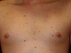
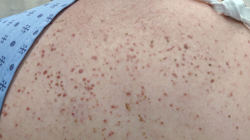
Urticaria Pigmentosa
Tryptases
Proto-Oncogene Proteins c-kit
Mast Cells
Leukemia, Mast-Cell
Bone Marrow
Methylhistamines
Chymases
Trichinella spiralis
Anaphylaxis
Trichinellosis
Interleukin-9
Intestinal Diseases, Parasitic
Hydroxyzine
Prostaglandins D
Trematoda
Stem Cell Factor
Interleukin-5 Receptor alpha Subunit
Arthropod Venoms
Biopsy
Association of transient dermal mastocytosis and elevated plasma tryptase levels with development of adverse reactions after treatment of onchocerciasis with ivermectin. (1/30)
To investigate the role of mast cells in treatment-associated adverse reactions in patients with onchocerciasis, changes in plasma tryptase levels and skin mast cell counts were examined in 2 groups of Onchocerca volvulus-infected subjects after ivermectin treatment. After treatment, an increase in tryptase levels was observed concurrent with the onset of blood eosinopenia and preceding the appearance of plasma eosinophil-derived neurotoxin (EDN) and interleukin-5. Tryptase levels were correlated with development of peripheral eosinopenia and markers of eosinophil activation and degranulation. Dermal mast cell numbers increased transiently at 24 h after treatment, preceding the onset of dermal eosinophil infiltration and the development of clinically apparent inflammation. Local reactions were strongly correlated with levels of plasma tryptase and EDN, and the severity of systemic reactions was correlated with levels of tryptase, EDN, and interleukin-5. The data indicate that mast cells play a role in initiation of tissue inflammatory reactions after ivermectin treatment of onchocerciasis. (+info)Expression of Bcl-2 and Bcl-xL in cutaneous and bone marrow lesions of mastocytosis. (2/30)
Mastocytosis is a rare disease characterized by accumulation of mast cells in tissues. To investigate whether an altered regulation of mast cell apoptosis might be involved in the pathogenesis of mastocytosis, expression of the apoptosis-preventing molecules bcl-2 and bcl-xL was studied by immunohistochemistry in skin and bone marrow lesions of mastocytosis patients. In addition, reverse transcription-polymerase chain reaction was used to investigate levels of bcl-2 and bcl-xL mRNA in cutaneous mastocytosis lesions. Since activating mutations of c-kit are known to be associated with some forms of mastocytosis, human mast cell cultures were also stimulated via c-kit and the expression of bcl-2 and bcl-xL was assessed by immunoblotting. In patients with mastocytosis, the expression of bcl-2 protein but not bcl-xL in cutaneous mast cells was significantly enhanced, compared to healthy controls. Evaluating different subgroups of adult and pediatric mastocytosis patients, all groups were found to express significantly increased levels of bcl-2 protein, and none of the patient groups was found to overexpress bcl-xL, with the exception of solitary mastocytomas that showed a tendency for up-regulated bcl-xL protein. Furthermore, the expression of bcl-2 mRNA was significantly enhanced in cutaneous lesions of adult and pediatric patients, while bcl-xL mRNA levels were only slightly increased in pediatric, but not in adult patients with mastocytosis. In contrast to the skin lesions, bone marrow infiltrates of patients with systemic mastocytosis showed only low or absent immunoreactivity for bcl-2, but marked expression of bcl-xL. In vitro, stimulation of two different mast cell culture systems by activation of c-kit resulted in up-regulation of bcl-2 and also in an increase of bcl-xL, although less pronounced. Thus, overexpression of bcl-2 and bcl-xL leading to prolonged survival of mast cells may contribute to the pathogenesis of mastocytosis. Our findings may help to develop new strategies for the treatment of this disease. (+info)Mast cell distribution in normal adult skin. (3/30)
AIMS: To investigate mast cell distribution in normal adult skin to provide a reference range for comparison with mastocytosis. METHODS: Mast cells (MCs) were counted in uninvolved skin adjacent to basal cell carcinomas and other dermatological disorders in adults. RESULTS: There was an uneven distribution of MCs in different body sites using the anti-tryptase monoclonal antibody technique. Numbers of MCs on the trunk, upper arm, and upper leg were similar, but were significantly different from those found on the lower leg and forearm. Two distinct groups were formed--proximal and distal. There were 77.0 MCs/mm2 at proximal body sites and 108.2 MCs/mm2 at distal sites. Adjusted for the adjacent diagnosis and age, this difference was consistent. The numbers of MCs in uninvolved skin adjacent to basal cell carcinomas and other dermatological disorders were not different from those in the control group. Differences in the numbers of MCs between the distal and the proximal body sites must be considered when MCs are counted for a reliable diagnosis of mastocytosis. A pilot study in patients with mastocytosis underlined the variation in the numbers of MCs in mastocytosis and normal skin, but showed a considerable overlap. The observed numbers of MCs in adults cannot be extrapolated to children. CONCLUSIONS: MC numbers varied significantly between proximal and distal body sites and these differences must be considered when MCs are counted for a reliable diagnosis of mastocytosis. There was a considerable overlap between the numbers of MCs in mastocytosis and normal skin. (+info)c-kit Mutations in patients with childhood-onset mastocytosis and genotype-phenotype correlation. (4/30)
Mastocytosis represents a clonal proliferation of mast cell hematopoietic progenitors caused by gain-of-function mutations of the c-kit gene. The heterogeneity of c-kit mutations may have contributed to difficulties in characterizing genotype-phenotype correlation of the disease. Our goal was to analyze a set of reported pathogenic c-kit mutations in patients with childhood-onset cutaneous mastocytosis, in comparison with those with adult-onset disease, and to correlate these with clinical presentation. We performed polymerase chain reaction and direct sequencing using genomic DNA samples from 16 nonfamilial Japanese patients with indolent cutaneous mastocytosis (12 with childhood-onset disease and 4 with adult-onset disease) to look for the most common c-kit mutations at codons 816, 560, 820, and 839. A substantial number of patients had missense codon 816 mutations (10 of 12 in the childhood-onset group, 83.3%; and 4 of 4 in the adult-onset group, 100%). The most common mutation was Asp816Val (9 of 16, 64.3%) followed by Asp816Phe (5 of 16, 35.7%). Moreover, children with the Asp816Phe mutation developed cutaneous mastocytosis at an earlier age as compared to those with the Asp816Val mutation (mean age of onset, 1.3 months versus 5.9 months, respectively; P = 0.068). No other mutation variations were found in our cohort. In summary, we confirmed a high incidence of two distinct c-kit mutations, Asp816Val and Asp816Phe, in patients with childhood-onset cutaneous mastocytosis. Our results provide new insights into common c-kit mutations, which may contribute to different clinical courses of the disease. (+info)Pediatric cutaneous mastocytosis: a review of 180 patients. (5/30)
BACKGROUND: Mastocytosis is a heterogeneous group of diseases characterized by the abnormal infiltration of mast cells in the skin and, sometimes, other organs. Some patients may experience symptoms related to mast cell mediator release. OBJECTIVE: To analyze the clinical features of cutaneous mastocytosis in a large series of children. METHODS: We conducted a file review of all children clinically diagnosed with cutaneous mastocytosis in our department over the last 20 years. We evaluated gender, age at onset, character and distribution of the lesions, associated symptoms, and course of the disease. RESULTS: Altogether, 180 patients with cutaneous mastocytosis were identified. The male to female ratio was 1.5:1. About one-third of patients had a mastocytoma, which was present at birth in over 40% and appeared during the first year of life in most of the remainder. Urticaria pigmentosa was noted in 65% of the patients, presenting at birth in 20% and during the first year in most of the remainder. The majority of lesions was distributed over the trunk and limbs. Different kinds of associated symptoms were noted. Prognosis in general was good. Only 11% of the cases, all urticaria pigmentosa, were familial. CONCLUSIONS: Most cases of pediatric mastocytosis are sporadic and appear during the first 2 years of life, especially on the trunk. Urticaria pigmentosa is the most frequent variant. The prognosis of pediatric mastocytosis, in general, is good. (+info)Variation among pathologists in the histologic grading of canine cutaneous mast cell tumors with uniform use of a single grading reference. (6/30)
Ten veterinary pathologists independently assigned histologic grades to the same 60 canine cutaneous mast cell tumors using the Patnaik classifications. The degree of agreement in grading among the pathologists was compared with the degree of agreement among the same pathologists in a previous study, in which each pathologist used the reference for grading that he/she uses routinely. Mean agreement improved significantly from 50.3% to 62.1% with uniform use of the Patnaik classifications (P = 0.00001), suggesting that there is value in uniform application of a single grading scheme for canine cutaneous mast cell tumors. Agreement among pathologists was still not 100%, suggesting that a more objective grading scheme should be developed and that other histologic indicators of prognosis should be investigated. (+info)Cutaneous mastocytosis: report of six cases. (7/30)
Cutaneous mastocytosis is a rare infiltrative disorder of the skin. Though often asymptomatic, systemic features may be associated with any clinical pattern of the disorder at any age group. We present our experience with six cases of cutaneous mastocytosis, including three with diffuse cutaneous mastocytosis, a rare entity. (+info)Regulation of normal and neoplastic human mast cell development in mastocytosis. (8/30)
Mastocytosis, a condition characterized by a pathologic accumulation of clonal mast cells in tissues, offers a unique opportunity to study the growth and differentiation of mast cells as well as their contribution to various pathologic processes. This is because molecular pathways governing the proliferation and survival of mast cells show striking similarities between normal mast cells and their counterparts in mastocytosis. For example, activation of Kit, a transmembrane receptor for stem cell factor (SCF) is critical for the growth and differentiation of normal mast cells. Mutations such as D816V resulting in its pathologic activation are strongly associated with mastocytosis. Treatment of mastocytosis is aimed at controlling symptoms, as no specific drug has yet been clinically demonstrated to selectively eliminate mast cells carrying the D816V gain-of-function c-kit mutation. Non-myeloablative bone marrow transplantation is performed in select patients to take advantage of the immunotherapeutic effects of the graft. (+info)Mastocytosis is a group of rare disorders caused by the accumulation of abnormal number of mast cells in various tissues of the body, particularly the skin and internal organs such as the bone marrow, liver, spleen, and gastrointestinal tract. Mast cells are types of white blood cells that play an important role in the immune system, releasing chemicals like histamine, heparin, and leukotrienes during allergic reactions or injury to help protect the body. However, excessive accumulation of mast cells can lead to chronic inflammation, tissue damage, and various symptoms.
There are two main types of mastocytosis: cutaneous mastocytosis (CM) and systemic mastocytosis (SM). CM primarily affects the skin, causing redness, itching, hives, and other skin abnormalities. SM, on the other hand, involves internal organs and can be more severe, with symptoms such as diarrhea, stomach pain, fatigue, bone pain, and anaphylaxis (a life-threatening allergic reaction).
Mastocytosis is typically caused by genetic mutations that lead to the overproduction of mast cells. The diagnosis of mastocytosis usually involves a combination of physical examination, medical history, blood tests, skin biopsy, and bone marrow aspiration. Treatment options depend on the type and severity of the disease and may include antihistamines, corticosteroids, chemotherapy, targeted therapy, and in severe cases, stem cell transplantation.
Systemic mastocytosis is a rare group of diseases characterized by the accumulation of abnormal number of mast cells in various organs and tissues of the body. Mast cells are a type of white blood cell that plays an important role in the immune system, particularly in allergic reactions and inflammation. In systemic mastocytosis, the excessive buildup of mast cells can cause a range of symptoms such as skin rashes, itching, gastrointestinal disturbances, bone pain, and in severe cases, organ damage or failure.
The diagnosis of systemic mastocytosis typically involves a combination of clinical evaluation, laboratory tests, imaging studies, and sometimes biopsies to confirm the presence of abnormal mast cells. Treatment for systemic mastocytosis depends on the severity and extent of the disease, but may include medications to manage symptoms, reduce mast cell activation and proliferation, and prevent complications. In some cases, cytoreductive therapies such as chemotherapy or stem cell transplantation may be recommended.
Cutaneous mastocytosis is a condition characterized by the abnormal accumulation of mast cells in the skin. Mast cells are a type of immune cell that releases chemicals such as histamine, heparin, and leukotrienes, which play a role in allergic reactions and inflammation. In cutaneous mastocytosis, the excessive buildup of mast cells can cause various skin symptoms, including redness, itching, swelling, and formation of lesions or tumors.
The condition is typically divided into several subtypes based on the age of onset and the clinical presentation. The most common form in children is called urticaria pigmentosa, which presents as small, reddish-brown spots or bumps that may become raised and itchy when scratched or rubbed (Darier's sign). In adults, a more severe form known as diffuse cutaneous mastocytosis can occur, where the entire skin becomes thickened, red, and swollen.
Cutaneous mastocytosis is usually diagnosed based on the patient's medical history, physical examination, and results from skin biopsies. Treatment typically focuses on relieving symptoms and preventing mast cell activation. Medications such as antihistamines, topical steroids, and mast cell stabilizers may be used to control itching, flushing, and other symptoms associated with the condition. In some cases, systemic therapies or phototherapy may also be recommended.
Urticaria pigmentosa is a rare mast cell disorder, characterized by the development of brownish-red, raised lesions (maculopapules) on the skin. These lesions are often found on the trunk and proximal extremities, but can occur anywhere on the body. They are typically asymptomatic, but may become itchy or even painful when subjected to friction, heat, or emotional stress. In some cases, these lesions may also release histamine, leading to symptoms such as flushing, headache, and hypotension. Urticaria pigmentosa is more common in children than adults, and typically resolves on its own over time. However, in some cases it can persist into adulthood or even progress to systemic mastocytosis, a more severe form of the disorder that can affect internal organs.
Tryptase is a type of enzyme that is found in the cells called mast cells, which are a part of the immune system. Specifically, tryptase is a serine protease, which means it helps to break down other proteins in the body. Tryptase is often released during an allergic reaction or as part of an inflammatory response. It can be measured in the blood and is sometimes used as a marker for mast cell activation or degranulation. High levels of tryptase may indicate the presence of certain medical conditions, such as systemic mastocytosis or anaphylaxis.
Proto-oncogene proteins c-kit, also known as CD117 or stem cell factor receptor, are transmembrane receptor tyrosine kinases that play crucial roles in various biological processes, including cell survival, proliferation, differentiation, and migration. They are encoded by the c-KIT gene located on human chromosome 4q12.
These proteins consist of an extracellular ligand-binding domain, a transmembrane domain, and an intracellular tyrosine kinase domain. The binding of their ligand, stem cell factor (SCF), leads to receptor dimerization, autophosphorylation, and activation of several downstream signaling pathways such as PI3K/AKT, MAPK/ERK, and JAK/STAT.
Abnormal activation or mutation of c-kit proto-oncogene proteins has been implicated in the development and progression of various malignancies, including gastrointestinal stromal tumors (GISTs), acute myeloid leukemia (AML), mast cell diseases, and melanoma. Targeted therapies against c-kit, such as imatinib mesylate (Gleevec), have shown promising results in the treatment of these malignancies.
Mast cells are a type of white blood cell that are found in connective tissues throughout the body, including the skin, respiratory tract, and gastrointestinal tract. They play an important role in the immune system and help to defend the body against pathogens by releasing chemicals such as histamine, heparin, and leukotrienes, which help to attract other immune cells to the site of infection or injury. Mast cells also play a role in allergic reactions, as they release histamine and other chemicals in response to exposure to an allergen, leading to symptoms such as itching, swelling, and redness. They are derived from hematopoietic stem cells in the bone marrow and mature in the tissues where they reside.
Mast cell leukemia is a rare and aggressive type of leukemia, which is a cancer of the white blood cells. Specifically, mast cell leukemia affects a particular type of white blood cell called a mast cell. Mast cells are part of the immune system and play a role in allergic reactions and inflammation.
In mast cell leukemia, the bone marrow produces too many immature mast cells, which then enter the bloodstream. These abnormal mast cells can accumulate in various organs, such as the spleen, liver, and lymph nodes, causing damage and enlargement of these organs.
Symptoms of mast cell leukemia may include fatigue, weight loss, frequent infections, easy bruising or bleeding, bone pain, and enlarged lymph nodes. Diagnosis typically involves blood tests, bone marrow aspiration and biopsy, and imaging studies to assess the extent of organ involvement.
Mast cell leukemia is a very aggressive cancer with a poor prognosis, and treatment options are limited. Current treatments may include chemotherapy, stem cell transplantation, and targeted therapy with drugs that target specific molecular abnormalities in mast cells. However, the response to treatment is often not durable, and the disease can progress rapidly.
Bone marrow is the spongy tissue found inside certain bones in the body, such as the hips, thighs, and vertebrae. It is responsible for producing blood-forming cells, including red blood cells, white blood cells, and platelets. There are two types of bone marrow: red marrow, which is involved in blood cell production, and yellow marrow, which contains fatty tissue.
Red bone marrow contains hematopoietic stem cells, which can differentiate into various types of blood cells. These stem cells continuously divide and mature to produce new blood cells that are released into the circulation. Red blood cells carry oxygen throughout the body, white blood cells help fight infections, and platelets play a crucial role in blood clotting.
Bone marrow also serves as a site for immune cell development and maturation. It contains various types of immune cells, such as lymphocytes, macrophages, and dendritic cells, which help protect the body against infections and diseases.
Abnormalities in bone marrow function can lead to several medical conditions, including anemia, leukopenia, thrombocytopenia, and various types of cancer, such as leukemia and multiple myeloma. Bone marrow aspiration and biopsy are common diagnostic procedures used to evaluate bone marrow health and function.
Methylhistamines are not a recognized medical term or a specific medical condition. However, the term "methylhistamine" may refer to the metabolic breakdown product of the antihistamine drug, diphenhydramine, which is also known as N-methyldiphenhydramine or dimenhydrinate.
Diphenhydramine is a first-generation antihistamine that works by blocking the action of histamine, a chemical released during an allergic reaction. When diphenhydramine is metabolized in the body, it is converted into several breakdown products, including methylhistamines.
Methylhistamines are not known to have any specific pharmacological activity or clinical significance. However, they can be used as a marker for the presence of diphenhydramine or its metabolism in the body.
Chymases are a type of enzyme that belong to the family of serine proteases. They are found in various tissues and organs, including the heart, lungs, and immune cells called mast cells. Chymases play a role in several physiological and pathological processes, such as inflammation, tissue remodeling, and blood pressure regulation.
One of the most well-known chymases is found in the mast cells and is often referred to as "mast cell chymase." This enzyme can cleave and activate various proteins, including angiotensin I to angiotensin II, a potent vasoconstrictor that increases blood pressure. Chymases have also been implicated in the development of cardiovascular diseases, such as hypertension and heart failure, as well as respiratory diseases like asthma and chronic obstructive pulmonary disease (COPD).
In summary, chymases are a group of serine protease enzymes that play important roles in various physiological and pathological processes, particularly in inflammation, tissue remodeling, and blood pressure regulation.
A bone marrow examination is a medical procedure in which a sample of bone marrow, the spongy tissue inside bones where blood cells are produced, is removed and examined. This test is used to diagnose or monitor various conditions affecting blood cell production, such as infections, leukemia, anemia, and other disorders of the bone marrow.
The sample is typically taken from the hipbone (iliac crest) or breastbone (sternum) using a special needle. The procedure may be done under local anesthesia or with sedation to minimize discomfort. Once the sample is obtained, it is examined under a microscope for the presence of abnormal cells, changes in cell size and shape, and other characteristics that can help diagnose specific conditions. Various stains, cultures, and other tests may also be performed on the sample to provide additional information.
Bone marrow examination is an important diagnostic tool in hematology and oncology, as it allows for a detailed assessment of blood cell production and can help guide treatment decisions for patients with various blood disorders.
"Trichinella spiralis" is a species of parasitic roundworm that causes the disease trichinosis in humans. The adult worms live in the intestine, where they produce larvae that migrate to striated muscle tissue, including the diaphragm, tongue, and skeletal muscles, where they encyst and form nurse cells. Infection typically occurs through the consumption of undercooked or raw meat, particularly pork, contaminated with the larvae. Symptoms can range from gastrointestinal disturbances to fever, muscle pain, and potentially life-threatening complications in severe cases. Prevention includes cooking meat thoroughly and freezing it at certain temperatures to kill the larvae.
Anaphylaxis is a severe, life-threatening systemic allergic reaction that occurs suddenly after exposure to an allergen (a substance that triggers an allergic reaction) to which the person has previously been sensitized. The symptoms of anaphylaxis include rapid onset of symptoms such as itching, hives, swelling of the throat and tongue, difficulty breathing, wheezing, cough, chest tightness, rapid heartbeat, hypotension (low blood pressure), shock, and in severe cases, loss of consciousness and death. Anaphylaxis is a medical emergency that requires immediate treatment with epinephrine (adrenaline) and other supportive measures to stabilize the patient's condition.
A mastocytoma is a type of tumor that develops from mast cells, which are a part of the immune system and play a role in allergic reactions and inflammation. Mastocytomas are most commonly found in the skin, but they can also occur in other organs such as the liver, spleen, and lymph nodes.
Mastocytomas are usually benign (non-cancerous), although malignant (cancerous) forms known as mast cell sarcomas can also occur. They typically appear as raised, red or brown lesions on the skin that may be itchy, painful, or bleed easily.
The diagnosis of a mastocytoma is usually made through a biopsy of the tumor, which involves removing a small sample of tissue for examination under a microscope. Treatment options for mastocytomas may include surgical removal, medication to manage symptoms such as itching or flushing, and in some cases, chemotherapy or radiation therapy.
Trichinellosis is a parasitic disease caused by the roundworm Trichinella spiralis. The infection typically occurs when contaminated raw or undercooked meat, often pork, is consumed. After ingestion, the larvae of the worm are released from the cysts in the meat and migrate to the small intestine, where they mature into adults.
The adult females then lay new larvae that penetrate the intestinal wall and travel through the bloodstream to striated muscle tissue (such as skeletal muscles), where they encapsulate and form new cysts. The symptoms of trichinellosis can vary widely, depending on the number of worms ingested and the intensity of infection. Early symptoms may include diarrhea, abdominal pain, nausea, vomiting, and fever. As the larvae migrate to muscle tissue, additional symptoms such as muscle pain, weakness, swelling of the face, eyelids, or tongue, and skin rashes can occur. Severe infections may lead to life-threatening complications, including heart and respiratory failure.
Prevention measures include cooking meat thoroughly (to an internal temperature of at least 160°F or 71°C), freezing meat properly (at -15°F or -26°C for several days) to kill the parasites, and avoiding consumption of raw or undercooked meat, especially from wild animals.
Interleukin-9 (IL-9) is a type of cytokine, which are small signaling proteins that mediate and regulate immunity, inflammation, and hematopoiesis. IL-9 is produced by several types of immune cells, including T cells (a type of white blood cell), mast cells, and eosinophils.
IL-9 plays a role in the development and function of various immune cells, and has been implicated in the pathogenesis of several inflammatory and allergic diseases, such as asthma, atopic dermatitis, and food allergy. It can promote the growth and survival of certain types of immune cells, including mast cells and B cells (another type of white blood cell), and can also enhance their activation and effector functions.
In addition to its role in immunity and inflammation, IL-9 has been shown to play a role in the development and progression of some types of cancer, such as lung cancer and leukemia. However, more research is needed to fully understand the complex functions of this cytokine and its potential as a therapeutic target.
Parasitic intestinal diseases are disorders caused by microscopic parasites that invade the gastrointestinal tract, specifically the small intestine. These parasites include protozoa (single-celled organisms) and helminths (parasitic worms). The most common protozoan parasites that cause intestinal disease are Giardia lamblia, Cryptosporidium parvum, and Entamoeba histolytica. Common helminthic parasites include roundworms (Ascaris lumbricoides), tapeworms (Taenia saginata and Taenia solium), hookworms (Ancylostoma duodenale and Necator americanus), and pinworms (Enterobius vermicularis).
Parasitic intestinal diseases can cause a variety of symptoms, including diarrhea, abdominal pain, bloating, nausea, vomiting, fatigue, and weight loss. The severity and duration of the symptoms depend on the type of parasite, the number of organisms present, and the immune status of the host.
Transmission of these parasites can occur through various routes, including contaminated food and water, person-to-person contact, and contact with contaminated soil or feces. Preventive measures include practicing good hygiene, washing hands thoroughly after using the toilet and before handling food, cooking food thoroughly, and avoiding consumption of raw or undercooked meat, poultry, or seafood.
Treatment of parasitic intestinal diseases typically involves the use of antiparasitic medications that target the specific parasite causing the infection. In some cases, supportive care such as fluid replacement and symptom management may also be necessary.
Hydroxyzine is an antihistamine medication that is primarily used to treat symptoms of allergies such as itching, hives, and swelling. It works by blocking the effects of histamine, a substance in the body that causes allergic reactions. In addition to its antihistaminic properties, hydroxyzine also has sedative and anxiety-reducing effects, which make it useful in treating anxiety disorders, symptoms of alcohol withdrawal, and as a sleep aid. It is available in both oral and injectable forms and is usually taken orally in the form of tablets, capsules, or syrup. As with any medication, hydroxyzine should be used under the supervision of a healthcare provider, and its use may be subject to certain precautions and contraindications depending on the individual's medical history and current health status.
I'm sorry for any confusion, but "Prostaglandins D" is not a recognized or established term in the field of medicine or physiology. Prostaglandins are a group of lipid compounds that are synthesized in the body from fatty acids and have diverse hormone-like effects in the body. They are classified into several types based on their chemical structure and biological activity, including Prostaglandin D (PGD), but referring to "Prostaglandins D" as a single entity is not standard.
Prostaglandin D (PGD) itself is a type of prostaglandin that is rapidly converted to other compounds in the body and has been studied for its potential role in various physiological processes, such as inflammation, fever, and blood flow regulation. However, it's important to note that specific medical definitions or clinical uses related to "Prostaglandins D" are not well-established.
If you have any further questions or need more information about a specific aspect of prostaglandins or their role in the body, I would be happy to help!
Trematode infections, also known as trematodiasis or fluke infections, are parasitic diseases caused by various species of flatworms called trematodes. These parasites have an indirect life cycle involving one or two intermediate hosts (such as snails or fish) and a definitive host (usually a mammal or bird).
Humans can become accidentally infected when they consume raw or undercooked aquatic plants, animals, or contaminated water that contains the larval stages of these parasites. The most common trematode infections affecting humans include:
1. Schistosomiasis (also known as bilharzia): Caused by several species of blood flukes (Schistosoma spp.). Adult worms live in the blood vessels, and their eggs can cause inflammation and damage to various organs, such as the liver, intestines, bladder, or lungs.
2. Liver flukes: Fasciola hepatica and Fasciola gigantica are common liver fluke species that infect humans through contaminated watercress or other aquatic plants. These parasites can cause liver damage, abdominal pain, diarrhea, and eosinophilia (elevated eosinophil count in the blood).
3. Lung flukes: Paragonimus spp. are lung fluke species that infect humans through consumption of raw or undercooked crustaceans. These parasites can cause coughing, chest pain, and bloody sputum.
4. Intestinal flukes: Various species of intestinal flukes (e.g., Haplorchis spp., Metagonimus yokogawai) infect humans through consumption of raw or undercooked fish. These parasites can cause abdominal pain, diarrhea, and eosinophilia.
5. Eye fluke: The oriental eye fluke (Drepanotrema spp.) can infect the human eye through contaminated water. It can cause eye inflammation, corneal ulcers, and vision loss.
Prevention measures include avoiding consumption of raw or undercooked aquatic plants, animals, and their products; practicing good hygiene; and treating drinking water to kill parasites. Treatment typically involves administering anthelmintic drugs such as praziquantel, albendazole, or mebendazole, depending on the specific fluke species involved.
Trematoda is a class of parasitic flatworms, also known as flukes. They have a complex life cycle involving one or more intermediate hosts and a definitive host. Adult trematodes are typically leaf-shaped and range in size from a few millimeters to several centimeters.
They have a characteristic oral sucker surrounding the mouth and a ventral sucker, which they use for locomotion and attachment to their host's tissues. Trematodes infect various organs of their hosts, including the liver, lungs, blood vessels, and intestines, causing a range of diseases in humans and animals.
Examples of human-infecting trematodes include Schistosoma spp., which cause schistosomiasis (also known as bilharzia), and Fasciola hepatica, which causes fascioliasis (liver fluke disease). Trematode infections are typically treated with antiparasitic drugs.
Stem Cell Factor (SCF), also known as Kit Ligand or Steel Factor, is a growth factor that plays a crucial role in the regulation of hematopoiesis, which is the process of producing various blood cells. It is a glycoprotein that binds to the c-Kit receptor found on hematopoietic stem cells and progenitor cells, promoting their survival, proliferation, and differentiation into mature blood cells.
SCF is involved in the development and function of several types of blood cells, including red blood cells, white blood cells, and platelets. It also plays a role in the maintenance and self-renewal of hematopoietic stem cells, which are essential for the continuous production of new blood cells throughout an individual's lifetime.
In addition to its role in hematopoiesis, SCF has been implicated in various other biological processes, such as melanogenesis, gametogenesis, and tissue repair and regeneration. Dysregulation of SCF signaling has been associated with several diseases, including certain types of cancer, bone marrow failure disorders, and autoimmune diseases.
Eosinophilia is a medical condition characterized by an abnormally high concentration of eosinophils in the circulating blood. Eosinophils are a type of white blood cell that play an important role in the immune system, particularly in fighting off parasitic infections and regulating allergic reactions. However, when their numbers become excessively high, they can contribute to tissue damage and inflammation.
Eosinophilia is typically defined as a count of more than 500 eosinophils per microliter of blood. Mild eosinophilia (up to 1,500 cells/μL) may not cause any symptoms and may be discovered during routine blood tests. However, higher levels of eosinophilia can lead to various symptoms such as coughing, wheezing, skin rashes, and organ damage, depending on the underlying cause.
The causes of eosinophilia are varied and can include allergic reactions, parasitic infections, autoimmune disorders, certain medications, and some types of cancer. Accurate diagnosis and treatment of eosinophilia require identification and management of the underlying cause.
The Interleukin-5 Receptor alpha Subunit (IL-5Rα) is a protein that forms part of the Type I cytokine receptor, specifically for the interleukin-5 (IL-5) cytokine. This receptor is found on the surface of hematopoietic cells, such as eosinophils and basophils. The binding of IL-5 to the IL-5Rα subunit initiates intracellular signaling cascades that regulate the growth, activation, differentiation, and survival of eosinophils and basophils, which are crucial in the immune response against parasitic infections and allergic reactions. Mutations in the gene encoding IL-5Rα can lead to altered immune responses and diseases such as hypereosinophilic syndromes.
Arthropod venoms are toxic secretions produced by the venom glands of various arthropods, such as spiders, scorpions, insects, and marine invertebrates. These venoms typically contain a complex mixture of bioactive molecules, including peptides, proteins, enzymes, and small molecules, which can cause a range of symptoms and effects in humans and other animals.
The specific composition of arthropod venoms varies widely depending on the species and can be tailored to serve various functions, such as prey immobilization, defense, or predation. Some arthropod venoms contain neurotoxins that can disrupt nerve function and cause paralysis, while others may contain cytotoxins that damage tissues or hemotoxins that affect the blood and cardiovascular system.
Arthropod venoms have been studied for their potential therapeutic applications, as some of their bioactive components have shown promise in treating various medical conditions, including pain, inflammation, and neurological disorders. However, it is important to note that arthropod venoms can also cause severe allergic reactions and other adverse effects in susceptible individuals, making it essential to exercise caution when handling or coming into contact with venomous arthropods.
A biopsy is a medical procedure in which a small sample of tissue is taken from the body to be examined under a microscope for the presence of disease. This can help doctors diagnose and monitor various medical conditions, such as cancer, infections, or autoimmune disorders. The type of biopsy performed will depend on the location and nature of the suspected condition. Some common types of biopsies include:
1. Incisional biopsy: In this procedure, a surgeon removes a piece of tissue from an abnormal area using a scalpel or other surgical instrument. This type of biopsy is often used when the lesion is too large to be removed entirely during the initial biopsy.
2. Excisional biopsy: An excisional biopsy involves removing the entire abnormal area, along with a margin of healthy tissue surrounding it. This technique is typically employed for smaller lesions or when cancer is suspected.
3. Needle biopsy: A needle biopsy uses a thin, hollow needle to extract cells or fluid from the body. There are two main types of needle biopsies: fine-needle aspiration (FNA) and core needle biopsy. FNA extracts loose cells, while a core needle biopsy removes a small piece of tissue.
4. Punch biopsy: In a punch biopsy, a round, sharp tool is used to remove a small cylindrical sample of skin tissue. This type of biopsy is often used for evaluating rashes or other skin abnormalities.
5. Shave biopsy: During a shave biopsy, a thin slice of tissue is removed from the surface of the skin using a sharp razor-like instrument. This technique is typically used for superficial lesions or growths on the skin.
After the biopsy sample has been collected, it is sent to a laboratory where a pathologist will examine the tissue under a microscope and provide a diagnosis based on their findings. The results of the biopsy can help guide further treatment decisions and determine the best course of action for managing the patient's condition.
Vulvar diseases refer to a range of medical conditions that affect the vulva, which is the external female genital area including the mons pubis, labia majora and minora, clitoris, and the vaginal opening. These conditions can cause various symptoms such as itching, burning, pain, soreness, irritation, or abnormal growths or lesions. Some common vulvar diseases include:
1. Vulvitis: inflammation of the vulva that can be caused by infection, allergies, or irritants.
2. Lichen sclerosus: a chronic skin condition that causes thin, white patches on the vulva.
3. Lichen planus: an inflammatory condition that affects the skin and mucous membranes, including the vulva.
4. Vulvar cancer: a rare type of cancer that develops in the tissues of the vulva.
5. Genital warts: caused by human papillomavirus (HPV) infection, these are small growths or bumps on the vulva.
6. Pudendal neuralgia: a nerve condition that causes pain in the vulvar area.
7. Vestibulodynia: pain or discomfort in the vestibule, the area surrounding the vaginal opening.
It is important to consult a healthcare professional if experiencing any symptoms related to vulvar diseases for proper diagnosis and treatment.
 Urticaria pigmentosa
Mast cell
Mariana Castells
Telangiectasia macularis eruptiva perstans
Roan (color)
Mast cell activation syndrome
Mastocytosis
List of skin conditions
Tumors of the hematopoietic and lymphoid tissues
List of MeSH codes (C04)
Leonine facies
Omalizumab
Midostaurin
List of MeSH codes (C17)
Darier's sign
Mast cell leukemia
Eosinophilia
Silvia Bulfone-Paus
Flushing (physiology)
International Classification of Diseases for Oncology
Cancer biomarker
Mastocytoma
Clonal hypereosinophilia
Masitinib
Mastocytoma in dogs
List of diseases (S)
Lymphocyte-variant hypereosinophilia
Autoimmune urticaria
Harrison's Principles of Internal Medicine
Histamine liberators
 Maculopapular Cutaneous Mastocytosis Successfully Treated with Omalizumab
 Cutaneous Mastocytosis | Cyberderm
 Dermatologic Manifestations of Hematologic Disease: Coagulation Disorders, Cutaneous Manifestations of Anemia, Plasma-Cell...
Urticaria pigmentosa - Wikipedia
 Mastocytosis
![KIT KIT proto-oncogene, receptor tyrosine kinase [Homo sapiens (human)] - Gene - NCBI](data:image/png;base64,iVBORw0KGgoAAAANSUhEUgAAABAAAAAQCAYAAAAf8/9hAAAB1ElEQVQ4jaWSPWgTcRjGf/ehuWhobO2JxGJRY3taTTRV2yoqSpW6iIWO4iAoUsRBioNDKUWKLU7i4KA4OfhVREQnETRia03k7IdiS0LaQYKJQg3mLtfc30GySNUDn/V5nx/vy/vAf0pqad3db2xquiBJku93s2Tb2eEHdw1rTcsxol23sObTjN7oIp9KVmaU9kMdTxcLAyiqGtA0bfms+XKQULSdQG2EmnUx0q9ughAA8p/CFW0IN3Sv0vUI5p2zIMpUrd5JeP/Jii//80ZJUlrb9lyV8qn3zI5dB8A4MoBWtcITAKBmZe3eRmPzccYf9uIUsyzx6zQd7fMMAIjFdgxpkuPy4clFANbu6qa6fouybXtznxeAoqoBn0/zz5kvBqVQ5DBasJ5gXaPnDQAWFpwCkiwLZekyAMp2wTPAsqy5d8nEZcIHThPQo7jlIua9854BibdvekqKX8PouARAOn6F+c8pT4Bc7svz6U8f77O1cwDVV439PcPU4yHw8AUhhDPyOn4OfWOMuuZfBZp41INTLACorhC2/Jc2zsxMX8vl8lMcPBUHFL5mnpEZGa748sS42esKYS8WLtl2NjE22s/6fScIhtr48W2S5O0zIFwvp3vST6Z+myCvkaonAAAAAElFTkSuQmCC) KIT KIT proto-oncogene, receptor tyrosine kinase [Homo sapiens (human)] - Gene - NCBI
KIT KIT proto-oncogene, receptor tyrosine kinase [Homo sapiens (human)] - Gene - NCBI
 Advanced Search Results - Public Health Image Library(PHIL)
 Mastocytosis: Risk Factors | Cancer.Net
 Patient Services | Johns Hopkins Medicine
 Urticaria Pigmentosa
Summary Report | CureHunter
Urticaria Pigmentosa
Summary Report | CureHunter
 "Brain fog" and mastocytosis: In patients and...
Urticaria Pigmentosa - DermIS
 National Coverage Determination: Cytogenetic Studies
 UVA1 Phototherapy: A Concise and Practical Review
 2016 ICD-10-CM Casefinding List
 Lymphocytic, Histiocytic, and Related Cutaneous Tumors in Animals - Integumentary System - Merck Veterinary Manual
 Gastrointestinal stromal tumor: MedlinePlus Genetics
 Mastocytosis - DermNet
Congenital and Hereditary Neoplasms and Hamartomas in Animals - Integumentary System - MSD Veterinary Manual
 Search | Preprints.org
Mastocytosis and Mast Cell Activation Syndrome - Immunology; Allergic Disorders - MSD Manual Professional Edition
 Diagnosis of mast cell activation syndrome: a global "consensus-2"
Diagnosis of mast cell activation syndrome: a global "consensus-2"
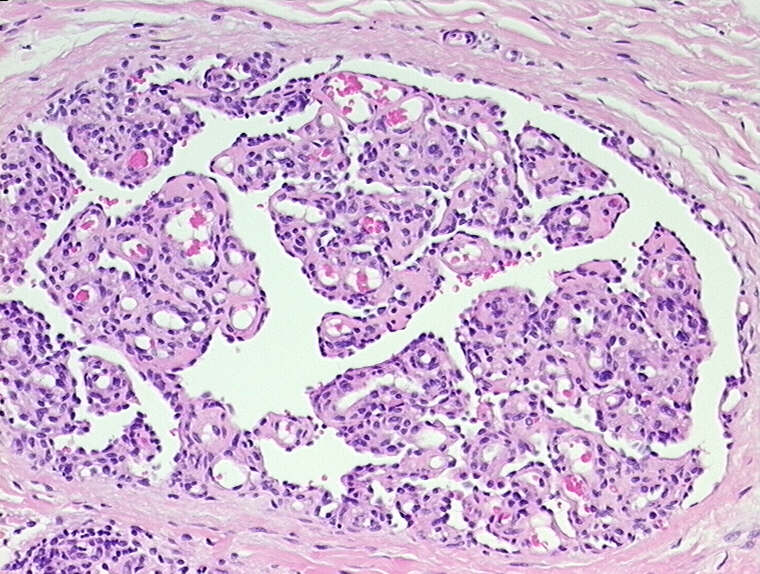
 Increase of bone marrow lymphocytes in systemic mastocytosis: reactive lymphocytosis or malignant lymphoma? Immunohistochemical...
Dermatologic Manifestations of Hematologic Disease: Coagulation Disorders, Cutaneous Manifestations of Anemia, Plasma-Cell...
 What Is Mastocytosis: Symptoms, Diagnosis And Treatment
 Mastocytosis | Encyclopedia MDPI
Mastocytosis in adults
 Pesquisa | Biblioteca Virtual em Saúde
 Collection of dermatological disorders
Urticaria12
- Urticaria pigmentosa (maculopapular mastocytosis): many red-brown, maculopapular lesions, with variable flushes and pruritus (most common form). (cyberderm.net)
- Cutaneous mastocytosis (urticaria pigmentosa and diffuse cutaneous mastocytosis) shows a disseminated appearance on the entire body. (cyberderm.net)
- The 2016 updated current WHO classification of mastocytosis identifies 3 forms of cutaneous mastocytosis: urticaria pigmentosa, diffuse cutaneous mastocytosis, and mastocytoma. (cyberderm.net)
- Urticaria pigmentosa (also known as generalized eruption of cutaneous mastocytosis (childhood type): 616 ) is the most common form of cutaneous mastocytosis. (wikipedia.org)
- Most treatments for mastocytosis can be used to treat urticaria pigmentosa. (wikipedia.org)
- When following patients with mastocytosis - whether classical urticaria pigmentosa in children or adult mastocytosis - I usually ask a directed review of systems, focusing on flushing, palpitations, and gastrointestinal symptoms. (aad.org)
- Some affected individuals have a skin condition called urticaria pigmentosa (also known as maculopapular cutaneous mastocytosis), which is characterized by raised patches of brownish skin that sting or itch when touched. (medlineplus.gov)
- With the exception of pure cutaneous mastocytosis (usually urticaria pigmentosa), indolent SM affecting bone marrow and skin is the most common subvariant of mastocytosis. (bmj.com)
- Inappropriate, recurrent mast cell activation (MCA) and secretion MC-derived mediators plays an essential role in many human diseases: allergy, asthma, allergic rhinitis, urticaria, anaphylaxis, atopic dermatitis, mastocytosis and mast cell activation syndrome (MCAS) [ 5 ] . (encyclopedia.pub)
- Mastocytosis most commonly manifests as cutaneous disease ( urticaria pigmentosa , mastocytoma ), seen more often in children with involvement typically limited to the skin. (logicalimages.com)
- Urticaria pigmentosa is found in most patients with mastocytosis. (clinicaladvisor.com)
- He referred me to the hospital - they did a biopsy and confirmed urticaria pigmentosa, cutaneous mastocytosis. (ukmasto.org)
Form of cutaneous mastocytosis5
- Dear Editor, Maculopapular cutaneous mastocytosis (MPCM), formerly telangiectasia macularis eruptiva perstans (TMEP), is an uncommon form of cutaneous mastocytosis first described on 1930 (1). (nih.gov)
- The most common form of cutaneous mastocytosis (MASTOCYTOSIS, CUTANEOUS) that occurs primarily in children. (curehunter.com)
- Rare and severe form of cutaneous mastocytosis. (dermnetnz.org)
- Telangiectasia macularis eruptiva perstans (TMEP): A form of cutaneous mastocytosis with potential systemic involvement. (nih.gov)
- For example, in the most common form of cutaneous mastocytosis , lesions appear on the skin's surface as brownish, flat or elevated spots. (osmosis.org)
Perstans2
Mastocytoma3
- Localized mastocytosis (mastocytoma) is a single lesion (or sometimes 1-3 lesions), most often found in children. (cyberderm.net)
- Less common are diffuse cutaneous mastocytosis, which is skin infiltration without discrete lesions, and mastocytoma, which is a large (1 to 5 cm) solitary collection of mast cells. (msdmanuals.com)
- Cutaneous Mastocytosis was divided into maculopapular type, diffuse type and mastocytoma. (biomedcentral.com)
Types of mastocytosis1
- Changed or mutated forms of the c-kit gene may cause some types of mastocytosis, including systemic mastocytosis. (cancer.net)
Lesions3
- Mastocytosis skin lesions respond with an urticarial reaction spontaneously or after irritation (Darier's sign: wheal, erythema, itch). (cyberderm.net)
- H&E staining of cutaneous lesions may show necrotizing granuloma formation and neutrophilic infiltrate. (amboss.com)
- We report a 38 years old female who, since her childhood, had a history of precisely limited, fixed maculo papular dark brown cutaneous lesions in the trunk and extremities. (uchile.cl)
Lymphoma4
- there is also evidence for its use in other skin diseases, including cutaneous T-cell lymphoma and mastocytosis. (skintherapyletter.com)
- Increase of bone marrow lymphocytes in systemic mastocytosis: reactive lymphocytosis or malignant lymphoma? (bmj.com)
- To clarify the nature (reactive or neoplastic) of lesional, perifocally aggregated lymphocytes in bone marrow infiltrates of systemic mastocytosis (SM), the histopathology of which can resemble malignant lymphoma with focal bone marrow involvement, particularly low grade malignant B cell lymphoma of lymphoplasmacytic immunocytoma subtype, which frequently exhibits increased mast cell (MC) numbers. (bmj.com)
- 38= Cutaneous T-cell lymphoma, NOS (C44. (cancercentrum.se)
20231
- Mastocytosis" Encyclopedia , https://encyclopedia.pub/entry/1647 (accessed December 06, 2023). (encyclopedia.pub)
Symptoms12
- H1 receptor antagonists are considered the first-choice therapeutic option for control of symptoms among patients with skin mastocytosis (4,5). (nih.gov)
- Mastocytosis can effect multiple organs and present variety of symptoms. (homehealthbeauty.in)
- The above symptoms are common symptoms of cutaneous mycocytosis. (homehealthbeauty.in)
- Mast cell activation syndrome - The more recently termed mast cell activation syndrome (MCAS) describes patients who have multiple mast cell mediator-induced symptoms that do not meet the WHO criteria (see Best Tests) for diagnosis of systemic mastocytosis when other underlying diseases have been excluded. (logicalimages.com)
- Due to an acquired niacin deficiency, pellagra-like cutaneous symptoms are also seen. (logicalimages.com)
- Systemic mastocytosis is a rare disorder that affects the body's mast cells, leading to symptoms such as skin rash, abdominal pain, and anaphylaxis. (dantelabs.com)
- This panel is designed for individuals with a family history of systemic mastocytosis or individuals with symptoms of the condition, such as skin rash, abdominal pain, and anaphylaxis. (dantelabs.com)
- Mastocytosis is a rare disease characterized by proliferation and accumulation of mast cells in various tissues, causing a wide variety of clinical symptoms. (ab-science.com)
- There is a high unmet need in indolent systemic mastocytosis (ISM) and smouldering systemic mastocytosis (SSM) for new therapeutic options with demonstrated activity on severe symptoms and adequate safety profile for a life-long treatment. (ab-science.com)
- Generally, the signs and symptoms of mastocytosis are similar to an allergic reaction . (osmosis.org)
- The article references a review by the Spanish Network on Mastocytosis [ 6 ] that followed 45 pregnant patients with the disease and found that "in most cases mastocytosis -related symptoms remained unchanged throughout pregnancy and after delivery compared to the pregestational clinical profile. (medscape.com)
- Patients with cutaneous mastocytosis experienced more mast cell-mediated symptoms than did those with indolent mastocytosis. (medscape.com)
Type of mastocytosis1
- In diffuse type of mastocytosis and KIT anomaly a bone marrow aspiration is indicated. (cyberderm.net)
Childhood-Onset Mastocytosis1
- In contrast to childhood-onset mastocytosis, adult-onset mastocytosis often persists for the lifetime of the patient and is also more likely to be a severe and systemic disease involving numerous organs. (nih.gov)
Clinical5
- Other than patients being displeased (or even depressed) by their clinical appearance, I have never given the neuropsychiatric aspects of mastocytosis much (if any) thought. (aad.org)
- Brain "fog" characterizes patients with autism spectrum disorders (ASDs), celiac disease, chronic fatigue syndrome, fibromyalgia, mastocytosis, and postural tachycardia syndrome (POTS), as well as "minimal cognitive impairment," an early clinical presentation of Alzheimer's disease (AD), and other neuropsychiatric disorders. (aad.org)
- What are the clinical features of mastocytosis? (dermnetnz.org)
- Mastocytosis, or mast cell disease, is a heterogeneous group of clinical disorders characterized by the abnormal accumulation of mast cells in various tissues, especially in the skin and hematopoietic organs. (nih.gov)
- The present study is focused on the presentation of the clinical diversity of Mastocytosis in the skin, demonstrating diagnostic algorithms and comparison of disease features in children and adults diagnosed in the Mastocytosis Centre, Medical University of Gdañsk. (biomedcentral.com)
Diagnosis4
- If more than one tissue/organ is affected, the diagnosis of systemic mastocytosis (SM) should be made. (bmj.com)
- Recently, a revised classification of mastocytosis has been published including clear cut criteria for diagnosis and subclassification. (bmj.com)
- Recent advances in diagnosis and therapy in systemic mastocytosis. (qxmd.com)
- MIS is an initial diagnosis which requires further diagnostic steps leading to the final diagnosis of Cutaneous Mastocytosis or Systemic Mastocytosis. (biomedcentral.com)
Classification1
- Pathogenesis, classification and treatment of mastocytosis: state of the art in 2010 and future perspectives. (librepathology.org)
Macules1
- Cutaneous mastocytosis is characterized by macules, papules, nodules, or diffuse infiltration of the skin, often associated with localized hyperpigmentation. (nih.gov)
Infiltration3
- Diffuse cutaneous mastocytosis: diffuse erythema, skin infiltration, blistering (rare, in the first month of life). (cyberderm.net)
- Mastocytosis is mast cell proliferation with infiltration of skin or other tissues and organs. (msdmanuals.com)
- Mastocytosis is a group of disorders characterized by proliferation of mast cells and infiltration of the skin, other organs, or both. (msdmanuals.com)
Adult4
- In contrast, adult cutaneous variants frequently have systemic disease. (logicalimages.com)
- In rare cases, the disease may remain active through adolescence as a systemic adult mastocytosis. (nih.gov)
- Adult-onset mastocytosis can also lead to the rare mast cell leukemia, which carries a high risk of mortality (summary by Bodemer et al. (nih.gov)
- By contrast, in most adult patients Systemic Mastocytosis was recognized. (biomedcentral.com)
Indolent mastocytosis1
- These two phase 2a studies enrolled a total of 46 patients in two sub-populations of patients suffering from indolent mastocytosis with handicap: one study in patients who did not carry the D816V mutation on the c-Kit gene and another in patients who did carry this mutation. (ab-science.com)
Aggressive7
- If systemic mastocytosis is aggressive it can be life threatening. (homehealthbeauty.in)
- In case of aggressive systemic mastocytosis chemotherapy is prescribed. (homehealthbeauty.in)
- WHO classified four major subtypes of extracutaneous systemic mastocytosis: (1) indolent systemic mastocytosis, (2) systemic mastocytosis with associated clonal hematologic non-mast cell lineage disease (SM-AHNMD), (3) aggressive systemic mastocytosis, and (4) mast cell leukemia . (logicalimages.com)
- Aggressive systemic mastocytosis, in which there is organ destruction from a mast cell infiltrate, is rare and should promote investigation for mast cell leukemia or other hematologic disorders such as myelodysplastic syndromes, myeloproliferative or myelodysplastic disorders, acute myeloid leukemia, and chronic myeloproliferative neoplasia. (logicalimages.com)
- Mast cell leukemia is seen in two-thirds of patients with aggressive systemic mastocytosis and portends rapid progression that could potentially result in multi-organ failure. (logicalimages.com)
- Pleomorphic undifferentiated sarcoma , Langerhans cell histiocytosis and other pleomorphic tumours - for aggressive systemic mastocytosis . (librepathology.org)
- Systemic mastocytosis (SM) can be further categorized into indolent SM, smouldering SM and aggressive SM. (ab-science.com)
Syndrome2
- This syndrome is a cutaneous marker for renal cystadenocarcinoma and uterine leiomyoma. (msdvetmanual.com)
- The concept that disease rooted principally in chronic aberrant constitutive and reactive activation of mast cells (MCs), without the gross MC neoplasia in mastocytosis, first emerged in the 1980s, but only in the last decade has recognition of "mast cell activation syndrome" (MCAS) grown significantly. (degruyter.com)
Clonal2
- Mastocytosis is a heterogeneous group of rare diseases defined by abnormal accumulation of clonal mast cells (MC) in the skin, bone marrow and/or other visceral organs. (encyclopedia.pub)
- Mastocytosis , or clonal mast cell disease, is a rare disorder that leads to increased numbers of incorrectly functioning mast cells . (osmosis.org)
Papules1
- One or more 5-10 mm large, erythematous , nontender cutaneous papules or vesicles develop approx. (amboss.com)
Mast Cell Sa1
- There are three main types: cutaneous mastocytosis (CM), systemic mastocytosis (SM), and mast cell sarcoma. (qxmd.com)
Extracutaneous2
- Systemic mastocytosis requires the presence of at least one major and one minor criterion or three minor criteria in the bone marrow or other extracutaneous organ. (clinicaladvisor.com)
- CM mainly affects children and is confined to the skin, whereas SM affects adults and is characterized by extracutaneous involvement, with or without cutaneous involvement. (qxmd.com)
Patients6
- The mechanisms of action for omalizumab in patients with mastocytosis are not well known. (nih.gov)
- According to Moura et al, approximately one-third of mastocytosis patients can display various disabling general and neuropsychological symptom, which may have a profound impact on their quality of life. (aad.org)
- Masitinib is developed in ISM and SSM, which are the most prevalent forms of mastocytosis, accounting for approximately 60% of patients. (ab-science.com)
- All patients were clinically diagnosed with Systemic Mastocytosis strictly according WHO criteria. (biomedcentral.com)
- Le pourcentage de CD44 dans les lymphocytes T périphériques était significativement plus élevé chez les patients que chez les témoins, comme détecté par la cytométrie en flux. (who.int)
- En outre, il y avait une aug- mentation significative de la forme soluble du c-kit dans le sérum des patients atteints de pemphigus vulgaire actif par rapport aux témoins. (who.int)
Organs3
- Mastocytosis is health condition where the mast cells (certain type of immune cells) under the skin or in intestine, bones or other organs. (homehealthbeauty.in)
- Mastocytosis is a heterogeneous neoplasm characterized by accumulation of neoplastic mast cells in various organs. (qxmd.com)
- cutaneous mastocytosis, which only affects the skin, and systemic mastocytosis, which involves multiple organs. (osmosis.org)
Squamous1
- Locally advanced and metastatic cutaneous squamous cell carcinoma treated with cemiplimab]. (bvsalud.org)
Lymphosarcoma3
- Cutaneous lymphosarcoma may occur as a disease in which the skin is the initial and primary site of involvement, or it may be secondary to systemic, internal disease. (merckvetmanual.com)
- Cutaneous lymphosarcoma is uncommon but has been identified in all domestic species. (merckvetmanual.com)
- Mastocytosis , melanocytosis , cutaneous lymphosarcoma , and vascular hamartomas are found in calves. (msdvetmanual.com)
Commonly1
- Cutaneous mastocytosis is most commonly diagnosed in childhood. (dermnetnz.org)
Skin5
- Basal cell carcinoma (BCC) is the most common of the cutaneous malignancies, accounting for 65-75% of all skin cancers. (e-ijd.org)
- Cutaneous Mastocytosis is a skin limited disease, whereas Systemic Mastocytosis usually involves bone marrow, spleen, liver, lymph nodes and gastrointestinal tract and may present with or without skin involvement. (biomedcentral.com)
- Mastocytosis in the skin (MIS) is defined by a typical exanthema and monomorphic mast cell infiltrate. (biomedcentral.com)
- In cutaneous mastocytosis , the mast cells accumulate only in the skin. (osmosis.org)
- Some skin changes may be present in systemic mastocytosis , but these forms also involve dysfunction in other tissues. (osmosis.org)
Myeloproliferative2
- Systemic mastocytosis is a less common myeloproliferative variant composed of a heterogeneous disease compilation. (logicalimages.com)
- Systemic mastocytosis with a chronic myeloproliferative neoplasia (SM-AHNMD) has a course and prognosis determined by efficacy of management of the underlying disease. (logicalimages.com)
Disorder1
- Mastocytosis is a rare disorder where mast cells are abnormally high in number (a type of white blood cell) and activated throughout the body . (osmosis.org)
Infancy2
- Most often, cutaneous mastocytosis begins during infancy and early childhood. (cancer.net)
- Mastocytosis usually appears in infancy or early adulthood. (nih.gov)
Disorders1
- Mastocytosis is a diverse group of disorders characterised by the expansion and accumulation of mast cells in one or more organ systems. (dermnetnz.org)
Bone3
- Mastocytosis (mast cell disease) is a relatively uncommon haematological tumour of bone marrow origin. (bmj.com)
- 2011). "Systemic mastocytosis (SM) and associated malignant bone marrow histiocytosis - a hitherto undescribed form of SM-AHNMD. (librepathology.org)
- Mastocytosis is a rare disease of bone marrow-derived hematopoietic progenitor cells, which is characterized by abnormal growth and excessive accumulation of mast cells in various tissues. (biomedcentral.com)
Involvement1
- This proliferation is generally classified as either cutaneous, with or without systemic involvement, or systemic without cutaneous disease. (logicalimages.com)
Occurs1
- Cutaneous mastocytosis typically occurs in children. (msdmanuals.com)
Mutation2
- Etiology in many cases of mastocytosis involves an activating mutation (D816V) in the gene coding for the stem cell factor receptor c-kit, which is present on mast cells. (msdmanuals.com)
- Systemic mastocytosis in particularly happens due to mutation. (homehealthbeauty.in)
Adults2
- Cutaneous forms rarely progress to systemic disease in children but may do so in adults. (msdmanuals.com)
- Due to a rare incidence of Systemic Mastocytosis in children, the diagnostic approach in children and adults presenting MIS should differ. (biomedcentral.com)
Mutations1
- Systemic mastocytosis is caused by mutations in genes involved in the regulation of mast cells, and this panel tests for genetic variants that are known to affect these processes. (dantelabs.com)
Subvariant1
- Indolent systemic mastocytosis, including the subvariant of smouldering systemic mastocytosis, is a lifelong condition associated with reduced quality-of-life. (ab-science.com)
Common3
- The most common form of mastocytosis. (dermis.net)
- reticulum cell sarcomas, cutaneous nodular amyloidosis) are relatively common cutaneous tumors. (merckvetmanual.com)
- Mastocytosis can occur at any age, although some types are more common in particular age groups. (dermnetnz.org)