Motor Neuron Disease
Neurons
Survival of Motor Neuron 1 Protein
Motor Cortex
Survival of Motor Neuron 2 Protein
Evoked Potentials, Motor
Amyotrophic Lateral Sclerosis
Spinal Cord
Muscular Atrophy, Spinal
SMN Complex Proteins
Action Potentials
Nerve Tissue Proteins
Molecular Motor Proteins
Nerve Degeneration
Synapses
Neuromuscular Diseases
Ganglia, Invertebrate
Electrophysiology
Interneurons
Synaptic Transmission
Movement
Cells, Cultured
Aplysia
Mice, Transgenic
Superoxide Dismutase
Electromyography
Anterior Horn Cells
Dendrites
Sensory Receptor Cells
Bulbar Palsy, Progressive
Ganglia, Spinal
Neural Inhibition
Patch-Clamp Techniques
Immunohistochemistry
Models, Neurological
Brain
Cerebral Cortex
Disease Models, Animal
Facial Nerve
Membrane Potentials
Motor Skills Disorders
Animals, Genetically Modified
Hippocampus
Brain Stem
Axonal Transport
Neuropeptides
Mutation
Neural Conduction
Choline O-Acetyltransferase
Ganglia
Grasshoppers
Nerve Net
Neurofilament Proteins
Locomotion
Central Nervous System
Green Fluorescent Proteins
Medulla Oblongata
Excitatory Postsynaptic Potentials
Neuronal Plasticity
Evoked Potentials
Cell Count
Mice, Inbred C57BL
Muscle, Skeletal
Gene Expression Regulation, Developmental
Olfactory Receptor Neurons
Glutamic Acid
Psychomotor Performance
Cats
Afferent Pathways
Dopamine
Leeches
Cyclic AMP Response Element-Binding Protein
Chick Embryo
Rats, Sprague-Dawley
Neuroglia
Rhombencephalon
Paralysis
Analysis of Variance
Pyramidal Cells
Spinal Muscular Atrophies of Childhood
Rats, Wistar
Cell Differentiation
Cerebellum
Astrocytes
Neurogenesis
Periodicity
Nerve Growth Factors
Mice, Knockout
Mesencephalon
Peripheral Nerves
FMRFamide
Serotonin
Neurites
Cell Death
Kinesin
Axotomy
Cell Survival
Signal Transduction
Functional Laterality
Recruitment, Neurophysiological
Nervous System
Tetrodotoxin
Cranial Nerves
Caenorhabditis elegans
Homeodomain Proteins
In Situ Hybridization
Presynaptic Terminals
Sciatic Nerve
Zebrafish
RNA-Binding Proteins
Astacoidea
Muscle Contraction
Calcium
Tyrosine 3-Monooxygenase
Efferent Pathways
Reflex
Macaca mulatta
Neurotransmitter Agents
LIM-Homeodomain Proteins
Caenorhabditis elegans Proteins
Embryo, Mammalian
Molecular Sequence Data
Myenteric Plexus
Movement Disorders
Brain Mapping
Thalamus
Hand
Brain-Derived Neurotrophic Factor
Pons
Growth Cones
RNA-Binding Protein FUS
Spinal Nerve Roots
Substantia Nigra
RNA, Messenger
Proprioception
Receptors, N-Methyl-D-Aspartate
Photic Stimulation
Corpus Striatum
Mechanoreceptors
Prosencephalon
Rotarod Performance Test
Neuroprotective Agents
Phenotype
Microelectrodes
Learning
Excitatory Amino Acid Antagonists
Gene Expression Regulation
Neurotoxins
Peripheral Nervous System
Transcription Factors
Receptors, AMPA
Nephropidae
Parkinson Disease
Spinal Cord Injuries
Models, Biological
Dose-Response Relationship, Drug
GABA Antagonists
Pyramidal Tracts
Motor Neurons, Gamma
Electrophysiological Phenomena
Rats, Transgenic
Vagus Nerve
DEAD Box Protein 20
N-Methylaspartate
Inclusion Bodies
Ganglia, Sympathetic
Drosophila
Drosophila Proteins
Nociceptors
Trigeminal Nerve
Inhibitory Postsynaptic Potentials
Larva
Stem Cells
Spinal Nerves
Regulation of neurotrophin-3 expression by epithelial-mesenchymal interactions: the role of Wnt factors. (1/7372)
Neurotrophins regulate survival, axonal growth, and target innervation of sensory and other neurons. Neurotrophin-3 (NT-3) is expressed specifically in cells adjacent to extending axons of dorsal root ganglia neurons, and its absence results in loss of most of these neurons before their axons reach their targets. However, axons are not required for NT-3 expression in limbs; instead, local signals from ectoderm induce NT-3 expression in adjacent mesenchyme. Wnt factors expressed in limb ectoderm induce NT-3 in the underlying mesenchyme. Thus, epithelial-mesenchymal interactions mediated by Wnt factors control NT-3 expression and may regulate axonal growth and guidance. (+info)Activity-dependent metaplasticity of inhibitory and excitatory synaptic transmission in the lamprey spinal cord locomotor network. (2/7372)
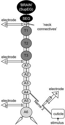
Paired intracellular recordings have been used to examine the activity-dependent plasticity and neuromodulator-induced metaplasticity of synaptic inputs from identified inhibitory and excitatory interneurons in the lamprey spinal cord. Trains of spikes at 5-20 Hz were used to mimic the frequency of spiking that occurs in network interneurons during NMDA or brainstem-evoked locomotor activity. Inputs from inhibitory and excitatory interneurons exhibited similar activity-dependent changes, with synaptic depression developing during the spike train. The level of depression reached was greater with lower stimulation frequencies. Significant activity-dependent depression of inputs from excitatory interneurons and inhibitory crossed caudal interneurons, which are central elements in the patterning of network activity, usually developed between the fifth and tenth spikes in the train. Because these interneurons typically fire bursts of up to five spikes during locomotor activity, this activity-dependent plasticity will presumably not contribute to the patterning of network activity. However, in the presence of the neuromodulators substance P and 5-HT, significant activity-dependent metaplasticity of these inputs developed over the first five spikes in the train. Substance P induced significant activity-dependent depression of inhibitory but potentiation of excitatory interneuron inputs, whereas 5-HT induced significant activity-dependent potentiation of both inhibitory and excitatory interneuron inputs. Because these metaplastic effects are consistent with the substance P and 5-HT-induced modulation of the network output, activity-dependent metaplasticity could be a potential mechanism underlying the coordination and modulation of rhythmic network activity. (+info)Neural changes after operant conditioning of the aerial respiratory behavior in Lymnaea stagnalis. (3/7372)
In this study, we demonstrate neural changes that occurred during operant conditioning of the aerial respiratory behavior of Lymnaea stagnalis. Aerial respiration in Lymnaea occurs at the water interface and is achieved by opening and closing movements of its respiratory orifice, the pneumostome. This behavior is controlled by a central pattern generator (CPG), the neurons of which, as well as the motoneurons innervating the pneumostome, have previously been identified and their synaptic connections well characterized. The respiratory behavior was operantly conditioned by applying a mechanical stimulus to the open pneumostome whenever the animal attempted to breathe. This negative reinforcement to the open pneumostome resulted in its immediate closure and a significant reduction in the overall respiratory activity. Electrophysiological recordings from the isolated CNSs after operant conditioning showed that the spontaneous patterned respiratory activity of the CPG neurons was significantly reduced. This included reduced spontaneous activity of the CPG interneuron involved in pneumostome opening (input 3 interneuron) and a reduced frequency of spontaneous tonic activity of the CPG interneuron [right pedal dorsal 1 (RPeD1)]. The ability to trigger the patterned respiratory activity by electrical stimulation of RPeD1 was also significantly reduced after operant conditioning. This study therefore demonstrates significant changes within a CPG that are associated with changes in a rhythmic homeostatic behavior after operant conditioning. (+info)GABAergic excitatory synapses and electrical coupling sustain prolonged discharges in the prey capture neural network of Clione limacina. (4/7372)
Afterdischarges represent a prominent characteristic of the neural network that controls prey capture reactions in the carnivorous mollusc Clione limacina. Their main functional implication is transformation of a brief sensory input from a prey into a lasting prey capture response. The present study, which focuses on the neuronal mechanisms of afterdischarges, demonstrates that a single pair of interneurons [cerebral A interneuron (Cr-Aint)] is responsible for afterdischarge generation in the network. Cr-Aint neurons are electrically coupled to all other neurons in the network and produce slow excitatory synaptic inputs to them. This excitatory transmission is found to be GABAergic, which is demonstrated by the use of GABA antagonists, uptake inhibitors, and double-labeling experiments showing that Cr-Aint neurons are GABA-immunoreactive. The Cr-Aint neurons organize three different pathways in the prey capture network, which provide positive feedback necessary for sustaining prolonged spike activity. The first pathway includes electrical coupling and slow chemical transmission from the Cr-Aint neurons to all other neurons in the network. The second feedback is based on excitatory reciprocal connections between contralateral interneurons. Recurrent excitation via the contralateral cell can sustain prolonged interneuron firing, which then drives the activity of all other cells in the network. The third positive feedback is represented by prominent afterdepolarizing potentials after individual spikes in the Cr-Aint neurons. Afterdepolarizations apparently represent recurrent GABAergic excitatory inputs. It is suggested here that these afterdepolarizing potentials are produced by GABAergic excitatory autapses. (+info)even-skipped determines the dorsal growth of motor axons in Drosophila. (5/7372)
Axon pathfinding and target choice are governed by cell type-specific responses to external cues. Here, we show that in the Drosophila embryo, motorneurons with targets in the dorsal muscle field express the homeobox gene even-skipped and that this expression is necessary and sufficient to direct motor axons into the dorsal muscle field. Previously, it was shown that motorneurons projecting to ventral targets express the LIM homeobox gene islet, which is sufficient to direct axons to the ventral muscle field. Thus, even-skipped complements the function of islet, and together these two genes constitute a bimodal switch regulating axonal growth and directing motor axons to ventral or to dorsal regions of the muscle field. (+info)Multiple point electrical stimulation of ulnar and median nerves. (6/7372)
A computer-assisted method of isolating single motor units (MUs) by multiple point stimulation (MPS) of peripheral nerves is described. MPS was used to isolate 10-30 single MUs from thenar and hypothenar muscles of normal subjects and patients with entrapment neuropathies, with the original purpose of obtaining a more representative mean motor unit potential for estimating the number of MUs in a muscle. The two important results that evolved from MPS however, were: (1) in the absence of 'alternation' MUs were recruited in an orderly pattern from small to large, and from longer to shorter latencies by graded electrical stimulation in both normal and pathological cases, (2) a comparison of the sizes of MUs recruited by stimulation proximal and distal to the elbow suggested that axonal branching can occur in the forearm 200 mm or more proximal to the motor point in intrinsic hand muscles. (+info)Role of mitochondrial dysfunction in the Ca2+-induced decline of transmitter release at K+-depolarized motor neuron terminals. (7/7372)
The present study tested whether a Ca2+-induced disruption of mitochondrial function was responsible for the decline in miniature endplate current (MEPC) frequency that occurs with nerve-muscle preparations maintained in a 35 mM potassium propionate (35 mM KP) solution containing elevated calcium. When the 35 mM KP contained control Ca2+ (1 mM), the MEPC frequency increased and remained elevated for many hours, and the mitochondria within twitch motor neuron terminals were similar in appearance to those in unstimulated terminals. All nerve terminals accumulated FM1-43 when the dye was present for the final 6 min of a 300-min exposure to 35 mM KP with control Ca2+. In contrast, when Ca2+ was increased to 3.6 mM in the 35 mM KP solution, the MEPC frequency initially reached frequencies >350 s-1 but then gradually fell approaching frequencies <50 s-1. A progressive swelling and eventual distortion of mitochondria within the twitch motor neuron terminals occurred during prolonged exposure to 35 mM KP with elevated Ca2+. After approximately 300 min in 35 mM KP with elevated Ca2+, only 58% of the twitch terminals accumulated FM1-43. The decline in MEPC frequency in 35 mM KP with elevated Ca2+ was less when 15 mM glucose was present or when preparations were pretreated with 10 microM oligomycin and then bathed in the 35 mM KP with glucose. When glucose was present, with or without oligomycin pretreatment, a greater percentage of twitch terminals accumulated FM1-43. However, the mitochondria in these preparations were still greatly swollen and distorted. We propose that prolonged depolarization of twitch motor neuron terminals by 35 mM KP with elevated Ca2+ produced a Ca2+-induced decrease in mitochondrial ATP production. Under these conditions, the cytosolic ATP/ADP ratio was decreased thereby compromising both transmitter release and refilling of recycled synaptic vesicles. The addition of glucose stimulated glycolysis which contributed to the maintenance of required ATP levels. (+info)Actions of a pair of identified cerebral-buccal interneurons (CBI-8/9) in Aplysia that contain the peptide myomodulin. (8/7372)
A combination of biocytin back-fills of the cerebral-buccal connectives and immunocytochemistry of the cerebral ganglion demonstrated that of the 13 bilateral pairs of cerebral-buccal interneurons in the cerebral ganglion, a subpopulation of 3 are immunopositive for the peptide myomodulin. The present paper describes the properties of two of these cells, which we have termed CBI-8 and CBI-9. CBI-8 and CBI-9 were found to be dye coupled and electrically coupled. The cells have virtually identical properties, and consequently we consider them to be "twin" pairs and refer to them as CBI-8/9. CBI-8/9 were identified by electrophysiological criteria and then labeled with dye. Labeled cells were found to be immunopositive for myomodulin, and, using high pressure liquid chromatography, the cells were shown to contain authentic myomodulin. CBI-8/9 were found to receive synaptic input after mechanical stimulation of the tentacles. They also received excitatory input from C-PR, a neuron involved in neck lengthening, and received a slow inhibitory input from CC5, a cell involved in neck shortening, suggesting that CBI-8/9 may be active during forward movements of the head or buccal mass. Firing of CBI-8 or CBI-9 resulted in the activation of a relatively small number of buccal neurons as evidenced by extracellular recordings from buccal nerves. Firing also produced local movements of the buccal mass, in particular a strong contraction of the I7 muscle, which mediates radula opening. CBI-8/9 were found to produce a slow depolarization and rhythmic activity of B48, the motor neuron for the I7 muscle. The data provide continuing evidence that the small population of cerebral buccal interneurons is composed of neurons that are highly diverse in their functional roles. CBI-8/9 may function as a type of premotor neuron, or perhaps as a peptidergic modulatory neuron, the functions of which are dependent on the coactivity of other neurons. (+info)Motor neurons are specialized nerve cells in the brain and spinal cord that play a crucial role in controlling voluntary muscle movements. They transmit electrical signals from the brain to the muscles, enabling us to perform actions such as walking, talking, and swallowing. There are two types of motor neurons: upper motor neurons, which originate in the brain's motor cortex and travel down to the brainstem and spinal cord; and lower motor neurons, which extend from the brainstem and spinal cord to the muscles. Damage or degeneration of these motor neurons can lead to various neurological disorders, such as amyotrophic lateral sclerosis (ALS) and spinal muscular atrophy (SMA).
Motor Neuron Disease (MND) is a progressive neurodegenerative disorder that affects the motor neurons, which are nerve cells in the brain and spinal cord responsible for controlling voluntary muscles involved in movement, speaking, breathing, and swallowing. As the motor neurons degenerate and die, they stop sending signals to the muscles, causing them to weaken, waste away (atrophy), and eventually lead to paralysis.
There are several types of MND, including:
1. Amyotrophic Lateral Sclerosis (ALS): Also known as Lou Gehrig's disease, this is the most common form of MND. It affects both upper and lower motor neurons, causing muscle weakness, stiffness, twitching, and atrophy throughout the body.
2. Progressive Bulbar Palsy (PBP): This type primarily affects the bulbar muscles in the brainstem, which control speech, swallowing, and chewing. Patients with PBP experience difficulties with speaking, slurred speech, and problems swallowing and may also have weak facial muscles and limb weakness.
3. Primary Lateral Sclerosis (PLS): This form of MND affects only the upper motor neurons, causing muscle stiffness, spasticity, and weakness, primarily in the legs. PLS progresses more slowly than ALS, and patients usually maintain their ability to speak and swallow for a longer period.
4. Progressive Muscular Atrophy (PMA): This type of MND affects only the lower motor neurons, causing muscle wasting, weakness, and fasciculations (muscle twitches). PMA progresses more slowly than ALS but can still be severely disabling over time.
5. Spinal Muscular Atrophy (SMA): This is a genetic form of MND that typically presents in infancy or childhood, although adult-onset forms exist. SMA affects the lower motor neurons in the spinal cord, causing muscle weakness and atrophy, primarily in the legs and trunk.
The exact cause of Motor Neuron Disease is not fully understood, but it is believed to involve a combination of genetic, environmental, and lifestyle factors. There is currently no cure for MND, and treatment focuses on managing symptoms, maintaining quality of life, and slowing disease progression through various therapies and medications.
Neurons, also known as nerve cells or neurocytes, are specialized cells that constitute the basic unit of the nervous system. They are responsible for receiving, processing, and transmitting information and signals within the body. Neurons have three main parts: the dendrites, the cell body (soma), and the axon. The dendrites receive signals from other neurons or sensory receptors, while the axon transmits these signals to other neurons, muscles, or glands. The junction between two neurons is called a synapse, where neurotransmitters are released to transmit the signal across the gap (synaptic cleft) to the next neuron. Neurons vary in size, shape, and structure depending on their function and location within the nervous system.
Survival of Motor Neuron 1 (SMN1) protein is a critical component for the survival of motor neurons, which are nerve cells that control muscle movements. The SMN1 protein is produced by the Survival of Motor Neuron 1 gene, located on human chromosome 5q13.
The primary function of the SMN1 protein is to assist in the biogenesis of small nuclear ribonucleoproteins (snRNPs), which are essential for spliceosomes - complex molecular machines responsible for RNA processing in the cell. The absence or significant reduction of SMN1 protein leads to defective snRNP assembly, impaired RNA splicing, and ultimately results in motor neuron degeneration.
Mutations in the SMN1 gene can cause Spinal Muscular Atrophy (SMA), a genetic disorder characterized by progressive muscle weakness, atrophy, and paralysis due to the loss of lower motor neurons in the spinal cord. The severity of SMA depends on the amount of functional SMN1 protein produced, with less protein leading to more severe symptoms.
Afferent neurons, also known as sensory neurons, are a type of nerve cell that conducts impulses or signals from peripheral receptors towards the central nervous system (CNS), which includes the brain and spinal cord. These neurons are responsible for transmitting sensory information such as touch, temperature, pain, sound, and light to the CNS for processing and interpretation. Afferent neurons have specialized receptor endings that detect changes in the environment and convert them into electrical signals, which are then transmitted to the CNS via synapses with other neurons. Once the signals reach the CNS, they are processed and integrated with other information to produce a response or reaction to the stimulus.
The motor cortex is a region in the frontal lobe of the brain that is responsible for controlling voluntary movements. It is involved in planning, initiating, and executing movements of the limbs, body, and face. The motor cortex contains neurons called Betz cells, which have large cell bodies and are responsible for transmitting signals to the spinal cord to activate muscles. Damage to the motor cortex can result in various movement disorders such as hemiplegia or paralysis on one side of the body.
Survival of Motor Neuron 2 (SMN2) protein is a functional copy of the Survival of Motor Neuron (SMN) protein, which is produced from the SMN2 gene. The SMN protein is crucial for the survival of motor neurons, the nerve cells that control muscle movement. In people with spinal muscular atrophy (SMA), a genetic disorder that causes progressive muscle weakness and loss of movement, there is a mutation in the main SMN1 gene that leads to reduced levels of functional SMN protein.
The SMN2 gene can also produce some functional SMN protein, but it mainly produces an unstable, truncated form of the protein due to a critical difference in its exon 7 splicing pattern. However, a small percentage (about 10-15%) of SMN2 transcripts can be correctly spliced and produce full-length, functional SMN protein. The amount of functional SMN protein produced from the SMN2 gene is directly related to the severity of SMA; more SMN protein production from SMN2 leads to less severe symptoms. Therefore, therapies aimed at increasing SMN2-derived SMN protein levels are being developed and tested for the treatment of SMA.
Evoked potentials, motor, are a category of tests used in clinical neurophysiology to measure the electrical activity generated by the nervous system in response to a stimulus that specifically activates the motor pathways. These tests can help assess the integrity and function of the motor neurons, which are responsible for controlling voluntary muscle movements.
During a motor evoked potentials test, electrodes are placed on the scalp or directly on the surface of the brain or spinal cord. A stimulus is then applied to the motor cortex or peripheral nerves, causing the muscles to contract. The resulting electrical signals are recorded and analyzed to evaluate the conduction velocity, amplitude, and latency of the motor responses.
Motor evoked potentials tests can be useful in diagnosing various neurological conditions, such as multiple sclerosis, spinal cord injuries, and motor neuron diseases. They can also help monitor the progression of these conditions and assess the effectiveness of treatments.
Amyotrophic Lateral Sclerosis (ALS) is a progressive neurodegenerative disorder that affects nerve cells in the brain and spinal cord responsible for controlling voluntary muscle movements, such as speaking, walking, breathing, and swallowing. The condition is characterized by the degeneration of motor neurons in the brain (upper motor neurons) and spinal cord (lower motor neurons), leading to their death.
The term "amyotrophic" comes from the Greek words "a" meaning no or negative, "myo" referring to muscle, and "trophic" relating to nutrition. When a motor neuron degenerates and can no longer send impulses to the muscle, the muscle becomes weak and eventually atrophies due to lack of use.
The term "lateral sclerosis" refers to the hardening or scarring (sclerosis) of the lateral columns of the spinal cord, which are primarily composed of nerve fibers that carry information from the brain to the muscles.
ALS is often called Lou Gehrig's disease, named after the famous American baseball player who was diagnosed with the condition in 1939. The exact cause of ALS remains unknown, but it is believed to involve a combination of genetic and environmental factors. There is currently no cure for ALS, and treatment primarily focuses on managing symptoms and maintaining quality of life.
The progression of ALS varies from person to person, with some individuals experiencing rapid decline over just a few years, while others may have a more slow-progressing form of the disease that lasts several decades. The majority of people with ALS die from respiratory failure within 3 to 5 years after the onset of symptoms. However, approximately 10% of those affected live for 10 or more years following diagnosis.
The spinal cord is a major part of the nervous system, extending from the brainstem and continuing down to the lower back. It is a slender, tubular bundle of nerve fibers (axons) and support cells (glial cells) that carries signals between the brain and the rest of the body. The spinal cord primarily serves as a conduit for motor information, which travels from the brain to the muscles, and sensory information, which travels from the body to the brain. It also contains neurons that can independently process and respond to information within the spinal cord without direct input from the brain.
The spinal cord is protected by the bony vertebral column (spine) and is divided into 31 segments: 8 cervical, 12 thoracic, 5 lumbar, 5 sacral, and 1 coccygeal. Each segment corresponds to a specific region of the body and gives rise to pairs of spinal nerves that exit through the intervertebral foramina at each level.
The spinal cord is responsible for several vital functions, including:
1. Reflexes: Simple reflex actions, such as the withdrawal reflex when touching a hot surface, are mediated by the spinal cord without involving the brain.
2. Muscle control: The spinal cord carries motor signals from the brain to the muscles, enabling voluntary movement and muscle tone regulation.
3. Sensory perception: The spinal cord transmits sensory information, such as touch, temperature, pain, and vibration, from the body to the brain for processing and awareness.
4. Autonomic functions: The sympathetic and parasympathetic divisions of the autonomic nervous system originate in the thoracolumbar and sacral regions of the spinal cord, respectively, controlling involuntary physiological responses like heart rate, blood pressure, digestion, and respiration.
Damage to the spinal cord can result in various degrees of paralysis or loss of sensation below the level of injury, depending on the severity and location of the damage.
Spinal muscular atrophy (SMA) is a genetic disorder that affects the motor neurons in the spinal cord, leading to muscle weakness and atrophy. It is caused by a mutation in the survival motor neuron 1 (SMN1) gene, which results in a deficiency of SMN protein necessary for the survival of motor neurons.
There are several types of SMA, classified based on the age of onset and severity of symptoms. The most common type is type 1, also known as Werdnig-Hoffmann disease, which presents in infancy and is characterized by severe muscle weakness, hypotonia, and feeding difficulties. Other types include type 2 (intermediate SMA), type 3 (Kugelberg-Welander disease), and type 4 (adult-onset SMA).
The symptoms of SMA may include muscle wasting, fasciculations, weakness, hypotonia, respiratory difficulties, and mobility impairment. The diagnosis of SMA typically involves genetic testing to confirm the presence of a mutation in the SMN1 gene. Treatment options for SMA may include medications, physical therapy, assistive devices, and respiratory support.
"Motor activity" is a general term used in the field of medicine and neuroscience to refer to any kind of physical movement or action that is generated by the body's motor system. The motor system includes the brain, spinal cord, nerves, and muscles that work together to produce movements such as walking, talking, reaching for an object, or even subtle actions like moving your eyes.
Motor activity can be voluntary, meaning it is initiated intentionally by the individual, or involuntary, meaning it is triggered automatically by the nervous system without conscious control. Examples of voluntary motor activity include deliberately lifting your arm or kicking a ball, while examples of involuntary motor activity include heartbeat, digestion, and reflex actions like jerking your hand away from a hot stove.
Abnormalities in motor activity can be a sign of neurological or muscular disorders, such as Parkinson's disease, cerebral palsy, or multiple sclerosis. Assessment of motor activity is often used in the diagnosis and treatment of these conditions.
An axon is a long, slender extension of a neuron (a type of nerve cell) that conducts electrical impulses (nerve impulses) away from the cell body to target cells, such as other neurons or muscle cells. Axons can vary in length from a few micrometers to over a meter long and are typically surrounded by a myelin sheath, which helps to insulate and protect the axon and allows for faster transmission of nerve impulses.
Axons play a critical role in the functioning of the nervous system, as they provide the means by which neurons communicate with one another and with other cells in the body. Damage to axons can result in serious neurological problems, such as those seen in spinal cord injuries or neurodegenerative diseases like multiple sclerosis.
The Survival Motor Neuron (SMN) complex is a protein complex that plays a crucial role in the biogenesis of small nuclear ribonucleoproteins (snRNPs), which are essential components of the spliceosome involved in pre-messenger RNA (pre-mRNA) splicing. The SMN complex consists of several proteins, including the SMN protein itself, Gemins2-8, and unrip.
The SMN protein is the central component of the complex and is encoded by the SMN1 gene located on chromosome 5q13.2. Mutations in this gene can lead to spinal muscular atrophy (SMA), a genetic disorder characterized by degeneration of motor neurons in the spinal cord, leading to muscle weakness and atrophy.
The SMN complex assembles in the cytoplasm and facilitates the assembly of spliceosomal snRNPs by helping to load Sm proteins onto small nuclear RNA (snRNA) molecules. Proper functioning of the SMN complex is essential for the correct splicing of pre-mRNA, and its dysfunction can lead to various developmental abnormalities and diseases, including SMA.
An action potential is a brief electrical signal that travels along the membrane of a nerve cell (neuron) or muscle cell. It is initiated by a rapid, localized change in the permeability of the cell membrane to specific ions, such as sodium and potassium, resulting in a rapid influx of sodium ions and a subsequent efflux of potassium ions. This ion movement causes a brief reversal of the electrical potential across the membrane, which is known as depolarization. The action potential then propagates along the cell membrane as a wave, allowing the electrical signal to be transmitted over long distances within the body. Action potentials play a crucial role in the communication and functioning of the nervous system and muscle tissue.
Nerve tissue proteins are specialized proteins found in the nervous system that provide structural and functional support to nerve cells, also known as neurons. These proteins include:
1. Neurofilaments: These are type IV intermediate filaments that provide structural support to neurons and help maintain their shape and size. They are composed of three subunits - NFL (light), NFM (medium), and NFH (heavy).
2. Neuronal Cytoskeletal Proteins: These include tubulins, actins, and spectrins that provide structural support to the neuronal cytoskeleton and help maintain its integrity.
3. Neurotransmitter Receptors: These are specialized proteins located on the postsynaptic membrane of neurons that bind neurotransmitters released by presynaptic neurons, triggering a response in the target cell.
4. Ion Channels: These are transmembrane proteins that regulate the flow of ions across the neuronal membrane and play a crucial role in generating and transmitting electrical signals in neurons.
5. Signaling Proteins: These include enzymes, receptors, and adaptor proteins that mediate intracellular signaling pathways involved in neuronal development, differentiation, survival, and death.
6. Adhesion Proteins: These are cell surface proteins that mediate cell-cell and cell-matrix interactions, playing a crucial role in the formation and maintenance of neural circuits.
7. Extracellular Matrix Proteins: These include proteoglycans, laminins, and collagens that provide structural support to nerve tissue and regulate neuronal migration, differentiation, and survival.
Molecular motor proteins are a type of protein that convert chemical energy into mechanical work at the molecular level. They play a crucial role in various cellular processes, such as cell division, muscle contraction, and intracellular transport. There are several types of molecular motor proteins, including myosin, kinesin, and dynein.
Myosin is responsible for muscle contraction and movement along actin filaments in the cytoplasm. Kinesin and dynein are involved in intracellular transport along microtubules, moving cargo such as vesicles, organelles, and mRNA to various destinations within the cell.
These motor proteins move in a stepwise fashion, with each step driven by the hydrolysis of adenosine triphosphate (ATP) into adenosine diphosphate (ADP) and inorganic phosphate (Pi). The directionality and speed of movement are determined by the structure and regulation of the motor proteins, as well as the properties of the tracks along which they move.
Electric stimulation, also known as electrical nerve stimulation or neuromuscular electrical stimulation, is a therapeutic treatment that uses low-voltage electrical currents to stimulate nerves and muscles. It is often used to help manage pain, promote healing, and improve muscle strength and mobility. The electrical impulses can be delivered through electrodes placed on the skin or directly implanted into the body.
In a medical context, electric stimulation may be used for various purposes such as:
1. Pain management: Electric stimulation can help to block pain signals from reaching the brain and promote the release of endorphins, which are natural painkillers produced by the body.
2. Muscle rehabilitation: Electric stimulation can help to strengthen muscles that have become weak due to injury, illness, or surgery. It can also help to prevent muscle atrophy and improve range of motion.
3. Wound healing: Electric stimulation can promote tissue growth and help to speed up the healing process in wounds, ulcers, and other types of injuries.
4. Urinary incontinence: Electric stimulation can be used to strengthen the muscles that control urination and reduce symptoms of urinary incontinence.
5. Migraine prevention: Electric stimulation can be used as a preventive treatment for migraines by applying electrical impulses to specific nerves in the head and neck.
It is important to note that electric stimulation should only be administered under the guidance of a qualified healthcare professional, as improper use can cause harm or discomfort.
Nerve degeneration, also known as neurodegeneration, is the progressive loss of structure and function of neurons, which can lead to cognitive decline, motor impairment, and various other symptoms. This process occurs due to a variety of factors, including genetics, environmental influences, and aging. It is a key feature in several neurological disorders such as Alzheimer's disease, Parkinson's disease, Huntington's disease, and multiple sclerosis. The degeneration can affect any part of the nervous system, leading to different symptoms depending on the location and extent of the damage.
A synapse is a structure in the nervous system that allows for the transmission of signals from one neuron (nerve cell) to another. It is the point where the axon terminal of one neuron meets the dendrite or cell body of another, and it is here that neurotransmitters are released and received. The synapse includes both the presynaptic and postsynaptic elements, as well as the cleft between them.
At the presynaptic side, an action potential travels down the axon and triggers the release of neurotransmitters into the synaptic cleft through exocytosis. These neurotransmitters then bind to receptors on the postsynaptic side, which can either excite or inhibit the receiving neuron. The strength of the signal between two neurons is determined by the number and efficiency of these synapses.
Synapses play a crucial role in the functioning of the nervous system, allowing for the integration and processing of information from various sources. They are also dynamic structures that can undergo changes in response to experience or injury, which has important implications for learning, memory, and recovery from neurological disorders.
Neuromuscular diseases are a group of disorders that involve the peripheral nervous system, which includes the nerves and muscles outside of the brain and spinal cord. These conditions can affect both children and adults, and they can be inherited or acquired. Neuromuscular diseases can cause a wide range of symptoms, including muscle weakness, numbness, tingling, pain, cramping, and twitching. Some common examples of neuromuscular diseases include muscular dystrophy, amyotrophic lateral sclerosis (ALS), peripheral neuropathy, and myasthenia gravis. The specific symptoms and severity of these conditions can vary widely depending on the underlying cause and the specific muscles and nerves that are affected. Treatment for neuromuscular diseases may include medications, physical therapy, assistive devices, or surgery, depending on the individual case.
In invertebrate biology, ganglia are clusters of neurons that function as a centralized nervous system. They can be considered as the equivalent to a vertebrate's spinal cord and brain. Ganglia serve to process sensory information, coordinate motor functions, and integrate various neural activities within an invertebrate organism.
Invertebrate ganglia are typically found in animals such as arthropods (insects, crustaceans), annelids (earthworms), mollusks (snails, squids), and cnidarians (jellyfish). The structure of the ganglia varies among different invertebrate groups.
For example, in arthropods, the central nervous system consists of a pair of connected ganglia called the supraesophageal ganglion or brain, and the subesophageal ganglion, located near the esophagus. The ventral nerve cord runs along the length of the body, containing pairs of ganglia that control specific regions of the body.
In mollusks, the central nervous system is composed of several ganglia, which can be fused or dispersed, depending on the species. In cephalopods (such as squids and octopuses), the brain is highly developed and consists of several lobes that perform various functions, including learning and memory.
Overall, invertebrate ganglia are essential components of the nervous system that allow these animals to respond to environmental stimuli, move, and interact with their surroundings.
Electrophysiology is a branch of medicine that deals with the electrical activities of the body, particularly the heart. In a medical context, electrophysiology studies (EPS) are performed to assess abnormal heart rhythms (arrhythmias) and to evaluate the effectiveness of certain treatments, such as medication or pacemakers.
During an EPS, electrode catheters are inserted into the heart through blood vessels in the groin or neck. These catheters can record the electrical activity of the heart and stimulate it to help identify the source of the arrhythmia. The information gathered during the study can help doctors determine the best course of treatment for each patient.
In addition to cardiac electrophysiology, there are also other subspecialties within electrophysiology, such as neuromuscular electrophysiology, which deals with the electrical activity of the nervous system and muscles.
Interneurons are a type of neuron that is located entirely within the central nervous system (CNS), including the brain and spinal cord. They are called "inter" neurons because they connect and communicate with other nearby neurons, forming complex networks within the CNS. Interneurons receive input from sensory neurons and/or other interneurons and then send output signals to motor neurons or other interneurons.
Interneurons are responsible for processing information and modulating neural circuits in the CNS. They can have either excitatory or inhibitory effects on their target neurons, depending on the type of neurotransmitters they release. Excitatory interneurons release neurotransmitters such as glutamate that increase the likelihood of an action potential in the postsynaptic neuron, while inhibitory interneurons release neurotransmitters such as GABA (gamma-aminobutyric acid) or glycine that decrease the likelihood of an action potential.
Interneurons are diverse and can be classified based on various criteria, including their morphology, electrophysiological properties, neurochemical characteristics, and connectivity patterns. They play crucial roles in many aspects of CNS function, such as sensory processing, motor control, cognition, and emotion regulation. Dysfunction or damage to interneurons has been implicated in various neurological and psychiatric disorders, including epilepsy, Parkinson's disease, schizophrenia, and autism spectrum disorder.
Synaptic transmission is the process by which a neuron communicates with another cell, such as another neuron or a muscle cell, across a junction called a synapse. It involves the release of neurotransmitters from the presynaptic terminal of the neuron, which then cross the synaptic cleft and bind to receptors on the postsynaptic cell, leading to changes in the electrical or chemical properties of the target cell. This process is critical for the transmission of signals within the nervous system and for controlling various physiological functions in the body.
In the context of medicine and healthcare, "movement" refers to the act or process of changing physical location or position. It involves the contraction and relaxation of muscles, which allows for the joints to move and the body to be in motion. Movement can also refer to the ability of a patient to move a specific body part or limb, which is assessed during physical examinations. Additionally, "movement" can describe the progression or spread of a disease within the body.
Cholinergic neurons are specialized types of nerve cells (neurons) that release the neurotransmitter acetylcholine to transmit signals to other neurons or effector cells, such as muscle cells. These neurons play important roles in various physiological functions, including modulation of motor control, cognition, memory, arousal, and sensory perception. Cholinergic neurons are widely distributed throughout the nervous system, with significant concentrations found in the basal forebrain, brainstem, and spinal cord. Dysfunction or degeneration of cholinergic neurons has been implicated in several neurological disorders, such as Alzheimer's disease, Parkinson's disease, and various forms of dementia.
"Cells, cultured" is a medical term that refers to cells that have been removed from an organism and grown in controlled laboratory conditions outside of the body. This process is called cell culture and it allows scientists to study cells in a more controlled and accessible environment than they would have inside the body. Cultured cells can be derived from a variety of sources, including tissues, organs, or fluids from humans, animals, or cell lines that have been previously established in the laboratory.
Cell culture involves several steps, including isolation of the cells from the tissue, purification and characterization of the cells, and maintenance of the cells in appropriate growth conditions. The cells are typically grown in specialized media that contain nutrients, growth factors, and other components necessary for their survival and proliferation. Cultured cells can be used for a variety of purposes, including basic research, drug development and testing, and production of biological products such as vaccines and gene therapies.
It is important to note that cultured cells may behave differently than they do in the body, and results obtained from cell culture studies may not always translate directly to human physiology or disease. Therefore, it is essential to validate findings from cell culture experiments using additional models and ultimately in clinical trials involving human subjects.
'Aplysia' is a genus of marine mollusks belonging to the family Aplysiidae, also known as sea hares. These are large, slow-moving herbivores that inhabit temperate and tropical coastal waters worldwide. They have a unique appearance with a soft, ear-like parapodia on either side of their body and a rhinophore at the front end, which they use to detect chemical cues in their environment.
One of the reasons 'Aplysia' is well-known in the medical and scientific community is because of its use as a model organism in neuroscience research. The simple nervous system of 'Aplysia' has made it an ideal subject for studying the basic principles of learning and memory at the cellular level.
In particular, the work of Nobel laureate Eric Kandel and his colleagues on 'Aplysia' helped to establish important concepts in synaptic plasticity, a key mechanism underlying learning and memory. By investigating how sensory stimulation can modify the strength of connections between neurons in 'Aplysia', researchers have gained valuable insights into the molecular and cellular mechanisms that underlie learning and memory processes in all animals, including humans.
Transgenic mice are genetically modified rodents that have incorporated foreign DNA (exogenous DNA) into their own genome. This is typically done through the use of recombinant DNA technology, where a specific gene or genetic sequence of interest is isolated and then introduced into the mouse embryo. The resulting transgenic mice can then express the protein encoded by the foreign gene, allowing researchers to study its function in a living organism.
The process of creating transgenic mice usually involves microinjecting the exogenous DNA into the pronucleus of a fertilized egg, which is then implanted into a surrogate mother. The offspring that result from this procedure are screened for the presence of the foreign DNA, and those that carry the desired genetic modification are used to establish a transgenic mouse line.
Transgenic mice have been widely used in biomedical research to model human diseases, study gene function, and test new therapies. They provide a valuable tool for understanding complex biological processes and developing new treatments for a variety of medical conditions.
Dopaminergic neurons are a type of specialized brain cells that produce, synthesize, and release the neurotransmitter dopamine. These neurons play crucial roles in various brain functions, including motivation, reward processing, motor control, and cognition. They are primarily located in several regions of the midbrain, such as the substantia nigra pars compacta (SNc) and the ventral tegmental area (VTA).
Dopaminergic neurons have a unique physiology characterized by their ability to generate slow, irregular electrical signals called pacemaker activity. This distinctive firing pattern allows dopamine to be released in a controlled manner, which is essential for proper brain function.
The degeneration and loss of dopaminergic neurons in the SNc are associated with Parkinson's disease, a neurodegenerative disorder characterized by motor impairments such as tremors, rigidity, and bradykinesia (slowness of movement). The reduction in dopamine levels caused by this degeneration leads to an imbalance in the brain's neural circuitry, resulting in the characteristic symptoms of Parkinson's disease.
Medical Definition:
Superoxide dismutase (SOD) is an enzyme that catalyzes the dismutation of superoxide radicals (O2-) into oxygen (O2) and hydrogen peroxide (H2O2). This essential antioxidant defense mechanism helps protect the body's cells from damage caused by reactive oxygen species (ROS), which are produced during normal metabolic processes and can lead to oxidative stress when their levels become too high.
There are three main types of superoxide dismutase found in different cellular locations:
1. Copper-zinc superoxide dismutase (CuZnSOD or SOD1) - Present mainly in the cytoplasm of cells.
2. Manganese superoxide dismutase (MnSOD or SOD2) - Located within the mitochondrial matrix.
3. Extracellular superoxide dismutase (EcSOD or SOD3) - Found in the extracellular spaces, such as blood vessels and connective tissues.
Imbalances in SOD levels or activity have been linked to various pathological conditions, including neurodegenerative diseases, cancer, and aging-related disorders.
Electromyography (EMG) is a medical diagnostic procedure that measures the electrical activity of skeletal muscles during contraction and at rest. It involves inserting a thin needle electrode into the muscle to record the electrical signals generated by the muscle fibers. These signals are then displayed on an oscilloscope and may be heard through a speaker.
EMG can help diagnose various neuromuscular disorders, such as muscle weakness, numbness, or pain, and can distinguish between muscle and nerve disorders. It is often used in conjunction with other diagnostic tests, such as nerve conduction studies, to provide a comprehensive evaluation of the nervous system.
EMG is typically performed by a neurologist or a physiatrist, and the procedure may cause some discomfort or pain, although this is usually minimal. The results of an EMG can help guide treatment decisions and monitor the progression of neuromuscular conditions over time.
Anterior horn cells, also known as motor neurons, are a type of nerve cell located in the anterior (ventral) horn of the spinal cord's gray matter. These cells play a crucial role in initiating and regulating voluntary muscle movement by transmitting signals from the brain to the muscles via the peripheral nervous system.
Damage or degeneration of the anterior horn cells can result in various neuromuscular disorders, such as spinal muscular atrophy (SMA) and amyotrophic lateral sclerosis (ALS). These conditions can lead to muscle weakness, atrophy, and paralysis.
The neuromuscular junction (NMJ) is the specialized synapse or chemical communication point, where the motor neuron's nerve terminal (presynaptic element) meets the muscle fiber's motor end plate (postsynaptic element). This junction plays a crucial role in controlling muscle contraction and relaxation.
At the NMJ, the neurotransmitter acetylcholine is released from the presynaptic nerve terminal into the synaptic cleft, following an action potential. Acetylcholine then binds to nicotinic acetylcholine receptors on the postsynaptic membrane of the muscle fiber, leading to the generation of an end-plate potential. If sufficient end-plate potentials are generated and summate, they will trigger an action potential in the muscle fiber, ultimately causing muscle contraction.
Dysfunction at the neuromuscular junction can result in various neuromuscular disorders, such as myasthenia gravis, where autoantibodies attack acetylcholine receptors, leading to muscle weakness and fatigue.
Dendrites are the branched projections of a neuron that receive and process signals from other neurons. They are typically short and highly branching, increasing the surface area for receiving incoming signals. Dendrites are covered in small protrusions called dendritic spines, which can form connections with the axon terminals of other neurons through chemical synapses. The structure and function of dendrites play a critical role in the integration and processing of information in the nervous system.
Sensory receptor cells are specialized structures that convert physical stimuli from our environment into electrical signals, which are then transmitted to the brain for interpretation. These receptors can be found in various tissues throughout the body and are responsible for detecting sensations such as touch, pressure, temperature, taste, and smell. They can be classified into two main types: exteroceptors, which respond to stimuli from the external environment, and interoceptors, which react to internal conditions within the body. Examples of sensory receptor cells include hair cells in the inner ear, photoreceptors in the eye, and taste buds on the tongue.
Progressive bulbar palsy (PBP) is a form of motor neuron disease (MND), also known as Amyotrophic Lateral Sclerosis (ALS). It is characterized by the progressive degeneration of the motor neurons in the brainstem, which control vital functions such as swallowing, speaking, chewing, and breathing.
In PBP, these symptoms gradually worsen over time, often resulting in severe disability and ultimately death due to respiratory failure. The progression of the disease can vary from person to person, but it typically advances more slowly than other forms of ALS. There is currently no cure for PBP or any other form of MND, and treatment is focused on managing symptoms and maintaining quality of life.
Spinal ganglia, also known as dorsal root ganglia, are clusters of nerve cell bodies located in the peripheral nervous system. They are situated along the length of the spinal cord and are responsible for transmitting sensory information from the body to the brain. Each spinal ganglion contains numerous neurons, or nerve cells, with long processes called axons that extend into the periphery and innervate various tissues and organs. The cell bodies within the spinal ganglia receive sensory input from these axons and transmit this information to the central nervous system via the dorsal roots of the spinal nerves. This allows the brain to interpret and respond to a wide range of sensory stimuli, including touch, temperature, pain, and proprioception (the sense of the position and movement of one's body).
Neural inhibition is a process in the nervous system that decreases or prevents the activity of neurons (nerve cells) in order to regulate and control communication within the nervous system. It is a fundamental mechanism that allows for the balance of excitation and inhibition necessary for normal neural function. Inhibitory neurotransmitters, such as GABA (gamma-aminobutyric acid) and glycine, are released from the presynaptic neuron and bind to receptors on the postsynaptic neuron, reducing its likelihood of firing an action potential. This results in a decrease in neural activity and can have various effects depending on the specific neurons and brain regions involved. Neural inhibition is crucial for many functions including motor control, sensory processing, attention, memory, and emotional regulation.
GABAergic neurons are a type of neuron that releases the neurotransmitter gamma-aminobutyric acid (GABA). GABA is the primary inhibitory neurotransmitter in the mature central nervous system, meaning it functions to decrease the excitability of neurons it acts upon.
GABAergic neurons are widely distributed throughout the brain and spinal cord and play a crucial role in regulating neural activity by balancing excitation and inhibition. They form synapses with various types of neurons, including both excitatory and inhibitory neurons, and their activation can lead to hyperpolarization or decreased firing rates of the target cells.
Dysfunction in GABAergic neurotransmission has been implicated in several neurological and psychiatric disorders, such as epilepsy, anxiety, and sleep disorders.
Patch-clamp techniques are a group of electrophysiological methods used to study ion channels and other electrical properties of cells. These techniques were developed by Erwin Neher and Bert Sakmann, who were awarded the Nobel Prize in Physiology or Medicine in 1991 for their work. The basic principle of patch-clamp techniques involves creating a high resistance seal between a glass micropipette and the cell membrane, allowing for the measurement of current flowing through individual ion channels or groups of channels.
There are several different configurations of patch-clamp techniques, including:
1. Cell-attached configuration: In this configuration, the micropipette is attached to the outer surface of the cell membrane, and the current flowing across a single ion channel can be measured. This configuration allows for the study of the properties of individual channels in their native environment.
2. Whole-cell configuration: Here, the micropipette breaks through the cell membrane, creating a low resistance electrical connection between the pipette and the inside of the cell. This configuration allows for the measurement of the total current flowing across all ion channels in the cell membrane.
3. Inside-out configuration: In this configuration, the micropipette is pulled away from the cell after establishing a seal, resulting in the exposure of the inner surface of the cell membrane to the solution in the pipette. This configuration allows for the study of the properties of ion channels in isolation from other cellular components.
4. Outside-out configuration: Here, the micropipette is pulled away from the cell after establishing a seal, resulting in the exposure of the outer surface of the cell membrane to the solution in the pipette. This configuration allows for the study of the properties of ion channels in their native environment, but with the ability to control the composition of the extracellular solution.
Patch-clamp techniques have been instrumental in advancing our understanding of ion channel function and have contributed to numerous breakthroughs in neuroscience, pharmacology, and physiology.
Immunohistochemistry (IHC) is a technique used in pathology and laboratory medicine to identify specific proteins or antigens in tissue sections. It combines the principles of immunology and histology to detect the presence and location of these target molecules within cells and tissues. This technique utilizes antibodies that are specific to the protein or antigen of interest, which are then tagged with a detection system such as a chromogen or fluorophore. The stained tissue sections can be examined under a microscope, allowing for the visualization and analysis of the distribution and expression patterns of the target molecule in the context of the tissue architecture. Immunohistochemistry is widely used in diagnostic pathology to help identify various diseases, including cancer, infectious diseases, and immune-mediated disorders.
Neurological models are simplified representations or simulations of various aspects of the nervous system, including its structure, function, and processes. These models can be theoretical, computational, or physical and are used to understand, explain, and predict neurological phenomena. They may focus on specific neurological diseases, disorders, or functions, such as memory, learning, or movement. The goal of these models is to provide insights into the complex workings of the nervous system that cannot be easily observed or understood through direct examination alone.
The brain is the central organ of the nervous system, responsible for receiving and processing sensory information, regulating vital functions, and controlling behavior, movement, and cognition. It is divided into several distinct regions, each with specific functions:
1. Cerebrum: The largest part of the brain, responsible for higher cognitive functions such as thinking, learning, memory, language, and perception. It is divided into two hemispheres, each controlling the opposite side of the body.
2. Cerebellum: Located at the back of the brain, it is responsible for coordinating muscle movements, maintaining balance, and fine-tuning motor skills.
3. Brainstem: Connects the cerebrum and cerebellum to the spinal cord, controlling vital functions such as breathing, heart rate, and blood pressure. It also serves as a relay center for sensory information and motor commands between the brain and the rest of the body.
4. Diencephalon: A region that includes the thalamus (a major sensory relay station) and hypothalamus (regulates hormones, temperature, hunger, thirst, and sleep).
5. Limbic system: A group of structures involved in emotional processing, memory formation, and motivation, including the hippocampus, amygdala, and cingulate gyrus.
The brain is composed of billions of interconnected neurons that communicate through electrical and chemical signals. It is protected by the skull and surrounded by three layers of membranes called meninges, as well as cerebrospinal fluid that provides cushioning and nutrients.
The cerebral cortex is the outermost layer of the brain, characterized by its intricate folded structure and wrinkled appearance. It is a region of great importance as it plays a key role in higher cognitive functions such as perception, consciousness, thought, memory, language, and attention. The cerebral cortex is divided into two hemispheres, each containing four lobes: the frontal, parietal, temporal, and occipital lobes. These areas are responsible for different functions, with some regions specializing in sensory processing while others are involved in motor control or associative functions. The cerebral cortex is composed of gray matter, which contains neuronal cell bodies, and is covered by a layer of white matter that consists mainly of myelinated nerve fibers.
"Newborn animals" refers to the very young offspring of animals that have recently been born. In medical terminology, newborns are often referred to as "neonates," and they are classified as such from birth until about 28 days of age. During this time period, newborn animals are particularly vulnerable and require close monitoring and care to ensure their survival and healthy development.
The specific needs of newborn animals can vary widely depending on the species, but generally, they require warmth, nutrition, hydration, and protection from harm. In many cases, newborns are unable to regulate their own body temperature or feed themselves, so they rely heavily on their mothers for care and support.
In medical settings, newborn animals may be examined and treated by veterinarians to ensure that they are healthy and receiving the care they need. This can include providing medical interventions such as feeding tubes, antibiotics, or other treatments as needed to address any health issues that arise. Overall, the care and support of newborn animals is an important aspect of animal medicine and conservation efforts.
Animal disease models are specialized animals, typically rodents such as mice or rats, that have been genetically engineered or exposed to certain conditions to develop symptoms and physiological changes similar to those seen in human diseases. These models are used in medical research to study the pathophysiology of diseases, identify potential therapeutic targets, test drug efficacy and safety, and understand disease mechanisms.
The genetic modifications can include knockout or knock-in mutations, transgenic expression of specific genes, or RNA interference techniques. The animals may also be exposed to environmental factors such as chemicals, radiation, or infectious agents to induce the disease state.
Examples of animal disease models include:
1. Mouse models of cancer: Genetically engineered mice that develop various types of tumors, allowing researchers to study cancer initiation, progression, and metastasis.
2. Alzheimer's disease models: Transgenic mice expressing mutant human genes associated with Alzheimer's disease, which exhibit amyloid plaque formation and cognitive decline.
3. Diabetes models: Obese and diabetic mouse strains like the NOD (non-obese diabetic) or db/db mice, used to study the development of type 1 and type 2 diabetes, respectively.
4. Cardiovascular disease models: Atherosclerosis-prone mice, such as ApoE-deficient or LDLR-deficient mice, that develop plaque buildup in their arteries when fed a high-fat diet.
5. Inflammatory bowel disease models: Mice with genetic mutations affecting intestinal barrier function and immune response, such as IL-10 knockout or SAMP1/YitFc mice, which develop colitis.
Animal disease models are essential tools in preclinical research, but it is important to recognize their limitations. Differences between species can affect the translatability of results from animal studies to human patients. Therefore, researchers must carefully consider the choice of model and interpret findings cautiously when applying them to human diseases.
In the field of medicine, "time factors" refer to the duration of symptoms or time elapsed since the onset of a medical condition, which can have significant implications for diagnosis and treatment. Understanding time factors is crucial in determining the progression of a disease, evaluating the effectiveness of treatments, and making critical decisions regarding patient care.
For example, in stroke management, "time is brain," meaning that rapid intervention within a specific time frame (usually within 4.5 hours) is essential to administering tissue plasminogen activator (tPA), a clot-busting drug that can minimize brain damage and improve patient outcomes. Similarly, in trauma care, the "golden hour" concept emphasizes the importance of providing definitive care within the first 60 minutes after injury to increase survival rates and reduce morbidity.
Time factors also play a role in monitoring the progression of chronic conditions like diabetes or heart disease, where regular follow-ups and assessments help determine appropriate treatment adjustments and prevent complications. In infectious diseases, time factors are crucial for initiating antibiotic therapy and identifying potential outbreaks to control their spread.
Overall, "time factors" encompass the significance of recognizing and acting promptly in various medical scenarios to optimize patient outcomes and provide effective care.
The facial nerve, also known as the seventh cranial nerve (CN VII), is a mixed nerve that carries both sensory and motor fibers. Its functions include controlling the muscles involved in facial expressions, taste sensation from the anterior two-thirds of the tongue, and secretomotor function to the lacrimal and salivary glands.
The facial nerve originates from the brainstem and exits the skull through the internal acoustic meatus. It then passes through the facial canal in the temporal bone before branching out to innervate various structures of the face. The main branches of the facial nerve include:
1. Temporal branch: Innervates the frontalis, corrugator supercilii, and orbicularis oculi muscles responsible for eyebrow movements and eyelid closure.
2. Zygomatic branch: Supplies the muscles that elevate the upper lip and wrinkle the nose.
3. Buccal branch: Innervates the muscles of the cheek and lips, allowing for facial expressions such as smiling and puckering.
4. Mandibular branch: Controls the muscles responsible for lower lip movement and depressing the angle of the mouth.
5. Cervical branch: Innervates the platysma muscle in the neck, which helps to depress the lower jaw and wrinkle the skin of the neck.
Damage to the facial nerve can result in various symptoms, such as facial weakness or paralysis, loss of taste sensation, and dry eyes or mouth due to impaired secretion.
Membrane potential is the electrical potential difference across a cell membrane, typically for excitable cells such as nerve and muscle cells. It is the difference in electric charge between the inside and outside of a cell, created by the selective permeability of the cell membrane to different ions. The resting membrane potential of a typical animal cell is around -70 mV, with the interior being negative relative to the exterior. This potential is generated and maintained by the active transport of ions across the membrane, primarily through the action of the sodium-potassium pump. Membrane potentials play a crucial role in many physiological processes, including the transmission of nerve impulses and the contraction of muscle cells.
Motor skills disorders are conditions that affect a person's ability to perform coordinated movements. These movements can be simple, such as buttoning a shirt, or complex, such as playing a musical instrument. Motor skills disorders can make it difficult for a person to perform everyday activities and can impact their quality of life.
There are two main types of motor skills: fine motor skills and gross motor skills. Fine motor skills involve the small movements of the hands, fingers, and wrists, such as writing or using utensils. Gross motor skills involve larger movements of the arms, legs, and torso, such as crawling, walking, or running.
Motor skills disorders can affect either fine or gross motor skills, or both. Some common types of motor skills disorders include:
* Developmental coordination disorder (DCD): a condition that affects a child's ability to perform coordinated movements and is often diagnosed in early childhood. Children with DCD may have difficulty with tasks such as tying their shoes, buttoning their clothes, or using scissors.
* Cerebral palsy: a group of disorders that affect movement and muscle tone, caused by damage to the brain before, during, or after birth. Cerebral palsy can cause stiff or floppy muscles, uncontrolled movements, and difficulty with balance and coordination.
* Dyspraxia: a condition that affects a person's ability to plan and perform coordinated movements. People with dyspraxia may have difficulty with tasks such as writing, buttoning their clothes, or playing sports.
* Ataxia: a group of disorders that affect coordination and balance, caused by damage to the cerebellum (the part of the brain that controls movement). Ataxia can cause unsteady gait, poor coordination, and difficulty with fine motor tasks.
Motor skills disorders can be caused by a variety of factors, including genetics, injury, illness, or developmental delays. Treatment for motor skills disorders may include physical therapy, occupational therapy, speech therapy, and medication. In some cases, surgery may also be necessary to treat the underlying cause of the disorder.
Genetically modified animals (GMAs) are those whose genetic makeup has been altered using biotechnological techniques. This is typically done by introducing one or more genes from another species into the animal's genome, resulting in a new trait or characteristic that does not naturally occur in that species. The introduced gene is often referred to as a transgene.
The process of creating GMAs involves several steps:
1. Isolation: The desired gene is isolated from the DNA of another organism.
2. Transfer: The isolated gene is transferred into the target animal's cells, usually using a vector such as a virus or bacterium.
3. Integration: The transgene integrates into the animal's chromosome, becoming a permanent part of its genetic makeup.
4. Selection: The modified cells are allowed to multiply, and those that contain the transgene are selected for further growth and development.
5. Breeding: The genetically modified individuals are bred to produce offspring that carry the desired trait.
GMAs have various applications in research, agriculture, and medicine. In research, they can serve as models for studying human diseases or testing new therapies. In agriculture, GMAs can be developed to exhibit enhanced growth rates, improved disease resistance, or increased nutritional value. In medicine, GMAs may be used to produce pharmaceuticals or other therapeutic agents within their bodies.
Examples of genetically modified animals include mice with added genes for specific proteins that make them useful models for studying human diseases, goats that produce a human protein in their milk to treat hemophilia, and pigs with enhanced resistance to certain viruses that could potentially be used as organ donors for humans.
It is important to note that the use of genetically modified animals raises ethical concerns related to animal welfare, environmental impact, and potential risks to human health. These issues must be carefully considered and addressed when developing and implementing GMA technologies.
The hippocampus is a complex, curved formation in the brain that resembles a seahorse (hence its name, from the Greek word "hippos" meaning horse and "kampos" meaning sea monster). It's part of the limbic system and plays crucial roles in the formation of memories, particularly long-term ones.
This region is involved in spatial navigation and cognitive maps, allowing us to recognize locations and remember how to get to them. Additionally, it's one of the first areas affected by Alzheimer's disease, which often results in memory loss as an early symptom.
Anatomically, it consists of two main parts: the Ammon's horn (or cornu ammonis) and the dentate gyrus. These structures are made up of distinct types of neurons that contribute to different aspects of learning and memory.
Gamma-Aminobutyric Acid (GABA) is a major inhibitory neurotransmitter in the mammalian central nervous system. It plays a crucial role in regulating neuronal excitability and preventing excessive neuronal firing, which helps to maintain neural homeostasis and reduce the risk of seizures. GABA functions by binding to specific receptors (GABA-A, GABA-B, and GABA-C) on the postsynaptic membrane, leading to hyperpolarization of the neuronal membrane and reduced neurotransmitter release from presynaptic terminals.
In addition to its role in the central nervous system, GABA has also been identified as a neurotransmitter in the peripheral nervous system, where it is involved in regulating various physiological processes such as muscle relaxation, hormone secretion, and immune function.
GABA can be synthesized in neurons from glutamate, an excitatory neurotransmitter, through the action of the enzyme glutamic acid decarboxylase (GAD). Once synthesized, GABA is stored in synaptic vesicles and released into the synapse upon neuronal activation. After release, GABA can be taken up by surrounding glial cells or degraded by the enzyme GABA transaminase (GABA-T) into succinic semialdehyde, which is further metabolized to form succinate and enter the Krebs cycle for energy production.
Dysregulation of GABAergic neurotransmission has been implicated in various neurological and psychiatric disorders, including epilepsy, anxiety, depression, and sleep disturbances. Therefore, modulating GABAergic signaling through pharmacological interventions or other therapeutic approaches may offer potential benefits for the treatment of these conditions.
The brainstem is the lower part of the brain that connects to the spinal cord. It consists of the midbrain, pons, and medulla oblongata. The brainstem controls many vital functions such as heart rate, breathing, and blood pressure. It also serves as a relay center for sensory and motor information between the cerebral cortex and the rest of the body. Additionally, several cranial nerves originate from the brainstem, including those that control eye movements, facial movements, and hearing.
Axonal transport is the controlled movement of materials and organelles within axons, which are the nerve fibers of neurons (nerve cells). This intracellular transport system is essential for maintaining the structural and functional integrity of axons, particularly in neurons with long axonal processes. There are two types of axonal transport: anterograde transport, which moves materials from the cell body toward the synaptic terminals, and retrograde transport, which transports materials from the synaptic terminals back to the cell body. Anterograde transport is typically slower than retrograde transport and can be divided into fast and slow components based on velocity. Fast anterograde transport moves vesicles containing neurotransmitters and their receptors, as well as mitochondria and other organelles, at speeds of up to 400 mm/day. Slow anterograde transport moves cytoskeletal elements, proteins, and RNA at speeds of 1-10 mm/day. Retrograde transport is primarily responsible for recycling membrane components, removing damaged organelles, and transmitting signals from the axon terminal to the cell body. Dysfunctions in axonal transport have been implicated in various neurodegenerative disorders, such as Alzheimer's disease, Parkinson's disease, and amyotrophic lateral sclerosis (ALS).
Neuropeptides are small protein-like molecules that are used by neurons to communicate with each other and with other cells in the body. They are produced in the cell body of a neuron, processed from larger precursor proteins, and then transported to the nerve terminal where they are stored in secretory vesicles. When the neuron is stimulated, the vesicles fuse with the cell membrane and release their contents into the extracellular space.
Neuropeptides can act as neurotransmitters or neuromodulators, depending on their target receptors and the duration of their effects. They play important roles in a variety of physiological processes, including pain perception, appetite regulation, stress response, and social behavior. Some neuropeptides also have hormonal functions, such as oxytocin and vasopressin, which are produced in the hypothalamus and released into the bloodstream to regulate reproductive and cardiovascular function, respectively.
There are hundreds of different neuropeptides that have been identified in the nervous system, and many of them have multiple functions and interact with other signaling molecules to modulate neural activity. Dysregulation of neuropeptide systems has been implicated in various neurological and psychiatric disorders, such as chronic pain, addiction, depression, and anxiety.
A mutation is a permanent change in the DNA sequence of an organism's genome. Mutations can occur spontaneously or be caused by environmental factors such as exposure to radiation, chemicals, or viruses. They may have various effects on the organism, ranging from benign to harmful, depending on where they occur and whether they alter the function of essential proteins. In some cases, mutations can increase an individual's susceptibility to certain diseases or disorders, while in others, they may confer a survival advantage. Mutations are the driving force behind evolution, as they introduce new genetic variability into populations, which can then be acted upon by natural selection.
Neural conduction is the process by which electrical signals, known as action potentials, are transmitted along the axon of a neuron (nerve cell) to transmit information between different parts of the nervous system. This electrical impulse is generated by the movement of ions across the neuronal membrane, and it propagates down the length of the axon until it reaches the synapse, where it can then stimulate the release of neurotransmitters to communicate with other neurons or target cells. The speed of neural conduction can vary depending on factors such as the diameter of the axon, the presence of myelin sheaths (which act as insulation and allow for faster conduction), and the temperature of the environment.
Choline O-Acetyltransferase (COAT, ChAT) is an enzyme that plays a crucial role in the synthesis of the neurotransmitter acetylcholine. It catalyzes the transfer of an acetyl group from acetyl CoA to choline, resulting in the formation of acetylcholine. Acetylcholine is a vital neurotransmitter involved in various physiological processes such as memory, cognition, and muscle contraction. COAT is primarily located in cholinergic neurons, which are nerve cells that use acetylcholine to transmit signals to other neurons or muscles. Inhibition of ChAT can lead to a decrease in acetylcholine levels and may contribute to neurological disorders such as Alzheimer's disease and myasthenia gravis.
A ganglion is a cluster of neuron cell bodies in the peripheral nervous system. Ganglia are typically associated with nerves and serve as sites for sensory processing, integration, and relay of information between the periphery and the central nervous system (CNS). The two main types of ganglia are sensory ganglia, which contain pseudounipolar neurons that transmit sensory information to the CNS, and autonomic ganglia, which contain multipolar neurons that control involuntary physiological functions.
Examples of sensory ganglia include dorsal root ganglia (DRG), which are associated with spinal nerves, and cranial nerve ganglia, such as the trigeminal ganglion. Autonomic ganglia can be further divided into sympathetic and parasympathetic ganglia, which regulate different aspects of the autonomic nervous system.
It's worth noting that in anatomy, "ganglion" refers to a group of nerve cell bodies, while in clinical contexts, "ganglion" is often used to describe a specific type of cystic structure that forms near joints or tendons, typically in the wrist or foot. These ganglia are not related to the peripheral nervous system's ganglia but rather are fluid-filled sacs that may cause discomfort or pain due to their size or location.
I'm sorry for any confusion, but "grasshoppers" is not a term used in medical definitions. Grasshoppers are a type of insect that belongs to the order Orthoptera and suborder Caelifera. They are known for their long hind legs which they use for jumping, and some species can jump over 20 times their own body length. If you have any questions about medical terminology or topics, I'd be happy to help with those instead!
Efferent neurons are specialized nerve cells that transmit signals from the central nervous system (CNS), which includes the brain and spinal cord, to effector organs such as muscles or glands. These signals typically result in a response or action, hence the term "efferent," derived from the Latin word "efferre" meaning "to carry away."
Efferent neurons are part of the motor pathway and can be further classified into two types:
1. Somatic efferent neurons: These neurons transmit signals to skeletal muscles, enabling voluntary movements and posture maintenance. They have their cell bodies located in the ventral horn of the spinal cord and send their axons through the ventral roots to innervate specific muscle fibers.
2. Autonomic efferent neurons: These neurons are responsible for controlling involuntary functions, such as heart rate, digestion, respiration, and pupil dilation. They have a two-neuron chain arrangement, with the preganglionic neuron having its cell body in the CNS (brainstem or spinal cord) and synapsing with the postganglionic neuron in an autonomic ganglion near the effector organ. Autonomic efferent neurons can be further divided into sympathetic, parasympathetic, and enteric subdivisions based on their functions and innervation patterns.
In summary, efferent neurons are a critical component of the nervous system, responsible for transmitting signals from the CNS to various effector organs, ultimately controlling and coordinating numerous bodily functions and responses.
A nerve net, also known as a neural net or neuronal network, is not a medical term per se, but rather a concept in neuroscience and artificial intelligence (AI). It refers to a complex network of interconnected neurons that process and transmit information. In the context of the human body, the nervous system can be thought of as a type of nerve net, with the brain and spinal cord serving as the central processing unit and peripheral nerves carrying signals to and from various parts of the body.
In the field of AI, artificial neural networks are computational models inspired by the structure and function of biological nerve nets. These models consist of interconnected nodes or "neurons" that process information and learn patterns through a process of training and adaptation. They have been used in a variety of applications, including image recognition, natural language processing, and machine learning.
Neurofilament proteins (NFs) are type IV intermediate filament proteins that are specific to neurons. They are the major structural components of the neuronal cytoskeleton and play crucial roles in maintaining the structural integrity, stability, and diameter of axons. Neurofilaments are composed of three subunits: light (NFL), medium (NFM), and heavy (NFH) neurofilament proteins, which differ in their molecular weights. These subunits assemble into heteropolymers to form the neurofilament core, while the C-terminal tails of NFH and NFM extend outward from the core, interacting with other cellular components and participating in various neuronal functions. Increased levels of neurofilament proteins, particularly NFL, in cerebrospinal fluid (CSF) and blood are considered biomarkers for axonal damage and neurodegeneration in several neurological disorders, such as Alzheimer's disease, Parkinson's disease, amyotrophic lateral sclerosis (ALS), and multiple sclerosis (MS).
Locomotion, in a medical context, refers to the ability to move independently and change location. It involves the coordinated movement of the muscles, bones, and nervous system that enables an individual to move from one place to another. This can include walking, running, jumping, or using assistive devices such as wheelchairs or crutches. Locomotion is a fundamental aspect of human mobility and is often assessed in medical evaluations to determine overall health and functioning.
The Central Nervous System (CNS) is the part of the nervous system that consists of the brain and spinal cord. It is called the "central" system because it receives information from, and sends information to, the rest of the body through peripheral nerves, which make up the Peripheral Nervous System (PNS).
The CNS is responsible for processing sensory information, controlling motor functions, and regulating various autonomic processes like heart rate, respiration, and digestion. The brain, as the command center of the CNS, interprets sensory stimuli, formulates thoughts, and initiates actions. The spinal cord serves as a conduit for nerve impulses traveling to and from the brain and the rest of the body.
The CNS is protected by several structures, including the skull (which houses the brain) and the vertebral column (which surrounds and protects the spinal cord). Despite these protective measures, the CNS remains vulnerable to injury and disease, which can have severe consequences due to its crucial role in controlling essential bodily functions.
Reaction time, in the context of medicine and physiology, refers to the time period between the presentation of a stimulus and the subsequent initiation of a response. This complex process involves the central nervous system, particularly the brain, which perceives the stimulus, processes it, and then sends signals to the appropriate muscles or glands to react.
There are different types of reaction times, including simple reaction time (responding to a single, expected stimulus) and choice reaction time (choosing an appropriate response from multiple possibilities). These measures can be used in clinical settings to assess various aspects of neurological function, such as cognitive processing speed, motor control, and alertness.
However, it is important to note that reaction times can be influenced by several factors, including age, fatigue, attention, and the use of certain medications or substances.
Green Fluorescent Protein (GFP) is not a medical term per se, but a scientific term used in the field of molecular biology. GFP is a protein that exhibits bright green fluorescence when exposed to light, particularly blue or ultraviolet light. It was originally discovered in the jellyfish Aequorea victoria.
In medical and biological research, scientists often use recombinant DNA technology to introduce the gene for GFP into other organisms, including bacteria, plants, and animals, including humans. This allows them to track the expression and localization of specific genes or proteins of interest in living cells, tissues, or even whole organisms.
The ability to visualize specific cellular structures or processes in real-time has proven invaluable for a wide range of research areas, from studying the development and function of organs and organ systems to understanding the mechanisms of diseases and the effects of therapeutic interventions.
'Animal behavior' refers to the actions or responses of animals to various stimuli, including their interactions with the environment and other individuals. It is the study of the actions of animals, whether they are instinctual, learned, or a combination of both. Animal behavior includes communication, mating, foraging, predator avoidance, and social organization, among other things. The scientific study of animal behavior is called ethology. This field seeks to understand the evolutionary basis for behaviors as well as their physiological and psychological mechanisms.
The medulla oblongata is a part of the brainstem that is located in the posterior portion of the brainstem and continues with the spinal cord. It plays a vital role in controlling several critical bodily functions, such as breathing, heart rate, and blood pressure. The medulla oblongata also contains nerve pathways that transmit sensory information from the body to the brain and motor commands from the brain to the muscles. Additionally, it is responsible for reflexes such as vomiting, swallowing, coughing, and sneezing.
Excitatory postsynaptic potentials (EPSPs) are electrical signals that occur in the dendrites and cell body of a neuron, or nerve cell. They are caused by the activation of excitatory synapses, which are connections between neurons that allow for the transmission of information.
When an action potential, or electrical impulse, reaches the end of an axon, it triggers the release of neurotransmitters into the synaptic cleft, the small gap between the presynaptic and postsynaptic membranes. The excitatory neurotransmitters then bind to receptors on the postsynaptic membrane, causing a local depolarization of the membrane potential. This depolarization is known as an EPSP.
EPSPs are responsible for increasing the likelihood that an action potential will be generated in the postsynaptic neuron. When multiple EPSPs occur simultaneously or in close succession, they can summate and cause a large enough depolarization to trigger an action potential. This allows for the transmission of information from one neuron to another.
It's important to note that there are also inhibitory postsynaptic potentials (IPSPs) which decrease the likelihood that an action potential will be generated in the postsynaptic neuron, by causing a local hyperpolarization of the membrane potential.
Neuronal plasticity, also known as neuroplasticity or neural plasticity, refers to the ability of the brain and nervous system to change and adapt as a result of experience, learning, injury, or disease. This can involve changes in the structure, organization, and function of neurons (nerve cells) and their connections (synapses) in the central and peripheral nervous systems.
Neuronal plasticity can take many forms, including:
* Synaptic plasticity: Changes in the strength or efficiency of synaptic connections between neurons. This can involve the formation, elimination, or modification of synapses.
* Neural circuit plasticity: Changes in the organization and connectivity of neural circuits, which are networks of interconnected neurons that process information.
* Structural plasticity: Changes in the physical structure of neurons, such as the growth or retraction of dendrites (branches that receive input from other neurons) or axons (projections that transmit signals to other neurons).
* Functional plasticity: Changes in the physiological properties of neurons, such as their excitability, responsiveness, or sensitivity to stimuli.
Neuronal plasticity is a fundamental property of the nervous system and plays a crucial role in many aspects of brain function, including learning, memory, perception, and cognition. It also contributes to the brain's ability to recover from injury or disease, such as stroke or traumatic brain injury.
Evoked potentials (EPs) are medical tests that measure the electrical activity in the brain or spinal cord in response to specific sensory stimuli, such as sight, sound, or touch. These tests are often used to help diagnose and monitor conditions that affect the nervous system, such as multiple sclerosis, brainstem tumors, and spinal cord injuries.
There are several types of EPs, including:
1. Visual Evoked Potentials (VEPs): These are used to assess the function of the visual pathway from the eyes to the back of the brain. A patient is typically asked to look at a patterned image or flashing light while electrodes placed on the scalp record the electrical responses.
2. Brainstem Auditory Evoked Potentials (BAEPs): These are used to evaluate the function of the auditory nerve and brainstem. Clicking sounds are presented to one or both ears, and electrodes placed on the scalp measure the response.
3. Somatosensory Evoked Potentials (SSEPs): These are used to assess the function of the peripheral nerves and spinal cord. Small electrical shocks are applied to a nerve at the wrist or ankle, and electrodes placed on the scalp record the response as it travels up the spinal cord to the brain.
4. Motor Evoked Potentials (MEPs): These are used to assess the function of the motor pathways in the brain and spinal cord. A magnetic or electrical stimulus is applied to the brain or spinal cord, and electrodes placed on a muscle measure the response as it travels down the motor pathway.
EPs can help identify abnormalities in the nervous system that may not be apparent through other diagnostic tests, such as imaging studies or clinical examinations. They are generally safe, non-invasive procedures with few risks or side effects.
"Cell count" is a medical term that refers to the process of determining the number of cells present in a given volume or sample of fluid or tissue. This can be done through various laboratory methods, such as counting individual cells under a microscope using a specialized grid called a hemocytometer, or using automated cell counters that use light scattering and electrical impedance techniques to count and classify different types of cells.
Cell counts are used in a variety of medical contexts, including hematology (the study of blood and blood-forming tissues), microbiology (the study of microscopic organisms), and pathology (the study of diseases and their causes). For example, a complete blood count (CBC) is a routine laboratory test that includes a white blood cell (WBC) count, red blood cell (RBC) count, hemoglobin level, hematocrit value, and platelet count. Abnormal cell counts can indicate the presence of various medical conditions, such as infections, anemia, or leukemia.
C57BL/6 (C57 Black 6) is an inbred strain of laboratory mouse that is widely used in biomedical research. The term "inbred" refers to a strain of animals where matings have been carried out between siblings or other closely related individuals for many generations, resulting in a population that is highly homozygous at most genetic loci.
The C57BL/6 strain was established in 1920 by crossing a female mouse from the dilute brown (DBA) strain with a male mouse from the black strain. The resulting offspring were then interbred for many generations to create the inbred C57BL/6 strain.
C57BL/6 mice are known for their robust health, longevity, and ease of handling, making them a popular choice for researchers. They have been used in a wide range of biomedical research areas, including studies of cancer, immunology, neuroscience, cardiovascular disease, and metabolism.
One of the most notable features of the C57BL/6 strain is its sensitivity to certain genetic modifications, such as the introduction of mutations that lead to obesity or impaired glucose tolerance. This has made it a valuable tool for studying the genetic basis of complex diseases and traits.
Overall, the C57BL/6 inbred mouse strain is an important model organism in biomedical research, providing a valuable resource for understanding the genetic and molecular mechanisms underlying human health and disease.
Skeletal muscle, also known as striated or voluntary muscle, is a type of muscle that is attached to bones by tendons or aponeuroses and functions to produce movements and support the posture of the body. It is composed of long, multinucleated fibers that are arranged in parallel bundles and are characterized by alternating light and dark bands, giving them a striped appearance under a microscope. Skeletal muscle is under voluntary control, meaning that it is consciously activated through signals from the nervous system. It is responsible for activities such as walking, running, jumping, and lifting objects.
Developmental gene expression regulation refers to the processes that control the activation or repression of specific genes during embryonic and fetal development. These regulatory mechanisms ensure that genes are expressed at the right time, in the right cells, and at appropriate levels to guide proper growth, differentiation, and morphogenesis of an organism.
Developmental gene expression regulation is a complex and dynamic process involving various molecular players, such as transcription factors, chromatin modifiers, non-coding RNAs, and signaling molecules. These regulators can interact with cis-regulatory elements, like enhancers and promoters, to fine-tune the spatiotemporal patterns of gene expression during development.
Dysregulation of developmental gene expression can lead to various congenital disorders and developmental abnormalities. Therefore, understanding the principles and mechanisms governing developmental gene expression regulation is crucial for uncovering the etiology of developmental diseases and devising potential therapeutic strategies.
Olfactory receptor neurons (ORNs) are specialized sensory nerve cells located in the olfactory epithelium, a patch of tissue inside the nasal cavity. These neurons are responsible for detecting and transmitting information about odors to the brain. Each ORN expresses only one type of olfactory receptor protein, which is specific to certain types of odor molecules. When an odor molecule binds to its corresponding receptor, it triggers a signal transduction pathway that generates an electrical impulse in the neuron. This impulse is then transmitted to the brain via the olfactory nerve, where it is processed and interpreted as a specific smell. ORNs are continuously replaced throughout an individual's lifetime due to their exposure to environmental toxins and other damaging agents.
Glutamic acid is an alpha-amino acid, which is one of the 20 standard amino acids in the genetic code. The systematic name for this amino acid is (2S)-2-Aminopentanedioic acid. Its chemical formula is HO2CCH(NH2)CH2CH2CO2H.
Glutamic acid is a crucial excitatory neurotransmitter in the human brain, and it plays an essential role in learning and memory. It's also involved in the metabolism of sugars and amino acids, the synthesis of proteins, and the removal of waste nitrogen from the body.
Glutamic acid can be found in various foods such as meat, fish, beans, eggs, dairy products, and vegetables. In the human body, glutamic acid can be converted into gamma-aminobutyric acid (GABA), another important neurotransmitter that has a calming effect on the nervous system.
Psychomotor performance refers to the integration and coordination of mental processes (cognitive functions) with physical movements. It involves the ability to perform complex tasks that require both cognitive skills, such as thinking, remembering, and perceiving, and motor skills, such as gross and fine motor movements. Examples of psychomotor performances include driving a car, playing a musical instrument, or performing surgical procedures.
In a medical context, psychomotor performance is often used to assess an individual's ability to perform activities of daily living (ADLs) and instrumental activities of daily living (IADLs), such as bathing, dressing, cooking, cleaning, and managing medications. Deficits in psychomotor performance can be a sign of neurological or psychiatric disorders, such as dementia, Parkinson's disease, or depression.
Assessment of psychomotor performance may involve tests that measure reaction time, coordination, speed, precision, and accuracy of movements, as well as cognitive functions such as attention, memory, and problem-solving skills. These assessments can help healthcare professionals develop appropriate treatment plans and monitor the progression of diseases or the effectiveness of interventions.
"Cat" is a common name that refers to various species of small carnivorous mammals that belong to the family Felidae. The domestic cat, also known as Felis catus or Felis silvestris catus, is a popular pet and companion animal. It is a subspecies of the wildcat, which is found in Europe, Africa, and Asia.
Domestic cats are often kept as pets because of their companionship, playful behavior, and ability to hunt vermin. They are also valued for their ability to provide emotional support and therapy to people. Cats are obligate carnivores, which means that they require a diet that consists mainly of meat to meet their nutritional needs.
Cats are known for their agility, sharp senses, and predatory instincts. They have retractable claws, which they use for hunting and self-defense. Cats also have a keen sense of smell, hearing, and vision, which allow them to detect prey and navigate their environment.
In medical terms, cats can be hosts to various parasites and diseases that can affect humans and other animals. Some common feline diseases include rabies, feline leukemia virus (FeLV), feline immunodeficiency virus (FIV), and toxoplasmosis. It is important for cat owners to keep their pets healthy and up-to-date on vaccinations and preventative treatments to protect both the cats and their human companions.
Afferent pathways, also known as sensory pathways, refer to the neural connections that transmit sensory information from the peripheral nervous system to the central nervous system (CNS), specifically to the brain and spinal cord. These pathways are responsible for carrying various types of sensory information, such as touch, temperature, pain, pressure, vibration, hearing, vision, and taste, to the CNS for processing and interpretation.
The afferent pathways begin with sensory receptors located throughout the body, which detect changes in the environment and convert them into electrical signals. These signals are then transmitted via afferent neurons, also known as sensory neurons, to the spinal cord or brainstem. Within the CNS, the information is further processed and integrated with other neural inputs before being relayed to higher cognitive centers for conscious awareness and response.
Understanding the anatomy and physiology of afferent pathways is essential for diagnosing and treating various neurological conditions that affect sensory function, such as neuropathies, spinal cord injuries, and brain disorders.
Dopamine is a type of neurotransmitter, which is a chemical messenger that transmits signals in the brain and nervous system. It plays several important roles in the body, including:
* Regulation of movement and coordination
* Modulation of mood and motivation
* Control of the reward and pleasure centers of the brain
* Regulation of muscle tone
* Involvement in memory and attention
Dopamine is produced in several areas of the brain, including the substantia nigra and the ventral tegmental area. It is released by neurons (nerve cells) and binds to specific receptors on other neurons, where it can either excite or inhibit their activity.
Abnormalities in dopamine signaling have been implicated in several neurological and psychiatric conditions, including Parkinson's disease, schizophrenia, and addiction.
Leeches are parasitic worms that belong to the family Hirudinidae and the phylum Annelida. They are typically cylindrical in shape, have a suction cup at both ends, and possess rows of sharp teeth that allow them to attach to a host and feed on their blood.
In a medical context, leeches have been used for therapeutic purposes in a practice known as hirudotherapy. This technique involves applying leeches to certain parts of the body to draw out blood and promote healing. The saliva of some leech species contains substances that act as anticoagulants, which can help improve circulation and reduce swelling in the affected area.
However, it's important to note that the use of leeches for medical purposes is not without risks, including infection and allergic reactions. Therefore, it should only be performed under the supervision of a trained healthcare professional.
CREB (Cyclic AMP Response Element-Binding Protein) is a transcription factor that plays a crucial role in regulating gene expression in response to various cellular signals. CREB binds to the cAMP response element (CRE) sequence in the promoter region of target genes and regulates their transcription.
When activated, CREB undergoes phosphorylation at a specific serine residue (Ser-133), which leads to its binding to the coactivator protein CBP/p300 and recruitment of additional transcriptional machinery to the promoter region. This results in the activation of target gene transcription.
CREB is involved in various cellular processes, including metabolism, differentiation, survival, and memory formation. Dysregulation of CREB has been implicated in several diseases, such as cancer, neurodegenerative disorders, and mood disorders.
A chick embryo refers to the developing organism that arises from a fertilized chicken egg. It is often used as a model system in biological research, particularly during the stages of development when many of its organs and systems are forming and can be easily observed and manipulated. The study of chick embryos has contributed significantly to our understanding of various aspects of developmental biology, including gastrulation, neurulation, organogenesis, and pattern formation. Researchers may use various techniques to observe and manipulate the chick embryo, such as surgical alterations, cell labeling, and exposure to drugs or other agents.
Sprague-Dawley rats are a strain of albino laboratory rats that are widely used in scientific research. They were first developed by researchers H.H. Sprague and R.C. Dawley in the early 20th century, and have since become one of the most commonly used rat strains in biomedical research due to their relatively large size, ease of handling, and consistent genetic background.
Sprague-Dawley rats are outbred, which means that they are genetically diverse and do not suffer from the same limitations as inbred strains, which can have reduced fertility and increased susceptibility to certain diseases. They are also characterized by their docile nature and low levels of aggression, making them easier to handle and study than some other rat strains.
These rats are used in a wide variety of research areas, including toxicology, pharmacology, nutrition, cancer, and behavioral studies. Because they are genetically diverse, Sprague-Dawley rats can be used to model a range of human diseases and conditions, making them an important tool in the development of new drugs and therapies.
A muscle is a soft tissue in our body that contracts to produce force and motion. It is composed mainly of specialized cells called muscle fibers, which are bound together by connective tissue. There are three types of muscles: skeletal (voluntary), smooth (involuntary), and cardiac. Skeletal muscles attach to bones and help in movement, while smooth muscles are found within the walls of organs and blood vessels, helping with functions like digestion and circulation. Cardiac muscle is the specific type that makes up the heart, allowing it to pump blood throughout the body.
Neuroglia, also known as glial cells or simply glia, are non-neuronal cells that provide support and protection for neurons in the nervous system. They maintain homeostasis, form myelin sheaths around nerve fibers, and provide structural support. They also play a role in the immune response of the central nervous system. Some types of neuroglia include astrocytes, oligodendrocytes, microglia, and ependymal cells.
The rhombencephalon is a term used in the field of neuroanatomy, which refers to the most posterior region of the developing brain during embryonic development. It is also known as the hindbrain and it gives rise to several important structures in the adult brain.
More specifically, the rhombencephalon can be further divided into two main parts: the metencephalon and the myelencephalon. The metencephalon eventually develops into the pons and cerebellum, while the myelencephalon becomes the medulla oblongata.
The rhombencephalon plays a crucial role in several critical functions of the nervous system, including regulating heart rate and respiration, maintaining balance and posture, and coordinating motor movements. Defects or abnormalities in the development of the rhombencephalon can lead to various neurological disorders, such as cerebellar hypoplasia, Chiari malformation, and certain forms of brainstem tumors.
Paralysis is a loss of muscle function in part or all of your body. It can be localized, affecting only one specific area, or generalized, impacting multiple areas or even the entire body. Paralysis often occurs when something goes wrong with the way messages pass between your brain and muscles. In most cases, paralysis is caused by damage to the nervous system, especially the spinal cord. Other causes include stroke, trauma, infections, and various neurological disorders.
It's important to note that paralysis doesn't always mean a total loss of movement or feeling. Sometimes, it may just cause weakness or numbness in the affected area. The severity and extent of paralysis depend on the underlying cause and the location of the damage in the nervous system.
Analysis of Variance (ANOVA) is a statistical technique used to compare the means of two or more groups and determine whether there are any significant differences between them. It is a way to analyze the variance in a dataset to determine whether the variability between groups is greater than the variability within groups, which can indicate that the groups are significantly different from one another.
ANOVA is based on the concept of partitioning the total variance in a dataset into two components: variance due to differences between group means (also known as "between-group variance") and variance due to differences within each group (also known as "within-group variance"). By comparing these two sources of variance, ANOVA can help researchers determine whether any observed differences between groups are statistically significant, or whether they could have occurred by chance.
ANOVA is a widely used technique in many areas of research, including biology, psychology, engineering, and business. It is often used to compare the means of two or more experimental groups, such as a treatment group and a control group, to determine whether the treatment had a significant effect. ANOVA can also be used to compare the means of different populations or subgroups within a population, to identify any differences that may exist between them.
Pyramidal cells, also known as pyramidal neurons, are a type of multipolar neuron found in the cerebral cortex and hippocampus of the brain. They have a characteristic triangular or pyramid-like shape with a single apical dendrite that extends from the apex of the cell body towards the pial surface, and multiple basal dendrites that branch out from the base of the cell body.
Pyramidal cells are excitatory neurons that play a crucial role in information processing and transmission within the brain. They receive inputs from various sources, including other neurons and sensory receptors, and generate action potentials that are transmitted to other neurons through their axons. The apical dendrite of pyramidal cells receives inputs from distant cortical areas, while the basal dendrites receive inputs from local circuits.
Pyramidal cells are named after their pyramid-like shape and are among the largest neurons in the brain. They are involved in various cognitive functions, including learning, memory, attention, and perception. Dysfunction of pyramidal cells has been implicated in several neurological disorders, such as Alzheimer's disease, epilepsy, and schizophrenia.
Spinal muscular atrophies (SMAs) of childhood are a group of inherited neuromuscular disorders characterized by degeneration and loss of lower motor neurons in the spinal cord, leading to progressive muscle weakness and atrophy. The severity and age of onset can vary significantly, with some forms presenting in infancy and others in later childhood or even adulthood.
The most common form of SMA is 5q autosomal recessive SMA, also known as survival motor neuron (SMN) disease, which results from mutations in the SMN1 gene. The severity of this form can range from severe (type I or Werdnig-Hoffmann disease), intermediate (type II or chronic infantile neurodegenerative disorder), to mild (type III or Kugelberg-Welander disease).
Type I SMA is the most severe form, with onset before 6 months of age and rapid progression leading to death within the first two years of life if left untreated. Type II SMA has an onset between 6 and 18 months of age, with affected children never achieving the ability to walk independently. Type III SMA has a later onset, typically after 18 months of age, and is characterized by a slower progression, allowing for the ability to walk unaided, although mobility may be lost over time.
Other forms of childhood-onset SMA include autosomal dominant distal SMA, X-linked SMA, and spinal bulbar muscular atrophy (SBMA or Kennedy's disease). These forms have distinct genetic causes and clinical presentations.
In general, SMAs are characterized by muscle weakness, hypotonia, fasciculations, tongue atrophy, and depressed or absent deep tendon reflexes. Respiratory and nutritional support is often required in more severe cases. Recent advances in gene therapy have led to the development of disease-modifying treatments for some forms of SMA.
"Wistar rats" are a strain of albino rats that are widely used in laboratory research. They were developed at the Wistar Institute in Philadelphia, USA, and were first introduced in 1906. Wistar rats are outbred, which means that they are genetically diverse and do not have a fixed set of genetic characteristics like inbred strains.
Wistar rats are commonly used as animal models in biomedical research because of their size, ease of handling, and relatively low cost. They are used in a wide range of research areas, including toxicology, pharmacology, nutrition, cancer, cardiovascular disease, and behavioral studies. Wistar rats are also used in safety testing of drugs, medical devices, and other products.
Wistar rats are typically larger than many other rat strains, with males weighing between 500-700 grams and females weighing between 250-350 grams. They have a lifespan of approximately 2-3 years. Wistar rats are also known for their docile and friendly nature, making them easy to handle and work with in the laboratory setting.
Cell differentiation is the process by which a less specialized cell, or stem cell, becomes a more specialized cell type with specific functions and structures. This process involves changes in gene expression, which are regulated by various intracellular signaling pathways and transcription factors. Differentiation results in the development of distinct cell types that make up tissues and organs in multicellular organisms. It is a crucial aspect of embryonic development, tissue repair, and maintenance of homeostasis in the body.
The cerebellum is a part of the brain that lies behind the brainstem and is involved in the regulation of motor movements, balance, and coordination. It contains two hemispheres and a central portion called the vermis. The cerebellum receives input from sensory systems and other areas of the brain and spinal cord and sends output to motor areas of the brain. Damage to the cerebellum can result in problems with movement, balance, and coordination.
Astrocytes are a type of star-shaped glial cell found in the central nervous system (CNS), including the brain and spinal cord. They play crucial roles in supporting and maintaining the health and function of neurons, which are the primary cells responsible for transmitting information in the CNS.
Some of the essential functions of astrocytes include:
1. Supporting neuronal structure and function: Astrocytes provide structural support to neurons by ensheathing them and maintaining the integrity of the blood-brain barrier, which helps regulate the entry and exit of substances into the CNS.
2. Regulating neurotransmitter levels: Astrocytes help control the levels of neurotransmitters in the synaptic cleft (the space between two neurons) by taking up excess neurotransmitters and breaking them down, thus preventing excessive or prolonged activation of neuronal receptors.
3. Providing nutrients to neurons: Astrocytes help supply energy metabolites, such as lactate, to neurons, which are essential for their survival and function.
4. Modulating synaptic activity: Through the release of various signaling molecules, astrocytes can modulate synaptic strength and plasticity, contributing to learning and memory processes.
5. Participating in immune responses: Astrocytes can respond to CNS injuries or infections by releasing pro-inflammatory cytokines and chemokines, which help recruit immune cells to the site of injury or infection.
6. Promoting neuronal survival and repair: In response to injury or disease, astrocytes can become reactive and undergo morphological changes that aid in forming a glial scar, which helps contain damage and promote tissue repair. Additionally, they release growth factors and other molecules that support the survival and regeneration of injured neurons.
Dysfunction or damage to astrocytes has been implicated in several neurological disorders, including Alzheimer's disease, Parkinson's disease, amyotrophic lateral sclerosis (ALS), and multiple sclerosis (MS).
Neurogenesis is the process by which new neurons (nerve cells) are generated in the brain. It occurs throughout life in certain areas of the brain, such as the hippocampus and subventricular zone, although the rate of neurogenesis decreases with age. Neurogenesis involves the proliferation, differentiation, and integration of new neurons into existing neural circuits. This process plays a crucial role in learning, memory, and recovery from brain injury or disease.
In the context of medicine, "periodicity" refers to the occurrence of events or phenomena at regular intervals or cycles. This term is often used in reference to recurring symptoms or diseases that have a pattern of appearing and disappearing over time. For example, some medical conditions like menstrual cycles, sleep-wake disorders, and certain infectious diseases exhibit periodicity. It's important to note that the duration and frequency of these cycles can vary depending on the specific condition or individual.
Nerve Growth Factors (NGFs) are a family of proteins that play an essential role in the growth, maintenance, and survival of certain neurons (nerve cells). They were first discovered by Rita Levi-Montalcini and Stanley Cohen in 1956. NGF is particularly crucial for the development and function of the peripheral nervous system, which connects the central nervous system to various organs and tissues throughout the body.
NGF supports the differentiation and survival of sympathetic and sensory neurons during embryonic development. In adults, NGF continues to regulate the maintenance and repair of these neurons, contributing to neuroplasticity – the brain's ability to adapt and change over time. Additionally, NGF has been implicated in pain transmission and modulation, as well as inflammatory responses.
Abnormal levels or dysfunctional NGF signaling have been associated with various medical conditions, including neurodegenerative diseases (e.g., Alzheimer's and Parkinson's), chronic pain disorders, and certain cancers (e.g., small cell lung cancer). Therefore, understanding the role of NGF in physiological and pathological processes may provide valuable insights into developing novel therapeutic strategies for these conditions.
A "knockout" mouse is a genetically engineered mouse in which one or more genes have been deleted or "knocked out" using molecular biology techniques. This allows researchers to study the function of specific genes and their role in various biological processes, as well as potential associations with human diseases. The mice are generated by introducing targeted DNA modifications into embryonic stem cells, which are then used to create a live animal. Knockout mice have been widely used in biomedical research to investigate gene function, disease mechanisms, and potential therapeutic targets.
Physical stimulation, in a medical context, refers to the application of external forces or agents to the body or its tissues to elicit a response. This can include various forms of touch, pressure, temperature, vibration, or electrical currents. The purpose of physical stimulation may be therapeutic, as in the case of massage or physical therapy, or diagnostic, as in the use of reflex tests. It is also used in research settings to study physiological responses and mechanisms.
In a broader sense, physical stimulation can also refer to the body's exposure to physical activity or exercise, which can have numerous health benefits, including improving cardiovascular function, increasing muscle strength and flexibility, and reducing the risk of chronic diseases.
The mesencephalon, also known as the midbrain, is the middle portion of the brainstem that connects the hindbrain (rhombencephalon) and the forebrain (prosencephalon). It plays a crucial role in several important functions including motor control, vision, hearing, and the regulation of consciousness and sleep-wake cycles. The mesencephalon contains several important structures such as the cerebral aqueduct, tectum, tegmentum, cerebral peduncles, and several cranial nerve nuclei (III and IV).
Peripheral nerves are nerve fibers that transmit signals between the central nervous system (CNS, consisting of the brain and spinal cord) and the rest of the body. These nerves convey motor, sensory, and autonomic information, enabling us to move, feel, and respond to changes in our environment. They form a complex network that extends from the CNS to muscles, glands, skin, and internal organs, allowing for coordinated responses and functions throughout the body. Damage or injury to peripheral nerves can result in various neurological symptoms, such as numbness, weakness, or pain, depending on the type and severity of the damage.
FMRFamide is not a medical term per se, but it is a neuropeptide that was first identified in the clam, Mytilus edulis. FMRFamide stands for Phe-Met-Arg-Phe-NH2, which are its five amino acid residues. It functions as a neurotransmitter or neuromodulator in various organisms, including humans. In mammals, related peptides are involved in the regulation of several physiological processes such as cardiovascular function, feeding behavior, and nociception (pain perception).
Serotonin, also known as 5-hydroxytryptamine (5-HT), is a monoamine neurotransmitter that is found primarily in the gastrointestinal (GI) tract, blood platelets, and the central nervous system (CNS) of humans and other animals. It is produced by the conversion of the amino acid tryptophan to 5-hydroxytryptophan (5-HTP), and then to serotonin.
In the CNS, serotonin plays a role in regulating mood, appetite, sleep, memory, learning, and behavior, among other functions. It also acts as a vasoconstrictor, helping to regulate blood flow and blood pressure. In the GI tract, it is involved in peristalsis, the contraction and relaxation of muscles that moves food through the digestive system.
Serotonin is synthesized and stored in serotonergic neurons, which are nerve cells that use serotonin as their primary neurotransmitter. These neurons are found throughout the brain and spinal cord, and they communicate with other neurons by releasing serotonin into the synapse, the small gap between two neurons.
Abnormal levels of serotonin have been linked to a variety of disorders, including depression, anxiety, schizophrenia, and migraines. Medications that affect serotonin levels, such as selective serotonin reuptake inhibitors (SSRIs), are commonly used to treat these conditions.
Nerve regeneration is the process of regrowth and restoration of functional nerve connections following damage or injury to the nervous system. This complex process involves various cellular and molecular events, such as the activation of support cells called glia, the sprouting of surviving nerve fibers (axons), and the reformation of neural circuits. The goal of nerve regeneration is to enable the restoration of normal sensory, motor, and autonomic functions impaired due to nerve damage or injury.
Neurites are extensions of a neuron (a type of cell in the nervous system) that can be either an axon or a dendrite. An axon is a thin, cable-like extension that carries signals away from the cell body, while a dendrite is a branching extension that receives signals from other neurons. Neurites play a crucial role in the communication between neurons and the formation of neural networks. They are involved in the transmission of electrical and chemical signals, as well as in the growth and development of the nervous system.
Cell death is the process by which cells cease to function and eventually die. There are several ways that cells can die, but the two most well-known and well-studied forms of cell death are apoptosis and necrosis.
Apoptosis is a programmed form of cell death that occurs as a normal and necessary process in the development and maintenance of healthy tissues. During apoptosis, the cell's DNA is broken down into small fragments, the cell shrinks, and the membrane around the cell becomes fragmented, allowing the cell to be easily removed by phagocytic cells without causing an inflammatory response.
Necrosis, on the other hand, is a form of cell death that occurs as a result of acute tissue injury or overwhelming stress. During necrosis, the cell's membrane becomes damaged and the contents of the cell are released into the surrounding tissue, causing an inflammatory response.
There are also other forms of cell death, such as autophagy, which is a process by which cells break down their own organelles and proteins to recycle nutrients and maintain energy homeostasis, and pyroptosis, which is a form of programmed cell death that occurs in response to infection and involves the activation of inflammatory caspases.
Cell death is an important process in many physiological and pathological processes, including development, tissue homeostasis, and disease. Dysregulation of cell death can contribute to the development of various diseases, including cancer, neurodegenerative disorders, and autoimmune diseases.
Kinesin is not a medical term per se, but a term from the field of cellular biology. However, understanding how kinesins work is important in the context of medical and cellular research.
Kinesins are a family of motor proteins that play a crucial role in transporting various cargoes within cells, such as vesicles, organelles, and chromosomes. They move along microtubule filaments, using the energy derived from ATP hydrolysis to generate mechanical force and motion. This process is essential for several cellular functions, including intracellular transport, mitosis, and meiosis.
In a medical context, understanding kinesin function can provide insights into various diseases and conditions related to impaired intracellular transport, such as neurodegenerative disorders (e.g., Alzheimer's disease, Parkinson's disease, and Huntington's disease) and certain genetic disorders affecting motor neurons. Research on kinesins can potentially lead to the development of novel therapeutic strategies targeting these conditions.
Axotomy is a medical term that refers to the surgical cutting or severing of an axon, which is the long, slender projection of a neuron (nerve cell) that conducts electrical impulses away from the cell body and toward other cells. Axons are a critical component of the nervous system, allowing for communication between different parts of the body.
Axotomy is often used in research settings to study the effects of axonal injury on neuronal function and regeneration. This procedure can provide valuable insights into the mechanisms underlying neurodegenerative disorders and potential therapies for nerve injuries. However, it is important to note that axotomy can also have significant consequences for the affected neuron, including changes in gene expression, metabolism, and overall survival.
Cell survival refers to the ability of a cell to continue living and functioning normally, despite being exposed to potentially harmful conditions or treatments. This can include exposure to toxins, radiation, chemotherapeutic drugs, or other stressors that can damage cells or interfere with their normal processes.
In scientific research, measures of cell survival are often used to evaluate the effectiveness of various therapies or treatments. For example, researchers may expose cells to a particular drug or treatment and then measure the percentage of cells that survive to assess its potential therapeutic value. Similarly, in toxicology studies, measures of cell survival can help to determine the safety of various chemicals or substances.
It's important to note that cell survival is not the same as cell proliferation, which refers to the ability of cells to divide and multiply. While some treatments may promote cell survival, they may also inhibit cell proliferation, making them useful for treating diseases such as cancer. Conversely, other treatments may be designed to specifically target and kill cancer cells, even if it means sacrificing some healthy cells in the process.
Signal transduction is the process by which a cell converts an extracellular signal, such as a hormone or neurotransmitter, into an intracellular response. This involves a series of molecular events that transmit the signal from the cell surface to the interior of the cell, ultimately resulting in changes in gene expression, protein activity, or metabolism.
The process typically begins with the binding of the extracellular signal to a receptor located on the cell membrane. This binding event activates the receptor, which then triggers a cascade of intracellular signaling molecules, such as second messengers, protein kinases, and ion channels. These molecules amplify and propagate the signal, ultimately leading to the activation or inhibition of specific cellular responses.
Signal transduction pathways are highly regulated and can be modulated by various factors, including other signaling molecules, post-translational modifications, and feedback mechanisms. Dysregulation of these pathways has been implicated in a variety of diseases, including cancer, diabetes, and neurological disorders.
Functional laterality, in a medical context, refers to the preferential use or performance of one side of the body over the other for specific functions. This is often demonstrated in hand dominance, where an individual may be right-handed or left-handed, meaning they primarily use their right or left hand for tasks such as writing, eating, or throwing.
However, functional laterality can also apply to other bodily functions and structures, including the eyes (ocular dominance), ears (auditory dominance), or legs. It's important to note that functional laterality is not a strict binary concept; some individuals may exhibit mixed dominance or no strong preference for one side over the other.
In clinical settings, assessing functional laterality can be useful in diagnosing and treating various neurological conditions, such as stroke or traumatic brain injury, where understanding any resulting lateralized impairments can inform rehabilitation strategies.
Neurophysiological recruitment refers to the phenomenon where there is an increase in the number of neurons or nerve fibers involved in generating a response to a stimulus. This can occur due to various physiological or pathological conditions that affect the nervous system. In a healthy nervous system, recruitment allows for the gradual and controlled activation of muscles during movement, with more nerve fibers being recruited as force is needed. However, in certain neurological disorders such as motor neuron disease, there may be abnormal neurophysiological recruitment patterns due to the loss of lower motor neurons, leading to weakness and muscle wasting. Neurophysiological tests like electromyography (EMG) can be used to assess recruitment patterns and help diagnose neurological conditions.
The nervous system is a complex, highly organized network of specialized cells called neurons and glial cells that communicate with each other via electrical and chemical signals to coordinate various functions and activities in the body. It consists of two main parts: the central nervous system (CNS), including the brain and spinal cord, and the peripheral nervous system (PNS), which includes all the nerves and ganglia outside the CNS.
The primary function of the nervous system is to receive, process, and integrate information from both internal and external environments and then respond by generating appropriate motor outputs or behaviors. This involves sensing various stimuli through specialized receptors, transmitting this information through afferent neurons to the CNS for processing, integrating this information with other inputs and memories, making decisions based on this processed information, and finally executing responses through efferent neurons that control effector organs such as muscles and glands.
The nervous system can be further divided into subsystems based on their functions, including the somatic nervous system, which controls voluntary movements and reflexes; the autonomic nervous system, which regulates involuntary physiological processes like heart rate, digestion, and respiration; and the enteric nervous system, which is a specialized subset of the autonomic nervous system that controls gut functions. Overall, the nervous system plays a critical role in maintaining homeostasis, regulating behavior, and enabling cognition and consciousness.
Tetrodotoxin (TTX) is a potent neurotoxin that is primarily found in certain species of pufferfish, blue-ringed octopuses, and other marine animals. It blocks voltage-gated sodium channels in nerve cell membranes, leading to muscle paralysis and potentially respiratory failure. TTX has no known antidote, and medical treatment focuses on supportive care for symptoms. Exposure can occur through ingestion, inhalation, or skin absorption, depending on the route of toxicity.
Cranial nerves are a set of twelve pairs of nerves that originate from the brainstem and skull, rather than the spinal cord. These nerves are responsible for transmitting sensory information (such as sight, smell, hearing, and taste) to the brain, as well as controlling various muscles in the head and neck (including those involved in chewing, swallowing, and eye movement). Each cranial nerve has a specific function and is named accordingly. For example, the optic nerve (cranial nerve II) transmits visual information from the eyes to the brain, while the vagus nerve (cranial nerve X) controls parasympathetic functions in the body such as heart rate and digestion.
'Caenorhabditis elegans' is a species of free-living, transparent nematode (roundworm) that is widely used as a model organism in scientific research, particularly in the fields of biology and genetics. It has a simple anatomy, short lifespan, and fully sequenced genome, making it an ideal subject for studying various biological processes and diseases.
Some notable features of C. elegans include:
* Small size: Adult hermaphrodites are about 1 mm in length.
* Short lifespan: The average lifespan of C. elegans is around 2-3 weeks, although some strains can live up to 4 weeks under laboratory conditions.
* Development: C. elegans has a well-characterized developmental process, with adults developing from eggs in just 3 days at 20°C.
* Transparency: The transparent body of C. elegans allows researchers to observe its internal structures and processes easily.
* Genetics: C. elegans has a fully sequenced genome, which contains approximately 20,000 genes. Many of these genes have human homologs, making it an excellent model for studying human diseases.
* Neurobiology: C. elegans has a simple nervous system, with only 302 neurons in the hermaphrodite and 383 in the male. This simplicity makes it an ideal organism for studying neural development, function, and behavior.
Research using C. elegans has contributed significantly to our understanding of various biological processes, including cell division, apoptosis, aging, learning, and memory. Additionally, studies on C. elegans have led to the discovery of many genes associated with human diseases such as cancer, neurodegenerative disorders, and metabolic conditions.
Homeodomain proteins are a group of transcription factors that play crucial roles in the development and differentiation of cells in animals and plants. They are characterized by the presence of a highly conserved DNA-binding domain called the homeodomain, which is typically about 60 amino acids long. The homeodomain consists of three helices, with the third helix responsible for recognizing and binding to specific DNA sequences.
Homeodomain proteins are involved in regulating gene expression during embryonic development, tissue maintenance, and organismal growth. They can act as activators or repressors of transcription, depending on the context and the presence of cofactors. Mutations in homeodomain proteins have been associated with various human diseases, including cancer, congenital abnormalities, and neurological disorders.
Some examples of homeodomain proteins include PAX6, which is essential for eye development, HOX genes, which are involved in body patterning, and NANOG, which plays a role in maintaining pluripotency in stem cells.
In situ hybridization (ISH) is a molecular biology technique used to detect and localize specific nucleic acid sequences, such as DNA or RNA, within cells or tissues. This technique involves the use of a labeled probe that is complementary to the target nucleic acid sequence. The probe can be labeled with various types of markers, including radioisotopes, fluorescent dyes, or enzymes.
During the ISH procedure, the labeled probe is hybridized to the target nucleic acid sequence in situ, meaning that the hybridization occurs within the intact cells or tissues. After washing away unbound probe, the location of the labeled probe can be visualized using various methods depending on the type of label used.
In situ hybridization has a wide range of applications in both research and diagnostic settings, including the detection of gene expression patterns, identification of viral infections, and diagnosis of genetic disorders.
Presynaptic terminals, also known as presynaptic boutons or nerve terminals, refer to the specialized structures located at the end of axons in neurons. These terminals contain numerous small vesicles filled with neurotransmitters, which are chemical messengers that transmit signals between neurons.
When an action potential reaches the presynaptic terminal, it triggers the influx of calcium ions into the terminal, leading to the fusion of the vesicles with the presynaptic membrane and the release of neurotransmitters into the synaptic cleft, a small gap between the presynaptic and postsynaptic terminals.
The released neurotransmitters then bind to receptors on the postsynaptic terminal, leading to the generation of an electrical or chemical signal that can either excite or inhibit the postsynaptic neuron. Presynaptic terminals play a crucial role in regulating synaptic transmission and are targets for various drugs and toxins that modulate neuronal communication.
The sciatic nerve is the largest and longest nerve in the human body, running from the lower back through the buttocks and down the legs to the feet. It is formed by the union of the ventral rami (branches) of the L4 to S3 spinal nerves. The sciatic nerve provides motor and sensory innervation to various muscles and skin areas in the lower limbs, including the hamstrings, calf muscles, and the sole of the foot. Sciatic nerve disorders or injuries can result in symptoms such as pain, numbness, tingling, or weakness in the lower back, hips, legs, and feet, known as sciatica.
A zebrafish is a freshwater fish species belonging to the family Cyprinidae and the genus Danio. Its name is derived from its distinctive striped pattern that resembles a zebra's. Zebrafish are often used as model organisms in scientific research, particularly in developmental biology, genetics, and toxicology studies. They have a high fecundity rate, transparent embryos, and a rapid development process, making them an ideal choice for researchers. However, it is important to note that providing a medical definition for zebrafish may not be entirely accurate or relevant since they are primarily used in biological research rather than clinical medicine.
RNA-binding proteins (RBPs) are a class of proteins that selectively interact with RNA molecules to form ribonucleoprotein complexes. These proteins play crucial roles in the post-transcriptional regulation of gene expression, including pre-mRNA processing, mRNA stability, transport, localization, and translation. RBPs recognize specific RNA sequences or structures through their modular RNA-binding domains, which can be highly degenerate and allow for the recognition of a wide range of RNA targets. The interaction between RBPs and RNA is often dynamic and can be regulated by various post-translational modifications of the proteins or by environmental stimuli, allowing for fine-tuning of gene expression in response to changing cellular needs. Dysregulation of RBP function has been implicated in various human diseases, including neurological disorders and cancer.
Serotonergic neurons are specialized types of nerve cells (neurons) that produce, synthesize, and release the neurotransmitter serotonin (5-hydroxytryptamine or 5-HT). These neurons have their cell bodies located in specific brainstem nuclei, such as the dorsal raphe nucleus and median raphe nucleus. They project and innervate various regions of the central nervous system, including the cerebral cortex, hippocampus, hypothalamus, and other brain areas. Serotonergic neurons play crucial roles in regulating numerous physiological functions, such as mood, appetite, sleep, memory, cognition, and sensorimotor activities. Alterations in serotonergic neurotransmission have been implicated in several neurological and psychiatric disorders, including depression, anxiety, schizophrenia, and neurodevelopmental conditions.
Astacoidea is a superfamily of freshwater decapod crustaceans, which includes crayfish and lobsters. This superfamily is divided into two families: Astacidae, which contains the true crayfishes, and Cambaridae, which contains the North American burrowing crayfishes. These animals are characterized by a robust exoskeleton, antennae, and pincers, and they are primarily scavengers and predators. They are found in freshwater environments around the world, and some species are of commercial importance as a food source.
Muscle contraction is the physiological process in which muscle fibers shorten and generate force, leading to movement or stability of a body part. This process involves the sliding filament theory where thick and thin filaments within the sarcomeres (the functional units of muscles) slide past each other, facilitated by the interaction between myosin heads and actin filaments. The energy required for this action is provided by the hydrolysis of adenosine triphosphate (ATP). Muscle contractions can be voluntary or involuntary, and they play a crucial role in various bodily functions such as locomotion, circulation, respiration, and posture maintenance.
Calcium is an essential mineral that is vital for various physiological processes in the human body. The medical definition of calcium is as follows:
Calcium (Ca2+) is a crucial cation and the most abundant mineral in the human body, with approximately 99% of it found in bones and teeth. It plays a vital role in maintaining structural integrity, nerve impulse transmission, muscle contraction, hormonal secretion, blood coagulation, and enzyme activation.
Calcium homeostasis is tightly regulated through the interplay of several hormones, including parathyroid hormone (PTH), calcitonin, and vitamin D. Dietary calcium intake, absorption, and excretion are also critical factors in maintaining optimal calcium levels in the body.
Hypocalcemia refers to low serum calcium levels, while hypercalcemia indicates high serum calcium levels. Both conditions can have detrimental effects on various organ systems and require medical intervention to correct.
Tyrosine 3-Monooxygenase (also known as Tyrosinase or Tyrosine hydroxylase) is an enzyme that plays a crucial role in the synthesis of catecholamines, which are neurotransmitters and hormones in the body. This enzyme catalyzes the conversion of the amino acid L-tyrosine to 3,4-dihydroxyphenylalanine (L-DOPA) by adding a hydroxyl group to the 3rd carbon atom of the tyrosine molecule.
The reaction is as follows:
L-Tyrosine + O2 + pterin (co-factor) -> L-DOPA + pterin (oxidized) + H2O
This enzyme requires molecular oxygen and a co-factor such as tetrahydrobiopterin to carry out the reaction. Tyrosine 3-Monooxygenase is found in various tissues, including the brain and adrenal glands, where it helps regulate the production of catecholamines like dopamine, norepinephrine, and epinephrine. Dysregulation of this enzyme has been implicated in several neurological disorders, such as Parkinson's disease.
Efferent pathways refer to the neural connections that carry signals from the central nervous system (CNS), which includes the brain and spinal cord, to the peripheral effectors such as muscles and glands. These pathways are responsible for the initiation and control of motor responses, as well as regulating various autonomic functions.
Efferent pathways can be divided into two main types:
1. Somatic efferent pathways: These pathways carry signals from the CNS to the skeletal muscles, enabling voluntary movements and postural control. The final common pathway for somatic motor innervation is the alpha-motor neuron, which synapses directly onto skeletal muscle fibers.
2. Autonomic efferent pathways: These pathways regulate the function of internal organs, smooth muscles, and glands. They are further divided into two subtypes: sympathetic and parasympathetic. The sympathetic system is responsible for the 'fight or flight' response, while the parasympathetic system promotes rest and digestion. Both systems use a two-neuron chain to transmit signals from the CNS to the effector organs. The preganglionic neuron has its cell body in the CNS and synapses with the postganglionic neuron in an autonomic ganglion located near the effector organ. The postganglionic neuron then innervates the target organ or tissue.
In summary, efferent pathways are the neural connections that carry signals from the CNS to peripheral effectors, enabling motor responses and regulating various autonomic functions. They can be divided into somatic and autonomic efferent pathways, with further subdivisions within the autonomic system.
The term "extremities" in a medical context refers to the most distant parts of the body, including the hands and feet (both fingers and toes), as well as the arms and legs. These are the farthest parts from the torso and head. Medical professionals may examine a patient's extremities for various reasons, such as checking circulation, assessing nerve function, or looking for injuries or abnormalities.
A reflex is an automatic, involuntary and rapid response to a stimulus that occurs without conscious intention. In the context of physiology and neurology, it's a basic mechanism that involves the transmission of nerve impulses between neurons, resulting in a muscle contraction or glandular secretion.
Reflexes are important for maintaining homeostasis, protecting the body from harm, and coordinating movements. They can be tested clinically to assess the integrity of the nervous system, such as the knee-j jerk reflex, which tests the function of the L3-L4 spinal nerve roots and the sensitivity of the stretch reflex arc.
Neural pathways, also known as nerve tracts or fasciculi, refer to the highly organized and specialized routes through which nerve impulses travel within the nervous system. These pathways are formed by groups of neurons (nerve cells) that are connected in a series, creating a continuous communication network for electrical signals to transmit information between different regions of the brain, spinal cord, and peripheral nerves.
Neural pathways can be classified into two main types: sensory (afferent) and motor (efferent). Sensory neural pathways carry sensory information from various receptors in the body (such as those for touch, temperature, pain, and vision) to the brain for processing. Motor neural pathways, on the other hand, transmit signals from the brain to the muscles and glands, controlling movements and other effector functions.
The formation of these neural pathways is crucial for normal nervous system function, as it enables efficient communication between different parts of the body and allows for complex behaviors, cognitive processes, and adaptive responses to internal and external stimuli.
"Macaca mulatta" is the scientific name for the Rhesus macaque, a species of monkey that is native to South, Central, and Southeast Asia. They are often used in biomedical research due to their genetic similarity to humans.
Neurotransmitter agents are substances that affect the synthesis, storage, release, uptake, degradation, or reuptake of neurotransmitters, which are chemical messengers that transmit signals across a chemical synapse from one neuron to another. These agents can be either agonists, which mimic the action of a neurotransmitter and bind to its receptor, or antagonists, which block the action of a neurotransmitter by binding to its receptor without activating it. They are used in medicine to treat various neurological and psychiatric disorders, such as depression, anxiety, and Parkinson's disease.
LIM-homeodomain proteins are a family of transcription factors that contain both LIM domains and homeodomains. LIM domains are cysteine-rich motifs that function in protein-protein interactions, often mediating the formation of multimeric complexes. Homeodomains are DNA-binding domains that recognize and bind to specific DNA sequences, thereby regulating gene transcription.
LIM-homeodomain proteins play important roles in various developmental processes, including cell fate determination, differentiation, and migration. They have been implicated in the regulation of muscle, nerve, and cardiovascular development, as well as in cancer and other diseases. Some examples of LIM-homeodomain proteins include LMX1A, LHX2, and ISL1.
These proteins are characterized by the presence of two LIM domains at the N-terminus and a homeodomain at the C-terminus. The LIM domains are involved in protein-protein interactions, while the homeodomain is responsible for DNA binding and transcriptional regulation. Some LIM-homeodomain proteins also contain other functional domains, such as zinc fingers or leucine zippers, which contribute to their diverse functions.
Overall, LIM-homeodomain proteins are important regulators of gene expression and play critical roles in various developmental and disease processes.
'Caenorhabditis elegans' (C. elegans) is a type of free-living, transparent nematode (roundworm) that is often used as a model organism in scientific research. C. elegans proteins refer to the various types of protein molecules that are produced by the organism's genes and play crucial roles in maintaining its biological functions.
Proteins are complex molecules made up of long chains of amino acids, and they are involved in virtually every cellular process, including metabolism, DNA replication, signal transduction, and transportation of molecules within the cell. In C. elegans, proteins are encoded by genes, which are transcribed into messenger RNA (mRNA) molecules that are then translated into protein sequences by ribosomes.
Studying C. elegans proteins is important for understanding the basic biology of this organism and can provide insights into more complex biological systems, including humans. Because C. elegans has a relatively simple nervous system and a short lifespan, it is often used to study neurobiology, aging, and development. Additionally, because many of the genes and proteins in C. elegans have counterparts in other organisms, including humans, studying them can provide insights into human disease processes and potential therapeutic targets.
A mammalian embryo is the developing offspring of a mammal, from the time of implantation of the fertilized egg (blastocyst) in the uterus until the end of the eighth week of gestation. During this period, the embryo undergoes rapid cell division and organ differentiation to form a complex structure with all the major organs and systems in place. This stage is followed by fetal development, which continues until birth. The study of mammalian embryos is important for understanding human development, evolution, and reproductive biology.
Molecular sequence data refers to the specific arrangement of molecules, most commonly nucleotides in DNA or RNA, or amino acids in proteins, that make up a biological macromolecule. This data is generated through laboratory techniques such as sequencing, and provides information about the exact order of the constituent molecules. This data is crucial in various fields of biology, including genetics, evolution, and molecular biology, allowing for comparisons between different organisms, identification of genetic variations, and studies of gene function and regulation.
The myenteric plexus, also known as Auerbach's plexus, is a component of the enteric nervous system located in the wall of the gastrointestinal tract. It is a network of nerve cells (neurons) and supporting cells (neuroglia) that lies between the inner circular layer and outer longitudinal muscle layers of the digestive system's muscularis externa.
The myenteric plexus plays a crucial role in controlling gastrointestinal motility, secretion, and blood flow, primarily through its intrinsic nerve circuits called reflex arcs. These reflex arcs regulate peristalsis (the coordinated muscle contractions that move food through the digestive tract) and segmentation (localized contractions that mix and churn the contents within a specific region of the gut).
Additionally, the myenteric plexus receives input from both the sympathetic and parasympathetic divisions of the autonomic nervous system, allowing for central nervous system regulation of gastrointestinal functions. Dysfunction in the myenteric plexus has been implicated in various gastrointestinal disorders, such as irritable bowel syndrome, achalasia, and intestinal pseudo-obstruction.
Movement disorders are a group of neurological conditions that affect the control and coordination of voluntary movements. These disorders can result from damage to or dysfunction of the cerebellum, basal ganglia, or other parts of the brain that regulate movement. Symptoms may include tremors, rigidity, bradykinesia (slowness of movement), akathisia (restlessness and inability to remain still), dystonia (sustained muscle contractions leading to abnormal postures), chorea (rapid, unpredictable movements), tics, and gait disturbances. Examples of movement disorders include Parkinson's disease, Huntington's disease, Tourette syndrome, and dystonic disorders.
Brain mapping is a broad term that refers to the techniques used to understand the structure and function of the brain. It involves creating maps of the various cognitive, emotional, and behavioral processes in the brain by correlating these processes with physical locations or activities within the nervous system. Brain mapping can be accomplished through a variety of methods, including functional magnetic resonance imaging (fMRI), positron emission tomography (PET) scans, electroencephalography (EEG), and others. These techniques allow researchers to observe which areas of the brain are active during different tasks or thoughts, helping to shed light on how the brain processes information and contributes to our experiences and behaviors. Brain mapping is an important area of research in neuroscience, with potential applications in the diagnosis and treatment of neurological and psychiatric disorders.
The thalamus is a large, paired structure in the brain that serves as a relay station for sensory and motor signals to the cerebral cortex. It is located in the dorsal part of the diencephalon and is made up of two symmetrical halves, each connected to the corresponding cerebral hemisphere.
The thalamus receives inputs from almost all senses, except for the olfactory system, and processes them before sending them to specific areas in the cortex. It also plays a role in regulating consciousness, sleep, and alertness. Additionally, the thalamus is involved in motor control by relaying information between the cerebellum and the motor cortex.
The thalamus is divided into several nuclei, each with distinct connections and functions. Some of these nuclei are involved in sensory processing, while others are involved in motor function or regulation of emotions and cognition. Overall, the thalamus plays a critical role in integrating information from various brain regions and modulating cognitive and emotional processes.
In medical terms, a hand is the part of the human body that is attached to the forearm and consists of the carpus (wrist), metacarpus, and phalanges. It is made up of 27 bones, along with muscles, tendons, ligaments, and other soft tissues. The hand is a highly specialized organ that is capable of performing a wide range of complex movements and functions, including grasping, holding, manipulating objects, and communicating through gestures. It is also richly innervated with sensory receptors that provide information about touch, temperature, pain, and proprioception (the sense of the position and movement of body parts).
Brain-Derived Neurotrophic Factor (BDNF) is a type of protein called a neurotrophin, which is involved in the growth and maintenance of neurons (nerve cells) in the brain. BDNFA is encoded by the BDNF gene and is widely expressed throughout the central nervous system. It plays an essential role in supporting the survival of existing neurons, encouraging the growth and differentiation of new neurons and synapses, and contributing to neuroplasticity - the ability of the brain to change and adapt as a result of experience. Low levels of BDNF have been associated with several neurological disorders, including depression, Alzheimer's disease, and Huntington's disease.
The pons is a part of the brainstem that lies between the medulla oblongata and the midbrain. Its name comes from the Latin word "ponte" which means "bridge," as it serves to connect these two regions of the brainstem. The pons contains several important structures, including nerve fibers that carry signals between the cerebellum (the part of the brain responsible for coordinating muscle movements) and the rest of the nervous system. It also contains nuclei (clusters of neurons) that help regulate various functions such as respiration, sleep, and facial movements.
Growth cones are specialized structures found at the tips of growing neurites (axons and dendrites) during the development and regeneration of the nervous system. They were first described by Santiago Ramón y Cajal in the late 19th century. Growth cones play a crucial role in the process of neurogenesis, guiding the extension and pathfinding of axons to their appropriate targets through a dynamic interplay with environmental cues. These cues include various guidance molecules, such as netrins, semaphorins, ephrins, and slits, which bind to receptors on the growth cone membrane and trigger intracellular signaling cascades that ultimately determine the direction of axonal outgrowth.
Morphologically, a growth cone consists of three main parts: the central domain (or "C-domain"), the peripheral domain (or "P-domain"), and the transition zone connecting them. The C-domain contains microtubules and neurofilaments, which provide structural support and transport materials to the growing neurite. The P-domain is rich in actin filaments and contains numerous membrane protrusions called filopodia and lamellipodia, which explore the environment for guidance cues and facilitate motility.
The dynamic behavior of growth cones allows them to navigate complex environments, make decisions at choice points, and ultimately form precise neural circuits during development. Understanding the mechanisms that regulate growth cone function is essential for developing strategies to promote neural repair and regeneration in various neurological disorders and injuries.
FUS (Fused in Sarcoma) is a protein that in humans is encoded by the FUS gene. It is primarily located in the nucleus of the cell, but can also be found in the cytoplasm. FUS belongs to the family of RNA-binding proteins, which means it has the ability to bind to RNA molecules and play a role in post-transcriptional regulation of gene expression.
FUS has several functions, including:
1. Transcriptional regulation: FUS can interact with transcription factors and modulate the transcription of genes.
2. mRNA processing: FUS is involved in various aspects of mRNA processing, such as splicing, transport, localization, and stability.
3. DNA repair: FUS plays a role in DNA damage response and repair mechanisms.
4. Translational regulation: FUS can also regulate translation by interacting with ribosomes and other translational factors.
Mutations in the FUS gene have been associated with several neurodegenerative disorders, such as amyotrophic lateral sclerosis (ALS) and frontotemporal lobar degeneration (FTLD). These mutations often lead to an abnormal cytoplasmic accumulation of FUS protein, which can form aggregates and contribute to the pathogenesis of these diseases.
Spinal nerve roots are the initial parts of spinal nerves that emerge from the spinal cord through the intervertebral foramen, which are small openings between each vertebra in the spine. These nerve roots carry motor, sensory, and autonomic fibers to and from specific regions of the body. There are 31 pairs of spinal nerve roots in total, with 8 cervical, 12 thoracic, 5 lumbar, 5 sacral, and 1 coccygeal pair. Each root has a dorsal (posterior) and ventral (anterior) ramus that branch off to form the peripheral nervous system. Irritation or compression of these nerve roots can result in pain, numbness, weakness, or loss of reflexes in the affected area.
The Substantia Nigra is a region in the midbrain that plays a crucial role in movement control and reward processing. It is composed of two parts: the pars compacta and the pars reticulata. The pars compacta contains dopamine-producing neurons, whose loss or degeneration is associated with Parkinson's disease, leading to motor symptoms such as tremors, rigidity, and bradykinesia.
In summary, Substantia Nigra is a brain structure that contains dopamine-producing cells and is involved in movement control and reward processing. Its dysfunction or degeneration can lead to neurological disorders like Parkinson's disease.
Messenger RNA (mRNA) is a type of RNA (ribonucleic acid) that carries genetic information copied from DNA in the form of a series of three-base code "words," each of which specifies a particular amino acid. This information is used by the cell's machinery to construct proteins, a process known as translation. After being transcribed from DNA, mRNA travels out of the nucleus to the ribosomes in the cytoplasm where protein synthesis occurs. Once the protein has been synthesized, the mRNA may be degraded and recycled. Post-transcriptional modifications can also occur to mRNA, such as alternative splicing and addition of a 5' cap and a poly(A) tail, which can affect its stability, localization, and translation efficiency.
Proprioception is the unconscious perception of movement and spatial orientation arising from stimuli within the body itself. It is sometimes described as the "sixth sense" and it's all about knowing where your body parts are, how they are moving, and the effort being used to move them. This information is crucial for motor control, balance, and coordination.
The proprioceptive system includes sensory receptors called proprioreceptors located in muscles, tendons, and joints that send messages to the brain through nerves regarding body position and movement. These messages are then integrated with information from other senses, such as vision and vestibular sense (related to balance), to create a complete understanding of the body's position and motion in space.
Deficits in proprioception can lead to problems with coordination, balance, and fine motor skills.
N-Methyl-D-Aspartate (NMDA) receptors are a type of ionotropic glutamate receptor, which are found in the membranes of excitatory neurons in the central nervous system. They play a crucial role in synaptic plasticity, learning, and memory processes. NMDA receptors are ligand-gated channels that are permeable to calcium ions (Ca2+) and other cations.
NMDA receptors are composed of four subunits, which can be a combination of NR1, NR2A-D, and NR3A-B subunits. The binding of the neurotransmitter glutamate to the NR2 subunit and glycine to the NR1 subunit leads to the opening of the ion channel and the influx of Ca2+ ions.
NMDA receptors have a unique property in that they require both agonist binding and membrane depolarization for full activation, making them sensitive to changes in the electrical activity of the neuron. This property allows NMDA receptors to act as coincidence detectors, playing a critical role in synaptic plasticity and learning.
Abnormal functioning of NMDA receptors has been implicated in various neurological disorders, including Alzheimer's disease, Parkinson's disease, epilepsy, and chronic pain. Therefore, NMDA receptors are a common target for drug development in the treatment of these conditions.
Photic stimulation is a medical term that refers to the exposure of the eyes to light, specifically repetitive pulses of light, which is used as a method in various research and clinical settings. In neuroscience, it's often used in studies related to vision, circadian rhythms, and brain function.
In a clinical context, photic stimulation is sometimes used in the diagnosis of certain medical conditions such as seizure disorders (like epilepsy). By observing the response of the brain to this light stimulus, doctors can gain valuable insights into the functioning of the brain and the presence of any neurological disorders.
However, it's important to note that photic stimulation should be conducted under the supervision of a trained healthcare professional, as improper use can potentially trigger seizures in individuals who are susceptible to them.
The corpus striatum is a part of the brain that plays a crucial role in movement, learning, and cognition. It consists of two structures called the caudate nucleus and the putamen, which are surrounded by the external and internal segments of the globus pallidus. Together, these structures form the basal ganglia, a group of interconnected neurons that help regulate voluntary movement.
The corpus striatum receives input from various parts of the brain, including the cerebral cortex, thalamus, and other brainstem nuclei. It processes this information and sends output to the globus pallidus and substantia nigra, which then project to the thalamus and back to the cerebral cortex. This feedback loop helps coordinate and fine-tune movements, allowing for smooth and coordinated actions.
Damage to the corpus striatum can result in movement disorders such as Parkinson's disease, Huntington's disease, and dystonia. These conditions are characterized by abnormal involuntary movements, muscle stiffness, and difficulty initiating or controlling voluntary movements.
Mechanoreceptors are specialized sensory receptor cells that convert mechanical stimuli such as pressure, tension, or deformation into electrical signals that can be processed and interpreted by the nervous system. They are found in various tissues throughout the body, including the skin, muscles, tendons, joints, and internal organs. Mechanoreceptors can detect different types of mechanical stimuli depending on their specific structure and location. For example, Pacinian corpuscles in the skin respond to vibrations, while Ruffini endings in the joints detect changes in joint angle and pressure. Overall, mechanoreceptors play a crucial role in our ability to perceive and interact with our environment through touch, proprioception (the sense of the position and movement of body parts), and visceral sensation (awareness of internal organ activity).
The prosencephalon is a term used in the field of neuroembryology, which refers to the developmental stage of the forebrain in the embryonic nervous system. It is one of the three primary vesicles that form during the initial stages of neurulation, along with the mesencephalon (midbrain) and rhombencephalon (hindbrain).
The prosencephalon further differentiates into two secondary vesicles: the telencephalon and diencephalon. The telencephalon gives rise to structures such as the cerebral cortex, basal ganglia, and olfactory bulbs, while the diencephalon develops into structures like the thalamus, hypothalamus, and epithalamus.
It is important to note that 'prosencephalon' itself is not used as a medical term in adult neuroanatomy, but it is crucial for understanding the development of the human brain during embryogenesis.
The Rotarod performance test is not a medical diagnosis or condition, but rather a laboratory test used in both preclinical research and clinical settings to evaluate various aspects of motor function and balance in animals, including mice and rats. The test is often used to assess the neurological status, sensorimotor function, and coordination abilities of animals following drug treatments, surgical interventions, or in models of neurodegenerative diseases.
In this test, a rodent is placed on a rotating rod with a diameter that allows the animal to comfortably grip it. The rotation speed gradually increases over time, and the researcher records how long the animal can maintain its balance and stay on the rod without falling off. This duration is referred to as the "latency to fall" or "rotarod performance."
The Rotarod performance test offers several advantages, such as its sensitivity to various neurological impairments, ease of use, and ability to provide quantitative data for statistical analysis. It can help researchers evaluate potential therapeutic interventions, monitor disease progression, and investigate the underlying mechanisms of motor function and balance in health and disease.
Neuroprotective agents are substances that protect neurons or nerve cells from damage, degeneration, or death caused by various factors such as trauma, inflammation, oxidative stress, or excitotoxicity. These agents work through different mechanisms, including reducing the production of free radicals, inhibiting the release of glutamate (a neurotransmitter that can cause cell damage in high concentrations), promoting the growth and survival of neurons, and preventing apoptosis (programmed cell death). Neuroprotective agents have been studied for their potential to treat various neurological disorders, including stroke, traumatic brain injury, Parkinson's disease, Alzheimer's disease, and multiple sclerosis. However, more research is needed to fully understand their mechanisms of action and to develop effective therapies.
A phenotype is the physical or biochemical expression of an organism's genes, or the observable traits and characteristics resulting from the interaction of its genetic constitution (genotype) with environmental factors. These characteristics can include appearance, development, behavior, and resistance to disease, among others. Phenotypes can vary widely, even among individuals with identical genotypes, due to differences in environmental influences, gene expression, and genetic interactions.
A microelectrode is a small electrode with dimensions ranging from several micrometers to a few tens of micrometers in diameter. They are used in various biomedical applications, such as neurophysiological studies, neuromodulation, and brain-computer interfaces. In these applications, microelectrodes serve to record electrical activity from individual or small groups of neurons or deliver electrical stimuli to specific neural structures with high spatial resolution.
Microelectrodes can be fabricated using various materials, including metals (e.g., tungsten, stainless steel, platinum), metal alloys, carbon fibers, and semiconductor materials like silicon. The design of microelectrodes may vary depending on the specific application, with some common types being sharpened metal wires, glass-insulated metal microwires, and silicon-based probes with multiple recording sites.
The development and use of microelectrodes have significantly contributed to our understanding of neural function in health and disease, enabling researchers and clinicians to investigate the underlying mechanisms of neurological disorders and develop novel therapies for conditions such as Parkinson's disease, epilepsy, and hearing loss.
In the context of medicine and healthcare, learning is often discussed in relation to learning abilities or disabilities that may impact an individual's capacity to acquire, process, retain, and apply new information or skills. Learning can be defined as the process of acquiring knowledge, understanding, behaviors, and skills through experience, instruction, or observation.
Learning disorders, also known as learning disabilities, are a type of neurodevelopmental disorder that affects an individual's ability to learn and process information in one or more areas, such as reading, writing, mathematics, or reasoning. These disorders are not related to intelligence or motivation but rather result from differences in the way the brain processes information.
It is important to note that learning can also be influenced by various factors, including age, cognitive abilities, physical and mental health status, cultural background, and educational experiences. Therefore, a comprehensive assessment of an individual's learning abilities and needs should take into account these various factors to provide appropriate support and interventions.
Neurologic mutant mice are genetically engineered or spontaneously mutated rodents that are used as models to study various neurological disorders and conditions. These mice have specific genetic modifications or mutations that affect their nervous system, leading to phenotypes that resemble human neurological diseases.
Some examples of neurologic mutant mice include:
1. Alzheimer's disease models: Mice that overexpress genes associated with Alzheimer's disease, such as the amyloid precursor protein (APP) or presenilin 1 (PS1), to study the pathogenesis and potential treatments of this disorder.
2. Parkinson's disease models: Mice that have genetic mutations in genes associated with Parkinson's disease, such as alpha-synuclein or parkin, to investigate the mechanisms underlying this condition and develop new therapies.
3. Huntington's disease models: Mice that carry an expanded CAG repeat in the huntingtin gene to replicate the genetic defect seen in humans with Huntington's disease and study disease progression and treatment strategies.
4. Epilepsy models: Mice with genetic mutations that cause spontaneous seizures or increased susceptibility to seizures, used to investigate the underlying mechanisms of epilepsy and develop new treatments.
5. Stroke models: Mice that have surgical induction of stroke or genetic modifications that increase the risk of stroke, used to study the pathophysiology of stroke and identify potential therapeutic targets.
Neurologic mutant mice are essential tools in biomedical research, allowing scientists to investigate the complex interactions between genes and the environment that contribute to neurological disorders. These models help researchers better understand disease mechanisms, develop new therapies, and test their safety and efficacy before moving on to clinical trials in humans.
Excitatory amino acid antagonists are a class of drugs that block the action of excitatory neurotransmitters, particularly glutamate and aspartate, in the brain. These drugs work by binding to and blocking the receptors for these neurotransmitters, thereby reducing their ability to stimulate neurons and produce an excitatory response.
Excitatory amino acid antagonists have been studied for their potential therapeutic benefits in a variety of neurological conditions, including stroke, epilepsy, traumatic brain injury, and neurodegenerative disorders such as Alzheimer's disease and Parkinson's disease. However, their use is limited by the fact that blocking excitatory neurotransmission can also have negative effects on cognitive function and memory.
There are several types of excitatory amino acid receptors, including N-methyl-D-aspartate (NMDA), alpha-amino-3-hydroxy-5-methyl-4-isoxazolepropionic acid (AMPA), and kainite receptors. Different excitatory amino acid antagonists may target one or more of these receptor subtypes, depending on their specific mechanism of action.
Examples of excitatory amino acid antagonists include ketamine, memantine, and dextromethorphan. These drugs have been used in clinical practice for various indications, such as anesthesia, sedation, and treatment of neurological disorders. However, their use must be carefully monitored due to potential side effects and risks associated with blocking excitatory neurotransmission.
'Gene expression regulation' refers to the processes that control whether, when, and where a particular gene is expressed, meaning the production of a specific protein or functional RNA encoded by that gene. This complex mechanism can be influenced by various factors such as transcription factors, chromatin remodeling, DNA methylation, non-coding RNAs, and post-transcriptional modifications, among others. Proper regulation of gene expression is crucial for normal cellular function, development, and maintaining homeostasis in living organisms. Dysregulation of gene expression can lead to various diseases, including cancer and genetic disorders.
Neurotoxins are substances that are poisonous or destructive to nerve cells (neurons) and the nervous system. They can cause damage by destroying neurons, disrupting communication between neurons, or interfering with the normal functioning of the nervous system. Neurotoxins can be produced naturally by certain organisms, such as bacteria, plants, and animals, or they can be synthetic compounds created in a laboratory. Examples of neurotoxins include botulinum toxin (found in botulism), tetrodotoxin (found in pufferfish), and heavy metals like lead and mercury. Neurotoxic effects can range from mild symptoms such as headaches, muscle weakness, and tremors, to more severe symptoms such as paralysis, seizures, and cognitive impairment. Long-term exposure to neurotoxins can lead to chronic neurological conditions and other health problems.
The Peripheral Nervous System (PNS) is that part of the nervous system which lies outside of the brain and spinal cord. It includes all the nerves and ganglia ( clusters of neurons) outside of the central nervous system (CNS). The PNS is divided into two components: the somatic nervous system and the autonomic nervous system.
The somatic nervous system is responsible for transmitting sensory information from the skin, muscles, and joints to the CNS, and for controlling voluntary movements of the skeletal muscles.
The autonomic nervous system, on the other hand, controls involuntary actions, such as heart rate, digestion, respiratory rate, salivation, perspiration, pupillary dilation, and sexual arousal. It is further divided into the sympathetic and parasympathetic systems, which generally have opposing effects and maintain homeostasis in the body.
Damage to the peripheral nervous system can result in various medical conditions such as neuropathies, neuritis, plexopathies, and radiculopathies, leading to symptoms like numbness, tingling, pain, weakness, or loss of reflexes in the affected area.
A forelimb is a term used in animal anatomy to refer to the upper limbs located in the front of the body, primarily involved in movement and manipulation of the environment. In humans, this would be equivalent to the arms, while in quadrupedal animals (those that move on four legs), it includes the structures that are comparable to both the arms and legs of humans, such as the front legs of dogs or the forepaws of cats. The bones that make up a typical forelimb include the humerus, radius, ulna, carpals, metacarpals, and phalanges.
Transcription factors are proteins that play a crucial role in regulating gene expression by controlling the transcription of DNA to messenger RNA (mRNA). They function by binding to specific DNA sequences, known as response elements, located in the promoter region or enhancer regions of target genes. This binding can either activate or repress the initiation of transcription, depending on the properties and interactions of the particular transcription factor. Transcription factors often act as part of a complex network of regulatory proteins that determine the precise spatiotemporal patterns of gene expression during development, differentiation, and homeostasis in an organism.
Nitrergic neurons are specialized cells within the nervous system that release nitric oxide (NO) as their primary neurotransmitter. Nitric oxide is a small, gaseous molecule that plays an essential role in various physiological processes, including neurotransmission, vasodilation, and immune response.
In the context of the nervous system, nitrergic neurons are involved in several functions:
1. Neurotransmission: Nitric oxide acts as a retrograde messenger, transmitting signals backward across synapses to modulate the activity of presynaptic neurons. This unique mode of communication allows for fine-tuning of neural circuits and contributes to various cognitive processes, such as learning and memory.
2. Vasodilation: Nitrergic neurons are present in blood vessel walls, where they release nitric oxide to cause vasodilation. This process helps regulate blood flow and pressure in different organs and tissues.
3. Immune response: Nitrergic neurons can interact with immune cells, releasing nitric oxide to modulate their activity and contribute to the body's defense mechanisms.
4. Gastrointestinal motility: In the gastrointestinal tract, nitrergic neurons are involved in regulating smooth muscle contractility and relaxation, which influences gut motility and secretion.
5. Reproductive system function: Nitrergic neurons play a role in the regulation of sexual behavior, penile erection, and sperm motility in the male reproductive system.
It is important to note that nitrergic neurons can be found throughout the nervous system, including the central and peripheral nervous systems, and are involved in various physiological processes. Dysfunction of these neurons has been implicated in several pathological conditions, such as neurodegenerative diseases, cardiovascular disorders, and gastrointestinal motility dysfunctions.
AMPA (α-amino-3-hydroxy-5-methyl-4-isoxazolepropionic acid) receptors are ligand-gated ion channels found in the postsynaptic membrane of excitatory synapses in the central nervous system. They play a crucial role in fast synaptic transmission and are responsible for the majority of the fast excitatory postsynaptic currents (EPSCs) in the brain.
AMPA receptors are tetramers composed of four subunits, which can be any combination of GluA1-4 (previously known as GluR1-4). When the neurotransmitter glutamate binds to the AMPA receptor, it causes a conformational change that opens the ion channel, allowing the flow of sodium and potassium ions. This leads to depolarization of the postsynaptic membrane and the generation of an action potential if the depolarization is sufficient.
In addition to their role in synaptic transmission, AMPA receptors are also involved in synaptic plasticity, which is the ability of synapses to strengthen or weaken over time in response to changes in activity. This process is thought to underlie learning and memory.
I'm sorry for any confusion, but "Nephropidae" is not a medical term. It is actually a taxonomic category in zoology, specifically a family of decapod crustaceans that includes lobsters and crayfish. If you have a question related to biology or veterinary medicine, I'd be happy to try to help with that.
Parkinson's disease is a progressive neurodegenerative disorder that affects movement. It is characterized by the death of dopamine-producing cells in the brain, specifically in an area called the substantia nigra. The loss of these cells leads to a decrease in dopamine levels, which results in the motor symptoms associated with Parkinson's disease. These symptoms can include tremors at rest, stiffness or rigidity of the limbs and trunk, bradykinesia (slowness of movement), and postural instability (impaired balance and coordination). In addition to these motor symptoms, non-motor symptoms such as cognitive impairment, depression, anxiety, and sleep disturbances are also common in people with Parkinson's disease. The exact cause of Parkinson's disease is unknown, but it is thought to be a combination of genetic and environmental factors. There is currently no cure for Parkinson's disease, but medications and therapies can help manage the symptoms and improve quality of life.
Stilbamidines are a class of chemical compounds that are primarily used as veterinary medicines, specifically as parasiticides for the treatment and prevention of ectoparasites such as ticks and lice in livestock animals. Stilbamidines belong to the family of chemicals known as formamidines, which are known to have insecticidal and acaricidal properties.
The most common stilbamidine compound is chlorphentermine, which has been used as an appetite suppressant in human medicine. However, its use as a weight loss drug was discontinued due to its addictive properties and potential for serious side effects.
It's important to note that Stilbamidines are not approved for use in humans and should only be used under the supervision of a veterinarian for the intended purpose of treating and preventing ectoparasites in animals.
Spinal cord injuries (SCI) refer to damage to the spinal cord that results in a loss of function, such as mobility or feeling. This injury can be caused by direct trauma to the spine or by indirect damage resulting from disease or degeneration of surrounding bones, tissues, or blood vessels. The location and severity of the injury on the spinal cord will determine which parts of the body are affected and to what extent.
The effects of SCI can range from mild sensory changes to severe paralysis, including loss of motor function, autonomic dysfunction, and possible changes in sensation, strength, and reflexes below the level of injury. These injuries are typically classified as complete or incomplete, depending on whether there is any remaining function below the level of injury.
Immediate medical attention is crucial for spinal cord injuries to prevent further damage and improve the chances of recovery. Treatment usually involves immobilization of the spine, medications to reduce swelling and pressure, surgery to stabilize the spine, and rehabilitation to help regain lost function. Despite advances in treatment, SCI can have a significant impact on a person's quality of life and ability to perform daily activities.
Biological models, also known as physiological models or organismal models, are simplified representations of biological systems, processes, or mechanisms that are used to understand and explain the underlying principles and relationships. These models can be theoretical (conceptual or mathematical) or physical (such as anatomical models, cell cultures, or animal models). They are widely used in biomedical research to study various phenomena, including disease pathophysiology, drug action, and therapeutic interventions.
Examples of biological models include:
1. Mathematical models: These use mathematical equations and formulas to describe complex biological systems or processes, such as population dynamics, metabolic pathways, or gene regulation networks. They can help predict the behavior of these systems under different conditions and test hypotheses about their underlying mechanisms.
2. Cell cultures: These are collections of cells grown in a controlled environment, typically in a laboratory dish or flask. They can be used to study cellular processes, such as signal transduction, gene expression, or metabolism, and to test the effects of drugs or other treatments on these processes.
3. Animal models: These are living organisms, usually vertebrates like mice, rats, or non-human primates, that are used to study various aspects of human biology and disease. They can provide valuable insights into the pathophysiology of diseases, the mechanisms of drug action, and the safety and efficacy of new therapies.
4. Anatomical models: These are physical representations of biological structures or systems, such as plastic models of organs or tissues, that can be used for educational purposes or to plan surgical procedures. They can also serve as a basis for developing more sophisticated models, such as computer simulations or 3D-printed replicas.
Overall, biological models play a crucial role in advancing our understanding of biology and medicine, helping to identify new targets for therapeutic intervention, develop novel drugs and treatments, and improve human health.
A dose-response relationship in the context of drugs refers to the changes in the effects or symptoms that occur as the dose of a drug is increased or decreased. Generally, as the dose of a drug is increased, the severity or intensity of its effects also increases. Conversely, as the dose is decreased, the effects of the drug become less severe or may disappear altogether.
The dose-response relationship is an important concept in pharmacology and toxicology because it helps to establish the safe and effective dosage range for a drug. By understanding how changes in the dose of a drug affect its therapeutic and adverse effects, healthcare providers can optimize treatment plans for their patients while minimizing the risk of harm.
The dose-response relationship is typically depicted as a curve that shows the relationship between the dose of a drug and its effect. The shape of the curve may vary depending on the drug and the specific effect being measured. Some drugs may have a steep dose-response curve, meaning that small changes in the dose can result in large differences in the effect. Other drugs may have a more gradual dose-response curve, where larger changes in the dose are needed to produce significant effects.
In addition to helping establish safe and effective dosages, the dose-response relationship is also used to evaluate the potential therapeutic benefits and risks of new drugs during clinical trials. By systematically testing different doses of a drug in controlled studies, researchers can identify the optimal dosage range for the drug and assess its safety and efficacy.
GABA (gamma-aminobutyric acid) antagonists are substances that block the action of GABA, which is the primary inhibitory neurotransmitter in the central nervous system. GABA plays a crucial role in regulating neuronal excitability and reducing the transmission of nerve impulses.
GABA antagonists work by binding to the GABA receptors without activating them, thereby preventing the normal function of GABA and increasing neuronal activity. These agents can cause excitation of the nervous system, leading to various effects depending on the specific type of GABA receptor they target.
GABA antagonists are used in medical treatments for certain conditions, such as sleep disorders, depression, and cognitive enhancement. However, they can also have adverse effects, including anxiety, agitation, seizures, and even neurotoxicity at high doses. Examples of GABA antagonists include picrotoxin, bicuculline, and flumazenil.
The pyramidal tracts, also known as the corticospinal tracts, are bundles of nerve fibers that run through the brainstem and spinal cord, originating from the cerebral cortex. These tracts are responsible for transmitting motor signals from the brain to the muscles, enabling voluntary movement and control of the body.
The pyramidal tracts originate from the primary motor cortex in the frontal lobe of the brain and decussate (cross over) in the lower medulla oblongata before continuing down the spinal cord. The left pyramidal tract controls muscles on the right side of the body, while the right pyramidal tract controls muscles on the left side of the body.
Damage to the pyramidal tracts can result in various motor impairments, such as weakness or paralysis, spasticity, and loss of fine motor control, depending on the location and extent of the damage.
Gamma motor neurons are a type of motor neuron found in the spinal cord and brainstem. They innervate the intrafusal fibers of muscle spindles, which are specialized sensory receptors that detect changes in muscle length and stretch. Gamma motor neurons help regulate the sensitivity of muscle spindles by adjusting the tension in the intrafusal fibers. This is important for maintaining muscle tone, coordinating movements, and providing feedback to the brain about the position and movement of body parts.
Gamma motor neurons are activated by various signals from the brain, including descending pathways that carry information about planned movements and sensory inputs from other parts of the nervous system. They are also influenced by reflex circuits that help regulate muscle tone and posture. Dysfunction in gamma motor neurons has been implicated in several neurological conditions, including spasticity, dystonia, and some forms of muscle weakness.
Electrophysiological phenomena refer to the electrical properties and activities of biological tissues, cells, or organ systems, particularly in relation to nerve and muscle function. These phenomena can be studied using various techniques such as electrocardiography (ECG), electromyography (EMG), and electroencephalography (EEG).
In the context of cardiology, electrophysiological phenomena are often used to describe the electrical activity of the heart. The ECG is a non-invasive test that measures the electrical activity of the heart as it contracts and relaxes. By analyzing the patterns of electrical activity, doctors can diagnose various heart conditions such as arrhythmias, myocardial infarction, and electrolyte imbalances.
In neurology, electrophysiological phenomena are used to study the electrical activity of the brain. The EEG is a non-invasive test that measures the electrical activity of the brain through sensors placed on the scalp. By analyzing the patterns of electrical activity, doctors can diagnose various neurological conditions such as epilepsy, sleep disorders, and brain injuries.
Overall, electrophysiological phenomena are an important tool in medical diagnostics and research, providing valuable insights into the function of various organ systems.
Adrenergic neurons are specialized type of nerve cells that release and utilize catecholamines, particularly norepinephrine (noradrenaline) and to a lesser extent, epinephrine (adrenaline), as their primary neurotransmitters. These neurotransmitters play crucial roles in the body's sympathetic nervous system, which is responsible for the "fight or flight" response during stressful situations.
Adrenergic neurons are primarily located in the central nervous system (CNS) and the peripheral nervous system (PNS). In the CNS, they are found mainly in brainstem nuclei, such as the locus coeruleus, which is the primary source of norepinephrine. In the PNS, adrenergic neurons are part of the sympathetic ganglia and innervate various target organs, including the heart, blood vessels, lungs, glands, and other smooth muscles.
The activation of adrenergic receptors by norepinephrine or epinephrine leads to a range of physiological responses, such as increased heart rate, contractility, and blood pressure; bronchodilation in the lungs; and modulation of pain perception, attention, and arousal in the CNS. Dysfunction of adrenergic neurons has been implicated in several neurological and psychiatric disorders, including depression, anxiety, post-traumatic stress disorder (PTSD), and neurodegenerative diseases like Parkinson's disease.
Transgenic rats are genetically modified rats that have incorporated foreign DNA (transgene) into their own genome. This is typically done through the use of recombinant DNA techniques in the laboratory. The transgene can come from any species, including other mammals, plants, or even bacteria. Once the transgene is introduced into the rat's embryonic cells, it becomes a permanent part of the rat's genetic makeup and is passed on to its offspring.
Transgenic rats are used in biomedical research as models for studying human diseases, developing new therapies, and testing the safety and efficacy of drugs. They offer several advantages over traditional laboratory rats, including the ability to manipulate specific genes, study gene function and regulation, and investigate the underlying mechanisms of disease.
Some common applications of transgenic rats in research include:
1. Modeling human diseases: Transgenic rats can be engineered to develop symptoms and characteristics of human diseases, such as cancer, diabetes, Alzheimer's, and Parkinson's. This allows researchers to study the disease progression, test new treatments, and evaluate their effectiveness.
2. Gene function and regulation: By introducing specific genes into rats, scientists can investigate their role in various biological processes, such as development, aging, and metabolism. They can also study how genes are regulated and how they interact with each other.
3. Drug development and testing: Transgenic rats can be used to test the safety and efficacy of new drugs before they are tested in humans. By studying the effects of drugs on transgenic rats, researchers can gain insights into their potential benefits and risks.
4. Toxicology studies: Transgenic rats can be used to study the toxicity of chemicals, pollutants, and other substances. This helps ensure that new products and treatments are safe for human use.
In summary, transgenic rats are genetically modified rats that have incorporated foreign DNA into their own genome. They are widely used in biomedical research to model human diseases, study gene function and regulation, develop new therapies, and test the safety and efficacy of drugs.
The vagus nerve, also known as the 10th cranial nerve (CN X), is the longest of the cranial nerves and extends from the brainstem to the abdomen. It has both sensory and motor functions and plays a crucial role in regulating various bodily functions such as heart rate, digestion, respiratory rate, speech, and sweating, among others.
The vagus nerve is responsible for carrying sensory information from the internal organs to the brain, and it also sends motor signals from the brain to the muscles of the throat and voice box, as well as to the heart, lungs, and digestive tract. The vagus nerve helps regulate the body's involuntary responses, such as controlling heart rate and blood pressure, promoting relaxation, and reducing inflammation.
Dysfunction in the vagus nerve can lead to various medical conditions, including gastroparesis, chronic pain, and autonomic nervous system disorders. Vagus nerve stimulation (VNS) is a therapeutic intervention that involves delivering electrical impulses to the vagus nerve to treat conditions such as epilepsy, depression, and migraine headaches.
DEAD-Box Protein 20 (DDX20) is a member of the DEAD-box protein family, which are named for the conserved amino acid sequence "Asp-Glu-Ala-Asp" within their helicase domains. These proteins are involved in various aspects of RNA metabolism, including splicing, transport, translation, and degradation.
DDX20, also known as p68 or DP103, is a DNA/RNA helicase that plays a role in transcriptional regulation, pre-mRNA processing, and RNA export. It has been implicated in several cellular processes, including cell cycle progression, differentiation, and apoptosis. DDX20 can interact with various proteins involved in transcription, such as RNA polymerase II and the basal transcription factor TFIID, as well as components of the spliceosome and other RNA-binding proteins.
Mutations or dysregulation of DDX20 have been associated with several human diseases, including cancer, neurodevelopmental disorders, and autoimmune diseases. For example, increased expression of DDX20 has been observed in various types of cancer, such as breast, lung, and ovarian cancers, and may contribute to tumor progression by promoting cell proliferation and inhibiting apoptosis. Additionally, mutations in the gene encoding DDX20 have been identified in patients with intellectual disability, epilepsy, and autism spectrum disorder.
N-Methyl-D-Aspartate (NMDA) is not a medication but a type of receptor, specifically a glutamate receptor, found in the post-synaptic membrane in the central nervous system. Glutamate is a major excitatory neurotransmitter in the brain. NMDA receptors are involved in various functions such as synaptic plasticity, learning, and memory. They also play a role in certain neurological disorders like epilepsy, neurodegenerative diseases, and chronic pain.
NMDA receptors are named after N-Methyl-D-Aspartate, a synthetic analog of the amino acid aspartic acid, which is a selective agonist for this type of receptor. An agonist is a substance that binds to a receptor and causes a response similar to that of the natural ligand (in this case, glutamate).
Inclusion bodies are abnormal, intracellular accumulations or aggregations of various misfolded proteins, protein complexes, or other materials within the cells of an organism. They can be found in various tissues and cell types and are often associated with several pathological conditions, including infectious diseases, neurodegenerative disorders, and genetic diseases.
Inclusion bodies can vary in size, shape, and location depending on the specific disease or condition. Some inclusion bodies have a characteristic appearance under the microscope, such as eosinophilic (pink) staining with hematoxylin and eosin (H&E) histological stain, while others may require specialized stains or immunohistochemical techniques to identify the specific misfolded proteins involved.
Examples of diseases associated with inclusion bodies include:
1. Infectious diseases: Some viral infections, such as HIV, hepatitis B and C, and herpes simplex virus, can lead to the formation of inclusion bodies within infected cells.
2. Neurodegenerative disorders: Several neurodegenerative diseases are characterized by the presence of inclusion bodies, including Alzheimer's disease (amyloid-beta plaques and tau tangles), Parkinson's disease (Lewy bodies), Huntington's disease (Huntingtin aggregates), and amyotrophic lateral sclerosis (TDP-43 and SOD1 inclusions).
3. Genetic diseases: Certain genetic disorders, such as Danon disease, neuronal intranuclear inclusion disease, and some lysosomal storage disorders, can also present with inclusion bodies due to the accumulation of abnormal proteins or metabolic products within cells.
The exact role of inclusion bodies in disease pathogenesis remains unclear; however, they are often associated with cellular dysfunction, oxidative stress, and increased inflammation, which can contribute to disease progression and neurodegeneration.
Sympathetic ganglia are part of the autonomic nervous system, which controls involuntary bodily functions. These ganglia are clusters of nerve cell bodies located outside the central nervous system, along the spinal cord. They serve as a relay station for signals sent from the central nervous system to the organs and glands. The sympathetic ganglia are responsible for the "fight or flight" response, releasing neurotransmitters such as norepinephrine that prepare the body for action in response to stress or danger.
"Drosophila" is a genus of small flies, also known as fruit flies. The most common species used in scientific research is "Drosophila melanogaster," which has been a valuable model organism for many areas of biological and medical research, including genetics, developmental biology, neurobiology, and aging.
The use of Drosophila as a model organism has led to numerous important discoveries in genetics and molecular biology, such as the identification of genes that are associated with human diseases like cancer, Parkinson's disease, and obesity. The short reproductive cycle, large number of offspring, and ease of genetic manipulation make Drosophila a powerful tool for studying complex biological processes.
'Drosophila proteins' refer to the proteins that are expressed in the fruit fly, Drosophila melanogaster. This organism is a widely used model system in genetics, developmental biology, and molecular biology research. The study of Drosophila proteins has contributed significantly to our understanding of various biological processes, including gene regulation, cell signaling, development, and aging.
Some examples of well-studied Drosophila proteins include:
1. HSP70 (Heat Shock Protein 70): A chaperone protein involved in protein folding and protection from stress conditions.
2. TUBULIN: A structural protein that forms microtubules, important for cell division and intracellular transport.
3. ACTIN: A cytoskeletal protein involved in muscle contraction, cell motility, and maintenance of cell shape.
4. BETA-GALACTOSIDASE (LACZ): A reporter protein often used to monitor gene expression patterns in transgenic flies.
5. ENDOGLIN: A protein involved in the development of blood vessels during embryogenesis.
6. P53: A tumor suppressor protein that plays a crucial role in preventing cancer by regulating cell growth and division.
7. JUN-KINASE (JNK): A signaling protein involved in stress response, apoptosis, and developmental processes.
8. DECAPENTAPLEGIC (DPP): A member of the TGF-β (Transforming Growth Factor Beta) superfamily, playing essential roles in embryonic development and tissue homeostasis.
These proteins are often studied using various techniques such as biochemistry, genetics, molecular biology, and structural biology to understand their functions, interactions, and regulation within the cell.
Nociceptors are specialized peripheral sensory neurons that detect and transmit signals indicating potentially harmful stimuli in the form of pain. They are activated by various noxious stimuli such as extreme temperatures, intense pressure, or chemical irritants. Once activated, nociceptors transmit these signals to the central nervous system (spinal cord and brain) where they are interpreted as painful sensations, leading to protective responses like withdrawing from the harmful stimulus or seeking medical attention. Nociceptors play a crucial role in our perception of pain and help protect the body from further harm.
The trigeminal nerve, also known as the fifth cranial nerve or CNV, is a paired nerve that carries both sensory and motor information. It has three major branches: ophthalmic (V1), maxillary (V2), and mandibular (V3). The ophthalmic branch provides sensation to the forehead, eyes, and upper portion of the nose; the maxillary branch supplies sensation to the lower eyelid, cheek, nasal cavity, and upper lip; and the mandibular branch is responsible for sensation in the lower lip, chin, and parts of the oral cavity, as well as motor function to the muscles involved in chewing. The trigeminal nerve plays a crucial role in sensations of touch, pain, temperature, and pressure in the face and mouth, and it also contributes to biting, chewing, and swallowing functions.
Inhibitory postsynaptic potentials (IPSPs) are electrical signals that occur in the postsynaptic neuron when an inhibitory neurotransmitter is released from the presynaptic neuron and binds to receptors on the postsynaptic membrane. This binding causes a decrease in the excitability of the postsynaptic neuron, making it less likely to fire an action potential.
IPSPs are typically caused by neurotransmitters such as gamma-aminobutyric acid (GABA) and glycine, which open chloride channels in the postsynaptic membrane. The influx of negatively charged chloride ions into the neuron causes a hyperpolarization of the membrane potential, making it more difficult for the neuron to reach the threshold needed to generate an action potential.
IPSPs play an important role in regulating the activity of neural circuits and controlling the flow of information through the nervous system. By inhibiting the activity of certain neurons, IPSPs can help to sharpen the signals that are transmitted between neurons and prevent unwanted noise or interference from disrupting communication within the circuit.
Denervation is a medical term that refers to the loss or removal of nerve supply to an organ or body part. This can occur as a result of surgical intervention, injury, or disease processes that damage the nerves leading to the affected area. The consequences of denervation depend on the specific organ or tissue involved, but generally, it can lead to changes in function, sensation, and muscle tone. For example, denervation of a skeletal muscle can cause weakness, atrophy, and altered reflexes. Similarly, denervation of an organ such as the heart can lead to abnormalities in heart rate and rhythm. In some cases, denervation may be intentional, such as during surgical procedures aimed at treating chronic pain or spasticity.
A larva is a distinct stage in the life cycle of various insects, mites, and other arthropods during which they undergo significant metamorphosis before becoming adults. In a medical context, larvae are known for their role in certain parasitic infections. Specifically, some helminth (parasitic worm) species use larval forms to infect human hosts. These invasions may lead to conditions such as cutaneous larva migrans, visceral larva migrans, or gnathostomiasis, depending on the specific parasite involved and the location of the infection within the body.
The larval stage is characterized by its markedly different morphology and behavior compared to the adult form. Larvae often have a distinct appearance, featuring unsegmented bodies, simple sense organs, and undeveloped digestive systems. They are typically adapted for a specific mode of life, such as free-living or parasitic existence, and rely on external sources of nutrition for their development.
In the context of helminth infections, larvae may be transmitted to humans through various routes, including ingestion of contaminated food or water, direct skin contact with infective stages, or transmission via an intermediate host (such as a vector). Once inside the human body, these parasitic larvae can cause tissue damage and provoke immune responses, leading to the clinical manifestations of disease.
It is essential to distinguish between the medical definition of 'larva' and its broader usage in biology and zoology. In those fields, 'larva' refers to any juvenile form that undergoes metamorphosis before reaching adulthood, regardless of whether it is parasitic or not.
According to the National Institutes of Health (NIH), stem cells are "initial cells" or "precursor cells" that have the ability to differentiate into many different cell types in the body. They can also divide without limit to replenish other cells for as long as the person or animal is still alive.
There are two main types of stem cells: embryonic stem cells, which come from human embryos, and adult stem cells, which are found in various tissues throughout the body. Embryonic stem cells have the ability to differentiate into all cell types in the body, while adult stem cells have more limited differentiation potential.
Stem cells play an essential role in the development and repair of various tissues and organs in the body. They are currently being studied for their potential use in the treatment of a wide range of diseases and conditions, including cancer, diabetes, heart disease, and neurological disorders. However, more research is needed to fully understand the properties and capabilities of these cells before they can be used safely and effectively in clinical settings.
Spinal nerves are the bundles of nerve fibers that transmit signals between the spinal cord and the rest of the body. There are 31 pairs of spinal nerves in the human body, which can be divided into five regions: 8 cervical, 12 thoracic, 5 lumbar, 5 sacral, and 1 coccygeal. Each spinal nerve carries both sensory information (such as touch, temperature, and pain) from the periphery to the spinal cord, and motor information (such as muscle control) from the spinal cord to the muscles and other structures in the body. Spinal nerves also contain autonomic fibers that regulate involuntary functions such as heart rate, digestion, and blood pressure.
An amino acid sequence is the specific order of amino acids in a protein or peptide molecule, formed by the linking of the amino group (-NH2) of one amino acid to the carboxyl group (-COOH) of another amino acid through a peptide bond. The sequence is determined by the genetic code and is unique to each type of protein or peptide. It plays a crucial role in determining the three-dimensional structure and function of proteins.
Organ culture techniques refer to the methods used to maintain or grow intact organs or pieces of organs under controlled conditions in vitro, while preserving their structural and functional characteristics. These techniques are widely used in biomedical research to study organ physiology, pathophysiology, drug development, and toxicity testing.
Organ culture can be performed using a variety of methods, including:
1. Static organ culture: In this method, the organs or tissue pieces are placed on a porous support in a culture dish and maintained in a nutrient-rich medium. The medium is replaced periodically to ensure adequate nutrition and removal of waste products.
2. Perfusion organ culture: This method involves perfusing the organ with nutrient-rich media, allowing for better distribution of nutrients and oxygen throughout the tissue. This technique is particularly useful for studying larger organs such as the liver or kidney.
3. Microfluidic organ culture: In this approach, microfluidic devices are used to create a controlled microenvironment for organ cultures. These devices allow for precise control over the flow of nutrients and waste products, as well as the application of mechanical forces.
Organ culture techniques can be used to study various aspects of organ function, including metabolism, secretion, and response to drugs or toxins. Additionally, these methods can be used to generate three-dimensional tissue models that better recapitulate the structure and function of intact organs compared to traditional two-dimensional cell cultures.
The neocortex, also known as the isocortex, is the most recently evolved and outermost layer of the cerebral cortex in mammalian brains. It plays a crucial role in higher cognitive functions such as sensory perception, spatial reasoning, conscious thought, language, and memory. The neocortex is characterized by its six-layered structure and is divided into several functional regions, including the primary motor, somatosensory, and visual cortices. It is highly expanded in humans and other primates, reflecting our advanced cognitive abilities compared to other animals.
Glutamate decarboxylase (GAD) is an enzyme that plays a crucial role in the synthesis of the neurotransmitter gamma-aminobutyric acid (GABA) in the brain. GABA is an inhibitory neurotransmitter that helps to balance the excitatory effects of glutamate, another neurotransmitter.
Glutamate decarboxylase catalyzes the conversion of glutamate to GABA by removing a carboxyl group from the glutamate molecule. This reaction occurs in two steps, with the enzyme first converting glutamate to glutamic acid semialdehyde and then converting that intermediate product to GABA.
There are two major isoforms of glutamate decarboxylase, GAD65 and GAD67, which differ in their molecular weight, subcellular localization, and function. GAD65 is primarily responsible for the synthesis of GABA in neuronal synapses, while GAD67 is responsible for the synthesis of GABA in the cell body and dendrites of neurons.
Glutamate decarboxylase is an important target for research in neurology and psychiatry because dysregulation of GABAergic neurotransmission has been implicated in a variety of neurological and psychiatric disorders, including epilepsy, anxiety, depression, and schizophrenia.
A fasciculation is an involuntary muscle contraction and relaxation that occurs randomly and spontaneously, causing a visible twitching of the muscle. Fasciculations can occur in any skeletal muscle of the body and are often described as feeling like a "mini-charley horse." They are generally harmless and can occur in people without any underlying neurological conditions. However, they can also be a symptom of certain neuromuscular disorders, such as amyotrophic lateral sclerosis (ALS) or motor neuron disease. In these cases, fasciculations are often accompanied by other symptoms, such as muscle weakness, atrophy, and cramping. If you are experiencing persistent or frequent fasciculations, it is important to consult with a healthcare professional for further evaluation and diagnosis.
Cholinergic fibers are nerve cell extensions (neurons) that release the neurotransmitter acetylcholine at their synapses, which are the junctions where they transmit signals to other neurons or effector cells such as muscles and glands. These fibers are a part of the cholinergic system, which plays crucial roles in various physiological processes including learning and memory, attention, arousal, sleep, and muscle contraction.
Cholinergic fibers can be found in both the central nervous system (CNS) and the peripheral nervous system (PNS). In the CNS, cholinergic neurons are primarily located in the basal forebrain and brainstem, and their projections innervate various regions of the cerebral cortex, hippocampus, thalamus, and other brain areas. In the PNS, cholinergic fibers are responsible for activating skeletal muscles through neuromuscular junctions, as well as regulating functions in smooth muscles, cardiac muscles, and glands via the autonomic nervous system.
Dysfunction of the cholinergic system has been implicated in several neurological disorders, such as Alzheimer's disease, Parkinson's disease, and myasthenia gravis.
Confocal microscopy is a powerful imaging technique used in medical and biological research to obtain high-resolution, contrast-rich images of thick samples. This super-resolution technology provides detailed visualization of cellular structures and processes at various depths within a specimen.
In confocal microscopy, a laser beam focused through a pinhole illuminates a small spot within the sample. The emitted fluorescence or reflected light from this spot is then collected by a detector, passing through a second pinhole that ensures only light from the focal plane reaches the detector. This process eliminates out-of-focus light, resulting in sharp images with improved contrast compared to conventional widefield microscopy.
By scanning the laser beam across the sample in a raster pattern and collecting fluorescence at each point, confocal microscopy generates optical sections of the specimen. These sections can be combined to create three-dimensional reconstructions, allowing researchers to study cellular architecture and interactions within complex tissues.
Confocal microscopy has numerous applications in medical research, including studying protein localization, tracking intracellular dynamics, analyzing cell morphology, and investigating disease mechanisms at the cellular level. Additionally, it is widely used in clinical settings for diagnostic purposes, such as analyzing skin lesions or detecting pathogens in patient samples.
Luminescent proteins are a type of protein that emit light through a chemical reaction, rather than by absorbing and re-emitting light like fluorescent proteins. This process is called bioluminescence. The light emitted by luminescent proteins is often used in scientific research as a way to visualize and track biological processes within cells and organisms.
One of the most well-known luminescent proteins is Green Fluorescent Protein (GFP), which was originally isolated from jellyfish. However, GFP is actually a fluorescent protein, not a luminescent one. A true example of a luminescent protein is the enzyme luciferase, which is found in fireflies and other bioluminescent organisms. When luciferase reacts with its substrate, luciferin, it produces light through a process called oxidation.
Luminescent proteins have many applications in research, including as reporters for gene expression, as markers for protein-protein interactions, and as tools for studying the dynamics of cellular processes. They are also used in medical imaging and diagnostics, as well as in the development of new therapies.
Dyneins are a type of motor protein that play an essential role in the movement of cellular components and structures within eukaryotic cells. They are responsible for generating force and motion along microtubules, which are critical components of the cell's cytoskeleton. Dyneins are involved in various cellular processes, including intracellular transport, organelle positioning, and cell division.
There are several types of dyneins, but the two main categories are cytoplasmic dyneins and axonemal dyneins. Cytoplasmic dyneins are responsible for moving various cargoes, such as vesicles, organelles, and mRNA complexes, toward the minus-end of microtubules, which is usually located near the cell center. Axonemal dyneins, on the other hand, are found in cilia and flagella and are responsible for their movement by sliding adjacent microtubules past each other.
Dyneins consist of multiple subunits, including heavy chains, intermediate chains, light-intermediate chains, and light chains. The heavy chains contain the motor domain that binds to microtubules and hydrolyzes ATP to generate force. Dysfunction in dynein proteins has been linked to various human diseases, such as neurodevelopmental disorders, ciliopathies, and cancer.
A hindlimb, also known as a posterior limb, is one of the pair of extremities that are located distally to the trunk in tetrapods (four-legged vertebrates) and include mammals, birds, reptiles, and amphibians. In humans and other primates, hindlimbs are equivalent to the lower limbs, which consist of the thigh, leg, foot, and toes.
The primary function of hindlimbs is locomotion, allowing animals to move from one place to another. However, they also play a role in other activities such as balance, support, and communication. In humans, the hindlimbs are responsible for weight-bearing, standing, walking, running, and jumping.
In medical terminology, the term "hindlimb" is not commonly used to describe human anatomy. Instead, healthcare professionals use terms like lower limbs or lower extremities to refer to the same region of the body. However, in comparative anatomy and veterinary medicine, the term hindlimb is still widely used to describe the corresponding structures in non-human animals.
A nonmammalian embryo refers to the developing organism in animals other than mammals, from the fertilized egg (zygote) stage until hatching or birth. In nonmammalian species, the developmental stages and terminology differ from those used in mammals. The term "embryo" is generally applied to the developing organism up until a specific stage of development that is characterized by the formation of major organs and structures. After this point, the developing organism is referred to as a "larva," "juvenile," or other species-specific terminology.
The study of nonmammalian embryos has played an important role in our understanding of developmental biology and evolutionary developmental biology (evo-devo). By comparing the developmental processes across different animal groups, researchers can gain insights into the evolutionary origins and diversification of body plans and structures. Additionally, nonmammalian embryos are often used as model systems for studying basic biological processes, such as cell division, gene regulation, and pattern formation.
The somatosensory cortex is a part of the brain located in the postcentral gyrus of the parietal lobe, which is responsible for processing sensory information from the body. It receives and integrates tactile, proprioceptive, and thermoception inputs from the skin, muscles, joints, and internal organs, allowing us to perceive and interpret touch, pressure, pain, temperature, vibration, position, and movement of our body parts. The somatosensory cortex is organized in a map-like manner, known as the sensory homunculus, where each body part is represented according to its relative sensitivity and density of innervation. This organization allows for precise localization and discrimination of tactile stimuli across the body surface.
The enteric nervous system (ENS) is a part of the autonomic nervous system that directly controls the gastrointestinal tract, including the stomach, small intestine, colon, and rectum. It is sometimes referred to as the "second brain" because it can operate independently of the central nervous system (CNS).
The ENS contains around 500 million neurons that are organized into two main plexuses: the myenteric plexus, which lies between the longitudinal and circular muscle layers of the gut, and the submucosal plexus, which is located in the submucosa. These plexuses contain various types of neurons that are responsible for regulating gastrointestinal motility, secretion, and blood flow.
The ENS can communicate with the CNS through afferent nerve fibers that transmit information about the state of the gut to the brain, and efferent nerve fibers that carry signals from the brain back to the ENS. However, the ENS is also capable of functioning independently of the CNS, allowing it to regulate gastrointestinal functions in response to local stimuli such as food intake, inflammation, or infection.
Medical Definition:
Microtubule-associated proteins (MAPs) are a diverse group of proteins that bind to microtubules, which are key components of the cytoskeleton in eukaryotic cells. MAPs play crucial roles in regulating microtubule dynamics and stability, as well as in mediating interactions between microtubules and other cellular structures. They can be classified into several categories based on their functions, including:
1. Microtubule stabilizers: These MAPs promote the assembly of microtubules and protect them from disassembly by enhancing their stability. Examples include tau proteins and MAP2.
2. Microtubule dynamics regulators: These MAPs modulate the rate of microtubule polymerization and depolymerization, allowing for dynamic reorganization of the cytoskeleton during cell division and other processes. Examples include stathmin and XMAP215.
3. Microtubule motor proteins: These MAPs use energy from ATP hydrolysis to move along microtubules, transporting various cargoes within the cell. Examples include kinesin and dynein.
4. Adapter proteins: These MAPs facilitate interactions between microtubules and other cellular structures, such as membranes, organelles, or signaling molecules. Examples include MAP4 and CLASPs.
Dysregulation of MAPs has been implicated in several diseases, including neurodegenerative disorders like Alzheimer's disease (where tau proteins form abnormal aggregates called neurofibrillary tangles) and cancer (where altered microtubule dynamics can contribute to uncontrolled cell division).
The superior cervical ganglion is a part of the autonomic nervous system, specifically the sympathetic division. It is a collection of nerve cell bodies (ganglion) that are located in the neck region (cervical) and is formed by the fusion of several smaller ganglia.
This ganglion is responsible for providing innervation to various structures in the head and neck, including the eyes, scalp, face muscles, meninges (membranes surrounding the brain and spinal cord), and certain glands such as the salivary and sweat glands. It does this through the postganglionic fibers that branch off from the ganglion and synapse with target organs or tissues.
The superior cervical ganglion is an essential component of the autonomic nervous system, which controls involuntary physiological functions such as heart rate, blood pressure, digestion, and respiration.
The vestibular nuclei are clusters of neurons located in the brainstem that receive and process information from the vestibular system, which is responsible for maintaining balance and spatial orientation. The vestibular nuclei help to coordinate movements of the eyes, head, and body in response to changes in position or movement. They also play a role in reflexes that help to maintain posture and stabilize vision during head movement. There are four main vestibular nuclei: the medial, lateral, superior, and inferior vestibular nuclei.
Small nuclear ribonucleoproteins (snRNPs) are a type of ribonucleoprotein (RNP) found within the nucleus of eukaryotic cells. They are composed of small nuclear RNA (snRNA) molecules and associated proteins, which are involved in various aspects of RNA processing, particularly in the modification and splicing of messenger RNA (mRNA).
The snRNPs play a crucial role in the formation of spliceosomes, large ribonucleoprotein complexes that remove introns (non-coding sequences) from pre-mRNA and join exons (coding sequences) together to form mature mRNA. Each snRNP contains a specific snRNA molecule, such as U1, U2, U4, U5, or U6, which recognizes and binds to specific sequences within the pre-mRNA during splicing. The associated proteins help stabilize the snRNP structure and facilitate its interactions with other components of the spliceosome.
In addition to their role in splicing, some snRNPs are also involved in other cellular processes, such as transcription regulation, RNA export, and DNA damage response. Dysregulation or mutations in snRNP components have been implicated in various human diseases, including cancer, neurological disorders, and autoimmune diseases.
The hypoglossal nerve, also known as the 12th cranial nerve (CN XII), is primarily responsible for innervating the muscles of the tongue, allowing for its movement and function. These muscles include the intrinsic muscles that alter the shape of the tongue and the extrinsic muscles that position it in the oral cavity. The hypoglossal nerve also has some minor contributions to the innervation of two muscles in the neck: the sternocleidomastoid and the trapezius. These functions are related to head turning and maintaining head position. Any damage to this nerve can lead to weakness or paralysis of the tongue, causing difficulty with speech, swallowing, and tongue movements.
Transcranial Magnetic Stimulation (TMS) is a non-invasive form of brain stimulation where a magnetic field is generated via an electromagnetic coil placed on the scalp. This magnetic field induces an electric current in the underlying brain tissue, which can lead to neuronal activation or inhibition, depending on the frequency and intensity of the stimulation. TMS has been used as a therapeutic intervention for various neurological and psychiatric conditions, such as depression, migraine, and tinnitus, among others. It is also used in research settings to investigate brain function and connectivity.
The lumbosacral region is the lower part of the back where the lumbar spine (five vertebrae in the lower back) connects with the sacrum (a triangular bone at the base of the spine). This region is subject to various conditions such as sprains, strains, herniated discs, and degenerative disorders that can cause pain and discomfort. It's also a common site for surgical intervention when non-surgical treatments fail to provide relief.
Brachyura is a term used in the classification of crustaceans, specifically referring to a group of decapods known as "true crabs." This infraorder includes a wide variety of crab species that are characterized by having a short and broad abdomen, which is typically tucked under the thorax and protected by the shell.
The term Brachyura comes from the Greek words "brachys," meaning short, and "oura," meaning tail. This refers to the reduced abdomen that distinguishes this group of crabs from other decapods such as shrimps, lobsters, and crayfish.
Brachyura species are found in a wide range of habitats, including freshwater, marine, and terrestrial environments. They can be found all over the world, with some species adapted to live in extreme conditions such as deep-sea hydrothermal vents or intertidal zones. Some well-known examples of Brachyura include the blue crab (Callinectes sapidus), the European shore crab (Carcinus maenas), and the coconut crab (Birgus latro).
Sodium channels are specialized protein structures that are embedded in the membranes of excitable cells, such as nerve and muscle cells. They play a crucial role in the generation and transmission of electrical signals in these cells. Sodium channels are responsible for the rapid influx of sodium ions into the cell during the initial phase of an action potential, which is the electrical signal that travels along the membrane of a neuron or muscle fiber. This sudden influx of sodium ions causes the membrane potential to rapidly reverse, leading to the depolarization of the cell. After the action potential, the sodium channels close and become inactivated, preventing further entry of sodium ions and helping to restore the resting membrane potential.
Sodium channels are composed of a large alpha subunit and one or two smaller beta subunits. The alpha subunit forms the ion-conducting pore, while the beta subunits play a role in modulating the function and stability of the channel. Mutations in sodium channel genes have been associated with various inherited diseases, including certain forms of epilepsy, cardiac arrhythmias, and muscle disorders.
Nerve fibers are specialized structures that constitute the long, slender processes (axons) of neurons (nerve cells). They are responsible for conducting electrical impulses, known as action potentials, away from the cell body and transmitting them to other neurons or effector organs such as muscles and glands. Nerve fibers are often surrounded by supportive cells called glial cells and are grouped together to form nerve bundles or nerves. These fibers can be myelinated (covered with a fatty insulating sheath called myelin) or unmyelinated, which influences the speed of impulse transmission.
"Macaca fascicularis" is the scientific name for the crab-eating macaque, also known as the long-tailed macaque. It's a species of monkey that is native to Southeast Asia. They are called "crab-eating" macaques because they are known to eat crabs and other crustaceans. These monkeys are omnivorous and their diet also includes fruits, seeds, insects, and occasionally smaller vertebrates.
Crab-eating macaques are highly adaptable and can be found in a wide range of habitats, including forests, grasslands, and wetlands. They are also known to live in close proximity to human settlements and are often considered pests due to their tendency to raid crops and steal food from humans.
These monkeys are social animals and live in large groups called troops. They have a complex social structure with a clear hierarchy and dominant males. Crab-eating macaques are also known for their intelligence and problem-solving abilities.
In medical research, crab-eating macaques are often used as animal models due to their close genetic relationship to humans. They are used in studies related to infectious diseases, neuroscience, and reproductive biology, among others.
In medical terms, the arm refers to the upper limb of the human body, extending from the shoulder to the wrist. It is composed of three major bones: the humerus in the upper arm, and the radius and ulna in the lower arm. The arm contains several joints, including the shoulder joint, elbow joint, and wrist joint, which allow for a wide range of motion. The arm also contains muscles, blood vessels, nerves, and other soft tissues that are essential for normal function.
Excitatory amino acid agonists are substances that bind to and activate excitatory amino acid receptors, leading to an increase in the excitation or activation of neurons. The most common excitatory amino acids in the central nervous system are glutamate and aspartate.
Agonists of excitatory amino acid receptors can be divided into two main categories: ionotropic and metabotropic. Ionotropic receptors, such as N-methyl-D-aspartate (NMDA), α-amino-3-hydroxy-5-methyl-4-isoxazolepropionic acid (AMPA), and kainite receptors, are ligand-gated ion channels that directly mediate fast excitatory synaptic transmission. Metabotropic receptors, on the other hand, are G protein-coupled receptors that modulate synaptic activity through second messenger systems.
Excitatory amino acid agonists have been implicated in various physiological and pathophysiological processes, including learning and memory, neurodevelopment, and neurodegenerative disorders such as stroke, epilepsy, and Alzheimer's disease. They are also used in research to study the functions of excitatory amino acid receptors and their roles in neuronal signaling. However, due to their potential neurotoxic effects, the therapeutic use of excitatory amino acid agonists is limited.
The trigeminal ganglion, also known as the semilunar or Gasserian ganglion, is a sensory ganglion (a cluster of nerve cell bodies) located near the base of the skull. It is a part of the trigeminal nerve (the fifth cranial nerve), which is responsible for sensation in the face and motor functions such as biting and chewing.
The trigeminal ganglion contains the cell bodies of sensory neurons that carry information from three major branches of the trigeminal nerve: the ophthalmic, maxillary, and mandibular divisions. These divisions provide sensation to different areas of the face, head, and oral cavity, including the skin, mucous membranes, muscles, and teeth.
Damage to the trigeminal ganglion or its nerve branches can result in various sensory disturbances, such as pain, numbness, or tingling in the affected areas. Conditions like trigeminal neuralgia, a disorder characterized by intense, stabbing facial pain, may involve the trigeminal ganglion and its associated nerves.
Horseradish peroxidase (HRP) is not a medical term, but a type of enzyme that is derived from the horseradish plant. In biological terms, HRP is defined as a heme-containing enzyme isolated from the roots of the horseradish plant (Armoracia rusticana). It is widely used in molecular biology and diagnostic applications due to its ability to catalyze various oxidative reactions, particularly in immunological techniques such as Western blotting and ELISA.
HRP catalyzes the conversion of hydrogen peroxide into water and oxygen, while simultaneously converting a variety of substrates into colored or fluorescent products that can be easily detected. This enzymatic activity makes HRP a valuable tool in detecting and quantifying specific biomolecules, such as proteins and nucleic acids, in biological samples.
Vesicular Acetylcholine Transport Proteins (VAChT) are specialized integral membrane proteins that play a crucial role in the storage and release of the neurotransmitter acetylcholine (ACh) within synaptic vesicles. These transport proteins are located in the membranes of synaptic vesicles, which are small, membrane-bound organelles found in nerve terminals.
VAChT is responsible for actively transporting ACh from the cytosol (the fluid inside the cell) into these synaptic vesicles. The protein uses the energy derived from the hydrolysis of ATP to move ACh against its concentration gradient, accumulating it within the vesicles to high concentrations. This allows for the efficient and rapid release of ACh into the synapse upon stimulation of the nerve terminal, facilitating neurotransmission between neurons.
Defects in VAChT function or expression have been implicated in several neurological disorders, including certain forms of epilepsy and mental retardation, highlighting its importance in maintaining normal neural communication.
Fluorescent dyes are substances that emit light upon excitation by absorbing light of a shorter wavelength. In a medical context, these dyes are often used in various diagnostic tests and procedures to highlight or mark certain structures or substances within the body. For example, fluorescent dyes may be used in imaging techniques such as fluorescence microscopy or fluorescence angiography to help visualize cells, tissues, or blood vessels. These dyes can also be used in flow cytometry to identify and sort specific types of cells. The choice of fluorescent dye depends on the specific application and the desired properties, such as excitation and emission spectra, quantum yield, and photostability.
In medical terms, sensation refers to the ability to perceive and interpret various stimuli from our environment through specialized receptor cells located throughout the body. These receptors convert physical stimuli such as light, sound, temperature, pressure, and chemicals into electrical signals that are transmitted to the brain via nerves. The brain then interprets these signals, allowing us to experience sensations like sight, hearing, touch, taste, and smell.
There are two main types of sensations: exteroceptive and interoceptive. Exteroceptive sensations involve stimuli from outside the body, such as light, sound, and touch. Interoceptive sensations, on the other hand, refer to the perception of internal bodily sensations, such as hunger, thirst, heartbeat, or emotions.
Disorders in sensation can result from damage to the nervous system, including peripheral nerves, spinal cord, or brain. Examples include numbness, tingling, pain, or loss of sensation in specific body parts, which can significantly impact a person's quality of life and ability to perform daily activities.
Zebrafish proteins refer to the diverse range of protein molecules that are produced by the organism Danio rerio, commonly known as the zebrafish. These proteins play crucial roles in various biological processes such as growth, development, reproduction, and response to environmental stimuli. They are involved in cellular functions like enzymatic reactions, signal transduction, structural support, and regulation of gene expression.
Zebrafish is a popular model organism in biomedical research due to its genetic similarity with humans, rapid development, and transparent embryos that allow for easy observation of biological processes. As a result, the study of zebrafish proteins has contributed significantly to our understanding of protein function, structure, and interaction in both zebrafish and human systems.
Some examples of zebrafish proteins include:
* Transcription factors that regulate gene expression during development
* Enzymes involved in metabolic pathways
* Structural proteins that provide support to cells and tissues
* Receptors and signaling molecules that mediate communication between cells
* Heat shock proteins that assist in protein folding and protect against stress
The analysis of zebrafish proteins can be performed using various techniques, including biochemical assays, mass spectrometry, protein crystallography, and computational modeling. These methods help researchers to identify, characterize, and understand the functions of individual proteins and their interactions within complex networks.
The visual cortex is the part of the brain that processes visual information. It is located in the occipital lobe, which is at the back of the brain. The visual cortex is responsible for receiving and interpreting signals from the retina, which are then transmitted through the optic nerve and optic tract.
The visual cortex contains several areas that are involved in different aspects of visual processing, such as identifying shapes, colors, and movements. These areas work together to help us recognize and understand what we see. Damage to the visual cortex can result in various visual impairments, such as blindness or difficulty with visual perception.
Gene expression is the process by which the information encoded in a gene is used to synthesize a functional gene product, such as a protein or RNA molecule. This process involves several steps: transcription, RNA processing, and translation. During transcription, the genetic information in DNA is copied into a complementary RNA molecule, known as messenger RNA (mRNA). The mRNA then undergoes RNA processing, which includes adding a cap and tail to the mRNA and splicing out non-coding regions called introns. The resulting mature mRNA is then translated into a protein on ribosomes in the cytoplasm through the process of translation.
The regulation of gene expression is a complex and highly controlled process that allows cells to respond to changes in their environment, such as growth factors, hormones, and stress signals. This regulation can occur at various stages of gene expression, including transcriptional activation or repression, RNA processing, mRNA stability, and translation. Dysregulation of gene expression has been implicated in many diseases, including cancer, genetic disorders, and neurological conditions.
S100 calcium binding protein G, also known as calgranulin A or S100A8, is a member of the S100 family of proteins. These proteins are characterized by their ability to bind calcium ions and play a role in intracellular signaling and regulation of various cellular processes.
S100 calcium binding protein G forms a heterodimer with S100 calcium binding protein B (S100A9) and is involved in the inflammatory response, immune function, and tumor growth and progression. The S100A8/A9 heterocomplex has been shown to play a role in neutrophil activation and recruitment, as well as the regulation of cytokine production and cell proliferation.
Elevated levels of S100 calcium binding protein G have been found in various inflammatory conditions, such as rheumatoid arthritis, Crohn's disease, and psoriasis, as well as in several types of cancer, including breast, lung, and colon cancer. Therefore, it has been suggested that S100 calcium binding protein G may be a useful biomarker for the diagnosis and prognosis of these conditions.
Aldicarb is a carbamate pesticide that acts as a systemic insecticide, nematicide, and acaricide. It is used to control a wide variety of pests in crops such as potatoes, corn, and soybeans. Aldicarb works by inhibiting the enzyme acetylcholinesterase, which leads to an accumulation of the neurotransmitter acetylcholine, causing paralysis and death in insects. However, it is highly toxic to both insects and mammals, including humans, and can cause serious health effects such as nausea, dizziness, and even death if ingested or absorbed through the skin. Therefore, its use is heavily regulated and restricted in many countries.
Implanted electrodes are medical devices that are surgically placed inside the body to interface directly with nerves, neurons, or other electrically excitable tissue for various therapeutic purposes. These electrodes can be used to stimulate or record electrical activity from specific areas of the body, depending on their design and application.
There are several types of implanted electrodes, including:
1. Deep Brain Stimulation (DBS) electrodes: These are placed deep within the brain to treat movement disorders such as Parkinson's disease, essential tremor, and dystonia. DBS electrodes deliver electrical impulses that modulate abnormal neural activity in targeted brain regions.
2. Spinal Cord Stimulation (SCS) electrodes: These are implanted along the spinal cord to treat chronic pain syndromes. SCS electrodes emit low-level electrical pulses that interfere with pain signals traveling to the brain, providing relief for patients.
3. Cochlear Implant electrodes: These are surgically inserted into the cochlea of the inner ear to restore hearing in individuals with severe to profound hearing loss. The electrodes stimulate the auditory nerve directly, bypassing damaged hair cells within the cochlea.
4. Retinal Implant electrodes: These are implanted in the retina to treat certain forms of blindness caused by degenerative eye diseases like retinitis pigmentosa. The electrodes convert visual information from a camera into electrical signals, which stimulate remaining retinal cells and transmit the information to the brain via the optic nerve.
5. Sacral Nerve Stimulation (SNS) electrodes: These are placed near the sacral nerves in the lower back to treat urinary or fecal incontinence and overactive bladder syndrome. SNS electrodes deliver electrical impulses that regulate the function of the affected muscles and nerves.
6. Vagus Nerve Stimulation (VNS) electrodes: These are wrapped around the vagus nerve in the neck to treat epilepsy and depression. VNS electrodes provide intermittent electrical stimulation to the vagus nerve, which has connections to various regions of the brain involved in these conditions.
Overall, implanted electrodes serve as a crucial component in many neuromodulation therapies, offering an effective treatment option for numerous neurological and sensory disorders.
The Respiratory Center is a group of neurons located in the medulla oblongata and pons within the brainstem that are responsible for controlling and regulating breathing. It receives inputs from various sources, including chemoreceptors that detect changes in oxygen and carbon dioxide levels in the blood, as well as mechanoreceptors that provide information about the status of the lungs and airways. Based on these inputs, the respiratory center generates signals that are sent to the diaphragm and intercostal muscles to control the rate and depth of breathing, ensuring adequate gas exchange in the lungs.
Damage to the respiratory center can result in abnormal breathing patterns or even respiratory failure, highlighting its critical role in maintaining proper respiratory function.
Muscular atrophy is a condition characterized by a decrease in the size and mass of muscles due to lack of use, disease, or injury. This occurs when there is a disruption in the balance between muscle protein synthesis and degradation, leading to a net loss of muscle proteins. There are two main types of muscular atrophy:
1. Disuse atrophy: This type of atrophy occurs when muscles are not used or are immobilized for an extended period, such as after an injury, surgery, or prolonged bed rest. In this case, the nerves that control the muscles may still be functioning properly, but the muscles themselves waste away due to lack of use.
2. Neurogenic atrophy: This type of atrophy is caused by damage to the nerves that supply the muscles, leading to muscle weakness and wasting. Conditions such as amyotrophic lateral sclerosis (ALS), spinal cord injuries, and peripheral neuropathies can cause neurogenic atrophy.
In both cases, the affected muscles may become weak, shrink in size, and lose their tone and mass. Treatment for muscular atrophy depends on the underlying cause and may include physical therapy, exercise, and medication to manage symptoms and improve muscle strength and function.
Bicuculline is a pharmacological agent that acts as a competitive antagonist at GABA-A receptors, which are inhibitory neurotransmitter receptors in the central nervous system. By blocking the action of GABA (gamma-aminobutyric acid) at these receptors, bicuculline can increase neuronal excitability and cause convulsions. It is used in research to study the role of GABAergic neurotransmission in various physiological processes and neurological disorders.
Hereditary Sensory and Motor Neuropathy (HSMN) is a group of inherited disorders that affect the peripheral nerves, which are the nerves outside the brain and spinal cord. These nerves transmit information between the brain and muscles, as well as sensations such as touch, pain, heat, and cold.
HSMN is characterized by progressive degeneration of these peripheral nerves, leading to muscle weakness, numbness, and tingling sensations, particularly in the hands and feet. The condition can also affect the autonomic nervous system, which controls involuntary functions such as heart rate, blood pressure, and digestion.
HSMN is caused by genetic mutations that are inherited from one or both parents. There are several types of HSMN, each with its own specific symptoms, severity, and pattern of inheritance. The most common form is Charcot-Marie-Tooth disease (CMT), which affects both motor and sensory nerves.
Treatment for HSMN typically focuses on managing the symptoms and preventing complications. This may include physical therapy, bracing or orthopedic surgery to support weakened muscles, pain management, and lifestyle modifications such as avoiding activities that aggravate symptoms. There is currently no cure for HSMN, but ongoing research is aimed at developing new treatments and therapies to slow or halt the progression of the disease.
The Raphe Nuclei are clusters of neurons located in the brainstem, specifically in the midline of the pons, medulla oblongata, and mesencephalon (midbrain). These neurons are characterized by their ability to synthesize and release serotonin, a neurotransmitter that plays a crucial role in regulating various functions such as mood, appetite, sleep, and pain perception.
The Raphe Nuclei project axons widely throughout the central nervous system, allowing serotonin to modulate the activity of other neurons. There are several subdivisions within the Raphe Nuclei, each with distinct connections and functions. Dysfunction in the Raphe Nuclei has been implicated in several neurological and psychiatric disorders, including depression, anxiety, and chronic pain.
The nodose ganglion is a part of the human autonomic nervous system. It is a collection of nerve cell bodies that are located in the upper neck, near the junction of the skull and the first vertebra (C1). The nodose ganglion is a component of the vagus nerve (cranial nerve X), which is a mixed nerve that carries both sensory and motor fibers.
The sensory fibers in the vagus nerve provide information about the state of the internal organs to the brain, including information about the heart, lungs, and digestive system. The cell bodies of these sensory fibers are located in the nodose ganglion.
The nodose ganglion contains neurons that have cell bodies with long processes called dendrites that extend into the mucous membranes of the respiratory and digestive tracts. These dendrites detect various stimuli, such as mechanical deformation (e.g., stretch), chemical changes (e.g., pH, osmolarity), and temperature changes in the internal environment. The information detected by these dendrites is then transmitted to the brain via the sensory fibers of the vagus nerve.
In summary, the nodose ganglion is a collection of nerve cell bodies that are part of the vagus nerve and provide sensory innervation to the internal organs in the thorax and abdomen.
"Biological clocks" refer to the internal time-keeping systems in living organisms that regulate the timing of various physiological processes and behaviors according to a daily (circadian) rhythm. These rhythms are driven by genetic mechanisms and can be influenced by environmental factors such as light and temperature.
In humans, biological clocks help regulate functions such as sleep-wake cycles, hormone release, body temperature, and metabolism. Disruptions to these internal timekeeping systems have been linked to various health problems, including sleep disorders, mood disorders, and cognitive impairment.
Acetylcholine is a neurotransmitter, a type of chemical messenger that transmits signals across a chemical synapse from one neuron (nerve cell) to another "target" neuron, muscle cell, or gland cell. It is involved in both peripheral and central nervous system functions.
In the peripheral nervous system, acetylcholine acts as a neurotransmitter at the neuromuscular junction, where it transmits signals from motor neurons to activate muscles. Acetylcholine also acts as a neurotransmitter in the autonomic nervous system, where it is involved in both the sympathetic and parasympathetic systems.
In the central nervous system, acetylcholine plays a role in learning, memory, attention, and arousal. Disruptions in cholinergic neurotransmission have been implicated in several neurological disorders, including Alzheimer's disease, Parkinson's disease, and myasthenia gravis.
Acetylcholine is synthesized from choline and acetyl-CoA by the enzyme choline acetyltransferase and is stored in vesicles at the presynaptic terminal of the neuron. When a nerve impulse arrives, the vesicles fuse with the presynaptic membrane, releasing acetylcholine into the synapse. The acetylcholine then binds to receptors on the postsynaptic membrane, triggering a response in the target cell. Acetylcholine is subsequently degraded by the enzyme acetylcholinesterase, which terminates its action and allows for signal transduction to be repeated.
Calbindins are a family of calcium-binding proteins that are widely distributed in various tissues, including the gastrointestinal tract, brain, and kidney. They play important roles in regulating intracellular calcium levels and modulating calcium-dependent signaling pathways. Calbindin D28k, one of the major isoforms, is particularly abundant in the central nervous system and has been implicated in neuroprotection, neuronal plasticity, and regulation of neurotransmitter release. Deficiencies or alterations in calbindins have been associated with various pathological conditions, including neurological disorders and cancer.
The reticular formation is not a single structure but rather a complex network of interconnected neurons located in the brainstem, extending from the medulla oblongata through the pons and mesencephalon (midbrain) up to the diencephalon (thalamus and hypothalamus). It forms part of the reticular activating system, which is involved in regulating arousal, awareness, and sleep-wake cycles.
The reticular formation plays a crucial role in various functions such as:
1. Modulation of sensory input: The neurons in the reticular formation receive inputs from all senses (visual, auditory, tactile, etc.) and help filter and prioritize this information before it reaches higher cognitive areas.
2. Control of motor function: The reticular formation contributes to the regulation of muscle tone, posture, and locomotion by modulating the activity of motor neurons in the spinal cord.
3. Regulation of autonomic functions: The reticular formation is involved in controlling heart rate, blood pressure, respiration, and other visceral functions through its connections with the autonomic nervous system.
4. Consciousness and arousal: The ascending reticular activating system (ARAS) originates from the reticular formation and projects to the thalamus and cerebral cortex, where it helps maintain wakefulness and arousal. Damage to the ARAS can lead to coma or other states of altered consciousness.
5. Sleep-wake cycle regulation: The reticular formation contains cells that release neurotransmitters like histamine, serotonin, and orexin/hypocretin, which are essential for sleep-wake regulation. Dysfunction in these circuits has been implicated in various sleep disorders, such as narcolepsy and insomnia.
GABA-A receptors are ligand-gated ion channels in the membrane of neuronal cells. They are the primary mediators of fast inhibitory synaptic transmission in the central nervous system. When the neurotransmitter gamma-aminobutyric acid (GABA) binds to these receptors, it opens an ion channel that allows chloride ions to flow into the neuron, resulting in hyperpolarization of the membrane and decreased excitability of the neuron. This inhibitory effect helps to regulate neural activity and maintain a balance between excitation and inhibition in the nervous system. GABA-A receptors are composed of multiple subunits, and the specific combination of subunits can determine the receptor's properties, such as its sensitivity to different drugs or neurotransmitters.
Kainic acid is not a medical term per se, but it is a compound that has been widely used in scientific research, particularly in neuroscience. It is a type of excitatory amino acid that acts as an agonist at certain types of receptors in the brain, specifically the AMPA and kainate receptors.
Kainic acid is often used in research to study the effects of excitotoxicity, which is a process that occurs when nerve cells are exposed to excessive amounts of glutamate or other excitatory neurotransmitters, leading to cell damage or death. Kainic acid can induce seizures and other neurological symptoms in animals, making it a valuable tool for studying epilepsy and related disorders.
While kainic acid itself is not a medical treatment or diagnosis, understanding its effects on the brain has contributed to our knowledge of neurological diseases and potential targets for therapy.
Posterior horn cells refer to the neurons located in the posterior (or dorsal) horn of the gray matter in the spinal cord. These cells are primarily responsible for receiving and processing sensory information from peripheral nerves, particularly related to touch, pressure, pain, and temperature. The axons of these cells form the ascending tracts that carry this information to the brain for further processing. It's worth noting that damage to posterior horn cells can result in various sensory deficits, such as those seen in certain neurological conditions.
The phrenic nerve is a motor nerve that originates from the cervical spine (C3-C5) and descends through the neck to reach the diaphragm, which is the primary muscle used for breathing. The main function of the phrenic nerve is to innervate the diaphragm and control its contraction and relaxation, thereby enabling respiration.
Damage or injury to the phrenic nerve can result in paralysis of the diaphragm, leading to difficulty breathing and potentially causing respiratory failure. Certain medical conditions, such as neuromuscular disorders, spinal cord injuries, and tumors, can affect the phrenic nerve and impair its function.
Parkinsonian disorders are a group of neurological conditions characterized by motor symptoms such as bradykinesia (slowness of movement), rigidity, resting tremor, and postural instability. These symptoms are caused by the degeneration of dopamine-producing neurons in the brain, particularly in the substantia nigra pars compacta.
The most common Parkinsonian disorder is Parkinson's disease (PD), which is a progressive neurodegenerative disorder. However, there are also several other secondary Parkinsonian disorders, including:
1. Drug-induced parkinsonism: This is caused by the use of certain medications, such as antipsychotics and metoclopramide.
2. Vascular parkinsonism: This is caused by small vessel disease in the brain, which can lead to similar symptoms as PD.
3. Dementia with Lewy bodies (DLB): This is a type of dementia that shares some features with PD, such as the presence of alpha-synuclein protein clumps called Lewy bodies.
4. Progressive supranuclear palsy (PSP): This is a rare brain disorder that affects movement, gait, and eye movements.
5. Multiple system atrophy (MSA): This is a progressive neurodegenerative disorder that affects multiple systems in the body, including the autonomic nervous system, motor system, and cerebellum.
6. Corticobasal degeneration (CBD): This is a rare neurological disorder that affects both movement and cognition.
It's important to note that while these disorders share some symptoms with PD, they have different underlying causes and may require different treatments.
Glial Cell Line-Derived Neurotrophic Factor (GDNF) is a protein that plays a crucial role in the survival, development, and function of certain neurons in the nervous system. It is a member of the transforming growth factor-β (TGF-β) superfamily and was initially identified for its ability to support the survival and differentiation of midbrain dopaminergic neurons, which are critical for movement control and motivation. GDNF also supports other types of neurons, including motor neurons and sensory neurons. It exerts its effects by binding to a receptor complex consisting of GFRα1 and RET tyrosine kinase receptors, activating intracellular signaling pathways that promote neuronal survival, growth, and synaptic plasticity. GDNF has been investigated as a potential therapeutic agent for various neurodegenerative disorders, including Parkinson's disease and amyotrophic lateral sclerosis (ALS).
Substance P is an undecapeptide neurotransmitter and neuromodulator, belonging to the tachykinin family of peptides. It is widely distributed in the central and peripheral nervous systems and is primarily found in sensory neurons. Substance P plays a crucial role in pain transmission, inflammation, and various autonomic functions. It exerts its effects by binding to neurokinin 1 (NK-1) receptors, which are expressed on the surface of target cells. Apart from nociception and inflammation, Substance P is also involved in regulating emotional behaviors, smooth muscle contraction, and fluid balance.
"Recovery of function" is a term used in medical rehabilitation to describe the process in which an individual regains the ability to perform activities or tasks that were previously difficult or impossible due to injury, illness, or disability. This can involve both physical and cognitive functions. The goal of recovery of function is to help the person return to their prior level of independence and participation in daily activities, work, and social roles as much as possible.
Recovery of function may be achieved through various interventions such as physical therapy, occupational therapy, speech-language therapy, and other rehabilitation strategies. The specific approach used will depend on the individual's needs and the nature of their impairment. Recovery of function can occur spontaneously as the body heals, or it may require targeted interventions to help facilitate the process.
It is important to note that recovery of function does not always mean a full return to pre-injury or pre-illness levels of ability. Instead, it often refers to the person's ability to adapt and compensate for any remaining impairments, allowing them to achieve their maximum level of functional independence and quality of life.
"Long-Evans" is a strain of laboratory rats commonly used in scientific research. They are named after their developers, the scientists Long and Evans. This strain is albino, with a brownish-black hood over their eyes and ears, and they have an agouti (salt-and-pepper) color on their backs. They are often used as a model organism due to their size, ease of handling, and genetic similarity to humans. However, I couldn't find any specific medical definition related to "Long-Evans rats" as they are not a medical condition or disease.
A "mutant strain of mice" in a medical context refers to genetically engineered mice that have specific genetic mutations introduced into their DNA. These mutations can be designed to mimic certain human diseases or conditions, allowing researchers to study the underlying biological mechanisms and test potential therapies in a controlled laboratory setting.
Mutant strains of mice are created through various techniques, including embryonic stem cell manipulation, gene editing technologies such as CRISPR-Cas9, and radiation-induced mutagenesis. These methods allow scientists to introduce specific genetic changes into the mouse genome, resulting in mice that exhibit altered physiological or behavioral traits.
These strains of mice are widely used in biomedical research because their short lifespan, small size, and high reproductive rate make them an ideal model organism for studying human diseases. Additionally, the mouse genome has been well-characterized, and many genetic tools and resources are available to researchers working with these animals.
Examples of mutant strains of mice include those that carry mutations in genes associated with cancer, neurodegenerative disorders, metabolic diseases, and immunological conditions. These mice provide valuable insights into the pathophysiology of human diseases and help advance our understanding of potential therapeutic interventions.
"Body patterning" is a general term that refers to the process of forming and organizing various tissues and structures into specific patterns during embryonic development. This complex process involves a variety of molecular mechanisms, including gene expression, cell signaling, and cell-cell interactions. It results in the creation of distinct body regions, such as the head, trunk, and limbs, as well as the organization of internal organs and systems.
In medical terminology, "body patterning" may refer to specific developmental processes or abnormalities related to embryonic development. For example, in genetic disorders such as Poland syndrome or Holt-Oram syndrome, mutations in certain genes can lead to abnormal body patterning, resulting in the absence or underdevelopment of certain muscles, bones, or other structures.
It's important to note that "body patterning" is not a formal medical term with a specific definition, but rather a general concept used in developmental biology and genetics.
Microtubules are hollow, cylindrical structures composed of tubulin proteins in the cytoskeleton of eukaryotic cells. They play crucial roles in various cellular processes such as maintaining cell shape, intracellular transport, and cell division (mitosis and meiosis). Microtubules are dynamic, undergoing continuous assembly and disassembly, which allows them to rapidly reorganize in response to cellular needs. They also form part of important cellular structures like centrioles, basal bodies, and cilia/flagella.
6-Cyano-7-nitroquinoxaline-2,3-dione is a chemical compound that is commonly used in research and scientific studies. It is a member of the quinoxaline family of compounds, which are aromatic heterocyclic organic compounds containing two nitrogen atoms.
The 6-Cyano-7-nitroquinoxaline-2,3-dione compound has several notable features, including:
* A quinoxaline ring structure, which is made up of two benzene rings fused to a pyrazine ring.
* A cyano group (-CN) at the 6th position of the quinoxaline ring.
* A nitro group (-NO2) at the 7th position of the quinoxaline ring.
* Two carbonyl groups (=O) at the 2nd and 3rd positions of the quinoxaline ring.
This compound is known to have various biological activities, such as antimicrobial, antifungal, and anticancer properties. However, its use in medical treatments is not widespread due to potential toxicity and lack of comprehensive studies on its safety and efficacy. As with any chemical compound, it should be handled with care and used only under appropriate laboratory conditions.
The locus coeruleus (LC) is a small nucleus in the brainstem, specifically located in the rostral pons and dorsal to the fourth ventricle. It is the primary site of noradrenaline (norepinephrine) synthesis, storage, and release in the central nervous system. The LC projects its neuronal fibers widely throughout the brain, including the cerebral cortex, thalamus, hippocampus, amygdala, and spinal cord. It plays a crucial role in various physiological functions such as arousal, attention, learning, memory, stress response, and regulation of the sleep-wake cycle. The LC's activity is associated with several neurological and psychiatric conditions, including anxiety disorders, depression, post-traumatic stress disorder (PTSD), and neurodegenerative diseases like Parkinson's and Alzheimer's disease.
Electrical synapses, also known as gap junctions, are specialized types of connections between neurons that allow for the direct and rapid transmission of electrical signals from one cell to another. Unlike chemical synapses, which use neurotransmitters to transmit signals, electrical synapses contain channels called connexons that directly connect the cytoplasm of two adjacent cells. These channels are composed of proteins called connexins, which form a gap junction channel spanning the narrow gap between the pre- and postsynaptic membranes.
Electrical synapses allow for the rapid and synchronous transmission of action potentials between neurons, making them important for coordinating activity in neural circuits that require precise timing. They are also capable of bidirectional communication, allowing signals to be transmitted in both directions between connected cells. Additionally, electrical synapses can contribute to the generation and maintenance of synchronized oscillations in neural networks, which have been implicated in various cognitive processes such as attention, memory, and sensory processing.
Overall, electrical synapses play a crucial role in the functioning of the nervous system, particularly in situations where rapid and precise communication between neurons is necessary.
Hand strength refers to the measure of force or power that an individual can generate using the muscles of the hand and forearm. It is often assessed through various tests, such as grip strength dynamometry, which measures the maximum force exerted by the hand when squeezing a device called a handgrip dynanometer. Hand strength is important for performing daily activities, maintaining independence, and can be indicative of overall health and well-being. Reduced hand strength may be associated with conditions such as neuromuscular disorders, arthritis, or injuries.
Western blotting is a laboratory technique used in molecular biology to detect and quantify specific proteins in a mixture of many different proteins. This technique is commonly used to confirm the expression of a protein of interest, determine its size, and investigate its post-translational modifications. The name "Western" blotting distinguishes this technique from Southern blotting (for DNA) and Northern blotting (for RNA).
The Western blotting procedure involves several steps:
1. Protein extraction: The sample containing the proteins of interest is first extracted, often by breaking open cells or tissues and using a buffer to extract the proteins.
2. Separation of proteins by electrophoresis: The extracted proteins are then separated based on their size by loading them onto a polyacrylamide gel and running an electric current through the gel (a process called sodium dodecyl sulfate-polyacrylamide gel electrophoresis or SDS-PAGE). This separates the proteins according to their molecular weight, with smaller proteins migrating faster than larger ones.
3. Transfer of proteins to a membrane: After separation, the proteins are transferred from the gel onto a nitrocellulose or polyvinylidene fluoride (PVDF) membrane using an electric current in a process called blotting. This creates a replica of the protein pattern on the gel but now immobilized on the membrane for further analysis.
4. Blocking: The membrane is then blocked with a blocking agent, such as non-fat dry milk or bovine serum albumin (BSA), to prevent non-specific binding of antibodies in subsequent steps.
5. Primary antibody incubation: A primary antibody that specifically recognizes the protein of interest is added and allowed to bind to its target protein on the membrane. This step may be performed at room temperature or 4°C overnight, depending on the antibody's properties.
6. Washing: The membrane is washed with a buffer to remove unbound primary antibodies.
7. Secondary antibody incubation: A secondary antibody that recognizes the primary antibody (often coupled to an enzyme or fluorophore) is added and allowed to bind to the primary antibody. This step may involve using a horseradish peroxidase (HRP)-conjugated or alkaline phosphatase (AP)-conjugated secondary antibody, depending on the detection method used later.
8. Washing: The membrane is washed again to remove unbound secondary antibodies.
9. Detection: A detection reagent is added to visualize the protein of interest by detecting the signal generated from the enzyme-conjugated or fluorophore-conjugated secondary antibody. This can be done using chemiluminescent, colorimetric, or fluorescent methods.
10. Analysis: The resulting image is analyzed to determine the presence and quantity of the protein of interest in the sample.
Western blotting is a powerful technique for identifying and quantifying specific proteins within complex mixtures. It can be used to study protein expression, post-translational modifications, protein-protein interactions, and more. However, it requires careful optimization and validation to ensure accurate and reproducible results.
The Fluorescent Antibody Technique (FAT) is a type of immunofluorescence assay used in laboratory medicine and pathology for the detection and localization of specific antigens or antibodies in tissues, cells, or microorganisms. In this technique, a fluorescein-labeled antibody is used to selectively bind to the target antigen or antibody, forming an immune complex. When excited by light of a specific wavelength, the fluorescein label emits light at a longer wavelength, typically visualized as green fluorescence under a fluorescence microscope.
The FAT is widely used in diagnostic microbiology for the identification and characterization of various bacteria, viruses, fungi, and parasites. It has also been applied in the diagnosis of autoimmune diseases and certain cancers by detecting specific antibodies or antigens in patient samples. The main advantage of FAT is its high sensitivity and specificity, allowing for accurate detection and differentiation of various pathogens and disease markers. However, it requires specialized equipment and trained personnel to perform and interpret the results.
Retinal neurons are the specialized nerve cells located in the retina, which is the light-sensitive tissue that lines the inner surface of the eye. The retina converts incoming light into electrical signals, which are then transmitted to the brain and interpreted as visual images. There are several types of retinal neurons, including:
1. Photoreceptors (rods and cones): These are the primary sensory cells that convert light into electrical signals. Rods are responsible for low-light vision, while cones are responsible for color vision and fine detail.
2. Bipolar cells: These neurons receive input from photoreceptors and transmit signals to ganglion cells. They can be either ON or OFF bipolar cells, depending on whether they respond to an increase or decrease in light intensity.
3. Ganglion cells: These are the output neurons of the retina that send visual information to the brain via the optic nerve. There are several types of ganglion cells, including parasol, midget, and small bistratified cells, which have different functions in processing visual information.
4. Horizontal cells: These interneurons connect photoreceptors to each other and help regulate the sensitivity of the retina to light.
5. Amacrine cells: These interneurons connect bipolar cells to ganglion cells and play a role in modulating the signals that are transmitted to the brain.
Overall, retinal neurons work together to process visual information and transmit it to the brain for further analysis and interpretation.
Neuroanatomical tract-tracing techniques are a set of neuroanatomical methods used to map the connections and pathways between different neurons, neural nuclei, or brain regions. These techniques involve introducing a tracer substance into a specific population of neurons, which is then transported through the axons and dendrites to other connected cells. The distribution of the tracer can be visualized and analyzed to determine the pattern of connectivity between different brain areas.
There are two main types of neuroanatomical tract-tracing techniques: anterograde and retrograde. Anterograde tracing involves introducing a tracer into the cell body or dendrites of a neuron, which is then transported to the axon terminals in target areas. Retrograde tracing, on the other hand, involves introducing a tracer into the axon terminals of a neuron, which is then transported back to the cell body and dendrites.
Examples of neuroanatomical tract-tracing techniques include the use of horseradish peroxidase (HRP), fluorescent tracers, radioactive tracers, and viral vectors. These techniques have been instrumental in advancing our understanding of brain circuitry and function, and continue to be an important tool in neuroscience research.
Hypoglossal nerve injuries refer to damages or impairments to the twelfth cranial nerve, also known as the hypoglossal nerve. This nerve is primarily responsible for controlling the movements of the tongue.
An injury to this nerve can result in various symptoms, depending on the severity and location of the damage. These may include:
1. Deviation of the tongue to one side when protruded (usually away from the side of the lesion)
2. Weakness or paralysis of the tongue muscles
3. Difficulty with speaking, swallowing, and articulation
4. Changes in taste and sensation on the back of the tongue (in some cases)
Hypoglossal nerve injuries can occur due to various reasons, such as trauma, surgical complications, tumors, or neurological disorders like stroke or multiple sclerosis. Treatment for hypoglossal nerve injuries typically focuses on managing symptoms and may involve speech and language therapy, exercises to strengthen the tongue muscles, and, in some cases, surgical intervention.
Gastrointestinal motility refers to the coordinated muscular contractions and relaxations that propel food, digestive enzymes, and waste products through the gastrointestinal tract. This process involves the movement of food from the mouth through the esophagus into the stomach, where it is mixed with digestive enzymes and acids to break down food particles.
The contents are then emptied into the small intestine, where nutrients are absorbed, and the remaining waste products are moved into the large intestine for further absorption of water and electrolytes and eventual elimination through the rectum and anus.
Gastrointestinal motility is controlled by a complex interplay between the autonomic nervous system, hormones, and local reflexes. Abnormalities in gastrointestinal motility can lead to various symptoms such as bloating, abdominal pain, nausea, vomiting, diarrhea, or constipation.
Glial Fibrillary Acidic Protein (GFAP) is a type of intermediate filament protein that is primarily found in astrocytes, which are a type of star-shaped glial cells in the central nervous system (CNS). These proteins play an essential role in maintaining the structural integrity and stability of astrocytes. They also participate in various cellular processes such as responding to injury, providing support to neurons, and regulating the extracellular environment.
GFAP is often used as a marker for astrocytic activation or reactivity, which can occur in response to CNS injuries, neuroinflammation, or neurodegenerative diseases. Elevated GFAP levels in cerebrospinal fluid (CSF) or blood can indicate astrocyte damage or dysfunction and are associated with several neurological conditions, including traumatic brain injury, stroke, multiple sclerosis, Alzheimer's disease, and Alexander's disease.
I'm sorry for any confusion, but "Macaca" is not a medical term. It is the name of a genus that includes several species of monkeys, commonly known as macaques. These primates are often used in biomedical research due to their similarities with humans in terms of genetics and physiology. If you have any questions related to medicine or health, I would be happy to try to help answer them.
DNA-binding proteins are a type of protein that have the ability to bind to DNA (deoxyribonucleic acid), the genetic material of organisms. These proteins play crucial roles in various biological processes, such as regulation of gene expression, DNA replication, repair and recombination.
The binding of DNA-binding proteins to specific DNA sequences is mediated by non-covalent interactions, including electrostatic, hydrogen bonding, and van der Waals forces. The specificity of binding is determined by the recognition of particular nucleotide sequences or structural features of the DNA molecule.
DNA-binding proteins can be classified into several categories based on their structure and function, such as transcription factors, histones, and restriction enzymes. Transcription factors are a major class of DNA-binding proteins that regulate gene expression by binding to specific DNA sequences in the promoter region of genes and recruiting other proteins to modulate transcription. Histones are DNA-binding proteins that package DNA into nucleosomes, the basic unit of chromatin structure. Restriction enzymes are DNA-binding proteins that recognize and cleave specific DNA sequences, and are widely used in molecular biology research and biotechnology applications.
Parasympathetic ganglia are collections of neurons located outside the central nervous system (CNS) that serve as relay stations for parasympathetic nerve impulses. The parasympathetic nervous system is one of the two subdivisions of the autonomic nervous system, which controls involuntary physiological responses.
The parasympathetic ganglia receive preganglionic fibers from the brainstem and sacral regions of the spinal cord. After synapsing in these ganglia, postganglionic fibers innervate target organs such as the heart, glands, and smooth muscles. The primary function of the parasympathetic nervous system is to promote rest, digestion, and energy conservation.
Parasympathetic ganglia are typically located close to or within the target organs they innervate. Examples include:
1. Ciliary ganglion: Innervates the ciliary muscle and iris sphincter in the eye, controlling accommodation and pupil constriction.
2. Pterygopalatine (sphenopalatine) ganglion: Supplies the lacrimal gland, mucous membranes of the nasal cavity, and palate, regulating tear production and nasal secretions.
3. Otic ganglion: Innervates the parotid gland, controlling salivary secretion.
4. Submandibular ganglion: Supplies the submandibular and sublingual salivary glands, regulating salivation.
5. Sacral parasympathetic ganglia: Located in the sacrum, they innervate the distal colon, rectum, and genitourinary organs, controlling defecation, urination, and sexual arousal.
These parasympathetic ganglia play crucial roles in maintaining homeostasis by regulating various bodily functions during rest and relaxation.
Electron microscopy (EM) is a type of microscopy that uses a beam of electrons to create an image of the sample being examined, resulting in much higher magnification and resolution than light microscopy. There are several types of electron microscopy, including transmission electron microscopy (TEM), scanning electron microscopy (SEM), and reflection electron microscopy (REM).
In TEM, a beam of electrons is transmitted through a thin slice of the sample, and the electrons that pass through the sample are focused to form an image. This technique can provide detailed information about the internal structure of cells, viruses, and other biological specimens, as well as the composition and structure of materials at the atomic level.
In SEM, a beam of electrons is scanned across the surface of the sample, and the electrons that are scattered back from the surface are detected to create an image. This technique can provide information about the topography and composition of surfaces, as well as the structure of materials at the microscopic level.
REM is a variation of SEM in which the beam of electrons is reflected off the surface of the sample, rather than scattered back from it. This technique can provide information about the surface chemistry and composition of materials.
Electron microscopy has a wide range of applications in biology, medicine, and materials science, including the study of cellular structure and function, disease diagnosis, and the development of new materials and technologies.
Acoustic stimulation refers to the use of sound waves or vibrations to elicit a response in an individual, typically for the purpose of assessing or treating hearing, balance, or neurological disorders. In a medical context, acoustic stimulation may involve presenting pure tones, speech sounds, or other types of auditory signals through headphones, speakers, or specialized devices such as bone conduction transducers.
The response to acoustic stimulation can be measured using various techniques, including electrophysiological tests like auditory brainstem responses (ABRs) or otoacoustic emissions (OAEs), behavioral observations, or functional imaging methods like fMRI. Acoustic stimulation is also used in therapeutic settings, such as auditory training programs for hearing impairment or vestibular rehabilitation for balance disorders.
It's important to note that acoustic stimulation should be administered under the guidance of a qualified healthcare professional to ensure safety and effectiveness.
Coiled bodies are nuclear structures found in the cells of higher organisms. They are composed of masses of DNA and RNA, as well as proteins. Coiled bodies are also known as Cajal bodies, after the Spanish histologist and neuroscientist Santiago Ramón y Cajal who first described them.
Coiled bodies are involved in various aspects of nuclear function, including the modification and processing of ribonucleoprotein (RNP) complexes, which are important for the regulation of gene expression. They also play a role in the biogenesis of telomerase, an enzyme that is responsible for maintaining the length and integrity of telomeres, the protective caps on the ends of chromosomes.
Coiled bodies are often associated with active genes and are thought to be involved in the regulation of gene expression. They have been implicated in a number of cellular processes, including transcription, splicing, and the transport of RNA. Coiled bodies are dynamic structures that can change in size and number in response to various stimuli, such as changes in the cell cycle or exposure to certain drugs.
It is worth noting that while coiled bodies have been well-studied, there is still much that is not known about their precise functions and how they contribute to normal cellular processes and disease.
A cell line is a culture of cells that are grown in a laboratory for use in research. These cells are usually taken from a single cell or group of cells, and they are able to divide and grow continuously in the lab. Cell lines can come from many different sources, including animals, plants, and humans. They are often used in scientific research to study cellular processes, disease mechanisms, and to test new drugs or treatments. Some common types of human cell lines include HeLa cells (which come from a cancer patient named Henrietta Lacks), HEK293 cells (which come from embryonic kidney cells), and HUVEC cells (which come from umbilical vein endothelial cells). It is important to note that cell lines are not the same as primary cells, which are cells that are taken directly from a living organism and have not been grown in the lab.
I'm sorry for any confusion, but "touch" is not a term that has a specific medical definition in the context you've provided. In a general sense, touch refers to the ability to perceive things through physically contacting them, which is a function of our nervous system. However, it's not a term used to describe a specific medical condition, diagnosis, treatment, or procedure. If you have any more specific context or question in mind, I'd be happy to try and help further!
Purkinje cells are a type of neuron located in the cerebellar cortex, which is the outer layer of the cerebellum, a part of the brain that plays a crucial role in motor control and coordination. These cells have large branching dendrites and receive input from many other neurons, particularly granule cells. The axons of Purkinje cells form the principal output pathway of the cerebellar cortex, synapsing with deep cerebellar nuclei. They are named after Johannes Evangelista Purkinje, a Czech physiologist who first described them in 1837.
Muscle denervation is a medical term that refers to the loss of nerve supply to a muscle or group of muscles. This can occur due to various reasons, such as injury to the nerves, nerve compression, or certain medical conditions like neuromuscular disorders. When the nerve supply to the muscle is interrupted, it can lead to muscle weakness, atrophy (wasting), and ultimately, paralysis.
In denervation, the communication between the nervous system and the muscle is disrupted, which means that the muscle no longer receives signals from the brain to contract and move. Over time, this can result in significant muscle wasting and disability, depending on the severity and extent of the denervation.
Denervation may be treated with various therapies, including physical therapy, medication, or surgical intervention, such as nerve grafting or muscle transfers, to restore function and prevent further muscle wasting. The specific treatment approach will depend on the underlying cause and severity of the denervation.
Microinjection is a medical technique that involves the use of a fine, precise needle to inject small amounts of liquid or chemicals into microscopic structures, cells, or tissues. This procedure is often used in research settings to introduce specific substances into individual cells for study purposes, such as introducing DNA or RNA into cell nuclei to manipulate gene expression.
In clinical settings, microinjections may be used in various medical and cosmetic procedures, including:
1. Intracytoplasmic Sperm Injection (ICSI): A type of assisted reproductive technology where a single sperm is injected directly into an egg to increase the chances of fertilization during in vitro fertilization (IVF) treatments.
2. Botulinum Toxin Injections: Microinjections of botulinum toxin (Botox, Dysport, or Xeomin) are used for cosmetic purposes to reduce wrinkles and fine lines by temporarily paralyzing the muscles responsible for their formation. They can also be used medically to treat various neuromuscular disorders, such as migraines, muscle spasticity, and excessive sweating (hyperhidrosis).
3. Drug Delivery: Microinjections may be used to deliver drugs directly into specific tissues or organs, bypassing the systemic circulation and potentially reducing side effects. This technique can be particularly useful in treating localized pain, delivering growth factors for tissue regeneration, or administering chemotherapy agents directly into tumors.
4. Gene Therapy: Microinjections of genetic material (DNA or RNA) can be used to introduce therapeutic genes into cells to treat various genetic disorders or diseases, such as cystic fibrosis, hemophilia, or cancer.
Overall, microinjection is a highly specialized and precise technique that allows for the targeted delivery of substances into small structures, cells, or tissues, with potential applications in research, medical diagnostics, and therapeutic interventions.
Fluorescence microscopy is a type of microscopy that uses fluorescent dyes or proteins to highlight and visualize specific components within a sample. In this technique, the sample is illuminated with high-energy light, typically ultraviolet (UV) or blue light, which excites the fluorescent molecules causing them to emit lower-energy, longer-wavelength light, usually visible light in the form of various colors. This emitted light is then collected by the microscope and detected to produce an image.
Fluorescence microscopy has several advantages over traditional brightfield microscopy, including the ability to visualize specific structures or molecules within a complex sample, increased sensitivity, and the potential for quantitative analysis. It is widely used in various fields of biology and medicine, such as cell biology, neuroscience, and pathology, to study the structure, function, and interactions of cells and proteins.
There are several types of fluorescence microscopy techniques, including widefield fluorescence microscopy, confocal microscopy, two-photon microscopy, and total internal reflection fluorescence (TIRF) microscopy, each with its own strengths and limitations. These techniques can provide valuable insights into the behavior of cells and proteins in health and disease.
TDP-43 proteinopathies refer to a group of neurodegenerative disorders characterized by the abnormal accumulation and aggregation of the TAR DNA-binding protein 43 (TDP-43) in neuronal and glial cells. The accumulated TDP-43 forms inclusions that are rich in ubiquitin and are a hallmark of these disorders.
TDP-43 is a nuclear protein involved in various cellular processes, including transcription, splicing, and transport of RNA. In TDP-43 proteinopathies, the protein undergoes post-translational modifications that lead to its mislocalization from the nucleus to the cytoplasm, where it forms aggregates.
TDP-43 proteinopathies include several neurodegenerative diseases such as amyotrophic lateral sclerosis (ALS), frontotemporal lobar degeneration (FTLD), and Alzheimer's disease (AD). In ALS, TDP-43 pathology is present in almost all cases, while in FTLD, it is found in about half of the cases. In AD, TDP-43 pathology is less common but still significant, particularly in patients with coexisting dementia.
TDP-43 proteinopathies are associated with various clinical manifestations depending on the specific disorder and the extent and location of TDP-43 aggregation. These manifestations include motor neuron degeneration, cognitive decline, behavioral changes, and language impairments. The underlying mechanisms leading to TDP-43 mislocalization and aggregation are not fully understood but are thought to involve genetic, environmental, and aging factors.
Apoptosis is a programmed and controlled cell death process that occurs in multicellular organisms. It is a natural process that helps maintain tissue homeostasis by eliminating damaged, infected, or unwanted cells. During apoptosis, the cell undergoes a series of morphological changes, including cell shrinkage, chromatin condensation, and fragmentation into membrane-bound vesicles called apoptotic bodies. These bodies are then recognized and engulfed by neighboring cells or phagocytic cells, preventing an inflammatory response. Apoptosis is regulated by a complex network of intracellular signaling pathways that involve proteins such as caspases, Bcl-2 family members, and inhibitors of apoptosis (IAPs).
Potassium channels are membrane proteins that play a crucial role in regulating the electrical excitability of cells, including cardiac, neuronal, and muscle cells. These channels facilitate the selective passage of potassium ions (K+) across the cell membrane, maintaining the resting membrane potential and shaping action potentials. They are composed of four or six subunits that assemble to form a central pore through which potassium ions move down their electrochemical gradient. Potassium channels can be modulated by various factors such as voltage, ligands, mechanical stimuli, or temperature, allowing cells to fine-tune their electrical properties and respond to different physiological demands. Dysfunction of potassium channels has been implicated in several diseases, including cardiac arrhythmias, epilepsy, and neurodegenerative disorders.
Sodium channel blockers are a class of medications that work by blocking sodium channels in the heart, which prevents the rapid influx of sodium ions into the cells during depolarization. This action slows down the rate of impulse generation and propagation in the heart, which in turn decreases the heart rate and prolongs the refractory period.
Sodium channel blockers are primarily used to treat cardiac arrhythmias, including atrial fibrillation, atrial flutter, and ventricular tachycardia. They may also be used to treat certain types of neuropathic pain. Examples of sodium channel blockers include Class I antiarrhythmics such as flecainide, propafenone, lidocaine, and mexiletine.
It's important to note that sodium channel blockers can have potential side effects, including proarrhythmia (i.e., the development of new arrhythmias or worsening of existing ones), negative inotropy (decreased contractility of the heart muscle), and cardiac conduction abnormalities. Therefore, these medications should be used with caution and under the close supervision of a healthcare provider.
Basic Helix-Loop-Helix (bHLH) transcription factors are a type of proteins that regulate gene expression through binding to specific DNA sequences. They play crucial roles in various biological processes, including cell growth, differentiation, and apoptosis. The bHLH domain is composed of two amphipathic α-helices separated by a loop region. This structure allows the formation of homodimers or heterodimers, which then bind to the E-box DNA motif (5'-CANNTG-3') to regulate transcription.
The bHLH family can be further divided into several subfamilies based on their sequence similarities and functional characteristics. Some members of this family are involved in the development and function of the nervous system, while others play critical roles in the development of muscle and bone. Dysregulation of bHLH transcription factors has been implicated in various human diseases, including cancer and neurodevelopmental disorders.
Electric conductivity, also known as electrical conductance, is a measure of a material's ability to allow the flow of electric current through it. It is usually measured in units of Siemens per meter (S/m) or ohm-meters (Ω-m).
In medical terms, electric conductivity can refer to the body's ability to conduct electrical signals, which is important for various physiological processes such as nerve impulse transmission and muscle contraction. Abnormalities in electrical conductivity can be associated with various medical conditions, including neurological disorders and heart diseases.
For example, in electrocardiography (ECG), the electric conductivity of the heart is measured to assess its electrical activity and identify any abnormalities that may indicate heart disease. Similarly, in electromyography (EMG), the electric conductivity of muscles is measured to diagnose neuromuscular disorders.
I'm sorry for any confusion, but "swimming" is not typically considered a medical term. It refers to the act of moving through water using your arms and legs in a rhythmic pattern, often as a form of exercise, recreation, or competition. However, if you're referring to a medical condition related to swimming, such as "swimmer's ear" (otitis externa), I would be happy to provide a definition for that.
Swimmer's ear is a type of outer ear infection caused by water remaining in the ear after swimming or bathing, creating a moist environment that can lead to bacterial growth. It can also be caused by scratching or damaging the lining of the ear canal through the use of cotton swabs or other objects. Symptoms may include itching, redness, pain, and sometimes discharge from the ear. If left untreated, swimmer's ear can lead to more serious complications, such as hearing loss or damage to the inner ear.
Protein transport, in the context of cellular biology, refers to the process by which proteins are actively moved from one location to another within or between cells. This is a crucial mechanism for maintaining proper cell function and regulation.
Intracellular protein transport involves the movement of proteins within a single cell. Proteins can be transported across membranes (such as the nuclear envelope, endoplasmic reticulum, Golgi apparatus, or plasma membrane) via specialized transport systems like vesicles and transport channels.
Intercellular protein transport refers to the movement of proteins from one cell to another, often facilitated by exocytosis (release of proteins in vesicles) and endocytosis (uptake of extracellular substances via membrane-bound vesicles). This is essential for communication between cells, immune response, and other physiological processes.
It's important to note that any disruption in protein transport can lead to various diseases, including neurological disorders, cancer, and metabolic conditions.
Nerve endings, also known as terminal branches or sensory receptors, are the specialized structures present at the termination point of nerve fibers (axons) that transmit electrical signals to and from the central nervous system (CNS). They primarily function in detecting changes in the external environment or internal body conditions and converting them into electrical impulses.
There are several types of nerve endings, including:
1. Free Nerve Endings: These are unencapsulated nerve endings that respond to various stimuli like temperature, pain, and touch. They are widely distributed throughout the body, especially in the skin, mucous membranes, and visceral organs.
2. Encapsulated Nerve Endings: These are wrapped by specialized connective tissue sheaths, which can modify their sensitivity to specific stimuli. Examples include Pacinian corpuscles (responsible for detecting deep pressure and vibration), Meissner's corpuscles (for light touch), Ruffini endings (for stretch and pressure), and Merkel cells (for sustained touch).
3. Specialised Nerve Endings: These are nerve endings that respond to specific stimuli, such as auditory, visual, olfactory, gustatory, and vestibular information. They include hair cells in the inner ear, photoreceptors in the retina, taste buds in the tongue, and olfactory receptors in the nasal cavity.
Nerve endings play a crucial role in relaying sensory information to the CNS for processing and initiating appropriate responses, such as reflex actions or conscious perception of the environment.
Neuropil refers to the complex network of interwoven nerve cell processes (dendrites, axons, and their synaptic connections) in the central nervous system that forms the basis for information processing and transmission. It is the part of the brain or spinal cord where the neuronal cell bodies are not present, and it mainly consists of unmyelinated axons, dendrites, and synapses. Neuropil plays a crucial role in neural communication and is often the site of various neurochemical interactions.
The basal ganglia are a group of interconnected nuclei, or clusters of neurons, located in the base of the brain. They play a crucial role in regulating motor function, cognition, and emotion. The main components of the basal ganglia include the striatum (made up of the caudate nucleus, putamen, and ventral striatum), globus pallidus (divided into external and internal segments), subthalamic nucleus, and substantia nigra (with its pars compacta and pars reticulata).
The basal ganglia receive input from various regions of the cerebral cortex and other brain areas. They process this information and send output back to the thalamus and cortex, helping to modulate and coordinate movement. The basal ganglia also contribute to higher cognitive functions such as learning, decision-making, and habit formation. Dysfunction in the basal ganglia can lead to neurological disorders like Parkinson's disease, Huntington's disease, and dystonia.
Thalamic nuclei refer to specific groupings of neurons within the thalamus, a key relay station in the brain that receives sensory information from various parts of the body and transmits it to the cerebral cortex for processing. The thalamus is divided into several distinct nuclei, each with its own unique functions and connections. These nuclei can be broadly categorized into three groups:
1. Sensory relay nuclei: These nuclei receive sensory information from different modalities such as vision, audition, touch, and taste, and project this information to specific areas of the cerebral cortex for further processing. Examples include the lateral geniculate nucleus (vision), medial geniculate nucleus (audition), and ventral posterior nucleus (touch and taste).
2. Association nuclei: These nuclei are involved in higher-order cognitive functions, such as attention, memory, and executive control. They receive inputs from various cortical areas and project back to those same areas, forming closed loops that facilitate information processing and integration. Examples include the mediodorsal nucleus and pulvinar.
3. Motor relay nuclei: These nuclei are involved in motor control and coordination. They receive inputs from the cerebral cortex and basal ganglia and project to the brainstem and spinal cord, helping to regulate movement and posture. Examples include the ventral anterior and ventral lateral nuclei.
Overall, thalamic nuclei play a crucial role in integrating sensory, motor, and cognitive information, allowing for adaptive behavior and conscious experience.
The oculomotor nerve, also known as the third cranial nerve (CN III), is a motor nerve that originates from the midbrain. It controls the majority of the eye muscles, including the levator palpebrae superioris muscle that raises the upper eyelid, and the extraocular muscles that enable various movements of the eye such as looking upward, downward, inward, and outward. Additionally, it carries parasympathetic fibers responsible for pupillary constriction and accommodation (focusing on near objects). Damage to this nerve can result in various ocular motor disorders, including strabismus, ptosis, and pupillary abnormalities.
A ganglion cyst is a type of fluid-filled sac that commonly develops on the back of the wrist, hands, or fingers. These cysts usually contain a clear, jelly-like material and are connected to a joint or tendon sheath. The exact cause of ganglion cysts is unknown, but they may form as a result of repetitive trauma or degeneration of the joint tissue.
Ganglion cysts can vary in size from small (pea-sized) to large (golf ball-sized). They are usually painless, but if they press on a nerve, they can cause tingling, numbness, or discomfort. In some cases, ganglion cysts may resolve on their own without treatment, while others may require medical intervention such as aspiration (draining the fluid) or surgical removal.
The Ventral Tegmental Area (VTA) is a collection of neurons located in the midbrain that is part of the dopamine system. It is specifically known as the A10 group and is the largest source of dopaminergic neurons in the brain. These neurons project to various regions, including the prefrontal cortex, amygdala, hippocampus, and nucleus accumbens, and are involved in reward, motivation, addiction, and various cognitive functions. The VTA also contains GABAergic and glutamatergic neurons that modulate dopamine release and have various other functions.
Visual pathways, also known as the visual system or the optic pathway, refer to the series of specialized neurons in the nervous system that transmit visual information from the eyes to the brain. This complex network includes the retina, optic nerve, optic chiasma, optic tract, lateral geniculate nucleus, pulvinar, and the primary and secondary visual cortices located in the occipital lobe of the brain.
The process begins when light enters the eye and strikes the photoreceptor cells (rods and cones) in the retina, converting the light energy into electrical signals. These signals are then transmitted to bipolar cells and subsequently to ganglion cells, whose axons form the optic nerve. The fibers from each eye's nasal hemiretina cross at the optic chiasma, while those from the temporal hemiretina continue without crossing. This results in the formation of the optic tract, which carries visual information from both eyes to the opposite side of the brain.
The majority of fibers in the optic tract synapse with neurons in the lateral geniculate nucleus (LGN), a part of the thalamus. The LGN sends this information to the primary visual cortex, also known as V1 or Brodmann area 17, located in the occipital lobe. Here, simple features like lines and edges are initially processed. Further processing occurs in secondary (V2) and tertiary (V3-V5) visual cortices, where more complex features such as shape, motion, and depth are analyzed. Ultimately, this information is integrated to form our perception of the visual world.
Membrane proteins are a type of protein that are embedded in the lipid bilayer of biological membranes, such as the plasma membrane of cells or the inner membrane of mitochondria. These proteins play crucial roles in various cellular processes, including:
1. Cell-cell recognition and signaling
2. Transport of molecules across the membrane (selective permeability)
3. Enzymatic reactions at the membrane surface
4. Energy transduction and conversion
5. Mechanosensation and signal transduction
Membrane proteins can be classified into two main categories: integral membrane proteins, which are permanently associated with the lipid bilayer, and peripheral membrane proteins, which are temporarily or loosely attached to the membrane surface. Integral membrane proteins can further be divided into three subcategories based on their topology:
1. Transmembrane proteins, which span the entire width of the lipid bilayer with one or more alpha-helices or beta-barrels.
2. Lipid-anchored proteins, which are covalently attached to lipids in the membrane via a glycosylphosphatidylinositol (GPI) anchor or other lipid modifications.
3. Monotopic proteins, which are partially embedded in the membrane and have one or more domains exposed to either side of the bilayer.
Membrane proteins are essential for maintaining cellular homeostasis and are targets for various therapeutic interventions, including drug development and gene therapy. However, their structural complexity and hydrophobicity make them challenging to study using traditional biochemical methods, requiring specialized techniques such as X-ray crystallography, nuclear magnetic resonance (NMR) spectroscopy, and single-particle cryo-electron microscopy (cryo-EM).
Capsaicin is defined in medical terms as the active component of chili peppers (genus Capsicum) that produces a burning sensation when it comes into contact with mucous membranes or skin. It is a potent irritant and is used topically as a counterirritant in some creams and patches to relieve pain. Capsaicin works by depleting substance P, a neurotransmitter that relays pain signals to the brain, from nerve endings.
Here is the medical definition of capsaicin from the Merriam-Webster's Medical Dictionary:
caпсаісіn : an alkaloid (C18H27NO3) that is the active principle of red peppers and is used in topical preparations as a counterirritant and analgesic.
Glycine is a simple amino acid that plays a crucial role in the body. According to the medical definition, glycine is an essential component for the synthesis of proteins, peptides, and other biologically important compounds. It is also involved in various metabolic processes, such as the production of creatine, which supports muscle function, and the regulation of neurotransmitters, affecting nerve impulse transmission and brain function. Glycine can be found as a free form in the body and is also present in many dietary proteins.
Hedgehog proteins are a group of signaling molecules that play crucial roles in the development and regulation of various biological processes in animals. They are named after the hedgehog mutant fruit flies, which have spiky bristles due to defects in this pathway. These proteins are involved in cell growth, differentiation, and tissue regeneration. They exert their effects by binding to specific receptors on the surface of target cells, leading to a cascade of intracellular signaling events that ultimately influence gene expression and cell behavior.
There are three main types of Hedgehog proteins in mammals: Sonic hedgehog (Shh), Indian hedgehog (Ihh), and Desert hedgehog (Dhh). These protecules undergo post-translational modifications, including cleavage and lipid modification, which are essential for their activity. Dysregulation of Hedgehog signaling has been implicated in various diseases, including cancer, developmental abnormalities, and degenerative disorders.
Neural stem cells (NSCs) are a type of undifferentiated cells found in the central nervous system, including the brain and spinal cord. They have the ability to self-renew and generate the main types of cells found in the nervous system, such as neurons, astrocytes, and oligodendrocytes. NSCs are capable of dividing symmetrically to increase their own population or asymmetrically to produce one stem cell and one differentiated cell. They play a crucial role in the development and maintenance of the nervous system, and have the potential to be used in regenerative medicine and therapies for neurological disorders and injuries.
Cell size refers to the volume or spatial dimensions of a cell, which can vary widely depending on the type and function of the cell. In general, eukaryotic cells (cells with a true nucleus) tend to be larger than prokaryotic cells (cells without a true nucleus). The size of a cell is determined by various factors such as genetic makeup, the cell's role in the organism, and its environment.
The study of cell size and its relationship to cell function is an active area of research in biology, with implications for our understanding of cellular processes, evolution, and disease. For example, changes in cell size have been linked to various pathological conditions, including cancer and neurodegenerative disorders. Therefore, measuring and analyzing cell size can provide valuable insights into the health and function of cells and tissues.
The Ulnar nerve is one of the major nerves in the forearm and hand, which provides motor function to the majority of the intrinsic muscles of the hand (except for those innervated by the median nerve) and sensory innervation to the little finger and half of the ring finger. It originates from the brachial plexus, passes through the cubital tunnel at the elbow, and continues down the forearm, where it runs close to the ulna bone. The ulnar nerve then passes through the Guyon's canal in the wrist before branching out to innervate the hand muscles and provide sensation to the skin on the little finger and half of the ring finger.
Microglia are a type of specialized immune cell found in the brain and spinal cord. They are part of the glial family, which provide support and protection to the neurons in the central nervous system (CNS). Microglia account for about 10-15% of all cells found in the CNS.
The primary role of microglia is to constantly survey their environment and eliminate any potentially harmful agents, such as pathogens, dead cells, or protein aggregates. They do this through a process called phagocytosis, where they engulf and digest foreign particles or cellular debris. In addition to their phagocytic function, microglia also release various cytokines, chemokines, and growth factors that help regulate the immune response in the CNS, promote neuronal survival, and contribute to synaptic plasticity.
Microglia can exist in different activation states depending on the nature of the stimuli they encounter. In a resting state, microglia have a small cell body with numerous branches that are constantly monitoring their surroundings. When activated by an injury, infection, or neurodegenerative process, microglia change their morphology and phenotype, retracting their processes and adopting an amoeboid shape to migrate towards the site of damage or inflammation. Based on the type of activation, microglia can release both pro-inflammatory and anti-inflammatory factors that contribute to either neuroprotection or neurotoxicity.
Dysregulation of microglial function has been implicated in several neurological disorders, including Alzheimer's disease, Parkinson's disease, multiple sclerosis, and Amyotrophic Lateral Sclerosis (ALS). Therefore, understanding the role of microglia in health and disease is crucial for developing novel therapeutic strategies to treat these conditions.
Histochemistry is the branch of pathology that deals with the microscopic localization of cellular or tissue components using specific chemical reactions. It involves the application of chemical techniques to identify and locate specific biomolecules within tissues, cells, and subcellular structures. This is achieved through the use of various staining methods that react with specific antigens or enzymes in the sample, allowing for their visualization under a microscope. Histochemistry is widely used in diagnostic pathology to identify different types of tissues, cells, and structures, as well as in research to study cellular and molecular processes in health and disease.
Cholinergic receptors are a type of receptor in the body that are activated by the neurotransmitter acetylcholine. Acetylcholine is a chemical that nerve cells use to communicate with each other and with muscles. There are two main types of cholinergic receptors: muscarinic and nicotinic.
Muscarinic receptors are found in the heart, smooth muscle, glands, and the central nervous system. They are activated by muscarine, a type of alkaloid found in certain mushrooms. When muscarinic receptors are activated, they can cause changes in heart rate, blood pressure, and other bodily functions.
Nicotinic receptors are found in the nervous system and at the junction between nerves and muscles (the neuromuscular junction). They are activated by nicotine, a type of alkaloid found in tobacco plants. When nicotinic receptors are activated, they can cause the release of neurotransmitters and the contraction of muscles.
Cholinergic receptors play an important role in many physiological processes, including learning, memory, and movement. They are also targets for drugs used to treat a variety of medical conditions, such as Alzheimer's disease, Parkinson's disease, and myasthenia gravis (a disorder that causes muscle weakness).
A base sequence in the context of molecular biology refers to the specific order of nucleotides in a DNA or RNA molecule. In DNA, these nucleotides are adenine (A), guanine (G), cytosine (C), and thymine (T). In RNA, uracil (U) takes the place of thymine. The base sequence contains genetic information that is transcribed into RNA and ultimately translated into proteins. It is the exact order of these bases that determines the genetic code and thus the function of the DNA or RNA molecule.
Physiological adaptation refers to the changes or modifications that occur in an organism's biological functions or structures as a result of environmental pressures or changes. These adaptations enable the organism to survive and reproduce more successfully in its environment. They can be short-term, such as the constriction of blood vessels in response to cold temperatures, or long-term, such as the evolution of longer limbs in animals that live in open environments.
In the context of human physiology, examples of physiological adaptation include:
1. Acclimatization: The process by which the body adjusts to changes in environmental conditions, such as altitude or temperature. For example, when a person moves to a high-altitude location, their body may produce more red blood cells to compensate for the lower oxygen levels, leading to improved oxygen delivery to tissues.
2. Exercise adaptation: Regular physical activity can lead to various physiological adaptations, such as increased muscle strength and endurance, enhanced cardiovascular function, and improved insulin sensitivity.
3. Hormonal adaptation: The body can adjust hormone levels in response to changes in the environment or internal conditions. For instance, during prolonged fasting, the body releases stress hormones like cortisol and adrenaline to help maintain energy levels and prevent muscle wasting.
4. Sensory adaptation: Our senses can adapt to different stimuli over time. For example, when we enter a dark room after being in bright sunlight, it takes some time for our eyes to adjust to the new light level. This process is known as dark adaptation.
5. Aging-related adaptations: As we age, various physiological changes occur that help us adapt to the changing environment and maintain homeostasis. These include changes in body composition, immune function, and cognitive abilities.
Invertebrate hormones refer to the chemical messengers that regulate various physiological processes in invertebrate animals, which include insects, mollusks, worms, and other animals without a backbone. These hormones are produced by specialized endocrine cells or glands and released into the bloodstream to target organs, where they elicit specific responses that help control growth, development, reproduction, metabolism, and behavior.
Examples of invertebrate hormones include:
1. Ecdysteroids: These are steroid hormones found in arthropods such as insects and crustaceans. They regulate molting (ecdysis) and metamorphosis by stimulating the growth and differentiation of new cuticle layers.
2. Juvenile hormone (JH): This is a sesquiterpenoid hormone produced by the corpora allata glands in insects. JH plays a crucial role in maintaining the juvenile stage, regulating reproduction, and controlling diapause (a period of suspended development during unfavorable conditions).
3. Neuropeptides: These are short chains of amino acids that act as hormones or neurotransmitters in invertebrates. They regulate various functions such as feeding behavior, growth, reproduction, and circadian rhythms. Examples include the neuropeptide F (NPF), which controls food intake and energy balance, and the insulin-like peptides (ILPs) that modulate metabolism and growth.
4. Molluscan cardioactive peptides: These are neuropeptides found in mollusks that regulate heart function by controlling heart rate and contractility. An example is FMRFamide, which has been identified in various mollusk species and influences several physiological processes, including feeding behavior, muscle contraction, and reproduction.
5. Vertebrate-like hormones: Some invertebrates produce hormones that are structurally and functionally similar to those found in vertebrates. For example, some annelids (segmented worms) and cephalopods (squid and octopus) have insulin-like peptides that regulate metabolism and growth, while certain echinoderms (starfish and sea urchins) produce steroid hormones that control reproduction.
In summary, invertebrates utilize various types of hormones to regulate their physiological functions, including neuropeptides, cardioactive peptides, insulin-like peptides, and vertebrate-like hormones. These hormones play crucial roles in controlling growth, development, reproduction, feeding behavior, and other essential processes that maintain homeostasis and ensure survival. Understanding the mechanisms of hormone action in invertebrates can provide valuable insights into the evolution of hormonal systems and their functions across different animal taxa.
In medical terms, fingers are not specifically defined as they are common anatomical structures. However, I can provide you with a general anatomy definition:
Fingers are the terminal parts of the upper limb in primates, including humans, consisting of four digits (thumb, index, middle, and ring fingers) and one opposable thumb. They contain bones called phalanges, connected by joints that allow for movement and flexibility. Each finger has a nail, nerve endings for sensation, and blood vessels to supply nutrients and oxygen. Fingers are crucial for various activities such as grasping, manipulating objects, and tactile exploration of the environment.
"Lymnaea" is a genus of freshwater snails, specifically aquatic pulmonate gastropod mollusks. These snails are commonly known as pond snails or ram's horn snails due to their spiral shell shape that resembles a ram's horn. They have a wide global distribution and can be found in various freshwater habitats, such as ponds, lakes, streams, and wetlands.
Some Lymnaea species are known for their use in scientific research, particularly in the fields of neurobiology and malacology (the study of mollusks). For instance, Lymnaea stagnalis is a well-studied model organism used to investigate learning and memory processes at the molecular, cellular, and behavioral levels.
However, it's important to note that "Lymnaea" itself does not have a direct medical definition as it refers to a genus of snails rather than a specific medical condition or disease.
Calcium channels are specialized proteins that span the membrane of cells and allow calcium ions (Ca²+) to flow in and out of the cell. They are crucial for many physiological processes, including muscle contraction, neurotransmitter release, hormone secretion, and gene expression.
There are several types of calcium channels, classified based on their biophysical and pharmacological properties. The most well-known are:
1. Voltage-gated calcium channels (VGCCs): These channels are activated by changes in the membrane potential. They are further divided into several subtypes, including L-type, P/Q-type, N-type, R-type, and T-type. VGCCs play a critical role in excitation-contraction coupling in muscle cells and neurotransmitter release in neurons.
2. Receptor-operated calcium channels (ROCCs): These channels are activated by the binding of an extracellular ligand, such as a hormone or neurotransmitter, to a specific receptor on the cell surface. ROCCs are involved in various physiological processes, including smooth muscle contraction and platelet activation.
3. Store-operated calcium channels (SOCCs): These channels are activated by the depletion of intracellular calcium stores, such as those found in the endoplasmic reticulum. SOCCs play a critical role in maintaining calcium homeostasis and signaling within cells.
Dysregulation of calcium channel function has been implicated in various diseases, including hypertension, arrhythmias, migraine, epilepsy, and neurodegenerative disorders. Therefore, calcium channels are an important target for drug development and therapy.
Iontophoresis is a medical technique in which a mild electrical current is used to deliver medications through the skin. This process enhances the absorption of medication into the body, allowing it to reach deeper tissues that may not be accessible through topical applications alone. Iontophoresis is often used for local treatment of conditions such as inflammation, pain, or spasms, and is particularly useful in treating conditions affecting the hands and feet, like hyperhidrosis (excessive sweating). The medications used in iontophoresis are typically anti-inflammatory drugs, anesthetics, or corticosteroids.
Glutamate receptors are a type of neuroreceptor in the central nervous system that bind to the neurotransmitter glutamate. They play a crucial role in excitatory synaptic transmission, plasticity, and neuronal development. There are several types of glutamate receptors, including ionotropic and metabotropic receptors, which can be further divided into subclasses based on their pharmacological properties and molecular structure.
Ionotropic glutamate receptors, also known as iGluRs, are ligand-gated ion channels that directly mediate fast synaptic transmission. They include N-methyl-D-aspartate (NMDA) receptors, α-amino-3-hydroxy-5-methyl-4-isoxazolepropionic acid (AMPA) receptors, and kainite receptors.
Metabotropic glutamate receptors, also known as mGluRs, are G protein-coupled receptors that modulate synaptic transmission through second messenger systems. They include eight subtypes (mGluR1-8) that are classified into three groups based on their sequence homology, pharmacological properties, and signal transduction mechanisms.
Glutamate receptors have been implicated in various physiological processes, including learning and memory, motor control, sensory perception, and emotional regulation. Dysfunction of glutamate receptors has also been associated with several neurological disorders, such as epilepsy, Alzheimer's disease, Parkinson's disease, and psychiatric conditions like schizophrenia and depression.
Cell adhesion molecules (CAMs) are a type of protein that mediates the attachment or binding of cells to their surrounding extracellular matrix or to other cells. Neuronal cell adhesion molecules (NCAMs) are a specific subtype of CAMs that are primarily expressed on neurons and play crucial roles in the development, maintenance, and function of the nervous system.
NCAMs are involved in various processes such as cell recognition, migration, differentiation, synaptic plasticity, and neural circuit formation. They can interact with other NCAMs or other types of CAMs to form homophilic or heterophilic bonds, respectively. The binding of NCAMs can activate intracellular signaling pathways that regulate various cellular responses.
NCAMs are classified into three major families based on their molecular structure: the immunoglobulin superfamily (Ig-CAMs), the cadherin family, and the integrin family. The Ig-CAMs include NCAM1 (also known as CD56), which is a glycoprotein with multiple extracellular Ig-like domains and intracellular signaling motifs. The cadherin family includes N-cadherin, which mediates calcium-dependent cell-cell adhesion. The integrin family includes integrins such as α5β1 and αVβ3, which mediate cell-matrix adhesion.
Abnormalities in NCAMs have been implicated in various neurological disorders, including schizophrenia, Alzheimer's disease, and autism spectrum disorder. Therefore, understanding the structure and function of NCAMs is essential for developing therapeutic strategies to treat these conditions.
Proto-oncogene proteins, such as c-Fos, are normal cellular proteins that play crucial roles in various biological processes including cell growth, differentiation, and survival. They can be activated or overexpressed due to genetic alterations, leading to the formation of cancerous cells. The c-Fos protein is a nuclear phosphoprotein involved in signal transduction pathways and forms a heterodimer with c-Jun to create the activator protein-1 (AP-1) transcription factor complex. This complex binds to specific DNA sequences, thereby regulating the expression of target genes that contribute to various cellular responses, including proliferation, differentiation, and apoptosis. Dysregulation of c-Fos can result in uncontrolled cell growth and malignant transformation, contributing to tumor development and progression.
Ciliary Neurotrophic Factor (CNTF) is a protein that belongs to the neurotrophin family and plays a crucial role in the survival, development, and maintenance of certain neurons in the nervous system. It was initially identified as a factor that supports the survival of ciliary ganglion neurons, hence its name.
CNTF has a broad range of effects on various types of neurons, including motor neurons, sensory neurons, and autonomic neurons. It promotes the differentiation and survival of these cells during embryonic development and helps maintain their function in adulthood. CNTF also exhibits neuroprotective properties, protecting neurons from various forms of injury and degeneration.
In addition to its role in the nervous system, CNTF has been implicated in the regulation of immune responses and energy metabolism. It is primarily produced by glial cells, such as astrocytes and microglia, in response to inflammation or injury. The receptors for CNTF are found on various cell types, including neurons, muscle cells, and immune cells.
Overall, CNTF is an essential protein that plays a critical role in the development, maintenance, and protection of the nervous system. Its functions have attracted significant interest in the context of neurodegenerative diseases and potential therapeutic applications.
The olfactory bulb is the primary center for the sense of smell in the brain. It's a structure located in the frontal part of the brain, specifically in the anterior cranial fossa, and is connected to the nasal cavity through tiny holes called the cribriform plates. The olfactory bulb receives signals from olfactory receptors in the nose that detect different smells, processes this information, and then sends it to other areas of the brain for further interpretation and perception of smell.
Neurotrophin 3 (NT-3) is a protein that belongs to the family of neurotrophic factors, which are essential for the growth, survival, and differentiation of neurons. NT-3 specifically plays a crucial role in the development and maintenance of the nervous system, particularly in the peripheral nervous system. It has high affinity binding to two receptors: TrkC and p75NTR. The activation of these receptors by NT-3 promotes the survival and differentiation of sensory neurons, motor neurons, and some sympathetic neurons. Additionally, it contributes to the regulation of synaptic plasticity and neural circuit formation during development and in adulthood.
Cell movement, also known as cell motility, refers to the ability of cells to move independently and change their location within tissue or inside the body. This process is essential for various biological functions, including embryonic development, wound healing, immune responses, and cancer metastasis.
There are several types of cell movement, including:
1. **Crawling or mesenchymal migration:** Cells move by extending and retracting protrusions called pseudopodia or filopodia, which contain actin filaments. This type of movement is common in fibroblasts, immune cells, and cancer cells during tissue invasion and metastasis.
2. **Amoeboid migration:** Cells move by changing their shape and squeezing through tight spaces without forming protrusions. This type of movement is often observed in white blood cells (leukocytes) as they migrate through the body to fight infections.
3. **Pseudopodial extension:** Cells extend pseudopodia, which are temporary cytoplasmic projections containing actin filaments. These protrusions help the cell explore its environment and move forward.
4. **Bacterial flagellar motion:** Bacteria use a whip-like structure called a flagellum to propel themselves through their environment. The rotation of the flagellum is driven by a molecular motor in the bacterial cell membrane.
5. **Ciliary and ependymal movement:** Ciliated cells, such as those lining the respiratory tract and fallopian tubes, have hair-like structures called cilia that beat in coordinated waves to move fluids or mucus across the cell surface.
Cell movement is regulated by a complex interplay of signaling pathways, cytoskeletal rearrangements, and adhesion molecules, which enable cells to respond to environmental cues and navigate through tissues.
Alpha-Amino-3-hydroxy-5-methyl-4-isoxazolepropionic acid (AMPA) is a type of excitatory amino acid that functions as a neurotransmitter in the central nervous system. It plays a crucial role in fast synaptic transmission and plasticity in the brain. AMPA receptors are ligand-gated ion channels that are activated by the binding of glutamate or AMPA, allowing the flow of sodium and potassium ions across the neuronal membrane. This ion flux leads to the depolarization of the postsynaptic neuron and the initiation of action potentials. AMPA receptors are also targets for various drugs and toxins that modulate synaptic transmission and plasticity in the brain.
Animal vocalization refers to the production of sound by animals through the use of the vocal organs, such as the larynx in mammals or the syrinx in birds. These sounds can serve various purposes, including communication, expressing emotions, attracting mates, warning others of danger, and establishing territory. The complexity and diversity of animal vocalizations are vast, with some species capable of producing intricate songs or using specific calls to convey different messages. In a broader sense, animal vocalizations can also include sounds produced through other means, such as stridulation in insects.
I'm sorry for any confusion, but "Palinuridae" is not a medical term. It is a taxonomic family name in the classification of organisms, specifically for a group of deep-sea swimming lobsters known as "slipper lobsters." They are called this because their large antennae look like slippers. If you have any questions about medical terminology or concepts, I'd be happy to help with those!
The arcuate nucleus is a part of the hypothalamus in the brain. It is involved in the regulation of various physiological functions, including appetite, satiety, and reproductive hormones. The arcuate nucleus contains two main types of neurons: those that produce neuropeptide Y and agouti-related protein, which stimulate feeding and reduce energy expenditure; and those that produce pro-opiomelanocortin and cocaine-and-amphetamine-regulated transcript, which suppress appetite and increase energy expenditure. These neurons communicate with other parts of the brain to help maintain energy balance and reproductive function.
"Nonlinear dynamics is a branch of mathematics and physics that deals with the study of systems that exhibit nonlinear behavior, where the output is not directly proportional to the input. In the context of medicine, nonlinear dynamics can be used to model complex biological systems such as the human cardiovascular system or the brain, where the interactions between different components can lead to emergent properties and behaviors that are difficult to predict using traditional linear methods. Nonlinear dynamic models can help to understand the underlying mechanisms of these systems, make predictions about their behavior, and develop interventions to improve health outcomes."
Aging is a complex, progressive and inevitable process of bodily changes over time, characterized by the accumulation of cellular damage and degenerative changes that eventually lead to increased vulnerability to disease and death. It involves various biological, genetic, environmental, and lifestyle factors that contribute to the decline in physical and mental functions. The medical field studies aging through the discipline of gerontology, which aims to understand the underlying mechanisms of aging and develop interventions to promote healthy aging and extend the human healthspan.
Riluzole is a prescription medication that is primarily used in the treatment of amyotrophic lateral sclerosis (ALS), also known as Lou Gehrig's disease. It is a benzothiazole derivative that acts as a glutamate antagonist, reducing the release of the neurotransmitter glutamate in the brain and spinal cord.
Glutamate is an important excitatory neurotransmitter in the central nervous system, but excessive levels of glutamate can lead to neuronal damage and death, which is believed to contribute to the progression of ALS. By reducing glutamate levels, Riluzole may help slow down the degeneration of motor neurons and prolong survival in people with ALS.
Riluzole is available as a tablet or liquid formulation and is typically taken twice daily. Common side effects include dizziness, gastrointestinal symptoms such as nausea and vomiting, and liver enzyme elevations. Riluzole should be used with caution in patients with liver impairment and should not be used in those with a history of hypersensitivity to the drug or its components.
Auditory pathways refer to the series of structures and nerves in the body that are involved in processing sound and transmitting it to the brain for interpretation. The process begins when sound waves enter the ear and cause vibrations in the eardrum, which then move the bones in the middle ear. These movements stimulate hair cells in the cochlea, a spiral-shaped structure in the inner ear, causing them to release neurotransmitters that activate auditory nerve fibers.
The auditory nerve carries these signals to the brainstem, where they are relayed through several additional structures before reaching the auditory cortex in the temporal lobe of the brain. Here, the signals are processed and interpreted as sounds, allowing us to hear and understand speech, music, and other environmental noises.
Damage or dysfunction at any point along the auditory pathway can lead to hearing loss or impairment.
Synaptic vesicles are tiny membrane-enclosed sacs within the presynaptic terminal of a neuron, containing neurotransmitters. They play a crucial role in the process of neurotransmission, which is the transmission of signals between nerve cells. When an action potential reaches the presynaptic terminal, it triggers the fusion of synaptic vesicles with the plasma membrane, releasing neurotransmitters into the synaptic cleft. These neurotransmitters can then bind to receptors on the postsynaptic neuron and trigger a response. After release, synaptic vesicles are recycled through endocytosis, allowing them to be refilled with neurotransmitters and used again in subsequent rounds of neurotransmission.
The Globus Pallidus is a structure in the brain that is part of the basal ganglia, a group of nuclei associated with movement control and other functions. It has two main subdivisions: the external (GPe) and internal (GPi) segments. The GPe receives input from the striatum and sends inhibitory projections to the subthalamic nucleus, while the GPi sends inhibitory projections to the thalamus, which in turn projects to the cerebral cortex. These connections allow for the regulation of motor activity, with abnormal functioning of the Globus Pallidus being implicated in various movement disorders such as Parkinson's disease and Huntington's disease.
NAIP (Neuronal Apoptosis Inhibitory Protein) is a protein involved in inhibiting programmed cell death, also known as apoptosis. It is a member of the inhibitor of apoptosis (IAP) family and is primarily expressed in neurons. NAIP plays a crucial role in preventing excessive cell death during nervous system development and after nerve injury. It functions by binding to and inhibiting certain caspases, which are enzymes that play an essential role in initiating and executing apoptosis. Mutations in the gene encoding NAIP have been associated with neurodegenerative disorders such as spinal muscular atrophy and amyotrophic lateral sclerosis (ALS).
The neostriatum is a component of the basal ganglia, a group of subcortical nuclei in the brain that are involved in motor control, procedural learning, and other cognitive functions. It is composed primarily of two types of neurons: medium spiny neurons and aspiny interneurons. The neostriatum receives input from various regions of the cerebral cortex and projects to other parts of the basal ganglia, forming an important part of the cortico-basal ganglia-thalamo-cortical loop.
In medical terminology, the neostriatum is often used interchangeably with the term "striatum," although some sources reserve the term "neostriatum" for the caudate nucleus and putamen specifically, while using "striatum" to refer to the entire structure including the ventral striatum (also known as the nucleus accumbens).
Damage to the neostriatum has been implicated in various neurological conditions, such as Huntington's disease and Parkinson's disease.
Calcium signaling is the process by which cells regulate various functions through changes in intracellular calcium ion concentrations. Calcium ions (Ca^2+^) are crucial second messengers that play a critical role in many cellular processes, including muscle contraction, neurotransmitter release, gene expression, and programmed cell death (apoptosis).
Intracellular calcium levels are tightly regulated by a complex network of channels, pumps, and exchangers located on the plasma membrane and intracellular organelles such as the endoplasmic reticulum (ER) and mitochondria. These proteins control the influx, efflux, and storage of calcium ions within the cell.
Calcium signaling is initiated when an external signal, such as a hormone or neurotransmitter, binds to a specific receptor on the plasma membrane. This interaction triggers the opening of ion channels, allowing extracellular Ca^2+^ to flow into the cytoplasm. In some cases, this influx of calcium ions is sufficient to activate downstream targets directly. However, in most instances, the increase in intracellular Ca^2+^ serves as a trigger for the release of additional calcium from internal stores, such as the ER.
The release of calcium from the ER is mediated by ryanodine receptors (RyRs) and inositol trisphosphate receptors (IP3Rs), which are activated by specific second messengers generated in response to the initial external signal. The activation of these channels leads to a rapid increase in cytoplasmic Ca^2+^, creating a transient intracellular calcium signal known as a "calcium spark" or "calcium puff."
These localized increases in calcium concentration can then propagate throughout the cell as waves of elevated calcium, allowing for the spatial and temporal coordination of various cellular responses. The duration and amplitude of these calcium signals are finely tuned by the interplay between calcium-binding proteins, pumps, and exchangers, ensuring that appropriate responses are elicited in a controlled manner.
Dysregulation of intracellular calcium signaling has been implicated in numerous pathological conditions, including neurodegenerative diseases, cardiovascular disorders, and cancer. Therefore, understanding the molecular mechanisms governing calcium homeostasis and signaling is crucial for the development of novel therapeutic strategies targeting these diseases.
Neurodegenerative diseases are a group of disorders characterized by progressive and persistent loss of neuronal structure and function, often leading to cognitive decline, functional impairment, and ultimately death. These conditions are associated with the accumulation of abnormal protein aggregates, mitochondrial dysfunction, oxidative stress, chronic inflammation, and genetic mutations in the brain. Examples of neurodegenerative diseases include Alzheimer's disease, Parkinson's disease, Huntington's disease, Amyotrophic Lateral Sclerosis (ALS), and Spinal Muscular Atrophy (SMA). The underlying causes and mechanisms of these diseases are not fully understood, and there is currently no cure for most neurodegenerative disorders. Treatment typically focuses on managing symptoms and slowing disease progression.
Nitric Oxide Synthase Type I, also known as NOS1 or neuronal nitric oxide synthase (nNOS), is an enzyme that catalyzes the production of nitric oxide (NO) from L-arginine. It is primarily expressed in the nervous system, particularly in neurons, and plays a crucial role in the regulation of neurotransmission, synaptic plasticity, and cerebral blood flow. NOS1 is calcium-dependent and requires several cofactors for its activity, including NADPH, FAD, FMN, and calmodulin. It is involved in various physiological and pathological processes, such as learning and memory, seizure susceptibility, and neurodegenerative disorders.
Parvalbumins are a group of calcium-binding proteins that are primarily found in muscle and nerve tissues. They belong to the EF-hand superfamily, which is characterized by a specific structure containing helix-loop-helix motifs that bind calcium ions. Parvalbumins have a high affinity for calcium and play an essential role in regulating intracellular calcium concentrations during muscle contraction and nerve impulse transmission.
In muscle tissue, parvalbumins are found in fast-twitch fibers and help to facilitate rapid relaxation after muscle contraction by binding calcium ions and removing them from the cytoplasm. In nerve tissue, parvalbumins are expressed in inhibitory interneurons and modulate neuronal excitability by regulating intracellular calcium concentrations during synaptic transmission.
Parvalbumins have also been identified as potential allergens in certain foods, such as fish and shellfish, and may cause allergic reactions in sensitive individuals.
Coculture techniques refer to a type of experimental setup in which two or more different types of cells or organisms are grown and studied together in a shared culture medium. This method allows researchers to examine the interactions between different cell types or species under controlled conditions, and to study how these interactions may influence various biological processes such as growth, gene expression, metabolism, and signal transduction.
Coculture techniques can be used to investigate a wide range of biological phenomena, including the effects of host-microbe interactions on human health and disease, the impact of different cell types on tissue development and homeostasis, and the role of microbial communities in shaping ecosystems. These techniques can also be used to test the efficacy and safety of new drugs or therapies by examining their effects on cells grown in coculture with other relevant cell types.
There are several different ways to establish cocultures, depending on the specific research question and experimental goals. Some common methods include:
1. Mixed cultures: In this approach, two or more cell types are simply mixed together in a culture dish or flask and allowed to grow and interact freely.
2. Cell-layer cultures: Here, one cell type is grown on a porous membrane or other support structure, while the second cell type is grown on top of it, forming a layered coculture.
3. Conditioned media cultures: In this case, one cell type is grown to confluence and its culture medium is collected and then used to grow a second cell type. This allows the second cell type to be exposed to any factors secreted by the first cell type into the medium.
4. Microfluidic cocultures: These involve growing cells in microfabricated channels or chambers, which allow for precise control over the spatial arrangement and flow of nutrients, waste products, and signaling molecules between different cell types.
Overall, coculture techniques provide a powerful tool for studying complex biological systems and gaining insights into the mechanisms that underlie various physiological and pathological processes.
The H-reflex, or Hoffmann reflex, is a monosynaptic reflex that tests the integrity of the Ia afferent nerve fibers and the corresponding alpha motor neurons in the spinal cord. It's often used in clinical and research settings to assess the function of the lower motor neuron and the sensitivity of the stretch reflex.
The H-reflex is elicited by applying an electrical stimulus to a sensory nerve, typically the tibial nerve at the popliteal fossa or the median nerve at the wrist. This stimulation activates Ia afferent fibers, which then synapse directly onto alpha motor neurons in the spinal cord, causing a muscle contraction in the corresponding agonist muscle (e.g., soleus or flexor carpi radialis). The latency of the H-reflex provides information about the conduction velocity of Ia afferent fibers and the excitability of alpha motor neurons.
It's important to note that the H-reflex is influenced by various factors, such as muscle length, contraction state, and the overall excitability of the nervous system. Therefore, interpreting H-reflex results requires a thorough understanding of these influencing factors and careful consideration of the clinical context.
'Drosophila melanogaster' is the scientific name for a species of fruit fly that is commonly used as a model organism in various fields of biological research, including genetics, developmental biology, and evolutionary biology. Its small size, short generation time, large number of offspring, and ease of cultivation make it an ideal subject for laboratory studies. The fruit fly's genome has been fully sequenced, and many of its genes have counterparts in the human genome, which facilitates the understanding of genetic mechanisms and their role in human health and disease.
Here is a brief medical definition:
Drosophila melanogaster (droh-suh-fih-luh meh-lon-guh-ster): A species of fruit fly used extensively as a model organism in genetic, developmental, and evolutionary research. Its genome has been sequenced, revealing many genes with human counterparts, making it valuable for understanding genetic mechanisms and their role in human health and disease.
2-Amino-5-phosphonovalerate (APV) is a neurotransmitter receptor antagonist that is used in research to study the N-methyl-D-aspartate (NMDA) subtype of glutamate receptors. These receptors are involved in various physiological processes, including learning and memory, and are also implicated in a number of neurological disorders. APV works by binding to the NMDA receptor and blocking its activity, which allows researchers to study the role of these receptors in different biological processes. It is not used as a therapeutic drug in humans.
Nerve Growth Factor (NGF) is a small secreted protein that is involved in the growth, maintenance, and survival of certain neurons (nerve cells). It was the first neurotrophin to be discovered and is essential for the development and function of the nervous system. NGF binds to specific receptors on the surface of nerve cells and helps to promote their differentiation, axonal growth, and synaptic plasticity. Additionally, NGF has been implicated in various physiological processes such as inflammation, immune response, and wound healing. Deficiencies or excesses of NGF have been linked to several neurological disorders, including Alzheimer's disease, Parkinson's disease, and pain conditions.
A transgene is a segment of DNA that has been artificially transferred from one organism to another, typically between different species, to introduce a new trait or characteristic. The term "transgene" specifically refers to the genetic material that has been transferred and has become integrated into the host organism's genome. This technology is often used in genetic engineering and biomedical research, including the development of genetically modified organisms (GMOs) for agricultural purposes or the creation of animal models for studying human diseases.
Transgenes can be created using various techniques, such as molecular cloning, where a desired gene is isolated, manipulated, and then inserted into a vector (a small DNA molecule, such as a plasmid) that can efficiently enter the host organism's cells. Once inside the cell, the transgene can integrate into the host genome, allowing for the expression of the new trait in the resulting transgenic organism.
It is important to note that while transgenes can provide valuable insights and benefits in research and agriculture, their use and release into the environment are subjects of ongoing debate due to concerns about potential ecological impacts and human health risks.
Ion channels are specialized transmembrane proteins that form hydrophilic pores or gaps in the lipid bilayer of cell membranes. They regulate the movement of ions (such as sodium, potassium, calcium, and chloride) across the cell membrane by allowing these charged particles to pass through selectively in response to various stimuli, including voltage changes, ligand binding, mechanical stress, or temperature changes. This ion movement is essential for many physiological processes, including electrical signaling, neurotransmission, muscle contraction, and maintenance of resting membrane potential. Ion channels can be categorized based on their activation mechanisms, ion selectivity, and structural features. Dysfunction of ion channels can lead to various diseases, making them important targets for drug development.
Protein binding, in the context of medical and biological sciences, refers to the interaction between a protein and another molecule (known as the ligand) that results in a stable complex. This process is often reversible and can be influenced by various factors such as pH, temperature, and concentration of the involved molecules.
In clinical chemistry, protein binding is particularly important when it comes to drugs, as many of them bind to proteins (especially albumin) in the bloodstream. The degree of protein binding can affect a drug's distribution, metabolism, and excretion, which in turn influence its therapeutic effectiveness and potential side effects.
Protein-bound drugs may be less available for interaction with their target tissues, as only the unbound or "free" fraction of the drug is active. Therefore, understanding protein binding can help optimize dosing regimens and minimize adverse reactions.
Biomechanics is the application of mechanical laws to living structures and systems, particularly in the field of medicine and healthcare. A biomechanical phenomenon refers to a observable event or occurrence that involves the interaction of biological tissues or systems with mechanical forces. These phenomena can be studied at various levels, from the molecular and cellular level to the tissue, organ, and whole-body level.
Examples of biomechanical phenomena include:
1. The way that bones and muscles work together to produce movement (known as joint kinematics).
2. The mechanical behavior of biological tissues such as bone, cartilage, tendons, and ligaments under various loads and stresses.
3. The response of cells and tissues to mechanical stimuli, such as the way that bone tissue adapts to changes in loading conditions (known as Wolff's law).
4. The biomechanics of injury and disease processes, such as the mechanisms of joint injury or the development of osteoarthritis.
5. The use of mechanical devices and interventions to treat medical conditions, such as orthopedic implants or assistive devices for mobility impairments.
Understanding biomechanical phenomena is essential for developing effective treatments and prevention strategies for a wide range of medical conditions, from musculoskeletal injuries to neurological disorders.
Vesicular Glutamate Transport Protein 2 (VGLUT2) is a type of protein responsible for transporting the neurotransmitter glutamate from the cytoplasm into synaptic vesicles within neurons. This protein is specifically located in the presynaptic terminals and plays a crucial role in the packaging, storage, and release of glutamate, which is the primary excitatory neurotransmitter in the central nervous system.
Glutamate is involved in various physiological functions, such as learning, memory, and synaptic plasticity. Dysfunction of VGLUT2 has been implicated in several neurological disorders, including epilepsy, chronic pain, and neurodevelopmental conditions like autism and schizophrenia.
Embryonic stem cells are a type of pluripotent stem cell that are derived from the inner cell mass of a blastocyst, which is a very early-stage embryo. These cells have the ability to differentiate into any cell type in the body, making them a promising area of research for regenerative medicine and the study of human development and disease. Embryonic stem cells are typically obtained from surplus embryos created during in vitro fertilization (IVF) procedures, with the consent of the donors. The use of embryonic stem cells is a controversial issue due to ethical concerns surrounding the destruction of human embryos.
Enzyme inhibitors are substances that bind to an enzyme and decrease its activity, preventing it from catalyzing a chemical reaction in the body. They can work by several mechanisms, including blocking the active site where the substrate binds, or binding to another site on the enzyme to change its shape and prevent substrate binding. Enzyme inhibitors are often used as drugs to treat various medical conditions, such as high blood pressure, abnormal heart rhythms, and bacterial infections. They can also be found naturally in some foods and plants, and can be used in research to understand enzyme function and regulation.
Mollusca is not a medical term per se, but a major group of invertebrate animals that includes snails, clams, octopuses, and squids. However, medically, some mollusks can be relevant as they can act as vectors for various diseases, such as schistosomiasis (transmitted by freshwater snails) and fascioliasis (transmitted by aquatic snails). Therefore, a medical definition might describe Mollusca as a phylum of mostly marine invertebrates that can sometimes play a role in the transmission of certain infectious diseases.
Picrotoxin is a toxic, white, crystalline compound that is derived from the seeds of the Asian plant Anamirta cocculus (also known as Colchicum luteum or C. autummale). It is composed of two stereoisomers, picrotin and strychnine, in a 1:2 ratio.
Medically, picrotoxin has been used as an antidote for barbiturate overdose and as a stimulant to the respiratory center in cases of respiratory depression caused by various drugs or conditions. However, its use is limited due to its narrow therapeutic index and potential for causing seizures and other adverse effects.
Picrotoxin works as a non-competitive antagonist at GABA (gamma-aminobutyric acid) receptors in the central nervous system, blocking the inhibitory effects of GABA and increasing neuronal excitability. This property also makes it a convulsant agent and explains its use as a research tool to study seizure mechanisms and as an insecticide.
It is important to note that picrotoxin should only be used under medical supervision, and its handling requires appropriate precautions due to its high toxicity.
Muscle spasticity is a motor disorder characterized by an involuntary increase in muscle tone, leading to stiffness and difficulty in moving muscles. It is often seen in people with damage to the brain or spinal cord, such as those with cerebral palsy, multiple sclerosis, or spinal cord injuries.
In muscle spasticity, the muscles may contract excessively, causing rigid limbs, awkward movements, and abnormal postures. The severity of muscle spasticity can vary from mild stiffness to severe contractures that limit mobility and function.
Muscle spasticity is caused by an imbalance between excitatory and inhibitory signals in the central nervous system, leading to overactivity of the alpha motor neurons that control muscle contraction. This can result in hyperreflexia (overactive reflexes), clonus (rapid, rhythmic muscle contractions), and flexor or extensor spasms.
Effective management of muscle spasticity may involve a combination of physical therapy, medication, surgery, or other interventions to improve function, reduce pain, and prevent complications such as contractures and pressure sores.
X-linked bulbospinal neuronopathy, also known as Kennedy's disease, is a rare inherited motor neuron disorder that affects males. It is caused by a mutation in the androgen receptor (AR) gene on the X chromosome. The condition is characterized by progressive muscle weakness and atrophy, primarily affecting the bulbar muscles of the throat and tongue, as well as the limbs.
The mutation in the AR gene leads to an abnormal accumulation of the protein within nerve cells, which can ultimately result in their death. This can cause symptoms such as difficulty speaking, swallowing, and breathing, as well as muscle cramps and fasciculations (twitching). The condition typically progresses slowly over several decades.
There is no cure for X-linked bulbospinal neuronopathy, but treatments can help manage the symptoms. This may include physical therapy, speech therapy, and assistive devices to aid in breathing and swallowing.
In the context of medical and clinical neuroscience, memory is defined as the brain's ability to encode, store, retain, and recall information or experiences. Memory is a complex cognitive process that involves several interconnected regions of the brain and can be categorized into different types based on various factors such as duration and the nature of the information being remembered.
The major types of memory include:
1. Sensory memory: The shortest form of memory, responsible for holding incoming sensory information for a brief period (less than a second to several seconds) before it is either transferred to short-term memory or discarded.
2. Short-term memory (also called working memory): A temporary storage system that allows the brain to hold and manipulate information for approximately 20-30 seconds, although this duration can be extended through rehearsal strategies. Short-term memory has a limited capacity, typically thought to be around 7±2 items.
3. Long-term memory: The memory system responsible for storing large amounts of information over extended periods, ranging from minutes to a lifetime. Long-term memory has a much larger capacity compared to short-term memory and is divided into two main categories: explicit (declarative) memory and implicit (non-declarative) memory.
Explicit (declarative) memory can be further divided into episodic memory, which involves the recollection of specific events or episodes, including their temporal and spatial contexts, and semantic memory, which refers to the storage and retrieval of general knowledge, facts, concepts, and vocabulary, independent of personal experience or context.
Implicit (non-declarative) memory encompasses various forms of learning that do not require conscious awareness or intention, such as procedural memory (skills and habits), priming (facilitated processing of related stimuli), classical conditioning (associative learning), and habituation (reduced responsiveness to repeated stimuli).
Memory is a crucial aspect of human cognition and plays a significant role in various aspects of daily life, including learning, problem-solving, decision-making, social interactions, and personal identity. Memory dysfunction can result from various neurological and psychiatric conditions, such as dementia, Alzheimer's disease, stroke, traumatic brain injury, and depression.
Muscimol is defined as a cyclic psychoactive ingredient found in certain mushrooms, including Amanita muscaria and Amanita pantherina. It acts as a potent agonist at GABA-A receptors, which are involved in inhibitory neurotransmission in the central nervous system. Muscimol can cause symptoms such as altered consciousness, delirium, hallucinations, and seizures. It is used in research but has no medical applications.
Feeding behavior refers to the various actions and mechanisms involved in the intake of food and nutrition for the purpose of sustaining life, growth, and health. This complex process encompasses a coordinated series of activities, including:
1. Food selection: The identification, pursuit, and acquisition of appropriate food sources based on sensory cues (smell, taste, appearance) and individual preferences.
2. Preparation: The manipulation and processing of food to make it suitable for consumption, such as chewing, grinding, or chopping.
3. Ingestion: The act of transferring food from the oral cavity into the digestive system through swallowing.
4. Digestion: The mechanical and chemical breakdown of food within the gastrointestinal tract to facilitate nutrient absorption and eliminate waste products.
5. Assimilation: The uptake and utilization of absorbed nutrients by cells and tissues for energy production, growth, repair, and maintenance.
6. Elimination: The removal of undigested material and waste products from the body through defecation.
Feeding behavior is regulated by a complex interplay between neural, hormonal, and psychological factors that help maintain energy balance and ensure adequate nutrient intake. Disruptions in feeding behavior can lead to various medical conditions, such as malnutrition, obesity, eating disorders, and gastrointestinal motility disorders.
Mitochondria are specialized structures located inside cells that convert the energy from food into ATP (adenosine triphosphate), which is the primary form of energy used by cells. They are often referred to as the "powerhouses" of the cell because they generate most of the cell's supply of chemical energy. Mitochondria are also involved in various other cellular processes, such as signaling, differentiation, and apoptosis (programmed cell death).
Mitochondria have their own DNA, known as mitochondrial DNA (mtDNA), which is inherited maternally. This means that mtDNA is passed down from the mother to her offspring through the egg cells. Mitochondrial dysfunction has been linked to a variety of diseases and conditions, including neurodegenerative disorders, diabetes, and aging.
A neurological examination is a series of tests used to evaluate the functioning of the nervous system, including both the central nervous system (the brain and spinal cord) and peripheral nervous system (the nerves that extend from the brain and spinal cord to the rest of the body). It is typically performed by a healthcare professional such as a neurologist or a primary care physician with specialized training in neurology.
During a neurological examination, the healthcare provider will assess various aspects of neurological function, including:
1. Mental status: This involves evaluating a person's level of consciousness, orientation, memory, and cognitive abilities.
2. Cranial nerves: There are 12 cranial nerves that control functions such as vision, hearing, smell, taste, and movement of the face and neck. The healthcare provider will test each of these nerves to ensure they are functioning properly.
3. Motor function: This involves assessing muscle strength, tone, coordination, and reflexes. The healthcare provider may ask the person to perform certain movements or tasks to evaluate these functions.
4. Sensory function: The healthcare provider will test a person's ability to feel different types of sensations, such as touch, pain, temperature, vibration, and proprioception (the sense of where your body is in space).
5. Coordination and balance: The healthcare provider may assess a person's ability to perform coordinated movements, such as touching their finger to their nose or walking heel-to-toe.
6. Reflexes: The healthcare provider will test various reflexes throughout the body using a reflex hammer.
The results of a neurological examination can help healthcare providers diagnose and monitor conditions that affect the nervous system, such as stroke, multiple sclerosis, Parkinson's disease, or peripheral neuropathy.
The pylorus is the lower, narrow part of the stomach that connects to the first part of the small intestine (duodenum). It consists of the pyloric canal, which is a short muscular tube, and the pyloric sphincter, a circular muscle that controls the passage of food from the stomach into the duodenum. The pylorus regulates the entry of chyme (partially digested food) into the small intestine by adjusting the size and frequency of the muscular contractions that push the chyme through the pyloric sphincter. This process helps in further digestion and absorption of nutrients in the small intestine.
Gliosis is a term used in histopathology and neuroscience to describe the reaction of support cells in the brain, called glial cells, to injury or disease. This response includes an increase in the number and size of glial cells, as well as changes in their shape and function. The most common types of glial cells involved in gliosis are astrocytes and microglia.
Gliosis can be triggered by a variety of factors, including trauma, infection, inflammation, neurodegenerative diseases, and stroke. In response to injury or disease, astrocytes become hypertrophied (enlarged) and undergo changes in their gene expression profile that can lead to the production of various proteins, such as glial fibrillary acidic protein (GFAP). These changes can result in the formation of a dense network of astrocytic processes, which can contribute to the formation of a glial scar.
Microglia, another type of glial cell, become activated during gliosis and play a role in the immune response in the central nervous system (CNS). They can release pro-inflammatory cytokines, chemokines, and reactive oxygen species that contribute to the inflammatory response.
While gliosis is a protective response aimed at containing damage and promoting tissue repair, it can also have negative consequences. For example, the formation of glial scars can impede axonal regeneration and contribute to neurological deficits. Additionally, chronic activation of microglia has been implicated in various neurodegenerative diseases, such as Alzheimer's disease and Parkinson's disease.
Schwann cells, also known as neurolemmocytes, are a type of glial cell that form the myelin sheath around peripheral nervous system (PNS) axons, allowing for the rapid and efficient transmission of nerve impulses. These cells play a crucial role in the maintenance and function of the PNS.
Schwann cells originate from the neural crest during embryonic development and migrate to the developing nerves. They wrap around the axons in a spiral fashion, forming multiple layers of myelin, which insulates the nerve fibers and increases the speed of electrical impulse transmission. Each Schwann cell is responsible for myelinating a single segment of an axon, with the gaps between these segments called nodes of Ranvier.
Schwann cells also provide structural support to the neurons and contribute to the regeneration of injured peripheral nerves by helping to guide the regrowth of axons to their targets. Additionally, Schwann cells can participate in immune responses within the PNS, such as releasing cytokines and chemokines to recruit immune cells during injury or infection.
Dendritic spines are small, specialized protrusions found on the dendrites of neurons, which are cells that transmit information in the nervous system. These structures receive and process signals from other neurons. Dendritic spines have a small head connected to the dendrite by a thin neck, and they vary in shape, size, and number depending on the type of neuron and its function. They are dynamic structures that can change their morphology and strength of connections with other neurons in response to various stimuli, such as learning and memory processes.
I must clarify that the term "Guinea Pigs" is not typically used in medical definitions. However, in colloquial or informal language, it may refer to people who are used as the first to try out a new medical treatment or drug. This is known as being a "test subject" or "in a clinical trial."
In the field of scientific research, particularly in studies involving animals, guinea pigs are small rodents that are often used as experimental subjects due to their size, cost-effectiveness, and ease of handling. They are not actually pigs from Guinea, despite their name's origins being unclear. However, they do not exactly fit the description of being used in human medical experiments.
The trigeminal nuclei are a collection of sensory nerve cell bodies (nuclei) located in the brainstem that receive and process sensory information from the face and head, including pain, temperature, touch, and proprioception. There are four main trigeminal nuclei: the ophthalmic, maxillary, mandibular, and mesencephalic nuclei. Each nucleus is responsible for processing sensory information from specific areas of the face and head. The trigeminal nerve (cranial nerve V) carries these sensory signals to the brainstem, where they synapse with neurons in the trigeminal nuclei before being relayed to higher brain centers for further processing.
Reverse Transcriptase Polymerase Chain Reaction (RT-PCR) is a laboratory technique used in molecular biology to amplify and detect specific DNA sequences. This technique is particularly useful for the detection and quantification of RNA viruses, as well as for the analysis of gene expression.
The process involves two main steps: reverse transcription and polymerase chain reaction (PCR). In the first step, reverse transcriptase enzyme is used to convert RNA into complementary DNA (cDNA) by reading the template provided by the RNA molecule. This cDNA then serves as a template for the PCR amplification step.
In the second step, the PCR reaction uses two primers that flank the target DNA sequence and a thermostable polymerase enzyme to repeatedly copy the targeted cDNA sequence. The reaction mixture is heated and cooled in cycles, allowing the primers to anneal to the template, and the polymerase to extend the new strand. This results in exponential amplification of the target DNA sequence, making it possible to detect even small amounts of RNA or cDNA.
RT-PCR is a sensitive and specific technique that has many applications in medical research and diagnostics, including the detection of viruses such as HIV, hepatitis C virus, and SARS-CoV-2 (the virus that causes COVID-19). It can also be used to study gene expression, identify genetic mutations, and diagnose genetic disorders.
In animal anatomy, the tarsus is the section of the lower limb that is equivalent to the human ankle and rearfoot. It is the part of the leg between the metatarsus, which contains the bones of the toes, and the crus (the lower leg), which contains the tibia and fibula bones. The tarsus is made up of several bones, including the talus, calcaneus, cuboid, navicular, and three cuneiform bones in humans. In animals, these bones may be fused or partially fused, depending on the species. The tarsus plays a crucial role in weight-bearing and movement, providing stability and support for the animal's body.
Oxidopamine is not a recognized medical term or a medication commonly used in clinical practice. However, it is a chemical compound that is often used in scientific research, particularly in the field of neuroscience.
Oxidopamine is a synthetic catecholamine that can be selectively taken up by dopaminergic neurons and subsequently undergo oxidation, leading to the production of reactive oxygen species. This property makes it a useful tool for studying the effects of oxidative stress on dopaminergic neurons in models of Parkinson's disease and other neurological disorders.
In summary, while not a medical definition per se, oxidopamine is a chemical compound used in research to study the effects of oxidative stress on dopaminergic neurons.
The Paraventricular Hypothalamic Nucleus (PVN) is a nucleus in the hypothalamus, which is a part of the brain that regulates various autonomic functions and homeostatic processes. The PVN plays a crucial role in the regulation of neuroendocrine and autonomic responses to stress, as well as the control of fluid and electrolyte balance, cardiovascular function, and energy balance.
The PVN is composed of several subdivisions, including the magnocellular and parvocellular divisions. The magnocellular neurons produce and release two neuropeptides, oxytocin and vasopressin (also known as antidiuretic hormone), into the circulation via the posterior pituitary gland. These neuropeptides play important roles in social behavior, reproduction, and fluid balance.
The parvocellular neurons, on the other hand, project to various brain regions and the pituitary gland, where they release neurotransmitters and neuropeptides that regulate the hypothalamic-pituitary-adrenal (HPA) axis, which is responsible for the stress response. The PVN also contains neurons that produce corticotropin-releasing hormone (CRH), a key neurotransmitter involved in the regulation of the HPA axis and the stress response.
Overall, the Paraventricular Hypothalamic Nucleus is an essential component of the brain's regulatory systems that help maintain homeostasis and respond to stressors. Dysfunction of the PVN has been implicated in various pathological conditions, including hypertension, obesity, and mood disorders.
Octopamine is not primarily used in medical definitions, but it is a significant neurotransmitter in invertebrates, including insects. It is the equivalent to noradrenaline (norepinephrine) in vertebrates and has similar functions related to the "fight or flight" response, arousal, and motivation. Insects use octopamine for various physiological processes such as learning, memory, regulation of heart rate, and modulation of muscle contraction. It also plays a role in the social behavior of insects like aggression and courtship.
The supraoptic nucleus (SON) is a collection of neurons located in the hypothalamus, near the optic chiasm, in the brain. It plays a crucial role in regulating osmoregulation and fluid balance within the body through the production and release of vasopressin, also known as antidiuretic hormone (ADH).
Vasopressin is released into the bloodstream and acts on the kidneys to promote water reabsorption, thereby helping to maintain normal blood pressure and osmolarity. The supraoptic nucleus receives input from osmoreceptors in the circumventricular organs of the brain, which detect changes in the concentration of solutes in the extracellular fluid. When the osmolarity increases, such as during dehydration, the supraoptic nucleus is activated to release vasopressin and help restore normal fluid balance.
Additionally, the supraoptic nucleus also contains oxytocin-producing neurons, which play a role in social bonding, maternal behavior, and childbirth. Oxytocin is released into the bloodstream and acts on various tissues, including the uterus and mammary glands, to promote contraction and milk ejection.
Paresis is a medical term that refers to a partial loss of voluntary muscle function. It is often described as muscle weakness, and it can affect one or several parts of the body. Paresis can be caused by various conditions, including nerve damage, stroke, spinal cord injuries, multiple sclerosis, and infections like polio or botulism. The severity of paresis can range from mild to severe, depending on the underlying cause and the specific muscles involved. Treatment for paresis typically focuses on addressing the underlying condition causing it.
Spinal cord diseases refer to a group of conditions that affect the spinal cord, which is a part of the central nervous system responsible for transmitting messages between the brain and the rest of the body. These diseases can cause damage to the spinal cord, leading to various symptoms such as muscle weakness, numbness, pain, bladder and bowel dysfunction, and difficulty with movement and coordination.
Spinal cord diseases can be congenital or acquired, and they can result from a variety of causes, including infections, injuries, tumors, degenerative conditions, autoimmune disorders, and genetic factors. Some examples of spinal cord diseases include multiple sclerosis, spina bifida, spinal cord injury, herniated discs, spinal stenosis, and motor neuron diseases such as amyotrophic lateral sclerosis (ALS).
The treatment for spinal cord diseases varies depending on the underlying cause and severity of the condition. Treatment options may include medication, physical therapy, surgery, and rehabilitation. In some cases, the damage to the spinal cord may be irreversible, leading to permanent disability or paralysis.
Agrin is a protein that plays a crucial role in the formation and maintenance of the neuromuscular junction, which is the specialized synapse between motor neurons and muscle fibers. It is produced by the motor neuron and released into the synaptic cleft, where it helps to cluster acetylcholine receptors on the muscle fiber membrane. This clustering of receptors is essential for efficient neuromuscular transmission and normal muscle function.
Agrin is a large heparan sulfate proteoglycan that contains a number of functional domains, including a unique alternatively spliced region that determines its activity in acetylcholine receptor clustering. Mutations in the gene encoding agrin have been associated with certain forms of congenital myasthenic syndrome, a group of inherited disorders characterized by muscle weakness and fatigability.
GABA (gamma-aminobutyric acid) agonists are substances that bind to and activate GABA receptors in the brain, mimicking the actions of GABA, which is the primary inhibitory neurotransmitter in the central nervous system. These agents can produce various effects such as sedation, anxiolysis, muscle relaxation, and anticonvulsant activity by enhancing the inhibitory tone in the brain. They are used clinically to treat conditions such as anxiety disorders, seizures, and muscle spasticity. Examples of GABA agonists include benzodiazepines, barbiturates, and certain non-benzodiazepine hypnotics.
The Differential Threshold, also known as the Just Noticeable Difference (JND), is the minimum change in a stimulus that can be detected or perceived as different from another stimulus by an average human observer. It is a fundamental concept in psychophysics, which deals with the relationship between physical stimuli and the sensations and perceptions they produce.
The differential threshold is typically measured using methods such as the method of limits or the method of constant stimuli, in which the intensity of a stimulus is gradually increased or decreased until the observer can reliably detect a difference. The difference between the original stimulus and the barely detectable difference is then taken as the differential threshold.
The differential threshold can vary depending on a number of factors, including the type of stimulus (e.g., visual, auditory, tactile), the intensity of the original stimulus, the observer's attention and expectations, and individual differences in sensory sensitivity. Understanding the differential threshold is important for many applications, such as designing sensory aids for people with hearing or vision impairments, optimizing the design of multimedia systems, and developing more effective methods for detecting subtle changes in physiological signals.
Transfection is a term used in molecular biology that refers to the process of deliberately introducing foreign genetic material (DNA, RNA or artificial gene constructs) into cells. This is typically done using chemical or physical methods, such as lipofection or electroporation. Transfection is widely used in research and medical settings for various purposes, including studying gene function, producing proteins, developing gene therapies, and creating genetically modified organisms. It's important to note that transfection is different from transduction, which is the process of introducing genetic material into cells using viruses as vectors.
Carrier proteins, also known as transport proteins, are a type of protein that facilitates the movement of molecules across cell membranes. They are responsible for the selective and active transport of ions, sugars, amino acids, and other molecules from one side of the membrane to the other, against their concentration gradient. This process requires energy, usually in the form of ATP (adenosine triphosphate).
Carrier proteins have a specific binding site for the molecule they transport, and undergo conformational changes upon binding, which allows them to move the molecule across the membrane. Once the molecule has been transported, the carrier protein returns to its original conformation, ready to bind and transport another molecule.
Carrier proteins play a crucial role in maintaining the balance of ions and other molecules inside and outside of cells, and are essential for many physiological processes, including nerve impulse transmission, muscle contraction, and nutrient uptake.
Muscle spindles are specialized sensory organs found within the muscle belly, which primarily function as proprioceptors, providing information about the length and rate of change in muscle length. They consist of small, encapsulated bundles of intrafusal muscle fibers that are interspersed among the extrafusal muscle fibers (the ones responsible for force generation).
Muscle spindles have two types of sensory receptors called primary and secondary endings. Primary endings are located near the equatorial region of the intrafusal fiber, while secondary endings are situated more distally. These endings detect changes in muscle length and transmit this information to the central nervous system (CNS) through afferent nerve fibers.
The activation of muscle spindles plays a crucial role in reflexive responses, such as the stretch reflex (myotatic reflex), which helps maintain muscle tone and joint stability. Additionally, they contribute to our sense of body position and movement awareness, known as kinesthesia.
Biophysical processes refer to the physical mechanisms and phenomena that occur within living organisms and their constituent parts, such as cells, tissues, and organs. These processes are governed by the principles of physics and chemistry and play a critical role in maintaining life and enabling biological functions. Examples of biophysical processes include:
1. Diffusion: The passive movement of molecules from an area of high concentration to an area of low concentration, which enables the exchange of gases, nutrients, and waste products between cells and their environment.
2. Osmosis: The diffusion of solvent molecules (usually water) across a semi-permeable membrane from an area of lower solute concentration to an area of higher solute concentration. This process is critical for maintaining cell volume and hydration.
3. Electrochemical gradients: The distribution of ions and charged particles across a membrane, which generates an electrical potential that can drive the movement of molecules and ions across the membrane. This process plays a crucial role in nerve impulse transmission and muscle contraction.
4. Enzyme kinetics: The study of how enzymes catalyze chemical reactions within cells, including the rate of reaction, substrate affinity, and inhibition or activation by other molecules.
5. Cell signaling: The communication between cells through the release and detection of signaling molecules, which can trigger a variety of responses, such as cell division, differentiation, or apoptosis.
6. Mechanical forces: The physical forces exerted by cells and tissues, such as tension, compression, and shear stress, which play a critical role in development, maintenance, and repair of biological structures.
7. Thermodynamics: The study of energy flow and transformation within living systems, including the conversion of chemical energy into mechanical work, heat, or electrical signals.
Understanding biophysical processes is essential for gaining insights into the fundamental mechanisms that underlie life and disease, as well as for developing new diagnostic tools and therapies.
Tetanus toxin, also known as tetanospasmin, is a potent neurotoxin produced by the bacterium Clostridium tetani. This toxin binds to nerve endings and is transported to the nervous system's inhibitory neurons, where it blocks the release of inhibitory neurotransmitters, particularly glycine and GABA (gamma-aminobutyric acid). As a result, it causes uncontrolled muscle contractions or spasms, which are the hallmark symptoms of tetanus disease.
The toxin has two main components: an N-terminal portion called the light chain, which is the enzymatically active part that inhibits neurotransmitter release, and a C-terminal portion called the heavy chain, which facilitates the toxin's entry into neurons. The heavy chain also contains a binding domain that allows the toxin to recognize specific receptors on nerve cells.
Tetanus toxin is one of the most potent toxins known, with an estimated human lethal dose of just 2.5-3 nanograms per kilogram of body weight when introduced into the bloodstream. Fortunately, tetanus can be prevented through vaccination with the tetanus toxoid, which is part of the standard diphtheria-tetanus-pertussis (DTaP or Tdap) immunization series for children and adolescents and the tetanus-diphtheria (Td) booster for adults.
Oligodendroglia are a type of neuroglial cell found in the central nervous system (CNS) of vertebrates, including humans. These cells play a crucial role in providing support and insulation to nerve fibers (axons) in the CNS, which includes the brain and spinal cord.
More specifically, oligodendroglia produce a fatty substance called myelin that wraps around axons, forming myelin sheaths. This myelination process helps to increase the speed of electrical impulse transmission (nerve impulses) along the axons, allowing for efficient communication between different neurons.
In addition to their role in myelination, oligodendroglia also contribute to the overall health and maintenance of the CNS by providing essential nutrients and supporting factors to neurons. Dysfunction or damage to oligodendroglia has been implicated in various neurological disorders, such as multiple sclerosis (MS), where demyelination of axons leads to impaired nerve function and neurodegeneration.
An "escape reaction" is a behavioral response displayed by an organism when it attempts to escape from a harmful, noxious, or stressful stimulus or situation. This response is typically characterized by rapid and directed movement away from the source of discomfort or danger. It is a fundamental survival mechanism that is observed across many species, including humans.
In a medical context, an escape reaction may be observed in response to painful medical procedures or treatments. For example, a patient may try to move or pull away during an injection or other invasive procedure. Healthcare providers must be aware of and prepared to manage escape reactions to ensure the safety and comfort of their patients during medical procedures.
The superior colliculi are a pair of prominent eminences located on the dorsal surface of the midbrain, forming part of the tectum or roof of the midbrain. They play a crucial role in the integration and coordination of visual, auditory, and somatosensory information for the purpose of directing spatial attention and ocular movements. Essentially, they are involved in the reflexive orienting of the head and eyes towards novel or significant stimuli in the environment.
In a more detailed medical definition, the superior colliculi are two rounded, convex mounds of gray matter that are situated on the roof of the midbrain, specifically at the level of the rostral mesencephalic tegmentum. Each superior colliculus has a stratified laminated structure, consisting of several layers that process different types of sensory information and control specific motor outputs.
The superficial layers of the superior colliculi primarily receive and process visual input from the retina, lateral geniculate nucleus, and other visual areas in the brain. These layers are responsible for generating spatial maps of the visual field, which allow for the localization and identification of visual stimuli.
The intermediate and deep layers of the superior colliculi receive and process auditory and somatosensory information from various sources, including the inferior colliculus, medial geniculate nucleus, and ventral posterior nucleus of the thalamus. These layers are involved in the localization and identification of auditory and tactile stimuli, as well as the coordination of head and eye movements towards these stimuli.
The superior colliculi also contain a population of neurons called "motor command neurons" that directly control the muscles responsible for orienting the eyes, head, and body towards novel or significant sensory events. These motor command neurons are activated in response to specific patterns of activity in the sensory layers of the superior colliculus, allowing for the rapid and automatic orientation of attention and gaze towards salient stimuli.
In summary, the superior colliculi are a pair of structures located on the dorsal surface of the midbrain that play a critical role in the integration and coordination of visual, auditory, and somatosensory information for the purpose of orienting attention and gaze towards salient stimuli. They contain sensory layers that generate spatial maps of the environment, as well as motor command neurons that directly control the muscles responsible for orienting the eyes, head, and body.
'Nervous system physiological phenomena' refer to the functions, activities, and processes that occur within the nervous system in a healthy or normal state. This includes:
1. Neuronal Activity: The transmission of electrical signals (action potentials) along neurons, which allows for communication between different cells and parts of the nervous system.
2. Neurotransmission: The release and binding of neurotransmitters to receptors on neighboring cells, enabling the transfer of information across the synapse or junction between two neurons.
3. Sensory Processing: The conversion of external stimuli into electrical signals by sensory receptors, followed by the transmission and interpretation of these signals within the central nervous system (brain and spinal cord).
4. Motor Function: The generation and execution of motor commands, allowing for voluntary movement and control of muscles and glands.
5. Autonomic Function: The regulation of internal organs and glands through the sympathetic and parasympathetic divisions of the autonomic nervous system, maintaining homeostasis within the body.
6. Cognitive Processes: Higher brain functions such as perception, attention, memory, language, learning, and emotion, which are supported by complex neural networks and interactions.
7. Sleep-Wake Cycle: The regulation of sleep and wakefulness through interactions between the brainstem, thalamus, hypothalamus, and basal forebrain, ensuring proper rest and recovery.
8. Development and Plasticity: The growth, maturation, and adaptation of the nervous system throughout life, including processes such as neuronal migration, synaptogenesis, and neural plasticity.
9. Endocrine Regulation: The interaction between the nervous system and endocrine system, with the hypothalamus playing a key role in controlling hormone release and maintaining homeostasis.
10. Immune Function: The communication between the nervous system and immune system, allowing for the coordination of responses to infection, injury, or stress.
Calbindin 2 is a calcium-binding protein that belongs to the calbindin family and is found in various tissues, including the brain and intestines. It has a molecular weight of approximately 28 kDa and plays a crucial role in regulating intracellular calcium levels, neurotransmitter release, and protecting neurons from excitotoxicity. Calbindin 2 is also known as calbindin D-28k or calbindin-D9k, depending on the species and its molecular weight. It has multiple isoforms generated by alternative splicing and is involved in various physiological processes, including muscle contraction, hormone secretion, and cell proliferation. In the nervous system, calbindin 2 is expressed in specific populations of neurons and glial cells, where it functions as a neuroprotective agent and modulates synaptic plasticity.
Synaptic potentials refer to the electrical signals generated at the synapse, which is the junction where two neurons (or a neuron and another type of cell) meet and communicate with each other. These electrical signals are responsible for transmitting information from one neuron to another and play a crucial role in neural communication and information processing in the nervous system.
There are two main types of synaptic potentials: excitatory postsynaptic potentials (EPSPs) and inhibitory postsynaptic potentials (IPSPs). EPSPs are generated when the neurotransmitter released from the presynaptic neuron binds to receptors on the postsynaptic neuron, causing an influx of positively charged ions (such as sodium) into the cell. This results in a depolarization of the membrane potential and makes it more likely that the postsynaptic neuron will generate an action potential.
In contrast, IPSPs are generated when the neurotransmitter binds to receptors that cause an influx of negatively charged ions (such as chloride) into the cell or an efflux of positively charged ions (such as potassium) out of the cell. This results in a hyperpolarization of the membrane potential and makes it less likely that the postsynaptic neuron will generate an action potential.
The summation of multiple synaptic potentials can lead to the generation of an action potential, which is then transmitted down the axon to other neurons or target cells. The strength and duration of synaptic potentials can be modulated by various factors, including the amount and type of neurotransmitter released, the number and location of receptors on the postsynaptic membrane, and the presence of modulatory molecules such as neuromodulators and second messengers.
I'm sorry for any confusion, but "lampreys" is not a medical term. It refers to a type of jawless aquatic vertebrate that are eel-like in appearance. They are parasitic creatures, with a suction cup-like mouth and circular rows of teeth, which they use to attach to fish and suck their body fluids. If you have any questions about medical terminology or concepts, I'd be happy to help with those!
Ciliary Neurotrophic Factor (CNTF) is a neurotrophic factor, which is a type of protein that supports the growth, survival, and differentiation of neurons. CNTF specifically plays a role in the survival and maintenance of motor neurons, which are nerve cells that control voluntary muscle movements.
A receptor is a molecule on the surface of a cell that receives chemical signals from outside the cell. The Ciliary Neurotrophic Factor Receptor (CNTFR) is a complex of three proteins: CNTFRα, LIFRβ, and gp130. When CNTF binds to its receptor, it activates a series of intracellular signaling pathways that promote the survival and differentiation of motor neurons.
In summary, the medical definition of 'Receptor, Ciliary Neurotrophic Factor' is a protein complex on the surface of a cell that binds to CNTF and activates signaling pathways that support the survival and maintenance of motor neurons.
The olivary nucleus is a structure located in the medulla oblongata, which is a part of the brainstem. It consists of two main parts: the inferior olive and the accessory olive. The inferior olive is further divided into several subnuclei.
The olivary nucleus plays an important role in the coordination of movements, particularly in the regulation of fine motor control and rhythmic movements. It receives input from various sources, including the cerebellum, spinal cord, and other brainstem nuclei, and sends output to the cerebellum via the climbing fibers.
Damage to the olivary nucleus can result in a variety of neurological symptoms, including ataxia (loss of coordination), tremors, and dysarthria (speech difficulties). Certain neurodegenerative disorders, such as multiple system atrophy, may also affect the olivary nucleus and contribute to its degeneration.
I'm sorry for any confusion, but "Glycine Agents" is not a widely recognized or established term in medical or scientific communities. Glycine is an amino acid that serves as a neurotransmitter and has various other functions in the body. However, I'm not aware of any specific agents or medications that are referred to as "Glycine Agents."
If you have more context or information about where you encountered this term, I might be able to provide a more accurate and helpful response.
Nerve Growth Factor (NGF) receptors are a type of protein molecule found on the surface of certain cells, specifically those associated with the nervous system. They play a crucial role in the development, maintenance, and survival of neurons (nerve cells). There are two main types of NGF receptors:
1. Tyrosine Kinase Receptor A (TrkA): This is a high-affinity receptor for NGF and is primarily found on sensory neurons and sympathetic neurons. TrkA activation by NGF leads to the initiation of various intracellular signaling pathways that promote neuronal survival, differentiation, and growth.
2. P75 Neurotrophin Receptor (p75NTR): This is a low-affinity receptor for NGF and other neurotrophins. It can function as a coreceptor with Trk receptors to modulate their signals or act independently to mediate cell death under certain conditions.
Together, these two types of NGF receptors help regulate the complex interactions between neurons and their targets during development and throughout adult life.
"Age factors" refer to the effects, changes, or differences that age can have on various aspects of health, disease, and medical care. These factors can encompass a wide range of issues, including:
1. Physiological changes: As people age, their bodies undergo numerous physical changes that can affect how they respond to medications, illnesses, and medical procedures. For example, older adults may be more sensitive to certain drugs or have weaker immune systems, making them more susceptible to infections.
2. Chronic conditions: Age is a significant risk factor for many chronic diseases, such as heart disease, diabetes, cancer, and arthritis. As a result, age-related medical issues are common and can impact treatment decisions and outcomes.
3. Cognitive decline: Aging can also lead to cognitive changes, including memory loss and decreased decision-making abilities. These changes can affect a person's ability to understand and comply with medical instructions, leading to potential complications in their care.
4. Functional limitations: Older adults may experience physical limitations that impact their mobility, strength, and balance, increasing the risk of falls and other injuries. These limitations can also make it more challenging for them to perform daily activities, such as bathing, dressing, or cooking.
5. Social determinants: Age-related factors, such as social isolation, poverty, and lack of access to transportation, can impact a person's ability to obtain necessary medical care and affect their overall health outcomes.
Understanding age factors is critical for healthcare providers to deliver high-quality, patient-centered care that addresses the unique needs and challenges of older adults. By taking these factors into account, healthcare providers can develop personalized treatment plans that consider a person's age, physical condition, cognitive abilities, and social circumstances.
The median nerve is one of the major nerves in the human body, providing sensation and motor function to parts of the arm and hand. It originates from the brachial plexus, a network of nerves that arise from the spinal cord in the neck. The median nerve travels down the arm, passing through the cubital tunnel at the elbow, and continues into the forearm and hand.
In the hand, the median nerve supplies sensation to the palm side of the thumb, index finger, middle finger, and half of the ring finger. It also provides motor function to some of the muscles that control finger movements, allowing for flexion of the fingers and opposition of the thumb.
Damage to the median nerve can result in a condition called carpal tunnel syndrome, which is characterized by numbness, tingling, and weakness in the hand and fingers.
GAP-43 protein, also known as growth-associated protein 43 or B-50, is a neuronal protein that is highly expressed during development and axonal regeneration. It is involved in the regulation of synaptic plasticity, nerve impulse transmission, and neurite outgrowth. GAP-43 is localized to the growth cones of growing axons and is thought to play a role in the guidance and navigation of axonal growth during development and regeneration. It is a member of the calcium/calmodulin-dependent protein kinase substrate family and undergoes phosphorylation by several protein kinases, including PKC (protein kinase C), which regulates its function. GAP-43 has been implicated in various neurological disorders, such as Alzheimer's disease, Parkinson's disease, and schizophrenia.
TrkB (Tropomyosin receptor kinase B) is a type of receptor tyrosine kinase that binds to and is activated by the neurotrophin called brain-derived neurotrophic factor (BDNF). TrkB receptors are widely expressed in the nervous system, including the brain and spinal cord.
The binding of BDNF to TrkB receptors leads to the activation of several intracellular signaling pathways that play important roles in neuronal survival, differentiation, synaptic plasticity, and neurotransmission. Dysregulation of TrkB signaling has been implicated in various neurological disorders, including depression, anxiety, and neurodegenerative diseases such as Alzheimer's disease and Parkinson's disease.
Therefore, targeting TrkB receptors and their signaling pathways has emerged as a potential therapeutic strategy for the treatment of these conditions.
Sensory ganglia are clusters of nerve cell bodies located outside the central nervous system (the brain and spinal cord). They are primarily associated with sensory neurons, which are responsible for transmitting sensory information from various parts of the body to the central nervous system.
In humans, there are two main types of sensory ganglia: dorsal root ganglia and cranial nerve ganglia. Dorsal root ganglia are located along the spinal cord and contain the cell bodies of sensory neurons that innervate the skin, muscles, joints, and other tissues of the body. These neurons transmit information about touch, temperature, pain, and proprioception (the sense of the position and movement of the body).
Cranial nerve ganglia are associated with the cranial nerves, which are responsible for transmitting sensory information from the head and neck to the brain. For example, the trigeminal ganglion is a cranial nerve ganglion that contains the cell bodies of neurons that transmit sensory information from the face, mouth, and other structures of the head.
Overall, sensory ganglia play a critical role in our ability to perceive and interact with the world around us by transmitting important sensory information to the brain for processing.
The lateral hypothalamic area (LHA) is a region in the hypothalamus, which is a part of the brain that plays a crucial role in regulating various autonomic functions and maintaining homeostasis. The LHA is located laterally to the third ventricle and contains several neuronal populations that are involved in diverse physiological processes such as feeding behavior, energy balance, sleep-wake regulation, and neuroendocrine function.
Some of the key neurons found in the LHA include orexin/hypocretin neurons, melanin-concentrating hormone (MCH) neurons, and agouti-related protein (AGRP) neurons. These neurons release neurotransmitters and neuropeptides that modulate various physiological functions, including appetite regulation, energy expenditure, and arousal. Dysfunction in the LHA has been implicated in several neurological and psychiatric disorders, such as narcolepsy, obesity, and depression.
The Tibial nerve is a major branch of the sciatic nerve that originates in the lower back and runs through the buttock and leg. It provides motor (nerve impulses that control muscle movement) and sensory (nerve impulses that convey information about touch, temperature, and pain) innervation to several muscles and skin regions in the lower limb.
More specifically, the Tibial nerve supplies the following structures:
1. Motor Innervation: The Tibial nerve provides motor innervation to the muscles in the back of the leg (posterior compartment), including the calf muscles (gastrocnemius and soleus) and the small muscles in the foot (intrinsic muscles). These muscles are responsible for plantarflexion (pointing the foot downward) and inversion (turning the foot inward) of the foot.
2. Sensory Innervation: The Tibial nerve provides sensory innervation to the skin on the sole of the foot, as well as the heel and some parts of the lower leg.
The Tibial nerve travels down the leg, passing behind the knee and through the calf, where it eventually joins with the common fibular (peroneal) nerve to form the tibial-fibular trunk. This trunk then divides into several smaller nerves that innervate the foot's intrinsic muscles and skin.
Damage or injury to the Tibial nerve can result in various symptoms, such as weakness or paralysis of the calf and foot muscles, numbness or tingling sensations in the sole of the foot, and difficulty walking or standing on tiptoes.
Frontotemporal dementia (FTD) is a group of disorders caused by progressive degeneration of the frontal and temporal lobes of the brain. These areas of the brain are associated with personality, behavior, and language.
There are three main types of FTD:
1. Behavioral variant FTD (bvFTD): This type is characterized by changes in personality, behavior, and judgment. Individuals may become socially inappropriate, emotionally indifferent, or impulsive. They may lose interest in things they used to enjoy and have difficulty with tasks that require planning and organization.
2. Primary progressive aphasia (PPA): This type affects language abilities. There are two main subtypes of PPA: semantic dementia and progressive nonfluent aphasia. Semantic dementia is characterized by difficulty understanding words and objects, while progressive nonfluent aphasia is characterized by problems with speech production and articulation.
3. Motor neuron disease (MND) associated FTD: Some individuals with FTD may also develop motor neuron disease, which affects the nerves that control muscle movement. This can lead to weakness, stiffness, and wasting of muscles, as well as difficulty swallowing and speaking.
FTD is a degenerative disorder, meaning that symptoms get worse over time. There is no cure for FTD, but there are treatments available to help manage symptoms and improve quality of life. The exact cause of FTD is not known, but it is believed to be related to abnormalities in certain proteins in the brain. In some cases, FTD may run in families and be caused by genetic mutations.
Exons are the coding regions of DNA that remain in the mature, processed mRNA after the removal of non-coding intronic sequences during RNA splicing. These exons contain the information necessary to encode proteins, as they specify the sequence of amino acids within a polypeptide chain. The arrangement and order of exons can vary between different genes and even between different versions of the same gene (alternative splicing), allowing for the generation of multiple protein isoforms from a single gene. This complexity in exon structure and usage significantly contributes to the diversity and functionality of the proteome.
The frontal lobe is the largest lobes of the human brain, located at the front part of each cerebral hemisphere and situated in front of the parietal and temporal lobes. It plays a crucial role in higher cognitive functions such as decision making, problem solving, planning, parts of social behavior, emotional expressions, physical reactions, and motor function. The frontal lobe is also responsible for what's known as "executive functions," which include the ability to focus attention, understand rules, switch focus, plan actions, and inhibit inappropriate behaviors. It is divided into five areas, each with its own specific functions: the primary motor cortex, premotor cortex, Broca's area, prefrontal cortex, and orbitofrontal cortex. Damage to the frontal lobe can result in a wide range of impairments, depending on the location and extent of the injury.
Recombinant fusion proteins are artificially created biomolecules that combine the functional domains or properties of two or more different proteins into a single protein entity. They are generated through recombinant DNA technology, where the genes encoding the desired protein domains are linked together and expressed as a single, chimeric gene in a host organism, such as bacteria, yeast, or mammalian cells.
The resulting fusion protein retains the functional properties of its individual constituent proteins, allowing for novel applications in research, diagnostics, and therapeutics. For instance, recombinant fusion proteins can be designed to enhance protein stability, solubility, or immunogenicity, making them valuable tools for studying protein-protein interactions, developing targeted therapies, or generating vaccines against infectious diseases or cancer.
Examples of recombinant fusion proteins include:
1. Etaglunatide (ABT-523): A soluble Fc fusion protein that combines the heavy chain fragment crystallizable region (Fc) of an immunoglobulin with the extracellular domain of the human interleukin-6 receptor (IL-6R). This fusion protein functions as a decoy receptor, neutralizing IL-6 and its downstream signaling pathways in rheumatoid arthritis.
2. Etanercept (Enbrel): A soluble TNF receptor p75 Fc fusion protein that binds to tumor necrosis factor-alpha (TNF-α) and inhibits its proinflammatory activity, making it a valuable therapeutic option for treating autoimmune diseases like rheumatoid arthritis, ankylosing spondylitis, and psoriasis.
3. Abatacept (Orencia): A fusion protein consisting of the extracellular domain of cytotoxic T-lymphocyte antigen 4 (CTLA-4) linked to the Fc region of an immunoglobulin, which downregulates T-cell activation and proliferation in autoimmune diseases like rheumatoid arthritis.
4. Belimumab (Benlysta): A monoclonal antibody that targets B-lymphocyte stimulator (BLyS) protein, preventing its interaction with the B-cell surface receptor and inhibiting B-cell activation in systemic lupus erythematosus (SLE).
5. Romiplostim (Nplate): A fusion protein consisting of a thrombopoietin receptor agonist peptide linked to an immunoglobulin Fc region, which stimulates platelet production in patients with chronic immune thrombocytopenia (ITP).
6. Darbepoetin alfa (Aranesp): A hyperglycosylated erythropoiesis-stimulating protein that functions as a longer-acting form of recombinant human erythropoietin, used to treat anemia in patients with chronic kidney disease or cancer.
7. Palivizumab (Synagis): A monoclonal antibody directed against the F protein of respiratory syncytial virus (RSV), which prevents RSV infection and is administered prophylactically to high-risk infants during the RSV season.
8. Ranibizumab (Lucentis): A recombinant humanized monoclonal antibody fragment that binds and inhibits vascular endothelial growth factor A (VEGF-A), used in the treatment of age-related macular degeneration, diabetic retinopathy, and other ocular disorders.
9. Cetuximab (Erbitux): A chimeric monoclonal antibody that binds to epidermal growth factor receptor (EGFR), used in the treatment of colorectal cancer and head and neck squamous cell carcinoma.
10. Adalimumab (Humira): A fully humanized monoclonal antibody that targets tumor necrosis factor-alpha (TNF-α), used in the treatment of various inflammatory diseases, including rheumatoid arthritis, psoriasis, and Crohn's disease.
11. Bevacizumab (Avastin): A recombinant humanized monoclonal antibody that binds to VEGF-A, used in the treatment of various cancers, including colorectal, lung, breast, and kidney cancer.
12. Trastuzumab (Herceptin): A humanized monoclonal antibody that targets HER2/neu receptor, used in the treatment of breast cancer.
13. Rituximab (Rituxan): A chimeric monoclonal antibody that binds to CD20 antigen on B cells, used in the treatment of non-Hodgkin's lymphoma and rheumatoid arthritis.
14. Palivizumab (Synagis): A humanized monoclonal antibody that binds to the F protein of respiratory syncytial virus, used in the prevention of respiratory syncytial virus infection in high-risk infants.
15. Infliximab (Remicade): A chimeric monoclonal antibody that targets TNF-α, used in the treatment of various inflammatory diseases, including Crohn's disease, ulcerative colitis, rheumatoid arthritis, and ankylosing spondylitis.
16. Natalizumab (Tysabri): A humanized monoclonal antibody that binds to α4β1 integrin, used in the treatment of multiple sclerosis and Crohn's disease.
17. Adalimumab (Humira): A fully human monoclonal antibody that targets TNF-α, used in the treatment of various inflammatory diseases, including rheumatoid arthritis, psoriatic arthritis, ankylosing spondylitis, Crohn's disease, and ulcerative colitis.
18. Golimumab (Simponi): A fully human monoclonal antibody that targets TNF-α, used in the treatment of rheumatoid arthritis, psoriatic arthritis, ankylosing spondylitis, and ulcerative colitis.
19. Certolizumab pegol (Cimzia): A PEGylated Fab' fragment of a humanized monoclonal antibody that targets TNF-α, used in the treatment of rheumatoid arthritis, psoriatic arthritis, ankylosing spondylitis, and Crohn's disease.
20. Ustekinumab (Stelara): A fully human monoclonal antibody that targets IL-12 and IL-23, used in the treatment of psoriasis, psoriatic arthritis, and Crohn's disease.
21. Secukinumab (Cosentyx): A fully human monoclonal antibody that targets IL-17A, used in the treatment of psoriasis, psoriatic arthritis, and ankylosing spondylitis.
22. Ixekizumab (Taltz): A fully human monoclonal antibody that targets IL-17A, used in the treatment of psoriasis and psoriatic arthritis.
23. Brodalumab (Siliq): A fully human monoclonal antibody that targets IL-17 receptor A, used in the treatment of psoriasis.
24. Sarilumab (Kevzara): A fully human monoclonal antibody that targets the IL-6 receptor, used in the treatment of rheumatoid arthritis.
25. Tocilizumab (Actemra): A humanized monoclonal antibody that targets the IL-6 receptor, used in the treatment of rheumatoid arthritis, systemic juvenile idiopathic arthritis, polyarticular juvenile idiopathic arthritis, giant cell arteritis, and chimeric antigen receptor T-cell-induced cytokine release syndrome.
26. Siltuximab (Sylvant): A chimeric monoclonal antibody that targets IL-6, used in the treatment of multicentric Castleman disease.
27. Satralizumab (Enspryng): A humanized monoclonal antibody that targets IL-6 receptor alpha, used in the treatment of neuromyelitis optica spectrum disorder.
28. Sirukumab (Plivensia): A human monoclonal antibody that targets IL-6, used in the treatment
Strychnine is a highly toxic, colorless, bitter-tasting crystalline alkaloid that is derived from the seeds of the Strychnos nux-vomica tree, native to India and Southeast Asia. It is primarily used in the manufacture of pesticides and rodenticides due to its high toxicity to insects and mammals.
Medically, strychnine has been used in the past as a stimulant and a treatment for various conditions such as asthma, heart failure, and neurological disorders. However, its use in modern medicine is extremely rare due to its narrow therapeutic index and high toxicity.
Strychnine works by blocking inhibitory neurotransmitters in the central nervous system, leading to increased muscle contractions, stiffness, and convulsions. Ingestion of even small amounts can cause severe symptoms such as muscle spasms, rigidity, seizures, and respiratory failure, which can be fatal if left untreated.
It is important to note that strychnine has no legitimate medical use in humans and its possession and use are highly regulated due to its high toxicity and potential for abuse.
The abducens nerve, also known as the sixth cranial nerve (CN VI), is a motor nerve that controls the lateral rectus muscle of the eye. This muscle is responsible for moving the eye away from the midline (towards the temple) and enables the eyes to look towards the side while keeping them aligned. Any damage or dysfunction of the abducens nerve can result in strabismus, where the eyes are misaligned and point in different directions, specifically an adduction deficit, also known as abducens palsy or sixth nerve palsy.
Electroporation is a medical procedure that involves the use of electrical fields to create temporary pores or openings in the cell membrane, allowing for the efficient uptake of molecules, drugs, or genetic material into the cell. This technique can be used for various purposes, including delivering genes in gene therapy, introducing drugs for cancer treatment, or transforming cells in laboratory research. The electrical pulses are carefully controlled to ensure that they are strong enough to create pores in the membrane without causing permanent damage to the cell. After the electrical field is removed, the pores typically close and the cell membrane returns to its normal state.
In medical terms, the tongue is a muscular organ in the oral cavity that plays a crucial role in various functions such as taste, swallowing, and speech. It's covered with a mucous membrane and contains papillae, which are tiny projections that contain taste buds to help us perceive different tastes - sweet, salty, sour, and bitter. The tongue also assists in the initial process of digestion by moving food around in the mouth for chewing and mixing with saliva. Additionally, it helps in forming words and speaking clearly by shaping the sounds produced in the mouth.
"Silver staining" is a histological term that refers to a technique used to selectively stain various components of biological tissues, making them more visible under a microscope. This technique is often used in the study of histopathology and cytology. The most common type of silver staining is known as "silver impregnation," which is used to demonstrate the presence of argyrophilic structures, such as nerve fibers and neurofibrillary tangles, in tissues.
The process of silver staining involves the use of silver salts, which are reduced by a developer to form metallic silver that deposits on the tissue components. The intensity of the stain depends on the degree of reduction of the silver ions, and it can be modified by adjusting the concentration of the silver salt, the development time, and other factors.
Silver staining is widely used in diagnostic pathology to highlight various structures such as nerve fibers, axons, collagen, basement membranes, and microorganisms like fungi and bacteria. It has also been used in research to study the distribution and organization of these structures in tissues. However, it's important to note that silver staining is not specific for any particular substance, so additional tests are often needed to confirm the identity of the stained structures.
The femoral nerve is a major nerve in the thigh region of the human body. It originates from the lumbar plexus, specifically from the ventral rami (anterior divisions) of the second, third, and fourth lumbar nerves (L2-L4). The femoral nerve provides motor and sensory innervation to various muscles and areas in the lower limb.
Motor Innervation:
The femoral nerve is responsible for providing motor innervation to several muscles in the anterior compartment of the thigh, including:
1. Iliacus muscle
2. Psoas major muscle
3. Quadriceps femoris muscle (consisting of four heads: rectus femoris, vastus lateralis, vastus medialis, and vastus intermedius)
These muscles are involved in hip flexion, knee extension, and stabilization of the hip joint.
Sensory Innervation:
The sensory distribution of the femoral nerve includes:
1. Anterior and medial aspects of the thigh
2. Skin over the anterior aspect of the knee and lower leg (via the saphenous nerve, a branch of the femoral nerve)
The saphenous nerve provides sensation to the skin on the inner side of the leg and foot, as well as the medial malleolus (the bony bump on the inside of the ankle).
In summary, the femoral nerve is a crucial component of the lumbar plexus that controls motor functions in the anterior thigh muscles and provides sensory innervation to the anterior and medial aspects of the thigh and lower leg.
The prefrontal cortex is the anterior (frontal) part of the frontal lobe in the brain, involved in higher-order cognitive processes such as planning complex cognitive behavior, personality expression, decision making, and moderating social behavior. It also plays a significant role in working memory and executive functions. The prefrontal cortex is divided into several subregions, each associated with specific cognitive and emotional functions. Damage to the prefrontal cortex can result in various impairments, including difficulties with planning, decision making, and social behavior regulation.
Electroencephalography (EEG) is a medical procedure that records electrical activity in the brain. It uses small, metal discs called electrodes, which are attached to the scalp with paste or a specialized cap. These electrodes detect tiny electrical charges that result from the activity of brain cells, and the EEG machine then amplifies and records these signals.
EEG is used to diagnose various conditions related to the brain, such as seizures, sleep disorders, head injuries, infections, and degenerative diseases like Alzheimer's or Parkinson's. It can also be used during surgery to monitor brain activity and ensure that surgical procedures do not interfere with vital functions.
EEG is a safe and non-invasive procedure that typically takes about 30 minutes to an hour to complete, although longer recordings may be necessary in some cases. Patients are usually asked to relax and remain still during the test, as movement can affect the quality of the recording.
Wheat Germ Agglutinin (WGA) is a lectin protein found in wheat germ, which binds specifically to certain sugars on the surface of cells. Horseradish Peroxidase (HRP) is an enzyme derived from horseradish that catalyzes the conversion of certain substrates, producing a chemiluminescent or colorimetric signal.
A WGA-HRP conjugate refers to the formation of a covalent bond between WGA and HRP, creating an immunoconjugate. This complex is often used as a detection tool in various assays, such as ELISA (Enzyme-Linked Immunosorbent Assay) or Western blotting, where it can bind to specific carbohydrates on the target molecule and catalyze a colorimetric or chemiluminescent reaction, allowing for the visualization of the target.
The cerebellar nuclei are clusters of neurons located within the white matter of the cerebellum, a region of the brain responsible for motor coordination, balance, and fine movement regulation. There are four main pairs of cerebellar nuclei: the fastigial, interpositus, dentate, and vestibular nuclei. These nuclei receive input from various parts of the cerebellar cortex and project to different areas of the brainstem and thalamus, contributing to the regulation of muscle tone, posture, and movement.
Potassium is a essential mineral and an important electrolyte that is widely distributed in the human body. The majority of potassium in the body (approximately 98%) is found within cells, with the remaining 2% present in blood serum and other bodily fluids. Potassium plays a crucial role in various physiological processes, including:
1. Regulation of fluid balance and maintenance of normal blood pressure through its effects on vascular tone and sodium excretion.
2. Facilitation of nerve impulse transmission and muscle contraction by participating in the generation and propagation of action potentials.
3. Protein synthesis, enzyme activation, and glycogen metabolism.
4. Regulation of acid-base balance through its role in buffering systems.
The normal serum potassium concentration ranges from 3.5 to 5.0 mEq/L (milliequivalents per liter) or mmol/L (millimoles per liter). Potassium levels outside this range can have significant clinical consequences, with both hypokalemia (low potassium levels) and hyperkalemia (high potassium levels) potentially leading to serious complications such as cardiac arrhythmias, muscle weakness, and respiratory failure.
Potassium is primarily obtained through the diet, with rich sources including fruits (e.g., bananas, oranges, and apricots), vegetables (e.g., leafy greens, potatoes, and tomatoes), legumes, nuts, dairy products, and meat. In cases of deficiency or increased needs, potassium supplements may be recommended under the guidance of a healthcare professional.
A nerve growth factor (NGF) receptor is a type of protein found on the surface of certain cells that selectively binds to NGF, a neurotrophin or a small signaling protein that promotes the growth and survival of nerve cells. There are two main types of NGF receptors: tyrosine kinase receptor A (TrkA) and p75 neurotrophin receptor (p75NTR). TrkA is a high-affinity receptor that activates various signaling pathways leading to the survival, differentiation, and growth of nerve cells. In contrast, p75NTR has lower affinity for NGF and can either promote or inhibit NGF signaling depending on its interactions with other proteins. Together, these two types of receptors help regulate the development, maintenance, and function of the nervous system.
Neuropeptide Y (NPY) is a neurotransmitter and neuropeptide that is widely distributed in the central and peripheral nervous systems. It is a member of the pancreatic polypeptide family, which includes peptide YY and pancreatic polypeptide. NPY plays important roles in various physiological functions such as energy balance, feeding behavior, stress response, anxiety, memory, and cardiovascular regulation. It is involved in the modulation of neurotransmitter release, synaptic plasticity, and neural development. NPY is synthesized from a larger precursor protein called prepro-NPY, which is post-translationally processed to generate the mature NPY peptide. The NPY system has been implicated in various pathological conditions such as obesity, depression, anxiety disorders, hypertension, and drug addiction.
Tissue distribution, in the context of pharmacology and toxicology, refers to the way that a drug or xenobiotic (a chemical substance found within an organism that is not naturally produced by or expected to be present within that organism) is distributed throughout the body's tissues after administration. It describes how much of the drug or xenobiotic can be found in various tissues and organs, and is influenced by factors such as blood flow, lipid solubility, protein binding, and the permeability of cell membranes. Understanding tissue distribution is important for predicting the potential effects of a drug or toxin on different parts of the body, and for designing drugs with improved safety and efficacy profiles.
Protein isoforms are different forms or variants of a protein that are produced from a single gene through the process of alternative splicing, where different exons (or parts of exons) are included in the mature mRNA molecule. This results in the production of multiple, slightly different proteins that share a common core structure but have distinct sequences and functions. Protein isoforms can also arise from genetic variations such as single nucleotide polymorphisms or mutations that alter the protein-coding sequence of a gene. These differences in protein sequence can affect the stability, localization, activity, or interaction partners of the protein isoform, leading to functional diversity and specialization within cells and organisms.
Atrophic muscular disorders are medical conditions that involve the progressive loss of muscle mass and weakness due to the degeneration of muscle tissue. This process occurs because of a decrease in the size or number of muscle fibers, which can be caused by various factors such as nerve damage, lack of use, or underlying diseases.
There are two main types of atrophic muscular disorders: neurogenic and myopathic. Neurogenic atrophy is caused by damage to the nerves that supply the muscles, leading to muscle weakness and wasting. Examples of conditions that can cause neurogenic atrophy include motor neuron disease, spinal cord injury, and peripheral neuropathy.
Myopathic atrophy, on the other hand, is caused by primary muscle diseases that affect the muscle fibers themselves. Conditions such as muscular dystrophy, metabolic myopathies, and inflammatory myopathies can all lead to myopathic atrophy.
Symptoms of atrophic muscular disorders may include muscle weakness, wasting, cramping, spasms, and difficulty with movement and coordination. Treatment for these conditions depends on the underlying cause and may involve physical therapy, medication, or surgery. In some cases, the damage to the muscles may be irreversible, and the goal of treatment is to manage symptoms and maintain function as much as possible.
The sympathetic nervous system (SNS) is a part of the autonomic nervous system that operates largely below the level of consciousness, and it functions to produce appropriate physiological responses to perceived danger. It's often associated with the "fight or flight" response. The SNS uses nerve impulses to stimulate target organs, causing them to speed up (e.g., increased heart rate), prepare for action, or otherwise respond to stressful situations.
The sympathetic nervous system is activated due to stressful emotional or physical situations and it prepares the body for immediate actions. It dilates the pupils, increases heart rate and blood pressure, accelerates breathing, and slows down digestion. The primary neurotransmitter involved in this system is norepinephrine (also known as noradrenaline).
Vibrissae are stiff, tactile hairs that are highly sensitive to touch and movement. They are primarily found in various mammals, including humans (in the form of eyelashes and eyebrows), but they are especially prominent in certain animals such as cats, rats, and seals. These hairs are deeply embedded in skin and have a rich supply of nerve endings that provide the animal with detailed information about its environment. They are often used for detecting nearby objects, navigating in the dark, and maintaining balance.
Quinoxalines are not a medical term, but rather an organic chemical compound. They are a class of heterocyclic aromatic compounds made up of a benzene ring fused to a pyrazine ring. Quinoxalines have no specific medical relevance, but some of their derivatives have been synthesized and used in medicinal chemistry as antibacterial, antifungal, and antiviral agents. They are also used in the production of dyes and pigments.
Pyrrolidonecarboxylic acid, also known as Proline or Prolinic acid, is an organic compound with the formula N-pyrrolidinecarboxylic acid. It is a cyclic amino acid, which means that its side chain is bonded to the rest of the molecule in a ring structure.
Proline is an important constituent of many proteins and plays a crucial role in maintaining the structural integrity of the protein. It is classified as a non-essential amino acid because it can be synthesized by the human body from other amino acids, such as glutamic acid.
Pyrrolidonecarboxylic acid has a variety of uses in medicine and industry, including as a chiral auxiliary in organic synthesis, a building block for pharmaceuticals, and a component in cosmetics and personal care products. It is also used as a buffering agent and a stabilizer in various medical and industrial applications.
Biotin is a water-soluble vitamin, also known as Vitamin B7 or Vitamin H. It is a cofactor for several enzymes involved in metabolism, particularly in the synthesis and breakdown of fatty acids, amino acids, and carbohydrates. Biotin plays a crucial role in maintaining healthy skin, hair, nails, nerves, and liver function. It is found in various foods such as nuts, seeds, whole grains, milk, and vegetables. Biotin deficiency is rare but can occur in people with malnutrition, alcoholism, pregnancy, or certain genetic disorders.
Carbocyanines are a class of organic compounds that contain a polymethine chain, which is a type of carbon-based structure with alternating single and double bonds, and one or more cyanine groups. A cyanine group is a functional group consisting of a nitrogen atom connected to two carbon atoms by double bonds, with the remaining valences on the carbon atoms being satisfied by other groups.
Carbocyanines are known for their strong absorption and fluorescence properties in the visible and near-infrared regions of the electromagnetic spectrum. These properties make them useful as dyes and fluorescent labels in various applications, including biomedical research, clinical diagnostics, and material science.
In medicine, carbocyanines are sometimes used as fluorescent contrast agents for imaging purposes. They can be injected into the body and accumulate in certain tissues or organs, where they emit light when excited by a specific wavelength of light. This allows doctors to visualize the distribution of the agent and potentially detect abnormalities such as tumors or inflammation.
It is important to note that while carbocyanines have potential medical applications, they are not themselves medications or drugs. They are tools used in various medical procedures and research.
Wakefulness is a state of consciousness in which an individual is alert and aware of their surroundings. It is characterized by the ability to perceive, process, and respond to stimuli in a purposeful manner. In a medical context, wakefulness is often assessed using measures such as the electroencephalogram (EEG) to evaluate brain activity patterns associated with consciousness.
Wakefulness is regulated by several interconnected neural networks that promote arousal and attention. These networks include the ascending reticular activating system (ARAS), which consists of a group of neurons located in the brainstem that project to the thalamus and cerebral cortex, as well as other regions involved in regulating arousal and attention, such as the basal forebrain and hypothalamus.
Disorders of wakefulness can result from various underlying conditions, including neurological disorders, sleep disorders, medication side effects, or other medical conditions that affect brain function. Examples of such disorders include narcolepsy, insomnia, hypersomnia, and various forms of encephalopathy or brain injury.
Synaptophysin is a protein found in the presynaptic vesicles of neurons, which are involved in the release of neurotransmitters during synaptic transmission. It is often used as a marker for neuronal differentiation and is widely expressed in neuroendocrine cells and tumors. Synaptophysin plays a role in the regulation of neurotransmitter release and has been implicated in various neurological disorders, including Alzheimer's disease and synaptic dysfunction-related conditions.
A computer simulation is a process that involves creating a model of a real-world system or phenomenon on a computer and then using that model to run experiments and make predictions about how the system will behave under different conditions. In the medical field, computer simulations are used for a variety of purposes, including:
1. Training and education: Computer simulations can be used to create realistic virtual environments where medical students and professionals can practice their skills and learn new procedures without risk to actual patients. For example, surgeons may use simulation software to practice complex surgical techniques before performing them on real patients.
2. Research and development: Computer simulations can help medical researchers study the behavior of biological systems at a level of detail that would be difficult or impossible to achieve through experimental methods alone. By creating detailed models of cells, tissues, organs, or even entire organisms, researchers can use simulation software to explore how these systems function and how they respond to different stimuli.
3. Drug discovery and development: Computer simulations are an essential tool in modern drug discovery and development. By modeling the behavior of drugs at a molecular level, researchers can predict how they will interact with their targets in the body and identify potential side effects or toxicities. This information can help guide the design of new drugs and reduce the need for expensive and time-consuming clinical trials.
4. Personalized medicine: Computer simulations can be used to create personalized models of individual patients based on their unique genetic, physiological, and environmental characteristics. These models can then be used to predict how a patient will respond to different treatments and identify the most effective therapy for their specific condition.
Overall, computer simulations are a powerful tool in modern medicine, enabling researchers and clinicians to study complex systems and make predictions about how they will behave under a wide range of conditions. By providing insights into the behavior of biological systems at a level of detail that would be difficult or impossible to achieve through experimental methods alone, computer simulations are helping to advance our understanding of human health and disease.
Sciatic neuropathy is a condition that results from damage or injury to the sciatic nerve, which is the largest nerve in the human body. The sciatic nerve originates from the lower spine (lumbar and sacral regions) and travels down through the buttocks, hips, and legs to the feet.
Sciatic neuropathy can cause various symptoms, including pain, numbness, tingling, weakness, or difficulty moving the affected leg or foot. The pain associated with sciatic neuropathy is often described as sharp, shooting, or burning and may worsen with movement, coughing, or sneezing.
The causes of sciatic neuropathy include compression or irritation of the nerve due to conditions such as herniated discs, spinal stenosis, bone spurs, tumors, or piriformis syndrome. Trauma or injury to the lower back, hip, or buttocks can also cause sciatic neuropathy.
Diagnosing sciatic neuropathy typically involves a physical examination and medical history, as well as imaging tests such as X-rays, MRI, or CT scans to visualize the spine and surrounding structures. Treatment options may include pain management, physical therapy, steroid injections, or surgery, depending on the severity and underlying cause of the condition.
'Cell lineage' is a term used in biology and medicine to describe the developmental history or relationship of a cell or group of cells to other cells, tracing back to the original progenitor or stem cell. It refers to the series of cell divisions and differentiation events that give rise to specific types of cells in an organism over time.
In simpler terms, cell lineage is like a family tree for cells, showing how they are related to each other through a chain of cell division and specialization events. This concept is important in understanding the development, growth, and maintenance of tissues and organs in living beings.
The inferior colliculi are a pair of rounded eminences located in the midbrain, specifically in the tectum of the mesencephalon. They play a crucial role in auditory processing and integration. The inferior colliculi receive inputs from various sources, including the cochlear nuclei, superior olivary complex, and cortical areas. They then send their outputs to the medial geniculate body, which is a part of the thalamus that relays auditory information to the auditory cortex.
In summary, the inferior colliculi are important structures in the auditory pathway that help process and integrate auditory information before it reaches the cerebral cortex for further analysis and perception.
Intermediate filament proteins (IFPs) are a type of cytoskeletal protein that form the intermediate filaments (IFs), which are one of the three major components of the cytoskeleton in eukaryotic cells, along with microtubules and microfilaments. These proteins have a unique structure, characterized by an alpha-helical rod domain flanked by non-helical head and tail domains.
Intermediate filament proteins are classified into six major types based on their amino acid sequence: Type I (acidic) and Type II (basic) keratins, Type III (desmin, vimentin, glial fibrillary acidic protein, and peripherin), Type IV (neurofilaments), Type V (lamins), and Type VI (nestin). Each type of IFP has a distinct pattern of expression in different tissues and cell types.
Intermediate filament proteins play important roles in maintaining the structural integrity and mechanical strength of cells, providing resilience to mechanical stress, and regulating various cellular processes such as cell division, migration, and signal transduction. Mutations in IFP genes have been associated with several human diseases, including cancer, neurodegenerative disorders, and genetic skin fragility disorders.
In the context of medicine, "odors" refer to smells or scents that are produced by certain medical conditions, substances, or bodily functions. These odors can sometimes provide clues about underlying health issues. For example, sweet-smelling urine could indicate diabetes, while foul-smelling breath might suggest a dental problem or gastrointestinal issue. However, it's important to note that while odors can sometimes be indicative of certain medical conditions, they are not always reliable diagnostic tools and should be considered in conjunction with other symptoms and medical tests.
Skeletal muscle fibers, also known as striated muscle fibers, are the type of muscle cells that make up skeletal muscles, which are responsible for voluntary movements of the body. These muscle fibers are long, cylindrical, and multinucleated, meaning they contain multiple nuclei. They are surrounded by a connective tissue layer called the endomysium, and many fibers are bundled together into fascicles, which are then surrounded by another layer of connective tissue called the perimysium.
Skeletal muscle fibers are composed of myofibrils, which are long, thread-like structures that run the length of the fiber. Myofibrils contain repeating units called sarcomeres, which are responsible for the striated appearance of skeletal muscle fibers. Sarcomeres are composed of thick and thin filaments, which slide past each other during muscle contraction to shorten the sarcomere and generate force.
Skeletal muscle fibers can be further classified into two main types based on their contractile properties: slow-twitch (type I) and fast-twitch (type II). Slow-twitch fibers have a high endurance capacity and are used for sustained, low-intensity activities such as maintaining posture. Fast-twitch fibers, on the other hand, have a higher contractile speed and force generation capacity but fatigue more quickly and are used for powerful, explosive movements.
A saccade is a quick, rapid, and ballistic conjugate eye movement that shifts the point of fixation from one target to another. It helps in rapidly repositioning the fovea (the central part of the retina with the highest visual acuity) to focus on different targets of interest in the visual scene. Saccades are essential for efficient scanning and exploration of our environment, allowing us to direct our high-resolution vision towards various points of interest. They typically take only about 20-200 milliseconds to complete and can reach peak velocities of up to 500 degrees per second or more, depending on the amplitude of the movement. Saccades are a critical component of normal visual function and are often studied in fields such as ophthalmology, neurology, and neuroscience.
Transmission electron microscopy (TEM) is a type of microscopy in which an electron beam is transmitted through a ultra-thin specimen, interacting with it as it passes through. An image is formed from the interaction of the electrons with the specimen; the image is then magnified and visualized on a fluorescent screen or recorded on an electronic detector (or photographic film in older models).
TEM can provide high-resolution, high-magnification images that can reveal the internal structure of specimens including cells, viruses, and even molecules. It is widely used in biological and materials science research to investigate the ultrastructure of cells, tissues and materials. In medicine, TEM is used for diagnostic purposes in fields such as virology and bacteriology.
It's important to note that preparing a sample for TEM is a complex process, requiring specialized techniques to create thin (50-100 nm) specimens. These include cutting ultrathin sections of embedded samples using an ultramicrotome, staining with heavy metal salts, and positive staining or negative staining methods.
Neuroepithelial cells are stem cells that line the developing central nervous system (CNS) in embryos. These cells have the ability to differentiate into various cell types, including neurons and glial cells, which make up the brain and spinal cord. Neuroepithelial cells form a pseudostratified epithelium, meaning that the nuclei of the cells are at varying heights within the cell layer, giving it a striped appearance. These cells play a crucial role in the development and growth of the CNS.
Long-term potentiation (LTP) is a persistent strengthening of synapses following high-frequency stimulation of their afferents. It is a cellular mechanism for learning and memory, where the efficacy of neurotransmission is increased at synapses in the hippocampus and other regions of the brain. LTP can last from hours to days or even weeks, depending on the type and strength of stimulation. It involves complex biochemical processes, including changes in the number and sensitivity of receptors for neurotransmitters, as well as alterations in the structure and function of synaptic connections between neurons. LTP is widely studied as a model for understanding the molecular basis of learning and memory.
In medical terms, the sense of smell is referred to as olfaction. It is the ability to detect and identify different types of chemicals in the air through the use of the olfactory system. The olfactory system includes the nose, nasal passages, and the olfactory bulbs located in the brain.
When a person inhales air containing volatile substances, these substances bind to specialized receptor cells in the nasal passage called olfactory receptors. These receptors then transmit signals to the olfactory bulbs, which process the information and send it to the brain's limbic system, including the hippocampus and amygdala, as well as to the cortex. The brain interprets these signals and identifies the various scents or smells.
Impairment of the sense of smell can occur due to various reasons such as upper respiratory infections, sinusitis, nasal polyps, head trauma, or neurodegenerative disorders like Parkinson's disease and Alzheimer's disease. Loss of smell can significantly impact a person's quality of life, including their ability to taste food, detect dangers such as smoke or gas leaks, and experience emotions associated with certain smells.
The glossopharyngeal nerve, also known as the ninth cranial nerve (IX), is a mixed nerve that carries both sensory and motor fibers. It originates from the medulla oblongata in the brainstem and has several functions:
1. Sensory function: The glossopharyngeal nerve provides general sensation to the posterior third of the tongue, the tonsils, the back of the throat (pharynx), and the middle ear. It also carries taste sensations from the back one-third of the tongue.
2. Special visceral afferent function: The nerve transmits information about the stretch of the carotid artery and blood pressure to the brainstem.
3. Motor function: The glossopharyngeal nerve innervates the stylopharyngeus muscle, which helps elevate the pharynx during swallowing. It also provides parasympathetic fibers to the parotid gland, stimulating saliva production.
4. Visceral afferent function: The glossopharyngeal nerve carries information about the condition of the internal organs in the thorax and abdomen to the brainstem.
Overall, the glossopharyngeal nerve plays a crucial role in swallowing, taste, saliva production, and monitoring blood pressure and heart rate.
I'm sorry for any confusion, but "Motor Vehicles" is not a term that typically has a medical definition. It is a general term used to describe vehicles that are powered by a motor or engine and are designed for land transportation. This would include cars, trucks, motorcycles, buses, and other similar types of vehicles.
However, in a legal context, a "motor vehicle" may have a specific definition that varies by jurisdiction. For example, in some places, the definition might only include vehicles that are intended for use on public roads, excluding things like golf carts or construction equipment.
If you're looking for a medical term related to motor vehicles, there are many that could apply, such as "motor vehicle accident," "whiplash injury," or "traumatic brain injury due to motor vehicle collision." But the term "motor vehicles" itself does not have a specific medical definition.
Phosphopyruvate Hydratase is an enzyme also known as Enolase. It plays a crucial role in the glycolytic pathway, which is a series of reactions that occur in the cell to break down glucose into pyruvate, producing ATP and NADH as energy-rich intermediates.
Specifically, Phosphopyruvate Hydratase catalyzes the conversion of 2-phospho-D-glycerate (2-PG) to phosphoenolpyruvate (PEP), which is the second to last step in the glycolytic pathway. This reaction includes the removal of a water molecule from 2-PG, resulting in the formation of PEP and the release of a molecule of water.
The enzyme requires magnesium ions as a cofactor for its activity, and it is inhibited by fluoride ions. Deficiency or dysfunction of Phosphopyruvate Hydratase can lead to various metabolic disorders, including some forms of muscular dystrophy and neurodegenerative diseases.
Computer-assisted image processing is a medical term that refers to the use of computer systems and specialized software to improve, analyze, and interpret medical images obtained through various imaging techniques such as X-ray, CT (computed tomography), MRI (magnetic resonance imaging), ultrasound, and others.
The process typically involves several steps, including image acquisition, enhancement, segmentation, restoration, and analysis. Image processing algorithms can be used to enhance the quality of medical images by adjusting contrast, brightness, and sharpness, as well as removing noise and artifacts that may interfere with accurate diagnosis. Segmentation techniques can be used to isolate specific regions or structures of interest within an image, allowing for more detailed analysis.
Computer-assisted image processing has numerous applications in medical imaging, including detection and characterization of lesions, tumors, and other abnormalities; assessment of organ function and morphology; and guidance of interventional procedures such as biopsies and surgeries. By automating and standardizing image analysis tasks, computer-assisted image processing can help to improve diagnostic accuracy, efficiency, and consistency, while reducing the potential for human error.
The preoptic area (POA) is a region within the anterior hypothalamus of the brain. It is named for its location near the optic chiasm, where the optic nerves cross. The preoptic area is involved in various functions, including body temperature regulation, sexual behavior, and sleep-wake regulation.
The preoptic area contains several groups of neurons that are sensitive to changes in temperature and are responsible for generating heat through shivering or non-shivering thermogenesis. It also contains neurons that release inhibitory neurotransmitters such as GABA and galanin, which help regulate arousal and sleep.
Additionally, the preoptic area has been implicated in the regulation of sexual behavior, particularly in males. Certain populations of neurons within the preoptic area are involved in the expression of male sexual behavior, such as mounting and intromission.
Overall, the preoptic area is a critical region for the regulation of various physiological and behavioral functions, making it an important area of study in neuroscience research.
4-Aminopyridine is a type of medication that is used to treat symptoms of certain neurological disorders, such as multiple sclerosis or spinal cord injuries. It works by blocking the action of potassium channels in nerve cells, which helps to improve the transmission of nerve impulses and enhance muscle function.
The chemical name for 4-Aminopyridine is 4-AP or fampridine. It is available as a prescription medication in some countries and can be taken orally in the form of tablets or capsules. Common side effects of 4-Aminopyridine include dizziness, lightheadedness, and numbness or tingling sensations in the hands or feet.
It is important to note that 4-Aminopyridine should only be used under the supervision of a healthcare professional, as it can have serious side effects if not used properly.
Pain is an unpleasant sensory and emotional experience associated with actual or potential tissue damage, or described in terms of such damage. It is a complex phenomenon that can result from various stimuli, such as thermal, mechanical, or chemical irritation, and it can be acute or chronic. The perception of pain involves the activation of specialized nerve cells called nociceptors, which transmit signals to the brain via the spinal cord. These signals are then processed in different regions of the brain, leading to the conscious experience of pain. It's important to note that pain is a highly individual and subjective experience, and its perception can vary widely among individuals.
The laryngeal nerves are a pair of nerves that originate from the vagus nerve (cranial nerve X) and provide motor and sensory innervation to the larynx. There are two branches of the laryngeal nerves: the superior laryngeal nerve and the recurrent laryngeal nerve.
The superior laryngeal nerve has two branches: the external branch, which provides motor innervation to the cricothyroid muscle and sensation to the mucous membrane of the laryngeal vestibule; and the internal branch, which provides sensory innervation to the mucous membrane of the laryngeal vestibule.
The recurrent laryngeal nerve provides motor innervation to all the intrinsic muscles of the larynx, except for the cricothyroid muscle, and sensation to the mucous membrane below the vocal folds. The right recurrent laryngeal nerve has a longer course than the left one, as it hooks around the subclavian artery before ascending to the larynx.
Damage to the laryngeal nerves can result in voice changes, difficulty swallowing, and respiratory distress.
A nerve crush injury is a type of peripheral nerve injury that occurs when there is excessive pressure or compression applied to a nerve, causing it to become damaged or dysfunctional. This can happen due to various reasons such as trauma from accidents, surgical errors, or prolonged pressure on the nerve from tight casts, clothing, or positions.
The compression disrupts the normal functioning of the nerve, leading to symptoms such as numbness, tingling, weakness, or pain in the affected area. In severe cases, a nerve crush injury can cause permanent damage to the nerve, leading to long-term disability or loss of function. Treatment for nerve crush injuries typically involves relieving the pressure on the nerve, providing supportive care, and in some cases, surgical intervention may be necessary to repair the damaged nerve.
Ion channel gating refers to the process by which ion channels in cell membranes open and close in response to various stimuli, allowing ions such as sodium, potassium, and calcium to flow into or out of the cell. This movement of ions is crucial for many physiological processes, including the generation and transmission of electrical signals in nerve cells, muscle contraction, and the regulation of hormone secretion.
Ion channel gating can be regulated by various factors, including voltage changes across the membrane (voltage-gated channels), ligand binding (ligand-gated channels), mechanical stress (mechanosensitive channels), or other intracellular signals (second messenger-gated channels). The opening and closing of ion channels are highly regulated and coordinated processes that play a critical role in maintaining the proper functioning of cells and organ systems.
'Hirudo medicinalis' is the scientific name for the European medicinal leech, which is a species of parasitic worm that belongs to the family Hirudinidae. These leeches are commonly used in medicine for therapeutic purposes, particularly in microvascular surgery and rehabilitation of arterial and venous insufficiencies.
The saliva of 'Hirudo medicinalis' contains various bioactive substances, including anticoagulants (hirudin), vasodilators, and anesthetics, which help to prevent blood clotting, improve local circulation, and reduce pain during bloodletting. These properties make them useful in promoting wound healing, reducing swelling, and alleviating symptoms of osteoarthritis and other inflammatory conditions.
It is important to note that the use of 'Hirudo medicinalis' should be carried out under the supervision of trained medical professionals, as improper application can lead to infection or other complications.
"Inbred strains of rats" are genetically identical rodents that have been produced through many generations of brother-sister mating. This results in a high degree of homozygosity, where the genes at any particular locus in the genome are identical in all members of the strain.
Inbred strains of rats are widely used in biomedical research because they provide a consistent and reproducible genetic background for studying various biological phenomena, including the effects of drugs, environmental factors, and genetic mutations on health and disease. Additionally, inbred strains can be used to create genetically modified models of human diseases by introducing specific mutations into their genomes.
Some commonly used inbred strains of rats include the Wistar Kyoto (WKY), Sprague-Dawley (SD), and Fischer 344 (F344) rat strains. Each strain has its own unique genetic characteristics, making them suitable for different types of research.
The telencephalon is the most anterior (front) region of the embryonic brain, which eventually develops into the largest portion of the adult human brain, including the cerebral cortex, basal ganglia, and olfactory bulbs. It is derived from the prosencephalon (forebrain) during embryonic development and is responsible for higher cognitive functions such as thinking, perception, and language. The telencephalon can be further divided into two hemispheres, each containing regions associated with different functions.
The cerebellar cortex is the outer layer of the cerebellum, which is a part of the brain that plays a crucial role in motor control, balance, and coordination of muscle movements. The cerebellar cortex contains numerous small neurons called granule cells, as well as other types of neurons such as Purkinje cells, basket cells, and stellate cells. These neurons are organized into distinct layers and microcircuits that process information related to motor function and possibly other functions such as cognition and emotion. The cerebellar cortex receives input from various sources, including the spinal cord, vestibular system, and cerebral cortex, and sends output to brainstem nuclei and thalamus, which in turn project to the cerebral cortex. Damage to the cerebellar cortex can result in ataxia, dysmetria, dysdiadochokinesia, and other motor symptoms.
Mirror neurons are a type of brain cells that activate both when an individual performs a specific action and when they observe the same action being performed by someone else. These neurons are thought to play a crucial role in understanding the intentions and emotions of others, as well as in learning new skills through imitation. They are located in various parts of the brain, including the premotor cortex and the inferior parietal lobule. The discovery of mirror neurons has shed light on the neural basis of social cognition and their dysfunction may be associated with certain neurological disorders such as autism spectrum disorder.
Calcitonin gene-related peptide (CGRP) is a neurotransmitter and vasodilator peptide that is widely distributed in the nervous system. It is encoded by the calcitonin gene, which also encodes calcitonin and catestatin. CGRP is produced and released by sensory nerves and plays important roles in pain transmission, modulation of inflammation, and regulation of blood flow.
CGRP exists as two forms, α-CGRP and β-CGRP, which differ slightly in their amino acid sequences but have similar biological activities. α-CGRP is found primarily in the central and peripheral nervous systems, while β-CGRP is expressed mainly in the gastrointestinal tract.
CGRP exerts its effects by binding to specific G protein-coupled receptors, which are widely distributed in various tissues, including blood vessels, smooth muscles, and sensory neurons. Activation of CGRP receptors leads to increased intracellular cyclic AMP levels, activation of protein kinase A, and subsequent relaxation of vascular smooth muscle, resulting in vasodilation.
CGRP has been implicated in several clinical conditions, including migraine, cluster headache, and inflammatory pain. Inhibition of CGRP signaling has emerged as a promising therapeutic strategy for the treatment of these disorders.
Hereditary Spastic Paraplegia (HSP) is a group of genetic disorders that affect the long motor neurons in the spinal cord, leading to lower limb spasticity and weakness. It is characterized by progressive stiffness and contraction of the leg muscles, resulting in difficulty with walking and balance.
The symptoms of HSP typically begin in childhood or early adulthood and worsen over time. The severity of the condition can vary widely, even within the same family, depending on the specific genetic mutation involved. In addition to lower limb spasticity, some individuals with HSP may also experience bladder dysfunction, sensory loss, or other neurological symptoms.
HSP is inherited in an autosomal dominant or autosomal recessive pattern, depending on the specific genetic mutation involved. There are over 70 different genes that have been identified as causing HSP, and genetic testing can be used to confirm the diagnosis and identify the specific genetic mutation responsible.
Treatment for HSP is focused on managing symptoms and maintaining mobility. Physical therapy, orthotics, and medications such as baclofen or tizanidine may be used to help reduce muscle spasticity and improve mobility. In some cases, surgery may be necessary to relieve muscle contractures or other complications.
Deglutition is the medical term for swallowing. It refers to the process by which food or liquid is transferred from the mouth to the stomach through a series of coordinated muscle movements and neural responses. The deglutition process involves several stages, including oral preparatory, oral transit, pharyngeal, and esophageal phases, each of which plays a critical role in ensuring safe and efficient swallowing.
Dysphagia is the medical term for difficulty with swallowing, which can result from various underlying conditions such as neurological disorders, structural abnormalities, or muscular weakness. Proper evaluation and management of deglutition disorders are essential to prevent complications such as aspiration pneumonia, malnutrition, and dehydration.
Somatosensory evoked potentials (SEPs) are electrical signals generated in the brain and spinal cord in response to the stimulation of peripheral nerves. These responses are recorded and measured to assess the functioning of the somatosensory system, which is responsible for processing sensations such as touch, temperature, vibration, and proprioception (the sense of the position and movement of body parts).
SEPs are typically elicited by applying electrical stimuli to peripheral nerves in the arms or legs. The resulting neural responses are then recorded using electrodes placed on the scalp or other locations on the body. These recordings can provide valuable information about the integrity and function of the nervous system, and are often used in clinical settings to diagnose and monitor conditions such as nerve damage, spinal cord injury, multiple sclerosis, and other neurological disorders.
SEPs can be further categorized based on the specific type of stimulus used and the location of the recording electrodes. For example, short-latency SEPs (SLSEPs) are those that occur within the first 50 milliseconds after stimulation, and are typically recorded from the scalp over the primary sensory cortex. These responses reflect the earliest stages of sensory processing and can be used to assess the integrity of the peripheral nerves and the ascending sensory pathways in the spinal cord.
In contrast, long-latency SEPs (LLSEPs) occur after 50 milliseconds and are typically recorded from more posterior regions of the scalp over the parietal cortex. These responses reflect later stages of sensory processing and can be used to assess higher-level cognitive functions such as attention, memory, and perception.
Overall, SEPs provide a valuable tool for clinicians and researchers seeking to understand the functioning of the somatosensory system and diagnose or monitor neurological disorders.
Peripheral Nervous System (PNS) diseases, also known as Peripheral Neuropathies, refer to conditions that affect the functioning of the peripheral nervous system, which includes all the nerves outside the brain and spinal cord. These nerves transmit signals between the central nervous system (CNS) and the rest of the body, controlling sensations, movements, and automatic functions such as heart rate and digestion.
PNS diseases can be caused by various factors, including genetics, infections, toxins, metabolic disorders, trauma, or autoimmune conditions. The symptoms of PNS diseases depend on the type and extent of nerve damage but often include:
1. Numbness, tingling, or pain in the hands and feet
2. Muscle weakness or cramps
3. Loss of reflexes
4. Decreased sensation to touch, temperature, or vibration
5. Coordination problems and difficulty with balance
6. Sexual dysfunction
7. Digestive issues, such as constipation or diarrhea
8. Dizziness or fainting due to changes in blood pressure
Examples of PNS diseases include Guillain-Barre syndrome, Charcot-Marie-Tooth disease, diabetic neuropathy, and peripheral nerve injuries. Treatment for these conditions varies depending on the underlying cause but may involve medications, physical therapy, lifestyle changes, or surgery.
Onecut transcription factors are a family of proteins that regulate gene expression by binding to specific DNA sequences in the promoter or enhancer regions of target genes. They are named after the Onecut homeobox gene, which was originally identified in mice and is also known as HNF6 (hepatocyte nuclear factor 6).
Onecut transcription factors contain a conserved domain called the Onecut/HNF6 domain, which is responsible for DNA binding. They play important roles in various biological processes, including cell fate determination, organ development, and metabolism. In particular, they have been shown to be crucial for the development of the pancreas, liver, and nervous system.
Mutations in Onecut transcription factors have been associated with several human diseases, including diabetes, pancreatic cancer, and neurological disorders. Therefore, understanding the function and regulation of these proteins is important for developing new therapies for these conditions.
In a medical context, "orientation" typically refers to an individual's awareness and understanding of their personal identity, place, time, and situation. It is a critical component of cognitive functioning and mental status. Healthcare professionals often assess a person's orientation during clinical evaluations, using tests that inquire about their name, location, the current date, and the circumstances of their hospitalization or visit.
There are different levels of orientation:
1. Person (or self): The individual knows their own identity, including their name, age, and other personal details.
2. Place: The individual is aware of where they are, such as the name of the city, hospital, or healthcare facility.
3. Time: The individual can accurately state the current date, day of the week, month, and year.
4. Situation or event: The individual understands why they are in the healthcare setting, what happened leading to their hospitalization or visit, and the nature of any treatments or procedures they are undergoing.
Impairments in orientation can be indicative of various neurological or psychiatric conditions, such as delirium, dementia, or substance intoxication or withdrawal. It is essential for healthcare providers to monitor and address orientation issues to ensure appropriate diagnosis, treatment, and patient safety.
Transient receptor potential vanilloid (TRPV) cation channels are a subfamily of transient receptor potential (TRP) channels, which are non-selective cation channels that play important roles in various physiological processes such as nociception, thermosensation, and mechanosensation. TRPV channels are activated by a variety of stimuli including temperature, chemical ligands, and mechanical forces.
TRPV channels are composed of six transmembrane domains with intracellular N- and C-termini. The TRPV subfamily includes six members: TRPV1 to TRPV6. Among them, TRPV1 is also known as the vanilloid receptor 1 (VR1) and is activated by capsaicin, the active component of hot chili peppers, as well as noxious heat. TRPV2 is activated by noxious heat and mechanical stimuli, while TRPV3 and TRPV4 are activated by warm temperatures and various chemical ligands. TRPV5 and TRPV6 are primarily involved in calcium transport and are activated by low pH and divalent cations.
TRPV channels play important roles in pain sensation, neurogenic inflammation, and temperature perception. Dysfunction of these channels has been implicated in various pathological conditions such as chronic pain, inflammatory diseases, and cancer. Therefore, TRPV channels are considered promising targets for the development of novel therapeutics for these conditions.
Peripherins are a family of neuron-specific type III intermediate filament proteins that are expressed in the peripheral nervous system. They play crucial roles in maintaining the structural integrity and stability of nerve cells, particularly during development and regeneration. Peripherins have also been implicated in various neurodegenerative disorders, including amyotrophic lateral sclerosis (ALS) and Charcot-Marie-Tooth disease (CMT). There are several isoforms of peripherins, with peripherin 2 being the most widely studied. Mutations in the gene encoding peripherin 2 have been linked to certain forms of CMT.
Preganglionic autonomic fibers are the nerve fibers that originate from neurons located in the brainstem and spinal cord, and synapse with postganglionic neurons in autonomic ganglia. These preganglionic fibers release acetylcholine as a neurotransmitter to activate the postganglionic neurons, which then innervate effector organs such as smooth muscle, cardiac muscle, and glands.
The autonomic nervous system is divided into two main subdivisions: the sympathetic and parasympathetic systems. The preganglionic fibers of the sympathetic nervous system originate from the lateral horn of the spinal cord from levels T1 to L2/L3, while those of the parasympathetic nervous system originate from cranial nerves III, VII, IX, and X, as well as sacral segments S2 to S4.
Preganglionic fibers are generally longer than postganglionic fibers, and their cell bodies are located in the central nervous system. They are responsible for transmitting signals from the CNS to the peripheral autonomic ganglia, where they synapse with postganglionic neurons that innervate target organs.
The amygdala is an almond-shaped group of nuclei located deep within the temporal lobe of the brain, specifically in the anterior portion of the temporal lobes and near the hippocampus. It forms a key component of the limbic system and plays a crucial role in processing emotions, particularly fear and anxiety. The amygdala is involved in the integration of sensory information with emotional responses, memory formation, and decision-making processes.
In response to emotionally charged stimuli, the amygdala can modulate various physiological functions, such as heart rate, blood pressure, and stress hormone release, via its connections to the hypothalamus and brainstem. Additionally, it contributes to social behaviors, including recognizing emotional facial expressions and responding appropriately to social cues. Dysfunctions in amygdala function have been implicated in several psychiatric and neurological conditions, such as anxiety disorders, depression, post-traumatic stress disorder (PTSD), and autism spectrum disorder (ASD).
A missense mutation is a type of point mutation in which a single nucleotide change results in the substitution of a different amino acid in the protein that is encoded by the affected gene. This occurs when the altered codon (a sequence of three nucleotides that corresponds to a specific amino acid) specifies a different amino acid than the original one. The function and/or stability of the resulting protein may be affected, depending on the type and location of the missense mutation. Missense mutations can have various effects, ranging from benign to severe, depending on the importance of the changed amino acid for the protein's structure or function.
GABA-A receptor antagonists are pharmacological agents that block the action of gamma-aminobutyric acid (GABA) at GABA-A receptors. GABA is the primary inhibitory neurotransmitter in the central nervous system, and it exerts its effects by binding to GABA-A receptors, which are ligand-gated chloride channels. When GABA binds to these receptors, it opens the chloride channel, leading to an influx of chloride ions into the neuron and hyperpolarization of the membrane, making it less likely to fire.
GABA-A receptor antagonists work by binding to the GABA-A receptor and preventing GABA from binding, thereby blocking the inhibitory effects of GABA. This can lead to increased neuronal excitability and can result in a variety of effects depending on the specific antagonist and the location of the receptors involved.
GABA-A receptor antagonists have been used in research to study the role of GABA in various physiological processes, and some have been investigated as potential therapeutic agents for conditions such as anxiety, depression, and insomnia. However, their use is limited by their potential to cause seizures and other adverse effects due to excessive neuronal excitation. Examples of GABA-A receptor antagonists include picrotoxin, bicuculline, and flumazenil.
Central pattern generators (CPGs) are neural networks located within the central nervous system that are capable of generating and controlling rhythmic movements without sensory feedback. These networks are responsible for producing patterns of muscle activation necessary for various motor behaviors, such as walking, swimming, and breathing. CPGs can generate these patterns autonomously, allowing for the coordination of movement even in the absence of input from the environment or higher-level cognitive processes. They are thought to consist of interconnected populations of neurons that can produce oscillatory activity, which forms the basis for rhythmic movements. The properties and organization of CPGs have been studied extensively in various animal models, including invertebrates and vertebrates, and they are an active area of research in neuroscience and robotics.
Nuclear proteins are a category of proteins that are primarily found in the nucleus of a eukaryotic cell. They play crucial roles in various nuclear functions, such as DNA replication, transcription, repair, and RNA processing. This group includes structural proteins like lamins, which form the nuclear lamina, and regulatory proteins, such as histones and transcription factors, that are involved in gene expression. Nuclear localization signals (NLS) often help target these proteins to the nucleus by interacting with importin proteins during active transport across the nuclear membrane.
Tertiary protein structure refers to the three-dimensional arrangement of all the elements (polypeptide chains) of a single protein molecule. It is the highest level of structural organization and results from interactions between various side chains (R groups) of the amino acids that make up the protein. These interactions, which include hydrogen bonds, ionic bonds, van der Waals forces, and disulfide bridges, give the protein its unique shape and stability, which in turn determines its function. The tertiary structure of a protein can be stabilized by various factors such as temperature, pH, and the presence of certain ions. Any changes in these factors can lead to denaturation, where the protein loses its tertiary structure and thus its function.
PC12 cells are a type of rat pheochromocytoma cell line, which are commonly used in scientific research. Pheochromocytomas are tumors that develop from the chromaffin cells of the adrenal gland, and PC12 cells are a subtype of these cells.
PC12 cells have several characteristics that make them useful for research purposes. They can be grown in culture and can be differentiated into a neuron-like phenotype when treated with nerve growth factor (NGF). This makes them a popular choice for studies involving neuroscience, neurotoxicity, and neurodegenerative disorders.
PC12 cells are also known to express various neurotransmitter receptors, ion channels, and other proteins that are relevant to neuronal function, making them useful for studying the mechanisms of drug action and toxicity. Additionally, PC12 cells can be used to study the regulation of cell growth and differentiation, as well as the molecular basis of cancer.
Phosphorylation is the process of adding a phosphate group (a molecule consisting of one phosphorus atom and four oxygen atoms) to a protein or other organic molecule, which is usually done by enzymes called kinases. This post-translational modification can change the function, localization, or activity of the target molecule, playing a crucial role in various cellular processes such as signal transduction, metabolism, and regulation of gene expression. Phosphorylation is reversible, and the removal of the phosphate group is facilitated by enzymes called phosphatases.
Eye movements, also known as ocular motility, refer to the voluntary or involuntary motion of the eyes that allows for visual exploration of our environment. There are several types of eye movements, including:
1. Saccades: rapid, ballistic movements that quickly shift the gaze from one point to another.
2. Pursuits: smooth, slow movements that allow the eyes to follow a moving object.
3. Vergences: coordinated movements of both eyes in opposite directions, usually in response to a three-dimensional stimulus.
4. Vestibulo-ocular reflex (VOR): automatic eye movements that help stabilize the gaze during head movement.
5. Optokinetic nystagmus (OKN): rhythmic eye movements that occur in response to large moving visual patterns, such as when looking out of a moving vehicle.
Abnormalities in eye movements can indicate neurological or ophthalmological disorders and are often assessed during clinical examinations.
In a medical context, feedback refers to the information or data about the results of a process, procedure, or treatment that is used to evaluate and improve its effectiveness. This can include both quantitative data (such as vital signs or laboratory test results) and qualitative data (such as patient-reported symptoms or satisfaction). Feedback can come from various sources, including patients, healthcare providers, medical equipment, and electronic health records. It is an essential component of quality improvement efforts, allowing healthcare professionals to make informed decisions about changes to care processes and treatments to improve patient outcomes.
Acetylcholinesterase (AChE) is an enzyme that catalyzes the hydrolysis of acetylcholine (ACh), a neurotransmitter, into choline and acetic acid. This enzyme plays a crucial role in regulating the transmission of nerve impulses across the synapse, the junction between two neurons or between a neuron and a muscle fiber.
Acetylcholinesterase is located in the synaptic cleft, the narrow gap between the presynaptic and postsynaptic membranes. When ACh is released from the presynaptic membrane and binds to receptors on the postsynaptic membrane, it triggers a response in the target cell. Acetylcholinesterase rapidly breaks down ACh, terminating its action and allowing for rapid cycling of neurotransmission.
Inhibition of acetylcholinesterase leads to an accumulation of ACh in the synaptic cleft, prolonging its effects on the postsynaptic membrane. This can result in excessive stimulation of cholinergic receptors and overactivation of the cholinergic system, which may cause a range of symptoms, including muscle weakness, fasciculations, sweating, salivation, lacrimation, urination, defecation, bradycardia, and bronchoconstriction.
Acetylcholinesterase inhibitors are used in the treatment of various medical conditions, such as Alzheimer's disease, myasthenia gravis, and glaucoma. However, they can also be used as chemical weapons, such as nerve agents, due to their ability to disrupt the nervous system and cause severe toxicity.
Ataxia is a medical term that refers to a group of disorders affecting coordination, balance, and speech. It is characterized by a lack of muscle control during voluntary movements, causing unsteady or awkward movements, and often accompanied by tremors. Ataxia can affect various parts of the body, such as the limbs, trunk, eyes, and speech muscles. The condition can be congenital or acquired, and it can result from damage to the cerebellum, spinal cord, or sensory nerves. There are several types of ataxia, including hereditary ataxias, degenerative ataxias, cerebellar ataxias, and acquired ataxias, each with its own specific causes, symptoms, and prognosis. Treatment for ataxia typically focuses on managing symptoms and improving quality of life, as there is no cure for most forms of the disorder.
Spinal cord ischemia refers to a reduction or interruption of blood flow to the spinal cord, leading to insufficient oxygen and nutrient supply. This condition can cause damage to the spinal cord tissue, potentially resulting in neurological deficits, such as muscle weakness, sensory loss, or autonomic dysfunction. Spinal cord ischemia may be caused by various factors, including atherosclerosis, embolism, spinal artery stenosis, or complications during surgery. The severity and extent of the neurological impairment depend on the duration and location of the ischemic event in the spinal cord.
Miniature postsynaptic potentials (mPSPs) are small electrical signals that occur in the postsynaptic neuron at a chemical synapse. They are caused by the random release of a single vesicle of neurotransmitters from the presynaptic neuron, even when there is no action potential or nerve impulse.
mPSPs are typically too small to trigger an action potential on their own, but they can contribute to the overall excitability of the postsynaptic neuron and influence its likelihood of firing an action potential in response to subsequent stimuli. The amplitude of mPSPs is influenced by several factors, including the number and location of receptors on the postsynaptic membrane, the concentration of neurotransmitters released, and the distance between the presynaptic and postsynaptic neurons.
mPSPs are an important tool for studying synaptic transmission and plasticity, as they provide a way to measure the strength and reliability of individual synapses in isolation from other inputs. They have also been implicated in various physiological processes, such as learning and memory, and may play a role in neurological disorders that affect synaptic function.
A homozygote is an individual who has inherited the same allele (version of a gene) from both parents and therefore possesses two identical copies of that allele at a specific genetic locus. This can result in either having two dominant alleles (homozygous dominant) or two recessive alleles (homozygous recessive). In contrast, a heterozygote has inherited different alleles from each parent for a particular gene.
The term "homozygote" is used in genetics to describe the genetic makeup of an individual at a specific locus on their chromosomes. Homozygosity can play a significant role in determining an individual's phenotype (observable traits), as having two identical alleles can strengthen the expression of certain characteristics compared to having just one dominant and one recessive allele.
In the context of medicine, particularly in anatomy and physiology, "rotation" refers to the movement of a body part around its own axis or the long axis of another structure. This type of motion is three-dimensional and can occur in various planes. A common example of rotation is the movement of the forearm bones (radius and ulna) around each other during pronation and supination, which allows the hand to be turned palm up or down. Another example is the rotation of the head during mastication (chewing), where the mandible moves in a circular motion around the temporomandibular joint.
Chemoreceptor cells are specialized sensory neurons that detect and respond to chemical changes in the internal or external environment. They play a crucial role in maintaining homeostasis within the body by converting chemical signals into electrical impulses, which are then transmitted to the central nervous system for further processing and response.
There are two main types of chemoreceptor cells:
1. Oxygen Chemoreceptors: These cells are located in the carotid bodies near the bifurcation of the common carotid artery and in the aortic bodies close to the aortic arch. They monitor the levels of oxygen, carbon dioxide, and pH in the blood and respond to decreases in oxygen concentration or increases in carbon dioxide and hydrogen ions (indicating acidity) by increasing their firing rate. This signals the brain to increase respiratory rate and depth, thereby restoring normal oxygen levels.
2. Taste Cells: These chemoreceptor cells are found within the taste buds of the tongue and other areas of the oral cavity. They detect specific tastes (salty, sour, sweet, bitter, and umami) by interacting with molecules from food. When a tastant binds to receptors on the surface of a taste cell, it triggers a series of intracellular signaling events that ultimately lead to the generation of an action potential. This information is then relayed to the brain, where it is interpreted as taste sensation.
In summary, chemoreceptor cells are essential for maintaining physiological balance by detecting and responding to chemical stimuli in the body. They play a critical role in regulating vital functions such as respiration and digestion.
Stereotaxic techniques are minimally invasive surgical procedures used in neuroscience and neurology that allow for precise targeting and manipulation of structures within the brain. These methods use a stereotactic frame, which is attached to the skull and provides a three-dimensional coordinate system to guide the placement of instruments such as electrodes, cannulas, or radiation sources. The main goal is to reach specific brain areas with high precision and accuracy, minimizing damage to surrounding tissues. Stereotaxic techniques are widely used in research, diagnosis, and treatment of various neurological disorders, including movement disorders, pain management, epilepsy, and psychiatric conditions.
Excitatory Amino Acid Transporter 2 (EAAT2) is a type of glutamate transporter protein found in the membranes of glial cells in the central nervous system. Glutamate is the primary excitatory neurotransmitter in the brain, and its levels must be carefully regulated to maintain normal neuronal function and survival. EAAT2 plays a critical role in this regulation by removing excess glutamate from the synaptic cleft and returning it to glial cells for storage or breakdown.
EAAT2 is responsible for the majority of glutamate reuptake in the brain, and its expression and function are crucial for maintaining proper neuronal excitability and preventing excitotoxicity, a form of neurodegeneration that can occur when glutamate levels become too high. Mutations or dysfunction in EAAT2 have been implicated in several neurological disorders, including amyotrophic lateral sclerosis (ALS), Alzheimer's disease, and epilepsy.
Glutamates are the salt or ester forms of glutamic acid, which is a naturally occurring amino acid and the most abundant excitatory neurotransmitter in the central nervous system. Glutamate plays a crucial role in various brain functions, such as learning, memory, and cognition. However, excessive levels of glutamate can lead to neuronal damage or death, contributing to several neurological disorders, including stroke, epilepsy, and neurodegenerative diseases like Alzheimer's and Parkinson's.
Glutamates are also commonly found in food as a natural flavor enhancer, often listed under the name monosodium glutamate (MSG). While MSG has been extensively studied, its safety remains a topic of debate, with some individuals reporting adverse reactions after consuming foods containing this additive.
A stretch reflex, also known as myotatic reflex, is a rapid muscle contraction in response to stretching within the muscle itself. It is a type of reflex that helps to maintain muscle tone, protect muscles and tendons from injury, and assists in coordinating movements.
The stretch reflex is mediated by the stretch (or length) receptors called muscle spindles, which are located within the muscle fibers. When a muscle is stretched suddenly or rapidly, the muscle spindles detect the change in muscle length and activate a rapid motor neuron response, leading to muscle contraction. This reflex helps to stabilize the joint and prevent further stretching or injury.
The most common example of a stretch reflex is the knee-jerk reflex (also known as the patellar reflex), which is elicited by tapping the patellar tendon just below the knee, causing the quadriceps muscle to stretch and contract. This results in a quick extension of the lower leg. Other examples of stretch reflexes include the ankle jerk reflex (Achilles reflex) and the biceps reflex.
In anatomical terms, the stomach is a muscular, J-shaped organ located in the upper left portion of the abdomen. It is part of the gastrointestinal tract and plays a crucial role in digestion. The stomach's primary functions include storing food, mixing it with digestive enzymes and hydrochloric acid to break down proteins, and slowly emptying the partially digested food into the small intestine for further absorption of nutrients.
The stomach is divided into several regions, including the cardia (the area nearest the esophagus), the fundus (the upper portion on the left side), the body (the main central part), and the pylorus (the narrowed region leading to the small intestine). The inner lining of the stomach, called the mucosa, is protected by a layer of mucus that prevents the digestive juices from damaging the stomach tissue itself.
In medical contexts, various conditions can affect the stomach, such as gastritis (inflammation of the stomach lining), peptic ulcers (sores in the stomach or duodenum), gastroesophageal reflux disease (GERD), and stomach cancer. Symptoms related to the stomach may include abdominal pain, bloating, nausea, vomiting, heartburn, and difficulty swallowing.
Medical Definition of Respiration:
Respiration, in physiology, is the process by which an organism takes in oxygen and gives out carbon dioxide. It's also known as breathing. This process is essential for most forms of life because it provides the necessary oxygen for cellular respiration, where the cells convert biochemical energy from nutrients into adenosine triphosphate (ATP), and releases waste products, primarily carbon dioxide.
In humans and other mammals, respiration is a two-stage process:
1. Breathing (or external respiration): This involves the exchange of gases with the environment. Air enters the lungs through the mouth or nose, then passes through the pharynx, larynx, trachea, and bronchi, finally reaching the alveoli where the actual gas exchange occurs. Oxygen from the inhaled air diffuses into the blood, while carbon dioxide, a waste product of metabolism, diffuses from the blood into the alveoli to be exhaled.
2. Cellular respiration (or internal respiration): This is the process by which cells convert glucose and other nutrients into ATP, water, and carbon dioxide in the presence of oxygen. The carbon dioxide produced during this process then diffuses out of the cells and into the bloodstream to be exhaled during breathing.
In summary, respiration is a vital physiological function that enables organisms to obtain the necessary oxygen for cellular metabolism while eliminating waste products like carbon dioxide.
The notochord is a flexible, rod-shaped structure that is present in the embryos of chordates, including humans. It is composed of cells called chordocytes and is surrounded by a sheath. The notochord runs along the length of the body, providing support and flexibility. In human embryos, the notochord eventually becomes part of the discs between the vertebrae in the spine. An abnormal or absent notochord can lead to developmental problems with the spine and nervous system.
Sense organs are specialized structures in living organisms that are responsible for receiving and processing various external or internal stimuli, such as light, sound, taste, smell, temperature, and touch. They convert these stimuli into electrical signals that can be interpreted by the nervous system, allowing the organism to interact with and respond to its environment. Examples of sense organs include the eyes, ears, nose, tongue, and skin.
Contactin 2 is a gene that encodes for a protein involved in the nervous system. It belongs to the immunoglobulin superfamily and is a transmembrane protein that is primarily expressed in the brain. Contactin 2 plays a crucial role in the formation and maintenance of neural connections, also known as synapses.
The Contactin 2 protein is located on the surface of neurons and interacts with other proteins to help form and stabilize synapses. It is also involved in the development and function of the cerebellum, a part of the brain that controls motor coordination and balance. Mutations in the Contactin 2 gene have been associated with several neurological disorders, including epilepsy, intellectual disability, and autism spectrum disorder.
In the context of medicine, "cues" generally refer to specific pieces of information or signals that can help healthcare professionals recognize and respond to a particular situation or condition. These cues can come in various forms, such as:
1. Physical examination findings: For example, a patient's abnormal heart rate or blood pressure reading during a physical exam may serve as a cue for the healthcare professional to investigate further.
2. Patient symptoms: A patient reporting chest pain, shortness of breath, or other concerning symptoms can act as a cue for a healthcare provider to consider potential diagnoses and develop an appropriate treatment plan.
3. Laboratory test results: Abnormal findings on laboratory tests, such as elevated blood glucose levels or abnormal liver function tests, may serve as cues for further evaluation and diagnosis.
4. Medical history information: A patient's medical history can provide valuable cues for healthcare professionals when assessing their current health status. For example, a history of smoking may increase the suspicion for chronic obstructive pulmonary disease (COPD) in a patient presenting with respiratory symptoms.
5. Behavioral or environmental cues: In some cases, behavioral or environmental factors can serve as cues for healthcare professionals to consider potential health risks. For instance, exposure to secondhand smoke or living in an area with high air pollution levels may increase the risk of developing respiratory conditions.
Overall, "cues" in a medical context are essential pieces of information that help healthcare professionals make informed decisions about patient care and treatment.
Sequence homology, amino acid, refers to the similarity in the order of amino acids in a protein or a portion of a protein between two or more species. This similarity can be used to infer evolutionary relationships and functional similarities between proteins. The higher the degree of sequence homology, the more likely it is that the proteins are related and have similar functions. Sequence homology can be determined through various methods such as pairwise alignment or multiple sequence alignment, which compare the sequences and calculate a score based on the number and type of matching amino acids.
Calcium channel blockers (CCBs) are a class of medications that work by inhibiting the influx of calcium ions into cardiac and smooth muscle cells. This action leads to relaxation of the muscles, particularly in the blood vessels, resulting in decreased peripheral resistance and reduced blood pressure. Calcium channel blockers also have anti-arrhythmic effects and are used in the management of various cardiovascular conditions such as hypertension, angina, and certain types of arrhythmias.
Calcium channel blockers can be further classified into two main categories based on their chemical structure: dihydropyridines (e.g., nifedipine, amlodipine) and non-dihydropyridines (e.g., verapamil, diltiazem). Dihydropyridines are more selective for vascular smooth muscle and have a greater effect on blood pressure than heart rate or conduction. Non-dihydropyridines have a more significant impact on cardiac conduction and contractility, in addition to their vasodilatory effects.
It is important to note that calcium channel blockers may interact with other medications and should be used under the guidance of a healthcare professional. Potential side effects include dizziness, headache, constipation, and peripheral edema.
DNA primers are short single-stranded DNA molecules that serve as a starting point for DNA synthesis. They are typically used in laboratory techniques such as the polymerase chain reaction (PCR) and DNA sequencing. The primer binds to a complementary sequence on the DNA template through base pairing, providing a free 3'-hydroxyl group for the DNA polymerase enzyme to add nucleotides and synthesize a new strand of DNA. This allows for specific and targeted amplification or analysis of a particular region of interest within a larger DNA molecule.
A mutant protein is a protein that has undergone a genetic mutation, resulting in an altered amino acid sequence and potentially changed structure and function. These changes can occur due to various reasons such as errors during DNA replication, exposure to mutagenic substances, or inherited genetic disorders. The alterations in the protein's structure and function may have no significant effects, lead to benign phenotypic variations, or cause diseases, depending on the type and location of the mutation. Some well-known examples of diseases caused by mutant proteins include cystic fibrosis, sickle cell anemia, and certain types of cancer.
Nervous system diseases, also known as neurological disorders, refer to a group of conditions that affect the nervous system, which includes the brain, spinal cord, nerves, and muscles. These diseases can affect various functions of the body, such as movement, sensation, cognition, and behavior. They can be caused by genetics, infections, injuries, degeneration, or tumors. Examples of nervous system diseases include Alzheimer's disease, Parkinson's disease, multiple sclerosis, epilepsy, migraine, stroke, and neuroinfections like meningitis and encephalitis. The symptoms and severity of these disorders can vary widely, ranging from mild to severe and debilitating.
Optogenetics is not a term with a specific medical definition, but it is a scientific technique that is used in biomedical research. Here's a general definition:
Optogenetics is a neuroscientific technique that involves the use of light to control and manipulate the activity of individual neurons or groups of neurons in living organisms, typically using genetic modification to introduce light-sensitive proteins into specific cells. This allows researchers to precisely control the electrical activity of targeted neurons with high temporal resolution, providing insights into their function and connectivity in various physiological and pathological processes.
Optogenetics has been used to study a wide range of neurological disorders, including epilepsy, Parkinson's disease, and addiction, among others. It is an interdisciplinary field that combines optics, genetics, molecular biology, and neuroscience.
Dopamine D2 receptor is a type of metabotropic G protein-coupled receptor that binds to the neurotransmitter dopamine. It is one of five subtypes of dopamine receptors (D1-D5) and is encoded by the gene DRD2. The activation of D2 receptors leads to a decrease in the activity of adenylyl cyclase, which results in reduced levels of cAMP and modulation of ion channels.
D2 receptors are widely distributed throughout the central nervous system (CNS) and play important roles in various physiological functions, including motor control, reward processing, emotion regulation, and cognition. They are also involved in several neurological and psychiatric disorders, such as Parkinson's disease, schizophrenia, drug addiction, and Tourette syndrome.
D2 receptors have two main subtypes: D2 short (D2S) and D2 long (D2L). The D2S subtype is primarily located in the presynaptic terminals and functions as an autoreceptor that regulates dopamine release, while the D2L subtype is mainly found in the postsynaptic neurons and modulates intracellular signaling pathways.
Antipsychotic drugs, which are used to treat schizophrenia and other psychiatric disorders, work by blocking D2 receptors. However, excessive blockade of these receptors can lead to side effects such as extrapyramidal symptoms (EPS), tardive dyskinesia, and hyperprolactinemia. Therefore, the development of drugs that selectively target specific subtypes of dopamine receptors is an active area of research in the field of neuropsychopharmacology.
The retina is the innermost, light-sensitive layer of tissue in the eye of many vertebrates and some cephalopods. It receives light that has been focused by the cornea and lens, converts it into neural signals, and sends these to the brain via the optic nerve. The retina contains several types of photoreceptor cells including rods (which handle vision in low light) and cones (which are active in bright light and are capable of color vision).
In medical terms, any pathological changes or diseases affecting the retinal structure and function can lead to visual impairment or blindness. Examples include age-related macular degeneration, diabetic retinopathy, retinal detachment, and retinitis pigmentosa among others.
The parietal lobe is a region of the brain that is located in the posterior part of the cerebral cortex, covering the upper and rear portions of the brain. It is involved in processing sensory information from the body, such as touch, temperature, and pain, as well as spatial awareness and perception, visual-spatial cognition, and the integration of different senses.
The parietal lobe can be divided into several functional areas, including the primary somatosensory cortex (which receives tactile information from the body), the secondary somatosensory cortex (which processes more complex tactile information), and the posterior parietal cortex (which is involved in spatial attention, perception, and motor planning).
Damage to the parietal lobe can result in various neurological symptoms, such as neglect of one side of the body, difficulty with spatial orientation, problems with hand-eye coordination, and impaired mathematical and language abilities.
Peripheral nerve injuries refer to damage or trauma to the peripheral nerves, which are the nerves outside the brain and spinal cord. These nerves transmit information between the central nervous system (CNS) and the rest of the body, including sensory, motor, and autonomic functions. Peripheral nerve injuries can result in various symptoms, depending on the type and severity of the injury, such as numbness, tingling, weakness, or paralysis in the affected area.
Peripheral nerve injuries are classified into three main categories based on the degree of damage:
1. Neuropraxia: This is the mildest form of nerve injury, where the nerve remains intact but its function is disrupted due to a local conduction block. The nerve fiber is damaged, but the supporting structures remain intact. Recovery usually occurs within 6-12 weeks without any residual deficits.
2. Axonotmesis: In this type of injury, there is damage to both the axons and the supporting structures (endoneurium, perineurium). The nerve fibers are disrupted, but the connective tissue sheaths remain intact. Recovery can take several months or even up to a year, and it may be incomplete, with some residual deficits possible.
3. Neurotmesis: This is the most severe form of nerve injury, where there is complete disruption of the nerve fibers and supporting structures (endoneurium, perineurium, epineurium). Recovery is unlikely without surgical intervention, which may involve nerve grafting or repair.
Peripheral nerve injuries can be caused by various factors, including trauma, compression, stretching, lacerations, or chemical exposure. Treatment options depend on the type and severity of the injury and may include conservative management, such as physical therapy and pain management, or surgical intervention for more severe cases.
Charcot-Marie-Tooth disease (CMT) is a group of inherited disorders that cause nerve damage, primarily affecting the peripheral nerves. These are the nerves that transmit signals between the brain and spinal cord to the rest of the body. CMT affects both motor and sensory nerves, leading to muscle weakness and atrophy, as well as numbness or tingling in the hands and feet.
The disease is named after the three physicians who first described it: Jean-Martin Charcot, Pierre Marie, and Howard Henry Tooth. CMT is characterized by its progressive nature, meaning symptoms typically worsen over time, although the rate of progression can vary significantly among individuals.
There are several types of CMT, classified based on their genetic causes and patterns of inheritance. The two most common forms are CMT1 and CMT2:
1. CMT1: This form is caused by mutations in the genes responsible for the myelin sheath, which insulates peripheral nerves and allows for efficient signal transmission. As a result, demyelination occurs, slowing down nerve impulses and causing muscle weakness, particularly in the lower limbs. Symptoms usually begin in childhood or adolescence and include foot drop, high arches, and hammertoes.
2. CMT2: This form is caused by mutations in the genes responsible for the axons, the nerve fibers that transmit signals within peripheral nerves. As a result, axonal degeneration occurs, leading to muscle weakness and atrophy. Symptoms usually begin in early adulthood and progress more slowly than CMT1. They primarily affect the lower limbs but can also involve the hands and arms.
Diagnosis of CMT typically involves a combination of clinical evaluation, family history, nerve conduction studies, and genetic testing. While there is no cure for CMT, treatment focuses on managing symptoms and maintaining mobility and function through physical therapy, bracing, orthopedic surgery, and pain management.
Facial nerve injuries refer to damages or trauma inflicted on the facial nerve, also known as the seventh cranial nerve (CN VII). This nerve is responsible for controlling the muscles involved in facial expressions, eyelid movement, and taste sensation in the front two-thirds of the tongue.
There are two main types of facial nerve injuries:
1. Peripheral facial nerve injury: This type of injury occurs when damage affects the facial nerve outside the skull base, usually due to trauma from cuts, blunt force, or surgical procedures in the parotid gland or neck region. The injury may result in weakness or paralysis on one side of the face, known as Bell's palsy, and may also impact taste sensation and salivary function.
2. Central facial nerve injury: This type of injury occurs when damage affects the facial nerve within the skull base, often due to stroke, brain tumors, or traumatic brain injuries. Central facial nerve injuries typically result in weakness or paralysis only on the lower half of the face, as the upper motor neurons responsible for controlling the upper face receive innervation from both sides of the brain.
Treatment for facial nerve injuries depends on the severity and location of the damage. For mild to moderate injuries, physical therapy, protective eyewear, and medications like corticosteroids and antivirals may be prescribed. Severe cases might require surgical intervention, such as nerve grafts or muscle transfers, to restore function. In some instances, facial nerve injuries may heal on their own over time, particularly when the injury is mild and there is no ongoing compression or tension on the nerve.
The Parasympathetic Nervous System (PNS) is the part of the autonomic nervous system that primarily controls vegetative functions during rest, relaxation, and digestion. It is responsible for the body's "rest and digest" activities including decreasing heart rate, lowering blood pressure, increasing digestive activity, and stimulating sexual arousal. The PNS utilizes acetylcholine as its primary neurotransmitter and acts in opposition to the Sympathetic Nervous System (SNS), which is responsible for the "fight or flight" response.
Up-regulation is a term used in molecular biology and medicine to describe an increase in the expression or activity of a gene, protein, or receptor in response to a stimulus. This can occur through various mechanisms such as increased transcription, translation, or reduced degradation of the molecule. Up-regulation can have important functional consequences, for example, enhancing the sensitivity or response of a cell to a hormone, neurotransmitter, or drug. It is a normal physiological process that can also be induced by disease or pharmacological interventions.
Disease progression is the worsening or advancement of a medical condition over time. It refers to the natural course of a disease, including its development, the severity of symptoms and complications, and the impact on the patient's overall health and quality of life. Understanding disease progression is important for developing appropriate treatment plans, monitoring response to therapy, and predicting outcomes.
The rate of disease progression can vary widely depending on the type of medical condition, individual patient factors, and the effectiveness of treatment. Some diseases may progress rapidly over a short period of time, while others may progress more slowly over many years. In some cases, disease progression may be slowed or even halted with appropriate medical interventions, while in other cases, the progression may be inevitable and irreversible.
In clinical practice, healthcare providers closely monitor disease progression through regular assessments, imaging studies, and laboratory tests. This information is used to guide treatment decisions and adjust care plans as needed to optimize patient outcomes and improve quality of life.
Tubulin is a type of protein that forms microtubules, which are hollow cylindrical structures involved in the cell's cytoskeleton. These structures play important roles in various cellular processes, including maintaining cell shape, cell division, and intracellular transport. There are two main types of tubulin proteins: alpha-tubulin and beta-tubulin. They polymerize to form heterodimers, which then assemble into microtubules. The assembly and disassembly of microtubules are dynamic processes that are regulated by various factors, including GTP hydrolysis, motor proteins, and microtubule-associated proteins (MAPs). Tubulin is an essential component of the eukaryotic cell and has been a target for anti-cancer drugs such as taxanes and vinca alkaloids.
Glycine receptors (GlyRs) are ligand-gated ion channel proteins that play a crucial role in mediating inhibitory neurotransmission in the central nervous system. They belong to the Cys-loop family of receptors, which also includes GABA(A), nicotinic acetylcholine, and serotonin receptors.
GlyRs are composed of pentameric assemblies of subunits, with four different subunit isoforms (α1, α2, α3, and β) identified in vertebrates. The most common GlyR composition consists of α and β subunits, although homomeric receptors composed solely of α subunits can also be formed.
When glycine binds to the orthosteric site on the extracellular domain of the receptor, it triggers a conformational change that leads to the opening of an ion channel, allowing chloride ions (Cl-) to flow through and hyperpolarize the neuronal membrane. This inhibitory neurotransmission is essential for regulating synaptic excitability, controlling motor function, and modulating sensory processing in the brainstem, spinal cord, and other regions of the central nervous system.
Dysfunction of GlyRs has been implicated in various neurological disorders, including hyperekplexia (startle disease), epilepsy, chronic pain, and neurodevelopmental conditions such as autism spectrum disorder.
Organ specificity, in the context of immunology and toxicology, refers to the phenomenon where a substance (such as a drug or toxin) or an immune response primarily affects certain organs or tissues in the body. This can occur due to various reasons such as:
1. The presence of specific targets (like antigens in the case of an immune response or receptors in the case of drugs) that are more abundant in these organs.
2. The unique properties of certain cells or tissues that make them more susceptible to damage.
3. The way a substance is metabolized or cleared from the body, which can concentrate it in specific organs.
For example, in autoimmune diseases, organ specificity describes immune responses that are directed against antigens found only in certain organs, such as the thyroid gland in Hashimoto's disease. Similarly, some toxins or drugs may have a particular affinity for liver cells, leading to liver damage or specific drug interactions.
Bromodeoxyuridine (BrdU) is a synthetic thymidine analog that can be incorporated into DNA during cell replication. It is often used in research and medical settings as a marker for cell proliferation or as a tool to investigate DNA synthesis and repair. When cells are labeled with BrdU and then examined using immunofluorescence or other detection techniques, the presence of BrdU can indicate which cells have recently divided or are actively synthesizing DNA.
In medical contexts, BrdU has been used in cancer research to study tumor growth and response to treatment. It has also been explored as a potential therapeutic agent for certain conditions, such as neurodegenerative diseases, where promoting cell proliferation and replacement of damaged cells may be beneficial. However, its use as a therapeutic agent is still experimental and requires further investigation.
The Peroneal nerve, also known as the common fibular nerve, is a branch of the sciatic nerve that supplies the muscles of the lower leg and provides sensation to the skin on the outer part of the lower leg and the top of the foot. It winds around the neck of the fibula (calf bone) and can be vulnerable to injury in this area, leading to symptoms such as weakness or numbness in the foot and leg.
Gene deletion is a type of mutation where a segment of DNA, containing one or more genes, is permanently lost or removed from a chromosome. This can occur due to various genetic mechanisms such as homologous recombination, non-homologous end joining, or other types of genomic rearrangements.
The deletion of a gene can have varying effects on the organism, depending on the function of the deleted gene and its importance for normal physiological processes. If the deleted gene is essential for survival, the deletion may result in embryonic lethality or developmental abnormalities. However, if the gene is non-essential or has redundant functions, the deletion may not have any noticeable effects on the organism's phenotype.
Gene deletions can also be used as a tool in genetic research to study the function of specific genes and their role in various biological processes. For example, researchers may use gene deletion techniques to create genetically modified animal models to investigate the impact of gene deletion on disease progression or development.
I'm sorry for any confusion, but "imagnation" is not a term typically used in medical definitions. Imagination generally refers to the ability to form mental images or concepts of things that are not present or have never been experienced. It involves the cognitive process of creating new ideas, scenarios, or concepts from existing knowledge and experiences.
However, if you meant to ask for a medical term related to imagination, one possibility could be "**productive thinking**" or **"generative cognitive processes"**. These terms are used in neuropsychology and cognitive science to describe the mental activities involved in creating new ideas, problem-solving, and generating novel responses.
If you had something specific in mind or if there's a different context you'd like me to consider, please provide more information, and I will do my best to help.
The dentate gyrus is a region of the brain that is located in the hippocampal formation, which is a part of the limbic system and plays a crucial role in learning, memory, and spatial navigation. It is characterized by the presence of densely packed granule cells, which are a type of neuron. The dentate gyrus is involved in the formation of new memories and the integration of information from different brain regions. It is also one of the few areas of the adult brain where new neurons can be generated throughout life, a process known as neurogenesis. Damage to the dentate gyrus has been linked to memory impairments, cognitive decline, and neurological disorders such as Alzheimer's disease and epilepsy.
Tau proteins are a type of microtubule-associated protein (MAP) found primarily in neurons of the central nervous system. They play a crucial role in maintaining the stability and structure of microtubules, which are essential components of the cell's cytoskeleton. Tau proteins bind to and stabilize microtubules, helping to regulate their assembly and disassembly.
In Alzheimer's disease and other neurodegenerative disorders known as tauopathies, tau proteins can become abnormally hyperphosphorylated, leading to the formation of insoluble aggregates called neurofibrillary tangles (NFTs) within neurons. These aggregates disrupt the normal function of microtubules and contribute to the degeneration and death of nerve cells, ultimately leading to cognitive decline and other symptoms associated with these disorders.
In the context of medicine and pharmacology, "kinetics" refers to the study of how a drug moves throughout the body, including its absorption, distribution, metabolism, and excretion (often abbreviated as ADME). This field is called "pharmacokinetics."
1. Absorption: This is the process of a drug moving from its site of administration into the bloodstream. Factors such as the route of administration (e.g., oral, intravenous, etc.), formulation, and individual physiological differences can affect absorption.
2. Distribution: Once a drug is in the bloodstream, it gets distributed throughout the body to various tissues and organs. This process is influenced by factors like blood flow, protein binding, and lipid solubility of the drug.
3. Metabolism: Drugs are often chemically modified in the body, typically in the liver, through processes known as metabolism. These changes can lead to the formation of active or inactive metabolites, which may then be further distributed, excreted, or undergo additional metabolic transformations.
4. Excretion: This is the process by which drugs and their metabolites are eliminated from the body, primarily through the kidneys (urine) and the liver (bile).
Understanding the kinetics of a drug is crucial for determining its optimal dosing regimen, potential interactions with other medications or foods, and any necessary adjustments for special populations like pediatric or geriatric patients, or those with impaired renal or hepatic function.
Embryonic induction is a process that occurs during the development of a multicellular organism, where one group of cells in the embryo signals and influences the developmental fate of another group of cells. This interaction leads to the formation of specific structures or organs in the developing embryo. The signaling cells that initiate the process are called organizers, and they release signaling molecules known as morphogens that bind to receptors on the target cells and trigger a cascade of intracellular signals that ultimately lead to changes in gene expression and cell fate. Embryonic induction is a crucial step in the development of complex organisms and plays a key role in establishing the body plan and organizing the different tissues and organs in the developing embryo.
The CA1 region, also known as the cornu ammonis 1 region, is a subfield located in the hippocampus, a complex brain structure that plays a crucial role in learning and memory. The hippocampus is divided into several subregions, including the CA fields (CA1, CA2, CA3, and CA4).
The CA1 region is situated in the hippocampal formation's hippocampus proper and is characterized by its distinct neuronal architecture. It contains densely packed pyramidal cells, which are the primary excitatory neurons in this area. These pyramidal cells receive input from various sources, including the entorhinal cortex, another crucial region for memory functions.
The CA1 region plays a significant role in spatial memory and contextual learning. It is particularly vulnerable to damage and degeneration in several neurological conditions, such as Alzheimer's disease, epilepsy, and ischemic injuries. The selective loss of CA1 pyramidal cells is one of the earliest signs of Alzheimer's disease, which contributes to memory impairments observed in this disorder.
Sensory feedback refers to the information that our senses (such as sight, sound, touch, taste, and smell) provide to our nervous system about our body's interaction with its environment. This information is used by our brain and muscles to make adjustments in movement, posture, and other functions to maintain balance, coordination, and stability.
For example, when we walk, our sensory receptors in the skin, muscles, and joints provide feedback to our brain about the position and movement of our limbs. This information is used to adjust our muscle contractions and make small corrections in our gait to maintain balance and avoid falling. Similarly, when we touch a hot object, sensory receptors in our skin send signals to our brain that activate the withdrawal reflex, causing us to quickly pull away our hand.
In summary, sensory feedback is an essential component of our nervous system's ability to monitor and control our body's movements and responses to the environment.
The cell nucleus is a membrane-bound organelle found in the eukaryotic cells (cells with a true nucleus). It contains most of the cell's genetic material, organized as DNA molecules in complex with proteins, RNA molecules, and histones to form chromosomes.
The primary function of the cell nucleus is to regulate and control the activities of the cell, including growth, metabolism, protein synthesis, and reproduction. It also plays a crucial role in the process of mitosis (cell division) by separating and protecting the genetic material during this process. The nuclear membrane, or nuclear envelope, surrounding the nucleus is composed of two lipid bilayers with numerous pores that allow for the selective transport of molecules between the nucleoplasm (nucleus interior) and the cytoplasm (cell exterior).
The cell nucleus is a vital structure in eukaryotic cells, and its dysfunction can lead to various diseases, including cancer and genetic disorders.
The trochlear nerve, also known as the fourth cranial nerve (CN IV), is a nerve that originates in the midbrain and innervates the superior oblique muscle of the eye. This muscle helps with the downward and outward movement of the eye, playing a crucial role in controlling eye movements and maintaining binocular vision. The trochlear nerve's main function is to provide motor (efferent) innervation to the superior oblique muscle, enabling fine-tuning of eye movements during activities such as reading, writing, or driving. Damage to this nerve can result in vertical diplopia (double vision), strabismus (eye misalignment), and other visual impairments.
The submucosal plexus, also known as Meissner's plexus, is a component of the autonomic nervous system located in the submucosa layer of the gastrointestinal tract. It is a network of nerve fibers and ganglia that primarily regulates local reflexes and secretions, contributing to the control of gut motility, blood flow, and mucosal transport.
Meissner's plexus is part of the enteric nervous system (ENS), which can operate independently from the central nervous system (CNS). The ENS consists of two interconnected plexuses: Meissner's submucosal plexus and Auerbach's myenteric plexus.
Meissner's plexus is responsible for regulating functions such as absorption, secretion, vasodilation, and local immune responses in the gastrointestinal tract. Dysfunction of this plexus can lead to various gastrointestinal disorders, including irritable bowel syndrome (IBS) and other motility-related conditions.
The auditory cortex is the region of the brain that is responsible for processing and analyzing sounds, including speech. It is located in the temporal lobe of the cerebral cortex, specifically within the Heschl's gyrus and the surrounding areas. The auditory cortex receives input from the auditory nerve, which carries sound information from the inner ear to the brain.
The auditory cortex is divided into several subregions that are responsible for different aspects of sound processing, such as pitch, volume, and location. These regions work together to help us recognize and interpret sounds in our environment, allowing us to communicate with others and respond appropriately to our surroundings. Damage to the auditory cortex can result in hearing loss or difficulty understanding speech.
Cell communication, also known as cell signaling, is the process by which cells exchange and transmit signals between each other and their environment. This complex system allows cells to coordinate their functions and maintain tissue homeostasis. Cell communication can occur through various mechanisms including:
1. Autocrine signaling: When a cell releases a signal that binds to receptors on the same cell, leading to changes in its behavior or function.
2. Paracrine signaling: When a cell releases a signal that binds to receptors on nearby cells, influencing their behavior or function.
3. Endocrine signaling: When a cell releases a hormone into the bloodstream, which then travels to distant target cells and binds to specific receptors, triggering a response.
4. Synaptic signaling: In neurons, communication occurs through the release of neurotransmitters that cross the synapse and bind to receptors on the postsynaptic cell, transmitting electrical or chemical signals.
5. Contact-dependent signaling: When cells physically interact with each other, allowing for the direct exchange of signals and information.
Cell communication is essential for various physiological processes such as growth, development, differentiation, metabolism, immune response, and tissue repair. Dysregulation in cell communication can contribute to diseases, including cancer, diabetes, and neurological disorders.
The subthalamic nucleus (STN) is a small, lens-shaped structure located in the basal ganglia of the brain. It plays a crucial role in motor control and has been identified as a key target for deep brain stimulation surgery in the treatment of Parkinson's disease and other movement disorders.
The STN is involved in the regulation of movement, balance, and posture, and helps to filter and coordinate signals that are sent from the cerebral cortex to the thalamus and then on to the motor neurons in the brainstem and spinal cord. In Parkinson's disease, abnormal activity in the STN can contribute to symptoms such as tremors, rigidity, and difficulty initiating movements.
Deep brain stimulation of the STN involves implanting electrodes into the nucleus and delivering electrical impulses that help to regulate its activity. This can lead to significant improvements in motor function and quality of life for some people with Parkinson's disease.
Peristalsis is an involuntary muscular movement that occurs in the digestive tract, including the esophagus, stomach, and intestines. It is characterized by alternate contraction and relaxation of the smooth muscles in the walls of these organs, which creates a wave-like motion that helps propel food, fluids, and waste through the digestive system.
The process of peristalsis begins with a narrowing or constriction of the muscle in one area of the digestive tract, followed by a relaxation of the muscle in the adjacent area. This creates a localized contraction that moves along the length of the organ, pushing its contents forward. The wave of contractions continues to move along the digestive tract until it reaches the anus, where waste is eliminated from the body.
Peristalsis plays a crucial role in maintaining proper digestion and absorption of nutrients, as well as in the elimination of waste products from the body. Disorders that affect peristalsis, such as gastrointestinal motility disorders, can lead to symptoms such as abdominal pain, bloating, constipation, or diarrhea.
Facial muscles, also known as facial nerves or cranial nerve VII, are a group of muscles responsible for various expressions and movements of the face. These muscles include:
1. Orbicularis oculi: muscle that closes the eyelid and raises the upper eyelid
2. Corrugator supercilii: muscle that pulls the eyebrows down and inward, forming wrinkles on the forehead
3. Frontalis: muscle that raises the eyebrows and forms horizontal wrinkles on the forehead
4. Procerus: muscle that pulls the medial ends of the eyebrows downward, forming vertical wrinkles between the eyebrows
5. Nasalis: muscle that compresses or dilates the nostrils
6. Depressor septi: muscle that pulls down the tip of the nose
7. Levator labii superioris alaeque nasi: muscle that raises the upper lip and flares the nostrils
8. Levator labii superioris: muscle that raises the upper lip
9. Zygomaticus major: muscle that raises the corner of the mouth, producing a smile
10. Zygomaticus minor: muscle that raises the nasolabial fold and corner of the mouth
11. Risorius: muscle that pulls the angle of the mouth laterally, producing a smile
12. Depressor anguli oris: muscle that pulls down the angle of the mouth
13. Mentalis: muscle that raises the lower lip and forms wrinkles on the chin
14. Buccinator: muscle that retracts the cheek and helps with chewing
15. Platysma: muscle that depresses the corner of the mouth and wrinkles the skin of the neck.
These muscles are innervated by the facial nerve, which arises from the brainstem and exits the skull through the stylomastoid foramen. Damage to the facial nerve can result in facial paralysis or weakness on one or both sides of the face.
Dopamine agonists are a class of medications that mimic the action of dopamine, a neurotransmitter in the brain that regulates movement, emotion, motivation, and reinforcement of rewarding behaviors. These medications bind to dopamine receptors in the brain and activate them, leading to an increase in dopaminergic activity.
Dopamine agonists are used primarily to treat Parkinson's disease, a neurological disorder characterized by motor symptoms such as tremors, rigidity, bradykinesia (slowness of movement), and postural instability. By increasing dopaminergic activity in the brain, dopamine agonists can help alleviate some of these symptoms.
Examples of dopamine agonists include:
1. Pramipexole (Mirapex)
2. Ropinirole (Requip)
3. Rotigotine (Neupro)
4. Apomorphine (Apokyn)
Dopamine agonists may also be used off-label to treat other conditions, such as restless legs syndrome or certain types of dopamine-responsive dystonia. However, these medications can have significant side effects, including nausea, dizziness, orthostatic hypotension, compulsive behaviors (such as gambling, shopping, or sexual addiction), and hallucinations. Therefore, they should be used with caution and under the close supervision of a healthcare provider.
Dopamine agents are medications that act on dopamine receptors in the brain. Dopamine is a neurotransmitter, a chemical messenger that transmits signals in the brain and other areas of the body. It plays important roles in many functions, including movement, motivation, emotion, and cognition.
Dopamine agents can be classified into several categories based on their mechanism of action:
1. Dopamine agonists: These medications bind to dopamine receptors and mimic the effects of dopamine. They are used to treat conditions such as Parkinson's disease, restless legs syndrome, and certain types of dopamine-responsive dystonia. Examples include pramipexole, ropinirole, and rotigotine.
2. Dopamine precursors: These medications provide the building blocks for the body to produce dopamine. Levodopa is a commonly used dopamine precursor that is converted to dopamine in the brain. It is often used in combination with carbidopa, which helps to prevent levodopa from being broken down before it reaches the brain.
3. Dopamine antagonists: These medications block the action of dopamine at its receptors. They are used to treat conditions such as schizophrenia and certain types of nausea and vomiting. Examples include haloperidol, risperidone, and metoclopramide.
4. Dopamine reuptake inhibitors: These medications increase the amount of dopamine available in the synapse (the space between two neurons) by preventing its reuptake into the presynaptic neuron. They are used to treat conditions such as attention deficit hyperactivity disorder (ADHD) and depression. Examples include bupropion and nomifensine.
5. Dopamine release inhibitors: These medications prevent the release of dopamine from presynaptic neurons. They are used to treat conditions such as Tourette's syndrome and certain types of chronic pain. Examples include tetrabenazine and deutetrabenazine.
It is important to note that dopamine agents can have significant side effects, including addiction, movement disorders, and psychiatric symptoms. Therefore, they should be used under the close supervision of a healthcare provider.
"Mushroom bodies" is a term that is primarily used in the field of insect neuroanatomy, rather than human or mammalian medicine. They are a pair of prominent structures in the insect brain, located in the olfactory processing center and involved in sensory integration, learning, and memory.
These structures have a distinctive morphology, resembling a mushroom with a large cap-like structure (the calyx) sitting atop a stalk (the peduncle). The calyx receives input from various sensory neurons, while the peduncle and its downstream processes are involved in information processing and output.
While not directly relevant to human medicine, understanding the organization and function of insect nervous systems can provide valuable insights into the evolution of neural circuits and behaviors across species.
Gene knockdown techniques are methods used to reduce the expression or function of specific genes in order to study their role in biological processes. These techniques typically involve the use of small RNA molecules, such as siRNAs (small interfering RNAs) or shRNAs (short hairpin RNAs), which bind to and promote the degradation of complementary mRNA transcripts. This results in a decrease in the production of the protein encoded by the targeted gene.
Gene knockdown techniques are often used as an alternative to traditional gene knockout methods, which involve completely removing or disrupting the function of a gene. Knockdown techniques allow for more subtle and reversible manipulation of gene expression, making them useful for studying genes that are essential for cell survival or have redundant functions.
These techniques are widely used in molecular biology research to investigate gene function, genetic interactions, and disease mechanisms. However, it is important to note that gene knockdown can have off-target effects and may not completely eliminate the expression of the targeted gene, so results should be interpreted with caution.
Frontotemporal lobar degeneration (FTLD) is a group of disorders characterized by the progressive degeneration of the frontal and temporal lobes of the brain. These areas of the brain are involved in decision-making, behavior, emotion, and language. FTLD can be divided into several subtypes based on the specific clinical features and the underlying protein abnormalities.
The three main subtypes of FTLD are:
1. Behavioral variant frontotemporal dementia (bvFTD): This subtype is characterized by changes in personality, behavior, and judgment. People with bvFTD may lose their social inhibitions, become impulsive, or develop compulsive behaviors. They may also have difficulty with emotional processing and empathy.
2. Primary progressive aphasia (PPA): This subtype is characterized by the gradual deterioration of language skills. People with PPA may have difficulty speaking, understanding spoken or written language, or both. There are three subtypes of PPA: nonfluent/agrammatic variant, semantic variant, and logopenic variant.
3. Motor neuron disease (MND) with FTLD: This subtype is characterized by the degeneration of motor neurons, which are the nerve cells responsible for controlling voluntary muscle movements. People with MND with FTLD may develop symptoms of amyotrophic lateral sclerosis (ALS), such as muscle weakness, stiffness, and twitching, as well as cognitive and behavioral changes associated with FTLD.
The underlying protein abnormalities in FTLD include:
1. Tau protein: In some forms of FTLD, the tau protein accumulates and forms clumps called tangles inside nerve cells. This is also seen in Alzheimer's disease.
2. TDP-43 protein: In other forms of FTLD, the TDP-43 protein accumulates and forms clumps inside nerve cells.
3. Fused in sarcoma (FUS) protein: In a small number of cases, the FUS protein accumulates and forms clumps inside nerve cells.
FTLD is typically a progressive disorder, meaning that symptoms worsen over time. There is currently no cure for FTLD, but there are treatments available to help manage symptoms and improve quality of life.
Brain chemistry refers to the chemical processes that occur within the brain, particularly those involving neurotransmitters, neuromodulators, and neuropeptides. These chemicals are responsible for transmitting signals between neurons (nerve cells) in the brain, allowing for various cognitive, emotional, and physical functions.
Neurotransmitters are chemical messengers that transmit signals across the synapse (the tiny gap between two neurons). Examples of neurotransmitters include dopamine, serotonin, norepinephrine, GABA (gamma-aminobutyric acid), and glutamate. Each neurotransmitter has a specific role in brain function, such as regulating mood, motivation, attention, memory, and movement.
Neuromodulators are chemicals that modify the effects of neurotransmitters on neurons. They can enhance or inhibit the transmission of signals between neurons, thereby modulating brain activity. Examples of neuromodulators include acetylcholine, histamine, and substance P.
Neuropeptides are small protein-like molecules that act as neurotransmitters or neuromodulators. They play a role in various physiological functions, such as pain perception, stress response, and reward processing. Examples of neuropeptides include endorphins, enkephalins, and oxytocin.
Abnormalities in brain chemistry can lead to various neurological and psychiatric conditions, such as depression, anxiety disorders, schizophrenia, Parkinson's disease, and Alzheimer's disease. Understanding brain chemistry is crucial for developing effective treatments for these conditions.
Cell culture is a technique used in scientific research to grow and maintain cells from plants, animals, or humans in a controlled environment outside of their original organism. This environment typically consists of a sterile container called a cell culture flask or plate, and a nutrient-rich liquid medium that provides the necessary components for the cells' growth and survival, such as amino acids, vitamins, minerals, and hormones.
There are several different types of cell culture techniques used in research, including:
1. Adherent cell culture: In this technique, cells are grown on a flat surface, such as the bottom of a tissue culture dish or flask. The cells attach to the surface and spread out, forming a monolayer that can be observed and manipulated under a microscope.
2. Suspension cell culture: In suspension culture, cells are grown in liquid medium without any attachment to a solid surface. These cells remain suspended in the medium and can be agitated or mixed to ensure even distribution of nutrients.
3. Organoid culture: Organoids are three-dimensional structures that resemble miniature organs and are grown from stem cells or other progenitor cells. They can be used to study organ development, disease processes, and drug responses.
4. Co-culture: In co-culture, two or more different types of cells are grown together in the same culture dish or flask. This technique is used to study cell-cell interactions and communication.
5. Conditioned medium culture: In this technique, cells are grown in a medium that has been conditioned by previous cultures of other cells. The conditioned medium contains factors secreted by the previous cells that can influence the growth and behavior of the new cells.
Cell culture techniques are widely used in biomedical research to study cellular processes, develop drugs, test toxicity, and investigate disease mechanisms. However, it is important to note that cell cultures may not always accurately represent the behavior of cells in a living organism, and results from cell culture experiments should be validated using other methods.
The geniculate bodies are part of the auditory pathway in the brainstem. They are two small, rounded eminences located on the lateral side of the upper pons, near the junction with the midbrain. The geniculate bodies are divided into an anterior and a posterior portion, known as the anterior and posterior geniculate bodies, respectively.
The anterior geniculate body receives inputs from the contralateral cochlear nucleus via the trapezoid body, and it is involved in the processing of sound localization. The posterior geniculate body receives inputs from the inferior colliculus via the lateral lemniscus and is involved in the processing of auditory information for conscious perception.
Overall, the geniculate bodies play a critical role in the processing and transmission of auditory information to higher brain areas for further analysis and interpretation.
'Staining and labeling' are techniques commonly used in pathology, histology, cytology, and molecular biology to highlight or identify specific components or structures within tissues, cells, or molecules. These methods enable researchers and medical professionals to visualize and analyze the distribution, localization, and interaction of biological entities, contributing to a better understanding of diseases, cellular processes, and potential therapeutic targets.
Medical definitions for 'staining' and 'labeling' are as follows:
1. Staining: A process that involves applying dyes or stains to tissues, cells, or molecules to enhance their contrast and reveal specific structures or components. Stains can be categorized into basic stains (which highlight acidic structures) and acidic stains (which highlight basic structures). Common staining techniques include Hematoxylin and Eosin (H&E), which differentiates cell nuclei from the surrounding cytoplasm and extracellular matrix; special stains, such as PAS (Periodic Acid-Schiff) for carbohydrates or Masson's trichrome for collagen fibers; and immunostains, which use antibodies to target specific proteins.
2. Labeling: A process that involves attaching a detectable marker or tag to a molecule of interest, allowing its identification, quantification, or tracking within a biological system. Labels can be direct, where the marker is directly conjugated to the targeting molecule, or indirect, where an intermediate linker molecule is used to attach the label to the target. Common labeling techniques include fluorescent labels (such as FITC, TRITC, or Alexa Fluor), enzymatic labels (such as horseradish peroxidase or alkaline phosphatase), and radioactive labels (such as ³²P or ¹⁴C). Labeling is often used in conjunction with staining techniques to enhance the specificity and sensitivity of detection.
Together, staining and labeling provide valuable tools for medical research, diagnostics, and therapeutic development, offering insights into cellular and molecular processes that underlie health and disease.
Adenosine Triphosphate (ATP) is a high-energy molecule that stores and transports energy within cells. It is the main source of energy for most cellular processes, including muscle contraction, nerve impulse transmission, and protein synthesis. ATP is composed of a base (adenine), a sugar (ribose), and three phosphate groups. The bonds between these phosphate groups contain a significant amount of energy, which can be released when the bond between the second and third phosphate group is broken, resulting in the formation of adenosine diphosphate (ADP) and inorganic phosphate. This process is known as hydrolysis and can be catalyzed by various enzymes to drive a wide range of cellular functions. ATP can also be regenerated from ADP through various metabolic pathways, such as oxidative phosphorylation or substrate-level phosphorylation, allowing for the continuous supply of energy to cells.
Calcium channels, N-type ( Cav2.2) are voltage-gated calcium channels found in excitable cells such as neurons and cardiac myocytes. They play a crucial role in regulating various cellular functions, including neurotransmitter release, gene expression, and cell excitability.
N-type calcium channels are composed of five subunits: an alpha1 (Cav2.2) subunit that forms the ion-conducting pore, and four auxiliary subunits (alpha2delta, beta, and gamma) that modulate channel function and stability. The alpha1 subunit contains the voltage sensor and the selectivity filter for calcium ions.
N-type calcium channels are activated by depolarization of the cell membrane and mediate a rapid influx of calcium ions into the cytoplasm. This calcium influx triggers neurotransmitter release from presynaptic terminals, regulates gene expression in the nucleus, and contributes to the electrical excitability of neurons.
N-type calcium channels are also targets for various drugs and toxins that modulate their activity. For example, the peptide toxin from cone snail venom, known as ω-conotoxin MVIIA (Ziconotide), specifically binds to N-type calcium channels and inhibits their activity, making it a potent analgesic for treating chronic pain.
Nissl bodies, also known as Nissl substance or chromatophilic substance, are granular structures present in the cytoplasm of neurons. They are composed of rough endoplasmic reticulum and ribosomes, which are involved in protein synthesis. These bodies were first described by Franz Nissl in the late 19th century and are often used as a marker for neural degeneration in various neurological conditions. They stain deeply with basic dyes such as methylene blue or cresyl violet, making them visible under a microscope.
 Motor neuron
Upper motor neuron
Lower motor neuron
Alpha motor neuron
Motor neuron diseases
Beta motor neuron
Gamma motor neuron
Upper motor neuron syndrome
Madras motor neuron disease
Upper motor neuron lesion
Lower motor neuron lesion
Survival of motor neuron
Motor Neurone Disease Association
Survival motor neuron domain containing 1
Motor Neurone Disease New Zealand Charitable Trust
List of people with motor neuron disease
Descending neuron
Fish anatomy
Nerve
Darcy Kelley
Non-spiking neuron
Henneman's size principle
Primary lateral sclerosis
Sheffield Medical School
Frank Clifford Rose
OpenWorm
Donald L. Price
Preferential motor reinnervation
Motor nerve
George John Romanes (anatomist)
Motor neuron - Wikipedia
 Motor Neurons News, Research
 Dementia in Motor Neuron Disease: Overview, Etiology, Epidemiology
Pathology of Motor Neuron Disorders: Definition, Etiology, Epidemiology
 Kirsty Winders is fundraising for Motor Neurone Disease Association
Patrick Smith is fundraising for Motor Neurone Disease Association
Scott Doswell is fundraising for Motor Neurone Disease Association
 Motor Neurone Disease Latest News | Yorkshire Post
 Motor Neuron Definition & Meaning | YourDictionary
 Motor Neurone Disease: Assessment and Management - NCBI Bookshelf
 Study identifies gene tied to motor neuron loss in ALS | ScienceDaily
 Recommendations | Motor neurone disease: assessment and management | Guidance | NICE
 lower motor neuron Archives - The New Atlantis
 Donate to Motor Neurone Disease research
 FTD Presentation with Motor Neurone Disease | Dementia Research Centre - UCL - University College London
 motor neurons News Research Articles
 A screen for constituents of motor control and decision making in Drosophila reveals visual distance-estimation neurons |...
 Motor Neuron Disease Research Breakthrough
 Non-invasive ventilation in motor neuron disease: current UK practice
 V2b neurons act via multiple targets in spinal motor networks | bioRxiv
 Last additions - Motor neurons - histology slide
sensory motor neurons News Research Articles
 Coronation Street's Paul Foreman diagnosed with motor neurone disease | Radio Times
 Cell Type | Motor neuron
 Rehabilitating Lower Motor Neuron Damage with Interactive Metronome® - Interactive Metronome
 From motor neuron to Huntington's, rare diseases are more common than you might realize | World Economic Forum
 New Gene Mutants Identified in Rare Motor Neuron Diseases | TS Digest | The Scientist
 Motor Neuron Medication May Assist in Treatment of PTSD
Functional significance of excitatory projections from the precerebellar nuclei to interpositus and dentate nucleus neurons for...
 Irish Motor Neurone Disease Association : About IMNDA
Degeneration11
- It is characterized by pyramidal cell loss in the frontal and temporal lobes and degeneration of motor neurons in the hypoglossal nucleus and spinal motor neurons. (medscape.com)
- Data now suggest that delocalization, accumulation, and ubiquitination of TDP-43 in the cytoplasm of motor neurons are early dysfunctions in the cascade of the events leading to motor neuron degeneration in ALS. (medscape.com)
- Different studies have shown that mutant SOD1 causes motor neuron degeneration by a toxic gain of function, but the mechanism is not clear. (yourdictionary.com)
- Genetic characterization of the legs at odd angles angles locus, a new mutation causing motor neuron degeneration in a gene dose dependent manner. (yourdictionary.com)
- MND is characterised by the degeneration of primarily motor neurones, leading to muscle weakness. (nih.gov)
- Researchers have identified a gene, called matrix metalloproteinase-9 (MMP-9), that appears to play a major role in motor neuron degeneration in amyotrophic lateral sclerosis (ALS), also known as Lou Gehrig's disease. (sciencedaily.com)
- The disorder induces muscle weakness and atrophy throughout the body caused by the degeneration of the upper and lower motor neurons. (evotec.com)
- Lower motor neuron degeneration in HSP? (hspersunite.org.au)
- The electrophysiological findings may be explained by degeneration spreading to the lower motor neurons, thus contributing to disease progression. (hspersunite.org.au)
- Amyotrophic lateral sclerosis and other motor neuron diseases are characterized by steady, relentless, progressive degeneration of corticospinal tracts, anterior horn cells, bulbar motor nuclei, or a combination. (msdmanuals.com)
- Amyotrophic lateral sclerosis (ALS), sometimes called Lou Gehrig's disease, is a rapidly progressive, fatal neurological disease caused by degeneration of motor neurons in the brain and spinal cord. (cdc.gov)
Diseases9
- Degenerative diseases of the human motor system studied at Sheffield include motor neuron disease (MND) and hereditary spastic paraplegia. (yourdictionary.com)
- These neurons degenerate in motor neuron diseases, and their damage contributes to loss of motor function in spinal cord injuries. (sci-info-pages.com)
- Their previous work suggested that cellular pathways involved in processing fat molecules may be a common factor underlying HSP and related motor neuron diseases. (the-scientist.com)
- ALS is the most common of the five motor neuron diseases. (evotec.com)
- It turns out that actually eye movements are rather preserved in motor neuron diseases which is a fascinating thing because they are very strikingly involved in other forms of muscle disease and neuromuscular junction disorder but in motor neuron diseases these are spared. (serious-science.org)
- The discovery emerged as researchers sought to understand the fractal nature of neurons as part of a project to design fractal-shaped electrodes to connect with retinal neurons to address vision loss due to retinal diseases. (sciencedaily.com)
- The discovery, reported in Nature Scientific Reports Jan. 27, emerged as researchers sought to understand the fractal nature of neurons as part of a University of Oregon project to design fractal-shaped electrodes to connect with retinal neurons to address vision loss due to retinal diseases. (sciencedaily.com)
- A lot of diseases result in losing connectivity, and neuron D values may be dropping as they move into a pathological state," he said. (sciencedaily.com)
- The purpose of this research is to get a better picture of who gets ALS or other motor neuron diseases. (cdc.gov)
Dysfunction6
- MND, as the name suggests, is a pure motor disorder without any significant evidence of sensory symptoms, extraocular movement disturbances, bladder and bowel dysfunction, or cognitive impairment. (medscape.com)
- Progressive dementia with symptoms of executive dysfunction, personality change, and motor weakness leads to severe morbidity. (medscape.com)
- Signs and symptoms reflect frontal and temporal lobe dysfunction with lower motor neuron-type weakness, muscle atrophy, and fasciculations. (medscape.com)
- A study suggests that spinal muscular atrophy (SMA), a genetic neuromuscular disease in infants and children, results primarily from motor circuit dysfunction, not motor neuron or muscle cell dysfunction, as is commonly thought. (neurosciencenews.com)
- Signs of lower motor neuron dysfunction were found in this study of people with HSP and PLS via electrophysiological markers. (hspersunite.org.au)
- Importantly, individuals with the mutant HD gene suffer from psychiatric symptoms and metabolic dysfunction many years before onset of typical motor symptoms. (lu.se)
Muscles14
- A motor neuron (or motoneuron or efferent neuron) is a neuron whose cell body is located in the motor cortex, brainstem or the spinal cord, and whose axon (fiber) projects to the spinal cord or outside of the spinal cord to directly or indirectly control effector organs, mainly muscles and glands. (wikipedia.org)
- Their axons synapse on the spinal motor neurons of multiple muscles as well as on spinal interneurons. (wikipedia.org)
- According to their targets, motor neurons are classified into three broad categories: Somatic motor neurons Special visceral motor neurons General visceral motor neurons Somatic motor neurons originate in the central nervous system, project their axons to skeletal muscles (such as the muscles of the limbs, abdominal, and intercostal muscles), which are involved in locomotion. (wikipedia.org)
- Amyotrophic lateral sclerosis (ALS), also known as Lou Gehrig's disease, is a fatal motor neuron disease that causes people to gradually lose control of their muscles. (news-medical.net)
- PBP is a progressive degenerative disorder of the motor nuclei in the medulla (specifically involving the glossopharyngeal, vagus, and hypoglossal nerves) that produces atrophy and fasciculations of the lingual muscles, dysarthria, and dysphagia. (medscape.com)
- A research team describes the entire network of brain cells that are connected to specific motor neurons controlling whisker muscles in newborn mice. (neurosciencenews.com)
- Researchers have found that the insulin-like growth factor 1 (IGF-1) dramatically increases the in vitro growth of corticospinal motor neuron (CSMN) axons - projections that carry nerve impulses to the spinal motor neurons that connect to muscles. (sci-info-pages.com)
- Local neurons within the spinal cord guide activity of specific motor neurons and muscles for execution of different movements. (biorxiv.org)
- Lower MNDs affect the anterior horn cells or cranial nerve motor nuclei or their efferent axons to the skeletal muscles. (msdmanuals.com)
- Motor neurons are amongst the largest cells in the body: typically they have a diameter of 20 or 30 microns, and in the case of motor neurons in your spinal cord which innervate the muscles in your foot, maybe up to a meter long. (serious-science.org)
- The motor unit of muscles which are in your trunk, for example, for posture, may include 200-300 fibres innervated by one motor neuron. (serious-science.org)
- The motor unit in your eye, extraocular muscles which are tonically active at high frequency, may include only two or three individual muscle fibres, so quite different architectures, different energetics and different biology. (serious-science.org)
- Motor neurons send signals to the muscles. (cdc.gov)
- Upper motor neuron signs included increased tone, increased reflexes, Babinski sign, or abnormally slow finger or foot taps, and patterns of weakness included hemiparesis or weakness preferentially involving the distal extensor muscles. (cdc.gov)
Population of neurons1
- In addition to providing insight into the development and circuit formation of this critical population of neurons, these results might lead to the future ability to treat motor neuron disorders and spinal cord injuries. (sci-info-pages.com)
Cerebral cortex3
- Cognitive impairment in amyotrophic lateral sclerosis (ALS) is correlated with pathologic and radiographic changes in the cerebral cortex beyond the motor regions. (medscape.com)
- Since they are embedded among hundreds of other types of neurons in the cerebral cortex, it has been difficult to study CSMN, and little has been known about cellular and molecular factors that control their growth and development. (sci-info-pages.com)
- 2001. Subchronic dermal application of N,N-diethyl m-toluamide (DEET) and permethrin to adult rats, alone or in combination, causes diffuse neuronal cell death and cytoskeletal abnormalities in the cerebral cortex and the hippocampus, and purkinje neuron loss in the cerebellum. (cdc.gov)
Neurology1
- One of the most striking aspects of ALS is that some motor neurons -- specifically, those that control eye movement and eliminative and sexual functions -- remain relatively unimpaired in the disease," said study leader Christopher E. Henderson, PhD, the Gurewitsch and Vidda Foundation Professor of Rehabilitation and Regenerative Medicine, professor of pathology & cell biology and neuroscience (in neurology), and co-director of Columbia's Motor Neuron Center. (sciencedaily.com)
Spinal muscula5
- Whole blood survival motor neuron protein levels correlate with severity of denervation in spinal muscular atrophy. (harvard.edu)
- Survival Motor Neuron Gene Copy Number Analysis by Exome Sequencing: Assisting Spinal Muscular Atrophy Diagnosis and Carrier Screening. (harvard.edu)
- SMN2 splicing modifiers improve motor function and longevity in mice with spinal muscular atrophy. (broadinstitute.org)
- Spinal muscular atrophy (SMA) is a genetic disease caused by mutation or deletion of the survival of motor neuron 1 (SMN1) gene. (broadinstitute.org)
- I have been working on that for more than 20 years, and spinal muscular atrophy is a predominantly childhood disorder in which only lower motor neurons, those motion neurons that are in the spinal cord, are affected. (serious-science.org)
Axon4
- Jeffrey Macklis, director of the MGH-Harvard Medical School Center for Nervous System Repair, said: "Our findings that IGF-1 specifically enhances both the speed and extent of axon outgrowth of corticospinal motor neurons are the first direct evidence of growth factor control over the differentiation of these neurons. (sci-info-pages.com)
- Impaired prenatal motor axon development necessitates early therapeutic intervention in severe SMA. (harvard.edu)
- So as with other forms of neuron, hippocampal neurons for example, where new memories are being formed, dendrites are forming rapidly, the concept has arisen that RNA may actually be sorted in the cell and actually may be transported down the axon with a series of proteins as a complex. (serious-science.org)
- So in the ventral horn of your spinal cord the cell body is there, the motor neuron, its axon goes out into the limb, the neuromuscular junctions that arise from that and the individual muscle fibres are the motor unit. (serious-science.org)
Neurodegenerative disease5
- ALS, also known as Lou Gehrig disease, is the most common neurodegenerative disease of adult onset involving the motor neuron system. (medscape.com)
- The findings, made in mice, explain why most but not all motor neurons are affected by the disease and identify a potential therapeutic target for this still-incurable neurodegenerative disease. (sciencedaily.com)
- Researchers have identified mutations in a number of genes which could be associated with the development of spontaneously occurring cases of ALS, a progressive and fatal neurodegenerative disease in which motor neurons gradually cease to function. (neurosciencenews.com)
- On Friday, the motor neuron research group took on the ALS Ice Bucket Challenge to help raise awareness for this rapidly progressive neurodegenerative disease. (evotec.com)
- Amyotrophic lateral sclerosis (ALS) is a fatal multifactorial neurodegenerative disease characterized by the selective death of motor neurons . (bvsalud.org)
Hippocampal1
- Working with collaborators at the University of Auckland and University of Canterbury in New Zealand, confocal microscopy of neurons in the hippocampal region of a rat's brain revealed an intricate interplay of branches weaving through space at multiple size scales before connecting to other neurons. (sciencedaily.com)
Treatments for motor neuron1
- The research may lead to treatments for motor neuron disease and spinal cord injury. (sci-info-pages.com)
Cortex6
- Upper motor neurons originate in the motor cortex located in the precentral gyrus. (wikipedia.org)
- The cells that make up the primary motor cortex are Betz cells, which are giant pyramidal cells. (wikipedia.org)
- Corticomotorneurons project from the primary cortex directly onto motor neurons in the ventral horn of the spinal cord. (wikipedia.org)
- Corticomotorneurons have so far only been found in the primary motor cortex and not in secondary motor areas. (wikipedia.org)
- Pyramidal neurons in the premotor cortex usually are preserved. (medscape.com)
- Upper MNDs (eg, primary lateral sclerosis) affect neurons of the motor cortex, which extend to the brain stem (corticobulbar tracts) or spinal cord (corticospinal tracts). (msdmanuals.com)
Embryonic3
- Motor neurons begin to develop early in embryonic development, and motor function continues to develop well into childhood. (wikipedia.org)
- Researchers created a wide variety of motor neurons using human embryonic stem cells and a new technique. (neurosciencenews.com)
- Specifically, cnd-1 reporter genes were expressed in the precursors of the embryonic ventral cord motor neurons and their progeny. (biologists.com)
Axons5
- Axons from upper motor neurons synapse onto interneurons in the spinal cord and occasionally directly onto lower motor neurons. (wikipedia.org)
- The axons from the lower motor neurons are efferent nerve fibers that carry signals from the spinal cord to the effectors. (wikipedia.org)
- The axons of motor neurons begin to appear in the fourth week of development from the ventral region of the ventral-dorsal axis (the basal plate). (wikipedia.org)
- Although their cell bodies are located in the brain, CSMN axons extend down to the neurons they control in the spinal cord - extending as far as three feet in adult humans. (sci-info-pages.com)
- The brown stain shows the neurofilament in the axons of the commisural neurons (running down the sides of the neural tube) an. (cellimagelibrary.org)
Efferent1
- The three types of these neurons are the alpha efferent neurons, beta efferent neurons, and gamma efferent neurons. (wikipedia.org)
Sensory1
- In addition to motor neurons, V2bs inhibit other prominent interneuron classes including V2a, V1 and other V2b neurons, but neither of two categories of dorsal horn sensory neurons. (biorxiv.org)
Innervate4
- A single motor neuron may innervate many muscle fibres and a muscle fibre can undergo many action potentials in the time taken for a single muscle twitch. (wikipedia.org)
- There are seven major descending motor tracts to be found in the spinal cord: Lateral corticospinal tract Rubrospinal tract Lateral reticulospinal tract Vestibulospinal tract Medial reticulospinal tract Tectospinal tract Anterior corticospinal tract Lower motor neurons are those that originate in the spinal cord and directly or indirectly innervate effector targets. (wikipedia.org)
- Alpha motor neurons innervate extrafusal muscle fibers, which are the main force-generating component of a muscle. (wikipedia.org)
- A single motor neuron that initially stimulated 1,000 muscle cells might eventually innervate 5,000 to 10,000 cells, creating a giant motor unit. (yourdictionary.com)
Gene6
- The OLIG2 gene being the most important due to its role in promoting Ngn2 expression, a gene that causes cell cycle exiting as well as promoting further transcription factors associated with motor neuron development. (wikipedia.org)
- Progeny from this cross with no MMP-9 exhibited an 80-day delay in loss of fast-fatigable motor neuron function and a 25 percent longer lifespan, compared with littermates with two copies of the MMP-9 gene. (sciencedaily.com)
- The same effect on motor neuron function was seen when MMP-9 was inactivated in SOD1 mutant mice using chemical injections or virally mediated gene therapy. (sciencedaily.com)
- Mutations in the gene coding for survival of motor neuron 1 protein may result in SPINAL MUSCULAR ATROPHIES OF CHILDHOOD. (harvard.edu)
- It's actually one of the commoner genetic disorders, one in 40 people carry a genetic deletion of a gene called SMN which stands for survival of motor neurons. (serious-science.org)
- This gene, if deleted out in two copies, one on each chromosome, chromosome five, leads to deficiency of the protein, and motor neurons clearly have a requirement for that protein which other cells don't. (serious-science.org)
Amyotrophic7
- Researchers at the Francis Crick Institute and UCL have shown that hundreds of proteins and mRNA molecules are found in the wrong place in nerve cells affected by Motor Neuron Disease (MND), also known as Amyotrophic Lateral Sclerosis (ALS). (news-medical.net)
- Amyotrophic Lateral Sclerosis (ALS, also known as Lou Gehrig's Disease) motor neurons created from induced Pluripotent Stem (iPS) cells made from the skin cells of an 83-year-old ALS patient. (cellimagelibrary.org)
- 1 Amyotrophic lateral sclerosis (ALS) is the commonest MND phenotype, clinically characterised by progressive neurological deterioration and co-existence of upper and lower motor neuron signs. (acnr.co.uk)
- Amyotrophic lateral sclerosis ("ALS") - also referred to as motor neuron disease or Lou Gehrig's disease in some part of the United States - is a debilitating disease with varied etiology characterised by rapidly progressive weakness, muscle atrophy and fasciculations, muscle spasticity, difficulty speaking (dysarthria), difficulty swallowing (dysphagia) and difficulty breathing (dyspnea). (evotec.com)
- Amyotrophic lateral sclerosis (ALS) is the most common motor neuron disease (MND). (msdmanuals.com)
- in motor neurons is induced by misfolded SOD1 in a mouse model of amyotrophic lateral sclerosis. (bvsalud.org)
- Early upregulation of cytosolic phospholipase A 2 α in motor neurons is induced by misfolded SOD1 in a mouse model of amyotrophic lateral sclerosis. (bvsalud.org)
Confocal2
- Confocal micrograph of an intact Drosophila larva imaged through the translucent cuticle showing the innervation of the dorsal (towards the back) muscle fibres by motor nerves. (cellimagelibrary.org)
- Confocal micrograph showing the development of motor neurons in the mouse forelimb at 11.5 days post coitum. (cellimagelibrary.org)
Pathology2
Synapse3
- A single motor neuron may synapse with 150 muscle fibers on average. (wikipedia.org)
- Scientists have created one of the most detailed 3D images of the synapse, the important juncture where neurons communicate with each other through an exchange of chemical signals. (news-medical.net)
- If we compare them to a simple blood cell, lymphocyte which is a sort of ovoid or spherical structure of 10 microns where the nucleus and the cytoplasm are in close apposition, it doesn't have any polarity, a motor neuron is the opposite: it's highly polar, the synapse is very distant from the cell body in the nucleus. (serious-science.org)
Nerve2
- A type of neuron that carries nerve impulses that activate a muscle, gland, etc. (yourdictionary.com)
- In bulbar palsies, only the cranial nerve motor nuclei in the brain stem (bulbar nuclei) are affected. (msdmanuals.com)
Survival4
- Survival of Motor Neuron 1 Protein" is a descriptor in the National Library of Medicine's controlled vocabulary thesaurus, MeSH (Medical Subject Headings) . (harvard.edu)
- This graph shows the total number of publications written about "Survival of Motor Neuron 1 Protein" by people in Harvard Catalyst Profiles by year, and whether "Survival of Motor Neuron 1 Protein" was a major or minor topic of these publication. (harvard.edu)
- Below are the most recent publications written about "Survival of Motor Neuron 1 Protein" by people in Profiles. (harvard.edu)
- Objective: To test the hypothesis that A2A receptor antagonism promotes phrenic motor neuron survival and preserves diaphragm function when faced with toxic, neurodegenerative insults that lead to phrenic motor neuron death. (atsbio.com)
Clinically3
- 2 In addition, the varied clinical presentations of MND also include (i) progressive muscle atrophy (PMA, ~ 10% of MND cases), a clinically pure lower motor neuron (LMN) phenotype, (ii) primary lateral sclerosis (PLS, 1-3% of MND cases), a clinically pure upper motor neuron (UMN) phenotype and (iii) progressive bulbar palsy (PBP, 1-2% of MND cases), an isolated bulbar phenotype with relative preservation of spinal motor neurons. (acnr.co.uk)
- Clinically, ALS is characterised by co-existence of upper and lower motor neuron signs encompassing multiple body regions, with evidence of progressive deterioration. (acnr.co.uk)
- 2 Lower motor neuron signs are clinically characterised by fasciculations, muscle wasting and weakness, while UMN signs include slowness of movement, increased tone, hyper-reflexia and extensor plantar responses. (acnr.co.uk)
University of Queensl1
- Research at The University of Queensland could eventually help develop viable treatments - and ultimately a cure - for motor neuron disease (MND). (news-medical.net)
Upper9
- There are two types of motor neuron - upper motor neurons and lower motor neurons. (wikipedia.org)
- The basis of hyper-excitability in upper motor neurons in a model of motor neuron disease. (brainfoundation.org.au)
- Evidence from human motor neuron disease patients and from animal models of motor neurone disease suggests that both upper and lower motor neurones may generate higher than normal levels of activity, and this hyperactivity may play a role in neuron death in motor neurone disease. (brainfoundation.org.au)
- In contrast to the extensive research carried out on lower motor neurones in animal models of motor neurone disease, little is known about changes in activity in upper motor neurones in these animal models. (brainfoundation.org.au)
- One reason for this is that upper motor neurones are only a fraction of cortical neurones, and it is consequently difficult to positively identify the upper motor neurones in experimental research. (brainfoundation.org.au)
- Whether this early hyper-excitability also exists in upper motor neurones is unknown. (brainfoundation.org.au)
- The aim of this grant is to develop a novel animal model, in which upper motor neurones are positively identified due to their selective expression of a fluorescent protein marker, and then to test the hypothesis that early hyper-excitability, due to this increased sodium current, is present in upper motor neurones in this novel animal model of motor neurone disease. (brainfoundation.org.au)
- The electrical activity of upper motor neurones and the levels of expression of sodium channel genes and proteins will then be measured for the first time in these novel animals. (brainfoundation.org.au)
- ALS is the most common motor neuron disease which causes the deterioration of the upper and lower motor neurons. (cdc.gov)
Neurones1
- My research has already shown that lower motor neurones show hyper-activity from birth in an animal model of this disease, and this hyper-activity is associated with an increase in a specific type of sodium current and with increased levels of several sodium channel genes. (brainfoundation.org.au)
Protein5
- In a follow-up experiment, the researchers confirmed that the product of MMP-9, MMP-9 protein, is present in ALS-vulnerable motor neurons, but not in ALS-resistant ones. (sciencedaily.com)
- Fast-fatigable neurons (which are involved in movements like jumping and sprinting and are the first to die in ALS) were found to have the most MMP-9 protein, whereas slow neurons (which control posture and are only partially affected in ALS) had none. (sciencedaily.com)
- Administration of these compounds to Δ7 mice, a model of severe SMA, led to an increase in SMN protein levels, improvement of motor function, and protection of the neuromuscular circuit. (broadinstitute.org)
- Adenosine 2A receptor inhibition protects phrenic motor neurons from cell death induced by protein synthesis inhibition. (atsbio.com)
- Seven YB, Simon AK, Sajjadi E, Zwick A, Satriotomo I, Mitchell GS (2020) Adenosine 2A receptor inhibition protects phrenic motor neurons from cell death induced by protein synthesis inhibition. (atsbio.com)
SOD15
- Immunofluorescence analysis and western blot analysis of misfolded SOD1, cPLA2α and inflammatory markers were performed in the spinal cord sections of SOD1G93A transgenic mice and in primary motor neurons . (bvsalud.org)
- Over expression of mutant SOD1 was performed by induction or transfection in primary motor neurons and in differentiated NSC34 motor neuron like cells . (bvsalud.org)
- Elevated expression of both misfolded SOD1 and cPLA2α was specifically detected in the motor neurons at 6 weeks with a high correlation between them. (bvsalud.org)
- Induction of mutant SOD1 in primary motor neurons induced cPLA2α and TNFα upregulation . (bvsalud.org)
- Accumulated misfolded SOD1 in the motor neurons induced cPLA2α upregulation via induction of TNFα. (bvsalud.org)
Tracts1
- These tracts also serve as the place of origin for lower motor neurons. (wikipedia.org)
Histology1
- This is a histology slide showing motor neurons. (histology-world.com)
Fatal neurodegenerative1
- Motor neuron disease (MND) encompasses a group of rapidly progressive and universally fatal neurodegenerative disorders of the human motor system, first described in the mid-19th century by the French Neurologist Jean Martin Charcot. (acnr.co.uk)
Huntington's1
- Background: Early non-motor features including anxiety, depression and altered social cognition are present in Huntington's disease (HD). (lu.se)
Lower4
- Types of lower motor neurons are alpha motor neurons, beta motor neurons, and gamma motor neurons. (wikipedia.org)
- There are three primary categories of lower motor neurons, which can be further divided in sub-categories. (wikipedia.org)
- To understand and adapt the Interactive Metronome technology, the rehabilitation professional must have a good understanding of the benefits of the IM program related to the anatomy, physiology and limitations caused by lower motor neuron damage. (interactivemetronome.com)
- The present study is an examination of possible subclinical involvement of lower motor neuron (LMN) in patients with primary lateral sclerosis (PLS) and hereditary spastic paraparesis (HSP) electrophysiologically. (hspersunite.org.au)
Disorders1
- Strikingly interesting thing about that is that genetic disorders of the motor neuron turn out to include things that arise through mutation in RNA-binding proteins. (serious-science.org)
Mutant3
- The aim of the study was to investigate the potential causal relationship between the selective expression of mutant huntingtin (mHTT) in OXT neurons and the development of non-motor features and neuropathology. (lu.se)
- A loss-of-function mutant, cnd-1(ju29 ), exhibited multiple defects in the ventral cord motor neurons. (biologists.com)
- Motor neurons expressing elevated cPLA2α and TNFα are in an inflammatory state as early as at 6 weeks old mutant SOD1G93A mice long before the development of the disease . (bvsalud.org)
Researchers6
- To understand why only some motor neurons are vulnerable to ALS, the researchers used DNA microarray profiling to compare the activity of tens of thousands of genes in neurons that resist ALS (oculomotor neurons/eye movement and Onuf's nuclei/continence) with neurons affected by ALS (lumbar 5 spinal neurons/leg movement). (sciencedaily.com)
- Further, the researchers found that MMP-9 can be detected not just in lumbar 5 neurons, but also in other types of motor neurons affected by ALS. (sciencedaily.com)
- Taking a closer look at the groups of vulnerable motor neurons, the researchers found differences in MMP-9 expression at the single-cell level. (sciencedaily.com)
- The researchers are still investigating how MMP-9 affects motor neuron function. (sciencedaily.com)
- In a second study, the researchers identified the molecular pathway in SMA that leads to problems with motor function. (neurosciencenews.com)
- According to a recent study by researchers at the Uniformed Services University of the Health Sciences (USU) 1 , a motor neuron disease medication might also have the potential to help treat posttraumatic stress disorder (PTSD). (psychiatrictimes.com)
Inhibition2
- We show that V2b neurons are active during fictive swimming, providing on cycle inhibition that mostly leads the motor burst. (biorxiv.org)
- Furthermore, V2b inhibition exhibited a distinct pattern of contacts along the R-C axis, providing long-range inhibition to motor neurons and V2as but more localized innervation of inhibitory V1s. (biorxiv.org)
Frontotemporal2
- Patients with motor neuron disease (MND) are generally free of cognitive impairment, but evidence is growing to support an association between MND and frontal lobe or frontotemporal dementia (FTD). (medscape.com)
- Worldwide, frontotemporal lobe dementia with motor neuron disease (FTD/MND) is a sporadic condition with an unknown etiology. (medscape.com)
Differentiation1
- Third, motor neurons also exhibited defects in terminal differentiation characteristics including axonal morphology and synaptic connectivity. (biologists.com)
Anterior1
- Poliovirus-induced destruction of motor neurons of the anterior horn of the spinal cord and brain stem cells results in distinctive paralysis. (cdc.gov)
Progression1
- New research could help find a solution to slowing down the progression of motor neuron disease. (neurosciencenews.com)
Specification1
- Further specification of motor neurons occurs when retinoic acid, fibroblast growth factor, Wnts, and TGFb, are integrated into the various Hox transcription factors. (wikipedia.org)