Carcinoma, Ductal, Breast
Carcinoma, Ductal
Carcinoma, Intraductal, Noninfiltrating
Pancreatic Ducts
Pancreatic Neoplasms
Neoplasms
Carcinoma in Situ
Neoplasms, Cystic, Mucinous, and Serous
Immunohistochemistry
Adenocarcinoma, Mucinous
Neoplasms, Multiple Primary
Carcinoma, Acinar Cell
Tumor Markers, Biological
Neoplasms, Second Primary
Pancreas
Breast
Cystadenoma
Neoplasm Staging
Myeloproliferative Disorders
Neoplasm Proteins
Carcinoma, Papillary
Cystadenocarcinoma, Mucinous
Adenocarcinoma, Papillary
Hyperplasia
Salivary Ducts
Neoplasms, Connective and Soft Tissue
Neoplasms, Plasma Cell
Sweat Gland Neoplasms
Neoplasms, Vascular Tissue
Ovarian Neoplasms
Precancerous Conditions
Neoplasms, Experimental
Cystadenoma, Mucinous
Prognosis
Neoplasms, Radiation-Induced
Carcinoma, Pancreatic Ductal
Gastrointestinal Neoplasms
Neoplasm Recurrence, Local
Biopsy, Needle
Antigens, Neoplasm
Cystadenocarcinoma
Cystadenoma, Serous
Testicular Neoplasms
Neoplasms, Muscle Tissue
Neoplasms, Glandular and Epithelial
Retrospective Studies
Carcinoma
Soft Tissue Neoplasms
Hematologic Neoplasms
Therapeutic Irrigation
Gene Expression Regulation, Neoplastic
Neoplasms, Adnexal and Skin Appendage
Vascular Neoplasms
Neoplasm Metastasis
Neoplasm Grading
Palatal Neoplasms
Receptors, Progesterone
Ductus Arteriosus, Patent
Immunoenzyme Techniques
Keratins
Ki-67 Antigen
Dog Diseases
Papilloma, Intraductal
Heart Neoplasms
Pancreatitis, Chronic
Receptors, Estrogen
Pancreaticoduodenectomy
Fibroadenoma
Common Bile Duct Neoplasms
Pancreatitis
Bile Ducts, Intrahepatic
Neoplasms, Germ Cell and Embryonal
Bone Marrow Neoplasms
Receptor, erbB-2
Neoplasms, Adipose Tissue
Epithelial Cells
Colorectal Neoplasms
Disease Progression
Meningeal Neoplasms
Tomography, X-Ray Computed
Mastectomy, Segmental
Mammary Glands, Human
Genes, ras
Tissue Array Analysis
Lymphatic Metastasis
Epithelium
Neoplasm Transplantation
Mammary Neoplasms, Experimental
Keratin-7
Cell Transformation, Neoplastic
Biopsy
Spinal Cord Neoplasms
Treatment Outcome
Adenoma, Oxyphilic
Nervous System Neoplasms
Biopsy, Fine-Needle
Janus Kinase 2
Muscle Neoplasms
Hemangiosarcoma
Mutation
Fatal Outcome
Apocrine Glands
Myelodysplastic-Myeloproliferative Diseases
Metaplasia
Fibrocystic Breast Disease
Peripheral Nervous System Neoplasms
Reverse Transcriptase Polymerase Chain Reaction
Cerebral Ventricle Neoplasms
Pleural Neoplasms
Cytodiagnosis
Follow-Up Studies
Neoplasms, Fibroepithelial
Abdominal Neoplasms
Pancreas, Exocrine
Cerebellar Neoplasms
RNA, Messenger
Morphogenesis
Adenoma, Pleomorphic
Lipoma
Facial Neoplasms
Pancreatic Juice
In Situ Hybridization, Fluorescence
Neoplasms by Site
Carcinogens
Carcinoma, Islet Cell
Neoplasms, Ductal, Lobular, and Medullary
Cysts
Mice, Transgenic
Histiocytic Disorders, Malignant
Survival Rate
Adenofibroma
Spinal Neoplasms
Tumor Suppressor Protein p53
Neuroendocrine Tumors
Neoplasms, Neuroepithelial
Carcinoma, Neuroendocrine
Ear Neoplasms
Loss of Heterozygosity
Magnetic Resonance Imaging
Tissue-specific gene expression in medullary thyroid carcinoma cells employing calcitonin regulatory elements and AAV vectors. (1/27)
Calcitonin (CT), the major secretory product of the C cell, is also expressed in C-cell-derived neoplasia. To investigate the role of the CT gene regulatory sequence in tissue-specific gene expression, the genes coding for the herpes simplex virus thymidine kinase (HSVtk) and for the enhanced green fluorescent protein (EGFP) regulated by the CT promoter (rAAV/CT266tkneo), the CT promoter/enhancer element (rAAV/CTenhtkneo), or the cytomegalovirus (CMV) promoter (rAAV/CMVtkneo) were transduced by recombinant adenoassociated viral (AAV) vectors into the medullary thyroid carcinoma (MTC) cell lines TT and hMTC and into HeLa cells as controls. In TT cell lines and hMTC cell lines transiently infected by the rAAV/CT266tkneo viruses, a significant increase in (3)H ganciclovir uptake was observed. Upon ganciclovir treatment, TT cells infected by rAAV/CT266tkneo revealed a significant growth inhibition, which was less tissue-specific because HeLa cells were also affected by these particles (74.5%). In contrast, a minor but more tissue-specific growth inhibition (33.6%) was observed for TT cells after transient infection with the rAAV/CTenhtkneo particles. Employing EGFP controlled by CMV promoter and the individual CT regulatory sequences for transduction by rAAV particles, similar results were obtained indicating that both the CT promoter and enhancer element are required for tissue-specific gene expression in MTC. (+info)Maspin and mammaglobin genes are specific markers for RT-PCR detection of minimal residual disease in patients with breast cancer. (2/27)
BACKGROUND: This study evaluates the specificity of some reverse-transcriptase polymerase chain reaction (RT-PCR) assays for the detection of residual tumor cells in breast cancer patients. The following markers have been analysed: carcinoembryonic antigen (CEA), cytokeratins (CK19 and CK20), polymorphic epithelial mucin (MUC-1), epidermal growth factor receptor (EGFR), maspin, and mammaglobin. RT-PCR was employed to detect breast cancer cells in peripheral blood (PB), bone marrow (BM), and stem cell leukoaphereses (PBPC). PATIENTS AND METHODS: We evaluated the specificity of our RT-PCR assays on a panel of breast cancer specimens (n = 30), on PBPC in patients undergoing high-dose chemotherapy (n = 38), on BM (n = 7) and PB (n = 5) samples obtained from patients with breast cancer. Marrow cells, PB, and PBPC from normal subjects or hematological tumor patients were tested as negative controls. RESULTS: Only maspin and mammaglobin met the criteria of sensitivity and specificity required for the detection of residual disease; they were expressed in 80% and 97% of breast cancer specimens, respectively, and not expressed in normal controls. CK19, CK20. EGFR, MUC-1, and CEA were sometimes expressed in normal blood cells and/or hematological tumors. CONCLUSIONS: Our data support the notion that maspin and mammaglobin are useful markers for RT-PCR detection of minimal residual disease (MRD) in breast cancer patients, and that perspective clinical studies are needed to determine wether RT-PCR assays will be useful in assessing prognosis, tailoring therapy, or developing new strategies for ex vivo purging. (+info)The predictive value of bcl-2, bax, bcl-xL, bag-1, fas, and fasL for chemotherapy response in advanced breast cancer. (3/27)
PURPOSE: The purpose was to evaluate the utility of some bcl-2 family proteins fas and fasL as predictive indicators for chemotherapy response in advanced breast cancer. EXPERIMENTAL DESIGN: Between October 1994 and October 1997, 283 patients with advanced breast cancer were included in a multicenter randomized study comparing docetaxel (D) to sequential methotrexate and 5-fluorouracil (MF) after anthracycline failure. The response rates (complete response + partial response) were 42 and 21% in the D and MF arms, respectively (P < 0.001). In 126 patients, histological blocks of primary tumors were available for immunohistochemical analysis of bax, bcl-2, bcl-xL, bag-1, fas and fasL. RESULTS: Of the investigated factors, bag-1 correlated positively with bax, bcl-2, and fasL, and fasL correlated positively with fas and bax. None of these apoptosis-related factors was associated with a response to chemotherapy either in the whole patient population or in the D or MF arms. Interestingly, low bcl-2 expression was associated with shorter time to progression (P = 0.02) and shorter overall survival (OS; P = 0.001). High fasL expression showed a trend toward shorter OS. In multivariate backwards stepwise Cox analysis, in which histological grade and estrogen receptor status (ER) were also included, bcl-2 (P = 0.01) and fasL (P = 0.005) remained highly significantly associated with OS, whereas histological grade and ER lost their significance. CONCLUSIONS: None of the investigated apoptosis-related factors of primary tumor could predict the later response to either D or MF treatment. However, fasL and bcl-2 were strong prognostic factors. Patients who had tumors with high fasL and low bcl-2 expression had the shortest OS. (+info)Racial/ethnic differences in survival rates in a population-based series of men with breast carcinoma. (4/27)
BACKGROUND: A rare occurrence, about 1500 men in the United States develop breast carcinoma each year. Little is known about survival patterns at the population level, particularly about racial/ethnic variation. METHODS: Using data from the Surveillance, Epidemiology, and End Results Program, we examined survival rates in 1979 men diagnosed with primary invasive breast carcinoma between 1973 and 1997. Race was defined as non-Hispanic white, non-Hispanic black, and other race/ethnicity (predominantly Asian/Pacific Islander and Hispanic). The two outcomes were all-cause and breast carcinoma- specific mortality. Survival curves were drawn using Kaplan-Meier estimates and Cox regression was used to estimate the risk of death with hazard ratios and 95% confidence intervals. For both outcomes, the racial/ethnic survival curves differed significantly when the log rank test was used. Therefore, separate models were run for each racial/ethnic group. Covariates included age, stage, histology, surgery, radiation therapy, and year of diagnosis. Estrogen and progesterone receptor status were available for 616 men. RESULTS: Survival rates differed significantly by race/ethnicity. Overall, 5-year survival rates were 66% for whites, 57% for blacks, and 75% for men of other race/ethnicity. Blacks presented with more advanced disease. By stage, whites and blacks had worse survival rates compared with men of other race/ethnicity. The effects of prognostic factors such as age, surgery type, and radiation were similar, but not always significant, for all groups. Diagnosis year and estrogen receptor status did not affect survival. CONCLUSIONS: Survival following male breast carcinoma differed by race/ethnicity, whereas the prognostic factors associated with survival were similar. (+info)Familial breast carcinoma risks by morphology: a nationwide epidemiologic study from Sweden. (5/27)
BACKGROUND: Familial risks in patients with breast carcinoma have not been assessed by morphologic types of medically verified cancers. Reliable data on familial risks would help to establish prevention programs and guide clinical decisions. METHODS: We used the nationwide Swedish Family-Cancer Database to calculate standardized incidence ratios (SIRs) and 95% confidence intervals (CIs) for invasive and in situ breast carcinomas in women with mothers and sisters. This database has information on 10.2 million individuals and on more than 13,000 morphology-specific breast carcinomas. RESULTS: SIRs for all invasive breast carcinomas were 1.82 (95% CI 1.71-1.93) for breast carcinoma in the mother and 1.89 (1.70-2.01) for breast carcinoma in a sister. The respective risks were 1.81 and 1.85 for a mother and sisters with ductal breast carcinoma. The SIRs were equally for lobular, tubuloductal, comedo, and mucinous breast carcinomas. However, the SIRs for lobular carcinoma were lower than those for the ductal type, whereas the opposite trend was noted for the comedo and mucinous type; none of the differences were significant. The risks for all morphologic types were highest when both a mother and a sister were affected, SIR 3.19 (2.36-4.22). The risks for in situ breast carcinomas were 2.09 (1.78-2.44) for an affected mother, 2.24 (1.88-2.85) for an affected sister, and 5.23 (2.59-9.39) when both a mother and a sister were affected. CONCLUSIONS: The data suggest that the familial risk of breast carcinoma is independent of the morphologic type. The higher risks in in situ cancer may be due to medical surveillance. The risks were identical from a mother or sister proband, suggesting that recessive effects are unlikely as a heritable cause of breast carcinoma. (+info)Carcinogenesis and translational controls: TACC1 is down-regulated in human cancers and associates with mRNA regulators. (6/27)
The three human TACC genes encode a family of proteins that are suspected to play a role in carcinogenesis. Their function is not precisely known; a Xenopus TACC protein called Maskin is involved in translational control, while the Drosophila D-TACC associates with microtubules and centrosomes. We have characterized the human TACC1 gene and its products. The TACC1 gene is located in region p12 of chromosome 8; its mRNA is ubiquitously expressed and encodes a protein with an apparent molecular mass of 125 kDa, which is cytoplasmic and mainly perinuclear. We show that TACC1 mRNA gene expression is down-regulated in various types of tumors. Using immunohistochemistry of tumor tissue-microarrays and sections, we confirm that the level of TACC1 protein is down-regulated in breast cancer. Finally, using the two-hybrid screen in yeast, GST pull-downs and co-immunoprecipitations, we identified two potential binding partners for TACC1, LSM7 and SmG. They constitute a conserved subfamily of Sm-like small proteins that associate with U6 snRNPs and play a role in several aspects of mRNA processing. We speculate that down-regulation of TACC1 may alter the control of mRNA homeostasis in polarized cells and participates in the oncogenic processes. (+info)Comparison of quality of life and arm complaints after axillary lymph node dissection vs sentinel lymph node biopsy in breast cancer patients. (7/27)
The sentinel lymph node biopsy (SLNB) represents a minimal invasive surgical method for axillary staging in patients with primary breast cancer. In a prospective study, evaluation of quality of life (QOL) and arm morbidity was performed before surgery on a total of 56 breast cancer patients. The EORTC QLQ-C30 and EORTC QLQ-BR23 questionnaires were used for QOL assessment. Assessment of pain was additionally observed using the McGill Pain Questionnaire. Arm mobility was observed by goniometric measurement of arm movement. Data were collected before surgery (t1), 1 week after discharge (t2) and 9-12 months after surgery (t3). The type of axillary surgery does not seem to affect global QOL at a short-time follow-up, but patients recover sooner after SLNB. Body image and sexual functioning remain stable in both types of axillary surgery. Arm/shoulder pain was reported in 36% of patients after SLNB in comparison to 68% receiving axillary lymph node dissection (ALND), and 'numbness' was reported only in 4% of patients in the SLNB group vs 19.3% after ALND. Abduction, flexion and horizontal adduction of the affected arm show significant impairment after ALND. Breast cancer patients should be counselled about the benefits of SLNB over ALND concerning QOL and postsurgery side effects in a short-term follow-up. (+info)Diagnosis of axillary nodal metastases by ultrasound-guided core biopsy in primary operable breast cancer. (8/27)
The purpose of this study was to examine the use of ultrasound (US)-guided core biopsy of axillary nodes in patients with operable breast cancer. The ipsilateral axillae of 187 patients with suspected primary operable breast cancer were scanned. Nodes were classified based on their shape and cortical morphology. Abnormal nodes underwent US-guided core biopsy/fine needle aspiration (FNA), and the results correlated with subsequent axillary surgery. The nodes were identified on US in 103 of 166 axillae of patients with confirmed invasive carcinoma. In total, 54 (52%) met the criteria for biopsy: 48 core biopsies (26 malignant, 20 benign node, two normal) and six FNA were performed. On subsequent definitive histological examination, 64 of 166 (39%) had axillary metastases. Of the 64 patients with involved nodes at surgery, preoperative US identified nodes in 46 patients (72%), of which 35 (55%) met the criteria for biopsy and 27 (42%) of these were diagnosed preoperatively by US-guided biopsy. In conclusion, US can identify abnormal nodes in patients presenting with primary operable breast cancer. In all, 65% of these nodes are malignant and this can often be confirmed with US-guided core biopsy. (+info)Carcinoma, ductal, breast is a type of breast cancer that begins in the milk ducts (the tubes that carry milk from the lobules of the breast to the nipple). It is called "ductal" because it starts in the cells that line the milk ducts. Ductal carcinoma can be further classified as either non-invasive or invasive, based on whether the cancer cells are confined to the ducts or have spread beyond them into the surrounding breast tissue.
Non-invasive ductal carcinoma (also known as intraductal carcinoma or ductal carcinoma in situ) is a condition where abnormal cells have been found in the lining of the milk ducts, but they have not spread outside of the ducts. These cells have the potential to become invasive and spread to other parts of the breast or body if left untreated.
Invasive ductal carcinoma (IDC) is a type of breast cancer that starts in a milk duct and then grows into the surrounding breast tissue. From there, it can spread to other parts of the body through the bloodstream and lymphatic system. IDC is the most common form of breast cancer, accounting for about 80% of all cases.
Symptoms of ductal carcinoma may include a lump or thickening in the breast, changes in the size or shape of the breast, dimpling or puckering of the skin on the breast, nipple discharge (especially if it is clear or bloody), and/or redness or scaling of the nipple or breast skin. However, many cases of ductal carcinoma are detected through mammography before any symptoms develop.
Treatment for ductal carcinoma depends on several factors, including the stage and grade of the cancer, as well as the patient's overall health and personal preferences. Treatment options may include surgery (such as a lumpectomy or mastectomy), radiation therapy, chemotherapy, hormone therapy, and/or targeted therapies.
Carcinoma, ductal refers to a type of cancer that begins in the milk ducts (tubes that carry milk from the breast to the nipple). It is most commonly found in the breast and is often referred to as "invasive ductal carcinoma" when it has spread beyond the ducts into the surrounding breast tissue. Ductal carcinoma can also occur in other organs, such as the pancreas, where it is called "pancreatic ductal adenocarcinoma." This type of cancer is usually aggressive and can metastasize (spread) to other parts of the body.
Intraductal carcinoma, noninfiltrating is a medical term used to describe a type of breast cancer that is confined to the milk ducts of the breast. It is also sometimes referred to as ductal carcinoma in situ (DCIS). Noninfiltrating means that the cancer cells have not spread beyond the ducts into the surrounding breast tissue or elsewhere in the body.
In this type of cancer, abnormal cells line the milk ducts and fill the inside of the ducts. These abnormal cells may look like cancer cells under a microscope, but they have not grown through the walls of the ducts into the surrounding breast tissue. However, if left untreated, noninfiltrating intraductal carcinoma can progress to an invasive form of breast cancer where the cancer cells spread beyond the milk ducts and invade the surrounding breast tissue.
It is important to note that while noninfiltrating intraductal carcinoma is considered a precancerous condition, it still requires medical treatment to prevent the development of invasive breast cancer. Treatment options may include surgery, radiation therapy, or hormone therapy, depending on the size and location of the tumor and other individual factors.
The pancreatic ducts are a set of tubular structures within the pancreas that play a crucial role in the digestive system. The main pancreatic duct, also known as the duct of Wirsung, is responsible for transporting pancreatic enzymes and bicarbonate-rich fluid from the pancreas to the duodenum, which is the first part of the small intestine.
The exocrine portion of the pancreas contains numerous smaller ducts called interlobular ducts and intralobular ducts that merge and ultimately join the main pancreatic duct. This system ensures that the digestive enzymes and fluids produced by the pancreas are effectively delivered to the small intestine, where they aid in the breakdown and absorption of nutrients from food.
In addition to the main pancreatic duct, there is an accessory pancreatic duct, also known as Santorini's duct, which can sometimes join the common bile duct before emptying into the duodenum through a shared opening called the ampulla of Vater. However, in most individuals, the accessory pancreatic duct usually drains into the main pancreatic duct before entering the duodenum.
Pancreatic neoplasms refer to abnormal growths in the pancreas that can be benign or malignant. The pancreas is a gland located behind the stomach that produces hormones and digestive enzymes. Pancreatic neoplasms can interfere with the normal functioning of the pancreas, leading to various health complications.
Benign pancreatic neoplasms are non-cancerous growths that do not spread to other parts of the body. They are usually removed through surgery to prevent any potential complications, such as blocking the bile duct or causing pain.
Malignant pancreatic neoplasms, also known as pancreatic cancer, are cancerous growths that can invade and destroy surrounding tissues and organs. They can also spread (metastasize) to other parts of the body, such as the liver, lungs, or bones. Pancreatic cancer is often aggressive and difficult to treat, with a poor prognosis.
There are several types of pancreatic neoplasms, including adenocarcinomas, neuroendocrine tumors, solid pseudopapillary neoplasms, and cystic neoplasms. The specific type of neoplasm is determined through various diagnostic tests, such as imaging studies, biopsies, and blood tests. Treatment options depend on the type, stage, and location of the neoplasm, as well as the patient's overall health and preferences.
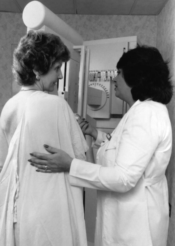
Breast neoplasms refer to abnormal growths in the breast tissue that can be benign or malignant. Benign breast neoplasms are non-cancerous tumors or growths, while malignant breast neoplasms are cancerous tumors that can invade surrounding tissues and spread to other parts of the body.
Breast neoplasms can arise from different types of cells in the breast, including milk ducts, milk sacs (lobules), or connective tissue. The most common type of breast cancer is ductal carcinoma, which starts in the milk ducts and can spread to other parts of the breast and nearby structures.
Breast neoplasms are usually detected through screening methods such as mammography, ultrasound, or MRI, or through self-examination or clinical examination. Treatment options for breast neoplasms depend on several factors, including the type and stage of the tumor, the patient's age and overall health, and personal preferences. Treatment may include surgery, radiation therapy, chemotherapy, hormone therapy, or targeted therapy.
Neoplasms are abnormal growths of cells or tissues in the body that serve no physiological function. They can be benign (non-cancerous) or malignant (cancerous). Benign neoplasms are typically slow growing and do not spread to other parts of the body, while malignant neoplasms are aggressive, invasive, and can metastasize to distant sites.
Neoplasms occur when there is a dysregulation in the normal process of cell division and differentiation, leading to uncontrolled growth and accumulation of cells. This can result from genetic mutations or other factors such as viral infections, environmental exposures, or hormonal imbalances.
Neoplasms can develop in any organ or tissue of the body and can cause various symptoms depending on their size, location, and type. Treatment options for neoplasms include surgery, radiation therapy, chemotherapy, immunotherapy, and targeted therapy, among others.
Carcinoma in situ is a medical term used to describe the earliest stage of cancer, specifically a type of cancer that begins in the epithelial tissue, which is the tissue that lines the outer surfaces of organs and body structures. In this stage, the cancer cells are confined to the layer of cells where they first developed and have not spread beyond that layer into the surrounding tissues or organs.
Carcinoma in situ can occur in various parts of the body, including the skin, cervix, breast, lung, prostate, bladder, and other areas. It is often detected through routine screening tests, such as Pap smears for cervical cancer or mammograms for breast cancer.
While carcinoma in situ is not invasive, it can still be a serious condition because it has the potential to develop into an invasive cancer if left untreated. Treatment options for carcinoma in situ may include surgery, radiation therapy, or other forms of treatment, depending on the location and type of cancer. It is important to consult with a healthcare provider to determine the best course of action for each individual case.
Neoplasms: Neoplasms refer to abnormal growths of tissue that can be benign (non-cancerous) or malignant (cancerous). They occur when the normal control mechanisms that regulate cell growth and division are disrupted, leading to uncontrolled cell proliferation.
Cystic Neoplasms: Cystic neoplasms are tumors that contain fluid-filled sacs or cysts. These tumors can be benign or malignant and can occur in various organs of the body, including the pancreas, ovary, and liver.
Mucinous Neoplasms: Mucinous neoplasms are a type of cystic neoplasm that is characterized by the production of mucin, a gel-like substance produced by certain types of cells. These tumors can occur in various organs, including the ovary, pancreas, and colon. Mucinous neoplasms can be benign or malignant, and malignant forms are often aggressive and have a poor prognosis.
Serous Neoplasms: Serous neoplasms are another type of cystic neoplasm that is characterized by the production of serous fluid, which is a thin, watery fluid. These tumors commonly occur in the ovary and can be benign or malignant. Malignant serous neoplasms are often aggressive and have a poor prognosis.
In summary, neoplasms refer to abnormal tissue growths that can be benign or malignant. Cystic neoplasms contain fluid-filled sacs and can occur in various organs of the body. Mucinous neoplasms produce a gel-like substance called mucin and can also occur in various organs, while serous neoplasms produce thin, watery fluid and commonly occur in the ovary. Both mucinous and serous neoplasms can be benign or malignant, with malignant forms often being aggressive and having a poor prognosis.
Immunohistochemistry (IHC) is a technique used in pathology and laboratory medicine to identify specific proteins or antigens in tissue sections. It combines the principles of immunology and histology to detect the presence and location of these target molecules within cells and tissues. This technique utilizes antibodies that are specific to the protein or antigen of interest, which are then tagged with a detection system such as a chromogen or fluorophore. The stained tissue sections can be examined under a microscope, allowing for the visualization and analysis of the distribution and expression patterns of the target molecule in the context of the tissue architecture. Immunohistochemistry is widely used in diagnostic pathology to help identify various diseases, including cancer, infectious diseases, and immune-mediated disorders.
Adenocarcinoma, mucinous is a type of cancer that begins in the glandular cells that line certain organs and produce mucin, a substance that lubricates and protects tissues. This type of cancer is characterized by the presence of abundant pools of mucin within the tumor. It typically develops in organs such as the colon, rectum, lungs, pancreas, and ovaries.
Mucinous adenocarcinomas tend to have a distinct appearance under the microscope, with large pools of mucin pushing aside the cancer cells. They may also have a different clinical behavior compared to other types of adenocarcinomas, such as being more aggressive or having a worse prognosis in some cases.
It is important to note that while a diagnosis of adenocarcinoma, mucinous can be serious, the prognosis and treatment options may vary depending on several factors, including the location of the cancer, the stage at which it was diagnosed, and the individual's overall health.
Multiple primary neoplasms refer to the occurrence of more than one primary malignant tumor in an individual, where each tumor is unrelated to the other and originates from separate cells or organs. This differs from metastatic cancer, where a single malignancy spreads to multiple sites in the body. Multiple primary neoplasms can be synchronous (occurring at the same time) or metachronous (occurring at different times). The risk of developing multiple primary neoplasms increases with age and is associated with certain genetic predispositions, environmental factors, and lifestyle choices such as smoking and alcohol consumption.
Neoplasm invasiveness is a term used in pathology and oncology to describe the aggressive behavior of cancer cells as they invade surrounding tissues and organs. This process involves the loss of cell-to-cell adhesion, increased motility and migration, and the ability of cancer cells to degrade the extracellular matrix (ECM) through the production of enzymes such as matrix metalloproteinases (MMPs).
Invasive neoplasms are cancers that have spread beyond the original site where they first developed and have infiltrated adjacent tissues or structures. This is in contrast to non-invasive or in situ neoplasms, which are confined to the epithelial layer where they originated and have not yet invaded the underlying basement membrane.
The invasiveness of a neoplasm is an important prognostic factor in cancer diagnosis and treatment, as it can indicate the likelihood of metastasis and the potential effectiveness of various therapies. In general, more invasive cancers are associated with worse outcomes and require more aggressive treatment approaches.
Skin neoplasms refer to abnormal growths or tumors in the skin that can be benign (non-cancerous) or malignant (cancerous). They result from uncontrolled multiplication of skin cells, which can form various types of lesions. These growths may appear as lumps, bumps, sores, patches, or discolored areas on the skin.
Benign skin neoplasms include conditions such as moles, warts, and seborrheic keratoses, while malignant skin neoplasms are primarily classified into melanoma, squamous cell carcinoma, and basal cell carcinoma. These three types of cancerous skin growths are collectively known as non-melanoma skin cancers (NMSCs). Melanoma is the most aggressive and dangerous form of skin cancer, while NMSCs tend to be less invasive but more common.
It's essential to monitor any changes in existing skin lesions or the appearance of new growths and consult a healthcare professional for proper evaluation and treatment if needed.
Carcinoma, acinar cell is a type of pancreatic cancer that originates in the acinar cells of the pancreas. The acinar cells are responsible for producing digestive enzymes. This type of cancer is relatively rare and accounts for less than 5% of all pancreatic cancers. It typically presents with symptoms such as abdominal pain, weight loss, and jaundice. Treatment options may include surgery, chemotherapy, and radiation therapy.
Tumor markers are substances that can be found in the body and their presence can indicate the presence of certain types of cancer or other conditions. Biological tumor markers refer to those substances that are produced by cancer cells or by other cells in response to cancer or certain benign (non-cancerous) conditions. These markers can be found in various bodily fluids such as blood, urine, or tissue samples.
Examples of biological tumor markers include:
1. Proteins: Some tumor markers are proteins that are produced by cancer cells or by other cells in response to the presence of cancer. For example, prostate-specific antigen (PSA) is a protein produced by normal prostate cells and in higher amounts by prostate cancer cells.
2. Genetic material: Tumor markers can also include genetic material such as DNA, RNA, or microRNA that are shed by cancer cells into bodily fluids. For example, circulating tumor DNA (ctDNA) is genetic material from cancer cells that can be found in the bloodstream.
3. Metabolites: Tumor markers can also include metabolic products produced by cancer cells or by other cells in response to cancer. For example, lactate dehydrogenase (LDH) is an enzyme that is released into the bloodstream when cancer cells break down glucose for energy.
It's important to note that tumor markers are not specific to cancer and can be elevated in non-cancerous conditions as well. Therefore, they should not be used alone to diagnose cancer but rather as a tool in conjunction with other diagnostic tests and clinical evaluations.
A "second primary neoplasm" is a distinct, new cancer or malignancy that develops in a person who has already had a previous cancer. It is not a recurrence or metastasis of the original tumor, but rather an independent cancer that arises in a different location or organ system. The development of second primary neoplasms can be influenced by various factors such as genetic predisposition, environmental exposures, and previous treatments like chemotherapy or radiation therapy.
It is important to note that the definition of "second primary neoplasm" may vary slightly depending on the specific source or context. In general medical usage, it refers to a new, separate cancer; however, in some research or clinical settings, there might be more precise criteria for defining and diagnosing second primary neoplasms.
Kidney neoplasms refer to abnormal growths or tumors in the kidney tissues that can be benign (non-cancerous) or malignant (cancerous). These growths can originate from various types of kidney cells, including the renal tubules, glomeruli, and the renal pelvis.
Malignant kidney neoplasms are also known as kidney cancers, with renal cell carcinoma being the most common type. Benign kidney neoplasms include renal adenomas, oncocytomas, and angiomyolipomas. While benign neoplasms are generally not life-threatening, they can still cause problems if they grow large enough to compromise kidney function or if they undergo malignant transformation.
Early detection and appropriate management of kidney neoplasms are crucial for improving patient outcomes and overall prognosis. Regular medical check-ups, imaging studies, and urinalysis can help in the early identification of these growths, allowing for timely intervention and treatment.
A nipple is a small projection or tubular structure located at the center of the areola, which is the darker circle of skin surrounding the nipple on the breast. The primary function of the nipple is to provide a pathway for milk flow from the mammary glands during lactation in females.
The nipple contains smooth muscle fibers that contract and cause the nipple to become erect when stimulated, such as during sexual arousal or cold temperatures. Nipples can come in various shapes, sizes, and colors, and some individuals may have inverted or flat nipples. It is essential to monitor any changes in the appearance or sensation of the nipples, as these could be indicative of underlying medical conditions, such as breast cancer.
The term "DNA, neoplasm" is not a standard medical term or concept. DNA refers to deoxyribonucleic acid, which is the genetic material present in the cells of living organisms. A neoplasm, on the other hand, is a tumor or growth of abnormal tissue that can be benign (non-cancerous) or malignant (cancerous).
In some contexts, "DNA, neoplasm" may refer to genetic alterations found in cancer cells. These genetic changes can include mutations, amplifications, deletions, or rearrangements of DNA sequences that contribute to the development and progression of cancer. Identifying these genetic abnormalities can help doctors diagnose and treat certain types of cancer more effectively.
However, it's important to note that "DNA, neoplasm" is not a term that would typically be used in medical reports or research papers without further clarification. If you have any specific questions about DNA changes in cancer cells or neoplasms, I would recommend consulting with a healthcare professional or conducting further research on the topic.
The pancreas is a glandular organ located in the abdomen, posterior to the stomach. It has both exocrine and endocrine functions. The exocrine portion of the pancreas consists of acinar cells that produce and secrete digestive enzymes into the duodenum via the pancreatic duct. These enzymes help in the breakdown of proteins, carbohydrates, and fats in food.
The endocrine portion of the pancreas consists of clusters of cells called islets of Langerhans, which include alpha, beta, delta, and F cells. These cells produce and secrete hormones directly into the bloodstream, including insulin, glucagon, somatostatin, and pancreatic polypeptide. Insulin and glucagon are critical regulators of blood sugar levels, with insulin promoting glucose uptake and storage in tissues and glucagon stimulating glycogenolysis and gluconeogenesis to raise blood glucose when it is low.
The breast is the upper ventral region of the human body in females, which contains the mammary gland. The main function of the breast is to provide nutrition to infants through the production and secretion of milk, a process known as lactation. The breast is composed of fibrous connective tissue, adipose (fatty) tissue, and the mammary gland, which is made up of 15-20 lobes that are arranged in a radial pattern. Each lobe contains many smaller lobules, where milk is produced during lactation. The milk is then transported through a network of ducts to the nipple, where it can be expressed by the infant.
In addition to its role in lactation, the breast also has important endocrine and psychological functions. It contains receptors for hormones such as estrogen and progesterone, which play a key role in sexual development and reproduction. The breast is also a source of sexual pleasure and can be an important symbol of femininity and motherhood.
It's worth noting that males also have breast tissue, although it is usually less developed than in females. Male breast tissue consists mainly of adipose tissue and does not typically contain functional mammary glands. However, some men may develop enlarged breast tissue due to conditions such as gynecomastia, which can be caused by hormonal imbalances or certain medications.
Cystadenoma is a type of benign tumor (not cancerous), which arises from glandular epithelial cells and is covered by a thin layer of connective tissue. These tumors can develop in various locations within the body, including the ovaries, pancreas, and other organs that contain glands.
There are two main types of cystadenomas: serous and mucinous. Serous cystadenomas are filled with a clear or watery fluid, while mucinous cystadenomas contain a thick, gelatinous material. Although they are generally not harmful, these tumors can grow quite large and cause discomfort or other symptoms due to their size or location. In some cases, cystadenomas may undergo malignant transformation and develop into cancerous tumors, known as cystadenocarcinomas. Regular medical follow-up and monitoring are essential for individuals diagnosed with cystadenomas to ensure early detection and treatment of any potential complications.
Neoplasm staging is a systematic process used in medicine to describe the extent of spread of a cancer, including the size and location of the original (primary) tumor and whether it has metastasized (spread) to other parts of the body. The most widely accepted system for this purpose is the TNM classification system developed by the American Joint Committee on Cancer (AJCC) and the Union for International Cancer Control (UICC).
In this system, T stands for tumor, and it describes the size and extent of the primary tumor. N stands for nodes, and it indicates whether the cancer has spread to nearby lymph nodes. M stands for metastasis, and it shows whether the cancer has spread to distant parts of the body.
Each letter is followed by a number that provides more details about the extent of the disease. For example, a T1N0M0 cancer means that the primary tumor is small and has not spread to nearby lymph nodes or distant sites. The higher the numbers, the more advanced the cancer.
Staging helps doctors determine the most appropriate treatment for each patient and estimate the patient's prognosis. It is an essential tool for communication among members of the healthcare team and for comparing outcomes of treatments in clinical trials.
Parotid neoplasms refer to abnormal growths or tumors in the parotid gland, which is the largest of the salivary glands and is located in front of the ear and extends down the neck. These neoplasms can be benign (non-cancerous) or malignant (cancerous).
Benign parotid neoplasms are typically slow-growing, painless masses that may cause facial asymmetry or difficulty in chewing or swallowing if they become large enough to compress surrounding structures. The most common type of benign parotid tumor is a pleomorphic adenoma.
Malignant parotid neoplasms, on the other hand, are more aggressive and can invade nearby tissues and spread to other parts of the body. They may present as rapidly growing masses that are firm or fixed to surrounding structures. Common types of malignant parotid tumors include mucoepidermoid carcinoma, adenoid cystic carcinoma, and squamous cell carcinoma.
The diagnosis of parotid neoplasms typically involves a thorough clinical evaluation, imaging studies such as CT or MRI scans, and fine-needle aspiration biopsy (FNAB) to determine the nature of the tumor. Treatment options depend on the type, size, and location of the neoplasm but may include surgical excision, radiation therapy, and chemotherapy.
Thyroid neoplasms refer to abnormal growths or tumors in the thyroid gland, which can be benign (non-cancerous) or malignant (cancerous). These growths can vary in size and may cause a noticeable lump or nodule in the neck. Thyroid neoplasms can also affect the function of the thyroid gland, leading to hormonal imbalances and related symptoms. The exact causes of thyroid neoplasms are not fully understood, but risk factors include radiation exposure, family history, and certain genetic conditions. It is important to note that most thyroid nodules are benign, but a proper medical evaluation is necessary to determine the nature of the growth and develop an appropriate treatment plan.
Myeloproliferative disorders (MPDs) are a group of rare, chronic blood cancers that originate from the abnormal proliferation or growth of one or more types of blood-forming cells in the bone marrow. These disorders result in an overproduction of mature but dysfunctional blood cells, which can lead to serious complications such as blood clots, bleeding, and organ damage.
There are several subtypes of MPDs, including:
1. Chronic Myeloid Leukemia (CML): A disorder characterized by the overproduction of mature granulocytes (a type of white blood cell) in the bone marrow, leading to an increased number of these cells in the blood. CML is caused by a genetic mutation that results in the formation of the BCR-ABL fusion protein, which drives uncontrolled cell growth and division.
2. Polycythemia Vera (PV): A disorder characterized by the overproduction of all three types of blood cells - red blood cells, white blood cells, and platelets - in the bone marrow. This can lead to an increased risk of blood clots, bleeding, and enlargement of the spleen.
3. Essential Thrombocythemia (ET): A disorder characterized by the overproduction of platelets in the bone marrow, leading to an increased risk of blood clots and bleeding.
4. Primary Myelofibrosis (PMF): A disorder characterized by the replacement of normal bone marrow tissue with scar tissue, leading to impaired blood cell production and anemia, enlargement of the spleen, and increased risk of infections and bleeding.
5. Chronic Neutrophilic Leukemia (CNL): A rare disorder characterized by the overproduction of neutrophils (a type of white blood cell) in the bone marrow, leading to an increased number of these cells in the blood. CNL can lead to an increased risk of infections and organ damage.
MPDs are typically treated with a combination of therapies, including chemotherapy, targeted therapy, immunotherapy, and stem cell transplantation. The choice of treatment depends on several factors, including the subtype of MPD, the patient's age and overall health, and the presence of any comorbidities.
A pancreatectomy is a surgical procedure in which all or part of the pancreas is removed. There are several types of pancreatectomies, including:
* **Total pancreatectomy:** Removal of the entire pancreas, as well as the spleen and nearby lymph nodes. This type of pancreatectomy is usually done for patients with cancer that has spread throughout the pancreas or for those who have had multiple surgeries to remove pancreatic tumors.
* **Distal pancreatectomy:** Removal of the body and tail of the pancreas, as well as nearby lymph nodes. This type of pancreatectomy is often done for patients with tumors in the body or tail of the pancreas.
* **Partial (or segmental) pancreatectomy:** Removal of a portion of the head or body of the pancreas, as well as nearby lymph nodes. This type of pancreatectomy is often done for patients with tumors in the head or body of the pancreas that can be removed without removing the entire organ.
* **Pylorus-preserving pancreaticoduodenectomy (PPPD):** A type of surgery used to treat tumors in the head of the pancreas, as well as other conditions such as chronic pancreatitis. In this procedure, the head of the pancreas, duodenum, gallbladder, and bile duct are removed, but the stomach and lower portion of the esophagus (pylorus) are left in place.
After a pancreatectomy, patients may experience problems with digestion and blood sugar regulation, as the pancreas plays an important role in these functions. Patients may need to take enzyme supplements to help with digestion and may require insulin therapy to manage their blood sugar levels.
Adenocarcinoma is a type of cancer that arises from glandular epithelial cells. These cells line the inside of many internal organs, including the breasts, prostate, colon, and lungs. Adenocarcinomas can occur in any of these organs, as well as in other locations where glands are present.
The term "adenocarcinoma" is used to describe a cancer that has features of glandular tissue, such as mucus-secreting cells or cells that produce hormones. These cancers often form glandular structures within the tumor mass and may produce mucus or other substances.
Adenocarcinomas are typically slow-growing and tend to spread (metastasize) to other parts of the body through the lymphatic system or bloodstream. They can be treated with surgery, radiation therapy, chemotherapy, targeted therapy, or a combination of these treatments. The prognosis for adenocarcinoma depends on several factors, including the location and stage of the cancer, as well as the patient's overall health and age.
A neoplasm is a tumor or growth that is formed by an abnormal and excessive proliferation of cells, which can be benign or malignant. Neoplasm proteins are therefore any proteins that are expressed or produced in these neoplastic cells. These proteins can play various roles in the development, progression, and maintenance of neoplasms.
Some neoplasm proteins may contribute to the uncontrolled cell growth and division seen in cancer, such as oncogenic proteins that promote cell cycle progression or inhibit apoptosis (programmed cell death). Others may help the neoplastic cells evade the immune system, allowing them to proliferate undetected. Still others may be involved in angiogenesis, the formation of new blood vessels that supply the tumor with nutrients and oxygen.
Neoplasm proteins can also serve as biomarkers for cancer diagnosis, prognosis, or treatment response. For example, the presence or level of certain neoplasm proteins in biological samples such as blood or tissue may indicate the presence of a specific type of cancer, help predict the likelihood of cancer recurrence, or suggest whether a particular therapy will be effective.
Overall, understanding the roles and behaviors of neoplasm proteins can provide valuable insights into the biology of cancer and inform the development of new diagnostic and therapeutic strategies.
Mammary glands are specialized exocrine glands found in mammals, including humans and other animals. These glands are responsible for producing milk, which is used to nurse offspring after birth. The mammary glands are located in the breast region of female mammals and are usually rudimentary or absent in males.
In animals, mammary glands can vary in number and location depending on the species. For example, humans and other primates have two mammary glands, one in each breast. Cows, goats, and sheep, on the other hand, have multiple pairs of mammary glands located in their lower abdominal region.
Mammary glands are made up of several structures, including lobules, ducts, and connective tissue. The lobules contain clusters of milk-secreting cells called alveoli, which produce and store milk. The ducts transport the milk from the lobules to the nipple, where it is released during lactation.
Mammary glands are an essential feature of mammals, as they provide a source of nutrition for newborn offspring. They also play a role in the development and maintenance of the mother-infant bond, as nursing provides opportunities for physical contact and bonding between the mother and her young.
Carcinoma, papillary is a type of cancer that begins in the cells that line the glandular structures or the lining of organs. In a papillary carcinoma, the cancerous cells grow and form small finger-like projections, called papillae, within the tumor. This type of cancer most commonly occurs in the thyroid gland, but can also be found in other organs such as the lung, breast, and kidney. Papillary carcinoma of the thyroid gland is usually slow-growing and has a good prognosis, especially when it is diagnosed at an early stage.
The Ductus Arteriosus is a fetal blood vessel that connects the pulmonary trunk (the artery that carries blood from the heart to the lungs) and the aorta (the largest artery in the body, which carries oxygenated blood from the heart to the rest of the body). This vessel allows most of the blood from the right ventricle of the fetal heart to bypass the lungs, as the fetus receives oxygen through the placenta rather than breathing air.
After birth, with the first breaths, the blood oxygen level increases and the pressure in the lungs rises. As a result, the circulation in the newborn's body changes, and the Ductus Arteriosus is no longer needed. Within the first few days or weeks of life, this vessel usually closes spontaneously, turning into a fibrous cord called the Ligamentum Arteriosum.
Persistent Patency of the Ductus Arteriosus (PDA) occurs when the Ductus Arteriosus does not close after birth, which can lead to various complications such as heart failure and pulmonary hypertension. This condition is often seen in premature infants and may require medical intervention or surgical closure of the vessel.
Mucinous cystadenocarcinoma is a type of cancer that arises from the mucin-producing cells in the lining of a cyst. It is a subtype of cystadenocarcinoma, which is a malignant tumor that develops within a cyst. Mucinous cystadenocarcinomas are typically found in the ovary or pancreas but can also occur in other organs such as the appendix and the respiratory tract.
These tumors are characterized by the production of large amounts of mucin, a gel-like substance that can accumulate within the cyst and cause it to grow. Mucinous cystadenocarcinomas tend to grow slowly but can become quite large and may eventually spread (metastasize) to other parts of the body if left untreated.
Symptoms of mucinous cystadenocarcinoma depend on the location and size of the tumor, but they may include abdominal pain or discomfort, bloating, changes in bowel movements, or vaginal bleeding. Treatment typically involves surgical removal of the tumor, followed by chemotherapy or radiation therapy to kill any remaining cancer cells. The prognosis for mucinous cystadenocarcinoma depends on several factors, including the stage of the disease at diagnosis and the patient's overall health.
Adenocarcinoma, papillary is a type of cancer that begins in the glandular cells and grows in a finger-like projection (called a papilla). This type of cancer can occur in various organs, including the lungs, pancreas, thyroid, and female reproductive system. The prognosis and treatment options for papillary adenocarcinoma depend on several factors, such as the location and stage of the tumor, as well as the patient's overall health. It is important to consult with a healthcare professional for an accurate diagnosis and personalized treatment plan.
Hyperplasia is a medical term that refers to an abnormal increase in the number of cells in an organ or tissue, leading to an enlargement of the affected area. It's a response to various stimuli such as hormones, chronic irritation, or inflammation. Hyperplasia can be physiological, like the growth of breast tissue during pregnancy, or pathological, like in the case of benign or malignant tumors. The process is generally reversible if the stimulus is removed. It's important to note that hyperplasia itself is not cancerous, but some forms of hyperplasia can increase the risk of developing cancer over time.
Lung neoplasms refer to abnormal growths or tumors in the lung tissue. These tumors can be benign (non-cancerous) or malignant (cancerous). Malignant lung neoplasms are further classified into two main types: small cell lung carcinoma and non-small cell lung carcinoma. Lung neoplasms can cause symptoms such as cough, chest pain, shortness of breath, and weight loss. They are often caused by smoking or exposure to secondhand smoke, but can also occur due to genetic factors, radiation exposure, and other environmental carcinogens. Early detection and treatment of lung neoplasms is crucial for improving outcomes and survival rates.
Liver neoplasms refer to abnormal growths in the liver that can be benign or malignant. Benign liver neoplasms are non-cancerous tumors that do not spread to other parts of the body, while malignant liver neoplasms are cancerous tumors that can invade and destroy surrounding tissue and spread to other organs.
Liver neoplasms can be primary, meaning they originate in the liver, or secondary, meaning they have metastasized (spread) to the liver from another part of the body. Primary liver neoplasms can be further classified into different types based on their cell of origin and behavior, including hepatocellular carcinoma, cholangiocarcinoma, and hepatic hemangioma.
The diagnosis of liver neoplasms typically involves a combination of imaging studies, such as ultrasound, CT scan, or MRI, and biopsy to confirm the type and stage of the tumor. Treatment options depend on the type and extent of the neoplasm and may include surgery, radiation therapy, chemotherapy, or liver transplantation.
Salivary ducts are the excretory tubules that transport saliva from the major and minor salivary glands to the oral cavity. The main function of these ducts is to convey the salivary secretions, which contain enzymes and lubricants, into the mouth to aid in digestion, speech, and swallowing.
There are two pairs of major salivary glands: the parotid glands and the submandibular glands. Each pair has its own set of ducts. The parotid gland's saliva is drained through the parotid duct, also known as Stensen's duct, which opens into the oral cavity opposite the upper second molar tooth. The submandibular gland's saliva is transported through the submandibular duct, or Wharton's duct, which empties into the floor of the mouth near the base of the tongue.
Minor salivary glands are scattered throughout the oral cavity and pharynx, and their secretions are drained via small ducts directly into the oral mucosa.
Endocrine gland neoplasms refer to abnormal growths (tumors) that develop in the endocrine glands. These glands are responsible for producing hormones, which are chemical messengers that regulate various functions and processes in the body. Neoplasms can be benign or malignant (cancerous). Benign neoplasms tend to grow slowly and do not spread to other parts of the body. Malignant neoplasms, on the other hand, can invade nearby tissues and organs and may also metastasize (spread) to distant sites.
Endocrine gland neoplasms can occur in any of the endocrine glands, including:
1. Pituitary gland: located at the base of the brain, it produces several hormones that regulate growth and development, as well as other bodily functions.
2. Thyroid gland: located in the neck, it produces thyroid hormones that regulate metabolism and calcium balance.
3. Parathyroid glands: located near the thyroid gland, they produce parathyroid hormone that regulates calcium levels in the blood.
4. Adrenal glands: located on top of each kidney, they produce hormones such as adrenaline, cortisol, and aldosterone that regulate stress response, metabolism, and blood pressure.
5. Pancreas: located behind the stomach, it produces insulin and glucagon, which regulate blood sugar levels, and digestive enzymes that help break down food.
6. Pineal gland: located in the brain, it produces melatonin, a hormone that regulates sleep-wake cycles.
7. Gonads (ovaries and testicles): located in the pelvis (ovaries) and scrotum (testicles), they produce sex hormones such as estrogen, progesterone, and testosterone that regulate reproductive function and secondary sexual characteristics.
Endocrine gland neoplasms can cause various symptoms depending on the type and location of the tumor. For example, a pituitary gland neoplasm may cause headaches, vision problems, or hormonal imbalances, while an adrenal gland neoplasm may cause high blood pressure, weight gain, or mood changes.
Diagnosis of endocrine gland neoplasms typically involves a combination of medical history, physical examination, imaging studies such as CT or MRI scans, and laboratory tests to measure hormone levels. Treatment options may include surgery, radiation therapy, chemotherapy, or hormonal therapy, depending on the type and stage of the tumor.
Salivary gland neoplasms refer to abnormal growths or tumors that develop in the salivary glands. These glands are responsible for producing saliva, which helps in digestion, lubrication of food and maintaining oral health. Salivary gland neoplasms can be benign (non-cancerous) or malignant (cancerous).
Benign neoplasms are slow-growing and typically do not spread to other parts of the body. They may cause symptoms such as swelling, painless lumps, or difficulty swallowing if they grow large enough to put pressure on surrounding tissues.
Malignant neoplasms, on the other hand, can be aggressive and have the potential to invade nearby structures and metastasize (spread) to distant organs. Symptoms of malignant salivary gland neoplasms may include rapid growth, pain, numbness, or paralysis of facial nerves.
Salivary gland neoplasms can occur in any of the major salivary glands (parotid, submandibular, and sublingual glands) or in the minor salivary glands located throughout the mouth and throat. The exact cause of these neoplasms is not fully understood, but risk factors may include exposure to radiation, certain viral infections, and genetic predisposition.
Neoplasms of connective and soft tissue are abnormal growths or tumors that develop in the body's supportive tissues, such as cartilage, tendons, ligaments, fascia, and fat. These neoplasms can be benign (non-cancerous) or malignant (cancerous).
Benign connective and soft tissue neoplasms include:
- Lipomas: slow-growing, fatty tumors that develop under the skin.
- Fibromas: firm, benign tumors that develop in connective tissue such as tendons or ligaments.
- Nevi (plural of nevus): benign growths made up of cells called melanocytes, which produce pigment.
Malignant connective and soft tissue neoplasms include:
- Sarcomas: a type of cancer that develops in the body's supportive tissues such as muscle, bone, fat, cartilage, or blood vessels. There are many different types of sarcomas, including liposarcoma (fatty tissue), rhabdomyosarcoma (muscle), and osteosarcoma (bone).
- Desmoid tumors: a rare type of benign tumor that can become aggressive and invade surrounding tissues. While not considered cancerous, desmoid tumors can cause significant morbidity due to their tendency to grow and infiltrate nearby structures.
Connective and soft tissue neoplasms can present with various symptoms depending on their location and size. Treatment options include surgery, radiation therapy, chemotherapy, or a combination of these modalities. Regular follow-up care is essential to monitor for recurrence or metastasis (spread) of the tumor.
Plasma cell neoplasms are a type of cancer that originates from plasma cells, which are a type of white blood cell found in the bone marrow. These cells are responsible for producing antibodies to help fight off infections. When plasma cells become cancerous and multiply out of control, they can form a tumor called a plasmacytoma.
There are two main types of plasma cell neoplasms: solitary plasmacytoma and multiple myeloma. Solitary plasmacytoma is a localized tumor that typically forms in the bone, while multiple myeloma is a systemic disease that affects multiple bones and can cause a variety of symptoms such as bone pain, fatigue, and anemia.
Plasma cell neoplasms are diagnosed through a combination of tests, including blood tests, imaging studies, and bone marrow biopsy. Treatment options depend on the stage and extent of the disease, but may include radiation therapy, chemotherapy, and stem cell transplantation.
Sweat gland neoplasms are abnormal growths that develop in the sweat glands. These growths can be benign (noncancerous) or malignant (cancerous). Benign sweat gland neoplasms include hidradenomas and syringomas, which are usually slow-growing and cause little to no symptoms. Malignant sweat gland neoplasms, also known as sweat gland carcinomas, are rare but aggressive cancers that can spread to other parts of the body. They may cause symptoms such as a lump or mass under the skin, pain, swelling, and redness. Treatment typically involves surgical removal of the growth.
Appendiceal neoplasms refer to various types of tumors that can develop in the appendix, a small tube-like structure attached to the large intestine. These neoplasms can be benign or malignant and can include:
1. Adenomas: These are benign tumors that arise from the glandular cells lining the appendix. They are usually slow-growing and may not cause any symptoms.
2. Carcinoids: These are neuroendocrine tumors that arise from the hormone-producing cells in the appendix. They are typically small and slow-growing, but some can be aggressive and spread to other parts of the body.
3. Mucinous neoplasms: These are tumors that produce mucin, a slippery substance that can cause the appendix to become distended and filled with mucus. They can be low-grade (less aggressive) or high-grade (more aggressive) and may spread to other parts of the abdomen.
4. Adenocarcinomas: These are malignant tumors that arise from the glandular cells lining the appendix. They are relatively rare but can be aggressive and spread to other parts of the body.
5. Pseudomyxoma peritonei: This is a condition in which mucin produced by an appendiceal neoplasm leaks into the abdominal cavity, causing a jelly-like accumulation of fluid and tissue. It can be caused by both benign and malignant tumors.
Treatment for appendiceal neoplasms depends on the type and stage of the tumor, as well as the patient's overall health. Treatment options may include surgery, chemotherapy, or radiation therapy.
A neoplasm of vascular tissue is an abnormal growth or mass of cells in the blood vessels or lymphatic vessels. These growths can be benign (non-cancerous) or malignant (cancerous). Benign neoplasms, such as hemangiomas and lymphangiomas, are typically not harmful and may not require treatment. However, they can cause symptoms if they grow large enough to press on nearby organs or tissues. Malignant neoplasms, such as angiosarcomas, are cancerous and can invade and destroy surrounding tissue, as well as spread (metastasize) to other parts of the body. Treatment for vascular tissue neoplasms depends on the type, size, location, and stage of the growth, and may include surgery, radiation therapy, chemotherapy, or a combination of these.
Ovarian neoplasms refer to abnormal growths or tumors in the ovary, which can be benign (non-cancerous) or malignant (cancerous). These growths can originate from various cell types within the ovary, including epithelial cells, germ cells, and stromal cells. Ovarian neoplasms are often classified based on their cell type of origin, histological features, and potential for invasive or metastatic behavior.
Epithelial ovarian neoplasms are the most common type and can be further categorized into several subtypes, such as serous, mucinous, endometrioid, clear cell, and Brenner tumors. Some of these epithelial tumors have a higher risk of becoming malignant and spreading to other parts of the body.
Germ cell ovarian neoplasms arise from the cells that give rise to eggs (oocytes) and can include teratomas, dysgerminomas, yolk sac tumors, and embryonal carcinomas. Stromal ovarian neoplasms develop from the connective tissue cells supporting the ovary and can include granulosa cell tumors, thecomas, and fibromas.
It is essential to diagnose and treat ovarian neoplasms promptly, as some malignant forms can be aggressive and potentially life-threatening if not managed appropriately. Regular gynecological exams, imaging studies, and tumor marker tests are often used for early detection and monitoring of ovarian neoplasms. Treatment options may include surgery, chemotherapy, or radiation therapy, depending on the type, stage, and patient's overall health condition.
A precancerous condition, also known as a premalignant condition, is a state of abnormal cellular growth and development that has a higher-than-normal potential to progress into cancer. These conditions are characterized by the presence of certain anomalies in the cells, such as dysplasia (abnormal changes in cell shape or size), which can indicate an increased risk for malignant transformation.
It is important to note that not all precancerous conditions will eventually develop into cancer, and some may even regress on their own. However, individuals with precancerous conditions are often at a higher risk of developing cancer compared to the general population. Regular monitoring and appropriate medical interventions, if necessary, can help manage this risk and potentially prevent or detect cancer at an early stage when it is more treatable.
Examples of precancerous conditions include:
1. Dysplasia in the cervix (cervical intraepithelial neoplasia or CIN)
2. Atypical ductal hyperplasia or lobular hyperplasia in the breast
3. Actinic keratosis on the skin
4. Leukoplakia in the mouth
5. Barrett's esophagus in the digestive tract
Regular medical check-ups, screenings, and lifestyle modifications are crucial for individuals with precancerous conditions to monitor their health and reduce the risk of cancer development.
Experimental neoplasms refer to abnormal growths or tumors that are induced and studied in a controlled laboratory setting, typically in animals or cell cultures. These studies are conducted to understand the fundamental mechanisms of cancer development, progression, and potential treatment strategies. By manipulating various factors such as genetic mutations, environmental exposures, and pharmacological interventions, researchers can gain valuable insights into the complex processes underlying neoplasm formation and identify novel targets for cancer therapy. It is important to note that experimental neoplasms may not always accurately represent human cancers, and further research is needed to translate these findings into clinically relevant applications.
Mucinous cystadenoma is a type of benign tumor that arises from the epithelial cells lining the mucous membranes of the body. It is most commonly found in the ovary, but can also occur in other locations such as the pancreas or appendix.
Mucinous cystadenomas are characterized by the production of large amounts of mucin, a slippery, gel-like substance that accumulates inside the tumor and causes it to grow into a cystic mass. These tumors can vary in size, ranging from a few centimeters to over 20 centimeters in diameter.
While mucinous cystadenomas are generally benign, they have the potential to become cancerous (mucinous cystadenocarcinoma) if left untreated. Symptoms of mucinous cystadenoma may include abdominal pain or swelling, bloating, and changes in bowel movements or urinary habits. Treatment typically involves surgical removal of the tumor.
Eye neoplasms, also known as ocular tumors or eye cancer, refer to abnormal growths of tissue in the eye. These growths can be benign (non-cancerous) or malignant (cancerous). Eye neoplasms can develop in various parts of the eye, including the eyelid, conjunctiva, cornea, iris, ciliary body, choroid, retina, and optic nerve.
Benign eye neoplasms are typically slow-growing and do not spread to other parts of the body. They may cause symptoms such as vision changes, eye pain, or a noticeable mass in the eye. Treatment options for benign eye neoplasms include monitoring, surgical removal, or radiation therapy.
Malignant eye neoplasms, on the other hand, can grow and spread rapidly to other parts of the body. They may cause symptoms such as vision changes, eye pain, floaters, or flashes of light. Treatment options for malignant eye neoplasms depend on the type and stage of cancer but may include surgery, radiation therapy, chemotherapy, or a combination of these treatments.
It is important to note that early detection and treatment of eye neoplasms can improve outcomes and prevent complications. Regular eye exams with an ophthalmologist are recommended for early detection and prevention of eye diseases, including eye neoplasms.
Bile duct neoplasms, also known as cholangiocarcinomas, refer to a group of malignancies that arise from the bile ducts. These are the tubes that carry bile from the liver to the gallbladder and small intestine. Bile duct neoplasms can be further classified based on their location as intrahepatic (within the liver), perihilar (at the junction of the left and right hepatic ducts), or distal (in the common bile duct).
These tumors are relatively rare, but their incidence has been increasing in recent years. They can cause a variety of symptoms, including jaundice, abdominal pain, weight loss, and fever. The diagnosis of bile duct neoplasms typically involves imaging studies such as CT or MRI scans, as well as blood tests to assess liver function. In some cases, a biopsy may be necessary to confirm the diagnosis.
Treatment options for bile duct neoplasms depend on several factors, including the location and stage of the tumor, as well as the patient's overall health. Surgical resection is the preferred treatment for early-stage tumors, while chemotherapy and radiation therapy may be used in more advanced cases. For patients who are not candidates for surgery, palliative treatments such as stenting or bypass procedures may be recommended to relieve symptoms and improve quality of life.
Prognosis is a medical term that refers to the prediction of the likely outcome or course of a disease, including the chances of recovery or recurrence, based on the patient's symptoms, medical history, physical examination, and diagnostic tests. It is an important aspect of clinical decision-making and patient communication, as it helps doctors and patients make informed decisions about treatment options, set realistic expectations, and plan for future care.
Prognosis can be expressed in various ways, such as percentages, categories (e.g., good, fair, poor), or survival rates, depending on the nature of the disease and the available evidence. However, it is important to note that prognosis is not an exact science and may vary depending on individual factors, such as age, overall health status, and response to treatment. Therefore, it should be used as a guide rather than a definitive forecast.
Radiation-induced neoplasms are a type of cancer or tumor that develops as a result of exposure to ionizing radiation. Ionizing radiation is radiation with enough energy to remove tightly bound electrons from atoms or molecules, leading to the formation of ions. This type of radiation can damage DNA and other cellular structures, which can lead to mutations and uncontrolled cell growth, resulting in the development of a neoplasm.
Radiation-induced neoplasms can occur after exposure to high levels of ionizing radiation, such as that received during radiation therapy for cancer treatment or from nuclear accidents. The risk of developing a radiation-induced neoplasm depends on several factors, including the dose and duration of radiation exposure, the type of radiation, and the individual's genetic susceptibility to radiation-induced damage.
Radiation-induced neoplasms can take many years to develop after initial exposure to ionizing radiation, and they often occur at the site of previous radiation therapy. Common types of radiation-induced neoplasms include sarcomas, carcinomas, and thyroid cancer. It is important to note that while ionizing radiation can increase the risk of developing cancer, the overall risk is still relatively low, especially when compared to other well-established cancer risk factors such as smoking and exposure to certain chemicals.
Pancreatic ductal carcinoma (PDC) is a specific type of cancer that forms in the ducts that carry digestive enzymes out of the pancreas. It's the most common form of exocrine pancreatic cancer, making up about 90% of all cases.
The symptoms of PDC are often vague and can include abdominal pain, jaundice (yellowing of the skin and eyes), unexplained weight loss, and changes in bowel movements. These symptoms can be similar to those caused by other less serious conditions, which can make diagnosis difficult.
Pancreatic ductal carcinoma is often aggressive and difficult to treat. The prognosis for PDC is generally poor, with a five-year survival rate of only about 9%. Treatment options may include surgery, chemotherapy, radiation therapy, or a combination of these approaches. However, because PDC is often not detected until it has advanced, treatment is frequently focused on palliative care to relieve symptoms and improve quality of life.
Gastrointestinal (GI) neoplasms refer to abnormal growths in the gastrointestinal tract, which can be benign or malignant. The gastrointestinal tract includes the mouth, esophagus, stomach, small intestine, large intestine, rectum, and anus.
Benign neoplasms are non-cancerous growths that do not invade nearby tissues or spread to other parts of the body. They can sometimes be removed completely and may not cause any further health problems.
Malignant neoplasms, on the other hand, are cancerous growths that can invade nearby tissues and organs and spread to other parts of the body through the bloodstream or lymphatic system. These types of neoplasms can be life-threatening if not diagnosed and treated promptly.
GI neoplasms can cause various symptoms, including abdominal pain, bloating, changes in bowel habits, nausea, vomiting, weight loss, and anemia. The specific symptoms may depend on the location and size of the neoplasm.
There are many types of GI neoplasms, including adenocarcinomas, gastrointestinal stromal tumors (GISTs), lymphomas, and neuroendocrine tumors. The diagnosis of GI neoplasms typically involves a combination of medical history, physical examination, imaging studies, and biopsy. Treatment options may include surgery, radiation therapy, chemotherapy, targeted therapy, or immunotherapy.
Local neoplasm recurrence is the return or regrowth of a tumor in the same location where it was originally removed or treated. This means that cancer cells have survived the initial treatment and started to grow again in the same area. It's essential to monitor and detect any local recurrence as early as possible, as it can affect the prognosis and may require additional treatment.
A needle biopsy is a medical procedure in which a thin, hollow needle is used to remove a small sample of tissue from a suspicious or abnormal area of the body. The tissue sample is then examined under a microscope to check for cancer cells or other abnormalities. Needle biopsies are often used to diagnose lumps or masses that can be felt through the skin, but they can also be guided by imaging techniques such as ultrasound, CT scan, or MRI to reach areas that cannot be felt. There are several types of needle biopsy procedures, including fine-needle aspiration (FNA) and core needle biopsy. FNA uses a thin needle and gentle suction to remove fluid and cells from the area, while core needle biopsy uses a larger needle to remove a small piece of tissue. The type of needle biopsy used depends on the location and size of the abnormal area, as well as the reason for the procedure.
An adenoma is a benign (noncancerous) tumor that develops from glandular epithelial cells. These types of cells are responsible for producing and releasing fluids, such as hormones or digestive enzymes, into the surrounding tissues. Adenomas can occur in various organs and glands throughout the body, including the thyroid, pituitary, adrenal, and digestive systems.
Depending on their location, adenomas may cause different symptoms or remain asymptomatic. Some common examples of adenomas include:
1. Colorectal adenoma (also known as a polyp): These growths occur in the lining of the colon or rectum and can develop into colorectal cancer if left untreated. Regular screenings, such as colonoscopies, are essential for early detection and removal of these polyps.
2. Thyroid adenoma: This type of adenoma affects the thyroid gland and may result in an overproduction or underproduction of hormones, leading to conditions like hyperthyroidism (overactive thyroid) or hypothyroidism (underactive thyroid).
3. Pituitary adenoma: These growths occur in the pituitary gland, which is located at the base of the brain and controls various hormonal functions. Depending on their size and location, pituitary adenomas can cause vision problems, headaches, or hormonal imbalances that affect growth, reproduction, and metabolism.
4. Liver adenoma: These rare benign tumors develop in the liver and may not cause any symptoms unless they become large enough to press on surrounding organs or structures. In some cases, liver adenomas can rupture and cause internal bleeding.
5. Adrenal adenoma: These growths occur in the adrenal glands, which are located above the kidneys and produce hormones that regulate stress responses, metabolism, and blood pressure. Most adrenal adenomas are nonfunctioning, meaning they do not secrete excess hormones. However, functioning adrenal adenomas can lead to conditions like Cushing's syndrome or Conn's syndrome, depending on the type of hormone being overproduced.
It is essential to monitor and manage benign tumors like adenomas to prevent potential complications, such as rupture, bleeding, or hormonal imbalances. Treatment options may include surveillance with imaging studies, medication to manage hormonal issues, or surgical removal of the tumor in certain cases.
Neoplasm antigens, also known as tumor antigens, are substances that are produced by cancer cells (neoplasms) and can stimulate an immune response. These antigens can be proteins, carbohydrates, or other molecules that are either unique to the cancer cells or are overexpressed or mutated versions of normal cellular proteins.
Neoplasm antigens can be classified into two main categories: tumor-specific antigens (TSAs) and tumor-associated antigens (TAAs). TSAs are unique to cancer cells and are not expressed by normal cells, while TAAs are present at low levels in normal cells but are overexpressed or altered in cancer cells.
TSAs can be further divided into viral antigens and mutated antigens. Viral antigens are produced when cancer is caused by a virus, such as human papillomavirus (HPV) in cervical cancer. Mutated antigens are the result of genetic mutations that occur during cancer development and are unique to each patient's tumor.
Neoplasm antigens play an important role in the immune response against cancer. They can be recognized by the immune system, leading to the activation of immune cells such as T cells and natural killer (NK) cells, which can then attack and destroy cancer cells. However, cancer cells often develop mechanisms to evade the immune response, allowing them to continue growing and spreading.
Understanding neoplasm antigens is important for the development of cancer immunotherapies, which aim to enhance the body's natural immune response against cancer. These therapies include checkpoint inhibitors, which block proteins that inhibit T cell activation, and therapeutic vaccines, which stimulate an immune response against specific tumor antigens.
Nose neoplasms refer to abnormal growths or tumors in the nasal cavity or paranasal sinuses. These growths can be benign (non-cancerous) or malignant (cancerous). Benign neoplasms are typically slow-growing and do not spread to other parts of the body, while malignant neoplasms can invade surrounding tissues and have the potential to metastasize.
Nose neoplasms can cause various symptoms such as nasal congestion, nosebleeds, difficulty breathing through the nose, loss of smell, facial pain or numbness, and visual changes if they affect the eye. The diagnosis of nose neoplasms usually involves a combination of physical examination, imaging studies (such as CT or MRI scans), and biopsy to determine the type and extent of the growth. Treatment options depend on the type, size, location, and stage of the neoplasm and may include surgery, radiation therapy, chemotherapy, or a combination of these approaches.
Cystadenocarcinoma is a type of tumor that arises from the epithelial lining of a cyst, and it has the potential to invade surrounding tissues and spread (metastasize) to other parts of the body. It typically affects glandular organs such as the ovaries, pancreas, and salivary glands.
Cystadenocarcinomas can be classified into two types: serous and mucinous. Serous cystadenocarcinomas produce a watery fluid, while mucinous cystadenocarcinomas produce a thick, mucus-like fluid. Both types of tumors can be benign or malignant, but malignant cystadenocarcinomas are more aggressive and have a higher risk of metastasis.
Symptoms of cystadenocarcinoma depend on the location and size of the tumor. In some cases, there may be no symptoms until the tumor has grown large enough to cause pain or other problems. Treatment typically involves surgical removal of the tumor, along with any affected surrounding tissue. Chemotherapy and radiation therapy may also be used in some cases to help prevent recurrence or spread of the cancer.
A serous cystadenoma is a type of benign tumor that arises from the epithelial cells lining the serous glands, which are glands that produce a watery, lubricating fluid. This type of tumor typically develops in the ovary or the pancreas.
Serous cystadenomas of the ovary are usually filled with a clear, watery fluid and have multiple loculations (compartments). They can vary in size from a few millimeters to several centimeters in diameter. Although these tumors are benign, they can cause symptoms if they become large enough to press on surrounding organs or if they rupture and release their contents into the abdominal cavity.
Serous cystadenomas of the pancreas are less common than ovarian serous cystadenomas. They typically occur in the tail of the pancreas and can range in size from a few millimeters to several centimeters in diameter. These tumors are usually asymptomatic, but they can cause symptoms such as abdominal pain or discomfort if they become large enough to press on surrounding organs.
It is important to note that while serous cystadenomas are generally benign, there is a small risk that they may undergo malignant transformation and develop into a type of cancer known as a serous cystadenocarcinoma. For this reason, it is important for patients with these tumors to be followed closely by a healthcare provider and to have regular imaging studies and/or surgical excision to monitor for any changes in the tumor.
Testicular neoplasms are abnormal growths or tumors in the testicle that can be benign (non-cancerous) or malignant (cancerous). They are a type of genitourinary cancer, which affects the reproductive and urinary systems. Testicular neoplasms can occur in men of any age but are most commonly found in young adults between the ages of 15 and 40.
Testicular neoplasms can be classified into two main categories: germ cell tumors and non-germ cell tumors. Germ cell tumors, which arise from the cells that give rise to sperm, are further divided into seminomas and non-seminomas. Seminomas are typically slow-growing and have a good prognosis, while non-seminomas tend to grow more quickly and can spread to other parts of the body.
Non-germ cell tumors are less common than germ cell tumors and include Leydig cell tumors, Sertoli cell tumors, and lymphomas. These tumors can have a variety of clinical behaviors, ranging from benign to malignant.
Testicular neoplasms often present as a painless mass or swelling in the testicle. Other symptoms may include a feeling of heaviness or discomfort in the scrotum, a dull ache in the lower abdomen or groin, and breast enlargement (gynecomastia).
Diagnosis typically involves a physical examination, imaging studies such as ultrasound or CT scan, and blood tests to detect tumor markers. Treatment options depend on the type and stage of the neoplasm but may include surgery, radiation therapy, chemotherapy, or a combination of these modalities. Regular self-examinations of the testicles are recommended for early detection and improved outcomes.
Neoplasms in muscle tissue refer to abnormal and excessive growths of muscle cells that can be benign or malignant. These growths can arise from any of the three types of muscle tissue: skeletal, cardiac, or smooth muscle. Neoplasms in muscle tissue are classified based on their origin, behavior, and histological features.
Benign neoplasms in muscle tissue include leiomyomas (smooth muscle), rhabdomyomas (skeletal muscle), and myxomas (cardiac muscle). These tumors are usually slow-growing and do not invade surrounding tissues or spread to other parts of the body.
Malignant neoplasms in muscle tissue, also known as sarcomas, include leiomyosarcoma (smooth muscle), rhabdomyosarcoma (skeletal muscle), and angiosarcoma (cardiac muscle). These tumors are aggressive, invasive, and have the potential to metastasize to other parts of the body.
Symptoms of neoplasms in muscle tissue depend on their location, size, and type. They may include a painless or painful mass, weakness, fatigue, weight loss, and difficulty swallowing or breathing. Treatment options for neoplasms in muscle tissue include surgery, radiation therapy, chemotherapy, and targeted therapy. The choice of treatment depends on the type, stage, location, and patient's overall health condition.
Neoplasms are abnormal growths of cells or tissues that serve no purpose and can be benign (non-cancerous) or malignant (cancerous). Glandular and epithelial neoplasms refer to specific types of tumors that originate from the glandular and epithelial tissues, respectively.
Glandular neoplasms arise from the glandular tissue, which is responsible for producing and secreting substances such as hormones, enzymes, or other fluids. These neoplasms can be further classified into adenomas (benign) and adenocarcinomas (malignant).
Epithelial neoplasms, on the other hand, develop from the epithelial tissue that lines the outer surfaces of organs and the inner surfaces of cavities. These neoplasms can also be benign or malignant and are classified as papillomas (benign) and carcinomas (malignant).
It is important to note that while both glandular and epithelial neoplasms can become cancerous, not all of them do. However, if they do, the malignant versions can invade surrounding tissues and spread to other parts of the body, making them potentially life-threatening.
Retrospective studies, also known as retrospective research or looking back studies, are a type of observational study that examines data from the past to draw conclusions about possible causal relationships between risk factors and outcomes. In these studies, researchers analyze existing records, medical charts, or previously collected data to test a hypothesis or answer a specific research question.
Retrospective studies can be useful for generating hypotheses and identifying trends, but they have limitations compared to prospective studies, which follow participants forward in time from exposure to outcome. Retrospective studies are subject to biases such as recall bias, selection bias, and information bias, which can affect the validity of the results. Therefore, retrospective studies should be interpreted with caution and used primarily to generate hypotheses for further testing in prospective studies.
Carcinoma is a type of cancer that develops from epithelial cells, which are the cells that line the inner and outer surfaces of the body. These cells cover organs, glands, and other structures within the body. Carcinomas can occur in various parts of the body, including the skin, lungs, breasts, prostate, colon, and pancreas. They are often characterized by the uncontrolled growth and division of abnormal cells that can invade surrounding tissues and spread to other parts of the body through a process called metastasis. Carcinomas can be further classified based on their appearance under a microscope, such as adenocarcinoma, squamous cell carcinoma, and basal cell carcinoma.
Soft tissue neoplasms refer to abnormal growths or tumors that develop in the soft tissues of the body. Soft tissues include muscles, tendons, ligaments, fascia, nerves, blood vessels, fat, and synovial membranes (the thin layer of cells that line joints and tendons). Neoplasms can be benign (non-cancerous) or malignant (cancerous), and their behavior and potential for spread depend on the specific type of neoplasm.
Benign soft tissue neoplasms are typically slow-growing, well-circumscribed, and rarely spread to other parts of the body. They can often be removed surgically with a low risk of recurrence. Examples of benign soft tissue neoplasms include lipomas (fat tumors), schwannomas (nerve sheath tumors), and hemangiomas (blood vessel tumors).
Malignant soft tissue neoplasms, on the other hand, can grow rapidly, invade surrounding tissues, and may metastasize (spread) to distant parts of the body. They are often more difficult to treat than benign neoplasms and require a multidisciplinary approach, including surgery, radiation therapy, and chemotherapy. Examples of malignant soft tissue neoplasms include sarcomas, such as rhabdomyosarcoma (arising from skeletal muscle), leiomyosarcoma (arising from smooth muscle), and angiosarcoma (arising from blood vessels).
It is important to note that soft tissue neoplasms can occur in any part of the body, and their diagnosis and treatment require a thorough evaluation by a healthcare professional with expertise in this area.
Hematologic neoplasms, also known as hematological malignancies, are a group of diseases characterized by the uncontrolled growth and accumulation of abnormal blood cells or bone marrow cells. These disorders can originate from the myeloid or lymphoid cell lines, which give rise to various types of blood cells, including red blood cells, white blood cells, and platelets.
Hematologic neoplasms can be broadly classified into three categories:
1. Leukemias: These are cancers that primarily affect the bone marrow and blood-forming tissues. They result in an overproduction of abnormal white blood cells, which interfere with the normal functioning of the blood and immune system. There are several types of leukemia, including acute lymphoblastic leukemia (ALL), chronic lymphocytic leukemia (CLL), acute myeloid leukemia (AML), and chronic myeloid leukemia (CML).
2. Lymphomas: These are cancers that develop from the lymphatic system, which is a part of the immune system responsible for fighting infections. Lymphomas can affect lymph nodes, spleen, bone marrow, and other organs. The two main types of lymphoma are Hodgkin lymphoma (HL) and non-Hodgkin lymphoma (NHL).
3. Myelomas: These are cancers that arise from the plasma cells, a type of white blood cell responsible for producing antibodies. Multiple myeloma is the most common type of myeloma, characterized by an excessive proliferation of malignant plasma cells in the bone marrow, leading to the production of abnormal amounts of monoclonal immunoglobulins (M proteins) and bone destruction.
Hematologic neoplasms can have various symptoms, such as fatigue, weakness, frequent infections, easy bruising or bleeding, weight loss, swollen lymph nodes, and bone pain. The diagnosis typically involves a combination of medical history, physical examination, laboratory tests, imaging studies, and sometimes bone marrow biopsy. Treatment options depend on the type and stage of the disease and may include chemotherapy, radiation therapy, targeted therapy, immunotherapy, stem cell transplantation, or a combination of these approaches.
Therapeutic irrigation, also known as lavage, is a medical procedure that involves the introduction of fluids or other agents into a body cavity or natural passageway for therapeutic purposes. This technique is used to cleanse, flush out, or introduce medication into various parts of the body, such as the bladder, lungs, stomach, or colon.
The fluid used in therapeutic irrigation can be sterile saline solution, distilled water, or a medicated solution, depending on the specific purpose of the procedure. The flow and pressure of the fluid are carefully controlled to ensure that it reaches the desired area without causing damage to surrounding tissues.
Therapeutic irrigation is used to treat a variety of medical conditions, including infections, inflammation, obstructions, and toxic exposures. It can also be used as a diagnostic tool to help identify abnormalities or lesions within body cavities.
Overall, therapeutic irrigation is a valuable technique in modern medicine that allows healthcare providers to deliver targeted treatment directly to specific areas of the body, improving patient outcomes and quality of life.
Uterine neoplasms refer to abnormal growths in the uterus, which can be benign (non-cancerous) or malignant (cancerous). These growths can originate from different types of cells within the uterus, leading to various types of uterine neoplasms. The two main categories of uterine neoplasms are endometrial neoplasms and uterine sarcomas.
Endometrial neoplasms develop from the endometrium, which is the inner lining of the uterus. Most endometrial neoplasms are classified as endometrioid adenocarcinomas, arising from glandular cells in the endometrium. Other types include serous carcinoma, clear cell carcinoma, and mucinous carcinoma.
Uterine sarcomas, on the other hand, are less common and originate from the connective tissue (stroma) or muscle (myometrium) of the uterus. Uterine sarcomas can be further divided into several subtypes, such as leiomyosarcoma, endometrial stromal sarcoma, and undifferentiated uterine sarcoma.
Uterine neoplasms can cause various symptoms, including abnormal vaginal bleeding or discharge, pelvic pain, and difficulty urinating or having bowel movements. The diagnosis typically involves a combination of imaging tests (such as ultrasound, CT, or MRI scans) and tissue biopsies to determine the type and extent of the neoplasm. Treatment options depend on the type, stage, and patient's overall health but may include surgery, radiation therapy, chemotherapy, or hormone therapy.
Neoplastic gene expression regulation refers to the processes that control the production of proteins and other molecules from genes in neoplastic cells, or cells that are part of a tumor or cancer. In a normal cell, gene expression is tightly regulated to ensure that the right genes are turned on or off at the right time. However, in cancer cells, this regulation can be disrupted, leading to the overexpression or underexpression of certain genes.
Neoplastic gene expression regulation can be affected by a variety of factors, including genetic mutations, epigenetic changes, and signals from the tumor microenvironment. These changes can lead to the activation of oncogenes (genes that promote cancer growth and development) or the inactivation of tumor suppressor genes (genes that prevent cancer).
Understanding neoplastic gene expression regulation is important for developing new therapies for cancer, as targeting specific genes or pathways involved in this process can help to inhibit cancer growth and progression.
Intestinal neoplasms refer to abnormal growths in the tissues of the intestines, which can be benign or malignant. These growths are called neoplasms and they result from uncontrolled cell division. In the case of intestinal neoplasms, these growths occur in the small intestine, large intestine (colon), rectum, or appendix.
Benign intestinal neoplasms are not cancerous and often do not invade surrounding tissues or spread to other parts of the body. However, they can still cause problems if they grow large enough to obstruct the intestines or cause bleeding. Common types of benign intestinal neoplasms include polyps, leiomyomas, and lipomas.
Malignant intestinal neoplasms, on the other hand, are cancerous and can invade surrounding tissues and spread to other parts of the body. The most common type of malignant intestinal neoplasm is adenocarcinoma, which arises from the glandular cells lining the inside of the intestines. Other types of malignant intestinal neoplasms include lymphomas, sarcomas, and carcinoid tumors.
Symptoms of intestinal neoplasms can vary depending on their size, location, and type. Common symptoms include abdominal pain, bloating, changes in bowel habits, rectal bleeding, weight loss, and fatigue. If you experience any of these symptoms, it is important to seek medical attention promptly.
Neoplasms, adnexal and skin appendage refer to abnormal growths or tumors that develop in the sweat glands, hair follicles, or other structures associated with the skin. These growths can be benign (non-cancerous) or malignant (cancerous), and they can occur anywhere on the body.
Adnexal neoplasms are tumors that arise from the sweat glands or hair follicles, including the sebaceous glands, eccrine glands, and apocrine glands. These tumors can range in size and severity, and they may cause symptoms such as pain, itching, or changes in the appearance of the skin.
Skin appendage neoplasms are similar to adnexal neoplasms, but they specifically refer to tumors that arise from structures such as hair follicles, nails, and sweat glands. Examples of skin appendage neoplasms include pilomatricomas (tumors of the hair follicle), trichilemmomas (tumors of the outer root sheath of the hair follicle), and sebaceous adenomas (tumors of the sebaceous glands).
It is important to note that while many adnexal and skin appendage neoplasms are benign, some can be malignant and may require aggressive treatment. If you notice any unusual growths or changes in your skin, it is important to consult with a healthcare professional for further evaluation and care.
Vascular neoplasms are a type of tumor that develops from cells that line the blood vessels or lymphatic vessels. These tumors can be benign (non-cancerous) or malignant (cancerous). Benign vascular neoplasms, such as hemangiomas and lymphangiomas, are usually harmless and may not require treatment unless they cause symptoms or complications. Malignant vascular neoplasms, on the other hand, are known as angiosarcomas and can be aggressive, spreading to other parts of the body and potentially causing serious health problems.
Angiosarcomas can develop in any part of the body but are most commonly found in the skin, particularly in areas exposed to radiation or chronic lymph edema. They can also occur in the breast, liver, spleen, and heart. Treatment for vascular neoplasms depends on the type, location, size, and stage of the tumor, as well as the patient's overall health. Treatment options may include surgery, radiation therapy, chemotherapy, or a combination of these approaches.
Neoplasm metastasis is the spread of cancer cells from the primary site (where the original or primary tumor formed) to other places in the body. This happens when cancer cells break away from the original (primary) tumor and enter the bloodstream or lymphatic system. The cancer cells can then travel to other parts of the body and form new tumors, called secondary tumors or metastases.
Metastasis is a key feature of malignant neoplasms (cancers), and it is one of the main ways that cancer can cause harm in the body. The metastatic tumors may continue to grow and may cause damage to the organs and tissues where they are located. They can also release additional cancer cells into the bloodstream or lymphatic system, leading to further spread of the cancer.
The metastatic tumors are named based on the location where they are found, as well as the type of primary cancer. For example, if a patient has a primary lung cancer that has metastasized to the liver, the metastatic tumor would be called a liver metastasis from lung cancer.
It is important to note that the presence of metastases can significantly affect a person's prognosis and treatment options. In general, metastatic cancer is more difficult to treat than cancer that has not spread beyond its original site. However, there are many factors that can influence a person's prognosis and response to treatment, so it is important for each individual to discuss their specific situation with their healthcare team.
Lymphoma is a type of cancer that originates from the white blood cells called lymphocytes, which are part of the immune system. These cells are found in various parts of the body such as the lymph nodes, spleen, bone marrow, and other organs. Lymphoma can be classified into two main types: Hodgkin lymphoma (HL) and non-Hodgkin lymphoma (NHL).
HL is characterized by the presence of a specific type of abnormal lymphocyte called Reed-Sternberg cells, while NHL includes a diverse group of lymphomas that lack these cells. The symptoms of lymphoma may include swollen lymph nodes, fever, night sweats, weight loss, and fatigue.
The exact cause of lymphoma is not known, but it is believed to result from genetic mutations in the lymphocytes that lead to uncontrolled cell growth and division. Exposure to certain viruses, chemicals, and radiation may increase the risk of developing lymphoma. Treatment options for lymphoma depend on various factors such as the type and stage of the disease, age, and overall health of the patient. Common treatments include chemotherapy, radiation therapy, immunotherapy, and stem cell transplantation.
Neoplasm grading is a system used by pathologists to classify the degree of abnormality in cells that make up a tumor (neoplasm). It provides an assessment of how quickly the tumor is likely to grow and spread. The grade helps doctors predict the prognosis and determine the best treatment options.
Neoplasm grading typically involves evaluating certain cellular features under a microscope, such as:
1. Differentiation or degree of maturity: This refers to how closely the tumor cells resemble their normal counterparts in terms of size, shape, and organization. Well-differentiated tumors have cells that look more like normal cells and are usually slower growing. Poorly differentiated tumors have cells that appear very abnormal and tend to grow and spread more aggressively.
2. Mitotic count: This is the number of times the tumor cells divide (mitosis) within a given area. A higher mitotic count indicates a faster-growing tumor.
3. Necrosis: This refers to areas of dead tissue within the tumor. A significant amount of necrosis may suggest a more aggressive tumor.
Based on these and other factors, pathologists assign a grade to the tumor using a standardized system, such as the Bloom-Richardson or Scarff-Bloom-Richardson grading systems for breast cancer or the Fuhrman grading system for kidney cancer. The grade usually consists of a number or a range (e.g., G1, G2, G3, or G4) or a combination of grades (e.g., low grade, intermediate grade, and high grade).
In general, higher-grade tumors have a worse prognosis than lower-grade tumors because they are more likely to grow quickly, invade surrounding tissues, and metastasize (spread) to other parts of the body. However, neoplasm grading is just one aspect of cancer diagnosis and treatment planning. Other factors, such as the stage of the disease, location of the tumor, patient's overall health, and specific molecular markers, are also considered when making treatment decisions.
Bone neoplasms are abnormal growths or tumors that develop in the bone. They can be benign (non-cancerous) or malignant (cancerous). Benign bone neoplasms do not spread to other parts of the body and are rarely a threat to life, although they may cause problems if they grow large enough to press on surrounding tissues or cause fractures. Malignant bone neoplasms, on the other hand, can invade and destroy nearby tissue and may spread (metastasize) to other parts of the body.
There are many different types of bone neoplasms, including:
1. Osteochondroma - a benign tumor that develops from cartilage and bone
2. Enchondroma - a benign tumor that forms in the cartilage that lines the inside of the bones
3. Chondrosarcoma - a malignant tumor that develops from cartilage
4. Osteosarcoma - a malignant tumor that develops from bone cells
5. Ewing sarcoma - a malignant tumor that develops in the bones or soft tissues around the bones
6. Giant cell tumor of bone - a benign or occasionally malignant tumor that develops from bone tissue
7. Fibrosarcoma - a malignant tumor that develops from fibrous tissue in the bone
The symptoms of bone neoplasms vary depending on the type, size, and location of the tumor. They may include pain, swelling, stiffness, fractures, or limited mobility. Treatment options depend on the type and stage of the tumor but may include surgery, radiation therapy, chemotherapy, or a combination of these treatments.
Mammary neoplasms in animals refer to abnormal growths or tumors that occur in the mammary glands. These tumors can be benign (non-cancerous) or malignant (cancerous). Benign tumors are slow growing and rarely spread to other parts of the body, while malignant tumors are aggressive, can invade surrounding tissues, and may metastasize to distant organs.
Mammary neoplasms are more common in female animals, particularly those that have not been spayed. The risk factors for developing mammary neoplasms include age, reproductive status, hormonal influences, and genetic predisposition. Certain breeds of dogs, such as poodles, cocker spaniels, and dachshunds, are more prone to developing mammary tumors.
Clinical signs of mammary neoplasms may include the presence of a firm, discrete mass in the mammary gland, changes in the overlying skin such as ulceration or discoloration, and evidence of pain or discomfort in the affected area. Diagnosis is typically made through a combination of physical examination, imaging studies (such as mammography or ultrasound), and biopsy with histopathological evaluation.
Treatment options for mammary neoplasms depend on the type, size, location, and stage of the tumor, as well as the animal's overall health status. Surgical removal is often the primary treatment modality, and may be curative for benign tumors or early-stage malignant tumors. Radiation therapy and chemotherapy may also be used in cases where the tumor has spread to other parts of the body. Regular veterinary check-ups and monitoring are essential to ensure early detection and treatment of any recurrence or new mammary neoplasms.
Palatal neoplasms refer to abnormal growths or tumors that occur on the palate, which is the roof of the mouth. These growths can be benign (non-cancerous) or malignant (cancerous). Benign neoplasms are typically slower growing and less likely to spread, while malignant neoplasms are more aggressive and can invade nearby tissues and organs.
Palatal neoplasms can have various causes, including genetic factors, environmental exposures, and viral infections. They may present with symptoms such as mouth pain, difficulty swallowing, swelling or lumps in the mouth, bleeding, or numbness in the mouth or face.
The diagnosis of palatal neoplasms typically involves a thorough clinical examination, imaging studies, and sometimes biopsy to determine the type and extent of the growth. Treatment options depend on the type, size, location, and stage of the neoplasm but may include surgery, radiation therapy, chemotherapy, or a combination of these approaches. Regular follow-up care is essential to monitor for recurrence or spread of the neoplasm.
Progesterone receptors (PRs) are a type of nuclear receptor proteins that are expressed in the nucleus of certain cells and play a crucial role in the regulation of various physiological processes, including the menstrual cycle, embryo implantation, and maintenance of pregnancy. These receptors bind to the steroid hormone progesterone, which is produced primarily in the ovaries during the second half of the menstrual cycle and during pregnancy.
Once progesterone binds to the PRs, it triggers a series of molecular events that lead to changes in gene expression, ultimately resulting in the modulation of various cellular functions. Progesterone receptors exist in two main isoforms, PR-A and PR-B, which differ in their size, structure, and transcriptional activity. Both isoforms are expressed in a variety of tissues, including the female reproductive tract, breast, brain, and bone.
Abnormalities in progesterone receptor expression or function have been implicated in several pathological conditions, such as uterine fibroids, endometriosis, breast cancer, and osteoporosis. Therefore, understanding the molecular mechanisms underlying PR signaling is essential for developing novel therapeutic strategies to treat these disorders.
Neoplasms are abnormal growths of cells or tissues in the body that can be benign (non-cancerous) or malignant (cancerous). When referring to "Complex and Mixed Neoplasms," it is typically used in the context of histopathology, where it describes tumors with a mixture of different types of cells or growth patterns.
A complex neoplasm usually contains areas with various architectural patterns, cell types, or both, making its classification challenging. It may require extensive sampling and careful examination to determine its nature and behavior. These neoplasms can be either benign or malignant, depending on the specific characteristics of the tumor cells and their growth pattern.
A mixed neoplasm, on the other hand, is a tumor that contains more than one type of cell or tissue component, often arising from different germ layers (the three primary layers of embryonic development: ectoderm, mesoderm, and endoderm). A common example of a mixed neoplasm is a teratoma, which can contain tissues derived from all three germ layers, such as skin, hair, teeth, bone, and muscle. Mixed neoplasms can also be benign or malignant, depending on the specific components of the tumor.
It's important to note that the classification and behavior of complex and mixed neoplasms can vary significantly based on their location in the body, cellular composition, and other factors. Accurate diagnosis typically requires a thorough examination by an experienced pathologist and may involve additional tests, such as immunohistochemistry or molecular analysis, to determine the appropriate treatment and management strategies.
Patent Ductus Arteriosus (PDA) is a congenital heart defect in which the ductus arteriosus, a normal fetal blood vessel that connects the pulmonary artery and the aorta, fails to close after birth. The ductus arteriosus allows blood to bypass the lungs while the fetus is still in the womb, but it should close shortly after birth as the newborn begins to breathe and oxygenate their own blood.
If the ductus arteriosus remains open or "patent," it can result in abnormal blood flow between the pulmonary artery and aorta. This can lead to various cardiovascular complications, such as:
1. Pulmonary hypertension (high blood pressure in the lungs)
2. Congestive heart failure
3. Increased risk of respiratory infections
The severity of the symptoms and the need for treatment depend on the size of the PDA and the amount of blood flow that is shunted from the aorta to the pulmonary artery. Small PDAs may close on their own over time, while larger PDAs typically require medical intervention, such as medication or surgical closure.
Immunoenzyme techniques are a group of laboratory methods used in immunology and clinical chemistry that combine the specificity of antibody-antigen reactions with the sensitivity and amplification capabilities of enzyme reactions. These techniques are primarily used for the detection, quantitation, or identification of various analytes (such as proteins, hormones, drugs, viruses, or bacteria) in biological samples.
In immunoenzyme techniques, an enzyme is linked to an antibody or antigen, creating a conjugate. This conjugate then interacts with the target analyte in the sample, forming an immune complex. The presence and amount of this immune complex can be visualized or measured by detecting the enzymatic activity associated with it.
There are several types of immunoenzyme techniques, including:
1. Enzyme-linked Immunosorbent Assay (ELISA): A widely used method for detecting and quantifying various analytes in a sample. In ELISA, an enzyme is attached to either the capture antibody or the detection antibody. After the immune complex formation, a substrate is added that reacts with the enzyme, producing a colored product that can be measured spectrophotometrically.
2. Immunoblotting (Western blot): A method used for detecting specific proteins in a complex mixture, such as a protein extract from cells or tissues. In this technique, proteins are separated by gel electrophoresis and transferred to a membrane, where they are probed with an enzyme-conjugated antibody directed against the target protein.
3. Immunohistochemistry (IHC): A method used for detecting specific antigens in tissue sections or cells. In IHC, an enzyme-conjugated primary or secondary antibody is applied to the sample, and the presence of the antigen is visualized using a chromogenic substrate that produces a colored product at the site of the antigen-antibody interaction.
4. Immunofluorescence (IF): A method used for detecting specific antigens in cells or tissues by employing fluorophore-conjugated antibodies. The presence of the antigen is visualized using a fluorescence microscope.
5. Enzyme-linked immunosorbent assay (ELISA): A method used for detecting and quantifying specific antigens or antibodies in liquid samples, such as serum or culture supernatants. In ELISA, an enzyme-conjugated detection antibody is added after the immune complex formation, and a substrate is added that reacts with the enzyme to produce a colored product that can be measured spectrophotometrically.
These techniques are widely used in research and diagnostic laboratories for various applications, including protein characterization, disease diagnosis, and monitoring treatment responses.
Keratins are a type of fibrous structural proteins that constitute the main component of the integumentary system, which includes the hair, nails, and skin of vertebrates. They are also found in other tissues such as horns, hooves, feathers, and reptilian scales. Keratins are insoluble proteins that provide strength, rigidity, and protection to these structures.
Keratins are classified into two types: soft keratins (Type I) and hard keratins (Type II). Soft keratins are found in the skin and simple epithelial tissues, while hard keratins are present in structures like hair, nails, horns, and hooves.
Keratin proteins have a complex structure consisting of several domains, including an alpha-helical domain, beta-pleated sheet domain, and a non-repetitive domain. These domains provide keratin with its unique properties, such as resistance to heat, chemicals, and mechanical stress.
In summary, keratins are fibrous structural proteins that play a crucial role in providing strength, rigidity, and protection to various tissues in the body.
Colonic neoplasms refer to abnormal growths in the large intestine, also known as the colon. These growths can be benign (non-cancerous) or malignant (cancerous). The two most common types of colonic neoplasms are adenomas and carcinomas.
Adenomas are benign tumors that can develop into cancer over time if left untreated. They are often found during routine colonoscopies and can be removed during the procedure.
Carcinomas, on the other hand, are malignant tumors that invade surrounding tissues and can spread to other parts of the body. Colorectal cancer is the third leading cause of cancer-related deaths in the United States, and colonic neoplasms are a significant risk factor for developing this type of cancer.
Regular screenings for colonic neoplasms are recommended for individuals over the age of 50 or those with a family history of colorectal cancer or other risk factors. Early detection and removal of colonic neoplasms can significantly reduce the risk of developing colorectal cancer.
Mandibular neoplasms refer to abnormal growths or tumors that develop in the mandible, which is the lower jawbone. These growths can be benign (non-cancerous) or malignant (cancerous). Benign neoplasms are typically slow-growing and rarely spread to other parts of the body, while malignant neoplasms can invade surrounding tissues and may metastasize (spread) to distant sites.
Mandibular neoplasms can have various causes, including genetic mutations, exposure to certain chemicals or radiation, and infection with certain viruses. The symptoms of mandibular neoplasms may include swelling or pain in the jaw, difficulty chewing or speaking, numbness in the lower lip or chin, loose teeth, and/or a lump or mass in the mouth or neck.
The diagnosis of mandibular neoplasms typically involves a thorough clinical examination, imaging studies such as X-rays, CT scans, or MRI scans, and sometimes a biopsy to confirm the type and extent of the tumor. Treatment options depend on the type, stage, and location of the neoplasm, and may include surgery, radiation therapy, chemotherapy, or a combination of these approaches. Regular follow-up care is essential to monitor for recurrence or metastasis.
The Ki-67 antigen is a cellular protein that is expressed in all active phases of the cell cycle (G1, S, G2, and M), but not in the resting phase (G0). It is often used as a marker for cell proliferation and can be found in high concentrations in rapidly dividing cells. Immunohistochemical staining for Ki-67 can help to determine the growth fraction of a group of cells, which can be useful in the diagnosis and prognosis of various malignancies, including cancer. The level of Ki-67 expression is often associated with the aggressiveness of the tumor and its response to treatment.
There is no medical definition for "dog diseases" as it is too broad a term. However, dogs can suffer from various health conditions and illnesses that are specific to their species or similar to those found in humans. Some common categories of dog diseases include:
1. Infectious Diseases: These are caused by viruses, bacteria, fungi, or parasites. Examples include distemper, parvovirus, kennel cough, Lyme disease, and heartworms.
2. Hereditary/Genetic Disorders: Some dogs may inherit certain genetic disorders from their parents. Examples include hip dysplasia, elbow dysplasia, progressive retinal atrophy (PRA), and degenerative myelopathy.
3. Age-Related Diseases: As dogs age, they become more susceptible to various health issues. Common age-related diseases in dogs include arthritis, dental disease, cancer, and cognitive dysfunction syndrome (CDS).
4. Nutritional Disorders: Malnutrition or improper feeding can lead to various health problems in dogs. Examples include obesity, malnutrition, and vitamin deficiencies.
5. Environmental Diseases: These are caused by exposure to environmental factors such as toxins, allergens, or extreme temperatures. Examples include heatstroke, frostbite, and toxicities from ingesting harmful substances.
6. Neurological Disorders: Dogs can suffer from various neurological conditions that affect their nervous system. Examples include epilepsy, intervertebral disc disease (IVDD), and vestibular disease.
7. Behavioral Disorders: Some dogs may develop behavioral issues due to various factors such as anxiety, fear, or aggression. Examples include separation anxiety, noise phobias, and resource guarding.
It's important to note that regular veterinary care, proper nutrition, exercise, and preventative measures can help reduce the risk of many dog diseases.
Thymus neoplasms are abnormal growths in the thymus gland that result from uncontrolled cell division. The term "neoplasm" refers to any new and abnormal growth of tissue, also known as a tumor. Thymus neoplasms can be benign or malignant (cancerous).
Malignant thymus neoplasms are called thymomas or thymic carcinomas. Thymomas are the most common type and tend to grow slowly, invading nearby tissues and organs. They can also spread (metastasize) to other parts of the body. Thymic carcinomas are rarer and more aggressive, growing and spreading more quickly than thymomas.
Symptoms of thymus neoplasms may include coughing, chest pain, difficulty breathing, or swelling in the neck or upper chest. Treatment options for thymus neoplasms depend on the type, size, location, and stage of the tumor, as well as the patient's overall health. Treatment may include surgery, radiation therapy, chemotherapy, or a combination of these approaches.
Intraductal papilloma is a benign (non-cancerous) tumor that develops within the breast ducts. It is called "papilloma" because of its characteristic growth pattern, which resembles a small sea anemone or a fern frond, with finger-like projections called papillae.
Intraductal papillomas are typically small and can be solitary (single) or multiple. They usually occur in the larger milk ducts just behind the nipple. When they become numerous or are located deeper within the breast, they are referred to as "multiple intraductal papillomas" or "papillomatosis."
These growths can cause various symptoms, such as a discharge from the nipple (often clear or blood-streaked), a lump in the breast, or pain. While intraductal papillomas are generally benign, they can sometimes undergo malignant transformation into papillary carcinoma or ductal carcinoma in situ (DCIS). Therefore, it is essential to have any suspicious symptoms evaluated by a healthcare professional for proper diagnosis and management.
Duodenal neoplasms refer to abnormal growths in the duodenum, which is the first part of the small intestine that receives digestive secretions from the pancreas and bile duct. These growths can be benign or malignant (cancerous).
Benign neoplasms include adenomas, leiomyomas, lipomas, and hamartomas. They are usually slow-growing and do not spread to other parts of the body. However, they may cause symptoms such as abdominal pain, bleeding, or obstruction of the intestine.
Malignant neoplasms include adenocarcinomas, neuroendocrine tumors (carcinoids), lymphomas, and sarcomas. They are more aggressive and can invade surrounding tissues and spread to other parts of the body. Symptoms may include abdominal pain, weight loss, jaundice, anemia, or bowel obstruction.
The diagnosis of duodenal neoplasms is usually made through imaging tests such as CT scans, MRI, or endoscopy with biopsy. Treatment depends on the type and stage of the tumor and may include surgery, chemotherapy, radiation therapy, or a combination of these modalities.
Splenic neoplasms refer to abnormal growths or tumors in the spleen, which can be benign (non-cancerous) or malignant (cancerous). These growths can arise from various cell types present within the spleen, including hematopoietic cells (red and white blood cells, platelets), stromal cells (supporting tissue), or lymphoid cells (part of the immune system).
There are several types of splenic neoplasms:
1. Hematologic malignancies: These are cancers that affect the blood and bone marrow, such as leukemias, lymphomas, and multiple myeloma. They often involve the spleen, causing enlargement (splenomegaly) and neoplastic infiltration of splenic tissue.
2. Primary splenic tumors: These are rare and include benign lesions like hemangiomas, lymphangiomas, and hamartomas, as well as malignant tumors such as angiosarcoma, littoral cell angiosarcoma, and primary splenic lymphoma.
3. Metastatic splenic tumors: These occur when cancer cells from other primary sites spread (metastasize) to the spleen. Common sources of metastasis include lung, breast, colon, and ovarian cancers, as well as melanomas and sarcomas.
Symptoms of splenic neoplasms may vary depending on the type and extent of the disease but often include abdominal pain or discomfort, fatigue, weight loss, and anemia. Diagnosis typically involves imaging studies (such as ultrasound, CT, or MRI scans) and sometimes requires a biopsy for confirmation. Treatment options depend on the type of neoplasm and may include surgery, chemotherapy, radiation therapy, targeted therapy, or immunotherapy.
Heart neoplasms are abnormal growths or tumors that develop within the heart tissue. They can be benign (noncancerous) or malignant (cancerous). Benign tumors, such as myxomas and rhabdomyomas, are typically slower growing and less likely to spread, but they can still cause serious complications if they obstruct blood flow or damage heart valves. Malignant tumors, such as angiosarcomas and rhabdomyosarcomas, are fast-growing and have a higher risk of spreading to other parts of the body. Symptoms of heart neoplasms can include shortness of breath, chest pain, fatigue, and irregular heart rhythms. Treatment options depend on the type, size, and location of the tumor, and may include surgery, radiation therapy, or chemotherapy.
Breast diseases refer to a wide range of conditions that affect the breast tissue. These can be broadly categorized into non-cancerous and cancerous conditions.
Non-cancerous breast diseases include:
1. Fibrocystic breast changes: This is a common condition where the breast tissue becomes lumpy, tender, and sometimes painful. It is caused by hormonal changes and is most common in women aged 20 to 50.
2. Mastitis: This is an infection of the breast tissue, usually occurring in breastfeeding women. Symptoms include redness, swelling, warmth, and pain in the affected area.
3. Breast abscess: This is a collection of pus in the breast tissue, often caused by bacterial infection. It can be painful and may require surgical drainage.
4. Fibroadenomas: These are benign tumors made up of glandular and fibrous tissue. They are usually round, firm, and mobile, and can be removed if they cause discomfort.
5. Intraductal papillomas: These are small, wart-like growths that occur in the milk ducts. They may cause nipple discharge, which can be bloody or clear.
Cancerous breast diseases include:
1. Breast cancer: This is a malignant tumor that starts in the breast tissue. It can spread to other parts of the body if left untreated. There are several types of breast cancer, including ductal carcinoma, lobular carcinoma, and inflammatory breast cancer.
2. Paget's disease of the nipple: This is a rare form of breast cancer that affects the skin of the nipple and areola. It can cause symptoms such as redness, itching, burning, and flaking of the nipple skin.
3. Phyllodes tumors: These are rare breast tumors that can be benign or malignant. They usually grow quickly and may require surgical removal.
It is important to note that not all breast lumps are cancerous, and many non-cancerous conditions can cause breast changes. However, any new or unusual breast symptoms should be evaluated by a healthcare professional to rule out serious conditions such as breast cancer.
Maxillary neoplasms refer to abnormal growths or tumors in the maxilla, which is the upper jaw bone. These growths can be benign (non-cancerous) or malignant (cancerous). Benign neoplasms are slow-growing and do not spread to other parts of the body, while malignant neoplasms can invade surrounding tissues and spread to distant sites.
Maxillary neoplasms can cause various symptoms such as swelling, pain, numbness, loose teeth, or difficulty in chewing or swallowing. They may also cause nasal congestion, nosebleeds, or visual changes if they affect the eye or orbit. The diagnosis of maxillary neoplasms usually involves a combination of clinical examination, imaging studies such as CT or MRI scans, and biopsy to determine the type and extent of the tumor.
Treatment options for maxillary neoplasms depend on several factors, including the type, size, location, and stage of the tumor, as well as the patient's overall health and preferences. Treatment may include surgery, radiation therapy, chemotherapy, or a combination of these modalities. Regular follow-up care is essential to monitor for recurrence or metastasis and ensure optimal outcomes.
Chronic pancreatitis is a long-standing inflammation of the pancreas that leads to irreversible structural changes and impaired function of the pancreas. It is characterized by recurrent or persistent abdominal pain, often radiating to the back, and maldigestion with steatorrhea (fatty stools) due to exocrine insufficiency. The pancreatic damage results from repeated episodes of acute pancreatitis, alcohol abuse, genetic predisposition, or autoimmune processes. Over time, the pancreas may lose its ability to produce enough digestive enzymes and hormones like insulin, which can result in diabetes mellitus. Chronic pancreatitis also increases the risk of developing pancreatic cancer.
Estrogen receptors (ERs) are a type of nuclear receptor protein that are expressed in various tissues and cells throughout the body. They play a critical role in the regulation of gene expression and cellular responses to the hormone estrogen. There are two main subtypes of ERs, ERα and ERβ, which have distinct molecular structures, expression patterns, and functions.
ERs function as transcription factors that bind to specific DNA sequences called estrogen response elements (EREs) in the promoter regions of target genes. When estrogen binds to the ER, it causes a conformational change in the receptor that allows it to recruit co-activator proteins and initiate transcription of the target gene. This process can lead to a variety of cellular responses, including changes in cell growth, differentiation, and metabolism.
Estrogen receptors are involved in a wide range of physiological processes, including the development and maintenance of female reproductive tissues, bone homeostasis, cardiovascular function, and cognitive function. They have also been implicated in various pathological conditions, such as breast cancer, endometrial cancer, and osteoporosis. As a result, ERs are an important target for therapeutic interventions in these diseases.
Pancreaticoduodenectomy, also known as the Whipple procedure, is a complex surgical operation that involves the removal of the head of the pancreas, the duodenum (the first part of the small intestine), the gallbladder, and the distal common bile duct. In some cases, a portion of the stomach may also be removed. The remaining parts of the pancreas, bile duct, and intestines are then reconnected to allow for the digestion of food and drainage of bile.
This procedure is typically performed as a treatment for various conditions affecting the pancreas, such as tumors (including pancreatic cancer), chronic pancreatitis, or traumatic injuries. It is a major surgical operation that requires significant expertise and experience to perform safely and effectively.
A fibroadenoma is a benign (noncancerous) breast tumor that is most commonly found in women between the ages of 15 and 35, although it can occur at any age. It is composed of glandular and connective tissue. The tumor typically feels firm, smooth, and rubbery, and its size may vary from quite small to over 2 inches in diameter.
Fibroadenomas are usually mobile within the breast tissue, which means they can be moved around easily when touched. They can occur as a single lump or multiple lumps (known as fibroadenomatosis). The exact cause of fibroadenomas is not known, but hormonal factors may play a role in their development.
Fibroadenomas are generally not painful, although some women may experience discomfort or tenderness, especially before their menstrual period. In most cases, fibroadenomas do not require treatment and can be monitored with regular breast exams and imaging studies such as mammography or ultrasound. However, if a fibroadenoma grows larger or becomes uncomfortable, it may be removed through a surgical procedure.
Anal gland neoplasms, also known as anal sac tumors, are abnormal growths that develop from the cells lining the anal glands. These glands are located on either side of the anus in dogs and some other animals, and they produce a scent used for marking territory.
Anal gland neoplasms can be benign or malignant (cancerous). Malignant tumors are more common and tend to grow quickly, invading surrounding tissues and spreading to other parts of the body (metastasis). Common symptoms of anal gland neoplasms include straining to defecate, bleeding from the rectum, and a firm mass that can be felt near the anus.
Treatment for anal gland neoplasms typically involves surgical removal of the tumor. In some cases, radiation therapy or chemotherapy may also be recommended. The prognosis for animals with anal gland neoplasms depends on several factors, including the size and location of the tumor, whether it has spread to other parts of the body, and the overall health of the animal.
Common bile duct neoplasms refer to abnormal growths that can occur in the common bile duct, which is a tube that carries bile from the liver and gallbladder into the small intestine. These growths can be benign or malignant (cancerous).
Benign neoplasms of the common bile duct include papillomas, adenomas, and leiomyomas. Malignant neoplasms are typically adenocarcinomas, which arise from the glandular cells lining the duct. Other types of malignancies that can affect the common bile duct include cholangiocarcinoma, gallbladder carcinoma, and metastatic cancer from other sites.
Symptoms of common bile duct neoplasms may include jaundice (yellowing of the skin and eyes), abdominal pain, dark urine, and light-colored stools. Diagnosis may involve imaging tests such as CT scans or MRCP (magnetic resonance cholangiopancreatography) and biopsy to confirm the type of neoplasm. Treatment options depend on the type and stage of the neoplasm and may include surgery, radiation therapy, chemotherapy, or a combination of these approaches.
Pancreatitis is a medical condition characterized by inflammation of the pancreas, a gland located in the abdomen that plays a crucial role in digestion and regulating blood sugar levels. The inflammation can be acute (sudden and severe) or chronic (persistent and recurring), and it can lead to various complications if left untreated.
Acute pancreatitis often results from gallstones or excessive alcohol consumption, while chronic pancreatitis may be caused by long-term alcohol abuse, genetic factors, autoimmune conditions, or metabolic disorders like high triglyceride levels. Symptoms of acute pancreatitis include severe abdominal pain, nausea, vomiting, fever, and increased heart rate, while chronic pancreatitis may present with ongoing abdominal pain, weight loss, diarrhea, and malabsorption issues due to impaired digestive enzyme production. Treatment typically involves supportive care, such as intravenous fluids, pain management, and addressing the underlying cause. In severe cases, hospitalization and surgery may be necessary.
Intrahepatic bile ducts are the small tubular structures inside the liver that collect bile from the liver cells (hepatocytes). Bile is a digestive fluid produced by the liver that helps in the absorption of fats and fat-soluble vitamins from food. The intrahepatic bile ducts merge to form larger ducts, which eventually exit the liver and join with the cystic duct from the gallbladder to form the common bile duct. The common bile duct then empties into the duodenum, the first part of the small intestine, where bile aids in digestion. Intrahepatic bile ducts can become obstructed or damaged due to various conditions such as gallstones, tumors, or inflammation, leading to complications like jaundice, liver damage, and infection.
Neoplasms, germ cell and embryonal are types of tumors that originate from the abnormal growth of cells. Here's a brief medical definition for each:
1. Neoplasms: Neoplasms refer to abnormal tissue growths or masses, which can be benign (non-cancerous) or malignant (cancerous). They result from uncontrolled cell division and may invade surrounding tissues or spread to other parts of the body through a process called metastasis.
2. Germ Cell Tumors: These are rare tumors that develop from the germ cells, which give rise to sperm and eggs in the reproductive organs (ovaries and testes). They can be benign or malignant and may occur in both children and adults. Germ cell tumors can also arise outside of the reproductive organs, a condition known as extragonadal germ cell tumors.
3. Embryonal Tumors: These are a type of malignant neoplasm that primarily affects infants and young children. They develop from embryonic cells, which are immature cells present during fetal development. Embryonal tumors can occur in various organs, including the brain (medulloblastomas), nervous system (primitive neuroectodermal tumors or PNETs), and other areas like the kidneys and liver.
It is essential to note that these conditions require professional medical evaluation and treatment by healthcare professionals with expertise in oncology and related fields.
Bone marrow neoplasms are a type of cancer that originates in the bone marrow, which is the spongy tissue inside bones where blood cells are produced. These neoplasms can be divided into two main categories: hematologic (or liquid) malignancies and solid tumors.
Hematologic malignancies include leukemias, lymphomas, and multiple myeloma. Leukemias are cancers of the white blood cells, which normally fight infections. In leukemia, the bone marrow produces abnormal white blood cells that do not function properly, leading to an increased risk of infection, anemia, and bleeding.
Lymphomas are cancers of the lymphatic system, which helps to fight infections and remove waste from the body. Lymphoma can affect the lymph nodes, spleen, thymus gland, and bone marrow. There are two main types of lymphoma: Hodgkin's lymphoma and non-Hodgkin's lymphoma.
Multiple myeloma is a cancer of the plasma cells, which are a type of white blood cell that produces antibodies to help fight infections. In multiple myeloma, abnormal plasma cells accumulate in the bone marrow and produce large amounts of abnormal antibodies, leading to bone damage, anemia, and an increased risk of infection.
Solid tumors of the bone marrow are rare and include conditions such as chordomas, Ewing sarcomas, and osteosarcomas. These cancers originate in the bones themselves or in other tissues that support the bones, but they can also spread to the bone marrow.
Treatment for bone marrow neoplasms depends on the type and stage of cancer, as well as the patient's overall health. Treatment options may include chemotherapy, radiation therapy, stem cell transplantation, targeted therapy, or a combination of these approaches.
"ErbB-2" is also known as "HER2" or "human epidermal growth factor receptor 2." It is a type of receptor tyrosine kinase (RTK) found on the surface of some cells. ErbB-2 does not bind to any known ligands, but it can form heterodimers with other ErbB family members, such as ErbB-3 and ErbB-4, which do have identified ligands. When a ligand binds to one of these receptors, it causes a conformational change that allows the ErbB-2 receptor to become activated through transphosphorylation. This activation triggers a signaling cascade that regulates cell growth, differentiation, and survival.
Overexpression or amplification of the ERBB2 gene, which encodes the ErbB-2 protein, is observed in approximately 20-30% of breast cancers and is associated with a more aggressive disease phenotype and poorer prognosis. Therefore, ErbB-2 has become an important target for cancer therapy, and several drugs that target this receptor have been developed, including trastuzumab (Herceptin), lapatinib (Tykerb), and pertuzumab (Perjeta).
Neoplasms in adipose tissue refer to abnormal and excessive growths of cells that form tumors within the fatty connective tissue. These neoplasms can be benign or malignant (cancerous). Benign neoplasms, such as lipomas, are slow-growing and typically do not spread to other parts of the body. Malignant neoplasms, on the other hand, are cancerous and can invade surrounding tissues and spread to distant sites in the body (metastasis). An example of a malignant neoplasm in adipose tissue is liposarcoma. It's important to note that while some neoplasms may not cause any symptoms, others can cause pain, swelling or other uncomfortable sensations, and therefore should be evaluated by a medical professional for proper diagnosis and treatment.
Epithelial cells are types of cells that cover the outer surfaces of the body, line the inner surfaces of organs and glands, and form the lining of blood vessels and body cavities. They provide a protective barrier against the external environment, regulate the movement of materials between the internal and external environments, and are involved in the sense of touch, temperature, and pain. Epithelial cells can be squamous (flat and thin), cuboidal (square-shaped and of equal height), or columnar (tall and narrow) in shape and are classified based on their location and function.
Colorectal neoplasms refer to abnormal growths in the colon or rectum, which can be benign or malignant. These growths can arise from the inner lining (mucosa) of the colon or rectum and can take various forms such as polyps, adenomas, or carcinomas.
Benign neoplasms, such as hyperplastic polyps and inflammatory polyps, are not cancerous but may need to be removed to prevent the development of malignant tumors. Adenomas, on the other hand, are precancerous lesions that can develop into colorectal cancer if left untreated.
Colorectal cancer is a malignant neoplasm that arises from the uncontrolled growth and division of cells in the colon or rectum. It is one of the most common types of cancer worldwide and can spread to other parts of the body through the bloodstream or lymphatic system.
Regular screening for colorectal neoplasms is recommended for individuals over the age of 50, as early detection and removal of precancerous lesions can significantly reduce the risk of developing colorectal cancer.
Disease progression is the worsening or advancement of a medical condition over time. It refers to the natural course of a disease, including its development, the severity of symptoms and complications, and the impact on the patient's overall health and quality of life. Understanding disease progression is important for developing appropriate treatment plans, monitoring response to therapy, and predicting outcomes.
The rate of disease progression can vary widely depending on the type of medical condition, individual patient factors, and the effectiveness of treatment. Some diseases may progress rapidly over a short period of time, while others may progress more slowly over many years. In some cases, disease progression may be slowed or even halted with appropriate medical interventions, while in other cases, the progression may be inevitable and irreversible.
In clinical practice, healthcare providers closely monitor disease progression through regular assessments, imaging studies, and laboratory tests. This information is used to guide treatment decisions and adjust care plans as needed to optimize patient outcomes and improve quality of life.
Meningeal neoplasms, also known as malignant meningitis or leptomeningeal carcinomatosis, refer to cancerous tumors that originate in the meninges, which are the membranes covering the brain and spinal cord. These tumors can arise primarily from the meningeal cells themselves, although they more commonly result from the spread (metastasis) of cancer cells from other parts of the body, such as breast, lung, or melanoma.
Meningeal neoplasms can cause a variety of symptoms, including headaches, nausea and vomiting, mental status changes, seizures, and focal neurological deficits. Diagnosis typically involves imaging studies (such as MRI) and analysis of cerebrospinal fluid obtained through a spinal tap. Treatment options may include radiation therapy, chemotherapy, or surgery, depending on the type and extent of the tumor. The prognosis for patients with meningeal neoplasms is generally poor, with a median survival time of several months to a year.
Mammography is defined as a specialized medical imaging technique used to create detailed X-ray images of the breast tissue. It's primarily used as a screening tool to detect early signs of breast cancer in women who have no symptoms or complaints, as well as a diagnostic tool for further evaluation of abnormalities detected by other imaging techniques or during a clinical breast exam.
There are two primary types of mammography: film-screen mammography and digital mammography. Film-screen mammography uses traditional X-ray films to capture the images, while digital mammography utilizes digital detectors to convert X-rays into electronic signals, which are then displayed on a computer screen. Digital mammography offers several advantages over film-screen mammography, including lower radiation doses, improved image quality, and the ability to manipulate and enhance the images for better interpretation.
Mammography plays a crucial role in reducing breast cancer mortality by enabling early detection and treatment of this disease. Regular mammography screenings are recommended for women over a certain age (typically starting at age 40 or 50, depending on individual risk factors) to increase the chances of detecting breast cancer at an early stage when it is most treatable.
X-ray computed tomography (CT or CAT scan) is a medical imaging method that uses computer-processed combinations of many X-ray images taken from different angles to produce cross-sectional (tomographic) images (virtual "slices") of the body. These cross-sectional images can then be used to display detailed internal views of organs, bones, and soft tissues in the body.
The term "computed tomography" is used instead of "CT scan" or "CAT scan" because the machines take a series of X-ray measurements from different angles around the body and then use a computer to process these data to create detailed images of internal structures within the body.
CT scanning is a noninvasive, painless medical test that helps physicians diagnose and treat medical conditions. CT imaging provides detailed information about many types of tissue including lung, bone, soft tissue and blood vessels. CT examinations can be performed on every part of the body for a variety of reasons including diagnosis, surgical planning, and monitoring of therapeutic responses.
In computed tomography (CT), an X-ray source and detector rotate around the patient, measuring the X-ray attenuation at many different angles. A computer uses this data to construct a cross-sectional image by the process of reconstruction. This technique is called "tomography". The term "computed" refers to the use of a computer to reconstruct the images.
CT has become an important tool in medical imaging and diagnosis, allowing radiologists and other physicians to view detailed internal images of the body. It can help identify many different medical conditions including cancer, heart disease, lung nodules, liver tumors, and internal injuries from trauma. CT is also commonly used for guiding biopsies and other minimally invasive procedures.
In summary, X-ray computed tomography (CT or CAT scan) is a medical imaging technique that uses computer-processed combinations of many X-ray images taken from different angles to produce cross-sectional images of the body. It provides detailed internal views of organs, bones, and soft tissues in the body, allowing physicians to diagnose and treat medical conditions.
A segmental mastectomy, also known as a partial mastectomy, is a surgical procedure that involves the removal of a portion of the breast tissue. This type of mastectomy is typically used to treat breast cancer that is limited to a specific area of the breast. During the procedure, the surgeon removes the cancerous tumor along with some surrounding healthy tissue, as well as the lining of the chest wall below the tumor and the lymph nodes in the underarm area.
In a segmental mastectomy, the goal is to remove the cancer while preserving as much of the breast tissue as possible. This approach can help to achieve a more cosmetic outcome compared to a total or simple mastectomy, which involves removing the entire breast. However, the extent of the surgery will depend on the size and location of the tumor, as well as other factors such as the patient's overall health and personal preferences.
It is important to note that while a segmental mastectomy can be an effective treatment option for breast cancer, it may not be appropriate for all patients or tumors. The decision to undergo this procedure should be made in consultation with a healthcare provider, taking into account the individual patient's medical history, diagnosis, and treatment goals.
Mammary glands in humans are specialized exocrine glands that develop as modified sweat glands. They are primarily responsible for producing milk to feed infants after childbirth. In females, the mammary glands are located in the breast tissue on the chest region and are composed of lobules, ducts, and supportive tissues. During pregnancy, hormonal changes stimulate the growth and development of these glands, preparing them for milk production and lactation after the baby is born.
Ras genes are a group of genes that encode for proteins involved in cell signaling pathways that regulate cell growth, differentiation, and survival. Mutations in Ras genes have been associated with various types of cancer, as well as other diseases such as developmental disorders and autoimmune diseases. The Ras protein family includes H-Ras, K-Ras, and N-Ras, which are activated by growth factor receptors and other signals to activate downstream effectors involved in cell proliferation and survival. Abnormal activation of Ras signaling due to mutations or dysregulation can contribute to tumor development and progression.
Tissue Microarray (TMA) analysis is a surgical pathology technique that allows for the simultaneous analysis of multiple tissue samples (known as "cores") from different patients or even different regions of the same tumor, on a single microscope slide. This technique involves the extraction of small cylindrical samples of tissue, which are then arrayed in a grid-like pattern on a recipient paraffin block. Once the TMA is created, sections can be cut and stained with various histochemical or immunohistochemical stains to evaluate the expression of specific proteins or other molecules of interest.
Tissue Array Analysis has become an important tool in biomedical research, enabling high-throughput analysis of tissue samples for molecular markers, gene expression patterns, and other features that can help inform clinical decision making, drug development, and our understanding of disease processes. It's widely used in cancer research to study the heterogeneity of tumors, identify new therapeutic targets, and evaluate patient prognosis.
Lymphatic metastasis is the spread of cancer cells from a primary tumor to distant lymph nodes through the lymphatic system. It occurs when malignant cells break away from the original tumor, enter the lymphatic vessels, and travel to nearby or remote lymph nodes. Once there, these cancer cells can multiply and form new tumors, leading to further progression of the disease. Lymphatic metastasis is a common way for many types of cancer to spread and can have significant implications for prognosis and treatment strategies.
Epithelium is the tissue that covers the outer surface of the body, lines the internal cavities and organs, and forms various glands. It is composed of one or more layers of tightly packed cells that have a uniform shape and size, and rest on a basement membrane. Epithelial tissues are avascular, meaning they do not contain blood vessels, and are supplied with nutrients by diffusion from the underlying connective tissue.
Epithelial cells perform a variety of functions, including protection, secretion, absorption, excretion, and sensation. They can be classified based on their shape and the number of cell layers they contain. The main types of epithelium are:
1. Squamous epithelium: composed of flat, scalelike cells that fit together like tiles on a roof. It forms the lining of blood vessels, air sacs in the lungs, and the outermost layer of the skin.
2. Cuboidal epithelium: composed of cube-shaped cells with equal height and width. It is found in glands, tubules, and ducts.
3. Columnar epithelium: composed of tall, rectangular cells that are taller than they are wide. It lines the respiratory, digestive, and reproductive tracts.
4. Pseudostratified epithelium: appears stratified or layered but is actually made up of a single layer of cells that vary in height. The nuclei of these cells appear at different levels, giving the tissue a stratified appearance. It lines the respiratory and reproductive tracts.
5. Transitional epithelium: composed of several layers of cells that can stretch and change shape to accommodate changes in volume. It is found in the urinary bladder and ureters.
Epithelial tissue provides a barrier between the internal and external environments, protecting the body from physical, chemical, and biological damage. It also plays a crucial role in maintaining homeostasis by regulating the exchange of substances between the body and its environment.
Adrenal cortex neoplasms refer to abnormal growths (tumors) in the adrenal gland's outer layer, known as the adrenal cortex. These neoplasms can be benign or malignant (cancerous). Benign tumors are called adrenal adenomas, while cancerous tumors are called adrenocortical carcinomas.
Adrenal cortex neoplasms can produce various hormones, leading to different clinical presentations. For instance, they may cause Cushing's syndrome (characterized by excessive cortisol production), Conn's syndrome (caused by aldosterone excess), or virilization (due to androgen excess). Some tumors may not produce any hormones and are discovered incidentally during imaging studies for unrelated conditions.
The diagnosis of adrenal cortex neoplasms typically involves a combination of imaging techniques, such as CT or MRI scans, and hormonal assessments to determine if the tumor is functional or non-functional. In some cases, a biopsy may be necessary to confirm the diagnosis and differentiate between benign and malignant tumors. Treatment options depend on the type, size, location, and hormonal activity of the neoplasm and may include surgical excision, radiation therapy, chemotherapy, or a combination of these approaches.
Stomach neoplasms refer to abnormal growths in the stomach that can be benign or malignant. They include a wide range of conditions such as:
1. Gastric adenomas: These are benign tumors that develop from glandular cells in the stomach lining.
2. Gastrointestinal stromal tumors (GISTs): These are rare tumors that can be found in the stomach and other parts of the digestive tract. They originate from the stem cells in the wall of the digestive tract.
3. Leiomyomas: These are benign tumors that develop from smooth muscle cells in the stomach wall.
4. Lipomas: These are benign tumors that develop from fat cells in the stomach wall.
5. Neuroendocrine tumors (NETs): These are tumors that develop from the neuroendocrine cells in the stomach lining. They can be benign or malignant.
6. Gastric carcinomas: These are malignant tumors that develop from the glandular cells in the stomach lining. They are the most common type of stomach neoplasm and include adenocarcinomas, signet ring cell carcinomas, and others.
7. Lymphomas: These are malignant tumors that develop from the immune cells in the stomach wall.
Stomach neoplasms can cause various symptoms such as abdominal pain, nausea, vomiting, weight loss, and difficulty swallowing. The diagnosis of stomach neoplasms usually involves a combination of imaging tests, endoscopy, and biopsy. Treatment options depend on the type and stage of the neoplasm and may include surgery, chemotherapy, radiation therapy, or targeted therapy.
A mouth neoplasm refers to an abnormal growth or tumor in the oral cavity, which can be benign (non-cancerous) or malignant (cancerous). Malignant mouth neoplasms are also known as oral cancer. They can develop on the lips, gums, tongue, roof and floor of the mouth, inside the cheeks, and in the oropharynx (the middle part of the throat at the back of the mouth).
Mouth neoplasms can have various causes, including genetic factors, tobacco use, alcohol consumption, and infection with human papillomavirus (HPV). Symptoms may include a lump or thickening in the oral soft tissues, white or red patches, persistent mouth sores, difficulty swallowing or speaking, and numbness in the mouth. Early detection and treatment of mouth neoplasms are crucial for improving outcomes and preventing complications.
Neoplasm transplantation is not a recognized or established medical procedure in the field of oncology. The term "neoplasm" refers to an abnormal growth of cells, which can be benign or malignant (cancerous). "Transplantation" typically refers to the surgical transfer of living cells, tissues, or organs from one part of the body to another or between individuals.
The concept of neoplasm transplantation may imply the transfer of cancerous cells or tissues from a donor to a recipient, which is not a standard practice due to ethical considerations and the potential harm it could cause to the recipient. In some rare instances, researchers might use laboratory animals to study the transmission and growth of human cancer cells, but this is done for scientific research purposes only and under strict regulatory guidelines.
In summary, there is no medical definition for 'Neoplasm Transplantation' as it does not represent a standard or ethical medical practice.
'Mammary neoplasms, experimental' is not a recognized medical term. However, I can provide definitions for the individual terms:
1. Mammary: Pertaining to the breast or mammary glands in females, which are responsible for milk production.
2. Neoplasms: Abnormal growths of tissue, also known as tumors or masses, that can be benign (non-cancerous) or malignant (cancerous).
3. Experimental: Relating to a scientific experiment or study, typically conducted in a controlled setting to test hypotheses and gather data.
In the context of medical research, 'experimental mammary neoplasms' may refer to artificially induced breast tumors in laboratory animals (such as rats or mice) for the purpose of studying the development, progression, treatment, and prevention of breast cancer. These studies can help researchers better understand the biology of breast cancer and develop new therapies and strategies for its diagnosis and management.
A cell line that is derived from tumor cells and has been adapted to grow in culture. These cell lines are often used in research to study the characteristics of cancer cells, including their growth patterns, genetic changes, and responses to various treatments. They can be established from many different types of tumors, such as carcinomas, sarcomas, and leukemias. Once established, these cell lines can be grown and maintained indefinitely in the laboratory, allowing researchers to conduct experiments and studies that would not be feasible using primary tumor cells. It is important to note that tumor cell lines may not always accurately represent the behavior of the original tumor, as they can undergo genetic changes during their time in culture.
Myoepithelioma is a very rare, benign (non-cancerous) tumor that arises from the myoepithelial cells, which are found in various glands throughout the body, including salivary glands, sweat glands, and mammary glands. These tumors typically appear as slow-growing, painless masses. While they are usually benign, some myoepitheliomas can become malignant (cancerous) and invasive, leading to more serious health concerns. Treatment for myoepithelioma typically involves surgical removal of the tumor.
Mediastinal neoplasms refer to abnormal growths or tumors located in the mediastinum, which is the central compartment of the thoracic cavity that lies between the lungs and contains various vital structures such as the heart, esophagus, trachea, blood vessels, lymph nodes, and nerves. Mediastinal neoplasms can be benign (non-cancerous) or malignant (cancerous), and they can arise from any of the tissues or organs within the mediastinum.
Benign mediastinal neoplasms may include thymomas, lipomas, neurofibromas, or teratomas, among others. These tumors are typically slow-growing and rarely spread to other parts of the body. However, they can still cause symptoms or complications by compressing adjacent structures within the mediastinum, such as the airways, blood vessels, or nerves.
Malignant mediastinal neoplasms are cancerous tumors that can invade and destroy surrounding tissues and may spread (metastasize) to other parts of the body. Common types of malignant mediastinal neoplasms include thymic carcinomas, lymphomas, germ cell tumors, and neuroendocrine tumors. These tumors often require aggressive treatment, such as surgery, radiation therapy, and chemotherapy, to control their growth and spread.
It is important to note that mediastinal neoplasms can present with various symptoms depending on their location, size, and type. Some patients may be asymptomatic, while others may experience cough, chest pain, difficulty breathing, hoarseness, or swallowing difficulties. A thorough diagnostic workup, including imaging studies and biopsies, is necessary to confirm the diagnosis and determine the best course of treatment for mediastinal neoplasms.
Tongue neoplasms refer to abnormal growths or tumors that develop in the tongue tissue. These growths can be benign (non-cancerous) or malignant (cancerous).
Benign tongue neoplasms may include entities such as papillomas, fibromas, or granular cell tumors. They are typically slow growing and less likely to spread to other parts of the body.
Malignant tongue neoplasms, on the other hand, are cancers that can invade surrounding tissues and spread to other parts of the body. The most common type of malignant tongue neoplasm is squamous cell carcinoma, which arises from the thin, flat cells (squamous cells) that line the surface of the tongue.
Tongue neoplasms can cause various symptoms such as a lump or thickening on the tongue, pain or burning sensation in the mouth, difficulty swallowing or speaking, and unexplained bleeding from the mouth. Early detection and treatment are crucial for improving outcomes and preventing complications.
Keratin-7 is not a medical term itself, but it is a specific type of keratin protein that is often used in pathology as a marker for certain types of carcinomas. Keratins are a family of fibrous proteins that make up the structural framework of epithelial cells, which line the surfaces and glands of the body.
Keratin-7 is typically expressed in simple epithelia, such as those found in the gastrointestinal tract, pancreas, bile ducts, and respiratory and genitourinary tracts. It can be used as a marker to help identify carcinomas that arise from these tissues, such as adenocarcinomas of the pancreas or biliary system.
In medical terminology, keratin-7 positivity is often reported in the pathology report of a biopsy or surgical specimen to indicate the presence of this protein in cancer cells. This information can be helpful in determining the origin and behavior of the tumor, as well as guiding treatment decisions.
Ileal neoplasms refer to abnormal growths in the ileum, which is the final portion of the small intestine. These growths can be benign or malignant (cancerous). Common types of ileal neoplasms include:
1. Adenomas: These are benign tumors that can develop in the ileum and have the potential to become cancerous over time if not removed.
2. Carcinoids: These are slow-growing neuroendocrine tumors that typically start in the ileum. They can produce hormones that cause symptoms such as diarrhea, flushing, and heart problems.
3. Adenocarcinomas: These are malignant tumors that develop from the glandular cells lining the ileum. They are relatively rare but can be aggressive and require prompt treatment.
4. Lymphomas: These are cancers that start in the immune system cells found in the ileum's lining. They can cause symptoms such as abdominal pain, diarrhea, and weight loss.
5. Gastrointestinal stromal tumors (GISTs): These are rare tumors that develop from the connective tissue of the ileum's wall. While most GISTs are benign, some can be malignant and require treatment.
It is important to note that early detection and treatment of ileal neoplasms can significantly improve outcomes and prognosis. Regular screenings and check-ups with a healthcare provider are recommended for individuals at higher risk for developing these growths.
Neoplastic cell transformation is a process in which a normal cell undergoes genetic alterations that cause it to become cancerous or malignant. This process involves changes in the cell's DNA that result in uncontrolled cell growth and division, loss of contact inhibition, and the ability to invade surrounding tissues and metastasize (spread) to other parts of the body.
Neoplastic transformation can occur as a result of various factors, including genetic mutations, exposure to carcinogens, viral infections, chronic inflammation, and aging. These changes can lead to the activation of oncogenes or the inactivation of tumor suppressor genes, which regulate cell growth and division.
The transformation of normal cells into cancerous cells is a complex and multi-step process that involves multiple genetic and epigenetic alterations. It is characterized by several hallmarks, including sustained proliferative signaling, evasion of growth suppressors, resistance to cell death, enabling replicative immortality, induction of angiogenesis, activation of invasion and metastasis, reprogramming of energy metabolism, and evading immune destruction.
Neoplastic cell transformation is a fundamental concept in cancer biology and is critical for understanding the molecular mechanisms underlying cancer development and progression. It also has important implications for cancer diagnosis, prognosis, and treatment, as identifying the specific genetic alterations that underlie neoplastic transformation can help guide targeted therapies and personalized medicine approaches.
RNA (Ribonucleic acid) is a single-stranded molecule similar in structure to DNA, involved in the process of protein synthesis in the cell. It acts as a messenger carrying genetic information from DNA to the ribosomes, where proteins are produced.
A neoplasm, on the other hand, is an abnormal growth of cells, which can be benign or malignant. Benign neoplasms are not cancerous and do not invade nearby tissues or spread to other parts of the body. Malignant neoplasms, however, are cancerous and have the potential to invade surrounding tissues and spread to distant sites in the body through a process called metastasis.
Therefore, an 'RNA neoplasm' is not a recognized medical term as RNA is not a type of growth or tumor. However, there are certain types of cancer-causing viruses known as oncoviruses that contain RNA as their genetic material and can cause neoplasms. For example, human T-cell leukemia virus (HTLV-1) and hepatitis C virus (HCV) are RNA viruses that can cause certain types of cancer in humans.
A biopsy is a medical procedure in which a small sample of tissue is taken from the body to be examined under a microscope for the presence of disease. This can help doctors diagnose and monitor various medical conditions, such as cancer, infections, or autoimmune disorders. The type of biopsy performed will depend on the location and nature of the suspected condition. Some common types of biopsies include:
1. Incisional biopsy: In this procedure, a surgeon removes a piece of tissue from an abnormal area using a scalpel or other surgical instrument. This type of biopsy is often used when the lesion is too large to be removed entirely during the initial biopsy.
2. Excisional biopsy: An excisional biopsy involves removing the entire abnormal area, along with a margin of healthy tissue surrounding it. This technique is typically employed for smaller lesions or when cancer is suspected.
3. Needle biopsy: A needle biopsy uses a thin, hollow needle to extract cells or fluid from the body. There are two main types of needle biopsies: fine-needle aspiration (FNA) and core needle biopsy. FNA extracts loose cells, while a core needle biopsy removes a small piece of tissue.
4. Punch biopsy: In a punch biopsy, a round, sharp tool is used to remove a small cylindrical sample of skin tissue. This type of biopsy is often used for evaluating rashes or other skin abnormalities.
5. Shave biopsy: During a shave biopsy, a thin slice of tissue is removed from the surface of the skin using a sharp razor-like instrument. This technique is typically used for superficial lesions or growths on the skin.
After the biopsy sample has been collected, it is sent to a laboratory where a pathologist will examine the tissue under a microscope and provide a diagnosis based on their findings. The results of the biopsy can help guide further treatment decisions and determine the best course of action for managing the patient's condition.
Urinary Bladder Neoplasms are abnormal growths or tumors in the urinary bladder, which can be benign (non-cancerous) or malignant (cancerous). Malignant neoplasms can be further classified into various types of bladder cancer, such as urothelial carcinoma, squamous cell carcinoma, and adenocarcinoma. These malignant tumors often invade surrounding tissues and organs, potentially spreading to other parts of the body (metastasis), which can lead to serious health consequences if not detected and treated promptly and effectively.
Spinal cord neoplasms refer to abnormal growths or tumors within the spinal cord. These can be benign (non-cancerous) or malignant (cancerous). They originate from the cells within the spinal cord itself (primary tumors), or they may spread to the spinal cord from other parts of the body (metastatic tumors). Spinal cord neoplasms can cause various symptoms depending on their location and size, including back pain, neurological deficits, and even paralysis. Treatment options include surgery, radiation therapy, and chemotherapy.
Vaginal neoplasms refer to abnormal growths or tumors in the vagina. These growths can be benign (non-cancerous) or malignant (cancerous). The two main types of vaginal neoplasms are:
1. Vaginal intraepithelial neoplasia (VAIN): This is a condition where the cells on the inner lining of the vagina become abnormal but have not invaded deeper tissues. VAIN can be low-grade or high-grade, depending on the severity of the cell changes.
2. Vaginal cancer: This is a malignant tumor that arises from the cells in the vagina. The two main types of vaginal cancer are squamous cell carcinoma and adenocarcinoma. Squamous cell carcinoma is the most common type, accounting for about 85% of all cases.
Risk factors for vaginal neoplasms include human papillomavirus (HPV) infection, smoking, older age, history of cervical cancer or precancerous changes, and exposure to diethylstilbestrol (DES) in utero. Treatment options depend on the type, stage, and location of the neoplasm but may include surgery, radiation therapy, chemotherapy, or a combination of these approaches.
Treatment outcome is a term used to describe the result or effect of medical treatment on a patient's health status. It can be measured in various ways, such as through symptoms improvement, disease remission, reduced disability, improved quality of life, or survival rates. The treatment outcome helps healthcare providers evaluate the effectiveness of a particular treatment plan and make informed decisions about future care. It is also used in clinical research to compare the efficacy of different treatments and improve patient care.
An oxyphilic adenoma is a type of benign tumor that develops in the endocrine glands, specifically in the parathyroid gland. This type of adenoma is characterized by the presence of cells called oxyphils, which have an abundance of mitochondria and appear pink on histological examination due to their high oxidative enzyme activity. Oxyphilic adenomas are a common cause of primary hyperparathyroidism, a condition in which the parathyroid glands produce too much parathyroid hormone (PTH), leading to an imbalance of calcium and phosphorus metabolism. Symptoms of primary hyperparathyroidism may include fatigue, weakness, bone pain, kidney stones, and psychological disturbances. Treatment typically involves surgical removal of the affected parathyroid gland.
Nervous system neoplasms are abnormal growths or tumors that occur within the nervous system, which includes the brain, spinal cord, and peripheral nerves. These tumors can be benign (non-cancerous) or malignant (cancerous), and their growth can compress or infiltrate surrounding tissues, leading to various neurological symptoms. The causes of nervous system neoplasms are not fully understood but may involve genetic factors, exposure to certain chemicals or radiation, and certain viral infections. Treatment options depend on the type, location, and size of the tumor and can include surgery, radiation therapy, chemotherapy, or a combination of these approaches.
A fine-needle biopsy (FNB) is a medical procedure in which a thin, hollow needle is used to obtain a sample of cells or tissue from a suspicious or abnormal area in the body, such as a lump or mass. The needle is typically smaller than that used in a core needle biopsy, and it is guided into place using imaging techniques such as ultrasound, CT scan, or MRI.
The sample obtained during an FNB can be used to diagnose various medical conditions, including cancer, infection, or inflammation. The procedure is generally considered safe and well-tolerated, with minimal risks of complications such as bleeding, infection, or discomfort. However, the accuracy of the diagnosis depends on the skill and experience of the healthcare provider performing the biopsy, as well as the adequacy of the sample obtained.
Overall, FNB is a valuable diagnostic tool that can help healthcare providers make informed decisions about treatment options and improve patient outcomes.
Janus Kinase 2 (JAK2) is a tyrosine kinase enzyme that plays a crucial role in intracellular signal transduction. It is named after the Roman god Janus, who is depicted with two faces, as JAK2 has two similar phosphate-transferring domains. JAK2 is involved in various cytokine receptor-mediated signaling pathways and contributes to hematopoiesis, immune function, and cell growth.
Mutations in the JAK2 gene have been associated with several myeloproliferative neoplasms (MPNs), including polycythemia vera, essential thrombocythemia, and primary myelofibrosis. The most common mutation is JAK2 V617F, which results in a constitutively active enzyme that promotes uncontrolled cell proliferation and survival, contributing to the development of these MPNs.
Muscle neoplasms are abnormal growths or tumors that develop in the muscle tissue. They can be benign (non-cancerous) or malignant (cancerous). Benign muscle neoplasms are typically slow-growing and do not spread to other parts of the body, while malignant muscle neoplasms, also known as soft tissue sarcomas, can grow quickly, invade nearby tissues, and metastasize (spread) to distant parts of the body.
Soft tissue sarcomas can arise from any of the muscles in the body, including the skeletal muscles (voluntary muscles that attach to bones and help with movement), smooth muscles (involuntary muscles found in the walls of blood vessels, digestive tract, and other organs), or cardiac muscle (the specialized muscle found in the heart).
There are many different types of soft tissue sarcomas, each with its own set of characteristics and prognosis. Treatment for muscle neoplasms typically involves a combination of surgery, radiation therapy, and chemotherapy, depending on the type, size, location, and stage of the tumor.
Experimental liver neoplasms refer to abnormal growths or tumors in the liver that are intentionally created or manipulated in a laboratory setting for the purpose of studying their development, progression, and potential treatment options. These experimental models can be established using various methods such as chemical induction, genetic modification, or transplantation of cancerous cells or tissues. The goal of this research is to advance our understanding of liver cancer biology and develop novel therapies for liver neoplasms in humans. It's important to note that these experiments are conducted under strict ethical guidelines and regulations to minimize harm and ensure the humane treatment of animals involved in such studies.
Hemangiosarcoma is a type of cancer that arises from the cells that line the blood vessels (endothelial cells). It most commonly affects middle-aged to older dogs, but it can also occur in cats and other animals, as well as rarely in humans.
This cancer can develop in various parts of the body, including the skin, heart, spleen, liver, and lungs. Hemangiosarcomas of the skin tend to be more benign and have a better prognosis than those that arise internally.
Hemangiosarcomas are highly invasive and often metastasize (spread) to other organs, making them difficult to treat. The exact cause of hemangiosarcoma is not known, but exposure to certain chemicals, radiation, and viruses may increase the risk of developing this cancer. Treatment options typically include surgery, chemotherapy, and/or radiation therapy, depending on the location and stage of the tumor.
A mutation is a permanent change in the DNA sequence of an organism's genome. Mutations can occur spontaneously or be caused by environmental factors such as exposure to radiation, chemicals, or viruses. They may have various effects on the organism, ranging from benign to harmful, depending on where they occur and whether they alter the function of essential proteins. In some cases, mutations can increase an individual's susceptibility to certain diseases or disorders, while in others, they may confer a survival advantage. Mutations are the driving force behind evolution, as they introduce new genetic variability into populations, which can then be acted upon by natural selection.
A fatal outcome is a term used in medical context to describe a situation where a disease, injury, or illness results in the death of an individual. It is the most severe and unfortunate possible outcome of any medical condition, and is often used as a measure of the severity and prognosis of various diseases and injuries. In clinical trials and research, fatal outcome may be used as an endpoint to evaluate the effectiveness and safety of different treatments or interventions.
Apocrine glands are a type of sweat gland found in mammals, including humans. They are most concentrated in areas with dense hair follicles, such as the axillae (armpits) and genital region. These glands release their secretions into the hair follicle, which then reaches the skin surface through the pores.
Apocrine glands become active during puberty and are associated with the production of odorous sweat. The sweat produced by apocrine glands is initially odorless but can acquire a smell when it comes into contact with bacteria on the skin surface, which break down the organic compounds in the sweat. This can contribute to body odor.
It's important to note that while apocrine glands are often associated with body odor, they do not cause body odor directly. The odor is produced when the sweat from apocrine glands mixes with bacteria on the skin surface.
Myelodysplastic-myeloproliferative diseases (MDS/MPD) are a group of rare and complex bone marrow disorders that exhibit features of both myelodysplastic syndromes (MDS) and myeloproliferative neoplasms (MPN). MDS is characterized by ineffective hematopoiesis, leading to cytopenias, and dysplastic changes in the bone marrow. MPNs are clonal disorders of the hematopoietic stem cells resulting in increased proliferation of one or more cell lines, often leading to elevated blood counts.
MDS/MPD share features of both these entities, with patients showing signs of both ineffective hematopoiesis and increased cell production. These disorders have overlapping clinical, laboratory, and morphological characteristics, making their classification challenging. The World Health Organization (WHO) has recognized several MDS/MPD subtypes, including chronic myelomonocytic leukemia (CMML), juvenile myelomonocytic leukemia (JMML), atypical chronic myeloid leukemia (aCML), and myelodysplastic/myeloproliferative neoplasm, unclassifiable (MDS/MPN, U).
The pathogenesis of MDS/MPD involves genetic mutations that affect various cellular processes, such as signal transduction, epigenetic regulation, and splicing machinery. The prognosis for patients with MDS/MPD varies depending on the specific subtype, age, performance status, and the presence of certain genetic abnormalities. Treatment options may include supportive care, chemotherapy, targeted therapy, or stem cell transplantation.
Metaplasia is a term used in pathology to describe the replacement of one differentiated cell type with another differentiated cell type within a tissue or organ. It is an adaptive response of epithelial cells to chronic irritation, inflammation, or injury and can be reversible if the damaging stimulus is removed. Metaplastic changes are often associated with an increased risk of cancer development in the affected area.
For example, in the case of gastroesophageal reflux disease (GERD), chronic exposure to stomach acid can lead to metaplasia of the esophageal squamous epithelium into columnar epithelium, a condition known as Barrett's esophagus. This metaplastic change is associated with an increased risk of developing esophageal adenocarcinoma.
Fibrocystic breast disease, also known as fibrocystic change or chronic cystic mastitis, is not actually a disease but a condition that affects many women at some point in their lives. It is characterized by the formation of benign (non-cancerous) lumps or cysts in the breasts, often accompanied by breast pain, tenderness, and swelling.
The condition is caused by hormonal fluctuations that affect the breast tissue, making it more prone to developing fibrous tissue and fluid-filled sacs called cysts. Fibrocystic breast changes are usually harmless and do not increase the risk of breast cancer. However, in some cases, they can make it harder to detect early signs of breast cancer through mammography or self-examination.
The symptoms of fibrocystic breast change may vary from woman to woman and can range from mild to severe. They tend to be more noticeable just before a woman's menstrual period and may improve after menopause. Treatment options for fibrocystic breast changes include pain relievers, hormonal medications, and lifestyle modifications such as reducing caffeine intake and wearing a well-supportive bra. In some cases, draining or removing the cysts may be necessary to alleviate symptoms.
Peripheral nervous system (PNS) neoplasms refer to tumors that originate in the peripheral nerves, which are the nerves outside the brain and spinal cord. These tumors can be benign or malignant (cancerous). Benign tumors, such as schwannomas and neurofibromas, grow slowly and do not spread to other parts of the body. Malignant tumors, such as malignant peripheral nerve sheath tumors (MPNSTs), can invade nearby tissues and may metastasize (spread) to other organs.
PNS neoplasms can cause various symptoms depending on their location and size. Common symptoms include pain, weakness, numbness, or tingling in the affected area. In some cases, PNS neoplasms may not cause any symptoms until they become quite large. Treatment options for PNS neoplasms depend on several factors, including the type, size, and location of the tumor, as well as the patient's overall health. Treatment options may include surgery, radiation therapy, chemotherapy, or a combination of these approaches.
Reverse Transcriptase Polymerase Chain Reaction (RT-PCR) is a laboratory technique used in molecular biology to amplify and detect specific DNA sequences. This technique is particularly useful for the detection and quantification of RNA viruses, as well as for the analysis of gene expression.
The process involves two main steps: reverse transcription and polymerase chain reaction (PCR). In the first step, reverse transcriptase enzyme is used to convert RNA into complementary DNA (cDNA) by reading the template provided by the RNA molecule. This cDNA then serves as a template for the PCR amplification step.
In the second step, the PCR reaction uses two primers that flank the target DNA sequence and a thermostable polymerase enzyme to repeatedly copy the targeted cDNA sequence. The reaction mixture is heated and cooled in cycles, allowing the primers to anneal to the template, and the polymerase to extend the new strand. This results in exponential amplification of the target DNA sequence, making it possible to detect even small amounts of RNA or cDNA.
RT-PCR is a sensitive and specific technique that has many applications in medical research and diagnostics, including the detection of viruses such as HIV, hepatitis C virus, and SARS-CoV-2 (the virus that causes COVID-19). It can also be used to study gene expression, identify genetic mutations, and diagnose genetic disorders.
Cerebral ventricle neoplasms refer to tumors that develop within the cerebral ventricles, which are fluid-filled spaces in the brain. These tumors can arise from various types of cells within the ventricular system, including the ependymal cells that line the ventricles, choroid plexus cells that produce cerebrospinal fluid, or other surrounding tissues.
Cerebral ventricle neoplasms can cause a variety of symptoms depending on their size and location, such as headaches, nausea, vomiting, vision changes, imbalance, weakness, or difficulty with mental tasks. The treatment options for these tumors may include surgical resection, radiation therapy, and chemotherapy, depending on the type and extent of the tumor. Regular follow-up care is essential to monitor for recurrence and manage any long-term effects of treatment.
Paranasal sinus neoplasms refer to abnormal growths or tumors that develop within the paranasal sinuses, which are air-filled cavities located inside the skull near the nasal cavity. These tumors can be benign (noncancerous) or malignant (cancerous), and they can arise from various types of tissue within the sinuses, such as the lining of the sinuses (mucosa), bone, or other soft tissues.
Paranasal sinus neoplasms can cause a variety of symptoms, including nasal congestion, nosebleeds, facial pain or numbness, and visual disturbances. The diagnosis of these tumors typically involves a combination of imaging studies (such as CT or MRI scans) and biopsy to determine the type and extent of the tumor. Treatment options may include surgery, radiation therapy, chemotherapy, or a combination of these approaches, depending on the specific type and stage of the neoplasm.
Pleural neoplasms refer to abnormal growths or tumors that develop in the pleura, which is the thin, double layered membrane that surrounds the lungs and lines the inside of the chest wall. These neoplasms can be benign (non-cancerous) or malignant (cancerous).
Malignant pleural neoplasms are often associated with lung cancer, mesothelioma, or metastasis from other types of cancer. They can cause symptoms such as chest pain, cough, shortness of breath, and weight loss. Diagnosis typically involves imaging tests like X-rays or CT scans, followed by biopsy to confirm the type of tumor. Treatment options may include surgery, radiation therapy, chemotherapy, or a combination of these approaches.
Cytodiagnosis is the rapid, initial evaluation and diagnosis of a disease based on the examination of individual cells obtained from a body fluid or tissue sample. This technique is often used in cytopathology to investigate abnormalities such as lumps, bumps, or growths that may be caused by cancerous or benign conditions.
The process involves collecting cells through various methods like fine-needle aspiration (FNA), body fluids such as urine, sputum, or washings from the respiratory, gastrointestinal, or genitourinary tracts. The collected sample is then spread onto a microscope slide, stained, and examined under a microscope for abnormalities in cell size, shape, structure, and organization.
Cytodiagnosis can provide crucial information to guide further diagnostic procedures and treatment plans. It is often used as an initial screening tool due to its speed, simplicity, and cost-effectiveness compared to traditional histopathological methods that require tissue biopsy and more extensive processing. However, cytodiagnosis may not always be able to distinguish between benign and malignant conditions definitively; therefore, additional tests or follow-up evaluations might be necessary for a conclusive diagnosis.
Follow-up studies are a type of longitudinal research that involve repeated observations or measurements of the same variables over a period of time, in order to understand their long-term effects or outcomes. In medical context, follow-up studies are often used to evaluate the safety and efficacy of medical treatments, interventions, or procedures.
In a typical follow-up study, a group of individuals (called a cohort) who have received a particular treatment or intervention are identified and then followed over time through periodic assessments or data collection. The data collected may include information on clinical outcomes, adverse events, changes in symptoms or functional status, and other relevant measures.
The results of follow-up studies can provide important insights into the long-term benefits and risks of medical interventions, as well as help to identify factors that may influence treatment effectiveness or patient outcomes. However, it is important to note that follow-up studies can be subject to various biases and limitations, such as loss to follow-up, recall bias, and changes in clinical practice over time, which must be carefully considered when interpreting the results.
Fibroepithelial neoplasms are benign (non-cancerous) growths that consist of both fibrous and epithelial tissue. These types of neoplasms can occur in various parts of the body, but they are most commonly found in the skin and mucous membranes. A well-known example of a fibroepithelial neoplasm is a skin tag (acrochordon). Other examples include fibroma, papilloma, and neurofibroma.
Fibroepithelial neoplasms are typically slow-growing and cause little to no discomfort or symptoms. However, they may be removed for cosmetic reasons or if they become irritated, inflamed, or start to bleed. In rare cases, a fibroepithelial neoplasm can undergo malignant transformation and develop into cancer. It is essential to have any new or changing growths evaluated by a healthcare professional to determine the appropriate course of action.
Cell proliferation is the process by which cells increase in number, typically through the process of cell division. In the context of biology and medicine, it refers to the reproduction of cells that makes up living tissue, allowing growth, maintenance, and repair. It involves several stages including the transition from a phase of quiescence (G0 phase) to an active phase (G1 phase), DNA replication in the S phase, and mitosis or M phase, where the cell divides into two daughter cells.
Abnormal or uncontrolled cell proliferation is a characteristic feature of many diseases, including cancer, where deregulated cell cycle control leads to excessive and unregulated growth of cells, forming tumors that can invade surrounding tissues and metastasize to distant sites in the body.
Pancreatic diseases refer to a group of medical conditions that affect the structure and function of the pancreas, a vital organ located in the abdomen. The pancreas has two main functions: an exocrine function, which involves the production of digestive enzymes that help break down food in the small intestine, and an endocrine function, which involves the production of hormones such as insulin and glucagon that regulate blood sugar levels.
Pancreatic diseases can be broadly classified into two categories: inflammatory and non-inflammatory. Inflammatory pancreatic diseases include conditions such as acute pancreatitis, which is characterized by sudden inflammation of the pancreas, and chronic pancreatitis, which is a long-term inflammation that can lead to scarring and loss of function.
Non-inflammatory pancreatic diseases include conditions such as pancreatic cancer, which is a malignant tumor that can arise from the cells of the pancreas, and benign tumors such as cysts or adenomas. Other non-inflammatory conditions include pancreatic insufficiency, which can occur when the pancreas does not produce enough digestive enzymes, and diabetes mellitus, which can result from impaired insulin production or action.
Overall, pancreatic diseases can have serious consequences on a person's health and quality of life, and early diagnosis and treatment are essential for optimal outcomes.
Orbital neoplasms refer to abnormal growths or tumors that develop in the orbit, which is the bony cavity that contains the eyeball, muscles, nerves, fat, and blood vessels. These neoplasms can be benign (non-cancerous) or malignant (cancerous), and they can arise from various types of cells within the orbit.
Orbital neoplasms can cause a variety of symptoms depending on their size, location, and rate of growth. Common symptoms include protrusion or displacement of the eyeball, double vision, limited eye movement, pain, swelling, and numbness in the face. In some cases, orbital neoplasms may not cause any noticeable symptoms, especially if they are small and slow-growing.
There are many different types of orbital neoplasms, including:
1. Optic nerve glioma: a rare tumor that arises from the optic nerve's supportive tissue.
2. Orbital meningioma: a tumor that originates from the membranes covering the brain and extends into the orbit.
3. Lacrimal gland tumors: benign or malignant growths that develop in the lacrimal gland, which produces tears.
4. Orbital lymphangioma: a non-cancerous tumor that arises from the lymphatic vessels in the orbit.
5. Rhabdomyosarcoma: a malignant tumor that develops from the skeletal muscle cells in the orbit.
6. Metastatic tumors: cancerous growths that spread to the orbit from other parts of the body, such as the breast, lung, or prostate.
The diagnosis and treatment of orbital neoplasms depend on several factors, including the type, size, location, and extent of the tumor. Imaging tests, such as CT scans and MRI, are often used to visualize the tumor and determine its extent. A biopsy may also be performed to confirm the diagnosis and determine the tumor's type and grade. Treatment options include surgery, radiation therapy, chemotherapy, or a combination of these approaches.
Abdominal neoplasms refer to abnormal growths or tumors in the abdomen that can be benign (non-cancerous) or malignant (cancerous). These growths can occur in any of the organs within the abdominal cavity, including the stomach, small intestine, large intestine, liver, pancreas, spleen, and kidneys.
Abdominal neoplasms can cause various symptoms depending on their size, location, and type. Some common symptoms include abdominal pain or discomfort, bloating, changes in bowel habits, unexplained weight loss, fatigue, and fever. In some cases, abdominal neoplasms may not cause any symptoms until they have grown quite large or spread to other parts of the body.
The diagnosis of abdominal neoplasms typically involves a combination of physical exam, medical history, imaging studies such as CT scans or MRIs, and sometimes biopsy to confirm the type of tumor. Treatment options depend on the type, stage, and location of the neoplasm but may include surgery, radiation therapy, chemotherapy, targeted therapy, or a combination of these approaches.
The exocrine portion of the pancreas refers to the part that releases digestive enzymes into the duodenum, which is the first section of the small intestine. These enzymes help in the breakdown of proteins, fats, and carbohydrates in food, enabling their absorption and utilization by the body.
The exocrine pancreas is made up of acinar cells that cluster together to form acini (singular: acinus), which are small sac-like structures. When stimulated by hormones such as secretin and cholecystokinin, these acinar cells release digestive enzymes like amylase, lipase, and trypsin into a network of ducts that ultimately merge into the main pancreatic duct. This duct then joins the common bile duct, which carries bile from the liver and gallbladder, before emptying into the duodenum.
It is important to note that the pancreas has both exocrine and endocrine functions. The endocrine portion of the pancreas consists of the islets of Langerhans, which release hormones like insulin and glucagon directly into the bloodstream, regulating blood sugar levels.
Cerebellar neoplasms refer to abnormal growths or tumors that develop in the cerebellum, which is the part of the brain responsible for coordinating muscle movements and maintaining balance. These tumors can be benign (non-cancerous) or malignant (cancerous), and they can arise from various types of cells within the cerebellum.
The most common type of cerebellar neoplasm is a medulloblastoma, which arises from primitive nerve cells in the cerebellum. Other types of cerebellar neoplasms include astrocytomas, ependymomas, and brain stem gliomas. Symptoms of cerebellar neoplasms may include headaches, vomiting, unsteady gait, coordination problems, and visual disturbances. Treatment options depend on the type, size, and location of the tumor, as well as the patient's overall health and age. Treatment may involve surgery, radiation therapy, chemotherapy, or a combination of these approaches.
Gallbladder neoplasms refer to abnormal growths in the tissue of the gallbladder, which can be benign or malignant. Benign neoplasms are non-cancerous and typically do not spread to other parts of the body. Malignant neoplasms, also known as gallbladder cancer, can invade nearby tissues and organs and may metastasize (spread) to distant parts of the body. Gallbladder neoplasms can cause symptoms such as abdominal pain, jaundice, and nausea, but they are often asymptomatic until they have advanced to an advanced stage. The exact causes of gallbladder neoplasms are not fully understood, but risk factors include gallstones, chronic inflammation of the gallbladder, and certain inherited genetic conditions.
Messenger RNA (mRNA) is a type of RNA (ribonucleic acid) that carries genetic information copied from DNA in the form of a series of three-base code "words," each of which specifies a particular amino acid. This information is used by the cell's machinery to construct proteins, a process known as translation. After being transcribed from DNA, mRNA travels out of the nucleus to the ribosomes in the cytoplasm where protein synthesis occurs. Once the protein has been synthesized, the mRNA may be degraded and recycled. Post-transcriptional modifications can also occur to mRNA, such as alternative splicing and addition of a 5' cap and a poly(A) tail, which can affect its stability, localization, and translation efficiency.
Morphogenesis is a term used in developmental biology and refers to the process by which cells give rise to tissues and organs with specific shapes, structures, and patterns during embryonic development. This process involves complex interactions between genes, cells, and the extracellular environment that result in the coordinated movement and differentiation of cells into specialized functional units.
Morphogenesis is a dynamic and highly regulated process that involves several mechanisms, including cell proliferation, death, migration, adhesion, and differentiation. These processes are controlled by genetic programs and signaling pathways that respond to environmental cues and regulate the behavior of individual cells within a developing tissue or organ.
The study of morphogenesis is important for understanding how complex biological structures form during development and how these processes can go awry in disease states such as cancer, birth defects, and degenerative disorders.
Acinar cells are the type of exocrine gland cells that produce and release enzymes or other secretory products into a lumen or duct. These cells are most commonly found in the acini (plural of acinus) of the pancreas, where they produce digestive enzymes that are released into the small intestine to help break down food.
The acinar cells in the pancreas are arranged in clusters called acini, which are surrounded by a network of ducts that transport the secreted enzymes to the duodenum. Each acinus contains a central lumen, into which the digestive enzymes are released by the acinar cells.
Acinar cells have a distinctive morphology, with a large, centrally located nucleus and abundant cytoplasm that contains numerous secretory granules. These granules contain the enzymes that are synthesized and stored within the acinar cells until they are released in response to hormonal or neural signals.
In addition to their role in digestion, acinar cells can also be found in other exocrine glands, such as the salivary glands, where they produce and release enzymes that help to break down food in the mouth.
A pleomorphic adenoma is a type of benign (non-cancerous) tumor that typically develops in the salivary glands, although they can also occur in other areas such as the nasopharynx and skin. "Pleomorphic" refers to the diverse appearance of the cells within the tumor, which can vary in size, shape, and arrangement.
Pleomorphic adenomas are composed of a mixture of epithelial and mesenchymal cells, which can form glandular structures, squamous (scale-like) cells, and areas that resemble cartilage or bone. These tumors tend to grow slowly and usually do not spread to other parts of the body.
While pleomorphic adenomas are generally not dangerous, they can cause problems if they become large enough to press on surrounding tissues or structures. In some cases, these tumors may also undergo malignant transformation, leading to a cancerous growth known as carcinoma ex pleomorphic adenoma. Surgical removal is the standard treatment for pleomorphic adenomas, and the prognosis is generally good with proper management.
In the field of medicine, "time factors" refer to the duration of symptoms or time elapsed since the onset of a medical condition, which can have significant implications for diagnosis and treatment. Understanding time factors is crucial in determining the progression of a disease, evaluating the effectiveness of treatments, and making critical decisions regarding patient care.
For example, in stroke management, "time is brain," meaning that rapid intervention within a specific time frame (usually within 4.5 hours) is essential to administering tissue plasminogen activator (tPA), a clot-busting drug that can minimize brain damage and improve patient outcomes. Similarly, in trauma care, the "golden hour" concept emphasizes the importance of providing definitive care within the first 60 minutes after injury to increase survival rates and reduce morbidity.
Time factors also play a role in monitoring the progression of chronic conditions like diabetes or heart disease, where regular follow-ups and assessments help determine appropriate treatment adjustments and prevent complications. In infectious diseases, time factors are crucial for initiating antibiotic therapy and identifying potential outbreaks to control their spread.
Overall, "time factors" encompass the significance of recognizing and acting promptly in various medical scenarios to optimize patient outcomes and provide effective care.
A lipoma is a common, benign (non-cancerous) soft tissue growth. It is composed of adipose or fatty tissue and typically found just beneath the skin, but they can also occur deeper within the body. Lipomas are usually round, moveable, and painless, although they may cause discomfort if they grow large enough to put pressure on nearby nerves or if they're located in a sensitive area. They generally grow slowly over time. Surgical removal is an option if the lipoma becomes bothersome or grows significantly in size. It's important to note that while lipomas are typically harmless, any new lumps or bumps should be evaluated by a healthcare professional to confirm the diagnosis and rule out other more serious conditions.
Facial neoplasms refer to abnormal growths or tumors that develop in the tissues of the face. These growths can be benign (non-cancerous) or malignant (cancerous). Facial neoplasms can occur in any of the facial structures, including the skin, muscles, bones, nerves, and glands.
Benign facial neoplasms are typically slow-growing and do not spread to other parts of the body. Examples include papillomas, hemangiomas, and neurofibromas. While these tumors are usually harmless, they can cause cosmetic concerns or interfere with normal facial function.
Malignant facial neoplasms, on the other hand, can be aggressive and invasive. They can spread to other parts of the face, as well as to distant sites in the body. Common types of malignant facial neoplasms include basal cell carcinoma, squamous cell carcinoma, and melanoma.
Treatment for facial neoplasms depends on several factors, including the type, size, location, and stage of the tumor. Treatment options may include surgery, radiation therapy, chemotherapy, or a combination of these approaches. It is important to seek medical attention promptly if you notice any unusual growths or changes in the skin or tissues of your face.
A mastectomy is a surgical procedure where the entire breast tissue along with the nipple and areola is removed. This is usually performed to treat or prevent breast cancer. There are different types of mastectomies, such as simple (total) mastectomy, skin-sparing mastectomy, and nipple-sparing mastectomy. The choice of procedure depends on various factors including the type and stage of cancer, patient's preference, and the recommendation of the surgical team.
Pancreatic juice is an alkaline fluid secreted by the exocrine component of the pancreas, primarily containing digestive enzymes such as amylase, lipase, and trypsin. These enzymes aid in the breakdown of carbohydrates, fats, and proteins, respectively, in the small intestine during the digestion process. The bicarbonate ions present in pancreatic juice help neutralize the acidic chyme that enters the duodenum from the stomach, creating an optimal environment for enzymatic activity.
In situ hybridization, fluorescence (FISH) is a type of molecular cytogenetic technique used to detect and localize the presence or absence of specific DNA sequences on chromosomes through the use of fluorescent probes. This technique allows for the direct visualization of genetic material at a cellular level, making it possible to identify chromosomal abnormalities such as deletions, duplications, translocations, and other rearrangements.
The process involves denaturing the DNA in the sample to separate the double-stranded molecules into single strands, then adding fluorescently labeled probes that are complementary to the target DNA sequence. The probe hybridizes to the complementary sequence in the sample, and the location of the probe is detected by fluorescence microscopy.
FISH has a wide range of applications in both clinical and research settings, including prenatal diagnosis, cancer diagnosis and monitoring, and the study of gene expression and regulation. It is a powerful tool for identifying genetic abnormalities and understanding their role in human disease.
"Neoplasms by site" refers to the classification and description of abnormal growths or tumors based on their location within the body. This term is often used in pathology reports, medical literature, and research to provide a more specific identification and understanding of the type of neoplasm, its behavior, and potential impact on the patient's health.
Neoplasms can develop in any organ or tissue in the body, and their growth patterns and characteristics may vary depending on the site. For example, a neoplasm arising in the lung will have different clinical symptoms, diagnostic approaches, treatment options, and prognosis compared to a neoplasm found in the breast or colon.
By specifying the site of the neoplasm, healthcare providers can make more informed decisions about diagnosis, staging, and treatment, as well as monitor potential recurrence or metastasis. Additionally, researchers can use this information to better understand the underlying causes and risk factors associated with specific types of neoplasms, which may lead to the development of new prevention strategies and treatments.
Bronchial neoplasms refer to abnormal growths or tumors in the bronchi, which are the large airways that lead into the lungs. These neoplasms can be benign (non-cancerous) or malignant (cancerous). Malignant bronchial neoplasms are often referred to as lung cancer and can be further classified into small cell lung cancer and non-small cell lung cancer, depending on the type of cells involved.
Benign bronchial neoplasms are less common than malignant ones and may include growths such as papillomas, hamartomas, or chondromas. While benign neoplasms are not cancerous, they can still cause symptoms and complications if they grow large enough to obstruct the airways or if they become infected.
Treatment for bronchial neoplasms depends on several factors, including the type, size, location, and stage of the tumor, as well as the patient's overall health and medical history. Treatment options may include surgery, radiation therapy, chemotherapy, or a combination of these approaches.
Carcinogens are agents (substances or mixtures of substances) that can cause cancer. They may be naturally occurring or man-made. Carcinogens can increase the risk of cancer by altering cellular DNA, disrupting cellular function, or promoting cell growth. Examples of carcinogens include certain chemicals found in tobacco smoke, asbestos, UV radiation from the sun, and some viruses.
It's important to note that not all exposures to carcinogens will result in cancer, and the risk typically depends on factors such as the level and duration of exposure, individual genetic susceptibility, and lifestyle choices. The International Agency for Research on Cancer (IARC) classifies carcinogens into different groups based on the strength of evidence linking them to cancer:
Group 1: Carcinogenic to humans
Group 2A: Probably carcinogenic to humans
Group 2B: Possibly carcinogenic to humans
Group 3: Not classifiable as to its carcinogenicity to humans
Group 4: Probably not carcinogenic to humans
This information is based on medical research and may be subject to change as new studies become available. Always consult a healthcare professional for medical advice.
Peritoneal neoplasms refer to tumors or cancerous growths that develop in the peritoneum, which is the thin, transparent membrane that lines the inner wall of the abdomen and covers the organs within it. These neoplasms can be benign (non-cancerous) or malignant (cancerous). Malignant peritoneal neoplasms are often associated with advanced stages of gastrointestinal, ovarian, or uterine cancers and can spread (metastasize) to other parts of the abdomen.
Peritoneal neoplasms can cause various symptoms such as abdominal pain, bloating, nausea, vomiting, loss of appetite, and weight loss. Diagnosis typically involves imaging tests like CT scans or MRIs, followed by a biopsy to confirm the presence of cancerous cells. Treatment options may include surgery, chemotherapy, radiation therapy, or a combination of these approaches, depending on the type, stage, and location of the neoplasm.
Carcinoma, islet cell, also known as pancreatic neuroendocrine tumor or pancreatic endocrine carcinoma, is a type of malignancy that arises from the islets of Langerhans within the pancreas. These tumors can produce and release hormones such as insulin, glucagon, gastrin, and somatostatin, leading to various clinical syndromes depending on the specific hormone produced.
Islet cell carcinomas are relatively rare, accounting for less than 5% of all pancreatic malignancies. They can occur at any age but are more common in adults between 40 and 60 years old. The prognosis for islet cell carcinoma varies widely depending on the stage and grade of the tumor, as well as the presence or absence of metastases. Treatment options may include surgery, chemotherapy, radiation therapy, and targeted therapies.
Neoplasms: Neoplasms are abnormal growths of cells or tissues in the body that serve no physiological function. They can be benign (non-cancerous) or malignant (cancerous).
Ductal Neoplasms: Ductal neoplasms refer to abnormal growths that occur in the milk ducts of the breast. The most common type of ductal neoplasm is ductal carcinoma, which can be either non-invasive (also known as ductal carcinoma in situ or DCIS) or invasive (also known as infiltrating ductal carcinoma).
Lobular Neoplasms: Lobular neoplasms refer to abnormal growths that occur in the milk-producing lobules of the breast. The two main types of lobular neoplasms are lobular carcinoma in situ (LCIS) and invasive lobular carcinoma.
Medullary Neoplasms: Medullary neoplasms refer to a type of cancer that can occur in various organs, including the breast, thyroid gland, and salivary glands. In the breast, medullary carcinoma is a rare type of invasive breast cancer that accounts for less than 5% of all breast cancers. Medullary carcinoma typically affects younger women and tends to have a better prognosis compared to other types of invasive breast cancer.
In summary, neoplasms refer to abnormal growths in the body, while ductal, lobular, and medullary neoplasms refer to specific types of abnormal growths that occur in different parts of the breast or other organs.
A cyst is a closed sac, having a distinct membrane and division between the sac and its surrounding tissue, that contains fluid, air, or semisolid material. Cysts can occur in various parts of the body, including the skin, internal organs, and bones. They can be caused by various factors, such as infection, genetic predisposition, or blockage of a duct or gland. Some cysts may cause symptoms, such as pain or discomfort, while others may not cause any symptoms at all. Treatment for cysts depends on the type and location of the cyst, as well as whether it is causing any problems. Some cysts may go away on their own, while others may need to be drained or removed through a surgical procedure.
Transgenic mice are genetically modified rodents that have incorporated foreign DNA (exogenous DNA) into their own genome. This is typically done through the use of recombinant DNA technology, where a specific gene or genetic sequence of interest is isolated and then introduced into the mouse embryo. The resulting transgenic mice can then express the protein encoded by the foreign gene, allowing researchers to study its function in a living organism.
The process of creating transgenic mice usually involves microinjecting the exogenous DNA into the pronucleus of a fertilized egg, which is then implanted into a surrogate mother. The offspring that result from this procedure are screened for the presence of the foreign DNA, and those that carry the desired genetic modification are used to establish a transgenic mouse line.
Transgenic mice have been widely used in biomedical research to model human diseases, study gene function, and test new therapies. They provide a valuable tool for understanding complex biological processes and developing new treatments for a variety of medical conditions.
Malignant histiocytic disorders are a group of rare and aggressive cancers that affect the mononuclear phagocyte system, which includes histiocytes or cells that originate from bone marrow precursors called monoblasts. These disorders are characterized by the uncontrolled proliferation of malignant histiocytes, leading to tissue invasion and damage.
There are several types of malignant histiocytic disorders, including:
1. Acute Monocytic Leukemia (AML-M5): This is a subtype of acute myeloid leukemia that affects the monocyte cell lineage and can involve the skin, lymph nodes, and other organs.
2. Langerhans Cell Histiocytosis (LCH): Although primarily considered a benign histiocytic disorder, some cases of LCH can progress to a malignant form with aggressive behavior and poor prognosis.
3. Malignant Histiocytosis (MH): This is a rare and aggressive disorder characterized by the infiltration of malignant histiocytes into various organs, including the liver, spleen, and lymph nodes.
4. Histiocytic Sarcoma (HS): This is a highly aggressive cancer that arises from malignant histiocytes and can affect various organs, such as the skin, lymph nodes, and soft tissues.
Symptoms of malignant histiocytic disorders depend on the type and extent of organ involvement but may include fever, fatigue, weight loss, anemia, and enlarged lymph nodes or organs. Treatment typically involves a combination of chemotherapy, radiation therapy, and/or stem cell transplantation. The prognosis for malignant histiocytic disorders is generally poor, with a high risk of relapse and a low overall survival rate.
Urogenital neoplasms refer to abnormal growths or tumors that occur in the urinary and genital organs. These can include various types of cancer, such as bladder cancer, kidney cancer, prostate cancer, testicular cancer, cervical cancer, ovarian cancer, and others. Some urogenital neoplasms may be benign (non-cancerous), while others are malignant (cancerous) and can spread to other parts of the body.
The term "urogenital" refers to the combined urinary and genital systems in the human body. The urinary system includes the kidneys, ureters, bladder, and urethra, which are responsible for filtering waste from the blood and eliminating it as urine. The genital system includes the reproductive organs such as the ovaries, fallopian tubes, uterus, vagina, prostate gland, testicles, and penis.
Urogenital neoplasms can cause various symptoms depending on their location and size. Common symptoms include blood in urine, pain during urination, difficulty urinating, abnormal discharge, lumps or swelling in the genital area, and unexplained weight loss. If you experience any of these symptoms, it is important to consult a healthcare professional for further evaluation and treatment.
Medical survival rate is a statistical measure used to determine the percentage of patients who are still alive for a specific period of time after their diagnosis or treatment for a certain condition or disease. It is often expressed as a five-year survival rate, which refers to the proportion of people who are alive five years after their diagnosis. Survival rates can be affected by many factors, including the stage of the disease at diagnosis, the patient's age and overall health, the effectiveness of treatment, and other health conditions that the patient may have. It is important to note that survival rates are statistical estimates and do not necessarily predict an individual patient's prognosis.
Adenofibroma is a rare, benign tumor that occurs most commonly in the salivary glands. It is composed of both glandular tissue (adeno-) and fibrous tissue (-fibroma). These tumors are slow-growing and typically do not spread to other parts of the body.
Adenofibromas can also occur in other areas of the body, such as the skin, where they may be referred to as "fibroepithelial polyps" or "skin tags." In general, adenofibromas are not cancerous and can often be removed surgically. However, it is important to have any new growths or lumps evaluated by a healthcare professional to determine the appropriate course of treatment.
Spinal neoplasms refer to abnormal growths or tumors found within the spinal column, which can be benign (non-cancerous) or malignant (cancerous). These tumors can originate in the spine itself, called primary spinal neoplasms, or they can spread to the spine from other parts of the body, known as secondary or metastatic spinal neoplasms. Spinal neoplasms can cause various symptoms, such as back pain, neurological deficits, and even paralysis, depending on their location and size. Early diagnosis and treatment are crucial to prevent or minimize long-term complications and improve the patient's prognosis.
Tumor suppressor protein p53, also known as p53 or tumor protein p53, is a nuclear phosphoprotein that plays a crucial role in preventing cancer development and maintaining genomic stability. It does so by regulating the cell cycle and acting as a transcription factor for various genes involved in apoptosis (programmed cell death), DNA repair, and cell senescence (permanent cell growth arrest).
In response to cellular stress, such as DNA damage or oncogene activation, p53 becomes activated and accumulates in the nucleus. Activated p53 can then bind to specific DNA sequences and promote the transcription of target genes that help prevent the proliferation of potentially cancerous cells. These targets include genes involved in cell cycle arrest (e.g., CDKN1A/p21), apoptosis (e.g., BAX, PUMA), and DNA repair (e.g., GADD45).
Mutations in the TP53 gene, which encodes p53, are among the most common genetic alterations found in human cancers. These mutations often lead to a loss or reduction of p53's tumor suppressive functions, allowing cancer cells to proliferate uncontrollably and evade apoptosis. As a result, p53 has been referred to as "the guardian of the genome" due to its essential role in preventing tumorigenesis.
Skull neoplasms refer to abnormal growths or tumors that develop within the skull. These growths can be benign (non-cancerous) or malignant (cancerous). They can originate from various types of cells, such as bone cells, nerve cells, or soft tissues. Skull neoplasms can cause various symptoms depending on their size and location, including headaches, seizures, vision problems, hearing loss, and neurological deficits. Treatment options include surgery, radiation therapy, and chemotherapy. It is important to note that a neoplasm in the skull can also refer to metastatic cancer, which has spread from another part of the body to the skull.
Cyst fluid refers to the fluid accumulated within a cyst, which is a closed sac-like or capsular structure, typically filled with liquid or semi-solid material. Cysts can develop in various parts of the body for different reasons, and the composition of cyst fluid may vary depending on the type of cyst and its location.
In some cases, cyst fluid might contain proteins, sugars, hormones, or even cells from the surrounding tissue. Infected cysts may have pus-like fluid, while cancerous or precancerous cysts might contain abnormal cells or tumor markers. The analysis of cyst fluid can help medical professionals diagnose and manage various medical conditions, including infections, inflammatory diseases, genetic disorders, and cancers.
It is important to note that the term 'cyst fluid' generally refers to the liquid content within a cyst, but the specific composition and appearance of this fluid may vary significantly depending on the underlying cause and type of cyst.
Neuroendocrine tumors (NETs) are a diverse group of neoplasms that arise from cells of the neuroendocrine system, which is composed of dispersed neuroendocrine cells throughout the body, often in close association with nerves and blood vessels. These cells have the ability to produce and secrete hormones or hormone-like substances in response to various stimuli. NETs can occur in a variety of organs, including the lungs, pancreas, small intestine, colon, rectum, stomach, and thyroid gland, as well as in some less common sites such as the thymus, adrenal glands, and nervous system.
NETs can be functional or nonfunctional, depending on whether they produce and secrete hormones or hormone-like substances that cause specific symptoms related to hormonal excess. Functional NETs may give rise to a variety of clinical syndromes, such as carcinoid syndrome, Zollinger-Ellison syndrome, pancreatic neuroendocrine tumor syndrome (also known as Verner-Morrison or WDHA syndrome), and others. Nonfunctional NETs are more likely to present with symptoms related to the size and location of the tumor, such as abdominal pain, intestinal obstruction, or bleeding.
The diagnosis of NETs typically involves a combination of imaging studies, biochemical tests (e.g., measurement of serum hormone levels), and histopathological examination of tissue samples obtained through biopsy or surgical resection. Treatment options depend on the type, location, stage, and grade of the tumor, as well as the presence or absence of functional symptoms. They may include surgery, radiation therapy, chemotherapy, targeted therapy, and/or peptide receptor radionuclide therapy (PRRT).
Vulvar neoplasms refer to abnormal growths or tumors in the vulvar region, which is the exterior female genital area including the mons pubis, labia majora, labia minora, clitoris, and the vaginal vestibule. These neoplasms can be benign (non-cancerous) or malignant (cancerous).
Benign vulvar neoplasms may include conditions such as vulvar cysts, fibromas, lipomas, or condylomas (genital warts). They are typically slow-growing and less likely to spread or invade surrounding tissues.
Malignant vulvar neoplasms, on the other hand, are cancers that can invade nearby tissues and potentially metastasize (spread) to distant parts of the body. The most common types of malignant vulvar neoplasms are squamous cell carcinoma, vulvar melanoma, and adenocarcinoma.
Early detection and treatment of vulvar neoplasms are essential for improving prognosis and reducing the risk of complications or recurrence. Regular gynecological examinations, self-examinations, and prompt attention to any unusual symptoms or changes in the vulvar area can help ensure timely diagnosis and management.
Neuroepithelial neoplasms are a type of tumor that arises from the neuroepithelium, which is the tissue in the developing embryo that gives rise to the nervous system. These tumors can occur anywhere along the nervous system, including the brain and spinal cord (central nervous system) or the peripheral nerves.
Neuroepithelial neoplasms can be benign or malignant, and they can vary widely in their behavior and prognosis. Some common types of neuroepithelial neoplasms include:
1. Astrocytomas: These are tumors that arise from astrocytes, a type of star-shaped glial cell in the brain. Astrocytomas can be low-grade (slow-growing) or high-grade (fast-growing), and they can occur in different parts of the brain.
2. Oligodendrogliomas: These are tumors that arise from oligodendrocytes, a type of glial cell that provides support and insulation to nerve cells in the brain. Oligodendrogliomas are typically low-grade and slow-growing.
3. Ependymomas: These are tumors that arise from the ependyma, which is the tissue that lines the ventricles (fluid-filled spaces) in the brain and the spinal cord canal. Ependymomas can be benign or malignant, and they can occur in the brain or the spinal cord.
4. Medulloblastomas: These are fast-growing tumors that arise from primitive neuroectodermal cells in the cerebellum (the part of the brain that controls balance and coordination). Medulloblastomas are highly malignant and can spread to other parts of the brain and spinal cord.
5. Glioblastomas: These are the most common and aggressive type of primary brain tumor. They arise from astrocytes and can grow rapidly, invading surrounding brain tissue.
Neuroepithelial neoplasms are typically treated with surgery, radiation therapy, and chemotherapy, depending on the type and location of the tumor. The prognosis varies widely depending on the specific type and stage of the tumor.
Carcinoma, neuroendocrine is a type of cancer that arises from the neuroendocrine cells, which are specialized cells that have both nerve and hormone-producing functions. These cells are found throughout the body, but neuroendocrine tumors (NETs) most commonly occur in the lungs, gastrointestinal tract, pancreas, and thyroid gland.
Neuroendocrine carcinomas can be classified as well-differentiated or poorly differentiated based on how closely they resemble normal neuroendocrine cells under a microscope. Well-differentiated tumors tend to grow more slowly and are less aggressive than poorly differentiated tumors.
Neuroendocrine carcinomas can produce and release hormones and other substances that can cause a variety of symptoms, such as flushing, diarrhea, wheezing, and heart palpitations. Treatment for neuroendocrine carcinoma depends on the location and extent of the tumor, as well as the patient's overall health. Treatment options may include surgery, radiation therapy, chemotherapy, targeted therapy, or a combination of these approaches.
Ear neoplasms refer to abnormal growths or tumors that occur in the ear. These growths can be benign (non-cancerous) or malignant (cancerous) and can affect any part of the ear, including the outer ear, middle ear, inner ear, and the ear canal.
Benign ear neoplasms are typically slow-growing and do not spread to other parts of the body. Examples include exostoses, osteomas, and ceruminous adenomas. These types of growths are usually removed surgically for cosmetic reasons or if they cause discomfort or hearing problems.
Malignant ear neoplasms, on the other hand, can be aggressive and may spread to other parts of the body. Examples include squamous cell carcinoma, basal cell carcinoma, and adenoid cystic carcinoma. These types of tumors often require more extensive treatment, such as surgery, radiation therapy, and chemotherapy.
It is important to note that any new growth or change in the ear should be evaluated by a healthcare professional to determine the nature of the growth and develop an appropriate treatment plan.
Loss of Heterozygosity (LOH) is a term used in genetics to describe the loss of one copy of a gene or a segment of a chromosome, where there was previously a pair of different genes or chromosomal segments (heterozygous). This can occur due to various genetic events such as mutation, deletion, or mitotic recombination.
LOH is often associated with the development of cancer, as it can lead to the loss of tumor suppressor genes, which normally help to regulate cell growth and division. When both copies of a tumor suppressor gene are lost or inactivated, it can result in uncontrolled cell growth and the formation of a tumor.
In medical terms, LOH is used as a biomarker for cancer susceptibility, progression, and prognosis. It can also be used to identify individuals who may be at increased risk for certain types of cancer, or to monitor patients for signs of cancer recurrence.
Lip neoplasms refer to abnormal growths or tumors that occur in the lip tissue. These growths can be benign (non-cancerous) or malignant (cancerous). Benign lip neoplasms include conditions such as papillomas, fibromas, and mucocele, while malignant lip neoplasms are typically squamous cell carcinomas.
Squamous cell carcinoma of the lip is the most common type of lip cancer, accounting for about 90% of all lip cancers. It usually develops on the lower lip, and is often associated with prolonged sun exposure, smoking, and alcohol consumption. Symptoms may include a sore or lump on the lip that does not heal, bleeding, pain, numbness, or difficulty moving the lips.
It's important to note that any abnormal growth or change in the lips should be evaluated by a healthcare professional for proper diagnosis and treatment.
Medical Definition:
Magnetic Resonance Imaging (MRI) is a non-invasive diagnostic imaging technique that uses a strong magnetic field and radio waves to create detailed cross-sectional or three-dimensional images of the internal structures of the body. The patient lies within a large, cylindrical magnet, and the scanner detects changes in the direction of the magnetic field caused by protons in the body. These changes are then converted into detailed images that help medical professionals to diagnose and monitor various medical conditions, such as tumors, injuries, or diseases affecting the brain, spinal cord, heart, blood vessels, joints, and other internal organs. MRI does not use radiation like computed tomography (CT) scans.
A fibroma is a benign (non-cancerous) tumor that consists primarily of fibrous or connective tissue. It can occur in various parts of the body, including the skin, mouth, and internal organs. The term "fibroma" is often used to describe any benign fibrous growth, but there are specific types of fibromas such as dermatofibroma (found in the skin), oral fibroma (found in the mouth), and benign fibrous histiocytoma (found in soft tissues).
It's important to note that while fibromas are generally harmless, they can cause discomfort or problems depending on their size and location. If a fibroma is causing issues or there's concern about its growth or malignancy, it should be evaluated by a healthcare professional for potential removal or further assessment.
Histochemistry is the branch of pathology that deals with the microscopic localization of cellular or tissue components using specific chemical reactions. It involves the application of chemical techniques to identify and locate specific biomolecules within tissues, cells, and subcellular structures. This is achieved through the use of various staining methods that react with specific antigens or enzymes in the sample, allowing for their visualization under a microscope. Histochemistry is widely used in diagnostic pathology to identify different types of tissues, cells, and structures, as well as in research to study cellular and molecular processes in health and disease.
Microdissection is a surgical technique that involves the use of a microscope to allow for precise, minimalistic dissection of tissue. It is often used in research and clinical settings to isolate specific cells, tissues or structures while minimizing damage to surrounding areas. This technique can be performed using various methods such as laser capture microdissection (LCM) or manual microdissection with microsurgical tools. The size and scale of the dissection required will determine the specific method used. In general, microdissection allows for the examination and analysis of very small and delicate structures that would otherwise be difficult to access and study.
Adrenal gland neoplasms refer to abnormal growths or tumors in the adrenal glands. These glands are located on top of each kidney and are responsible for producing hormones that regulate various bodily functions such as metabolism, blood pressure, and stress response. Adrenal gland neoplasms can be benign (non-cancerous) or malignant (cancerous).
Benign adrenal tumors are called adenomas and are usually small and asymptomatic. However, some adenomas may produce excessive amounts of hormones, leading to symptoms such as high blood pressure, weight gain, and mood changes.
Malignant adrenal tumors are called adrenocortical carcinomas and are rare but aggressive cancers that can spread to other parts of the body. Symptoms of adrenocortical carcinoma may include abdominal pain, weight loss, and hormonal imbalances.
It is important to diagnose and treat adrenal gland neoplasms early to prevent complications and improve outcomes. Diagnostic tests may include imaging studies such as CT scans or MRIs, as well as hormone level testing and biopsy. Treatment options may include surgery, radiation therapy, chemotherapy, or a combination of these approaches.
Pelvic neoplasms refer to abnormal growths or tumors located in the pelvic region. These growths can be benign (non-cancerous) or malignant (cancerous). They can originate from various tissues within the pelvis, including the reproductive organs (such as ovaries, uterus, cervix, vagina, and vulva in women; and prostate, testicles, and penis in men), the urinary system (kidneys, ureters, bladder, and urethra), the gastrointestinal tract (colon, rectum, and anus), as well as the muscles, nerves, blood vessels, and other connective tissues.
Malignant pelvic neoplasms can invade surrounding tissues and spread to distant parts of the body (metastasize). The symptoms of pelvic neoplasms may vary depending on their location, size, and type but often include abdominal or pelvic pain, bloating, changes in bowel or bladder habits, unusual vaginal bleeding or discharge, and unintentional weight loss. Early detection and prompt treatment are crucial for improving the prognosis of malignant pelvic neoplasms.
Gingival neoplasms refer to abnormal growths or tumors that occur in the gingiva, which are the part of the gums that surround the teeth. These growths can be benign (non-cancerous) or malignant (cancerous). Benign neoplasms include conditions such as fibromas, papillomas, and hemangiomas, while malignant neoplasms are typically squamous cell carcinomas.
Gingival neoplasms can present with a variety of symptoms, including swelling, bleeding, pain, and loose teeth. They may also cause difficulty with chewing, speaking, or swallowing. The exact cause of these neoplasms is not always known, but risk factors include tobacco use, alcohol consumption, poor oral hygiene, and certain viral infections.
Diagnosis of gingival neoplasms typically involves a thorough clinical examination, including a dental exam and biopsy. Treatment options depend on the type and stage of the neoplasm, but may include surgery, radiation therapy, chemotherapy, or a combination of these approaches. Regular dental check-ups and good oral hygiene practices can help to detect gingival neoplasms at an early stage and improve treatment outcomes.
Bile ducts are tubular structures that carry bile from the liver to the gallbladder for storage or directly to the small intestine to aid in digestion. There are two types of bile ducts: intrahepatic and extrahepatic. Intrahepatic bile ducts are located within the liver and drain bile from liver cells, while extrahepatic bile ducts are outside the liver and include the common hepatic duct, cystic duct, and common bile duct. These ducts can become obstructed or inflamed, leading to various medical conditions such as cholestasis, cholecystitis, and gallstones.
Neoplasm seeding, also known as tumor seeding or iatrogenic implantation, is a rare complication that can occur during surgical procedures. It refers to the accidental spread of cancer cells from the primary tumor site to other locations in the body, usually along the path of a surgical incision or via bodily fluids. This can result in new tumor growths (metastases) at these sites, which may complicate treatment and worsen the patient's prognosis.
Neoplasm seeding is more commonly associated with certain types of surgeries, such as those involving the liver, pancreas, or other organs with highly vascular tumors. It can also occur during biopsy procedures, where a needle is used to remove tissue samples for diagnostic purposes. While neoplasm seeding is a known risk of these procedures, it is relatively uncommon and often outweighed by the benefits of timely and effective treatment.
F344 is a strain code used to designate an outbred stock of rats that has been inbreeded for over 100 generations. The F344 rats, also known as Fischer 344 rats, were originally developed at the National Institutes of Health (NIH) and are now widely used in biomedical research due to their consistent and reliable genetic background.
Inbred strains, like the F344, are created by mating genetically identical individuals (siblings or parents and offspring) for many generations until a state of complete homozygosity is reached, meaning that all members of the strain have identical genomes. This genetic uniformity makes inbred strains ideal for use in studies where consistent and reproducible results are important.
F344 rats are known for their longevity, with a median lifespan of around 27-31 months, making them useful for aging research. They also have a relatively low incidence of spontaneous tumors compared to other rat strains. However, they may be more susceptible to certain types of cancer and other diseases due to their inbred status.
It's important to note that while F344 rats are often used as a standard laboratory rat strain, there can still be some genetic variation between individual animals within the same strain, particularly if they come from different suppliers or breeding colonies. Therefore, it's always important to consider the source and history of any animal model when designing experiments and interpreting results.
Polymerase Chain Reaction (PCR) is a laboratory technique used to amplify specific regions of DNA. It enables the production of thousands to millions of copies of a particular DNA sequence in a rapid and efficient manner, making it an essential tool in various fields such as molecular biology, medical diagnostics, forensic science, and research.
The PCR process involves repeated cycles of heating and cooling to separate the DNA strands, allow primers (short sequences of single-stranded DNA) to attach to the target regions, and extend these primers using an enzyme called Taq polymerase, resulting in the exponential amplification of the desired DNA segment.
In a medical context, PCR is often used for detecting and quantifying specific pathogens (viruses, bacteria, fungi, or parasites) in clinical samples, identifying genetic mutations or polymorphisms associated with diseases, monitoring disease progression, and evaluating treatment effectiveness.
Sensitivity and specificity are statistical measures used to describe the performance of a diagnostic test or screening tool in identifying true positive and true negative results.
* Sensitivity refers to the proportion of people who have a particular condition (true positives) who are correctly identified by the test. It is also known as the "true positive rate" or "recall." A highly sensitive test will identify most or all of the people with the condition, but may also produce more false positives.
* Specificity refers to the proportion of people who do not have a particular condition (true negatives) who are correctly identified by the test. It is also known as the "true negative rate." A highly specific test will identify most or all of the people without the condition, but may also produce more false negatives.
In medical testing, both sensitivity and specificity are important considerations when evaluating a diagnostic test. High sensitivity is desirable for screening tests that aim to identify as many cases of a condition as possible, while high specificity is desirable for confirmatory tests that aim to rule out the condition in people who do not have it.
It's worth noting that sensitivity and specificity are often influenced by factors such as the prevalence of the condition in the population being tested, the threshold used to define a positive result, and the reliability and validity of the test itself. Therefore, it's important to consider these factors when interpreting the results of a diagnostic test.
Papillary cystadenoma is a type of benign (non-cancerous) tumor that arises from the glandular cells in various organs. It is characterized by the growth of finger-like projections (papillae) inside the cysts. These tumors can occur in different parts of the body, including the ovaries, pancreas, and the lining of the abdominal cavity (peritoneum).
In general, papillary cystadenomas are slow-growing and do not typically spread to other organs. However, they can cause symptoms such as pain or discomfort if they become large enough to press on surrounding tissues. Treatment usually involves surgical removal of the tumor. It is important to note that while papillary cystadenomas are generally benign, there is a small risk that they may undergo malignant transformation and develop into cancerous tumors over time. Regular follow-up with a healthcare provider is recommended to monitor for any changes in the tumor or the development of new symptoms.
Biliary tract neoplasms refer to abnormal growths or tumors that develop in the biliary system, which includes the gallbladder, bile ducts inside and outside the liver, and the ducts that connect the liver to the small intestine. These neoplasms can be benign (non-cancerous) or malignant (cancerous).
Malignant biliary tract neoplasms are often referred to as cholangiocarcinoma if they originate in the bile ducts, or gallbladder cancer if they arise in the gallbladder. These cancers are relatively rare but can be aggressive and difficult to treat. They can cause symptoms such as jaundice (yellowing of the skin and eyes), abdominal pain, weight loss, and dark urine.
Risk factors for biliary tract neoplasms include chronic inflammation of the biliary system, primary sclerosing cholangitis, liver cirrhosis, hepatitis B or C infection, parasitic infections, and certain genetic conditions. Early detection and treatment can improve outcomes for patients with these neoplasms.
Survival analysis is a branch of statistics that deals with the analysis of time to event data. It is used to estimate the time it takes for a certain event of interest to occur, such as death, disease recurrence, or treatment failure. The event of interest is called the "failure" event, and survival analysis estimates the probability of not experiencing the failure event until a certain point in time, also known as the "survival" probability.
Survival analysis can provide important information about the effectiveness of treatments, the prognosis of patients, and the identification of risk factors associated with the event of interest. It can handle censored data, which is common in medical research where some participants may drop out or be lost to follow-up before the event of interest occurs.
Survival analysis typically involves estimating the survival function, which describes the probability of surviving beyond a certain time point, as well as hazard functions, which describe the instantaneous rate of failure at a given time point. Other important concepts in survival analysis include median survival times, restricted mean survival times, and various statistical tests to compare survival curves between groups.
The submandibular glands are one of the major salivary glands in the human body. They are located beneath the mandible (jawbone) and produce saliva that helps in digestion, lubrication, and protection of the oral cavity. The saliva produced by the submandibular glands contains enzymes like amylase and mucin, which aid in the digestion of carbohydrates and provide moisture to the mouth and throat. Any medical condition or disease that affects the submandibular gland may impact its function and could lead to problems such as dry mouth (xerostomia), swelling, pain, or infection.
Submandibular gland neoplasms refer to abnormal growths or tumors that develop in the submandibular glands. These are one of the three pairs of major salivary glands located beneath the jaw and produce saliva that helps in digestion. Submandibular gland neoplasms can be benign (non-cancerous) or malignant (cancerous).
Benign neoplasms are typically slow-growing, do not invade surrounding tissues, and rarely spread to other parts of the body. Common types of benign submandibular gland neoplasms include pleomorphic adenomas and monomorphic adenomas.
Malignant neoplasms, on the other hand, are aggressive and can invade nearby structures or metastasize (spread) to distant organs. Common types of malignant submandibular gland neoplasms include mucoepidermoid carcinoma, adenoid cystic carcinoma, and acinic cell carcinoma.
Symptoms of submandibular gland neoplasms may include a painless swelling or mass in the neck, difficulty swallowing, speaking, or breathing, numbness or tingling in the tongue or lips, and unexplained weight loss. Treatment options depend on the type, size, location, and stage of the tumor but often involve surgical excision, radiation therapy, and/or chemotherapy. Regular follow-up care is essential to monitor for recurrence or metastasis.
Magnetic resonance cholangiopancreatography (MRCP) is a non-invasive medical imaging technique that uses magnetic resonance imaging (MRI) to visualize the bile ducts and pancreatic duct. This diagnostic test does not use radiation like other imaging techniques such as computed tomography (CT) scans or endoscopic retrograde cholangiopancreatography (ERCP).
During an MRCP, the patient lies on a table that slides into the MRI machine. Contrast agents may be used to enhance the visibility of the ducts. The MRI machine uses a strong magnetic field and radio waves to produce detailed images of the internal structures, allowing radiologists to assess any abnormalities or blockages in the bile and pancreatic ducts.
MRCP is often used to diagnose conditions such as gallstones, tumors, inflammation, or strictures in the bile or pancreatic ducts. It can also be used to monitor the effectiveness of treatments for these conditions. However, it does not allow for therapeutic interventions like ERCP, which can remove stones or place stents.
Gene expression profiling is a laboratory technique used to measure the activity (expression) of thousands of genes at once. This technique allows researchers and clinicians to identify which genes are turned on or off in a particular cell, tissue, or organism under specific conditions, such as during health, disease, development, or in response to various treatments.
The process typically involves isolating RNA from the cells or tissues of interest, converting it into complementary DNA (cDNA), and then using microarray or high-throughput sequencing technologies to determine which genes are expressed and at what levels. The resulting data can be used to identify patterns of gene expression that are associated with specific biological states or processes, providing valuable insights into the underlying molecular mechanisms of diseases and potential targets for therapeutic intervention.
In recent years, gene expression profiling has become an essential tool in various fields, including cancer research, drug discovery, and personalized medicine, where it is used to identify biomarkers of disease, predict patient outcomes, and guide treatment decisions.
Mucins are high molecular weight, heavily glycosylated proteins that are the major components of mucus. They are produced and secreted by specialized epithelial cells in various organs, including the respiratory, gastrointestinal, and urogenital tracts, as well as the eyes and ears.
Mucins have a characteristic structure consisting of a protein backbone with numerous attached oligosaccharide side chains, which give them their gel-forming properties and provide a protective barrier against pathogens, environmental insults, and digestive enzymes. They also play important roles in lubrication, hydration, and cell signaling.
Mucins can be classified into two main groups based on their structure and function: secreted mucins and membrane-bound mucins. Secreted mucins are released from cells and form a physical barrier on the surface of mucosal tissues, while membrane-bound mucins are integrated into the cell membrane and participate in cell adhesion and signaling processes.
Abnormalities in mucin production or function have been implicated in various diseases, including chronic inflammation, cancer, and cystic fibrosis.
Respiratory tract neoplasms refer to abnormal growths or tumors that occur in the respiratory system, which includes the nose, throat (pharynx), voice box (larynx), windpipe (trachea), bronchi, and lungs. These growths can be benign or malignant (cancerous). Malignant neoplasms are cancerous tumors that can invade nearby tissues, spread to other parts of the body, and interfere with normal respiratory function, leading to serious health consequences.
Respiratory tract neoplasms can have various causes, including genetic factors, exposure to environmental carcinogens such as tobacco smoke, asbestos, and radon, and certain viral infections. Symptoms of respiratory tract neoplasms may include coughing, wheezing, shortness of breath, chest pain, hoarseness, or blood in the sputum. Diagnosis typically involves imaging tests such as X-rays, CT scans, or PET scans, as well as biopsies to determine the type and extent of the tumor. Treatment options may include surgery, radiation therapy, chemotherapy, targeted therapy, or a combination of these approaches.
Neoplasms of connective tissue are abnormal growths or tumors that develop from the cells that form the body's supportive framework, including bones, cartilage, tendons, ligaments, and other connective tissues. These neoplasms can be benign (non-cancerous) or malignant (cancerous), and they can cause various symptoms depending on their location and size.
There are several types of connective tissue neoplasms, including:
1. Fibroma: A benign tumor that arises from fibrous connective tissue.
2. Fibrosarcoma: A malignant tumor that develops from fibrous connective tissue.
3. Lipoma: A benign tumor that arises from fat cells.
4. Liposarcoma: A malignant tumor that develops from fat cells.
5. Chondroma: A benign tumor that arises from cartilage.
6. Chondrosarcoma: A malignant tumor that develops from cartilage.
7. Osteoma: A benign tumor that arises from bone.
8. Osteosarcoma: A malignant tumor that develops from bone.
9. Giant cell tumors: Benign or malignant tumors that contain many giant cells, which are large, multinucleated cells.
10. Synovial sarcoma: A malignant tumor that arises from the synovial tissue that lines joints and tendons.
Connective tissue neoplasms can cause various symptoms depending on their location and size. For example, a benign lipoma may cause a painless lump under the skin, while a malignant osteosarcoma may cause bone pain, swelling, and fractures. Treatment options for connective tissue neoplasms include surgery, radiation therapy, chemotherapy, or a combination of these approaches.
Cell differentiation is the process by which a less specialized cell, or stem cell, becomes a more specialized cell type with specific functions and structures. This process involves changes in gene expression, which are regulated by various intracellular signaling pathways and transcription factors. Differentiation results in the development of distinct cell types that make up tissues and organs in multicellular organisms. It is a crucial aspect of embryonic development, tissue repair, and maintenance of homeostasis in the body.
Cell division is the process by which a single eukaryotic cell (a cell with a true nucleus) divides into two identical daughter cells. This complex process involves several stages, including replication of DNA, separation of chromosomes, and division of the cytoplasm. There are two main types of cell division: mitosis and meiosis.
Mitosis is the type of cell division that results in two genetically identical daughter cells. It is a fundamental process for growth, development, and tissue repair in multicellular organisms. The stages of mitosis include prophase, prometaphase, metaphase, anaphase, and telophase, followed by cytokinesis, which divides the cytoplasm.
Meiosis, on the other hand, is a type of cell division that occurs in the gonads (ovaries and testes) during the production of gametes (sex cells). Meiosis results in four genetically unique daughter cells, each with half the number of chromosomes as the parent cell. This process is essential for sexual reproduction and genetic diversity. The stages of meiosis include meiosis I and meiosis II, which are further divided into prophase, prometaphase, metaphase, anaphase, and telophase.
In summary, cell division is the process by which a single cell divides into two daughter cells, either through mitosis or meiosis. This process is critical for growth, development, tissue repair, and sexual reproduction in multicellular organisms.
Primary myelofibrosis (PMF) is a rare, chronic bone marrow disorder characterized by the replacement of normal bone marrow tissue with fibrous scar tissue, leading to impaired production of blood cells. This results in cytopenias (anemia, leukopenia, thrombocytopenia), which can cause fatigue, infection susceptibility, and bleeding tendencies. Additionally, PMF is often accompanied by the proliferation of abnormal megakaryocytes (large, atypical bone marrow cells that produce platelets) and extramedullary hematopoiesis (blood cell formation outside the bone marrow, typically in the spleen and liver).
PMF is a type of myeloproliferative neoplasm (MPN), which is a group of clonal stem cell disorders characterized by excessive proliferation of one or more types of blood cells. PMF can present with various symptoms such as fatigue, weight loss, night sweats, abdominal discomfort due to splenomegaly (enlarged spleen), and bone pain. In some cases, PMF may progress to acute myeloid leukemia (AML).
The exact cause of PMF remains unclear; however, genetic mutations are known to play a significant role in its development. The Janus kinase 2 (JAK2), calreticulin (CALR), and MPL genes have been identified as commonly mutated in PMF patients. These genetic alterations contribute to the dysregulated production of blood cells and the activation of signaling pathways that promote fibrosis.
Diagnosis of PMF typically involves a combination of clinical evaluation, complete blood count (CBC), bone marrow aspiration and biopsy, cytogenetic analysis, and molecular testing to identify genetic mutations. Treatment options depend on the individual patient's symptoms, risk stratification, and disease progression. They may include observation, supportive care, medications to manage symptoms and control the disease (such as JAK inhibitors), and stem cell transplantation for eligible patients.
'Tumor cells, cultured' refers to the process of removing cancerous cells from a tumor and growing them in controlled laboratory conditions. This is typically done by isolating the tumor cells from a patient's tissue sample, then placing them in a nutrient-rich environment that promotes their growth and multiplication.
The resulting cultured tumor cells can be used for various research purposes, including the study of cancer biology, drug development, and toxicity testing. They provide a valuable tool for researchers to better understand the behavior and characteristics of cancer cells outside of the human body, which can lead to the development of more effective cancer treatments.
It is important to note that cultured tumor cells may not always behave exactly the same way as they do in the human body, so findings from cell culture studies must be validated through further research, such as animal models or clinical trials.
Polycythemia Vera is a type of myeloproliferative neoplasm, a group of rare blood cancers. In Polycythemia Vera, the body produces too many red blood cells, leading to an increased risk of blood clots and thickening of the blood, which can cause various symptoms such as fatigue, headache, dizziness, and itching. It can also lead to enlargement of the spleen. The exact cause of Polycythemia Vera is not known, but it is associated with genetic mutations in the JAK2 gene in most cases. It is a progressive disease that can lead to complications such as bleeding, thrombosis, and transformation into acute leukemia if left untreated.
Essential thrombocythemia (ET) is a myeloproliferative neoplasm (MPN), a type of blood cancer characterized by the overproduction of platelets (thrombocytosis) in the bone marrow. In ET, there is an excessive proliferation of megakaryocytes, the precursor cells that produce platelets. This leads to increased platelet counts in the peripheral blood, which can increase the risk of blood clots (thrombosis) and bleeding episodes (hemorrhage).
The term "essential" is used to indicate that the cause of this condition is not known or idiopathic. ET is primarily a disease of older adults, but it can also occur in younger individuals. The diagnosis of essential thrombocythemia requires careful evaluation and exclusion of secondary causes of thrombocytosis, such as reactive conditions, inflammation, or other myeloproliferative neoplasms.
The clinical presentation of ET can vary widely among patients. Some individuals may be asymptomatic and discovered only during routine blood tests, while others may experience symptoms related to thrombosis or bleeding. Common symptoms include headaches, visual disturbances, dizziness, weakness, numbness, or tingling in the extremities, if there are complications due to blood clots in the brain or other parts of the body. Excessive bruising, nosebleeds, or blood in the stool can indicate bleeding complications.
Treatment for essential thrombocythemia is aimed at reducing the risk of thrombosis and managing symptoms. Hydroxyurea is a commonly used medication to lower platelet counts, while aspirin may be prescribed to decrease the risk of blood clots. In some cases, interferon-alpha or ruxolitinib might be considered as treatment options. Regular follow-up with a hematologist and monitoring of blood counts are essential for managing this condition and detecting potential complications early.
Endosonography, also known as endoscopic ultrasound (EUS), is a medical procedure that combines endoscopy and ultrasound to obtain detailed images and information about the digestive tract and surrounding organs. An endoscope, which is a flexible tube with a light and camera at its tip, is inserted through the mouth or rectum to reach the area of interest. A high-frequency ultrasound transducer at the tip of the endoscope generates sound waves that bounce off body tissues and create echoes, which are then translated into detailed images by a computer.
Endosonography allows doctors to visualize structures such as the esophageal, stomach, and intestinal walls, lymph nodes, blood vessels, and organs like the pancreas, liver, and gallbladder. It can help diagnose conditions such as tumors, inflammation, and infections, and it can also be used to guide biopsies or fine-needle aspirations of suspicious lesions.
Overall, endosonography is a valuable tool for the diagnosis and management of various gastrointestinal and related disorders.
Mucin-1, also known as MUC1, is a type of protein called a transmembrane mucin. It is heavily glycosylated and found on the surface of many types of epithelial cells, including those that line the respiratory, gastrointestinal, and urogenital tracts.
Mucin-1 has several functions, including:
* Protecting the underlying epithelial cells from damage caused by friction, chemicals, and microorganisms
* Helping to maintain the integrity of the mucosal barrier
* Acting as a receptor for various signaling molecules
* Participating in immune responses
In cancer, MUC1 can be overexpressed or aberrantly glycosylated, which can contribute to tumor growth and metastasis. As a result, MUC1 has been studied as a potential target for cancer immunotherapy.
Trophoblastic neoplasms are a group of rare tumors that originate from the trophoblast, which is the outer layer of cells that surrounds a developing embryo and helps to form the placenta during pregnancy. These tumors can be benign or malignant and are characterized by their ability to produce human chorionic gonadotropin (hCG), a hormone that is normally produced during pregnancy.
There are several types of trophoblastic neoplasms, including:
1. Hydatidiform mole: A benign growth that forms in the uterus when a fertilized egg implants but does not develop into a normal embryo. There are two types of hydatidiform moles: complete and partial. Complete moles have no fetal tissue, while partial moles have some fetal tissue.
2. Invasive mole: A malignant form of hydatidiform mole that invades the uterine wall and may spread to other parts of the body.
3. Choriocarcinoma: A rapidly growing and highly invasive malignant tumor that can arise from a hydatidiform mole, a normal pregnancy, or an ectopic pregnancy. It can spread quickly to other parts of the body, such as the lungs, liver, and brain.
4. Placental site trophoblastic tumor (PSTT): A rare type of trophoblastic neoplasm that arises from the cells that attach the placenta to the uterine wall. It is usually slow-growing but can be aggressive in some cases.
5. Epithelioid trophoblastic tumor (ETT): Another rare type of trophoblastic neoplasm that arises from the cells that form the placental villi. It is typically low-grade and has a good prognosis, but it can recur in some cases.
The treatment for trophoblastic neoplasms depends on the type and stage of the tumor. Treatment options may include surgery, chemotherapy, radiation therapy, or a combination of these approaches. Regular monitoring of hCG levels is also important to ensure that the tumor has been completely removed and to detect any recurrence early.
A hemangioma is a benign (noncancerous) vascular tumor or growth that originates from blood vessels. It is characterized by an overgrowth of endothelial cells, which line the interior surface of blood vessels. Hemangiomas can occur in various parts of the body, but they are most commonly found on the skin and mucous membranes.
Hemangiomas can be classified into two main types:
1. Capillary hemangioma (also known as strawberry hemangioma): This type is more common and typically appears during the first few weeks of life. It grows rapidly for several months before gradually involuting (or shrinking) on its own, usually within the first 5 years of life. Capillary hemangiomas can be superficial, appearing as a bright red, raised lesion on the skin, or deep, forming a bluish, compressible mass beneath the skin.
2. Cavernous hemangioma: This type is less common and typically appears during infancy or early childhood. It consists of large, dilated blood vessels and can occur in various organs, including the skin, liver, brain, and gastrointestinal tract. Cavernous hemangiomas on the skin appear as a rubbery, bluish mass that does not typically involute like capillary hemangiomas.
Most hemangiomas do not require treatment, especially if they are small and not causing any significant problems. However, in cases where hemangiomas interfere with vital functions, impair vision or hearing, or become infected, various treatments may be considered, such as medication (e.g., corticosteroids, propranolol), laser therapy, surgical excision, or embolization.
Head and neck neoplasms refer to abnormal growths or tumors in the head and neck region, which can be benign (non-cancerous) or malignant (cancerous). These tumors can develop in various sites, including the oral cavity, nasopharynx, oropharynx, larynx, hypopharynx, paranasal sinuses, salivary glands, and thyroid gland.
Benign neoplasms are slow-growing and generally do not spread to other parts of the body. However, they can still cause problems if they grow large enough to press on surrounding tissues or structures. Malignant neoplasms, on the other hand, can invade nearby tissues and organs and may also metastasize (spread) to other parts of the body.
Head and neck neoplasms can have various symptoms depending on their location and size. Common symptoms include difficulty swallowing, speaking, or breathing; pain in the mouth, throat, or ears; persistent coughing or hoarseness; and swelling or lumps in the neck or face. Early detection and treatment of head and neck neoplasms are crucial for improving outcomes and reducing the risk of complications.
Cadherins are a type of cell adhesion molecule that play a crucial role in the development and maintenance of intercellular junctions. They are transmembrane proteins that mediate calcium-dependent homophilic binding between adjacent cells, meaning that they bind to identical cadherin molecules on neighboring cells.
There are several types of cadherins, including classical cadherins, desmosomal cadherins, and protocadherins, each with distinct functions and localization in tissues. Classical cadherins, also known as type I cadherins, are the most well-studied and are essential for the formation of adherens junctions, which help to maintain cell-to-cell contact and tissue architecture.
Desmosomal cadherins, on the other hand, are critical for the formation and maintenance of desmosomes, which are specialized intercellular junctions that provide mechanical strength and stability to tissues. Protocadherins are a diverse family of cadherin-related proteins that have been implicated in various developmental processes, including neuronal connectivity and tissue patterning.
Mutations in cadherin genes have been associated with several human diseases, including cancer, neurological disorders, and heart defects. Therefore, understanding the structure, function, and regulation of cadherins is essential for elucidating their roles in health and disease.
Mammary ultrasonography, also known as breast ultrasound, is a non-invasive diagnostic imaging technique that uses high-frequency sound waves to produce detailed images of the internal structures of the breast tissue. It is often used in conjunction with mammography to help identify and characterize breast abnormalities, such as lumps, cysts, or tumors, and to guide biopsy procedures.
Ultrasonography is particularly useful for evaluating palpable masses, assessing the integrity of breast implants, and distinguishing between solid and fluid-filled lesions. It is also a valuable tool for monitoring treatment response in patients with known breast cancer. Because it does not use radiation like mammography, mammary ultrasonography is considered safe and can be repeated as often as necessary. However, its effectiveness is highly dependent on the skill and experience of the sonographer performing the examination.
Rodent-borne diseases are infectious diseases transmitted to humans (and other animals) by rodents, their parasites or by contact with rodent urine, feces, or saliva. These diseases can be caused by viruses, bacteria, fungi, or parasites. Some examples of rodent-borne diseases include Hantavirus Pulmonary Syndrome, Leptospirosis, Salmonellosis, Rat-bite fever, and Plague. It's important to note that rodents can also cause allergic reactions in some people through their dander, urine, or saliva. Proper sanitation, rodent control measures, and protective equipment when handling rodents can help prevent the spread of these diseases.
Vimentin is a type III intermediate filament protein that is expressed in various cell types, including mesenchymal cells, endothelial cells, and hematopoietic cells. It plays a crucial role in maintaining cell structure and integrity by forming part of the cytoskeleton. Vimentin is also involved in various cellular processes such as cell division, motility, and intracellular transport.
In addition to its structural functions, vimentin has been identified as a marker for epithelial-mesenchymal transition (EMT), a process that occurs during embryonic development and cancer metastasis. During EMT, epithelial cells lose their polarity and cell-cell adhesion properties and acquire mesenchymal characteristics, including increased migratory capacity and invasiveness. Vimentin expression is upregulated during EMT, making it a potential target for therapeutic intervention in cancer.
In diagnostic pathology, vimentin immunostaining is used to identify mesenchymal cells and to distinguish them from epithelial cells. It can also be used to diagnose certain types of sarcomas and carcinomas that express vimentin.
Thoracic neoplasms refer to abnormal growths or tumors that develop in the thorax, which is the area of the body that includes the chest and lungs. These neoplasms can be benign (non-cancerous) or malignant (cancerous). Malignant thoracic neoplasms are often referred to as lung cancer, but they can also include other types of cancer such as mesothelioma, thymoma, and esophageal cancer.
Thoracic neoplasms can cause various symptoms depending on their location and size. Common symptoms include coughing, chest pain, shortness of breath, hoarseness, and difficulty swallowing. Treatment options for thoracic neoplasms depend on the type, stage, and location of the tumor, as well as the patient's overall health. Treatment may include surgery, radiation therapy, chemotherapy, targeted therapy, or a combination of these approaches.
Cecal neoplasms refer to abnormal growths in the cecum, which is the first part of the large intestine or colon. These growths can be benign (non-cancerous) or malignant (cancerous). Common types of cecal neoplasms include adenomas (benign tumors that can become cancerous over time), carcinoids (slow-growing tumors that usually don't spread), and adenocarcinomas (cancers that start in the glands that line the inside of the cecum).
Symptoms of cecal neoplasms may include changes in bowel habits, such as diarrhea or constipation; abdominal pain or cramping; blood in the stool; and unexplained weight loss. Treatment options depend on the type and stage of the neoplasm but may include surgery, chemotherapy, radiation therapy, or a combination of these approaches. Regular screening is recommended for people at high risk for developing colorectal cancer, including those with a family history of the disease or certain genetic mutations.
Leukemia, B-cell is a type of cancer that affects the blood and bone marrow, characterized by an overproduction of abnormal B-lymphocytes, a type of white blood cell. These abnormal cells accumulate in the bone marrow and interfere with the production of normal blood cells, leading to anemia, infection, and bleeding.
B-cells are a type of lymphocyte that plays a crucial role in the immune system by producing antibodies to help fight off infections. In B-cell leukemia, the cancerous B-cells do not mature properly and accumulate in the bone marrow, leading to a decrease in the number of healthy white blood cells, red blood cells, and platelets.
There are several types of B-cell leukemia, including acute lymphoblastic leukemia (ALL) and chronic lymphocytic leukemia (CLL). ALL is more common in children and young adults, while CLL is more common in older adults. Treatment options for B-cell leukemia depend on the type and stage of the disease and may include chemotherapy, radiation therapy, stem cell transplantation, or targeted therapies.
In epidemiology, the incidence of a disease is defined as the number of new cases of that disease within a specific population over a certain period of time. It is typically expressed as a rate, with the number of new cases in the numerator and the size of the population at risk in the denominator. Incidence provides information about the risk of developing a disease during a given time period and can be used to compare disease rates between different populations or to monitor trends in disease occurrence over time.
Hemangioendothelioma is a rare type of vascular tumor, which means it arises from the endothelial cells that line the blood vessels. It can occur in various parts of the body, but it most commonly involves the soft tissues and bones. Hemangioendotheliomas are often classified as borderline malignant tumors because they can behave either indolently (like a benign tumor) or aggressively (like a malignant tumor), depending on their specific type and location.
There are several subtypes of hemangioendothelioma, including:
1. Epithelioid hemangioendothelioma: This subtype typically affects young adults and can involve various organs, such as the liver, lungs, or soft tissues. It tends to have a more indolent course but can metastasize in some cases.
2. Kaposiform hemangioendothelioma: This is an aggressive subtype that usually occurs in infants and children. It often involves the skin and soft tissues, causing local invasion and consumptive coagulopathy (Kasabach-Merritt phenomenon).
3. Retiform hemangioendothelioma: A rare and low-grade malignant tumor that typically affects the skin and subcutaneous tissue of adults. It has a favorable prognosis with a low risk of metastasis.
4. Papillary intralymphatic angioendothelioma (PILA): This is a rare, slow-growing tumor that usually occurs in the head and neck region of children and young adults. It has an excellent prognosis with no reported cases of metastasis or recurrence after complete surgical resection.
Treatment for hemangioendotheliomas typically involves surgical excision when possible. Other treatment options, such as radiation therapy, chemotherapy, or targeted therapies, may be considered depending on the tumor's location, size, and behavior. Regular follow-up is essential to monitor for potential recurrence or metastasis.
Prostatic neoplasms refer to abnormal growths in the prostate gland, which can be benign or malignant. The term "neoplasm" simply means new or abnormal tissue growth. When it comes to the prostate, neoplasms are often referred to as tumors.
Benign prostatic neoplasms, such as prostate adenomas, are non-cancerous overgrowths of prostate tissue. They usually grow slowly and do not spread to other parts of the body. While they can cause uncomfortable symptoms like difficulty urinating, they are generally not life-threatening.
Malignant prostatic neoplasms, on the other hand, are cancerous growths. The most common type of prostate cancer is adenocarcinoma, which arises from the glandular cells in the prostate. Prostate cancer often grows slowly and may not cause any symptoms for many years. However, some types of prostate cancer can be aggressive and spread quickly to other parts of the body, such as the bones or lymph nodes.
It's important to note that while prostate neoplasms can be concerning, early detection and treatment can significantly improve outcomes for many men. Regular check-ups with a healthcare provider are key to monitoring prostate health and catching any potential issues early on.
A papilloma is a benign (noncancerous) tumor that grows on a stalk, often appearing as a small cauliflower-like growth. It can develop in various parts of the body, but when it occurs in the mucous membranes lining the respiratory, digestive, or genitourinary tracts, they are called squamous papillomas. The most common type is the skin papilloma, which includes warts. They are usually caused by human papillomavirus (HPV) infection and can be removed through various medical procedures if they become problematic or unsightly.
A hamartoma is a benign tumor-like growth that is composed of an unusual mixture of cells and tissues that are normally found in the affected area. These growths can occur anywhere in the body, but they are most commonly found in the skin, lungs, and brain. Hamartomas are typically slow growing and do not spread to other parts of the body (metastasize). They are usually harmless, but in some cases, they may cause symptoms or complications depending on their size and location. In general, hamartomas do not require treatment unless they are causing problems.
Medical Definition:
"Risk factors" are any attribute, characteristic or exposure of an individual that increases the likelihood of developing a disease or injury. They can be divided into modifiable and non-modifiable risk factors. Modifiable risk factors are those that can be changed through lifestyle choices or medical treatment, while non-modifiable risk factors are inherent traits such as age, gender, or genetic predisposition. Examples of modifiable risk factors include smoking, alcohol consumption, physical inactivity, and unhealthy diet, while non-modifiable risk factors include age, sex, and family history. It is important to note that having a risk factor does not guarantee that a person will develop the disease, but rather indicates an increased susceptibility.
'Digestive System Neoplasms' refer to new and abnormal growths of tissue in the digestive system that can be benign or malignant. These growths are also known as tumors, and they can occur in any part of the digestive system, including the esophagus, stomach, small intestine, large intestine (colon and rectum), liver, bile ducts, pancreas, and gallbladder. Neoplasms in the digestive system can interfere with normal digestion and absorption of nutrients, cause bleeding, obstruct the digestive tract, and spread to other parts of the body (metastasis) if they are malignant.
Benign neoplasms are not cancerous and do not usually spread to other parts of the body. They can often be removed surgically and may not require further treatment. Malignant neoplasms, on the other hand, are cancerous and can invade nearby tissues and organs and spread to other parts of the body. Treatment for malignant neoplasms in the digestive system typically involves a combination of surgery, radiation therapy, and chemotherapy.
The causes of digestive system neoplasms are varied and include genetic factors, environmental exposures, lifestyle factors (such as diet and smoking), and infectious agents. Prevention strategies may include maintaining a healthy diet, avoiding tobacco and excessive alcohol consumption, practicing safe sex, getting vaccinated against certain viral infections, and undergoing regular screenings for certain types of neoplasms (such as colonoscopies for colorectal cancer).
Stromal cells, also known as stromal/stroma cells, are a type of cell found in various tissues and organs throughout the body. They are often referred to as the "connective tissue" or "supporting framework" of an organ because they play a crucial role in maintaining the structure and function of the tissue. Stromal cells include fibroblasts, adipocytes (fat cells), and various types of progenitor/stem cells. They produce and maintain the extracellular matrix, which is the non-cellular component of tissues that provides structural support and biochemical cues for other cells. Stromal cells also interact with immune cells and participate in the regulation of the immune response. In some contexts, "stromal cells" can also refer to cells found in the microenvironment of tumors, which can influence cancer growth and progression.
Signal transduction is the process by which a cell converts an extracellular signal, such as a hormone or neurotransmitter, into an intracellular response. This involves a series of molecular events that transmit the signal from the cell surface to the interior of the cell, ultimately resulting in changes in gene expression, protein activity, or metabolism.
The process typically begins with the binding of the extracellular signal to a receptor located on the cell membrane. This binding event activates the receptor, which then triggers a cascade of intracellular signaling molecules, such as second messengers, protein kinases, and ion channels. These molecules amplify and propagate the signal, ultimately leading to the activation or inhibition of specific cellular responses.
Signal transduction pathways are highly regulated and can be modulated by various factors, including other signaling molecules, post-translational modifications, and feedback mechanisms. Dysregulation of these pathways has been implicated in a variety of diseases, including cancer, diabetes, and neurological disorders.
Tracheal neoplasms refer to abnormal growths or tumors in the trachea, which is the windpipe that carries air from the nose and throat to the lungs. These growths can be benign (non-cancerous) or malignant (cancerous). Malignant tracheal neoplasms are relatively rare and can be primary (originating in the trachea) or secondary (spreading from another part of the body, such as lung cancer). Primary tracheal cancers can be squamous cell carcinoma, adenoid cystic carcinoma, mucoepidermoid carcinoma, or sarcomas. Symptoms may include cough, difficulty breathing, wheezing, or chest pain. Treatment options depend on the type, size, and location of the neoplasm and can include surgery, radiation therapy, chemotherapy, or a combination of these approaches.
Adenocarcinoma, follicular is a type of cancer that develops in the follicular cells of the thyroid gland. The thyroid gland is a butterfly-shaped endocrine gland located in the neck that produces hormones responsible for regulating various bodily functions such as metabolism and growth.
Follicular adenocarcinoma arises from the follicular cells, which are responsible for producing thyroid hormones. This type of cancer is typically slow-growing and may not cause any symptoms in its early stages. However, as it progresses, it can lead to a variety of symptoms such as a lump or nodule in the neck, difficulty swallowing, hoarseness, or pain in the neck or throat.
Follicular adenocarcinoma is usually treated with surgical removal of the thyroid gland (thyroidectomy), followed by radioactive iodine therapy to destroy any remaining cancer cells. In some cases, additional treatments such as radiation therapy or chemotherapy may be necessary. The prognosis for follicular adenocarcinoma is generally good, with a five-year survival rate of around 90%. However, this can vary depending on the stage and aggressiveness of the cancer at the time of diagnosis.
Jejunal neoplasms refer to abnormal growths or tumors in the jejunum, which is the middle section of the small intestine. These neoplasms can be benign (non-cancerous) or malignant (cancerous). Malignant jejunal neoplasms are often aggressive and can spread to other parts of the body, making them potentially life-threatening.
There are several types of jejunal neoplasms, including:
1. Adenocarcinomas: These are cancerous tumors that develop from the glandular cells lining the jejunum. They are the most common type of jejunal neoplasm.
2. Carcinoid tumors: These are slow-growing neuroendocrine tumors that arise from the hormone-producing cells in the jejunum. While they are usually benign, some can become malignant and spread to other parts of the body.
3. Gastrointestinal stromal tumors (GISTs): These are rare tumors that develop from the connective tissue cells in the jejunum. They can be benign or malignant.
4. Lymphomas: These are cancerous tumors that develop from the immune system cells in the jejunum. They are less common than adenocarcinomas but can be aggressive and spread to other parts of the body.
5. Sarcomas: These are rare cancerous tumors that develop from the connective tissue cells in the jejunum. They can be aggressive and spread to other parts of the body.
Symptoms of jejunal neoplasms may include abdominal pain, bloating, diarrhea, weight loss, and bleeding in the stool. Treatment options depend on the type and stage of the neoplasm but may include surgery, chemotherapy, radiation therapy, or a combination of these approaches.
Lacrimal apparatus diseases refer to conditions that affect the structure and function of the lacrimal system, which is responsible for producing, storing, and draining tears. The lacrimal apparatus includes the lacrimal glands, lacrimal canaliculi, lacrimal sac, and nasolacrimal duct.
Diseases of the lacrimal apparatus can cause a range of symptoms, including watery eyes, redness, pain, swelling, and discharge. Some common conditions that affect the lacrimal apparatus include:
1. Dry eye syndrome: A condition in which the lacrimal glands do not produce enough tears or the tears are of poor quality, leading to dryness, irritation, and inflammation of the eyes.
2. Dacryocystitis: An infection of the lacrimal sac that can cause pain, swelling, redness, and discharge from the eye.
3. Nasolacrimal duct obstruction: A blockage in the nasolacrimal duct that can cause watery eyes, discharge, and recurrent infections.
4. Epiphora: Excessive tearing or watering of the eyes due to overflow of tears from the eye because of blocked tear ducts or increased production of tears.
5. Canaliculitis: An infection of the lacrimal canaliculi that can cause swelling, redness, and discharge from the eye.
6. Lacrimal gland tumors: Rare tumors that can affect the lacrimal glands and cause symptoms such as pain, swelling, and protrusion of the eyeball.
Treatment for lacrimal apparatus diseases depends on the specific condition and its severity. Treatment options may include medications, surgery, or a combination of both.
A neurilemmoma, also known as schwannoma or peripheral nerve sheath tumor, is a benign, slow-growing tumor that arises from the Schwann cells, which produce the myelin sheath that surrounds and insulates peripheral nerves. These tumors can occur anywhere along the course of a peripheral nerve, but they most commonly affect the acoustic nerve (vestibulocochlear nerve), leading to a type of tumor called vestibular schwannoma or acoustic neuroma. Neurilemmomas are typically encapsulated and do not invade the surrounding tissue, although larger ones may cause pressure-related symptoms due to compression of nearby structures. Rarely, these tumors can undergo malignant transformation, leading to a condition called malignant peripheral nerve sheath tumor or neurofibrosarcoma.
Smad4 protein is a transcription factor that plays a crucial role in the signaling pathways of transforming growth factor-beta (TGF-β), bone morphogenetic proteins (BMPs), and activins. These signaling pathways are involved in various cellular processes, including cell proliferation, differentiation, apoptosis, and migration.
Smad4 is the common mediator of these pathways and forms a complex with Smad2 or Smad3 upon TGF-β/activin stimulation or with Smad1, Smad5, or Smad8 upon BMP stimulation. The resulting complex then translocates to the nucleus, where it regulates gene expression by binding to specific DNA sequences and interacting with other transcription factors.
Smad4 also plays a role in negative feedback regulation of TGF-β signaling by promoting the expression of inhibitory Smads (Smad6 and Smad7), which compete for receptor binding and prevent further signal transduction. Mutations in the Smad4 gene have been associated with various human diseases, including cancer and vascular disorders.
Central nervous system (CNS) neoplasms refer to a group of abnormal growths or tumors that develop within the brain or spinal cord. These tumors can be benign or malignant, and their growth can compress or disrupt the normal functioning of surrounding brain or spinal cord tissue.
Benign CNS neoplasms are slow-growing and rarely spread to other parts of the body. However, they can still cause significant problems if they grow large enough to put pressure on vital structures within the brain or spinal cord. Malignant CNS neoplasms, on the other hand, are aggressive tumors that can invade and destroy surrounding tissue. They may also spread to other parts of the CNS or, rarely, to other organs in the body.
CNS neoplasms can arise from various types of cells within the brain or spinal cord, including nerve cells, glial cells (which provide support and insulation for nerve cells), and supportive tissues such as blood vessels. The specific type of CNS neoplasm is often used to help guide treatment decisions and determine prognosis.
Symptoms of CNS neoplasms can vary widely depending on the location and size of the tumor, but may include headaches, seizures, weakness or paralysis, vision or hearing changes, balance problems, memory loss, and changes in behavior or personality. Treatment options for CNS neoplasms may include surgery, radiation therapy, chemotherapy, or a combination of these approaches.
Sarcoma is a type of cancer that develops from certain types of connective tissue (such as muscle, fat, fibrous tissue, blood vessels, or nerves) found throughout the body. It can occur in any part of the body, but it most commonly occurs in the arms, legs, chest, and abdomen.
Sarcomas are classified into two main groups: bone sarcomas and soft tissue sarcomas. Bone sarcomas develop in the bones, while soft tissue sarcomas develop in the soft tissues of the body, such as muscles, tendons, ligaments, fat, blood vessels, and nerves.
Sarcomas can be further classified into many subtypes based on their specific characteristics, such as the type of tissue they originate from, their genetic makeup, and their appearance under a microscope. The different subtypes of sarcoma have varying symptoms, prognoses, and treatment options.
Overall, sarcomas are relatively rare cancers, accounting for less than 1% of all cancer diagnoses in the United States each year. However, they can be aggressive and may require intensive treatment, such as surgery, radiation therapy, and chemotherapy.
The Predictive Value of Tests, specifically the Positive Predictive Value (PPV) and Negative Predictive Value (NPV), are measures used in diagnostic tests to determine the probability that a positive or negative test result is correct.
Positive Predictive Value (PPV) is the proportion of patients with a positive test result who actually have the disease. It is calculated as the number of true positives divided by the total number of positive results (true positives + false positives). A higher PPV indicates that a positive test result is more likely to be a true positive, and therefore the disease is more likely to be present.
Negative Predictive Value (NPV) is the proportion of patients with a negative test result who do not have the disease. It is calculated as the number of true negatives divided by the total number of negative results (true negatives + false negatives). A higher NPV indicates that a negative test result is more likely to be a true negative, and therefore the disease is less likely to be present.
The predictive value of tests depends on the prevalence of the disease in the population being tested, as well as the sensitivity and specificity of the test. A test with high sensitivity and specificity will generally have higher predictive values than a test with low sensitivity and specificity. However, even a highly sensitive and specific test can have low predictive values if the prevalence of the disease is low in the population being tested.
Carcinosarcoma is a rare and aggressive type of cancer that occurs when malignant epithelial cells (carcinoma) coexist with malignant mesenchymal cells (sarcoma) in the same tumor. This mixed malignancy can arise in various organs, but it is most commonly found in the female reproductive tract, particularly in the uterus and ovaries.
In a carcinosarcoma, the epithelial component typically forms glands or nests, while the mesenchymal component can differentiate into various tissue types such as bone, cartilage, muscle, or fat. The presence of both malignant components in the same tumor makes carcinosarcomas particularly aggressive and challenging to treat.
Carcinosarcomas are also known by other names, including sarcomatoid carcinoma, spindle cell carcinoma, or pseudosarcoma. The prognosis for patients with carcinosarcoma is generally poor due to its high propensity for local recurrence and distant metastasis. Treatment usually involves a combination of surgery, radiation therapy, and chemotherapy.
A liver cell adenoma is a benign tumor that develops in the liver and is composed of cells similar to those normally found in the liver (hepatocytes). These tumors are usually solitary, but multiple adenomas can occur, especially in women who have taken oral contraceptives for many years. Liver cell adenomas are typically asymptomatic and are often discovered incidentally during imaging studies performed for other reasons. In rare cases, they may cause symptoms such as abdominal pain or discomfort, or complications such as bleeding or rupture. Treatment options include monitoring with periodic imaging studies or surgical removal of the tumor.
Salivary glands are exocrine glands that produce saliva, which is secreted into the oral cavity to keep the mouth and throat moist, aid in digestion by initiating food breakdown, and help maintain dental health. There are three major pairs of salivary glands: the parotid glands located in the cheeks, the submandibular glands found beneath the jaw, and the sublingual glands situated under the tongue. Additionally, there are numerous minor salivary glands distributed throughout the oral cavity lining. These glands release their secretions through a system of ducts into the mouth.
Pituitary neoplasms refer to abnormal growths or tumors in the pituitary gland, a small endocrine gland located at the base of the brain. These neoplasms can be benign (non-cancerous) or malignant (cancerous), with most being benign. They can vary in size and may cause various symptoms depending on their location, size, and hormonal activity.
Pituitary neoplasms can produce and secrete excess hormones, leading to a variety of endocrine disorders such as Cushing's disease (caused by excessive ACTH production), acromegaly (caused by excessive GH production), or prolactinoma (caused by excessive PRL production). They can also cause local compression symptoms due to their size, leading to headaches, vision problems, and cranial nerve palsies.
The exact causes of pituitary neoplasms are not fully understood, but genetic factors, radiation exposure, and certain inherited conditions may increase the risk of developing these tumors. Treatment options for pituitary neoplasms include surgical removal, radiation therapy, and medical management with drugs that can help control hormonal imbalances.
"Nude mice" is a term used in the field of laboratory research to describe a strain of mice that have been genetically engineered to lack a functional immune system. Specifically, nude mice lack a thymus gland and have a mutation in the FOXN1 gene, which results in a failure to develop a mature T-cell population. This means that they are unable to mount an effective immune response against foreign substances or organisms.
The name "nude" refers to the fact that these mice also have a lack of functional hair follicles, resulting in a hairless or partially hairless phenotype. This feature is actually a secondary consequence of the same genetic mutation that causes their immune deficiency.
Nude mice are commonly used in research because their weakened immune system makes them an ideal host for transplanted tumors, tissues, and cells from other species, including humans. This allows researchers to study the behavior of these foreign substances in a living organism without the complication of an immune response. However, it's important to note that because nude mice lack a functional immune system, they must be kept in sterile conditions and are more susceptible to infection than normal mice.
Keratin 5 is a type of keratin protein that is primarily expressed in the basal layer of epithelial tissues, including the skin, hair follicles, and nails. It forms heterodimers with keratin 14 and plays a crucial role in maintaining the structural integrity and stability of these tissues. Mutations in the gene that encodes keratin 5 (KRT5) can lead to several genetic disorders, such as epidermolysis bullosa simplex, which is characterized by blistering of the skin and mucous membranes.
Histiocytic sarcoma is a rare type of cancer that originates from histiocytes, which are cells that are part of the immune system and found in various tissues throughout the body. These cells normally function to help fight infection and remove foreign substances. In histiocytic sarcoma, there is an abnormal accumulation and proliferation of these cells, leading to the formation of tumors.
Histiocytic sarcoma can affect people of any age but is more commonly found in adults, with a slight male predominance. It can occur in various parts of the body, such as the lymph nodes, skin, soft tissues, and internal organs like the spleen, liver, and lungs. The exact cause of histiocytic sarcoma remains unknown, but it is not considered to be hereditary.
The symptoms of histiocytic sarcoma depend on the location and extent of the tumor(s). Common signs include swollen lymph nodes, fatigue, fever, weight loss, night sweats, and pain or discomfort in the affected area. Diagnosis typically involves a combination of imaging studies (like CT scans, PET scans, or MRI), biopsies, and laboratory tests to confirm the presence of histiocytic sarcoma and assess its extent.
Treatment for histiocytic sarcoma usually involves a multidisciplinary approach, including surgery, radiation therapy, and chemotherapy. The choice of treatment depends on several factors, such as the location and stage of the disease, the patient's overall health, and their personal preferences. Clinical trials may also be an option for some patients, allowing them to access new and experimental therapies.
Prognosis for histiocytic sarcoma is generally poor, with a five-year survival rate of approximately 15-30%. However, outcomes can vary significantly depending on individual factors, such as the patient's age, the extent of the disease at diagnosis, and the effectiveness of treatment. Continued research is necessary to improve our understanding of this rare cancer and develop more effective therapies for those affected.
A phenotype is the physical or biochemical expression of an organism's genes, or the observable traits and characteristics resulting from the interaction of its genetic constitution (genotype) with environmental factors. These characteristics can include appearance, development, behavior, and resistance to disease, among others. Phenotypes can vary widely, even among individuals with identical genotypes, due to differences in environmental influences, gene expression, and genetic interactions.
Paget's disease of the nipple, also known as Paget's disease of the breast, is a rare type of cancer that starts in the breast ducts and spreads to the skin of the nipple and areola. The symptoms often include redness, itching, tingling, or burning of the nipple, which can also become flaky, scaly, or crusty. There may also be a discharge from the nipple.
The exact cause of Paget's disease is not known, but it is thought to be associated with underlying breast cancer in about 90% of cases. It is more common in women over the age of 50 and is usually diagnosed through a biopsy of the affected skin. Treatment typically involves removing the affected breast tissue, which may include a mastectomy, followed by radiation therapy.
It's important to note that Paget's disease of the nipple is different from benign paget's disease of the breast, which is a non-cancerous condition that can cause similar symptoms but does not spread to other parts of the body.
In situ hybridization (ISH) is a molecular biology technique used to detect and localize specific nucleic acid sequences, such as DNA or RNA, within cells or tissues. This technique involves the use of a labeled probe that is complementary to the target nucleic acid sequence. The probe can be labeled with various types of markers, including radioisotopes, fluorescent dyes, or enzymes.
During the ISH procedure, the labeled probe is hybridized to the target nucleic acid sequence in situ, meaning that the hybridization occurs within the intact cells or tissues. After washing away unbound probe, the location of the labeled probe can be visualized using various methods depending on the type of label used.
In situ hybridization has a wide range of applications in both research and diagnostic settings, including the detection of gene expression patterns, identification of viral infections, and diagnosis of genetic disorders.
Keratoacanthoma is a rapidly growing, dome-shaped, skin tumor that typically arises on sun-exposed areas such as the face, arms, and legs. It is considered a low-grade squamous cell carcinoma (a type of skin cancer) because it shares some characteristics with both benign and malignant tumors.
Keratoacanthomas usually develop over a period of several weeks to months, growing rapidly in size before eventually stabilizing and then gradually regressing on their own within a few months to a year. However, the regression process can take years, and some lesions may not regress completely, leading to cosmetic concerns or even local invasion.
Histologically, keratoacanthomas are characterized by a central keratin-filled crater surrounded by a well-differentiated layer of squamous epithelial cells. The tumor's growth pattern and histological features can make it difficult to distinguish from other types of skin cancer, such as squamous cell carcinoma.
Treatment options for keratoacanthomas include surgical excision, cryosurgery, curettage and electrodesiccation, and topical therapies like imiquimod or 5-fluorouracil. The choice of treatment depends on various factors such as the size, location, and number of lesions, as well as patient preferences and overall health status.
Pseudomyxoma Peritonei (PMP) is a rare, slow-growing, and invasive cancer that typically starts in the appendix as a low-grade mucinous neoplasm, although it can also arise from other organs of the abdominal cavity. The primary characteristic of PMP is the accumulation of copious amounts of gelatinous ascites (peritoneal fluid containing mucin) within the peritoneal cavity, causing progressive abdominal distension and discomfort.
The condition is classified into three main histological subtypes: disseminated peritoneal adenomucinosis (DPAM), peritoneal mucinous carcinomatosis (PMCA), and hybrid tumors. DPAM is the least aggressive form, while PMCA is more invasive and has a worse prognosis.
The primary treatment for Pseudomyxoma Peritonei involves cytoreductive surgery (CRS) combined with hyperthermic intraperitoneal chemotherapy (HIPEC). This approach aims to remove all visible tumors and destroy any remaining cancer cells within the abdominal cavity. Early diagnosis and aggressive treatment can significantly improve the prognosis for patients with PMP, although long-term survival rates remain variable due to the disease's rarity and heterogeneity.
Carcinogenicity tests are a type of toxicity test used to determine the potential of a chemical or physical agent to cause cancer. These tests are typically conducted on animals, such as rats or mice, and involve exposing the animals to the agent over a long period of time, often for the majority of their lifespan. The animals are then closely monitored for any signs of tumor development or other indicators of cancer.
The results of carcinogenicity tests can be used by regulatory agencies, such as the U.S. Environmental Protection Agency (EPA) and the Food and Drug Administration (FDA), to help determine safe exposure levels for chemicals and other agents. The tests are also used by industry to assess the potential health risks associated with their products and to develop safer alternatives.
It is important to note that carcinogenicity tests have limitations, including the use of animals, which may not always accurately predict the effects of a chemical on humans. Additionally, these tests can be time-consuming and expensive, which has led to the development of alternative test methods, such as in vitro (test tube) assays and computational models, that aim to provide more efficient and ethical alternatives for carcinogenicity testing.
S100 proteins are a family of calcium-binding proteins that are involved in the regulation of various cellular processes, including cell growth and differentiation, intracellular signaling, and inflammation. They are found in high concentrations in certain types of cells, such as nerve cells (neurons), glial cells (supporting cells in the nervous system), and skin cells (keratinocytes).
The S100 protein family consists of more than 20 members, which are divided into several subfamilies based on their structural similarities. Some of the well-known members of this family include S100A1, S100B, S100 calcium-binding protein A8 (S100A8), and S100 calcium-binding protein A9 (S100A9).
Abnormal expression or regulation of S100 proteins has been implicated in various pathological conditions, such as neurodegenerative diseases, cancer, and inflammatory disorders. For example, increased levels of S100B have been found in the brains of patients with Alzheimer's disease, while overexpression of S100A8 and S100A9 has been associated with the development and progression of certain types of cancer.
Therefore, understanding the functions and regulation of S100 proteins is important for developing new diagnostic and therapeutic strategies for various diseases.
Tumor suppressor genes are a type of gene that helps to regulate and prevent cells from growing and dividing too rapidly or in an uncontrolled manner. They play a critical role in preventing the formation of tumors and cancer. When functioning properly, tumor suppressor genes help to repair damaged DNA, control the cell cycle, and trigger programmed cell death (apoptosis) when necessary. However, when these genes are mutated or altered, they can lose their ability to function correctly, leading to uncontrolled cell growth and the development of tumors. Examples of tumor suppressor genes include TP53, BRCA1, and BRCA2.
Lymph nodes are small, bean-shaped organs that are part of the immune system. They are found throughout the body, especially in the neck, armpits, groin, and abdomen. Lymph nodes filter lymph fluid, which carries waste and unwanted substances such as bacteria, viruses, and cancer cells. They contain white blood cells called lymphocytes that help fight infections and diseases by attacking and destroying the harmful substances found in the lymph fluid. When an infection or disease is present, lymph nodes may swell due to the increased number of immune cells and fluid accumulation as they work to fight off the invaders.
Leukemia is a type of cancer that originates from the bone marrow - the soft, inner part of certain bones where new blood cells are made. It is characterized by an abnormal production of white blood cells, known as leukocytes or blasts. These abnormal cells accumulate in the bone marrow and interfere with the production of normal blood cells, leading to a decrease in red blood cells (anemia), platelets (thrombocytopenia), and healthy white blood cells (leukopenia).
There are several types of leukemia, classified based on the specific type of white blood cell affected and the speed at which the disease progresses:
1. Acute Leukemias - These types of leukemia progress rapidly, with symptoms developing over a few weeks or months. They involve the rapid growth and accumulation of immature, nonfunctional white blood cells (blasts) in the bone marrow and peripheral blood. The two main categories are:
- Acute Lymphoblastic Leukemia (ALL) - Originates from lymphoid progenitor cells, primarily affecting children but can also occur in adults.
- Acute Myeloid Leukemia (AML) - Develops from myeloid progenitor cells and is more common in older adults.
2. Chronic Leukemias - These types of leukemia progress slowly, with symptoms developing over a period of months to years. They involve the production of relatively mature, but still abnormal, white blood cells that can accumulate in large numbers in the bone marrow and peripheral blood. The two main categories are:
- Chronic Lymphocytic Leukemia (CLL) - Affects B-lymphocytes and is more common in older adults.
- Chronic Myeloid Leukemia (CML) - Originates from myeloid progenitor cells, characterized by the presence of a specific genetic abnormality called the Philadelphia chromosome. It can occur at any age but is more common in middle-aged and older adults.
Treatment options for leukemia depend on the type, stage, and individual patient factors. Treatments may include chemotherapy, targeted therapy, immunotherapy, stem cell transplantation, or a combination of these approaches.
Genital neoplasms in males refer to abnormal growths or tumors that develop in the male reproductive organs. These can be benign (non-cancerous) or malignant (cancerous).
Malignant genital neoplasms are often referred to as genital cancers. The most common types of male genital cancers include:
1. Penile Cancer: This occurs when cancer cells form in the tissues of the penis.
2. Testicular Cancer: This forms in the testicles (testes), which are located inside the scrotum.
3. Prostate Cancer: This is a common cancer in men, forming in the prostate gland, which is part of the male reproductive system that helps make semen.
4. Scrotal Cancer: This is a rare form of cancer that forms in the skin or tissue of the scrotum.
5. Penile Intraepithelial Neoplasia (PeIN): This is not cancer, but it is considered a pre-cancerous condition of the penis.
Early detection and treatment of genital neoplasms can significantly improve the prognosis. Regular self-examinations and medical check-ups are recommended, especially for individuals with risk factors such as smoking, HIV infection, or a family history of these cancers.
Proto-oncogene proteins are normal cellular proteins that play crucial roles in various cellular processes, such as signal transduction, cell cycle regulation, and apoptosis (programmed cell death). They are involved in the regulation of cell growth, differentiation, and survival under physiological conditions.
When proto-oncogene proteins undergo mutations or aberrations in their expression levels, they can transform into oncogenic forms, leading to uncontrolled cell growth and division. These altered proteins are then referred to as oncogene products or oncoproteins. Oncogenic mutations can occur due to various factors, including genetic predisposition, environmental exposures, and aging.
Examples of proto-oncogene proteins include:
1. Ras proteins: Involved in signal transduction pathways that regulate cell growth and differentiation. Activating mutations in Ras genes are found in various human cancers.
2. Myc proteins: Regulate gene expression related to cell cycle progression, apoptosis, and metabolism. Overexpression of Myc proteins is associated with several types of cancer.
3. EGFR (Epidermal Growth Factor Receptor): A transmembrane receptor tyrosine kinase that regulates cell proliferation, survival, and differentiation. Mutations or overexpression of EGFR are linked to various malignancies, such as lung cancer and glioblastoma.
4. Src family kinases: Intracellular tyrosine kinases that regulate signal transduction pathways involved in cell proliferation, survival, and migration. Dysregulation of Src family kinases is implicated in several types of cancer.
5. Abl kinases: Cytoplasmic tyrosine kinases that regulate various cellular processes, including cell growth, differentiation, and stress responses. Aberrant activation of Abl kinases, as seen in chronic myelogenous leukemia (CML), leads to uncontrolled cell proliferation.
Understanding the roles of proto-oncogene proteins and their dysregulation in cancer development is essential for developing targeted cancer therapies that aim to inhibit or modulate these aberrant signaling pathways.
Combined modality therapy (CMT) is a medical treatment approach that utilizes more than one method or type of therapy simultaneously or in close succession, with the goal of enhancing the overall effectiveness of the treatment. In the context of cancer care, CMT often refers to the combination of two or more primary treatment modalities, such as surgery, radiation therapy, and systemic therapies (chemotherapy, immunotherapy, targeted therapy, etc.).
The rationale behind using combined modality therapy is that each treatment method can target cancer cells in different ways, potentially increasing the likelihood of eliminating all cancer cells and reducing the risk of recurrence. The specific combination and sequence of treatments will depend on various factors, including the type and stage of cancer, patient's overall health, and individual preferences.
For example, a common CMT approach for locally advanced rectal cancer may involve preoperative (neoadjuvant) chemoradiation therapy, followed by surgery to remove the tumor, and then postoperative (adjuvant) chemotherapy. This combined approach allows for the reduction of the tumor size before surgery, increases the likelihood of complete tumor removal, and targets any remaining microscopic cancer cells with systemic chemotherapy.
It is essential to consult with a multidisciplinary team of healthcare professionals to determine the most appropriate CMT plan for each individual patient, considering both the potential benefits and risks associated with each treatment method.
Adenolymphoma is a rare, benign tumor that arises from the lymphoid tissue found in glandular structures, such as the salivary glands. It is also known as Warthin's tumor or cystic papillary adenolymphoma.
The tumor is composed of multiple cyst-like spaces lined by columnar epithelial cells and surrounded by lymphoid tissue, which may contain lymphocytes, plasma cells, and occasionally, germinal centers. The etiology of adenolymphoma is unclear, but it has been associated with smoking and genetic factors.
Adenolymphomas are typically slow-growing and painless, although they can cause discomfort or facial asymmetry if they become large enough. They are usually diagnosed through imaging studies such as ultrasound, CT scan, or MRI, followed by a biopsy to confirm the diagnosis.
Treatment of adenolymphoma typically involves surgical excision, which is usually curative. Recurrence after surgery is rare, but long-term follow-up is recommended due to the potential for malignant transformation into squamous cell carcinoma or other malignancies.
Infratentorial neoplasms refer to tumors that originate in the region of the brain called the posterior fossa, which is located below the tentorium cerebelli (a membranous structure that separates the cerebrum from the cerebellum). This area contains several important structures such as the cerebellum, pons, medulla oblongata, and fourth ventricle. Infratentorial neoplasms can be benign or malignant and can arise from various cell types including nerve cells, glial cells, or supportive tissues. They can cause a variety of symptoms depending on their location and size, such as headache, vomiting, unsteady gait, weakness, numbness, vision changes, hearing loss, and difficulty swallowing or speaking. Treatment options may include surgery, radiation therapy, and chemotherapy.
Keratin-6 is a specific type of keratin protein that is expressed in the epithelial tissues, including the skin and hair follicles. It is a member of the keratin family of intermediate filament proteins, which provide structural support to cells. There are several subtypes of Keratin-6 (A, B, C, and D), each with distinct functions and expression patterns.
Keratin-6A and -6B are expressed in response to injury or stress in the epithelial tissues, where they play a role in wound healing by promoting cell migration and proliferation. They have also been implicated in the development of certain skin disorders, such as psoriasis and epidermolysis bullosa simplex.
Keratin-6C is primarily expressed in the hair follicles, where it helps to regulate the growth and structure of the hair shaft. Mutations in the gene encoding Keratin-6C have been associated with certain forms of hair loss, such as monilethrix and pili torti.
Keratin-6D is also expressed in the hair follicles, where it plays a role in maintaining the integrity of the hair shaft. Mutations in the gene encoding Keratin-6D have been linked to certain forms of wooly hair and hair loss.
Iris neoplasms refer to abnormal growths or tumors that develop in the iris, which is the colored part of the eye. These neoplasms can be benign (non-cancerous) or malignant (cancerous). Benign iris neoplasms are typically slow-growing and do not spread to other parts of the body. Malignant iris neoplasms, on the other hand, can grow quickly and may spread to other parts of the eye or nearby structures, such as the ciliary body or choroid.
Iris neoplasms can cause various symptoms, including changes in the appearance of the eye, such as a visible mass or discoloration, pain, redness, light sensitivity, blurred vision, or changes in the size or shape of the pupil. The diagnosis of iris neoplasms typically involves a comprehensive eye examination, including a visual acuity test, refraction, slit-lamp examination, and sometimes imaging tests such as ultrasound or optical coherence tomography (OCT).
Treatment options for iris neoplasms depend on the type, size, location, and severity of the tumor. Small, benign iris neoplasms may not require treatment and can be monitored over time. Larger or malignant iris neoplasms may require surgical removal, radiation therapy, or other treatments to prevent complications or spread to other parts of the eye or body. It is essential to seek medical attention promptly if you experience any symptoms of iris neoplasms or notice any changes in your vision or the appearance of your eyes.
Urethral neoplasms refer to abnormal growths or tumors in the urethra, which is the tube that carries urine from the bladder out of the body. These growths can be benign (non-cancerous) or malignant (cancerous).
Benign urethral neoplasms may include conditions such as urethral polyps or papillomas, which are usually not life-threatening and can often be removed with surgery.
Malignant urethral neoplasms, on the other hand, are cancerous tumors that can invade surrounding tissues and spread to other parts of the body. These include urethral carcinomas, which can be further classified into different types such as squamous cell carcinoma, transitional cell carcinoma, and adenocarcinoma, depending on the type of cells involved.
Urethral neoplasms are relatively rare, but when they do occur, they can cause a variety of symptoms such as difficulty urinating, blood in the urine, pain during urination or sexual intercourse, and discharge from the urethra. Treatment options depend on the type, location, and stage of the neoplasm, and may include surgery, radiation therapy, chemotherapy, or a combination of these approaches.
In the context of medical terminology, "vacuum" is not typically used as a standalone term with a specific medical definition. However, it can be used in certain medical procedures or conditions in relation to creating a partial vacuum or absence of pressure. For example:
1. In surgical procedures, such as a vacuum-assisted closure, a vacuum is applied to help promote wound healing by removing fluids and infectious materials from the wound site.
2. In some cases, a therapeutic vacuum may be used to treat soft tissue injuries or conditions like lymphedema, where controlled negative pressure is applied to improve circulation, reduce swelling, and promote healing.
3. A rare medical condition called "spontaneous intracranial hypotension" can occur when there is a leak in the dura mater (the protective membrane surrounding the brain and spinal cord), causing cerebrospinal fluid to escape and creating a negative pressure or vacuum-like effect within the skull, which may result in headaches, neck pain, or other neurological symptoms.
In general, "vacuum" is not a commonly used medical term with a specific definition but can be found in relation to certain procedures or conditions where a partial vacuum or absence of pressure is involved.
Laryngeal neoplasms refer to abnormal growths or tumors in the larynx, also known as the voice box. These growths can be benign (non-cancerous) or malignant (cancerous). Laryngeal neoplasms can affect any part of the larynx, including the vocal cords, epiglottis, and the area around the vocal cords called the ventricle.
Benign laryngeal neoplasms may include papillomas, hemangiomas, or polyps. Malignant laryngeal neoplasms are typically squamous cell carcinomas, which account for more than 95% of all malignant laryngeal tumors. Other types of malignant laryngeal neoplasms include adenocarcinoma, sarcoma, and lymphoma.
Risk factors for developing laryngeal neoplasms include smoking, alcohol consumption, exposure to industrial chemicals, and a history of acid reflux. Symptoms may include hoarseness, difficulty swallowing, sore throat, ear pain, or a lump in the neck. Treatment options depend on the type, size, location, and stage of the neoplasm but may include surgery, radiation therapy, chemotherapy, or a combination of these treatments.
Gene expression is the process by which the information encoded in a gene is used to synthesize a functional gene product, such as a protein or RNA molecule. This process involves several steps: transcription, RNA processing, and translation. During transcription, the genetic information in DNA is copied into a complementary RNA molecule, known as messenger RNA (mRNA). The mRNA then undergoes RNA processing, which includes adding a cap and tail to the mRNA and splicing out non-coding regions called introns. The resulting mature mRNA is then translated into a protein on ribosomes in the cytoplasm through the process of translation.
The regulation of gene expression is a complex and highly controlled process that allows cells to respond to changes in their environment, such as growth factors, hormones, and stress signals. This regulation can occur at various stages of gene expression, including transcriptional activation or repression, RNA processing, mRNA stability, and translation. Dysregulation of gene expression has been implicated in many diseases, including cancer, genetic disorders, and neurological conditions.
A Granulosa Cell Tumor is a type of sex cord-stromal tumor, which are uncommon neoplasms that arise from the supporting cells of the ovary or testis. These tumors account for approximately 5% of all ovarian tumors and can occur at any age, but they are most commonly found in perimenopausal and postmenopausal women.
Granulosa cell tumors originate from the granulosa cells, which are normally responsible for producing estrogen and supporting the development of the egg within the ovarian follicle. These tumors can be functional, meaning they produce hormones, or nonfunctional. Functional granulosa cell tumors often secrete estrogen, leading to symptoms such as irregular menstrual periods, postmenopausal bleeding, and, in rare cases, the development of male characteristics (virilization) due to androgen production.
Granulosa cell tumors are typically slow-growing and can vary in size. They are often diagnosed at an early stage because they cause symptoms related to hormonal imbalances or, less commonly, due to abdominal pain or distention caused by the growing mass. The diagnosis is usually confirmed through imaging studies (such as ultrasound, CT, or MRI) and a biopsy or surgical removal of the tumor, followed by histopathological examination.
Treatment for granulosa cell tumors typically involves surgery to remove the tumor and, in some cases, adjacent organs if there is evidence of spread. The role of chemotherapy and radiation therapy is less clear, but they may be used in certain situations, such as advanced-stage disease or high-risk features. Regular follow-up with imaging studies and tumor marker measurements (such as inhibin) is essential due to the risk of recurrence, even many years after initial treatment.
Perivascular Epithelioid Cell Neoplasms (PEComas) are a rare group of mesenchymal tumors that demonstrate unique clinical and pathological features. These neoplasms are characterized by the proliferation of perivascular epithelioid cells (PECs), which are distinctive cells with an epithelioid appearance and a close association with blood vessel walls.
PEComas can occur in various organs, such as the kidney, liver, lung, pancreas, and gastrointestinal tract, but they most commonly involve the uterus. The World Health Organization (WHO) recognizes three main types of PEComas: epithelioid angiomyolipoma, clear cell "sugar" tumor, and lymphangioleiomyomatosis (LAM).
PEComas exhibit a wide range of clinical behaviors, from benign to malignant. Malignant PEComas typically display features such as infiltrative growth, high cellularity, nuclear atypia, increased mitotic activity, and necrosis. The pathogenesis of PEComas is not well understood, but recent studies suggest that they may be related to the TSC1 or TSC2 gene mutations, which are also associated with tuberous sclerosis complex (TSC), a genetic disorder characterized by benign tumors in multiple organs.
Diagnosis of PEComas is based on histopathological examination and immunohistochemical staining. The typical immunophenotype of PECs includes positivity for both melanocytic markers (such as HMB-45 and Melan-A) and smooth muscle markers (such as actin and desmin).
Treatment options for PEComas depend on the tumor's location, size, and clinical behavior. Surgical resection is the primary treatment modality for localized, symptomatic, or malignant PEComas. In some cases, systemic therapy with mammalian target of rapamycin (mTOR) inhibitors may be considered, particularly in metastatic or recurrent tumors.
A modified radical mastectomy is a surgical procedure that involves the removal of the whole breast tissue (including the nipple and areola), some of the axillary lymph nodes, and the lining over the chest muscles. However, unlike a radical mastectomy, the underlying major chest muscle (the pectoralis major) is left intact unless it is directly involved by cancer. This type of mastectomy is often performed for breast cancer staging, particularly in cases where there's confirmation or suspicion of cancer in the lymph nodes, but the tumor is too large to be treated with breast-conserving surgery (lumpectomy).
Adenoid cystic carcinoma (AdCC) is a rare type of cancer that can occur in various glands and tissues of the body, most commonly in the salivary glands. AdCC is characterized by its slow growth and tendency to spread along nerves. It typically forms solid, cystic, or mixed tumors with distinct histological features, including epithelial cells arranged in tubular, cribriform, or solid patterns.
The term "carcinoma" refers to a malignant tumor originating from the epithelial cells lining various organs and glands. In this case, adenoid cystic carcinoma is a specific type of carcinoma that arises in the salivary glands or other glandular tissues.
The primary treatment options for AdCC include surgical resection, radiation therapy, and sometimes chemotherapy. Despite its slow growth, adenoid cystic carcinoma has a propensity to recur locally and metastasize to distant sites such as the lungs, bones, and liver. Long-term follow-up is essential due to the risk of late recurrences.
The lacrimal apparatus is a complex system in the eye that produces, stores, and drains tears. It consists of several components including:
1. Lacrimal glands: These are located in the upper outer part of the eyelid and produce tears to keep the eye surface moist and protected from external agents.
2. Tear ducts (lacrimal canaliculi): These are small tubes that drain tears from the surface of the eye into the lacrimal sac.
3. Lacrimal sac: This is a small pouch-like structure located in the inner part of the eyelid, which collects tears from the tear ducts and drains them into the nasolacrimal duct.
4. Nasolacrimal duct: This is a tube that runs from the lacrimal sac to the nose and drains tears into the nasal cavity.
The lacrimal apparatus helps maintain the health and comfort of the eye by keeping it lubricated, protecting it from infection, and removing any foreign particles or debris.
Oligonucleotide Array Sequence Analysis is a type of microarray analysis that allows for the simultaneous measurement of the expression levels of thousands of genes in a single sample. In this technique, oligonucleotides (short DNA sequences) are attached to a solid support, such as a glass slide, in a specific pattern. These oligonucleotides are designed to be complementary to specific target mRNA sequences from the sample being analyzed.
During the analysis, labeled RNA or cDNA from the sample is hybridized to the oligonucleotide array. The level of hybridization is then measured and used to determine the relative abundance of each target sequence in the sample. This information can be used to identify differences in gene expression between samples, which can help researchers understand the underlying biological processes involved in various diseases or developmental stages.
It's important to note that this technique requires specialized equipment and bioinformatics tools for data analysis, as well as careful experimental design and validation to ensure accurate and reproducible results.
A poroma is a type of benign skin tumor that originates from the intraepidermal sweat glands, specifically the eccrine glands. These tumors are typically slow-growing and can appear as small, flesh-colored to brown or black nodules or plaques on the skin. They are most commonly found on the soles of the feet, hands, and other areas with high concentrations of sweat glands.
Poromas can be further classified into several subtypes based on their clinical and histological features, including hidroacanthoma simplex, dermal duct tumor, and digital papillary adenoma. While poromas are generally benign, there is a small risk of malignant transformation in some cases. Treatment typically involves surgical excision of the tumor.
Astrocytoma is a type of brain tumor that arises from astrocytes, which are star-shaped glial cells in the brain. These tumors can occur in various parts of the brain and can have different grades of malignancy, ranging from low-grade (I or II) to high-grade (III or IV). Low-grade astrocytomas tend to grow slowly and may not cause any symptoms for a long time, while high-grade astrocytomas are more aggressive and can grow quickly, causing neurological problems.
Symptoms of astrocytoma depend on the location and size of the tumor but may include headaches, seizures, weakness or numbness in the limbs, difficulty speaking or swallowing, changes in vision or behavior, and memory loss. Treatment options for astrocytomas include surgery, radiation therapy, chemotherapy, or a combination of these approaches. The prognosis for astrocytoma varies widely depending on the grade and location of the tumor, as well as the age and overall health of the patient.
DNA Mutational Analysis is a laboratory test used to identify genetic variations or changes (mutations) in the DNA sequence of a gene. This type of analysis can be used to diagnose genetic disorders, predict the risk of developing certain diseases, determine the most effective treatment for cancer, or assess the likelihood of passing on an inherited condition to offspring.
The test involves extracting DNA from a patient's sample (such as blood, saliva, or tissue), amplifying specific regions of interest using polymerase chain reaction (PCR), and then sequencing those regions to determine the precise order of nucleotide bases in the DNA molecule. The resulting sequence is then compared to reference sequences to identify any variations or mutations that may be present.
DNA Mutational Analysis can detect a wide range of genetic changes, including single-nucleotide polymorphisms (SNPs), insertions, deletions, duplications, and rearrangements. The test is often used in conjunction with other diagnostic tests and clinical evaluations to provide a comprehensive assessment of a patient's genetic profile.
It is important to note that not all mutations are pathogenic or associated with disease, and the interpretation of DNA Mutational Analysis results requires careful consideration of the patient's medical history, family history, and other relevant factors.
Mesenchymoma is a very rare type of tumor that contains a mixture of different types of mesenchymal tissues, such as muscle, fat, bone, cartilage, or fibrous tissue. It typically occurs in children and young adults, and can be found in various parts of the body, including the head, neck, retroperitoneum (the area behind the abdominal cavity), and the limbs.
Mesenchymomas are usually slow-growing and may not cause any symptoms until they reach a large size. Treatment typically involves surgical removal of the tumor, but radiation therapy or chemotherapy may also be used in some cases. The prognosis for mesenchymoma depends on several factors, including the location and size of the tumor, the patient's age and overall health, and the specific types of tissue that are present in the tumor.
I believe there might be a misunderstanding in your question. "Dogs" is not a medical term or condition. It is the common name for a domesticated carnivore of the family Canidae, specifically the genus Canis, which includes wolves, foxes, and other extant and extinct species of mammals. Dogs are often kept as pets and companions, and they have been bred in a wide variety of forms and sizes for different purposes, such as hunting, herding, guarding, assisting police and military forces, and providing companionship and emotional support.
If you meant to ask about a specific medical condition or term related to dogs, please provide more context so I can give you an accurate answer.
Rectal neoplasms refer to abnormal growths in the tissues of the rectum, which can be benign or malignant. They are characterized by uncontrolled cell division and can invade nearby tissues or spread to other parts of the body (metastasis). The most common type of rectal neoplasm is rectal cancer, which often begins as a small polyp or growth in the lining of the rectum. Other types of rectal neoplasms include adenomas, carcinoids, and gastrointestinal stromal tumors (GISTs). Regular screenings are recommended for early detection and treatment of rectal neoplasms.
T-cell lymphoma is a type of cancer that affects the T-cells, which are a specific type of white blood cell responsible for immune function. These lymphomas develop from mature T-cells and can be classified into various subtypes based on their clinical and pathological features.
T-cell lymphomas can arise in many different organs, including the lymph nodes, skin, and other soft tissues. They often present with symptoms such as enlarged lymph nodes, fever, night sweats, and weight loss. The diagnosis of T-cell lymphoma typically involves a biopsy of the affected tissue, followed by immunophenotyping and genetic analysis to determine the specific subtype.
Treatment for T-cell lymphomas may include chemotherapy, radiation therapy, immunotherapy, or stem cell transplantation, depending on the stage and aggressiveness of the disease. The prognosis for T-cell lymphoma varies widely depending on the subtype and individual patient factors.
Esophageal neoplasms refer to abnormal growths in the tissue of the esophagus, which is the muscular tube that connects the throat to the stomach. These growths can be benign (non-cancerous) or malignant (cancerous). Malignant esophageal neoplasms are typically classified as either squamous cell carcinomas or adenocarcinomas, depending on the type of cell from which they originate.
Esophageal cancer is a serious and often life-threatening condition that can cause symptoms such as difficulty swallowing, chest pain, weight loss, and coughing. Risk factors for esophageal neoplasms include smoking, heavy alcohol consumption, gastroesophageal reflux disease (GERD), and Barrett's esophagus. Treatment options may include surgery, radiation therapy, chemotherapy, or a combination of these approaches.
There are many diseases that can affect cats, and the specific medical definitions for these conditions can be quite detailed and complex. However, here are some common categories of feline diseases and examples of each:
1. Infectious diseases: These are caused by viruses, bacteria, fungi, or parasites. Examples include:
* Feline panleukopenia virus (FPV), also known as feline parvovirus, which can cause severe gastrointestinal symptoms and death in kittens.
* Feline calicivirus (FCV), which can cause upper respiratory symptoms such as sneezing and nasal discharge.
* Feline leukemia virus (FeLV), which can suppress the immune system and lead to a variety of secondary infections and diseases.
* Bacterial infections, such as those caused by Pasteurella multocida or Bartonella henselae, which can cause abscesses or other symptoms.
2. Neoplastic diseases: These are cancerous conditions that can affect various organs and tissues in cats. Examples include:
* Lymphoma, which is a common type of cancer in cats that can affect the lymph nodes, spleen, liver, and other organs.
* Fibrosarcoma, which is a type of soft tissue cancer that can arise from fibrous connective tissue.
* Squamous cell carcinoma, which is a type of skin cancer that can be caused by exposure to sunlight or tobacco smoke.
3. Degenerative diseases: These are conditions that result from the normal wear and tear of aging or other factors. Examples include:
* Osteoarthritis, which is a degenerative joint disease that can cause pain and stiffness in older cats.
* Dental disease, which is a common condition in cats that can lead to tooth loss, gum inflammation, and other problems.
* Heart disease, such as hypertrophic cardiomyopathy (HCM), which is a thickening of the heart muscle that can lead to congestive heart failure.
4. Hereditary diseases: These are conditions that are inherited from a cat's parents and are present at birth or develop early in life. Examples include:
* Polycystic kidney disease (PKD), which is a genetic disorder that causes cysts to form in the kidneys and can lead to kidney failure.
* Hypertrophic cardiomyopathy (HCM), which can be inherited as an autosomal dominant trait in some cats.
* Progressive retinal atrophy (PRA), which is a group of genetic disorders that cause degeneration of the retina and can lead to blindness.
I'm sorry for any confusion, but "Mucin-6" is not a widely recognized or established medical term. Mucins are high molecular weight glycoproteins that are the major component of mucus, which is produced by specialized epithelial cells in various organs. Each mucin is identified by a number, such as Mucin-1, Mucin-2, and so on, based on their order of discovery.
However, I couldn't find any reputable medical sources that mention "Mucin-6." It's possible that it may be a topic of ongoing research or that it might be referred to by another name. I would recommend consulting a specific scientific study or researcher for more accurate and detailed information.
Genital neoplasms in females refer to abnormal growths or tumors that occur in the female reproductive organs. These can be benign (non-cancerous) or malignant (cancerous). The most common types of female genital neoplasms are:
1. Cervical cancer: This is a malignancy that arises from the cells lining the cervix, usually caused by human papillomavirus (HPV) infection.
2. Uterine cancer: Also known as endometrial cancer, this type of female genital neoplasm originates in the lining of the uterus (endometrium).
3. Ovarian cancer: This is a malignancy that develops from the cells in the ovaries, which can be difficult to detect at an early stage due to its location and lack of symptoms.
4. Vulvar cancer: A rare type of female genital neoplasm that affects the external female genital area (vulva).
5. Vaginal cancer: This is a malignancy that occurs in the vagina, often caused by HPV infection.
6. Gestational trophoblastic neoplasia: A rare group of tumors that develop from placental tissue and can occur during or after pregnancy.
Regular screening and early detection are crucial for successful treatment and management of female genital neoplasms.
Apoptosis is a programmed and controlled cell death process that occurs in multicellular organisms. It is a natural process that helps maintain tissue homeostasis by eliminating damaged, infected, or unwanted cells. During apoptosis, the cell undergoes a series of morphological changes, including cell shrinkage, chromatin condensation, and fragmentation into membrane-bound vesicles called apoptotic bodies. These bodies are then recognized and engulfed by neighboring cells or phagocytic cells, preventing an inflammatory response. Apoptosis is regulated by a complex network of intracellular signaling pathways that involve proteins such as caspases, Bcl-2 family members, and inhibitors of apoptosis (IAPs).
Incidental findings are diagnoses or conditions that are discovered unintentionally while evaluating a patient for a different condition or symptom. These findings are not related to the primary reason for the medical examination, investigation, or procedure. They can occur in various contexts such as radiology studies, laboratory tests, or physical examinations.
Incidental findings can sometimes lead to further evaluation and management, depending on their nature and potential clinical significance. However, they also pose challenges related to communication, informed consent, and potential patient anxiety or harm. Therefore, it is essential to have clear guidelines for managing incidental findings in clinical practice.
A cohort study is a type of observational study in which a group of individuals who share a common characteristic or exposure are followed up over time to determine the incidence of a specific outcome or outcomes. The cohort, or group, is defined based on the exposure status (e.g., exposed vs. unexposed) and then monitored prospectively to assess for the development of new health events or conditions.
Cohort studies can be either prospective or retrospective in design. In a prospective cohort study, participants are enrolled and followed forward in time from the beginning of the study. In contrast, in a retrospective cohort study, researchers identify a cohort that has already been assembled through medical records, insurance claims, or other sources and then look back in time to assess exposure status and health outcomes.
Cohort studies are useful for establishing causality between an exposure and an outcome because they allow researchers to observe the temporal relationship between the two. They can also provide information on the incidence of a disease or condition in different populations, which can be used to inform public health policy and interventions. However, cohort studies can be expensive and time-consuming to conduct, and they may be subject to bias if participants are not representative of the population or if there is loss to follow-up.
Chromosome aberrations refer to structural and numerical changes in the chromosomes that can occur spontaneously or as a result of exposure to mutagenic agents. These changes can affect the genetic material encoded in the chromosomes, leading to various consequences such as developmental abnormalities, cancer, or infertility.
Structural aberrations include deletions, duplications, inversions, translocations, and rings, which result from breaks and rearrangements of chromosome segments. Numerical aberrations involve changes in the number of chromosomes, such as aneuploidy (extra or missing chromosomes) or polyploidy (multiples of a complete set of chromosomes).
Chromosome aberrations can be detected and analyzed using various cytogenetic techniques, including karyotyping, fluorescence in situ hybridization (FISH), and comparative genomic hybridization (CGH). These methods allow for the identification and characterization of chromosomal changes at the molecular level, providing valuable information for genetic counseling, diagnosis, and research.
Minor salivary glands are numerous small exocrine glands that produce saliva and are distributed throughout the oral cavity, nasal cavity, pharynx, larynx, and paranasal sinuses. They are classified as "minor" due to their smaller size compared to the three pairs of major salivary glands (parotid, submandibular, and sublingual). The minor salivary glands are primarily mucous glands, although some contain serous cells. They are responsible for producing approximately 5-10% of the total saliva in the mouth. These glands help moisten the oral cavity, protect the mucosal lining, and facilitate speaking, chewing, and swallowing.
Cholangiocarcinoma is a type of cancer that arises from the cells that line the bile ducts, which are small tubes that carry digestive enzymes from the liver to the small intestine. It can occur in different parts of the bile duct system, including the bile ducts inside the liver (intrahepatic), the bile ducts outside the liver (extrahepatic), and the area where the bile ducts join the pancreas and small intestine (ampulla of Vater).
Cholangiocarcinoma is a relatively rare cancer, but its incidence has been increasing in recent years. It can be difficult to diagnose because its symptoms are often nonspecific and similar to those of other conditions, such as gallstones or pancreatitis. Treatment options depend on the location and stage of the cancer, and may include surgery, radiation therapy, chemotherapy, or a combination of these approaches.
The Kaplan-Meier estimate is a statistical method used to calculate the survival probability over time in a population. It is commonly used in medical research to analyze time-to-event data, such as the time until a patient experiences a specific event like disease progression or death. The Kaplan-Meier estimate takes into account censored data, which occurs when some individuals are lost to follow-up before experiencing the event of interest.
The method involves constructing a survival curve that shows the proportion of subjects still surviving at different time points. At each time point, the survival probability is calculated as the product of the conditional probabilities of surviving from one time point to the next. The Kaplan-Meier estimate provides an unbiased and consistent estimator of the survival function, even when censoring is present.
In summary, the Kaplan-Meier estimate is a crucial tool in medical research for analyzing time-to-event data and estimating survival probabilities over time while accounting for censored observations.
Eyelid neoplasms refer to abnormal growths or tumors that develop in the tissues of the eyelids. These growths can be benign (non-cancerous) or malignant (cancerous). Common types of benign eyelid neoplasms include papillomas, hemangiomas, and nevi. Malignant eyelid neoplasms are typically classified as basal cell carcinomas, squamous cell carcinomas, or melanomas. These malignant tumors can be aggressive and may spread to other parts of the body if left untreated. Treatment options for eyelid neoplasms depend on the type, size, and location of the growth, as well as the patient's overall health. Surgical excision is often the preferred treatment approach, although radiation therapy and chemotherapy may also be used in some cases. Regular follow-up care is important to monitor for recurrence or new growths.
Paraffin embedding is a process in histology (the study of the microscopic structure of tissues) where tissue samples are impregnated with paraffin wax to create a solid, stable block. This allows for thin, uniform sections of the tissue to be cut and mounted on slides for further examination under a microscope.
The process involves fixing the tissue sample with a chemical fixative to preserve its structure, dehydrating it through a series of increasing concentrations of alcohol, clearing it in a solvent such as xylene to remove the alcohol, and then impregnating it with melted paraffin wax. The tissue is then cooled and hardened into a block, which can be stored, transported, and sectioned as needed.
Paraffin embedding is a commonly used technique in histology due to its relative simplicity, low cost, and ability to produce high-quality sections for microscopic examination.
An islet cell adenoma is a rare, typically benign tumor that develops in the islets of Langerhans, which are clusters of hormone-producing cells in the pancreas. The islets of Langerhans contain several types of cells, including beta cells that produce insulin, alpha cells that produce glucagon, and delta cells that produce somatostatin.
Islet cell adenomas can cause various endocrine disorders depending on the type of hormone-producing cells involved. For example, if the tumor consists mainly of beta cells, it may secrete excessive amounts of insulin, leading to hypoglycemia (low blood sugar). Conversely, if the tumor is composed primarily of alpha cells, it may produce too much glucagon, resulting in hyperglycemia (high blood sugar) and a condition known as glucagonoma.
Islet cell adenomas are usually slow-growing and small but can become quite large in some cases. They are typically diagnosed through imaging tests such as CT scans or MRI, and hormone levels may be measured to determine the type of cells involved. Treatment options include surgical removal of the tumor, medication to manage hormonal imbalances, and, in rare cases, radiofrequency ablation or embolization.
A "large-core needle biopsy" is a medical procedure in which a large-bore needle is used to obtain a tissue sample from the body for diagnostic examination. This type of biopsy allows for the removal of a larger piece of tissue than what can be obtained with a fine-needle aspiration biopsy, and it is often used when a mass or abnormality can be felt during a physical exam.
During the procedure, the healthcare provider will use imaging guidance (such as ultrasound, CT scan, or MRI) to help guide the needle into the appropriate location. Once the needle is in place, it is advanced into the mass or abnormality and a core of tissue is removed for analysis. The sample is then sent to a laboratory where a pathologist will examine the tissue under a microscope to determine if there are any abnormal cells present that may indicate cancer or other diseases.
Large-core needle biopsies are generally considered safe, but like all medical procedures, they do carry some risks, such as bleeding, infection, and discomfort at the biopsy site. Patients should discuss any concerns with their healthcare provider before undergoing the procedure.
Adjuvant radiotherapy is a type of cancer treatment that uses radiation therapy as an adjunct to a primary surgical procedure. The goal of adjuvant radiotherapy is to eliminate any remaining microscopic cancer cells that may be present in the surrounding tissues after surgery, thereby reducing the risk of local recurrence and improving the chances of cure.
Radiotherapy involves the use of high-energy radiation to destroy cancer cells and shrink tumors. In adjuvant radiotherapy, the radiation is usually delivered to the tumor bed and regional lymph nodes in order to target any potential sites of residual disease. The timing and dosing of adjuvant radiotherapy may vary depending on the type and stage of cancer being treated, as well as other factors such as patient age and overall health status.
Adjuvant radiotherapy is commonly used in the treatment of various types of cancer, including breast, colorectal, lung, head and neck, and gynecologic cancers. Its use has been shown to improve survival rates and reduce the risk of recurrence in many cases, making it an important component of comprehensive cancer care.
p53 is a tumor suppressor gene that encodes a protein responsible for controlling cell growth and division. The p53 protein plays a crucial role in preventing the development of cancer by regulating the cell cycle and activating DNA repair processes when genetic damage is detected. If the damage is too severe to be repaired, p53 can trigger apoptosis, or programmed cell death, to prevent the propagation of potentially cancerous cells. Mutations in the TP53 gene, which encodes the p53 protein, are among the most common genetic alterations found in human cancers and are often associated with a poor prognosis.
Hypothalamic neoplasms refer to tumors that originate in the hypothalamus, a small region of the brain that is located at the base of the brain and forms part of the limbic system. The hypothalamus plays a critical role in regulating many bodily functions, including hormone release, temperature regulation, hunger, thirst, sleep, and emotional behavior.
Hypothalamic neoplasms can be benign or malignant and can arise from various cell types within the hypothalamus, such as neurons, glial cells, or supportive tissue. These tumors can cause a variety of symptoms depending on their size, location, and rate of growth. Common symptoms include endocrine disorders (such as diabetes insipidus or precocious puberty), visual disturbances, headaches, behavioral changes, and cognitive impairment.
The diagnosis of hypothalamic neoplasms typically involves a combination of clinical evaluation, imaging studies (such as MRI or CT scans), and sometimes biopsy or surgical removal of the tumor. Treatment options depend on the type, size, and location of the tumor but may include surgery, radiation therapy, chemotherapy, or a combination of these approaches. Regular follow-up care is essential to monitor for recurrence or progression of the tumor.
Animal disease models are specialized animals, typically rodents such as mice or rats, that have been genetically engineered or exposed to certain conditions to develop symptoms and physiological changes similar to those seen in human diseases. These models are used in medical research to study the pathophysiology of diseases, identify potential therapeutic targets, test drug efficacy and safety, and understand disease mechanisms.
The genetic modifications can include knockout or knock-in mutations, transgenic expression of specific genes, or RNA interference techniques. The animals may also be exposed to environmental factors such as chemicals, radiation, or infectious agents to induce the disease state.
Examples of animal disease models include:
1. Mouse models of cancer: Genetically engineered mice that develop various types of tumors, allowing researchers to study cancer initiation, progression, and metastasis.
2. Alzheimer's disease models: Transgenic mice expressing mutant human genes associated with Alzheimer's disease, which exhibit amyloid plaque formation and cognitive decline.
3. Diabetes models: Obese and diabetic mouse strains like the NOD (non-obese diabetic) or db/db mice, used to study the development of type 1 and type 2 diabetes, respectively.
4. Cardiovascular disease models: Atherosclerosis-prone mice, such as ApoE-deficient or LDLR-deficient mice, that develop plaque buildup in their arteries when fed a high-fat diet.
5. Inflammatory bowel disease models: Mice with genetic mutations affecting intestinal barrier function and immune response, such as IL-10 knockout or SAMP1/YitFc mice, which develop colitis.
Animal disease models are essential tools in preclinical research, but it is important to recognize their limitations. Differences between species can affect the translatability of results from animal studies to human patients. Therefore, researchers must carefully consider the choice of model and interpret findings cautiously when applying them to human diseases.
Squamous cell carcinoma is a type of skin cancer that begins in the squamous cells, which are flat, thin cells that form the outer layer of the skin (epidermis). It commonly occurs on sun-exposed areas such as the face, ears, lips, and backs of the hands. Squamous cell carcinoma can also develop in other areas of the body including the mouth, lungs, and cervix.
This type of cancer usually develops slowly and may appear as a rough or scaly patch of skin, a red, firm nodule, or a sore or ulcer that doesn't heal. While squamous cell carcinoma is not as aggressive as some other types of cancer, it can metastasize (spread) to other parts of the body if left untreated, making early detection and treatment important.
Risk factors for developing squamous cell carcinoma include prolonged exposure to ultraviolet (UV) radiation from the sun or tanning beds, fair skin, a history of sunburns, a weakened immune system, and older age. Prevention measures include protecting your skin from the sun by wearing protective clothing, using a broad-spectrum sunscreen with an SPF of at least 30, avoiding tanning beds, and getting regular skin examinations.
Oligodendroglioma is a type of brain tumor that originates from the glial cells, specifically the oligodendrocytes, which normally provide support and protection for the nerve cells (neurons) within the brain. This type of tumor is typically slow-growing and located in the cerebrum, particularly in the frontal or temporal lobes.
Oligodendrogliomas are characterized by their distinct appearance under a microscope, where the tumor cells have a round nucleus with a clear halo around it, resembling a "fried egg." They often contain calcifications and have a tendency to infiltrate the brain tissue, making them difficult to completely remove through surgery.
Oligodendrogliomas are classified based on their genetic profile, which includes the presence or absence of certain chromosomal abnormalities like 1p/19q co-deletion. This genetic information can help predict the tumor's behavior and response to specific treatments. Overall, oligodendrogliomas tend to have a better prognosis compared to other types of brain tumors, but their treatment and management depend on various factors, including the patient's age, overall health, and the extent of the tumor.
Benign fibrous histiocytoma (BFH) is a common benign tumor of the skin and superficial soft tissues. It primarily affects middle-aged adults and is more prevalent in men than women. The exact cause of BFH is unknown, but it's thought to arise from dermal fibroblasts or histiocytes.
Medical Definition: Benign Fibrous Histiocytoma (BFH) is a benign, slowly growing, solitary cutaneous or subcutaneous nodular tumor predominantly composed of a mixture of fibroblastic and histiocytic-like cells. The tumor typically presents as a well-circumscribed, firm, dome-shaped papule or nodule, ranging in size from a few millimeters to several centimeters. Histologically, BFH is characterized by the proliferation of spindle-shaped fibroblasts and histiocytes arranged in a storiform pattern, along with variable amounts of collagen deposition, multinucleated giant cells, and hemosiderin deposits. The lesion usually has a pushing border with no invasion into the surrounding tissues. BFH generally follows a benign clinical course, with local recurrence being uncommon following complete surgical excision.
A meningioma is a type of slow-growing tumor that forms on the membranes (meninges) surrounding the brain and spinal cord. It's usually benign, meaning it doesn't spread to other parts of the body, but it can still cause serious problems if it grows and presses on nearby tissues.
Meningiomas most commonly occur in adults, and are more common in women than men. They can cause various symptoms depending on their location and size, including headaches, seizures, vision or hearing problems, memory loss, and changes in personality or behavior. In some cases, they may not cause any symptoms at all and are discovered only during imaging tests for other conditions.
Treatment options for meningiomas include monitoring with regular imaging scans, surgery to remove the tumor, and radiation therapy to shrink or kill the tumor cells. The best treatment approach depends on factors such as the size and location of the tumor, the patient's age and overall health, and their personal preferences.
"Age factors" refer to the effects, changes, or differences that age can have on various aspects of health, disease, and medical care. These factors can encompass a wide range of issues, including:
1. Physiological changes: As people age, their bodies undergo numerous physical changes that can affect how they respond to medications, illnesses, and medical procedures. For example, older adults may be more sensitive to certain drugs or have weaker immune systems, making them more susceptible to infections.
2. Chronic conditions: Age is a significant risk factor for many chronic diseases, such as heart disease, diabetes, cancer, and arthritis. As a result, age-related medical issues are common and can impact treatment decisions and outcomes.
3. Cognitive decline: Aging can also lead to cognitive changes, including memory loss and decreased decision-making abilities. These changes can affect a person's ability to understand and comply with medical instructions, leading to potential complications in their care.
4. Functional limitations: Older adults may experience physical limitations that impact their mobility, strength, and balance, increasing the risk of falls and other injuries. These limitations can also make it more challenging for them to perform daily activities, such as bathing, dressing, or cooking.
5. Social determinants: Age-related factors, such as social isolation, poverty, and lack of access to transportation, can impact a person's ability to obtain necessary medical care and affect their overall health outcomes.
Understanding age factors is critical for healthcare providers to deliver high-quality, patient-centered care that addresses the unique needs and challenges of older adults. By taking these factors into account, healthcare providers can develop personalized treatment plans that consider a person's age, physical condition, cognitive abilities, and social circumstances.
Antineoplastic agents are a class of drugs used to treat malignant neoplasms or cancer. These agents work by inhibiting the growth and proliferation of cancer cells, either by killing them or preventing their division and replication. Antineoplastic agents can be classified based on their mechanism of action, such as alkylating agents, antimetabolites, topoisomerase inhibitors, mitotic inhibitors, and targeted therapy agents.
Alkylating agents work by adding alkyl groups to DNA, which can cause cross-linking of DNA strands and ultimately lead to cell death. Antimetabolites interfere with the metabolic processes necessary for DNA synthesis and replication, while topoisomerase inhibitors prevent the relaxation of supercoiled DNA during replication. Mitotic inhibitors disrupt the normal functioning of the mitotic spindle, which is essential for cell division. Targeted therapy agents are designed to target specific molecular abnormalities in cancer cells, such as mutated oncogenes or dysregulated signaling pathways.
It's important to note that antineoplastic agents can also affect normal cells and tissues, leading to various side effects such as nausea, vomiting, hair loss, and myelosuppression (suppression of bone marrow function). Therefore, the use of these drugs requires careful monitoring and management of their potential adverse effects.
A carcinoid tumor is a type of slow-growing neuroendocrine tumor that usually originates in the digestive tract, particularly in the small intestine. These tumors can also arise in other areas such as the lungs, appendix, and rarely in other organs. Carcinoid tumors develop from cells of the diffuse endocrine system (also known as the neuroendocrine system) that are capable of producing hormones or biologically active amines.
Carcinoid tumors can produce and release various hormones and bioactive substances, such as serotonin, histamine, bradykinins, prostaglandins, and tachykinins, which can lead to a variety of symptoms. The most common syndrome associated with carcinoid tumors is the carcinoid syndrome, characterized by flushing, diarrhea, abdominal cramping, and wheezing or difficulty breathing.
Carcinoid tumors are typically classified as functional or nonfunctional based on whether they produce and secrete hormones that cause symptoms. Functional carcinoid tumors account for approximately 30% of cases and can lead to the development of carcinoid syndrome, while nonfunctional tumors do not produce significant amounts of hormones and are often asymptomatic until they grow large enough to cause local or distant complications.
Treatment options for carcinoid tumors depend on the location, size, and extent of the tumor, as well as whether it is functional or nonfunctional. Treatment may include surgery, medications (such as somatostatin analogs, chemotherapy, or targeted therapies), and radiation therapy. Regular follow-up with imaging studies and biochemical tests is essential to monitor for recurrence and assess treatment response.
B-cell lymphoma is a type of cancer that originates from the B-lymphocytes, which are a part of the immune system and play a crucial role in fighting infections. These cells can develop mutations in their DNA, leading to uncontrolled growth and division, resulting in the formation of a tumor.
B-cell lymphomas can be classified into two main categories: Hodgkin's lymphoma and non-Hodgkin's lymphoma. B-cell lymphomas are further divided into subtypes based on their specific characteristics, such as the appearance of the cells under a microscope, the genetic changes present in the cancer cells, and the aggressiveness of the disease.
Some common types of B-cell lymphomas include diffuse large B-cell lymphoma, follicular lymphoma, mantle cell lymphoma, and Burkitt lymphoma. Treatment options for B-cell lymphomas depend on the specific subtype, stage of the disease, and other individual factors. Treatment may include chemotherapy, radiation therapy, immunotherapy, targeted therapy, or stem cell transplantation.
Maxillary sinus neoplasms refer to abnormal growths or tumors that develop in the maxillary sinuses, which are located in the upper part of your cheekbones, below your eyes. These growths can be benign (non-cancerous) or malignant (cancerous).
Benign neoplasms may include conditions such as an osteoma (a benign bone tumor), a papilloma (a benign growth of the lining of the sinus), or a fibrous dysplasia (a condition where bone is replaced by fibrous tissue).
Malignant neoplasms, on the other hand, can be primary (originating in the maxillary sinuses) or secondary (spreading to the maxillary sinuses from another site in the body). Common types of malignant tumors that arise in the maxillary sinus include squamous cell carcinoma, adenocarcinoma, and mucoepidermoid carcinoma.
Symptoms of maxillary sinus neoplasms may include nasal congestion, nosebleeds, facial pain or numbness, vision changes, and difficulty swallowing or speaking. Treatment options depend on the type, size, and location of the tumor but may include surgery, radiation therapy, chemotherapy, or a combination of these approaches.
Secretin is a hormone that is produced and released by the S cells in the duodenum, which is the first part of the small intestine. It is released in response to the presence of acidic chyme (partially digested food) entering the duodenum from the stomach. Secretin stimulates the pancreas to produce bicarbonate-rich alkaline secretions, which help neutralize the acidity of the chyme and create an optimal environment for enzymatic digestion in the small intestine.
Additionally, secretin also promotes the production of watery fluids from the liver, which aids in the digestion process. Overall, secretin plays a crucial role in maintaining the pH balance and facilitating proper nutrient absorption in the gastrointestinal tract.
Endoscopy is a medical procedure that involves the use of an endoscope, which is a flexible tube with a light and camera at the end, to examine the interior of a body cavity or organ. The endoscope is inserted through a natural opening in the body, such as the mouth or anus, or through a small incision. The images captured by the camera are transmitted to a monitor, allowing the physician to visualize the internal structures and detect any abnormalities, such as inflammation, ulcers, or tumors. Endoscopy can also be used for diagnostic purposes, such as taking tissue samples for biopsy, or for therapeutic purposes, such as removing polyps or performing minimally invasive surgeries.
A Sertoli cell tumor is a rare type of sex-cord stromal tumor that develops in the testicles or, more rarely, in the ovaries. These tumors arise from the Sertoli cells, which are specialized cells within the testicle that help to nurture and protect the developing sperm cells. In the ovary, Sertoli cell tumors are thought to arise from similar cells that are part of the supporting tissue in the ovary.
Sertoli cell tumors can occur in people of any age but are most commonly found in middle-aged adults. They are usually slow-growing and may not cause any symptoms, especially if they are small. However, larger tumors or those that have spread (metastasized) may cause various symptoms depending on their location and size.
Symptoms of a Sertoli cell tumor can include:
* A painless lump or swelling in the testicle or ovary
* Abdominal pain or discomfort
* Bloating or a feeling of fullness in the abdomen
* Changes in bowel habits or urinary frequency
* Pain during sexual intercourse (in women)
* Hormonal imbalances, such as gynecomastia (breast development) in men or menstrual irregularities in women.
Diagnosis of a Sertoli cell tumor typically involves a combination of imaging tests, such as ultrasound, CT scan, or MRI, and blood tests to check for elevated levels of certain hormones that may be produced by the tumor. A biopsy may also be performed to confirm the diagnosis and determine the tumor's grade and stage.
Treatment for Sertoli cell tumors typically involves surgical removal of the tumor, along with any affected lymph nodes or other tissues. Additional treatments, such as radiation therapy or chemotherapy, may be recommended in cases where the tumor has spread or is at a higher risk of recurrence. Regular follow-up care is also important to monitor for any signs of recurrence or new tumors.
Mucoepidermoid carcinoma is a type of cancer that develops in the salivary glands or, less commonly, in other areas such as the lungs or skin. It is called "mucoepidermoid" because it contains two types of cells: mucus-secreting cells and squamous (or epidermoid) cells.
Mucoepidermoid carcinomas can vary in their behavior, ranging from low-grade tumors that grow slowly and rarely spread to other parts of the body, to high-grade tumors that are aggressive and can metastasize. The treatment and prognosis for mucoepidermoid carcinoma depend on several factors, including the grade and stage of the tumor, as well as the patient's overall health.
It is important to note that while I strive to provide accurate and up-to-date information, this definition may not capture all the nuances of this medical condition. Therefore, it is always best to consult with a healthcare professional for medical advice.
Leiomyosarcoma is a type of cancer that arises from the smooth muscle cells, which are responsible for the involuntary contractions of various organs and blood vessels. It most commonly occurs in the uterus, soft tissues (such as muscles and fat), and the gastrointestinal tract.
Leiomyosarcomas can vary in their aggressiveness and may spread to other parts of the body (metastasize) through the bloodstream or lymphatic system. The prognosis for leiomyosarcoma depends on several factors, including the location and size of the tumor, the patient's age and overall health, and the extent of metastasis. Treatment typically involves surgical removal of the tumor, along with radiation therapy and/or chemotherapy to help prevent recurrence or spread of the cancer.
Pregnancy is a physiological state or condition where a fertilized egg (zygote) successfully implants and grows in the uterus of a woman, leading to the development of an embryo and finally a fetus. This process typically spans approximately 40 weeks, divided into three trimesters, and culminates in childbirth. Throughout this period, numerous hormonal and physical changes occur to support the growing offspring, including uterine enlargement, breast development, and various maternal adaptations to ensure the fetus's optimal growth and well-being.
Immunophenotyping is a medical laboratory technique used to identify and classify cells, usually in the context of hematologic (blood) disorders and malignancies (cancers), based on their surface or intracellular expression of various proteins and antigens. This technique utilizes specific antibodies tagged with fluorochromes, which bind to the target antigens on the cell surface or within the cells. The labeled cells are then analyzed using flow cytometry, allowing for the detection and quantification of multiple antigenic markers simultaneously.
Immunophenotyping helps in understanding the distribution of different cell types, their subsets, and activation status, which can be crucial in diagnosing various hematological disorders, immunodeficiencies, and distinguishing between different types of leukemias, lymphomas, and other malignancies. Additionally, it can also be used to monitor the progression of diseases, evaluate the effectiveness of treatments, and detect minimal residual disease (MRD) during follow-up care.
Sigmoid neoplasms refer to abnormal growths or tumors in the sigmoid colon, which is the lower portion of the large intestine that extends from the descending colon to the rectum. These neoplasms can be benign (non-cancerous) or malignant (cancerous).
Benign neoplasms, such as adenomas, are typically removed through a polypectomy during a colonoscopy to prevent their potential transformation into malignant tumors. Malignant neoplasms, on the other hand, are often referred to as sigmoid colon cancers and can be classified into different types based on their cellular origin, such as adenocarcinomas, lymphomas, carcinoids, or sarcomas.
Adenocarcinomas are the most common type of sigmoid neoplasm, accounting for more than 95% of all cases. These tumors originate from the glandular cells lining the colon's inner surface and can invade surrounding tissues, leading to local spread or distant metastasis if left untreated. Early detection and removal of sigmoid neoplasms significantly improve treatment outcomes and overall prognosis.
A case-control study is an observational research design used to identify risk factors or causes of a disease or health outcome. In this type of study, individuals with the disease or condition (cases) are compared with similar individuals who do not have the disease or condition (controls). The exposure history or other characteristics of interest are then compared between the two groups to determine if there is an association between the exposure and the disease.
Case-control studies are often used when it is not feasible or ethical to conduct a randomized controlled trial, as they can provide valuable insights into potential causes of diseases or health outcomes in a relatively short period of time and at a lower cost than other study designs. However, because case-control studies rely on retrospective data collection, they are subject to biases such as recall bias and selection bias, which can affect the validity of the results. Therefore, it is important to carefully design and conduct case-control studies to minimize these potential sources of bias.
Retinal neoplasms are abnormal growths or tumors that develop in the retina, which is the light-sensitive tissue located at the back of the eye. These neoplasms can be benign or malignant and can have varying effects on vision depending on their size, location, and type.
Retinal neoplasms can be classified into two main categories: primary and secondary. Primary retinal neoplasms originate from the retina or its surrounding tissues, while secondary retinal neoplasms spread to the retina from other parts of the body.
The most common type of primary retinal neoplasm is a retinoblastoma, which is a malignant tumor that typically affects children under the age of five. Other types of primary retinal neoplasms include capillary hemangioma, cavernous hemangioma, and combined hamartoma of the retina and RPE (retinal pigment epithelium).
Secondary retinal neoplasms are usually metastatic tumors that spread to the eye from other parts of the body, such as the lung, breast, or skin. These tumors can cause vision loss, eye pain, or floaters, and may require treatment with radiation therapy, chemotherapy, or surgery.
It is important to note that retinal neoplasms are relatively rare, and any symptoms or changes in vision should be evaluated by an ophthalmologist as soon as possible to rule out other potential causes and develop an appropriate treatment plan.
A neoplasm of gonadal tissue refers to an abnormal growth or tumor that develops in the reproductive organs, specifically the ovaries in women and the testes in men. These tumors can be benign (non-cancerous) or malignant (cancerous), and their growth can interfere with the normal function of the gonads.
Gonadal tissue neoplasms can have various causes, including genetic mutations, environmental factors, and hormonal imbalances. The symptoms of these tumors may vary depending on their size, location, and type, but they can include pelvic pain, bloating, abnormal menstruation, or a palpable mass in the affected area.
It is essential to diagnose and treat gonadal tissue neoplasms as early as possible to prevent complications such as infertility, metastasis, or death. Diagnostic procedures may include imaging tests, blood tests, and biopsies, while treatment options may include surgery, radiation therapy, chemotherapy, or hormone therapy.
Western blotting is a laboratory technique used in molecular biology to detect and quantify specific proteins in a mixture of many different proteins. This technique is commonly used to confirm the expression of a protein of interest, determine its size, and investigate its post-translational modifications. The name "Western" blotting distinguishes this technique from Southern blotting (for DNA) and Northern blotting (for RNA).
The Western blotting procedure involves several steps:
1. Protein extraction: The sample containing the proteins of interest is first extracted, often by breaking open cells or tissues and using a buffer to extract the proteins.
2. Separation of proteins by electrophoresis: The extracted proteins are then separated based on their size by loading them onto a polyacrylamide gel and running an electric current through the gel (a process called sodium dodecyl sulfate-polyacrylamide gel electrophoresis or SDS-PAGE). This separates the proteins according to their molecular weight, with smaller proteins migrating faster than larger ones.
3. Transfer of proteins to a membrane: After separation, the proteins are transferred from the gel onto a nitrocellulose or polyvinylidene fluoride (PVDF) membrane using an electric current in a process called blotting. This creates a replica of the protein pattern on the gel but now immobilized on the membrane for further analysis.
4. Blocking: The membrane is then blocked with a blocking agent, such as non-fat dry milk or bovine serum albumin (BSA), to prevent non-specific binding of antibodies in subsequent steps.
5. Primary antibody incubation: A primary antibody that specifically recognizes the protein of interest is added and allowed to bind to its target protein on the membrane. This step may be performed at room temperature or 4°C overnight, depending on the antibody's properties.
6. Washing: The membrane is washed with a buffer to remove unbound primary antibodies.
7. Secondary antibody incubation: A secondary antibody that recognizes the primary antibody (often coupled to an enzyme or fluorophore) is added and allowed to bind to the primary antibody. This step may involve using a horseradish peroxidase (HRP)-conjugated or alkaline phosphatase (AP)-conjugated secondary antibody, depending on the detection method used later.
8. Washing: The membrane is washed again to remove unbound secondary antibodies.
9. Detection: A detection reagent is added to visualize the protein of interest by detecting the signal generated from the enzyme-conjugated or fluorophore-conjugated secondary antibody. This can be done using chemiluminescent, colorimetric, or fluorescent methods.
10. Analysis: The resulting image is analyzed to determine the presence and quantity of the protein of interest in the sample.
Western blotting is a powerful technique for identifying and quantifying specific proteins within complex mixtures. It can be used to study protein expression, post-translational modifications, protein-protein interactions, and more. However, it requires careful optimization and validation to ensure accurate and reproducible results.
Monoclonal antibodies are a type of antibody that are identical because they are produced by a single clone of cells. They are laboratory-produced molecules that act like human antibodies in the immune system. They can be designed to attach to specific proteins found on the surface of cancer cells, making them useful for targeting and treating cancer. Monoclonal antibodies can also be used as a therapy for other diseases, such as autoimmune disorders and inflammatory conditions.
Monoclonal antibodies are produced by fusing a single type of immune cell, called a B cell, with a tumor cell to create a hybrid cell, or hybridoma. This hybrid cell is then able to replicate indefinitely, producing a large number of identical copies of the original antibody. These antibodies can be further modified and engineered to enhance their ability to bind to specific targets, increase their stability, and improve their effectiveness as therapeutic agents.
Monoclonal antibodies have several mechanisms of action in cancer therapy. They can directly kill cancer cells by binding to them and triggering an immune response. They can also block the signals that promote cancer growth and survival. Additionally, monoclonal antibodies can be used to deliver drugs or radiation directly to cancer cells, increasing the effectiveness of these treatments while minimizing their side effects on healthy tissues.
Monoclonal antibodies have become an important tool in modern medicine, with several approved for use in cancer therapy and other diseases. They are continuing to be studied and developed as a promising approach to treating a wide range of medical conditions.
DNA methylation is a process by which methyl groups (-CH3) are added to the cytosine ring of DNA molecules, often at the 5' position of cytospine phosphate-deoxyguanosine (CpG) dinucleotides. This modification is catalyzed by DNA methyltransferase enzymes and results in the formation of 5-methylcytosine.
DNA methylation plays a crucial role in the regulation of gene expression, genomic imprinting, X chromosome inactivation, and suppression of transposable elements. Abnormal DNA methylation patterns have been associated with various diseases, including cancer, where tumor suppressor genes are often silenced by promoter methylation.
In summary, DNA methylation is a fundamental epigenetic modification that influences gene expression and genome stability, and its dysregulation has important implications for human health and disease.
Tumor suppressor proteins are a type of regulatory protein that helps control the cell cycle and prevent cells from dividing and growing in an uncontrolled manner. They work to inhibit tumor growth by preventing the formation of tumors or slowing down their progression. These proteins can repair damaged DNA, regulate gene expression, and initiate programmed cell death (apoptosis) if the damage is too severe for repair.
Mutations in tumor suppressor genes, which provide the code for these proteins, can lead to a decrease or loss of function in the resulting protein. This can result in uncontrolled cell growth and division, leading to the formation of tumors and cancer. Examples of tumor suppressor proteins include p53, Rb (retinoblastoma), and BRCA1/2.
Body fluids refer to the various liquids that can be found within and circulating throughout the human body. These fluids include, but are not limited to:
1. Blood: A fluid that carries oxygen, nutrients, hormones, and waste products throughout the body via the cardiovascular system. It is composed of red and white blood cells suspended in plasma.
2. Lymph: A clear-to-white fluid that circulates through the lymphatic system, helping to remove waste products, bacteria, and damaged cells from tissues while also playing a crucial role in the immune system.
3. Interstitial fluid: Also known as tissue fluid or extracellular fluid, it is the fluid that surrounds the cells in the body's tissues, allowing for nutrient exchange and waste removal between cells and blood vessels.
4. Cerebrospinal fluid (CSF): A clear, colorless fluid that circulates around the brain and spinal cord, providing protection, cushioning, and nutrients to these delicate structures while also removing waste products.
5. Pleural fluid: A small amount of lubricating fluid found in the pleural space between the lungs and the chest wall, allowing for smooth movement during respiration.
6. Pericardial fluid: A small amount of lubricating fluid found within the pericardial sac surrounding the heart, reducing friction during heart contractions.
7. Synovial fluid: A viscous, lubricating fluid found in joint spaces, allowing for smooth movement and protecting the articular cartilage from wear and tear.
8. Urine: A waste product produced by the kidneys, consisting of water, urea, creatinine, and various ions, which is excreted through the urinary system.
9. Gastrointestinal secretions: Fluids produced by the digestive system, including saliva, gastric juice, bile, pancreatic juice, and intestinal secretions, which aid in digestion, absorption, and elimination of food particles.
10. Reproductive fluids: Secretions from the male (semen) and female (cervical mucus, vaginal lubrication) reproductive systems that facilitate fertilization and reproduction.
Nitrosamines are a type of chemical compound that are formed by the reaction between nitrous acid (or any nitrogen oxide) and secondary amines. They are often found in certain types of food, such as cured meats and cheeses, as well as in tobacco products and cosmetics.
Nitrosamines have been classified as probable human carcinogens by the International Agency for Research on Cancer (IARC). Exposure to high levels of nitrosamines has been linked to an increased risk of cancer, particularly in the digestive tract. They can also cause DNA damage and interfere with the normal functioning of cells.
In the medical field, nitrosamines have been a topic of concern due to their potential presence as contaminants in certain medications. For example, some drugs that contain nitrofurantoin, a medication used to treat urinary tract infections, have been found to contain low levels of nitrosamines. While the risk associated with these low levels is not well understood, efforts are underway to minimize the presence of nitrosamines in medications and other products.
A pancreatic fistula is an abnormal connection or passage between the pancreas and another organ, often the digestive system. It usually occurs as a complication following trauma, surgery, or inflammation of the pancreas (such as pancreatitis). The pancreas secretes digestive enzymes, and when these enzymes escape the pancreas through a damaged or disrupted duct, they can cause irritation and inflammation in nearby tissues, leading to the formation of a fistula.
Pancreatic fistulas are typically characterized by the drainage of pancreatic fluid, which contains high levels of digestive enzymes, into other parts of the body. This can lead to various symptoms, including abdominal pain, swelling, fever, and malnutrition. Treatment may involve surgical repair of the fistula, as well as supportive care such as antibiotics, nutritional support, and drainage of any fluid collections.
Neoplasms of nerve tissue are abnormal growths or tumors that originate in the nervous system, including the brain, spinal cord, and peripheral nerves. These neoplasms can be benign or malignant (cancerous) and can cause a variety of symptoms depending on their location and size.
Benign nerve tissue neoplasms are typically slow-growing and do not spread to other parts of the body. Examples include schwannomas, neurofibromas, and meningiomas. These tumors arise from the supporting cells of the nervous system, such as Schwann cells, which produce the myelin sheath that insulates nerve fibers.
Malignant nerve tissue neoplasms, on the other hand, are cancerous and can invade nearby tissues and spread to other parts of the body. These tumors are less common than benign neoplasms and can be difficult to treat. Examples include glioblastoma multiforme, a highly aggressive brain cancer, and malignant peripheral nerve sheath tumors, which arise from the cells that surround peripheral nerves.
Symptoms of nerve tissue neoplasms can vary widely depending on their location and size. Some common symptoms include headaches, seizures, weakness or numbness in the limbs, difficulty with coordination or balance, and changes in vision, hearing, or speech. Treatment options for nerve tissue neoplasms may include surgery, radiation therapy, chemotherapy, or a combination of these approaches.
Transcription factors are proteins that play a crucial role in regulating gene expression by controlling the transcription of DNA to messenger RNA (mRNA). They function by binding to specific DNA sequences, known as response elements, located in the promoter region or enhancer regions of target genes. This binding can either activate or repress the initiation of transcription, depending on the properties and interactions of the particular transcription factor. Transcription factors often act as part of a complex network of regulatory proteins that determine the precise spatiotemporal patterns of gene expression during development, differentiation, and homeostasis in an organism.
Non-Hodgkin lymphoma (NHL) is a type of cancer that originates in the lymphatic system, which is part of the immune system. It involves the abnormal growth and proliferation of malignant lymphocytes (a type of white blood cell), leading to the formation of tumors in lymph nodes, spleen, bone marrow, or other organs. NHL can be further classified into various subtypes based on the specific type of lymphocyte involved and its characteristics.
The symptoms of Non-Hodgkin lymphoma may include:
* Painless swelling of lymph nodes in the neck, armpits, or groin
* Persistent fatigue
* Unexplained weight loss
* Fever
* Night sweats
* Itchy skin
The exact cause of Non-Hodgkin lymphoma is not well understood, but it has been associated with certain risk factors such as age (most common in people over 60), exposure to certain chemicals, immune system deficiencies, and infection with viruses like Epstein-Barr virus or HIV.
Treatment for Non-Hodgkin lymphoma depends on the stage and subtype of the disease, as well as the patient's overall health. Treatment options may include chemotherapy, radiation therapy, immunotherapy, targeted therapy, stem cell transplantation, or a combination of these approaches. Regular follow-up care is essential to monitor the progression of the disease and manage any potential long-term side effects of treatment.
Uterine cervical neoplasms, also known as cervical cancer or cervical dysplasia, refer to abnormal growths or lesions on the lining of the cervix that have the potential to become cancerous. These growths are usually caused by human papillomavirus (HPV) infection and can be detected through routine Pap smears.
Cervical neoplasms are classified into different grades based on their level of severity, ranging from mild dysplasia (CIN I) to severe dysplasia or carcinoma in situ (CIN III). In some cases, cervical neoplasms may progress to invasive cancer if left untreated.
Risk factors for developing cervical neoplasms include early sexual activity, multiple sexual partners, smoking, and a weakened immune system. Regular Pap smears and HPV testing are recommended for early detection and prevention of cervical cancer.
Penile neoplasms refer to abnormal growths or tumors in the penis. These can be benign (non-cancerous) or malignant (cancerous). The most common type of penile cancer is squamous cell carcinoma, which begins in the flat cells that line the surface of the penis. Other types of penile cancer include melanoma, basal cell carcinoma, and adenocarcinoma.
Benign penile neoplasms include conditions such as papillomas, condylomas, and peyronie's disease. These growths are usually not life-threatening, but they can cause discomfort, pain, or other symptoms that may require medical treatment.
It is important to note that any unusual changes in the penis, such as lumps, bumps, or sores, should be evaluated by a healthcare professional to determine the underlying cause and appropriate treatment.
Disease-free survival (DFS) is a term used in medical research and clinical practice, particularly in the field of oncology. It refers to the length of time after primary treatment for a cancer during which no evidence of the disease can be found. This means that the patient shows no signs or symptoms of the cancer, and any imaging studies or other tests do not reveal any tumors or other indications of the disease.
DFS is often used as an important endpoint in clinical trials to evaluate the effectiveness of different treatments for cancer. By measuring the length of time until the cancer recurs or a new cancer develops, researchers can get a better sense of how well a particular treatment is working and whether it is improving patient outcomes.
It's important to note that DFS is not the same as overall survival (OS), which refers to the length of time from primary treatment until death from any cause. While DFS can provide valuable information about the effectiveness of cancer treatments, it does not necessarily reflect the impact of those treatments on patients' overall survival.
A colonoscopy is a medical procedure used to examine the large intestine, also known as the colon and rectum. It is performed using a flexible tube with a tiny camera on the end, called a colonoscope, which is inserted into the rectum and gently guided through the entire length of the colon.
The procedure allows doctors to visually inspect the lining of the colon for any abnormalities such as polyps, ulcers, inflammation, or cancer. If any polyps are found during the procedure, they can be removed immediately using special tools passed through the colonoscope. Colonoscopy is an important tool in the prevention and early detection of colorectal cancer, which is one of the leading causes of cancer-related deaths worldwide.
Patients are usually given a sedative to help them relax during the procedure, which is typically performed on an outpatient basis in a hospital or clinic setting. The entire procedure usually takes about 30-60 minutes to complete, although patients should plan to spend several hours at the medical facility for preparation and recovery.
Neoplasms by histologic type refer to the classification and identification of tumors based on their microscopic appearance and characteristics. "Histology" is the study of the microscopic structure of tissues, and in the context of neoplasms, it involves examining tissue samples from a tumor to determine its cellular makeup and growth patterns.
Neoplasms can be broadly categorized into two main types: benign and malignant. Benign neoplasms are generally slow-growing, localized tumors that do not invade surrounding tissues or spread to other parts of the body. Malignant neoplasms, on the other hand, are cancerous tumors that can invade nearby tissues and spread (metastasize) to distant sites in the body.
Histologic typing of neoplasms involves examining tissue samples under a microscope to identify specific cell types, growth patterns, and other features that help determine the type of tumor. This information is crucial for making an accurate diagnosis, determining prognosis, and guiding treatment decisions. Some common histologic types of neoplasms include:
1. Carcinomas: These are malignant tumors that originate from epithelial cells, which line the surfaces of organs and glands. Examples include adenocarcinoma, squamous cell carcinoma, and transitional cell carcinoma.
2. Sarcomas: These are malignant tumors that develop from connective tissues such as bone, cartilage, muscle, fat, and blood vessels. Examples include osteosarcoma, chondrosarcoma, liposarcoma, and angiosarcoma.
3. Lymphomas: These are malignant tumors that arise from lymphocytes, a type of white blood cell involved in the immune system. Examples include Hodgkin lymphoma and non-Hodgkin lymphoma.
4. Leukemias: These are malignant disorders that affect the bone marrow and blood, leading to an overproduction of abnormal white blood cells.
5. Melanomas: These are malignant tumors that develop from melanocytes, the pigment-producing cells in the skin.
6. Germ cell tumors: These are malignant tumors that originate from reproductive cells (germ cells) and can occur in the ovaries or testicles. Examples include seminoma and nonseminomatous germ cell tumor.
7. Neuroendocrine tumors: These are malignant tumors that develop from cells of the neuroendocrine system, which regulates various bodily functions through hormone production. Examples include carcinoid tumors, pancreatic neuroendocrine tumors, and small cell lung cancer.
8. Mesotheliomas: These are rare malignant tumors that develop from the mesothelial cells lining the pleura (lungs), peritoneum (abdomen), or pericardium (heart). They are strongly associated with asbestos exposure.
Nerve sheath neoplasms are a group of tumors that arise from the cells surrounding and supporting the nerves. These tumors can be benign or malignant and include schwannomas, neurofibromas, and malignant peripheral nerve sheath tumors (MPNSTs). Schwannomas develop from the Schwann cells that produce the myelin sheath of the nerve, while neurofibromas arise from the nerve's supporting cells called fibroblasts. MPNSTs are cancerous tumors that can grow rapidly and invade surrounding tissues. Nerve sheath neoplasms can cause various symptoms depending on their location and size, including pain, numbness, weakness, or paralysis in the affected area.
Estrogen Receptor alpha (ERα) is a type of nuclear receptor protein that is activated by the hormone estrogen. It is encoded by the gene ESR1 and is primarily expressed in the cells of the reproductive system, breast, bone, liver, heart, and brain tissue.
When estrogen binds to ERα, it causes a conformational change in the receptor, which allows it to dimerize and translocate to the nucleus. Once in the nucleus, ERα functions as a transcription factor, binding to specific DNA sequences called estrogen response elements (EREs) and regulating the expression of target genes.
ERα plays important roles in various physiological processes, including the development and maintenance of female reproductive organs, bone homeostasis, and lipid metabolism. It is also a critical factor in the growth and progression of certain types of breast cancer, making ERα status an important consideration in the diagnosis and treatment of this disease.
Skull base neoplasms refer to abnormal growths or tumors located in the skull base, which is the region where the skull meets the spine and where the brain connects with the blood vessels and nerves that supply the head and neck. These neoplasms can be benign (non-cancerous) or malignant (cancerous), and they can arise from various types of cells in this area, including bone, nerve, glandular, and vascular tissue.
Skull base neoplasms can cause a range of symptoms depending on their size, location, and growth rate. Some common symptoms include headaches, vision changes, hearing loss, facial numbness or weakness, difficulty swallowing, and balance problems. Treatment options for skull base neoplasms may include surgery, radiation therapy, chemotherapy, or a combination of these approaches. The specific treatment plan will depend on the type, size, location, and stage of the tumor, as well as the patient's overall health and medical history.
Inbred strains of mice are defined as lines of mice that have been brother-sister mated for at least 20 consecutive generations. This results in a high degree of homozygosity, where the mice of an inbred strain are genetically identical to one another, with the exception of spontaneous mutations.
Inbred strains of mice are widely used in biomedical research due to their genetic uniformity and stability, which makes them useful for studying the genetic basis of various traits, diseases, and biological processes. They also provide a consistent and reproducible experimental system, as compared to outbred or genetically heterogeneous populations.
Some commonly used inbred strains of mice include C57BL/6J, BALB/cByJ, DBA/2J, and 129SvEv. Each strain has its own unique genetic background and phenotypic characteristics, which can influence the results of experiments. Therefore, it is important to choose the appropriate inbred strain for a given research question.
Cyclin-Dependent Kinase Inhibitor p16, also known as CDKN2A or INK4a, is a protein that regulates the cell cycle. It functions as an inhibitor of cyclin-dependent kinases (CDKs) 4 and 6, which are enzymes that play a crucial role in regulating the progression of the cell cycle.
The p16 protein is produced in response to various signals, including DNA damage and oncogene activation, and its main function is to prevent the phosphorylation and activation of the retinoblastoma protein (pRb) by CDK4/6. When pRb is not phosphorylated, it binds to and inhibits the E2F transcription factor, which results in the suppression of genes required for cell cycle progression.
Therefore, p16 acts as a tumor suppressor protein by preventing the uncontrolled proliferation of cells that can lead to cancer. Mutations or deletions in the CDKN2A gene, which encodes the p16 protein, have been found in many types of human cancers, including lung, breast, and head and neck cancers.
Trans-activators are proteins that increase the transcriptional activity of a gene or a set of genes. They do this by binding to specific DNA sequences and interacting with the transcription machinery, thereby enhancing the recruitment and assembly of the complexes needed for transcription. In some cases, trans-activators can also modulate the chromatin structure to make the template more accessible to the transcription machinery.
In the context of HIV (Human Immunodeficiency Virus) infection, the term "trans-activator" is often used specifically to refer to the Tat protein. The Tat protein is a viral regulatory protein that plays a critical role in the replication of HIV by activating the transcription of the viral genome. It does this by binding to a specific RNA structure called the Trans-Activation Response Element (TAR) located at the 5' end of all nascent HIV transcripts, and recruiting cellular cofactors that enhance the processivity and efficiency of RNA polymerase II, leading to increased viral gene expression.
ERBB-2, also known as HER2/neu or HER2, is a gene that encodes for a tyrosine kinase receptor protein. This receptor is part of the EGFR/ERBB family and plays crucial roles in cell growth, differentiation, and survival. Amplification or overexpression of this gene has been found in various types of human cancers, including breast, ovarian, lung, and gastric cancers. In breast cancer, ERBB-2 overexpression is associated with aggressive tumor behavior and poorer prognosis. Therefore, ERBB-2 has become an important therapeutic target for cancer treatment, with various targeted therapies developed to inhibit its activity.
Mucin-2, also known as MUC2, is a type of mucin that is primarily produced by the goblet cells in the mucous membranes lining the gastrointestinal tract. It is a large, heavily glycosylated protein that forms the gel-like structure of mucus, which provides lubrication and protection to the epithelial surfaces. Mucin-2 is the major component of intestinal mucus and plays an important role in maintaining the integrity of the gut barrier by preventing the adhesion and colonization of harmful microorganisms. Additionally, it has been shown to have anti-inflammatory properties and may play a role in regulating immune responses in the gut.
Risk assessment in the medical context refers to the process of identifying, evaluating, and prioritizing risks to patients, healthcare workers, or the community related to healthcare delivery. It involves determining the likelihood and potential impact of adverse events or hazards, such as infectious diseases, medication errors, or medical devices failures, and implementing measures to mitigate or manage those risks. The goal of risk assessment is to promote safe and high-quality care by identifying areas for improvement and taking action to minimize harm.
Hodgkin disease, also known as Hodgkin lymphoma, is a type of cancer that originates in the white blood cells called lymphocytes. It typically affects the lymphatic system, which is a network of vessels and glands spread throughout the body. The disease is characterized by the presence of a specific type of abnormal cell, known as a Reed-Sternberg cell, within the affected lymph nodes.
The symptoms of Hodgkin disease may include painless swelling of the lymph nodes in the neck, armpits, or groin; fever; night sweats; weight loss; and fatigue. The exact cause of Hodgkin disease is unknown, but it is thought to involve a combination of genetic, environmental, and infectious factors.
Hodgkin disease is typically treated with a combination of chemotherapy, radiation therapy, and/or immunotherapy, depending on the stage and extent of the disease. With appropriate treatment, the prognosis for Hodgkin disease is generally very good, with a high cure rate. However, long-term side effects of treatment may include an increased risk of secondary cancers and other health problems.
'Antibodies, Neoplasm' is a medical term that refers to abnormal antibodies produced by neoplastic cells, which are cells that have undergone uncontrolled division and form a tumor or malignancy. These antibodies can be produced in large quantities and may have altered structures or functions compared to normal antibodies.
Neoplastic antibodies can arise from various types of malignancies, including leukemias, lymphomas, and multiple myeloma. In some cases, these abnormal antibodies can interfere with the normal functioning of the immune system and contribute to the progression of the disease.
In addition, neoplastic antibodies can also be used as tumor markers for diagnostic purposes. For example, certain types of monoclonal gammopathy, such as multiple myeloma, are characterized by the overproduction of a single type of immunoglobulin, which can be detected in the blood or urine and used to monitor the disease.
Overall, 'Antibodies, Neoplasm' is a term that encompasses a wide range of abnormal antibodies produced by neoplastic cells, which can have significant implications for both the diagnosis and treatment of malignancies.
A choristoma is a type of growth that occurs when normally functioning tissue is found in an abnormal location within the body. It is not cancerous or harmful, but it can cause problems if it presses on surrounding structures or causes symptoms. Choristomas are typically congenital, meaning they are present at birth, and are thought to occur due to developmental errors during embryonic growth. They can be found in various organs and tissues throughout the body, including the brain, eye, skin, and gastrointestinal tract.
Calcinosis is a medical condition characterized by the abnormal deposit of calcium salts in various tissues of the body, commonly under the skin or in the muscles and tendons. These calcium deposits can form hard lumps or nodules that can cause pain, inflammation, and restricted mobility. Calcinosis can occur as a complication of other medical conditions, such as autoimmune disorders, kidney disease, and hypercalcemia (high levels of calcium in the blood). In some cases, the cause of calcinosis may be unknown. Treatment for calcinosis depends on the underlying cause and may include medications to manage calcium levels, physical therapy, and surgical removal of large deposits.
Gene amplification is a process in molecular biology where a specific gene or set of genes are copied multiple times, leading to an increased number of copies of that gene within the genome. This can occur naturally in cells as a response to various stimuli, such as stress or exposure to certain chemicals, but it can also be induced artificially through laboratory techniques for research purposes.
In cancer biology, gene amplification is often associated with tumor development and progression, where the amplified genes can contribute to increased cell growth, survival, and drug resistance. For example, the overamplification of the HER2/neu gene in breast cancer has been linked to more aggressive tumors and poorer patient outcomes.
In diagnostic and research settings, gene amplification techniques like polymerase chain reaction (PCR) are commonly used to detect and analyze specific genes or genetic sequences of interest. These methods allow researchers to quickly and efficiently generate many copies of a particular DNA sequence, facilitating downstream analysis and detection of low-abundance targets.
Conjunctival neoplasms refer to abnormal growths or tumors that develop on the conjunctiva, which is the thin, clear mucous membrane that covers the inner surface of the eyelids and the outer surface of the eye. These neoplasms can be benign (non-cancerous) or malignant (cancerous).
Benign conjunctival neoplasms are typically slow-growing and do not spread to other parts of the body. They may include lesions such as conjunctival cysts, papillomas, or naevi (moles). These growths can usually be removed through simple surgical procedures with a good prognosis.
Malignant conjunctival neoplasms, on the other hand, are cancerous and have the potential to invade surrounding tissues and spread to other parts of the body. The most common type of malignant conjunctival neoplasm is squamous cell carcinoma, which arises from the epithelial cells that line the surface of the conjunctiva. Other less common types include melanoma, lymphoma, and adenocarcinoma.
Malignant conjunctival neoplasms typically require more extensive treatment, such as surgical excision, radiation therapy, or chemotherapy. The prognosis for malignant conjunctival neoplasms depends on the type and stage of the cancer at the time of diagnosis, as well as the patient's overall health and age. Early detection and prompt treatment are key to improving outcomes in patients with these conditions.
Lithostathine is a protein that is primarily produced in the pancreas. It is a component of pancreatic stones or calculi, also known as pancreatic lithiasis. These stones can cause blockages in the pancreatic ducts, leading to inflammation (pancreatitis) and damage to the pancreas. Lithostathine is believed to play a role in the formation of these stones, although the exact mechanisms are not fully understood. It's worth noting that the medical literature might use the term "lithostathine" or "pancreatic lithostathine" to refer to this protein.
Reproducibility of results in a medical context refers to the ability to obtain consistent and comparable findings when a particular experiment or study is repeated, either by the same researcher or by different researchers, following the same experimental protocol. It is an essential principle in scientific research that helps to ensure the validity and reliability of research findings.
In medical research, reproducibility of results is crucial for establishing the effectiveness and safety of new treatments, interventions, or diagnostic tools. It involves conducting well-designed studies with adequate sample sizes, appropriate statistical analyses, and transparent reporting of methods and findings to allow other researchers to replicate the study and confirm or refute the results.
The lack of reproducibility in medical research has become a significant concern in recent years, as several high-profile studies have failed to produce consistent findings when replicated by other researchers. This has led to increased scrutiny of research practices and a call for greater transparency, rigor, and standardization in the conduct and reporting of medical research.
Multivariate analysis is a statistical method used to examine the relationship between multiple independent variables and a dependent variable. It allows for the simultaneous examination of the effects of two or more independent variables on an outcome, while controlling for the effects of other variables in the model. This technique can be used to identify patterns, associations, and interactions among multiple variables, and is commonly used in medical research to understand complex health outcomes and disease processes. Examples of multivariate analysis methods include multiple regression, factor analysis, cluster analysis, and discriminant analysis.
Extrahepatic bile ducts refer to the portion of the biliary system that lies outside the liver. The biliary system is responsible for producing, storing, and transporting bile, a digestive fluid produced by the liver.
The extrahepatic bile ducts include:
1. The common hepatic duct: This duct is formed by the union of the right and left hepatic ducts, which drain bile from the corresponding lobes of the liver.
2. The cystic duct: This short duct connects the gallbladder to the common hepatic duct, allowing bile to flow into the gallbladder for storage and concentration.
3. The common bile duct: This is the result of the fusion of the common hepatic duct and the cystic duct. It transports bile from the liver and gallbladder to the duodenum, the first part of the small intestine, where it aids in fat digestion.
4. The ampulla of Vater (or hepatopancreatic ampulla): This is a dilated area where the common bile duct and the pancreatic duct join and empty their contents into the duodenum through a shared opening called the major duodenal papilla.
Extrahepatic bile ducts can be affected by various conditions, such as gallstones, inflammation (cholangitis), strictures, or tumors, which may require medical or surgical intervention.
A base sequence in the context of molecular biology refers to the specific order of nucleotides in a DNA or RNA molecule. In DNA, these nucleotides are adenine (A), guanine (G), cytosine (C), and thymine (T). In RNA, uracil (U) takes the place of thymine. The base sequence contains genetic information that is transcribed into RNA and ultimately translated into proteins. It is the exact order of these bases that determines the genetic code and thus the function of the DNA or RNA molecule.
A gene is the basic unit of heredity in living organisms. It is a segment of DNA (deoxyribonucleic acid) that contains the instructions for the development and function of an organism. Genes are passed down from parents to offspring and determine many of an individual's traits, such as eye color and height.
A neoplasm, on the other hand, is a term used to describe an abnormal growth of cells, also known as a tumor. Neoplasms can be benign (non-cancerous) or malignant (cancerous). Benign neoplasms are generally not harmful and do not spread to other parts of the body. Malignant neoplasms, however, can invade and destroy nearby tissues and organs, and may also metastasize (spread) to other parts of the body.
In some cases, genetic mutations can lead to the development of neoplasms. These genetic changes can be inherited from parents or can occur spontaneously during a person's lifetime. Some genes are known to play a role in the development of certain types of cancer. For example, mutations in the BRCA1 and BRCA2 genes can increase a person's risk of developing breast and ovarian cancer.
It is important to note that not all neoplasms are caused by genetic mutations. Other factors, such as exposure to certain chemicals or viruses, can also contribute to the development of neoplasms.
Ureteral neoplasms refer to abnormal growths or tumors in the ureters, which are the tubes that carry urine from the kidneys to the bladder. These neoplasms can be benign (non-cancerous) or malignant (cancerous). Benign ureteral neoplasms are rare and usually do not pose a significant health risk, although they may need to be removed if they cause obstructions or other complications.
Malignant ureteral neoplasms, on the other hand, are more serious and can spread to other parts of the body. The most common type of malignant ureteral neoplasm is transitional cell carcinoma (TCC), which arises from the cells that line the inside of the ureters. Other types of malignant ureteral neoplasms include squamous cell carcinoma, adenocarcinoma, and sarcoma.
Symptoms of ureteral neoplasms may include hematuria (blood in the urine), flank pain, weight loss, and fatigue. Diagnosis typically involves imaging tests such as CT scans or MRIs, as well as urine cytology and biopsy to confirm the presence of cancer cells. Treatment options may include surgery, radiation therapy, chemotherapy, or a combination of these approaches.
DNA-binding proteins are a type of protein that have the ability to bind to DNA (deoxyribonucleic acid), the genetic material of organisms. These proteins play crucial roles in various biological processes, such as regulation of gene expression, DNA replication, repair and recombination.
The binding of DNA-binding proteins to specific DNA sequences is mediated by non-covalent interactions, including electrostatic, hydrogen bonding, and van der Waals forces. The specificity of binding is determined by the recognition of particular nucleotide sequences or structural features of the DNA molecule.
DNA-binding proteins can be classified into several categories based on their structure and function, such as transcription factors, histones, and restriction enzymes. Transcription factors are a major class of DNA-binding proteins that regulate gene expression by binding to specific DNA sequences in the promoter region of genes and recruiting other proteins to modulate transcription. Histones are DNA-binding proteins that package DNA into nucleosomes, the basic unit of chromatin structure. Restriction enzymes are DNA-binding proteins that recognize and cleave specific DNA sequences, and are widely used in molecular biology research and biotechnology applications.
Neoplasms of fibrous tissue are abnormal growths or tumors that originate from fibroblasts, the cells responsible for producing connective tissue in the body. These neoplasms can be benign or malignant (cancerous). Benign fibrous neoplasms include fibromas and fibrohistiocytic tumors, while malignant fibrous neoplasms are called fibrosarcomas. Fibrosarcomas are aggressive tumors that invade surrounding tissues and can metastasize (spread) to other parts of the body.
Fibrous tissue neoplasms can occur in any part of the body, but they are most commonly found in the soft tissues such as muscles, tendons, and ligaments. They can also develop in bones, where they are called osteosarcomas. Symptoms of fibrous tissue neoplasms depend on their size and location, but may include a painless mass or swelling, limited mobility, or pain if the tumor is pressing on nerves or blood vessels.
Diagnosis of fibrous tissue neoplasms typically involves imaging tests such as X-rays, CT scans, or MRI scans, followed by a biopsy to confirm the type and grade of the tumor. Treatment options may include surgery, radiation therapy, chemotherapy, or a combination of these approaches. Regular follow-up care is important to monitor for recurrence or metastasis.
Endometrial neoplasms refer to abnormal growths or tumors in the endometrium, which is the innermost lining of the uterus. These neoplasms can be benign (non-cancerous) or malignant (cancerous). The two main types of endometrial cancer are type I, also known as endometrioid adenocarcinoma, and type II, which includes serous carcinoma, clear cell carcinoma, and carcinosarcoma.
Type I endometrial cancers are usually estrogen-dependent and associated with risk factors such as obesity, diabetes, and prolonged exposure to estrogen without progesterone. They tend to grow more slowly and have a better prognosis than type II cancers.
Type II endometrial cancers are less common but more aggressive, often presenting at an advanced stage and having a worse prognosis. They are not typically associated with hormonal factors and may occur in women who have gone through menopause.
Endometrial neoplasms can also include benign growths such as polyps, hyperplasia, and endometriosis. While these conditions are not cancerous, they can increase the risk of developing endometrial cancer and should be monitored closely by a healthcare provider.
Sublingual gland neoplasms refer to the abnormal growths or tumors that develop in the sublingual salivary glands, which are located beneath the tongue in the floor of the mouth. These neoplasms can be benign (non-cancerous) or malignant (cancerous).
Benign sublingual gland neoplasms are typically slow-growing and cause little to no discomfort, although they may become large enough to interfere with speaking, swallowing, or breathing. Malignant sublingual gland neoplasms, on the other hand, can grow rapidly, invade surrounding tissues, and potentially spread (metastasize) to other parts of the body.
The most common type of benign sublingual gland neoplasm is a pleomorphic adenoma, while malignant tumors may include mucoepidermoid carcinoma, adenoid cystic carcinoma, or squamous cell carcinoma. Treatment options for sublingual gland neoplasms depend on the type, size, location, and stage of the tumor but often involve surgical excision, with or without radiation therapy or chemotherapy. Regular follow-up care is essential to monitor for recurrence or metastasis.
The Islets of Langerhans are clusters of specialized cells within the pancreas, an organ located behind the stomach. These islets are named after Paul Langerhans, who first identified them in 1869. They constitute around 1-2% of the total mass of the pancreas and are distributed throughout its substance.
The Islets of Langerhans contain several types of cells, including:
1. Alpha (α) cells: These produce and release glucagon, a hormone that helps to regulate blood sugar levels by promoting the conversion of glycogen to glucose in the liver when blood sugar levels are low.
2. Beta (β) cells: These produce and release insulin, a hormone that promotes the uptake and utilization of glucose by cells throughout the body, thereby lowering blood sugar levels.
3. Delta (δ) cells: These produce and release somatostatin, a hormone that inhibits the release of both insulin and glucagon and helps regulate their secretion in response to changing blood sugar levels.
4. PP cells (gamma or γ cells): These produce and release pancreatic polypeptide, which plays a role in regulating digestive enzyme secretion and gastrointestinal motility.
Dysfunction of the Islets of Langerhans can lead to various endocrine disorders, such as diabetes mellitus, where insulin-producing beta cells are damaged or destroyed, leading to impaired blood sugar regulation.
A Sertoli-Leydig cell tumor is a rare type of sex cord-stromal tumor that develops in the ovaries. These tumors arise from the cells that produce hormones and help to form and maintain the ovarian tissue. Sertoli-Leydig cell tumors can occur in people of any age but are most commonly found in women between the ages of 20 and 40.
These tumors can be functional, meaning they produce hormones, or nonfunctional. Functional Sertoli-Leydig cell tumors may cause symptoms related to the production of male hormones (androgens), such as excess facial hair, a deepened voice, and irregular menstrual periods. Nonfunctional tumors typically do not cause any specific symptoms and are often found during routine pelvic examinations or imaging studies performed for other reasons.
Sertoli-Leydig cell tumors are usually slow-growing and can vary in size. Most of these tumors are benign (not cancerous), but some can be malignant (cancerous) and may spread to other parts of the body. Treatment typically involves surgical removal of the tumor, and additional therapies such as chemotherapy or radiation therapy may be recommended depending on the stage and grade of the tumor. Regular follow-up care is essential to monitor for any recurrence of the tumor.
Necrosis is the premature death of cells or tissues due to damage or injury, such as from infection, trauma, infarction (lack of blood supply), or toxic substances. It's a pathological process that results in the uncontrolled and passive degradation of cellular components, ultimately leading to the release of intracellular contents into the extracellular space. This can cause local inflammation and may lead to further tissue damage if not treated promptly.
There are different types of necrosis, including coagulative, liquefactive, caseous, fat, fibrinoid, and gangrenous necrosis, each with distinct histological features depending on the underlying cause and the affected tissues or organs.
A Brenner tumor is a rare type of benign (non-cancerous) ovarian tumor that originates from the tissue that lines the ovary (the epithelium). These tumors are typically small, slow-growing, and asymptomatic, although in some cases they may cause abdominal discomfort or bloating.
Brenner tumors are composed of transitional cells, which are similar to the cells found in the urinary bladder. They are usually solid and contain areas of calcification (calcium deposits). While most Brenner tumors are benign, a small percentage may become malignant (cancerous) and spread to other parts of the body.
The exact cause of Brenner tumors is not known, but they are more common in older women and are often found incidentally during routine pelvic exams or imaging studies. Treatment typically involves surgical removal of the tumor, and the prognosis is generally excellent, especially for benign tumors.
Beta-catenin is a protein that plays a crucial role in gene transcription and cell-cell adhesion. It is a key component of the Wnt signaling pathway, which regulates various processes such as cell proliferation, differentiation, and migration during embryonic development and tissue homeostasis in adults.
In the absence of Wnt signals, beta-catenin forms a complex with other proteins, including adenomatous polyposis coli (APC) and axin, which targets it for degradation by the proteasome. When Wnt ligands bind to their receptors, this complex is disrupted, allowing beta-catenin to accumulate in the cytoplasm and translocate to the nucleus. In the nucleus, beta-catenin interacts with T cell factor/lymphoid enhancer-binding factor (TCF/LEF) transcription factors to activate the transcription of target genes involved in cell fate determination, survival, and proliferation.
Mutations in the genes encoding components of the Wnt signaling pathway, including beta-catenin, have been implicated in various human diseases, such as cancer, developmental disorders, and degenerative conditions.
Salivary gland diseases refer to a group of conditions that affect the function and structure of the salivary glands. These glands are responsible for producing saliva, which helps in digestion, lubrication, and protection of the mouth and throat. The major salivary glands include the parotid, submandibular, and sublingual glands.
There are several types of salivary gland diseases, including:
1. Salivary Gland Infections: These are usually caused by bacteria or viruses that infect the gland, ducts, or surrounding tissues. The most common infection is called sialadenitis, which can cause pain, swelling, redness, and difficulty swallowing.
2. Salivary Gland Stones (Sialolithiasis): These are small, hard deposits that form in the ducts of the salivary glands, causing blockages and leading to swelling, pain, and infection.
3. Salivary Gland Tumors: Both benign and malignant tumors can develop in the salivary glands. Benign tumors are usually slow-growing and cause localized swelling, while malignant tumors may be more aggressive and spread to other parts of the body.
4. Salivary Gland Dysfunction: This refers to conditions that affect the production or flow of saliva, such as Sjogren's syndrome, radiation therapy, dehydration, or certain medications.
5. Autoimmune Disorders: Conditions like Sjogren's syndrome, lupus, and rheumatoid arthritis can affect the salivary glands and cause inflammation, dry mouth, and other symptoms.
6. Salivary Gland Trauma: Injuries to the face or neck can damage the salivary glands and lead to swelling, bleeding, or decreased function.
Proper diagnosis and treatment of salivary gland diseases require a thorough evaluation by a healthcare professional, often involving imaging studies, laboratory tests, and biopsies. Treatment options may include antibiotics, surgery, radiation therapy, or changes in medication or lifestyle.
Melanoma is defined as a type of cancer that develops from the pigment-containing cells known as melanocytes. It typically occurs in the skin but can rarely occur in other parts of the body, including the eyes and internal organs. Melanoma is characterized by the uncontrolled growth and multiplication of melanocytes, which can form malignant tumors that invade and destroy surrounding tissue.
Melanoma is often caused by exposure to ultraviolet (UV) radiation from the sun or tanning beds, but it can also occur in areas of the body not exposed to the sun. It is more likely to develop in people with fair skin, light hair, and blue or green eyes, but it can affect anyone, regardless of their skin type.
Melanoma can be treated effectively if detected early, but if left untreated, it can spread to other parts of the body and become life-threatening. Treatment options for melanoma include surgery, radiation therapy, chemotherapy, immunotherapy, and targeted therapy, depending on the stage and location of the cancer. Regular skin examinations and self-checks are recommended to detect any changes or abnormalities in moles or other pigmented lesions that may indicate melanoma.
The term "axilla" is used in anatomical context to refer to the armpit region, specifically the space located lateral to the upper part of the chest wall and medial to the upper arm. This area contains a number of important structures such as blood vessels, nerves, and lymph nodes, which play a critical role in the health and functioning of the upper limb. Understanding the anatomy of the axilla is essential for medical professionals performing various procedures, including surgeries and injections, in this region.
Paraneoplastic syndromes refer to a group of rare disorders that are caused by an abnormal immune system response to a cancerous (malignant) tumor. These syndromes are characterized by symptoms or signs that do not result directly from the growth of the tumor itself, but rather from substances produced by the tumor or the body's immune system in response to the tumor.
Paraneoplastic syndromes can affect various organs and systems in the body, including the nervous system, endocrine system, skin, and joints. Examples of paraneoplastic syndromes include Lambert-Eaton myasthenic syndrome (LEMS), which affects nerve function and causes muscle weakness; cerebellar degeneration, which can cause difficulty with coordination and balance; and dermatomyositis, which is an inflammatory condition that affects the skin and muscles.
Paraneoplastic syndromes can occur in association with a variety of different types of cancer, including lung cancer, breast cancer, ovarian cancer, and lymphoma. Treatment typically involves addressing the underlying cancer, as well as managing the symptoms of the paraneoplastic syndrome.
Muir-Torre syndrome is a rare autosomal dominant genetic disorder that is a variant of Lynch syndrome, also known as hereditary non-polyposis colorectal cancer (HNPCC). It is characterized by the development of multiple benign and malignant skin tumors, including sebaceous adenomas, sebaceous epitheliomas, and basal cell carcinomas, in addition to an increased risk of various internal malignancies, particularly colorectal, endometrial, gastric, small intestine, pancreatic, and genitourinary tract cancers.
The syndrome is caused by mutations in the DNA mismatch repair genes, most commonly MLH1 and MSH2, but also including MSH6, PMS2, and EPCAM. These genetic defects lead to an accumulation of errors during DNA replication and a predisposition to cancer development.
Diagnosis of Muir-Torre syndrome is typically made based on the presence of both skin lesions and a personal or family history of internal malignancies. Genetic testing for mutations in the DNA mismatch repair genes can confirm the diagnosis and help guide cancer surveillance and management strategies. Treatment involves surgical excision of skin tumors, along with appropriate screening and treatment for internal malignancies based on individual risk assessments.
Karyotyping is a medical laboratory test used to study the chromosomes in a cell. It involves obtaining a sample of cells from a patient, usually from blood or bone marrow, and then staining the chromosomes so they can be easily seen under a microscope. The chromosomes are then arranged in pairs based on their size, shape, and other features to create a karyotype. This visual representation allows for the identification and analysis of any chromosomal abnormalities, such as extra or missing chromosomes, or structural changes like translocations or inversions. These abnormalities can provide important information about genetic disorders, diseases, and developmental problems.
Carcinoembryonic antigen (CEA) is a protein that is normally produced in small amounts during fetal development. In adults, low levels of CEA can be found in the blood, but elevated levels are typically associated with various types of cancer, particularly colon, rectal, and breast cancer.
Measurement of CEA levels in the blood is sometimes used as a tumor marker to monitor response to treatment, detect recurrence, or screen for secondary cancers in patients with a history of certain types of cancer. However, it's important to note that CEA is not a specific or sensitive indicator of cancer and can be elevated in various benign conditions such as inflammation, smoking, and some gastrointestinal diseases. Therefore, the test should be interpreted in conjunction with other clinical and diagnostic findings.
"Mesocricetus" is a genus of rodents, more commonly known as hamsters. It includes several species of hamsters that are native to various parts of Europe and Asia. The best-known member of this genus is the Syrian hamster, also known as the golden hamster or Mesocricetus auratus, which is a popular pet due to its small size and relatively easy care. These hamsters are burrowing animals and are typically solitary in the wild.
The Mitotic Index (MI) is a measure of cell proliferation that reflects the percentage of cells in a population or sample that are undergoing mitosis, which is the process of cell division. It is often expressed as the number of mitotic figures (dividing cells) per 100 or 1,000 cells counted in a microscopic field. The Mitotic Index is used in various fields, including pathology and research, to assess the growth fraction of cells in tissues or cultures, and to monitor the effects of treatments that affect cell division, such as chemotherapy or radiation therapy.
Chondrosarcoma is a type of cancer that develops in the cartilaginous tissue, which is the flexible and smooth connective tissue found in various parts of the body such as the bones, ribs, and nose. It is characterized by the production of malignant cartilage cells that can invade surrounding tissues and spread to other parts of the body (metastasis).
Chondrosarcomas are typically slow-growing tumors but can be aggressive in some cases. They usually occur in adults over the age of 40, and men are more commonly affected than women. The most common sites for chondrosarcoma development include the bones of the pelvis, legs, and arms.
Treatment for chondrosarcoma typically involves surgical removal of the tumor, along with radiation therapy or chemotherapy in some cases. The prognosis for chondrosarcoma depends on several factors, including the size and location of the tumor, the grade of malignancy, and whether it has spread to other parts of the body.
"Frozen sections" is a medical term that refers to the process of quickly preparing and examining a small piece of tissue during surgery. This procedure is typically performed by a pathologist in order to provide immediate diagnostic information to the surgeon, who can then make informed decisions about the course of the operation.
To create a frozen section, the surgical team first removes a small sample of tissue from the patient's body. This sample is then quickly frozen, typically using a special machine that can freeze the tissue in just a few seconds. Once the tissue is frozen, it can be cut into thin slices and stained with dyes to help highlight its cellular structures.
The stained slides are then examined under a microscope by a pathologist, who looks for any abnormalities or signs of disease. The results of this examination are typically available within 10-30 minutes, allowing the surgeon to make real-time decisions about whether to remove more tissue, change the surgical approach, or take other actions based on the findings.
Frozen sections are often used in cancer surgery to help ensure that all of the cancerous tissue has been removed, and to guide decisions about whether additional treatments such as radiation therapy or chemotherapy are necessary. They can also be used in other types of surgeries to help diagnose conditions and make treatment decisions during the procedure.
Salivary duct calculi, also known as salivary gland stones or salivary duct stones, are small, hard deposits that form in the salivary glands or their ducts. These stones typically consist of calcium salts and other minerals, and they can range in size from tiny grains to larger pebbles.
Salivary duct calculi can cause a variety of symptoms, including pain, swelling, and difficulty swallowing. They may also lead to infection or inflammation of the salivary glands. In severe cases, surgery may be necessary to remove the stones and relieve the associated symptoms.
The formation of salivary duct calculi is thought to be related to a variety of factors, including dehydration, decreased saliva production, and changes in the composition of saliva. People who have certain medical conditions, such as gout or hyperparathyroidism, may also be at increased risk for developing these stones.
Aquaporin 5 (AQP5) is a type of aquaporin, which is a family of water channel proteins that facilitate the transport of water molecules across cell membranes. Specifically, AQP5 is found in various tissues, including the lungs, salivary and lacrimal glands, sweat glands, and cornea. It plays a crucial role in maintaining water homeostasis and lubrication in these tissues.
In the lungs, AQP5 helps regulate airway surface liquid volume and composition, contributing to proper lung function. In the salivary and lacrimal glands, it aids in fluid secretion, ensuring adequate moisture in the mouth and eyes. In sweat glands, AQP5 facilitates water transport during sweating, helping to regulate body temperature. Lastly, in the cornea, AQP5 helps maintain transparency and hydration, contributing to clear vision.
Defects or dysfunctions in AQP5 can lead to various conditions, such as dry mouth (xerostomia), dry eye (keratoconjunctivitis sicca), and potentially impaired lung function.
Jaw neoplasms refer to abnormal growths or tumors in the jawbone (mandible) or maxilla (upper jaw). These growths can be benign (non-cancerous) or malignant (cancerous). Benign neoplasms are not considered life-threatening, but they can still cause problems by invading nearby tissues and causing damage. Malignant neoplasms, on the other hand, can spread to other parts of the body and can be life-threatening if not treated promptly and effectively.
Jaw neoplasms can present with various symptoms such as swelling, pain, loose teeth, numbness or tingling in the lips or tongue, difficulty chewing or swallowing, and jaw stiffness or limited movement. The diagnosis of jaw neoplasms typically involves a thorough clinical examination, imaging studies such as X-rays, CT scans, or MRI, and sometimes a biopsy to determine the type and extent of the tumor.
Treatment options for jaw neoplasms depend on several factors, including the type, size, location, and stage of the tumor, as well as the patient's overall health and medical history. Treatment may involve surgery, radiation therapy, chemotherapy, or a combination of these modalities. Regular follow-up care is essential to monitor for recurrence or metastasis (spread) of the neoplasm.
Thymoma is a type of tumor that originates from the thymus gland, which is a part of the immune system located in the chest behind the breastbone. Thymomas are typically slow-growing and often do not cause any symptoms until they have grown quite large or spread to other parts of the body.
Thymomas can be classified into different types based on their appearance under a microscope, such as type A, AB, B1, B2, and B3. These classifications are important because they can help predict how aggressive the tumor is likely to be and how it should be treated.
Symptoms of thymoma may include cough, chest pain, difficulty breathing, or swelling in the face or neck. Thymomas can also be associated with autoimmune disorders such as myasthenia gravis, which affects muscle strength and mobility. Treatment for thymoma typically involves surgical removal of the tumor, often followed by radiation therapy or chemotherapy to help prevent recurrence.
'Staining and labeling' are techniques commonly used in pathology, histology, cytology, and molecular biology to highlight or identify specific components or structures within tissues, cells, or molecules. These methods enable researchers and medical professionals to visualize and analyze the distribution, localization, and interaction of biological entities, contributing to a better understanding of diseases, cellular processes, and potential therapeutic targets.
Medical definitions for 'staining' and 'labeling' are as follows:
1. Staining: A process that involves applying dyes or stains to tissues, cells, or molecules to enhance their contrast and reveal specific structures or components. Stains can be categorized into basic stains (which highlight acidic structures) and acidic stains (which highlight basic structures). Common staining techniques include Hematoxylin and Eosin (H&E), which differentiates cell nuclei from the surrounding cytoplasm and extracellular matrix; special stains, such as PAS (Periodic Acid-Schiff) for carbohydrates or Masson's trichrome for collagen fibers; and immunostains, which use antibodies to target specific proteins.
2. Labeling: A process that involves attaching a detectable marker or tag to a molecule of interest, allowing its identification, quantification, or tracking within a biological system. Labels can be direct, where the marker is directly conjugated to the targeting molecule, or indirect, where an intermediate linker molecule is used to attach the label to the target. Common labeling techniques include fluorescent labels (such as FITC, TRITC, or Alexa Fluor), enzymatic labels (such as horseradish peroxidase or alkaline phosphatase), and radioactive labels (such as ³²P or ¹⁴C). Labeling is often used in conjunction with staining techniques to enhance the specificity and sensitivity of detection.
Together, staining and labeling provide valuable tools for medical research, diagnostics, and therapeutic development, offering insights into cellular and molecular processes that underlie health and disease.
The Epidermal Growth Factor Receptor (EGFR) is a type of receptor found on the surface of many cells in the body, including those of the epidermis or outer layer of the skin. It is a transmembrane protein that has an extracellular ligand-binding domain and an intracellular tyrosine kinase domain.
EGFR plays a crucial role in various cellular processes such as proliferation, differentiation, migration, and survival. When EGF (Epidermal Growth Factor) or other ligands bind to the extracellular domain of EGFR, it causes the receptor to dimerize and activate its intrinsic tyrosine kinase activity. This leads to the autophosphorylation of specific tyrosine residues on the receptor, which in turn recruits and activates various downstream signaling molecules, resulting in a cascade of intracellular signaling events that ultimately regulate gene expression and cell behavior.
Abnormal activation of EGFR has been implicated in several human diseases, including cancer. Overexpression or mutation of EGFR can lead to uncontrolled cell growth and division, angiogenesis, and metastasis, making it an important target for cancer therapy.
Sialadenitis is a medical condition characterized by inflammation of the salivary gland. It can occur in any of the major salivary glands, including the parotid, submandibular, and sublingual glands. The inflammation may result from bacterial or viral infections, autoimmune disorders, or obstruction of the salivary ducts.
Acute sialadenitis is often caused by bacterial infections and can lead to symptoms such as pain, swelling, redness, and difficulty swallowing. Chronic sialadenitis, on the other hand, may be caused by recurrent infections, autoimmune disorders like Sjogren's syndrome, or stones in the salivary ducts. Symptoms of chronic sialadenitis can include intermittent swelling, pain, and dry mouth.
Treatment for sialadenitis depends on the underlying cause but may include antibiotics, anti-inflammatory medications, hydration, and massage of the salivary glands. In some cases, surgery may be necessary to remove obstructions or damaged tissue in the salivary gland.
CA 19-9 antigen, also known as carbohydrate antigen 19-9, is a tumor marker that is commonly found in the blood. It is a type of sialylated Lewis blood group antigen, which is a complex carbohydrate molecule found on the surface of many cells in the body.
CA 19-9 antigen is often elevated in people with certain types of cancer, particularly pancreatic cancer, bile duct cancer, and colon cancer. However, it can also be elevated in noncancerous conditions such as pancreatitis, liver cirrhosis, and cholestasis. Therefore, CA 19-9 antigen is not a specific or sensitive marker for cancer, and its use as a screening test for cancer is not recommended.
Instead, CA 19-9 antigen is often used as a tumor marker to monitor the response to treatment in people with known cancers, particularly pancreatic cancer. A decrease in CA 19-9 antigen levels may indicate that the cancer is responding to treatment, while an increase may suggest that the cancer is growing or has recurred. However, it is important to note that CA 19-9 antigen levels can also be affected by other factors, such as the size and location of the tumor, the presence of obstructive jaundice, and the patient's overall health status. Therefore, CA 19-9 antigen should always be interpreted in conjunction with other clinical and diagnostic findings.
Human chromosome pair 17 consists of two rod-shaped structures present in the nucleus of each human cell. Each chromosome is made up of DNA tightly coiled around histone proteins, forming a complex called chromatin. Chromosomes carry genetic information in the form of genes, which are segments of DNA that contain instructions for the development and function of an organism.
Human cells typically have 23 pairs of chromosomes, for a total of 46 chromosomes. Pair 17 is one of the autosomal pairs, meaning it is not a sex chromosome (X or Y). Chromosome 17 is a medium-sized chromosome and contains an estimated 800 million base pairs of DNA. It contains approximately 1,500 genes that provide instructions for making proteins and regulating various cellular processes.
Chromosome 17 is associated with several genetic disorders, including inherited cancer syndromes such as Li-Fraumeni syndrome and hereditary nonpolyposis colorectal cancer (HNPCC). Mutations in genes located on chromosome 17 can increase the risk of developing various types of cancer, including breast, ovarian, colon, and pancreatic cancer.
Methylnitrosourea (MNU) is not a medical term per se, but it is a chemical compound that has been widely used in biomedical research, particularly in cancer studies. Therefore, I will provide you with a scientific definition of this compound.
Methylnitrosourea (MNU) is an alkylating agent and a nitrosourea compound. It is known to be highly mutagenic and carcinogenic. MNU acts by transferring its methyl group (-CH3) to DNA, RNA, and proteins, causing damage to these macromolecules. This methylation can lead to point mutations, chromosomal aberrations, and DNA strand breaks, which contribute to genomic instability and cancer initiation and progression.
In research settings, MNU has been used as a model carcinogen to induce tumors in various animal models, primarily rodents, to study the mechanisms of carcinogenesis and evaluate potential chemopreventive or therapeutic agents. However, due to its high toxicity and mutagenicity, handling and use of MNU require strict safety measures and precautions.
Keratin-14 is a type of keratin protein that is specifically expressed in the suprabasal layers of stratified epithelia, including the epidermis. It is a component of the intermediate filament cytoskeleton and plays an important role in maintaining the structural integrity and stability of epithelial cells. Mutations in the gene encoding keratin-14 have been associated with several genetic skin disorders, such as epidermolysis bullosa simplex and white sponge nevus.
Ras proteins are a group of small GTPases that play crucial roles as regulators of intracellular signaling pathways in cells. They are involved in various cellular processes, such as cell growth, differentiation, and survival. Ras proteins cycle between an inactive GDP-bound state and an active GTP-bound state to transmit signals from membrane receptors to downstream effectors. Mutations in Ras genes can lead to constitutive activation of Ras proteins, which has been implicated in various human cancers and developmental disorders.
Mesothelioma is a rare and aggressive form of cancer that develops in the mesothelial cells, which are the thin layers of tissue that cover many of the internal organs. The most common site for mesothelioma to occur is in the pleura, the membrane that surrounds the lungs. This type is called pleural mesothelioma. Other types include peritoneal mesothelioma (which occurs in the lining of the abdominal cavity) and pericardial mesothelioma (which occurs in the lining around the heart).
Mesothelioma is almost always caused by exposure to asbestos, a group of naturally occurring minerals that were widely used in construction, insulation, and other industries because of their heat resistance and insulating properties. When asbestos fibers are inhaled or ingested, they can become lodged in the mesothelium, leading to inflammation, scarring, and eventually cancerous changes in the cells.
The symptoms of mesothelioma can take many years to develop after exposure to asbestos, and they may include chest pain, coughing, shortness of breath, fatigue, and weight loss. Treatment options for mesothelioma depend on the stage and location of the cancer, but may include surgery, radiation therapy, chemotherapy, or a combination of these approaches. Unfortunately, the prognosis for mesothelioma is often poor, with a median survival time of around 12-18 months after diagnosis.
A newborn infant is a baby who is within the first 28 days of life. This period is also referred to as the neonatal period. Newborns require specialized care and attention due to their immature bodily systems and increased vulnerability to various health issues. They are closely monitored for signs of well-being, growth, and development during this critical time.
Intermediate filament proteins (IFPs) are a type of cytoskeletal protein that form the intermediate filaments (IFs), which are one of the three major components of the cytoskeleton in eukaryotic cells, along with microtubules and microfilaments. These proteins have a unique structure, characterized by an alpha-helical rod domain flanked by non-helical head and tail domains.
Intermediate filament proteins are classified into six major types based on their amino acid sequence: Type I (acidic) and Type II (basic) keratins, Type III (desmin, vimentin, glial fibrillary acidic protein, and peripherin), Type IV (neurofilaments), Type V (lamins), and Type VI (nestin). Each type of IFP has a distinct pattern of expression in different tissues and cell types.
Intermediate filament proteins play important roles in maintaining the structural integrity and mechanical strength of cells, providing resilience to mechanical stress, and regulating various cellular processes such as cell division, migration, and signal transduction. Mutations in IFP genes have been associated with several human diseases, including cancer, neurodegenerative disorders, and genetic skin fragility disorders.
Nuclear proteins are a category of proteins that are primarily found in the nucleus of a eukaryotic cell. They play crucial roles in various nuclear functions, such as DNA replication, transcription, repair, and RNA processing. This group includes structural proteins like lamins, which form the nuclear lamina, and regulatory proteins, such as histones and transcription factors, that are involved in gene expression. Nuclear localization signals (NLS) often help target these proteins to the nucleus by interacting with importin proteins during active transport across the nuclear membrane.
Ependymoma is a type of brain or spinal cord tumor that develops from the ependymal cells that line the ventricles (fluid-filled spaces) in the brain, or the central canal of the spinal cord. These tumors can be benign or malignant, and they can cause various symptoms depending on their location and size.
Ependymomas are relatively rare, accounting for about 2-3% of all primary brain and central nervous system tumors. They most commonly occur in children and young adults, but they can also affect older individuals. Treatment typically involves surgical removal of the tumor, followed by radiation therapy or chemotherapy, depending on the grade and location of the tumor. The prognosis for ependymomas varies widely, with some patients experiencing long-term survival and others having more aggressive tumors that are difficult to treat.
Keratin-19 is a type I acidic keratin that is primarily expressed in simple epithelia, such as the gastrointestinal tract, respiratory tract, and epidermal appendages (e.g., hair follicles, sweat glands). It plays an essential role in maintaining the structure and integrity of these tissues by forming intermediate filaments that provide mechanical support to cells.
Keratin-19 is often used as a marker for simple epithelial differentiation and has been implicated in various pathological conditions, including cancer progression and metastasis. Mutations in the KRT19 gene, which encodes keratin-19, have been associated with certain genetic disorders, such as epidermolysis bullosa simplex, a blistering skin disorder.
In summary, Keratin-19 is an important structural protein expressed in simple epithelia that plays a crucial role in maintaining tissue integrity and has implications in various pathological conditions.
Radiotherapy, also known as radiation therapy, is a medical treatment that uses ionizing radiation to kill cancer cells, shrink tumors, and prevent the growth and spread of cancer. The radiation can be delivered externally using machines or internally via radioactive substances placed in or near the tumor. Radiotherapy works by damaging the DNA of cancer cells, which prevents them from dividing and growing. Normal cells are also affected by radiation, but they have a greater ability to repair themselves compared to cancer cells. The goal of radiotherapy is to destroy as many cancer cells as possible while minimizing damage to healthy tissue.
Supratentorial neoplasms refer to tumors that originate in the region of the brain located above the tentorium cerebelli, which is a dual layer of dura mater (the protective outer covering of the brain) that separates the cerebrum from the cerebellum. This area includes the cerebral hemispheres, basal ganglia, thalamus, hypothalamus, and pineal gland. Supratentorial neoplasms can be benign or malignant and may arise from various cell types such as neurons, glial cells, meninges, or blood vessels. They can cause a variety of neurological symptoms depending on their size, location, and rate of growth.
A plasmacytoma is a discrete tumor mass that is composed of neoplastic plasma cells, which are a type of white blood cell found in the bone marrow. Plasmacytomas can be solitary (a single tumor) or multiple (many tumors), and they can develop in various locations throughout the body.
Solitary plasmacytoma is a rare cancer that typically affects older adults, and it usually involves a single bone lesion, most commonly found in the vertebrae, ribs, or pelvis. In some cases, solitary plasmacytomas can also occur outside of the bone (extramedullary plasmacytoma), which can affect soft tissues such as the upper respiratory tract, gastrointestinal tract, or skin.
Multiple myeloma is a more common and aggressive cancer that involves multiple plasmacytomas in the bone marrow, leading to the replacement of normal bone marrow cells with malignant plasma cells. This can result in various symptoms such as bone pain, anemia, infections, and kidney damage.
The diagnosis of plasmacytoma typically involves a combination of imaging studies, biopsy, and laboratory tests to assess the extent of the disease and determine the appropriate treatment plan. Treatment options for solitary plasmacytoma may include surgery or radiation therapy, while multiple myeloma is usually treated with chemotherapy, targeted therapy, immunotherapy, and/or stem cell transplantation.
C57BL/6 (C57 Black 6) is an inbred strain of laboratory mouse that is widely used in biomedical research. The term "inbred" refers to a strain of animals where matings have been carried out between siblings or other closely related individuals for many generations, resulting in a population that is highly homozygous at most genetic loci.
The C57BL/6 strain was established in 1920 by crossing a female mouse from the dilute brown (DBA) strain with a male mouse from the black strain. The resulting offspring were then interbred for many generations to create the inbred C57BL/6 strain.
C57BL/6 mice are known for their robust health, longevity, and ease of handling, making them a popular choice for researchers. They have been used in a wide range of biomedical research areas, including studies of cancer, immunology, neuroscience, cardiovascular disease, and metabolism.
One of the most notable features of the C57BL/6 strain is its sensitivity to certain genetic modifications, such as the introduction of mutations that lead to obesity or impaired glucose tolerance. This has made it a valuable tool for studying the genetic basis of complex diseases and traits.
Overall, the C57BL/6 inbred mouse strain is an important model organism in biomedical research, providing a valuable resource for understanding the genetic and molecular mechanisms underlying human health and disease.
Epidural neoplasms refer to abnormal growths or tumors that develop in the epidural space, which is the area between the dura mater (the outermost protective covering of the spinal cord) and the vertebral column. These tumors can be either primary, originating directly from the cells in the epidural space, or secondary, resulting from the spread (metastasis) of cancerous cells from other parts of the body.
Epidural neoplasms can cause various symptoms due to the compression of the spinal cord and nerve roots. These symptoms may include localized back pain, radiating pain, sensory changes, motor weakness, and autonomic dysfunction. The diagnosis typically involves imaging studies such as MRI or CT scans, followed by a biopsy for histopathological examination to confirm the type and grade of the tumor. Treatment options depend on several factors, including the patient's overall health, the location and size of the tumor, and the type and extent of neurological deficits. Treatment may involve surgical resection, radiation therapy, chemotherapy, or a combination of these approaches.
A pancreatic pseudocyst is a fluid-filled sac that forms in the abdomen, usually as a result of pancreatitis or trauma to the pancreas. It is composed of cells and tissues from the pancreas, along with enzymes, debris, and fluids. Unlike true cysts, pseudocysts do not have an epithelial lining. They can vary in size and may cause symptoms such as abdominal pain, nausea, vomiting, or fever. In some cases, they may resolve on their own, but larger or symptomatic pseudocysts may require medical intervention, such as drainage or surgery.
Proto-oncogene proteins c-kit, also known as CD117 or stem cell factor receptor, are transmembrane receptor tyrosine kinases that play crucial roles in various biological processes, including cell survival, proliferation, differentiation, and migration. They are encoded by the c-KIT gene located on human chromosome 4q12.
These proteins consist of an extracellular ligand-binding domain, a transmembrane domain, and an intracellular tyrosine kinase domain. The binding of their ligand, stem cell factor (SCF), leads to receptor dimerization, autophosphorylation, and activation of several downstream signaling pathways such as PI3K/AKT, MAPK/ERK, and JAK/STAT.
Abnormal activation or mutation of c-kit proto-oncogene proteins has been implicated in the development and progression of various malignancies, including gastrointestinal stromal tumors (GISTs), acute myeloid leukemia (AML), mast cell diseases, and melanoma. Targeted therapies against c-kit, such as imatinib mesylate (Gleevec), have shown promising results in the treatment of these malignancies.
Prospective studies, also known as longitudinal studies, are a type of cohort study in which data is collected forward in time, following a group of individuals who share a common characteristic or exposure over a period of time. The researchers clearly define the study population and exposure of interest at the beginning of the study and follow up with the participants to determine the outcomes that develop over time. This type of study design allows for the investigation of causal relationships between exposures and outcomes, as well as the identification of risk factors and the estimation of disease incidence rates. Prospective studies are particularly useful in epidemiology and medical research when studying diseases with long latency periods or rare outcomes.
Cyclin D1 is a type of cyclin protein that plays a crucial role in the regulation of the cell cycle, which is the process by which cells divide and grow. Specifically, Cyclin D1 is involved in the transition from the G1 phase to the S phase of the cell cycle. It does this by forming a complex with and acting as a regulatory subunit of cyclin-dependent kinase 4 (CDK4) or CDK6, which phosphorylates and inactivates the retinoblastoma protein (pRb). This allows the E2F transcription factors to be released and activate the transcription of genes required for DNA replication and cell cycle progression.
Overexpression of Cyclin D1 has been implicated in the development of various types of cancer, as it can lead to uncontrolled cell growth and division. Therefore, Cyclin D1 is an important target for cancer therapy, and inhibitors of CDK4/6 have been developed to treat certain types of cancer that overexpress Cyclin D1.
Estrogens are a group of steroid hormones that are primarily responsible for the development and regulation of female sexual characteristics and reproductive functions. They are also present in lower levels in males. The main estrogen hormone is estradiol, which plays a key role in promoting the growth and development of the female reproductive system, including the uterus, fallopian tubes, and breasts. Estrogens also help regulate the menstrual cycle, maintain bone density, and have important effects on the cardiovascular system, skin, hair, and cognitive function.
Estrogens are produced primarily by the ovaries in women, but they can also be produced in smaller amounts by the adrenal glands and fat cells. In men, estrogens are produced from the conversion of testosterone, the primary male sex hormone, through a process called aromatization.
Estrogen levels vary throughout a woman's life, with higher levels during reproductive years and lower levels after menopause. Estrogen therapy is sometimes used to treat symptoms of menopause, such as hot flashes and vaginal dryness, or to prevent osteoporosis in postmenopausal women. However, estrogen therapy also carries risks, including an increased risk of certain cancers, blood clots, and stroke, so it is typically recommended only for women who have a high risk of these conditions.
Flow cytometry is a medical and research technique used to measure physical and chemical characteristics of cells or particles, one cell at a time, as they flow in a fluid stream through a beam of light. The properties measured include:
* Cell size (light scatter)
* Cell internal complexity (granularity, also light scatter)
* Presence or absence of specific proteins or other molecules on the cell surface or inside the cell (using fluorescent antibodies or other fluorescent probes)
The technique is widely used in cell counting, cell sorting, protein engineering, biomarker discovery and monitoring disease progression, particularly in hematology, immunology, and cancer research.
Carcinoma, basal cell is a type of skin cancer that arises from the basal cells, which are located in the lower part of the epidermis (the outermost layer of the skin). It is also known as basal cell carcinoma (BCC) and is the most common form of skin cancer.
BCC typically appears as a small, shiny, pearly bump or nodule on the skin, often in sun-exposed areas such as the face, ears, neck, hands, and arms. It may also appear as a scar-like area that is white, yellow, or waxy. BCCs are usually slow growing and rarely spread (metastasize) to other parts of the body. However, they can be locally invasive and destroy surrounding tissue if left untreated.
The exact cause of BCC is not known, but it is thought to be related to a combination of genetic and environmental factors, including exposure to ultraviolet (UV) radiation from the sun or tanning beds. People with fair skin, light hair, and blue or green eyes are at increased risk of developing BCC.
Treatment for BCC typically involves surgical removal of the tumor, along with a margin of healthy tissue. Other treatment options may include radiation therapy, topical chemotherapy, or photodynamic therapy. Prevention measures include protecting your skin from UV radiation by wearing protective clothing, using sunscreen, and avoiding tanning beds.
Medical Definition:
Mammary tumor virus, mouse (MMTV) is a type of retrovirus that specifically infects mice and is associated with the development of mammary tumors or breast cancer in these animals. The virus is primarily transmitted through mother's milk, leading to a high incidence of mammary tumors in female offspring.
MMTV contains an oncogene, which can integrate into the host's genome and induce uncontrolled cell growth and division, ultimately resulting in the formation of tumors. While MMTV is not known to infect humans, it has been a valuable model for studying retroviral pathogenesis and cancer biology.
A teratoma is a type of germ cell tumor, which is a broad category of tumors that originate from the reproductive cells. A teratoma contains developed tissues from all three embryonic germ layers: ectoderm, mesoderm, and endoderm. This means that a teratoma can contain various types of tissue such as hair, teeth, bone, and even more complex organs like eyes, thyroid, or neural tissue.
Teratomas are usually benign (non-cancerous), but they can sometimes be malignant (cancerous) and can spread to other parts of the body. They can occur anywhere in the body, but they're most commonly found in the ovaries and testicles. When found in these areas, they are typically removed surgically.
Teratomas can also occur in other locations such as the sacrum, coccyx (tailbone), mediastinum (the area between the lungs), and pineal gland (a small gland in the brain). These types of teratomas can be more complex to treat due to their location and potential to cause damage to nearby structures.
Electron microscopy (EM) is a type of microscopy that uses a beam of electrons to create an image of the sample being examined, resulting in much higher magnification and resolution than light microscopy. There are several types of electron microscopy, including transmission electron microscopy (TEM), scanning electron microscopy (SEM), and reflection electron microscopy (REM).
In TEM, a beam of electrons is transmitted through a thin slice of the sample, and the electrons that pass through the sample are focused to form an image. This technique can provide detailed information about the internal structure of cells, viruses, and other biological specimens, as well as the composition and structure of materials at the atomic level.
In SEM, a beam of electrons is scanned across the surface of the sample, and the electrons that are scattered back from the surface are detected to create an image. This technique can provide information about the topography and composition of surfaces, as well as the structure of materials at the microscopic level.
REM is a variation of SEM in which the beam of electrons is reflected off the surface of the sample, rather than scattered back from it. This technique can provide information about the surface chemistry and composition of materials.
Electron microscopy has a wide range of applications in biology, medicine, and materials science, including the study of cellular structure and function, disease diagnosis, and the development of new materials and technologies.
Hemangiopericytoma is a rare type of soft tissue sarcoma, which is a cancer that develops from the cells that surround blood vessels. It specifically arises from the pericytes, which are cells that help regulate blood flow in capillaries. Hemangiopericytomas typically form in the membranes surrounding the brain and spinal cord (meninges), but they can also occur in other parts of the body such as the lungs, abdomen, or extremities.
These tumors usually grow slowly, but they can become aggressive and spread to other parts of the body (metastasize). Symptoms depend on the location of the tumor, but may include headaches, seizures, weakness, or numbness in the arms or legs. Diagnosis typically involves imaging tests like MRI or CT scans, followed by a biopsy to confirm the presence of cancer cells. Treatment usually consists of surgical removal of the tumor, often accompanied by radiation therapy and/or chemotherapy to help prevent recurrence or spread of the disease.
Wilms tumor, also known as nephroblastoma, is a type of kidney cancer that primarily affects children. It occurs in the cells of the developing kidneys and is named after Dr. Max Wilms, who first described this type of tumor in 1899. Wilms tumor typically develops before the age of 5, with most cases occurring in children under the age of 3.
The medical definition of Wilms tumor is:
A malignant, embryonal kidney tumor originating from the metanephric blastema, which is a mass of undifferentiated cells in the developing kidney. Wilms tumor is characterized by its rapid growth and potential for spread (metastasis) to other parts of the body, particularly the lungs and liver. The tumor usually presents as a large, firm, and irregular mass in the abdomen, and it may be associated with various symptoms such as abdominal pain, swelling, or blood in the urine.
Wilms tumor is typically treated with a combination of surgery, chemotherapy, and radiation therapy. The prognosis for children with Wilms tumor has improved significantly over the past few decades due to advances in treatment methods and early detection.
A registry in the context of medicine is a collection or database of standardized information about individuals who share a certain condition or attribute, such as a disease, treatment, exposure, or demographic group. These registries are used for various purposes, including:
* Monitoring and tracking the natural history of diseases and conditions
* Evaluating the safety and effectiveness of medical treatments and interventions
* Conducting research and generating hypotheses for further study
* Providing information to patients, clinicians, and researchers
* Informing public health policy and decision-making
Registries can be established for a wide range of purposes, including disease-specific registries (such as cancer or diabetes registries), procedure-specific registries (such as joint replacement or cardiac surgery registries), and population-based registries (such as birth defects or cancer registries). Data collected in registries may include demographic information, clinical data, laboratory results, treatment details, and outcomes.
Registries can be maintained by a variety of organizations, including hospitals, clinics, academic medical centers, professional societies, government agencies, and industry. Participation in registries is often voluntary, although some registries may require informed consent from participants. Data collected in registries are typically de-identified to protect the privacy of individuals.
Colonic polyps are abnormal growths that protrude from the inner wall of the colon (large intestine). They can vary in size, shape, and number. Most colonic polyps are benign, meaning they are not cancerous. However, some types of polyps, such as adenomas, have a higher risk of becoming cancerous over time if left untreated.
Colonic polyps often do not cause any symptoms, especially if they are small. Larger polyps may lead to symptoms like rectal bleeding, changes in bowel habits, abdominal pain, or iron deficiency anemia. The exact cause of colonic polyps is not known, but factors such as age, family history, and certain medical conditions (like inflammatory bowel disease) can increase the risk of developing them.
Regular screening exams, such as colonoscopies, are recommended for individuals over the age of 50 to detect and remove polyps before they become cancerous. If you have a family history of colonic polyps or colorectal cancer, your doctor may recommend earlier or more frequent screenings.
BRCA1 (BReast CAncer gene 1) is a tumor suppressor gene that produces a protein involved in repairing damaged DNA and maintaining genetic stability. Mutations in the BRCA1 gene are associated with an increased risk of developing hereditary breast and ovarian cancers. Inherited mutations in this gene account for about 5% of all breast cancers and about 10-15% of ovarian cancers. Women who have a mutation in the BRCA1 gene have a significantly higher risk of developing breast cancer and ovarian cancer compared to women without mutations. The protein produced by the BRCA1 gene also interacts with other proteins to regulate cell growth and division, so its disruption can lead to uncontrolled cell growth and tumor formation.
Heterologous transplantation is a type of transplantation where an organ or tissue is transferred from one species to another. This is in contrast to allogeneic transplantation, where the donor and recipient are of the same species, or autologous transplantation, where the donor and recipient are the same individual.
In heterologous transplantation, the immune systems of the donor and recipient are significantly different, which can lead to a strong immune response against the transplanted organ or tissue. This is known as a graft-versus-host disease (GVHD), where the immune cells in the transplanted tissue attack the recipient's body.
Heterologous transplantation is not commonly performed in clinical medicine due to the high risk of rejection and GVHD. However, it may be used in research settings to study the biology of transplantation and to develop new therapies for transplant rejection.
Liposarcoma is a type of soft tissue sarcoma, which is a cancer that develops in the soft tissues of the body, such as fat, muscle, nerves, blood vessels, and fibrous tissues. Specifically, liposarcoma arises from fat cells (adipocytes) or their precursors.
There are several subtypes of liposarcoma, which differ in their appearance under the microscope, genetic features, and clinical behavior. These include well-differentiated, dedifferentiated, myxoid, round cell, and pleomorphic liposarcomas. The most common sites for liposarcoma are the thigh, retroperitoneum (the area behind the abdominal cavity), and the buttock.
Liposarcomas can grow slowly or rapidly, and they may spread to other parts of the body (metastasize) through the bloodstream or lymphatic system. Treatment typically involves surgical removal of the tumor, often followed by radiation therapy and/or chemotherapy. The prognosis for liposarcoma depends on several factors, including the type and grade of the tumor, its size and location, and whether it has spread to other parts of the body.
A chondroma is a benign, slow-growing tumor that develops in the cartilage. Cartilage is a type of connective tissue found in various parts of the body, including the joints, ribcage, and nose. Chondromas are most commonly found in the hands and feet.
Chondromas are typically small, measuring less than 2 centimeters in diameter, and they usually do not cause any symptoms. However, if a chondroma grows large enough to press on nearby nerves or blood vessels, it may cause pain, numbness, or weakness in the affected area.
Chondromas are usually diagnosed through imaging tests such as X-rays, CT scans, or MRI scans. If a chondroma is suspected based on these tests, a biopsy may be performed to confirm the diagnosis and rule out other types of tumors.
Treatment for chondromas typically involves surgical removal of the tumor. In most cases, this can be done using minimally invasive techniques that allow for quicker recovery times. After surgery, patients will need to follow up with their healthcare provider to ensure that the tumor has been completely removed and to monitor for any signs of recurrence.
Gastrointestinal Stromal Tumors (GISTs) are rare, but potentially aggressive neoplasms that arise from the interstitial cells of Cajal or their precursors in the gastrointestinal tract. These tumors can be found anywhere along the digestive tract, including the stomach, small intestine, colon, and rectum. They are usually characterized by the presence of specific genetic mutations, most commonly involving the KIT (CD117) or PDGFRA genes. GISTs can vary in size and may present with a range of symptoms, such as abdominal pain, bleeding, or obstruction, depending on their location and size. Treatment typically involves surgical resection, and in some cases, targeted therapy with kinase inhibitors.
Pharyngeal neoplasms refer to abnormal growths or tumors in the pharynx, which is the part of the throat that lies behind the nasal cavity and mouth, and above the esophagus and larynx. These growths can be benign (non-cancerous) or malignant (cancerous).
Pharyngeal neoplasms can occur in any part of the pharynx, which is divided into three regions: the nasopharynx, oropharynx, and hypopharynx. The most common type of pharyngeal cancer is squamous cell carcinoma, which arises from the flat cells that line the mucosal surface of the pharynx.
Risk factors for developing pharyngeal neoplasms include tobacco use, heavy alcohol consumption, and infection with human papillomavirus (HPV). Symptoms may include sore throat, difficulty swallowing, ear pain, neck masses, and changes in voice or speech. Treatment options depend on the type, size, location, and stage of the neoplasm, and may include surgery, radiation therapy, chemotherapy, or a combination of these approaches.
Bile duct diseases refer to a group of medical conditions that affect the bile ducts, which are tiny tubes that carry bile from the liver to the gallbladder and small intestine. Bile is a digestive juice produced by the liver that helps break down fats in food.
There are several types of bile duct diseases, including:
1. Choledocholithiasis: This occurs when stones form in the common bile duct, causing blockage and leading to symptoms such as abdominal pain, jaundice, and fever.
2. Cholangitis: This is an infection of the bile ducts that can cause inflammation, pain, and fever. It can occur due to obstruction of the bile ducts or as a complication of other medical procedures.
3. Primary Biliary Cirrhosis (PBC): This is a chronic autoimmune disease that affects the bile ducts in the liver, causing inflammation and scarring that can lead to cirrhosis and liver failure.
4. Primary Sclerosing Cholangitis (PSC): This is another autoimmune disease that causes inflammation and scarring of the bile ducts, leading to liver damage and potential liver failure.
5. Bile Duct Cancer: Also known as cholangiocarcinoma, this is a rare form of cancer that affects the bile ducts and can cause jaundice, abdominal pain, and weight loss.
6. Benign Strictures: These are narrowing of the bile ducts that can occur due to injury, inflammation, or surgery, leading to blockage and potential infection.
Symptoms of bile duct diseases may include jaundice, abdominal pain, fever, itching, dark urine, and light-colored stools. Treatment depends on the specific condition and may involve medication, surgery, or other medical interventions.
Carcinoma of the skin appendages refers to a type of cancer that originates in the specialized cells of the skin's sweat glands, hair follicles, or sebaceous glands. These cancers are relatively rare and can present as various subtypes, including eccrine carcinoma, apocrine carcinoma, hidradenocarcinoma, and malignant adnexal tumors.
The symptoms of skin appendage carcinomas may include:
1. A firm, painless lump or nodule under the skin that may be skin-colored, red, or blue.
2. Ulceration, crusting, or bleeding from the lesion.
3. Itching, burning, or pain in the affected area.
4. Lymph node enlargement near the tumor site.
Treatment typically involves surgical excision of the tumor, often followed by radiation therapy and/or chemotherapy to ensure complete removal and reduce the risk of recurrence. Regular follow-up appointments with a dermatologist or oncologist are essential for monitoring and early detection of any potential recurrences or new primary cancers.
A heterozygote is an individual who has inherited two different alleles (versions) of a particular gene, one from each parent. This means that the individual's genotype for that gene contains both a dominant and a recessive allele. The dominant allele will be expressed phenotypically (outwardly visible), while the recessive allele may or may not have any effect on the individual's observable traits, depending on the specific gene and its function. Heterozygotes are often represented as 'Aa', where 'A' is the dominant allele and 'a' is the recessive allele.
The ampulla of Vater, also known as hepatopancreatic ampulla, is a dilated portion of the common bile duct where it joins the main pancreatic duct and empties into the second part of the duodenum. It serves as a conduit for both bile from the liver and digestive enzymes from the pancreas to reach the small intestine, facilitating the digestion and absorption of nutrients. The ampulla of Vater is surrounded by a muscular sphincter, the sphincter of Oddi, which controls the flow of these secretions into the duodenum.
Tissue transplantation is a medical procedure where tissues from one part of the body or from another individual's body are removed and implanted in a recipient to replace damaged, diseased, or missing tissues. The tissues may include skin, bone, tendons, ligaments, heart valves, corneas, or even entire organs such as hearts, lungs, livers, and kidneys.
The donor tissue must be compatible with the recipient's body to reduce the risk of rejection, which is the immune system attacking and destroying the transplanted tissue. This often requires matching certain proteins called human leukocyte antigens (HLAs) found on the surface of most cells in the body.
Tissue transplantation can significantly improve a patient's quality of life or, in some cases, save their life. However, it does carry risks such as infection, bleeding, and rejection, which require careful monitoring and management.
Pathologic neovascularization is the abnormal growth of new blood vessels in previously avascular tissue or excessive growth within existing vasculature, which occurs as a result of hypoxia, inflammation, or angiogenic stimuli. These newly formed vessels are often disorganized, fragile, and lack proper vessel hierarchy, leading to impaired blood flow and increased vascular permeability. Pathologic neovascularization can be observed in various diseases such as cancer, diabetic retinopathy, age-related macular degeneration, and chronic inflammation. This process contributes to disease progression by promoting tumor growth, metastasis, and edema formation, ultimately leading to tissue damage and organ dysfunction.
Adjuvant chemotherapy is a medical treatment that is given in addition to the primary therapy, such as surgery or radiation, to increase the chances of a cure or to reduce the risk of recurrence in patients with cancer. It involves the use of chemicals (chemotherapeutic agents) to destroy any remaining cancer cells that may not have been removed by the primary treatment. This type of chemotherapy is typically given after the main treatment has been completed, and its goal is to kill any residual cancer cells that may be present in the body and reduce the risk of the cancer coming back. The specific drugs used and the duration of treatment will depend on the type and stage of cancer being treated.
Inverted papilloma is a specific type of benign (non-cancerous) growth that occurs in the mucosal lining of the nasal cavity or paranasal sinuses. It is also known as schneiderian papilloma or cylindrical cell papilloma.
This condition is characterized by the growth of finger-like projections (papillae) that invert or grow inward into the underlying tissue, hence the name "inverted." The lesions are usually composed of an outer layer of stratified squamous epithelium and an inner core of connective tissue.
Inverted papillomas can cause symptoms such as nasal congestion, nosebleeds, sinus pressure, and difficulty breathing through the nose. In some cases, they may also lead to more serious complications, including recurrence after removal and a small risk of malignant transformation into squamous cell carcinoma.
It is important to note that while inverted papillomas are benign, they can still cause significant problems due to their location and tendency to recur. Therefore, they typically require surgical removal and close follow-up with an otolaryngologist (ear, nose, and throat specialist).
A mucocele is a mucus-containing cystic lesion that results from the accumulation of mucin within a damaged minor salivary gland duct or mucous gland. It is typically caused by trauma, injury, or blockage of the duct. Mucocele appears as a round, dome-shaped, fluid-filled swelling, which may be bluish or clear in color. They are most commonly found on the lower lip but can also occur on other areas of the oral cavity. Mucocele is generally painless unless it becomes secondarily infected; however, it can cause discomfort during speaking, chewing, or swallowing, and may affect aesthetics. Treatment usually involves surgical excision of the mucocele to prevent recurrence.
'Cell lineage' is a term used in biology and medicine to describe the developmental history or relationship of a cell or group of cells to other cells, tracing back to the original progenitor or stem cell. It refers to the series of cell divisions and differentiation events that give rise to specific types of cells in an organism over time.
In simpler terms, cell lineage is like a family tree for cells, showing how they are related to each other through a chain of cell division and specialization events. This concept is important in understanding the development, growth, and maintenance of tissues and organs in living beings.
A dermoid cyst is a type of benign (non-cancerous) growth that typically develops during embryonic development. It is a congenital condition, which means it is present at birth, although it may not become apparent until later in life. Dermoid cysts are most commonly found in the skin or the ovaries of women, but they can also occur in other areas of the body, such as the spine or the brain.
Dermoid cysts form when cells that are destined to develop into skin and its associated structures, such as hair follicles and sweat glands, become trapped during fetal development. These cells continue to grow and multiply, forming a sac-like structure that contains various types of tissue, including skin, fat, hair, and sometimes even teeth or bone.
Dermoid cysts are usually slow-growing and may not cause any symptoms unless they become infected or rupture. In some cases, they may cause pain or discomfort if they press on nearby structures. Treatment typically involves surgical removal of the cyst to prevent complications and alleviate symptoms.
Medullary carcinoma is a type of cancer that develops in the neuroendocrine cells of the thyroid gland. These cells produce hormones that help regulate various bodily functions. Medullary carcinoma is a relatively rare form of thyroid cancer, accounting for about 5-10% of all cases.
Medullary carcinoma is characterized by the presence of certain genetic mutations that cause the overproduction of calcitonin, a hormone produced by the neuroendocrine cells. This overproduction can lead to the formation of tumors in the thyroid gland.
Medullary carcinoma can be hereditary or sporadic. Hereditary forms of the disease are caused by mutations in the RET gene and are often associated with multiple endocrine neoplasia type 2 (MEN 2), a genetic disorder that affects the thyroid gland, adrenal glands, and parathyroid glands. Sporadic forms of medullary carcinoma, on the other hand, are not inherited and occur randomly in people with no family history of the disease.
Medullary carcinoma is typically more aggressive than other types of thyroid cancer and tends to spread (metastasize) to other parts of the body, such as the lymph nodes, lungs, and liver. Symptoms may include a lump or nodule in the neck, difficulty swallowing, hoarseness, and coughing. Treatment options may include surgery, radiation therapy, and chemotherapy. Regular monitoring of calcitonin levels is also recommended to monitor the effectiveness of treatment and detect any recurrence of the disease.
Oncogenes are genes that have the potential to cause cancer. They can do this by promoting cell growth and division (cellular proliferation), preventing cell death (apoptosis), or enabling cells to invade surrounding tissue and spread to other parts of the body (metastasis). Oncogenes can be formed when normal genes, called proto-oncogenes, are mutated or altered in some way. This can happen as a result of exposure to certain chemicals or radiation, or through inherited genetic mutations. When activated, oncogenes can contribute to the development of cancer by causing cells to divide and grow in an uncontrolled manner.
Adrenocortical carcinoma (ACC) is a rare cancer that develops in the outer layer of the adrenal gland, known as the adrenal cortex. The adrenal glands are small hormone-producing glands located on top of each kidney. They produce important hormones such as cortisol, aldosterone, and sex steroids.
ACC is a malignant tumor that can invade surrounding tissues and organs and may metastasize (spread) to distant parts of the body. Symptoms of ACC depend on the size and location of the tumor and whether it produces excess hormones. Common symptoms include abdominal pain, a mass in the abdomen, weight loss, and weakness. Excessive production of hormones can lead to additional symptoms such as high blood pressure, Cushing's syndrome, virilization (excessive masculinization), or feminization.
The exact cause of ACC is not known, but genetic factors, exposure to certain chemicals, and radiation therapy may increase the risk of developing this cancer. Treatment options for ACC include surgery, chemotherapy, radiation therapy, and targeted therapy. The prognosis for ACC varies depending on the stage and extent of the disease at diagnosis, as well as the patient's overall health.
Up-regulation is a term used in molecular biology and medicine to describe an increase in the expression or activity of a gene, protein, or receptor in response to a stimulus. This can occur through various mechanisms such as increased transcription, translation, or reduced degradation of the molecule. Up-regulation can have important functional consequences, for example, enhancing the sensitivity or response of a cell to a hormone, neurotransmitter, or drug. It is a normal physiological process that can also be induced by disease or pharmacological interventions.
HSP47 (Heat Shock Protein 47) is a type of molecular chaperone that assists in the proper folding and assembly of collagen molecules within the endoplasmic reticulum (ER) of eukaryotic cells. It is also known as SERPINH1, which stands for serine protease inhibitor, clade H (heat shock protein 47).
HSP47 binds to procollagen molecules in a highly specific manner and helps facilitate their correct folding and assembly into higher-order structures. Once the collagen molecules are properly assembled, HSP47 dissociates from them and allows for their transport out of the ER and further processing in the Golgi apparatus.
HSP47 is upregulated under conditions of cellular stress, such as heat shock or oxidative stress, which can lead to an accumulation of misfolded proteins within the ER. This upregulation helps to enhance the protein folding capacity of the ER and prevent the aggregation of misfolded proteins, thereby maintaining cellular homeostasis.
Defects in HSP47 function have been implicated in various connective tissue disorders, such as osteogenesis imperfecta and Ehlers-Danlos syndrome, which are characterized by abnormal collagen structure and function.
Breast neoplasms in males refer to abnormal growths or tumors in the male breast tissue. These neoplasms can be benign (non-cancerous) or malignant (cancerous). While breast cancer is much less common in men than in women, it can still occur and should be taken seriously.
The most common type of breast cancer in men is invasive ductal carcinoma, which starts in the milk ducts and spreads to surrounding tissue. Other types of breast cancer that can occur in men include inflammatory breast cancer, lobular carcinoma, and Paget's disease of the nipple.
Risk factors for developing male breast cancer include age (most cases are diagnosed after age 60), family history of breast cancer, genetic mutations such as BRCA1 or BRCA2, radiation exposure, obesity, liver disease, and testicular conditions such as undescended testicles.
Symptoms of male breast neoplasms may include a painless lump in the breast tissue, skin changes such as dimpling or redness, nipple discharge, or a retracted nipple. If you notice any of these symptoms, it is important to consult with a healthcare professional for further evaluation and treatment.
A "mixed tumor, malignant" is a rare and aggressive type of cancer that contains two or more different types of malignant tissue. It is also known as a "malignant mixed Mullerian tumor" (MMMT) or "carcinosarcoma." This type of tumor most commonly arises in the female reproductive organs, such as the uterus or ovaries, but can also occur in other parts of the body.
The malignant mixed Mullerian tumor is composed of both epithelial and mesenchymal components, which are two different types of tissue. The epithelial component is made up of cancerous glandular or squamous cells, while the mesenchymal component consists of cancerous connective tissue, such as muscle, fat, or bone.
Mixed tumors, malignant can be aggressive and have a high risk of recurrence and metastasis. Treatment typically involves surgical removal of the tumor, followed by radiation therapy and/or chemotherapy to kill any remaining cancer cells. The prognosis for mixed tumors, malignant varies depending on several factors, including the size and location of the tumor, the stage of the disease at diagnosis, and the patient's overall health.
A thyroid nodule is a growth or lump that forms within the thyroid gland, a small butterfly-shaped endocrine gland located in the front of your neck. Thyroid nodules can be solid or fluid-filled (cystic) and vary in size. Most thyroid nodules are benign (noncancerous) and do not cause symptoms. However, some thyroid nodules may be cancerous or overproduce hormones, leading to hyperthyroidism. The exact cause of thyroid nodules is not always known, but factors such as iodine deficiency, Hashimoto's disease, and family history can increase the risk of developing them. A healthcare professional typically diagnoses a thyroid nodule through physical examination, imaging tests like ultrasound, or fine-needle aspiration biopsy to determine if further treatment is necessary.
Endocrine cells are a type of cell that produce and secrete hormones into the bloodstream. These cells are part of the endocrine system, which is responsible for regulating various functions and processes in the body through the production of hormones. Endocrine cells can be found in endocrine glands, such as the pituitary gland, thyroid gland, and pancreas, as well as in other organs, such as the gonads and placenta. When these cells release hormones, they are transported through the bloodstream to target cells or organs, where they bind to specific receptors and elicit a response. This allows endocrine cells to play a crucial role in maintaining homeostasis and coordinating various physiological processes in the body.
Molecular sequence data refers to the specific arrangement of molecules, most commonly nucleotides in DNA or RNA, or amino acids in proteins, that make up a biological macromolecule. This data is generated through laboratory techniques such as sequencing, and provides information about the exact order of the constituent molecules. This data is crucial in various fields of biology, including genetics, evolution, and molecular biology, allowing for comparisons between different organisms, identification of genetic variations, and studies of gene function and regulation.
Tonsillar neoplasms refer to abnormal growths or tumors that develop in the tonsils, which are two masses of lymphoid tissue located on either side of the back of the throat (oropharynx). These growths can be benign or malignant (cancerous), and their symptoms may include difficulty swallowing, sore throat, ear pain, and swollen lymph nodes in the neck.
Tonsillar neoplasms are relatively rare, but they can occur at any age. The most common type of malignant tonsillar neoplasm is squamous cell carcinoma, which accounts for about 90% of all cases. Other types of malignant tonsillar neoplasms include lymphomas and sarcomas.
The diagnosis of tonsillar neoplasms typically involves a physical examination, imaging studies such as CT or MRI scans, and sometimes a biopsy to confirm the type of tumor. Treatment options depend on the stage and location of the tumor, as well as the patient's overall health. Treatment may include surgery, radiation therapy, chemotherapy, or a combination of these approaches. Regular follow-up care is essential to monitor for recurrence and manage any long-term effects of treatment.
Human chromosome pair 11 consists of two rod-shaped structures present in the nucleus of each cell in the human body. Each member of the pair is a single chromosome, and together they contain the genetic material that is inherited from both parents. They are located on the eleventh position in the standard karyotype, which is a visual representation of the 23 pairs of human chromosomes.
Chromosome 11 is one of the largest human chromosomes and contains an estimated 135 million base pairs. It contains approximately 1,400 genes that provide instructions for making proteins, as well as many non-coding RNA molecules that play a role in regulating gene expression.
Chromosome 11 is known to contain several important genes and genetic regions associated with various human diseases and conditions. For example, it contains the Wilms' tumor 1 (WT1) gene, which is associated with kidney cancer in children, and the neurofibromatosis type 1 (NF1) gene, which is associated with a genetic disorder that causes benign tumors to grow on nerves throughout the body. Additionally, chromosome 11 contains the region where the ABO blood group genes are located, which determine a person's blood type.
It's worth noting that human chromosomes come in pairs because they contain two copies of each gene, one inherited from the mother and one from the father. This redundancy allows for genetic diversity and provides a backup copy of essential genes, ensuring their proper function and maintaining the stability of the genome.
A biliary fistula is an abnormal connection or passage between the biliary system (which includes the gallbladder, bile ducts, and liver) and another organ or structure, usually in the abdominal cavity. This connection allows bile, which is a digestive fluid produced by the liver, to leak out of its normal pathway and into other areas of the body.
Biliary fistulas can occur as a result of trauma, surgery, infection, or inflammation in the biliary system. Symptoms may include abdominal pain, fever, jaundice (yellowing of the skin and eyes), nausea, vomiting, and clay-colored stools. Treatment typically involves addressing the underlying cause of the fistula, such as draining an infection or repairing damaged tissue, and diverting bile flow away from the site of the leak. In some cases, surgery may be necessary to repair the fistula.
Chondrosarcoma, mesenchymal is a type of chondrosarcoma, which is a malignant (cancerous) tumor that arises from cartilaginous tissue. It is a rare and aggressive subtype of chondrosarcoma, accounting for less than 10% of all cases.
Mesenchymal chondrosarcomas are characterized by their undifferentiated small round blue cells intermixed with well-differentiated cartilaginous areas. They can occur in any age group but are more common in children and young adults. These tumors can arise in any bone, but they most commonly involve the long bones of the extremities, pelvis, and spine.
Mesenchymal chondrosarcomas tend to be aggressive with a high risk of local recurrence and metastasis (spread) to other parts of the body, such as the lungs, lymph nodes, or other bones. Treatment typically involves surgical resection of the tumor, often followed by radiation therapy and/or chemotherapy. The prognosis for mesenchymal chondrosarcoma is generally poorer than for other subtypes of chondrosarcoma due to its aggressive behavior and higher likelihood of metastasis.
A residual neoplasm is a term used in pathology and oncology to describe the remaining abnormal tissue or cancer cells after a surgical procedure or treatment aimed at completely removing a tumor. This means that some cancer cells have been left behind and continue to persist in the body. The presence of residual neoplasm can increase the risk of recurrence or progression of the disease, as these remaining cells may continue to grow and divide.
Residual neoplasm is often assessed during follow-up appointments and monitoring, using imaging techniques like CT scans, MRIs, or PET scans, and sometimes through biopsies. The extent of residual neoplasm can influence the choice of further treatment options, such as additional surgery, radiation therapy, chemotherapy, or targeted therapies, to eliminate the remaining cancer cells and reduce the risk of recurrence.
Epithelioid cells are a type of cell that can be found in certain types of tissue in the body, including connective tissue and some organs. These cells have a characteristic appearance under a microscope, with an enlarged, oval or round shape and a pale, abundant cytoplasm. They may also have a nucleus that is centrally located and has a uniform, rounded shape.
Epithelioid cells are often seen in the context of inflammation or disease, particularly in relation to granulomatous disorders such as sarcoidosis and tuberculosis. In these conditions, epithelioid cells can form clusters known as granulomas, which are a hallmark of the diseases. The exact function of epithelioid cells is not fully understood, but they are thought to play a role in the immune response and may help to contain and eliminate foreign substances or pathogens from the body.
Thrombopoietin receptors are a type of cell surface receptor found on megakaryocytes and platelets. They are also known as MPL (myeloproliferative leukemia virus) receptors. Thrombopoietin is a hormone that regulates the production of platelets in the body, and it binds to these receptors to stimulate the proliferation and differentiation of megakaryocytes, which are large bone marrow cells that produce platelets.
The thrombopoietin receptor is a type I transmembrane protein with an extracellular domain that contains the thrombopoietin-binding site, a single transmembrane domain, and an intracellular domain that contains several tyrosine residues that become phosphorylated upon thrombopoietin binding. This triggers a signaling cascade that leads to the activation of various downstream pathways involved in cell proliferation, differentiation, and survival.
Mutations in the thrombopoietin receptor gene have been associated with certain myeloproliferative neoplasms, such as essential thrombocythemia and primary myelofibrosis, which are characterized by excessive platelet production and bone marrow fibrosis.
Nasopharyngeal neoplasms refer to abnormal growths or tumors in the nasopharynx, which is the upper part of the pharynx (throat) behind the nose. These growths can be benign (non-cancerous) or malignant (cancerous).
Malignant nasopharyngeal neoplasms are often referred to as nasopharyngeal carcinoma or cancer. There are different types of nasopharyngeal carcinomas, including keratinizing squamous cell carcinoma, non-keratinizing carcinoma, and basaloid squamous cell carcinoma.
The risk factors for developing nasopharyngeal neoplasms include exposure to the Epstein-Barr virus (EBV), consumption of certain foods, smoking, and genetic factors. Symptoms may include a lump in the neck, nosebleeds, hearing loss, ringing in the ears, and difficulty swallowing or speaking. Treatment options depend on the type, size, and stage of the neoplasm and may include surgery, radiation therapy, chemotherapy, or a combination of these treatments.
The Chi-square distribution is a continuous probability distribution that is often used in statistical hypothesis testing. It is the distribution of a sum of squares of k independent standard normal random variables. The resulting quantity follows a chi-square distribution with k degrees of freedom, denoted as χ²(k).
The probability density function (pdf) of the Chi-square distribution with k degrees of freedom is given by:
f(x; k) = (1/ (2^(k/2) * Γ(k/2))) \* x^((k/2)-1) \* e^(-x/2), for x > 0 and 0, otherwise.
Where Γ(k/2) is the gamma function evaluated at k/2. The mean and variance of a Chi-square distribution with k degrees of freedom are k and 2k, respectively.
The Chi-square distribution has various applications in statistical inference, including testing goodness-of-fit, homogeneity of variances, and independence in contingency tables.
Sex cord-gonadal stromal tumors are a type of rare cancer that develops in the cells of the ovaries or testicles that produce hormones and help to form ova or sperm. These tumors can be benign (noncancerous) or malignant (cancerous), and they can occur in both males and females, although they are more common in females.
There are several subtypes of sex cord-gonadal stromal tumors, including granulosa cell tumors, thecomas, fibromas, Sertoli cell tumors, Leydig cell tumors, and gonadoblastomas. The symptoms and treatment options for these tumors depend on several factors, including the type and stage of the tumor, the patient's age and overall health, and whether the tumor is producing hormones.
Common symptoms of sex cord-gonadal stromal tumors may include abdominal pain or swelling, bloating, irregular menstrual periods, vaginal bleeding, or a feeling of fullness in the abdomen. In some cases, these tumors may produce hormones that can cause additional symptoms, such as breast tenderness, acne, or voice deepening.
Treatment for sex cord-gonadal stromal tumors typically involves surgery to remove the tumor and any affected tissue. Depending on the stage and type of the tumor, additional treatments such as chemotherapy or radiation therapy may also be recommended. Regular follow-up care is important to monitor for recurrence and manage any long-term effects of treatment.
Single-Stranded Conformational Polymorphism (SSCP) is not a medical condition but rather a laboratory technique used in molecular biology and genetics. It refers to the phenomenon where a single-stranded DNA or RNA molecule can adopt different conformations or shapes based on its nucleotide sequence, even if the difference in the sequence is as small as a single base pair change. This property is used in SSCP analysis to detect mutations or variations in DNA or RNA sequences.
In SSCP analysis, the denatured single-stranded DNA or RNA sample is subjected to electrophoresis on a non-denaturing polyacrylamide gel. The different conformations of the single-stranded molecules migrate at different rates in the gel, creating multiple bands that can be visualized by staining or other detection methods. The presence of additional bands or shifts in band patterns can indicate the presence of a sequence variant or mutation.
SSCP analysis is often used as a screening tool for genetic diseases, cancer, and infectious diseases to identify genetic variations associated with these conditions. However, it has largely been replaced by more sensitive and accurate methods such as next-generation sequencing.
BALB/c is an inbred strain of laboratory mouse that is widely used in biomedical research. The strain was developed at the Institute of Cancer Research in London by Henry Baldwin and his colleagues in the 1920s, and it has since become one of the most commonly used inbred strains in the world.
BALB/c mice are characterized by their black coat color, which is determined by a recessive allele at the tyrosinase locus. They are also known for their docile and friendly temperament, making them easy to handle and work with in the laboratory.
One of the key features of BALB/c mice that makes them useful for research is their susceptibility to certain types of tumors and immune responses. For example, they are highly susceptible to developing mammary tumors, which can be induced by chemical carcinogens or viral infection. They also have a strong Th2-biased immune response, which makes them useful models for studying allergic diseases and asthma.
BALB/c mice are also commonly used in studies of genetics, neuroscience, behavior, and infectious diseases. Because they are an inbred strain, they have a uniform genetic background, which makes it easier to control for genetic factors in experiments. Additionally, because they have been bred in the laboratory for many generations, they are highly standardized and reproducible, making them ideal subjects for scientific research.
A myxoma is a type of benign (non-cancerous) tumor that develops in the heart, specifically in the heart's chambers or valves. It is the most common primary cardiac tumor in adults and typically affects the left atrium. Myxomas are composed of gelatinous, mucoid material and may have a stalk-like attachment to the endocardium (the inner lining of the heart).
Myxomas can vary in size and may cause symptoms such as shortness of breath, fatigue, chest pain, coughing, and fever. These symptoms are due to obstruction of blood flow within the heart or embolization (detachment and travel) of tumor fragments to other parts of the body. Surgical removal is usually required to treat myxomas, as they can lead to serious complications if left untreated.
Papillary cystadenocarcinoma is a type of cancer that arises from the epithelial cells lining a cyst. It is called "papillary" because the tumor has finger-like projections called papillae, which are made up of fibrovascular cores covered by neoplastic cells.
Cystadenocarcinoma is a malignant tumor that has the potential to invade surrounding tissues and spread (metastasize) to other parts of the body. Papillary cystadenocarcinomas can occur in various organs, including the ovaries, pancreas, and lungs.
The symptoms of papillary cystadenocarcinoma depend on the location of the tumor. For example, an ovarian papillary cystadenocarcinoma may cause abdominal pain or bloating, while a lung papillary cystadenocarcinoma may cause coughing or shortness of breath.
The diagnosis of papillary cystadenocarcinoma typically involves imaging tests such as ultrasound, CT scan, or MRI, followed by a biopsy to confirm the presence of cancer cells. Treatment options include surgery to remove the tumor, chemotherapy, and radiation therapy. The prognosis for papillary cystadenocarcinoma depends on several factors, including the stage of the disease at diagnosis, the location of the tumor, and the patient's overall health.
Gene silencing is a process by which the expression of a gene is blocked or inhibited, preventing the production of its corresponding protein. This can occur naturally through various mechanisms such as RNA interference (RNAi), where small RNAs bind to and degrade specific mRNAs, or DNA methylation, where methyl groups are added to the DNA molecule, preventing transcription. Gene silencing can also be induced artificially using techniques such as RNAi-based therapies, antisense oligonucleotides, or CRISPR-Cas9 systems, which allow for targeted suppression of gene expression in research and therapeutic applications.
Eccrine glands are the most numerous type of sweat glands in the human body, found in virtually all skin locations. They play a crucial role in thermoregulation by producing a watery sweat that cools the body when it evaporates on the skin surface. These glands are distributed over the entire body, with a higher concentration on the soles of the feet, palms of the hands, and forehead.
Structurally, eccrine glands consist of two main parts: the coiled secretory portion located in the dermis and the straight duct that extends through the dermis and epidermis to reach the skin surface. The secretory portion is lined with a simple cuboidal epithelium, while the duct is lined with a simple squamous or low cuboidal epithelium.
Eccrine glands are stimulated to produce sweat by the activation of the sympathetic nervous system, particularly through the release of acetylcholine at the neuro-glandular junction. The sweat produced is primarily water with small amounts of electrolytes, such as sodium, chloride, and potassium. This composition helps maintain the body's electrolyte balance while facilitating heat loss during physical exertion or in hot environments.
Membrane proteins are a type of protein that are embedded in the lipid bilayer of biological membranes, such as the plasma membrane of cells or the inner membrane of mitochondria. These proteins play crucial roles in various cellular processes, including:
1. Cell-cell recognition and signaling
2. Transport of molecules across the membrane (selective permeability)
3. Enzymatic reactions at the membrane surface
4. Energy transduction and conversion
5. Mechanosensation and signal transduction
Membrane proteins can be classified into two main categories: integral membrane proteins, which are permanently associated with the lipid bilayer, and peripheral membrane proteins, which are temporarily or loosely attached to the membrane surface. Integral membrane proteins can further be divided into three subcategories based on their topology:
1. Transmembrane proteins, which span the entire width of the lipid bilayer with one or more alpha-helices or beta-barrels.
2. Lipid-anchored proteins, which are covalently attached to lipids in the membrane via a glycosylphosphatidylinositol (GPI) anchor or other lipid modifications.
3. Monotopic proteins, which are partially embedded in the membrane and have one or more domains exposed to either side of the bilayer.
Membrane proteins are essential for maintaining cellular homeostasis and are targets for various therapeutic interventions, including drug development and gene therapy. However, their structural complexity and hydrophobicity make them challenging to study using traditional biochemical methods, requiring specialized techniques such as X-ray crystallography, nuclear magnetic resonance (NMR) spectroscopy, and single-particle cryo-electron microscopy (cryo-EM).
DNA primers are short single-stranded DNA molecules that serve as a starting point for DNA synthesis. They are typically used in laboratory techniques such as the polymerase chain reaction (PCR) and DNA sequencing. The primer binds to a complementary sequence on the DNA template through base pairing, providing a free 3'-hydroxyl group for the DNA polymerase enzyme to add nucleotides and synthesize a new strand of DNA. This allows for specific and targeted amplification or analysis of a particular region of interest within a larger DNA molecule.
'Hyalin' is not a medical condition or disease, but rather a histological term used to describe a particular type of tissue structure. Hyalin refers to the homogeneous, translucent, and eosinophilic (pink) appearance of a tissue under a microscope due to the accumulation of an amorphous, acellular, and protein-rich matrix.
Hyalinization can occur in various tissues, including blood vessels, cardiac valves, cartilage, and other connective tissues. It is often associated with aging, injury, inflammation, or degenerative changes, such as those seen in hyaline membrane disease (a respiratory disorder in premature infants) or hypertrophic cardiomyopathy (thickening of the heart muscle).
In summary, Hyalin is a histological term used to describe the appearance of tissue under a microscope due to the accumulation of an amorphous, acellular, and protein-rich matrix.
A "knockout" mouse is a genetically engineered mouse in which one or more genes have been deleted or "knocked out" using molecular biology techniques. This allows researchers to study the function of specific genes and their role in various biological processes, as well as potential associations with human diseases. The mice are generated by introducing targeted DNA modifications into embryonic stem cells, which are then used to create a live animal. Knockout mice have been widely used in biomedical research to investigate gene function, disease mechanisms, and potential therapeutic targets.
Keratin 20 is a type of keratin protein that is specifically expressed in the differentiated cells of the upper layer of the epidermis, particularly in the small intestine and colon. It is often used as a marker for the identification and study of these cell types. Mutations in the gene that encodes keratin 20 have been associated with certain diseases, such as benign and malignant tumors of the gastrointestinal tract.
Myelodysplastic syndromes (MDS) are a group of diverse bone marrow disorders characterized by dysplasia (abnormal development or maturation) of one or more types of blood cells or by ineffective hematopoiesis, resulting in cytopenias (lower than normal levels of one or more types of blood cells). MDS can be classified into various subtypes based on the number and type of cytopenias, the degree of dysplasia, the presence of ring sideroblasts, and cytogenetic abnormalities.
The condition primarily affects older adults, with a median age at diagnosis of around 70 years. MDS can evolve into acute myeloid leukemia (AML) in approximately 30-40% of cases. The pathophysiology of MDS involves genetic mutations and chromosomal abnormalities that lead to impaired differentiation and increased apoptosis of hematopoietic stem and progenitor cells, ultimately resulting in cytopenias and an increased risk of developing AML.
The diagnosis of MDS typically requires a bone marrow aspiration and biopsy, along with cytogenetic and molecular analyses to identify specific genetic mutations and chromosomal abnormalities. Treatment options for MDS depend on the subtype, severity of cytopenias, and individual patient factors. These may include supportive care measures, such as transfusions and growth factor therapy, or more aggressive treatments, such as chemotherapy and stem cell transplantation.
A neurofibroma is a benign (non-cancerous) tumor that develops from the nerve sheath, which is the protective covering around nerves. These tumors can grow anywhere on the body and can be found under the skin or deep inside the body. Neurofibromas can vary in size, and they may cause symptoms such as pain, numbness, or tingling if they press on nearby nerves.
Neurofibromas are a common feature of neurofibromatosis type 1 (NF1), a genetic disorder that affects approximately 1 in every 3,000 people worldwide. NF1 is characterized by the development of multiple neurofibromas and other tumors, as well as skin changes such as café-au-lait spots and freckling.
It's important to note that while most neurofibromas are benign, they can rarely undergo malignant transformation and become cancerous. If you have a neurofibroma or are concerned about your risk of developing one, it's important to seek medical advice from a healthcare professional who is familiar with this condition.
A germ-line mutation is a genetic change that occurs in the egg or sperm cells (gametes), and thus can be passed down from parents to their offspring. These mutations are present throughout the entire body of the offspring, as they are incorporated into the DNA of every cell during embryonic development.
Germ-line mutations differ from somatic mutations, which occur in other cells of the body that are not involved in reproduction. While somatic mutations can contribute to the development of cancer and other diseases within an individual, they are not passed down to future generations.
It's important to note that germ-line mutations can have significant implications for medical genetics and inherited diseases. For example, if a parent has a germ-line mutation in a gene associated with a particular disease, their offspring may have an increased risk of developing that disease as well.
The cell nucleus is a membrane-bound organelle found in the eukaryotic cells (cells with a true nucleus). It contains most of the cell's genetic material, organized as DNA molecules in complex with proteins, RNA molecules, and histones to form chromosomes.
The primary function of the cell nucleus is to regulate and control the activities of the cell, including growth, metabolism, protein synthesis, and reproduction. It also plays a crucial role in the process of mitosis (cell division) by separating and protecting the genetic material during this process. The nuclear membrane, or nuclear envelope, surrounding the nucleus is composed of two lipid bilayers with numerous pores that allow for the selective transport of molecules between the nucleoplasm (nucleus interior) and the cytoplasm (cell exterior).
The cell nucleus is a vital structure in eukaryotic cells, and its dysfunction can lead to various diseases, including cancer and genetic disorders.
Chromogranin A is a protein that is widely used as a marker for neuroendocrine tumors. These are tumors that arise from cells of the neuroendocrine system, which is a network of cells throughout the body that produce hormones and help to regulate various bodily functions. Chromogranin A is stored in secretory granules within these cells and is released into the bloodstream when the cells are stimulated to release their hormones.
Chromogranin A is measured in the blood as a way to help diagnose neuroendocrine tumors, monitor the effectiveness of treatment, and track the progression of the disease. Elevated levels of chromogranin A in the blood may indicate the presence of a neuroendocrine tumor, although other factors can also cause an increase in this protein.
It's important to note that while chromogranin A is a useful marker for neuroendocrine tumors, it is not specific to any one type of tumor and should be used in conjunction with other diagnostic tests and clinical evaluation.
Tocolytic agents are a type of medication used in obstetrics to suppress premature labor. They work by relaxing the smooth muscle of the uterus, thereby reducing contractions and delaying delivery. Commonly used tocolytic agents include beta-adrenergic agonists (such as terbutaline), calcium channel blockers (such as nifedipine), and prostaglandin synthesis inhibitors (such as indomethacin). It's important to note that the use of tocolytic agents is typically reserved for specific clinical situations, and their benefits must be weighed against potential risks to both the mother and fetus.
Carcinoma, renal cell (also known as renal cell carcinoma or RCC) is a type of cancer that originates in the lining of the tubules of the kidney. These tubules are small structures within the kidney that help filter waste and fluids from the blood to form urine.
Renal cell carcinoma is the most common type of kidney cancer in adults, accounting for about 80-85% of all cases. It can affect people of any age, but it is more commonly diagnosed in those over the age of 50.
There are several subtypes of renal cell carcinoma, including clear cell, papillary, chromophobe, and collecting duct carcinomas, among others. Each subtype has a different appearance under the microscope and may have a different prognosis and response to treatment.
Symptoms of renal cell carcinoma can vary but may include blood in the urine, flank pain, a lump or mass in the abdomen, unexplained weight loss, fatigue, and fever. Treatment options for renal cell carcinoma depend on the stage and grade of the cancer, as well as the patient's overall health and preferences. Treatment may include surgery, radiation therapy, chemotherapy, immunotherapy, or targeted therapy.
Large B-cell lymphoma, diffuse is a type of cancer that starts in cells called B-lymphocytes, which are part of the body's immune system. "Large B-cell" refers to the size and appearance of the abnormal cells when viewed under a microscope. "Diffuse" means that the abnormal cells are spread throughout the lymph node or tissue where the cancer has started, rather than being clustered in one area.
This type of lymphoma is typically aggressive, which means it grows and spreads quickly. It can occur almost anywhere in the body, but most commonly affects the lymph nodes, spleen, and bone marrow. Symptoms may include swollen lymph nodes, fever, night sweats, weight loss, and fatigue.
Treatment for large B-cell lymphoma, diffuse typically involves chemotherapy, radiation therapy, or a combination of both. In some cases, stem cell transplantation or targeted therapy may also be recommended. The prognosis varies depending on several factors, including the stage and location of the cancer, as well as the patient's age and overall health.
According to the National Institutes of Health (NIH), stem cells are "initial cells" or "precursor cells" that have the ability to differentiate into many different cell types in the body. They can also divide without limit to replenish other cells for as long as the person or animal is still alive.
There are two main types of stem cells: embryonic stem cells, which come from human embryos, and adult stem cells, which are found in various tissues throughout the body. Embryonic stem cells have the ability to differentiate into all cell types in the body, while adult stem cells have more limited differentiation potential.
Stem cells play an essential role in the development and repair of various tissues and organs in the body. They are currently being studied for their potential use in the treatment of a wide range of diseases and conditions, including cancer, diabetes, heart disease, and neurological disorders. However, more research is needed to fully understand the properties and capabilities of these cells before they can be used safely and effectively in clinical settings.
Mesoblastic Nephroma is a rare type of kidney tumor that typically occurs in infants and young children. It is usually diagnosed within the first year of life, with most cases occurring in the first three months.
The term "mesoblastic" refers to the origin of the tumor cells, which are thought to arise from the mesenchymal tissue, a type of connective tissue that gives rise to various structures during embryonic development.
Mesoblastic Nephroma is classified into two types: classic and cellular. The classic type is composed of fascicles of spindle-shaped cells with interspersed mature adipose tissue, while the cellular type is made up of sheets of closely packed cells that resemble embryonal rhabdomyosarcoma.
The tumor can be asymptomatic or may present with abdominal distension, palpable mass, hematuria, or hypertension. The diagnosis is usually made by imaging studies such as ultrasound, CT scan, or MRI, followed by a biopsy to confirm the histological type.
Treatment typically involves surgical resection of the tumor, and the prognosis is generally excellent, with a high cure rate. However, close follow-up is necessary to monitor for any signs of recurrence or metastasis.
Proportional hazards models are a type of statistical analysis used in medical research to investigate the relationship between covariates (predictor variables) and survival times. The most common application of proportional hazards models is in the Cox regression model, which is named after its developer, Sir David Cox.
In a proportional hazards model, the hazard rate or risk of an event occurring at a given time is assumed to be proportional to the hazard rate of a reference group, after adjusting for the covariates. This means that the ratio of the hazard rates between any two individuals remains constant over time, regardless of their survival times.
Mathematically, the hazard function h(t) at time t for an individual with a set of covariates X can be expressed as:
h(t|X) = h0(t) \* exp(β1X1 + β2X2 + ... + βpXp)
where h0(t) is the baseline hazard function, X1, X2, ..., Xp are the covariates, and β1, β2, ..., βp are the regression coefficients that represent the effect of each covariate on the hazard rate.
The assumption of proportionality is crucial in the interpretation of the results from a Cox regression model. If the assumption is violated, then the estimated regression coefficients may be biased and misleading. Therefore, it is important to test for the proportional hazards assumption before interpreting the results of a Cox regression analysis.
Down-regulation is a process that occurs in response to various stimuli, where the number or sensitivity of cell surface receptors or the expression of specific genes is decreased. This process helps maintain homeostasis within cells and tissues by reducing the ability of cells to respond to certain signals or molecules.
In the context of cell surface receptors, down-regulation can occur through several mechanisms:
1. Receptor internalization: After binding to their ligands, receptors can be internalized into the cell through endocytosis. Once inside the cell, these receptors may be degraded or recycled back to the cell surface in smaller numbers.
2. Reduced receptor synthesis: Down-regulation can also occur at the transcriptional level, where the expression of genes encoding for specific receptors is decreased, leading to fewer receptors being produced.
3. Receptor desensitization: Prolonged exposure to a ligand can lead to a decrease in receptor sensitivity or affinity, making it more difficult for the cell to respond to the signal.
In the context of gene expression, down-regulation refers to the decreased transcription and/or stability of specific mRNAs, leading to reduced protein levels. This process can be induced by various factors, including microRNA (miRNA)-mediated regulation, histone modification, or DNA methylation.
Down-regulation is an essential mechanism in many physiological processes and can also contribute to the development of several diseases, such as cancer and neurodegenerative disorders.
Tamoxifen is a selective estrogen receptor modulator (SERM) medication that is primarily used in the treatment and prevention of breast cancer. It works by blocking the action of estrogen in the body, particularly in breast tissue. This can help to stop or slow the growth of hormone-sensitive tumors.
Tamoxifen has been approved by the U.S. Food and Drug Administration (FDA) for use in both men and women. It is often used as a part of adjuvant therapy, which is treatment given after surgery to reduce the risk of cancer recurrence. Tamoxifen may also be used to treat metastatic breast cancer that has spread to other parts of the body.
Common side effects of tamoxifen include hot flashes, vaginal discharge, and changes in mood or vision. Less commonly, tamoxifen can increase the risk of blood clots, stroke, and endometrial cancer (cancer of the lining of the uterus). However, for many women with breast cancer, the benefits of taking tamoxifen outweigh the risks.
It's important to note that while tamoxifen can be an effective treatment option for some types of breast cancer, it is not appropriate for all patients. A healthcare professional will consider a variety of factors when determining whether tamoxifen is the right choice for an individual patient.
Heptachlor epoxide is a metabolite and environmental breakdown product of heptachlor, which is a chlorinated hydrocarbon insecticide. It is an organochlorine compound that was widely used in the past for agricultural and residential pest control purposes, including termite treatments and crop protection.
Heptachlor epoxide is formed through the oxidation of heptachlor by various biological and environmental processes. It is more stable and persistent in the environment compared to heptachlor, making it a significant contaminant in soil, water, and air. Heptachlor epoxide has been classified as a probable human carcinogen by the International Agency for Research on Cancer (IARC) and the United States Environmental Protection Agency (EPA). It can accumulate in the fatty tissues of living organisms, including humans, and poses potential risks to human health and the environment.
Drug resistance in neoplasms (also known as cancer drug resistance) refers to the ability of cancer cells to withstand the effects of chemotherapeutic agents or medications designed to kill or inhibit the growth of cancer cells. This can occur due to various mechanisms, including changes in the cancer cell's genetic makeup, alterations in drug targets, increased activity of drug efflux pumps, and activation of survival pathways.
Drug resistance can be intrinsic (present at the beginning of treatment) or acquired (developed during the course of treatment). It is a significant challenge in cancer therapy as it often leads to reduced treatment effectiveness, disease progression, and poor patient outcomes. Strategies to overcome drug resistance include the use of combination therapies, development of new drugs that target different mechanisms, and personalized medicine approaches that consider individual patient and tumor characteristics.
Diagnostic errors refer to inaccurate or delayed diagnoses of a patient's medical condition, which can lead to improper or unnecessary treatment and potentially serious harm to the patient. These errors can occur due to various factors such as lack of clinical knowledge, failure to consider all possible diagnoses, inadequate communication between healthcare providers and patients, and problems with testing or interpretation of test results. Diagnostic errors are a significant cause of preventable harm in medical care and have been identified as a priority area for quality improvement efforts.
Submandibular gland diseases refer to a group of disorders that affect the function or structure of the submandibular glands, which are salivary glands located beneath the jaw and produce saliva. These diseases can be categorized into inflammatory, infectious, obstructive, neoplastic (benign or malignant), and autoimmune disorders.
Some common submandibular gland diseases include:
1. Submandibular sialadenitis: Inflammation of the submandibular gland due to bacterial or viral infections, stones, or autoimmune conditions.
2. Salivary gland stones (sialolithiasis): Calcified deposits that obstruct the ducts leading from the submandibular gland, causing swelling and pain, especially during meals.
3. Submandibular gland tumors: Abnormal growths in the submandibular gland, which can be benign or malignant (cancerous). Malignant tumors may invade surrounding tissues and spread to other parts of the body.
4. Sjögren's syndrome: An autoimmune disorder that affects the exocrine glands, including the submandibular gland, leading to dry mouth and eyes.
5. IgG4-related disease: A systemic inflammatory condition characterized by the infiltration of IgG4-positive plasma cells into various organs, including the submandibular gland, causing swelling and damage.
6. Mikulicz's disease: A rare benign lymphoepithelial lesion that affects the salivary and lacrimal glands, including the submandibular gland, leading to enlargement and dryness of the affected glands.
7. Salivary gland dysfunction: Reduced or impaired saliva production due to aging, medications, radiation therapy, or systemic diseases, which can affect the submandibular gland.
Proper diagnosis and treatment of submandibular gland diseases require a thorough clinical evaluation, imaging studies, and sometimes biopsy or surgical intervention.
Early detection of cancer refers to the identification of malignant cells or tumors in their initial stages, before they have had a chance to grow and spread. This is typically achieved through various screening methods and tests that are designed to detect specific types of cancers. The goal of early detection is to increase the chances of successful treatment and improve the overall prognosis for patients.
Some common methods used for early cancer detection include:
1. Regular screenings such as mammograms, colonoscopies, and Pap tests, which can help identify precancerous or cancerous cells in their earliest stages.
2. Imaging tests like CT scans, MRIs, and PET scans, which can help detect tumors that may not be visible through other screening methods.
3. Blood tests that look for specific biomarkers or tumor markers, which can indicate the presence of cancer in the body.
4. Genetic testing to identify individuals who may be at higher risk of developing certain types of cancer due to inherited genetic mutations.
It's important to note that while early detection is an important tool in the fight against cancer, it is not a guarantee of successful treatment or cure. However, it can significantly improve the odds of successful treatment and increase the chances of survival for many patients.
Gene deletion is a type of mutation where a segment of DNA, containing one or more genes, is permanently lost or removed from a chromosome. This can occur due to various genetic mechanisms such as homologous recombination, non-homologous end joining, or other types of genomic rearrangements.
The deletion of a gene can have varying effects on the organism, depending on the function of the deleted gene and its importance for normal physiological processes. If the deleted gene is essential for survival, the deletion may result in embryonic lethality or developmental abnormalities. However, if the gene is non-essential or has redundant functions, the deletion may not have any noticeable effects on the organism's phenotype.
Gene deletions can also be used as a tool in genetic research to study the function of specific genes and their role in various biological processes. For example, researchers may use gene deletion techniques to create genetically modified animal models to investigate the impact of gene deletion on disease progression or development.
The common bile duct is a duct that results from the union of the cystic duct (which drains bile from the gallbladder) and the common hepatic duct (which drains bile from the liver). The common bile duct transports bile, a digestive enzyme, from the liver and gallbladder to the duodenum, which is the first part of the small intestine.
The common bile duct runs through the head of the pancreas before emptying into the second part of the duodenum, either alone or in conjunction with the pancreatic duct, via a small opening called the ampulla of Vater. The common bile duct plays a crucial role in the digestion of fats by helping to break them down into smaller molecules that can be absorbed by the body.
Homeodomain proteins are a group of transcription factors that play crucial roles in the development and differentiation of cells in animals and plants. They are characterized by the presence of a highly conserved DNA-binding domain called the homeodomain, which is typically about 60 amino acids long. The homeodomain consists of three helices, with the third helix responsible for recognizing and binding to specific DNA sequences.
Homeodomain proteins are involved in regulating gene expression during embryonic development, tissue maintenance, and organismal growth. They can act as activators or repressors of transcription, depending on the context and the presence of cofactors. Mutations in homeodomain proteins have been associated with various human diseases, including cancer, congenital abnormalities, and neurological disorders.
Some examples of homeodomain proteins include PAX6, which is essential for eye development, HOX genes, which are involved in body patterning, and NANOG, which plays a role in maintaining pluripotency in stem cells.
A thecoma is a type of ovarian sex cord-stromal tumor, which are rare tumors that develop from the supporting cells (stromal cells) or the cells that produce hormones (sex cord cells) in the ovary. These tumors account for about 2% of all ovarian tumors.
Thecomas specifically arise from stromal cells that produce estrogen and other sex hormones. They are typically slow-growing and may not cause any symptoms, or they may cause symptoms related to hormonal imbalances such as irregular menstrual periods, vaginal bleeding, or postmenopausal bleeding. In some cases, thecomas can also grow large enough to cause abdominal discomfort or bloating.
Most thecomas are benign (non-cancerous), but a small percentage of them can be malignant (cancerous) and may spread to other parts of the body. Treatment for thecomas typically involves surgical removal of the tumor, and in some cases, hormonal therapy or chemotherapy may also be recommended.
A Granular Cell Tumor (GCT) is a rare, usually benign neoplasm that can occur in various parts of the body. These tumors are typically composed of large polygonal cells with abundant eosinophilic granular cytoplasm, which contain numerous mitochondria. They often involve the skin and subcutaneous tissues, but they can also arise in the oral cavity, gastrointestinal tract, respiratory system, and other visceral organs.
Granular Cell Tumors are thought to originate from Schwann cells, which are nerve sheath cells, although their exact origin is still a matter of debate. They usually present as solitary, slow-growing nodules or masses that are often painless, but they can become symptomatic if they involve sensitive areas or if they undergo malignant transformation, which occurs in about 1-2% of cases.
The diagnosis of Granular Cell Tumors is usually made based on histopathological examination of a biopsy specimen. Immunohistochemical staining can be used to confirm the Schwann cell origin of these tumors, as they typically express S-100 protein and other markers of neural differentiation.
Treatment options for Granular Cell Tumors depend on their location, size, and behavior. Solitary, benign tumors can often be excised surgically with a wide margin to reduce the risk of recurrence. However, malignant tumors or those that cannot be completely removed may require more aggressive treatment, such as radiation therapy or chemotherapy. Regular follow-up is recommended to monitor for recurrence or metastasis.
The World Health Organization (WHO) is not a medical condition or term, but rather a specialized agency of the United Nations responsible for international public health. Here's a brief description:
The World Health Organization (WHO) is a specialized agency of the United Nations that acts as the global authority on public health issues. Established in 1948, WHO's primary role is to coordinate and collaborate with its member states to promote health, prevent diseases, and ensure universal access to healthcare services. WHO is headquartered in Geneva, Switzerland, and has regional offices around the world. It plays a crucial role in setting global health standards, monitoring disease outbreaks, and providing guidance on various public health concerns, including infectious diseases, non-communicable diseases, mental health, environmental health, and maternal, newborn, child, and adolescent health.
Pulmonary blastoma is a rare and aggressive type of lung cancer that primarily affects adults, but it can also occur in children. It's characterized by the rapid growth of primitive, undifferentiated cells that form tumors in the lungs. There are two main types of pulmonary blastomas:
1. Pleuropulmonary blastoma (PPB): This type is more common in children and adolescents. PPB can be further divided into three subtypes based on the age at diagnosis and the extent of tumor spread: Type I, Type II, and Type III. Types II and III are more aggressive and have a higher risk of metastasis compared to Type I.
2. Lung sarcomatoid carcinoma with pulmonary blastomatous components (LSC-PBC): This type is primarily found in adults and is considered a variant of lung sarcomatoid carcinoma, which is an aggressive subtype of non-small cell lung cancer. LSC-PBC contains both epithelial and mesenchymal elements, with the latter showing blastomatous features.
Both types of pulmonary blastomas have a poor prognosis due to their rapid growth and high likelihood of metastasis. Treatment typically involves surgical resection, chemotherapy, and radiation therapy. However, given the rarity of this condition, treatment options may vary depending on individual cases and access to specialized care.
Chromogranins are a group of proteins that are stored in the secretory vesicles of neuroendocrine cells, including neurons and endocrine cells. These proteins are co-released with neurotransmitters and hormones upon stimulation of the cells. Chromogranin A is the most abundant and best studied member of this protein family.
Chromogranins have several functions in the body. They play a role in the biogenesis, processing, and storage of neuropeptides and neurotransmitters within secretory vesicles. Additionally, chromogranins can be cleaved into smaller peptides, some of which have hormonal or regulatory activities. For example, vasostatin-1, a peptide derived from chromogranin A, has been shown to have vasodilatory and cardioprotective effects.
Measurement of chromogranin levels in blood can be used as a biomarker for the diagnosis and monitoring of neuroendocrine tumors, which are characterized by excessive secretion of chromogranins and other neuroendocrine markers.
Microsatellite repeats, also known as short tandem repeats (STRs), are repetitive DNA sequences made up of units of 1-6 base pairs that are repeated in a head-to-tail manner. These repeats are spread throughout the human genome and are highly polymorphic, meaning they can have different numbers of repeat units in different individuals.
Microsatellites are useful as genetic markers because of their high degree of variability. They are commonly used in forensic science to identify individuals, in genealogy to trace ancestry, and in medical research to study genetic diseases and disorders. Mutations in microsatellite repeats have been associated with various neurological conditions, including Huntington's disease and fragile X syndrome.
Human chromosome pair 16 consists of two rod-shaped structures present in the nucleus of each cell in the human body. Each chromosome is made up of DNA tightly coiled around histone proteins, forming a complex structure called a chromatin.
Chromosomes come in pairs, with one chromosome inherited from each parent. Chromosome pair 16 contains two homologous chromosomes, which are similar in size, shape, and genetic content but may have slight variations due to differences in the DNA sequences inherited from each parent.
Chromosome pair 16 is one of the 22 autosomal pairs, meaning it contains non-sex chromosomes that are present in both males and females. Chromosome 16 is a medium-sized chromosome, and it contains around 2,800 genes that provide instructions for making proteins and regulating various cellular processes.
Abnormalities in chromosome pair 16 can lead to genetic disorders such as chronic myeloid leukemia, some forms of mental retardation, and other developmental abnormalities.
Lactation is the process by which milk is produced and secreted from the mammary glands of female mammals, including humans, for the nourishment of their young. This physiological function is initiated during pregnancy and continues until it is deliberately stopped or weaned off. The primary purpose of lactation is to provide essential nutrients, antibodies, and other bioactive components that support the growth, development, and immune system of newborns and infants.
The process of lactation involves several hormonal and physiological changes in a woman's body. During pregnancy, the hormones estrogen and progesterone stimulate the growth and development of the mammary glands. After childbirth, the levels of these hormones drop significantly, allowing another hormone called prolactin to take over. Prolactin is responsible for triggering the production of milk in the alveoli, which are tiny sacs within the breast tissue.
Another hormone, oxytocin, plays a crucial role in the release or "let-down" of milk from the alveoli to the nipple during lactation. This reflex is initiated by suckling or thinking about the baby, which sends signals to the brain to release oxytocin. The released oxytocin then binds to receptors in the mammary glands, causing the smooth muscles around the alveoli to contract and push out the milk through the ducts and into the nipple.
Lactation is a complex and highly regulated process that ensures the optimal growth and development of newborns and infants. It provides not only essential nutrients but also various bioactive components, such as immunoglobulins, enzymes, and growth factors, which protect the infant from infections and support their immune system.
In summary, lactation is the physiological process by which milk is produced and secreted from the mammary glands of female mammals for the nourishment of their young. It involves hormonal changes, including the actions of prolactin, oxytocin, estrogen, and progesterone, to regulate the production, storage, and release of milk.
Proto-oncogene proteins c-bcl-2 are a group of proteins that play a role in regulating cell death (apoptosis). The c-bcl-2 gene produces one of these proteins, which helps to prevent cells from undergoing apoptosis. This protein is located on the membrane of mitochondria and endoplasmic reticulum and it can inhibit the release of cytochrome c, a key player in the activation of caspases, which are enzymes that trigger apoptosis.
In normal cells, the regulation of c-bcl-2 protein helps to maintain a balance between cell proliferation and cell death, ensuring proper tissue homeostasis. However, when the c-bcl-2 gene is mutated or its expression is dysregulated, it can contribute to cancer development by allowing cancer cells to survive and proliferate. High levels of c-bcl-2 protein have been found in many types of cancer, including leukemia, lymphoma, and carcinomas, and are often associated with a poor prognosis.
Mucin 5AC, also known as MUC5AC, is a type of mucin protein that is heavily glycosylated and secreted by the goblet cells in the mucous membranes of the respiratory and gastrointestinal tracts. It plays an essential role in the protection and lubrication of these surfaces, as well as in the clearance of inhaled particles and microorganisms from the lungs.
MUC5AC is a high molecular weight mucin that forms a gel-like substance when secreted, which traps foreign particles and pathogens, facilitating their removal from the body. Abnormalities in MUC5AC production or function have been implicated in various respiratory and gastrointestinal diseases, including chronic obstructive pulmonary disease (COPD), asthma, cystic fibrosis, and inflammatory bowel disease (IBD).
In summary, Mucin 5AC is a crucial component of the mucosal defense system in the respiratory and gastrointestinal tracts, contributing to the maintenance of tissue homeostasis and protection against infection and injury.
Cell growth processes refer to the series of events that occur within a cell leading to an increase in its size, mass, and number of organelles. These processes are essential for the development, maintenance, and reproduction of all living organisms. The main cell growth processes include:
1. Cell Cycle: It is the sequence of events that a eukaryotic cell goes through from one cell division (mitosis) to the next. The cell cycle consists of four distinct phases: G1 phase (growth and preparation for DNA replication), S phase (DNA synthesis), G2 phase (preparation for mitosis), and M phase (mitosis or meiosis).
2. DNA Replication: It is the process by which a cell makes an identical copy of its DNA molecule before cell division. This ensures that each daughter cell receives an exact replica of the parent cell's genetic material.
3. Protein Synthesis: Cells grow by increasing their protein content, which is achieved through the process of protein synthesis. This involves transcribing DNA into mRNA (transcription) and then translating that mRNA into a specific protein sequence (translation).
4. Cellular Metabolism: It refers to the sum total of all chemical reactions that occur within a cell to maintain life. These reactions include catabolic processes, which break down nutrients to release energy, and anabolic processes, which use energy to build complex molecules like proteins, lipids, and carbohydrates.
5. Cell Signaling: Cells communicate with each other through intricate signaling pathways that help coordinate growth, differentiation, and survival. These signals can come from within the cell (intracellular) or from outside the cell (extracellular).
6. Cell Division: Also known as mitosis, it is the process by which a single cell divides into two identical daughter cells. This ensures that each new cell contains an exact copy of the parent cell's genetic material and allows for growth and repair of tissues.
7. Apoptosis: It is a programmed cell death process that helps maintain tissue homeostasis by eliminating damaged or unnecessary cells. Dysregulation of apoptosis can lead to diseases such as cancer and autoimmune disorders.
Cytoplasm is the material within a eukaryotic cell (a cell with a true nucleus) that lies between the nuclear membrane and the cell membrane. It is composed of an aqueous solution called cytosol, in which various organelles such as mitochondria, ribosomes, endoplasmic reticulum, Golgi apparatus, lysosomes, and vacuoles are suspended. Cytoplasm also contains a variety of dissolved nutrients, metabolites, ions, and enzymes that are involved in various cellular processes such as metabolism, signaling, and transport. It is where most of the cell's metabolic activities take place, and it plays a crucial role in maintaining the structure and function of the cell.
Neuroectodermal tumors, primitive, peripheral (PNET) are a group of rare and aggressive malignancies that primarily affect children and young adults. These tumors arise from the primitive neuroectodermal cells, which are the precursors to the nervous system. PNETs can occur in various locations throughout the body, but when they occur outside the central nervous system (CNS), they are referred to as peripheral PNETs (pPNETs).
Peripheral PNETs are similar to Ewing sarcoma, another type of small, round blue cell tumor that arises from primitive neuroectodermal cells. In fact, some researchers consider pPNETs and Ewing sarcomas to be part of the same disease spectrum, known as the Ewing family of tumors (EFT).
Peripheral PNETs can occur in any part of the body, but they most commonly affect the bones and soft tissues of the trunk, extremities, and head and neck region. The symptoms of pPNET depend on the location and size of the tumor, but they may include pain, swelling, decreased mobility, and systemic symptoms such as fever and weight loss.
The diagnosis of pPNET typically involves a combination of imaging studies (such as MRI or CT scans), biopsy, and molecular testing. The treatment usually involves a multimodal approach that includes surgery, chemotherapy, and radiation therapy. Despite aggressive treatment, the prognosis for patients with pPNET remains poor, with a five-year survival rate of approximately 30%.
The Fluorescent Antibody Technique (FAT) is a type of immunofluorescence assay used in laboratory medicine and pathology for the detection and localization of specific antigens or antibodies in tissues, cells, or microorganisms. In this technique, a fluorescein-labeled antibody is used to selectively bind to the target antigen or antibody, forming an immune complex. When excited by light of a specific wavelength, the fluorescein label emits light at a longer wavelength, typically visualized as green fluorescence under a fluorescence microscope.
The FAT is widely used in diagnostic microbiology for the identification and characterization of various bacteria, viruses, fungi, and parasites. It has also been applied in the diagnosis of autoimmune diseases and certain cancers by detecting specific antibodies or antigens in patient samples. The main advantage of FAT is its high sensitivity and specificity, allowing for accurate detection and differentiation of various pathogens and disease markers. However, it requires specialized equipment and trained personnel to perform and interpret the results.
The liver is a large, solid organ located in the upper right portion of the abdomen, beneath the diaphragm and above the stomach. It plays a vital role in several bodily functions, including:
1. Metabolism: The liver helps to metabolize carbohydrates, fats, and proteins from the food we eat into energy and nutrients that our bodies can use.
2. Detoxification: The liver detoxifies harmful substances in the body by breaking them down into less toxic forms or excreting them through bile.
3. Synthesis: The liver synthesizes important proteins, such as albumin and clotting factors, that are necessary for proper bodily function.
4. Storage: The liver stores glucose, vitamins, and minerals that can be released when the body needs them.
5. Bile production: The liver produces bile, a digestive juice that helps to break down fats in the small intestine.
6. Immune function: The liver plays a role in the immune system by filtering out bacteria and other harmful substances from the blood.
Overall, the liver is an essential organ that plays a critical role in maintaining overall health and well-being.
Parathyroid neoplasms refer to abnormal growths in the parathyroid glands, which are small endocrine glands located in the neck, near or within the thyroid gland. These neoplasms can be benign (non-cancerous) or malignant (cancerous).
Benign parathyroid neoplasms are typically called parathyroid adenomas and are the most common type of parathyroid disorder. They result in overproduction of parathyroid hormone (PTH), leading to a condition known as primary hyperparathyroidism. Symptoms may include kidney stones, osteoporosis, fatigue, depression, and abdominal pain.
Malignant parathyroid neoplasms are called parathyroid carcinomas. They are rare but more aggressive than adenomas, with a higher risk of recurrence and metastasis. Symptoms are similar to those of benign neoplasms but may also include hoarseness, difficulty swallowing, and enlarged lymph nodes in the neck.
It is important to note that parathyroid neoplasms can only be definitively diagnosed through biopsy or surgical removal and subsequent histopathological examination.
CD (cluster of differentiation) antigens are cell-surface proteins that are expressed on leukocytes (white blood cells) and can be used to identify and distinguish different subsets of these cells. They are important markers in the field of immunology and hematology, and are commonly used to diagnose and monitor various diseases, including cancer, autoimmune disorders, and infectious diseases.
CD antigens are designated by numbers, such as CD4, CD8, CD19, etc., which refer to specific proteins found on the surface of different types of leukocytes. For example, CD4 is a protein found on the surface of helper T cells, while CD8 is found on cytotoxic T cells.
CD antigens can be used as targets for immunotherapy, such as monoclonal antibody therapy, in which antibodies are designed to bind to specific CD antigens and trigger an immune response against cancer cells or infected cells. They can also be used as markers to monitor the effectiveness of treatments and to detect minimal residual disease (MRD) after treatment.
It's important to note that not all CD antigens are exclusive to leukocytes, some can be found on other cell types as well, and their expression can vary depending on the activation state or differentiation stage of the cells.
Microscopy is a technical field in medicine that involves the use of microscopes to observe structures and phenomena that are too small to be seen by the naked eye. It allows for the examination of samples such as tissues, cells, and microorganisms at high magnifications, enabling the detection and analysis of various medical conditions, including infections, diseases, and cellular abnormalities.
There are several types of microscopy used in medicine, including:
1. Light Microscopy: This is the most common type of microscopy, which uses visible light to illuminate and magnify samples. It can be used to examine a wide range of biological specimens, such as tissue sections, blood smears, and bacteria.
2. Electron Microscopy: This type of microscopy uses a beam of electrons instead of light to produce highly detailed images of samples. It is often used in research settings to study the ultrastructure of cells and tissues.
3. Fluorescence Microscopy: This technique involves labeling specific molecules within a sample with fluorescent dyes, allowing for their visualization under a microscope. It can be used to study protein interactions, gene expression, and cell signaling pathways.
4. Confocal Microscopy: This type of microscopy uses a laser beam to scan a sample point by point, producing high-resolution images with reduced background noise. It is often used in medical research to study the structure and function of cells and tissues.
5. Scanning Probe Microscopy: This technique involves scanning a sample with a physical probe, allowing for the measurement of topography, mechanical properties, and other characteristics at the nanoscale. It can be used in medical research to study the structure and function of individual molecules and cells.
"Cells, cultured" is a medical term that refers to cells that have been removed from an organism and grown in controlled laboratory conditions outside of the body. This process is called cell culture and it allows scientists to study cells in a more controlled and accessible environment than they would have inside the body. Cultured cells can be derived from a variety of sources, including tissues, organs, or fluids from humans, animals, or cell lines that have been previously established in the laboratory.
Cell culture involves several steps, including isolation of the cells from the tissue, purification and characterization of the cells, and maintenance of the cells in appropriate growth conditions. The cells are typically grown in specialized media that contain nutrients, growth factors, and other components necessary for their survival and proliferation. Cultured cells can be used for a variety of purposes, including basic research, drug development and testing, and production of biological products such as vaccines and gene therapies.
It is important to note that cultured cells may behave differently than they do in the body, and results obtained from cell culture studies may not always translate directly to human physiology or disease. Therefore, it is essential to validate findings from cell culture experiments using additional models and ultimately in clinical trials involving human subjects.
Uveal neoplasms refer to tumors that originate in the uveal tract, which is the middle layer of the eye. The uveal tract includes the iris (the colored part of the eye), ciliary body (structures behind the iris that help focus light), and choroid (a layer of blood vessels that provides nutrients to the retina). Uveal neoplasms can be benign or malignant, with malignant uveal melanoma being the most common primary intraocular cancer in adults. These tumors can cause various symptoms, such as visual disturbances, eye pain, or floaters, and may require treatment to preserve vision and prevent metastasis.
A pancreatic cyst is a fluid-filled sac that forms in the pancreas, a gland located behind the stomach that produces enzymes to help with digestion and hormones to regulate blood sugar levels. Pancreatic cysts can be classified into several types, including congenital (present at birth), retention (formed due to blockage of pancreatic ducts), and pseudocysts (formed as a result of injury or inflammation).
While some pancreatic cysts may not cause any symptoms, others can lead to abdominal pain, bloating, nausea, vomiting, or jaundice. Some cysts may also have the potential to become cancerous over time. Therefore, it is essential to monitor and evaluate pancreatic cysts through imaging tests such as ultrasound, CT scan, or MRI, and in some cases, endoscopic ultrasound (EUS) with fine-needle aspiration (FNA) may be necessary for further evaluation.
Treatment options for pancreatic cysts depend on the type, size, location, and symptoms of the cyst, as well as the patient's overall health condition. Some cysts may require surgical removal, while others can be managed with regular monitoring and follow-up care. It is essential to consult a healthcare provider for proper evaluation and management of pancreatic cysts.
Gamma-synuclein is a protein that belongs to the synuclein family, which also includes alpha-synuclein and beta-synuclein. These proteins are abundantly expressed in the brain and are involved in various cellular processes such as vesicle trafficking, neurotransmitter release, and maintaining the structure of neurons.
Gamma-synuclein is primarily found in the central nervous system and is expressed at lower levels compared to alpha-synuclein. It has been identified as a component of Lewy bodies, which are intracellular inclusions found in the brains of patients with Parkinson's disease and other synucleinopathies. However, its precise role in these diseases remains unclear.
Like other synucleins, gamma-synuclein can form aggregates under certain conditions, and this property has been suggested to contribute to its pathological role in neurodegenerative disorders. Additionally, gamma-synuclein has been implicated in cancer, where it promotes tumor growth and metastasis.
Overall, further research is needed to fully understand the physiological and pathological functions of gamma-synuclein.
Leiomyoma is a benign (non-cancerous) tumor that originates from the smooth muscle cells. It most commonly occurs in the uterus, where it is also known as a fibroid, but can also develop in other parts of the body such as the skin, gastrointestinal tract, and genitourinary system. Leiomyomas are typically slow-growing and often cause no symptoms, although they can lead to various complications depending on their size and location. Treatment options for leiomyomas include surveillance, medication, or surgical removal.
Antineoplastic combined chemotherapy protocols refer to a treatment plan for cancer that involves the use of more than one antineoplastic (chemotherapy) drug given in a specific sequence and schedule. The combination of drugs is used because they may work better together to destroy cancer cells compared to using a single agent alone. This approach can also help to reduce the likelihood of cancer cells becoming resistant to the treatment.
The choice of drugs, dose, duration, and frequency are determined by various factors such as the type and stage of cancer, patient's overall health, and potential side effects. Combination chemotherapy protocols can be used in various settings, including as a primary treatment, adjuvant therapy (given after surgery or radiation to kill any remaining cancer cells), neoadjuvant therapy (given before surgery or radiation to shrink the tumor), or palliative care (to alleviate symptoms and prolong survival).
It is important to note that while combined chemotherapy protocols can be effective in treating certain types of cancer, they can also cause significant side effects, including nausea, vomiting, hair loss, fatigue, and an increased risk of infection. Therefore, patients undergoing such treatment should be closely monitored and managed by a healthcare team experienced in administering chemotherapy.
A rhabdoid tumor is a rare and aggressive type of cancer that typically develops in the kidneys of children, but can also occur in other areas of the body such as the brain, soft tissues, and lungs. These tumors are characterized by the presence of cells with a unique appearance, known as rhabdoid cells, which have large nuclei, prominent nucleoli, and eosinophilic inclusions.
Rhabdoid tumors can occur in both children and adults, but they are most commonly found in children under the age of 3. They are often resistant to conventional cancer treatments such as chemotherapy and radiation therapy, making them difficult to treat. The prognosis for patients with rhabdoid tumors is generally poor, with a high rate of recurrence and metastasis.
The exact cause of rhabdoid tumors is not known, but they are associated with mutations in the SMARCB1 or SMARCA4 genes, which are involved in regulating gene expression and maintaining genomic stability. These genetic changes can occur spontaneously or may be inherited from a parent.
Treatment for rhabdoid tumors typically involves a combination of surgery, chemotherapy, and radiation therapy. In some cases, stem cell transplantation or targeted therapies may also be used. Despite aggressive treatment, the prognosis for patients with rhabdoid tumors remains poor, with a five-year survival rate of less than 20%.
Epithelioid Hemangioendothelioma is a rare type of vascular tumor that can develop in various parts of the body, such as the liver, lungs, bones, and soft tissues. It is characterized by the abnormal growth of endothelial cells, which line the interior surface of blood vessels.
Epithelioid Hemangioendothelioma is classified as a borderline malignant tumor, meaning it has the potential to behave in a benign or malignant manner. The tumor typically grows slowly and may remain localized for an extended period, but it can also metastasize (spread) to other parts of the body.
The epithelioid variant of Hemangioendothelioma is named for its distinctive appearance under a microscope. The tumor cells are large and have an epithelial-like morphology, which means they resemble the cells that make up the outer layer of the skin and other organs.
Clinical presentation and management of Epithelioid Hemangioendothelioma depend on the location and extent of the tumor. Treatment options may include surgery, radiation therapy, chemotherapy, or a combination of these approaches. Regular follow-up is essential to monitor for any signs of recurrence or progression.
Drainage, in medical terms, refers to the removal of excess fluid or accumulated collections of fluids from various body parts or spaces. This is typically accomplished through the use of medical devices such as catheters, tubes, or drains. The purpose of drainage can be to prevent the buildup of fluids that may cause discomfort, infection, or other complications, or to treat existing collections of fluid such as abscesses, hematomas, or pleural effusions. Drainage may also be used as a diagnostic tool to analyze the type and composition of the fluid being removed.
High Mobility Group AT-Hook 2 (HMGA2) protein is a non-histone chromatin protein that belongs to the HMGA family. This protein contains structural DNA-binding domains called AT-hooks, which allow it to bind to the minor groove of AT-rich sequences in the promoter or enhancer regions of genes.
HMGA2 protein plays a crucial role in regulating gene transcription, chromatin architecture, and nuclear organization during development and differentiation. It is involved in various cellular processes such as proliferation, apoptosis, and senescence. Moreover, HMGA2 has been implicated in several human diseases, including cancer, where its overexpression is often associated with poor prognosis and aggressive tumor behavior.
In summary, HMGA2 protein is a DNA-binding protein that regulates gene expression and is involved in development, differentiation, and disease, particularly cancer.
Promoter regions in genetics refer to specific DNA sequences located near the transcription start site of a gene. They serve as binding sites for RNA polymerase and various transcription factors that regulate the initiation of gene transcription. These regulatory elements help control the rate of transcription and, therefore, the level of gene expression. Promoter regions can be composed of different types of sequences, such as the TATA box and CAAT box, and their organization and composition can vary between different genes and species.
Ploidy is a term used in genetics to describe the number of sets of chromosomes in a cell or an organism. The ploidy level can have important implications for genetic inheritance and expression, as well as for evolutionary processes such as speciation and hybridization.
In most animals, including humans, the normal ploidy level is diploid, meaning that each cell contains two sets of chromosomes - one set inherited from each parent. However, there are also many examples of polyploidy, in which an organism has more than two sets of chromosomes.
Polyploidy can arise through various mechanisms, such as genome duplication or hybridization between different species. In some cases, polyploidy may confer evolutionary advantages, such as increased genetic diversity and adaptability to new environments. However, it can also lead to reproductive isolation and the formation of new species.
In plants, polyploidy is relatively common and has played a significant role in their evolution and diversification. Many crop plants are polyploids, including wheat, cotton, and tobacco. In some cases, artificial induction of polyploidy has been used to create new varieties with desirable traits for agriculture and horticulture.
Overall, ploidy is an important concept in genetics and evolution, with implications for a wide range of biological processes and phenomena.
Nucleic acid hybridization is a process in molecular biology where two single-stranded nucleic acids (DNA, RNA) with complementary sequences pair together to form a double-stranded molecule through hydrogen bonding. The strands can be from the same type of nucleic acid or different types (i.e., DNA-RNA or DNA-cDNA). This process is commonly used in various laboratory techniques, such as Southern blotting, Northern blotting, polymerase chain reaction (PCR), and microarray analysis, to detect, isolate, and analyze specific nucleic acid sequences. The hybridization temperature and conditions are critical to ensure the specificity of the interaction between the two strands.
Systemic mastocytosis is a rare group of diseases characterized by the accumulation of abnormal number of mast cells in various organs and tissues of the body. Mast cells are a type of white blood cell that plays an important role in the immune system, particularly in allergic reactions and inflammation. In systemic mastocytosis, the excessive buildup of mast cells can cause a range of symptoms such as skin rashes, itching, gastrointestinal disturbances, bone pain, and in severe cases, organ damage or failure.
The diagnosis of systemic mastocytosis typically involves a combination of clinical evaluation, laboratory tests, imaging studies, and sometimes biopsies to confirm the presence of abnormal mast cells. Treatment for systemic mastocytosis depends on the severity and extent of the disease, but may include medications to manage symptoms, reduce mast cell activation and proliferation, and prevent complications. In some cases, cytoreductive therapies such as chemotherapy or stem cell transplantation may be recommended.
A glioma is a type of tumor that originates from the glial cells in the brain. Glial cells are non-neuronal cells that provide support and protection for nerve cells (neurons) within the central nervous system, including providing nutrients, maintaining homeostasis, and insulating neurons.
Gliomas can be classified into several types based on the specific type of glial cell from which they originate. The most common types include:
1. Astrocytoma: Arises from astrocytes, a type of star-shaped glial cells that provide structural support to neurons.
2. Oligodendroglioma: Develops from oligodendrocytes, which produce the myelin sheath that insulates nerve fibers.
3. Ependymoma: Originate from ependymal cells, which line the ventricles (fluid-filled spaces) in the brain and spinal cord.
4. Glioblastoma multiforme (GBM): A highly aggressive and malignant type of astrocytoma that tends to spread quickly within the brain.
Gliomas can be further classified based on their grade, which indicates how aggressive and fast-growing they are. Lower-grade gliomas tend to grow more slowly and may be less aggressive, while higher-grade gliomas are more likely to be aggressive and rapidly growing.
Symptoms of gliomas depend on the location and size of the tumor but can include headaches, seizures, cognitive changes, and neurological deficits such as weakness or paralysis in certain parts of the body. Treatment options for gliomas may include surgery, radiation therapy, chemotherapy, or a combination of these approaches.
A fetus is the developing offspring in a mammal, from the end of the embryonic period (approximately 8 weeks after fertilization in humans) until birth. In humans, the fetal stage of development starts from the eleventh week of pregnancy and continues until childbirth, which is termed as full-term pregnancy at around 37 to 40 weeks of gestation. During this time, the organ systems become fully developed and the body grows in size. The fetus is surrounded by the amniotic fluid within the amniotic sac and is connected to the placenta via the umbilical cord, through which it receives nutrients and oxygen from the mother. Regular prenatal care is essential during this period to monitor the growth and development of the fetus and ensure a healthy pregnancy and delivery.
The endocrine system is a complex network of glands and organs that produce, store, and secrete hormones. It plays a crucial role in regulating various functions in the body, including metabolism, growth and development, tissue function, sexual function, reproduction, sleep, and mood.
Endocrine system diseases or disorders occur when there is a problem with the production or regulation of hormones. This can result from:
1. Overproduction or underproduction of hormones by the endocrine glands.
2. Impaired response of target cells to hormones.
3. Disruption in the feedback mechanisms that regulate hormone production.
Examples of endocrine system diseases include:
1. Diabetes Mellitus - a group of metabolic disorders characterized by high blood sugar levels due to insulin deficiency or resistance.
2. Hypothyroidism - underactive thyroid gland leading to slow metabolism, weight gain, fatigue, and depression.
3. Hyperthyroidism - overactive thyroid gland causing rapid heartbeat, anxiety, weight loss, and heat intolerance.
4. Cushing's Syndrome - excess cortisol production resulting in obesity, high blood pressure, and weak muscles.
5. Addison's Disease - insufficient adrenal hormone production leading to weakness, fatigue, and low blood pressure.
6. Acromegaly - overproduction of growth hormone after puberty causing enlargement of bones, organs, and soft tissues.
7. Gigantism - similar to acromegaly but occurs before puberty resulting in excessive height and body size.
8. Hypopituitarism - underactive pituitary gland leading to deficiencies in various hormones.
9. Hyperparathyroidism - overactivity of the parathyroid glands causing calcium imbalances and kidney stones.
10. Precocious Puberty - early onset of puberty due to premature activation of the pituitary gland.
Treatment for endocrine system diseases varies depending on the specific disorder and may involve medication, surgery, lifestyle changes, or a combination of these approaches.
Anus neoplasms refer to abnormal growths or tumors in the anus, which is the opening at the end of the digestive tract where solid waste leaves the body. These growths can be benign (non-cancerous) or malignant (cancerous). Common types of anus neoplasms include squamous cell carcinoma, adenocarcinoma, and melanoma.
Squamous cell carcinoma is the most common type of anus cancer, accounting for about 80% of all cases. It begins in the squamous cells that line the anal canal and can spread to other parts of the body if left untreated.
Adenocarcinoma is a less common type of anus cancer that arises from glandular cells in the anus. This type of cancer is often associated with long-standing inflammatory conditions, such as anal fistulas or ulcerative colitis.
Melanoma is a rare form of skin cancer that can also occur in the anus. It develops from pigment-producing cells called melanocytes and tends to be aggressive with a high risk of spreading to other parts of the body.
Other less common types of anus neoplasms include basal cell carcinoma, sarcoma, and lymphoma. Treatment options for anus neoplasms depend on the type, stage, and location of the tumor, as well as the patient's overall health.
Neprilysin (NEP), also known as membrane metallo-endopeptidase or CD10, is a type II transmembrane glycoprotein that functions as a zinc-dependent metalloprotease. It is widely expressed in various tissues, including the kidney, brain, heart, and vasculature. Neprilysin plays a crucial role in the breakdown and regulation of several endogenous bioactive peptides, such as natriuretic peptides, bradykinin, substance P, and angiotensin II. By degrading these peptides, neprilysin helps maintain cardiovascular homeostasis, modulate inflammation, and regulate neurotransmission. In the context of heart failure, neprilysin inhibitors have been developed to increase natriuretic peptide levels, promoting diuresis and vasodilation, ultimately improving cardiac function.
In the context of medicine, risk is the probability or likelihood of an adverse health effect or the occurrence of a negative event related to treatment or exposure to certain hazards. It is usually expressed as a ratio or percentage and can be influenced by various factors such as age, gender, lifestyle, genetics, and environmental conditions. Risk assessment involves identifying, quantifying, and prioritizing risks to make informed decisions about prevention, mitigation, or treatment strategies.
Parotid diseases refer to conditions that affect the parotid glands, which are the largest of the salivary glands and are located in front of each ear. These glands produce saliva that helps in digestion and keeps the mouth moist. Parotid diseases can cause swelling, pain, dry mouth, or difficulty swallowing, among other symptoms. Some common parotid diseases include:
1. Parotid gland infection (also called parotitis) - an inflammation of the parotid gland due to bacterial or viral infections.
2. Salivary gland stones (also called sialolithiasis) - calcified deposits that form in the salivary ducts and can block the flow of saliva.
3. Salivary gland tumors - abnormal growths that can be benign or malignant, and may require surgical removal.
4. Parotid gland inflammation (also called sialadenitis) - an inflammation of the parotid gland due to autoimmune disorders, radiation therapy, or dehydration.
5. Parotid gland cysts (also called ranula or mucocele) - fluid-filled sacs that form in the salivary gland or duct.
Proper diagnosis and treatment of parotid diseases require a thorough evaluation by a healthcare professional, often involving imaging studies, laboratory tests, and biopsies.
BRCA2 is a specific gene that provides instructions for making a protein that helps suppress the growth of cells and plays a crucial role in repairing damaged DNA. Mutations in the BRCA2 gene are known to significantly increase the risk of developing breast cancer, ovarian cancer, and several other types of cancer.
The BRCA2 protein is involved in the process of homologous recombination, which is a type of DNA repair that occurs during cell division. When DNA is damaged, this protein helps to fix the damage by finding a similar sequence on a sister chromatid (a copy of the chromosome) and using it as a template to accurately repair the break.
If the BRCA2 gene is mutated and cannot produce a functional protein, then the cell may not be able to repair damaged DNA effectively. Over time, this can lead to an increased risk of developing cancer due to the accumulation of genetic alterations that cause cells to grow and divide uncontrollably.
It's worth noting that while mutations in the BRCA2 gene are associated with an increased risk of cancer, not everyone who has a mutation will develop cancer. However, those who do develop cancer tend to have an earlier onset and more aggressive form of the disease. Genetic testing can be used to identify mutations in the BRCA2 gene, which can help inform medical management and screening recommendations for individuals and their families.
Developmental gene expression regulation refers to the processes that control the activation or repression of specific genes during embryonic and fetal development. These regulatory mechanisms ensure that genes are expressed at the right time, in the right cells, and at appropriate levels to guide proper growth, differentiation, and morphogenesis of an organism.
Developmental gene expression regulation is a complex and dynamic process involving various molecular players, such as transcription factors, chromatin modifiers, non-coding RNAs, and signaling molecules. These regulators can interact with cis-regulatory elements, like enhancers and promoters, to fine-tune the spatiotemporal patterns of gene expression during development.
Dysregulation of developmental gene expression can lead to various congenital disorders and developmental abnormalities. Therefore, understanding the principles and mechanisms governing developmental gene expression regulation is crucial for uncovering the etiology of developmental diseases and devising potential therapeutic strategies.
Actuarial analysis is a process used in the field of actuarial science to evaluate and manage risk, typically for financial or insurance purposes. It involves the use of statistical modeling, mathematical calculations, and data analysis to estimate the probability and potential financial impact of various events or outcomes.
In a medical context, actuarial analysis may be used to assess the risks and costs associated with different health conditions, treatments, or patient populations. For example, an actuary might use data on morbidity rates, mortality rates, and healthcare utilization patterns to estimate the expected costs of providing coverage to a group of patients with a particular medical condition.
Actuarial analysis can help healthcare organizations, insurers, and policymakers make informed decisions about resource allocation, pricing, and risk management. It can also be used to develop predictive models that identify high-risk populations or forecast future trends in healthcare utilization and costs.
I'm sorry for any confusion, but "Sarcoma, Experimental" is not a recognized medical term or definition. Sarcomas are a type of cancer that develop in the body's connective tissues, such as bones, muscles, tendons, cartilage, and fat. There are many different types of sarcomas, classified based on the specific type of tissue they originate from.
Experimental, on the other hand, refers to something that is being tested or tried out for the first time, typically as part of a scientific experiment or clinical trial. In the context of cancer treatment, an experimental therapy might refer to a new drug, procedure, or device that is still being studied in clinical trials to determine its safety and effectiveness.
Therefore, "Sarcoma, Experimental" could potentially refer to a clinical trial or research study involving a new treatment for sarcoma, but it would not be a medical definition in and of itself. If you have any specific questions about sarcomas or experimental treatments, I would recommend consulting with a healthcare professional or medical researcher for more accurate information.
A cell line is a culture of cells that are grown in a laboratory for use in research. These cells are usually taken from a single cell or group of cells, and they are able to divide and grow continuously in the lab. Cell lines can come from many different sources, including animals, plants, and humans. They are often used in scientific research to study cellular processes, disease mechanisms, and to test new drugs or treatments. Some common types of human cell lines include HeLa cells (which come from a cancer patient named Henrietta Lacks), HEK293 cells (which come from embryonic kidney cells), and HUVEC cells (which come from umbilical vein endothelial cells). It is important to note that cell lines are not the same as primary cells, which are cells that are taken directly from a living organism and have not been grown in the lab.
Histiocytes are a type of immune cell that are part of the mononuclear phagocyte system. They originate from monocytes, which are derived from hematopoietic stem cells in the bone marrow. Histiocytes play an important role in the immune system by engulfing and destroying foreign substances, such as bacteria and viruses, as well as removing dead cells and other debris from the body. They can be found in various tissues throughout the body, including the skin, lymph nodes, spleen, and liver.
Histiocytes include several different types of cells, such as macrophages, dendritic cells, and Langerhans cells. These cells have different functions but all play a role in the immune response. For example, macrophages are involved in inflammation and tissue repair, while dendritic cells are important for presenting antigens to T cells and initiating an immune response.
Abnormal accumulations or dysfunction of histiocytes can lead to various diseases, such as histiocytosis, which is a group of disorders characterized by the abnormal proliferation and accumulation of histiocytes in various tissues.
Pancreaticojejunostomy is a surgical procedure that involves connecting the pancreas to a portion of the small intestine called the jejunum. This connection is typically created after the head of the pancreas has been removed, as in the case of a pancreaticoduodenectomy (or "Whipple") procedure. The purpose of this anastomosis is to allow digestive enzymes from the pancreas to flow into the small intestine, where they can aid in the digestion of food.
The connection between the pancreas and jejunum can be created using several different techniques, including a hand-sewn anastomosis or a stapled anastomosis. The choice of technique may depend on various factors, such as the patient's individual anatomy, the surgeon's preference, and the reason for the surgery.
Pancreaticojejunostomy is a complex surgical procedure that requires significant skill and expertise to perform. It carries risks such as leakage of pancreatic enzymes into the abdominal cavity, which can lead to serious complications such as infection, bleeding, or even organ failure. As such, it is typically performed by experienced surgeons in specialized medical centers.
"Age distribution" is a term used to describe the number of individuals within a population or sample that fall into different age categories. It is often presented in the form of a graph, table, or chart, and can provide important information about the demographic structure of a population.
The age distribution of a population can be influenced by a variety of factors, including birth rates, mortality rates, migration patterns, and aging. Public health officials and researchers use age distribution data to inform policies and programs related to healthcare, social services, and other areas that affect the well-being of populations.
For example, an age distribution graph might show a larger number of individuals in the younger age categories, indicating a population with a high birth rate. Alternatively, it might show a larger number of individuals in the older age categories, indicating a population with a high life expectancy or an aging population. Understanding the age distribution of a population can help policymakers plan for future needs and allocate resources more effectively.
A hidrocystoma is a benign, cystic lesion that typically occurs on the eyelid or periorbital skin. It arises from the eccrine sweat glands and can be either simple (solitary) or compound (multiple).
Simple hidrocystomas are more common and appear as a clear, fluid-filled cyst that ranges in size from a few millimeters to a centimeter in diameter. Compound hidrocystomas, on the other hand, are less common and appear as multiple, smaller cysts that can coalesce into larger lesions.
Hidrocystomas are usually asymptomatic but can become inflamed or infected, causing discomfort or pain. Treatment options include observation, drainage, or surgical excision.
Human chromosome pair 1 refers to the first pair of chromosomes in a set of 23 pairs found in the cells of the human body, excluding sex cells (sperm and eggs). Each cell in the human body, except for the gametes, contains 46 chromosomes arranged in 23 pairs. These chromosomes are rod-shaped structures that contain genetic information in the form of DNA.
Chromosome pair 1 is the largest pair, making up about 8% of the total DNA in a cell. Each chromosome in the pair consists of two arms - a shorter p arm and a longer q arm - connected at a centromere. Chromosome 1 carries an estimated 2,000-2,500 genes, which are segments of DNA that contain instructions for making proteins or regulating gene expression.
Defects or mutations in the genes located on chromosome 1 can lead to various genetic disorders and diseases, such as Charcot-Marie-Tooth disease type 1A, Huntington's disease, and certain types of cancer.
There is no single medical definition for "Monkey Diseases." However, monkeys can carry and be infected with various diseases that are zoonotic, meaning they can be transmitted from animals to humans. Some examples include:
1. Simian Immunodeficiency Virus (SIV): A virus similar to Human Immunodeficiency Virus (HIV) that causes AIDS in monkeys. It is not typically harmful to monkeys but can cause AIDS in humans if transmitted, which is rare.
2. Herpes B Virus: Also known as Macacine herpesvirus 1 or Cercopithecine herpesvirus 1, it is a virus that commonly infects macaque monkeys. It can be transmitted to humans through direct contact with an infected monkey's saliva, eye fluid, or cerebrospinal fluid, causing a severe and potentially fatal illness called B encephalitis.
3. Tuberculosis (TB): Monkeys can contract and transmit tuberculosis to humans, although it is not common.
4. Simian Retrovirus (SRV): A virus that can infect both monkeys and great apes, causing immunodeficiency similar to HIV/AIDS in humans. It is not known to infect or cause disease in humans.
5. Various parasitic diseases: Monkeys can carry and transmit several parasites, including malaria-causing Plasmodium species, intestinal worms, and other parasites that can affect human health.
It's important to note that while monkeys can carry and transmit these diseases, the risk of transmission is generally low, and most cases occur in individuals who have close contact with monkeys, such as primatologists, zookeepers, or laboratory workers. Always follow safety guidelines when interacting with animals, including monkeys, to minimize the risk of disease transmission.
Antineoplastic agents, hormonal, are a class of drugs used to treat cancers that are sensitive to hormones. These agents work by interfering with the production or action of hormones in the body. They can be used to slow down or stop the growth of cancer cells and may also help to relieve symptoms caused by the spread of cancer.
Hormonal therapies can work in one of two ways: they can either block the production of hormones or prevent their action on cancer cells. For example, some hormonal therapies work by blocking the action of estrogen or testosterone, which are hormones that can stimulate the growth of certain types of cancer cells.
Examples of hormonal agents used to treat cancer include:
* Aromatase inhibitors (such as letrozole, anastrozole, and exemestane), which block the production of estrogen in postmenopausal women
* Selective estrogen receptor modulators (such as tamoxifen and raloxifene), which block the action of estrogen on cancer cells
* Luteinizing hormone-releasing hormone agonists (such as leuprolide, goserelin, and triptorelin), which block the production of testosterone in men
* Antiandrogens (such as bicalutamide, flutamide, and enzalutamide), which block the action of testosterone on cancer cells
Hormonal therapies are often used in combination with other treatments, such as surgery or radiation therapy. They may be used to shrink tumors before surgery, to kill any remaining cancer cells after surgery, or to help control the spread of cancer that cannot be removed by surgery. Hormonal therapies can also be used to relieve symptoms and improve quality of life in people with advanced cancer.
It's important to note that hormonal therapies are not effective for all types of cancer. They are most commonly used to treat breast, prostate, and endometrial cancers, which are known to be sensitive to hormones. Hormonal therapies may also be used to treat other types of cancer in certain situations.
Like all medications, hormonal therapies can have side effects. These can vary depending on the specific drug and the individual person. Common side effects of hormonal therapies include hot flashes, fatigue, mood changes, and sexual dysfunction. Some hormonal therapies can also cause more serious side effects, such as an increased risk of osteoporosis or blood clots. It's important to discuss the potential risks and benefits of hormonal therapy with a healthcare provider before starting treatment.
Leukemia, lymphoid is a type of cancer that affects the lymphoid cells, which are a vital part of the body's immune system. It is characterized by the uncontrolled production of abnormal white blood cells (leukocytes or WBCs) in the bone marrow, specifically the lymphocytes. These abnormal lymphocytes accumulate and interfere with the production of normal blood cells, leading to a decrease in red blood cells (anemia), platelets (thrombocytopenia), and healthy white blood cells (leukopenia).
There are two main types of lymphoid leukemia: acute lymphoblastic leukemia (ALL) and chronic lymphocytic leukemia (CLL). Acute lymphoblastic leukemia progresses rapidly, while chronic lymphocytic leukemia has a slower onset and progression.
Symptoms of lymphoid leukemia may include fatigue, frequent infections, easy bruising or bleeding, weight loss, swollen lymph nodes, and bone pain. Treatment options depend on the type, stage, and individual patient factors but often involve chemotherapy, radiation therapy, targeted therapy, immunotherapy, or stem cell transplantation.
Diethylnitrosamine (DEN) is a potent chemical carcinogen that belongs to the class of nitrosamines. It is known to induce tumors in various organs, including the liver, kidney, and lungs, in different animal species. Diethylnitrosamine requires metabolic activation by enzymes such as cytochrome P450 to exert its carcinogenic effects.
Diethylnitrosamine is not typically used for medical purposes but may be employed in laboratory research to study the mechanisms of chemical carcinogenesis and cancer development. It is essential to handle this compound with care, following appropriate safety protocols, due to its potential hazards.
A ganglioneuroma is a type of benign (noncancerous) tumor that arises from the nerve cells called ganglia in the autonomic nervous system. These tumors typically develop in the abdomen or chest and are most commonly found in children and adolescents, although they can occur at any age.
Ganglioneuromas are composed of mature nerve cells (ganglion cells) and supporting tissue called stroma. They tend to grow slowly and usually do not cause any symptoms unless they become very large or press on nearby organs. In some cases, ganglioneuromas may produce hormones that can cause symptoms such as diarrhea, flushing, or heart palpitations.
While ganglioneuromas are generally benign, there is a small risk that they may become malignant (cancerous) and develop into a type of tumor called a ganglioneuroblastoma or neuroblastoma. For this reason, it is important to monitor these tumors closely and remove them if they grow too large or cause symptoms.
Treatment for ganglioneuromas typically involves surgical removal of the tumor. In some cases, radiation therapy or chemotherapy may also be recommended, particularly if there is a risk of malignant transformation.
Translocation, genetic, refers to a type of chromosomal abnormality in which a segment of a chromosome is transferred from one chromosome to another, resulting in an altered genome. This can occur between two non-homologous chromosomes (non-reciprocal translocation) or between two homologous chromosomes (reciprocal translocation). Genetic translocations can lead to various clinical consequences, depending on the genes involved and the location of the translocation. Some translocations may result in no apparent effects, while others can cause developmental abnormalities, cancer, or other genetic disorders. In some cases, translocations can also increase the risk of having offspring with genetic conditions.
Dacryocystitis is a medical condition that refers to the inflammation of the lacrimal sac, which is a small sac-like structure located in the inner corner of the eye near the nose. The lacrimal sac is responsible for draining tears from the eye into the nasal cavity.
Dacryocystitis can occur as a result of an infection or obstruction in the tear drainage system, leading to the accumulation of tears and other debris in the lacrimal sac. This can cause symptoms such as redness, swelling, pain, and tenderness in the affected area, as well as discharge from the eye or nose.
In some cases, dacryocystitis may be treated with antibiotics to clear up any infection. In more severe cases, surgery may be required to remove any blockages and improve tear drainage. If left untreated, dacryocystitis can lead to complications such as the formation of an abscess or damage to the eye.
Hedgehog proteins are a group of signaling molecules that play crucial roles in the development and regulation of various biological processes in animals. They are named after the hedgehog mutant fruit flies, which have spiky bristles due to defects in this pathway. These proteins are involved in cell growth, differentiation, and tissue regeneration. They exert their effects by binding to specific receptors on the surface of target cells, leading to a cascade of intracellular signaling events that ultimately influence gene expression and cell behavior.
There are three main types of Hedgehog proteins in mammals: Sonic hedgehog (Shh), Indian hedgehog (Ihh), and Desert hedgehog (Dhh). These protecules undergo post-translational modifications, including cleavage and lipid modification, which are essential for their activity. Dysregulation of Hedgehog signaling has been implicated in various diseases, including cancer, developmental abnormalities, and degenerative disorders.
Genetic predisposition to disease refers to an increased susceptibility or vulnerability to develop a particular illness or condition due to inheriting specific genetic variations or mutations from one's parents. These genetic factors can make it more likely for an individual to develop a certain disease, but it does not guarantee that the person will definitely get the disease. Environmental factors, lifestyle choices, and interactions between genes also play crucial roles in determining if a genetically predisposed person will actually develop the disease. It is essential to understand that having a genetic predisposition only implies a higher risk, not an inevitable outcome.
The Surveillance, Epidemiology, and End Results (SEER) Program is not a medical condition or diagnosis, but rather a research program run by the National Cancer Institute (NCI), which is part of the National Institutes of Health (NIH). The SEER Program collects and publishes cancer incidence and survival data from population-based cancer registries covering approximately 34.6% of the U.S. population.
The primary goal of the SEER Program is to provide reliable, up-to-date, and accessible information about cancer incidence and survival in the United States. This information is used by researchers, clinicians, policymakers, and the public to monitor cancer trends, identify factors that influence cancer risk, inform cancer prevention and control efforts, and improve cancer care.
The SEER Program collects data on patient demographics, primary tumor site, morphology, stage at diagnosis, first course of treatment, and survival. The program also supports research on the causes and effects of cancer, as well as the development of new methods for cancer surveillance and data analysis.
Notch receptors are a type of transmembrane receptor proteins that play crucial roles in cell-cell communication and regulation of various biological processes, including cell fate determination, differentiation, proliferation, and apoptosis. These receptors are highly conserved across species and are essential for normal development and tissue homeostasis.
The Notch signaling pathway is initiated when the extracellular domain of a Notch receptor on one cell interacts with its ligand (such as Delta or Jagged) on an adjacent cell. This interaction triggers a series of proteolytic cleavage events that release the intracellular domain of the Notch receptor, which then translocates to the nucleus and regulates gene expression by interacting with transcription factors like CSL (CBF1/RBP-Jκ/Su(H)/Lag-1).
There are four known Notch receptors in humans (Notch1-4) that share a similar structure, consisting of an extracellular domain containing multiple epidermal growth factor (EGF)-like repeats, a transmembrane domain, and an intracellular domain. Mutations or dysregulation of the Notch signaling pathway have been implicated in various human diseases, including cancer, cardiovascular disorders, and developmental abnormalities.
Chronic myelomonocytic leukemia (CMML) is a type of cancer that affects the blood-forming cells of the bone marrow. It is characterized by an overproduction of white blood cells, specifically monocytes and myeloid cells. These abnormal cells accumulate in the bone marrow and interfere with normal blood cell production, leading to a shortage of red blood cells, platelets, and normal white blood cells.
CMML is considered a myelodysplastic/myeloproliferative neoplasm (MDS/MPN), which means it has features of both myelodysplastic syndromes (MDS) and myeloproliferative neoplasms (MPN). MDS are conditions in which the bone marrow does not produce enough healthy blood cells, while MPNs are conditions in which the bone marrow produces too many abnormal blood cells.
In CMML, the myelomonocytic cells may accumulate in various organs and tissues, leading to enlargement of the spleen (splenomegaly) and liver (hepatomegaly), as well as an increased risk of infections, bleeding, and anemia. The symptoms of CMML can vary widely depending on the severity of the disease and the specific organs affected.
CMML is typically a disease of older adults, with a median age at diagnosis of around 70 years. It is slightly more common in men than in women. The exact cause of CMML is not known, but it is thought to be related to genetic mutations that occur over time and are associated with aging. Treatment options for CMML depend on the stage and severity of the disease, as well as the patient's overall health and preferences.
Retinoblastoma is a rare type of eye cancer that primarily affects young children, typically developing in the retina (the light-sensitive tissue at the back of the eye) before the age of 5. This malignancy originates from immature retinal cells called retinoblasts and can occur in one or both eyes (bilateral or unilateral).
There are two main types of Retinoblastoma: heritable and non-heritable. The heritable form is caused by a genetic mutation that can be inherited from a parent or may occur spontaneously during embryonic development. This type often affects both eyes and has an increased risk of developing other cancers. Non-heritable Retinoblastoma, on the other hand, occurs due to somatic mutations (acquired during life) that affect only the retinal cells in one eye.
Symptoms of Retinoblastoma may include a white pupil or glow in photographs, crossed eyes, strabismus (misalignment of the eyes), poor vision, redness, or swelling in the eye. Treatment options depend on various factors such as the stage and location of the tumor(s), patient's age, and overall health. These treatments may include chemotherapy, radiation therapy, laser therapy, cryotherapy (freezing), thermotherapy (heating), or enucleation (removal of the affected eye) in advanced cases.
Early detection and prompt treatment are crucial for improving the prognosis and preserving vision in children with Retinoblastoma. Regular eye examinations by a pediatric ophthalmologist or oncologist are recommended to monitor any changes and ensure timely intervention if necessary.
In a medical context, "survivors" typically refers to individuals who have lived through or recovered from a serious illness, injury, or life-threatening event. This may include people who have survived cancer, heart disease, trauma, or other conditions that posed a significant risk to their health and well-being. The term is often used to describe the resilience and strength of these individuals, as well as to highlight the importance of ongoing support and care for those who have faced serious medical challenges. It's important to note that the definition may vary depending on the context in which it's used.
Proliferating Cell Nuclear Antigen (PCNA) is a protein that plays an essential role in the process of DNA replication and repair in eukaryotic cells. It functions as a cofactor for DNA polymerase delta, enhancing its activity during DNA synthesis. PCNA forms a sliding clamp around DNA, allowing it to move along the template and coordinate the actions of various enzymes involved in DNA metabolism.
PCNA is often used as a marker for cell proliferation because its levels increase in cells that are actively dividing or have been stimulated to enter the cell cycle. Immunostaining techniques can be used to detect PCNA and determine the proliferative status of tissues or cultures. In this context, 'proliferating' refers to the rapid multiplication of cells through cell division.
A radical mastectomy is a surgical procedure to remove the entire breast tissue along with the underlying chest muscle (the pectoralis major) and the lymph nodes in the armpit (axillary lymph nodes). This type of mastectomy was once commonly used as a primary treatment for breast cancer, but it has largely been replaced by less invasive procedures such as modified radical mastectomy or breast-conserving surgery (lumpectomy) with radiation therapy.
Radical mastectomy may still be recommended in certain cases of advanced breast cancer, particularly when the tumor is large and has invaded the chest muscle or skin. However, this procedure is associated with a higher risk of complications, including lymphedema (swelling of the arm), decreased shoulder mobility, and altered body image. Therefore, the decision to undergo a radical mastectomy should be made carefully, taking into account the individual patient's needs and preferences, as well as the latest medical evidence.
CD56 is a type of antigen that is found on the surface of certain cells in the human body. It is also known as neural cell adhesion molecule 1 (NCAM-1) and is a member of the immunoglobulin superfamily. CD56 antigens are primarily expressed on natural killer (NK) cells, a type of immune cell that plays a role in the body's defense against viruses and cancer.
CD56 antigens help NK cells recognize and bind to other cells in the body, such as infected or abnormal cells. This binding can trigger the NK cells to release chemicals that can kill the target cells. CD56 antigens also play a role in the development and function of NK cells, including their ability to communicate with other immune cells and coordinate an effective response to threats.
In addition to NK cells, CD56 antigens are also found on some subsets of T cells, another type of immune cell. In these cells, CD56 antigens help regulate the activation and function of the T cells.
Abnormalities in the expression of CD56 antigens have been associated with various diseases, including certain types of cancer and autoimmune disorders.
Paraganglioma is a rare type of tumor that develops in the nervous system, specifically in the paraganglia. Paraganglia are clusters of specialized nerve cells throughout the body that release hormones in response to stress or physical activity. Most paragangliomas are benign (noncancerous), but some can be malignant (cancerous) and may spread to other parts of the body.
Paragangliomas can occur in various locations, including the head and neck region (called "head and neck paragangliomas") or near the spine, abdomen, or chest (called "extra-adrenal paragangliomas"). When they develop in the adrenal glands, which are located on top of each kidney, they are called pheochromocytomas.
Paragangliomas can produce and release hormones such as epinephrine (adrenaline) and norepinephrine, leading to symptoms like high blood pressure, rapid heart rate, sweating, anxiety, and headaches. Treatment typically involves surgical removal of the tumor, along with medications to manage symptoms and control hormone levels before and after surgery.
Aneuploidy is a medical term that refers to an abnormal number of chromosomes in a cell. Chromosomes are thread-like structures located inside the nucleus of cells that contain genetic information in the form of genes.
In humans, the normal number of chromosomes in a cell is 46, arranged in 23 pairs. Aneuploidy occurs when there is an extra or missing chromosome in one or more of these pairs. For example, Down syndrome is a condition that results from an extra copy of chromosome 21, also known as trisomy 21.
Aneuploidy can arise during the formation of gametes (sperm or egg cells) due to errors in the process of cell division called meiosis. These errors can result in eggs or sperm with an abnormal number of chromosomes, which can then lead to aneuploidy in the resulting embryo.
Aneuploidy is a significant cause of birth defects and miscarriages. The severity of the condition depends on which chromosomes are affected and the extent of the abnormality. In some cases, aneuploidy may have no noticeable effects, while in others it can lead to serious health problems or developmental delays.
Burkitt lymphoma is a type of aggressive non-Hodgkin lymphoma (NHL), which is a cancer that originates in the lymphatic system. It is named after Denis Parsons Burkitt, an Irish surgeon who first described this form of cancer in African children in the 1950s.
Burkitt lymphoma is characterized by the rapid growth and spread of abnormal B-lymphocytes (a type of white blood cell), which can affect various organs and tissues, including the lymph nodes, spleen, liver, gastrointestinal tract, and central nervous system.
There are three main types of Burkitt lymphoma: endemic, sporadic, and immunodeficiency-associated. The endemic form is most common in equatorial Africa and is strongly associated with Epstein-Barr virus (EBV) infection. The sporadic form occurs worldwide but is rare, accounting for less than 1% of all NHL cases in the United States. Immunodeficiency-associated Burkitt lymphoma is seen in individuals with weakened immune systems due to HIV/AIDS or immunosuppressive therapy after organ transplantation.
Burkitt lymphoma typically presents as a rapidly growing mass, often involving the jaw, facial bones, or abdominal organs. Symptoms may include swollen lymph nodes, fever, night sweats, weight loss, and fatigue. Diagnosis is made through a biopsy of the affected tissue, followed by immunohistochemical staining and genetic analysis to confirm the presence of characteristic chromosomal translocations involving the MYC oncogene.
Treatment for Burkitt lymphoma typically involves intensive chemotherapy regimens, often combined with targeted therapy or immunotherapy. The prognosis is generally good when treated aggressively and promptly, with a high cure rate in children and young adults. However, the prognosis may be poorer in older patients or those with advanced-stage disease at diagnosis.
A clone is a group of cells that are genetically identical to each other because they are derived from a common ancestor cell through processes such as mitosis or asexual reproduction. Therefore, the term "clone cells" refers to a population of cells that are genetic copies of a single parent cell.
In the context of laboratory research, cells can be cloned by isolating a single cell and allowing it to divide in culture, creating a population of genetically identical cells. This is useful for studying the behavior and characteristics of individual cell types, as well as for generating large quantities of cells for use in experiments.
It's important to note that while clone cells are genetically identical, they may still exhibit differences in their phenotype (physical traits) due to epigenetic factors or environmental influences.
Histological techniques are a set of laboratory methods and procedures used to study the microscopic structure of tissues, also known as histology. These techniques include:
1. Tissue fixation: The process of preserving tissue specimens to maintain their structural integrity and prevent decomposition. This is typically done using formaldehyde or other chemical fixatives.
2. Tissue processing: The preparation of fixed tissues for embedding by removing water, fat, and other substances that can interfere with sectioning and staining. This is usually accomplished through a series of dehydration, clearing, and infiltration steps.
3. Embedding: The placement of processed tissue specimens into a solid support medium, such as paraffin or plastic, to facilitate sectioning.
4. Sectioning: The cutting of thin slices (usually 4-6 microns thick) from embedded tissue blocks using a microtome.
5. Staining: The application of dyes or stains to tissue sections to highlight specific structures or components. This can be done through a variety of methods, including hematoxylin and eosin (H&E) staining, immunohistochemistry, and special stains for specific cell types or molecules.
6. Mounting: The placement of stained tissue sections onto glass slides and covering them with a mounting medium to protect the tissue from damage and improve microscopic visualization.
7. Microscopy: The examination of stained tissue sections using a light or electron microscope to observe and analyze their structure and composition.
These techniques are essential for the diagnosis and study of various diseases, including cancer, neurological disorders, and infections. They allow pathologists and researchers to visualize and understand the cellular and molecular changes that occur in tissues during disease processes.
Papillary and follicular carcinomas are both types of differentiated thyroid cancer. They are called "differentiated" because the cells still have some features of normal thyroid cells. These cancers tend to grow slowly and usually have a good prognosis, especially if they are treated early.
Papillary carcinoma is the most common type of thyroid cancer, accounting for about 80% of all cases. It tends to grow in finger-like projections called papillae, which give the tumor its name. Papillary carcinoma often spreads to nearby lymph nodes, but it is usually still treatable and curable.
Follicular carcinoma is less common than papillary carcinoma, accounting for about 10-15% of all thyroid cancers. It tends to grow in round clusters called follicles, which give the tumor its name. Follicular carcinoma is more likely to spread to distant parts of the body, such as the lungs or bones, than papillary carcinoma. However, it is still usually treatable and curable if it is caught early.
It's important to note that while these cancers are called "papillary" and "follicular," they are not the same as benign (non-cancerous) tumors called papillomas or follicular adenomas, which do not have the potential to spread or become life-threatening.
A chordoma is a rare, slow-growing tumor that typically develops in the bones of the spine or skull. These tumors originate from remnants of the notochord, a structure that forms during embryonic development and eventually becomes part of the spinal cord. Chordomas are usually low-grade malignancies but can be aggressive and locally invasive, potentially causing pain, neurological symptoms, or structural damage to the spine or skull. Treatment typically involves surgical resection, often combined with radiation therapy.
Transitional cell carcinoma (TCC) is a type of cancer that develops in the transitional epithelium, which is the tissue that lines the inner surface of the urinary tract. This includes the renal pelvis, ureters, bladder, and urethra. Transitional cell carcinoma is the most common type of bladder cancer and can also occur in other parts of the urinary system.
Transitional cells are specialized epithelial cells that can stretch and change shape as the organs they line expand or contract. These cells normally have a flat, squamous appearance when at rest but become more cuboidal and columnar when the organ is full. Transitional cell carcinomas typically start in the urothelium, which is the innermost lining of the urinary tract.
Transitional cell carcinoma can be classified as non-invasive (also called papillary or superficial), invasive, or both. Non-invasive TCCs are confined to the urothelium and have not grown into the underlying connective tissue. Invasive TCCs have grown through the urothelium and invaded the lamina propria (a layer of connective tissue beneath the urothelium) or the muscle wall of the bladder.
Transitional cell carcinoma can also be categorized as low-grade or high-grade, depending on how abnormal the cancer cells look under a microscope and how likely they are to grow and spread. Low-grade TCCs tend to have a better prognosis than high-grade TCCs.
Treatment for transitional cell carcinoma depends on the stage and grade of the cancer, as well as other factors such as the patient's overall health. Treatment options may include surgery, radiation therapy, chemotherapy, or immunotherapy.
Rhabdomyosarcoma is a type of cancer that develops in the body's soft tissues, specifically in the muscle cells. It is a rare and aggressive form of sarcoma, which is a broader category of cancers that affect the connective tissues such as muscles, tendons, cartilages, bones, blood vessels, and fatty tissues.
Rhabdomyosarcomas can occur in various parts of the body, including the head, neck, arms, legs, trunk, and genitourinary system. They are more common in children than adults, with most cases diagnosed before the age of 18. The exact cause of rhabdomyosarcoma is not known, but genetic factors and exposure to radiation or certain chemicals may increase the risk.
There are several subtypes of rhabdomyosarcoma, including embryonal, alveolar, pleomorphic, and spindle cell/sclerosing. The type and stage of the cancer determine the treatment options, which may include surgery, radiation therapy, chemotherapy, or a combination of these approaches. Early diagnosis and prompt treatment are crucial for improving the prognosis and long-term survival rates.
Kaposi sarcoma (KS) is a type of cancer that causes abnormal growths in the skin, lymph nodes, or other organs. It is caused by the Kaposi sarcoma-associated herpesvirus (KSHV), also known as human herpesvirus 8 (HHV8). There are several forms of KS, including:
1. Classic KS: This form primarily affects older men of Mediterranean, Middle Eastern, or Ashkenazi Jewish descent. It tends to progress slowly and mainly involves the skin.
2. Endemic KS: Found in parts of Africa, this form predominantly affects children and young adults, regardless of their HIV status.
3. Immunosuppression-associated KS: This form is more aggressive and occurs in people with weakened immune systems due to organ transplantation or other causes.
4. Epidemic KS (AIDS-related KS): This is the most common form of KS, seen primarily in people with HIV/AIDS. The widespread use of antiretroviral therapy (ART) has significantly reduced its incidence.
KS lesions can appear as red, purple, or brown spots on the skin and may also affect internal organs such as the lungs, lymph nodes, or gastrointestinal tract. Symptoms vary depending on the location of the lesions but often include fever, fatigue, weight loss, and swelling in the legs or abdomen. Treatment options depend on the extent and severity of the disease and may involve local therapies (e.g., radiation, topical treatments), systemic therapies (e.g., chemotherapy, immunotherapy), or a combination of these approaches.
Stereotaxic techniques are minimally invasive surgical procedures used in neuroscience and neurology that allow for precise targeting and manipulation of structures within the brain. These methods use a stereotactic frame, which is attached to the skull and provides a three-dimensional coordinate system to guide the placement of instruments such as electrodes, cannulas, or radiation sources. The main goal is to reach specific brain areas with high precision and accuracy, minimizing damage to surrounding tissues. Stereotaxic techniques are widely used in research, diagnosis, and treatment of various neurological disorders, including movement disorders, pain management, epilepsy, and psychiatric conditions.
Scirrhous adenocarcinoma is a subtype of adenocarcinoma, which is a type of cancer that begins in glandular cells. "Scirrhous" describes a particularly aggressive and invasive form of the disease, characterized by the rapid growth and spread of cancerous cells, as well as the formation of dense, fibrous scar tissue. This scar tissue can cause the affected organs or tissues to become hardened and thickened, which can lead to organ dysfunction and other serious complications.
Scirrhous adenocarcinoma most commonly affects the stomach and breasts, but it can also occur in other areas of the body, such as the colon, rectum, and lungs. Treatment typically involves a combination of surgery, chemotherapy, and radiation therapy, with the goal of removing as much of the cancerous tissue as possible and preventing the spread of the disease to other parts of the body.
It's important to note that medical terminology can be complex and nuanced, and the specific definition and clinical implications of terms like "scirrhous adenocarcinoma" may vary depending on the context in which they are used. If you have any questions or concerns about a particular medical term or diagnosis, it's always best to consult with a qualified healthcare professional for accurate information and guidance.
A chronic disease is a long-term medical condition that often progresses slowly over a period of years and requires ongoing management and care. These diseases are typically not fully curable, but symptoms can be managed to improve quality of life. Common chronic diseases include heart disease, stroke, cancer, diabetes, arthritis, and COPD (chronic obstructive pulmonary disease). They are often associated with advanced age, although they can also affect children and younger adults. Chronic diseases can have significant impacts on individuals' physical, emotional, and social well-being, as well as on healthcare systems and society at large.
Medical oncology is a branch of medicine that deals with the prevention, diagnosis, and treatment of cancer using systemic medications, including chemotherapy, hormonal therapy, targeted therapy, and immunotherapy. Medical oncologists are specialized physicians who manage cancer patients throughout their illness, from diagnosis to survivorship or end-of-life care. They work closely with other healthcare professionals, such as surgeons, radiation oncologists, radiologists, pathologists, and nurses, to provide comprehensive cancer care for their patients. The primary goal of medical oncology is to improve the quality of life and overall survival of cancer patients while minimizing side effects and toxicities associated with cancer treatments.
Aggressive fibromatosis, also known as Desmoid tumor or Desmoid-type fibromatosis, is a rare, non-cancerous (benign) connective tissue neoplasm. It is characterized by the proliferation of fibroblasts and excessive deposition of collagen in the affected area.
Aggressive fibromatosis typically involves the deep soft tissues such as muscle, fascia, or aponeurosis. The tumor can grow aggressively, invading surrounding tissues but rarely metastasizing to distant organs. It can cause significant morbidity due to local invasion and destruction of adjacent structures.
The exact cause of aggressive fibromatosis is unknown, although it has been associated with genetic mutations in the beta-catenin gene (CTNNB1) or familial adenomatous polyposis (FAP). Treatment options for aggressive fibromatosis include surgical resection, radiation therapy, medical management with nonsteroidal anti-inflammatory drugs (NSAIDs), and targeted therapies such as tyrosine kinase inhibitors. The choice of treatment depends on the location, size, growth rate, and symptoms associated with the tumor.
Synaptophysin is a protein found in the presynaptic vesicles of neurons, which are involved in the release of neurotransmitters during synaptic transmission. It is often used as a marker for neuronal differentiation and is widely expressed in neuroendocrine cells and tumors. Synaptophysin plays a role in the regulation of neurotransmitter release and has been implicated in various neurological disorders, including Alzheimer's disease and synaptic dysfunction-related conditions.
Solitary fibrous tumors (SFTs) are rare type of slow-growing neoplasms that typically arise from the pleura, the thin layer of tissue that covers the lungs. However, they can also occur in other locations throughout the body such as the peritoneum, meninges, and deep soft tissues.
SFTs are composed of spindle-shaped cells arranged in a patternless architecture, with a variably collagenous stroma. They are usually well-circumscribed and encapsulated, although they can become invasive in some cases. The cellularity of SFTs varies from low to high, and the tumors can contain staghorn vessels, which are dilated blood vessels with a branching pattern.
The majority of SFTs are benign, but approximately 10-20% of them can be malignant or have aggressive behavior, with potential for local recurrence and distant metastasis. The diagnosis of SFT is usually made by histopathological examination of the tumor tissue, which shows characteristic features such as CD34 and Bcl-2 positivity on immunohistochemistry.
Treatment options for SFTs include surgical resection with wide margins, radiation therapy, and systemic therapy with chemotherapy or targeted agents. The choice of treatment depends on the location, size, and behavior of the tumor, as well as the patient's overall health status. Regular follow-up is necessary to monitor for recurrence or metastasis.
Glycoproteins are complex proteins that contain oligosaccharide chains (glycans) covalently attached to their polypeptide backbone. These glycans are linked to the protein through asparagine residues (N-linked) or serine/threonine residues (O-linked). Glycoproteins play crucial roles in various biological processes, including cell recognition, cell-cell interactions, cell adhesion, and signal transduction. They are widely distributed in nature and can be found on the outer surface of cell membranes, in extracellular fluids, and as components of the extracellular matrix. The structure and composition of glycoproteins can vary significantly depending on their function and location within an organism.
A point mutation is a type of genetic mutation where a single nucleotide base (A, T, C, or G) in DNA is altered, deleted, or substituted with another nucleotide. Point mutations can have various effects on the organism, depending on the location of the mutation and whether it affects the function of any genes. Some point mutations may not have any noticeable effect, while others might lead to changes in the amino acids that make up proteins, potentially causing diseases or altering traits. Point mutations can occur spontaneously due to errors during DNA replication or be inherited from parents.
p16, also known as CDKN2A, is a tumor suppressor gene that encodes the protein p16INK4a. This protein plays a crucial role in regulating the cell cycle by inhibiting the activity of cyclin-dependent kinases (CDKs) 4 and 6, which are essential for the progression from G1 to S phase.
The p16 gene is located on chromosome 9p21 and is often inactivated or deleted in various types of cancer, including lung, breast, and head and neck cancers. Inactivation of the p16 gene leads to uncontrolled cell growth and division, which can contribute to tumor development and progression.
Therefore, the p16 gene is an important tumor suppressor gene that helps prevent cancer by regulating cell growth and division.
Cranial nerve neoplasms refer to abnormal growths or tumors that develop within or near the cranial nerves. These nerves are responsible for transmitting sensory and motor information between the brain and various parts of the head, neck, and trunk. There are 12 pairs of cranial nerves, each with a specific function and location in the skull.
Cranial nerve neoplasms can be benign or malignant and may arise from the nerve itself (schwannoma, neurofibroma) or from surrounding tissues that invade the nerve (meningioma, epidermoid cyst). The growth of these tumors can cause various symptoms depending on their size, location, and rate of growth. Common symptoms include:
* Facial weakness or numbness
* Double vision or other visual disturbances
* Hearing loss or tinnitus (ringing in the ears)
* Difficulty swallowing or speaking
* Loss of smell or taste
* Uncontrollable eye movements or drooping eyelids
Treatment for cranial nerve neoplasms depends on several factors, including the type, size, location, and extent of the tumor, as well as the patient's overall health. Treatment options may include surgery, radiation therapy, chemotherapy, or a combination of these approaches. Regular follow-up care is essential to monitor for recurrence or complications.
A sentinel lymph node biopsy is a surgical procedure used in cancer staging to determine if the cancer has spread beyond the primary tumor to the lymphatic system. This procedure involves identifying and removing the sentinel lymph node(s), which are the first few lymph nodes to which cancer cells are most likely to spread from the primary tumor site.
The sentinel lymph node(s) are identified by injecting a tracer substance (usually a radioactive material and/or a blue dye) near the tumor site. The tracer substance is taken up by the lymphatic vessels and transported to the sentinel lymph node(s), allowing the surgeon to locate and remove them.
The removed sentinel lymph node(s) are then examined under a microscope for the presence of cancer cells. If no cancer cells are found, it is unlikely that the cancer has spread to other lymph nodes or distant sites in the body. However, if cancer cells are present, further lymph node dissection and/or additional treatment may be necessary.
Sentinel lymph node biopsy is commonly used in the staging of melanoma, breast cancer, and some types of head and neck cancer.
SOX9 (SRY-related HMG-box gene 9) is a transcription factor that belongs to the SOX family of proteins, which are characterized by a high mobility group (HMG) box DNA-binding domain. SOX9 plays crucial roles in various developmental processes, including sex determination, chondrogenesis, and neurogenesis.
As a transcription factor, SOX9 binds to specific DNA sequences in the promoter or enhancer regions of its target genes and regulates their expression. In the context of sex determination, SOX9 is essential for the development of Sertoli cells in the male gonad, which are responsible for supporting sperm production. SOX9 also plays a role in maintaining the undifferentiated state of stem cells and promoting cell differentiation in various tissues.
Mutations in the SOX9 gene have been associated with several human genetic disorders, including campomelic dysplasia, a severe skeletal disorder characterized by bowed legs, and sex reversal in individuals with XY chromosomes.
Neoadjuvant therapy is a treatment regimen that is administered to patients before they undergo definitive or curative surgery for their cancer. The main goal of neoadjuvant therapy is to reduce the size and extent of the tumor, making it easier to remove surgically and increasing the likelihood of complete resection. This type of therapy often involves the use of chemotherapy, radiation therapy, or targeted therapy, and it can help improve treatment outcomes by reducing the risk of recurrence and improving overall survival rates. Neoadjuvant therapy is commonly used in the treatment of various types of cancer, including breast, lung, esophageal, rectal, and bladder cancer.
A biological marker, often referred to as a biomarker, is a measurable indicator that reflects the presence or severity of a disease state, or a response to a therapeutic intervention. Biomarkers can be found in various materials such as blood, tissues, or bodily fluids, and they can take many forms, including molecular, histologic, radiographic, or physiological measurements.
In the context of medical research and clinical practice, biomarkers are used for a variety of purposes, such as:
1. Diagnosis: Biomarkers can help diagnose a disease by indicating the presence or absence of a particular condition. For example, prostate-specific antigen (PSA) is a biomarker used to detect prostate cancer.
2. Monitoring: Biomarkers can be used to monitor the progression or regression of a disease over time. For instance, hemoglobin A1c (HbA1c) levels are monitored in diabetes patients to assess long-term blood glucose control.
3. Predicting: Biomarkers can help predict the likelihood of developing a particular disease or the risk of a negative outcome. For example, the presence of certain genetic mutations can indicate an increased risk for breast cancer.
4. Response to treatment: Biomarkers can be used to evaluate the effectiveness of a specific treatment by measuring changes in the biomarker levels before and after the intervention. This is particularly useful in personalized medicine, where treatments are tailored to individual patients based on their unique biomarker profiles.
It's important to note that for a biomarker to be considered clinically valid and useful, it must undergo rigorous validation through well-designed studies, including demonstrating sensitivity, specificity, reproducibility, and clinical relevance.
Exocrine glands are a type of gland in the human body that produce and release substances through ducts onto an external or internal surface. These glands are responsible for secreting various substances such as enzymes, hormones, and lubricants that help in digestion, protection, and other bodily functions.
Exocrine glands can be further classified into three types based on their mode of secretion:
1. Merocrine glands: These glands release their secretions by exocytosis, where the secretory product is enclosed in a vesicle that fuses with the cell membrane and releases its contents outside the cell. Examples include sweat glands and mucous glands.
2. Apocrine glands: These glands release their secretions by pinching off a portion of the cytoplasm along with the secretory product. An example is the apocrine sweat gland found in the armpits and genital area.
3. Holocrine glands: These glands release their secretions by disintegrating and releasing the entire cell, including its organelles and secretory products. An example is the sebaceous gland found in the skin, which releases an oily substance called sebum.
SCID mice is an acronym for Severe Combined Immunodeficiency mice. These are genetically modified mice that lack a functional immune system due to the mutation or knockout of several key genes required for immunity. This makes them ideal for studying the human immune system, infectious diseases, and cancer, as well as testing new therapies and treatments in a controlled environment without the risk of interference from the mouse's own immune system. SCID mice are often used in xenotransplantation studies, where human cells or tissues are transplanted into the mouse to study their behavior and interactions with the human immune system.
The fetal heart is the cardiovascular organ that develops in the growing fetus during pregnancy. It starts to form around 22 days after conception and continues to develop throughout the first trimester. By the end of the eighth week of gestation, the fetal heart has developed enough to pump blood throughout the body.
The fetal heart is similar in structure to the adult heart but has some differences. It is smaller and more compact, with a four-chambered structure that includes two atria and two ventricles. The fetal heart also has unique features such as the foramen ovale, which is a hole between the right and left atria that allows blood to bypass the lungs, and the ductus arteriosus, a blood vessel that connects the pulmonary artery to the aorta and diverts blood away from the lungs.
The fetal heart is responsible for pumping oxygenated blood from the placenta to the rest of the body and returning deoxygenated blood back to the placenta for re-oxygenation. The rate of the fetal heartbeat is faster than that of an adult, typically ranging from 120 to 160 beats per minute. Fetal heart rate monitoring is a common method used during pregnancy and childbirth to assess the health and well-being of the developing fetus.
Real-Time Polymerase Chain Reaction (RT-PCR) is a laboratory technique used in molecular biology to amplify and detect specific DNA sequences in real-time. It is a sensitive and specific method that allows for the quantification of target nucleic acids, such as DNA or RNA, through the use of fluorescent reporter molecules.
The RT-PCR process involves several steps: first, the template DNA is denatured to separate the double-stranded DNA into single strands. Then, primers (short sequences of DNA) specific to the target sequence are added and allowed to anneal to the template DNA. Next, a heat-stable enzyme called Taq polymerase adds nucleotides to the annealed primers, extending them along the template DNA until a new double-stranded DNA molecule is formed.
During each amplification cycle, fluorescent reporter molecules are added that bind specifically to the newly synthesized DNA. As more and more copies of the target sequence are generated, the amount of fluorescence increases in proportion to the number of copies present. This allows for real-time monitoring of the PCR reaction and quantification of the target nucleic acid.
RT-PCR is commonly used in medical diagnostics, research, and forensics to detect and quantify specific DNA or RNA sequences. It has been widely used in the diagnosis of infectious diseases, genetic disorders, and cancer, as well as in the identification of microbial pathogens and the detection of gene expression.
Gene dosage, in genetic terms, refers to the number of copies of a particular gene present in an organism's genome. Each gene usually has two copies (alleles) in diploid organisms, one inherited from each parent. An increase or decrease in the number of copies of a specific gene can lead to changes in the amount of protein it encodes, which can subsequently affect various biological processes and phenotypic traits.
For example, gene dosage imbalances have been associated with several genetic disorders, such as Down syndrome (trisomy 21), where an individual has three copies of chromosome 21 instead of the typical two copies, leading to developmental delays and intellectual disabilities. Similarly, in certain cases of cancer, gene amplification (an increase in the number of copies of a particular gene) can result in overexpression of oncogenes, contributing to tumor growth and progression.
Reference values, also known as reference ranges or reference intervals, are the set of values that are considered normal or typical for a particular population or group of people. These values are often used in laboratory tests to help interpret test results and determine whether a patient's value falls within the expected range.
The process of establishing reference values typically involves measuring a particular biomarker or parameter in a large, healthy population and then calculating the mean and standard deviation of the measurements. Based on these statistics, a range is established that includes a certain percentage of the population (often 95%) and excludes extreme outliers.
It's important to note that reference values can vary depending on factors such as age, sex, race, and other demographic characteristics. Therefore, it's essential to use reference values that are specific to the relevant population when interpreting laboratory test results. Additionally, reference values may change over time due to advances in measurement technology or changes in the population being studied.
A dose-response relationship in the context of drugs refers to the changes in the effects or symptoms that occur as the dose of a drug is increased or decreased. Generally, as the dose of a drug is increased, the severity or intensity of its effects also increases. Conversely, as the dose is decreased, the effects of the drug become less severe or may disappear altogether.
The dose-response relationship is an important concept in pharmacology and toxicology because it helps to establish the safe and effective dosage range for a drug. By understanding how changes in the dose of a drug affect its therapeutic and adverse effects, healthcare providers can optimize treatment plans for their patients while minimizing the risk of harm.
The dose-response relationship is typically depicted as a curve that shows the relationship between the dose of a drug and its effect. The shape of the curve may vary depending on the drug and the specific effect being measured. Some drugs may have a steep dose-response curve, meaning that small changes in the dose can result in large differences in the effect. Other drugs may have a more gradual dose-response curve, where larger changes in the dose are needed to produce significant effects.
In addition to helping establish safe and effective dosages, the dose-response relationship is also used to evaluate the potential therapeutic benefits and risks of new drugs during clinical trials. By systematically testing different doses of a drug in controlled studies, researchers can identify the optimal dosage range for the drug and assess its safety and efficacy.
Sweat glands are specialized tubular structures in the skin that produce and secrete sweat, also known as perspiration. They are part of the body's thermoregulatory system, helping to maintain optimal body temperature by releasing water and heat through evaporation. There are two main types of sweat glands: eccrine and apocrine.
1. Eccrine sweat glands: These are distributed throughout the body, with a higher concentration on areas like the palms, soles, and forehead. They are responsible for producing a watery, odorless sweat that primarily helps to cool down the body through evaporation.
2. Apocrine sweat glands: These are mainly found in the axillary (armpit) region and around the anogenital area. They become active during puberty and produce a thick, milky fluid that does not have a strong odor on its own but can mix with bacteria on the skin's surface, leading to body odor.
Sweat glands are controlled by the autonomic nervous system, meaning they function involuntarily in response to various stimuli such as emotions, physical activity, or changes in environmental temperature.
Tumor burden is a term used to describe the total amount of cancer in the body. It can refer to the number of tumors, the size of the tumors, or the amount of cancer cells in the body. In research and clinical trials, tumor burden is often measured to assess the effectiveness of treatments or to monitor disease progression. High tumor burden can cause various symptoms and complications, depending on the type and location of the cancer. It can also affect a person's prognosis and treatment options.
Intercellular signaling peptides and proteins are molecules that mediate communication and interaction between different cells in living organisms. They play crucial roles in various biological processes, including cell growth, differentiation, migration, and apoptosis (programmed cell death). These signals can be released into the extracellular space, where they bind to specific receptors on the target cell's surface, triggering intracellular signaling cascades that ultimately lead to a response.
Peptides are short chains of amino acids, while proteins are larger molecules made up of one or more polypeptide chains. Both can function as intercellular signaling molecules by acting as ligands for cell surface receptors or by being cleaved from larger precursor proteins and released into the extracellular space. Examples of intercellular signaling peptides and proteins include growth factors, cytokines, chemokines, hormones, neurotransmitters, and their respective receptors.
These molecules contribute to maintaining homeostasis within an organism by coordinating cellular activities across tissues and organs. Dysregulation of intercellular signaling pathways has been implicated in various diseases, such as cancer, autoimmune disorders, and neurodegenerative conditions. Therefore, understanding the mechanisms underlying intercellular signaling is essential for developing targeted therapies to treat these disorders.
Keratin-9 is not a well-known or widely studied type of keratin. According to available scientific literature, it is one of the many types of keratins that are expressed in certain tissues, such as the nails and hair. However, there is limited information available specifically about Keratin-9's medical definition, structure, or function.
Keratins are a family of fibrous proteins that provide structural support to epithelial cells, which line the outer surfaces of organs and blood vessels, as well as the inner surfaces of various body structures, such as the respiratory and digestive tracts. They are essential for maintaining the integrity and resilience of these tissues, particularly in areas exposed to mechanical stress or environmental damage.
In summary, while Keratin-9 is a recognized member of the keratin family, there is limited information available about its specific medical definition or role.
Ligation, in the context of medical terminology, refers to the process of tying off a part of the body, usually blood vessels or tissue, with a surgical suture or another device. The goal is to stop the flow of fluids such as blood or other substances within the body. It is commonly used during surgeries to control bleeding or to block the passage of fluids, gases, or solids in various parts of the body.
"Fish diseases" is a broad term that refers to various health conditions and infections affecting fish populations in aquaculture, ornamental fish tanks, or wild aquatic environments. These diseases can be caused by bacteria, viruses, fungi, parasites, or environmental factors such as water quality, temperature, and stress.
Some common examples of fish diseases include:
1. Bacterial diseases: Examples include furunculosis (caused by Aeromonas salmonicida), columnaris disease (caused by Flavobacterium columnare), and enteric septicemia of catfish (caused by Edwardsiella ictaluri).
2. Viral diseases: Examples include infectious pancreatic necrosis virus (IPNV) in salmonids, viral hemorrhagic septicemia virus (VHSV), and koi herpesvirus (KHV).
3. Fungal diseases: Examples include saprolegniasis (caused by Saprolegnia spp.) and cotton wool disease (caused by Aphanomyces spp.).
4. Parasitic diseases: Examples include ichthyophthirius multifiliis (Ich), costia, trichodina, and various worm infestations such as anchor worms (Lernaea spp.) and tapeworms (Diphyllobothrium spp.).
5. Environmental diseases: These are caused by poor water quality, temperature stress, or other environmental factors that weaken the fish's immune system and make them more susceptible to infections. Examples include osmoregulatory disorders, ammonia toxicity, and low dissolved oxygen levels.
It is essential to diagnose and treat fish diseases promptly to prevent their spread among fish populations and maintain healthy aquatic ecosystems. Preventative measures such as proper sanitation, water quality management, biosecurity practices, and vaccination can help reduce the risk of fish diseases in both farmed and ornamental fish settings.
Leukemia, T-cell is a type of cancer that affects the T-cells or T-lymphocytes, which are a type of white blood cells responsible for cell-mediated immunity. It is characterized by an excessive and uncontrolled production of abnormal T-cells in the bone marrow, leading to the displacement of healthy cells and impairing the body's ability to fight infections and regulate immune responses.
T-cell leukemia can be acute or chronic, depending on the rate at which the disease progresses. Acute T-cell leukemia progresses rapidly, while chronic T-cell leukemia has a slower course of progression. Symptoms may include fatigue, fever, frequent infections, weight loss, easy bruising or bleeding, and swollen lymph nodes. Treatment typically involves chemotherapy, radiation therapy, stem cell transplantation, or targeted therapy, depending on the type and stage of the disease.
An endoscope is a medical device used for visualizing the internal surfaces of hollow organs or cavities in the body. Gastrointestinal (GI) endoscopes are specifically designed to examine the digestive tract, including the esophagus, stomach, small intestine, large intestine (colon), and rectum.
There are several types of GI endoscopes, including:
1. Gastroscope: Used for examining the stomach and upper part of the small intestine (duodenum).
2. Colonoscope: Used for examining the large intestine (colon) and rectum.
3. Sigmoidoscope: A shorter version of a colonoscope, used for examining the lower part of the large intestine (sigmoid colon) and rectum.
4. Duodenoscope: Used for examining and treating conditions in the pancreas and bile ducts.
5. Enteroscope: A longer endoscope used to examine the small intestine, which is more challenging to reach due to its length and location.
GI endoscopes typically consist of a long, flexible tube with a light source, camera, and channels for instruments to be passed through. The images captured by the camera are transmitted to a monitor, allowing the medical professional to inspect the internal surfaces of the digestive tract and perform various procedures, such as taking biopsies or removing polyps.
Postoperative complications refer to any unfavorable condition or event that occurs during the recovery period after a surgical procedure. These complications can vary in severity and may include, but are not limited to:
1. Infection: This can occur at the site of the incision or inside the body, such as pneumonia or urinary tract infection.
2. Bleeding: Excessive bleeding (hemorrhage) can lead to a drop in blood pressure and may require further surgical intervention.
3. Blood clots: These can form in the deep veins of the legs (deep vein thrombosis) and can potentially travel to the lungs (pulmonary embolism).
4. Wound dehiscence: This is when the surgical wound opens up, which can lead to infection and further complications.
5. Pulmonary issues: These include atelectasis (collapsed lung), pneumonia, or respiratory failure.
6. Cardiovascular problems: These include abnormal heart rhythms (arrhythmias), heart attack, or stroke.
7. Renal failure: This can occur due to various reasons such as dehydration, blood loss, or the use of certain medications.
8. Pain management issues: Inadequate pain control can lead to increased stress, anxiety, and decreased mobility.
9. Nausea and vomiting: These can be caused by anesthesia, opioid pain medication, or other factors.
10. Delirium: This is a state of confusion and disorientation that can occur in the elderly or those with certain medical conditions.
Prompt identification and management of these complications are crucial to ensure the best possible outcome for the patient.
A chromosome deletion is a type of genetic abnormality that occurs when a portion of a chromosome is missing or deleted. Chromosomes are thread-like structures located in the nucleus of cells that contain our genetic material, which is organized into genes.
Chromosome deletions can occur spontaneously during the formation of reproductive cells (eggs or sperm) or can be inherited from a parent. They can affect any chromosome and can vary in size, from a small segment to a large portion of the chromosome.
The severity of the symptoms associated with a chromosome deletion depends on the size and location of the deleted segment. In some cases, the deletion may be so small that it does not cause any noticeable symptoms. However, larger deletions can lead to developmental delays, intellectual disabilities, physical abnormalities, and various medical conditions.
Chromosome deletions are typically detected through a genetic test called karyotyping, which involves analyzing the number and structure of an individual's chromosomes. Other more precise tests, such as fluorescence in situ hybridization (FISH) or chromosomal microarray analysis (CMA), may also be used to confirm the diagnosis and identify the specific location and size of the deletion.
The thyroid gland is a major endocrine gland located in the neck, anterior to the trachea and extends from the lower third of the Adams apple to the suprasternal notch. It has two lateral lobes, connected by an isthmus, and sometimes a pyramidal lobe. This gland plays a crucial role in the metabolism, growth, and development of the human body through the production of thyroid hormones (triiodothyronine/T3 and thyroxine/T4) and calcitonin. The thyroid hormones regulate body temperature, heart rate, and the production of protein, while calcitonin helps in controlling calcium levels in the blood. The function of the thyroid gland is controlled by the hypothalamus and pituitary gland through the thyroid-stimulating hormone (TSH).
Mesonephroma is a very rare type of kidney tumor that originates from the mesonephric duct remnants, which are the embryonic precursors of the male reproductive system. This tumor typically affects older adults and is more common in men than women.
Mesonephromas are usually slow-growing and asymptomatic, making them difficult to detect at an early stage. When symptoms do occur, they may include flank pain, hematuria (blood in the urine), a palpable abdominal mass, and weight loss.
On imaging studies such as CT or MRI scans, mesonephromas typically appear as well-circumscribed masses within the kidney. The diagnosis is usually confirmed through a biopsy or surgical excision of the tumor.
Mesonephromas are composed of tubular structures lined with cuboidal to low columnar epithelial cells, often with clear cytoplasm. They may also contain areas of necrosis and hemorrhage. The treatment of mesonephroma typically involves surgical excision, and the prognosis is generally favorable, with a low risk of recurrence or metastasis. However, long-term follow-up is recommended due to the rarity and limited data on this type of tumor.
Lymphangioma is a benign (noncancerous) tumor or malformation that occurs due to the abnormal development of the lymphatic system, a part of the immune system that helps fight infection and eliminate waste products. Lymphangiomas are typically composed of dilated lymphatic vessels filled with clear fluid called lymph. These masses can occur in various parts of the body but are most commonly found in the head, neck, and axilla (armpit) regions.
There are three main types of lymphangiomas:
1. Capillary lymphangioma: Also known as "lymphangiectasia" or "lymphangiomatosis," this is the most superficial and least aggressive type, often presenting as small vesicles or blisters on the skin.
2. Cavernous lymphangioma: This type consists of larger, more dilated lymphatic spaces and can involve deeper tissues. It usually appears as a soft, compressible mass beneath the skin.
3. Cystic hygroma: A subtype of cavernous lymphangioma, cystic hygromas are typically found in the neck or axilla regions and present as large, fluid-filled sacs or cysts.
Lymphangiomas can cause various symptoms depending on their size and location, including swelling, pain, infection, difficulty swallowing, or breathing problems if they compress vital structures such as airways or blood vessels. Treatment options may include surgical excision, sclerotherapy (injection of a substance to shrink the lesion), or observation, depending on the individual case and patient's preferences.
I'm sorry for any confusion, but "United States" is a geopolitical entity, specifically the name of the country consisting of 50 states, and it is not a medical term or concept. Therefore, I can't provide a medical definition for it. If you have any questions related to health, medicine, or biology, I would be happy to try to help answer those!
A splenectomy is a surgical procedure in which the spleen is removed from the body. The spleen is an organ located in the upper left quadrant of the abdomen, near the stomach and behind the ribs. It plays several important roles in the body, including fighting certain types of infections, removing old or damaged red blood cells from the circulation, and storing platelets and white blood cells.
There are several reasons why a splenectomy may be necessary, including:
* Trauma to the spleen that cannot be repaired
* Certain types of cancer, such as Hodgkin's lymphoma or non-Hodgkin's lymphoma
* Sickle cell disease, which can cause the spleen to enlarge and become damaged
* A ruptured spleen, which can be life-threatening if not treated promptly
* Certain blood disorders, such as idiopathic thrombocytopenic purpura (ITP) or hemolytic anemia
A splenectomy is typically performed under general anesthesia and may be done using open surgery or laparoscopically. After the spleen is removed, the incision(s) are closed with sutures or staples. Recovery time varies depending on the individual and the type of surgery performed, but most people are able to return to their normal activities within a few weeks.
It's important to note that following a splenectomy, individuals may be at increased risk for certain types of infections, so it's recommended that they receive vaccinations to help protect against these infections. They should also seek medical attention promptly if they develop fever, chills, or other signs of infection.
The sublingual glands are a pair of salivary glands located in the floor of the mouth, beneath the tongue. They are the smallest of the major salivary glands and produce around 5-10% of the total saliva in the mouth. The sublingual glands secrete saliva containing electrolytes, enzymes (such as amylase), and antibacterial compounds that help in digestion, lubrication, and protection against microorganisms.
The sublingual glands' secretions are released through multiple small ducts called the ducts of Rivinus or minor sublingual ducts, as well as a larger duct called the duct of Wharton, which is a common excretory duct for both sublingual and submandibular glands.
Sublingual gland dysfunction can lead to conditions such as dry mouth (xerostomia), dental caries, or oral infections.
The tumor microenvironment (TME) is a complex and dynamic setting that consists of various cellular and non-cellular components, which interact with each other and contribute to the growth, progression, and dissemination of cancer. The TME includes:
1. Cancer cells: These are the malignant cells that grow uncontrollably, invade surrounding tissues, and can spread to distant organs.
2. Stromal cells: These are non-cancerous cells present within the tumor, including fibroblasts, immune cells, adipocytes, and endothelial cells. They play a crucial role in supporting the growth of cancer cells by providing structural and nutritional support, modulating the immune response, and promoting angiogenesis (the formation of new blood vessels).
3. Extracellular matrix (ECM): This is the non-cellular component of the TME, consisting of a network of proteins, glycoproteins, and polysaccharides that provide structural support and regulate cell behavior. The ECM can be remodeled by both cancer and stromal cells, leading to changes in tissue stiffness, architecture, and signaling pathways.
4. Soluble factors: These include various cytokines, chemokines, growth factors, and metabolites that are secreted by both cancer and stromal cells. They can act as signaling molecules, influencing cell behavior, survival, proliferation, and migration.
5. Blood vessels: The formation of new blood vessels (angiogenesis) within the TME is essential for providing nutrients and oxygen to support the growth of cancer cells. The vasculature in the TME is often disorganized, leading to hypoxic (low oxygen) regions and altered drug delivery.
6. Immune cells: The TME contains various immune cell populations, such as tumor-associated macrophages (TAMs), dendritic cells, natural killer (NK) cells, and different subsets of T lymphocytes. These cells can either promote or inhibit the growth and progression of cancer, depending on their phenotype and activation status.
7. Niche: A specific microenvironment within the TME that supports the survival and function of cancer stem cells (CSCs) or tumor-initiating cells. The niche is often characterized by unique cellular components, signaling molecules, and physical properties that contribute to the maintenance and propagation of CSCs.
Understanding the complex interactions between these various components in the TME can provide valuable insights into cancer biology and help inform the development of novel therapeutic strategies.
Bodily secretions are substances that are produced and released by various glands and organs in the body. These secretions help maintain the body's homeostasis, protect it from external threats, and aid in digestion and other physiological processes. Examples of bodily secretions include:
1. Sweat: A watery substance produced by sweat glands to regulate body temperature through evaporation.
2. Sebaceous secretions: Oily substances produced by sebaceous glands to lubricate and protect the skin and hair.
3. Saliva: A mixture of water, enzymes, electrolytes, and mucus produced by salivary glands to aid in digestion and speech.
4. Tears: A mixture of water, electrolytes, and proteins produced by the lacrimal glands to lubricate and protect the eyes.
5. Mucus: A slippery substance produced by mucous membranes lining various body cavities, such as the respiratory and gastrointestinal tracts, to trap and remove foreign particles and pathogens.
6. Gastric juices: Digestive enzymes and hydrochloric acid produced by the stomach to break down food.
7. Pancreatic juices: Digestive enzymes produced by the pancreas to further break down food in the small intestine.
8. Bile: A greenish-brown alkaline fluid produced by the liver and stored in the gallbladder, which helps digest fats and eliminate waste products.
9. Menstrual blood: The shedding of the uterine lining that occurs during menstruation, containing blood, mucus, and endometrial tissue.
10. Vaginal secretions: Fluid produced by the vagina to maintain its moisture, pH balance, and provide a protective barrier against infections.
11. Semen: A mixture of sperm cells, fluids from the seminal vesicles, prostate gland, and bulbourethral glands that aids in the transportation and survival of sperm during sexual reproduction.
Ganglioglioma is a rare, typically slow-growing tumor that occurs in the brain or spinal cord. It is composed of both neuronal (ganglion cell) and glial elements. These tumors most commonly occur in the temporal lobe of the brain and are usually found in children and young adults.
Gangliogliomas can be benign or malignant, with the majority being low-grade (benign). Symptoms vary depending on the location of the tumor but may include seizures, headaches, changes in behavior or cognition, and motor weakness or paralysis. Treatment typically involves surgical removal of the tumor, and in some cases, radiation therapy or chemotherapy may be recommended.
It's important to note that while I strive to provide accurate information, my responses should not be used as a substitute for professional medical advice, diagnosis, or treatment. Always consult with a qualified healthcare provider for any medical concerns.
Bromodeoxyuridine (BrdU) is a synthetic thymidine analog that can be incorporated into DNA during cell replication. It is often used in research and medical settings as a marker for cell proliferation or as a tool to investigate DNA synthesis and repair. When cells are labeled with BrdU and then examined using immunofluorescence or other detection techniques, the presence of BrdU can indicate which cells have recently divided or are actively synthesizing DNA.
In medical contexts, BrdU has been used in cancer research to study tumor growth and response to treatment. It has also been explored as a potential therapeutic agent for certain conditions, such as neurodegenerative diseases, where promoting cell proliferation and replacement of damaged cells may be beneficial. However, its use as a therapeutic agent is still experimental and requires further investigation.
Nipple aspirate fluid (NAF) refers to the liquid that can be extracted from the nipples, usually through suction or manual compression. It is commonly used in breast cancer screening and research, as it may contain cells that can provide valuable information about the breast tissue's health status. However, its clinical significance remains a topic of ongoing debate and study.
It's important to note that not all women are able to express NAF, and its presence or absence is not considered a reliable indicator of breast cancer risk or other breast health issues.
Diagnostic imaging is a medical specialty that uses various technologies to produce visual representations of the internal structures and functioning of the body. These images are used to diagnose injury, disease, or other abnormalities and to monitor the effectiveness of treatment. Common modalities of diagnostic imaging include:
1. Radiography (X-ray): Uses ionizing radiation to produce detailed images of bones, teeth, and some organs.
2. Computed Tomography (CT) Scan: Combines X-ray technology with computer processing to create cross-sectional images of the body.
3. Magnetic Resonance Imaging (MRI): Uses a strong magnetic field and radio waves to generate detailed images of soft tissues, organs, and bones.
4. Ultrasound: Employs high-frequency sound waves to produce real-time images of internal structures, often used for obstetrics and gynecology.
5. Nuclear Medicine: Involves the administration of radioactive tracers to assess organ function or detect abnormalities within the body.
6. Positron Emission Tomography (PET) Scan: Uses a small amount of radioactive material to produce detailed images of metabolic activity in the body, often used for cancer detection and monitoring treatment response.
7. Fluoroscopy: Utilizes continuous X-ray imaging to observe moving structures or processes within the body, such as swallowing studies or angiography.
Diagnostic imaging plays a crucial role in modern medicine, allowing healthcare providers to make informed decisions about patient care and treatment plans.
Amelanotic melanoma is a type of melanoma, which is the most serious and deadly form of skin cancer. While most melanomas contain dark pigments called melanin, amelanotic melanomas lack melanin, giving them a pink, red, or white color. This absence of color can make amelanotic melanomas harder to detect and diagnose at an early stage compared to other types of melanoma.
Amelanotic melanomas may arise from existing moles or develop on their own in normal skin. They can occur anywhere on the body, but they are more common in sun-exposed areas such as the head, neck, and trunk.
Like other forms of melanoma, amelanotic melanoma can spread quickly to other parts of the body if left untreated. Therefore, it is essential to recognize any changes in the skin and consult a healthcare professional for proper evaluation and diagnosis. Treatment typically involves surgical excision, with additional therapies such as radiation therapy, immunotherapy, or targeted therapy recommended depending on the stage and specific features of the cancer.
Cystadenocarcinoma, serous is a type of cystic tumor that arises from the lining of the abdominal or pelvic cavity (the peritoneum). It is called "serous" because the tumor cells produce a thin, watery fluid similar to serum.
Cystadenocarcinoma is a malignant (cancerous) tumor that can invade surrounding tissues and spread (metastasize) to other parts of the body. It typically affects women over the age of 50 and can cause symptoms such as abdominal pain, bloating, and changes in bowel or bladder habits.
Serous cystadenocarcinoma is a subtype of ovarian cancer that arises from the surface of the ovary. It can also occur in other organs, including the fallopian tubes, peritoneum, and endometrium. This type of tumor tends to grow slowly but can spread widely throughout the abdominal cavity, making it difficult to treat.
Treatment for serous cystadenocarcinoma typically involves surgery to remove the tumor and any affected tissues, followed by chemotherapy to kill any remaining cancer cells. The prognosis for this type of cancer depends on several factors, including the stage of the disease at diagnosis, the patient's age and overall health, and the response to treatment.
Acute myeloid leukemia (AML) is a type of cancer that originates in the bone marrow, the soft inner part of certain bones where new blood cells are made. In AML, the immature cells, called blasts, in the bone marrow fail to mature into normal blood cells. Instead, these blasts accumulate and interfere with the production of normal blood cells, leading to a shortage of red blood cells (anemia), platelets (thrombocytopenia), and normal white blood cells (leukopenia).
AML is called "acute" because it can progress quickly and become severe within days or weeks without treatment. It is a type of myeloid leukemia, which means that it affects the myeloid cells in the bone marrow. Myeloid cells are a type of white blood cell that includes monocytes and granulocytes, which help fight infection and defend the body against foreign invaders.
In AML, the blasts can build up in the bone marrow and spread to other parts of the body, including the blood, lymph nodes, liver, spleen, and brain. This can cause a variety of symptoms, such as fatigue, fever, frequent infections, easy bruising or bleeding, and weight loss.
AML is typically treated with a combination of chemotherapy, radiation therapy, and/or stem cell transplantation. The specific treatment plan will depend on several factors, including the patient's age, overall health, and the type and stage of the leukemia.
Seminoma is a type of germ cell tumor that develops in the testicle. It is a malignant tumor, meaning it can spread to other parts of the body if left untreated. Seminomas are typically slow-growing and tend to remain localized to the testicle for a longer period compared to other types of testicular cancer. They usually occur in men between the ages of 25 and 45 but can develop at any age.
Seminomas can be classified into two main subtypes: classical seminoma and spermatocytic seminoma. Classical seminoma is more common and typically responds well to treatment, while spermatocytic seminoma is rarer and tends to have a better prognosis with a lower risk of spreading.
Seminomas are usually treated with surgery to remove the affected testicle (orchiectomy), followed by radiation therapy or chemotherapy to kill any remaining cancer cells. The prognosis for seminoma is generally good, especially when caught and treated early. Regular self-examinations of the testicles can help detect any lumps or abnormalities that may indicate the presence of a seminoma or other type of testicular cancer.
Odontogenic tumors are a group of neoplasms that originate from the dental tissues or their remnants, including the odontogenic epithelium, ectomesenchyme, and/or their derivatives. These tumors can be benign or malignant and may affect the jaw bones and surrounding structures. They can cause various symptoms, such as swelling, pain, loosening of teeth, and altered bite. The classification of odontogenic tumors includes a wide range of entities with different biological behaviors, clinical features, and treatment approaches. Accurate diagnosis is essential for proper management and prognosis.
Hairy cell leukemia (HCL) is a rare, slow-growing type of cancer in which the bone marrow makes too many B cells (a type of white blood cell). These excess B cells are often referred to as "hairy cells" because they look abnormal under the microscope, with fine projections or "hair-like" cytoplasmic protrusions.
In HCL, these abnormal B cells can build up in the bone marrow and spleen, causing both of them to enlarge. The accumulation of hairy cells in the bone marrow can crowd out healthy blood cells, leading to a shortage of red blood cells (anemia), platelets (thrombocytopenia), and normal white blood cells (leukopenia). This can result in fatigue, increased risk of infection, and easy bruising or bleeding.
HCL is typically an indolent disease, meaning that it progresses slowly over time. However, some cases may require treatment to manage symptoms and prevent complications. Treatment options for HCL include chemotherapy, immunotherapy, targeted therapy, and stem cell transplantation. Regular follow-up with a healthcare provider is essential to monitor the disease's progression and adjust treatment plans as needed.
Proteins are complex, large molecules that play critical roles in the body's functions. They are made up of amino acids, which are organic compounds that are the building blocks of proteins. Proteins are required for the structure, function, and regulation of the body's tissues and organs. They are essential for the growth, repair, and maintenance of body tissues, and they play a crucial role in many biological processes, including metabolism, immune response, and cellular signaling. Proteins can be classified into different types based on their structure and function, such as enzymes, hormones, antibodies, and structural proteins. They are found in various foods, especially animal-derived products like meat, dairy, and eggs, as well as plant-based sources like beans, nuts, and grains.
Regeneration in a medical context refers to the process of renewal, restoration, and growth that replaces damaged or missing cells, tissues, organs, or even whole limbs in some organisms. This complex biological process involves various cellular and molecular mechanisms, such as cell proliferation, differentiation, and migration, which work together to restore the structural and functional integrity of the affected area.
In human medicine, regeneration has attracted significant interest due to its potential therapeutic applications in treating various conditions, including degenerative diseases, trauma, and congenital disorders. Researchers are actively studying the underlying mechanisms of regeneration in various model organisms to develop novel strategies for promoting tissue repair and regeneration in humans.
Examples of regeneration in human medicine include liver regeneration after partial hepatectomy, where the remaining liver lobes can grow back to their original size within weeks, and skin wound healing, where keratinocytes migrate and proliferate to close the wound and restore the epidermal layer. However, the regenerative capacity of humans is limited compared to some other organisms, such as planarians and axolotls, which can regenerate entire body parts or even their central nervous system.
Cell movement, also known as cell motility, refers to the ability of cells to move independently and change their location within tissue or inside the body. This process is essential for various biological functions, including embryonic development, wound healing, immune responses, and cancer metastasis.
There are several types of cell movement, including:
1. **Crawling or mesenchymal migration:** Cells move by extending and retracting protrusions called pseudopodia or filopodia, which contain actin filaments. This type of movement is common in fibroblasts, immune cells, and cancer cells during tissue invasion and metastasis.
2. **Amoeboid migration:** Cells move by changing their shape and squeezing through tight spaces without forming protrusions. This type of movement is often observed in white blood cells (leukocytes) as they migrate through the body to fight infections.
3. **Pseudopodial extension:** Cells extend pseudopodia, which are temporary cytoplasmic projections containing actin filaments. These protrusions help the cell explore its environment and move forward.
4. **Bacterial flagellar motion:** Bacteria use a whip-like structure called a flagellum to propel themselves through their environment. The rotation of the flagellum is driven by a molecular motor in the bacterial cell membrane.
5. **Ciliary and ependymal movement:** Ciliated cells, such as those lining the respiratory tract and fallopian tubes, have hair-like structures called cilia that beat in coordinated waves to move fluids or mucus across the cell surface.
Cell movement is regulated by a complex interplay of signaling pathways, cytoskeletal rearrangements, and adhesion molecules, which enable cells to respond to environmental cues and navigate through tissues.
Computer-assisted image processing is a medical term that refers to the use of computer systems and specialized software to improve, analyze, and interpret medical images obtained through various imaging techniques such as X-ray, CT (computed tomography), MRI (magnetic resonance imaging), ultrasound, and others.
The process typically involves several steps, including image acquisition, enhancement, segmentation, restoration, and analysis. Image processing algorithms can be used to enhance the quality of medical images by adjusting contrast, brightness, and sharpness, as well as removing noise and artifacts that may interfere with accurate diagnosis. Segmentation techniques can be used to isolate specific regions or structures of interest within an image, allowing for more detailed analysis.
Computer-assisted image processing has numerous applications in medical imaging, including detection and characterization of lesions, tumors, and other abnormalities; assessment of organ function and morphology; and guidance of interventional procedures such as biopsies and surgeries. By automating and standardizing image analysis tasks, computer-assisted image processing can help to improve diagnostic accuracy, efficiency, and consistency, while reducing the potential for human error.
Giant cell tumors (GCTs) are a type of benign or rarely malignant bone tumor that is characterized by the presence of multinucleated giant cells. These tumors typically affect adults between the ages of 20 and 40, and they can occur in any bone, but they most commonly involve the long bones near the knee joint.
GCTs are composed of three types of cells: mononuclear stromal cells, which produce the matrix of the tumor; multinucleated osteoclast-like giant cells, which resemble the bone-resorbing cells found in normal bone; and macrophages, which are part of the body's immune system.
The mononuclear stromal cells produce a variety of growth factors that stimulate the formation and activity of the osteoclast-like giant cells, leading to localized bone destruction. The tumor may cause pain, swelling, and limited mobility in the affected area.
While GCTs are typically benign, they can be aggressive and locally destructive, with a tendency to recur after surgical removal. In some cases, GCTs may undergo malignant transformation, leading to the development of sarcomas. Treatment options for GCTs include curettage (scraping out) of the tumor, followed by bone grafting or the use of a cement spacer to fill the defect, and/or adjuvant therapy with radiation or chemotherapy.
Neoplastic pregnancy complications refer to the abnormal growth of cells (neoplasia) that can occur during pregnancy. These growths can be benign or malignant and can arise from any type of tissue in the body. However, when they occur in pregnant women, they can pose unique challenges due to the potential effects on the developing fetus and the changes in the mother's body.
Some common neoplastic pregnancy complications include:
1. Gestational trophoblastic disease (GTD): This is a group of rare tumors that occur in the uterus during pregnancy. GTD can range from benign conditions like hydatidiform mole to malignant forms like choriocarcinoma.
2. Breast cancer: Pregnancy-associated breast cancer (PABC) is a type of breast cancer that occurs during pregnancy or within one year after delivery. It can be aggressive and challenging to diagnose due to the changes in the breast tissue during pregnancy.
3. Cervical cancer: Cervical cancer can occur during pregnancy, and its management depends on the stage of the disease and the gestational age. In some cases, treatment may need to be delayed until after delivery.
4. Lung cancer: Pregnancy does not increase the risk of lung cancer, but it can make diagnosis and treatment more challenging.
5. Melanoma: Melanoma is the most common malignant skin cancer during pregnancy. It can spread quickly and requires prompt treatment.
The management of neoplastic pregnancy complications depends on several factors, including the type and stage of the tumor, gestational age, and the patient's wishes. In some cases, surgery, chemotherapy, or radiation therapy may be necessary. However, these treatments can have potential risks to the developing fetus, so a multidisciplinary team of healthcare providers is often involved in the care of pregnant women with neoplastic complications.
Small interfering RNA (siRNA) is a type of short, double-stranded RNA molecule that plays a role in the RNA interference (RNAi) pathway. The RNAi pathway is a natural cellular process that regulates gene expression by targeting and destroying specific messenger RNA (mRNA) molecules, thereby preventing the translation of those mRNAs into proteins.
SiRNAs are typically 20-25 base pairs in length and are generated from longer double-stranded RNA precursors called hairpin RNAs or dsRNAs by an enzyme called Dicer. Once generated, siRNAs associate with a protein complex called the RNA-induced silencing complex (RISC), which uses one strand of the siRNA (the guide strand) to recognize and bind to complementary sequences in the target mRNA. The RISC then cleaves the target mRNA, leading to its degradation and the inhibition of protein synthesis.
SiRNAs have emerged as a powerful tool for studying gene function and have shown promise as therapeutic agents for a variety of diseases, including viral infections, cancer, and genetic disorders. However, their use as therapeutics is still in the early stages of development, and there are challenges associated with delivering siRNAs to specific cells and tissues in the body.
Creosote is a thick, dark brown or black liquid that has a strong, tarry odor and is produced when wood, coal, or other organic materials are burned or distilled. It is a complex mixture of chemicals, including polycyclic aromatic hydrocarbons (PAHs), which have been linked to an increased risk of cancer.
In the medical context, creosote is not typically used as a treatment for any condition. However, it has been used historically as a topical antiseptic and wound dressing, due to its antibacterial properties. However, its use in this way has largely been replaced by more modern and effective treatments.
It's important to note that creosote is considered a hazardous substance and can be harmful if swallowed, inhaled, or comes into contact with the skin. It can cause irritation to the eyes, skin, and respiratory tract, and prolonged exposure has been linked to an increased risk of cancer. Therefore, it should be handled with care and used only under the supervision of a medical professional.
A Glomus tumor is a rare, benign (non-cancerous) neoplasm that arises from the glomus body, a specialized form of blood vessel found in the skin, particularly in the fingers and toes. These tumors are highly vascular and usually appear as small, blue or red nodules just beneath the nail bed or on the fingertips. They can also occur in other parts of the body such as the stomach, lung, and kidney, but these locations are much less common.
Glomus tumors typically present with symptoms like severe pain, especially when exposed to cold temperatures or pressure. The pain is often described as sharp, stabbing, or throbbing, and it can be debilitating for some individuals. Diagnosis of glomus tumors usually involves a physical examination, imaging studies such as MRI or CT scans, and sometimes biopsy. Treatment options include surgical excision, which is often curative, and in some cases, embolization or sclerotherapy may be used to reduce the blood flow to the tumor before surgery.
Contrast media are substances that are administered to a patient in order to improve the visibility of internal body structures or processes in medical imaging techniques such as X-rays, CT scans, MRI scans, and ultrasounds. These media can be introduced into the body through various routes, including oral, rectal, or intravenous administration.
Contrast media work by altering the appearance of bodily structures in imaging studies. For example, when a patient undergoes an X-ray examination, contrast media can be used to highlight specific organs, tissues, or blood vessels, making them more visible on the resulting images. In CT and MRI scans, contrast media can help to enhance the differences between normal and abnormal tissues, allowing for more accurate diagnosis and treatment planning.
There are several types of contrast media available, each with its own specific properties and uses. Some common examples include barium sulfate, which is used as a contrast medium in X-ray studies of the gastrointestinal tract, and iodinated contrast media, which are commonly used in CT scans to highlight blood vessels and other structures.
While contrast media are generally considered safe, they can sometimes cause adverse reactions, ranging from mild symptoms such as nausea or hives to more serious complications such as anaphylaxis or kidney damage. As a result, it is important for healthcare providers to carefully evaluate each patient's medical history and individual risk factors before administering contrast media.
Skin diseases, also known as dermatological conditions, refer to any medical condition that affects the skin, which is the largest organ of the human body. These diseases can affect the skin's function, appearance, or overall health. They can be caused by various factors, including genetics, infections, allergies, environmental factors, and aging.
Skin diseases can present in many different forms, such as rashes, blisters, sores, discolorations, growths, or changes in texture. Some common examples of skin diseases include acne, eczema, psoriasis, dermatitis, fungal infections, viral infections, bacterial infections, and skin cancer.
The symptoms and severity of skin diseases can vary widely depending on the specific condition and individual factors. Some skin diseases are mild and can be treated with over-the-counter medications or topical creams, while others may require more intensive treatments such as prescription medications, light therapy, or even surgery.
It is important to seek medical attention if you experience any unusual or persistent changes in your skin, as some skin diseases can be serious or indicative of other underlying health conditions. A dermatologist is a medical doctor who specializes in the diagnosis and treatment of skin diseases.
CD31 (also known as PECAM-1 or Platelet Endothelial Cell Adhesion Molecule-1) is a type of protein that is found on the surface of certain cells in the body, including platelets, endothelial cells (which line the blood vessels), and some immune cells.
CD31 functions as a cell adhesion molecule, meaning it helps cells stick together and interact with each other. It plays important roles in various physiological processes, such as the regulation of leukocyte migration, angiogenesis (the formation of new blood vessels), hemostasis (the process that stops bleeding), and thrombosis (the formation of a blood clot inside a blood vessel).
As an antigen, CD31 is used in immunological techniques to identify and characterize cells expressing this protein. Antigens are substances that can be recognized by the immune system and stimulate an immune response. In the case of CD31, antibodies specific to this protein can be used to detect its presence on the surface of cells, providing valuable information for research and diagnostic purposes.
Cell survival refers to the ability of a cell to continue living and functioning normally, despite being exposed to potentially harmful conditions or treatments. This can include exposure to toxins, radiation, chemotherapeutic drugs, or other stressors that can damage cells or interfere with their normal processes.
In scientific research, measures of cell survival are often used to evaluate the effectiveness of various therapies or treatments. For example, researchers may expose cells to a particular drug or treatment and then measure the percentage of cells that survive to assess its potential therapeutic value. Similarly, in toxicology studies, measures of cell survival can help to determine the safety of various chemicals or substances.
It's important to note that cell survival is not the same as cell proliferation, which refers to the ability of cells to divide and multiply. While some treatments may promote cell survival, they may also inhibit cell proliferation, making them useful for treating diseases such as cancer. Conversely, other treatments may be designed to specifically target and kill cancer cells, even if it means sacrificing some healthy cells in the process.
Carney Complex is a rare genetic disorder characterized by the development of various types of tumors and pigmented spots on the skin. It is caused by mutations in the PRKAR1A gene, which regulates the activity of enzymes involved in cell growth and division. The condition is typically inherited in an autosomal dominant manner, meaning that a child has a 50% chance of inheriting the disorder if one parent has it.
The tumors associated with Carney Complex can develop in various parts of the body, including the heart, endocrine glands, and skin. Some common types of tumors include:
* Myxomas: benign tumors that typically develop in the heart, but can also occur in other parts of the body
* Pigmented nodular adrenocortical disease (PNAD): benign tumors that develop in the adrenal glands and produce excess hormones
* Thyroid tumors: benign or malignant tumors that develop in the thyroid gland
* Breast tumors: benign or malignant tumors that develop in the breast
* Skin tumors: including pigmented spots, skin tags, and growths
People with Carney Complex may also experience other symptoms such as Cushing's syndrome (a hormonal disorder caused by excess cortisol), acromegaly (a hormonal disorder caused by excess growth hormone), and various endocrine disorders.
Treatment for Carney Complex typically involves surgical removal of tumors when necessary, as well as monitoring for the development of new tumors and other symptoms. Regular follow-up with a healthcare provider is important to manage the condition and prevent complications.
Carrier proteins, also known as transport proteins, are a type of protein that facilitates the movement of molecules across cell membranes. They are responsible for the selective and active transport of ions, sugars, amino acids, and other molecules from one side of the membrane to the other, against their concentration gradient. This process requires energy, usually in the form of ATP (adenosine triphosphate).
Carrier proteins have a specific binding site for the molecule they transport, and undergo conformational changes upon binding, which allows them to move the molecule across the membrane. Once the molecule has been transported, the carrier protein returns to its original conformation, ready to bind and transport another molecule.
Carrier proteins play a crucial role in maintaining the balance of ions and other molecules inside and outside of cells, and are essential for many physiological processes, including nerve impulse transmission, muscle contraction, and nutrient uptake.
The "cause of death" is a medical determination of the disease, injury, or event that directly results in a person's death. This information is typically documented on a death certificate and may be used for public health surveillance, research, and legal purposes. The cause of death is usually determined by a physician based on their clinical judgment and any available medical evidence, such as laboratory test results, autopsy findings, or eyewitness accounts. In some cases, the cause of death may be uncertain or unknown, and the death may be classified as "natural," "accidental," "homicide," or "suicide" based on the available information.
Medical mass screening, also known as population screening, is a public health service that aims to identify and detect asymptomatic individuals in a given population who have or are at risk of a specific disease. The goal is to provide early treatment, reduce morbidity and mortality, and prevent the spread of diseases within the community.
A mass screening program typically involves offering a simple, quick, and non-invasive test to a large number of people in a defined population, regardless of their risk factors or symptoms. Those who test positive are then referred for further diagnostic tests and appropriate medical interventions. Examples of mass screening programs include mammography for breast cancer detection, PSA (prostate-specific antigen) testing for prostate cancer, and fecal occult blood testing for colorectal cancer.
It is important to note that mass screening programs should be evidence-based, cost-effective, and ethically sound, with clear benefits outweighing potential harms. They should also consider factors such as the prevalence of the disease in the population, the accuracy and reliability of the screening test, and the availability and effectiveness of treatment options.
I'm sorry for any confusion, but "Prostatein" is not a recognized term in medical literature or anatomy. It's possible there may be a misunderstanding or typo in your query. The prostate is a gland in males that is part of the reproductive system. If you have any questions about the prostate or related health concerns, I'd be happy to try and help clarify those.
Positron-Emission Tomography (PET) is a type of nuclear medicine imaging that uses small amounts of radioactive material, called a radiotracer, to produce detailed, three-dimensional images. This technique measures metabolic activity within the body, such as sugar metabolism, to help distinguish between healthy and diseased tissue, identify cancerous cells, or examine the function of organs.
During a PET scan, the patient is injected with a radiotracer, typically a sugar-based compound labeled with a positron-emitting radioisotope, such as fluorine-18 (^18^F). The radiotracer accumulates in cells that are metabolically active, like cancer cells. As the radiotracer decays, it emits positrons, which then collide with electrons in nearby tissue, producing gamma rays. A special camera, called a PET scanner, detects these gamma rays and uses this information to create detailed images of the body's internal structures and processes.
PET is often used in conjunction with computed tomography (CT) or magnetic resonance imaging (MRI) to provide both functional and anatomical information, allowing for more accurate diagnosis and treatment planning. Common applications include detecting cancer recurrence, staging and monitoring cancer, evaluating heart function, and assessing brain function in conditions like dementia and epilepsy.
A "false positive reaction" in medical testing refers to a situation where a diagnostic test incorrectly indicates the presence of a specific condition or disease in an individual who does not actually have it. This occurs when the test results give a positive outcome, while the true health status of the person is negative or free from the condition being tested for.
False positive reactions can be caused by various factors including:
1. Presence of unrelated substances that interfere with the test result (e.g., cross-reactivity between similar molecules).
2. Low specificity of the test, which means it may detect other conditions or irrelevant factors as positive.
3. Contamination during sample collection, storage, or analysis.
4. Human errors in performing or interpreting the test results.
False positive reactions can have significant consequences, such as unnecessary treatments, anxiety, and increased healthcare costs. Therefore, it is essential to confirm any positive test result with additional tests or clinical evaluations before making a definitive diagnosis.
An oncogene protein fusion is a result of a genetic alteration in which parts of two different genes combine to create a hybrid gene that can contribute to the development of cancer. This fusion can lead to the production of an abnormal protein that promotes uncontrolled cell growth and division, ultimately resulting in a malignant tumor. Oncogene protein fusions are often caused by chromosomal rearrangements such as translocations, inversions, or deletions and are commonly found in various types of cancer, including leukemia and sarcoma. These genetic alterations can serve as potential targets for cancer diagnosis and therapy.
Osteosarcoma is defined as a type of cancerous tumor that arises from the cells that form bones (osteoblasts). It's the most common primary bone cancer, and it typically develops in the long bones of the body, such as the arms or legs, near the growth plates. Osteosarcoma can metastasize (spread) to other parts of the body, including the lungs, making it a highly malignant form of cancer. Symptoms may include bone pain, swelling, and fractures. Treatment usually involves a combination of surgery, chemotherapy, and/or radiation therapy.
An allele is a variant form of a gene that is located at a specific position on a specific chromosome. Alleles are alternative forms of the same gene that arise by mutation and are found at the same locus or position on homologous chromosomes.
Each person typically inherits two copies of each gene, one from each parent. If the two alleles are identical, a person is said to be homozygous for that trait. If the alleles are different, the person is heterozygous.
For example, the ABO blood group system has three alleles, A, B, and O, which determine a person's blood type. If a person inherits two A alleles, they will have type A blood; if they inherit one A and one B allele, they will have type AB blood; if they inherit two B alleles, they will have type B blood; and if they inherit two O alleles, they will have type O blood.
Alleles can also influence traits such as eye color, hair color, height, and other physical characteristics. Some alleles are dominant, meaning that only one copy of the allele is needed to express the trait, while others are recessive, meaning that two copies of the allele are needed to express the trait.
Cyclooxygenase-2 (COX-2) is an enzyme involved in the synthesis of prostaglandins, which are hormone-like substances that play a role in inflammation, pain, and fever. COX-2 is primarily expressed in response to stimuli such as cytokines and growth factors, and its expression is associated with the development of inflammation.
COX-2 inhibitors are a class of nonsteroidal anti-inflammatory drugs (NSAIDs) that selectively block the activity of COX-2, reducing the production of prostaglandins and providing analgesic, anti-inflammatory, and antipyretic effects. These medications are often used to treat pain and inflammation associated with conditions such as arthritis, menstrual cramps, and headaches.
It's important to note that while COX-2 inhibitors can be effective in managing pain and inflammation, they may also increase the risk of cardiovascular events such as heart attack and stroke, particularly when used at high doses or for extended periods. Therefore, it's essential to use these medications under the guidance of a healthcare provider and to follow their instructions carefully.
In medical and embryological terms, the mesoderm is one of the three primary germ layers in the very early stages of embryonic development. It forms between the ectoderm and endoderm during gastrulation, and it gives rise to a wide variety of cell types, tissues, and organs in the developing embryo.
The mesoderm contributes to the formation of structures such as:
1. The connective tissues (including tendons, ligaments, and most of the bones)
2. Muscular system (skeletal, smooth, and cardiac muscles)
3. Circulatory system (heart, blood vessels, and blood cells)
4. Excretory system (kidneys and associated structures)
5. Reproductive system (gonads, including ovaries and testes)
6. Dermis of the skin
7. Parts of the eye and inner ear
8. Several organs in the urogenital system
Dysfunctions or abnormalities in mesoderm development can lead to various congenital disorders and birth defects, highlighting its importance during embryogenesis.
SERPINs are an acronym for "serine protease inhibitors." They are a group of proteins that inhibit serine proteases, which are enzymes that cut other proteins. SERPINs are found in various tissues and body fluids, including blood, and play important roles in regulating biological processes such as inflammation, blood clotting, and cell death. They do this by forming covalent complexes with their target proteases, thereby preventing them from carrying out their proteolytic activities. Mutations in SERPIN genes have been associated with several genetic disorders, including emphysema, cirrhosis, and dementia.
Cricetinae is a subfamily of rodents that includes hamsters, gerbils, and relatives. These small mammals are characterized by having short limbs, compact bodies, and cheek pouches for storing food. They are native to various parts of the world, particularly in Europe, Asia, and Africa. Some species are popular pets due to their small size, easy care, and friendly nature. In a medical context, understanding the biology and behavior of Cricetinae species can be important for individuals who keep them as pets or for researchers studying their physiology.
Prolactin receptors are proteins found on the surface of various cells throughout the body that bind to the hormone prolactin. Once prolactin binds to its receptor, it activates a series of intracellular signaling pathways that regulate diverse physiological functions, including lactation, growth and development, metabolism, immune function, and behavior.
Prolactin receptors belong to the class I cytokine receptor family and are expressed in many tissues, including the mammary gland, pituitary gland, liver, kidney, adipose tissue, brain, and immune cells. In the mammary gland, prolactin signaling through its receptor is essential for milk production and breast development during pregnancy and lactation.
Abnormalities in prolactin receptor function have been implicated in several diseases, including cancer, infertility, and metabolic disorders. Therefore, understanding the structure, regulation, and function of prolactin receptors is crucial for developing new therapies to treat these conditions.
Fibrosarcoma is a type of soft tissue cancer that develops in the fibrous (or connective) tissue found throughout the body, including tendons, ligaments, and muscles. It is characterized by the malignant proliferation of fibroblasts, which are the cells responsible for producing collagen, a structural protein found in connective tissue.
The tumor typically presents as a painless, firm mass that grows slowly over time. Fibrosarcomas can occur at any age but are more common in adults between 30 and 60 years old. The exact cause of fibrosarcoma is not well understood, but it has been linked to radiation exposure, certain chemicals, and genetic factors.
There are several subtypes of fibrosarcoma, including adult-type fibrosarcoma, infantile fibrosarcoma, and dedifferentiated fibrosarcoma. Treatment usually involves surgical removal of the tumor, often followed by radiation therapy and/or chemotherapy to reduce the risk of recurrence. The prognosis for patients with fibrosarcoma depends on several factors, including the size and location of the tumor, the patient's age and overall health, and the presence or absence of metastasis (spread of cancer to other parts of the body).
Southern blotting is a type of membrane-based blotting technique that is used in molecular biology to detect and locate specific DNA sequences within a DNA sample. This technique is named after its inventor, Edward M. Southern.
In Southern blotting, the DNA sample is first digested with one or more restriction enzymes, which cut the DNA at specific recognition sites. The resulting DNA fragments are then separated based on their size by gel electrophoresis. After separation, the DNA fragments are denatured to convert them into single-stranded DNA and transferred onto a nitrocellulose or nylon membrane.
Once the DNA has been transferred to the membrane, it is hybridized with a labeled probe that is complementary to the sequence of interest. The probe can be labeled with radioactive isotopes, fluorescent dyes, or chemiluminescent compounds. After hybridization, the membrane is washed to remove any unbound probe and then exposed to X-ray film (in the case of radioactive probes) or scanned (in the case of non-radioactive probes) to detect the location of the labeled probe on the membrane.
The position of the labeled probe on the membrane corresponds to the location of the specific DNA sequence within the original DNA sample. Southern blotting is a powerful tool for identifying and characterizing specific DNA sequences, such as those associated with genetic diseases or gene regulation.
Recurrence, in a medical context, refers to the return of symptoms or signs of a disease after a period of improvement or remission. It indicates that the condition has not been fully eradicated and may require further treatment. Recurrence is often used to describe situations where a disease such as cancer comes back after initial treatment, but it can also apply to other medical conditions. The likelihood of recurrence varies depending on the type of disease and individual patient factors.
Deoxycytidine is a chemical compound that is a component of DNA, one of the nucleic acids in living organisms. It is a nucleoside, consisting of the sugar deoxyribose and the base cytosine. Deoxycytidine pairs with guanine via hydrogen bonds to form base pairs in the double helix structure of DNA.
In biochemistry, deoxycytidine can also exist as a free nucleoside, not bound to other molecules. It is involved in various cellular processes related to DNA metabolism and replication. Deoxycytidine can be phosphorylated to form deoxycytidine monophosphate (dCMP), which is an important intermediate in the synthesis of DNA.
It's worth noting that while deoxycytidine is a component of DNA, its counterpart in RNA is cytidine, which contains ribose instead of deoxyribose as the sugar component.
Cytoskeletal proteins are a type of structural proteins that form the cytoskeleton, which is the internal framework of cells. The cytoskeleton provides shape, support, and structure to the cell, and plays important roles in cell division, intracellular transport, and maintenance of cell shape and integrity.
There are three main types of cytoskeletal proteins: actin filaments, intermediate filaments, and microtubules. Actin filaments are thin, rod-like structures that are involved in muscle contraction, cell motility, and cell division. Intermediate filaments are thicker than actin filaments and provide structural support to the cell. Microtubules are hollow tubes that are involved in intracellular transport, cell division, and maintenance of cell shape.
Cytoskeletal proteins are composed of different subunits that polymerize to form filamentous structures. These proteins can be dynamically assembled and disassembled, allowing cells to change their shape and move. Mutations in cytoskeletal proteins have been linked to various human diseases, including cancer, neurological disorders, and muscular dystrophies.
Basal cell neoplasms are a type of skin cancer that originates from the basal cells, which are located in the lower epidermis (outermost layer of the skin). These neoplasms can be benign or malignant. The most common malignant form is Basal Cell Carcinoma (BCC), which is a slow-growing cancer that rarely spreads to other parts of the body. BCC typically appears as a pearly or waxy bump, or a flat, flesh-colored or brown scar-like lesion on the skin, often occurring in sun-exposed areas such as the face, ears, and arms.
Benign basal cell neoplasms include Basal Cell Papillomas and Basal Cell Adenomas. These are typically found in the head and neck region, and they appear as small, firm, skin-colored bumps. They are usually not harmful but can cause cosmetic concerns or local tissue damage if they grow large enough to cause pressure on surrounding structures.
It is important to note that while basal cell neoplasms are generally not life-threatening, it is still crucial to have any suspicious skin lesions evaluated by a healthcare professional for proper diagnosis and treatment.
"Surgical Pathology" is a medical specialty that focuses on the examination of tissue specimens removed during surgery or biopsy procedures. The primary goal is to provide a definitive diagnosis of various diseases, including cancer and other abnormalities. This involves a detailed microscopic examination of the tissue architecture, cellular morphology, and molecular features. Surgical pathologists work closely with surgeons and other healthcare professionals to guide treatment decisions and assess patient prognosis based on their findings. They also play a crucial role in identifying new disease patterns and contributing to medical research.
Tissue distribution, in the context of pharmacology and toxicology, refers to the way that a drug or xenobiotic (a chemical substance found within an organism that is not naturally produced by or expected to be present within that organism) is distributed throughout the body's tissues after administration. It describes how much of the drug or xenobiotic can be found in various tissues and organs, and is influenced by factors such as blood flow, lipid solubility, protein binding, and the permeability of cell membranes. Understanding tissue distribution is important for predicting the potential effects of a drug or toxin on different parts of the body, and for designing drugs with improved safety and efficacy profiles.
Transfection is a term used in molecular biology that refers to the process of deliberately introducing foreign genetic material (DNA, RNA or artificial gene constructs) into cells. This is typically done using chemical or physical methods, such as lipofection or electroporation. Transfection is widely used in research and medical settings for various purposes, including studying gene function, producing proteins, developing gene therapies, and creating genetically modified organisms. It's important to note that transfection is different from transduction, which is the process of introducing genetic material into cells using viruses as vectors.
Adenocarcinoma, sebaceous is a type of cancer that develops from the sebaceous glands, which are glands in the skin that produce an oily substance called sebum. This type of cancer is a malignant tumor that forms in the glandular cells and can spread to other parts of the body. It most commonly occurs in the glands found in the eyelids (known as meibomian glands), but it can also occur in other areas of the body such as the genitals, breasts, and skin.
Sebaceous adenocarcinoma is a rare type of cancer, accounting for less than 1% of all skin cancers. It typically affects older adults and has been linked to exposure to radiation and certain genetic mutations. Treatment usually involves surgical removal of the tumor, along with radiation therapy or chemotherapy in some cases.
It is important to note that while I strive to provide accurate and up-to-date information, this definition may not be complete or fully comprehensive. If you have any concerns about your health or a medical condition, it is always best to consult with a qualified healthcare professional for personalized advice and treatment.
Fibroblast Growth Factor 10 (FGF10) is a growth factor that belongs to the fibroblast growth factor family. It is a protein involved in cell signaling and plays a crucial role in embryonic development, tissue repair, and regeneration. Specifically, FGF10 binds to its receptor, FGFR2b, and activates intracellular signaling pathways that regulate various biological processes such as cell proliferation, differentiation, migration, and survival. In the developing embryo, FGF10 is essential for the normal development of organs, including the lungs, teeth, and limbs. In adults, it contributes to tissue repair and regeneration in various organs.
Pilomatrixoma is a benign skin tumor that originates from the hair follicle's matrix. It is also known as calcifying epithelioma of Malherbe. This slow-growing tumor typically appears as a hard, mobile, small nodule, often on the head or neck region. Pilomatrixomas are usually painless but can become inflamed or infected. They are more common in children and young adults and are slightly more prevalent in females than males. Histologically, pilomatrixoma is characterized by the presence of shadow cells, basaloid cells, and calcifications. Surgical excision is the standard treatment for this condition.
Biological models, also known as physiological models or organismal models, are simplified representations of biological systems, processes, or mechanisms that are used to understand and explain the underlying principles and relationships. These models can be theoretical (conceptual or mathematical) or physical (such as anatomical models, cell cultures, or animal models). They are widely used in biomedical research to study various phenomena, including disease pathophysiology, drug action, and therapeutic interventions.
Examples of biological models include:
1. Mathematical models: These use mathematical equations and formulas to describe complex biological systems or processes, such as population dynamics, metabolic pathways, or gene regulation networks. They can help predict the behavior of these systems under different conditions and test hypotheses about their underlying mechanisms.
2. Cell cultures: These are collections of cells grown in a controlled environment, typically in a laboratory dish or flask. They can be used to study cellular processes, such as signal transduction, gene expression, or metabolism, and to test the effects of drugs or other treatments on these processes.
3. Animal models: These are living organisms, usually vertebrates like mice, rats, or non-human primates, that are used to study various aspects of human biology and disease. They can provide valuable insights into the pathophysiology of diseases, the mechanisms of drug action, and the safety and efficacy of new therapies.
4. Anatomical models: These are physical representations of biological structures or systems, such as plastic models of organs or tissues, that can be used for educational purposes or to plan surgical procedures. They can also serve as a basis for developing more sophisticated models, such as computer simulations or 3D-printed replicas.
Overall, biological models play a crucial role in advancing our understanding of biology and medicine, helping to identify new targets for therapeutic intervention, develop novel drugs and treatments, and improve human health.
Echocardiography, Doppler, color is a type of ultrasound test that uses sound waves to create detailed moving images of the heart and its blood vessels. In this technique, color Doppler is used to visualize the direction and speed of blood flow through the heart and great vessels. The movement of the red blood cells causes a change in frequency of the reflected sound waves (Doppler shift), which can be used to calculate the velocity and direction of the blood flow. By adding color to the Doppler image, it becomes easier for the interpreting physician to understand the complex three-dimensional motion of blood through the heart. This test is often used to diagnose and monitor various heart conditions, including valve disorders, congenital heart defects, and cardiac muscle diseases.
Oxyphil cells, also known as oncocytes, are large granular cells with abundant mitochondria. They can be found in various organs, including the thyroid gland, parathyroid gland, salivary glands, and skin. In the thyroid gland, oxyphil cells are often observed in the context of follicular adenomas or follicular carcinomas, where they can make up a significant portion of the tumor. The exact function of oxyphil cells is not well understood, but it is thought that they may play a role in the production and metabolism of hormones or other substances. In general, the presence of oxyphil cells in a tumor is not considered to be indicative of a specific type or behavior of the tumor, but rather a histological feature that can be observed in a variety of contexts.
Immunoblotting, also known as western blotting, is a laboratory technique used in molecular biology and immunogenetics to detect and quantify specific proteins in a complex mixture. This technique combines the electrophoretic separation of proteins by gel electrophoresis with their detection using antibodies that recognize specific epitopes (protein fragments) on the target protein.
The process involves several steps: first, the protein sample is separated based on size through sodium dodecyl sulfate-polyacrylamide gel electrophoresis (SDS-PAGE). Next, the separated proteins are transferred onto a nitrocellulose or polyvinylidene fluoride (PVDF) membrane using an electric field. The membrane is then blocked with a blocking agent to prevent non-specific binding of antibodies.
After blocking, the membrane is incubated with a primary antibody that specifically recognizes the target protein. Following this, the membrane is washed to remove unbound primary antibodies and then incubated with a secondary antibody conjugated to an enzyme such as horseradish peroxidase (HRP) or alkaline phosphatase (AP). The enzyme catalyzes a colorimetric or chemiluminescent reaction that allows for the detection of the target protein.
Immunoblotting is widely used in research and clinical settings to study protein expression, post-translational modifications, protein-protein interactions, and disease biomarkers. It provides high specificity and sensitivity, making it a valuable tool for identifying and quantifying proteins in various biological samples.
Pathologic dilatation refers to an abnormal and excessive widening or enlargement of a body cavity or organ, which can result from various medical conditions. This abnormal dilation can occur in different parts of the body, including the blood vessels, digestive tract, airways, or heart chambers.
In the context of the cardiovascular system, pathologic dilatation may indicate a weakening or thinning of the heart muscle, leading to an enlarged chamber that can no longer pump blood efficiently. This condition is often associated with various heart diseases, such as cardiomyopathy, valvular heart disease, or long-standing high blood pressure.
In the gastrointestinal tract, pathologic dilatation may occur due to mechanical obstruction, neuromuscular disorders, or inflammatory conditions that affect the normal motility of the intestines. Examples include megacolon in Hirschsprung's disease, toxic megacolon in ulcerative colitis, or volvulus (twisting) of the bowel.
Pathologic dilatation can lead to various complications, such as reduced organ function, impaired circulation, and increased risk of infection or perforation. Treatment depends on the underlying cause and may involve medications, surgery, or other interventions to address the root problem and prevent further enlargement.
A polyp is a general term for a small growth that protrudes from a mucous membrane, such as the lining of the nose or the digestive tract. Polyps can vary in size and shape, but they are usually cherry-sized or smaller and have a stalk or a broad base. They are often benign (noncancerous), but some types of polyps, especially those in the colon, can become cancerous over time.
In the digestive tract, polyps can form in the colon, rectum, stomach, or small intestine. Colorectal polyps are the most common type and are usually found during routine colonoscopies. There are several types of colorectal polyps, including:
* Adenomatous polyps (adenomas): These polyps can become cancerous over time and are the most likely to turn into cancer.
* Hyperplastic polyps: These polyps are usually small and benign, but some types may have a higher risk of becoming cancerous.
* Inflammatory polyps: These polyps are caused by chronic inflammation in the digestive tract, such as from inflammatory bowel disease (IBD).
Polyps can also form in other parts of the body, including the nose, sinuses, ears, and uterus. In most cases, polyps are benign and do not cause any symptoms. However, if they become large enough, they may cause problems such as bleeding, obstruction, or discomfort. Treatment typically involves removing the polyp through a surgical procedure.
Northern blotting is a laboratory technique used in molecular biology to detect and analyze specific RNA molecules (such as mRNA) in a mixture of total RNA extracted from cells or tissues. This technique is called "Northern" blotting because it is analogous to the Southern blotting method, which is used for DNA detection.
The Northern blotting procedure involves several steps:
1. Electrophoresis: The total RNA mixture is first separated based on size by running it through an agarose gel using electrical current. This separates the RNA molecules according to their length, with smaller RNA fragments migrating faster than larger ones.
2. Transfer: After electrophoresis, the RNA bands are denatured (made single-stranded) and transferred from the gel onto a nitrocellulose or nylon membrane using a technique called capillary transfer or vacuum blotting. This step ensures that the order and relative positions of the RNA fragments are preserved on the membrane, similar to how they appear in the gel.
3. Cross-linking: The RNA is then chemically cross-linked to the membrane using UV light or heat treatment, which helps to immobilize the RNA onto the membrane and prevent it from washing off during subsequent steps.
4. Prehybridization: Before adding the labeled probe, the membrane is prehybridized in a solution containing blocking agents (such as salmon sperm DNA or yeast tRNA) to minimize non-specific binding of the probe to the membrane.
5. Hybridization: A labeled nucleic acid probe, specific to the RNA of interest, is added to the prehybridization solution and allowed to hybridize (form base pairs) with its complementary RNA sequence on the membrane. The probe can be either a DNA or an RNA molecule, and it is typically labeled with a radioactive isotope (such as ³²P) or a non-radioactive label (such as digoxigenin).
6. Washing: After hybridization, the membrane is washed to remove unbound probe and reduce background noise. The washing conditions (temperature, salt concentration, and detergent concentration) are optimized based on the stringency required for specific hybridization.
7. Detection: The presence of the labeled probe is then detected using an appropriate method, depending on the type of label used. For radioactive probes, this typically involves exposing the membrane to X-ray film or a phosphorimager screen and analyzing the resulting image. For non-radioactive probes, detection can be performed using colorimetric, chemiluminescent, or fluorescent methods.
8. Data analysis: The intensity of the signal is quantified and compared to controls (such as housekeeping genes) to determine the relative expression level of the RNA of interest. This information can be used for various purposes, such as identifying differentially expressed genes in response to a specific treatment or comparing gene expression levels across different samples or conditions.
Cholestasis is a medical condition characterized by the interruption or reduction of bile flow from the liver to the small intestine. Bile is a digestive fluid produced by the liver that helps in the breakdown and absorption of fats. When the flow of bile is blocked or reduced, it can lead to an accumulation of bile components, such as bilirubin, in the blood, which can cause jaundice, itching, and other symptoms.
Cholestasis can be caused by various factors, including liver diseases (such as hepatitis, cirrhosis, or cancer), gallstones, alcohol abuse, certain medications, pregnancy, and genetic disorders. Depending on the underlying cause, cholestasis may be acute or chronic, and it can range from mild to severe in its symptoms and consequences. Treatment for cholestasis typically involves addressing the underlying cause and managing the symptoms with supportive care.
Hepatectomy is a surgical procedure that involves the removal of part or all of the liver. This procedure can be performed for various reasons, such as removing cancerous or non-cancerous tumors, treating liver trauma, or donating a portion of the liver to another person in need of a transplant (live donor hepatectomy). The extent of the hepatectomy depends on the medical condition and overall health of the patient. It is a complex procedure that requires significant expertise and experience from the surgical team due to the liver's unique anatomy, blood supply, and regenerative capabilities.
Exons are the coding regions of DNA that remain in the mature, processed mRNA after the removal of non-coding intronic sequences during RNA splicing. These exons contain the information necessary to encode proteins, as they specify the sequence of amino acids within a polypeptide chain. The arrangement and order of exons can vary between different genes and even between different versions of the same gene (alternative splicing), allowing for the generation of multiple protein isoforms from a single gene. This complexity in exon structure and usage significantly contributes to the diversity and functionality of the proteome.
Pancreatic stellate cells (PSCs) are adult, tissue-specific mesenchymal cells that are found in the exocrine portion of the pancreas. They are star-shaped and are located in the periacinar area, where they normally remain quiescent. However, in response to injury or inflammation, such as in chronic pancreatitis or pancreatic cancer, PSCs become activated and transform into a myofibroblast-like phenotype.
Activated PSCs play a key role in the pathogenesis of pancreatic fibrosis, which is characterized by an excessive accumulation of extracellular matrix (ECM) proteins, such as collagen and fibronectin. This process can lead to the destruction of the normal pancreatic architecture and function. Activated PSCs also produce various growth factors and cytokines that promote the growth and survival of pancreatic cancer cells, contributing to the aggressive behavior of this disease.
Overall, PSCs play a critical role in the development and progression of pancreatic diseases, making them an important target for therapeutic intervention.
B-lymphocytes, also known as B-cells, are a type of white blood cell that plays a key role in the immune system's response to infection. They are responsible for producing antibodies, which are proteins that help to neutralize or destroy pathogens such as bacteria and viruses.
When a B-lymphocyte encounters a pathogen, it becomes activated and begins to divide and differentiate into plasma cells, which produce and secrete large amounts of antibodies specific to the antigens on the surface of the pathogen. These antibodies bind to the pathogen, marking it for destruction by other immune cells such as neutrophils and macrophages.
B-lymphocytes also have a role in presenting antigens to T-lymphocytes, another type of white blood cell involved in the immune response. This helps to stimulate the activation and proliferation of T-lymphocytes, which can then go on to destroy infected cells or help to coordinate the overall immune response.
Overall, B-lymphocytes are an essential part of the adaptive immune system, providing long-lasting immunity to previously encountered pathogens and helping to protect against future infections.
Fibroblast Growth Factor 7 (FGF-7), also known as Keratinocyte Growth Factor (KGF), is a protein that belongs to the fibroblast growth factor family. It plays an essential role in the regulation of cell growth, survival, and differentiation. Specifically, FGF-7/KGF primarily targets epithelial cells, including those found in the skin, lungs, and gastrointestinal tract. In the skin, FGF-7/KGF is produced by fibroblasts and stimulates the growth and migration of keratinocytes, which are crucial for wound healing and epidermal maintenance. Additionally, FGF-7/KGF has been implicated in various physiological and pathological processes, such as tissue repair, development, and cancer progression.
MicroRNAs (miRNAs) are a class of small non-coding RNAs, typically consisting of around 20-24 nucleotides, that play crucial roles in post-transcriptional regulation of gene expression. They primarily bind to the 3' untranslated region (3' UTR) of target messenger RNAs (mRNAs), leading to mRNA degradation or translational repression. MicroRNAs are involved in various biological processes, including development, differentiation, proliferation, and apoptosis, and have been implicated in numerous diseases, such as cancers and neurological disorders. They can be found in various organisms, from plants to animals, and are often conserved across species. MicroRNAs are usually transcribed from DNA sequences located in introns or exons of protein-coding genes or in intergenic regions. After transcription, they undergo a series of processing steps, including cleavage by ribonucleases Drosha and Dicer, to generate mature miRNA molecules capable of binding to their target mRNAs.
The odds ratio (OR) is a statistical measure used in epidemiology and research to estimate the association between an exposure and an outcome. It represents the odds that an event will occur in one group versus the odds that it will occur in another group, assuming that all other factors are held constant.
In medical research, the odds ratio is often used to quantify the strength of the relationship between a risk factor (exposure) and a disease outcome. An OR of 1 indicates no association between the exposure and the outcome, while an OR greater than 1 suggests that there is a positive association between the two. Conversely, an OR less than 1 implies a negative association.
It's important to note that the odds ratio is not the same as the relative risk (RR), which compares the incidence rates of an outcome in two groups. While the OR can approximate the RR when the outcome is rare, they are not interchangeable and can lead to different conclusions about the association between an exposure and an outcome.
Mantle cell lymphoma (MCL) is a type of non-Hodgkin lymphoma (NHL), which is a cancer of the lymphatic system. Specifically, MCL arises from abnormal B-lymphocytes (a type of white blood cell) that typically reside in the "mantle zone" of the lymph node. The malignant cells in MCL tend to have a characteristic genetic abnormality where the cyclin D1 gene is translocated to the immunoglobulin heavy chain gene locus, resulting in overexpression of cyclin D1 protein. This leads to uncontrolled cell division and proliferation.
Mantle cell lymphoma often presents with advanced-stage disease, involving multiple lymph nodes, bone marrow, and sometimes extranodal sites such as the gastrointestinal tract. Symptoms may include swollen lymph nodes, fatigue, weight loss, night sweats, and abdominal pain or discomfort.
Treatment for MCL typically involves a combination of chemotherapy, immunotherapy, and sometimes targeted therapy or stem cell transplantation. However, the prognosis for MCL is generally less favorable compared to other types of NHL, with a median overall survival of around 5-7 years.
Thyroidectomy is a surgical procedure where all or part of the thyroid gland is removed. The thyroid gland is a butterfly-shaped endocrine gland located in the neck, responsible for producing hormones that regulate metabolism, growth, and development.
There are different types of thyroidectomy procedures, including:
1. Total thyroidectomy: Removal of the entire thyroid gland.
2. Partial (or subtotal) thyroidectomy: Removal of a portion of the thyroid gland.
3. Hemithyroidectomy: Removal of one lobe of the thyroid gland, often performed to treat benign solitary nodules or differentiated thyroid cancer.
Thyroidectomy may be recommended for various reasons, such as treating thyroid nodules, goiter, hyperthyroidism (overactive thyroid), or thyroid cancer. Potential risks and complications of the procedure include bleeding, infection, damage to nearby structures like the parathyroid glands and recurrent laryngeal nerve, and hypoparathyroidism or hypothyroidism due to removal of or damage to the parathyroid glands or thyroid gland, respectively. Close postoperative monitoring and management are essential to minimize these risks and ensure optimal patient outcomes.
Medulloblastoma is a type of malignant brain tumor that originates in the cerebellum, which is the part of the brain located at the back of the skull and controls coordination and balance. It is one of the most common types of pediatric brain tumors, although it can also occur in adults.
Medulloblastomas are typically made up of small, round cancer cells that grow quickly and can spread to other parts of the central nervous system, such as the spinal cord. They are usually treated with a combination of surgery, radiation therapy, and chemotherapy. The exact cause of medulloblastoma is not known, but it is thought to be related to genetic mutations or abnormalities that occur during development.
Estradiol is a type of estrogen, which is a female sex hormone. It is the most potent and dominant form of estrogen in humans. Estradiol plays a crucial role in the development and maintenance of secondary sexual characteristics in women, such as breast development and regulation of the menstrual cycle. It also helps maintain bone density, protect the lining of the uterus, and is involved in cognition and mood regulation.
Estradiol is produced primarily by the ovaries, but it can also be synthesized in smaller amounts by the adrenal glands and fat cells. In men, estradiol is produced from testosterone through a process called aromatization. Abnormal levels of estradiol can contribute to various health issues, such as hormonal imbalances, infertility, osteoporosis, and certain types of cancer.
Lymphatic diseases refer to a group of conditions that affect the lymphatic system, which is an important part of the immune and circulatory systems. The lymphatic system consists of a network of vessels, organs, and tissues that help to transport lymph fluid throughout the body, fight infection, and remove waste products.
Lymphatic diseases can be caused by various factors, including genetics, infections, cancer, and autoimmune disorders. Some common types of lymphatic diseases include:
1. Lymphedema: A condition that causes swelling in the arms or legs due to a blockage or damage in the lymphatic vessels.
2. Lymphoma: A type of cancer that affects the lymphatic system, including Hodgkin's and non-Hodgkin's lymphoma.
3. Infections: Certain bacterial and viral infections can affect the lymphatic system, such as tuberculosis, cat-scratch disease, and HIV/AIDS.
4. Autoimmune disorders: Conditions such as rheumatoid arthritis, lupus, and scleroderma can cause inflammation and damage to the lymphatic system.
5. Congenital abnormalities: Some people are born with abnormalities in their lymphatic system, such as malformations or missing lymph nodes.
Symptoms of lymphatic diseases may vary depending on the specific condition and its severity. Treatment options may include medication, physical therapy, surgery, or radiation therapy. It is important to seek medical attention if you experience symptoms of a lymphatic disease, as early diagnosis and treatment can improve outcomes.
Adenosquamous carcinoma is a rare type of cancer that contains two types of cells: glandular (adeno) and squamous. This mixed composition leads to a unique microscopic appearance and more aggressive behavior compared to other types of carcinomas. Adenosquamous carcinoma can occur in various organs, such as the lung, pancreas, cervix, and skin.
The glandular (adeno) component is made up of columnar epithelial cells that form glands or tubular structures. These cells produce mucus or other secretions. The squamous component consists of flat, scale-like cells that resemble the cells found in the outer layer of the skin.
The presence of both adeno and squamous components in a single tumor can lead to more rapid growth, increased likelihood of metastasis (spreading to other parts of the body), and poorer prognosis compared to carcinomas with only one cell type. Treatment typically involves surgical resection, radiation therapy, chemotherapy, or a combination of these approaches, depending on the location and stage of the cancer.
Orchiectomy is a surgical procedure where one or both of the testicles are removed. It is also known as castration. This procedure can be performed for various reasons, including the treatment of testicular cancer, prostate cancer, or other conditions that may affect the testicles. It can also be done to reduce levels of male hormones in the body, such as in the case of transgender women undergoing gender affirming surgery. The specific medical definition may vary slightly depending on the context and the extent of the procedure.
The scalp is the anatomical region located at the upper part of the human head, covering the skull except for the face and the ears. It is made up of several layers: the skin, the connective tissue, the galea aponeurotica (a strong, flat, tendinous sheet), loose areolar tissue, and the periosteum (the highly vascularized innermost layer that attaches directly to the skull bones). The scalp has a rich blood supply and is home to numerous sensory receptors, including those for touch, pain, and temperature. It also contains hair follicles, sebaceous glands, and sweat glands.
An ovarian cyst is a sac or pouch filled with fluid that forms on the ovary. Ovarian cysts are quite common in women during their childbearing years, and they often cause no symptoms. In most cases, ovarian cysts disappear without treatment over a few months. However, larger or persistent cysts may require medical intervention, including surgical removal.
There are various types of ovarian cysts, such as functional cysts (follicular and corpus luteum cysts), which develop during the menstrual cycle due to hormonal changes, and non-functional cysts (dermoid cysts, endometriomas, and cystadenomas), which can form due to different causes.
While many ovarian cysts are benign, some may have malignant potential or indicate an underlying medical condition like polycystic ovary syndrome (PCOS). Regular gynecological check-ups, including pelvic examinations and ultrasounds, can help detect and monitor ovarian cysts.
Spontaneous neoplasm regression is a rare and somewhat controversial phenomenon in which a tumor or malignancy appears to decrease in size or disappear without any treatment or with treatment that is typically not expected to produce such an effect. This can occur through various mechanisms, including immune-mediated processes, apoptosis (programmed cell death), differentiation of cancer cells into normal cells, and angiogenesis inhibition (preventing the growth of new blood vessels that feed the tumor).
Spontaneous regression of neoplasms is not well understood and is considered unpredictable. It has been reported in various types of cancers, including neuroblastoma, melanoma, renal cell carcinoma, and others. However, it should be noted that spontaneous regression does not imply a cure, as the tumor may still recur or metastasize later on.
In summary, spontaneous neoplasm regression refers to the partial or complete disappearance of a malignancy without any specific treatment or with treatment that is not typically associated with such an effect.
Adenocarcinoma, clear cell is a type of cancer that begins in the glandular cells lining various organs and appears "clear" under the microscope due to its characteristic appearance. These cells produce and release mucus or other fluids. This type of cancer can occur in several parts of the body including the lungs, breasts, ovaries, prostate, and kidneys. Clear cell adenocarcinoma is most commonly found in the ovary and accounts for around 5-10% of all ovarian cancers. It is also associated with endometriosis, a condition where tissue similar to the lining of the uterus grows outside the uterine cavity.
Clear cell adenocarcinoma has unique features that distinguish it from other types of cancer. The cells are often large and have distinct borders, giving them a "clear" appearance under the microscope due to their high lipid or glycogen content. This type of cancer tends to be more aggressive than some other forms of adenocarcinoma and may have a poorer prognosis, particularly if it has spread beyond its original site.
Treatment for clear cell adenocarcinoma typically involves surgery to remove the tumor, followed by chemotherapy or radiation therapy to kill any remaining cancer cells. The specific treatment plan will depend on several factors, including the location and stage of the cancer, as well as the patient's overall health and medical history.
Leukemia, myeloid is a type of cancer that originates in the bone marrow, where blood cells are produced. Myeloid leukemia affects the myeloid cells, which include red blood cells, platelets, and most types of white blood cells. In this condition, the bone marrow produces abnormal myeloid cells that do not mature properly and accumulate in the bone marrow and blood. These abnormal cells hinder the production of normal blood cells, leading to various symptoms such as anemia, fatigue, increased risk of infections, and easy bruising or bleeding.
There are several types of myeloid leukemias, including acute myeloid leukemia (AML) and chronic myeloid leukemia (CML). AML progresses rapidly and requires immediate treatment, while CML tends to progress more slowly. The exact causes of myeloid leukemia are not fully understood, but risk factors include exposure to radiation or certain chemicals, smoking, genetic disorders, and a history of chemotherapy or other cancer treatments.
T-cell peripheral lymphoma is a type of cancer that affects the T-cells, which are a type of white blood cell that plays a crucial role in the body's immune system. This type of lymphoma is called "peripheral" because it typically develops in T-cells that have matured and are found in various tissues and organs outside of the bone marrow, such as the lymph nodes, spleen, skin, and digestive tract.
Peripheral T-cell lymphomas (PTCL) are relatively rare and can be aggressive, with a tendency to spread quickly throughout the body. They can arise from different types of T-cells, leading to various subtypes of PTCL that may have different clinical features, treatment options, and prognoses.
Some common subtypes of peripheral T-cell lymphoma include:
1. PTCL, not otherwise specified (NOS): This is the most common subtype, accounting for about 25-30% of all PTCL cases. It includes cases that do not fit into any specific category or have features of more than one subtype.
2. Anaplastic large cell lymphoma (ALCL): ALCL can be further divided into two groups: systemic ALCL and cutaneous ALCL. Systemic ALCL is a more aggressive form, while cutaneous ALCL tends to be less aggressive and primarily affects the skin.
3. Angioimmunoblastic T-cell lymphoma (AITL): AITL is an aggressive subtype that often involves the lymph nodes and can affect other organs such as the spleen, liver, and bone marrow. It frequently presents with B symptoms (fever, night sweats, and weight loss) and abnormal blood tests.
4. Enteropathy-associated T-cell lymphoma (EATL): EATL is a rare but aggressive subtype that primarily affects the intestines, particularly in individuals with a history of celiac disease or gluten sensitivity.
5. Adult T-cell leukemia/lymphoma (ATLL): ATLL is caused by the human T-cell leukemia virus type 1 (HTLV-1) and primarily affects adults from regions where HTLV-1 is endemic, such as Japan, the Caribbean, and parts of Africa.
Treatment for PTCL depends on the specific subtype, stage, and individual patient factors. Common treatment options include chemotherapy, targeted therapy, immunotherapy, radiation therapy, stem cell transplantation, or a combination of these approaches. Clinical trials are also available for eligible patients to test new therapies and combinations.
The urogenital system is a part of the human body that includes the urinary and genital systems. The urinary system consists of the kidneys, ureters, bladder, and urethra, which work together to produce, store, and eliminate urine. On the other hand, the genital system, also known as the reproductive system, is responsible for the production, development, and reproduction of offspring. In males, this includes the testes, epididymis, vas deferens, seminal vesicles, prostate gland, bulbourethral glands, and penis. In females, it includes the ovaries, fallopian tubes, uterus, vagina, mammary glands, and external genitalia.
The urogenital system is closely related anatomically and functionally. For example, in males, the urethra serves as a shared conduit for both urine and semen, while in females, the urethra and vagina are separate but adjacent structures. Additionally, some organs, such as the prostate gland in males and the Skene's glands in females, have functions that overlap between the urinary and genital systems.
Disorders of the urogenital system can affect both the urinary and reproductive functions, leading to a range of symptoms such as pain, discomfort, infection, and difficulty with urination or sexual activity. Proper care and maintenance of the urogenital system are essential for overall health and well-being.
"Gene rearrangement" is a process that involves the alteration of the order, orientation, or copy number of genes or gene segments within an organism's genome. This natural mechanism plays a crucial role in generating diversity and specificity in the immune system, particularly in vertebrates.
In the context of the immune system, gene rearrangement occurs during the development of B-cells and T-cells, which are responsible for adaptive immunity. The process involves breaking and rejoining DNA segments that encode antigen recognition sites, resulting in a unique combination of gene segments and creating a vast array of possible antigen receptors.
There are two main types of gene rearrangement:
1. V(D)J recombination: This process occurs in both B-cells and T-cells. It involves the recombination of variable (V), diversity (D), and joining (J) gene segments to form a functional antigen receptor gene. In humans, there are multiple copies of V, D, and J segments for each antigen receptor gene, allowing for a vast number of possible combinations.
2. Class switch recombination: This process occurs only in mature B-cells after antigen exposure. It involves the replacement of the constant (C) region of the immunoglobulin heavy chain gene with another C region, resulting in the production of different isotypes of antibodies (IgG, IgA, or IgE) that have distinct effector functions while maintaining the same antigen specificity.
These processes contribute to the generation of a diverse repertoire of antigen receptors, allowing the immune system to recognize and respond effectively to a wide range of pathogens.
Bone marrow is the spongy tissue found inside certain bones in the body, such as the hips, thighs, and vertebrae. It is responsible for producing blood-forming cells, including red blood cells, white blood cells, and platelets. There are two types of bone marrow: red marrow, which is involved in blood cell production, and yellow marrow, which contains fatty tissue.
Red bone marrow contains hematopoietic stem cells, which can differentiate into various types of blood cells. These stem cells continuously divide and mature to produce new blood cells that are released into the circulation. Red blood cells carry oxygen throughout the body, white blood cells help fight infections, and platelets play a crucial role in blood clotting.
Bone marrow also serves as a site for immune cell development and maturation. It contains various types of immune cells, such as lymphocytes, macrophages, and dendritic cells, which help protect the body against infections and diseases.
Abnormalities in bone marrow function can lead to several medical conditions, including anemia, leukopenia, thrombocytopenia, and various types of cancer, such as leukemia and multiple myeloma. Bone marrow aspiration and biopsy are common diagnostic procedures used to evaluate bone marrow health and function.
Precursor Cell Lymphoblastic Leukemia-Lymphoma (previously known as Precursor T-lymphoblastic Leukemia/Lymphoma) is a type of cancer that affects the early stages of T-cell development. It is a subtype of acute lymphoblastic leukemia (ALL), which is characterized by the overproduction of immature white blood cells called lymphoblasts in the bone marrow, blood, and other organs.
In Precursor Cell Lymphoblastic Leukemia-Lymphoma, these abnormal lymphoblasts accumulate primarily in the lymphoid tissues such as the thymus and lymph nodes, leading to the enlargement of these organs. This subtype is more aggressive than other forms of ALL and has a higher risk of spreading to the central nervous system (CNS).
The medical definition of Precursor Cell Lymphoblastic Leukemia-Lymphoma includes:
1. A malignant neoplasm of immature T-cell precursors, also known as lymphoblasts.
2. Characterized by the proliferation and accumulation of these abnormal cells in the bone marrow, blood, and lymphoid tissues such as the thymus and lymph nodes.
3. Often associated with chromosomal abnormalities, genetic mutations, or aberrant gene expression that contribute to its aggressive behavior and poor prognosis.
4. Typically presents with symptoms related to bone marrow failure (anemia, neutropenia, thrombocytopenia), lymphadenopathy (swollen lymph nodes), hepatosplenomegaly (enlarged liver and spleen), and potential CNS involvement.
5. Diagnosed through a combination of clinical evaluation, imaging studies, and laboratory tests, including bone marrow aspiration and biopsy, immunophenotyping, cytogenetic analysis, and molecular genetic testing.
6. Treated with intensive multi-agent chemotherapy regimens, often combined with radiation therapy and/or stem cell transplantation to achieve remission and improve survival outcomes.
Lymphoproliferative disorders (LPDs) are a group of diseases characterized by the excessive proliferation of lymphoid cells, which are crucial components of the immune system. These disorders can arise from both B-cells and T-cells, leading to various clinical manifestations ranging from benign to malignant conditions.
LPDs can be broadly classified into reactive and neoplastic categories:
1. Reactive Lymphoproliferative Disorders: These are typically triggered by infections, autoimmune diseases, or immunodeficiency states. They involve an exaggerated response of the immune system leading to the excessive proliferation of lymphoid cells. Examples include:
* Infectious mononucleosis (IM) caused by Epstein-Barr virus (EBV)
* Lymph node enlargement due to various infections or autoimmune disorders
* Post-transplant lymphoproliferative disorder (PTLD), which occurs in the context of immunosuppression following organ transplantation
2. Neoplastic Lymphoproliferative Disorders: These are malignant conditions characterized by uncontrolled growth and accumulation of abnormal lymphoid cells, leading to the formation of tumors. They can be further classified into Hodgkin lymphoma (HL) and non-Hodgkin lymphoma (NHL). Examples include:
* Hodgkin lymphoma (HL): Classical HL and nodular lymphocyte-predominant HL
* Non-Hodgkin lymphoma (NHL): Various subtypes, such as diffuse large B-cell lymphoma, follicular lymphoma, mantle cell lymphoma, and Burkitt lymphoma
It is important to note that the distinction between reactive and neoplastic LPDs can sometimes be challenging, requiring careful clinical, histopathological, immunophenotypic, and molecular evaluations. Proper diagnosis and classification of LPDs are crucial for determining appropriate treatment strategies and predicting patient outcomes.
A Receiver Operating Characteristic (ROC) curve is a graphical representation used in medical decision-making and statistical analysis to illustrate the performance of a binary classifier system, such as a diagnostic test or a machine learning algorithm. It's a plot that shows the tradeoff between the true positive rate (sensitivity) and the false positive rate (1 - specificity) for different threshold settings.
The x-axis of an ROC curve represents the false positive rate (the proportion of negative cases incorrectly classified as positive), while the y-axis represents the true positive rate (the proportion of positive cases correctly classified as positive). Each point on the curve corresponds to a specific decision threshold, with higher points indicating better performance.
The area under the ROC curve (AUC) is a commonly used summary measure that reflects the overall performance of the classifier. An AUC value of 1 indicates perfect discrimination between positive and negative cases, while an AUC value of 0.5 suggests that the classifier performs no better than chance.
ROC curves are widely used in healthcare to evaluate diagnostic tests, predictive models, and screening tools for various medical conditions, helping clinicians make informed decisions about patient care based on the balance between sensitivity and specificity.
The biliary tract is a system of ducts that transport bile from the liver to the gallbladder and then to the small intestine. Bile is a digestive fluid produced by the liver that helps in the breakdown and absorption of fats in the small intestine. The main components of the biliary tract are:
1. Intrahepatic bile ducts: These are the smaller branches of bile ducts located within the liver that collect bile from the liver cells or hepatocytes.
2. Gallbladder: A small pear-shaped organ located beneath the liver, which stores and concentrates bile received from the intrahepatic bile ducts. The gallbladder releases bile into the small intestine when food is ingested, particularly fats, to aid digestion.
3. Common hepatic duct: This is a duct that forms by the union of the right and left hepatic ducts, which carry bile from the right and left lobes of the liver, respectively.
4. Cystic duct: A short duct that connects the gallbladder to the common hepatic duct, forming the beginning of the common bile duct.
5. Common bile duct: This is a larger duct formed by the union of the common hepatic duct and the cystic duct. It carries bile from the liver and gallbladder into the small intestine.
6. Pancreatic duct: A separate duct that originates from the pancreas, a gland located near the liver and stomach. The pancreatic duct joins the common bile duct just before they both enter the duodenum, the first part of the small intestine.
7. Ampulla of Vater: This is the dilated portion where the common bile duct and the pancreatic duct join together and empty their contents into the duodenum through a shared opening called the papilla of Vater.
Disorders related to the biliary tract include gallstones, cholecystitis (inflammation of the gallbladder), bile duct stones, bile duct strictures or obstructions, and primary sclerosing cholangitis, among others.
The cell cycle is a series of events that take place in a cell leading to its division and duplication. It consists of four main phases: G1 phase, S phase, G2 phase, and M phase.
During the G1 phase, the cell grows in size and synthesizes mRNA and proteins in preparation for DNA replication. In the S phase, the cell's DNA is copied, resulting in two complete sets of chromosomes. During the G2 phase, the cell continues to grow and produces more proteins and organelles necessary for cell division.
The M phase is the final stage of the cell cycle and consists of mitosis (nuclear division) and cytokinesis (cytoplasmic division). Mitosis results in two genetically identical daughter nuclei, while cytokinesis divides the cytoplasm and creates two separate daughter cells.
The cell cycle is regulated by various checkpoints that ensure the proper completion of each phase before progressing to the next. These checkpoints help prevent errors in DNA replication and division, which can lead to mutations and cancer.
Gestational age is the length of time that has passed since the first day of the last menstrual period (LMP) in pregnant women. It is the standard unit used to estimate the age of a pregnancy and is typically expressed in weeks. This measure is used because the exact date of conception is often not known, but the start of the last menstrual period is usually easier to recall.
It's important to note that since ovulation typically occurs around two weeks after the start of the LMP, gestational age is approximately two weeks longer than fetal age, which is the actual time elapsed since conception. Medical professionals use both gestational and fetal age to track the development and growth of the fetus during pregnancy.
A Phyllodes tumor is a rare type of breast tumor that originates from the connective tissue (stroma) that supports the breast lobules and ducts. These tumors can be benign, borderline, or malignant, depending on their level of invasiveness and cellular atypia.
Phyllodes tumors are typically large, firm, and well-circumscribed masses with a leaf-like (phyllode) internal architecture. They can grow quickly and may cause symptoms such as pain, swelling, or a palpable lump in the breast. Surgical excision is the primary treatment for Phyllodes tumors, and the extent of surgery depends on the tumor's size, grade, and margins. Regular follow-up is necessary to monitor for recurrence.
Insulinoma is a rare type of neuroendocrine tumor that originates from the beta cells of the pancreatic islets (islets of Langerhans). These tumors produce and secrete excessive amounts of insulin, leading to hypoglycemia (low blood sugar levels) even when the person hasn't eaten for a while. Insulinomas are typically slow-growing and benign (noncancerous), but about 10% of them can be malignant (cancerous) and may spread to other parts of the body. Common symptoms include sweating, confusion, dizziness, and weakness due to low blood sugar levels. The diagnosis is often confirmed through imaging tests like CT scans or MRI, and measuring insulin and C-peptide levels in the blood during a fasting test. Treatment usually involves surgical removal of the tumor.
Analysis of Variance (ANOVA) is a statistical technique used to compare the means of two or more groups and determine whether there are any significant differences between them. It is a way to analyze the variance in a dataset to determine whether the variability between groups is greater than the variability within groups, which can indicate that the groups are significantly different from one another.
ANOVA is based on the concept of partitioning the total variance in a dataset into two components: variance due to differences between group means (also known as "between-group variance") and variance due to differences within each group (also known as "within-group variance"). By comparing these two sources of variance, ANOVA can help researchers determine whether any observed differences between groups are statistically significant, or whether they could have occurred by chance.
ANOVA is a widely used technique in many areas of research, including biology, psychology, engineering, and business. It is often used to compare the means of two or more experimental groups, such as a treatment group and a control group, to determine whether the treatment had a significant effect. ANOVA can also be used to compare the means of different populations or subgroups within a population, to identify any differences that may exist between them.
Mastocytosis is a group of rare disorders caused by the accumulation of abnormal number of mast cells in various tissues of the body, particularly the skin and internal organs such as the bone marrow, liver, spleen, and gastrointestinal tract. Mast cells are types of white blood cells that play an important role in the immune system, releasing chemicals like histamine, heparin, and leukotrienes during allergic reactions or injury to help protect the body. However, excessive accumulation of mast cells can lead to chronic inflammation, tissue damage, and various symptoms.
There are two main types of mastocytosis: cutaneous mastocytosis (CM) and systemic mastocytosis (SM). CM primarily affects the skin, causing redness, itching, hives, and other skin abnormalities. SM, on the other hand, involves internal organs and can be more severe, with symptoms such as diarrhea, stomach pain, fatigue, bone pain, and anaphylaxis (a life-threatening allergic reaction).
Mastocytosis is typically caused by genetic mutations that lead to the overproduction of mast cells. The diagnosis of mastocytosis usually involves a combination of physical examination, medical history, blood tests, skin biopsy, and bone marrow aspiration. Treatment options depend on the type and severity of the disease and may include antihistamines, corticosteroids, chemotherapy, targeted therapy, and in severe cases, stem cell transplantation.
Oncogene proteins are derived from oncogenes, which are genes that have the potential to cause cancer. Normally, these genes help regulate cell growth and division, but when they become altered or mutated, they can become overactive and lead to uncontrolled cell growth and division, which is a hallmark of cancer. Oncogene proteins can contribute to tumor formation and progression by promoting processes such as cell proliferation, survival, angiogenesis, and metastasis. Examples of oncogene proteins include HER2/neu, EGFR, and BCR-ABL.
The preoperative period is the time period before a surgical procedure during which various preparations are made to ensure the best possible outcome for the surgery. This includes evaluating the patient's overall health status, identifying and managing any underlying medical conditions that could increase the risk of complications, obtaining informed consent from the patient, and providing preoperative instructions regarding medications, food and drink intake, and other aspects of preparation for the surgery.
The specific activities that occur during the preoperative period may vary depending on the type and complexity of the surgical procedure, as well as the individual needs and medical history of the patient. However, some common elements of the preoperative period include:
* A thorough medical history and physical examination to assess the patient's overall health status and identify any potential risk factors for complications
* Diagnostic tests such as blood tests, imaging studies, or electrocardiograms (ECGs) to provide additional information about the patient's health status
* Consultation with anesthesia providers to determine the appropriate type and dosage of anesthesia for the procedure
* Preoperative teaching to help the patient understand what to expect before, during, and after the surgery
* Management of any underlying medical conditions such as diabetes, heart disease, or lung disease to reduce the risk of complications
* Administration of medications such as antibiotics or anti-coagulants to prevent infection or bleeding
* Fasting instructions to ensure that the stomach is empty during the surgery and reduce the risk of aspiration (inhalation of stomach contents into the lungs)
Overall, the preoperative period is a critical time for ensuring the safety and success of surgical procedures. By taking a thorough and systematic approach to preparing patients for surgery, healthcare providers can help to minimize the risks of complications and ensure the best possible outcomes for their patients.
Endocrine glands are ductless glands in the human body that release hormones directly into the bloodstream, which then carry the hormones to various tissues and organs in the body. These glands play a crucial role in regulating many of the body's functions, including metabolism, growth and development, tissue function, sexual function, reproduction, sleep, and mood.
Examples of endocrine glands include the pituitary gland, thyroid gland, parathyroid glands, adrenal glands, pineal gland, pancreas, ovaries, and testes. Each of these glands produces specific hormones that have unique effects on various target tissues in the body.
The endocrine system works closely with the nervous system to regulate many bodily functions through a complex network of feedback mechanisms. Disorders of the endocrine system can result in a wide range of symptoms and health problems, including diabetes, thyroid disease, growth disorders, and sexual dysfunction.
Neurofibromatoses are a group of genetic disorders that primarily affect the nervous system. The term "neurofibromatosis" is often used to refer to two specific conditions: neurofibromatosis type 1 (NF1) and neurofibromatosis type 2 (NF2). These conditions are characterized by the growth of tumors on the nerves, called neurofibromas.
Neurofibromatosis type 1 (NF1): This is the most common form of neurofibromatosis, affecting about 1 in every 3,000 people worldwide. NF1 is caused by mutations in the NF1 gene and is characterized by the development of benign tumors on the nerves called neurofibromas. These tumors can develop anywhere on the body, including the skin, spinal cord, and brain. Other common features of NF1 include:
* Freckles in the underarms and groin area
* Lisch nodules (small, noncancerous growths) on the iris of the eye
* Bone abnormalities, such as scoliosis or bowing of the legs
* Learning disabilities or cognitive impairment
Neurofibromatosis type 2 (NF2): This form of neurofibromatosis is much rarer than NF1, affecting about 1 in every 30,000 people worldwide. NF2 is caused by mutations in the NF2 gene and is characterized by the development of benign tumors on the nerves that transmit sound from the inner ear to the brain (acoustic neuromas). These tumors can cause hearing loss, ringing in the ears, and balance problems. Other common features of NF2 include:
* Multiple schwannomas (tumors that develop on the protective covering of the nerves)
* Meningiomas (tumors that develop in the membranes surrounding the brain and spinal cord)
* Skin tumors called neurofibromas, although these are less common than in NF1
It is important to note that while neurofibromatoses can cause a range of symptoms and complications, most people with these conditions have a normal lifespan. With proper medical care and monitoring, it is possible to manage the symptoms and reduce the risk of complications.
A xenograft model antitumor assay is a type of preclinical cancer research study that involves transplanting human tumor cells or tissues into an immunodeficient mouse. This model allows researchers to study the effects of various treatments, such as drugs or immune therapies, on human tumors in a living organism.
In this assay, human tumor cells or tissues are implanted into the mouse, typically under the skin or in another organ, where they grow and form a tumor. Once the tumor has established, the mouse is treated with the experimental therapy, and the tumor's growth is monitored over time. The response of the tumor to the treatment is then assessed by measuring changes in tumor size or weight, as well as other parameters such as survival rate and metastasis.
Xenograft model antitumor assays are useful for evaluating the efficacy and safety of new cancer therapies before they are tested in human clinical trials. They provide valuable information on how the tumors respond to treatment, drug pharmacokinetics, and toxicity, which can help researchers optimize dosing regimens and identify potential side effects. However, it is important to note that xenograft models have limitations, such as differences in tumor biology between mice and humans, and may not always predict how well a therapy will work in human patients.
Molecular targeted therapy is a type of treatment that targets specific molecules involved in the growth, progression, and spread of cancer. These molecules can be proteins, genes, or other molecules that contribute to the development of cancer. By targeting these specific molecules, molecular targeted therapy aims to block the abnormal signals that promote cancer growth and progression, thereby inhibiting or slowing down the growth of cancer cells while minimizing harm to normal cells.
Examples of molecular targeted therapies include monoclonal antibodies, tyrosine kinase inhibitors, angiogenesis inhibitors, and immunotherapies that target specific immune checkpoints. These therapies can be used alone or in combination with other cancer treatments such as chemotherapy, radiation therapy, or surgery. The goal of molecular targeted therapy is to improve the effectiveness of cancer treatment while reducing side effects and improving quality of life for patients.
A nevus pigmentosus, also known as a pigmented mole or melanocytic nevus, is a benign proliferation of melanocytes, the pigment-producing cells in the skin. These lesions typically appear as well-circumscribed, brown to black macules or papules. They can vary in size and shape and may be flat or raised. Most nevi are harmless and do not require treatment; however, some may undergo malignant transformation into melanoma, a potentially life-threatening skin cancer. Regular self-skin examinations and professional skin checks are recommended to monitor for changes in nevi that may indicate malignancy.
"Sex factors" is a term used in medicine and epidemiology to refer to the differences in disease incidence, prevalence, or response to treatment that are observed between males and females. These differences can be attributed to biological differences such as genetics, hormones, and anatomy, as well as social and cultural factors related to gender.
For example, some conditions such as autoimmune diseases, depression, and osteoporosis are more common in women, while others such as cardiovascular disease and certain types of cancer are more prevalent in men. Additionally, sex differences have been observed in the effectiveness and side effects of various medications and treatments.
It is important to consider sex factors in medical research and clinical practice to ensure that patients receive appropriate and effective care.
Cathepsin D is a lysosomal aspartic protease that plays a role in intracellular protein degradation and turnover. It is produced as an inactive precursor and is activated by cleavage into two subunits within the acidic environment of the lysosome. Cathepsin D is also known to be secreted by certain cells, where it can contribute to extracellular matrix remodeling and tissue degradation. In addition, abnormal levels or activity of cathepsin D have been implicated in various diseases, including cancer, neurodegenerative disorders, and infectious diseases.
Wnt proteins are a family of secreted signaling molecules that play crucial roles in the regulation of fundamental biological processes, including cell proliferation, differentiation, migration, and survival. They were first discovered in 1982 through genetic studies in Drosophila melanogaster (fruit flies) and have since been found to be highly conserved across various species, from invertebrates to humans.
Wnt proteins exert their effects by binding to specific receptors on the target cell surface, leading to the activation of several intracellular signaling pathways:
1. Canonical Wnt/β-catenin pathway: In the absence of Wnt ligands, β-catenin is continuously degraded by a destruction complex consisting of Axin, APC (Adenomatous polyposis coli), and GSK3β (Glycogen synthase kinase 3 beta). When Wnt proteins bind to their receptors Frizzled and LRP5/6, the formation of a "signalosome" complex leads to the inhibition of the destruction complex, allowing β-catenin to accumulate in the cytoplasm and translocate into the nucleus. Here, it interacts with TCF/LEF (T-cell factor/lymphoid enhancer-binding factor) transcription factors to regulate the expression of target genes involved in cell proliferation, differentiation, and survival.
2. Non-canonical Wnt pathways: These include the Wnt/Ca^2+^ pathway and the planar cell polarity (PCP) pathway. In the Wnt/Ca^2+^ pathway, Wnt ligands bind to Frizzled receptors and activate heterotrimeric G proteins, leading to an increase in intracellular Ca^2+^ levels and activation of downstream targets such as protein kinase C (PKC) and calcium/calmodulin-dependent protein kinase II (CAMKII). These signaling events ultimately regulate cell movement, adhesion, and gene expression. In the PCP pathway, Wnt ligands bind to Frizzled receptors and coreceptor complexes containing Ror2 or Ryk, leading to activation of small GTPases such as RhoA and Rac1, which control cytoskeletal organization and cell polarity.
Dysregulation of Wnt signaling has been implicated in various human diseases, including cancer, developmental disorders, and degenerative conditions. In cancer, aberrant activation of the canonical Wnt/β-catenin pathway contributes to tumor initiation, progression, and metastasis by promoting cell proliferation, survival, and epithelial-mesenchymal transition (EMT). Inhibitors targeting different components of the Wnt signaling pathway are currently being developed as potential therapeutic strategies for cancer treatment.
Secretoglobins are a family of small, secreted proteins that are characterized by their unique structure, which includes two conserved cysteine residues and a characteristic pattern of disulfide bonds. They are found in various body fluids such as tears, saliva, and milk, and are believed to play a role in immune response and inflammation. Some secretoglobins have been shown to bind and transport small hydrophobic molecules, while others may function as growth factors or have anti-microbial properties. The specific functions of individual secretoglobins are still being studied and elucidated.
Ameloblastoma is a slow-growing, non-cancerous tumor that develops in the jawbone, typically in the lower jaw. It originates from the cells that form the enamel (the hard, outer surface of the teeth). This tumor can cause swelling, pain, and displacement or loosening of teeth. In some cases, it may also lead to fractures of the jawbone.
There are different types of ameloblastomas, including solid or multicystic, unicystic, and peripheral ameloblastoma. Treatment usually involves surgical removal of the tumor, with careful monitoring to ensure that it does not recur. In rare cases, more aggressive treatment may be necessary if the tumor is large or has invaded surrounding tissues.
It's important to note that while ameloblastomas are generally benign, they can still cause significant morbidity and should be treated promptly by an oral and maxillofacial surgeon or other qualified healthcare professional.
Sialography is a medical imaging technique used to examine the ducts (salivary glands) that carry saliva from the salivary glands to the mouth. In this procedure, a radiopaque contrast material is injected into the salivary gland, and then X-rays or other forms of imaging are taken to visualize the shape and any abnormalities in the ducts.
The contrast material outlines the ducts on the images, allowing healthcare professionals to identify any blockages, narrowing, dilations, stones, or other abnormalities that may be present in the salivary glands. Sialography is typically used to diagnose and manage conditions such as salivary gland inflammation, obstruction, or infection.
It's worth noting that sialography has been largely replaced by newer imaging techniques, such as ultrasound, CT scans, and MRI, which do not require the injection of a contrast material and are generally considered safer and more comfortable for patients. However, sialography may still be used in certain cases where these other methods are not sufficient to make an accurate diagnosis.
Endoscopic Ultrasound-Guided Fine Needle Aspiration (EUS-FNA) is a medical procedure that combines the use of endoscopy and ultrasound to guide the fine needle aspiration biopsy of internal organs or lesions. This technique allows for the sampling of tissue from inside the gastrointestinal tract and adjacent organs such as the pancreas, lymph nodes, and liver.
During the procedure, an endoscope equipped with an ultrasound probe is inserted through the patient's mouth and advanced to the area of interest. The ultrasound probe provides real-time images of the internal organs and lesions, allowing the physician to guide the fine needle into the target tissue. Once the needle is in position, suction is applied to collect a sample of cells or fluid for further examination under a microscope.
EUS-FNA is commonly used to diagnose and stage various types of cancer, as well as to evaluate other conditions such as pancreatitis, chronic liver disease, and gastrointestinal submucosal tumors. The procedure is generally safe and well-tolerated, with minimal risks and complications. However, as with any medical procedure, there are potential risks and benefits that should be discussed with a healthcare provider before undergoing EUS-FNA.
Desmin is a type of intermediate filament protein that is primarily found in the cardiac and skeletal muscle cells, as well as in some types of smooth muscle cells. It is an important component of the cytoskeleton, which provides structural support to the cell and helps maintain its shape. Desmin plays a crucial role in maintaining the integrity of the sarcomere, which is the basic contractile unit of the muscle fiber. Mutations in the desmin gene can lead to various forms of muscular dystrophy and other inherited muscle disorders.
I'm sorry for any confusion, but "Pentanes" is not a medical term. It is a chemical term that refers to a group of five-carbon alkane hydrocarbons, including n-pentane and iso-pentane. These substances can be used in medical settings as anesthetics or for medical research, but "Pentanes" itself does not have a specific medical definition.
Cyclin-Dependent Kinase Inhibitor p27, also known as CDKN1B or p27Kip1, is a protein that regulates the cell cycle. It inhibits the activity of certain cyclin-dependent kinases (CDKs), which are enzymes that play key roles in regulating the progression of the cell cycle.
The cell cycle is a series of events that cells undergo as they grow and divide. Cyclins and CDKs help to control the different stages of the cell cycle by activating and deactivating various proteins at specific times. The p27 protein acts as a brake on the cell cycle, preventing cells from dividing too quickly or abnormally.
When p27 binds to a CDK-cyclin complex, it prevents the complex from phosphorylating its target proteins, which are necessary for the progression of the cell cycle. By inhibiting CDK activity, p27 helps to ensure that cells divide only when the proper conditions are met.
Mutations in the CDKN1B gene, which encodes p27, have been associated with several types of cancer, including breast, lung, and prostate cancer. These mutations can lead to decreased levels of p27 or impaired function, allowing cells to divide uncontrollably and form tumors.
Lymph node excision is a surgical procedure in which one or more lymph nodes are removed from the body for the purpose of examination. This procedure is often conducted to help diagnose or stage various types of cancer, as malignant cells may spread to the lymphatic system and eventually accumulate within nearby lymph nodes.
During a lymph node excision, an incision is made in the skin overlying the affected lymph node(s). The surgeon carefully dissects the tissue surrounding the lymph node(s) to isolate them from adjacent structures before removing them. In some cases, a sentinel lymph node biopsy may be performed instead, where only the sentinel lymph node (the first lymph node to which cancer cells are likely to spread) is removed and examined.
The excised lymph nodes are then sent to a laboratory for histopathological examination, which involves staining and microscopic evaluation of the tissue to determine whether it contains any malignant cells. The results of this examination can help guide further treatment decisions and provide valuable prognostic information.
I'm sorry for any confusion, but "solar activity" is not a term typically used in medical definitions. Solar activity refers to the various phenomena that occur on the Sun, including solar flares, coronal mass ejections (CMEs), and solar wind. These events involve the release of energy and charged particles from the Sun's atmosphere and can have effects on space weather and technological systems in near-Earth space. If you have any questions related to medical terminology or health-related topics, I would be happy to help with those!
Comparative genomic hybridization (CGH) is a molecular cytogenetic technique used to detect and measure changes in the DNA content of an individual's genome. It is a type of microarray-based analysis that compares the DNA of two samples, typically a test sample and a reference sample, to identify copy number variations (CNVs), including gains or losses of genetic material.
In CGH, the DNA from both samples is labeled with different fluorescent dyes, typically one sample with a green fluorophore and the other with a red fluorophore. The labeled DNAs are then co-hybridized to a microarray, which contains thousands of DNA probes representing specific genomic regions. The intensity of each spot on the array reflects the amount of DNA from each sample that has hybridized to the probe.
By comparing the ratio of green to red fluorescence intensities for each probe, CGH can detect gains or losses of genetic material in the test sample relative to the reference sample. A ratio of 1 indicates no difference in copy number between the two samples, while a ratio greater than 1 suggests a gain of genetic material, and a ratio less than 1 suggests a loss.
CGH is a powerful tool for detecting genomic imbalances associated with various genetic disorders, including cancer, developmental delay, intellectual disability, and congenital abnormalities. It can also be used to study the genomics of organisms in evolutionary biology and ecological studies.
Alveolar Soft Part Sarcoma (ASPS) is a rare type of sarcoma, which is a cancer that develops in the body's connective or supportive tissues such as muscles, tendons, ligaments, cartilage, nerves, and blood vessels. ASPS typically arises in deep soft tissues, often in the legs or arms, but can also occur in other parts of the body like the head and neck region.
ASPS is called "alveolar" because the cancer cells sometimes form structures that look like the air sacs (alveoli) found in the lungs. The term "soft part" indicates that this type of sarcoma usually arises in the soft tissues of the body.
Histologically, ASPS is characterized by the presence of distinctive organoid nests or alveolar structures composed of large polygonal cells with eosinophilic cytoplasm and distinct cell borders. The nuclei are round to oval, with finely dispersed chromatin and prominent nucleoli. Immunohistochemically, ASPS cells typically express TFE3, a transcription factor that can be used in the diagnosis of this tumor type.
ASPS tends to grow slowly but can metastasize (spread) to other parts of the body, such as the lungs, brain, and bones. It primarily affects adolescents and young adults, with a slight female predominance. Treatment usually involves surgical resection, radiation therapy, and/or systemic treatment like targeted therapy or chemotherapy. The prognosis for ASPS is variable, depending on factors such as the tumor's size, location, and extent of metastasis at diagnosis.
Deoxyribonucleic acid (DNA) is the genetic material present in the cells of organisms where it is responsible for the storage and transmission of hereditary information. DNA is a long molecule that consists of two strands coiled together to form a double helix. Each strand is made up of a series of four nucleotide bases - adenine (A), guanine (G), cytosine (C), and thymine (T) - that are linked together by phosphate and sugar groups. The sequence of these bases along the length of the molecule encodes genetic information, with A always pairing with T and C always pairing with G. This base-pairing allows for the replication and transcription of DNA, which are essential processes in the functioning and reproduction of all living organisms.
Preoperative care refers to the series of procedures, interventions, and preparations that are conducted before a surgical operation. The primary goal of preoperative care is to ensure the patient's well-being, optimize their physical condition, reduce potential risks, and prepare them mentally and emotionally for the upcoming surgery.
Preoperative care typically includes:
1. Preoperative assessment: A thorough evaluation of the patient's overall health status, including medical history, physical examination, laboratory tests, and diagnostic imaging, to identify any potential risk factors or comorbidities that may impact the surgical procedure and postoperative recovery.
2. Informed consent: The process of ensuring the patient understands the nature of the surgery, its purpose, associated risks, benefits, and alternative treatment options. The patient signs a consent form indicating they have been informed and voluntarily agree to undergo the surgery.
3. Preoperative instructions: Guidelines provided to the patient regarding their diet, medication use, and other activities in the days leading up to the surgery. These instructions may include fasting guidelines, discontinuing certain medications, or arranging for transportation after the procedure.
4. Anesthesia consultation: A meeting with the anesthesiologist to discuss the type of anesthesia that will be used during the surgery and address any concerns related to anesthesia risks, side effects, or postoperative pain management.
5. Preparation of the surgical site: Cleaning and shaving the area where the incision will be made, as well as administering appropriate antimicrobial agents to minimize the risk of infection.
6. Medical optimization: Addressing any underlying medical conditions or correcting abnormalities that may negatively impact the surgical outcome. This may involve adjusting medications, treating infections, or managing chronic diseases such as diabetes.
7. Emotional and psychological support: Providing counseling, reassurance, and education to help alleviate anxiety, fear, or emotional distress related to the surgery.
8. Preoperative holding area: The patient is transferred to a designated area near the operating room where they are prepared for surgery by changing into a gown, having intravenous (IV) lines inserted, and receiving monitoring equipment.
By following these preoperative care guidelines, healthcare professionals aim to ensure that patients undergo safe and successful surgical procedures with optimal outcomes.
Chronic lymphocytic leukemia (CLL) is a type of cancer that starts from cells that become certain white blood cells (called lymphocytes) in the bone marrow. The cancer (leukemia) cells start in the bone marrow but then go into the blood.
In CLL, the leukemia cells often build up slowly. Many people don't have any symptoms for at least a few years. But over time, the cells can spread to other parts of the body, including the lymph nodes, liver, and spleen.
The "B-cell" part of the name refers to the fact that the cancer starts in a type of white blood cell called a B lymphocyte or B cell. The "chronic" part means that this leukemia usually progresses more slowly than other types of leukemia.
It's important to note that chronic lymphocytic leukemia is different from chronic myelogenous leukemia (CML). Although both are cancers of the white blood cells, they start in different types of white blood cells and progress differently.
A "mutant strain of mice" in a medical context refers to genetically engineered mice that have specific genetic mutations introduced into their DNA. These mutations can be designed to mimic certain human diseases or conditions, allowing researchers to study the underlying biological mechanisms and test potential therapies in a controlled laboratory setting.
Mutant strains of mice are created through various techniques, including embryonic stem cell manipulation, gene editing technologies such as CRISPR-Cas9, and radiation-induced mutagenesis. These methods allow scientists to introduce specific genetic changes into the mouse genome, resulting in mice that exhibit altered physiological or behavioral traits.
These strains of mice are widely used in biomedical research because their short lifespan, small size, and high reproductive rate make them an ideal model organism for studying human diseases. Additionally, the mouse genome has been well-characterized, and many genetic tools and resources are available to researchers working with these animals.
Examples of mutant strains of mice include those that carry mutations in genes associated with cancer, neurodegenerative disorders, metabolic diseases, and immunological conditions. These mice provide valuable insights into the pathophysiology of human diseases and help advance our understanding of potential therapeutic interventions.
"Newborn animals" refers to the very young offspring of animals that have recently been born. In medical terminology, newborns are often referred to as "neonates," and they are classified as such from birth until about 28 days of age. During this time period, newborn animals are particularly vulnerable and require close monitoring and care to ensure their survival and healthy development.
The specific needs of newborn animals can vary widely depending on the species, but generally, they require warmth, nutrition, hydration, and protection from harm. In many cases, newborns are unable to regulate their own body temperature or feed themselves, so they rely heavily on their mothers for care and support.
In medical settings, newborn animals may be examined and treated by veterinarians to ensure that they are healthy and receiving the care they need. This can include providing medical interventions such as feeding tubes, antibiotics, or other treatments as needed to address any health issues that arise. Overall, the care and support of newborn animals is an important aspect of animal medicine and conservation efforts.
Cell surface receptors, also known as membrane receptors, are proteins located on the cell membrane that bind to specific molecules outside the cell, known as ligands. These receptors play a crucial role in signal transduction, which is the process of converting an extracellular signal into an intracellular response.
Cell surface receptors can be classified into several categories based on their structure and mechanism of action, including:
1. Ion channel receptors: These receptors contain a pore that opens to allow ions to flow across the cell membrane when they bind to their ligands. This ion flux can directly activate or inhibit various cellular processes.
2. G protein-coupled receptors (GPCRs): These receptors consist of seven transmembrane domains and are associated with heterotrimeric G proteins that modulate intracellular signaling pathways upon ligand binding.
3. Enzyme-linked receptors: These receptors possess an intrinsic enzymatic activity or are linked to an enzyme, which becomes activated when the receptor binds to its ligand. This activation can lead to the initiation of various signaling cascades within the cell.
4. Receptor tyrosine kinases (RTKs): These receptors contain intracellular tyrosine kinase domains that become activated upon ligand binding, leading to the phosphorylation and activation of downstream signaling molecules.
5. Integrins: These receptors are transmembrane proteins that mediate cell-cell or cell-matrix interactions by binding to extracellular matrix proteins or counter-receptors on adjacent cells. They play essential roles in cell adhesion, migration, and survival.
Cell surface receptors are involved in various physiological processes, including neurotransmission, hormone signaling, immune response, and cell growth and differentiation. Dysregulation of these receptors can contribute to the development of numerous diseases, such as cancer, diabetes, and neurological disorders.
A rare disease, also known as an orphan disease, is a health condition that affects fewer than 200,000 people in the United States or fewer than 1 in 2,000 people in Europe. There are over 7,000 rare diseases identified, and many of them are severe, chronic, and often life-threatening. The causes of rare diseases can be genetic, infectious, environmental, or degenerative. Due to their rarity, research on rare diseases is often underfunded, and treatments may not be available or well-studied. Additionally, the diagnosis of rare diseases can be challenging due to a lack of awareness and understanding among healthcare professionals.
An adrenocortical adenoma is a benign tumor that arises from the cells of the adrenal cortex, which is the outer layer of the adrenal gland. These tumors can produce and release various hormones, such as cortisol, aldosterone, or androgens, depending on the type of cells they originate from.
Most adrenocortical adenomas are nonfunctioning, meaning that they do not secrete excess hormones and may not cause any symptoms. However, some functioning adenomas can produce excessive amounts of hormones, leading to a variety of clinical manifestations. For example:
* Cortisol-secreting adenomas can result in Cushing's syndrome, characterized by weight gain, muscle wasting, thin skin, easy bruising, and mood changes.
* Aldosterone-producing adenomas can cause Conn's syndrome, marked by hypertension (high blood pressure), hypokalemia (low potassium levels), and metabolic alkalosis.
* Androgen-secreting adenomas may lead to hirsutism (excessive hair growth) or virilization (development of male secondary sexual characteristics) in women.
The diagnosis of an adrenocortical adenoma typically involves imaging tests, such as CT or MRI scans, and hormonal evaluations to determine if the tumor is functioning or not. Treatment usually consists of surgical removal of the tumor, especially if it is causing hormonal imbalances or growing in size.
Hepatocyte Nuclear Factor 6 (HNF-6) is a transcription factor that belongs to the forkhead box (FOX) protein family and is primarily expressed in the liver, pancreas, and intestine. It plays crucial roles in regulating gene expression during embryonic development and maintaining tissue-specific functions in adults.
In the liver, HNF-6 helps control the differentiation and maturation of hepatocytes (the primary functional cells of the liver) by regulating genes involved in various metabolic processes such as glucose, lipid, and drug metabolism. Additionally, HNF-6 has been implicated in the development and function of bile ducts and pancreatic exocrine cells.
Mutations in the gene encoding HNF-6 (FOXA2) have been associated with several human diseases, including monogenic diabetes and congenital diarrhea disorders.
Multiple myeloma is a type of cancer that forms in a type of white blood cell called a plasma cell. Plasma cells help your body fight infection by producing antibodies. In multiple myeloma, cancerous plasma cells accumulate in the bone marrow and crowd out healthy blood cells. Rather than producing useful antibodies, the cancer cells produce abnormal proteins that can cause complications such as kidney damage, bone pain and fractures.
Multiple myeloma is a type of cancer called a plasma cell neoplasm. Plasma cell neoplasms are diseases in which there is an overproduction of a single clone of plasma cells. In multiple myeloma, this results in the crowding out of normal plasma cells, red and white blood cells and platelets, leading to many of the complications associated with the disease.
The abnormal proteins produced by the cancer cells can also cause damage to organs and tissues in the body. These abnormal proteins can be detected in the blood or urine and are often used to monitor the progression of multiple myeloma.
Multiple myeloma is a relatively uncommon cancer, but it is the second most common blood cancer after non-Hodgkin lymphoma. It typically occurs in people over the age of 65, and men are more likely to develop multiple myeloma than women. While there is no cure for multiple myeloma, treatments such as chemotherapy, radiation therapy, and stem cell transplantation can help manage the disease and its symptoms, and improve quality of life.
Synovial sarcoma is a rare type of cancer that typically develops in the soft tissues surrounding the joints, such as the synovial membrane, which lines the joint capsules. Despite its name, synovial sarcoma does not necessarily arise from the synovium. It is called so due to its resemblance to this tissue under a microscope.
This form of sarcoma primarily affects young adults and can be found in various parts of the body, but it most commonly occurs in the extremities, particularly near the knees. Synovial sarcoma is characterized by specific genetic changes that result in the formation of fusion proteins, which contribute to uncontrolled cell growth and tumor development.
There are two main subtypes of synovial sarcoma: monophasic and biphasic. Monophasic synovial sarcoma is composed of either spindle-shaped (spaghetti-like) cells or epithelioid (roundish) cells, while biphasic synovial sarcoma contains both types of cells. A third subtype, called poorly differentiated synovial sarcoma, has a more aggressive behavior and is composed of small round cells that do not resemble the typical spindle or epithelioid cells.
Treatment for synovial sarcoma usually involves surgical removal of the tumor, often followed by radiation therapy and/or chemotherapy to reduce the risk of recurrence and metastasis. The prognosis varies depending on factors such as the size and location of the tumor, the patient's age, and the presence of metastases at diagnosis.
Intestinal polyps are abnormal growths that protrude from the lining of the intestines. They can occur in any part of the digestive tract, including the colon and rectum (colorectal polyps), small intestine, or stomach. These growths vary in size, shape, and number. Most intestinal polyps are benign, meaning they are not cancerous. However, some types of polyps, such as adenomatous polyps, can become cancerous over time if left untreated.
Intestinal polyps can be asymptomatic or cause symptoms like rectal bleeding, abdominal pain, changes in bowel habits, or anemia (in cases where there is chronic, slow bleeding). The exact cause of intestinal polyps is not fully understood, but factors such as age, family history, and certain genetic conditions can increase the risk of developing them. Regular screening exams, like colonoscopies, are essential for early detection and removal of polyps to prevent potential complications, including colorectal cancer.
Milk proteins are a complex mixture of proteins that are naturally present in milk, consisting of casein and whey proteins. Casein makes up about 80% of the total milk protein and is divided into several types including alpha-, beta-, gamma- and kappa-casein. Whey proteins account for the remaining 20% and include beta-lactoglobulin, alpha-lactalbumin, bovine serum albumin, and immunoglobulins. These proteins are important sources of essential amino acids and play a crucial role in the nutrition of infants and young children. Additionally, milk proteins have various functional properties that are widely used in the food industry for their gelling, emulsifying, and foaming abilities.
Ceruletide is a synthetic analog of the natural hormone cholecystokinin (CCK). It is a decapeptide with the following sequence: cyclo(D-Asp-Tic-Phe-Ser-Leu-Hand-Ala-Lys-Thr-Nle-NH2).
Ceruletide has several pharmacological actions, including stimulation of the release of digestive enzymes from the pancreas, contraction of the gallbladder and sphincter of Oddi, and inhibition of gastric acid secretion. It is used in clinical medicine for diagnostic purposes to test the motor function of the biliary tract and to diagnose gastrointestinal motility disorders.
Ceruletide has also been investigated as a potential treatment for certain conditions such as pancreatitis, gallstones, and intestinal obstruction, but its use is limited due to its side effects, which include nausea, vomiting, abdominal cramps, and diarrhea.
Formaldehyde is a colorless, pungent, and volatile chemical compound with the formula CH2O. It is a naturally occurring substance that is found in certain fruits like apples and vegetables, as well as in animals. However, the majority of formaldehyde used in industry is synthetically produced.
In the medical field, formaldehyde is commonly used as a preservative for biological specimens such as organs, tissues, and cells. It works by killing bacteria and inhibiting the decaying process. Formaldehyde is also used in the production of various industrial products, including adhesives, resins, textiles, and paper products.
However, formaldehyde can be harmful to human health if inhaled or ingested in large quantities. It can cause irritation to the eyes, nose, throat, and skin, and prolonged exposure has been linked to respiratory problems and cancer. Therefore, it is essential to handle formaldehyde with care and use appropriate safety measures when working with this chemical compound.
I'm sorry for any confusion, but "killifishes" is not a term used in medical definitions. Killifishes are a type of small, freshwater or saltwater fish that belong to the family Cyprinodontidae. They are known for their bright colors and patterns, and are often kept as aquarium pets. If you have any questions about biology or animal species, I'd be happy to try to help answer them!
Hepatocellular carcinoma (HCC) is the most common type of primary liver cancer in adults. It originates from the hepatocytes, which are the main functional cells of the liver. This type of cancer is often associated with chronic liver diseases such as cirrhosis caused by hepatitis B or C virus infection, alcohol abuse, non-alcoholic fatty liver disease (NAFLD), and aflatoxin exposure.
The symptoms of HCC can vary but may include unexplained weight loss, lack of appetite, abdominal pain or swelling, jaundice, and fatigue. The diagnosis of HCC typically involves imaging tests such as ultrasound, CT scan, or MRI, as well as blood tests to measure alpha-fetoprotein (AFP) levels. Treatment options for Hepatocellular carcinoma depend on the stage and extent of the cancer, as well as the patient's overall health and liver function. Treatment options may include surgery, radiation therapy, chemotherapy, targeted therapy, or liver transplantation.
A cavernous hemangioma is a type of benign vascular tumor that is made up of large, dilated blood vessels. It is characterized by the presence of large, "cavernous" spaces or sacs filled with blood. These lesions can occur in various parts of the body, but when they occur in the skin or mucous membranes, they appear as well-circumscribed rubbery masses that are compressible and blanchable (turn pale when pressed).
Cavernous hemangiomas are most commonly found on the face and neck, but they can also occur in other parts of the body such as the liver. They typically grow slowly during infancy or early childhood and then stabilize or even regress spontaneously over time. However, if they are located in critical areas such as the airway or near vital organs, they may require treatment to prevent complications.
Histologically, cavernous hemangiomas are composed of large, irregularly shaped vascular spaces lined by a single layer of endothelial cells and surrounded by fibrous tissue. Treatment options for cavernous hemangiomas include observation, compression therapy, laser therapy, surgical excision, or embolization.
Optic nerve neoplasms refer to abnormal growths or tumors that develop within or near the optic nerve. These tumors can be benign (non-cancerous) or malignant (cancerous).
Benign optic nerve neoplasms include optic nerve meningiomas and schwannomas, which originate from the sheaths surrounding the optic nerve. They usually grow slowly and may not cause significant vision loss, but they can lead to compression of the optic nerve, resulting in visual field defects or optic disc swelling (papilledema).
Malignant optic nerve neoplasms are rare but more aggressive. The most common type is optic nerve glioma, which arises from the glial cells within the optic nerve. These tumors can quickly damage the optic nerve and cause severe vision loss.
It's important to note that any optic nerve neoplasm requires prompt medical evaluation and treatment, as they can potentially lead to significant visual impairment or even blindness if left untreated.
Human chromosome pair 13 consists of two rod-shaped structures present in the nucleus of each cell in the human body. Each chromosome is made up of DNA tightly coiled around histone proteins, forming a complex structure called a chromatin.
Chromosomes carry genetic information in the form of genes, which are sequences of DNA that code for specific traits and functions. Human cells typically have 23 pairs of chromosomes, for a total of 46 chromosomes. Chromosome pair 13 is one of the autosomal pairs, meaning it is not a sex chromosome (X or Y).
Chromosome pair 13 contains several important genes that are associated with various genetic disorders, such as cri-du-chat syndrome and Phelan-McDermid syndrome. Cri-du-chat syndrome is caused by a deletion of the short arm of chromosome 13 (13p), resulting in distinctive cat-like crying sounds in infants, developmental delays, and intellectual disabilities. Phelan-McDermid syndrome is caused by a deletion or mutation of the terminal end of the long arm of chromosome 13 (13q), leading to developmental delays, intellectual disability, absent or delayed speech, and autistic behaviors.
It's important to note that while some genetic disorders are associated with specific chromosomal abnormalities, many factors can contribute to the development and expression of these conditions, including environmental influences and interactions between multiple genes.
CD44 is a type of protein found on the surface of some cells in the human body. It is a cell adhesion molecule and is involved in various biological processes such as cell-cell interaction, lymphocyte activation, and migration of cells. CD44 also acts as a receptor for hyaluronic acid, a component of the extracellular matrix.
As an antigen, CD44 can be recognized by certain immune cells, including T cells and B cells, and can play a role in the immune response. There are several isoforms of CD44 that exist due to alternative splicing of its mRNA, leading to differences in its structure and function.
CD44 has been studied in the context of cancer, where it can contribute to tumor growth, progression, and metastasis. In some cases, high levels of CD44 have been associated with poor prognosis in certain types of cancer. However, CD44 also has potential roles in tumor suppression and immune surveillance, making its overall role in cancer complex and context-dependent.
'C3H' is the name of an inbred strain of laboratory mice that was developed at the Jackson Laboratory in Bar Harbor, Maine. The mice are characterized by their uniform genetic background and have been widely used in biomedical research for many decades.
The C3H strain is particularly notable for its susceptibility to certain types of cancer, including mammary tumors and lymphomas. It also has a high incidence of age-related macular degeneration and other eye diseases. The strain is often used in studies of immunology, genetics, and carcinogenesis.
Like all inbred strains, the C3H mice are the result of many generations of brother-sister matings, which leads to a high degree of genetic uniformity within the strain. This makes them useful for studying the effects of specific genes or environmental factors on disease susceptibility and other traits. However, it also means that they may not always be representative of the genetic diversity found in outbred populations, including humans.
Organ culture techniques refer to the methods used to maintain or grow intact organs or pieces of organs under controlled conditions in vitro, while preserving their structural and functional characteristics. These techniques are widely used in biomedical research to study organ physiology, pathophysiology, drug development, and toxicity testing.
Organ culture can be performed using a variety of methods, including:
1. Static organ culture: In this method, the organs or tissue pieces are placed on a porous support in a culture dish and maintained in a nutrient-rich medium. The medium is replaced periodically to ensure adequate nutrition and removal of waste products.
2. Perfusion organ culture: This method involves perfusing the organ with nutrient-rich media, allowing for better distribution of nutrients and oxygen throughout the tissue. This technique is particularly useful for studying larger organs such as the liver or kidney.
3. Microfluidic organ culture: In this approach, microfluidic devices are used to create a controlled microenvironment for organ cultures. These devices allow for precise control over the flow of nutrients and waste products, as well as the application of mechanical forces.
Organ culture techniques can be used to study various aspects of organ function, including metabolism, secretion, and response to drugs or toxins. Additionally, these methods can be used to generate three-dimensional tissue models that better recapitulate the structure and function of intact organs compared to traditional two-dimensional cell cultures.
Carcinoma, bronchogenic is a medical term that refers to a type of lung cancer that originates in the bronchi, which are the branching tubes that carry air into the lungs. It is the most common form of lung cancer and can be further classified into different types based on the specific cell type involved, such as squamous cell carcinoma, adenocarcinoma, or large cell carcinoma.
Bronchogenic carcinomas are often associated with smoking and exposure to environmental pollutants, although they can also occur in non-smokers. Symptoms may include coughing, chest pain, shortness of breath, wheezing, hoarseness, or unexplained weight loss. Treatment options depend on the stage and location of the cancer, as well as the patient's overall health and may include surgery, radiation therapy, chemotherapy, targeted therapy, or a combination of these approaches.
Cluster analysis is a statistical method used to group similar objects or data points together based on their characteristics or features. In medical and healthcare research, cluster analysis can be used to identify patterns or relationships within complex datasets, such as patient records or genetic information. This technique can help researchers to classify patients into distinct subgroups based on their symptoms, diagnoses, or other variables, which can inform more personalized treatment plans or public health interventions.
Cluster analysis involves several steps, including:
1. Data preparation: The researcher must first collect and clean the data, ensuring that it is complete and free from errors. This may involve removing outlier values or missing data points.
2. Distance measurement: Next, the researcher must determine how to measure the distance between each pair of data points. Common methods include Euclidean distance (the straight-line distance between two points) or Manhattan distance (the distance between two points along a grid).
3. Clustering algorithm: The researcher then applies a clustering algorithm, which groups similar data points together based on their distances from one another. Common algorithms include hierarchical clustering (which creates a tree-like structure of clusters) or k-means clustering (which assigns each data point to the nearest centroid).
4. Validation: Finally, the researcher must validate the results of the cluster analysis by evaluating the stability and robustness of the clusters. This may involve re-running the analysis with different distance measures or clustering algorithms, or comparing the results to external criteria.
Cluster analysis is a powerful tool for identifying patterns and relationships within complex datasets, but it requires careful consideration of the data preparation, distance measurement, and validation steps to ensure accurate and meaningful results.
Adenoma of the bile duct is a benign (noncancerous) tumor that develops in the bile ducts, which are tiny tubes that carry bile from the liver to the gallbladder and small intestine. Bile is a digestive fluid produced by the liver.
Bile duct adenomas are rare and usually do not cause any symptoms. However, if they grow large enough, they may obstruct the flow of bile and cause jaundice (yellowing of the skin and whites of the eyes), abdominal pain, or itching. In some cases, bile duct adenomas may become cancerous and develop into bile duct carcinomas.
The exact cause of bile duct adenomas is not known, but they are more common in people with certain genetic disorders, such as Gardner's syndrome and von Hippel-Lindau disease. Treatment for bile duct adenomas typically involves surgical removal of the tumor.
Transforming Growth Factor-alpha (TGF-α) is a type of growth factor, specifically a peptide growth factor, that plays a role in cell growth, proliferation, and differentiation. It belongs to the epidermal growth factor (EGF) family of growth factors. TGF-α binds to the EGF receptor (EGFR) on the surface of cells and activates intracellular signaling pathways that promote cellular growth and division.
TGF-α is involved in various biological processes, including embryonic development, wound healing, and tissue repair. However, abnormal regulation of TGF-α has been implicated in several diseases, such as cancer. Overexpression or hyperactivation of TGF-α can contribute to uncontrolled cell growth and tumor progression by stimulating the proliferation of cancer cells and inhibiting their differentiation and apoptosis (programmed cell death).
TGF-α is produced by various cell types, including epithelial cells, fibroblasts, and immune cells. It can be secreted in a membrane-bound form (pro-TGF-α) or as a soluble protein after proteolytic cleavage.
Choroid neoplasms are abnormal growths that develop in the choroid, a layer of blood vessels that lies between the retina and the sclera (the white of the eye). These growths can be benign or malignant (cancerous). Benign choroid neoplasms include choroidal hemangiomas and choroidal osteomas. Malignant choroid neoplasms are typically choroidal melanomas, which are the most common primary eye tumors in adults. Other types of malignant choroid neoplasms include metastatic tumors that have spread to the eye from other parts of the body. Symptoms of choroid neoplasms can vary depending on the size and location of the growth, but may include blurred vision, floaters, or a dark spot in the visual field. Treatment options depend on the type, size, and location of the tumor, as well as the patient's overall health and personal preferences.
Urologic neoplasms refer to abnormal growths or tumors in the urinary system, which includes the kidneys, ureters, bladder, prostate, and urethra. These growths can be benign (non-cancerous) or malignant (cancerous). Common types of urologic neoplasms include renal cell carcinoma, transitional cell carcinoma, bladder cancer, prostate cancer, and testicular cancer. It is important to note that early detection and treatment can significantly improve outcomes for patients with urologic neoplasms.
I'm sorry for any confusion, but "Japan" is not a medical term. Japan is the name of a country, officially known as Nippon-koku or Nihon-koku in Japanese, and is located in East Asia. It is an island nation in the Pacific Ocean with a population of about 126 million people.
If you have any medical questions or terms that you would like me to define, please let me know!
Urothelium is the specialized type of epithelial tissue that lines the urinary tract, including the renal pelvis, ureters, bladder, and urethra. It is a type of transitional epithelium that can change its shape and size depending on the degree of distension or stretching of the organs it lines.
The main function of urothelium is to provide a barrier against urine, which contains various waste products and potential irritants, while also allowing the exchange of ions and water. The urothelial cells are joined together by tight junctions that prevent the passage of substances through the paracellular space, and they also have the ability to transport ions and water through their cell membranes.
In addition to its barrier function, urothelium is also involved in sensory and immune functions. It contains specialized nerve endings that can detect mechanical and chemical stimuli, such as stretch or irritation, and it expresses various antimicrobial peptides and other defense mechanisms that help protect the urinary tract from infection.
Overall, urothelium plays a critical role in maintaining the health and function of the urinary tract, and its dysfunction has been implicated in various urinary tract disorders, such as interstitial cystitis/bladder pain syndrome and bladder cancer.
Proto-oncogene proteins, such as c-Myc, are crucial regulators of normal cell growth, differentiation, and apoptosis (programmed cell death). When proto-oncogenes undergo mutations or alterations in their regulation, they can become overactive or overexpressed, leading to the formation of oncogenes. Oncogenic forms of c-Myc contribute to uncontrolled cell growth and division, which can ultimately result in cancer development.
The c-Myc protein is a transcription factor that binds to specific DNA sequences, influencing the expression of target genes involved in various cellular processes, such as:
1. Cell cycle progression: c-Myc promotes the expression of genes required for the G1 to S phase transition, driving cells into the DNA synthesis and division phase.
2. Metabolism: c-Myc regulates genes associated with glucose metabolism, glycolysis, and mitochondrial function, enhancing energy production in rapidly dividing cells.
3. Apoptosis: c-Myc can either promote or inhibit apoptosis, depending on the cellular context and the presence of other regulatory factors.
4. Differentiation: c-Myc generally inhibits differentiation by repressing genes that are necessary for specialized cell functions.
5. Angiogenesis: c-Myc can induce the expression of pro-angiogenic factors, promoting the formation of new blood vessels to support tumor growth.
Dysregulation of c-Myc is frequently observed in various types of cancer, making it an important therapeutic target for cancer treatment.
I'm sorry for any confusion, but "Italy" is not a medical term or concept. It is a country located in Southern Europe. If you have any questions related to medical terminology or health-related topics, I would be happy to try and help answer those for you.
Cyclic S-oxides are a type of organic compound that contain a sulfur atom bonded to two carbon atoms and one oxygen atom, forming a cyclic structure. The term "cyclic" refers to the fact that the sulfur atom and the two carbon atoms are connected in a ring-like structure. The term "S-oxides" indicates that there is an oxygen atom bonded to the sulfur atom (sulfoxide).
Cyclic S-oxides can be formed through the oxidation of sulfides, which are compounds containing a sulfur atom bonded to two carbon atoms. The oxidation process introduces the oxygen atom into the molecule, resulting in the formation of the cyclic S-oxide.
Cyclic S-oxides have been studied for their potential use as building blocks in organic synthesis and for their possible role in biological processes. However, they can also be reactive and potentially toxic compounds, so they must be handled with care.
Dimethylhydrazines are organic compounds that consist of two methyl groups (-CH3) bonded to a hydrazine molecule (N2H4). The most common dimethylhydrazine is 1,2-dimethylhydrazine, which is a colorless liquid with an unpleasant odor. It is used as a rocket fuel and in the synthesis of other chemicals.
Dimethylhydrazines are highly reactive and can be hazardous to handle. They can cause skin and eye irritation, and prolonged exposure can lead to more serious health effects such as damage to the respiratory system, liver, and kidneys. Ingestion or inhalation of large amounts of dimethylhydrazines can be fatal.
It is important to handle dimethylhydrazines with care and follow proper safety precautions when working with them. This may include wearing protective clothing, gloves, and eye protection, as well as using appropriate ventilation and storage methods.
Gene knockdown techniques are methods used to reduce the expression or function of specific genes in order to study their role in biological processes. These techniques typically involve the use of small RNA molecules, such as siRNAs (small interfering RNAs) or shRNAs (short hairpin RNAs), which bind to and promote the degradation of complementary mRNA transcripts. This results in a decrease in the production of the protein encoded by the targeted gene.
Gene knockdown techniques are often used as an alternative to traditional gene knockout methods, which involve completely removing or disrupting the function of a gene. Knockdown techniques allow for more subtle and reversible manipulation of gene expression, making them useful for studying genes that are essential for cell survival or have redundant functions.
These techniques are widely used in molecular biology research to investigate gene function, genetic interactions, and disease mechanisms. However, it is important to note that gene knockdown can have off-target effects and may not completely eliminate the expression of the targeted gene, so results should be interpreted with caution.
I understand you are asking for a medical definition of "Horse Diseases." However, it's important to note that there isn't a specific medical field dedicated to horse diseases as we typically categorize medical fields by human diseases and conditions. Veterinary medicine is the field responsible for studying, diagnosing, and treating diseases in animals, including horses.
Here's a general definition of 'Horse Diseases':
Horse diseases are health issues or medical conditions that affect equine species, particularly horses. These diseases can be caused by various factors such as bacterial, viral, fungal, or parasitic infections; genetic predispositions; environmental factors; and metabolic disorders. Examples of horse diseases include Strangles (Streptococcus equi), Equine Influenza, Equine Herpesvirus, West Nile Virus, Rabies, Potomac Horse Fever, Lyme Disease, and internal or external parasites like worms and ticks. Additionally, horses can suffer from musculoskeletal disorders such as arthritis, laminitis, and various injuries. Regular veterinary care, preventative measures, and proper management are crucial for maintaining horse health and preventing diseases.
Antigens are substances (usually proteins) on the surface of cells, or viruses, bacteria, and other microorganisms, that can stimulate an immune response.
Differentiation in the context of myelomonocytic cells refers to the process by which these cells mature and develop into specific types of immune cells, such as monocytes, macrophages, and neutrophils.
Myelomonocytic cells are a type of white blood cell that originate from stem cells in the bone marrow. They give rise to two main types of immune cells: monocytes and granulocytes (which include neutrophils, eosinophils, and basophils).
Therefore, 'Antigens, Differentiation, Myelomonocytic' refers to the study or examination of how antigens affect the differentiation process of myelomonocytic cells into specific types of immune cells. This is an important area of research in immunology and hematology as it relates to understanding how the body responds to infections, inflammation, and cancer.
Osteoma is a benign (noncancerous) tumor that is made up of mature bone tissue. It usually grows slowly over a period of years and is most commonly found in the skull or jaw, although it can occur in other bones of the body as well. Osteomas are typically small, but they can grow to be several centimeters in size. They may cause symptoms if they press on nearby tissues or structures, such as nerves or blood vessels. In some cases, osteomas may not cause any symptoms and may only be discovered during routine imaging studies. Treatment for osteoma is typically not necessary unless it is causing problems or growing rapidly. If treatment is needed, it may involve surgical removal of the tumor.
Nonparametric statistics is a branch of statistics that does not rely on assumptions about the distribution of variables in the population from which the sample is drawn. In contrast to parametric methods, nonparametric techniques make fewer assumptions about the data and are therefore more flexible in their application. Nonparametric tests are often used when the data do not meet the assumptions required for parametric tests, such as normality or equal variances.
Nonparametric statistical methods include tests such as the Wilcoxon rank-sum test (also known as the Mann-Whitney U test) for comparing two independent groups, the Wilcoxon signed-rank test for comparing two related groups, and the Kruskal-Wallis test for comparing more than two independent groups. These tests use the ranks of the data rather than the actual values to make comparisons, which allows them to be used with ordinal or continuous data that do not meet the assumptions of parametric tests.
Overall, nonparametric statistics provide a useful set of tools for analyzing data in situations where the assumptions of parametric methods are not met, and can help researchers draw valid conclusions from their data even when the data are not normally distributed or have other characteristics that violate the assumptions of parametric tests.
Tumor-associated carbohydrate antigens (TACAs) are a type of tumor antigen that are expressed on the surface of cancer cells. These antigens are abnormal forms of carbohydrates, also known as glycans, which are attached to proteins and lipids on the cell surface.
TACAs are often overexpressed or expressed in a different form on cancer cells compared to normal cells. This makes them attractive targets for cancer immunotherapy because they can be recognized by the immune system as foreign and elicit an immune response. Some examples of TACAs include gangliosides, fucosylated glycans, and sialylated glycans.
Tumor-associated carbohydrate antigens have been studied as potential targets for cancer vaccines, antibody therapies, and other immunotherapeutic approaches. However, their use as targets for cancer therapy is still in the early stages of research and development.
Angiomyolipoma is a type of benign tumor that occurs most commonly in the kidney. It is composed of blood vessels (angio-), smooth muscle cells (myo-), and fat cells (lipo-). Angiomyolipomas are usually associated with the genetic disorder tuberous sclerosis complex, but they can also occur spontaneously or as a result of other genetic conditions.
These tumors can vary in size and may cause symptoms such as pain, blood in the urine, or a palpable mass in the abdomen if they grow large enough. In some cases, angiomyolipomas may also be at risk for rupture and bleeding, particularly if they are larger than 4 cm in size.
Treatment options for angiomyolipomas include surveillance with imaging tests, medication to reduce the risk of bleeding, or surgical removal of the tumor. The choice of treatment depends on factors such as the size and location of the tumor, the presence of symptoms, and the patient's overall health.
Bicarbonates, also known as sodium bicarbonate or baking soda, is a chemical compound with the formula NaHCO3. In the context of medical definitions, bicarbonates refer to the bicarbonate ion (HCO3-), which is an important buffer in the body that helps maintain normal pH levels in blood and other bodily fluids.
The balance of bicarbonate and carbonic acid in the body helps regulate the acidity or alkalinity of the blood, a condition known as pH balance. Bicarbonates are produced by the body and are also found in some foods and drinking water. They work to neutralize excess acid in the body and help maintain the normal pH range of 7.35 to 7.45.
In medical testing, bicarbonate levels may be measured as part of an electrolyte panel or as a component of arterial blood gas (ABG) analysis. Low bicarbonate levels can indicate metabolic acidosis, while high levels can indicate metabolic alkalosis. Both conditions can have serious consequences if not treated promptly and appropriately.
Chlordane is a man-made chlorinated hydrocarbon compound that was widely used as a pesticide, particularly for termite control, from the 1940s until it was banned in the United States in 1988 due to its toxicity and persistence in the environment. It is a colorless or light brown liquid with a mild, aromatic odor.
Chlordane is an extremely toxic compound to insects and has been shown to have negative effects on human health as well. Exposure to chlordane can cause a range of adverse health effects, including neurological damage, liver toxicity, and an increased risk of cancer. It is classified as a probable human carcinogen by the International Agency for Research on Cancer (IARC) and the United States Environmental Protection Agency (EPA).
Chlordane is highly persistent in the environment and can accumulate in the food chain, posing a particular risk to wildlife and humans who consume contaminated food or water. It can also volatilize from soil and water into the air, where it can be transported long distances and contribute to air pollution. As a result, chlordane continues to pose a significant environmental and health hazard, even though its use has been banned for several decades.
Neuroectodermal tumors (NETs) are a diverse group of neoplasms that arise from the embryonic cells of the neural crest, which is a part of the ectoderm that gives rise to various tissues such as peripheral nerves, nerve sheath, adrenal medulla, and melanocytes. These tumors can occur in both children and adults, and they can be benign or malignant.
The term "neuroectodermal tumor" encompasses a wide range of tumors, including:
1. Neuroblastoma: This is the most common extracranial solid tumor in children, which arises from the sympathetic nervous system. It typically affects children under the age of 5 and can occur anywhere along the sympathetic chain, but it most commonly occurs in the abdomen.
2. Ganglioneuroblastoma: This is a rare tumor that arises from the same cells as neuroblastoma, but it tends to have a more favorable prognosis. It can occur at any age, but it is most common in children under 10 years old.
3. Pheochromocytoma and Paraganglioma: These are rare tumors that arise from the chromaffin cells of the adrenal gland or other sympathetic ganglia. They can produce excessive amounts of catecholamines, leading to hypertension and other symptoms.
4. Medulloblastoma: This is a malignant brain tumor that arises from the cerebellum. It is the most common malignant brain tumor in children.
5. Malignant peripheral nerve sheath tumors (MPNSTs): These are rare tumors that arise from the cells that surround and protect nerves. They can occur sporadically or in association with neurofibromatosis type 1.
6. Merkel cell carcinoma: This is a rare and aggressive skin cancer that arises from the Merkel cells, which are located in the epidermis and function as touch receptors.
The diagnosis of NETs typically involves imaging studies such as CT or MRI scans, as well as biopsy and histopathological examination. Treatment may include surgery, radiation therapy, chemotherapy, or targeted therapy depending on the type and stage of the tumor.
14-3-3 proteins are a family of conserved regulatory molecules found in eukaryotic cells. They are involved in various cellular processes, such as signal transduction, cell cycle regulation, and apoptosis (programmed cell death). These proteins bind to specific phosphoserine-containing motifs on their target proteins, thereby modulating their activity, localization, or stability. Dysregulation of 14-3-3 proteins has been implicated in several human diseases, including cancer, neurodegenerative disorders, and diabetes.
Stat5 (Signal Transducer and Activator of Transcription 5) is a transcription factor that plays a crucial role in various cellular processes, including growth, survival, and differentiation. It exists in two closely related isoforms, Stat5a and Stat5b, which are encoded by separate genes but share significant sequence homology and functional similarity.
When activated through phosphorylation by receptor or non-receptor tyrosine kinases, Stat5 forms homodimers or heterodimers that translocate to the nucleus. Once in the nucleus, these dimers bind to specific DNA sequences called Stat-binding elements (SBEs) in the promoter regions of target genes, leading to their transcriptional activation or repression.
Stat5 is involved in various physiological and pathological conditions, such as hematopoiesis, lactation, immune response, and cancer progression. Dysregulation of Stat5 signaling has been implicated in several malignancies, including leukemias, lymphomas, and breast cancer, making it an attractive therapeutic target for these diseases.
Von Hippel-Lindau (VHL) disease is a rare genetic disorder characterized by the development of tumors and cysts in various parts of the body. It is caused by mutations in the VHL gene, which leads to the abnormal growth of blood vessels, resulting in the formation of these tumors.
The tumors associated with VHL disease can develop in several organs, including the eyes (in the form of retinal hemangioblastomas), the brain and spinal cord (in the form of cerebellar hemangioblastomas and spinal cord hemangioblastomas), the adrenal glands (in the form of pheochromocytomas or paragangliomas), the kidneys (in the form of clear cell renal cell carcinomas), and the pancreas (in the form of serous cystadenomas or neuroendocrine tumors).
Individuals with VHL disease are at risk for developing multiple tumors over their lifetime, and the severity of the disease can vary widely from person to person. The diagnosis of VHL disease is typically made through genetic testing, family history, and imaging studies to detect the presence of tumors. Treatment may involve surgical removal of the tumors, radiation therapy, or other interventions depending on the location and size of the tumors. Regular monitoring and follow-up are essential for individuals with VHL disease to manage their condition effectively.
Veratrum alkaloids are a group of steroidal alkaloids found in plants belonging to the genus Veratrum, such as Veratrum album (white hellebore) and Veratrum viride (American false hellebore). These compounds have complex structures and can be divided into several types, including veratrine, jervine, and cevadine. They have various pharmacological effects, such as being anticholinergic, antiarrhythmic, and emetic. Veratrum alkaloids are used in traditional medicine, but they can also be highly toxic if ingested or handled improperly.
Mammaplasty is a surgical procedure performed on the breast tissue. It involves various techniques to alter the size, shape, or position of the breasts. This can include breast augmentation using implants or fat transfer, breast reduction, or mastopexy (breast lift). The specific goal of the mammaplasty will depend on the individual patient's needs and desires.
Breast augmentation is performed to increase the size of the breasts, while breast reduction decreases the size of overly large breasts. Mastopexy or breast lift surgery raises sagging breasts by removing excess skin and tightening the surrounding tissue. These procedures can be done individually or in combination, depending on the patient's goals.
It is essential to consult a board-certified plastic surgeon who can provide detailed information about the different mammaplasty techniques and help determine which approach is best suited for an individual's needs and expectations.
Microfilament proteins are a type of structural protein that form part of the cytoskeleton in eukaryotic cells. They are made up of actin monomers, which polymerize to form long, thin filaments. These filaments are involved in various cellular processes such as muscle contraction, cell division, and cell motility. Microfilament proteins also interact with other cytoskeletal components like intermediate filaments and microtubules to maintain the overall shape and integrity of the cell. Additionally, they play a crucial role in the formation of cell-cell junctions and cell-matrix adhesions, which are essential for tissue structure and function.
CD34 is a type of antigen that is found on the surface of certain cells in the human body. Specifically, CD34 antigens are present on hematopoietic stem cells, which are immature cells that can develop into different types of blood cells. These stem cells are found in the bone marrow and are responsible for producing red blood cells, white blood cells, and platelets.
CD34 antigens are a type of cell surface marker that is used in medical research and clinical settings to identify and isolate hematopoietic stem cells. They are also used in the development of stem cell therapies and transplantation procedures. CD34 antigens can be detected using various laboratory techniques, such as flow cytometry or immunohistochemistry.
It's important to note that while CD34 is a useful marker for identifying hematopoietic stem cells, it is not exclusive to these cells and can also be found on other cell types, such as endothelial cells that line blood vessels. Therefore, additional markers are often used in combination with CD34 to more specifically identify and isolate hematopoietic stem cells.
Receptor Protein-Tyrosine Kinases (RTKs) are a type of transmembrane receptors found on the cell surface that play a crucial role in signal transduction and regulation of various cellular processes, including cell growth, differentiation, metabolism, and survival. They are called "tyrosine kinases" because they possess an intrinsic enzymatic activity that catalyzes the transfer of a phosphate group from ATP to tyrosine residues on target proteins, thereby modulating their function.
RTKs are composed of three main domains: an extracellular domain that binds to specific ligands (growth factors, hormones, or cytokines), a transmembrane domain that spans the cell membrane, and an intracellular domain with tyrosine kinase activity. Upon ligand binding, RTKs undergo conformational changes that lead to their dimerization or oligomerization, which in turn activates their tyrosine kinase activity. Activated RTKs then phosphorylate specific tyrosine residues on downstream signaling proteins, initiating a cascade of intracellular signaling events that ultimately result in the appropriate cellular response.
Dysregulation of RTK signaling has been implicated in various human diseases, including cancer, diabetes, and developmental disorders. As such, RTKs are important targets for therapeutic intervention in these conditions.
Cell cycle proteins are a group of regulatory proteins that control the progression of the cell cycle, which is the series of events that take place in a eukaryotic cell leading to its division and duplication. These proteins can be classified into several categories based on their functions during different stages of the cell cycle.
The major groups of cell cycle proteins include:
1. Cyclin-dependent kinases (CDKs): CDKs are serine/threonine protein kinases that regulate key transitions in the cell cycle. They require binding to a regulatory subunit called cyclin to become active. Different CDK-cyclin complexes are activated at different stages of the cell cycle.
2. Cyclins: Cyclins are a family of regulatory proteins that bind and activate CDKs. Their levels fluctuate throughout the cell cycle, with specific cyclins expressed during particular phases. For example, cyclin D is important for the G1 to S phase transition, while cyclin B is required for the G2 to M phase transition.
3. CDK inhibitors (CKIs): CKIs are regulatory proteins that bind to and inhibit CDKs, thereby preventing their activation. CKIs can be divided into two main families: the INK4 family and the Cip/Kip family. INK4 family members specifically inhibit CDK4 and CDK6, while Cip/Kip family members inhibit a broader range of CDKs.
4. Anaphase-promoting complex/cyclosome (APC/C): APC/C is an E3 ubiquitin ligase that targets specific proteins for degradation by the 26S proteasome. During the cell cycle, APC/C regulates the metaphase to anaphase transition and the exit from mitosis by targeting securin and cyclin B for degradation.
5. Other regulatory proteins: Several other proteins play crucial roles in regulating the cell cycle, such as p53, a transcription factor that responds to DNA damage and arrests the cell cycle, and the polo-like kinases (PLKs), which are involved in various aspects of mitosis.
Overall, cell cycle proteins work together to ensure the proper progression of the cell cycle, maintain genomic stability, and prevent uncontrolled cell growth, which can lead to cancer.
PTEN phosphohydrolase, also known as PTEN protein or phosphatase and tensin homolog deleted on chromosome ten, is a tumor suppressor protein that plays a crucial role in regulating cell growth and division. It works by dephosphorylating (removing a phosphate group from) the lipid second messenger PIP3, which is involved in signaling pathways that promote cell proliferation and survival. By negatively regulating these pathways, PTEN helps to prevent uncontrolled cell growth and tumor formation. Mutations in the PTEN gene can lead to a variety of cancer types, including breast, prostate, and endometrial cancer.
I'm sorry for any confusion, but "Genes, myc" is not a recognized medical term or abbreviation. It seems like there might be a misunderstanding or a missing word in the request. "Myc" could refer to the Myc family of transcription factors that are involved in cell growth and division, and are often deregulated in cancer. However, without more context, it's difficult to provide an accurate definition. If you could provide more information or clarify your question, I would be happy to help further!
In medical terms, dissection refers to the separation of the layers of a biological tissue or structure by cutting or splitting. It is often used to describe the process of surgically cutting through tissues, such as during an operation to separate organs or examine their internal structures.
However, "dissection" can also refer to a pathological condition in which there is a separation of the layers of a blood vessel wall by blood, creating a false lumen or aneurysm. This type of dissection is most commonly seen in the aorta and can be life-threatening if not promptly diagnosed and treated.
In summary, "dissection" has both surgical and pathological meanings related to the separation of tissue layers, and it's essential to consider the context in which the term is used.
Body weight is the measure of the force exerted on a scale or balance by an object's mass, most commonly expressed in units such as pounds (lb) or kilograms (kg). In the context of medical definitions, body weight typically refers to an individual's total weight, which includes their skeletal muscle, fat, organs, and bodily fluids.
Healthcare professionals often use body weight as a basic indicator of overall health status, as it can provide insights into various aspects of a person's health, such as nutritional status, metabolic function, and risk factors for certain diseases. For example, being significantly underweight or overweight can increase the risk of developing conditions like malnutrition, diabetes, heart disease, and certain types of cancer.
It is important to note that body weight alone may not provide a complete picture of an individual's health, as it does not account for factors such as muscle mass, bone density, or body composition. Therefore, healthcare professionals often use additional measures, such as body mass index (BMI), waist circumference, and blood tests, to assess overall health status more comprehensively.
Post-traumatic neoplasms refer to abnormal growths or tumors that develop as a direct result of previous trauma or injury to the affected area. The trauma can cause an alteration in the cellular growth and division, leading to the development of a neoplasm. These neoplasms can be benign or malignant, and their formation is often associated with chronic inflammation and tissue repair processes initiated by the trauma. It's important to note that not all traumas will result in neoplasms, but an increased risk has been observed in certain cases. A healthcare professional should evaluate any new growths or abnormalities to determine their nature and appropriate course of treatment.
Endoscopy of the digestive system, also known as gastrointestinal (GI) endoscopy, is a medical procedure that allows healthcare professionals to visually examine the inside lining of the digestive tract using a flexible tube with a light and camera attached to it, called an endoscope. This procedure can help diagnose and treat various conditions affecting the digestive system, including gastroesophageal reflux disease (GERD), ulcers, inflammatory bowel disease (IBD), and cancer.
There are several types of endoscopy procedures that focus on different parts of the digestive tract:
1. Esophagogastroduodenoscopy (EGD): This procedure examines the esophagus, stomach, and duodenum (the first part of the small intestine). It is often used to investigate symptoms such as difficulty swallowing, abdominal pain, or bleeding in the upper GI tract.
2. Colonoscopy: This procedure explores the large intestine (colon) and rectum. It is commonly performed to screen for colon cancer, as well as to diagnose and treat conditions like inflammatory bowel disease, diverticulosis, or polyps.
3. Sigmoidoscopy: Similar to a colonoscopy, this procedure examines the lower part of the colon (sigmoid colon) and rectum. It is often used as a screening tool for colon cancer and to investigate symptoms like rectal bleeding or changes in bowel habits.
4. Upper GI endoscopy: This procedure focuses on the esophagus, stomach, and duodenum, using a thin, flexible tube with a light and camera attached to it. It is used to diagnose and treat conditions such as GERD, ulcers, and difficulty swallowing.
5. Capsule endoscopy: This procedure involves swallowing a small capsule containing a camera that captures images of the digestive tract as it passes through. It can help diagnose conditions in the small intestine that may be difficult to reach with traditional endoscopes.
Endoscopy is typically performed under sedation or anesthesia to ensure patient comfort during the procedure. The images captured by the endoscope are displayed on a monitor, allowing the healthcare provider to assess the condition of the digestive tract and make informed treatment decisions.
Mast cell sarcoma is a very rare and aggressive type of cancer that arises from mast cells, which are immune cells found in various tissues throughout the body, particularly connective tissue. Mast cells play a crucial role in the body's immune response and allergic reactions by releasing histamine and other mediators.
Mast cell sarcoma is characterized by the malignant proliferation of mast cells, leading to the formation of tumors. These tumors can grow rapidly and may metastasize (spread) to other parts of the body. Unlike more common mast cell disorders such as mastocytosis, which typically affect the skin, mast cell sarcoma can occur in any part of the body.
The symptoms of mast cell sarcoma can vary widely depending on the location and extent of the tumor. Common signs and symptoms may include pain, swelling, or a palpable mass at the site of the tumor; fatigue; weight loss; and fever. Diagnosis typically involves a combination of clinical evaluation, imaging studies, and biopsy to confirm the presence of malignant mast cells.
Treatment for mast cell sarcoma is generally aggressive and may involve surgery, radiation therapy, chemotherapy, or a combination of these approaches. The prognosis for patients with this condition is often poor, with a high rate of recurrence and metastasis. As such, ongoing research is focused on developing new and more effective therapies for this rare and challenging cancer.
Chronic myeloid leukemia (CML), atypical, BCR-ABL negative is a rare subtype of CML that does not have the typical Philadelphia chromosome abnormality or the resulting BCR-ABL fusion gene. This means that the disease lacks the constitutively active tyrosine kinase that is targeted by imatinib mesylate (Gleevec) and other similar drugs.
The atypical form of CML is often characterized by a more aggressive clinical course, with a higher risk of transformation to acute leukemia compared to the classic form of CML. It can be difficult to diagnose and treat due to its rarity and heterogeneity. Treatment options may include chemotherapy, targeted therapy, stem cell transplantation, or a combination of these approaches. Regular follow-up with blood tests and bone marrow examinations is essential for monitoring the disease course and adjusting treatment as necessary.
The pulmonary artery is a large blood vessel that carries deoxygenated blood from the right ventricle of the heart to the lungs for oxygenation. It divides into two main branches, the right and left pulmonary arteries, which further divide into smaller vessels called arterioles, and then into a vast network of capillaries in the lungs where gas exchange occurs. The thin walls of these capillaries allow oxygen to diffuse into the blood and carbon dioxide to diffuse out, making the blood oxygen-rich before it is pumped back to the left side of the heart through the pulmonary veins. This process is crucial for maintaining proper oxygenation of the body's tissues and organs.
In the context of medicine and healthcare, 'probability' does not have a specific medical definition. However, in general terms, probability is a branch of mathematics that deals with the study of numerical quantities called probabilities, which are assigned to events or sets of events. Probability is a measure of the likelihood that an event will occur. It is usually expressed as a number between 0 and 1, where 0 indicates that the event is impossible and 1 indicates that the event is certain to occur.
In medical research and statistics, probability is often used to quantify the uncertainty associated with statistical estimates or hypotheses. For example, a p-value is a probability that measures the strength of evidence against a hypothesis. A small p-value (typically less than 0.05) suggests that the observed data are unlikely under the assumption of the null hypothesis, and therefore provides evidence in favor of an alternative hypothesis.
Probability theory is also used to model complex systems and processes in medicine, such as disease transmission dynamics or the effectiveness of medical interventions. By quantifying the uncertainty associated with these models, researchers can make more informed decisions about healthcare policies and practices.
Menopause is a natural biological process that typically occurs in women in their mid-40s to mid-50s. It marks the end of menstrual cycles and fertility, defined as the absence of menstruation for 12 consecutive months. This transition period can last several years and is often accompanied by various physical and emotional symptoms such as hot flashes, night sweats, mood changes, sleep disturbances, and vaginal dryness. The hormonal fluctuations during this time, particularly the decrease in estrogen levels, contribute to these symptoms. It's essential to monitor and manage these symptoms to maintain overall health and well-being during this phase of life.
'Neoplasms, Unknown Primary' is a medical term used to describe a condition where cancerous growths or tumors are found in the body, but the origin or primary site where the cancer started cannot be identified despite extensive diagnostic tests. This situation can occur when cancer cells spread (metastasize) to other parts of the body and form new tumors before the original (primary) tumor grows large enough to be detected or causes any symptoms. In some cases, the primary tumor may regress or become dormant, making it even more challenging to locate.
Healthcare professionals use various diagnostic techniques, such as imaging tests, biopsies, and laboratory analyses of tumor tissue samples, to identify the origin of metastatic cancer. However, when these methods fail to pinpoint the primary source, the condition is classified as 'Neoplasms, Unknown Primary.' Treatment for this condition typically involves addressing the symptoms and controlling the growth of the metastatic tumors, often involving a combination of surgery, radiation therapy, chemotherapy, or targeted therapies.
"Cell count" is a medical term that refers to the process of determining the number of cells present in a given volume or sample of fluid or tissue. This can be done through various laboratory methods, such as counting individual cells under a microscope using a specialized grid called a hemocytometer, or using automated cell counters that use light scattering and electrical impedance techniques to count and classify different types of cells.
Cell counts are used in a variety of medical contexts, including hematology (the study of blood and blood-forming tissues), microbiology (the study of microscopic organisms), and pathology (the study of diseases and their causes). For example, a complete blood count (CBC) is a routine laboratory test that includes a white blood cell (WBC) count, red blood cell (RBC) count, hemoglobin level, hematocrit value, and platelet count. Abnormal cell counts can indicate the presence of various medical conditions, such as infections, anemia, or leukemia.
Genetic markers are specific segments of DNA that are used in genetic mapping and genotyping to identify specific genetic locations, diseases, or traits. They can be composed of short tandem repeats (STRs), single nucleotide polymorphisms (SNPs), restriction fragment length polymorphisms (RFLPs), or variable number tandem repeats (VNTRs). These markers are useful in various fields such as genetic research, medical diagnostics, forensic science, and breeding programs. They can help to track inheritance patterns, identify genetic predispositions to diseases, and solve crimes by linking biological evidence to suspects or victims.
Membrane glycoproteins are proteins that contain oligosaccharide chains (glycans) covalently attached to their polypeptide backbone. They are integral components of biological membranes, spanning the lipid bilayer and playing crucial roles in various cellular processes.
The glycosylation of these proteins occurs in the endoplasmic reticulum (ER) and Golgi apparatus during protein folding and trafficking. The attached glycans can vary in structure, length, and composition, which contributes to the diversity of membrane glycoproteins.
Membrane glycoproteins can be classified into two main types based on their orientation within the lipid bilayer:
1. Type I (N-linked): These glycoproteins have a single transmembrane domain and an extracellular N-terminus, where the oligosaccharides are predominantly attached via asparagine residues (Asn-X-Ser/Thr sequon).
2. Type II (C-linked): These glycoproteins possess two transmembrane domains and an intracellular C-terminus, with the oligosaccharides linked to tryptophan residues via a mannose moiety.
Membrane glycoproteins are involved in various cellular functions, such as:
* Cell adhesion and recognition
* Receptor-mediated signal transduction
* Enzymatic catalysis
* Transport of molecules across membranes
* Cell-cell communication
* Immunological responses
Some examples of membrane glycoproteins include cell surface receptors (e.g., growth factor receptors, cytokine receptors), adhesion molecules (e.g., integrins, cadherins), and transporters (e.g., ion channels, ABC transporters).
Chronic toxicity tests are a type of experimental procedure in toxicology that are conducted over an extended period to evaluate the potential adverse health effects resulting from repeated exposure to low levels of chemical substances or physical agents. These tests are designed to assess the long-term effects of these agents on living organisms, including humans, and typically span a significant portion of the lifespan of the test species.
The primary objective of chronic toxicity testing is to identify potential health hazards associated with prolonged exposure to chemicals or physical agents, such as heavy metals, pesticides, pharmaceuticals, nanomaterials, and ionizing radiation. The tests provide information on the nature and severity of toxic effects, including cancer, reproductive and developmental toxicity, neurological damage, and other chronic health issues.
Standardized protocols for conducting chronic toxicity tests are established by regulatory agencies such as the US Environmental Protection Agency (EPA), the European Chemicals Agency (ECHA), and the Organisation for Economic Cooperation and Development (OECD). These guidelines typically involve testing on two or more species, often including rodents and non-rodents, to ensure the results are applicable across different taxonomic groups.
The data generated from chronic toxicity tests contribute significantly to risk assessment and help regulatory agencies establish safe exposure limits for chemical substances and physical agents in various settings, such as occupational, consumer, and environmental contexts.
Proto-oncogenes are normal genes that are present in all cells and play crucial roles in regulating cell growth, division, and death. They code for proteins that are involved in signal transduction pathways that control various cellular processes such as proliferation, differentiation, and survival. When these genes undergo mutations or are activated abnormally, they can become oncogenes, which have the potential to cause uncontrolled cell growth and lead to cancer. Oncogenes can contribute to tumor formation through various mechanisms, including promoting cell division, inhibiting programmed cell death (apoptosis), and stimulating blood vessel growth (angiogenesis).
Prolactin is a hormone produced by the pituitary gland, a small gland located at the base of the brain. Its primary function is to stimulate milk production in women after childbirth, a process known as lactation. However, prolactin also plays other roles in the body, including regulating immune responses, metabolism, and behavior. In men, prolactin helps maintain the sexual glands and contributes to paternal behaviors.
Prolactin levels are usually low in both men and non-pregnant women but increase significantly during pregnancy and after childbirth. Various factors can affect prolactin levels, including stress, sleep, exercise, and certain medications. High prolactin levels can lead to medical conditions such as amenorrhea (absence of menstruation), galactorrhea (spontaneous milk production not related to childbirth), infertility, and reduced sexual desire in both men and women.
Nerve tissue proteins are specialized proteins found in the nervous system that provide structural and functional support to nerve cells, also known as neurons. These proteins include:
1. Neurofilaments: These are type IV intermediate filaments that provide structural support to neurons and help maintain their shape and size. They are composed of three subunits - NFL (light), NFM (medium), and NFH (heavy).
2. Neuronal Cytoskeletal Proteins: These include tubulins, actins, and spectrins that provide structural support to the neuronal cytoskeleton and help maintain its integrity.
3. Neurotransmitter Receptors: These are specialized proteins located on the postsynaptic membrane of neurons that bind neurotransmitters released by presynaptic neurons, triggering a response in the target cell.
4. Ion Channels: These are transmembrane proteins that regulate the flow of ions across the neuronal membrane and play a crucial role in generating and transmitting electrical signals in neurons.
5. Signaling Proteins: These include enzymes, receptors, and adaptor proteins that mediate intracellular signaling pathways involved in neuronal development, differentiation, survival, and death.
6. Adhesion Proteins: These are cell surface proteins that mediate cell-cell and cell-matrix interactions, playing a crucial role in the formation and maintenance of neural circuits.
7. Extracellular Matrix Proteins: These include proteoglycans, laminins, and collagens that provide structural support to nerve tissue and regulate neuronal migration, differentiation, and survival.
Sprague-Dawley rats are a strain of albino laboratory rats that are widely used in scientific research. They were first developed by researchers H.H. Sprague and R.C. Dawley in the early 20th century, and have since become one of the most commonly used rat strains in biomedical research due to their relatively large size, ease of handling, and consistent genetic background.
Sprague-Dawley rats are outbred, which means that they are genetically diverse and do not suffer from the same limitations as inbred strains, which can have reduced fertility and increased susceptibility to certain diseases. They are also characterized by their docile nature and low levels of aggression, making them easier to handle and study than some other rat strains.
These rats are used in a wide variety of research areas, including toxicology, pharmacology, nutrition, cancer, and behavioral studies. Because they are genetically diverse, Sprague-Dawley rats can be used to model a range of human diseases and conditions, making them an important tool in the development of new drugs and therapies.
Proto-oncogene proteins c-MET are a group of proteins that play a crucial role in normal cell growth and development. They are encoded by the c-MET gene, which provides instructions for making a receptor protein called MET. This receptor is located on the surface of certain cells and becomes active when it binds to a specific molecule called hepatocyte growth factor (HGF).
Activation of the MET receptor triggers a series of signaling pathways inside the cell that promote cell growth, survival, and motility. Proto-oncogene proteins c-MET help regulate various biological processes, including embryonic development, tissue repair, and angiogenesis (the formation of new blood vessels).
However, when the c-MET gene undergoes mutations or is abnormally activated, it can lead to the production of excessive or constantly active MET receptors. This results in uncontrolled cell growth and division, contributing to the development and progression of various types of cancer, such as carcinomas, sarcomas, and glioblastomas. Therefore, c-MET and its signaling pathways are attractive targets for cancer therapy.
Choriocarcinoma is a rapidly growing and invasive type of gestational trophoblastic disease (GTD), which are abnormal growths that develop in the tissues that are supposed to become the placenta during pregnancy. It occurs when a malignant tumor develops from trophoblast cells, which are normally found in the developing embryo and help to form the placenta.
Choriocarcinoma can occur after any type of pregnancy, including normal pregnancies, molar pregnancies (a rare mass that forms inside the uterus after conception), or ectopic pregnancies (when a fertilized egg implants outside the uterus). It is characterized by the presence of both trophoblastic and cancerous cells, which can produce human chorionic gonadotropin (hCG) hormone.
Choriocarcinoma can spread quickly to other parts of the body, such as the lungs, liver, brain, or vagina, through the bloodstream. It is important to diagnose and treat choriocarcinoma early to prevent serious complications and improve the chances of a successful treatment outcome. Treatment typically involves surgery, chemotherapy, or radiation therapy.
Salivary gland calculi, also known as salivary duct stones or sialoliths, are small, hard deposits that form in the salivary glands or their ducts. These calculi typically consist of calcium salts and other minerals, and can vary in size from a few millimeters to over a centimeter in diameter.
Salivary gland calculi can cause a range of symptoms, including pain, swelling, and difficulty swallowing, particularly during meals. The obstruction of the salivary duct by the calculus can lead to infection or inflammation of the salivary gland (sialadenitis).
The most common location for salivary gland calculi is in the submandibular gland and its duct, followed by the parotid gland and then the sublingual gland. Treatment options for salivary gland calculi include conservative management with hydration, massage, and warm compresses, as well as more invasive procedures such as extracorporeal shock wave lithotripsy, sialendoscopy, or surgical removal of the calculus.
Wnt1 protein is a member of the Wnt family, which is a group of secreted signaling proteins that play crucial roles in embryonic development and tissue homeostasis in adults. Specifically, Wnt1 is a highly conserved gene that encodes a glycoprotein with a molecular weight of approximately 40 kDa. It is primarily expressed in the developing nervous system, where it functions as a key regulator of neural crest cell migration and differentiation during embryogenesis.
Wnt1 protein mediates its effects by binding to Frizzled receptors on the surface of target cells, leading to the activation of several intracellular signaling pathways, including the canonical Wnt/β-catenin pathway and non-canonical Wnt/planar cell polarity (PCP) pathway. In the canonical pathway, Wnt1 protein stabilizes β-catenin, which then translocates to the nucleus and interacts with TCF/LEF transcription factors to regulate gene expression.
Dysregulation of Wnt1 signaling has been implicated in several human diseases, including cancer. For example, aberrant activation of the Wnt/β-catenin pathway by Wnt1 protein has been observed in various types of tumors, such as medulloblastomas and breast cancers, leading to uncontrolled cell proliferation and tumor growth. Therefore, understanding the molecular mechanisms underlying Wnt1 signaling is essential for developing novel therapeutic strategies for treating these diseases.
Insulin-secreting cells, also known as beta cells, are a type of cell found in the pancreas. They are responsible for producing and releasing insulin, a hormone that regulates blood glucose levels by allowing cells in the body to take in glucose from the bloodstream. Insulin-secreting cells are clustered together in the pancreatic islets, along with other types of cells that produce other hormones such as glucagon and somatostatin. In people with diabetes, these cells may not function properly, leading to an impaired ability to regulate blood sugar levels.
Cholangiography is a medical procedure that involves taking X-ray images of the bile ducts (the tubes that carry bile from the liver to the small intestine). This is typically done by injecting a contrast dye into the bile ducts through an endoscope or a catheter that has been inserted into the body.
There are several types of cholangiography, including:
* Endoscopic retrograde cholangiopancreatography (ERCP): This procedure involves inserting an endoscope through the mouth and down the throat into the small intestine. A dye is then injected into the bile ducts through a small tube that is passed through the endoscope.
* Percutaneous transhepatic cholangiography (PTC): This procedure involves inserting a needle through the skin and into the liver to inject the contrast dye directly into the bile ducts.
* Operative cholangiography: This procedure is performed during surgery to examine the bile ducts for any abnormalities or blockages.
Cholangiography can help diagnose a variety of conditions that affect the bile ducts, such as gallstones, tumors, or inflammation. It can also be used to guide treatment decisions, such as whether surgery is necessary to remove a blockage.
Anal sacs, also known as scent glands or scent sacs, are small paired sac-like structures located on either side of the anus in many mammals, including dogs and cats. These sacs produce a foul-smelling liquid that is used for marking territory and communication with other animals. In some cases, the ducts leading from the anal sacs can become blocked, causing discomfort or infection, which may require medical intervention.
A nevus is a general term for a benign growth or mole on the skin. There are many different types of nevi, including epithelioid and spindle cell nevi.
Epithelioid cell: A type of cell that is typically found in certain types of nevi, as well as in some malignant tumors such as melanoma. Epithelioid cells are large, round cells with a pale, clear cytoplasm and centrally located nuclei.
Spindle cell: A type of cell that is often found in certain types of nevi, including Spitz nevi and deep penetrating nevi. Spindle cells are elongated, thin cells with cigar-shaped nuclei. They can also be found in some malignant tumors such as melanoma.
Epithelioid and spindle cell nevus: A type of nevus that contains both epithelioid and spindle cells. These nevi are typically benign, but they can sometimes be difficult to distinguish from melanoma, especially if they have atypical features. Therefore, it is important for these types of nevi to be evaluated by a dermatopathologist or a specialist in skin pathology.
Vascular Endothelial Growth Factor A (VEGFA) is a specific isoform of the vascular endothelial growth factor (VEGF) family. It is a well-characterized signaling protein that plays a crucial role in angiogenesis, the process of new blood vessel formation from pre-existing vessels. VEGFA stimulates the proliferation and migration of endothelial cells, which line the interior surface of blood vessels, thereby contributing to the growth and development of new vasculature. This protein is essential for physiological processes such as embryonic development and wound healing, but it has also been implicated in various pathological conditions, including cancer, age-related macular degeneration, and diabetic retinopathy. The regulation of VEGFA expression and activity is critical to maintaining proper vascular function and homeostasis.
Melanoma-specific antigens are proteins or other molecules that are present on melanoma cells but not normally found on healthy cells in the body. These antigens can be recognized by the immune system as foreign and trigger an immune response, making them potential targets for immunotherapy treatments for melanoma.
There are two main types of melanoma-specific antigens: tumor-specific antigens (TSAs) and tumor-associated antigens (TAAs). TSAs are unique to cancer cells and are not found on normal cells, while TAAs are overexpressed or mutated versions of proteins that are also present in normal cells.
Examples of melanoma-specific antigens include Melan-A/MART-1, gp100, and tyrosinase. These antigens have been studied extensively as targets for cancer vaccines, adoptive cell therapy, and other immunotherapy approaches to treat melanoma.
Pathological constriction refers to an abnormal narrowing or tightening of a body passage or organ, which can interfere with the normal flow of blood, air, or other substances through the area. This constriction can occur due to various reasons such as inflammation, scarring, or abnormal growths, and can affect different parts of the body, including blood vessels, airways, intestines, and ureters. Pathological constriction can lead to a range of symptoms and complications depending on its location and severity, and may require medical intervention to correct.
Adaptor proteins are a type of protein that play a crucial role in intracellular signaling pathways by serving as a link between different components of the signaling complex. Specifically, "signal transducing adaptor proteins" refer to those adaptor proteins that are involved in signal transduction processes, where they help to transmit signals from the cell surface receptors to various intracellular effectors. These proteins typically contain modular domains that allow them to interact with multiple partners, thereby facilitating the formation of large signaling complexes and enabling the integration of signals from different pathways.
Signal transducing adaptor proteins can be classified into several families based on their structural features, including the Src homology 2 (SH2) domain, the Src homology 3 (SH3) domain, and the phosphotyrosine-binding (PTB) domain. These domains enable the adaptor proteins to recognize and bind to specific motifs on other signaling molecules, such as receptor tyrosine kinases, G protein-coupled receptors, and cytokine receptors.
One well-known example of a signal transducing adaptor protein is the growth factor receptor-bound protein 2 (Grb2), which contains an SH2 domain that binds to phosphotyrosine residues on activated receptor tyrosine kinases. Grb2 also contains an SH3 domain that interacts with proline-rich motifs on other signaling proteins, such as the guanine nucleotide exchange factor SOS. This interaction facilitates the activation of the Ras small GTPase and downstream signaling pathways involved in cell growth, differentiation, and survival.
Overall, signal transducing adaptor proteins play a critical role in regulating various cellular processes by modulating intracellular signaling pathways in response to extracellular stimuli. Dysregulation of these proteins has been implicated in various diseases, including cancer and inflammatory disorders.
Integrases are enzymes that are responsible for the integration of genetic material into a host's DNA. In particular, integrases play a crucial role in the life cycle of retroviruses, such as HIV (Human Immunodeficiency Virus). These viruses have an RNA genome, which must be reverse-transcribed into DNA before it can be integrated into the host's chromosomal DNA.
The integrase enzyme, encoded by the virus's pol gene, is responsible for this critical step in the retroviral replication cycle. It mediates the cutting and pasting of the viral cDNA into a specific site within the host cell's genome, leading to the formation of a provirus. This provirus can then be transcribed and translated by the host cell's machinery, resulting in the production of new virus particles.
Integrase inhibitors are an important class of antiretroviral drugs used in the treatment of HIV infection. They work by blocking the activity of the integrase enzyme, thereby preventing the integration of viral DNA into the host genome and halting the replication of the virus.
CD24 is a cell surface glycoprotein that serves as a marker for B cells at various stages of development and differentiation. It is also expressed on the surface of certain other cell types, including neutrophils and some cancer cells. Antigens are substances that can stimulate an immune response and are recognized as foreign by the body's immune system. CD24 is not typically referred to as an antigen itself, but it can be used as a target for immunotherapy in certain types of cancer. In this context, monoclonal antibodies or other immune-based therapies may be developed to specifically recognize and bind to CD24 on the surface of cancer cells, with the goal of triggering an immune response against the cancer cells.
The nasal cavity is the air-filled space located behind the nose, which is divided into two halves by the nasal septum. It is lined with mucous membrane and is responsible for several functions including respiration, filtration, humidification, and olfaction (smell). The nasal cavity serves as an important part of the upper respiratory tract, extending from the nares (nostrils) to the choanae (posterior openings of the nasal cavity that lead into the pharynx). It contains specialized structures such as turbinate bones, which help to warm, humidify and filter incoming air.
Osteonectin, also known as SPARC (Secreted Protein Acidic and Rich in Cysteine), is a non-collagenous protein found in the extracellular matrix of bone and other tissues. It plays a crucial role in bone mineralization, collagen fibrillogenesis, and tissue remodeling by interacting with various molecules such as collagens, growth factors, and integrins. Osteonectin is involved in regulating cell adhesion, proliferation, differentiation, and apoptosis during bone development, repair, and homeostasis.
Phosphorylation is the process of adding a phosphate group (a molecule consisting of one phosphorus atom and four oxygen atoms) to a protein or other organic molecule, which is usually done by enzymes called kinases. This post-translational modification can change the function, localization, or activity of the target molecule, playing a crucial role in various cellular processes such as signal transduction, metabolism, and regulation of gene expression. Phosphorylation is reversible, and the removal of the phosphate group is facilitated by enzymes called phosphatases.
Protein kinase inhibitors (PKIs) are a class of drugs that work by interfering with the function of protein kinases. Protein kinases are enzymes that play a crucial role in many cellular processes by adding a phosphate group to specific proteins, thereby modifying their activity, localization, or interaction with other molecules. This process of adding a phosphate group is known as phosphorylation and is a key mechanism for regulating various cellular functions, including signal transduction, metabolism, and cell division.
In some diseases, such as cancer, protein kinases can become overactive or mutated, leading to uncontrolled cell growth and division. Protein kinase inhibitors are designed to block the activity of these dysregulated kinases, thereby preventing or slowing down the progression of the disease. These drugs can be highly specific, targeting individual protein kinases or families of kinases, making them valuable tools for targeted therapy in cancer and other diseases.
Protein kinase inhibitors can work in various ways to block the activity of protein kinases. Some bind directly to the active site of the enzyme, preventing it from interacting with its substrates. Others bind to allosteric sites, changing the conformation of the enzyme and making it inactive. Still, others target upstream regulators of protein kinases or interfere with their ability to form functional complexes.
Examples of protein kinase inhibitors include imatinib (Gleevec), which targets the BCR-ABL kinase in chronic myeloid leukemia, and gefitinib (Iressa), which inhibits the EGFR kinase in non-small cell lung cancer. These drugs have shown significant clinical benefits in treating these diseases and have become important components of modern cancer therapy.
Basic Helix-Loop-Helix (bHLH) transcription factors are a type of proteins that regulate gene expression through binding to specific DNA sequences. They play crucial roles in various biological processes, including cell growth, differentiation, and apoptosis. The bHLH domain is composed of two amphipathic α-helices separated by a loop region. This structure allows the formation of homodimers or heterodimers, which then bind to the E-box DNA motif (5'-CANNTG-3') to regulate transcription.
The bHLH family can be further divided into several subfamilies based on their sequence similarities and functional characteristics. Some members of this family are involved in the development and function of the nervous system, while others play critical roles in the development of muscle and bone. Dysregulation of bHLH transcription factors has been implicated in various human diseases, including cancer and neurodevelopmental disorders.
Goiter is a medical term that refers to an enlarged thyroid gland. The thyroid gland is a small, butterfly-shaped gland located in the front of your neck below the larynx or voice box. It produces hormones that regulate your body's metabolism, growth, and development.
Goiter can vary in size and may be visible as a swelling at the base of the neck. It can be caused by several factors, including iodine deficiency, autoimmune disorders, thyroid cancer, pregnancy, or the use of certain medications. Depending on the underlying cause and the severity of the goiter, treatment options may include medication, surgery, or radioactive iodine therapy.
Neurofibromatosis 1 (NF1) is a genetic disorder that affects the development and growth of nerve tissue. It's also known as von Recklinghausen disease. NF1 is characterized by the growth of non-cancerous tumors on the nerves, as well as skin and bone abnormalities.
The symptoms of Neurofibromatosis 1 can vary widely, even among members of the same family. Some common features include:
* Multiple café au lait spots (flat, light brown patches on the skin)
* Freckles in the underarms and groin area
* Benign growths on or under the skin called neurofibromas
* Larger, more complex tumors called plexiform neurofibromas
* Optic gliomas (tumors that form on the optic nerve)
* Distinctive bone abnormalities, such as a curved spine (scoliosis) or an enlarged head (macrocephaly)
* Learning disabilities and behavioral problems
Neurofibromatosis 1 is caused by mutations in the NF1 gene, which provides instructions for making a protein called neurofibromin. This protein helps regulate cell growth and division. When the NF1 gene is mutated, the production of neurofibromin is reduced or absent, leading to uncontrolled cell growth and the development of tumors.
NF1 is an autosomal dominant disorder, which means that a person has a 50% chance of inheriting the mutated gene from an affected parent. However, about half of all cases are the result of new mutations in the NF1 gene, and occur in people with no family history of the disorder.
There is currently no cure for Neurofibromatosis 1, but treatments are available to manage the symptoms and complications of the disease. These may include medications to control pain or reduce the size of tumors, surgery to remove tumors or correct bone abnormalities, and physical therapy to improve mobility and strength. Regular monitoring by a healthcare team experienced in treating Neurofibromatosis 1 is also important to detect any changes in the condition and provide appropriate care.
A Choledochal cyst is a congenital dilatation or abnormal enlargement of the bile ducts, which are the tubes that carry bile from the liver to the small intestine. Bile is a digestive juice produced by the liver that helps in the digestion of fats.
Choledochal cysts can be classified into several types based on their location and the anatomy of the biliary tree. The most common type, called Type I, involves dilatation of the common bile duct. Other types include dilatation of the intrahepatic bile ducts (Type II), dilatation of both the intrahepatic and extrahepatic bile ducts (Type III), and multiple cystic dilatations of the bile ducts (Type IV).
Choledochal cysts are more common in females than males, and they can present at any age. Symptoms may include abdominal pain, jaundice, vomiting, and fever. Complications of choledochal cysts can include bile duct stones, infection, and cancer. Treatment typically involves surgical removal of the cyst, followed by reconstruction of the biliary tree.
Cell transdifferentiation is a process in which one type of differentiated cell transforms into another type of differentiated cell, without going through the stage of pluripotent stem cells. This process involves a series of genetic and epigenetic changes that result in the activation of new genetic programs and repression of old ones, leading to the acquisition of a new cell identity.
Transdifferentiation is a rare event in nature, but it has been induced in the laboratory through various methods such as gene transfer, chemical treatment, or nuclear transplantation. This process has potential applications in regenerative medicine, tissue engineering, and disease modeling. However, it also raises ethical concerns related to the generation of chimeric organisms and the possibility of uncontrolled cell growth.
A transgene is a segment of DNA that has been artificially transferred from one organism to another, typically between different species, to introduce a new trait or characteristic. The term "transgene" specifically refers to the genetic material that has been transferred and has become integrated into the host organism's genome. This technology is often used in genetic engineering and biomedical research, including the development of genetically modified organisms (GMOs) for agricultural purposes or the creation of animal models for studying human diseases.
Transgenes can be created using various techniques, such as molecular cloning, where a desired gene is isolated, manipulated, and then inserted into a vector (a small DNA molecule, such as a plasmid) that can efficiently enter the host organism's cells. Once inside the cell, the transgene can integrate into the host genome, allowing for the expression of the new trait in the resulting transgenic organism.
It is important to note that while transgenes can provide valuable insights and benefits in research and agriculture, their use and release into the environment are subjects of ongoing debate due to concerns about potential ecological impacts and human health risks.
Tissue fixation is a process in histology (the study of the microscopic structure of tissues) where fixed tissue samples are prepared for further examination, typically through microscopy. The goal of tissue fixation is to preserve the original three-dimensional structure and biochemical composition of tissues and cells as much as possible, making them stable and suitable for various analyses.
The most common method for tissue fixation involves immersing the sample in a chemical fixative, such as formaldehyde or glutaraldehyde. These fixatives cross-link proteins within the tissue, creating a stable matrix that maintains the original structure and prevents decay. Other methods of tissue fixation may include freezing or embedding samples in various media to preserve their integrity.
Properly fixed tissue samples can be sectioned, stained, and examined under a microscope, allowing pathologists and researchers to study cellular structures, diagnose diseases, and understand biological processes at the molecular level.
Electrochemotherapy is a medical treatment that combines the use of certain drugs with electrical pulses to increase the permeability of cell membranes, allowing for enhanced uptake of the drugs into cells. This approach is often used in the treatment of cancer, particularly in cases where the tumor is localized and not responsive to other forms of therapy.
The drugs most commonly used in electrochemotherapy are cytotoxic agents, such as bleomycin or cisplatin, which can effectively kill cancer cells when delivered in high concentrations. However, these drugs typically have poor membrane permeability, making it difficult to achieve therapeutic levels inside the cells.
To overcome this challenge, electrochemotherapy applies short, intense electrical pulses to the tumor site, creating temporary pores in the cell membranes. This allows for increased drug uptake and improved distribution of the cytotoxic agents within the cancer cells. The electrical pulses also have a direct effect on the cancer cells, further contributing to their destruction.
The benefits of electrochemotherapy include its ability to treat tumors with minimal invasiveness, reduced side effects compared to traditional chemotherapy, and potential synergy between the electrical pulses and cytotoxic drugs for improved treatment outcomes. Electrochemotherapy is often used in palliative care or as an adjunct to other cancer treatments, such as surgery, radiation therapy, or immunotherapy.
A missense mutation is a type of point mutation in which a single nucleotide change results in the substitution of a different amino acid in the protein that is encoded by the affected gene. This occurs when the altered codon (a sequence of three nucleotides that corresponds to a specific amino acid) specifies a different amino acid than the original one. The function and/or stability of the resulting protein may be affected, depending on the type and location of the missense mutation. Missense mutations can have various effects, ranging from benign to severe, depending on the importance of the changed amino acid for the protein's structure or function.
A Twist Transcription Factor is a family of proteins that regulate gene expression through the process of transcription. The name "Twist" comes from the Drosophila melanogaster (fruit fly) gene, which was first identified due to its role in causing twisted or spiral patterns during embryonic development.
The Twist protein is a basic helix-loop-helix (bHLH) transcription factor that binds to specific DNA sequences and regulates the expression of target genes. It forms homodimers or heterodimers with other bHLH proteins, which then recognize and bind to E-box motifs in the promoter regions of target genes.
Twist proteins have been shown to play critical roles in various biological processes, including cell differentiation, proliferation, migration, and survival. In particular, they have been implicated in cancer progression and metastasis, as they can promote epithelial-mesenchymal transition (EMT), a key step in tumor invasion and dissemination.
Abnormal expression or mutations of Twist transcription factors have been associated with several human diseases, including various types of cancer, developmental disorders, and neurological conditions.
Ultrasonography, also known as sonography, is a diagnostic medical procedure that uses high-frequency sound waves (ultrasound) to produce dynamic images of organs, tissues, or blood flow inside the body. These images are captured in real-time and can be used to assess the size, shape, and structure of various internal structures, as well as detect any abnormalities such as tumors, cysts, or inflammation.
During an ultrasonography procedure, a small handheld device called a transducer is placed on the patient's skin, which emits and receives sound waves. The transducer sends high-frequency sound waves into the body, and these waves bounce back off internal structures and are recorded by the transducer. The recorded data is then processed and transformed into visual images that can be interpreted by a medical professional.
Ultrasonography is a non-invasive, painless, and safe procedure that does not use radiation like other imaging techniques such as CT scans or X-rays. It is commonly used to diagnose and monitor conditions in various parts of the body, including the abdomen, pelvis, heart, blood vessels, and musculoskeletal system.
Protein isoforms are different forms or variants of a protein that are produced from a single gene through the process of alternative splicing, where different exons (or parts of exons) are included in the mature mRNA molecule. This results in the production of multiple, slightly different proteins that share a common core structure but have distinct sequences and functions. Protein isoforms can also arise from genetic variations such as single nucleotide polymorphisms or mutations that alter the protein-coding sequence of a gene. These differences in protein sequence can affect the stability, localization, activity, or interaction partners of the protein isoform, leading to functional diversity and specialization within cells and organisms.
Amylases are enzymes that break down complex carbohydrates, such as starch and glycogen, into simpler sugars like maltose, glucose, and maltotriose. There are several types of amylases found in various organisms, including humans.
In humans, amylases are produced by the pancreas and salivary glands. Pancreatic amylase is released into the small intestine where it helps to digest dietary carbohydrates. Salivary amylase, also known as alpha-amylase, is secreted into the mouth and begins breaking down starches in food during chewing.
Deficiency or absence of amylases can lead to difficulties in digesting carbohydrates and may cause symptoms such as bloating, diarrhea, and abdominal pain. Elevated levels of amylase in the blood may indicate conditions such as pancreatitis, pancreatic cancer, or other disorders affecting the pancreas.
Carcinoma, small cell is a type of lung cancer that typically starts in the bronchi (the airways that lead to the lungs). It is called "small cell" because the cancer cells are small and appear round or oval in shape. This type of lung cancer is also sometimes referred to as "oat cell carcinoma" due to the distinctive appearance of the cells, which can resemble oats when viewed under a microscope.
Small cell carcinoma is a particularly aggressive form of lung cancer that tends to spread quickly to other parts of the body. It is strongly associated with smoking and is less common than non-small cell lung cancer (NSCLC), which accounts for about 85% of all lung cancers.
Like other types of lung cancer, small cell carcinoma may not cause any symptoms in its early stages. However, as the tumor grows and spreads, it can cause a variety of symptoms, including coughing, chest pain, shortness of breath, hoarseness, and weight loss. Treatment for small cell carcinoma typically involves a combination of chemotherapy, radiation therapy, and sometimes surgery.
Malignant fibrous histiocytoma (MFH) is not a specific type of histiocytoma; rather, it is a type of soft tissue sarcoma. Histiocytomas are benign tumors that arise from cells called histiocytes, which are part of the immune system. MFH, on the other hand, is a malignant (cancerous) tumor that can arise in various types of soft tissues, such as muscle, fat, tendons, and ligaments.
MFH was once thought to originate from histiocytes, but more recent research suggests that it may actually arise from undifferentiated mesenchymal cells, which are capable of developing into a variety of different cell types. MFH is the most common type of soft tissue sarcoma in adults over the age of 50 and typically presents as a painless mass in the extremities or retroperitoneum (the area in the back of the abdomen).
The tumor is characterized by the presence of fibroblastic and histiocytic-like cells, which can be quite pleomorphic (varied in shape and size) and may contain numerous mitotic figures (indicating rapid cell division). Treatment typically involves surgical excision, often followed by radiation therapy and/or chemotherapy. The prognosis for MFH depends on several factors, including the tumor's location, size, grade (degree of differentiation), and the patient's age and overall health.
Doxorubicin is a type of chemotherapy medication known as an anthracycline. It works by interfering with the DNA in cancer cells, which prevents them from growing and multiplying. Doxorubicin is used to treat a wide variety of cancers, including leukemia, lymphoma, breast cancer, lung cancer, ovarian cancer, and many others. It may be given alone or in combination with other chemotherapy drugs.
Doxorubicin is usually administered through a vein (intravenously) and can cause side effects such as nausea, vomiting, hair loss, mouth sores, and increased risk of infection. It can also cause damage to the heart muscle, which can lead to heart failure in some cases. For this reason, doctors may monitor patients' heart function closely while they are receiving doxorubicin treatment.
It is important for patients to discuss the potential risks and benefits of doxorubicin therapy with their healthcare provider before starting treatment.
Clinical trials are research studies that involve human participants and are designed to evaluate the safety and efficacy of new medical treatments, drugs, devices, or behavioral interventions. The purpose of clinical trials is to determine whether a new intervention is safe, effective, and beneficial for patients, as well as to compare it with currently available treatments. Clinical trials follow a series of phases, each with specific goals and criteria, before a new intervention can be approved by regulatory authorities for widespread use.
Clinical trials are conducted according to a protocol, which is a detailed plan that outlines the study's objectives, design, methodology, statistical analysis, and ethical considerations. The protocol is developed and reviewed by a team of medical experts, statisticians, and ethicists, and it must be approved by an institutional review board (IRB) before the trial can begin.
Participation in clinical trials is voluntary, and participants must provide informed consent before enrolling in the study. Informed consent involves providing potential participants with detailed information about the study's purpose, procedures, risks, benefits, and alternatives, as well as their rights as research subjects. Participants can withdraw from the study at any time without penalty or loss of benefits to which they are entitled.
Clinical trials are essential for advancing medical knowledge and improving patient care. They help researchers identify new treatments, diagnostic tools, and prevention strategies that can benefit patients and improve public health. However, clinical trials also pose potential risks to participants, including adverse effects from experimental interventions, time commitment, and inconvenience. Therefore, it is important for researchers to carefully design and conduct clinical trials to minimize risks and ensure that the benefits outweigh the risks.
Protein-kinase B, also known as AKT, is a group of intracellular proteins that play a crucial role in various cellular processes such as glucose metabolism, apoptosis, cell proliferation, transcription, and cell migration. The AKT family includes three isoforms: AKT1, AKT2, and AKT3, which are encoded by the genes PKBalpha, PKBbeta, and PKBgamma, respectively.
Proto-oncogene proteins c-AKT refer to the normal, non-mutated forms of these proteins that are involved in the regulation of cell growth and survival under physiological conditions. However, when these genes are mutated or overexpressed, they can become oncogenes, leading to uncontrolled cell growth and cancer development.
Activation of c-AKT occurs through a signaling cascade that begins with the binding of extracellular ligands such as insulin-like growth factor 1 (IGF-1) or epidermal growth factor (EGF) to their respective receptors on the cell surface. This triggers a series of phosphorylation events that ultimately lead to the activation of c-AKT, which then phosphorylates downstream targets involved in various cellular processes.
In summary, proto-oncogene proteins c-AKT are normal intracellular proteins that play essential roles in regulating cell growth and survival under physiological conditions. However, their dysregulation can contribute to cancer development and progression.
Isoenzymes, also known as isoforms, are multiple forms of an enzyme that catalyze the same chemical reaction but differ in their amino acid sequence, structure, and/or kinetic properties. They are encoded by different genes or alternative splicing of the same gene. Isoenzymes can be found in various tissues and organs, and they play a crucial role in biological processes such as metabolism, detoxification, and cell signaling. Measurement of isoenzyme levels in body fluids (such as blood) can provide valuable diagnostic information for certain medical conditions, including tissue damage, inflammation, and various diseases.
Androgen receptors (ARs) are a type of nuclear receptor protein that are expressed in various tissues throughout the body. They play a critical role in the development and maintenance of male sexual characteristics and reproductive function. ARs are activated by binding to androgens, which are steroid hormones such as testosterone and dihydrotestosterone (DHT). Once activated, ARs function as transcription factors that regulate gene expression, ultimately leading to various cellular responses.
In the context of medical definitions, androgen receptors can be defined as follows:
Androgen receptors are a type of nuclear receptor protein that bind to androgens, such as testosterone and dihydrotestosterone, and mediate their effects on gene expression in various tissues. They play critical roles in the development and maintenance of male sexual characteristics and reproductive function, and are involved in the pathogenesis of several medical conditions, including prostate cancer, benign prostatic hyperplasia, and androgen deficiency syndromes.
A goiter is an abnormal enlargement of the thyroid gland, which is a butterfly-shaped endocrine gland located in the front of the neck. Goiters can be either diffuse (uniformly enlarged) or nodular (lumpy with distinct nodules). Nodular goiter refers to a thyroid gland that has developed one or more discrete lumps or nodules while the remaining tissue is normal or may also be diffusely enlarged.
Nodular goiters can be classified into two types: multinodular goiter and solitary thyroid nodule. Multinodular goiter consists of multiple nodules in the thyroid gland, while a solitary thyroid nodule is an isolated nodule within an otherwise normal or diffusely enlarged thyroid gland.
The majority of nodular goiters are benign and do not cause symptoms. However, some patients may experience signs and symptoms related to compression of nearby structures (such as difficulty swallowing or breathing), hyperthyroidism (overactive thyroid), or hypothyroidism (underactive thyroid). The evaluation of a nodular goiter typically includes a physical examination, imaging studies like ultrasound, and sometimes fine-needle aspiration biopsy to determine the nature of the nodules and assess the risk of malignancy. Treatment options depend on various factors, including the size and number of nodules, the presence of compressive symptoms, and the patient's thyroid function.
Fluorodeoxyglucose F18 (FDG-18) is not a medical condition, but a radiopharmaceutical used in medical imaging. It is a type of glucose (a simple sugar) that has been chemically combined with a small amount of a radioactive isotope called fluorine-18.
FDG-18 is used in positron emission tomography (PET) scans to help identify areas of the body where cells are using more energy than normal, such as cancerous tumors. The FDG-18 is injected into the patient's vein and travels throughout the body. Because cancer cells often use more glucose than normal cells, they tend to absorb more FDG-18.
Once inside the body, the FDG-18 emits positrons, which interact with electrons in nearby tissue, producing gamma rays that can be detected by a PET scanner. The resulting images can help doctors locate and assess the size and activity of cancerous tumors, as well as monitor the effectiveness of treatment.
Actin is a type of protein that forms part of the contractile apparatus in muscle cells, and is also found in various other cell types. It is a globular protein that polymerizes to form long filaments, which are important for many cellular processes such as cell division, cell motility, and the maintenance of cell shape. In muscle cells, actin filaments interact with another type of protein called myosin to enable muscle contraction. Actins can be further divided into different subtypes, including alpha-actin, beta-actin, and gamma-actin, which have distinct functions and expression patterns in the body.
Juvenile Myelomonocytic Leukemia (JMML) is a rare and aggressive type of childhood leukemia, characterized by the overproduction of myeloid and monocytic white blood cells in the bone marrow. These cells accumulate in the bloodstream, leading to an increased risk of infection, anemia, and bleeding. JMML is different from other types of leukemia because it involves both the myeloid and monocytic cell lines, and it often affects younger children, typically those under 4 years old. The exact cause of JMML is not fully understood, but it has been linked to genetic mutations in certain genes, such as PTPN11, NRAS, KRAS, CBL, and NF1. Treatment for JMML usually involves a combination of chemotherapy, stem cell transplantation, and supportive care.
Securin is not a medical term, but rather a biological concept related to cell division. It's a protein that plays a crucial role in the regulation of chromosome separation during cell division (mitosis).
During mitosis, sister chromatids (identical copies of a chromosome) are held together by cohesin proteins until it's time for them to separate and move to opposite ends of the cell. Securin is one of the proteins that helps regulate this process. Specifically, securin inhibits an enzyme called separase, which is responsible for cleaving the cohesin rings that hold sister chromatids together.
Once the cell is ready to separate its chromosomes, a protease called separase is activated and degrades securin. This allows separase to cleave the cohesin rings, leading to the separation of sister chromatids and the continuation of mitosis. If securin function is disrupted, it can lead to errors in chromosome segregation, which can contribute to genomic instability and diseases like cancer.
Meibomian glands are sebaceous glands located in the eyelids, specifically at the rim of the eyelid near the lashes. They produce an oily substance called meibum that forms the outermost layer of the tear film, helping to prevent evaporation and keep the eye surface lubricated. The Meibomian glands play a crucial role in maintaining the health and comfort of the eyes by providing stability to the tear film and protecting the eye from irritants and dryness.
The Fluorescent Antibody Technique (FAT), Indirect is a type of immunofluorescence assay used to detect the presence of specific antigens in a sample. In this method, the sample is first incubated with a primary antibody that binds to the target antigen. After washing to remove unbound primary antibodies, a secondary fluorescently labeled antibody is added, which recognizes and binds to the primary antibody. This indirect labeling approach allows for amplification of the signal, making it more sensitive than direct methods. The sample is then examined under a fluorescence microscope to visualize the location and amount of antigen based on the emitted light from the fluorescent secondary antibody. It's commonly used in diagnostic laboratories for detection of various bacteria, viruses, and other antigens in clinical specimens.
Thrombocytosis is a medical condition characterized by an abnormally high platelet count (also known as thrombocytes) in the blood. Platelets are small cell fragments that play a crucial role in blood clotting. A normal platelet count ranges from 150,000 to 450,000 platelets per microliter of blood. Thrombocytosis is typically defined as a platelet count exceeding 450,000-500,000 platelets/µL.
Thrombocytosis can be classified into two types: reactive (or secondary) thrombocytosis and primary (or essential) thrombocytosis. Reactive thrombocytosis is more common and occurs as a response to an underlying condition, such as infection, inflammation, surgery, or certain types of cancer. Primary thrombocytosis, on the other hand, is caused by intrinsic abnormalities in the bone marrow cells responsible for platelet production (megakaryocytes), and it is often associated with myeloproliferative neoplasms like essential thrombocythemia.
While mild thrombocytosis may not cause any symptoms, higher platelet counts can increase the risk of blood clots (thrombosis) and bleeding disorders due to excessive platelet aggregation. Symptoms of thrombocytosis may include headaches, dizziness, visual disturbances, or chest pain if a blood clot forms in the brain or heart. Bleeding symptoms can manifest as easy bruising, nosebleeds, or gastrointestinal bleeding.
Treatment for thrombocytosis depends on the underlying cause and the severity of the condition. In cases of reactive thrombocytosis, treating the underlying disorder often resolves the high platelet count. For primary thrombocytosis, medications like aspirin or cytoreductive therapy (such as hydroxyurea) may be used to reduce the risk of blood clots and control platelet production. Regular monitoring of platelet counts is essential for managing this condition and preventing potential complications.
Proto-oncogene proteins c-cbl are a group of E3 ubiquitin ligases that play crucial roles in regulating various cellular processes, including cell survival, proliferation, differentiation, and migration. The c-cbl gene encodes for the c-Cbl protein, which is a member of the Cbl family of proteins that also includes Cbl-b and Cbl-c.
The c-Cbl protein contains several functional domains, including an N-terminal tyrosine kinase binding domain, a RING finger domain, a proline-rich region, and a C-terminal ubiquitin association domain. These domains enable c-Cbl to interact with various signaling molecules, such as receptor tyrosine kinases (RTKs), G protein-coupled receptors (GPCRs), and growth factor receptors, and regulate their activity through ubiquitination.
Ubiquitination is a post-translational modification that involves the addition of ubiquitin molecules to proteins, leading to their degradation or altered function. c-Cbl functions as an E3 ubiquitin ligase, which catalyzes the transfer of ubiquitin from an E2 ubiquitin-conjugating enzyme to a specific target protein.
Proto-oncogene proteins c-cbl can act as tumor suppressors by negatively regulating signaling pathways that promote cell growth and survival. Mutations in the c-cbl gene or dysregulation of c-Cbl function have been implicated in various types of cancer, including leukemia, lymphoma, and solid tumors. These mutations can lead to increased RTK signaling, enhanced cell proliferation, and decreased apoptosis, contributing to tumor development and progression.
A Mesenteric Cyst is a rare, benign abdominal mass that forms within the mesentery, which is the fold of membrane that attaches the intestine to the abdominal wall and contains blood vessels, lymphatic vessels, and nerves. These cysts can vary in size from a few centimeters to several inches in diameter. They are typically asymptomatic but can cause symptoms such as abdominal pain, bloating, or a palpable mass, depending on their size and location. The exact cause of mesenteric cysts is not well understood, but they may be congenital or acquired due to trauma, inflammation, or surgery. Treatment usually involves surgical removal of the cyst.
T-cell prolymphocytic leukemia (T-PLL) is a rare and aggressive type of leukemia, which is a cancer that affects the blood and bone marrow. Specifically, T-PLL arises from mature T-cells, a type of white blood cell that plays a crucial role in the body's immune response.
In T-PLL, there is an accumulation of abnormal prolymphocytes, a particular stage of T-cell development, in the peripheral blood, bone marrow, and sometimes lymph nodes and spleen. These malignant cells can crowd out healthy cells, leading to impaired immune function, anemia, and increased susceptibility to infections.
T-PLL is primarily a disease of older adults, with a median age at diagnosis around 65 years. It has a poor prognosis, with a median survival of less than two years, although treatment advances have improved outcomes for some patients. Treatment typically involves chemotherapy and/or stem cell transplantation.
Cell adhesion molecules (CAMs) are a type of protein found on the surface of cells that mediate the attachment or adhesion of cells to either other cells or to the extracellular matrix (ECM), which is the network of proteins and carbohydrates that provides structural and biochemical support to surrounding cells.
CAMs play crucial roles in various biological processes, including tissue development, differentiation, repair, and maintenance of tissue architecture and function. They are also involved in cell signaling, migration, and regulation of the immune response.
There are several types of CAMs, classified based on their structure and function, such as immunoglobulin-like CAMs (IgCAMs), cadherins, integrins, and selectins. Dysregulation of CAMs has been implicated in various diseases, including cancer, inflammation, and neurological disorders.
Matrix metalloproteinase 11 (MMP-11) is a type of enzyme that belongs to the matrix metalloproteinase (MMP) family. MMPs are involved in the breakdown and remodeling of extracellular matrix components, such as collagen, elastin, and proteoglycans.
MMP-11, also known as stromelysin-3, is a secreted enzyme that can degrade several extracellular matrix proteins, including gelatin, collagen types III, IV, and V, and laminin. It plays a role in tissue remodeling processes, such as wound healing, embryonic development, and cancer progression.
MMP-11 has been implicated in various pathological conditions, including rheumatoid arthritis, tumor invasion, and metastasis. Its expression is regulated at the transcriptional level by various growth factors, cytokines, and hormones, and its activity is controlled by endogenous inhibitors called tissue inhibitors of metalloproteinases (TIMPs).
Intrahepatic cholestasis is a medical condition characterized by the interruption or reduction of bile flow within the liver. Bile is a digestive fluid produced by the liver that helps in the absorption of fats and fat-soluble vitamins. Intrahepatic cholestasis occurs when there is a problem with the transport of bile components inside the liver cells (hepatocytes). This can lead to an accumulation of bile acids, bilirubin, and other substances in the liver, which can cause damage to liver cells and result in symptoms such as jaundice, itching, and dark urine.
Intrahepatic cholestasis can be caused by various factors, including medications, alcohol abuse, hepatitis viruses, autoimmune disorders, genetic defects, and cancer. Depending on the underlying cause, intrahepatic cholestasis can be acute or chronic, and it can range from mild to severe. Treatment typically involves addressing the underlying cause of the condition, as well as providing supportive care to manage symptoms and prevent complications.
Organ size refers to the volume or physical measurement of an organ in the body of an individual. It can be described in terms of length, width, and height or by using specialized techniques such as imaging studies (like CT scans or MRIs) to determine the volume. The size of an organ can vary depending on factors such as age, sex, body size, and overall health status. Changes in organ size may indicate various medical conditions, including growths, inflammation, or atrophy.
Genotype, in genetics, refers to the complete heritable genetic makeup of an individual organism, including all of its genes. It is the set of instructions contained in an organism's DNA for the development and function of that organism. The genotype is the basis for an individual's inherited traits, and it can be contrasted with an individual's phenotype, which refers to the observable physical or biochemical characteristics of an organism that result from the expression of its genes in combination with environmental influences.
It is important to note that an individual's genotype is not necessarily identical to their genetic sequence. Some genes have multiple forms called alleles, and an individual may inherit different alleles for a given gene from each parent. The combination of alleles that an individual inherits for a particular gene is known as their genotype for that gene.
Understanding an individual's genotype can provide important information about their susceptibility to certain diseases, their response to drugs and other treatments, and their risk of passing on inherited genetic disorders to their offspring.
Observer variation, also known as inter-observer variability or measurement agreement, refers to the difference in observations or measurements made by different observers or raters when evaluating the same subject or phenomenon. It is a common issue in various fields such as medicine, research, and quality control, where subjective assessments are involved.
In medical terms, observer variation can occur in various contexts, including:
1. Diagnostic tests: Different radiologists may interpret the same X-ray or MRI scan differently, leading to variations in diagnosis.
2. Clinical trials: Different researchers may have different interpretations of clinical outcomes or adverse events, affecting the consistency and reliability of trial results.
3. Medical records: Different healthcare providers may document medical histories, physical examinations, or treatment plans differently, leading to inconsistencies in patient care.
4. Pathology: Different pathologists may have varying interpretations of tissue samples or laboratory tests, affecting diagnostic accuracy.
Observer variation can be minimized through various methods, such as standardized assessment tools, training and calibration of observers, and statistical analysis of inter-rater reliability.
Chromosome mapping, also known as physical mapping, is the process of determining the location and order of specific genes or genetic markers on a chromosome. This is typically done by using various laboratory techniques to identify landmarks along the chromosome, such as restriction enzyme cutting sites or patterns of DNA sequence repeats. The resulting map provides important information about the organization and structure of the genome, and can be used for a variety of purposes, including identifying the location of genes associated with genetic diseases, studying evolutionary relationships between organisms, and developing genetic markers for use in breeding or forensic applications.
Prevalence, in medical terms, refers to the total number of people in a given population who have a particular disease or condition at a specific point in time, or over a specified period. It is typically expressed as a percentage or a ratio of the number of cases to the size of the population. Prevalence differs from incidence, which measures the number of new cases that develop during a certain period.
A laser is not a medical term per se, but a physical concept that has important applications in medicine. The term "LASER" stands for "Light Amplification by Stimulated Emission of Radiation." It refers to a device that produces and amplifies light with specific characteristics, such as monochromaticity (single wavelength), coherence (all waves moving in the same direction), and high intensity.
In medicine, lasers are used for various therapeutic and diagnostic purposes, including surgery, dermatology, ophthalmology, and dentistry. They can be used to cut, coagulate, or vaporize tissues with great precision, minimizing damage to surrounding structures. Additionally, lasers can be used to detect and measure physiological parameters, such as blood flow and oxygen saturation.
It's important to note that while lasers are powerful tools in medicine, they must be used by trained professionals to ensure safe and effective treatment.
A craniopharyngioma is a type of brain tumor that develops near the pituitary gland, which is a small gland located at the base of the brain. These tumors arise from remnants of Rathke's pouch, an embryonic structure involved in the development of the pituitary gland.
Craniopharyngiomas are typically slow-growing and benign (non-cancerous), but they can still cause significant health problems due to their location. They can compress nearby structures such as the optic nerves, hypothalamus, and pituitary gland, leading to symptoms like vision loss, hormonal imbalances, and cognitive impairment.
Treatment for craniopharyngiomas usually involves surgical removal of the tumor, followed by radiation therapy in some cases. Regular follow-up with a healthcare team is essential to monitor for recurrence and manage any long-term effects of treatment.
Abdominal pain is defined as discomfort or painful sensation in the abdomen. The abdomen is the region of the body between the chest and the pelvis, and contains many important organs such as the stomach, small intestine, large intestine, liver, gallbladder, pancreas, and spleen. Abdominal pain can vary in intensity from mild to severe, and can be acute or chronic depending on the underlying cause.
Abdominal pain can have many different causes, ranging from benign conditions such as gastritis, indigestion, or constipation, to more serious conditions such as appendicitis, inflammatory bowel disease, or abdominal aortic aneurysm. The location, quality, and duration of the pain can provide important clues about its cause. For example, sharp, localized pain in the lower right quadrant of the abdomen may indicate appendicitis, while crampy, diffuse pain in the lower abdomen may suggest irritable bowel syndrome.
It is important to seek medical attention if you experience severe or persistent abdominal pain, especially if it is accompanied by other symptoms such as fever, vomiting, or bloody stools. A thorough physical examination, including a careful history and a focused abdominal exam, can help diagnose the underlying cause of the pain and guide appropriate treatment.
Scientific societies are organizations that bring together professionals and researchers in a specific scientific field to promote the advancement of knowledge, research, and application of that science. These societies often engage in activities such as publishing scientific journals, organizing conferences and meetings, providing continuing education and professional development opportunities, and advocating for science policy and funding. Membership may be open to anyone with an interest in the field, or it may be restricted to individuals who meet certain qualifications, such as holding a degree in the relevant scientific discipline. Examples of scientific societies include the American Medical Association (AMA), the American Chemical Society (ACS), and the Royal Society of London.
Syndecan-1 is a type of transmembrane heparan sulfate proteoglycan that is widely expressed in various tissues, including epithelial cells and platelets. It plays a crucial role in cell proliferation, differentiation, migration, and angiogenesis by interacting with extracellular matrix components, growth factors, and cytokines. Syndecan-1 is also known as CD138 or Leu-19 and can be used as a marker for plasma cells in the diagnosis of certain diseases such as multiple myeloma.
Repressor proteins are a type of regulatory protein in molecular biology that suppress the transcription of specific genes into messenger RNA (mRNA) by binding to DNA. They function as part of gene regulation processes, often working in conjunction with an operator region and a promoter region within the DNA molecule. Repressor proteins can be activated or deactivated by various signals, allowing for precise control over gene expression in response to changing cellular conditions.
There are two main types of repressor proteins:
1. DNA-binding repressors: These directly bind to specific DNA sequences (operator regions) near the target gene and prevent RNA polymerase from transcribing the gene into mRNA.
2. Allosteric repressors: These bind to effector molecules, which then cause a conformational change in the repressor protein, enabling it to bind to DNA and inhibit transcription.
Repressor proteins play crucial roles in various biological processes, such as development, metabolism, and stress response, by controlling gene expression patterns in cells.
Medical Definition:
Matrix metalloproteinase 9 (MMP-9), also known as gelatinase B or 92 kDa type IV collagenase, is a member of the matrix metalloproteinase family. These enzymes are involved in degrading and remodeling the extracellular matrix (ECM) components, playing crucial roles in various physiological and pathological processes such as wound healing, tissue repair, and tumor metastasis.
MMP-9 is secreted as an inactive zymogen and activated upon removal of its propeptide domain. It can degrade several ECM proteins, including type IV collagen, elastin, fibronectin, and gelatin. MMP-9 has been implicated in numerous diseases, such as cancer, rheumatoid arthritis, neurological disorders, and cardiovascular diseases. Its expression is regulated at the transcriptional, translational, and post-translational levels, and its activity can be controlled by endogenous inhibitors called tissue inhibitors of metalloproteinases (TIMPs).
Matrix metalloproteinase 2 (MMP-2), also known as gelatinase A, is an enzyme that belongs to the matrix metalloproteinase family. MMPs are involved in the breakdown of extracellular matrix components, and MMP-2 is responsible for degrading type IV collagen, a major component of the basement membrane. This enzyme plays a crucial role in various physiological processes, including tissue remodeling, wound healing, and angiogenesis. However, its dysregulation has been implicated in several pathological conditions, such as cancer, arthritis, and cardiovascular diseases. MMP-2 is synthesized as an inactive proenzyme and requires activation by other proteases or chemical modifications before it can exert its proteolytic activity.
CpG islands are defined as short stretches of DNA that are characterized by a higher than expected frequency of CpG dinucleotides. A dinucleotide is a pair of adjacent nucleotides, and in the case of CpG, C represents cytosine and G represents guanine. These islands are typically found in the promoter regions of genes, where they play important roles in regulating gene expression.
Under normal circumstances, the cytosine residue in a CpG dinucleotide is often methylated, meaning that a methyl group (-CH3) is added to the cytosine base. However, in CpG islands, methylation is usually avoided, and these regions tend to be unmethylated. This has important implications for gene expression because methylation of CpG dinucleotides in promoter regions can lead to the silencing of genes.
CpG islands are also often targets for transcription factors, which bind to specific DNA sequences and help regulate gene expression. The unmethylated state of CpG islands is thought to be important for maintaining the accessibility of these regions to transcription factors and other regulatory proteins.
Abnormal methylation patterns in CpG islands have been associated with various diseases, including cancer. In many cancers, CpG islands become aberrantly methylated, leading to the silencing of tumor suppressor genes and contributing to the development and progression of the disease.
Aortic arch syndromes are a group of conditions that affect the aortic arch, which is the curved portion of the aorta that arises from the left ventricle of the heart and gives rise to the major branches of the arterial system. These syndromes are typically caused by congenital abnormalities or degenerative changes in the aorta and can result in various complications, such as obstruction of blood flow, aneurysm formation, and dissection.
There are several types of aortic arch syndromes, including:
1. Coarctation of the Aorta: This is a narrowing of the aorta at the point where it leaves the heart, just distal to the origin of the left subclavian artery. It can cause hypertension in the upper extremities and reduced blood flow to the lower extremities.
2. Aortic Arch Aneurysm: This is a localized dilation or bulging of the aorta in the region of the aortic arch. It can lead to dissection, rupture, or embolism.
3. Aortic Arch Dissection: This is a separation of the layers of the aortic wall, which can result from hypertension, trauma, or genetic disorders such as Marfan syndrome. It can cause severe chest pain, shortness of breath, and shock.
4. Kommerell's Diverticulum: This is an outpouching or bulge in the aorta at the origin of the ligamentum arteriosum, which is a remnant of the ductus arteriosus. It can cause compression of the airways or esophagus and increase the risk of dissection or rupture.
5. Abernethy Malformation: This is a rare congenital anomaly in which there is an abnormal connection between the portal vein and systemic venous circulation, leading to the bypass of the liver. It can cause various complications such as hepatic encephalopathy, pulmonary hypertension, and liver tumors.
The diagnosis and management of aortic arch syndromes require a multidisciplinary approach involving cardiologists, radiologists, surgeons, and other specialists. Treatment options may include medications, endovascular procedures, or surgical interventions depending on the severity and location of the lesion.
Human chromosome pair 14 consists of two rod-shaped structures present in the nucleus of human cells, which contain genetic material in the form of DNA and proteins. Each member of the pair contains a single very long DNA molecule that carries an identical set of genes and other genetic elements, totaling approximately 105 million base pairs. These chromosomes play a crucial role in the development, functioning, and reproduction of human beings.
Chromosome 14 is one of the autosomal chromosomes, meaning it is not involved in determining the sex of an individual. It contains around 800-1,000 genes that provide instructions for producing various proteins responsible for numerous cellular functions and processes. Some notable genes located on chromosome 14 include those associated with neurodevelopmental disorders, cancer susceptibility, and immune system regulation.
Human cells typically have 23 pairs of chromosomes, including 22 autosomal pairs (numbered 1-22) and one pair of sex chromosomes (XX for females or XY for males). Chromosome pair 14 is the eighth largest autosomal pair in terms of its total length.
It's important to note that genetic information on chromosome 14, like all human chromosomes, can vary between individuals due to genetic variations and mutations. These differences contribute to the unique characteristics and traits found among humans.
Exonucleases are a type of enzyme that cleaves nucleotides from the ends of a DNA or RNA molecule. They differ from endonucleases, which cut internal bonds within the nucleic acid chain. Exonucleases can be further classified based on whether they remove nucleotides from the 5' or 3' end of the molecule.
5' exonucleases remove nucleotides from the 5' end of the molecule, starting at the terminal phosphate group and working their way towards the interior of the molecule. This process releases nucleotide monophosphates (NMPs) as products.
3' exonucleases, on the other hand, remove nucleotides from the 3' end of the molecule, starting at the terminal hydroxyl group and working their way towards the interior of the molecule. This process releases nucleoside diphosphates (NDPs) as products.
Exonucleases play important roles in various biological processes, including DNA replication, repair, and degradation, as well as RNA processing and turnover. They are also used in molecular biology research for a variety of applications, such as DNA sequencing, cloning, and genome engineering.
Refractory anemia is a type of anemia that does not respond to typical treatments, such as iron supplements or hormonal therapy. It is often associated with various bone marrow disorders, including myelodysplastic syndromes (MDS), a group of conditions characterized by abnormal blood cell production in the bone marrow.
In refractory anemia, the bone marrow fails to produce enough healthy red blood cells, leading to symptoms such as fatigue, weakness, shortness of breath, and pale skin. The condition can be difficult to treat, and treatment options may include more aggressive therapies such as immunosuppressive drugs, chemotherapy, or stem cell transplantation.
It is important to note that the term "refractory" in this context refers specifically to the lack of response to initial treatments, rather than a specific severity or type of anemia.
Cutaneous T-cell lymphoma (CTCL) is a type of cancer that affects T-cells, a specific group of white blood cells called lymphocytes. These cells play a crucial role in the body's immune system and help protect against infection and disease. In CTCL, the T-cells become malignant and accumulate in the skin, leading to various skin symptoms and lesions.
CTCL is a subtype of non-Hodgkin lymphoma (NHL), which refers to a group of cancers that originate from lymphocytes. Within NHL, CTCL is categorized as a type of extranodal lymphoma since it primarily involves organs or tissues outside the lymphatic system, in this case, the skin.
The two most common subtypes of CTCL are mycosis fungoides and Sézary syndrome:
1. Mycosis fungoides (MF): This is the more prevalent form of CTCL, characterized by patches, plaques, or tumors on the skin. The lesions may be scaly, itchy, or change in size, shape, and color over time. MF usually progresses slowly, with early-stage disease often confined to the skin for several years before spreading to lymph nodes or other organs.
2. Sézary syndrome (SS): This is a more aggressive form of CTCL that involves not only the skin but also the blood and lymph nodes. SS is characterized by the presence of malignant T-cells, known as Sézary cells, in the peripheral blood. Patients with SS typically have generalized erythroderma (reddening and scaling of the entire body), pruritus (severe itching), lymphadenopathy (swollen lymph nodes), and alopecia (hair loss).
The diagnosis of CTCL usually involves a combination of clinical examination, skin biopsy, and immunophenotyping to identify the malignant T-cells. Treatment options depend on the stage and subtype of the disease and may include topical therapies, phototherapy, systemic medications, or targeted therapies.
Antimetabolites are a class of antineoplastic (chemotherapy) drugs that interfere with the metabolism of cancer cells and inhibit their growth and proliferation. These agents are structurally similar to naturally occurring metabolites, such as amino acids, nucleotides, and folic acid, which are essential for cellular replication and growth. Antimetabolites act as false analogs and get incorporated into the growing cells' DNA or RNA, causing disruption of the normal synthesis process, leading to cell cycle arrest and apoptosis (programmed cell death).
Examples of antimetabolite drugs include:
1. Folate antagonists: Methotrexate, Pemetrexed
2. Purine analogs: Mercaptopurine, Thioguanine, Fludarabine, Cladribine
3. Pyrimidine analogs: 5-Fluorouracil (5-FU), Capecitabine, Cytarabine, Gemcitabine
These drugs are used to treat various types of cancers, such as leukemias, lymphomas, breast, ovarian, and gastrointestinal cancers. Due to their mechanism of action, antimetabolites can also affect normal, rapidly dividing cells in the body, leading to side effects like myelosuppression (decreased production of blood cells), mucositis (inflammation and ulceration of the gastrointestinal tract), and alopecia (hair loss).
Dermatofibrosarcoma protuberans (DFSP) is a rare type of skin cancer that begins in the middle layer of the skin known as the dermis. It often appears as a scar or bruise that does not go away and may grow slowly over time, sometimes spreading to deeper tissues and other parts of the body. DFSP can be difficult to treat if it has spread, but when caught early, it is usually curable with surgery.
DFSP is characterized by the growth of abnormal fibroblasts, which are cells that produce collagen, a protein that helps make up connective tissues in the body. The exact cause of DFSP is not known, but it has been linked to genetic mutations and previous injuries or surgeries to the skin.
Treatment for DFSP typically involves surgical removal of the tumor, along with a margin of healthy tissue around it. In some cases, radiation therapy or targeted therapy may also be used to help ensure that all cancer cells have been removed. Regular follow-up care is important to monitor for any signs of recurrence or spread of the disease.
Organ specificity, in the context of immunology and toxicology, refers to the phenomenon where a substance (such as a drug or toxin) or an immune response primarily affects certain organs or tissues in the body. This can occur due to various reasons such as:
1. The presence of specific targets (like antigens in the case of an immune response or receptors in the case of drugs) that are more abundant in these organs.
2. The unique properties of certain cells or tissues that make them more susceptible to damage.
3. The way a substance is metabolized or cleared from the body, which can concentrate it in specific organs.
For example, in autoimmune diseases, organ specificity describes immune responses that are directed against antigens found only in certain organs, such as the thyroid gland in Hashimoto's disease. Similarly, some toxins or drugs may have a particular affinity for liver cells, leading to liver damage or specific drug interactions.
Perianal glands, also known as hepatoid glands or circumanal glands, are specialized sebaceous glands located in the perianal region of many mammals, including dogs and cats. These glands are found in the skin around the anus and are responsible for producing a scent that is unique to each individual animal. The secretions from these glands play a role in territorial marking and communication.
In humans, there are no true perianal glands, but there are some sweat glands located in the perianal region that can sometimes become inflamed or infected, leading to conditions such as hidradenitis suppurativa or perianal abscesses. However, these conditions are not related to the perianal glands found in animals.
Cyclins are a family of regulatory proteins that play a crucial role in the cell cycle, which is the series of events that take place as a cell grows, divides, and produces two daughter cells. They are called cyclins because their levels fluctuate or cycle during the different stages of the cell cycle.
Cyclins function as subunits of serine/threonine protein kinase complexes, forming an active enzyme that adds phosphate groups to other proteins, thereby modifying their activity. This post-translational modification is a critical mechanism for controlling various cellular processes, including the regulation of the cell cycle.
There are several types of cyclins (A, B, D, and E), each of which is active during specific phases of the cell cycle:
1. Cyclin D: Expressed in the G1 phase, it helps to initiate the cell cycle by activating cyclin-dependent kinases (CDKs) that promote progression through the G1 restriction point.
2. Cyclin E: Active during late G1 and early S phases, it forms a complex with CDK2 to regulate the transition from G1 to S phase, where DNA replication occurs.
3. Cyclin A: Expressed in the S and G2 phases, it associates with both CDK2 and CDK1 to control the progression through the S and G2 phases and entry into mitosis (M phase).
4. Cyclin B: Active during late G2 and M phases, it partners with CDK1 to regulate the onset of mitosis by controlling the breakdown of the nuclear envelope, chromosome condensation, and spindle formation.
The activity of cyclins is tightly controlled through several mechanisms, including transcriptional regulation, protein degradation, and phosphorylation/dephosphorylation events. Dysregulation of cyclin expression or function can lead to uncontrolled cell growth and proliferation, which are hallmarks of cancer.
A fibroma, ossifying is a benign (non-cancerous) tumor that typically develops in the periodontal ligament, which is the tissue that connects the tooth to the jawbone. This type of fibroma is characterized by the formation of bone-like tissue within the tumor. It usually appears as a firm, slow-growing nodule or mass that can cause pain or discomfort, particularly when biting down on the affected tooth.
The exact cause of ossifying fibromas is not well understood, but they are thought to arise from an overgrowth of cells in the periodontal ligament. They are more common in women than men and typically occur in people between the ages of 20 and 40. Treatment usually involves surgical removal of the tumor, along with any affected tissue or teeth. In some cases, recurrence may occur, so regular follow-up appointments with a dental professional are recommended.
Paracrine communication is a form of cell-to-cell communication in which a cell releases a signaling molecule, known as a paracrine factor, that acts on nearby cells within the local microenvironment. This type of communication allows for the coordination and regulation of various cellular processes, including growth, differentiation, and survival.
Paracrine factors can be released from a cell through various mechanisms, such as exocytosis or diffusion through the extracellular matrix. Once released, these factors bind to specific receptors on the surface of nearby cells, triggering intracellular signaling pathways that lead to changes in gene expression and cell behavior.
Paracrine communication is an important mechanism for maintaining tissue homeostasis and coordinating responses to injury or disease. For example, during wound healing, paracrine signals released by immune cells can recruit other cells to the site of injury and stimulate their proliferation and differentiation to promote tissue repair.
It's worth noting that paracrine communication should be distinguished from autocrine signaling, where a cell releases a signaling molecule that binds back to its own receptors, and endocrine signaling, where a hormone is released into the bloodstream and travels to distant target cells.
Common bile duct diseases refer to conditions that affect the common bile duct, a tube that carries bile from the liver and gallbladder into the small intestine. Some common examples of common bile duct diseases include:
1. Choledocholithiasis: This is the presence of stones (calculi) in the common bile duct, which can cause blockage, inflammation, and infection.
2. Cholangitis: This is an infection or inflammation of the common bile duct, often caused by obstruction due to stones, tumors, or strictures.
3. Common bile duct cancer (cholangiocarcinoma): This is a rare but aggressive cancer that arises from the cells lining the common bile duct.
4. Biliary strictures: These are narrowing or scarring of the common bile duct, which can be caused by injury, inflammation, or surgery.
5. Benign tumors: Non-cancerous growths in the common bile duct can also cause blockage and other symptoms.
Symptoms of common bile duct diseases may include abdominal pain, jaundice (yellowing of the skin and eyes), fever, chills, nausea, vomiting, and dark urine or light-colored stools. Treatment depends on the specific condition and severity but may include medications, endoscopic procedures, surgery, or a combination of these approaches.
Glioblastoma, also known as Glioblastoma multiforme (GBM), is a highly aggressive and malignant type of brain tumor that arises from the glial cells in the brain. These tumors are characterized by their rapid growth, invasion into surrounding brain tissue, and resistance to treatment.
Glioblastomas are composed of various cell types, including astrocytes and other glial cells, which make them highly heterogeneous and difficult to treat. They typically have a poor prognosis, with a median survival rate of 14-15 months from the time of diagnosis, even with aggressive treatment.
Symptoms of glioblastoma can vary depending on the location and size of the tumor but may include headaches, seizures, nausea, vomiting, memory loss, difficulty speaking or understanding speech, changes in personality or behavior, and weakness or paralysis on one side of the body.
Standard treatment for glioblastoma typically involves surgical resection of the tumor, followed by radiation therapy and chemotherapy with temozolomide. However, despite these treatments, glioblastomas often recur, leading to a poor overall prognosis.
Microarray analysis is a laboratory technique used to measure the expression levels of large numbers of genes (or other types of DNA sequences) simultaneously. This technology allows researchers to monitor the expression of thousands of genes in a single experiment, providing valuable information about which genes are turned on or off in response to various stimuli or diseases.
In microarray analysis, samples of RNA from cells or tissues are labeled with fluorescent dyes and then hybridized to a solid surface (such as a glass slide) onto which thousands of known DNA sequences have been spotted in an organized array. The intensity of the fluorescence at each spot on the array is proportional to the amount of RNA that has bound to it, indicating the level of expression of the corresponding gene.
Microarray analysis can be used for a variety of applications, including identifying genes that are differentially expressed between healthy and diseased tissues, studying genetic variations in populations, and monitoring gene expression changes over time or in response to environmental factors. However, it is important to note that microarray data must be analyzed carefully using appropriate statistical methods to ensure the accuracy and reliability of the results.
Extrahepatic cholestasis is a medical condition characterized by the impaired flow of bile outside of the liver. Bile is a digestive fluid produced by the liver that helps in the absorption and digestion of fats. When the flow of bile is obstructed or blocked, it can lead to an accumulation of bile components, such as bilirubin, in the bloodstream, resulting in jaundice, dark urine, light-colored stools, and itching.
Extrahepatic cholestasis can be caused by various factors, including gallstones, tumors, strictures, or inflammation of the bile ducts. It is essential to diagnose and treat extrahepatic cholestasis promptly to prevent further complications, such as liver damage or infection. Treatment options may include medications, endoscopic procedures, or surgery, depending on the underlying cause of the condition.
Aging is a complex, progressive and inevitable process of bodily changes over time, characterized by the accumulation of cellular damage and degenerative changes that eventually lead to increased vulnerability to disease and death. It involves various biological, genetic, environmental, and lifestyle factors that contribute to the decline in physical and mental functions. The medical field studies aging through the discipline of gerontology, which aims to understand the underlying mechanisms of aging and develop interventions to promote healthy aging and extend the human healthspan.
Islets of Langerhans transplantation is a surgical procedure that involves the transplantation of isolated islets from a deceased donor's pancreas into another person with type 1 diabetes. The islets of Langerhans are clusters of cells within the pancreas that produce hormones, including insulin, which regulates blood sugar levels.
In type 1 diabetes, the body's immune system mistakenly attacks and destroys these insulin-producing cells, leading to high blood sugar levels. Islet transplantation aims to replace the damaged islets with healthy ones from a donor, allowing the recipient's body to produce and regulate its own insulin again.
The procedure involves extracting the islets from the donor pancreas and infusing them into the recipient's liver through a small incision in the abdomen. Once inside the liver, the islets can sense glucose levels in the bloodstream and release insulin as needed to maintain normal blood sugar levels.
Islet transplantation has shown promising results in improving blood sugar control and reducing the risk of severe hypoglycemia (low blood sugar) in people with type 1 diabetes. However, it requires long-term immunosuppressive therapy to prevent rejection of the transplanted islets, which can have side effects and increase the risk of infections.
A "cell line, transformed" is a type of cell culture that has undergone a stable genetic alteration, which confers the ability to grow indefinitely in vitro, outside of the organism from which it was derived. These cells have typically been immortalized through exposure to chemical or viral carcinogens, or by introducing specific oncogenes that disrupt normal cell growth regulation pathways.
Transformed cell lines are widely used in scientific research because they offer a consistent and renewable source of biological material for experimentation. They can be used to study various aspects of cell biology, including signal transduction, gene expression, drug discovery, and toxicity testing. However, it is important to note that transformed cells may not always behave identically to their normal counterparts, and results obtained using these cells should be validated in more physiologically relevant systems when possible.
I'm sorry for any confusion, but "Ubiquitin Thiolesterase" is not a widely recognized medical term or a well-defined concept in the field of medicine. Ubiquitination, however, is a post-translational modification that plays a crucial role in various cellular processes, including protein degradation and regulation of signaling pathways.
Ubiquitin Thiolesterase could potentially refer to an enzyme that catalyzes the hydrolysis of a thioester bond between ubiquitin and a target protein. This process would be part of the ubiquitination cascade, where ubiquitin is transferred from one protein to another through various intermediates, including thioester bonds. However, I would recommend consulting primary literature or speaking with an expert in the field for more precise information on this topic.
Cystic lymphangioma is a benign (noncancerous) tumor that develops in the lymphatic system, which is a part of the immune system. It is typically present at birth or appears in early childhood. The tumor is caused by the abnormal development of lymphatic vessels, resulting in the formation of cystic spaces filled with lymph fluid.
Cystic lymphangioma can occur anywhere in the body but are most commonly found in the head and neck region, particularly in the tongue, mouth, and throat. They may also appear in the armpits or groin. The tumor usually grows slowly and can cause various symptoms depending on its location. For example, a cystic lymphangioma in the throat can cause difficulty breathing, swallowing, or speaking.
Treatment options for cystic lymphangioma include surgical removal of the tumor, sclerotherapy (injection of a solution that causes the cysts to harden and shrink), or observation if the tumor is not causing any symptoms. The choice of treatment depends on various factors, including the size and location of the tumor, as well as the patient's age and overall health.
GPI-linked proteins are a type of cell surface protein that are attached to the plasma membrane via a glycosylphosphatidylinositol (GPI) anchor. The GPI anchor is a complex glycolipid molecule that acts as a molecular tether, connecting the protein to the outer leaflet of the lipid bilayer of the cell membrane.
The GPI anchor is synthesized in the endoplasmic reticulum (ER) and added to proteins in the ER or Golgi apparatus during protein trafficking. The addition of the GPI anchor to a protein occurs in a post-translational modification process called GPI anchoring, which involves the transfer of the GPI moiety from a lipid carrier to the carboxyl terminus of the protein.
GPI-linked proteins are found on the surface of many different types of cells, including red blood cells, immune cells, and nerve cells. They play important roles in various cellular processes, such as cell signaling, cell adhesion, and enzyme function. Some GPI-linked proteins also serve as receptors for bacterial toxins and viruses, making them potential targets for therapeutic intervention.
The Wolffian ducts, also known as the mesonephric ducts, are a pair of embryological structures present in the developing urinary system of male fetuses. They originate from the intermediate mesoderm and descend towards the posterior end of the developing kidney, or the metanephros.
The Wolffian ducts play a crucial role in the formation of the male reproductive system. In males, these ducts give rise to the vas deferens, seminal vesicles, and ejaculatory ducts. They also contribute to the development of the kidneys, specifically the pronephros and mesonephros, which are transient structures that eventually give way to the permanent kidney, or metanephros.
In females, the Wolffian ducts regress due to the absence of testicular hormones, as they do not contribute to the formation of female reproductive organs. Instead, the paramesonephric ducts, also known as the Mullerian ducts, develop into the female reproductive structures such as the fallopian tubes, uterus, and vagina.
Ovariectomy is a surgical procedure in which one or both ovaries are removed. It is also known as "ovary removal" or "oophorectomy." This procedure is often performed as a treatment for various medical conditions, including ovarian cancer, endometriosis, uterine fibroids, and pelvic pain. Ovariectomy can also be part of a larger surgical procedure called an hysterectomy, in which the uterus is also removed.
In some cases, an ovariectomy may be performed as a preventative measure for individuals at high risk of developing ovarian cancer. This is known as a prophylactic ovariectomy. After an ovariectomy, a person will no longer have menstrual periods and will be unable to become pregnant naturally. Hormone replacement therapy may be recommended in some cases to help manage symptoms associated with the loss of hormones produced by the ovaries.
'Gene expression regulation' refers to the processes that control whether, when, and where a particular gene is expressed, meaning the production of a specific protein or functional RNA encoded by that gene. This complex mechanism can be influenced by various factors such as transcription factors, chromatin remodeling, DNA methylation, non-coding RNAs, and post-transcriptional modifications, among others. Proper regulation of gene expression is crucial for normal cellular function, development, and maintaining homeostasis in living organisms. Dysregulation of gene expression can lead to various diseases, including cancer and genetic disorders.
Rhabdomyoma is a rare, benign tumor that arises from the striated muscle tissue, which is the type of muscle that enables movement and action in the body. These tumors most commonly occur in the heart (cardiac rhabdomyomas) or in the head and neck region (extracardiac rhabdomyomas). Cardiac rhabdomyomas are often associated with genetic disorders such as tuberous sclerosis complex, while extracardiac rhabdomyomas can be found in various locations like the skin, tongue, or skeletal muscles.
Cardiac rhabdomyomas typically appear in infancy or early childhood and may not cause any symptoms. However, they can potentially lead to complications such as heart rhythm abnormalities, obstruction of blood flow, or heart failure. Extracardiac rhabdomyomas are usually slow-growing and asymptomatic but can cause issues depending on their size and location. Surgical removal may be necessary if the tumor interferes with vital functions or causes discomfort.
It is essential to note that while rhabdomyomas are generally benign, they can undergo malignant transformation in rare cases, leading to a more aggressive form called rhabdomyosarcoma. Regular follow-ups and monitoring are crucial for early detection and management of any changes in the tumor's behavior.
Ewing sarcoma is a type of cancer that originates in bones or the soft tissues surrounding them, such as muscles and tendons. It primarily affects children and adolescents, although it can occur in adults as well. The disease is characterized by small, round tumor cells that typically grow quickly and are prone to metastasize (spread) to other parts of the body, most commonly the lungs, bones, and bone marrow.
Ewing sarcoma is caused by a genetic abnormality, specifically a chromosomal translocation that results in the fusion of two genes, EWSR1 and FLI1. This gene fusion leads to the formation of an abnormal protein that disrupts normal cell growth and division processes, ultimately resulting in cancer.
Symptoms of Ewing sarcoma can vary depending on the location and size of the tumor but may include pain or swelling in the affected area, fever, fatigue, and weight loss. Diagnosis typically involves imaging studies such as X-rays, CT scans, or MRI scans to locate the tumor, followed by a biopsy to confirm the presence of cancer cells. Treatment may involve surgery, radiation therapy, chemotherapy, or a combination of these approaches, depending on the stage and location of the disease.
Adenomatous Polyposis Coli (APC) is a genetic disorder characterized by the development of numerous adenomatous polyps in the colon and rectum. APC is caused by mutations in the APC gene, which is a tumor suppressor gene that helps regulate cell growth and division. When the APC gene is mutated, it can lead to uncontrolled cell growth and the development of polyps, which can eventually become cancerous.
Individuals with APC typically develop hundreds to thousands of polyps in their colon and rectum, usually beginning in adolescence or early adulthood. If left untreated, APC can lead to colorectal cancer in nearly all affected individuals by the age of 40.
APC is an autosomal dominant disorder, which means that a person has a 50% chance of inheriting the mutated gene from an affected parent. However, some cases of APC may also occur spontaneously due to new mutations in the APC gene. Treatment for APC typically involves surgical removal of the colon and rectum (colectomy) to prevent the development of colorectal cancer. Regular surveillance with colonoscopy is also recommended to monitor for the development of new polyps.
A nevus, also known as a mole, is a benign growth or mark on the skin that is usually brown or black. It can be raised or flat and can appear anywhere on the body. Nevi are made up of cells called melanocytes, which produce the pigment melanin. Most nevi develop in childhood or adolescence, but they can also appear later in life. Some people have many nevi, while others have few or none.
There are several types of nevi, including:
* Common nevi: These are the most common type of mole and are usually small, round, and brown or black. They can be flat or raised and can appear anywhere on the body.
* Atypical nevi: These moles are larger than common nevi and have irregular borders and color. They may be flat or raised and can appear anywhere on the body, but are most commonly found on the trunk and extremities. Atypical nevi are more likely to develop into melanoma, a type of skin cancer, than common nevi.
* Congenital nevi: These moles are present at birth and can vary in size from small to large. They are more likely to develop into melanoma than moles that develop later in life.
* Spitz nevi: These are rare, benign growths that typically appear in children and adolescents. They are usually pink or red and dome-shaped.
It is important to monitor nevi for changes in size, shape, color, and texture, as these can be signs of melanoma. If you notice any changes in a mole, or if you have a new mole that is unusual or bleeding, it is important to see a healthcare provider for further evaluation.
Exudates and transudates are two types of bodily fluids that can accumulate in various body cavities or tissues as a result of injury, inflammation, or other medical conditions. Here are the medical definitions:
1. Exudates: These are fluids that accumulate due to an active inflammatory process. Exudates contain high levels of protein, white blood cells (such as neutrophils and macrophages), and sometimes other cells like red blood cells or cellular debris. They can be yellow, green, or brown in color and may have a foul odor due to the presence of dead cells and bacteria. Exudates are often seen in conditions such as abscesses, pneumonia, pleurisy, or wound infections.
Examples of exudative fluids include pus, purulent discharge, or inflammatory effusions.
2. Transudates: These are fluids that accumulate due to increased hydrostatic pressure or decreased oncotic pressure within the blood vessels. Transudates contain low levels of protein and cells compared to exudates. They are typically clear and pale yellow in color, with no odor. Transudates can be found in conditions such as congestive heart failure, liver cirrhosis, or nephrotic syndrome.
Examples of transudative fluids include ascites, pleural effusions, or pericardial effusions.
It is essential to differentiate between exudates and transudates because their underlying causes and treatment approaches may differ significantly. Medical professionals often use various tests, such as fluid analysis, to determine whether a fluid sample is an exudate or transudate.
Nestin is a type of class VI intermediate filament protein that is primarily expressed in various types of undifferentiated or progenitor cells in the nervous system, including neural stem cells and progenitor cells. It is often used as a marker for these cells due to its expression during stages of active cell division and migration. Nestin is also expressed in some other tissues undergoing regeneration or injury.
Human chromosome pair 8 consists of two rod-shaped structures present in the nucleus of each cell of the human body. Each chromosome is made up of DNA tightly coiled around histone proteins, forming a complex structure known as a chromatin.
Human cells have 23 pairs of chromosomes, for a total of 46 chromosomes. Pair 8 is one of the autosomal pairs, meaning that it is not a sex chromosome (X or Y). Each member of chromosome pair 8 has a similar size, shape, and banding pattern, and they are identical in males and females.
Chromosome pair 8 contains several genes that are essential for various cellular functions and human development. Some of the genes located on chromosome pair 8 include those involved in the regulation of metabolism, nerve function, immune response, and cell growth and division.
Abnormalities in chromosome pair 8 can lead to genetic disorders such as Wolf-Hirschhorn syndrome, which is caused by a partial deletion of the short arm of chromosome 4, or partial trisomy 8, which results from an extra copy of all or part of chromosome 8. Both of these conditions are associated with developmental delays, intellectual disability, and various physical abnormalities.
Retinal dehydrogenase, also known as Aldehyde Dehydrogenase 2 (ALDH2), is an enzyme involved in the metabolism of alcohol and other aldehydes in the body. In the eye, retinal dehydrogenase plays a specific role in the conversion of retinaldehyde to retinoic acid, which is an important molecule for the maintenance and regulation of the visual cycle and overall eye health.
Retinoic acid is involved in various physiological processes such as cell differentiation, growth, and survival, and has been shown to have a protective effect against oxidative stress in the retina. Therefore, retinal dehydrogenase deficiency or dysfunction may lead to impaired visual function and increased susceptibility to eye diseases such as age-related macular degeneration and diabetic retinopathy.
Human chromosome pair 9 consists of two rod-shaped structures present in the nucleus of each cell of the human body. Each member of the pair contains thousands of genes and other genetic material, encoded in the form of DNA molecules. The two chromosomes in a pair are identical or very similar to each other in terms of their size, shape, and genetic makeup.
Chromosome 9 is one of the autosomal chromosomes, meaning that it is not a sex chromosome (X or Y) and is present in two copies in all cells of the body, regardless of sex. Chromosome 9 is a medium-sized chromosome, and it is estimated to contain around 135 million base pairs of DNA and approximately 1200 genes.
Chromosome 9 contains several important genes that are associated with various human traits and diseases. For example, mutations in the gene that encodes the protein APOE on chromosome 9 have been linked to an increased risk of developing Alzheimer's disease. Additionally, variations in the gene that encodes the protein EGFR on chromosome 9 have been associated with an increased risk of developing certain types of cancer.
Overall, human chromosome pair 9 plays a critical role in the development and function of the human body, and variations in its genetic makeup can contribute to a wide range of traits and diseases.
Polyomavirus transforming antigens refer to specific proteins expressed by polyomaviruses that can induce cellular transformation and lead to the development of cancer. These antigens are called large T antigen (T-Ag) and small t antigen (t-Ag). They manipulate key cellular processes, such as cell cycle regulation and DNA damage response, leading to uncontrolled cell growth and malignant transformation.
The large T antigen is a multifunctional protein that plays a crucial role in viral replication and transformation. It has several domains with different functions:
1. Origin binding domain (OBD): Binds to the viral origin of replication, initiating DNA synthesis.
2. Helicase domain: Unwinds double-stranded DNA during replication.
3. DNA binding domain: Binds to specific DNA sequences and acts as a transcriptional regulator.
4. Protein phosphatase 1 (PP1) binding domain: Recruits PP1 to promote viral DNA replication and inhibit host cell defense mechanisms.
5. p53-binding domain: Binds and inactivates the tumor suppressor protein p53, promoting cell cycle progression and preventing apoptosis.
6. Rb-binding domain: Binds to and inactivates the retinoblastoma protein (pRb), leading to deregulation of the cell cycle and uncontrolled cell growth.
The small t antigen shares a common N-terminal region with large T antigen but lacks some functional domains, such as the OBD and helicase domain. Small t antigen can also bind to and inactivate PP1 and pRb, contributing to transformation. However, its primary role is to stabilize large T antigen by preventing its proteasomal degradation.
Polyomavirus transforming antigens are associated with various human cancers, such as Merkel cell carcinoma (caused by Merkel cell polyomavirus) and some forms of brain tumors, sarcomas, and lymphomas (associated with simian virus 40).
Acute necrotizing pancreatitis is a severe and potentially life-threatening form of acute pancreatitis, which is an inflammatory condition of the pancreas. In acute necrotizing pancreatitis, there is widespread death (necrosis) of pancreatic tissue due to autodigestion caused by the activation and release of digestive enzymes within the pancreas. This condition can lead to systemic inflammation, organ failure, and infection of the necrotic areas in the pancreas. It typically has a more complicated clinical course and worse prognosis compared to acute interstitial pancreatitis, which is another form of acute pancreatitis without significant necrosis.
Notch 1 is a type of receptor that belongs to the family of single-transmembrane receptors known as Notch receptors. It is a heterodimeric transmembrane protein composed of an extracellular domain and an intracellular domain, which play crucial roles in cell fate determination, proliferation, differentiation, and apoptosis during embryonic development and adult tissue homeostasis.
The Notch 1 receptor is activated through a conserved mechanism of ligand-receptor interaction, where the extracellular domain of the receptor interacts with the membrane-bound ligands Jagged 1 or 2 and Delta-like 1, 3, or 4 expressed on adjacent cells. This interaction triggers a series of proteolytic cleavages that release the intracellular domain of Notch 1 (NICD) from the membrane. NICD then translocates to the nucleus and interacts with the DNA-binding protein CSL (CBF1/RBPJκ in mammals) and coactivators Mastermind-like proteins to regulate the expression of target genes, including members of the HES and HEY families.
Mutations in NOTCH1 have been associated with various human diseases, such as T-cell acute lymphoblastic leukemia (T-ALL), a type of cancer that affects the immune system's T cells, and vascular diseases, including arterial calcification, atherosclerosis, and aneurysms.
"Oryzias" is not a medical term, but a genus name in the family Adrianichthyidae, which includes various species of small fish commonly known as "ricefishes" or "medaka." These fish are often used in scientific research, particularly in the fields of genetics and developmental biology. They are not associated with human diseases or medical conditions.
Logistic models, specifically logistic regression models, are a type of statistical analysis used in medical and epidemiological research to identify the relationship between the risk of a certain health outcome or disease (dependent variable) and one or more independent variables, such as demographic factors, exposure variables, or other clinical measurements.
In contrast to linear regression models, logistic regression models are used when the dependent variable is binary or dichotomous in nature, meaning it can only take on two values, such as "disease present" or "disease absent." The model uses a logistic function to estimate the probability of the outcome based on the independent variables.
Logistic regression models are useful for identifying risk factors and estimating the strength of associations between exposures and health outcomes, adjusting for potential confounders, and predicting the probability of an outcome given certain values of the independent variables. They can also be used to develop clinical prediction rules or scores that can aid in decision-making and patient care.
Hepatoblastoma is a rare type of liver cancer that primarily affects children, particularly those under the age of 3. It originates from the hepatoblasts, which are immature cells in the liver that eventually develop into mature liver cells (hepatocytes).
The tumor typically grows as a single mass in one lobe of the liver, although multiple tumors can also occur. Hepatoblastoma may cause symptoms such as abdominal pain or swelling, loss of appetite, weight loss, and early satiety. In some cases, it might lead to hormonal imbalances due to the production of certain proteins by the tumor.
The exact cause of hepatoblastoma remains unknown, but genetic factors and certain medical conditions like Beckwith-Wiedemann syndrome and familial adenomatous polyposis (FAP) have been associated with an increased risk of developing this type of cancer. Treatment usually involves surgical resection of the tumor, chemotherapy, and sometimes liver transplantation in advanced cases. Regular follow-up care is essential to monitor for potential recurrence.
Paraganglioma, extra-adrenal, is a type of rare tumor that develops in the nervous system's paraganglia, which are groups of specialized cells that are responsible for regulating blood pressure and other bodily functions. Unlike adrenal paragangliomas, which form in the adrenal glands located on top of the kidneys, extra-adrenal paragangliomas develop outside of the adrenal glands, in various locations along the sympathetic and parasympathetic nervous systems. These tumors can be functional or nonfunctional, meaning they may or may not produce hormones such as catecholamines (epinephrine, norepinephrine, and dopamine). Functional extra-adrenal paragangliomas can cause symptoms related to excessive hormone production, including hypertension, sweating, headaches, and rapid heartbeat. Treatment typically involves surgical removal of the tumor, along with preoperative preparation to manage potential hormonal imbalances.
Protein-Serine-Threonine Kinases (PSTKs) are a type of protein kinase that catalyzes the transfer of a phosphate group from ATP to the hydroxyl side chains of serine or threonine residues on target proteins. This phosphorylation process plays a crucial role in various cellular signaling pathways, including regulation of metabolism, gene expression, cell cycle progression, and apoptosis. PSTKs are involved in many physiological and pathological processes, and their dysregulation has been implicated in several diseases, such as cancer, diabetes, and neurodegenerative disorders.
"West Germany" is not a medical term. It is a geopolitical term that refers to the Federal Republic of Germany (FRG) which existed from 1949 to 1990. The FRG was established in the western part of defeated Nazi Germany and was supported by the Western Allies (the United States, the United Kingdom, and France) after World War II.
In medical contexts, references to "West Germany" might appear in older studies or publications that compare health outcomes, disease prevalence, or healthcare systems between different regions or countries, including East and West Germany before reunification in 1990. However, it is essential to understand that such distinctions are historical and do not have current medical relevance.
Inhibitor of Apoptosis Proteins (IAPs) are a family of proteins that play a crucial role in regulating programmed cell death, also known as apoptosis. These proteins function by binding to and inhibiting the activity of caspases, which are enzymes that drive the execution phase of apoptosis.
There are eight known human IAPs, including X-linked IAP (XIAP), cellular IAP1 (cIAP1), cIAP2, survivin, melanoma IAP (ML-IAP), ILP-2, NAIP, and Bruce. Each IAP contains at least one baculoviral IAP repeat (BIR) domain, which is responsible for binding to caspases and other regulatory proteins.
In addition to inhibiting caspases, some IAPs have been shown to regulate other cellular processes, such as inflammation, innate immunity, and cell cycle progression. Dysregulation of IAP function has been implicated in various diseases, including cancer, neurodegenerative disorders, and autoimmune diseases. Therefore, IAPs are considered important targets for the development of new therapeutic strategies aimed at modulating apoptosis and other cellular processes.
Plasma cells are a type of white blood cell that are derived from B cells (another type of white blood cell) and are responsible for producing antibodies. Antibodies are proteins that help the body to fight against infections by recognizing and binding to specific antigens, such as bacteria or viruses. Plasma cells are found in the bone marrow, spleen, and lymph nodes, and they play a crucial role in the immune system's response to infection.
Plasma cells are characterized by their large size, eccentric nucleus, and abundant cytoplasm filled with rough endoplasmic reticulum, which is where antibody proteins are synthesized and stored. When activated, plasma cells can produce and secrete large amounts of antibodies into the bloodstream and lymphatic system, where they can help to neutralize or eliminate pathogens.
It's worth noting that while plasma cells play an important role in the immune response, abnormal accumulations of these cells can also be a sign of certain diseases, such as multiple myeloma, a type of cancer that affects plasma cells.
An adenomatoid tumor is a benign (non-cancerous) neoplasm that typically arises in the serosal surfaces of the reproductive organs, such as the epididymis in men and the fallopian tube or uterus in women. These tumors are composed of epithelioid cells arranged in tubules, glands, or cysts, and they can sometimes be mistaken for malignant tumors due to their gross appearance. However, adenomatoid tumors are generally slow-growing and do not spread to other parts of the body. They are usually treated with surgical excision and have an excellent prognosis.
A villous adenoma is a type of polyp (a growth that protrudes from the lining of an organ) found in the colon or rectum. It is named for its appearance under a microscope, which reveals finger-like projections called "villi" on the surface of the polyp.
Villous adenomas are typically larger than other types of polyps and can be several centimeters in size. They are also more likely to be cancerous or precancerous, meaning that they have the potential to develop into colon or rectal cancer over time.
Because of this increased risk, it is important for villous adenomas to be removed surgically if they are found during a colonoscopy or other diagnostic procedure. Regular follow-up colonoscopies may also be recommended to monitor for the development of new polyps or recurrence of previous ones.
Pheochromocytoma is a rare type of tumor that develops in the adrenal glands, which are triangular-shaped glands located on top of each kidney. These tumors produce excessive amounts of hormones called catecholamines, including adrenaline and noradrenaline. This can lead to a variety of symptoms such as high blood pressure, sweating, headaches, rapid heartbeat, and anxiety.
Pheochromocytomas are typically slow-growing and can be benign or malignant (cancerous). While the exact cause of these tumors is not always known, some genetic factors have been identified that may increase a person's risk. Treatment usually involves surgical removal of the tumor, along with medications to manage symptoms and control blood pressure before and after surgery.
An acute disease is a medical condition that has a rapid onset, develops quickly, and tends to be short in duration. Acute diseases can range from minor illnesses such as a common cold or flu, to more severe conditions such as pneumonia, meningitis, or a heart attack. These types of diseases often have clear symptoms that are easy to identify, and they may require immediate medical attention or treatment.
Acute diseases are typically caused by an external agent or factor, such as a bacterial or viral infection, a toxin, or an injury. They can also be the result of a sudden worsening of an existing chronic condition. In general, acute diseases are distinct from chronic diseases, which are long-term medical conditions that develop slowly over time and may require ongoing management and treatment.
Examples of acute diseases include:
* Acute bronchitis: a sudden inflammation of the airways in the lungs, often caused by a viral infection.
* Appendicitis: an inflammation of the appendix that can cause severe pain and requires surgical removal.
* Gastroenteritis: an inflammation of the stomach and intestines, often caused by a viral or bacterial infection.
* Migraine headaches: intense headaches that can last for hours or days, and are often accompanied by nausea, vomiting, and sensitivity to light and sound.
* Myocardial infarction (heart attack): a sudden blockage of blood flow to the heart muscle, often caused by a buildup of plaque in the coronary arteries.
* Pneumonia: an infection of the lungs that can cause coughing, chest pain, and difficulty breathing.
* Sinusitis: an inflammation of the sinuses, often caused by a viral or bacterial infection.
It's important to note that while some acute diseases may resolve on their own with rest and supportive care, others may require medical intervention or treatment to prevent complications and promote recovery. If you are experiencing symptoms of an acute disease, it is always best to seek medical attention to ensure proper diagnosis and treatment.
Cyclin D2 is a type of cyclin protein that regulates the cell cycle, particularly in the G1 phase. It forms a complex with and acts as a regulatory subunit of cyclin-dependent kinase 4 (CDK4) or CDK6, promoting the transition from G1 to S phase of the cell cycle. The expression of cyclin D2 is regulated by various growth factors, hormones, and oncogenes, and its dysregulation has been implicated in the development of several types of cancer.
Mucin-4, also known as "Podocalyxin-like" or "PODXL," is a type of transmembrane mucin protein that is heavily glycosylated. It is primarily expressed on the surface of certain types of cells, including epithelial and endothelial cells.
Mucin-4 is a large molecule with a molecular weight ranging from 150 to 250 kDa, depending on its degree of glycosylation. It has a extracellular domain that contains several N-linked glycans and O-linked oligosaccharides, which give it a highly extended structure and contribute to its ability to form protective barriers on the cell surface.
Mucin-4 is involved in various biological processes, such as providing a barrier function, regulating cell adhesion, and modulating immune responses. It has been implicated in several diseases, including cancer, where it can promote tumor growth and metastasis by facilitating cell migration and invasion. In the kidney, Mucin-4 is expressed on the surface of podocytes, which are specialized epithelial cells that play a critical role in maintaining the filtration barrier in the glomerulus. Mutations in the MUC16 gene, which encodes Mucin-4, have been associated with nephrotic syndrome, a kidney disorder characterized by proteinuria and edema.
Endoscopic retrograde cholangiopancreatography (ERCP) is a medical procedure that combines upper gastrointestinal (GI) endoscopy and fluoroscopy to diagnose and treat certain problems of the bile ducts and pancreas.
During ERCP, a flexible endoscope (a long, thin, lighted tube with a camera on the end) is passed through the patient's mouth and throat, then through the stomach and into the first part of the small intestine (duodenum). A narrow plastic tube (catheter) is then inserted through the endoscope and into the bile ducts and/or pancreatic duct. Contrast dye is injected through the catheter, and X-rays are taken to visualize the ducts.
ERCP can be used to diagnose a variety of conditions affecting the bile ducts and pancreas, including gallstones, tumors, strictures (narrowing of the ducts), and chronic pancreatitis. It can also be used to treat certain conditions, such as removing gallstones from the bile duct or placing stents to keep the ducts open in cases of stricture.
ERCP is an invasive procedure that carries a risk of complications, including pancreatitis, infection, bleeding, and perforation (a tear in the lining of the GI tract). It should only be performed by experienced medical professionals in a hospital setting.
Acid anhydride hydrolases are a class of enzymes that catalyze the hydrolysis (breakdown) of acid anhydrides, which are chemical compounds formed by the reaction between two carboxylic acids. This reaction results in the formation of a molecule of water and the release of a new carboxylic acid.
Acid anhydride hydrolases play important roles in various biological processes, including the metabolism of lipids, carbohydrates, and amino acids. They are also involved in the regulation of intracellular pH and the detoxification of xenobiotics (foreign substances).
Examples of acid anhydride hydrolases include esterases, lipases, and phosphatases. These enzymes have different substrate specificities and catalytic mechanisms, but they all share the ability to hydrolyze acid anhydrides.
The term "acid anhydride hydrolase" is often used interchangeably with "esterase," although not all esterases are capable of hydrolyzing acid anhydrides.
Cytological techniques refer to the methods and procedures used to study individual cells, known as cytopathology. These techniques are used in the diagnosis and screening of various medical conditions, including cancer. The most common cytological technique is the Pap test, which involves collecting cells from the cervix and examining them for abnormalities. Other cytological techniques include fine-needle aspiration (FNA), which involves using a thin needle to withdraw cells from a tumor or lump, and body fluids analysis, which involves examining cells present in various bodily fluids such as urine, sputum, and pleural effusions. These techniques allow for the examination of cellular structure, morphology, and other characteristics to help diagnose and monitor diseases.
An ovary is a part of the female reproductive system in which ova or eggs are produced through the process of oogenesis. They are a pair of solid, almond-shaped structures located one on each side of the uterus within the pelvic cavity. Each ovary measures about 3 to 5 centimeters in length and weighs around 14 grams.
The ovaries have two main functions: endocrine (hormonal) function and reproductive function. They produce and release eggs (ovulation) responsible for potential fertilization and development of an embryo/fetus during pregnancy. Additionally, they are essential in the production of female sex hormones, primarily estrogen and progesterone, which regulate menstrual cycles, sexual development, and reproduction.
During each menstrual cycle, a mature egg is released from one of the ovaries into the fallopian tube, where it may be fertilized by sperm. If not fertilized, the egg, along with the uterine lining, will be shed, leading to menstruation.
Cell adhesion refers to the binding of cells to extracellular matrices or to other cells, a process that is fundamental to the development, function, and maintenance of multicellular organisms. Cell adhesion is mediated by various cell surface receptors, such as integrins, cadherins, and immunoglobulin-like cell adhesion molecules (Ig-CAMs), which interact with specific ligands in the extracellular environment. These interactions lead to the formation of specialized junctions, such as tight junctions, adherens junctions, and desmosomes, that help to maintain tissue architecture and regulate various cellular processes, including proliferation, differentiation, migration, and survival. Disruptions in cell adhesion can contribute to a variety of diseases, including cancer, inflammation, and degenerative disorders.
A tumor virus infection is a condition in which a person's cells become cancerous or transformed due to the integration and disruption of normal cellular functions by a viral pathogen. These viruses are also known as oncoviruses, and they can cause tumors or cancer by altering the host cell's genetic material, promoting uncontrolled cell growth and division, evading immune surveillance, and inhibiting apoptosis (programmed cell death).
Examples of tumor viruses include:
1. DNA tumor viruses: These are double-stranded DNA viruses that can cause cancer in humans. Examples include human papillomavirus (HPV), hepatitis B virus (HBV), and Merkel cell polyomavirus (MCV).
2. RNA tumor viruses: Also known as retroviruses, these single-stranded RNA viruses can cause cancer in humans. Examples include human T-cell leukemia virus type 1 (HTLV-1) and human immunodeficiency virus (HIV).
Tumor virus infections are responsible for approximately 15-20% of all cancer cases worldwide, making them a significant public health concern. Prevention strategies, such as vaccination against HPV and HBV, have been shown to reduce the incidence of associated cancers.
I'm sorry for any confusion, but "Northwestern United States" is not a term that has a medical definition. It generally refers to a geographical region in the US, consisting of states like Washington, Oregon, Idaho, and sometimes Montana and Wyoming. If you have any questions related to medical topics, I'd be happy to try to help answer those!
Occupational diseases are health conditions or illnesses that occur as a result of exposure to hazards in the workplace. These hazards can include physical, chemical, and biological agents, as well as ergonomic factors and work-related psychosocial stressors. Examples of occupational diseases include respiratory illnesses caused by inhaling dust or fumes, hearing loss due to excessive noise exposure, and musculoskeletal disorders caused by repetitive movements or poor ergonomics. The development of an occupational disease is typically related to the nature of the work being performed and the conditions in which it is carried out. It's important to note that these diseases can be prevented or minimized through proper risk assessment, implementation of control measures, and adherence to safety regulations.
Azacitidine is a medication that is primarily used to treat myelodysplastic syndrome (MDS), a type of cancer where the bone marrow does not produce enough healthy blood cells. It is also used to treat acute myeloid leukemia (AML) in some cases.
Azacitidine is a type of drug known as a hypomethylating agent, which means that it works by modifying the way that genes are expressed in cancer cells. Specifically, azacitidine inhibits the activity of an enzyme called DNA methyltransferase, which adds methyl groups to the DNA molecule and can silence the expression of certain genes. By inhibiting this enzyme, azacitidine can help to restore the normal function of genes that have been silenced in cancer cells.
Azacitidine is typically given as a series of subcutaneous (under the skin) or intravenous (into a vein) injections over a period of several days, followed by a rest period of several weeks before the next cycle of treatment. The specific dosage and schedule may vary depending on the individual patient's needs and response to treatment.
Like all medications, azacitidine can have side effects, which may include nausea, vomiting, diarrhea, constipation, fatigue, fever, and decreased appetite. More serious side effects are possible, but relatively rare, and may include bone marrow suppression, infections, and liver damage. Patients receiving azacitidine should be closely monitored by their healthcare provider to manage any side effects that may occur.
An algorithm is not a medical term, but rather a concept from computer science and mathematics. In the context of medicine, algorithms are often used to describe step-by-step procedures for diagnosing or managing medical conditions. These procedures typically involve a series of rules or decision points that help healthcare professionals make informed decisions about patient care.
For example, an algorithm for diagnosing a particular type of heart disease might involve taking a patient's medical history, performing a physical exam, ordering certain diagnostic tests, and interpreting the results in a specific way. By following this algorithm, healthcare professionals can ensure that they are using a consistent and evidence-based approach to making a diagnosis.
Algorithms can also be used to guide treatment decisions. For instance, an algorithm for managing diabetes might involve setting target blood sugar levels, recommending certain medications or lifestyle changes based on the patient's individual needs, and monitoring the patient's response to treatment over time.
Overall, algorithms are valuable tools in medicine because they help standardize clinical decision-making and ensure that patients receive high-quality care based on the latest scientific evidence.
Radiation-induced leukemia is a type of cancer that affects the blood-forming tissues of the body, such as the bone marrow. It is caused by exposure to high levels of radiation, which can damage the DNA of cells and lead to their uncontrolled growth and division.
There are several types of radiation-induced leukemia, depending on the specific type of blood cell that becomes cancerous. The most common types are acute myeloid leukemia (AML) and acute lymphoblastic leukemia (ALL). These forms of leukemia tend to progress quickly and require prompt treatment.
Radiation-induced leukemia is a rare complication of radiation therapy, which is used to treat many types of cancer. The risk of developing this type of leukemia increases with the dose and duration of radiation exposure. It is important to note that the benefits of radiation therapy in treating cancer generally outweigh the small increased risk of developing radiation-induced leukemia.
Symptoms of radiation-induced leukemia may include fatigue, fever, frequent infections, easy bruising or bleeding, and weight loss. If you have been exposed to high levels of radiation and are experiencing these symptoms, it is important to seek medical attention promptly. A diagnosis of radiation-induced leukemia is typically made through a combination of physical exam, medical history, and laboratory tests, such as blood counts and bone marrow biopsy. Treatment may include chemotherapy, radiation therapy, and/or stem cell transplantation.
Molecular biology is a branch of biology that deals with the structure, function, and organization of molecules involved in biological processes, especially informational molecules such as DNA, RNA, and proteins. It includes the study of molecular mechanisms of genetic inheritance, gene expression, protein synthesis, and cellular regulation. Molecular biology also involves the use of various experimental techniques to investigate and manipulate these molecules, including recombinant DNA technology, genomic sequencing, protein crystallography, and bioinformatics. The ultimate goal of molecular biology is to understand how biological systems work at a fundamental level and to apply this knowledge to improve human health and the environment.
Chondroblastoma is a rare, benign (non-cancerous) bone tumor that typically develops in the epiphysis, which is the rounded end of a long bone near a joint. It primarily affects children and adolescents, with around 90% of cases occurring before the age of 20.
The tumor arises from chondroblasts, cells responsible for producing cartilage during bone growth. Chondroblastoma is usually slow-growing and typically causes localized pain, swelling, or tenderness in the affected area. In some cases, it may weaken the bone and lead to fractures.
Treatment generally involves surgical removal of the tumor, followed by curettage (scraping) of the surrounding bone tissue and replacement with bone grafts or substitutes. Recurrence is possible but rare, and long-term prognosis is usually favorable.
Postmenopause is a stage in a woman's life that follows 12 months after her last menstrual period (menopause) has occurred. During this stage, the ovaries no longer release eggs and produce lower levels of estrogen and progesterone hormones. The reduced levels of these hormones can lead to various physical changes and symptoms, such as hot flashes, vaginal dryness, and mood changes. Postmenopause is also associated with an increased risk of certain health conditions, including osteoporosis and heart disease. It's important for women in postmenopause to maintain a healthy lifestyle, including regular exercise, a balanced diet, and routine medical check-ups to monitor their overall health and manage any potential risks.
4-Nitroquinoline-1-oxide is a chemical compound that is often used in laboratory research as a carcinogenic agent. Its molecular formula is C6H4N2O3, and it is known to cause DNA damage and mutations, which can lead to the development of cancer. It is primarily used in scientific research to study the mechanisms of carcinogenesis and to test the effectiveness of potential cancer treatments.
It is important to note that 4-Nitroquinoline-1-oxide is not a medication or a treatment for any medical condition, and it should only be handled by trained professionals in a controlled laboratory setting.
Dendritic cell sarcoma, follicular is a very rare type of cancer that affects the dendritic cells, which are a type of immune cell found in the body. Specifically, this type of sarcoma arises from follicular dendritic cells, which are found in the lymph nodes and other lymphoid organs. These cells play an important role in helping the immune system recognize and respond to foreign invaders such as viruses and bacteria.
Dendritic cell sarcoma, follicular typically presents as a mass or enlargement of a lymph node or other lymphoid organ. It can also spread (metastasize) to other parts of the body. The symptoms of this type of cancer may vary depending on the location and extent of the tumor, but they can include swelling of lymph nodes, fever, night sweats, weight loss, and fatigue.
The exact cause of dendritic cell sarcoma, follicular is not known, but it is thought to arise from genetic mutations that occur in the cells over time. Treatment for this type of cancer typically involves a combination of surgery, radiation therapy, and chemotherapy, depending on the stage and location of the tumor.
It's important to note that medical definitions can be complex and technical, and they should not be used as a substitute for professional medical advice. If you have any concerns about your health or symptoms, please consult with a qualified healthcare provider.
"Sex distribution" is a term used to describe the number of males and females in a study population or sample. It can be presented as a simple count, a percentage, or a ratio. This information is often used in research to identify any differences in health outcomes, disease prevalence, or response to treatment between males and females. Additionally, understanding sex distribution can help researchers ensure that their studies are representative of the general population and can inform the design of future studies.
Palpation is a medical examination technique in which a healthcare professional uses their hands to feel the size, shape, and consistency of body parts, including organs, tissues, and bones. It is used to assess the patient's overall health, identify any abnormalities or areas of pain, monitor healing and disease progression, and guide diagnostic and treatment decisions.
During palpation, the healthcare professional applies gentle pressure with their fingers or hands to specific areas of the body, feeling for any changes in texture, temperature, moisture, or movement. The technique can be used to assess various bodily systems, including the cardiovascular, respiratory, gastrointestinal, musculoskeletal, and nervous systems.
Palpation is a valuable tool in physical examinations because it is non-invasive, relatively quick, and cost-effective. It can provide important information that helps healthcare professionals make accurate diagnoses and develop effective treatment plans for their patients.
A homozygote is an individual who has inherited the same allele (version of a gene) from both parents and therefore possesses two identical copies of that allele at a specific genetic locus. This can result in either having two dominant alleles (homozygous dominant) or two recessive alleles (homozygous recessive). In contrast, a heterozygote has inherited different alleles from each parent for a particular gene.
The term "homozygote" is used in genetics to describe the genetic makeup of an individual at a specific locus on their chromosomes. Homozygosity can play a significant role in determining an individual's phenotype (observable traits), as having two identical alleles can strengthen the expression of certain characteristics compared to having just one dominant and one recessive allele.
Fibroblasts are specialized cells that play a critical role in the body's immune response and wound healing process. They are responsible for producing and maintaining the extracellular matrix (ECM), which is the non-cellular component present within all tissues and organs, providing structural support and biochemical signals for surrounding cells.
Fibroblasts produce various ECM proteins such as collagens, elastin, fibronectin, and laminins, forming a complex network of fibers that give tissues their strength and flexibility. They also help in the regulation of tissue homeostasis by controlling the turnover of ECM components through the process of remodeling.
In response to injury or infection, fibroblasts become activated and start to proliferate rapidly, migrating towards the site of damage. Here, they participate in the inflammatory response, releasing cytokines and chemokines that attract immune cells to the area. Additionally, they deposit new ECM components to help repair the damaged tissue and restore its functionality.
Dysregulation of fibroblast activity has been implicated in several pathological conditions, including fibrosis (excessive scarring), cancer (where they can contribute to tumor growth and progression), and autoimmune diseases (such as rheumatoid arthritis).
Carcinogenesis is the process by which normal cells are transformed into cancer cells. It is a complex, multi-step process that involves various genetic and epigenetic alterations in the cell's DNA. These changes can be caused by exposure to carcinogens, such as chemicals, radiation, or viruses, and can lead to the uncontrolled growth and division of cells, resulting in the formation of a tumor.
The process of carcinogenesis typically involves several stages: initiation, promotion, and progression. Initiation is the initial damage to the cell's DNA, which can be caused by exposure to a carcinogen. Promotion is the clonal expansion of the initiated cells due to the stimulation of cell growth and division. Progression is the accumulation of additional genetic changes that lead to the development of invasive cancer.
It is important to note that not all exposures to carcinogens will result in cancer, as the process of carcinogenesis depends on a variety of factors, including the dose and duration of exposure, the individual's genetic susceptibility, and the presence of co-carcinogens or protective factors.
Hepatocyte Growth Factor (HGF) is a paracrine growth factor that plays a crucial role in various biological processes, including embryonic development, tissue repair, and organ regeneration. It is primarily produced by mesenchymal cells and exerts its effects on epithelial cells, endothelial cells, and hepatocytes (liver parenchymal cells).
HGF has mitogenic, motogenic, and morphogenic properties, promoting cell proliferation, migration, and differentiation. It is particularly important in liver biology, where it stimulates the growth and regeneration of hepatocytes following injury or disease. HGF also exhibits anti-apoptotic effects, protecting cells from programmed cell death.
The receptor for HGF is a transmembrane tyrosine kinase called c-Met, which is expressed on the surface of various cell types, including hepatocytes and epithelial cells. Upon binding to its receptor, HGF activates several intracellular signaling pathways, such as the Ras/MAPK, PI3K/Akt, and JAK/STAT pathways, which ultimately regulate gene expression, cell survival, and cell cycle progression.
Dysregulation of HGF and c-Met signaling has been implicated in various pathological conditions, including cancer, fibrosis, and inflammatory diseases. Therefore, targeting this signaling axis represents a potential therapeutic strategy for these disorders.
Confocal microscopy is a powerful imaging technique used in medical and biological research to obtain high-resolution, contrast-rich images of thick samples. This super-resolution technology provides detailed visualization of cellular structures and processes at various depths within a specimen.
In confocal microscopy, a laser beam focused through a pinhole illuminates a small spot within the sample. The emitted fluorescence or reflected light from this spot is then collected by a detector, passing through a second pinhole that ensures only light from the focal plane reaches the detector. This process eliminates out-of-focus light, resulting in sharp images with improved contrast compared to conventional widefield microscopy.
By scanning the laser beam across the sample in a raster pattern and collecting fluorescence at each point, confocal microscopy generates optical sections of the specimen. These sections can be combined to create three-dimensional reconstructions, allowing researchers to study cellular architecture and interactions within complex tissues.
Confocal microscopy has numerous applications in medical research, including studying protein localization, tracking intracellular dynamics, analyzing cell morphology, and investigating disease mechanisms at the cellular level. Additionally, it is widely used in clinical settings for diagnostic purposes, such as analyzing skin lesions or detecting pathogens in patient samples.
The digestive system is a series of organs that work together to convert food into nutrients and energy. Digestive system surgical procedures involve operations on any part of the digestive system, including the esophagus, stomach, small intestine, large intestine, liver, pancreas, and gallbladder. These procedures can be performed for a variety of reasons, such as to treat diseases, repair damage, or remove cancerous growths.
Some common digestive system surgical procedures include:
1. Gastric bypass surgery: A procedure in which the stomach is divided into two parts and the smaller part is connected directly to the small intestine, bypassing a portion of the stomach and upper small intestine. This procedure is used to treat severe obesity.
2. Colonoscopy: A procedure in which a flexible tube with a camera on the end is inserted into the rectum and colon to examine the lining for polyps, cancer, or other abnormalities.
3. Colectomy: A procedure in which all or part of the colon is removed, often due to cancer, inflammatory bowel disease, or diverticulitis.
4. Gastrostomy: A procedure in which a hole is made through the abdominal wall and into the stomach to create an opening for feeding. This is often done for patients who have difficulty swallowing.
5. Esophagectomy: A procedure in which all or part of the esophagus is removed, often due to cancer. The remaining esophagus is then reconnected to the stomach or small intestine.
6. Liver resection: A procedure in which a portion of the liver is removed, often due to cancer or other diseases.
7. Pancreatectomy: A procedure in which all or part of the pancreas is removed, often due to cancer or chronic pancreatitis.
8. Cholecystectomy: A procedure in which the gallbladder is removed, often due to gallstones or inflammation.
These are just a few examples of digestive system surgical procedures. There are many other types of operations that can be performed on the digestive system depending on the specific needs and condition of each patient.
"Cellophane" is not a medical term. It is a type of thin, transparent sheet material made from regenerated cellulose, which is often used for packaging or wrapping purposes in various industries including food and medical. However, it does not have a specific medical definition.
Canidae is a biological family that includes dogs, wolves, foxes, and other members of the canine group. Canids are characterized by their long legs, narrow snouts, and sharp teeth adapted for hunting. They are generally social animals, often living in packs with complex hierarchies. Many species are known for their endurance and speed, as well as their strong sense of smell and hearing. Some members of this family are domesticated, such as dogs, while others remain wild and are sometimes kept as pets or used for hunting.
"Cat" is a common name that refers to various species of small carnivorous mammals that belong to the family Felidae. The domestic cat, also known as Felis catus or Felis silvestris catus, is a popular pet and companion animal. It is a subspecies of the wildcat, which is found in Europe, Africa, and Asia.
Domestic cats are often kept as pets because of their companionship, playful behavior, and ability to hunt vermin. They are also valued for their ability to provide emotional support and therapy to people. Cats are obligate carnivores, which means that they require a diet that consists mainly of meat to meet their nutritional needs.
Cats are known for their agility, sharp senses, and predatory instincts. They have retractable claws, which they use for hunting and self-defense. Cats also have a keen sense of smell, hearing, and vision, which allow them to detect prey and navigate their environment.
In medical terms, cats can be hosts to various parasites and diseases that can affect humans and other animals. Some common feline diseases include rabies, feline leukemia virus (FeLV), feline immunodeficiency virus (FIV), and toxoplasmosis. It is important for cat owners to keep their pets healthy and up-to-date on vaccinations and preventative treatments to protect both the cats and their human companions.
Chemoprevention is a medical term that refers to the use of chemical agents, usually in the form of drugs or dietary supplements, to prevent or delay the development of cancer. These agents are typically designed to interfere with the molecular processes involved in cancer initiation, promotion, or progression.
There are several different approaches to chemoprevention, depending on the specific type of cancer and the individual patient's risk factors. Some chemopreventive agents work by blocking the action of hormones that can promote cancer growth, while others may inhibit the activity of enzymes involved in DNA damage or repair.
Chemoprevention is often used in individuals who are at high risk of developing cancer due to inherited genetic mutations, a history of precancerous lesions, or other factors. However, it is important to note that chemopreventive agents can have side effects and may not be appropriate for everyone. Therefore, they should only be used under the close supervision of a healthcare provider.
Hormone Replacement Therapy (HRT) is a medical treatment that involves the use of hormones to replace or supplement those that the body is no longer producing or no longer producing in sufficient quantities. It is most commonly used to help manage symptoms associated with menopause and conditions related to hormonal imbalances.
In women, HRT typically involves the use of estrogen and/or progesterone to alleviate hot flashes, night sweats, vaginal dryness, and mood changes that can occur during menopause. In some cases, testosterone may also be prescribed to help improve energy levels, sex drive, and overall sense of well-being.
In men, HRT is often used to treat low testosterone levels (hypogonadism) and related symptoms such as fatigue, decreased muscle mass, and reduced sex drive.
It's important to note that while HRT can be effective in managing certain symptoms, it also carries potential risks, including an increased risk of blood clots, stroke, breast cancer (in women), and cardiovascular disease. Therefore, the decision to undergo HRT should be made carefully and discussed thoroughly with a healthcare provider.
SOXC transcription factors are a subgroup of the SOX (SRY-related HMG box) family of proteins, which are involved in various developmental processes. The SOXC group includes SOX4, SOX11, and SOX12, which share similar structures and functions. These transcription factors play crucial roles in regulating gene expression during embryonic development and in adult tissues. They are particularly known for their involvement in neural crest cell development, neurogenesis, and oncogenesis.
SOXC proteins contain a highly conserved HMG (High Mobility Group) box DNA-binding domain that allows them to recognize and bind to specific DNA sequences, thereby influencing the transcription of target genes. Dysregulation of SOXC transcription factors has been implicated in several human diseases, including various types of cancer.
Cocarcinogenesis is a term used in the field of oncology to describe a process where exposure to certain chemicals or physical agents enhances the tumor-forming ability of a cancer-causing agent (carcinogen). A cocarcinogen does not have the ability to initiate cancer on its own, but it can promote the development and progression of cancer when combined with a carcinogen.
In other words, a cocarcinogen is a substance or factor that acts synergistically with a known carcinogen to increase the likelihood or speed up the development of cancer. This process can occur through various mechanisms, such as suppressing the immune system, promoting inflammation, increasing cell proliferation, or inhibiting apoptosis (programmed cell death).
Examples of cocarcinogens include tobacco smoke, alcohol, certain viruses, and radiation. These agents can interact with carcinogens to increase the risk of cancer in individuals who are exposed to them. It is important to note that while cocarcinogens themselves may not directly cause cancer, they can significantly contribute to its development and progression when combined with other harmful substances or factors.
Hair diseases is a broad term that refers to various medical conditions affecting the hair shaft, follicle, or scalp. These conditions can be categorized into several types, including:
1. Hair shaft abnormalities: These are conditions that affect the structure and growth of the hair shaft. Examples include trichorrhexis nodosa, where the hair becomes weak and breaks easily, and pili torti, where the hair shaft is twisted and appears sparse and fragile.
2. Hair follicle disorders: These are conditions that affect the hair follicles, leading to hair loss or abnormal growth patterns. Examples include alopecia areata, an autoimmune disorder that causes patchy hair loss, and androgenetic alopecia, a genetic condition that leads to pattern baldness in both men and women.
3. Scalp disorders: These are conditions that affect the scalp, leading to symptoms such as itching, redness, scaling, or pain. Examples include seborrheic dermatitis, psoriasis, and tinea capitis (ringworm of the scalp).
4. Hair cycle abnormalities: These are conditions that affect the normal growth cycle of the hair, leading to excessive shedding or thinning. Examples include telogen effluvium, where a large number of hairs enter the resting phase and fall out, and anagen effluvium, which is typically caused by chemotherapy or radiation therapy.
5. Infectious diseases: Hair follicles can become infected with various bacteria, viruses, or fungi, leading to conditions such as folliculitis, furunculosis, and kerion.
6. Genetic disorders: Some genetic disorders can affect the hair, such as Menkes syndrome, which is a rare inherited disorder that affects copper metabolism and leads to kinky, sparse, and brittle hair.
Proper diagnosis and treatment of hair diseases require consultation with a healthcare professional, often a dermatologist or a trichologist who specializes in hair and scalp disorders.
A premature infant is a baby born before 37 weeks of gestation. They may face various health challenges because their organs are not fully developed. The earlier a baby is born, the higher the risk of complications. Prematurity can lead to short-term and long-term health issues, such as respiratory distress syndrome, jaundice, anemia, infections, hearing problems, vision problems, developmental delays, and cerebral palsy. Intensive medical care and support are often necessary for premature infants to ensure their survival and optimal growth and development.
Clonal evolution is a process in which cancer cells acquire and accumulate genetic mutations over time, leading to the development of distinct subpopulations within a tumor. This phenomenon is driven by the continuous proliferation and selection of cells with a growth advantage due to their genetic changes. These genetic alterations can include point mutations, copy number variations, or structural rearrangements that affect the function of oncogenes or tumor suppressor genes.
The evolution of cancer cell clones can result in intratumoral heterogeneity, where different regions of a single tumor may contain distinct subpopulations of cells with unique genetic profiles. This diversity among cancer cells can contribute to treatment resistance and disease progression, as some subclones may be more resistant to therapy than others.
Understanding clonal evolution is crucial for developing effective cancer treatments that target multiple cell populations within a tumor and prevent the emergence of resistant clones.
Adenosarcoma is a rare type of tumor that typically develops in the female reproductive system, particularly in the uterus. It is a mixed tumor, meaning it contains both glandular (epithelial) and connective tissue components.
The glandular component forms glands, which secrete substances, while the connective tissue component is made up of spindle-shaped cells called sarcoma cells. Adenosarcomas usually grow slowly and tend to remain localized, but they can sometimes spread (metastasize) to other parts of the body.
These tumors most commonly occur in the uterus, where they are known as adenosarcomas of the uterus or uterine adenosarcomas. They can also develop in other areas of the body, such as the ovaries, fallopian tubes, and the peritoneum (the lining of the abdominal cavity).
Adenosarcomas are typically treated with surgery to remove the tumor and surrounding tissue. The prognosis for adenosarcoma depends on several factors, including the stage of the disease at diagnosis, the patient's age and overall health, and the presence or absence of certain genetic mutations.
Neoplasms of bone tissue refer to abnormal and excessive growths or tumors that develop in the bone. These growths can be benign (non-cancerous) or malignant (cancerous). Benign neoplasms, such as osteochondromas and enchondromas, are slow-growing and rarely spread to other parts of the body. However, they may cause problems if they grow too large and compress surrounding tissues. Malignant neoplasms, on the other hand, can invade and destroy nearby bone tissue and may metastasize (spread) to other organs in the body. Examples of malignant bone tumors include osteosarcoma, chondrosarcoma, and Ewing sarcoma. Treatment for bone neoplasms depends on several factors, including the type, size, location, and stage of the tumor, as well as the patient's age and overall health.
Neoplasms, mesothelial are a type of tumor that develops from the mesothelial cells which form the lining of various body cavities such as the pleura (lungs), peritoneum (abdomen), and pericardium (heart). These tumors can be benign or malignant, with malignant mesotheliomas being associated with exposure to asbestos. Symptoms may include chest pain, cough, and difficulty breathing, depending on the location of the tumor. Treatment options include surgery, radiation therapy, and chemotherapy.
A capillary hemangioma is a benign (non-cancerous) vascular tumor that is made up of an overgrowth of small blood vessels called capillaries. These lesions are quite common and usually appear during the first few weeks or months of life, although they can also develop later in childhood or even in adulthood.
Capillary hemangiomas typically appear as a bright red, raised, and rubbery lesion on the skin. They may be small and localized, or they can grow and spread to cover a larger area of the body. In some cases, capillary hemangiomas may also form on internal organs such as the liver, brain, or gastrointestinal tract.
While capillary hemangiomas are generally harmless, they can cause cosmetic concerns if they appear on the face or other visible areas of the body. In some cases, these lesions may also interfere with vision, hearing, or other bodily functions if they grow too large or are located in sensitive areas.
Most capillary hemangiomas will eventually shrink and disappear on their own over time, typically within the first few years of life. However, in some cases, medical treatment may be necessary to help speed up this process or to address any complications that arise. Treatment options for capillary hemangiomas may include medications such as corticosteroids or beta-blockers, laser therapy, or surgical removal.
Bronchioloalveolar carcinoma (BAC) is a subtype of adenocarcinoma, which is a type of lung cancer that originates in the cells that line the alveoli (tiny air sacs) in the lungs. BAC is characterized by the spread of cancerous cells along the alveolar walls, without invading the surrounding tissues. It often appears as multiple nodules or a large mass in the lung and can be difficult to diagnose due to its growth pattern.
BAC is typically associated with a better prognosis compared to other types of lung cancer, but it can still be aggressive and spread to other parts of the body. Treatment options for BAC may include surgery, radiation therapy, chemotherapy, targeted therapy, or a combination of these approaches. It's important to note that medical definitions and classifications of diseases and conditions are constantly evolving as new research emerges, so it's always a good idea to consult with a healthcare professional for the most up-to-date information.
CD30 is a type of protein found on the surface of some cells in the human body, including certain immune cells like T-cells and B-cells. It is also known as Ki-1 antigen. CD30 plays a role in the regulation of the immune response and can be activated during an immune reaction.
CD30 is often used as a marker to identify certain types of cancer, such as Hodgkin lymphoma and anaplastic large cell lymphoma. These cancers are characterized by the presence of cells that express CD30 on their surface.
CD30 antigens can be targeted with immunotherapy, such as monoclonal antibodies, to treat these types of cancer. For example, brentuximab vedotin is a monoclonal antibody that targets CD30 and has been approved for the treatment of Hodgkin lymphoma and anaplastic large cell lymphoma.
Embryonic and fetal development is the process of growth and development that occurs from fertilization of the egg (conception) to birth. The terms "embryo" and "fetus" are used to describe different stages of this development:
* Embryonic development: This stage begins at fertilization and continues until the end of the 8th week of pregnancy. During this time, the fertilized egg (zygote) divides and forms a blastocyst, which implants in the uterus and begins to develop into a complex structure called an embryo. The embryo consists of three layers of cells that will eventually form all of the organs and tissues of the body. During this stage, the basic structures of the body, including the nervous system, heart, and gastrointestinal tract, begin to form.
* Fetal development: This stage begins at the end of the 8th week of pregnancy and continues until birth. During this time, the embryo is called a fetus, and it grows and develops rapidly. The organs and tissues that were formed during the embryonic stage continue to mature and become more complex. The fetus also begins to move and kick, and it can hear and respond to sounds from outside the womb.
Overall, embryonic and fetal development is a complex and highly regulated process that involves the coordinated growth and differentiation of cells and tissues. It is a critical period of development that lays the foundation for the health and well-being of the individual throughout their life.
Antibody specificity refers to the ability of an antibody to bind to a specific epitope or antigenic determinant on an antigen. Each antibody has a unique structure that allows it to recognize and bind to a specific region of an antigen, typically a small portion of the antigen's surface made up of amino acids or sugar residues. This highly specific binding is mediated by the variable regions of the antibody's heavy and light chains, which form a pocket that recognizes and binds to the epitope.
The specificity of an antibody is determined by its unique complementarity-determining regions (CDRs), which are loops of amino acids located in the variable domains of both the heavy and light chains. The CDRs form a binding site that recognizes and interacts with the epitope on the antigen. The precise fit between the antibody's binding site and the epitope is critical for specificity, as even small changes in the structure of either can prevent binding.
Antibody specificity is important in immune responses because it allows the immune system to distinguish between self and non-self antigens. This helps to prevent autoimmune reactions where the immune system attacks the body's own cells and tissues. Antibody specificity also plays a crucial role in diagnostic tests, such as ELISA assays, where antibodies are used to detect the presence of specific antigens in biological samples.
Ultrasonography, Doppler, color is a type of diagnostic ultrasound technique that uses the Doppler effect to produce visual images of blood flow in vessels and the heart. The Doppler effect is the change in frequency or wavelength of a wave in relation to an observer who is moving relative to the source of the wave. In this context, it refers to the change in frequency of the ultrasound waves as they reflect off moving red blood cells.
In color Doppler ultrasonography, different colors are used to represent the direction and speed of blood flow. Red typically represents blood flowing toward the transducer (the device that sends and receives sound waves), while blue represents blood flowing away from the transducer. The intensity or brightness of the color is proportional to the velocity of blood flow.
Color Doppler ultrasonography is often used in conjunction with grayscale ultrasound imaging, which provides information about the structure and composition of tissues. Together, these techniques can help diagnose a wide range of conditions, including heart disease, blood clots, and abnormalities in blood flow.
Neurofibromatosis 1 (NF1) is a genetic disorder caused by mutations in the NF1 gene, which is located on chromosome 17 and encodes the protein neurofibromin. Neurofibromin is a tumor suppressor protein that regulates cell growth and differentiation.
The NF1 gene mutation leads to the development of benign (non-cancerous) tumors on nerves and skin, called neurofibromas, as well as other clinical features such as café-au-lait spots (light brown patches on the skin), freckling in the axillary or inguinal regions, Lisch nodules (harmless growths on the iris of the eye), and skeletal abnormalities.
Neurofibromatosis 1 is an autosomal dominant disorder, which means that a person has a 50% chance of inheriting the mutated gene from an affected parent. However, up to 50% of cases result from new mutations in the NF1 gene and occur in people with no family history of the condition.
The clinical manifestations of Neurofibromatosis 1 can vary widely among individuals, even within the same family. The diagnosis is typically made based on clinical criteria established by the National Institutes of Health (NIH). Treatment is generally focused on managing symptoms and addressing complications as they arise, although surgery may be necessary to remove large or symptomatic tumors.
The chemical industry is a broad term that refers to the companies and organizations involved in the production or transformation of raw materials or intermediates into various chemical products. These products can be used for a wide range of applications, including manufacturing, agriculture, pharmaceuticals, and consumer goods. The chemical industry includes businesses that produce basic chemicals, such as petrochemicals, agrochemicals, polymers, and industrial gases, as well as those that manufacture specialty chemicals, such as dyestuffs, flavors, fragrances, and advanced materials. Additionally, the chemical industry encompasses companies that provide services related to the research, development, testing, and distribution of chemical products.
Genetic transcription is the process by which the information in a strand of DNA is used to create a complementary RNA molecule. This process is the first step in gene expression, where the genetic code in DNA is converted into a form that can be used to produce proteins or functional RNAs.
During transcription, an enzyme called RNA polymerase binds to the DNA template strand and reads the sequence of nucleotide bases. As it moves along the template, it adds complementary RNA nucleotides to the growing RNA chain, creating a single-stranded RNA molecule that is complementary to the DNA template strand. Once transcription is complete, the RNA molecule may undergo further processing before it can be translated into protein or perform its functional role in the cell.
Transcription can be either "constitutive" or "regulated." Constitutive transcription occurs at a relatively constant rate and produces essential proteins that are required for basic cellular functions. Regulated transcription, on the other hand, is subject to control by various intracellular and extracellular signals, allowing cells to respond to changing environmental conditions or developmental cues.
I'm sorry for any confusion, but "pilot projects" is not a medical term per se. Instead, it is a general term used in various fields, including healthcare and medicine, to describe a small-scale initiative that is implemented on a temporary basis to evaluate its feasibility, effectiveness, or impact before deciding whether to expand or continue it.
In the context of healthcare, pilot projects might involve testing new treatment protocols, implementing innovative care models, or introducing technology solutions in a limited setting to assess their potential benefits and drawbacks. The results of these projects can help inform decisions about broader implementation and provide valuable insights for improving the quality and efficiency of healthcare services.
Four-dimensional echocardiography (4D echo) is a type of ultrasound imaging that captures the movement and function of the heart in three dimensions over time. It uses advanced software to create a real-time 3D image of the heart, allowing cardiologists to visualize and analyze its structure and motion from various angles. This technique provides detailed information about the size, shape, and function of the heart chambers, valves, and surrounding structures, which can help in the diagnosis and management of various heart conditions.
In 4D echo, the fourth dimension refers to time, as it allows for the analysis of motion and change over time. This technique provides more comprehensive information compared to traditional two-dimensional (2D) echocardiography, which only captures a single plane of the heart at a time. Four-dimensional echocardiography is a valuable tool in the field of cardiology, as it can help clinicians make more informed decisions about patient care and treatment planning.
Anticarcinogenic agents are substances that prevent, inhibit or reduce the development of cancer. They can be natural or synthetic compounds that interfere with the process of carcinogenesis at various stages, such as initiation, promotion, and progression. Anticarcinogenic agents may work by preventing DNA damage, promoting DNA repair, reducing inflammation, inhibiting cell proliferation, inducing apoptosis (programmed cell death), or modulating immune responses.
Examples of anticarcinogenic agents include chemopreventive agents, such as nonsteroidal anti-inflammatory drugs (NSAIDs) and retinoids; phytochemicals found in fruits, vegetables, and other plant-based foods; and medications used to treat cancer, such as chemotherapy, radiation therapy, and targeted therapies.
It is important to note that while some anticarcinogenic agents have been shown to be effective in preventing or reducing the risk of certain types of cancer, they may also have potential side effects and risks. Therefore, it is essential to consult with a healthcare professional before using any anticarcinogenic agent for cancer prevention or treatment purposes.
Rhabdomyosarcoma, embryonal is a type of soft tissue sarcoma, which is a cancer that develops in the body's connective tissues, such as muscles, tendons, ligaments, and cartilage. Specifically, embryonal rhabdomyosarcoma is a subtype of rhabdomyosarcoma that arises from cells that are in the process of becoming muscle cells. This type of cancer typically affects children, with most cases diagnosed before the age of 10.
Embryonal rhabdomyosarcoma can develop in various parts of the body, including the head and neck, genitourinary tract (reproductive and urinary organs), and extremities. The tumors are often aggressive and fast-growing, but they can be treated with a combination of surgery, radiation therapy, and chemotherapy.
The medical definition of embryonal rhabdomyosarcoma is: "A malignant neoplasm composed of small, round to avoid cells with hyperchromatic nuclei and scant cytoplasm, often arranged in a loose, fascicular pattern. It arises from primitive muscle cells and typically affects children and adolescents. The tumor can develop in various parts of the body, including the head and neck, genitourinary tract, and extremities."
The Periodic Acid-Schiff (PAS) reaction is a histological staining method used to detect the presence of certain carbohydrates, such as glycogen and glycoproteins, in tissues or cells. This technique involves treating the tissue with periodic acid, which oxidizes the vicinal hydroxyl groups in the carbohydrates, creating aldehydes. The aldehydes then react with Schiff's reagent, forming a magenta-colored complex that is visible under a microscope.
The PAS reaction is commonly used to identify and analyze various tissue components, such as basement membranes, fungal cell walls, and mucins in the respiratory and gastrointestinal tracts. It can also be used to diagnose certain medical conditions, like kidney diseases, where abnormal accumulations of carbohydrates occur in the renal tubules or glomeruli.
In summary, the Periodic Acid-Schiff reaction is a staining method that detects specific carbohydrates in tissues or cells, which can aid in diagnostic and research applications.
Cytogenetic analysis is a laboratory technique used to identify and study the structure and function of chromosomes, which are the structures in the cell that contain genetic material. This type of analysis involves examining the number, size, shape, and banding pattern of chromosomes in cells, typically during metaphase when they are at their most condensed state.
There are several methods used for cytogenetic analysis, including karyotyping, fluorescence in situ hybridization (FISH), and comparative genomic hybridization (CGH). Karyotyping involves staining the chromosomes with a dye to visualize their banding patterns and then arranging them in pairs based on their size and shape. FISH uses fluorescent probes to label specific DNA sequences, allowing for the detection of genetic abnormalities such as deletions, duplications, or translocations. CGH compares the DNA content of two samples to identify differences in copy number, which can be used to detect chromosomal imbalances.
Cytogenetic analysis is an important tool in medical genetics and is used for a variety of purposes, including prenatal diagnosis, cancer diagnosis and monitoring, and the identification of genetic disorders.
Aquaporin 1 (AQP1) is a type of aquaporin, which is a family of water channel proteins that facilitate the transport of water molecules across biological membranes. Aquaporin 1 is primarily responsible for facilitating water movement in various tissues, including the kidneys, red blood cells, and the brain.
In the kidneys, AQP1 is located in the proximal tubule and descending thin limb of the loop of Henle, where it helps to reabsorb water from the filtrate back into the bloodstream. In the red blood cells, AQP1 aids in the regulation of cell volume by allowing water to move in and out of the cells in response to osmotic changes. In the brain, AQP1 is found in the choroid plexus and cerebral endothelial cells, where it plays a role in the formation and circulation of cerebrospinal fluid.
Defects or mutations in the AQP1 gene can lead to various medical conditions, such as kidney disease, neurological disorders, and blood disorders.
Chemical water pollutants refer to harmful chemicals or substances that contaminate bodies of water, making them unsafe for human use and harmful to aquatic life. These pollutants can come from various sources, including industrial and agricultural runoff, sewage and wastewater, oil spills, and improper disposal of hazardous materials.
Examples of chemical water pollutants include heavy metals (such as lead, mercury, and cadmium), pesticides and herbicides, volatile organic compounds (VOCs), polychlorinated biphenyls (PCBs), and petroleum products. These chemicals can have toxic effects on aquatic organisms, disrupt ecosystems, and pose risks to human health through exposure or consumption.
Regulations and standards are in place to monitor and limit the levels of chemical pollutants in water sources, with the aim of protecting public health and the environment.
Merkel cell carcinoma (MCC) is a rare and aggressive type of skin cancer that originates from the uncontrolled growth of Merkel cells, which are specialized nerve cells found in the top layer of the skin (epidermis). These cells are responsible for touch sensation. MCC typically presents as a painless, firm, rapidly growing nodule or mass, often on sun-exposed areas such as the head, neck, and arms of older adults.
The primary risk factors for Merkel cell carcinoma include:
1. Exposure to ultraviolet (UV) radiation from sunlight or tanning beds
2. Advanced age (most commonly occurs in people over 50)
3. A weakened immune system due to conditions like HIV/AIDS, organ transplantation, or long-term use of immunosuppressive medications
4. History of other types of skin cancer, such as melanoma or basal cell carcinoma
5. Fair skin and light eye color
MCC is considered an aggressive cancer because it can spread quickly to nearby lymph nodes and other parts of the body (metastasize). The major prognostic factor for MCC is the presence or absence of lymph node involvement at the time of diagnosis. Early detection and treatment are crucial for improving outcomes.
Standard treatments for Merkel cell carcinoma include surgical excision, radiation therapy, and chemotherapy. Immunotherapy with drugs like avelumab has also shown promising results in treating advanced stages of MCC. Regular follow-up care is essential to monitor for recurrence or metastasis.
Proteomics is the large-scale study and analysis of proteins, including their structures, functions, interactions, modifications, and abundance, in a given cell, tissue, or organism. It involves the identification and quantification of all expressed proteins in a biological sample, as well as the characterization of post-translational modifications, protein-protein interactions, and functional pathways. Proteomics can provide valuable insights into various biological processes, diseases, and drug responses, and has applications in basic research, biomedicine, and clinical diagnostics. The field combines various techniques from molecular biology, chemistry, physics, and bioinformatics to study proteins at a systems level.
Persistent Fetal Circulation Syndrome (PFCS), also known as Persistent Truncus Arteriosus or Failure of Infant Pulmonary Circulation to Develop, is a rare and complex congenital heart defect. It is a condition where the fetal circulatory patterns persist after birth, preventing the normal transition from fetal to neonatal circulation.
In a healthy newborn, the circulation changes so that oxygenated blood flows to the body through the aorta and deoxygenated blood returns to the lungs through the pulmonary artery. However, in PFCS, the blood bypasses the lungs because of a lack of communication between the systemic and pulmonary circulations. This results in insufficient oxygen supply to the body and cyanosis (bluish discoloration of the skin and mucous membranes).
The main features of PFCS include:
1. Patent Ductus Arteriosus (PDA): A persistent opening between the pulmonary artery and the aorta, which should normally close after birth.
2. Persistent Foramen Ovale (PFO): An opening between the two atria of the heart that should also close after birth.
3. Reversed or absent flow in the ductus arteriosus or ligamentum arteriosum.
4. Intact ventricular septum, meaning there is no hole between the lower chambers (ventricles) of the heart.
5. Underdevelopment or absence of the pulmonary arterial tree and/or decreased pulmonary blood flow.
PFCS can vary in severity, and its diagnosis typically requires a combination of clinical evaluation, imaging studies such as echocardiography, and sometimes cardiac catheterization. Treatment usually involves surgical intervention to establish normal circulation and improve oxygenation. The prognosis depends on the severity of the condition and the timeliness and effectiveness of the treatment.
Gastrointestinal endoscopy is a medical procedure that allows direct visualization of the inner lining of the digestive tract, which includes the esophagus, stomach, small intestine, large intestine (colon), and sometimes the upper part of the small intestine (duodenum). This procedure is performed using an endoscope, a long, thin, flexible tube with a light and camera at its tip. The endoscope is inserted through the mouth for upper endoscopy or through the rectum for lower endoscopy (colonoscopy), and the images captured by the camera are transmitted to a monitor for the physician to view.
Gastrointestinal endoscopy can help diagnose various conditions, such as inflammation, ulcers, tumors, polyps, or bleeding in the digestive tract. It can also be used for therapeutic purposes, such as removing polyps, taking tissue samples (biopsies), treating bleeding, and performing other interventions to manage certain digestive diseases.
There are different types of gastrointestinal endoscopy procedures, including:
1. Upper Endoscopy (Esophagogastroduodenoscopy or EGD): This procedure examines the esophagus, stomach, and duodenum.
2. Colonoscopy: This procedure examines the colon and rectum.
3. Sigmoidoscopy: A limited examination of the lower part of the colon (sigmoid colon) using a shorter endoscope.
4. Enteroscopy: An examination of the small intestine, which can be performed using various techniques, such as push enteroscopy, single-balloon enteroscopy, or double-balloon enteroscopy.
5. Capsule Endoscopy: A procedure that involves swallowing a small capsule containing a camera, which captures images of the digestive tract as it passes through.
Gastrointestinal endoscopy is generally considered safe when performed by experienced medical professionals. However, like any medical procedure, there are potential risks and complications, such as bleeding, infection, perforation, or adverse reactions to sedatives used during the procedure. Patients should discuss these risks with their healthcare provider before undergoing gastrointestinal endoscopy.
Retinoblastoma Protein (pRb or RB1) is a tumor suppressor protein that plays a critical role in regulating the cell cycle and preventing uncontrolled cell growth. It is encoded by the RB1 gene, located on chromosome 13. The retinoblastoma protein functions as a regulatory checkpoint in the cell cycle, preventing cells from progressing into the S phase (DNA synthesis phase) until certain conditions are met.
When pRb is in its active state, it binds to and inhibits the activity of E2F transcription factors, which promote the expression of genes required for DNA replication and cell cycle progression. Phosphorylation of pRb by cyclin-dependent kinases (CDKs) leads to the release of E2F factors, allowing them to activate their target genes and drive the cell into S phase.
Mutations in the RB1 gene can result in the production of a nonfunctional or reduced amount of pRb protein, leading to uncontrolled cell growth and an increased risk of developing retinoblastoma, a rare form of eye cancer, as well as other types of tumors.
T-lymphocytes, also known as T-cells, are a type of white blood cell that plays a key role in the adaptive immune system's response to infection. They are produced in the bone marrow and mature in the thymus gland. There are several different types of T-cells, including CD4+ helper T-cells, CD8+ cytotoxic T-cells, and regulatory T-cells (Tregs).
CD4+ helper T-cells assist in activating other immune cells, such as B-lymphocytes and macrophages. They also produce cytokines, which are signaling molecules that help coordinate the immune response. CD8+ cytotoxic T-cells directly kill infected cells by releasing toxic substances. Regulatory T-cells help maintain immune tolerance and prevent autoimmune diseases by suppressing the activity of other immune cells.
T-lymphocytes are important in the immune response to viral infections, cancer, and other diseases. Dysfunction or depletion of T-cells can lead to immunodeficiency and increased susceptibility to infections. On the other hand, an overactive T-cell response can contribute to autoimmune diseases and chronic inflammation.
Growth substances, in the context of medical terminology, typically refer to natural hormones or chemically synthesized agents that play crucial roles in controlling and regulating cell growth, differentiation, and division. They are also known as "growth factors" or "mitogens." These substances include:
1. Proteins: Examples include insulin-like growth factors (IGFs), transforming growth factor-beta (TGF-β), platelet-derived growth factor (PDGF), and fibroblast growth factors (FGFs). They bind to specific receptors on the cell surface, activating intracellular signaling pathways that promote cell proliferation, differentiation, and survival.
2. Steroids: Certain steroid hormones, such as androgens and estrogens, can also act as growth substances by binding to nuclear receptors and influencing gene expression related to cell growth and division.
3. Cytokines: Some cytokines, like interleukins (ILs) and hematopoietic growth factors (HGFs), contribute to the regulation of hematopoiesis, immune responses, and inflammation, thus indirectly affecting cell growth and differentiation.
These growth substances have essential roles in various physiological processes, such as embryonic development, tissue repair, and wound healing. However, abnormal or excessive production or response to these growth substances can lead to pathological conditions, including cancer, benign tumors, and other proliferative disorders.
Metabolic brain diseases are a group of disorders caused by genetic defects that affect the body's metabolism and result in abnormal accumulation of harmful substances in the brain. These conditions are present at birth (inborn) or develop during infancy or early childhood. Examples of metabolic brain diseases that are present at birth include:
1. Phenylketonuria (PKU): A disorder caused by a deficiency of the enzyme phenylalanine hydroxylase, which leads to an accumulation of phenylalanine in the brain and can cause intellectual disability, seizures, and behavioral problems if left untreated.
2. Maple syrup urine disease (MSUD): A disorder caused by a deficiency of the enzyme branched-chain ketoacid dehydrogenase, which leads to an accumulation of branched-chain amino acids in the body and can cause intellectual disability, seizures, and metabolic crisis if left untreated.
3. Urea cycle disorders: A group of disorders caused by defects in enzymes that help remove ammonia from the body. Accumulation of ammonia in the blood can lead to brain damage, coma, or death if not treated promptly.
4. Organic acidemias: A group of disorders caused by defects in enzymes that help break down certain amino acids and other organic compounds. These conditions can cause metabolic acidosis, seizures, and developmental delays if left untreated.
Early diagnosis and treatment of these conditions are crucial to prevent irreversible brain damage and other complications. Treatment typically involves dietary restrictions, supplements, and medications to manage the underlying metabolic imbalance. In some cases, enzyme replacement therapy or liver transplantation may be necessary.
A confidence interval (CI) is a range of values that is likely to contain the true value of a population parameter with a certain level of confidence. It is commonly used in statistical analysis to express the uncertainty associated with estimates derived from sample data.
For example, if we calculate a 95% confidence interval for the mean height of a population based on a sample of individuals, we can say that we are 95% confident that the true population mean height falls within the calculated range. The width of the confidence interval gives us an idea of how precise our estimate is - narrower intervals indicate more precise estimates, while wider intervals suggest greater uncertainty.
Confidence intervals are typically calculated using statistical formulas that take into account the sample size, standard deviation, and level of confidence desired. They can be used to compare different groups or to evaluate the effectiveness of interventions in medical research.
Notch2 is a type of receptor that belongs to the Notch family of single-pass transmembrane proteins. The Notch signaling pathway plays critical roles in various developmental processes, including cell fate determination, differentiation, proliferation, and apoptosis.
The Notch2 receptor is composed of several domains, including an extracellular domain containing multiple epidermal growth factor-like repeats, a transmembrane domain, and an intracellular domain. The extracellular domain of the Notch2 receptor interacts with its ligands, which are expressed on the surface of neighboring cells. This interaction triggers a series of proteolytic cleavage events that release the intracellular domain of the Notch2 receptor into the cytoplasm.
The intracellular domain of the Notch2 receptor then translocates to the nucleus, where it interacts with the DNA-binding protein CSL (CBF1/RBPJkappa in humans) and other cofactors to regulate gene transcription. Dysregulation of the Notch2 signaling pathway has been implicated in various human diseases, including cancer, cardiovascular disease, and neurological disorders.
Surface antigens are molecules found on the surface of cells that can be recognized by the immune system as being foreign or different from the host's own cells. Antigens are typically proteins or polysaccharides that are capable of stimulating an immune response, leading to the production of antibodies and activation of immune cells such as T-cells.
Surface antigens are important in the context of infectious diseases because they allow the immune system to identify and target infected cells for destruction. For example, viruses and bacteria often display surface antigens that are distinct from those found on host cells, allowing the immune system to recognize and attack them. In some cases, these surface antigens can also be used as targets for vaccines or other immunotherapies.
In addition to their role in infectious diseases, surface antigens are also important in the context of cancer. Tumor cells often display abnormal surface antigens that differ from those found on normal cells, allowing the immune system to potentially recognize and attack them. However, tumors can also develop mechanisms to evade the immune system, making it difficult to mount an effective response.
Overall, understanding the properties and behavior of surface antigens is crucial for developing effective immunotherapies and vaccines against infectious diseases and cancer.
p14ARF is a tumor suppressor protein that plays a crucial role in regulating the cell cycle and preventing uncontrolled cell growth, which can lead to cancer. It is encoded by the CDKN2A gene located on chromosome 9p21.3. The p14ARF protein functions by binding to and inhibiting the activity of MDM2, a negative regulator of the tumor suppressor protein p53. By inhibiting MDM2, p14ARF promotes the stabilization and activation of p53, leading to cell cycle arrest or apoptosis in response to oncogenic signals or DNA damage. Mutations or deletions in the CDKN2A gene can result in the loss of p14ARF function, contributing to tumorigenesis.
Organogenesis is the process of formation and development of organs during embryonic growth. It involves the complex interactions of cells, tissues, and signaling molecules that lead to the creation of specialized structures in the body. This process begins in the early stages of embryonic development, around week 4-8, and continues until birth. During organogenesis, the three primary germ layers (ectoderm, mesoderm, and endoderm) differentiate into various cell types and organize themselves into specific structures that will eventually form the functional organs of the body. Abnormalities in organogenesis can result in congenital disorders or birth defects.
Multiple Endocrine Neoplasia (MEN) is a group of inherited disorders characterized by the development of tumors in various endocrine glands, which can lead to overproduction of hormones. There are two main types: MEN type 1 and MEN type 2.
MEN type 1, also known as Wermer's syndrome, is caused by mutations in the MEN1 gene. It typically involves tumors in the parathyroid glands (leading to hyperparathyroidism), pancreas (often gastrinomas or insulinomas), and pituitary gland. Some individuals may also develop tumors in other organs, such as the adrenal glands, lungs, or thyroid gland.
MEN type 2, which includes MEN type 2A and MEN type 2B, is caused by mutations in the RET gene. MEN type 2A involves medullary thyroid carcinoma (MTC), pheochromocytomas (tumors of the adrenal glands), and parathyroid tumors. MEN type 2B includes MTC, pheochromocytomas, neuromas (nerve tissue tumors), and distinctive physical features such as a marfanoid habitus and mucosal neuromas.
Early detection and management of these tumors are crucial to prevent complications from hormone excess or tumor invasion. Regular screening and monitoring are recommended for individuals with MEN, even if they do not have symptoms. Treatment typically involves surgical removal of the affected glands or tumors, along with medications to manage hormonal imbalances.
Boidae is a family of snakes, also known as boas. This family includes many different species of large, non-venomous snakes found in various parts of the world, particularly in Central and South America, Africa, and Asia. Boas are known for their strong bodies and muscular tails, which they use to constrict their prey before swallowing it whole. Some well-known members of this family include the anaconda, the python, and the boa constrictor.
Cystic kidney diseases are a group of genetic disorders that cause fluid-filled sacs called cysts to form in the kidneys. These cysts can vary in size and can grow over time, which can lead to damage in the kidneys and affect their function. There are two main types of cystic kidney diseases: autosomal dominant polycystic kidney disease (ADPKD) and autosomal recessive polycystic kidney disease (ARPKD).
ADPKD is the most common type and is characterized by the presence of numerous cysts in both kidneys. It is usually diagnosed in adulthood, but it can also occur in children. The cysts can cause high blood pressure, kidney stones, urinary tract infections, and eventually kidney failure.
ARPKD is a rare, inherited disorder that affects both the kidneys and liver. It is characterized by the presence of numerous cysts in the kidneys and abnormalities in the bile ducts of the liver. ARPKD is usually diagnosed in infancy or early childhood and can cause serious complications such as respiratory distress, kidney failure, and liver fibrosis.
Other types of cystic kidney diseases include nephronophthisis, medullary cystic kidney disease, and glomerulocystic kidney disease. These conditions are also inherited and can cause kidney damage and kidney failure.
Treatment for cystic kidney diseases typically involves managing symptoms such as high blood pressure, pain, and infections. In some cases, surgery may be necessary to remove large cysts or to treat complications such as kidney stones. For individuals with advanced kidney disease, dialysis or a kidney transplant may be necessary.
Claudins are a group of proteins that play a crucial role in the formation and function of tight junctions, which are specialized structures found in the cell membranes of epithelial and endothelial cells. Tight junctions serve as barriers to regulate the paracellular movement of ions, solutes, and water between cells, and claudins are one of the major components that contribute to their selective permeability.
There are over 20 different types of claudins identified in various tissues throughout the body, with each type having a unique structure and function. Claudins can form homotypic or heterotypic interactions with other claudin molecules, allowing for the formation of tight junction strands with varying pore sizes and charge selectivity. This diversity in claudin composition enables the regulation of paracellular transport across different tissues, such as the blood-brain barrier, intestinal epithelium, and renal tubules.
Mutations or dysregulation of claudins have been implicated in several diseases, including cancer, inflammatory bowel disease, and neurological disorders. For example, altered expression levels of specific claudins can contribute to the development of drug resistance in certain types of cancer cells, making them more difficult to treat. Additionally, changes in claudin composition or distribution can disrupt tight junction function, leading to increased permeability and the onset of various pathological conditions.
Image enhancement in the medical context refers to the process of improving the quality and clarity of medical images, such as X-rays, CT scans, MRI scans, or ultrasound images, to aid in the diagnosis and treatment of medical conditions. Image enhancement techniques may include adjusting contrast, brightness, or sharpness; removing noise or artifacts; or applying specialized algorithms to highlight specific features or structures within the image.
The goal of image enhancement is to provide clinicians with more accurate and detailed information about a patient's anatomy or physiology, which can help inform medical decision-making and improve patient outcomes.
I am not aware of a specific medical definition for the term "China." Generally, it is used to refer to:
1. The People's Republic of China (PRC), which is a country in East Asia. It is the most populous country in the world and the fourth largest by geographical area. Its capital city is Beijing.
2. In a historical context, "China" was used to refer to various dynasties and empires that existed in East Asia over thousands of years. The term "Middle Kingdom" or "Zhongguo" (中国) has been used by the Chinese people to refer to their country for centuries.
3. In a more general sense, "China" can also be used to describe products or goods that originate from or are associated with the People's Republic of China.
If you have a specific context in which you encountered the term "China" related to medicine, please provide it so I can give a more accurate response.
Apoptosis regulatory proteins are a group of proteins that play an essential role in the regulation and execution of apoptosis, also known as programmed cell death. This process is a normal part of development and tissue homeostasis, allowing for the elimination of damaged or unnecessary cells. The balance between pro-apoptotic and anti-apoptotic proteins determines whether a cell will undergo apoptosis.
Pro-apoptotic proteins, such as BAX, BID, and PUMA, promote apoptosis by neutralizing or counteracting the effects of anti-apoptotic proteins or by directly activating the apoptotic pathway. These proteins can be activated in response to various stimuli, including DNA damage, oxidative stress, and activation of the death receptor pathway.
Anti-apoptotic proteins, such as BCL-2, BCL-XL, and MCL-1, inhibit apoptosis by binding and neutralizing pro-apoptotic proteins or by preventing the release of cytochrome c from the mitochondria, which is a key step in the intrinsic apoptotic pathway.
Dysregulation of apoptosis regulatory proteins has been implicated in various diseases, including cancer, neurodegenerative disorders, and autoimmune diseases. Therefore, understanding the role of these proteins in apoptosis regulation is crucial for developing new therapeutic strategies to treat these conditions.
An amino acid sequence is the specific order of amino acids in a protein or peptide molecule, formed by the linking of the amino group (-NH2) of one amino acid to the carboxyl group (-COOH) of another amino acid through a peptide bond. The sequence is determined by the genetic code and is unique to each type of protein or peptide. It plays a crucial role in determining the three-dimensional structure and function of proteins.
Splenomegaly is a medical term that refers to an enlargement or expansion of the spleen beyond its normal size. The spleen is a vital organ located in the upper left quadrant of the abdomen, behind the stomach and below the diaphragm. It plays a crucial role in filtering the blood, fighting infections, and storing red and white blood cells and platelets.
Splenomegaly can occur due to various underlying medical conditions, including infections, liver diseases, blood disorders, cancer, and inflammatory diseases. The enlarged spleen may put pressure on surrounding organs, causing discomfort or pain in the abdomen, and it may also lead to a decrease in red and white blood cells and platelets, increasing the risk of anemia, infections, and bleeding.
The diagnosis of splenomegaly typically involves a physical examination, medical history, and imaging tests such as ultrasound, CT scan, or MRI. Treatment depends on the underlying cause and may include medications, surgery, or other interventions to manage the underlying condition.
Tissue Inhibitor of Metalloproteinase-2 (TIMP-2) is a protein that inhibits the activity of matrix metalloproteinases (MMPs), which are enzymes involved in breaking down and remodeling extracellular matrix (ECM) components. TIMP-2 specifically inhibits MMP-2, also known as gelatinase A, by forming a 1:1 complex with it.
TIMP-2 is produced by various cell types, including fibroblasts, endothelial cells, and smooth muscle cells. It plays important roles in regulating ECM turnover, tissue remodeling, and wound healing. Imbalances between MMPs and TIMPs have been implicated in several pathological conditions, such as cancer, fibrosis, and cardiovascular diseases.
In the context of cancer, increased MMP-2 activity has been associated with tumor invasion and metastasis. TIMP-2 can counteract this effect by inhibiting MMP-2, thus potentially reducing tumor progression. However, the precise role of TIMP-2 in cancer is complex and may depend on various factors, including the type of cancer and the stage of disease progression.
In situ nick-end labeling (ISEL, also known as TUNEL) is a technique used in pathology and molecular biology to detect DNA fragmentation, which is a characteristic of apoptotic cells (cells undergoing programmed cell death). The method involves labeling the 3'-hydroxyl termini of double or single stranded DNA breaks in situ (within tissue sections or individual cells) using modified nucleotides that are coupled to a detectable marker, such as a fluorophore or an enzyme. This technique allows for the direct visualization and quantification of apoptotic cells within complex tissues or cell populations.
Thyroid diseases are a group of conditions that affect the function and structure of the thyroid gland, a small butterfly-shaped endocrine gland located in the base of the neck. The thyroid gland produces hormones that regulate many vital functions in the body, including metabolism, growth, and development.
Thyroid diseases can be classified into two main categories: hypothyroidism and hyperthyroidism. Hypothyroidism occurs when the thyroid gland does not produce enough hormones, leading to symptoms such as fatigue, weight gain, cold intolerance, constipation, and depression. Hyperthyroidism, on the other hand, occurs when the thyroid gland produces too much hormone, resulting in symptoms such as weight loss, heat intolerance, rapid heart rate, tremors, and anxiety.
Other common thyroid diseases include:
1. Goiter: an enlargement of the thyroid gland that can be caused by iodine deficiency or autoimmune disorders.
2. Thyroid nodules: abnormal growths on the thyroid gland that can be benign or malignant.
3. Thyroid cancer: a malignant tumor of the thyroid gland that requires medical treatment.
4. Hashimoto's disease: an autoimmune disorder that causes chronic inflammation of the thyroid gland, leading to hypothyroidism.
5. Graves' disease: an autoimmune disorder that causes hyperthyroidism and can also lead to eye problems and skin changes.
Thyroid diseases are diagnosed through a combination of physical examination, medical history, blood tests, and imaging studies such as ultrasound or CT scan. Treatment options depend on the specific type and severity of the disease and may include medication, surgery, or radioactive iodine therapy.
The endolymphatic sac is a small, fluid-filled structure that is part of the inner ear. It is located near the vestibular aqueduct and is responsible for maintaining the balance of fluids in the inner ear. The endolymphatic sac also plays a role in the resorption of endolymph, which is the fluid that fills the membranous labyrinth of the inner ear. Disorders of the endolymphatic sac can lead to conditions such as Meniere's disease, which is characterized by vertigo, hearing loss, and tinnitus.
Fluorescence microscopy is a type of microscopy that uses fluorescent dyes or proteins to highlight and visualize specific components within a sample. In this technique, the sample is illuminated with high-energy light, typically ultraviolet (UV) or blue light, which excites the fluorescent molecules causing them to emit lower-energy, longer-wavelength light, usually visible light in the form of various colors. This emitted light is then collected by the microscope and detected to produce an image.
Fluorescence microscopy has several advantages over traditional brightfield microscopy, including the ability to visualize specific structures or molecules within a complex sample, increased sensitivity, and the potential for quantitative analysis. It is widely used in various fields of biology and medicine, such as cell biology, neuroscience, and pathology, to study the structure, function, and interactions of cells and proteins.
There are several types of fluorescence microscopy techniques, including widefield fluorescence microscopy, confocal microscopy, two-photon microscopy, and total internal reflection fluorescence (TIRF) microscopy, each with its own strengths and limitations. These techniques can provide valuable insights into the behavior of cells and proteins in health and disease.
Lymphoid tissue is a specialized type of connective tissue that is involved in the immune function of the body. It is composed of lymphocytes (a type of white blood cell), which are responsible for producing antibodies and destroying infected or cancerous cells. Lymphoid tissue can be found throughout the body, but it is particularly concentrated in certain areas such as the lymph nodes, spleen, tonsils, and Peyer's patches in the small intestine.
Lymphoid tissue provides a site for the activation, proliferation, and differentiation of lymphocytes, which are critical components of the adaptive immune response. It also serves as a filter for foreign particles, such as bacteria and viruses, that may enter the body through various routes. The lymphatic system, which includes lymphoid tissue, helps to maintain the health and integrity of the body by protecting it from infection and disease.
Radiotherapy dosage refers to the total amount of radiation energy that is absorbed by tissues or organs, typically measured in units of Gray (Gy), during a course of radiotherapy treatment. It is the product of the dose rate (the amount of radiation delivered per unit time) and the duration of treatment. The prescribed dosage for cancer treatments can range from a few Gray to more than 70 Gy, depending on the type and location of the tumor, the patient's overall health, and other factors. The goal of radiotherapy is to deliver a sufficient dosage to destroy the cancer cells while minimizing damage to surrounding healthy tissues.
CD5 is a type of protein found on the surface of certain cells in the human body, including some immune cells like T cells and B cells. It is also known as a cell marker or identifier. Antigens are substances (usually proteins) on the surface of cells that can be recognized by the immune system, triggering an immune response.
In the context of CD5, antigens refer to foreign substances that can bind to the CD5 protein and stimulate an immune response. However, it's important to note that CD5 itself is not typically considered an antigen in the medical community. Instead, it is a marker used to identify certain types of cells and monitor their behavior in health and disease states.
In some cases, abnormal expression or regulation of CD5 has been associated with various diseases, including certain types of cancer. For example, some B-cell lymphomas may overexpress CD5, which can help doctors diagnose and monitor the progression of the disease. However, in these contexts, CD5 is not considered an antigen in the traditional sense.
Uteroglobin, also known as blastokinin or Clara cell 10-kDa protein (CC10), is a small molecular weight protein that is abundantly present in the respiratory tract and reproductive system of many mammals. It was first identified in the uterine fluid of pregnant animals, hence its name.
In the human body, uteroglobin is primarily produced by non-ciliated bronchial epithelial cells known as Clara cells, which are located in the respiratory tract. Uteroglobin has been found to have anti-inflammatory and immunomodulatory properties, and it may play a role in protecting the lungs from injury and inflammation.
In the reproductive system, uteroglobin is produced by the endometrial glands of the uterus during pregnancy, and it has been suggested to have a role in maintaining pregnancy and promoting fetal growth. However, its precise functions in both the respiratory and reproductive systems are not fully understood and are still the subject of ongoing research.
Prenatal ultrasonography, also known as obstetric ultrasound, is a medical diagnostic procedure that uses high-frequency sound waves to create images of the developing fetus, placenta, and amniotic fluid inside the uterus. It is a non-invasive and painless test that is widely used during pregnancy to monitor the growth and development of the fetus, detect any potential abnormalities or complications, and determine the due date.
During the procedure, a transducer (a small handheld device) is placed on the mother's abdomen and moved around to capture images from different angles. The sound waves travel through the mother's body and bounce back off the fetus, producing echoes that are then converted into electrical signals and displayed as images on a screen.
Prenatal ultrasonography can be performed at various stages of pregnancy, including early pregnancy to confirm the pregnancy and detect the number of fetuses, mid-pregnancy to assess the growth and development of the fetus, and late pregnancy to evaluate the position of the fetus and determine if it is head down or breech. It can also be used to guide invasive procedures such as amniocentesis or chorionic villus sampling.
Overall, prenatal ultrasonography is a valuable tool in modern obstetrics that helps ensure the health and well-being of both the mother and the developing fetus.
Phosphoproteins are proteins that have been post-translationally modified by the addition of a phosphate group (-PO3H2) onto specific amino acid residues, most commonly serine, threonine, or tyrosine. This process is known as phosphorylation and is mediated by enzymes called kinases. Phosphoproteins play crucial roles in various cellular processes such as signal transduction, cell cycle regulation, metabolism, and gene expression. The addition or removal of a phosphate group can activate or inhibit the function of a protein, thereby serving as a switch to control its activity. Phosphoproteins can be detected and quantified using techniques such as Western blotting, mass spectrometry, and immunofluorescence.
The colon, also known as the large intestine, is a part of the digestive system in humans and other vertebrates. It is an organ that eliminates waste from the body and is located between the small intestine and the rectum. The main function of the colon is to absorb water and electrolytes from digested food, forming and storing feces until they are eliminated through the anus.
The colon is divided into several regions, including the cecum, ascending colon, transverse colon, descending colon, sigmoid colon, rectum, and anus. The walls of the colon contain a layer of muscle that helps to move waste material through the organ by a process called peristalsis.
The inner surface of the colon is lined with mucous membrane, which secretes mucus to lubricate the passage of feces. The colon also contains a large population of bacteria, known as the gut microbiota, which play an important role in digestion and immunity.
Gastric mucins refer to the mucin proteins that are produced and secreted by the mucus-secreting cells in the stomach lining, also known as gastric mucosa. These mucins are part of the gastric mucus layer that coats and protects the stomach from damage caused by digestive acids and enzymes, as well as from physical and chemical injuries.
Gastric mucins have a complex structure and are composed of large glycoprotein molecules that contain both protein and carbohydrate components. They form a gel-like substance that provides a physical barrier between the stomach lining and the gastric juices, preventing acid and enzymes from damaging the underlying tissues.
There are several types of gastric mucins, including MUC5AC and MUC6, which have different structures and functions. MUC5AC is the predominant mucin in the stomach and is produced by surface mucous cells, while MUC6 is produced by deeper glandular cells.
Abnormalities in gastric mucin production or composition can contribute to various gastrointestinal disorders, including gastritis, gastric ulcers, and gastric cancer.
Pathology is a significant branch of medical science that deals with the study of the nature of diseases, their causes, processes, development, and consequences. It involves the examination of tissues, organs, bodily fluids, and autopsies to diagnose disease and determine the course of treatment. Pathology can be divided into various sub-specialties such as anatomical pathology, clinical pathology, molecular pathology, and forensic pathology. Ultimately, pathology aims to understand the mechanisms of diseases and improve patient care through accurate diagnosis and effective treatment plans.
Patient selection, in the context of medical treatment or clinical research, refers to the process of identifying and choosing appropriate individuals who are most likely to benefit from a particular medical intervention or who meet specific criteria to participate in a study. This decision is based on various factors such as the patient's diagnosis, stage of disease, overall health status, potential risks, and expected benefits. The goal of patient selection is to ensure that the selected individuals will receive the most effective and safe care possible while also contributing to meaningful research outcomes.
An acoustic neuroma, also known as vestibular schwannoma, is not actually a neuroma but rather a benign (noncancerous) tumor that develops on the vestibular nerve. This nerve is one of the two nerves that transmit sound and balance information from the inner ear to the brain. The tumor arises from an overproduction of Schwann cells, which normally provide a protective covering for the nerve fibers. As the tumor grows, it can press against the hearing and balance nerves, causing symptoms such as hearing loss, ringing in the ear (tinnitus), unsteadiness, and disequilibrium. In some cases, acoustic neuromas can become quite large and cause additional symptoms by pressing on nearby cranial nerves. Treatment options include observation, radiation therapy, or surgical removal of the tumor.
The Nucleolus Organizer Region (NOR) is a specific region within the chromosomes, primarily in the short arm of the acrocentric chromosomes (chromosomes 13, 14, 15, 21, and 22). It consists of clusters of repetitive DNA sequences that encode ribosomal RNA (rRNA) genes. During interphase, these regions form the nucleolus, a distinct structure within the nucleus where rRNA transcription, processing, and ribosome assembly occur. The number of NORs in an individual can vary, which has implications in certain genetic conditions and aging processes.
A kidney, in medical terms, is one of two bean-shaped organs located in the lower back region of the body. They are essential for maintaining homeostasis within the body by performing several crucial functions such as:
1. Regulation of water and electrolyte balance: Kidneys help regulate the amount of water and various electrolytes like sodium, potassium, and calcium in the bloodstream to maintain a stable internal environment.
2. Excretion of waste products: They filter waste products from the blood, including urea (a byproduct of protein metabolism), creatinine (a breakdown product of muscle tissue), and other harmful substances that result from normal cellular functions or external sources like medications and toxins.
3. Endocrine function: Kidneys produce several hormones with important roles in the body, such as erythropoietin (stimulates red blood cell production), renin (regulates blood pressure), and calcitriol (activated form of vitamin D that helps regulate calcium homeostasis).
4. pH balance regulation: Kidneys maintain the proper acid-base balance in the body by excreting either hydrogen ions or bicarbonate ions, depending on whether the blood is too acidic or too alkaline.
5. Blood pressure control: The kidneys play a significant role in regulating blood pressure through the renin-angiotensin-aldosterone system (RAAS), which constricts blood vessels and promotes sodium and water retention to increase blood volume and, consequently, blood pressure.
Anatomically, each kidney is approximately 10-12 cm long, 5-7 cm wide, and 3 cm thick, with a weight of about 120-170 grams. They are surrounded by a protective layer of fat and connected to the urinary system through the renal pelvis, ureters, bladder, and urethra.
Angiofibroma is a benign tumor that most commonly occurs in the nasopharynx (the upper part of the throat behind the nose) in adolescents and young adults, particularly males. It is composed of blood vessels and fibrous tissue. Angiofibromas are also known as juvenile nasopharyngeal angiofibromas because they often occur in young people and originate in the nasopharynx.
These tumors can cause symptoms such as nosebleeds, nasal congestion, and difficulty breathing through the nose. In some cases, they may also cause hearing problems or double vision. Angiofibromas are typically treated with surgery to remove the tumor. Radiation therapy may also be used in some cases.
It is important to note that angiofibroma is a specific type of tumor that has distinct characteristics and is treated differently from other types of tumors. If you have any concerns about this condition or if you are experiencing symptoms that you think may be related to an angiofibroma, it is important to consult with a healthcare professional for proper diagnosis and treatment.
"T-lymphocyte gene rearrangement" refers to the process that occurs during the development of T-cells (a type of white blood cell) in which the genes that code for their antigen receptors are rearranged to create a unique receptor that can recognize and bind to specific foreign molecules, such as viruses or tumor cells.
The T-cell receptor (TCR) is made up of two chains, alpha and beta, which are composed of variable and constant regions. During gene rearrangement, the variable region genes are rearranged through a process called V(D)J recombination, in which specific segments of DNA are cut and joined together to form a unique combination that encodes for a diverse range of antigen receptors.
This allows T-cells to recognize and respond to a wide variety of foreign molecules, contributing to the adaptive immune response. However, this process can also lead to errors and the generation of T-cells with self-reactive receptors, which can contribute to autoimmune diseases if not properly regulated.
Ovarian diseases refer to a range of conditions that affect the function and health of the ovaries, which are the female reproductive organs responsible for producing eggs (oocytes) and female hormones estrogen and progesterone. These diseases can be categorized into functional disorders, infectious and inflammatory diseases, neoplastic diseases, and other conditions that impact ovarian function. Here's a brief overview of some common ovarian diseases:
1. Functional Disorders: These are conditions where the ovaries experience hormonal imbalances or abnormal functioning, leading to issues such as:
* Polycystic Ovary Syndrome (PCOS): A condition characterized by hormonal imbalances that can cause irregular periods, cysts in the ovaries, and symptoms like acne, weight gain, and infertility.
* Functional Cysts: Fluid-filled sacs that develop within the ovary, usually as a result of normal ovulation (follicular or corpus luteum cysts). They're typically harmless and resolve on their own within a few weeks or months.
2. Infectious and Inflammatory Diseases: These conditions are caused by infections or inflammation affecting the ovaries, such as:
* Pelvic Inflammatory Disease (PID): An infection that spreads to the reproductive organs, including the ovaries, fallopian tubes, and uterus. It's often caused by sexually transmitted bacteria like Chlamydia trachomatis or Neisseria gonorrhoeae.
* Tuberculosis (TB): A bacterial infection that can spread to the ovaries and cause inflammation, abscesses, or scarring.
3. Neoplastic Diseases: These are conditions where abnormal growths or tumors develop in the ovaries, which can be benign (non-cancerous) or malignant (cancerous). Examples include:
* Ovarian Cysts: While some cysts are functional and harmless, others can be neoplastic. Benign tumors like fibromas, dermoids, or cystadenomas can grow significantly larger and cause symptoms like pain or bloating. Malignant tumors include epithelial ovarian cancer, germ cell tumors, and sex cord-stromal tumors.
4. Other Conditions: Various other conditions can affect the ovaries, such as:
* Polycystic Ovary Syndrome (PCOS): A hormonal disorder that causes enlarged ovaries with small cysts. It's associated with irregular periods, infertility, and increased risk of diabetes, high blood pressure, and heart disease.
* Premature Ovarian Failure (POF): Also known as primary ovarian insufficiency, it occurs when the ovaries stop functioning before age 40, leading to menstrual irregularities, infertility, and early onset of menopause.
It's essential to consult a healthcare professional if you experience any symptoms related to your reproductive system or suspect an issue with your ovaries. Early detection and treatment can significantly improve the prognosis for many conditions affecting the ovaries.
Calbindin 2 is a calcium-binding protein that belongs to the calbindin family and is found in various tissues, including the brain and intestines. It has a molecular weight of approximately 28 kDa and plays a crucial role in regulating intracellular calcium levels, neurotransmitter release, and protecting neurons from excitotoxicity. Calbindin 2 is also known as calbindin D-28k or calbindin-D9k, depending on the species and its molecular weight. It has multiple isoforms generated by alternative splicing and is involved in various physiological processes, including muscle contraction, hormone secretion, and cell proliferation. In the nervous system, calbindin 2 is expressed in specific populations of neurons and glial cells, where it functions as a neuroprotective agent and modulates synaptic plasticity.
The intestinal mucosa is the innermost layer of the intestines, which comes into direct contact with digested food and microbes. It is a specialized epithelial tissue that plays crucial roles in nutrient absorption, barrier function, and immune defense. The intestinal mucosa is composed of several cell types, including absorptive enterocytes, mucus-secreting goblet cells, hormone-producing enteroendocrine cells, and immune cells such as lymphocytes and macrophages.
The surface of the intestinal mucosa is covered by a single layer of epithelial cells, which are joined together by tight junctions to form a protective barrier against harmful substances and microorganisms. This barrier also allows for the selective absorption of nutrients into the bloodstream. The intestinal mucosa also contains numerous lymphoid follicles, known as Peyer's patches, which are involved in immune surveillance and defense against pathogens.
In addition to its role in absorption and immunity, the intestinal mucosa is also capable of producing hormones that regulate digestion and metabolism. Dysfunction of the intestinal mucosa can lead to various gastrointestinal disorders, such as inflammatory bowel disease, celiac disease, and food allergies.
 List of MeSH codes (C04)
Carcinoma
List of cancer types
International Classification of Diseases for Oncology
Index of oncology articles
Male breast cancer
 Advanced Search Results - Public Health Image Library(PHIL)
List of MeSH codes (C04) - Wikipedia
 Cancers of the Nervous System | Oncology Classifications
 Dr. David Davtyan, MD - Bariatric Surgery Specialist in Beverly Hills, CA | Healthgrades
 Invasive Ductal Carcinoma of the Breast with a Microglandular Adenosis Pattern<...
Pathologic predictors of recurrence in stage 1 (TINOMO) breast cancer<...
 Pathological Anatomy (LZ-H) 2023/2024 - University of Bologna
 Breast feeding. Medical search. Definitions
 Rosen's Diagnosis of Breast Pathology by Needle Core Biopsy PDF Free Download
Pathological aspects of breast cancer in Indonesian females, emphasizing on the modified W.H.O. classification<...
Hormonal therapy for menopause and breast-cancer risk by histological type: a cohort study and meta-analysis. - Nuffield...
 Pedram Argani, M.D., Professor of Pathology | Johns Hopkins Medicine
topic:"Cytological Techniques" found 21 records - Provincial Hospitals Library Catalogue - Nova Scotia Health Authority
HuGE Navigator|Genopedia|PHGKB
 Namespace
 Cancer Genome Interpreter - Identification of therapeutically actionable genomic alterations in tumors
 MeSH Browser
 Breast Cancer Ultrasonography: Practice Essentials, Role of Ultrasonography in Screening, Breast Imaging Reporting and Data...
Histologic types and hormone receptors in breast cancer in men: a population-based study in 282 United States men<...
JOURNAL OF SHANGHAI JIAOTONG UNIVERSITY (MEDICAL SCIENCE)
 DeCS
DeCS
Bio2Vec
 XIX - Female Genital System and the Breast Flashcards by Paula Victoria Catherine Cheng | Brainscape
Invasive Lobular Carcinoma | Basicmedical Key
 Grade 2 Invasive Breast Carcinoma (Concept Id: C1333838)
- MedGen - NCBI
Grade 2 Invasive Breast Carcinoma (Concept Id: C1333838)
- MedGen - NCBI
MeSH Browser
Breast Cancer Glossary of Terms | Women of Hope
 Is Stage 4 Breast Cancer Treatable - BreastCancerTalk.net
Carcinoma in S4
- The relative risk of in-situ disease in current users compared with never users of hormone therapy also varied significantly according to histological type (p=0.03), with a relative risk for lobular carcinoma in situ of 2.82 (1.72-4.63) and 1.56 (1.38-1.75) for ductal carcinoma in situ. (ox.ac.uk)
- Exceptions are ILC arising in florid lobular carcinoma in situ (F-LCIS) or pleomorphic lobular carcinoma in situ (P-LCIS), both of which show central necrosis and calcifications within glands (see Chapter 18 ). (basicmedicalkey.com)
- Lobular Carcinoma In Situ: Risk Factor or Cancer Precursor? (nshealth.ca)
- Ductal Carcinoma In Situ: Risk Factor or Cancer -- Diagnostic Management of Papillomas, Radial Scars, and Flat Epithelial Atypia: Core Biopsy Alone Versus Core Biopsy Plus Excision -- Diagnostic Management of Fibroepithelial Lesions: When Is Excision Indicated? (nshealth.ca)
Tumors3
- The editors and contributors to the fourth edition of the WHO Classification of Tumors of the Breast recommend, "that classic [medullary carcinoma], atypical [medullary carcinoma] and invasive carcinoma NST with medullary features be grouped within the category of carcinomas with medullary features. (basicmedicalkey.com)
- Until investigators have explored this group of tumors at the genetic level, it seems preferable to continue to classify certain carefully characterized carcinomas as medullary. (basicmedicalkey.com)
- Using a two-dimensional electrophoresis matrix-assisted laser desorption/ionisation-time of flight mass spectrometry investigation coupled to an immunohistochemical approach, we have assessed the expression of αB-crystallin in IDCAs, as well as, in other types of breast tumors (invasive lobular carcinomas, medullary carcinomas, and in situ ductal carcinomas). (cnrs.fr)
Carcinomas17
- Invasive breast carcinomas of special type are composed of up to 35% of all cases, such as tubular carcinoma, mucinous carcinoma, invasive cribriform carcinoma, lobular carcinoma, metaplastic carcinoma, medullary carcinoma, and other rare variants. (houstonmethodist.org)
- The proportion of mucinous and lobular carcinomas were higher in Japanese females, while the proportion of medullary carcinoma was higher in Indonesian females. (ui.ac.id)
- All noninvasive carcinomas were of the ductal type. (elsevierpure.com)
- Of invasive carcinomas, compared with women, men had smaller proportions of lobular and mucinous types and larger proportions of ductal and papillary types and Paget's disease. (elsevierpure.com)
- The breast in men is composed only of ducts and normally contains no lobules, and the histologic types of breast carcinomas that predominate in men are likely of ductal origin. (elsevierpure.com)
- A lower frequency of calcifications detected by mammography has been reported in ILC than in ductal carcinomas ( 14 , 15 , 16 ). (basicmedicalkey.com)
- 1 ). This movement away from classifying certain carcinomas as medullary seems regrettable and premature because well-conducted clinical follow-up studies continue to demonstrate the favorable prognosis afforded by this type of breast carcinoma. (basicmedicalkey.com)
- Medullary carcinomas constitute fewer than 5% of most series of breast carcinomas ( 2 , 3 , 4 , 5 ). (basicmedicalkey.com)
- Patients as young as 21 years ( 6 ) and as old as 95 years ( 7 ) with medullary carcinomas have been reported. (basicmedicalkey.com)
- The anatomic and size distributions of medullary carcinomas do not differ from those of commonplace breast carcinomas. (basicmedicalkey.com)
- On the other hand, bilateral carcinomas have been found in 3% to 12% of patients with medullary carcinoma ( 2 , 7 , 12 ). (basicmedicalkey.com)
- Radiologic images of medullary carcinomas typically demonstrate a dense, round or oval mass with indistinct or lobulated borders lacking calcifications ( 15 ). (basicmedicalkey.com)
- Because they have circumscribed margins, medullary carcinomas may be mistaken for fibroadenomas. (basicmedicalkey.com)
- 8 ) remain the most reliable for detecting survival differences between true medullary, atypical medullary, and nonmedullary carcinomas ( 17 ). (basicmedicalkey.com)
- Expression of the molecular chaperone αB-crystallin in infiltrating ductal breast carcinomas and the significance thereof: an immunohistochemical and proteomics-based strategy. (cnrs.fr)
- This study aims to evaluate αB-crystallin expression in infiltrating ductal breast carcinomas (IDCAs), as well as, its prognostic significance. (cnrs.fr)
- Assessment of the clinical significance of antigenic and functional levels of α1-proteinase inhibitor (α1-Pi) in infiltrating ductal breast carcinomas. (cnrs.fr)
Adenocarcinoma1
- PANCREATIC DUCTAL ADENOCARCINOMA CLINICAL FEATURES Ductal adenocarcinoma of the pancreas is the most common malignant neoplasm of the pancreas. (coek.info)
Patients with medullary carcinoma4
- Eighteen per cent (18/101) of patients with invasive ductal or lobular carcinoma developed recurrent disease, while none of the group of 21 patients with medullary carcinoma, tubular carcinoma, colloid carcinoma, Paget's disease, and intraductal carcinoma with minimal invasion suffered a recurrence (P = 0.036). (elsevierpure.com)
- This broad range of ages notwithstanding, patients with medullary carcinoma tend to be relatively young. (basicmedicalkey.com)
- Because of the presence of reactive hyperplasia, ipsilateral axillary lymph nodes tend to be enlarged in patients with medullary carcinoma even in the absence of nodal metastases. (basicmedicalkey.com)
- This phenomenon may complicate clinical staging ( 13 ), and the greater ease of detecting enlarged hyperplastic lymph nodes accounts for the larger number of lymph nodes retrieved from axillary dissection specimens from patients with medullary carcinoma compared with specimens from patients with conventional carcinoma ( 14 ). (basicmedicalkey.com)
Situ1
- and 1913 (14%) were in situ, including 1443 ductal and 86 lobular cancers. (ox.ac.uk)
Pancreatic Neoplasms4
- T his is a state-of-the-art review of the molecular genetics of pancreatic neoplasms. (coek.info)
- Although understanding of the molecular features underlying pancreatic neoplasms is still in its infancy, a strong emphasis on the relevance of these findings for the practicing surgical pathologist is provided. (coek.info)
- INTRODUCTION Pancreatic cancer is the fourth leading cause of cancer death in both men and women in the United States.1 There have been significant advances in our understanding of the genetics of pancreatic neoplasms in the past 2 decades, as well as an explosion of information over the past 3 years, largely because of the availability of whole genome and exome sequencing technologies. (coek.info)
- This article provides a state-of-the-art review of the molecular genetics of pancreatic neoplasms with a strong emphasis on the relevance of these findings for the practicing surgical pathologist. (coek.info)
Recurrence1
- Multiple logistic regression analysis of patients with invasive ductal or lobular carcinoma confirmed the results for individual factors, that only patients with lymphatic invasion were at higher risk of recurrence. (elsevierpure.com)
Radiation-induced1
- this would mean that compared with US, mammography would be associated with a slight increase in the small risk of developing radiation-induced neoplasms. (medscape.com)
Cancer3
- The risk of medullary cancer was not increased (0.74, 0.43-1.28). (ox.ac.uk)
- The effects of hormone therapy on invasive ductal, lobular, and tubular cancer were generally greater for oestrogen-progestagen therapy than for oestrogen-only therapy, and were attenuated with increasing body-mass index (BMI). (ox.ac.uk)
- Modulation and response of the immune system are of interest in cancer treatment due to the success of immunotherapy in the treatment of various neoplasms. (spandidos-publications.com)
Recurrent1
- AML with RUNX1 mutation which does not fulfill the diagnostic criteria for other specific AML subtypes in the categories of AML with recurrent genetic abnormalities, therapy-related myeloid neoplasms, or AML with myelodysplasia-related changes is now classified the provisional entity of AML with mutated RUNX1. (cornell.edu)
Cystic1
- Yilmaz & Deshpande occasionally, they are cystic and thus can mimic primary cystic neoplasms of the pancreas. (coek.info)
Intraductal1
- Argani, P. Intraductal spread by metastatic islet cell tumor (well-differentiated pancreatic endocrine neoplasm) involving the breast of a child, mimicking a primary mammary carcinoma. (hopkinsmedicine.org)
Lesions1
- PATHOLOGY Ductal adenocarcinomas are firm to hard mass lesions that are often poorly defined grossly. (coek.info)
Organ1
- The transfer of a neoplasm from one organ or part of the body to another remote from the primary site. (lookformedical.com)
Myeloid1
- Myeloid neoplasms, predominantly MDS/AML, developing in patients, usually at a young age, with a familial platelet disorder and germline monoallelic RUNX1 mutations are categorized as myeloid neoplasms with germline RUNX1 mutation. (cornell.edu)
Infiltrate3
- Ability of neoplasms to infiltrate and actively destroy surrounding tissue. (lookformedical.com)
- As classically described, medullary carcinoma is a well-circumscribed carcinoma composed of poorly differentiated cells with scant stroma and a prominent lymphoid infiltrate. (basicmedicalkey.com)
- Medullary carcinoma is defined by the following constellation of histopathologic features: prominent lymphoplasmacytic infiltrate, noninvasive microscopic circumscription, growth in sheets (syncytial pattern), poorly differentiated nuclear grade, and a high mitotic rate. (basicmedicalkey.com)
Cancers2
- The largest relative risks in current compared with never users of hormone therapy were seen for lobular (relative risk 2.25, 95% CI 2.00-2.52), mixed ductal-lobular (2.13, 1.68-2.70), and tubular cancers (2.66, 2.16-3.28). (ox.ac.uk)
- The relative risks for ductal and mucinous cancers were 1.63 (95% CI 1.55-1.72) and 1.58 (1.08-2.31), respectively. (ox.ac.uk)
Noninvasive1
- The CIH cases were 7,4% noninvasive carcinoma, 80,4% invasive ductal carcinoma and 11,3% special types. (ui.ac.id)
Pathology1
- Shen, S & Sahin, AA 2004, ' Invasive Ductal Carcinoma of the Breast with a Microglandular Adenosis Pattern ', Annals of Diagnostic Pathology , vol. 8, no. 1, pp. 39-42. (houstonmethodist.org)
Mammary3
- An invasive (infiltrating) CARCINOMA of the mammary ductal system (MAMMARY GLANDS) in the human BREAST . (lookformedical.com)
- The classic type of invasive lobular carcinoma (ILC) either infiltrates mammary stroma in linear cords or in a concentric pattern around uninvolved native glands. (basicmedicalkey.com)
- Medullary carcinoma is not especially common among patients with bilateral mammary carcinoma. (basicmedicalkey.com)
Classification1
- A tumor must display all of these attributes to merit classification as a medullary carcinoma. (basicmedicalkey.com)
Tissue2
- The concept does not refer to neoplasms located in tissue composed of squamous elements. (nih.gov)
- Neoplasms composed of tissue of the mesothelium, the layer of flat cells, derived from the mesoderm, which lines the body cavity of the embryo. (bvsalud.org)
Diagnosis3
- The most frequent application is the diagnosis of neoplasms of the female breast . (lookformedical.com)
- It would be a shame to abandon the diagnosis of medullary carcinoma at the moment when contemporary genomic studies offer the hope of a greater understanding of this type of tumor and improved specificity of the diagnosis. (basicmedicalkey.com)
- It is necessary to adhere to established morphologic criteria strictly if the diagnosis of medullary carcinoma is to be predictive of a relatively favorable prognosis ( 7 , 8 ). (basicmedicalkey.com)
Stroma1
- Histologically, most ductal adenocarcinomas are composed of glandlike structures embedded in an abundant desmoplastic stroma. (coek.info)
Incidence3
- Early pregnancy and early oophorectomy lower the incidence of breast neoplasm. (who.int)
- A population-based study of women with invasive breast carcinoma diagnosed from 1987 to 1999 revealed that the incidence rate of lobular carcinoma increased during this period ( 8 ). (basicmedicalkey.com)
- On the other hand, the incidence rate for invasive ductal carcinoma (IDC) was relatively constant. (basicmedicalkey.com)
Breasts1
- Synchronous or metachronous medullary carcinoma involving both breasts is very uncommon ( 7 , 12 ). (basicmedicalkey.com)
Contrast1
- It is often an area of ill-defined thickening in the breast , in contrast to the dominant lump characteristic of ductal carcinoma . (lookformedical.com)