Neural Crest
Coturnix
Branchial Region
Chick Embryo
Quail
SOXE Transcription Factors
Cell Movement
Gene Expression Regulation, Developmental
Rhombencephalon
Melanocytes
In Situ Hybridization
Embryo, Nonmammalian
Body Patterning
Zebrafish Proteins
Craniofacial Abnormalities
Zebrafish
Morphogenesis
Mesoderm
Xenopus Proteins
Cell Differentiation
CREST Syndrome
Cell Lineage
Enteric Nervous System
Embryonic Induction
Neural Tube
Bone Morphogenetic Proteins
Transcription Factors
Nervous System
Head
Antigens, CD57
Paired Box Transcription Factors
Ganglia
Somites
Peripheral Nervous System
Homeodomain Proteins
Hirschsprung Disease
Tissue Transplantation
High Mobility Group Proteins
Xenopus
Wnt Proteins
Neural Plate
Embryo, Mammalian
Wnt1 Protein
Mesencephalon
Signal Transduction
Immunohistochemistry
Transcription Factor AP-2
Cardiovascular Abnormalities
Cranial Nerves
Carbocyanines
Bone Morphogenetic Protein 4
MSX1 Transcription Factor
Early Growth Response Protein 2
Xenopus laevis
SOX9 Transcription Factor
Vertebrates
Melanophores
Neuroglia
Cartilage
Neurulation
Fibroblast Growth Factor 8
DNA-Binding Proteins
Mice, Transgenic
Neurons
Ganglia, Spinal
Central Nervous System
Facial Bones
Cells, Cultured
Lampreys
Morpholinos
rhoB GTP-Binding Protein
Ganglia, Sympathetic
Hyoid Bone
Ambystoma mexicanum
Heart Defects, Congenital
Chromatophores
Ganglia, Sensory
Nerve Tissue Proteins
Stem Cells
Alcian Blue
Gastrulation
Genes, Homeobox
Face
Multipotent Stem Cells
Schwann Cells
Mutation
Microinjections
Phenotype
Twist Transcription Factor
Chordata, Nonvertebrate
Gastrula
Integrases
Neuropilins
PAX7 Transcription Factor
Animals, Genetically Modified
Pharynx
Digestive System
DiGeorge Syndrome
Skeleton
Frontal Bone
Molecular Sequence Data
Models, Biological
Endothelin-3
Palate
Microphthalmia-Associated Transcription Factor
Basic Helix-Loop-Helix Transcription Factors
Cardiovascular System
Fibroblast Growth Factors
Fibronectins
Ambystoma
Embryonic Structures
Cadherins
Mandible
Notochord
Oligonucleotides, Antisense
Microscopy, Fluorescence
Fetal Heart
Culture Techniques
Galactosides
Developmental Biology
Biological Evolution
Electroporation
Neuropilin-1
Bone Morphogenetic Protein 2
Neuroblastoma
Ephrin-B1
Epithelial-Mesenchymal Transition
Semaphorin-3A
Forkhead Transcription Factors
Proto-Oncogene Proteins c-ret
Transplantation, Heterotopic
Lumbosacral Plexus
Neurogenesis
Base Sequence
Neural Tube Defects
Truncus Arteriosus, Persistent
Peanut Agglutinin
HMGB Proteins
RNA, Messenger
Tenascin
Microscopy, Video
Parietal Bone
Tretinoin
Petromyzon
Organ Culture Techniques
Mice, Knockout
Hedgehog Proteins
Endocardial Cushion Defects
Receptor Protein-Tyrosine Kinases
Vagus Nerve
Trans-Activators
Microscopy, Electron, Scanning
Eye
Amino Acid Sequence
Chickens
Reverse Transcriptase Polymerase Chain Reaction
Time-Lapse Imaging
Embryonic Stem Cells
Ephrin-B2
Transforming Growth Factor beta
Inhibition of in vitro enteric neuronal development by endothelin-3: mediation by endothelin B receptors. (1/2353)
The terminal colon is aganglionic in mice lacking endothelin-3 or its receptor, endothelin B. To analyze the effects of endothelin-3/endothelin B on the differentiation of enteric neurons, E11-13 mouse gut was dissociated, and positive and negative immunoselection with antibodies to p75(NTR )were used to isolate neural crest- and non-crest-derived cells. mRNA encoding endothelin B was present in both the crest-and non-crest-derived cells, but that encoding preproendothelin-3 was detected only in the non-crest-derived population. The crest- and non-crest-derived cells were exposed in vitro to endothelin-3, IRL 1620 (an endothelin B agonist), and/or BQ 788 (an endothelin B antagonist). Neurons and glia developed only in cultures of crest-derived cells, and did so even when endothelin-3 was absent and BQ 788 was present. Endothelin-3 inhibited neuronal development, an effect that was mimicked by IRL 1620 and blocked by BQ 788. Endothelin-3 failed to stimulate the incorporation of [3H]thymidine or bromodeoxyuridine. Smooth muscle development in non-crest-derived cell cultures was promoted by endothelin-3 and inhibited by BQ 788. In contrast, transcription of laminin alpha1, a smooth muscle-derived promoter of neuronal development, was inhibited by endothelin-3, but promoted by BQ 788. Neurons did not develop in explants of the terminal bowel of E12 ls/ls (endothelin-3-deficient) mice, but could be induced to do so by endothelin-3 if a source of neural precursors was present. We suggest that endothelin-3/endothelin B normally prevents the premature differentiation of crest-derived precursors migrating to and within the fetal bowel, enabling the precursor population to persist long enough to finish colonizing the bowel. (+info)A molecular pathway revealing a genetic basis for human cardiac and craniofacial defects. (2/2353)
Microdeletions of chromosome 22q11 are the most common genetic defects associated with cardiac and craniofacial anomalies in humans. A screen for mouse genes dependent on dHAND, a transcription factor implicated in neural crest development, identified Ufd1, which maps to human 22q11 and encodes a protein involved in degradation of ubiquitinated proteins. Mouse Ufd1 was specifically expressed in most tissues affected in patients with 22q11 deletion syndrome. The human UFD1L gene was deleted in all 182 patients studied with 22q11 deletion, and a smaller deletion of approximately 20 kilobases that removed exons 1 to 3 of UFD1L was found in one individual with features typical of 22q11 deletion syndrome. These data suggest that UFD1L haploinsufficiency contributes to the congenital heart and craniofacial defects seen in 22q11 deletion. (+info)A subpopulation of apoptosis-prone cardiac neural crest cells targets to the venous pole: multiple functions in heart development? (3/2353)
A well-described population of cardiac neural crest (NC) cells migrates toward the arterial pole of the embryonic heart and differentiates into various cell types, including smooth muscle cells of the pharyngeal arch arteries (but not the coronary arteries), cardiac ganglionic cells, and mesenchymal cells of the aortopulmonary septum. Using a replication-incompetent retrovirus containing the reporter gene LacZ, administered to the migratory neural crest of chicken embryos, we demonstrated another population of cardiac neural crest cells that employs the venous pole as entrance to the heart. On the basis of our present data we cannot exclude the possibility that precursors of these cells might not only originate from the dorsal part of the posterior rhombencephalon, but also from the ventral part. These NC cells migrate to locations surrounding the prospective conduction system as well as to the atrioventricular (AV) cushions. Concerning the prospective conduction system, the tagged neural crest cells can be found in regions where the atrioventricular node area, the retroaortic root bundle, the bundle of His, the left and right bundle branches, and the right atrioventricular ring bundle are positioned. The last area connects the posteriorly located AV node area with the retroaortic root bundle, which receives its neural crest cells through the arterial pole in concert with the cells giving rise to the aortopulmonary septum. The NC cells most probably do not form the conduction system proper, as they enter an apoptotic pathway as determined by concomitant TUNEL detection. It is possible that the NC cells in the heart become anoikic and, as a consequence, fail to differentiate further and merely die. However, because of the perfect timing of the arrival of crest cells, their apoptosis, and a change in electrophysiological behavior of the heart, we postulate that neural crest cells play a role in the last phase of differentiation of the cardiac conduction system. Alternatively, the separation of the central conduction system from the surrounding working myocardium is mediated by apoptotic neural crest cells. As for the presence of NC cells in both the outflow tract and the AV cushions, followed by apoptosis, a function is assigned in the muscularization of both areas, resulting in proper septation of the outflow tract and of the AV region. Failure of normal neural crest development may not only play a role in cardiac outflow tract anomalies but also in inflow tract abnormalities, such as atrioventricular septal defects. (+info)Development of cephalic neural crest cells in embryos of Lampetra japonica, with special reference to the evolution of the jaw. (4/2353)
Neural crest cells contribute extensively to vertebrate head morphogenesis and their origin is an important question to address in understanding the evolution of the craniate head. The distribution pattern of cephalic crest cells was examined in embryos of one of the living agnathan vertebrates, Lampetra japonica. The initial appearance of putative crest cells was observed on the dorsal aspect of the neural rod at stage 20.5 and ventral expansion of these cells was first seen at the level of rostral somites. As in gnathostomes, cephalic crest cells migrate beneath the surface ectoderm and form three major cell populations, each being separated at the levels of rhombomeres (r) 3 and r5. The neural crest seems initially to be produced at all neuraxial levels except for the rostral-most area, and cephalic crest cells are secondarily excluded from levels r3 and r5. Such a pattern of crest cell distribution prefigures the morphology of the cranial nerve anlage. The second or middle crest cell population passes medial to the otocyst, implying that the otocyst does not serve as a barrier to separate the crest cell populations. The three cephalic crest cell populations fill the pharyngeal arch ventrally, covering the pharyngeal mesoderm laterally with the rostral-most population covering the premandibular region and mandibular arch. The third cell population is equivalent to the circumpharyngeal crest cells in the chick, and its influx into the pharyngeal region precedes the formation of postotic pharyngeal arches. Focal injection of DiI revealed the existence of an anteroposterior organization in the neural crest at the neurular stage, destined for each pharyngeal region. The crest cells derived from the posterior midbrain that express the LjOtxA gene, the Otx2 cognate, were shown to migrate into the mandibular arch, a pattern which is identical to gnathostome embryos. It was concluded that the head region of the lamprey embryo shares a common set of morphological characters with gnathostome embryos and that the morphological deviation of the mandibular arch between the gnathostomes and the lamprey is not based on the early embryonic patterning. (+info)Regulation of Hoxa2 in cranial neural crest cells involves members of the AP-2 family. (5/2353)
Hoxa2 is expressed in cranial neural crest cells that migrate into the second branchial arch and is essential for proper patterning of neural-crest-derived structures in this region. We have used transgenic analysis to begin to address the regulatory mechanisms which underlie neural-crest-specific expression of Hoxa2. By performing a deletion analysis on an enhancer from the Hoxa2 gene that is capable of mediating expression in neural crest cells in a manner similar to the endogenous gene, we demonstrated that multiple cis-acting elements are required for neural-crest-specific activity. One of these elements consists of a sequence that binds to the three transcription factor AP-2 family members. Mutation or deletion of this site in the Hoxa2 enhancer abrogates reporter expression in cranial neural crest cells but not in the hindbrain. In both cell culture co-transfection assays and transgenic embryos AP-2 family members are able to trans-activate reporter expression, showing that this enhancer functions as an AP-2-responsive element in vivo. Reporter expression is not abolished in an AP-2(alpha) null mutant embryos, suggesting redundancy with other AP-2 family members for activation of the Hoxa2 enhancer. Other cis-elements identified in this study critical for neural-crest-specific expression include an element that influences levels of expression and a conserved sequence, which when multimerized directs expression in a broad subset of neural crest cells. These elements work together to co-ordinate and restrict neural crest expression to the second branchial arch and more posterior regions. Our findings have identified the cis-components that allow Hoxa2 to be regulated independently in rhombomeres and cranial neural crest cells. (+info)Early specification of sensory neuron fate revealed by expression and function of neurogenins in the chick embryo. (6/2353)
The generation of sensory and autonomic neurons from the neural crest requires the functions of two classes of basic helix-loop-helix (bHLH) transcription factors, the Neurogenins (NGNs) and MASH-1, respectively (Fode, C., Gradwohl, G., Morin, X., Dierich, A., LeMeur, M., Goridis, C. and Guillemot, F. (1998) Neuron 20, 483-494; Guillemot, F., Lo, L.-C., Johnson, J. E., Auerbach, A., Anderson, D. J. and Joyner, A. L. (1993) Cell 75, 463-476; Ma, Q., Chen, Z. F., Barrantes, I. B., de la Pompa, J. L. and Anderson, D. J. (1998 Neuron 20, 469-482). We have cloned two chick NGNs and found that they are expressed in a subset of neural crest cells early in their migration. Ectopic expression of the NGNs in vivo biases migrating neural crest cells to localize in the sensory ganglia, and induces the expression of sensory neuron-appropriate markers in non-sensory crest derivatives. Surprisingly, the NGNs can also induce the expression of multiple pan-neuronal and sensory-specific markers in the dermomyotome, a mesodermal derivative. Taken together, these data suggest that a subset of neural crest cells may already be specified for a sensory neuron fate early in migration, as a consequence of NGN expression. (+info)Early embryonic lethality in Bmp5;Bmp7 double mutant mice suggests functional redundancy within the 60A subgroup. (7/2353)
Members of the BMP family of signaling molecules display a high conservation of structure and function, and multiple BMPs are often coexpressed in a variety of tissues during development. Moreover, distinct BMP ligands are capable of activating common pathways. Here we describe the coexpression of two members of the 60A subfamily of BMPs, Bmp5 and Bmp7, at a number of different sites in the embryo from gastrulation onwards. Previous studies demonstrate that loss of either Bmp5 or Bmp7 has negligible effects on development, suggesting these molecules functionally compensate for each other at early stages of embryonic development. Here we show this is indeed the case. Thus we find that Bmp5;Bmp7 double mutants die at 10.5 dpc and display striking defects primarily affecting the tissues where these factors are coexpressed. The present analysis also uncovers novel roles for BMP signaling during the development of the allantois, heart, branchial arches, somites and forebrain. Bmp5 and Bmp7 do not appear to be involved in establishing pattern in these tissues, but are instead necessary for the proliferation and maintenance of specific cell populations. These findings are discussed with respect to potential mechanisms underlying cooperative signaling by multiple members of the TGF-beta superfamily. (+info)Prospective identification, isolation by flow cytometry, and in vivo self-renewal of multipotent mammalian neural crest stem cells. (8/2353)
Multipotent and self-renewing neural stem cells have been isolated in culture, but equivalent cells have not yet been prospectively identified in neural tissue. Using cell surface markers and flow cytometry, we have isolated neural crest stem cells (NCSCs) from mammalian fetal peripheral nerve. These cells are phenotypically and functionally indistinguishable from NCSCs previously isolated by culturing embryonic neural tube explants. Moreover, in vivo BrdU labeling indicates that these stem cells self-renew in vivo. NCSCs freshly isolated from nerve tissue can be directly transplanted in vivo, where they generate both neurons and glia. These data indicate that neural stem cells persist in peripheral nerve into late gestation by undergoing self-renewal. Such persistence may explain the origins of some PNS tumors in humans. (+info)The neural crest is a transient, multipotent embryonic cell population that originates from the ectoderm (outermost layer) of the developing neural tube (precursor to the central nervous system). These cells undergo an epithelial-to-mesenchymal transition and migrate throughout the embryo, giving rise to a diverse array of cell types and structures.
Neural crest cells differentiate into various tissues, including:
1. Peripheral nervous system (PNS) components: sensory neurons, sympathetic and parasympathetic ganglia, and glial cells (e.g., Schwann cells).
2. Facial bones and cartilage, as well as connective tissue of the skull.
3. Melanocytes, which are pigment-producing cells in the skin.
4. Smooth muscle cells in major blood vessels, heart, gastrointestinal tract, and other organs.
5. Secretory cells in endocrine glands (e.g., chromaffin cells of the adrenal medulla).
6. Parts of the eye, such as the cornea and iris stroma.
7. Dental tissues, including dentin, cementum, and dental pulp.
Due to their wide-ranging contributions to various tissues and organs, neural crest cells play a crucial role in embryonic development and organogenesis. Abnormalities in neural crest cell migration or differentiation can lead to several congenital disorders, such as neurocristopathies.
"Coturnix" is a genus of birds that includes several species of quails. The most common species is the Common Quail (Coturnix coturnix), which is also known as the European Quail or the Eurasian Quail. This small ground-dwelling bird is found throughout Europe, Asia, and parts of Africa, and it is known for its distinctive call and its migratory habits. Other species in the genus Coturnix include the Rain Quail (Coturnix coromandelica), the Stubble Quail (Coturnix pectoralis), and the Harlequin Quail (Coturnix delegorguei). These birds are all similar in appearance and behavior, with small, round bodies, short wings, and strong legs that are adapted for running and scratching in leaf litter. They are also known for their cryptic coloration, which helps them blend in with their surroundings and avoid predators. Quails are popular game birds and are also kept as pets and for ornamental purposes in some parts of the world.
The branchial region, also known as the pharyngeal region or viscerocranium, is a term used in human anatomy to refer to the area of the developing embryo that gives rise to structures derived from the branchial (or pharyngeal) arches. The branchial arches are a series of paired, rod-like structures that appear early in embryonic development and give rise to various head and neck structures, including the bones and muscles of the face, jaws, and neck, as well as the associated nerves, blood vessels, and connective tissues.
The branchial region is divided into several subregions, each corresponding to a specific branchial arch. The first branchial arch gives rise to structures such as the mandible (lower jaw), maxilla (upper jaw), and muscles of mastication (chewing). The second branchial arch forms the stapes and styloid process in the ear, as well as some neck muscles. The third and fourth branchial arches contribute to the formation of the larynx, thyroid cartilage, and other structures in the neck.
Abnormalities in the development of the branchial region can lead to a variety of congenital defects, such as cleft palate, micrognathia (small jaw), and branchial cysts or sinuses. These conditions may require surgical intervention to correct.
A chick embryo refers to the developing organism that arises from a fertilized chicken egg. It is often used as a model system in biological research, particularly during the stages of development when many of its organs and systems are forming and can be easily observed and manipulated. The study of chick embryos has contributed significantly to our understanding of various aspects of developmental biology, including gastrulation, neurulation, organogenesis, and pattern formation. Researchers may use various techniques to observe and manipulate the chick embryo, such as surgical alterations, cell labeling, and exposure to drugs or other agents.
I believe there may be some confusion in your question. "Quail" is typically used to refer to a group of small birds that belong to the family Phasianidae and the subfamily Perdicinae. There is no established medical definition for "quail."
However, if you're referring to the verb "to quail," it means to shrink back, draw back, or cower, often due to fear or intimidation. In a medical context, this term could be used metaphorically to describe a patient's psychological response to a threatening situation, such as receiving a difficult diagnosis. But again, "quail" itself is not a medical term.
SOXE transcription factors are a subgroup of the SOX (SRY-related HMG box) family of proteins, which are involved in various developmental processes, including cell fate specification and differentiation. The SOXE group includes SOX8, SOX9, and SOX10, all of which contain a conserved high mobility group (HMG) box DNA-binding domain. They play crucial roles in the development of several tissues, such as the nervous system, skeletal system, and urogenital system.
SOXE transcription factors are known to regulate gene expression by binding to specific DNA sequences, often acting in combination with other transcription factors to control various cellular processes. Dysregulation of SOXE transcription factors has been implicated in several human diseases, including cancer and neurodevelopmental disorders.
Cell movement, also known as cell motility, refers to the ability of cells to move independently and change their location within tissue or inside the body. This process is essential for various biological functions, including embryonic development, wound healing, immune responses, and cancer metastasis.
There are several types of cell movement, including:
1. **Crawling or mesenchymal migration:** Cells move by extending and retracting protrusions called pseudopodia or filopodia, which contain actin filaments. This type of movement is common in fibroblasts, immune cells, and cancer cells during tissue invasion and metastasis.
2. **Amoeboid migration:** Cells move by changing their shape and squeezing through tight spaces without forming protrusions. This type of movement is often observed in white blood cells (leukocytes) as they migrate through the body to fight infections.
3. **Pseudopodial extension:** Cells extend pseudopodia, which are temporary cytoplasmic projections containing actin filaments. These protrusions help the cell explore its environment and move forward.
4. **Bacterial flagellar motion:** Bacteria use a whip-like structure called a flagellum to propel themselves through their environment. The rotation of the flagellum is driven by a molecular motor in the bacterial cell membrane.
5. **Ciliary and ependymal movement:** Ciliated cells, such as those lining the respiratory tract and fallopian tubes, have hair-like structures called cilia that beat in coordinated waves to move fluids or mucus across the cell surface.
Cell movement is regulated by a complex interplay of signaling pathways, cytoskeletal rearrangements, and adhesion molecules, which enable cells to respond to environmental cues and navigate through tissues.
Developmental gene expression regulation refers to the processes that control the activation or repression of specific genes during embryonic and fetal development. These regulatory mechanisms ensure that genes are expressed at the right time, in the right cells, and at appropriate levels to guide proper growth, differentiation, and morphogenesis of an organism.
Developmental gene expression regulation is a complex and dynamic process involving various molecular players, such as transcription factors, chromatin modifiers, non-coding RNAs, and signaling molecules. These regulators can interact with cis-regulatory elements, like enhancers and promoters, to fine-tune the spatiotemporal patterns of gene expression during development.
Dysregulation of developmental gene expression can lead to various congenital disorders and developmental abnormalities. Therefore, understanding the principles and mechanisms governing developmental gene expression regulation is crucial for uncovering the etiology of developmental diseases and devising potential therapeutic strategies.
The skull is the bony structure that encloses and protects the brain, the eyes, and the ears. It is composed of two main parts: the cranium, which contains the brain, and the facial bones. The cranium is made up of several fused flat bones, while the facial bones include the upper jaw (maxilla), lower jaw (mandible), cheekbones, nose bones, and eye sockets (orbits).
The skull also provides attachment points for various muscles that control chewing, moving the head, and facial expressions. Additionally, it contains openings for blood vessels, nerves, and the spinal cord to pass through. The skull's primary function is to protect the delicate and vital structures within it from injury and trauma.
The rhombencephalon is a term used in the field of neuroanatomy, which refers to the most posterior region of the developing brain during embryonic development. It is also known as the hindbrain and it gives rise to several important structures in the adult brain.
More specifically, the rhombencephalon can be further divided into two main parts: the metencephalon and the myelencephalon. The metencephalon eventually develops into the pons and cerebellum, while the myelencephalon becomes the medulla oblongata.
The rhombencephalon plays a crucial role in several critical functions of the nervous system, including regulating heart rate and respiration, maintaining balance and posture, and coordinating motor movements. Defects or abnormalities in the development of the rhombencephalon can lead to various neurological disorders, such as cerebellar hypoplasia, Chiari malformation, and certain forms of brainstem tumors.
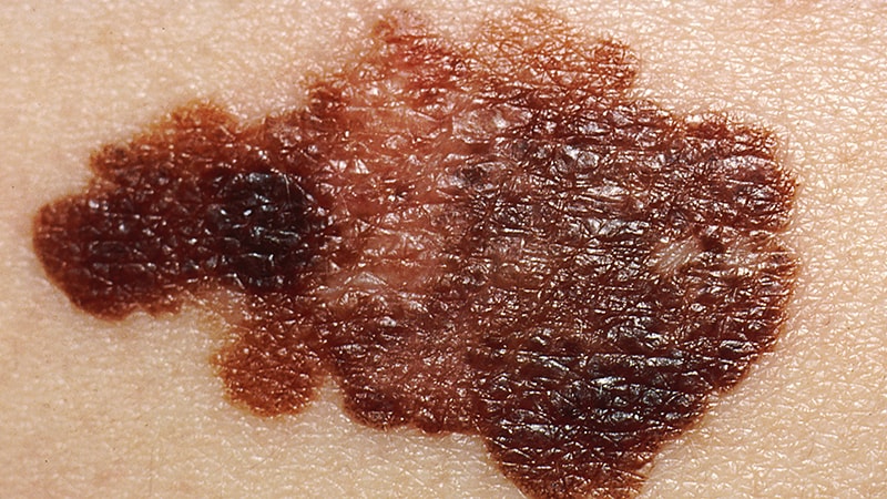
Melanocytes are specialized cells that produce, store, and transport melanin, the pigment responsible for coloring of the skin, hair, and eyes. They are located in the bottom layer of the epidermis (the outermost layer of the skin) and can also be found in the inner ear and the eye's retina. Melanocytes contain organelles called melanosomes, which produce and store melanin.
Melanin comes in two types: eumelanin (black or brown) and pheomelanin (red or yellow). The amount and type of melanin produced by melanocytes determine the color of a person's skin, hair, and eyes. Exposure to UV radiation from sunlight increases melanin production as a protective response, leading to skin tanning.
Melanocyte dysfunction or abnormalities can lead to various medical conditions, such as albinism (lack of melanin production), melasma (excessive pigmentation), and melanoma (cancerous growth of melanocytes).
In situ hybridization (ISH) is a molecular biology technique used to detect and localize specific nucleic acid sequences, such as DNA or RNA, within cells or tissues. This technique involves the use of a labeled probe that is complementary to the target nucleic acid sequence. The probe can be labeled with various types of markers, including radioisotopes, fluorescent dyes, or enzymes.
During the ISH procedure, the labeled probe is hybridized to the target nucleic acid sequence in situ, meaning that the hybridization occurs within the intact cells or tissues. After washing away unbound probe, the location of the labeled probe can be visualized using various methods depending on the type of label used.
In situ hybridization has a wide range of applications in both research and diagnostic settings, including the detection of gene expression patterns, identification of viral infections, and diagnosis of genetic disorders.
A nonmammalian embryo refers to the developing organism in animals other than mammals, from the fertilized egg (zygote) stage until hatching or birth. In nonmammalian species, the developmental stages and terminology differ from those used in mammals. The term "embryo" is generally applied to the developing organism up until a specific stage of development that is characterized by the formation of major organs and structures. After this point, the developing organism is referred to as a "larva," "juvenile," or other species-specific terminology.
The study of nonmammalian embryos has played an important role in our understanding of developmental biology and evolutionary developmental biology (evo-devo). By comparing the developmental processes across different animal groups, researchers can gain insights into the evolutionary origins and diversification of body plans and structures. Additionally, nonmammalian embryos are often used as model systems for studying basic biological processes, such as cell division, gene regulation, and pattern formation.
"Body patterning" is a general term that refers to the process of forming and organizing various tissues and structures into specific patterns during embryonic development. This complex process involves a variety of molecular mechanisms, including gene expression, cell signaling, and cell-cell interactions. It results in the creation of distinct body regions, such as the head, trunk, and limbs, as well as the organization of internal organs and systems.
In medical terminology, "body patterning" may refer to specific developmental processes or abnormalities related to embryonic development. For example, in genetic disorders such as Poland syndrome or Holt-Oram syndrome, mutations in certain genes can lead to abnormal body patterning, resulting in the absence or underdevelopment of certain muscles, bones, or other structures.
It's important to note that "body patterning" is not a formal medical term with a specific definition, but rather a general concept used in developmental biology and genetics.
Zebrafish proteins refer to the diverse range of protein molecules that are produced by the organism Danio rerio, commonly known as the zebrafish. These proteins play crucial roles in various biological processes such as growth, development, reproduction, and response to environmental stimuli. They are involved in cellular functions like enzymatic reactions, signal transduction, structural support, and regulation of gene expression.
Zebrafish is a popular model organism in biomedical research due to its genetic similarity with humans, rapid development, and transparent embryos that allow for easy observation of biological processes. As a result, the study of zebrafish proteins has contributed significantly to our understanding of protein function, structure, and interaction in both zebrafish and human systems.
Some examples of zebrafish proteins include:
* Transcription factors that regulate gene expression during development
* Enzymes involved in metabolic pathways
* Structural proteins that provide support to cells and tissues
* Receptors and signaling molecules that mediate communication between cells
* Heat shock proteins that assist in protein folding and protect against stress
The analysis of zebrafish proteins can be performed using various techniques, including biochemical assays, mass spectrometry, protein crystallography, and computational modeling. These methods help researchers to identify, characterize, and understand the functions of individual proteins and their interactions within complex networks.
Craniofacial abnormalities refer to a group of birth defects that affect the development of the skull and face. These abnormalities can range from mild to severe and may involve differences in the shape and structure of the head, face, and jaws, as well as issues with the formation of facial features such as the eyes, nose, and mouth.
Craniofacial abnormalities can be caused by genetic factors, environmental influences, or a combination of both. Some common examples of craniofacial abnormalities include cleft lip and palate, craniosynostosis (premature fusion of the skull bones), and hemifacial microsomia (underdevelopment of one side of the face).
Treatment for craniofacial abnormalities may involve a team of healthcare professionals, including plastic surgeons, neurosurgeons, orthodontists, speech therapists, and other specialists. Treatment options may include surgery, bracing, therapy, and other interventions to help improve function and appearance.
Ectoderm is the outermost of the three primary germ layers in a developing embryo, along with the endoderm and mesoderm. The ectoderm gives rise to the outer covering of the body, including the skin, hair, nails, glands, and the nervous system, which includes the brain, spinal cord, and peripheral nerves. It also forms the lining of the mouth, anus, nose, and ears. Essentially, the ectoderm is responsible for producing all the epidermal structures and the neural crest cells that contribute to various derivatives such as melanocytes, adrenal medulla, smooth muscle, and peripheral nervous system components.
A zebrafish is a freshwater fish species belonging to the family Cyprinidae and the genus Danio. Its name is derived from its distinctive striped pattern that resembles a zebra's. Zebrafish are often used as model organisms in scientific research, particularly in developmental biology, genetics, and toxicology studies. They have a high fecundity rate, transparent embryos, and a rapid development process, making them an ideal choice for researchers. However, it is important to note that providing a medical definition for zebrafish may not be entirely accurate or relevant since they are primarily used in biological research rather than clinical medicine.
Morphogenesis is a term used in developmental biology and refers to the process by which cells give rise to tissues and organs with specific shapes, structures, and patterns during embryonic development. This process involves complex interactions between genes, cells, and the extracellular environment that result in the coordinated movement and differentiation of cells into specialized functional units.
Morphogenesis is a dynamic and highly regulated process that involves several mechanisms, including cell proliferation, death, migration, adhesion, and differentiation. These processes are controlled by genetic programs and signaling pathways that respond to environmental cues and regulate the behavior of individual cells within a developing tissue or organ.
The study of morphogenesis is important for understanding how complex biological structures form during development and how these processes can go awry in disease states such as cancer, birth defects, and degenerative disorders.
In medical and embryological terms, the mesoderm is one of the three primary germ layers in the very early stages of embryonic development. It forms between the ectoderm and endoderm during gastrulation, and it gives rise to a wide variety of cell types, tissues, and organs in the developing embryo.
The mesoderm contributes to the formation of structures such as:
1. The connective tissues (including tendons, ligaments, and most of the bones)
2. Muscular system (skeletal, smooth, and cardiac muscles)
3. Circulatory system (heart, blood vessels, and blood cells)
4. Excretory system (kidneys and associated structures)
5. Reproductive system (gonads, including ovaries and testes)
6. Dermis of the skin
7. Parts of the eye and inner ear
8. Several organs in the urogenital system
Dysfunctions or abnormalities in mesoderm development can lead to various congenital disorders and birth defects, highlighting its importance during embryogenesis.
"Xenopus proteins" refer to the proteins that are expressed or isolated from the Xenopus species, which are primarily used as model organisms in biological and biomedical research. The most commonly used Xenopus species for research are the African clawed frogs, Xenopus laevis and Xenopus tropicalis. These proteins play crucial roles in various cellular processes and functions, and they serve as valuable tools to study different aspects of molecular biology, developmental biology, genetics, and biochemistry.
Some examples of Xenopus proteins that are widely studied include:
1. Xenopus Histones: These are the proteins that package DNA into nucleosomes, which are the fundamental units of chromatin in eukaryotic cells. They play a significant role in gene regulation and epigenetic modifications.
2. Xenopus Cyclins and Cyclin-dependent kinases (CDKs): These proteins regulate the cell cycle and control cell division, differentiation, and apoptosis.
3. Xenopus Transcription factors: These proteins bind to specific DNA sequences and regulate gene expression during development and in response to various stimuli.
4. Xenopus Signaling molecules: These proteins are involved in intracellular signaling pathways that control various cellular processes, such as cell growth, differentiation, migration, and survival.
5. Xenopus Cytoskeletal proteins: These proteins provide structural support to the cells and regulate their shape, motility, and organization.
6. Xenopus Enzymes: These proteins catalyze various biochemical reactions in the cell, such as metabolic pathways, DNA replication, transcription, and translation.
Overall, Xenopus proteins are essential tools for understanding fundamental biological processes and have contributed significantly to our current knowledge of molecular biology, genetics, and developmental biology.
Cell differentiation is the process by which a less specialized cell, or stem cell, becomes a more specialized cell type with specific functions and structures. This process involves changes in gene expression, which are regulated by various intracellular signaling pathways and transcription factors. Differentiation results in the development of distinct cell types that make up tissues and organs in multicellular organisms. It is a crucial aspect of embryonic development, tissue repair, and maintenance of homeostasis in the body.
CREST syndrome is a subtype of a autoimmune connective tissue disorder called scleroderma (systemic sclerosis). The name "CREST" is an acronym that stands for the following five features:
* Calcinosis: The formation of calcium deposits in the skin and underlying tissues, which can cause painful ulcers.
* Raynaud's phenomenon: A condition in which the blood vessels in the fingers and toes constrict in response to cold or stress, causing the digits to turn white or blue and become numb or painful.
* Esophageal dysmotility: Difficulty swallowing due to weakened muscles in the esophagus.
* Sclerodactyly: Thickening and tightening of the skin on the fingers.
* Telangiectasias: Dilated blood vessels near the surface of the skin, causing red spots or lines.
It's important to note that not everyone with CREST syndrome will have all five of these features, and some people may have additional symptoms not included in the acronym. Additionally, CREST syndrome is a chronic condition that can cause a range of complications, including lung fibrosis, kidney problems, and digital ulcers. Treatment typically focuses on managing specific symptoms and slowing the progression of the disease.
'Cell lineage' is a term used in biology and medicine to describe the developmental history or relationship of a cell or group of cells to other cells, tracing back to the original progenitor or stem cell. It refers to the series of cell divisions and differentiation events that give rise to specific types of cells in an organism over time.
In simpler terms, cell lineage is like a family tree for cells, showing how they are related to each other through a chain of cell division and specialization events. This concept is important in understanding the development, growth, and maintenance of tissues and organs in living beings.
The enteric nervous system (ENS) is a part of the autonomic nervous system that directly controls the gastrointestinal tract, including the stomach, small intestine, colon, and rectum. It is sometimes referred to as the "second brain" because it can operate independently of the central nervous system (CNS).
The ENS contains around 500 million neurons that are organized into two main plexuses: the myenteric plexus, which lies between the longitudinal and circular muscle layers of the gut, and the submucosal plexus, which is located in the submucosa. These plexuses contain various types of neurons that are responsible for regulating gastrointestinal motility, secretion, and blood flow.
The ENS can communicate with the CNS through afferent nerve fibers that transmit information about the state of the gut to the brain, and efferent nerve fibers that carry signals from the brain back to the ENS. However, the ENS is also capable of functioning independently of the CNS, allowing it to regulate gastrointestinal functions in response to local stimuli such as food intake, inflammation, or infection.
Embryonic induction is a process that occurs during the development of a multicellular organism, where one group of cells in the embryo signals and influences the developmental fate of another group of cells. This interaction leads to the formation of specific structures or organs in the developing embryo. The signaling cells that initiate the process are called organizers, and they release signaling molecules known as morphogens that bind to receptors on the target cells and trigger a cascade of intracellular signals that ultimately lead to changes in gene expression and cell fate. Embryonic induction is a crucial step in the development of complex organisms and plays a key role in establishing the body plan and organizing the different tissues and organs in the developing embryo.
The Neural Tube is a structure that forms during the development of an embryo and eventually becomes the brain, spinal cord, and other parts of the nervous system. It is a narrow channel that runs along the back of the embryo, forming from the ectoderm (one of the three germ layers) and closing around the 23rd or 26th day after conception. Defects in the closure of the neural tube can lead to conditions such as spina bifida and anencephaly.
Bone Morphogenetic Proteins (BMPs) are a group of growth factors that play crucial roles in the development, growth, and repair of bones and other tissues. They belong to the Transforming Growth Factor-β (TGF-β) superfamily and were first discovered when researchers found that certain proteins extracted from demineralized bone matrix had the ability to induce new bone formation.
BMPs stimulate the differentiation of mesenchymal stem cells into osteoblasts, which are the cells responsible for bone formation. They also promote the recruitment and proliferation of these cells, enhancing the overall process of bone regeneration. In addition to their role in bone biology, BMPs have been implicated in various other biological processes, including embryonic development, wound healing, and the regulation of fat metabolism.
There are several types of BMPs (BMP-2, BMP-4, BMP-7, etc.) that exhibit distinct functions and expression patterns. Due to their ability to stimulate bone formation, recombinant human BMPs have been used in clinical applications, such as spinal fusion surgery and non-healing fracture treatment. However, the use of BMPs in medicine has been associated with certain risks and complications, including uncontrolled bone growth, inflammation, and cancer development, which necessitates further research to optimize their therapeutic potential.
Transcription factors are proteins that play a crucial role in regulating gene expression by controlling the transcription of DNA to messenger RNA (mRNA). They function by binding to specific DNA sequences, known as response elements, located in the promoter region or enhancer regions of target genes. This binding can either activate or repress the initiation of transcription, depending on the properties and interactions of the particular transcription factor. Transcription factors often act as part of a complex network of regulatory proteins that determine the precise spatiotemporal patterns of gene expression during development, differentiation, and homeostasis in an organism.
In medical terms, the jaw is referred to as the mandible (in humans and some other animals), which is the lower part of the face that holds the lower teeth in place. It's a large, horseshoe-shaped bone that forms the lower jaw and serves as a attachment point for several muscles that are involved in chewing and moving the lower jaw.
In addition to the mandible, the upper jaw is composed of two bones known as the maxillae, which fuse together at the midline of the face to form the upper jaw. The upper jaw holds the upper teeth in place and forms the roof of the mouth, as well as a portion of the eye sockets and nasal cavity.
Together, the mandible and maxillae allow for various functions such as speaking, eating, and breathing.
The nervous system is a complex, highly organized network of specialized cells called neurons and glial cells that communicate with each other via electrical and chemical signals to coordinate various functions and activities in the body. It consists of two main parts: the central nervous system (CNS), including the brain and spinal cord, and the peripheral nervous system (PNS), which includes all the nerves and ganglia outside the CNS.
The primary function of the nervous system is to receive, process, and integrate information from both internal and external environments and then respond by generating appropriate motor outputs or behaviors. This involves sensing various stimuli through specialized receptors, transmitting this information through afferent neurons to the CNS for processing, integrating this information with other inputs and memories, making decisions based on this processed information, and finally executing responses through efferent neurons that control effector organs such as muscles and glands.
The nervous system can be further divided into subsystems based on their functions, including the somatic nervous system, which controls voluntary movements and reflexes; the autonomic nervous system, which regulates involuntary physiological processes like heart rate, digestion, and respiration; and the enteric nervous system, which is a specialized subset of the autonomic nervous system that controls gut functions. Overall, the nervous system plays a critical role in maintaining homeostasis, regulating behavior, and enabling cognition and consciousness.
In medical terms, the "head" is the uppermost part of the human body that contains the brain, skull, face, eyes, nose, mouth, and ears. It is connected to the rest of the body by the neck and is responsible for many vital functions such as sight, hearing, smell, taste, touch, and thought processing. The head also plays a crucial role in maintaining balance, speech, and eating.
CD57 is a protein found on the surface of some immune cells, specifically natural killer (NK) cells and certain T-cells. It is often used as a marker to identify these populations of cells. Antigens are substances that can stimulate an immune response, leading to the production of antibodies. In the context of CD57, antigens would refer to any substance that can bind to the CD57 protein on the surface of NK or T-cells.
It's worth noting that CD57 has been studied as a potential marker for certain diseases and conditions, such as HIV infection and some types of cancer. However, its use as a diagnostic or prognostic marker is still a subject of ongoing research and debate.
Paired box (PAX) transcription factors are a group of proteins that regulate gene expression during embryonic development and in some adult tissues. They are characterized by the presence of a paired box domain, a conserved DNA-binding motif that recognizes specific DNA sequences. PAX proteins play crucial roles in various developmental processes, such as the formation of the nervous system, eyes, and pancreas. Dysregulation of PAX genes has been implicated in several human diseases, including cancer.
A ganglion is a cluster of neuron cell bodies in the peripheral nervous system. Ganglia are typically associated with nerves and serve as sites for sensory processing, integration, and relay of information between the periphery and the central nervous system (CNS). The two main types of ganglia are sensory ganglia, which contain pseudounipolar neurons that transmit sensory information to the CNS, and autonomic ganglia, which contain multipolar neurons that control involuntary physiological functions.
Examples of sensory ganglia include dorsal root ganglia (DRG), which are associated with spinal nerves, and cranial nerve ganglia, such as the trigeminal ganglion. Autonomic ganglia can be further divided into sympathetic and parasympathetic ganglia, which regulate different aspects of the autonomic nervous system.
It's worth noting that in anatomy, "ganglion" refers to a group of nerve cell bodies, while in clinical contexts, "ganglion" is often used to describe a specific type of cystic structure that forms near joints or tendons, typically in the wrist or foot. These ganglia are not related to the peripheral nervous system's ganglia but rather are fluid-filled sacs that may cause discomfort or pain due to their size or location.
Somites are transient, segmentally repeated embryonic structures that form along the anterior-posterior body axis during vertebrate development. They are derived from the paraxial mesoderm and give rise to various tissues, including the sclerotome (which forms the vertebrae and ribs), myotome (which forms the skeletal muscles of the back and limbs), and dermatome (which forms the dermis of the skin).
Each somite is a block-like structure that is arranged in a repeating pattern along the notochord, which is a flexible rod-like structure that provides mechanical support to the developing embryo. The formation of somites is a critical step in the development of the vertebrate body plan, as they help to establish the segmental organization of the musculoskeletal system and contribute to the formation of other important structures such as the dermis and the circulatory system.
The process of somitogenesis, or the formation of somites, is a highly regulated and coordinated event that involves the interaction of various signaling molecules and genetic pathways. Defects in somite formation can lead to a range of developmental abnormalities, including spinal deformities, muscle weakness, and skin defects.
The Peripheral Nervous System (PNS) is that part of the nervous system which lies outside of the brain and spinal cord. It includes all the nerves and ganglia ( clusters of neurons) outside of the central nervous system (CNS). The PNS is divided into two components: the somatic nervous system and the autonomic nervous system.
The somatic nervous system is responsible for transmitting sensory information from the skin, muscles, and joints to the CNS, and for controlling voluntary movements of the skeletal muscles.
The autonomic nervous system, on the other hand, controls involuntary actions, such as heart rate, digestion, respiratory rate, salivation, perspiration, pupillary dilation, and sexual arousal. It is further divided into the sympathetic and parasympathetic systems, which generally have opposing effects and maintain homeostasis in the body.
Damage to the peripheral nervous system can result in various medical conditions such as neuropathies, neuritis, plexopathies, and radiculopathies, leading to symptoms like numbness, tingling, pain, weakness, or loss of reflexes in the affected area.
Homeodomain proteins are a group of transcription factors that play crucial roles in the development and differentiation of cells in animals and plants. They are characterized by the presence of a highly conserved DNA-binding domain called the homeodomain, which is typically about 60 amino acids long. The homeodomain consists of three helices, with the third helix responsible for recognizing and binding to specific DNA sequences.
Homeodomain proteins are involved in regulating gene expression during embryonic development, tissue maintenance, and organismal growth. They can act as activators or repressors of transcription, depending on the context and the presence of cofactors. Mutations in homeodomain proteins have been associated with various human diseases, including cancer, congenital abnormalities, and neurological disorders.
Some examples of homeodomain proteins include PAX6, which is essential for eye development, HOX genes, which are involved in body patterning, and NANOG, which plays a role in maintaining pluripotency in stem cells.
I'm not aware of a specific medical definition for "Avian Proteins." The term "avian" generally refers to birds or their characteristics. Therefore, "avian proteins" would likely refer to proteins that are found in birds or are produced by avian cells. These proteins could have various functions and roles, depending on the specific protein in question.
For example, avian proteins might be of interest in medical research if they have similarities to human proteins and can be used as models to study protein function, structure, or interaction with other molecules. Additionally, some avian proteins may have potential applications in therapeutic development, such as using chicken egg-derived proteins for wound healing or as vaccine components.
However, without a specific context or reference, it's difficult to provide a more precise definition of "avian proteins" in a medical context.
Hirschsprung disease is a gastrointestinal disorder that affects the large intestine, specifically the section known as the colon. This condition is congenital, meaning it is present at birth. It occurs due to the absence of ganglion cells (nerve cells) in the bowel's muscular wall, which are responsible for coordinating muscle contractions that move food through the digestive tract.
The affected segment of the colon cannot relax and propel the contents within it, leading to various symptoms such as constipation, intestinal obstruction, or even bowel perforation in severe cases. Common diagnostic methods include rectal suction biopsy, anorectal manometry, and contrast enema studies. Treatment typically involves surgical removal of the aganglionic segment and reattachment of the normal colon to the anus (known as a pull-through procedure).
Tissue transplantation is a medical procedure where tissues from one part of the body or from another individual's body are removed and implanted in a recipient to replace damaged, diseased, or missing tissues. The tissues may include skin, bone, tendons, ligaments, heart valves, corneas, or even entire organs such as hearts, lungs, livers, and kidneys.
The donor tissue must be compatible with the recipient's body to reduce the risk of rejection, which is the immune system attacking and destroying the transplanted tissue. This often requires matching certain proteins called human leukocyte antigens (HLAs) found on the surface of most cells in the body.
Tissue transplantation can significantly improve a patient's quality of life or, in some cases, save their life. However, it does carry risks such as infection, bleeding, and rejection, which require careful monitoring and management.
High mobility group proteins (HMG proteins) are a family of nuclear proteins that are characterized by their ability to bind to DNA and influence its structure and function. They are named "high mobility" because of their rapid movement in gel electrophoresis. HMG proteins are involved in various nuclear processes, including chromatin remodeling, transcription regulation, and DNA repair.
There are three main classes of HMG proteins: HMGA, HMGB, and HMGN. Each class has distinct structural features and functions. For example, HMGA proteins have a unique "AT-hook" domain that allows them to bind to the minor groove of AT-rich DNA sequences, while HMGB proteins have two "HMG-box" domains that enable them to bend and unwind DNA.
HMG proteins play important roles in many physiological and pathological processes, such as embryonic development, inflammation, and cancer. Dysregulation of HMG protein function has been implicated in various diseases, including neurodegenerative disorders, diabetes, and cancer. Therefore, understanding the structure, function, and regulation of HMG proteins is crucial for developing new therapeutic strategies for these diseases.
"Xenopus" is not a medical term, but it is a genus of highly invasive aquatic frogs native to sub-Saharan Africa. They are often used in scientific research, particularly in developmental biology and genetics. The most commonly studied species is Xenopus laevis, also known as the African clawed frog.
In a medical context, Xenopus might be mentioned when discussing their use in research or as a model organism to study various biological processes or diseases.
Wnt proteins are a family of secreted signaling molecules that play crucial roles in the regulation of fundamental biological processes, including cell proliferation, differentiation, migration, and survival. They were first discovered in 1982 through genetic studies in Drosophila melanogaster (fruit flies) and have since been found to be highly conserved across various species, from invertebrates to humans.
Wnt proteins exert their effects by binding to specific receptors on the target cell surface, leading to the activation of several intracellular signaling pathways:
1. Canonical Wnt/β-catenin pathway: In the absence of Wnt ligands, β-catenin is continuously degraded by a destruction complex consisting of Axin, APC (Adenomatous polyposis coli), and GSK3β (Glycogen synthase kinase 3 beta). When Wnt proteins bind to their receptors Frizzled and LRP5/6, the formation of a "signalosome" complex leads to the inhibition of the destruction complex, allowing β-catenin to accumulate in the cytoplasm and translocate into the nucleus. Here, it interacts with TCF/LEF (T-cell factor/lymphoid enhancer-binding factor) transcription factors to regulate the expression of target genes involved in cell proliferation, differentiation, and survival.
2. Non-canonical Wnt pathways: These include the Wnt/Ca^2+^ pathway and the planar cell polarity (PCP) pathway. In the Wnt/Ca^2+^ pathway, Wnt ligands bind to Frizzled receptors and activate heterotrimeric G proteins, leading to an increase in intracellular Ca^2+^ levels and activation of downstream targets such as protein kinase C (PKC) and calcium/calmodulin-dependent protein kinase II (CAMKII). These signaling events ultimately regulate cell movement, adhesion, and gene expression. In the PCP pathway, Wnt ligands bind to Frizzled receptors and coreceptor complexes containing Ror2 or Ryk, leading to activation of small GTPases such as RhoA and Rac1, which control cytoskeletal organization and cell polarity.
Dysregulation of Wnt signaling has been implicated in various human diseases, including cancer, developmental disorders, and degenerative conditions. In cancer, aberrant activation of the canonical Wnt/β-catenin pathway contributes to tumor initiation, progression, and metastasis by promoting cell proliferation, survival, and epithelial-mesenchymal transition (EMT). Inhibitors targeting different components of the Wnt signaling pathway are currently being developed as potential therapeutic strategies for cancer treatment.
The neural plate is a structure formed during the embryonic development of vertebrates. It is a thickened plate of ectodermal cells located on the dorsal surface of the developing embryo. The neural plate gives rise to the central nervous system, including the brain and spinal cord.
The process of neural plate formation begins with the specification of ectodermal cells into neural fated cells, a process that is regulated by various signaling molecules. Once specified, these cells undergo morphological changes, resulting in the thickening of the ectoderm to form the neural plate.
The neural plate then undergoes a series of folding movements, leading to the formation of the neural tube, which eventually develops into the brain and spinal cord. The edges of the neural plate, known as the neural folds, come together and fuse, forming a closed tube. Failure of the neural folds to fuse properly can result in neural tube defects, such as spina bifida.
Overall, the neural plate is a critical structure in the development of the nervous system in vertebrates, and its formation and subsequent development are tightly regulated by various genetic and environmental factors.
The ilium is the largest and broadest of the three parts that make up the hip bone or coxal bone. It is the uppermost portion of the pelvis and forms the side of the waist. The ilium has a curved, fan-like shape and articulates with the sacrum at the back to form the sacroiliac joint. The large, concave surface on the top of the ilium is called the iliac crest, which can be felt as a prominent ridge extending from the front of the hip to the lower back. This region is significant in orthopedics and physical examinations for its use in assessing various medical conditions and performing certain maneuvers during the physical examination.
A mammalian embryo is the developing offspring of a mammal, from the time of implantation of the fertilized egg (blastocyst) in the uterus until the end of the eighth week of gestation. During this period, the embryo undergoes rapid cell division and organ differentiation to form a complex structure with all the major organs and systems in place. This stage is followed by fetal development, which continues until birth. The study of mammalian embryos is important for understanding human development, evolution, and reproductive biology.
Wnt1 protein is a member of the Wnt family, which is a group of secreted signaling proteins that play crucial roles in embryonic development and tissue homeostasis in adults. Specifically, Wnt1 is a highly conserved gene that encodes a glycoprotein with a molecular weight of approximately 40 kDa. It is primarily expressed in the developing nervous system, where it functions as a key regulator of neural crest cell migration and differentiation during embryogenesis.
Wnt1 protein mediates its effects by binding to Frizzled receptors on the surface of target cells, leading to the activation of several intracellular signaling pathways, including the canonical Wnt/β-catenin pathway and non-canonical Wnt/planar cell polarity (PCP) pathway. In the canonical pathway, Wnt1 protein stabilizes β-catenin, which then translocates to the nucleus and interacts with TCF/LEF transcription factors to regulate gene expression.
Dysregulation of Wnt1 signaling has been implicated in several human diseases, including cancer. For example, aberrant activation of the Wnt/β-catenin pathway by Wnt1 protein has been observed in various types of tumors, such as medulloblastomas and breast cancers, leading to uncontrolled cell proliferation and tumor growth. Therefore, understanding the molecular mechanisms underlying Wnt1 signaling is essential for developing novel therapeutic strategies for treating these diseases.
The mesencephalon, also known as the midbrain, is the middle portion of the brainstem that connects the hindbrain (rhombencephalon) and the forebrain (prosencephalon). It plays a crucial role in several important functions including motor control, vision, hearing, and the regulation of consciousness and sleep-wake cycles. The mesencephalon contains several important structures such as the cerebral aqueduct, tectum, tegmentum, cerebral peduncles, and several cranial nerve nuclei (III and IV).
Signal transduction is the process by which a cell converts an extracellular signal, such as a hormone or neurotransmitter, into an intracellular response. This involves a series of molecular events that transmit the signal from the cell surface to the interior of the cell, ultimately resulting in changes in gene expression, protein activity, or metabolism.
The process typically begins with the binding of the extracellular signal to a receptor located on the cell membrane. This binding event activates the receptor, which then triggers a cascade of intracellular signaling molecules, such as second messengers, protein kinases, and ion channels. These molecules amplify and propagate the signal, ultimately leading to the activation or inhibition of specific cellular responses.
Signal transduction pathways are highly regulated and can be modulated by various factors, including other signaling molecules, post-translational modifications, and feedback mechanisms. Dysregulation of these pathways has been implicated in a variety of diseases, including cancer, diabetes, and neurological disorders.
Immunohistochemistry (IHC) is a technique used in pathology and laboratory medicine to identify specific proteins or antigens in tissue sections. It combines the principles of immunology and histology to detect the presence and location of these target molecules within cells and tissues. This technique utilizes antibodies that are specific to the protein or antigen of interest, which are then tagged with a detection system such as a chromogen or fluorophore. The stained tissue sections can be examined under a microscope, allowing for the visualization and analysis of the distribution and expression patterns of the target molecule in the context of the tissue architecture. Immunohistochemistry is widely used in diagnostic pathology to help identify various diseases, including cancer, infectious diseases, and immune-mediated disorders.
Transcription Factor AP-2 is a specific protein involved in the process of gene transcription. It belongs to a family of transcription factors known as Activating Enhancer-Binding Proteins (AP-2). These proteins regulate gene expression by binding to specific DNA sequences called enhancers, which are located near the genes they control.
AP-2 is composed of four subunits that form a homo- or heterodimer, which then binds to the consensus sequence 5'-GCCNNNGGC-3'. This sequence is typically found in the promoter regions of target genes. Once bound, AP-2 can either activate or repress gene transcription, depending on the context and the presence of cofactors.
AP-2 plays crucial roles during embryonic development, particularly in the formation of the nervous system, limbs, and face. It is also involved in cell cycle regulation, differentiation, and apoptosis (programmed cell death). Dysregulation of AP-2 has been implicated in several diseases, including various types of cancer.
Cardiovascular abnormalities refer to structural or functional anomalies in the heart or blood vessels. These abnormalities can be present at birth (congenital) or acquired later in life. They can affect the heart's chambers, valves, walls, or blood vessels, leading to various complications such as heart failure, stroke, or even death if left untreated.
Examples of congenital cardiovascular abnormalities include:
1. Septal defects - holes in the walls separating the heart's chambers (atrial septal defect, ventricular septal defect)
2. Valvular stenosis or insufficiency - narrowing or leakage of the heart valves
3. Patent ductus arteriosus - a persistent opening between the aorta and pulmonary artery
4. Coarctation of the aorta - narrowing of the aorta
5. Tetralogy of Fallot - a combination of four heart defects, including ventricular septal defect, overriding aorta, pulmonary stenosis, and right ventricular hypertrophy
Examples of acquired cardiovascular abnormalities include:
1. Atherosclerosis - the buildup of plaque in the arteries, leading to narrowing or blockage
2. Cardiomyopathy - disease of the heart muscle, causing it to become enlarged, thickened, or stiffened
3. Hypertension - high blood pressure, which can damage the heart and blood vessels over time
4. Myocardial infarction (heart attack) - damage to the heart muscle due to blocked blood supply
5. Infective endocarditis - infection of the inner lining of the heart chambers and valves
These abnormalities can be diagnosed through various tests, such as echocardiography, electrocardiogram (ECG), stress testing, cardiac catheterization, or magnetic resonance imaging (MRI). Treatment options depend on the type and severity of the abnormality and may include medications, medical procedures, or surgery.
A chimera, in the context of medicine and biology, is a single organism that is composed of cells with different genetics. This can occur naturally in some situations, such as when fraternal twins do not fully separate in utero and end up sharing some organs or tissues. The term "chimera" can also refer to an organism that contains cells from two different species, which can happen in certain types of genetic research or medical treatments. For example, a patient's cells might be genetically modified in a lab and then introduced into their body to treat a disease; if some of these modified cells mix with the patient's original cells, the result could be a chimera.
It's worth noting that the term "chimera" comes from Greek mythology, where it referred to a fire-breathing monster that was part lion, part goat, and part snake. In modern scientific usage, the term has a specific technical meaning related to genetics and organisms, but it may still evoke images of fantastical creatures for some people.
Cranial nerves are a set of twelve pairs of nerves that originate from the brainstem and skull, rather than the spinal cord. These nerves are responsible for transmitting sensory information (such as sight, smell, hearing, and taste) to the brain, as well as controlling various muscles in the head and neck (including those involved in chewing, swallowing, and eye movement). Each cranial nerve has a specific function and is named accordingly. For example, the optic nerve (cranial nerve II) transmits visual information from the eyes to the brain, while the vagus nerve (cranial nerve X) controls parasympathetic functions in the body such as heart rate and digestion.
Carbocyanines are a class of organic compounds that contain a polymethine chain, which is a type of carbon-based structure with alternating single and double bonds, and one or more cyanine groups. A cyanine group is a functional group consisting of a nitrogen atom connected to two carbon atoms by double bonds, with the remaining valences on the carbon atoms being satisfied by other groups.
Carbocyanines are known for their strong absorption and fluorescence properties in the visible and near-infrared regions of the electromagnetic spectrum. These properties make them useful as dyes and fluorescent labels in various applications, including biomedical research, clinical diagnostics, and material science.
In medicine, carbocyanines are sometimes used as fluorescent contrast agents for imaging purposes. They can be injected into the body and accumulate in certain tissues or organs, where they emit light when excited by a specific wavelength of light. This allows doctors to visualize the distribution of the agent and potentially detect abnormalities such as tumors or inflammation.
It is important to note that while carbocyanines have potential medical applications, they are not themselves medications or drugs. They are tools used in various medical procedures and research.
In medical terms, the heart is a muscular organ located in the thoracic cavity that functions as a pump to circulate blood throughout the body. It's responsible for delivering oxygen and nutrients to the tissues and removing carbon dioxide and other wastes. The human heart is divided into four chambers: two atria on the top and two ventricles on the bottom. The right side of the heart receives deoxygenated blood from the body and pumps it to the lungs, while the left side receives oxygenated blood from the lungs and pumps it out to the rest of the body. The heart's rhythmic contractions and relaxations are regulated by a complex electrical conduction system.
Bone Morphogenetic Protein 4 (BMP-4) is a growth factor that belongs to the transforming growth factor-beta (TGF-β) superfamily. It plays crucial roles in various biological processes, including embryonic development, cell growth, and differentiation. In the skeletal system, BMP-4 stimulates the formation of bone and cartilage by inducing the differentiation of mesenchymal stem cells into chondrocytes and osteoblasts. It also regulates the maintenance and repair of bones throughout life. An imbalance in BMP-4 signaling has been associated with several skeletal disorders, such as heterotopic ossification and osteoarthritis.
MSX1 (Homeobox protein MSX-1) is a transcription factor that belongs to the muscle segment homebox gene family, also known as the msh homeobox genes. These genes are involved in the development and differentiation of various tissues, including muscle, bone, and neural crest derivatives.
MSX1 plays crucial roles during embryonic development, such as regulating cell proliferation, differentiation, and apoptosis. It is widely expressed in the developing embryo, particularly in the oral ectoderm, neural crest, and mesenchyme. In the oral region, MSX1 helps control tooth development by interacting with other transcription factors and signaling molecules.
As a transcription factor, MSX1 binds to specific DNA sequences called homeobox response elements (HREs) in the promoter regions of its target genes. This binding either activates or represses gene expression, depending on the context and interacting partners. Dysregulation of MSX1 has been implicated in various developmental disorders and diseases, such as tooth agenesis, cleft lip/palate, and cancer.
Early Growth Response Protein 2 (EGR2) is a transcription factor that belongs to the EGR family of proteins, which are involved in various biological processes such as cell proliferation, differentiation, and apoptosis. EGR2 is specifically known to play crucial roles in the development and function of the nervous system, including the regulation of neuronal survival, axon guidance, and myelination. It is also expressed in immune cells and has been implicated in the regulation of immune responses. Mutations in the EGR2 gene have been associated with certain neurological disorders and diseases, such as Charcot-Marie-Tooth disease type 1B and congenital hypomyelinating neuropathy.
"Xenopus laevis" is not a medical term itself, but it refers to a specific species of African clawed frog that is often used in scientific research, including biomedical and developmental studies. Therefore, its relevance to medicine comes from its role as a model organism in laboratories.
In a broader sense, Xenopus laevis has contributed significantly to various medical discoveries, such as the understanding of embryonic development, cell cycle regulation, and genetic research. For instance, the Nobel Prize in Physiology or Medicine was awarded in 1963 to John R. B. Gurdon and Sir Michael J. Bishop for their discoveries concerning the genetic mechanisms of organism development using Xenopus laevis as a model system.
SOX9 (SRY-related HMG-box gene 9) is a transcription factor that belongs to the SOX family of proteins, which are characterized by a high mobility group (HMG) box DNA-binding domain. SOX9 plays crucial roles in various developmental processes, including sex determination, chondrogenesis, and neurogenesis.
As a transcription factor, SOX9 binds to specific DNA sequences in the promoter or enhancer regions of its target genes and regulates their expression. In the context of sex determination, SOX9 is essential for the development of Sertoli cells in the male gonad, which are responsible for supporting sperm production. SOX9 also plays a role in maintaining the undifferentiated state of stem cells and promoting cell differentiation in various tissues.
Mutations in the SOX9 gene have been associated with several human genetic disorders, including campomelic dysplasia, a severe skeletal disorder characterized by bowed legs, and sex reversal in individuals with XY chromosomes.
A group of chordate animals (Phylum Chordata) that have a vertebral column, or backbone, made up of individual vertebrae. This group includes mammals, birds, reptiles, amphibians, and fish. Vertebrates are characterized by the presence of a notochord, which is a flexible, rod-like structure that runs along the length of the body during development; a dorsal hollow nerve cord; and pharyngeal gill slits at some stage in their development. The vertebral column provides support and protection for the spinal cord and allows for the development of complex movements and behaviors.
Melanophores are specialized pigment-containing cells found in various organisms, including vertebrates and some invertebrates. In humans and other mammals, melanophores are primarily located within the skin's dermal layer and are part of the larger group of chromatophores.
Melanophores contain melanosomes, which are organelles that store and transport the pigment melanin. These cells play a crucial role in determining the coloration of an individual's skin, hair, and eyes by producing, storing, and distributing melanin granules within their cytoplasm.
In response to hormonal signals or neural stimulation, melanophores can undergo changes in the distribution of melanosomes, leading to variations in color intensity. This process is known as melanin dispersion or aggregation and is responsible for various physiological responses, such as skin tanning upon exposure to sunlight or the color-changing abilities observed in some animals like chameleons and cuttlefish.
It's important to note that while humans do not have the ability to change their skin color rapidly like some other animals, melanophores still play a significant role in protecting our skin from harmful ultraviolet radiation by producing melanin, which helps absorb and dissipate this energy, reducing damage to skin cells.
Neuroglia, also known as glial cells or simply glia, are non-neuronal cells that provide support and protection for neurons in the nervous system. They maintain homeostasis, form myelin sheaths around nerve fibers, and provide structural support. They also play a role in the immune response of the central nervous system. Some types of neuroglia include astrocytes, oligodendrocytes, microglia, and ependymal cells.
Cartilage is a type of connective tissue that is found throughout the body in various forms. It is made up of specialized cells called chondrocytes, which are embedded in a firm, flexible matrix composed of collagen fibers and proteoglycans. This unique structure gives cartilage its characteristic properties of being both strong and flexible.
There are three main types of cartilage in the human body: hyaline cartilage, elastic cartilage, and fibrocartilage.
1. Hyaline cartilage is the most common type and is found in areas such as the articular surfaces of bones (where they meet to form joints), the nose, trachea, and larynx. It has a smooth, glassy appearance and provides a smooth, lubricated surface for joint movement.
2. Elastic cartilage contains more elastin fibers than hyaline cartilage, which gives it greater flexibility and resilience. It is found in structures such as the external ear and parts of the larynx and epiglottis.
3. Fibrocartilage has a higher proportion of collagen fibers and fewer chondrocytes than hyaline or elastic cartilage. It is found in areas that require high tensile strength, such as the intervertebral discs, menisci (found in joints like the knee), and the pubic symphysis.
Cartilage plays a crucial role in supporting and protecting various structures within the body, allowing for smooth movement and providing a cushion between bones to absorb shock and prevent wear and tear. However, cartilage has limited capacity for self-repair and regeneration, making damage or degeneration of cartilage tissue a significant concern in conditions such as osteoarthritis.
Neurulation is the process by which the neural tube, which eventually develops into the brain and spinal cord, forms in an embryo. This complex process occurs during early embryonic development and involves a series of coordinated steps, including the formation of the neuroectoderm (a layer of cells that will give rise to the nervous system), the folding of this layer to form the neural plate, and the subsequent closure of the neural plate to create the neural tube. Disruptions in neurulation can result in serious birth defects, such as spina bifida or anencephaly.
Fibroblast Growth Factor 8 (FGF-8) is a growth factor that belongs to the fibroblast growth factor family. It plays crucial roles in various biological processes, including embryonic development, tissue repair, and cancer progression. Specifically, FGF-8 has been implicated in the regulation of cell proliferation, differentiation, migration, and survival.
During embryonic development, FGF-8 is involved in the formation of the nervous system, limbs, and other organs. It acts as a signaling molecule that helps to establish patterns of gene expression and cell behavior during development. In tissue repair, FGF-8 can stimulate the proliferation and migration of cells involved in wound healing, such as fibroblasts and endothelial cells.
In cancer, FGF-8 has been shown to promote tumor growth, angiogenesis (the formation of new blood vessels), and metastasis. It can do this by activating signaling pathways that promote cell proliferation, survival, and migration. Overexpression of FGF-8 has been found in various types of cancer, including breast, lung, prostate, and ovarian cancer.
In summary, Fibroblast Growth Factor 8 (FGF-8) is a signaling molecule that plays important roles in embryonic development, tissue repair, and cancer progression by regulating cell proliferation, differentiation, migration, and survival.
DNA-binding proteins are a type of protein that have the ability to bind to DNA (deoxyribonucleic acid), the genetic material of organisms. These proteins play crucial roles in various biological processes, such as regulation of gene expression, DNA replication, repair and recombination.
The binding of DNA-binding proteins to specific DNA sequences is mediated by non-covalent interactions, including electrostatic, hydrogen bonding, and van der Waals forces. The specificity of binding is determined by the recognition of particular nucleotide sequences or structural features of the DNA molecule.
DNA-binding proteins can be classified into several categories based on their structure and function, such as transcription factors, histones, and restriction enzymes. Transcription factors are a major class of DNA-binding proteins that regulate gene expression by binding to specific DNA sequences in the promoter region of genes and recruiting other proteins to modulate transcription. Histones are DNA-binding proteins that package DNA into nucleosomes, the basic unit of chromatin structure. Restriction enzymes are DNA-binding proteins that recognize and cleave specific DNA sequences, and are widely used in molecular biology research and biotechnology applications.
Transgenic mice are genetically modified rodents that have incorporated foreign DNA (exogenous DNA) into their own genome. This is typically done through the use of recombinant DNA technology, where a specific gene or genetic sequence of interest is isolated and then introduced into the mouse embryo. The resulting transgenic mice can then express the protein encoded by the foreign gene, allowing researchers to study its function in a living organism.
The process of creating transgenic mice usually involves microinjecting the exogenous DNA into the pronucleus of a fertilized egg, which is then implanted into a surrogate mother. The offspring that result from this procedure are screened for the presence of the foreign DNA, and those that carry the desired genetic modification are used to establish a transgenic mouse line.
Transgenic mice have been widely used in biomedical research to model human diseases, study gene function, and test new therapies. They provide a valuable tool for understanding complex biological processes and developing new treatments for a variety of medical conditions.
Neurons, also known as nerve cells or neurocytes, are specialized cells that constitute the basic unit of the nervous system. They are responsible for receiving, processing, and transmitting information and signals within the body. Neurons have three main parts: the dendrites, the cell body (soma), and the axon. The dendrites receive signals from other neurons or sensory receptors, while the axon transmits these signals to other neurons, muscles, or glands. The junction between two neurons is called a synapse, where neurotransmitters are released to transmit the signal across the gap (synaptic cleft) to the next neuron. Neurons vary in size, shape, and structure depending on their function and location within the nervous system.
Spinal ganglia, also known as dorsal root ganglia, are clusters of nerve cell bodies located in the peripheral nervous system. They are situated along the length of the spinal cord and are responsible for transmitting sensory information from the body to the brain. Each spinal ganglion contains numerous neurons, or nerve cells, with long processes called axons that extend into the periphery and innervate various tissues and organs. The cell bodies within the spinal ganglia receive sensory input from these axons and transmit this information to the central nervous system via the dorsal roots of the spinal nerves. This allows the brain to interpret and respond to a wide range of sensory stimuli, including touch, temperature, pain, and proprioception (the sense of the position and movement of one's body).
The Central Nervous System (CNS) is the part of the nervous system that consists of the brain and spinal cord. It is called the "central" system because it receives information from, and sends information to, the rest of the body through peripheral nerves, which make up the Peripheral Nervous System (PNS).
The CNS is responsible for processing sensory information, controlling motor functions, and regulating various autonomic processes like heart rate, respiration, and digestion. The brain, as the command center of the CNS, interprets sensory stimuli, formulates thoughts, and initiates actions. The spinal cord serves as a conduit for nerve impulses traveling to and from the brain and the rest of the body.
The CNS is protected by several structures, including the skull (which houses the brain) and the vertebral column (which surrounds and protects the spinal cord). Despite these protective measures, the CNS remains vulnerable to injury and disease, which can have severe consequences due to its crucial role in controlling essential bodily functions.
The facial bones, also known as the facial skeleton, are a series of bones that make up the framework of the face. They include:
1. Frontal bone: This bone forms the forehead and the upper part of the eye sockets.
2. Nasal bones: These two thin bones form the bridge of the nose.
3. Maxilla bones: These are the largest bones in the facial skeleton, forming the upper jaw, the bottom of the eye sockets, and the sides of the nose. They also contain the upper teeth.
4. Zygomatic bones (cheekbones): These bones form the cheekbones and the outer part of the eye sockets.
5. Palatine bones: These bones form the back part of the roof of the mouth, the side walls of the nasal cavity, and contribute to the formation of the eye socket.
6. Inferior nasal conchae: These are thin, curved bones that form the lateral walls of the nasal cavity and help to filter and humidify air as it passes through the nose.
7. Lacrimal bones: These are the smallest bones in the skull, located at the inner corner of the eye socket, and help to form the tear duct.
8. Mandible (lower jaw): This is the only bone in the facial skeleton that can move. It holds the lower teeth and forms the chin.
These bones work together to protect vital structures such as the eyes, brain, and nasal passages, while also providing attachment points for muscles that control chewing, expression, and other facial movements.
"Cells, cultured" is a medical term that refers to cells that have been removed from an organism and grown in controlled laboratory conditions outside of the body. This process is called cell culture and it allows scientists to study cells in a more controlled and accessible environment than they would have inside the body. Cultured cells can be derived from a variety of sources, including tissues, organs, or fluids from humans, animals, or cell lines that have been previously established in the laboratory.
Cell culture involves several steps, including isolation of the cells from the tissue, purification and characterization of the cells, and maintenance of the cells in appropriate growth conditions. The cells are typically grown in specialized media that contain nutrients, growth factors, and other components necessary for their survival and proliferation. Cultured cells can be used for a variety of purposes, including basic research, drug development and testing, and production of biological products such as vaccines and gene therapies.
It is important to note that cultured cells may behave differently than they do in the body, and results obtained from cell culture studies may not always translate directly to human physiology or disease. Therefore, it is essential to validate findings from cell culture experiments using additional models and ultimately in clinical trials involving human subjects.
Embryonic development is the series of growth and developmental stages that occur during the formation and early growth of the embryo. In humans, this stage begins at fertilization (when the sperm and egg cell combine) and continues until the end of the 8th week of pregnancy. During this time, the fertilized egg (now called a zygote) divides and forms a blastocyst, which then implants into the uterus. The cells in the blastocyst begin to differentiate and form the three germ layers: the ectoderm, mesoderm, and endoderm. These germ layers will eventually give rise to all of the different tissues and organs in the body.
Embryonic development is a complex and highly regulated process that involves the coordinated interaction of genetic and environmental factors. It is characterized by rapid cell division, migration, and differentiation, as well as programmed cell death (apoptosis) and tissue remodeling. Abnormalities in embryonic development can lead to birth defects or other developmental disorders.
It's important to note that the term "embryo" is used to describe the developing organism from fertilization until the end of the 8th week of pregnancy in humans, after which it is called a fetus.
I'm sorry for any confusion, but "lampreys" is not a medical term. It refers to a type of jawless aquatic vertebrate that are eel-like in appearance. They are parasitic creatures, with a suction cup-like mouth and circular rows of teeth, which they use to attach to fish and suck their body fluids. If you have any questions about medical terminology or concepts, I'd be happy to help with those!
Morpholinos are synthetic oligonucleotides that contain morpholine rings in their backbone instead of the ribose or deoxyribose sugars found in DNA and RNA. They are often used as antisense agents to inhibit gene expression by binding to complementary RNA sequences, preventing translation or splicing. Morpholinos are resistant to nucleases and have a neutral charge, which makes them more stable and less likely to cause off-target effects compared to other antisense technologies. They have been widely used in research to study gene function and have also shown promise as therapeutic agents for various diseases, including neuromuscular disorders and viral infections.
RhoB GTP-binding protein is a member of the Rho family of small GTPases, which are involved in regulating various cellular processes such as actin cytoskeleton organization, gene expression, and cell cycle progression. Specifically, RhoB functions as a molecular switch that cycles between an inactive GDP-bound state and an active GTP-bound state.
When RhoB is activated by GTP binding, it interacts with various downstream effectors to regulate the dynamics of the actin cytoskeleton, which is important for cell motility, adhesion, and membrane trafficking. RhoB has been implicated in several physiological processes, including angiogenesis, wound healing, and immune response.
RhoB is unique among the Rho GTPases because it can be localized to both the plasma membrane and endosomal compartments, allowing it to regulate various cellular processes in different subcellular locations. Dysregulation of RhoB has been associated with various pathological conditions, including cancer, inflammation, and neurodegenerative diseases.
Sympathetic ganglia are part of the autonomic nervous system, which controls involuntary bodily functions. These ganglia are clusters of nerve cell bodies located outside the central nervous system, along the spinal cord. They serve as a relay station for signals sent from the central nervous system to the organs and glands. The sympathetic ganglia are responsible for the "fight or flight" response, releasing neurotransmitters such as norepinephrine that prepare the body for action in response to stress or danger.
The hyoid bone is a U-shaped bone located in the anterior neck, superior to the thyroid cartilage. It does not articulate with any other bones and serves as an attachment point for various muscles, including those involved in swallowing, breathing, and speaking. The unique structure of the hyoid bone allows it to support the tongue and contribute to the stability of the airway.
Ambystoma mexicanum is the scientific name for the axolotl, a type of salamander that is native to Mexico. The axolotl is also known as the Mexican walking fish, although it is not actually a fish but an amphibian. It is unique because it exhibits neoteny, which means it can remain in its larval form throughout its entire life and never undergo complete metamorphosis into a terrestrial form.
The axolotl is a popular organism in scientific research due to its ability to regenerate lost body parts, including limbs, spinal cord, heart, and other organs. This has made it an important model organism for studying the mechanisms of regeneration and repair in mammals, including humans.
Congenital heart defects (CHDs) are structural abnormalities in the heart that are present at birth. They can affect any part of the heart's structure, including the walls of the heart, the valves inside the heart, and the major blood vessels that lead to and from the heart.
Congenital heart defects can range from mild to severe and can cause various symptoms depending on the type and severity of the defect. Some common symptoms of CHDs include cyanosis (a bluish tint to the skin, lips, and fingernails), shortness of breath, fatigue, poor feeding, and slow growth in infants and children.
There are many different types of congenital heart defects, including:
1. Septal defects: These are holes in the walls that separate the four chambers of the heart. The two most common septal defects are atrial septal defect (ASD) and ventricular septal defect (VSD).
2. Valve abnormalities: These include narrowed or leaky valves, which can affect blood flow through the heart.
3. Obstruction defects: These occur when blood flow is blocked or restricted due to narrowing or absence of a part of the heart's structure. Examples include pulmonary stenosis and coarctation of the aorta.
4. Cyanotic heart defects: These cause a lack of oxygen in the blood, leading to cyanosis. Examples include tetralogy of Fallot and transposition of the great arteries.
The causes of congenital heart defects are not fully understood, but genetic factors and environmental influences during pregnancy may play a role. Some CHDs can be detected before birth through prenatal testing, while others may not be diagnosed until after birth or later in childhood. Treatment for CHDs may include medication, surgery, or other interventions to improve blood flow and oxygenation of the body's tissues.
Chromatophores are pigment-containing cells found in various organisms, including animals and plants. In animals, chromatophores are primarily found in the skin, eyes, and hair or feathers, and they play a crucial role in color changes exhibited by many species. These cells contain pigments that can be concentrated or dispersed to change the color of the cell, allowing the animal to camouflage itself, communicate with other individuals, or regulate its body temperature.
There are several types of chromatophores, including:
1. Melanophores: These cells contain the pigment melanin and are responsible for producing dark colors such as black, brown, and gray. They are found in many animals, including mammals, birds, reptiles, amphibians, and fish.
2. Xanthophores: These cells contain yellow or orange pigments called pteridines and carotenoids. They are found in many animals, including fish, amphibians, and reptiles.
3. Iridophores: These cells do not contain pigments but instead reflect light to produce iridescent colors. They are found in many animals, including fish, reptiles, and amphibians.
4. Erythrophores: These cells contain red or pink pigments called porphyrins and are found in some species of fish and crustaceans.
5. Leucophores: These cells reflect white light and are found in some species of fish, cephalopods (such as squid and octopuses), and crustaceans.
The distribution and concentration of pigments within chromatophores can be controlled by hormones, neurotransmitters, or other signaling molecules, allowing the animal to change its color rapidly in response to environmental stimuli or social cues.
Sensory ganglia are clusters of nerve cell bodies located outside the central nervous system (the brain and spinal cord). They are primarily associated with sensory neurons, which are responsible for transmitting sensory information from various parts of the body to the central nervous system.
In humans, there are two main types of sensory ganglia: dorsal root ganglia and cranial nerve ganglia. Dorsal root ganglia are located along the spinal cord and contain the cell bodies of sensory neurons that innervate the skin, muscles, joints, and other tissues of the body. These neurons transmit information about touch, temperature, pain, and proprioception (the sense of the position and movement of the body).
Cranial nerve ganglia are associated with the cranial nerves, which are responsible for transmitting sensory information from the head and neck to the brain. For example, the trigeminal ganglion is a cranial nerve ganglion that contains the cell bodies of neurons that transmit sensory information from the face, mouth, and other structures of the head.
Overall, sensory ganglia play a critical role in our ability to perceive and interact with the world around us by transmitting important sensory information to the brain for processing.
Nerve tissue proteins are specialized proteins found in the nervous system that provide structural and functional support to nerve cells, also known as neurons. These proteins include:
1. Neurofilaments: These are type IV intermediate filaments that provide structural support to neurons and help maintain their shape and size. They are composed of three subunits - NFL (light), NFM (medium), and NFH (heavy).
2. Neuronal Cytoskeletal Proteins: These include tubulins, actins, and spectrins that provide structural support to the neuronal cytoskeleton and help maintain its integrity.
3. Neurotransmitter Receptors: These are specialized proteins located on the postsynaptic membrane of neurons that bind neurotransmitters released by presynaptic neurons, triggering a response in the target cell.
4. Ion Channels: These are transmembrane proteins that regulate the flow of ions across the neuronal membrane and play a crucial role in generating and transmitting electrical signals in neurons.
5. Signaling Proteins: These include enzymes, receptors, and adaptor proteins that mediate intracellular signaling pathways involved in neuronal development, differentiation, survival, and death.
6. Adhesion Proteins: These are cell surface proteins that mediate cell-cell and cell-matrix interactions, playing a crucial role in the formation and maintenance of neural circuits.
7. Extracellular Matrix Proteins: These include proteoglycans, laminins, and collagens that provide structural support to nerve tissue and regulate neuronal migration, differentiation, and survival.
According to the National Institutes of Health (NIH), stem cells are "initial cells" or "precursor cells" that have the ability to differentiate into many different cell types in the body. They can also divide without limit to replenish other cells for as long as the person or animal is still alive.
There are two main types of stem cells: embryonic stem cells, which come from human embryos, and adult stem cells, which are found in various tissues throughout the body. Embryonic stem cells have the ability to differentiate into all cell types in the body, while adult stem cells have more limited differentiation potential.
Stem cells play an essential role in the development and repair of various tissues and organs in the body. They are currently being studied for their potential use in the treatment of a wide range of diseases and conditions, including cancer, diabetes, heart disease, and neurological disorders. However, more research is needed to fully understand the properties and capabilities of these cells before they can be used safely and effectively in clinical settings.
Embryonic and fetal development is the process of growth and development that occurs from fertilization of the egg (conception) to birth. The terms "embryo" and "fetus" are used to describe different stages of this development:
* Embryonic development: This stage begins at fertilization and continues until the end of the 8th week of pregnancy. During this time, the fertilized egg (zygote) divides and forms a blastocyst, which implants in the uterus and begins to develop into a complex structure called an embryo. The embryo consists of three layers of cells that will eventually form all of the organs and tissues of the body. During this stage, the basic structures of the body, including the nervous system, heart, and gastrointestinal tract, begin to form.
* Fetal development: This stage begins at the end of the 8th week of pregnancy and continues until birth. During this time, the embryo is called a fetus, and it grows and develops rapidly. The organs and tissues that were formed during the embryonic stage continue to mature and become more complex. The fetus also begins to move and kick, and it can hear and respond to sounds from outside the womb.
Overall, embryonic and fetal development is a complex and highly regulated process that involves the coordinated growth and differentiation of cells and tissues. It is a critical period of development that lays the foundation for the health and well-being of the individual throughout their life.
Alcian Blue is a type of dye that is commonly used in histology, which is the study of the microscopic structure of tissues. It is particularly useful for staining acidic mucopolysaccharides and proteoglycans, which are important components of the extracellular matrix in many tissues.
Alcian Blue binds to these negatively charged molecules through ionic interactions, forming a complex that can be visualized under a microscope. The dye is often used in combination with other stains to provide contrast and highlight specific structures within tissues.
The intensity of the Alcian Blue stain can also provide information about the degree of sulfation or carboxylation of the mucopolysaccharides, which can be useful in diagnosing certain diseases or abnormalities. For example, changes in the staining pattern of proteoglycans have been associated with various types of arthritis and other joint disorders.
Overall, Alcian Blue is an important tool in the field of histology and has contributed significantly to our understanding of tissue structure and function.
Gastrulation is a fundamental process in embryonic development, characterized by the transformation of a initially flat layer of cells called the blastula into a three-layered structure known as the gastrula. This complex series of cellular movements and rearrangements establishes the foundation for the formation of the three primary germ layers: the ectoderm, mesoderm, and endoderm. These germ layers further differentiate to give rise to all the diverse cell types and tissues in the developing organism, including the nervous system, muscles, bones, and internal organs.
The precise mechanisms of gastrulation vary among different animal groups; however, common features include:
1. Formation of a blastopore: A small indentation or opening that forms on the surface of the blastula, which eventually develops into the primitive gut or anus in the gastrula.
2. Invagination: The process by which cells at the blastopore fold inward and migrate towards the interior of the embryo, forming the endodermal layer.
3. Epiboly: A coordinated movement of cells that spreads over and encloses the yolk within the embryo, contributing to the formation of the ectodermal layer.
4. Delamination: The separation and migration of cells from the epiblast (the outer layer of the blastula) to form the mesodermal layer in between the ectoderm and endoderm.
Gastrulation is a critical period in embryonic development, as errors during this process can lead to severe congenital abnormalities or even embryonic lethality. A thorough understanding of gastrulation has important implications for regenerative medicine, stem cell research, and the study of evolutionary developmental biology (Evo-Devo).
Homeobox genes are a specific class of genes that play a crucial role in the development and regulation of an organism's body plan. They encode transcription factors, which are proteins that regulate the expression of other genes. The homeobox region within these genes contains a highly conserved sequence of about 180 base pairs that encodes a DNA-binding domain called the homeodomain. This domain is responsible for recognizing and binding to specific DNA sequences, thereby controlling the transcription of target genes.
Homeobox genes are particularly important during embryonic development, where they help establish the anterior-posterior axis and regulate the development of various organs and body segments. They also play a role in maintaining adult tissue homeostasis and have been implicated in certain diseases, including cancer. Mutations in homeobox genes can lead to developmental abnormalities and congenital disorders.
Some examples of homeobox gene families include HOX genes, PAX genes, and NKX genes, among others. These genes are highly conserved across species, indicating their fundamental role in the development and regulation of body plans throughout the animal kingdom.
In medical terms, the face refers to the front part of the head that is distinguished by the presence of the eyes, nose, and mouth. It includes the bones of the skull (frontal bone, maxilla, zygoma, nasal bones, lacrimal bones, palatine bones, inferior nasal conchae, and mandible), muscles, nerves, blood vessels, skin, and other soft tissues. The face plays a crucial role in various functions such as breathing, eating, drinking, speaking, seeing, smelling, and expressing emotions. It also serves as an important identifier for individuals, allowing them to be recognized by others.
Multipotent stem cells are a type of stem cell that have the ability to differentiate into multiple cell types, but are more limited than pluripotent stem cells. These stem cells are found in various tissues and organs throughout the body, including bone marrow, adipose tissue, and dental pulp. They can give rise to a number of different cell types within their own germ layer (endoderm, mesoderm, or ectoderm), but cannot cross germ layer boundaries. For example, multipotent stem cells found in bone marrow can differentiate into various blood cells such as red and white blood cells, but they cannot differentiate into nerve cells or liver cells. These stem cells play important roles in tissue repair and regeneration, and have potential therapeutic applications in regenerative medicine.
Schwann cells, also known as neurolemmocytes, are a type of glial cell that form the myelin sheath around peripheral nervous system (PNS) axons, allowing for the rapid and efficient transmission of nerve impulses. These cells play a crucial role in the maintenance and function of the PNS.
Schwann cells originate from the neural crest during embryonic development and migrate to the developing nerves. They wrap around the axons in a spiral fashion, forming multiple layers of myelin, which insulates the nerve fibers and increases the speed of electrical impulse transmission. Each Schwann cell is responsible for myelinating a single segment of an axon, with the gaps between these segments called nodes of Ranvier.
Schwann cells also provide structural support to the neurons and contribute to the regeneration of injured peripheral nerves by helping to guide the regrowth of axons to their targets. Additionally, Schwann cells can participate in immune responses within the PNS, such as releasing cytokines and chemokines to recruit immune cells during injury or infection.
A mutation is a permanent change in the DNA sequence of an organism's genome. Mutations can occur spontaneously or be caused by environmental factors such as exposure to radiation, chemicals, or viruses. They may have various effects on the organism, ranging from benign to harmful, depending on where they occur and whether they alter the function of essential proteins. In some cases, mutations can increase an individual's susceptibility to certain diseases or disorders, while in others, they may confer a survival advantage. Mutations are the driving force behind evolution, as they introduce new genetic variability into populations, which can then be acted upon by natural selection.
Microinjection is a medical technique that involves the use of a fine, precise needle to inject small amounts of liquid or chemicals into microscopic structures, cells, or tissues. This procedure is often used in research settings to introduce specific substances into individual cells for study purposes, such as introducing DNA or RNA into cell nuclei to manipulate gene expression.
In clinical settings, microinjections may be used in various medical and cosmetic procedures, including:
1. Intracytoplasmic Sperm Injection (ICSI): A type of assisted reproductive technology where a single sperm is injected directly into an egg to increase the chances of fertilization during in vitro fertilization (IVF) treatments.
2. Botulinum Toxin Injections: Microinjections of botulinum toxin (Botox, Dysport, or Xeomin) are used for cosmetic purposes to reduce wrinkles and fine lines by temporarily paralyzing the muscles responsible for their formation. They can also be used medically to treat various neuromuscular disorders, such as migraines, muscle spasticity, and excessive sweating (hyperhidrosis).
3. Drug Delivery: Microinjections may be used to deliver drugs directly into specific tissues or organs, bypassing the systemic circulation and potentially reducing side effects. This technique can be particularly useful in treating localized pain, delivering growth factors for tissue regeneration, or administering chemotherapy agents directly into tumors.
4. Gene Therapy: Microinjections of genetic material (DNA or RNA) can be used to introduce therapeutic genes into cells to treat various genetic disorders or diseases, such as cystic fibrosis, hemophilia, or cancer.
Overall, microinjection is a highly specialized and precise technique that allows for the targeted delivery of substances into small structures, cells, or tissues, with potential applications in research, medical diagnostics, and therapeutic interventions.
A phenotype is the physical or biochemical expression of an organism's genes, or the observable traits and characteristics resulting from the interaction of its genetic constitution (genotype) with environmental factors. These characteristics can include appearance, development, behavior, and resistance to disease, among others. Phenotypes can vary widely, even among individuals with identical genotypes, due to differences in environmental influences, gene expression, and genetic interactions.
A Twist Transcription Factor is a family of proteins that regulate gene expression through the process of transcription. The name "Twist" comes from the Drosophila melanogaster (fruit fly) gene, which was first identified due to its role in causing twisted or spiral patterns during embryonic development.
The Twist protein is a basic helix-loop-helix (bHLH) transcription factor that binds to specific DNA sequences and regulates the expression of target genes. It forms homodimers or heterodimers with other bHLH proteins, which then recognize and bind to E-box motifs in the promoter regions of target genes.
Twist proteins have been shown to play critical roles in various biological processes, including cell differentiation, proliferation, migration, and survival. In particular, they have been implicated in cancer progression and metastasis, as they can promote epithelial-mesenchymal transition (EMT), a key step in tumor invasion and dissemination.
Abnormal expression or mutations of Twist transcription factors have been associated with several human diseases, including various types of cancer, developmental disorders, and neurological conditions.
Chordata is a phylum in the animal kingdom that includes animals with a notochord, dorsal hollow nerve cord, pharyngeal gill slits, and a post-anal tail at some point during their development. Nonvertebrate Chordates include two classes: Tunicata (sea squirts and salps) and Cephalochordata (lancelets). These animals do not have a backbone or vertebral column, which is why they are considered nonvertebrate. Despite the lack of a vertebral column, these animals share other common characteristics with Vertebrates, such as a circulatory system and a complex nervous system.
A gastrula is a stage in the early development of many animals, including humans, that occurs following fertilization and cleavage of the zygote. During this stage, the embryo undergoes a process called gastrulation, which involves a series of cell movements that reorganize the embryo into three distinct layers: the ectoderm, mesoderm, and endoderm. These germ layers give rise to all the different tissues and organs in the developing organism.
The gastrula is characterized by the presence of a central cavity called the archenteron, which will eventually become the gut or gastrointestinal tract. The opening of the archenteron is called the blastopore, which will give rise to either the mouth or anus, depending on the animal group.
In summary, a gastrula is a developmental stage in which an embryo undergoes gastrulation to form three germ layers and a central cavity, which will eventually develop into various organs and tissues of the body.
Integrases are enzymes that are responsible for the integration of genetic material into a host's DNA. In particular, integrases play a crucial role in the life cycle of retroviruses, such as HIV (Human Immunodeficiency Virus). These viruses have an RNA genome, which must be reverse-transcribed into DNA before it can be integrated into the host's chromosomal DNA.
The integrase enzyme, encoded by the virus's pol gene, is responsible for this critical step in the retroviral replication cycle. It mediates the cutting and pasting of the viral cDNA into a specific site within the host cell's genome, leading to the formation of a provirus. This provirus can then be transcribed and translated by the host cell's machinery, resulting in the production of new virus particles.
Integrase inhibitors are an important class of antiretroviral drugs used in the treatment of HIV infection. They work by blocking the activity of the integrase enzyme, thereby preventing the integration of viral DNA into the host genome and halting the replication of the virus.
A "mutant strain of mice" in a medical context refers to genetically engineered mice that have specific genetic mutations introduced into their DNA. These mutations can be designed to mimic certain human diseases or conditions, allowing researchers to study the underlying biological mechanisms and test potential therapies in a controlled laboratory setting.
Mutant strains of mice are created through various techniques, including embryonic stem cell manipulation, gene editing technologies such as CRISPR-Cas9, and radiation-induced mutagenesis. These methods allow scientists to introduce specific genetic changes into the mouse genome, resulting in mice that exhibit altered physiological or behavioral traits.
These strains of mice are widely used in biomedical research because their short lifespan, small size, and high reproductive rate make them an ideal model organism for studying human diseases. Additionally, the mouse genome has been well-characterized, and many genetic tools and resources are available to researchers working with these animals.
Examples of mutant strains of mice include those that carry mutations in genes associated with cancer, neurodegenerative disorders, metabolic diseases, and immunological conditions. These mice provide valuable insights into the pathophysiology of human diseases and help advance our understanding of potential therapeutic interventions.
Neuropilins are single-pass transmembrane proteins that function as coreceptors for class 3 semaphorins and vascular endothelial growth factors (VEGFs). They play crucial roles in various biological processes, including axonal guidance during development, angiogenesis, vasculogenesis, and immune cell migration. Neuropilins exist in two isoforms, neuropilin-1 and neuropilin-2, which share structural similarities but have distinct expression patterns and functions.
Neuropilin-1 primarily interacts with semaphorin 3A (Sema3A) to regulate axonal guidance and collapse of growth cones in the developing nervous system. Additionally, it serves as a receptor for VEGF-A isoforms, contributing to vascular development and tumor angiogenesis. Neuropilin-2 mainly binds to Sema3F and Sema3E, influencing axonal guidance and immune cell migration. It also acts as a coreceptor for VEGF-C and VEGF-D, promoting lymphangiogenesis.
Neuropilins have been implicated in several pathological conditions, such as cancer, neurodevelopmental disorders, and vascular diseases. Their diverse functions make them attractive targets for therapeutic interventions in various disease contexts.
PAX7 is a transcription factor that belongs to the PAX (paired box) family of proteins, which are characterized by the presence of a paired domain that binds to DNA. Specifically, PAX7 contains two DNA-binding domains: a paired domain and a homeodomain.
PAX7 is primarily expressed in satellite cells, which are muscle stem cells responsible for postnatal muscle growth, maintenance, and regeneration. PAX7 plays a critical role in the self-renewal and survival of satellite cells, and its expression is required for their activation and differentiation into mature muscle fibers.
As a transcription factor, PAX7 binds to specific DNA sequences in the regulatory regions of target genes and regulates their expression. This regulation can either activate or repress gene transcription, depending on the context and other factors that interact with PAX7.
PAX7 has been implicated in various muscle-related diseases, including muscular dystrophies and muscle wasting disorders. Its expression is often downregulated in these conditions, leading to a decrease in satellite cell function and muscle regeneration capacity. Therefore, understanding the role of PAX7 in muscle biology and disease has important implications for developing new therapies for muscle-related diseases.
Genetically modified animals (GMAs) are those whose genetic makeup has been altered using biotechnological techniques. This is typically done by introducing one or more genes from another species into the animal's genome, resulting in a new trait or characteristic that does not naturally occur in that species. The introduced gene is often referred to as a transgene.
The process of creating GMAs involves several steps:
1. Isolation: The desired gene is isolated from the DNA of another organism.
2. Transfer: The isolated gene is transferred into the target animal's cells, usually using a vector such as a virus or bacterium.
3. Integration: The transgene integrates into the animal's chromosome, becoming a permanent part of its genetic makeup.
4. Selection: The modified cells are allowed to multiply, and those that contain the transgene are selected for further growth and development.
5. Breeding: The genetically modified individuals are bred to produce offspring that carry the desired trait.
GMAs have various applications in research, agriculture, and medicine. In research, they can serve as models for studying human diseases or testing new therapies. In agriculture, GMAs can be developed to exhibit enhanced growth rates, improved disease resistance, or increased nutritional value. In medicine, GMAs may be used to produce pharmaceuticals or other therapeutic agents within their bodies.
Examples of genetically modified animals include mice with added genes for specific proteins that make them useful models for studying human diseases, goats that produce a human protein in their milk to treat hemophilia, and pigs with enhanced resistance to certain viruses that could potentially be used as organ donors for humans.
It is important to note that the use of genetically modified animals raises ethical concerns related to animal welfare, environmental impact, and potential risks to human health. These issues must be carefully considered and addressed when developing and implementing GMA technologies.
The pharynx is a part of the digestive and respiratory systems that serves as a conduit for food and air. It is a musculo-membranous tube extending from the base of the skull to the level of the sixth cervical vertebra where it becomes continuous with the esophagus.
The pharynx has three regions: the nasopharynx, oropharynx, and laryngopharynx. The nasopharynx is the uppermost region, which lies above the soft palate and is connected to the nasal cavity. The oropharynx is the middle region, which includes the area between the soft palate and the hyoid bone, including the tonsils and base of the tongue. The laryngopharynx is the lowest region, which lies below the hyoid bone and connects to the larynx.
The primary function of the pharynx is to convey food from the oral cavity to the esophagus during swallowing and to allow air to pass from the nasal cavity to the larynx during breathing. It also plays a role in speech, taste, and immune defense.
The digestive system is a complex group of organs and glands that process food. It converts the food we eat into nutrients, which the body uses for energy, growth, and cell repair. The digestive system also eliminates waste from the body. It is made up of the gastrointestinal tract (GI tract) and other organs that help the body break down and absorb food.
The GI tract includes the mouth, esophagus, stomach, small intestine, large intestine, and anus. Other organs that are part of the digestive system include the liver, pancreas, gallbladder, and salivary glands.
The process of digestion begins in the mouth, where food is chewed and mixed with saliva. The food then travels down the esophagus and into the stomach, where it is broken down further by stomach acids. The digested food then moves into the small intestine, where nutrients are absorbed into the bloodstream. The remaining waste material passes into the large intestine, where it is stored until it is eliminated through the anus.
The liver, pancreas, and gallbladder play important roles in the digestive process as well. The liver produces bile, a substance that helps break down fats in the small intestine. The pancreas produces enzymes that help digest proteins, carbohydrates, and fats. The gallbladder stores bile until it is needed in the small intestine.
Overall, the digestive system is responsible for breaking down food, absorbing nutrients, and eliminating waste. It plays a critical role in maintaining our health and well-being.
DiGeorge syndrome is a genetic disorder caused by the deletion of a small piece of chromosome 22. It is also known as 22q11.2 deletion syndrome. The symptoms and severity can vary widely among affected individuals, but often include birth defects such as congenital heart disease, poor immune system function, and palatal abnormalities. Characteristic facial features, learning disabilities, and behavioral problems are also common. Some people with DiGeorge syndrome may have mild symptoms while others may be more severely affected. The condition is typically diagnosed through genetic testing. Treatment is focused on managing the specific symptoms and may include surgery, medications, and therapy.
A skeleton is not a medical condition or term, but rather an anatomical structure. Medically, the skeletal system refers to the body's organic framework that provides support and shape to the body, protects vital organs, and enables motion through attachment to muscles. The human skeleton is made up of 206 bones in an adult, which are categorized into axial (80 bones) and appendicular (126 bones) skeletons.
The axial skeleton forms the central axis of the body and consists of the skull, vertebral column, sternum, and ribcage. The appendicular skeleton includes the upper and lower extremities (limbs), shoulder girdle, and pelvic girdle.
In summary, a skeleton is the collective term for all bones in an organism's body that provide structure, support, protection, and mobility.
The frontal bone is the bone that forms the forehead and the upper part of the eye sockets (orbits) in the skull. It is a single, flat bone that has a prominent ridge in the middle called the superior sagittal sinus, which contains venous blood. The frontal bone articulates with several other bones, including the parietal bones at the sides and back, the nasal bones in the center of the face, and the zygomatic (cheek) bones at the lower sides of the orbits.
Molecular sequence data refers to the specific arrangement of molecules, most commonly nucleotides in DNA or RNA, or amino acids in proteins, that make up a biological macromolecule. This data is generated through laboratory techniques such as sequencing, and provides information about the exact order of the constituent molecules. This data is crucial in various fields of biology, including genetics, evolution, and molecular biology, allowing for comparisons between different organisms, identification of genetic variations, and studies of gene function and regulation.
Biological models, also known as physiological models or organismal models, are simplified representations of biological systems, processes, or mechanisms that are used to understand and explain the underlying principles and relationships. These models can be theoretical (conceptual or mathematical) or physical (such as anatomical models, cell cultures, or animal models). They are widely used in biomedical research to study various phenomena, including disease pathophysiology, drug action, and therapeutic interventions.
Examples of biological models include:
1. Mathematical models: These use mathematical equations and formulas to describe complex biological systems or processes, such as population dynamics, metabolic pathways, or gene regulation networks. They can help predict the behavior of these systems under different conditions and test hypotheses about their underlying mechanisms.
2. Cell cultures: These are collections of cells grown in a controlled environment, typically in a laboratory dish or flask. They can be used to study cellular processes, such as signal transduction, gene expression, or metabolism, and to test the effects of drugs or other treatments on these processes.
3. Animal models: These are living organisms, usually vertebrates like mice, rats, or non-human primates, that are used to study various aspects of human biology and disease. They can provide valuable insights into the pathophysiology of diseases, the mechanisms of drug action, and the safety and efficacy of new therapies.
4. Anatomical models: These are physical representations of biological structures or systems, such as plastic models of organs or tissues, that can be used for educational purposes or to plan surgical procedures. They can also serve as a basis for developing more sophisticated models, such as computer simulations or 3D-printed replicas.
Overall, biological models play a crucial role in advancing our understanding of biology and medicine, helping to identify new targets for therapeutic intervention, develop novel drugs and treatments, and improve human health.
Endothelin-3 (ET-3) is a member of the endothelin family, which are small peptides with potent vasoconstrictor properties. ET-3 is primarily produced by neurons in the central and peripheral nervous system, and it plays important roles in the development and regulation of various physiological functions, including cardiovascular function, neurotransmission, and cell proliferation.
ET-3 exerts its effects by binding to specific G protein-coupled receptors, known as endothelin A (ETA) and endothelin B (ETB) receptors. These receptors are widely distributed throughout the body, including in the cardiovascular, respiratory, gastrointestinal, and genitourinary systems.
In addition to its role as a potent vasoconstrictor, ET-3 has been implicated in various pathological conditions, such as hypertension, heart failure, pulmonary arterial hypertension, and cancer. In recent years, there has been growing interest in the potential therapeutic use of endothelin receptor antagonists to treat these conditions.
In the field of medicine, "time factors" refer to the duration of symptoms or time elapsed since the onset of a medical condition, which can have significant implications for diagnosis and treatment. Understanding time factors is crucial in determining the progression of a disease, evaluating the effectiveness of treatments, and making critical decisions regarding patient care.
For example, in stroke management, "time is brain," meaning that rapid intervention within a specific time frame (usually within 4.5 hours) is essential to administering tissue plasminogen activator (tPA), a clot-busting drug that can minimize brain damage and improve patient outcomes. Similarly, in trauma care, the "golden hour" concept emphasizes the importance of providing definitive care within the first 60 minutes after injury to increase survival rates and reduce morbidity.
Time factors also play a role in monitoring the progression of chronic conditions like diabetes or heart disease, where regular follow-ups and assessments help determine appropriate treatment adjustments and prevent complications. In infectious diseases, time factors are crucial for initiating antibiotic therapy and identifying potential outbreaks to control their spread.
Overall, "time factors" encompass the significance of recognizing and acting promptly in various medical scenarios to optimize patient outcomes and provide effective care.
The palate is the roof of the mouth in humans and other mammals, separating the oral cavity from the nasal cavity. It consists of two portions: the anterior hard palate, which is composed of bone, and the posterior soft palate, which is composed of muscle and connective tissue. The palate plays a crucial role in speech, swallowing, and breathing, as it helps to direct food and air to their appropriate locations during these activities.
The Microphthalmia-Associated Transcription Factor (MITF) is a protein that functions as a transcription factor, which means it regulates the expression of specific genes. It belongs to the basic helix-loop-helix leucine zipper (bHLH-Zip) family of transcription factors and plays crucial roles in various biological processes such as cell growth, differentiation, and survival.
MITF is particularly well-known for its role in the development and function of melanocytes, the pigment-producing cells found in the skin, eyes, and inner ear. It regulates the expression of genes involved in melanin synthesis and thus influences hair and skin color. Mutations in the MITF gene have been associated with certain eye disorders, including microphthalmia (small or underdeveloped eyes), iris coloboma (a gap or hole in the iris), and Waardenburg syndrome type 2A (an inherited disorder characterized by hearing loss and pigmentation abnormalities).
In addition to its role in melanocytes, MITF also plays a part in the development and function of other cell types, including osteoclasts (cells involved in bone resorption), mast cells (immune cells involved in allergic reactions), and retinal pigment epithelial cells (a type of cell found in the eye).
Pigmentation, in a medical context, refers to the coloring of the skin, hair, or eyes due to the presence of pigment-producing cells called melanocytes. These cells produce a pigment called melanin, which determines the color of our skin, hair, and eyes.
There are two main types of melanin: eumelanin and pheomelanin. Eumelanin is responsible for brown or black coloration, while pheomelanin produces a red or yellow hue. The amount and type of melanin produced by melanocytes can vary from person to person, leading to differences in skin color and hair color.
Changes in pigmentation can occur due to various factors such as genetics, exposure to sunlight, hormonal changes, inflammation, or certain medical conditions. For example, hyperpigmentation refers to an excess production of melanin that results in darkened patches on the skin, while hypopigmentation is a condition where there is a decreased production of melanin leading to lighter or white patches on the skin.
Basic Helix-Loop-Helix (bHLH) transcription factors are a type of proteins that regulate gene expression through binding to specific DNA sequences. They play crucial roles in various biological processes, including cell growth, differentiation, and apoptosis. The bHLH domain is composed of two amphipathic α-helices separated by a loop region. This structure allows the formation of homodimers or heterodimers, which then bind to the E-box DNA motif (5'-CANNTG-3') to regulate transcription.
The bHLH family can be further divided into several subfamilies based on their sequence similarities and functional characteristics. Some members of this family are involved in the development and function of the nervous system, while others play critical roles in the development of muscle and bone. Dysregulation of bHLH transcription factors has been implicated in various human diseases, including cancer and neurodevelopmental disorders.
The cardiovascular system, also known as the circulatory system, is a biological system responsible for pumping and transporting blood throughout the body in animals and humans. It consists of the heart, blood vessels (comprising arteries, veins, and capillaries), and blood. The main function of this system is to transport oxygen, nutrients, hormones, and cellular waste products throughout the body to maintain homeostasis and support organ function.
The heart acts as a muscular pump that contracts and relaxes to circulate blood. It has four chambers: two atria on the top and two ventricles on the bottom. The right side of the heart receives deoxygenated blood from the body, pumps it through the lungs for oxygenation, and then sends it back to the left side of the heart. The left side of the heart then pumps the oxygenated blood through the aorta and into the systemic circulation, reaching all parts of the body via a network of arteries and capillaries. Deoxygenated blood is collected by veins and returned to the right atrium, completing the cycle.
The cardiovascular system plays a crucial role in regulating temperature, pH balance, and fluid balance throughout the body. It also contributes to the immune response and wound healing processes. Dysfunctions or diseases of the cardiovascular system can lead to severe health complications, such as hypertension, coronary artery disease, heart failure, stroke, and peripheral artery disease.
Fibroblast Growth Factors (FGFs) are a family of growth factors that play crucial roles in various biological processes, including cell survival, proliferation, migration, and differentiation. They bind to specific tyrosine kinase receptors (FGFRs) on the cell surface, leading to intracellular signaling cascades that regulate gene expression and downstream cellular responses. FGFs are involved in embryonic development, tissue repair, and angiogenesis (the formation of new blood vessels). There are at least 22 distinct FGFs identified in humans, each with unique functions and patterns of expression. Some FGFs, like FGF1 and FGF2, have mitogenic effects on fibroblasts and other cell types, while others, such as FGF7 and FGF10, are essential for epithelial-mesenchymal interactions during organ development. Dysregulation of FGF signaling has been implicated in various pathological conditions, including cancer, fibrosis, and developmental disorders.
Fibronectin is a high molecular weight glycoprotein that is found in many tissues and body fluids, including plasma, connective tissue, and the extracellular matrix. It is composed of two similar subunits that are held together by disulfide bonds. Fibronectin plays an important role in cell adhesion, migration, and differentiation by binding to various cell surface receptors, such as integrins, and other extracellular matrix components, such as collagen and heparan sulfate proteoglycans.
Fibronectin has several isoforms that are produced by alternative splicing of a single gene transcript. These isoforms differ in their biological activities and can be found in different tissues and developmental stages. Fibronectin is involved in various physiological processes, such as wound healing, tissue repair, and embryonic development, and has been implicated in several pathological conditions, including fibrosis, tumor metastasis, and thrombosis.
"Ambystoma" is a genus of salamanders, also known as the mole salamanders. These amphibians are characterized by their fossorial (burrowing) habits and typically have four limbs, a tail, and moist skin. They are found primarily in North America, with a few species in Asia and Europe. Some well-known members of this genus include the axolotl (A. mexicanum), which is famous for its ability to regenerate lost body parts, and the spotted salamander (A. maculatum). The name "Ambystoma" comes from the Greek words "amblys," meaning blunt, and "stoma," meaning mouth, in reference to the wide, blunt snout of these animals.
A tooth is a hard, calcified structure found in the jaws (upper and lower) of many vertebrates and used for biting and chewing food. In humans, a typical tooth has a crown, one or more roots, and three layers: the enamel (the outermost layer, hardest substance in the body), the dentin (the layer beneath the enamel), and the pulp (the innermost layer, containing nerves and blood vessels). Teeth are essential for proper nutrition, speech, and aesthetics. There are different types of teeth, including incisors, canines, premolars, and molars, each designed for specific functions in the mouth.
Embryonic structures refer to the various parts and components that develop during the embryonic stage of prenatal development, which occurs from fertilization to the end of the 8th week of gestation. These structures include the primitive streak, notochord, neural tube, heart, somites, and limb buds, among others.
During this stage, the embryo undergoes rapid cell division, differentiation, and organization to form these structures, which will eventually develop into the various organs and systems of the human body. The embryonic structures are formed through a complex process of gene expression, signaling pathways, and interactions between cells and tissues.
Understanding the development of embryonic structures is crucial for understanding normal human development, as well as for identifying abnormalities or defects that may occur during this critical period. This knowledge can also inform medical interventions and treatments to address developmental issues and improve health outcomes for individuals throughout their lives.
Cadherins are a type of cell adhesion molecule that play a crucial role in the development and maintenance of intercellular junctions. They are transmembrane proteins that mediate calcium-dependent homophilic binding between adjacent cells, meaning that they bind to identical cadherin molecules on neighboring cells.
There are several types of cadherins, including classical cadherins, desmosomal cadherins, and protocadherins, each with distinct functions and localization in tissues. Classical cadherins, also known as type I cadherins, are the most well-studied and are essential for the formation of adherens junctions, which help to maintain cell-to-cell contact and tissue architecture.
Desmosomal cadherins, on the other hand, are critical for the formation and maintenance of desmosomes, which are specialized intercellular junctions that provide mechanical strength and stability to tissues. Protocadherins are a diverse family of cadherin-related proteins that have been implicated in various developmental processes, including neuronal connectivity and tissue patterning.
Mutations in cadherin genes have been associated with several human diseases, including cancer, neurological disorders, and heart defects. Therefore, understanding the structure, function, and regulation of cadherins is essential for elucidating their roles in health and disease.
The mandible, also known as the lower jaw, is the largest and strongest bone in the human face. It forms the lower portion of the oral cavity and plays a crucial role in various functions such as mastication (chewing), speaking, and swallowing. The mandible is a U-shaped bone that consists of a horizontal part called the body and two vertical parts called rami.
The mandible articulates with the skull at the temporomandibular joints (TMJs) located in front of each ear, allowing for movements like opening and closing the mouth, protrusion, retraction, and side-to-side movement. The mandible contains the lower teeth sockets called alveolar processes, which hold the lower teeth in place.
In medical terminology, the term "mandible" refers specifically to this bone and its associated structures.
The notochord is a flexible, rod-shaped structure that is present in the embryos of chordates, including humans. It is composed of cells called chordocytes and is surrounded by a sheath. The notochord runs along the length of the body, providing support and flexibility. In human embryos, the notochord eventually becomes part of the discs between the vertebrae in the spine. An abnormal or absent notochord can lead to developmental problems with the spine and nervous system.
Antisense oligonucleotides (ASOs) are short synthetic single stranded DNA-like molecules that are designed to complementarily bind to a specific RNA sequence through base-pairing, with the goal of preventing the translation of the target RNA into protein or promoting its degradation.
The antisense oligonucleotides work by hybridizing to the targeted messenger RNA (mRNA) molecule and inducing RNase H-mediated degradation, sterically blocking ribosomal translation, or modulating alternative splicing of the pre-mRNA.
ASOs have shown promise as therapeutic agents for various genetic diseases, viral infections, and cancers by specifically targeting disease-causing genes. However, their clinical application is still facing challenges such as off-target effects, stability, delivery, and potential immunogenicity.
Fluorescence microscopy is a type of microscopy that uses fluorescent dyes or proteins to highlight and visualize specific components within a sample. In this technique, the sample is illuminated with high-energy light, typically ultraviolet (UV) or blue light, which excites the fluorescent molecules causing them to emit lower-energy, longer-wavelength light, usually visible light in the form of various colors. This emitted light is then collected by the microscope and detected to produce an image.
Fluorescence microscopy has several advantages over traditional brightfield microscopy, including the ability to visualize specific structures or molecules within a complex sample, increased sensitivity, and the potential for quantitative analysis. It is widely used in various fields of biology and medicine, such as cell biology, neuroscience, and pathology, to study the structure, function, and interactions of cells and proteins.
There are several types of fluorescence microscopy techniques, including widefield fluorescence microscopy, confocal microscopy, two-photon microscopy, and total internal reflection fluorescence (TIRF) microscopy, each with its own strengths and limitations. These techniques can provide valuable insights into the behavior of cells and proteins in health and disease.
The fetal heart is the cardiovascular organ that develops in the growing fetus during pregnancy. It starts to form around 22 days after conception and continues to develop throughout the first trimester. By the end of the eighth week of gestation, the fetal heart has developed enough to pump blood throughout the body.
The fetal heart is similar in structure to the adult heart but has some differences. It is smaller and more compact, with a four-chambered structure that includes two atria and two ventricles. The fetal heart also has unique features such as the foramen ovale, which is a hole between the right and left atria that allows blood to bypass the lungs, and the ductus arteriosus, a blood vessel that connects the pulmonary artery to the aorta and diverts blood away from the lungs.
The fetal heart is responsible for pumping oxygenated blood from the placenta to the rest of the body and returning deoxygenated blood back to the placenta for re-oxygenation. The rate of the fetal heartbeat is faster than that of an adult, typically ranging from 120 to 160 beats per minute. Fetal heart rate monitoring is a common method used during pregnancy and childbirth to assess the health and well-being of the developing fetus.
Culture techniques are methods used in microbiology to grow and multiply microorganisms, such as bacteria, fungi, or viruses, in a controlled laboratory environment. These techniques allow for the isolation, identification, and study of specific microorganisms, which is essential for diagnostic purposes, research, and development of medical treatments.
The most common culture technique involves inoculating a sterile growth medium with a sample suspected to contain microorganisms. The growth medium can be solid or liquid and contains nutrients that support the growth of the microorganisms. Common solid growth media include agar plates, while liquid growth media are used for broth cultures.
Once inoculated, the growth medium is incubated at a temperature that favors the growth of the microorganisms being studied. During incubation, the microorganisms multiply and form visible colonies on the solid growth medium or turbid growth in the liquid growth medium. The size, shape, color, and other characteristics of the colonies can provide important clues about the identity of the microorganism.
Other culture techniques include selective and differential media, which are designed to inhibit the growth of certain types of microorganisms while promoting the growth of others, allowing for the isolation and identification of specific pathogens. Enrichment cultures involve adding specific nutrients or factors to a sample to promote the growth of a particular type of microorganism.
Overall, culture techniques are essential tools in microbiology and play a critical role in medical diagnostics, research, and public health.
Galactosides are compounds that contain a galactose molecule. Galactose is a monosaccharide, or simple sugar, that is similar in structure to glucose but has a different chemical formula (C~6~H~10~O~5~). It is found in nature and is a component of lactose, the primary sugar in milk.
Galactosides are formed when a galactose molecule is linked to another molecule through a glycosidic bond. This type of bond is formed between a hydroxyl group (-OH) on the galactose molecule and a functional group on the other molecule. Galactosides can be found in various substances, including some plants and microorganisms, as well as in certain medications and medical supplements.
One common example of a galactoside is lactose, which is a disaccharide consisting of a glucose molecule linked to a galactose molecule through a glycosidic bond. Lactose is the primary sugar found in milk and dairy products, and it is broken down into its component monosaccharides (glucose and galactose) by an enzyme called lactase during digestion.
Other examples of galactosides include various glycoproteins, which are proteins that have one or more galactose molecules attached to them. These types of compounds play important roles in the body, including in cell-cell recognition and communication, as well as in the immune response.
Developmental biology is a branch of biological research that studies the processes by which organisms grow and develop from fertilized eggs (zygotes) to adults. This field of study encompasses understanding the genetic, epigenetic, environmental, and molecular mechanisms that guide the developmental trajectory of an organism, including cellular differentiation, pattern formation, morphogenesis, and growth control.
Developmental biology has important implications for understanding congenital disorders, regenerative medicine, and evolutionary biology. Researchers in this field use a variety of model organisms, such as fruit flies (Drosophila melanogaster), zebrafish (Danio rerio), mice (Mus musculus), and nematodes (Caenorhabditis elegans), to investigate the fundamental principles that govern developmental processes. These insights can then be applied to understanding human development and disease.
Biological evolution is the change in the genetic composition of populations of organisms over time, from one generation to the next. It is a process that results in descendants differing genetically from their ancestors. Biological evolution can be driven by several mechanisms, including natural selection, genetic drift, gene flow, and mutation. These processes can lead to changes in the frequency of alleles (variants of a gene) within populations, resulting in the development of new species and the extinction of others over long periods of time. Biological evolution provides a unifying explanation for the diversity of life on Earth and is supported by extensive evidence from many different fields of science, including genetics, paleontology, comparative anatomy, and biogeography.
Electroporation is a medical procedure that involves the use of electrical fields to create temporary pores or openings in the cell membrane, allowing for the efficient uptake of molecules, drugs, or genetic material into the cell. This technique can be used for various purposes, including delivering genes in gene therapy, introducing drugs for cancer treatment, or transforming cells in laboratory research. The electrical pulses are carefully controlled to ensure that they are strong enough to create pores in the membrane without causing permanent damage to the cell. After the electrical field is removed, the pores typically close and the cell membrane returns to its normal state.
Neuropilin-1 (NRP-1) is a cell surface glycoprotein receptor that has been identified as having roles in both nervous system development and cancer biology. It was initially described as a receptor for semaphorins, which are guidance cues involved in axon pathfinding during neuronal development. However, it is now known to also function as a co-receptor for vascular endothelial growth factor (VEGF), playing critical roles in angiogenesis and lymphangiogenesis.
NRP-1 contains several distinct domains that allow it to interact with various ligands and coreceptors, including a extracellular domain containing two complement-binding protein-like domains, a membrane-proximal MAM (meprin A5, reversion-inducing cysteine-rich protein, and KAZAL) domain, and an intracellular domain.
In cancer biology, NRP-1 has been found to be overexpressed in many tumor types, where it contributes to tumor growth, progression, and metastasis by promoting angiogenesis, lymphangiogenesis, and tumor cell survival, migration, and invasion. Therefore, NRP-1 is considered a promising therapeutic target for cancer treatment.
Bone Morphogenetic Protein 2 (BMP-2) is a growth factor that belongs to the transforming growth factor-beta (TGF-β) superfamily. It plays a crucial role in bone and cartilage formation, as well as in the regulation of wound healing and embryonic development. BMP-2 stimulates the differentiation of mesenchymal stem cells into osteoblasts, which are cells responsible for bone formation.
BMP-2 has been approved by the US Food and Drug Administration (FDA) as a medical device to promote bone growth in certain spinal fusion surgeries and in the treatment of open fractures that have not healed properly. It is usually administered in the form of a collagen sponge soaked with recombinant human BMP-2 protein, which is a laboratory-produced version of the natural protein.
While BMP-2 has shown promising results in some clinical applications, its use is not without risks and controversies. Some studies have reported adverse effects such as inflammation, ectopic bone formation, and increased rates of cancer, which have raised concerns about its safety and efficacy. Therefore, it is essential to weigh the benefits and risks of BMP-2 therapy on a case-by-case basis and under the guidance of a qualified healthcare professional.
Neuroblastoma is defined as a type of cancer that develops from immature nerve cells found in the fetal or early postnatal period, called neuroblasts. It typically occurs in infants and young children, with around 90% of cases diagnosed before age five. The tumors often originate in the adrenal glands but can also arise in the neck, chest, abdomen, or spine. Neuroblastoma is characterized by its ability to spread (metastasize) to other parts of the body, including bones, bone marrow, lymph nodes, and skin. The severity and prognosis of neuroblastoma can vary widely, depending on factors such as the patient's age at diagnosis, stage of the disease, and specific genetic features of the tumor.
Ephrin-B1 is a type of protein that belongs to the ephrin family and is involved in cell signaling, specifically in the process known as cell-cell communication. It is a transmembrane protein, which means it spans the membrane of the cell and has a portion that faces the outside of the cell (the extracellular domain) and a portion that faces the inside of the cell (the intracellular domain).
Ephrin-B1 binds to Eph receptors, which are tyrosine kinase receptors found on the surface of neighboring cells. This binding results in the initiation of a signaling cascade that can influence various cellular processes, including cell migration, adhesion, and proliferation.
Ephrin-B1 is widely expressed in various tissues throughout the body, including the nervous system, where it plays important roles in the development and function of the brain. Mutations in the gene that encodes ephrin-B1 have been associated with certain neurological disorders, such as intellectual disability and epilepsy.
Endoderm is the innermost of the three primary germ layers in a developing embryo, along with the ectoderm and mesoderm. The endoderm gives rise to several internal tissues and organs, most notably those found in the digestive system and respiratory system. Specifically, it forms the lining of the gut tube, which eventually becomes the epithelial lining of the gastrointestinal tract, liver, pancreas, lungs, and other associated structures.
During embryonic development, the endoderm arises from the inner cell mass of the blastocyst, following a series of cell divisions and migrations that help to establish the basic body plan of the organism. As the embryo grows and develops, the endoderm continues to differentiate into more specialized tissues and structures, playing a critical role in the formation of many essential bodily functions.
Epithelial-mesenchymal transition (EMT) is a biological process that involves the transformation of epithelial cells into mesenchymal cells. This process is characterized by distinct changes in cell shape, behavior, and molecular markers.
Epithelial cells are typically tightly packed together and have a polarized structure with distinct apical and basal surfaces. In contrast, mesenchymal cells are elongated, spindle-shaped cells that can migrate and invade surrounding tissues.
During EMT, epithelial cells lose their polarity and cell-to-cell adhesion molecules, such as E-cadherin, and acquire mesenchymal markers, such as vimentin and N-cadherin. This transition enables the cells to become more motile and invasive, which is critical for embryonic development, wound healing, and cancer metastasis.
EMT is a complex process that involves various signaling pathways, including TGF-β, Wnt, Notch, and Hedgehog, among others. Dysregulation of EMT has been implicated in several diseases, particularly cancer, where it contributes to tumor progression, metastasis, and drug resistance.
Semaphorin-3A is a protein that belongs to the larger family of semaphorins, which are signaling molecules involved in various biological processes including axon guidance during neural development. Specifically, Semaphorin-3A is known as a chemorepellent, meaning it repels growing nerve cells (neurons) and regulates their migration, growth, and pathfinding. It plays crucial roles in the formation of the nervous system by controlling the navigation and fasciculation (the clustering together) of axons during development. Additionally, Semaphorin-3A has been implicated in immune responses and cancer progression, acting as a tumor suppressor or promoter depending on the context.
Forkhead transcription factors (FOX) are a family of proteins that play crucial roles in the regulation of gene expression through the process of binding to specific DNA sequences, thereby controlling various biological processes such as cell growth, differentiation, and apoptosis. These proteins are characterized by a conserved DNA-binding domain, known as the forkhead box or FOX domain, which adopts a winged helix structure that recognizes and binds to the consensus sequence 5'-(G/A)(T/C)AA(C/A)A-3'.
The FOX family is further divided into subfamilies based on the structure of their DNA-binding domains, with each subfamily having distinct functions. For example, FOXP proteins are involved in brain development and function, while FOXO proteins play a key role in regulating cellular responses to stress and metabolism. Dysregulation of forkhead transcription factors has been implicated in various diseases, including cancer, diabetes, and neurodegenerative disorders.
Proto-oncogene proteins c-RET are a group of gene products that play crucial roles in the development and functioning of the nervous system, as well as in other tissues. The c-RET proto-oncogene encodes a receptor tyrosine kinase, which is a type of enzyme that helps transmit signals from the outside to the inside of cells. This receptor is activated by binding to its ligands, leading to the activation of various signaling pathways that regulate cell growth, differentiation, and survival.
Mutations in the c-RET proto-oncogene can lead to its overactivation, resulting in the conversion of this gene into an oncogene. Oncogenes are genes that have the potential to cause cancer when they are mutated or abnormally expressed. Activating mutations in c-RET have been implicated in several types of human cancers, including multiple endocrine neoplasia type 2 (MEN2), papillary thyroid carcinoma, and certain types of lung and kidney cancers. These mutations can lead to the constitutive activation of c-RET, resulting in uncontrolled cell growth and tumor formation.
Heterotopic transplantation is a type of organ or tissue transplant where the graft is placed in a different location from where it normally resides while still maintaining its original site. This is often done to supplement the function of the existing organ rather than replacing it. A common example of heterotopic transplantation is a heart transplant, where the donor's heart is placed in a new location in the recipient's body, while the recipient's own heart remains in place but is typically nonfunctional. This allows for the possibility of returning the function of the recipient's heart if the transplanted organ fails.
In heterotopic kidney transplantation, the donor kidney is placed in a different location, usually in the lower abdomen, while the recipient's own kidneys are left in place. This approach can be beneficial for recipients with poor renal function or other medical conditions that make traditional kidney transplantation too risky.
Heterotopic transplantation is also used in liver transplantation, where a portion of the donor liver is placed in a different location, typically in the recipient's abdomen, while the recipient's own liver remains in place. This approach can be useful for recipients with acute liver failure or other conditions that make traditional liver transplantation too risky.
One advantage of heterotopic transplantation is that it allows for the possibility of returning the function of the recipient's organ if the transplanted organ fails, as well as reducing the risk of rejection and improving overall outcomes for the recipient. However, this approach also has some disadvantages, such as increased complexity of the surgical procedure, potential for complications related to the placement of the graft, and the need for ongoing immunosuppression therapy to prevent rejection.
The lumbosacral plexus is a complex network of nerves that arises from the lower part of the spinal cord, specifically the lumbar (L1-L5) and sacral (S1-S4) roots. This plexus is responsible for providing innervation to the lower extremities, including the legs, feet, and some parts of the abdomen and pelvis.
The lumbosacral plexus can be divided into several major branches:
1. The femoral nerve: It arises from the L2-L4 roots and supplies motor innervation to the muscles in the anterior compartment of the thigh, as well as sensation to the anterior and medial aspects of the leg and thigh.
2. The obturator nerve: It originates from the L2-L4 roots and provides motor innervation to the adductor muscles of the thigh and sensation to the inner aspect of the thigh.
3. The sciatic nerve: This is the largest nerve in the body, formed by the union of the tibial and common fibular (peroneal) nerves. It arises from the L4-S3 roots and supplies motor innervation to the muscles of the lower leg and foot, as well as sensation to the posterior aspect of the leg and foot.
4. The pudendal nerve: It originates from the S2-S4 roots and is responsible for providing motor innervation to the pelvic floor muscles and sensory innervation to the genital region.
5. Other smaller nerves, such as the ilioinguinal, iliohypogastric, and genitofemoral nerves, also arise from the lumbosacral plexus and supply sensation to various regions in the lower abdomen and pelvis.
Damage or injury to the lumbosacral plexus can result in significant neurological deficits, including muscle weakness, numbness, and pain in the lower extremities.
Neurogenesis is the process by which new neurons (nerve cells) are generated in the brain. It occurs throughout life in certain areas of the brain, such as the hippocampus and subventricular zone, although the rate of neurogenesis decreases with age. Neurogenesis involves the proliferation, differentiation, and integration of new neurons into existing neural circuits. This process plays a crucial role in learning, memory, and recovery from brain injury or disease.
A base sequence in the context of molecular biology refers to the specific order of nucleotides in a DNA or RNA molecule. In DNA, these nucleotides are adenine (A), guanine (G), cytosine (C), and thymine (T). In RNA, uracil (U) takes the place of thymine. The base sequence contains genetic information that is transcribed into RNA and ultimately translated into proteins. It is the exact order of these bases that determines the genetic code and thus the function of the DNA or RNA molecule.
Neural Tube Defects (NTDs) are a group of birth defects that affect the brain, spine, or spinal cord. They occur when the neural tube, which forms the early brain and spinal cord of the embryo, does not close properly during fetal development. This can result in various conditions such as:
1. Anencephaly: a severe defect where most of the brain and skull are missing. Infants with anencephaly are usually stillborn or die shortly after birth.
2. Spina bifida: a condition where the spine does not close properly, leaving a portion of the spinal cord and nerves exposed. This can result in various neurological problems, including paralysis, bladder and bowel dysfunction, and hydrocephalus (fluid buildup in the brain).
3. Encephalocele: a condition where the skull does not close properly, allowing the brain to protrude through an opening in the skull. This can result in various neurological problems, including developmental delays, vision and hearing impairments, and seizures.
NTDs are thought to be caused by a combination of genetic and environmental factors, such as folic acid deficiency, obesity, diabetes, and exposure to certain medications during pregnancy. Folic acid supplementation before and during early pregnancy has been shown to reduce the risk of NTDs.
Persistent Truncus Arteriosus is a rare congenital heart defect that is characterized by the failure of the truncus arteriosus to divide into the separate pulmonary artery and aorta during fetal development. This results in a single large vessel, the truncus arteriosus, which gives rise to both the systemic and pulmonary circulations.
The truncus arteriosus contains a single semilunar valve, instead of the two separate semilunar valves (pulmonary and aortic) found in a normal heart. Additionally, there is often a ventricular septal defect (VSD), a hole in the wall between the two lower chambers of the heart, present.
This condition leads to mixing of oxygenated and deoxygenated blood within the truncus arteriosus, resulting in cyanosis (bluish discoloration of the skin and mucous membranes) and decreased oxygen delivery to the body. Symptoms typically appear soon after birth and may include difficulty breathing, poor feeding, rapid heart rate, and failure to thrive.
Persistent truncus arteriosus is usually treated with surgical repair in infancy or early childhood to separate the pulmonary and systemic circulations, close the VSD, and reconstruct the great vessels as needed.
Peanut agglutinin (PNA) is a lectin, a type of carbohydrate-binding protein, found in peanuts. It is known to bind specifically to Galβ1-3GalNAc, a disaccharide present on glycoproteins and glycolipids of various cells. PNA has been used in research as a tool for identifying and isolating specific cell types, such as immature red blood cells (reticulocytes) and certain types of cancer cells, due to its affinity for these structures. However, it's important to note that peanut agglutinin may also have potential implications in the development of allergies to peanuts.
High Mobility Group Box (HMGB) proteins are a family of nuclear proteins that are highly conserved and expressed in eukaryotic cells. They play a crucial role in the regulation of gene expression, DNA repair, and maintenance of nucleosome structure. HMGB proteins contain two positively charged DNA-binding domains (HMG boxes) and a negatively charged acidic tail. These proteins can bind to DNA in a variety of ways, bending it and altering its structure, which in turn affects the binding of other proteins and the transcriptional activity of genes. HMGB proteins can also be released from cells under conditions of stress or injury, where they act as damage-associated molecular patterns (DAMPs) and contribute to the inflammatory response.
Messenger RNA (mRNA) is a type of RNA (ribonucleic acid) that carries genetic information copied from DNA in the form of a series of three-base code "words," each of which specifies a particular amino acid. This information is used by the cell's machinery to construct proteins, a process known as translation. After being transcribed from DNA, mRNA travels out of the nucleus to the ribosomes in the cytoplasm where protein synthesis occurs. Once the protein has been synthesized, the mRNA may be degraded and recycled. Post-transcriptional modifications can also occur to mRNA, such as alternative splicing and addition of a 5' cap and a poly(A) tail, which can affect its stability, localization, and translation efficiency.
Tenascin is a large extracellular matrix protein that is involved in various biological processes, including cell adhesion, migration, and differentiation. It is found in high concentrations during embryonic development, tissue repair, and inflammation. Tenascin has a modular structure, consisting of multiple domains that can interact with various cell surface receptors and other extracellular matrix components. Its expression is regulated by a variety of growth factors, cytokines, and mechanical signals, making it an important player in the dynamic regulation of tissue architecture and function. In pathological conditions, abnormal tenascin expression has been implicated in various diseases, such as fibrosis, cancer, and autoimmune disorders.
Video microscopy is a medical technique that involves the use of a microscope equipped with a video camera to capture and display real-time images of specimens on a monitor. This allows for the observation and documentation of dynamic processes, such as cell movement or chemical reactions, at a level of detail that would be difficult or impossible to achieve with the naked eye. Video microscopy can also be used in conjunction with image analysis software to measure various parameters, such as size, shape, and motion, of individual cells or structures within the specimen.
There are several types of video microscopy, including brightfield, darkfield, phase contrast, fluorescence, and differential interference contrast (DIC) microscopy. Each type uses different optical techniques to enhance contrast and reveal specific features of the specimen. For example, fluorescence microscopy uses fluorescent dyes or proteins to label specific structures within the specimen, allowing them to be visualized against a dark background.
Video microscopy is used in various fields of medicine, including pathology, microbiology, and neuroscience. It can help researchers and clinicians diagnose diseases, study disease mechanisms, develop new therapies, and understand fundamental biological processes at the cellular and molecular level.
The parietal bone is one of the four flat bones that form the skull's cranial vault, which protects the brain. There are two parietal bones in the skull, one on each side, located posterior to the frontal bone and temporal bone, and anterior to the occipital bone. Each parietal bone has a squamous part, which forms the roof and sides of the skull, and a smaller, wing-like portion called the mastoid process. The parietal bones contribute to the formation of the coronal and lambdoid sutures, which are fibrous joints that connect the bones in the skull.
Tretinoin is a form of vitamin A that is used in the treatment of acne vulgaris, fine wrinkles, and dark spots caused by aging or sun damage. It works by increasing the turnover of skin cells, helping to unclog pores and promote the growth of new skin cells. Tretinoin is available as a cream, gel, or liquid, and is usually applied to the affected area once a day in the evening. Common side effects include redness, dryness, and peeling of the skin. It is important to avoid sunlight and use sunscreen while using tretinoin, as it can make the skin more sensitive to the sun.
"Petromyzon" is the name of a genus that includes the sea lampreys, an ancient type of jawless fish. They are eel-like aquatic creatures with circular mouths filled with teeth, which they use to attach themselves to other fish and feed on their blood. They have a long, slender body, and can grow up to 20-100 cm in length depending on the species. Sea lampreys are considered parasites and can cause significant damage to commercial fisheries.
Organ culture techniques refer to the methods used to maintain or grow intact organs or pieces of organs under controlled conditions in vitro, while preserving their structural and functional characteristics. These techniques are widely used in biomedical research to study organ physiology, pathophysiology, drug development, and toxicity testing.
Organ culture can be performed using a variety of methods, including:
1. Static organ culture: In this method, the organs or tissue pieces are placed on a porous support in a culture dish and maintained in a nutrient-rich medium. The medium is replaced periodically to ensure adequate nutrition and removal of waste products.
2. Perfusion organ culture: This method involves perfusing the organ with nutrient-rich media, allowing for better distribution of nutrients and oxygen throughout the tissue. This technique is particularly useful for studying larger organs such as the liver or kidney.
3. Microfluidic organ culture: In this approach, microfluidic devices are used to create a controlled microenvironment for organ cultures. These devices allow for precise control over the flow of nutrients and waste products, as well as the application of mechanical forces.
Organ culture techniques can be used to study various aspects of organ function, including metabolism, secretion, and response to drugs or toxins. Additionally, these methods can be used to generate three-dimensional tissue models that better recapitulate the structure and function of intact organs compared to traditional two-dimensional cell cultures.
A "knockout" mouse is a genetically engineered mouse in which one or more genes have been deleted or "knocked out" using molecular biology techniques. This allows researchers to study the function of specific genes and their role in various biological processes, as well as potential associations with human diseases. The mice are generated by introducing targeted DNA modifications into embryonic stem cells, which are then used to create a live animal. Knockout mice have been widely used in biomedical research to investigate gene function, disease mechanisms, and potential therapeutic targets.
Hedgehog proteins are a group of signaling molecules that play crucial roles in the development and regulation of various biological processes in animals. They are named after the hedgehog mutant fruit flies, which have spiky bristles due to defects in this pathway. These proteins are involved in cell growth, differentiation, and tissue regeneration. They exert their effects by binding to specific receptors on the surface of target cells, leading to a cascade of intracellular signaling events that ultimately influence gene expression and cell behavior.
There are three main types of Hedgehog proteins in mammals: Sonic hedgehog (Shh), Indian hedgehog (Ihh), and Desert hedgehog (Dhh). These protecules undergo post-translational modifications, including cleavage and lipid modification, which are essential for their activity. Dysregulation of Hedgehog signaling has been implicated in various diseases, including cancer, developmental abnormalities, and degenerative disorders.
An endocardial cushion defect is a type of congenital heart defect that affects the development of the heart's septum and valves. The endocardial cushions are a pair of tissue formations in the developing heart that eventually become part of the atrial and ventricular septums (the walls that divide the right and left chambers of the heart) as well as the tricuspid and mitral valves (which control blood flow between the chambers).
Endocardial cushion defects occur when these tissues fail to fuse properly during fetal development, resulting in abnormal openings or malformations of the septum and/or valves. This can lead to various heart-related symptoms and complications, such as:
* A hole between the right and left atria (atrial septal defect) or ventricles (ventricular septal defect)
* Improper functioning of the tricuspid or mitral valve, leading to leakage or regurgitation of blood
* Increased risk of infection in the heart (endocarditis) due to abnormal blood flow patterns
Endocardial cushion defects can range from mild to severe and may require medical intervention, such as surgery or medication, to correct. Symptoms may include shortness of breath, fatigue, poor feeding, and slow growth in infants and children. In some cases, endocardial cushion defects may not cause any noticeable symptoms until later in life.
Receptor Protein-Tyrosine Kinases (RTKs) are a type of transmembrane receptors found on the cell surface that play a crucial role in signal transduction and regulation of various cellular processes, including cell growth, differentiation, metabolism, and survival. They are called "tyrosine kinases" because they possess an intrinsic enzymatic activity that catalyzes the transfer of a phosphate group from ATP to tyrosine residues on target proteins, thereby modulating their function.
RTKs are composed of three main domains: an extracellular domain that binds to specific ligands (growth factors, hormones, or cytokines), a transmembrane domain that spans the cell membrane, and an intracellular domain with tyrosine kinase activity. Upon ligand binding, RTKs undergo conformational changes that lead to their dimerization or oligomerization, which in turn activates their tyrosine kinase activity. Activated RTKs then phosphorylate specific tyrosine residues on downstream signaling proteins, initiating a cascade of intracellular signaling events that ultimately result in the appropriate cellular response.
Dysregulation of RTK signaling has been implicated in various human diseases, including cancer, diabetes, and developmental disorders. As such, RTKs are important targets for therapeutic intervention in these conditions.
The vagus nerve, also known as the 10th cranial nerve (CN X), is the longest of the cranial nerves and extends from the brainstem to the abdomen. It has both sensory and motor functions and plays a crucial role in regulating various bodily functions such as heart rate, digestion, respiratory rate, speech, and sweating, among others.
The vagus nerve is responsible for carrying sensory information from the internal organs to the brain, and it also sends motor signals from the brain to the muscles of the throat and voice box, as well as to the heart, lungs, and digestive tract. The vagus nerve helps regulate the body's involuntary responses, such as controlling heart rate and blood pressure, promoting relaxation, and reducing inflammation.
Dysfunction in the vagus nerve can lead to various medical conditions, including gastroparesis, chronic pain, and autonomic nervous system disorders. Vagus nerve stimulation (VNS) is a therapeutic intervention that involves delivering electrical impulses to the vagus nerve to treat conditions such as epilepsy, depression, and migraine headaches.
Trans-activators are proteins that increase the transcriptional activity of a gene or a set of genes. They do this by binding to specific DNA sequences and interacting with the transcription machinery, thereby enhancing the recruitment and assembly of the complexes needed for transcription. In some cases, trans-activators can also modulate the chromatin structure to make the template more accessible to the transcription machinery.
In the context of HIV (Human Immunodeficiency Virus) infection, the term "trans-activator" is often used specifically to refer to the Tat protein. The Tat protein is a viral regulatory protein that plays a critical role in the replication of HIV by activating the transcription of the viral genome. It does this by binding to a specific RNA structure called the Trans-Activation Response Element (TAR) located at the 5' end of all nascent HIV transcripts, and recruiting cellular cofactors that enhance the processivity and efficiency of RNA polymerase II, leading to increased viral gene expression.
Scanning electron microscopy (SEM) is a type of electron microscopy that uses a focused beam of electrons to scan the surface of a sample and produce a high-resolution image. In SEM, a beam of electrons is scanned across the surface of a specimen, and secondary electrons are emitted from the sample due to interactions between the electrons and the atoms in the sample. These secondary electrons are then detected by a detector and used to create an image of the sample's surface topography. SEM can provide detailed images of the surface of a wide range of materials, including metals, polymers, ceramics, and biological samples. It is commonly used in materials science, biology, and electronics for the examination and analysis of surfaces at the micro- and nanoscale.
The eye is the organ of sight, primarily responsible for detecting and focusing on visual stimuli. It is a complex structure composed of various parts that work together to enable vision. Here are some of the main components of the eye:
1. Cornea: The clear front part of the eye that refracts light entering the eye and protects the eye from harmful particles and microorganisms.
2. Iris: The colored part of the eye that controls the amount of light reaching the retina by adjusting the size of the pupil.
3. Pupil: The opening in the center of the iris that allows light to enter the eye.
4. Lens: A biconvex structure located behind the iris that further refracts light and focuses it onto the retina.
5. Retina: A layer of light-sensitive cells (rods and cones) at the back of the eye that convert light into electrical signals, which are then transmitted to the brain via the optic nerve.
6. Optic Nerve: The nerve that carries visual information from the retina to the brain.
7. Vitreous: A clear, gel-like substance that fills the space between the lens and the retina, providing structural support to the eye.
8. Conjunctiva: A thin, transparent membrane that covers the front of the eye and the inner surface of the eyelids.
9. Extraocular Muscles: Six muscles that control the movement of the eye, allowing for proper alignment and focus.
The eye is a remarkable organ that allows us to perceive and interact with our surroundings. Various medical specialties, such as ophthalmology and optometry, are dedicated to the diagnosis, treatment, and management of various eye conditions and diseases.
An amino acid sequence is the specific order of amino acids in a protein or peptide molecule, formed by the linking of the amino group (-NH2) of one amino acid to the carboxyl group (-COOH) of another amino acid through a peptide bond. The sequence is determined by the genetic code and is unique to each type of protein or peptide. It plays a crucial role in determining the three-dimensional structure and function of proteins.
"Chickens" is a common term used to refer to the domesticated bird, Gallus gallus domesticus, which is widely raised for its eggs and meat. However, in medical terms, "chickens" is not a standard term with a specific definition. If you have any specific medical concern or question related to chickens, such as food safety or allergies, please provide more details so I can give a more accurate answer.
Reverse Transcriptase Polymerase Chain Reaction (RT-PCR) is a laboratory technique used in molecular biology to amplify and detect specific DNA sequences. This technique is particularly useful for the detection and quantification of RNA viruses, as well as for the analysis of gene expression.
The process involves two main steps: reverse transcription and polymerase chain reaction (PCR). In the first step, reverse transcriptase enzyme is used to convert RNA into complementary DNA (cDNA) by reading the template provided by the RNA molecule. This cDNA then serves as a template for the PCR amplification step.
In the second step, the PCR reaction uses two primers that flank the target DNA sequence and a thermostable polymerase enzyme to repeatedly copy the targeted cDNA sequence. The reaction mixture is heated and cooled in cycles, allowing the primers to anneal to the template, and the polymerase to extend the new strand. This results in exponential amplification of the target DNA sequence, making it possible to detect even small amounts of RNA or cDNA.
RT-PCR is a sensitive and specific technique that has many applications in medical research and diagnostics, including the detection of viruses such as HIV, hepatitis C virus, and SARS-CoV-2 (the virus that causes COVID-19). It can also be used to study gene expression, identify genetic mutations, and diagnose genetic disorders.
Time-lapse imaging is a medical imaging technique where images are captured at regular intervals over a period of time and then played back at a faster rate to show the progression or changes that occur during that time frame. This technique is often used in various fields of medicine, including microbiology, pathology, and reproductive medicine. In microbiology, for example, time-lapse imaging can be used to observe bacterial growth or the movement of individual cells. In pathology, it might help track the development of a lesion or the response of a tumor to treatment. In reproductive medicine, time-lapse imaging is commonly employed in embryo culture during in vitro fertilization (IVF) procedures to assess the development and quality of embryos before implantation.
Cell adhesion refers to the binding of cells to extracellular matrices or to other cells, a process that is fundamental to the development, function, and maintenance of multicellular organisms. Cell adhesion is mediated by various cell surface receptors, such as integrins, cadherins, and immunoglobulin-like cell adhesion molecules (Ig-CAMs), which interact with specific ligands in the extracellular environment. These interactions lead to the formation of specialized junctions, such as tight junctions, adherens junctions, and desmosomes, that help to maintain tissue architecture and regulate various cellular processes, including proliferation, differentiation, migration, and survival. Disruptions in cell adhesion can contribute to a variety of diseases, including cancer, inflammation, and degenerative disorders.
Embryonic stem cells are a type of pluripotent stem cell that are derived from the inner cell mass of a blastocyst, which is a very early-stage embryo. These cells have the ability to differentiate into any cell type in the body, making them a promising area of research for regenerative medicine and the study of human development and disease. Embryonic stem cells are typically obtained from surplus embryos created during in vitro fertilization (IVF) procedures, with the consent of the donors. The use of embryonic stem cells is a controversial issue due to ethical concerns surrounding the destruction of human embryos.
Ephrin-B2 is a type of protein that belongs to the ephrin family and is primarily involved in the development and function of the nervous system. It is a membrane-bound ligand for Eph receptor tyrosine kinases, and their interactions play crucial roles in cell-cell communication during embryogenesis and adult tissue homeostasis.
Ephrin-B2 is specifically a glycosylphosphatidylinositol (GPI)-anchored protein that is expressed on the cell membrane of various cell types, including endothelial cells, neurons, and some immune cells. Its interactions with Eph receptors, which are transmembrane proteins, lead to bidirectional signaling across the contacting cell membranes. This process regulates various aspects of cell behavior, such as adhesion, migration, repulsion, and proliferation.
In the context of the cardiovascular system, ephrin-B2 is essential for the development and maintenance of blood vessels. It is involved in the formation of arterial-venous boundaries, vascular branching, and remodeling. Mutations or dysregulation of ephrin-B2 have been implicated in various diseases, including cancer, where it can contribute to tumor angiogenesis and metastasis.
Transforming Growth Factor-beta (TGF-β) is a type of cytokine, which is a cell signaling protein involved in the regulation of various cellular processes, including cell growth, differentiation, and apoptosis (programmed cell death). TGF-β plays a critical role in embryonic development, tissue homeostasis, and wound healing. It also has been implicated in several pathological conditions such as fibrosis, cancer, and autoimmune diseases.
TGF-β exists in multiple isoforms (TGF-β1, TGF-β2, and TGF-β3) that are produced by many different cell types, including immune cells, epithelial cells, and fibroblasts. The protein is synthesized as a precursor molecule, which is cleaved to release the active TGF-β peptide. Once activated, TGF-β binds to its receptors on the cell surface, leading to the activation of intracellular signaling pathways that regulate gene expression and cell behavior.
In summary, Transforming Growth Factor-beta (TGF-β) is a multifunctional cytokine involved in various cellular processes, including cell growth, differentiation, apoptosis, embryonic development, tissue homeostasis, and wound healing. It has been implicated in several pathological conditions such as fibrosis, cancer, and autoimmune diseases.
 Neural crest
 SoxE factors as multifunctional neural crest regulatory factors
 neural crest cells (Cytokines & Cells Encyclopedia - COPE)
 talks.cam : Mending broken hearts with neural crest cells
 Analysis of Candidate Neural Crest Cells Derived from Human ES Cells - CIRM
 Frontiers | Evolutionary and Developmental Associations of Neural Crest and Placodes in the Vertebrate Head: Insights From...
 Stowers Institute for Medical Research | Neural crest invasion is a…
 Developmental Genetics of Neural Crest | Southard-Smith Lab
 KAKEN - Research Projects | Differentiation of neural crest cells in the gut (KAKENHI-PROJECT-04670020)
 Figures and data in Dual control of pcdh8l/PCNS expression and function in Xenopus laevis neural crest cells by adam13/33 via...
 Epigenetic Regulation in Neural Crest Development: Role of DNA Methyltransferases 3A and 3B - CaltechTHESIS
 Pdgfrα functions in endothelial-derived cells to regulate neural crest cells and the development of the great arteries |...
 Npr3 regulates neural crest and cranial placode progenitors formation through its dual function as clearance and signaling...
 Migratory neuronal progenitors in Tunicates provide insights into Neural Crest evolution - the Node
 Neural Crest Cells - pediagenosis
 Mosaic PTEN alteration in the neural crest during embryogenesis results in multiple nervous system hamartomas | Acta...
 Skin Cancers (with Neural Crest) | HyGuru
 neural crest cells - The Aggie Transcript
 Neural crest migration requires the activity of the extracellular sulphatases XtSulf1 and XtSulf2 - Projects
- York...
Neural crest migration requires the activity of the extracellular sulphatases XtSulf1 and XtSulf2 - Projects
- York...