Ocular Motility Disorders
Esophageal Motility Disorders
Orbital Fractures
Diplopia
Oculomotor Muscles
Esophageal Spasm, Diffuse
Electronystagmography
Esophageal Achalasia
Interstitial Cells of Cajal
Exophthalmos
Peristalsis
Intestinal Pseudo-Obstruction
Oculomotor Nerve
Esophagus
Strabismus
Gastroparesis
Nystagmus, Congenital
Ophthalmoplegia
Gastrointestinal Diseases
Sperm Motility
Enteric Nervous System
Spasm
Myoelectric Complex, Migrating
Deglutition Disorders
Diagnostic Techniques, Ophthalmological
Domperidone
Constipation
Visual Acuity
Anti-Dyskinesia Agents
Vision Disorders
Esophagogastric Junction
Deglutition
Muscle, Smooth
Myenteric Plexus
Bipolar Disorder
Gastroesophageal Reflux
Gastrointestinal Tract
Stomach
Muscle Contraction
Pressure
Ciliary Motility Disorders
Colon
Mental Disorders
Tomography, X-Ray Computed
Ocular Hypertension
Eye
Toxoplasmosis, Ocular
Diagnostic and Statistical Manual of Mental Disorders
Fixation disparity and nonius bias. (1/283)
Fixation disparity, i.e. the vergence error within Panum's area, can be measured psychophysically with two nonius (vernier) lines that are presented dichoptically, i.e. one to each eye. The observer adjusts these nonius lines to subjective alignment; the resulting physical nonius offset indicates the amount of fixation disparity. The present experiments investigate the relation between fixation disparity and the nonius bias, which is the physical offset of the nonius lines that is adjusted by the observer in order to perceive them as aligned when both nonius lines are presented to both eyes (binocular nonius bias) or both to the left or both to the right eye (monocular nonius bias). It was found that (1) the fixation disparity is correlated with the binocular nonius bias in the horizontal and vertical meridian and (2) the binocular nonius bias can be predicted from the average of the right eye and left eye monocular nonius bias. To remove the influence of the nonius bias on measured fixation disparity it is possible to calculate the fixation disparity relative to the individual binocular nonius bias, rather than to the physical coincidence of the nonius lines. This procedure tends to increase the correlation between fixation disparity and the tonic resting position of vergence. We discuss the clinical relevance of the dichoptic nonius method for measuring fixation disparity and its limitations as compared to physical recordings of eye position. (+info)Comparison of inferior oblique muscle weakening by anterior transposition or myectomy: a prospective study of 20 cases. (2/283)
BACKGROUND/AIMS: Among the various weakening techniques of inferior oblique muscle overaction, the most commonly used techniques include myectomy, recession, and anterior transposition. Anterior transposition and myectomy were compared to evaluate the surgical results in inferior oblique overaction. METHODS: 20 children with bilateral +3 overacting inferior oblique muscles underwent a prospective randomised study by which the anterior transposition procedure in one eye was compared with the myectomy procedure in the other eye. RESULTS: Postoperative follow up averaged 2 years. The success rates in two surgical procedures were 85% for the anterior transposition and 25% for the myectomy (standard of success was based on zero inferior oblique overaction). In only one case did the anterior transposition tend to limit the elevation of the eye in the midline, adduction, and abduction. Anterior transposition produced hypotropia at the primary position in only one case. Most eyes that underwent myectomy (75%) showed apparent residual overaction. CONCLUSION: The anterior transposition appeared to be more effective in eliminating the overaction of inferior oblique muscle than the myectomy. (+info)Palatal myoclonus in postinfectious opsoclonus myoclonus syndrome : a case report. (3/283)
An adult male presenting with acute onset opsoclonus, myoclonus and cerebellar ataxia is being reported. Patient had myoclonus involving limbs and palate. There are only a few reported cases associated with palatal myoclonus. Patient showed gradual spontaneous recovery. Possibility of underlying malignancy was excluded by detailed investigations. (+info)Gaze-shift dynamics in subjects with and without symptoms of convergence insufficiency: influence of monocular preference and the effect of training. (4/283)
We studied gaze-shift dynamics during several gaze-shift tasks and during reading, in five subjects with convergence insufficiency (C.I., a diminished ability to converge), and in ten subjects without C.I. Furthermore, we studied the effect of vergence training in order to verify previous claims that orthoptic exercises can improve vergence performance. We recorded binocular eye movements with the scleral coil technique. Subjects switched fixation between nearby and distant light emitting diodes (LEDs) arranged in isovergence arrays (distances 35 and 130 cm) in a dimly lit room. In both the C.I. and non-C.I. group, two classes of subjects occurred: vergence responders and saccadic responders. During pure vergence tasks, saccadic responders made saccades with no or little vergence; vergence responders made vergence movements with no or small saccadic components. In saccadic responders, fixation of nearby targets was monocular. Subjects with a preferred eye, according to our determination, used the preferred eye. The five C.I. subjects showed idiosyncratic responses with insufficient vergence during most trials. They all had a tendency to alternate fixation between the left and right eye. Vergence-version tasks always elicited larger vergence components than pure vergence tasks. During a reading task, vergence angles were more accurate than during gaze-shifts between LEDs. After the pre-training sessions, nine subjects (one of which had C.I.) practised a pure vergence task three times a day for at least 2 weeks. Vergence amplitudes of four of these subjects were larger after training. We conclude that vergence training can change oculomotor performance. Although C.I. is often associated with abnormal vergence dynamics, there are no typical C.I. vergence dynamics. Unstable monocular preferences may play a role in the aetiology of C.I. (+info)Brown's syndrome: diagnosis and management. (5/283)
PURPOSE: To better understand the various etiologies of Brown's syndrome, define specific clinical characteristics of Brown's syndrome, describe the natural history of Brown's syndrome, and evaluate the longterm outcome of a novel surgical procedure: the silicone tendon expander. Also, to utilize a computer model to simulate the pattern of strabismus seen clinically with Brown's syndrome and manipulate the model to show potential surgical outcomes of the silicone tendon expander. METHODS: Charts were reviewed on patients with the diagnosis of Brown's syndrome seen at a children's hospital ophthalmology clinic from 1982 to 1997, or seen in the author's private practice. Objective fundus torsion was assessed in up gaze, down gaze, and primary position in 7 Brown's syndrome patients and in 4 patients with primary superior oblique overaction. A fax survey was taken of members of the American Association of Ophthalmology and Strabismus (AAPOS) listed in the 1997-1998 directory regarding their results using the silicone tendon expander procedure for the treatment of Brown's syndrome. A computer model of Brown's syndrome was created using the Orbit 1.8 program by simulating a shortened superior oblique tendon or by changing stretch sensitivity to create an inelastic muscle. RESULTS: A total of 96 patients were studied: 85 with Brown's syndrome (38 with congenital and 47 with acquired disease), 6 with masquerade syndromes, 1 with Brown's syndrome operated on elsewhere, and 4 with primary superior oblique overaction in the torsion study. Three original clinical observations were made: 1. Significant limitation of elevation in abduction occurs in 70% of Brown's syndrome cases surgically verified as caused by a tight superior oblique tendon. Contralateral pseudo-inferior oblique overaction is associated with limited elevation in abduction. 2. Traumatic Brown's syndrome cases have larger hypotropias than nontraumatic cases (P < .001). There was no significant hypotropia in primary position in 56 (76%) of 74 congenital and nontraumatic acquired cases despite severe limitation of elevation. 3. Of 7 patients with Brown's syndrome, 6 had no significant fundus torsion in primary position, but had significant (+2 to +3) intorsion in up gaze. Spontaneous resolution occurred in approximately 16% of acquired nontraumatic Brown's syndrome patients. The silicone tendon expander was used on 15 patients, 13 (87%) were corrected with 1 surgery and 14 (93%) with 2 surgeries. The only failure was a Brown's syndrome not caused by superior oblique pathology. Five of the silicone tendon expander patients had at least 5 years follow-up (range, 5 to 11 years). Four (80%) of the 5 patients had an excellent outcome with 1 surgery, final results graded between 9 and 10 (on a scale of 1-10, 10 is best). The fifth patient had a consecutive superior oblique paresis and a good outcome after a recession of the ipsilateral inferior oblique muscle. The AAPOS survey had a mean outcome score of 7.3, with 65% between 8 and 10. There were 9 (6%) complications reported: 4 related to scarring and 5 extrusions of the implant. Three of the 5 extrusions were reported from the same surgeon. The computer model of an inelastic superior oblique muscle-tendon complex best simulated the motility pattern of Brown's syndrome with severe limitation of elevation in adduction, mild limitation of elevation in abduction, minimal hypotropia in primary position, no superior oblique overaction, and intorsion in up gaze. CONCLUSIONS: The presence of mild to moderate limitation of elevation in abduction is common, and its presence does not eliminate the diagnosis of Brown's syndrome. The majority of Brown's syndrome patients have a pattern of strabismus consistent with an inelastic superior oblique muscle-tendon complex that does not extend, but can contract normally; not the presence of a short tendon. The presence of inelastic or tethered superior oblique muscle-tendon can be diagnosed without forced duction testing by observing the pattern of strabismus including torsion. Because of the chance for spontaneous resolution, conservative management, not surgery, should be the first line of treatment for acquired Brown's syndrome. If surgery is indicated, a novel procedure, the silicone tendon expander, is an effective option with excellent long-term outcomes. (+info)A role for the substantia nigra pars reticulata in the gaze palsy of progressive supranuclear palsy. (6/283)
We examined the topography and degree of cell loss within basal ganglia structures commonly involved in progressive supranuclear palsy in order to identify any relationship between degeneration in these nuclei and gaze palsy. Serial section analyses and unbiased quantitative techniques were applied to brain tissue from six cases with progressive supranuclear palsy (four with gaze palsy and two without) and six controls with no neurological or neuropathological abnormalities. The total number of nucleolated neurons within the substantia nigra pars compacta (SNc) and reticulata (SNr), the subthalamic nucleus, and the internal and external segments of the globus pallidus was determined for all subjects and the data expressed as percentages of control values to compare degeneration across these basal ganglia structures. The density of neurofibrillary tangles was also evaluated within these structures. Despite significant subcortical neurofibrillary tangle formation in all cases, there was considerable variability in the degree of neuronal cell loss in all basal ganglia regions, except the SNc which was consistently affected. There was no correlation between the ranked density of neurofibrillary tangles and the degree of neuronal cell loss in any basal ganglia region. Comparisons between cases with and without gaze palsy revealed a 40% greater decrease in the number of SNr neurons in cases with gaze palsy (75 +/- 8% loss) compared with those without (35 +/- 14% loss). This was the largest difference between these cases. As the SNr projects to the superior colliculus, degeneration of this basal ganglia structure may disrupt eye movements in progressive supranuclear palsy. (+info)Adaptation of torsional eye alignment in relation to head roll. (7/283)
The coordination of head tilt, ocular counter-roll and vertical vergence is maintained by adaptive mechanisms; the desired outcome being clear single vision. A disruption or imbalance in otolith-ocular pathways may result in diplopia which stimulates these adaptive processes. In the present experiment, dove prisms were used to create cyclodisparities that varied with head tilt about a naso-occipital axis (roll). A stimulus for incyclovergence was presented with the head rolled 45 degrees to one side and a stimulus for an excyclovergence was presented with the head rolled 45 degrees to the other side. At the end of 1 h of training, all subjects demonstrated a change in open-loop cyclovergence that would help to correct for the cyclodisparities experienced during the closed-loop training period. The change appeared to be a simple gain change in the ocular counter-roll of one or both eyes. (+info)Ocular microtremor (OMT): a new neurophysiological approach to multiple sclerosis. (8/283)
Using a piezoelectric transducer, the frequency and pattern of ocular microtremor (OMT) between 50 normal subjects and 50 patients with multiple sclerosis were compared. Controls were age matched. All records were analysed blindly. The frequency of OMT in the normal group was 86 (SD 6) Hz, which was significantly different from that of the multiple sclerosis group (71 (SD) 10 Hz, p<0.001). Those in the multiple sclerosis group with clinical evidence of brain stem or cerebellar disease (n=36) had an average OMT frequency of 67 (SD 9) Hz (p<0.001) compared with normal (n=86), whereas those with no evidence of brain stem or cerebellar involvement (n=14) had a frequency of 81.2 (SD 6) Hz (p<0.05, n=64). The differences between the two multiple sclerosis groups were also significant (p<0. 001, n=50). At least one abnormality (frequency and pattern) of OMT activity was seen in 78% of patients with multiple sclerosis. In the presence of brain stem or cerebellar disease 89% had abnormal records whereas in the absence of such disease 50% had abnormal records. This is the first report of the application of this technique to patients with multiple sclerosis. The results suggest that OMT activity may be of value in the assessment of multiple sclerosis. (+info)Ocular motility disorders refer to a group of conditions that affect the movement of the eyes. These disorders can result from nerve damage, muscle dysfunction, or brain injuries. They can cause abnormal eye alignment, limited range of motion, and difficulty coordinating eye movements. Common symptoms include double vision, blurry vision, strabismus (crossed eyes), nystagmus (involuntary eye movement), and difficulty tracking moving objects. Ocular motility disorders can be congenital or acquired and may require medical intervention to correct or manage the condition.
Esophageal motility disorders are a group of conditions that affect the normal movement (motility) of the muscles in the esophagus, which is the tube that connects the throat to the stomach. The esophageal muscles normally contract and relax in a coordinated manner to help move food from the mouth to the stomach.
In esophageal motility disorders, this muscle movement is impaired, leading to difficulty swallowing (dysphagia), chest pain, heartburn, or regurgitation of food. Some common examples of esophageal motility disorders include:
1. Achalasia: a condition in which the lower esophageal sphincter muscle fails to relax properly, preventing food from passing into the stomach.
2. Diffuse esophageal spasm: a disorder characterized by uncoordinated contractions of the esophageal muscles, leading to difficulty swallowing and chest pain.
3. Nutcracker esophagus: a condition in which the esophageal muscles contract too forcefully, causing pain and difficulty swallowing.
4. Hypertensive lower esophageal sphincter: a disorder in which the lower esophageal sphincter muscle is too tight, making it difficult to swallow and leading to symptoms such as heartburn and regurgitation.
5. Ineffective esophageal motility: a condition in which the esophageal muscles have weak or disorganized contractions, leading to difficulty swallowing and other symptoms.
Esophageal motility disorders can be diagnosed through tests such as manometry, which measures the pressure and coordination of esophageal muscle contractions, or barium swallow studies, which use X-rays to visualize the movement of food through the esophagus. Treatment may include medications, lifestyle changes, or surgery, depending on the specific disorder and its severity.
Orbital fractures refer to breaks in the bones that make up the eye socket, also known as the orbit. These bones include the maxilla, zygoma, frontal bone, and palatine bone. Orbital fractures can occur due to trauma, such as a blunt force injury or a penetrating wound.
There are several types of orbital fractures, including:
1. Blowout fracture: This occurs when the thin bone of the orbital floor is broken, often due to a direct blow to the eye. The force of the impact can cause the eyeball to move backward, breaking the bone and sometimes trapping the muscle that moves the eye (the inferior rectus).
2. Blow-in fracture: This type of fracture involves the breakage of the orbital roof, which is the bone that forms the upper boundary of the orbit. It typically occurs due to high-impact trauma, such as a car accident or a fall from a significant height.
3. Direct fracture: A direct fracture happens when there is a break in one or more of the bones that form the walls of the orbit. This type of fracture can result from a variety of traumas, including motor vehicle accidents, sports injuries, and assaults.
4. Indirect fracture: An indirect fracture occurs when the force of an injury is transmitted to the orbit through tissues surrounding it, causing the bone to break. The most common type of indirect orbital fracture is a blowout fracture.
Orbital fractures can cause various symptoms, including pain, swelling, bruising, and double vision. In some cases, the fracture may also lead to enophthalmos (sinking of the eye into the orbit) or telecanthus (increased distance between the inner corners of the eyes). Imaging tests, such as CT scans, are often used to diagnose orbital fractures and determine the best course of treatment. Treatment may include observation, pain management, and in some cases, surgery to repair the fracture and restore normal function.
Gastrointestinal motility refers to the coordinated muscular contractions and relaxations that propel food, digestive enzymes, and waste products through the gastrointestinal tract. This process involves the movement of food from the mouth through the esophagus into the stomach, where it is mixed with digestive enzymes and acids to break down food particles.
The contents are then emptied into the small intestine, where nutrients are absorbed, and the remaining waste products are moved into the large intestine for further absorption of water and electrolytes and eventual elimination through the rectum and anus.
Gastrointestinal motility is controlled by a complex interplay between the autonomic nervous system, hormones, and local reflexes. Abnormalities in gastrointestinal motility can lead to various symptoms such as bloating, abdominal pain, nausea, vomiting, diarrhea, or constipation.
Diplopia is a medical term that refers to the condition where a person sees two images of a single object. It is commonly known as double vision. This can occur due to various reasons, such as nerve damage or misalignment of the eyes. Diplopia can be temporary or chronic and can affect one or both eyes. If you're experiencing diplopia, it's essential to consult an eye care professional for proper evaluation and treatment.
The oculomotor muscles are a group of extraocular muscles that control the movements of the eye. They include:
1. Superior rectus: This muscle is responsible for elevating the eye and helping with inward rotation (intorsion) when looking downwards.
2. Inferior rectus: It depresses the eye and helps with outward rotation (extorsion) when looking upwards.
3. Medial rectus: This muscle adducts, or moves, the eye towards the midline of the face.
4. Inferior oblique: The inferior oblique muscle intorts and elevates the eye.
5. Superior oblique: It extorts and depresses the eye.
These muscles work together to allow for smooth and precise movements of the eyes, enabling tasks such as tracking moving objects, reading, and maintaining visual fixation on a single point in space.
Manometry is a medical test that measures pressure inside various parts of the gastrointestinal tract. It is often used to help diagnose digestive disorders such as achalasia, gastroparesis, and irritable bowel syndrome. During the test, a thin, flexible tube called a manometer is inserted through the mouth or rectum and into the area being tested. The tube is connected to a machine that measures and records pressure readings. These readings can help doctors identify any abnormalities in muscle function or nerve reflexes within the digestive tract.
Diffuse Esophageal Spasm (DES) is a motility disorder of the esophagus, which is the muscular tube that connects the throat to the stomach. In DES, the esophagus involuntarily and uncoordinately contracts, causing difficulty swallowing (dysphagia), chest pain, and sometimes regurgitation of food or liquids.
The term "diffuse" refers to the fact that these spasms can occur throughout the entire length of the esophagus, rather than being localized to a specific area. The exact cause of diffuse esophageal spasm is not known, but it may be associated with abnormalities in the nerve cells that control muscle contractions in the esophagus.
Diagnosis of DES typically involves a combination of medical history, physical examination, and specialized tests such as esophageal manometry or ambulatory 24-hour pH monitoring. Treatment options may include medications to relax the esophageal muscles, lifestyle modifications such as avoiding trigger foods, and in some cases, surgery.
Electronystagmography (ENG) is a medical test used to assess the function of the vestibular system, which is responsible for maintaining balance and eye movements. This test measures involuntary eye movements, called nystagmus, which can be indicative of various conditions affecting the inner ear or brainstem.
During the ENG test, electrodes are placed around the eyes to record eye movements while the patient undergoes a series of stimuli, such as changes in head position, visual stimuli, and caloric irrigations (where warm or cool water is introduced into the ear canal to stimulate the inner ear). The recorded data is then analyzed to evaluate the function of the vestibular system and identify any abnormalities.
ENG testing can help diagnose conditions such as vestibular neuritis, labyrinthitis, benign paroxysmal positional vertigo (BPPV), Meniere's disease, and other balance disorders. It is also used to assess the effectiveness of various treatments for these conditions.
Eye movements, also known as ocular motility, refer to the voluntary or involuntary motion of the eyes that allows for visual exploration of our environment. There are several types of eye movements, including:
1. Saccades: rapid, ballistic movements that quickly shift the gaze from one point to another.
2. Pursuits: smooth, slow movements that allow the eyes to follow a moving object.
3. Vergences: coordinated movements of both eyes in opposite directions, usually in response to a three-dimensional stimulus.
4. Vestibulo-ocular reflex (VOR): automatic eye movements that help stabilize the gaze during head movement.
5. Optokinetic nystagmus (OKN): rhythmic eye movements that occur in response to large moving visual patterns, such as when looking out of a moving vehicle.
Abnormalities in eye movements can indicate neurological or ophthalmological disorders and are often assessed during clinical examinations.
Esophageal achalasia is a rare disorder of the esophagus, the tube that carries food from the mouth to the stomach. In this condition, the muscles at the lower end of the esophagus fail to relax properly during swallowing, making it difficult for food and liquids to pass into the stomach. This results in symptoms such as difficulty swallowing (dysphagia), regurgitation of food, chest pain, and weight loss. The cause of esophageal achalasia is not fully understood, but it is believed to be related to damage to the nerves that control the muscles of the esophagus. Treatment options include medications to relax the lower esophageal sphincter, botulinum toxin injections, and surgical procedures such as laparoscopic Heller myotomy or peroral endoscopic myotomy (POEM).
Interstitial Cells of Cajal (ICCs) are specialized cells found in the walls of the gastrointestinal tract, as well as in other organs such as the urinary and vascular systems. They play a crucial role in regulating the motility of the digestive system by acting as pacemakers and mediators of nerve impulses that control muscle contractions. ICCs have a unique morphology, characterized by numerous extensions and a large number of mitochondria, which allow them to generate electrical signals and communicate with surrounding cells. They are named after Santiago Ramón y Cajal, the Spanish histologist who first described these cells in the late 19th century.
In medical terms, the orbit refers to the bony cavity or socket in the skull that contains and protects the eye (eyeball) and its associated structures, including muscles, nerves, blood vessels, fat, and the lacrimal gland. The orbit is made up of several bones: the frontal bone, sphenoid bone, zygomatic bone, maxilla bone, and palatine bone. These bones form a pyramid-like shape that provides protection for the eye while also allowing for a range of movements.
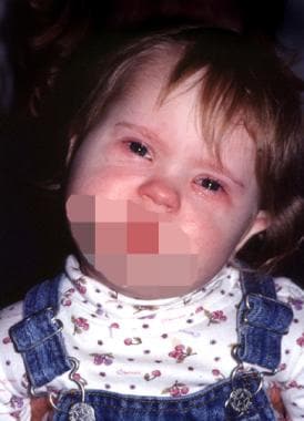
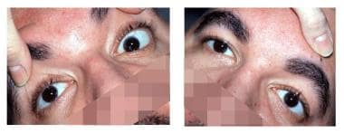
Exophthalmos is a medical condition that refers to the abnormal protrusion or bulging of one or both eyes beyond the normal orbit (eye socket). This condition is also known as proptosis. Exophthalmos can be caused by various factors, including thyroid eye disease (Graves' ophthalmopathy), tumors, inflammation, trauma, or congenital abnormalities. It can lead to various symptoms such as double vision, eye discomfort, redness, and difficulty closing the eyes. Treatment of exophthalmos depends on the underlying cause and may include medications, surgery, or radiation therapy.
Peristalsis is an involuntary muscular movement that occurs in the digestive tract, including the esophagus, stomach, and intestines. It is characterized by alternate contraction and relaxation of the smooth muscles in the walls of these organs, which creates a wave-like motion that helps propel food, fluids, and waste through the digestive system.
The process of peristalsis begins with a narrowing or constriction of the muscle in one area of the digestive tract, followed by a relaxation of the muscle in the adjacent area. This creates a localized contraction that moves along the length of the organ, pushing its contents forward. The wave of contractions continues to move along the digestive tract until it reaches the anus, where waste is eliminated from the body.
Peristalsis plays a crucial role in maintaining proper digestion and absorption of nutrients, as well as in the elimination of waste products from the body. Disorders that affect peristalsis, such as gastrointestinal motility disorders, can lead to symptoms such as abdominal pain, bloating, constipation, or diarrhea.
Esophageal diseases refer to a range of medical conditions that affect the esophagus, which is the muscular tube that connects the throat to the stomach. Here are some common esophageal diseases with their brief definitions:
1. Gastroesophageal reflux disease (GERD): A chronic condition in which stomach acid or bile flows back into the esophagus, causing symptoms such as heartburn, chest pain, and difficulty swallowing.
2. Esophagitis: Inflammation of the esophageal lining, often caused by GERD, infection, or medication.
3. Esophageal stricture: Narrowing of the esophagus due to scarring or inflammation, which can make swallowing difficult.
4. Esophageal cancer: Cancer that forms in the tissues of the esophagus, often as a result of long-term GERD or smoking.
5. Esophageal motility disorders: Disorders that affect the normal movement and function of the esophagus, such as achalasia, diffuse spasm, and nutcracker esophagus.
6. Barrett's esophagus: A condition in which the lining of the lower esophagus changes, increasing the risk of esophageal cancer.
7. Esophageal diverticula: Small pouches that form in the esophageal wall, often causing difficulty swallowing or regurgitation.
8. Eosinophilic esophagitis (EoE): A chronic immune-mediated disorder characterized by inflammation of the esophagus due to an allergic reaction.
These are some of the common esophageal diseases, and their diagnosis and treatment may vary depending on the severity and underlying cause of the condition.
Intestinal pseudo-obstruction, also known as paralytic ileus or functional obstruction, is a gastrointestinal motility disorder characterized by the absence of mechanical obstruction in the intestines, but with symptoms mimicking a mechanical small bowel obstruction. These symptoms may include abdominal distention, cramping, nausea, vomiting, and constipation or difficulty passing stools.
The condition is caused by impaired intestinal motility due to dysfunction of the nerves or muscles that control the movement of food and waste through the digestive system. It can be a chronic or acute condition and may occur as a primary disorder or secondary to other medical conditions, such as surgery, trauma, infections, metabolic disorders, neurological diseases, or certain medications.
Diagnosis of intestinal pseudo-obstruction typically involves imaging studies, such as X-rays or CT scans, to rule out mechanical obstruction and confirm the presence of dilated bowel loops. Manometry and other specialized tests may also be used to assess intestinal motility. Treatment options include medications to stimulate intestinal motility, dietary modifications, and in severe cases, surgery or intravenous nutrition.
The oculomotor nerve, also known as the third cranial nerve (CN III), is a motor nerve that originates from the midbrain. It controls the majority of the eye muscles, including the levator palpebrae superioris muscle that raises the upper eyelid, and the extraocular muscles that enable various movements of the eye such as looking upward, downward, inward, and outward. Additionally, it carries parasympathetic fibers responsible for pupillary constriction and accommodation (focusing on near objects). Damage to this nerve can result in various ocular motor disorders, including strabismus, ptosis, and pupillary abnormalities.
The esophagus is the muscular tube that connects the throat (pharynx) to the stomach. It is located in the midline of the neck and chest, passing through the diaphragm to enter the abdomen and join the stomach. The main function of the esophagus is to transport food and liquids from the mouth to the stomach for digestion.
The esophagus has a few distinct parts: the upper esophageal sphincter (a ring of muscle that separates the esophagus from the throat), the middle esophagus, and the lower esophageal sphincter (another ring of muscle that separates the esophagus from the stomach). The lower esophageal sphincter relaxes to allow food and liquids to enter the stomach and then contracts to prevent stomach contents from flowing back into the esophagus.
The walls of the esophagus are made up of several layers, including mucosa (a moist tissue that lines the inside of the tube), submucosa (a layer of connective tissue), muscle (both voluntary and involuntary types), and adventitia (an outer layer of connective tissue).
Common conditions affecting the esophagus include gastroesophageal reflux disease (GERD), Barrett's esophagus, esophageal cancer, esophageal strictures, and eosinophilic esophagitis.
Ophthalmologic surgical procedures refer to various types of surgeries performed on the eye and its surrounding structures by trained medical professionals called ophthalmologists. These procedures aim to correct or improve vision, diagnose and treat eye diseases or injuries, and enhance the overall health and functionality of the eye. Some common examples of ophthalmologic surgical procedures include:
1. Cataract Surgery: This procedure involves removing a cloudy lens (cataract) from the eye and replacing it with an artificial intraocular lens (IOL).
2. LASIK (Laser-Assisted In Situ Keratomileusis): A type of refractive surgery that uses a laser to reshape the cornea, correcting nearsightedness, farsightedness, and astigmatism.
3. Glaucoma Surgery: Several surgical options are available for treating glaucoma, including laser trabeculoplasty, traditional trabeculectomy, and various drainage device implantations. These procedures aim to reduce intraocular pressure (IOP) and prevent further optic nerve damage.
4. Corneal Transplant: This procedure involves replacing a damaged or diseased cornea with a healthy donor cornea to restore vision and improve the eye's appearance.
5. Vitreoretinal Surgery: These procedures focus on treating issues within the vitreous humor (gel-like substance filling the eye) and the retina, such as retinal detachment, macular holes, or diabetic retinopathy.
6. Strabismus Surgery: This procedure aims to correct misalignment of the eyes (strabismus) by adjusting the muscles responsible for eye movement.
7. Oculoplastic Surgery: These procedures involve reconstructive, cosmetic, and functional surgeries around the eye, such as eyelid repair, removal of tumors, or orbital fracture repairs.
8. Pediatric Ophthalmologic Procedures: Various surgical interventions are performed on children to treat conditions like congenital cataracts, amblyopia (lazy eye), or blocked tear ducts.
These are just a few examples of ophthalmic surgical procedures. The specific treatment plan will depend on the individual's condition and overall health.
Strabismus is a condition of the ocular muscles where the eyes are not aligned properly and point in different directions. One eye may turn inward, outward, upward, or downward while the other one remains fixed and aligns normally. This misalignment can occur occasionally or constantly. Strabismus is also commonly referred to as crossed eyes or walleye. The condition can lead to visual impairments such as amblyopia (lazy eye) and depth perception problems if not treated promptly and effectively, usually through surgery, glasses, or vision therapy.
Gastroparesis is a gastrointestinal disorder that affects the stomach's normal motility, resulting in the delayed emptying of food from the stomach into the small intestine. The term "gastroparesis" literally means "stomach paralysis," although the stomach doesn't actually become paralyzed in this condition. Instead, the muscles of the stomach wall become weakened or damaged, leading to a decrease in their ability to contract and push food through the digestive tract effectively.
The causes of gastroparesis can vary, but some common reasons include diabetes (both type 1 and type 2), viral infections, surgery involving the vagus nerve (which controls stomach muscle contractions), certain medications (such as narcotics, antidepressants, and high blood pressure drugs), gastroesophageal reflux disease (GERD), scleroderma, Parkinson's disease, multiple sclerosis, and Amyloidosis.
Symptoms of gastroparesis may include nausea, vomiting, feeling full quickly after starting to eat, bloating, heartburn, abdominal pain, lack of appetite, and unintended weight loss. These symptoms can significantly impact a person's quality of life and make it difficult for them to maintain proper nutrition.
Diagnosis typically involves a thorough medical history, physical examination, and various tests such as upper endoscopy, gastric emptying studies (such as the scintigraphy scan), and manometry to assess stomach muscle function. Treatment options may include dietary modifications, medications to stimulate stomach contractions or reduce symptoms like nausea and vomiting, botulinum toxin injections, electrical stimulation of the stomach muscles, or, in severe cases, feeding tubes or surgery.
Congenital nystagmus is a type of involuntary eye movement that is present at birth or develops within the first few months of life. It is characterized by rhythmic oscillations or repetitive, rapid movements of the eyes in either horizontal, vertical, or rotatory directions. These movements can impair vision and may be associated with other ocular conditions such as albinism, congenital cataracts, or optic nerve hypoplasia. The exact cause of congenital nystagmus is not fully understood, but it is believed to result from abnormal development or dysfunction in the areas of the brain that control eye movements. In some cases, congenital nystagmus may be inherited as a genetic trait. Treatment options for congenital nystagmus include corrective lenses, prism glasses, surgery, and vision therapy, depending on the underlying cause and severity of the condition.
Ophthalmoplegia is a medical term that refers to the paralysis or weakness of the eye muscles, which can result in double vision (diplopia) or difficulty moving the eyes. It can be caused by various conditions, including nerve damage, muscle disorders, or neurological diseases such as myasthenia gravis or multiple sclerosis. Ophthalmoplegia can affect one or more eye muscles and can be partial or complete. Depending on the underlying cause, ophthalmoplegia may be treatable with medications, surgery, or other interventions.
Gastrointestinal diseases refer to a group of conditions that affect the gastrointestinal (GI) tract, which includes the organs from the mouth to the anus, responsible for food digestion, absorption, and elimination of waste. These diseases can affect any part of the GI tract, causing various symptoms such as abdominal pain, bloating, diarrhea, constipation, nausea, vomiting, and weight loss.
Common gastrointestinal diseases include:
1. Gastroesophageal reflux disease (GERD) - a condition where stomach acid flows back into the esophagus, causing heartburn and other symptoms.
2. Peptic ulcers - sores that develop in the lining of the stomach or duodenum, often caused by bacterial infection or long-term use of nonsteroidal anti-inflammatory drugs (NSAIDs).
3. Inflammatory bowel disease (IBD) - a group of chronic inflammatory conditions of the intestine, including Crohn's disease and ulcerative colitis.
4. Irritable bowel syndrome (IBS) - a functional gastrointestinal disorder characterized by abdominal pain, bloating, and altered bowel habits.
5. Celiac disease - an autoimmune disorder where the ingestion of gluten leads to damage in the small intestine.
6. Diverticular disease - a condition that affects the colon, causing diverticula (small pouches) to form and potentially become inflamed or infected.
7. Constipation - a common gastrointestinal symptom characterized by infrequent bowel movements, hard stools, and difficulty passing stools.
8. Diarrhea - a common gastrointestinal symptom characterized by loose, watery stools and frequent bowel movements.
9. Food intolerances and allergies - adverse reactions to specific foods or food components that can cause various gastrointestinal symptoms.
10. Gastrointestinal infections - caused by bacteria, viruses, parasites, or fungi that can lead to a range of symptoms, including diarrhea, vomiting, and abdominal pain.
Eyelids are the thin folds of skin that cover and protect the front surface (cornea) of the eye when closed. They are composed of several layers, including the skin, muscle, connective tissue, and a mucous membrane called the conjunctiva. The upper and lower eyelids meet at the outer corner of the eye (lateral canthus) and the inner corner of the eye (medial canthus).
The main function of the eyelids is to protect the eye from foreign particles, light, and trauma. They also help to distribute tears evenly over the surface of the eye through blinking, which helps to keep the eye moist and healthy. Additionally, the eyelids play a role in facial expressions and non-verbal communication.
Gastric emptying is the process by which the stomach empties its contents into the small intestine. In medical terms, it refers to the rate and amount of food that leaves the stomach and enters the duodenum, which is the first part of the small intestine. This process is regulated by several factors, including the volume and composition of the meal, hormonal signals, and neural mechanisms. Abnormalities in gastric emptying can lead to various gastrointestinal symptoms and disorders, such as gastroparesis, where the stomach's ability to empty food is delayed.
Sperm motility is the ability of sperm to move actively and effectively through the female reproductive tract towards the egg for fertilization. It is typically measured as the percentage of moving sperm in a sample, and their progressiveness or velocity. Normal human sperm motility is generally defined as forward progression of at least 25 micrometers per second, with at least 50% of sperm showing progressive motility. Reduced sperm motility, also known as asthenozoospermia, can negatively impact fertility and reproductive outcomes.
The enteric nervous system (ENS) is a part of the autonomic nervous system that directly controls the gastrointestinal tract, including the stomach, small intestine, colon, and rectum. It is sometimes referred to as the "second brain" because it can operate independently of the central nervous system (CNS).
The ENS contains around 500 million neurons that are organized into two main plexuses: the myenteric plexus, which lies between the longitudinal and circular muscle layers of the gut, and the submucosal plexus, which is located in the submucosa. These plexuses contain various types of neurons that are responsible for regulating gastrointestinal motility, secretion, and blood flow.
The ENS can communicate with the CNS through afferent nerve fibers that transmit information about the state of the gut to the brain, and efferent nerve fibers that carry signals from the brain back to the ENS. However, the ENS is also capable of functioning independently of the CNS, allowing it to regulate gastrointestinal functions in response to local stimuli such as food intake, inflammation, or infection.
A spasm is a sudden, involuntary contraction or tightening of a muscle, group of muscles, or a hollow organ such as the ureter or bronchi. Spasms can occur as a result of various factors including muscle fatigue, injury, irritation, or abnormal nerve activity. They can cause pain and discomfort, and in some cases, interfere with normal bodily functions. For example, a spasm in the bronchi can cause difficulty breathing, while a spasm in the ureter can cause severe pain and may lead to a kidney stone blockage. The treatment for spasms depends on the underlying cause and may include medication, physical therapy, or lifestyle changes.
A myoelectric complex is a group of electromyographic (EMG) signals that are recorded from muscles during a specific physiological process. These signals can provide information about the electrical activity of the muscle and its functional state.
A migrating myoelectric complex (MMC), also known as a migrating motor complex, is a pattern of muscle contractions that occurs in the gastrointestinal (GI) tract during periods of fasting. These complexes are responsible for cleaning out the GI tract and preparing it for the next meal.
An MMC typically consists of four phases: phase I, which is a period of quiescence; phase II, which is characterized by irregular muscle contractions; phase III, which is a period of strong, rhythmic contractions that sweep through the GI tract; and phase IV, which is a transition phase back to phase I.
The term "migrating" refers to the fact that these complexes move along the GI tract at a rate of about 1-2 cm/min. This allows them to effectively clean out the entire length of the GI tract during periods of fasting.
It is important to note that dysfunction of MMCs has been implicated in various gastrointestinal disorders, such as gastroparesis and irritable bowel syndrome (IBS).
Deglutition disorders, also known as swallowing disorders, are conditions that affect the ability to move food or liquids from the mouth to the stomach safely and efficiently. These disorders can occur at any stage of the swallowing process, which includes oral preparation (chewing and manipulating food in the mouth), pharyngeal phase (activating muscles and structures in the throat to move food toward the esophagus), and esophageal phase (relaxing and contracting the esophagus to propel food into the stomach).
Symptoms of deglutition disorders may include coughing or choking during or after eating, difficulty initiating a swallow, food sticking in the throat or chest, regurgitation, unexplained weight loss, and aspiration (inhaling food or liquids into the lungs), which can lead to pneumonia.
Deglutition disorders can be caused by various factors, such as neurological conditions (e.g., stroke, Parkinson's disease, multiple sclerosis), structural abnormalities (e.g., narrowing or blockage of the esophagus), muscle weakness or dysfunction, and cognitive or behavioral issues. Treatment for deglutition disorders may involve dietary modifications, swallowing exercises, medications, or surgical interventions, depending on the underlying cause and severity of the condition.
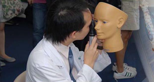
Diagnostic techniques in ophthalmology refer to the various methods and tests used by eye specialists (ophthalmologists) to examine, evaluate, and diagnose conditions related to the eyes and visual system. Here are some commonly used diagnostic techniques:
1. Visual Acuity Testing: This is a basic test to measure the sharpness of a person's vision. It typically involves reading letters or numbers from an eye chart at a specific distance.
2. Refraction Test: This test helps determine the correct lens prescription for glasses or contact lenses by measuring how light is bent as it passes through the cornea and lens.
3. Slit Lamp Examination: A slit lamp is a microscope that allows an ophthalmologist to examine the structures of the eye, including the cornea, iris, lens, and retina, in great detail.
4. Tonometry: This test measures the pressure inside the eye (intraocular pressure) to detect conditions like glaucoma. Common methods include applanation tonometry and non-contact tonometry.
5. Retinal Imaging: Several techniques are used to capture images of the retina, including fundus photography, fluorescein angiography, and optical coherence tomography (OCT). These tests help diagnose conditions like macular degeneration, diabetic retinopathy, and retinal detachments.
6. Color Vision Testing: This test evaluates a person's ability to distinguish between different colors, which can help detect color vision deficiencies or neurological disorders affecting the visual pathway.
7. Visual Field Testing: This test measures a person's peripheral (or side) vision and can help diagnose conditions like glaucoma, optic nerve damage, or brain injuries.
8. Pupillary Reactions Tests: These tests evaluate how the pupils respond to light and near objects, which can provide information about the condition of the eye's internal structures and the nervous system.
9. Ocular Motility Testing: This test assesses eye movements and alignment, helping diagnose conditions like strabismus (crossed eyes) or nystagmus (involuntary eye movement).
10. Corneal Topography: This non-invasive imaging technique maps the curvature of the cornea, which can help detect irregularities, assess the fit of contact lenses, and plan refractive surgery procedures.
Domperidone is a medication that belongs to the class of dopamine antagonists. It works by blocking the action of dopamine, a chemical in the brain that can cause nausea and vomiting. Domperidone is primarily used to treat symptoms of gastroesophageal reflux disease (GERD) and gastric motility disorders, including bloating, fullness, and regurgitation. It works by increasing the contractions of the stomach muscles, which helps to move food and digestive juices through the stomach more quickly.
Domperidone is available in various forms, such as tablets, suspension, and injection. The medication is generally well-tolerated, but it can cause side effects such as dry mouth, diarrhea, headache, and dizziness. In rare cases, domperidone may cause more serious side effects, including irregular heart rhythms, tremors, or muscle stiffness.
It is important to note that domperidone has a risk of causing cardiac arrhythmias, particularly at higher doses and in patients with pre-existing heart conditions. Therefore, it should be used with caution and only under the supervision of a healthcare professional.
Constipation is a condition characterized by infrequent bowel movements or difficulty in passing stools that are often hard and dry. The medical definition of constipation varies, but it is generally defined as having fewer than three bowel movements in a week. In addition to infrequent bowel movements, other symptoms of constipation can include straining during bowel movements, feeling like you haven't completely evacuated your bowels, and experiencing hard or lumpy stools.
Constipation can have many causes, including a low-fiber diet, dehydration, certain medications, lack of physical activity, and underlying medical conditions such as irritable bowel syndrome or hypothyroidism. In most cases, constipation can be treated with lifestyle changes, such as increasing fiber intake, drinking more water, and getting regular exercise. However, if constipation is severe, persistent, or accompanied by other symptoms, it's important to seek medical attention to rule out any underlying conditions that may require treatment.
Visual acuity is a measure of the sharpness or clarity of vision. It is usually tested by reading an eye chart from a specific distance, such as 20 feet (6 meters). The standard eye chart used for this purpose is called the Snellen chart, which contains rows of letters that decrease in size as you read down the chart.
Visual acuity is typically expressed as a fraction, with the numerator representing the testing distance and the denominator indicating the smallest line of type that can be read clearly. For example, if a person can read the line on the eye chart that corresponds to a visual acuity of 20/20, it means they have normal vision at 20 feet. If their visual acuity is 20/40, it means they must be as close as 20 feet to see what someone with normal vision can see at 40 feet.
It's important to note that visual acuity is just one aspect of overall vision and does not necessarily reflect other important factors such as peripheral vision, depth perception, color vision, or contrast sensitivity.
Intestinal diseases refer to a wide range of conditions that affect the function or structure of the small intestine, large intestine (colon), or both. These diseases can cause various symptoms such as abdominal pain, diarrhea, constipation, bloating, nausea, vomiting, and weight loss. They can be caused by infections, inflammation, genetic disorders, or other factors. Some examples of intestinal diseases include inflammatory bowel disease (IBD), irritable bowel syndrome (IBS), celiac disease, Crohn's disease, ulcerative colitis, and intestinal infections. The specific medical definition may vary depending on the context and the specific condition being referred to.
Anti-dyskinetic agents are a class of medications that are used to treat or manage dyskinesias, which are involuntary movements or abnormal muscle contractions. These medications work by blocking or reducing the activity of dopamine, a neurotransmitter in the brain that is involved in movement control.
Dyskinetic symptoms can occur as a side effect of long-term use of levodopa therapy in patients with Parkinson's disease. Anti-dyskinetic agents such as amantadine, anticholinergics, and dopamine agonists may be used to manage these symptoms.
Amantadine works by increasing the release of dopamine and blocking its reuptake, which can help reduce dyskinesias. Anticholinergic medications such as trihexyphenidyl and benztropine work by blocking the action of acetylcholine, another neurotransmitter that can contribute to dyskinesias. Dopamine agonists such as pramipexole and ropinirole mimic the effects of dopamine in the brain and can help reduce dyskinesias by reducing the dose of levodopa required for symptom control.
It is important to note that anti-dyskinetic agents may have side effects, and their use should be carefully monitored by a healthcare provider.
Vision disorders refer to a wide range of conditions that affect the visual system and result in various symptoms, such as blurry vision, double vision, distorted vision, impaired depth perception, and difficulty with visual tracking or focusing. These disorders can be categorized into several types, including:
1. Refractive errors: These occur when the shape of the eye prevents light from focusing directly on the retina, resulting in blurry vision. Examples include myopia (nearsightedness), hyperopia (farsightedness), astigmatism, and presbyopia (age-related loss of near vision).
2. Strabismus: Also known as crossed eyes or walleye, strabismus is a misalignment of the eyes where they point in different directions, which can lead to double vision or loss of depth perception.
3. Amblyopia: Often called lazy eye, amblyopia is a condition where one eye has reduced vision due to lack of proper visual development during childhood. It may be caused by strabismus, refractive errors, or other factors that interfere with normal visual development.
4. Accommodative disorders: These involve problems with the focusing ability of the eyes, such as convergence insufficiency (difficulty focusing on close objects) and accommodative dysfunction (inability to maintain clear vision at different distances).
5. Binocular vision disorders: These affect how the eyes work together as a team, leading to issues like poor depth perception, eye strain, and headaches. Examples include convergence insufficiency, divergence excess, and suppression.
6. Ocular motility disorders: These involve problems with eye movement, such as nystagmus (involuntary eye movements), strabismus, or restricted extraocular muscle function.
7. Visual processing disorders: These affect the brain's ability to interpret and make sense of visual information, even when the eyes themselves are healthy. Symptoms may include difficulty with reading, recognizing shapes and objects, and understanding spatial relationships.
8. Low vision: This term refers to significant visual impairment that cannot be fully corrected with glasses, contact lenses, medication, or surgery. It includes conditions like macular degeneration, diabetic retinopathy, glaucoma, and cataracts.
9. Blindness: Complete loss of sight in both eyes, which can be caused by various factors such as injury, disease, or genetic conditions.
The esophagogastric junction (EGJ) is the region of the gastrointestinal tract where the esophagus (the tube that carries food from the mouth to the stomach) meets the stomach. It serves as a physiological sphincter, which helps control the direction of flow and prevent reflux of gastric contents back into the esophagus. The EGJ is also known as the gastroesophageal junction or cardia.
Deglutition is the medical term for swallowing. It refers to the process by which food or liquid is transferred from the mouth to the stomach through a series of coordinated muscle movements and neural responses. The deglutition process involves several stages, including oral preparatory, oral transit, pharyngeal, and esophageal phases, each of which plays a critical role in ensuring safe and efficient swallowing.
Dysphagia is the medical term for difficulty with swallowing, which can result from various underlying conditions such as neurological disorders, structural abnormalities, or muscular weakness. Proper evaluation and management of deglutition disorders are essential to prevent complications such as aspiration pneumonia, malnutrition, and dehydration.
Smooth muscle, also known as involuntary muscle, is a type of muscle that is controlled by the autonomic nervous system and functions without conscious effort. These muscles are found in the walls of hollow organs such as the stomach, intestines, bladder, and blood vessels, as well as in the eyes, skin, and other areas of the body.
Smooth muscle fibers are shorter and narrower than skeletal muscle fibers and do not have striations or sarcomeres, which give skeletal muscle its striped appearance. Smooth muscle is controlled by the autonomic nervous system through the release of neurotransmitters such as acetylcholine and norepinephrine, which bind to receptors on the smooth muscle cells and cause them to contract or relax.
Smooth muscle plays an important role in many physiological processes, including digestion, circulation, respiration, and elimination. It can also contribute to various medical conditions, such as hypertension, gastrointestinal disorders, and genitourinary dysfunction, when it becomes overactive or underactive.
The myenteric plexus, also known as Auerbach's plexus, is a component of the enteric nervous system located in the wall of the gastrointestinal tract. It is a network of nerve cells (neurons) and supporting cells (neuroglia) that lies between the inner circular layer and outer longitudinal muscle layers of the digestive system's muscularis externa.
The myenteric plexus plays a crucial role in controlling gastrointestinal motility, secretion, and blood flow, primarily through its intrinsic nerve circuits called reflex arcs. These reflex arcs regulate peristalsis (the coordinated muscle contractions that move food through the digestive tract) and segmentation (localized contractions that mix and churn the contents within a specific region of the gut).
Additionally, the myenteric plexus receives input from both the sympathetic and parasympathetic divisions of the autonomic nervous system, allowing for central nervous system regulation of gastrointestinal functions. Dysfunction in the myenteric plexus has been implicated in various gastrointestinal disorders, such as irritable bowel syndrome, achalasia, and intestinal pseudo-obstruction.
Bipolar disorder, also known as manic-depressive illness, is a mental health condition that causes extreme mood swings that include emotional highs (mania or hypomania) and lows (depression). When you become depressed, you may feel sad or hopeless and lose interest or pleasure in most activities. When your mood shifts to mania or hypomania (a less severe form of mania), you may feel euphoric, full of energy, or unusually irritable. These mood swings can significantly affect your job, school, relationships, and overall quality of life.
Bipolar disorder is typically characterized by the presence of one or more manic or hypomanic episodes, often accompanied by depressive episodes. The episodes may be separated by periods of normal mood, but in some cases, a person may experience rapid cycling between mania and depression.
There are several types of bipolar disorder, including:
* Bipolar I Disorder: This type is characterized by the occurrence of at least one manic episode, which may be preceded or followed by hypomanic or major depressive episodes.
* Bipolar II Disorder: This type involves the presence of at least one major depressive episode and at least one hypomanic episode, but no manic episodes.
* Cyclothymic Disorder: This type is characterized by numerous periods of hypomania and depression that are not severe enough to meet the criteria for a full manic or depressive episode.
* Other Specified and Unspecified Bipolar and Related Disorders: These categories include bipolar disorders that do not fit the criteria for any of the other types.
The exact cause of bipolar disorder is unknown, but it appears to be related to a combination of genetic, environmental, and neurochemical factors. Treatment typically involves a combination of medication, psychotherapy, and lifestyle changes to help manage symptoms and prevent relapses.
Gastroesophageal reflux (GER) is the retrograde movement of stomach contents into the esophagus, which can cause discomfort and symptoms. It occurs when the lower esophageal sphincter (a ring of muscle between the esophagus and stomach) relaxes inappropriately, allowing the acidic or non-acidic gastric contents to flow back into the esophagus.
Gastroesophageal reflux becomes gastroesophageal reflux disease (GERD) when it is more severe, persistent, and/or results in complications such as esophagitis, strictures, or Barrett's esophagus. Common symptoms of GERD include heartburn, regurgitation, chest pain, difficulty swallowing, and chronic cough or hoarseness.
The gastrointestinal (GI) tract, also known as the digestive tract, is a continuous tube that starts at the mouth and ends at the anus. It is responsible for ingesting, digesting, absorbing, and excreting food and waste materials. The GI tract includes the mouth, esophagus, stomach, small intestine (duodenum, jejunum, ileum), large intestine (cecum, colon, rectum, anus), and accessory organs such as the liver, gallbladder, and pancreas. The primary function of this system is to process and extract nutrients from food while also protecting the body from harmful substances, pathogens, and toxins.
In anatomical terms, the stomach is a muscular, J-shaped organ located in the upper left portion of the abdomen. It is part of the gastrointestinal tract and plays a crucial role in digestion. The stomach's primary functions include storing food, mixing it with digestive enzymes and hydrochloric acid to break down proteins, and slowly emptying the partially digested food into the small intestine for further absorption of nutrients.
The stomach is divided into several regions, including the cardia (the area nearest the esophagus), the fundus (the upper portion on the left side), the body (the main central part), and the pylorus (the narrowed region leading to the small intestine). The inner lining of the stomach, called the mucosa, is protected by a layer of mucus that prevents the digestive juices from damaging the stomach tissue itself.
In medical contexts, various conditions can affect the stomach, such as gastritis (inflammation of the stomach lining), peptic ulcers (sores in the stomach or duodenum), gastroesophageal reflux disease (GERD), and stomach cancer. Symptoms related to the stomach may include abdominal pain, bloating, nausea, vomiting, heartburn, and difficulty swallowing.
Muscle contraction is the physiological process in which muscle fibers shorten and generate force, leading to movement or stability of a body part. This process involves the sliding filament theory where thick and thin filaments within the sarcomeres (the functional units of muscles) slide past each other, facilitated by the interaction between myosin heads and actin filaments. The energy required for this action is provided by the hydrolysis of adenosine triphosphate (ATP). Muscle contractions can be voluntary or involuntary, and they play a crucial role in various bodily functions such as locomotion, circulation, respiration, and posture maintenance.
In medical terms, pressure is defined as the force applied per unit area on an object or body surface. It is often measured in millimeters of mercury (mmHg) in clinical settings. For example, blood pressure is the force exerted by circulating blood on the walls of the arteries and is recorded as two numbers: systolic pressure (when the heart beats and pushes blood out) and diastolic pressure (when the heart rests between beats).
Pressure can also refer to the pressure exerted on a wound or incision to help control bleeding, or the pressure inside the skull or spinal canal. High or low pressure in different body systems can indicate various medical conditions and require appropriate treatment.
Ciliary motility disorders are a group of rare genetic conditions that affect the function of cilia, which are tiny hair-like structures on the surface of cells in the body. Cilia play an important role in moving fluids and particles across the cell surface, including the movement of mucus and other substances in the respiratory system, the movement of eggs and sperm in the reproductive system, and the movement of fluid in the inner ear.
Ciliary motility disorders are caused by mutations in genes that are responsible for the proper functioning of cilia. These mutations can lead to abnormalities in the structure or function of cilia, which can result in a range of symptoms depending on the specific disorder and the parts of the body that are affected.
Some common symptoms of ciliary motility disorders include recurrent respiratory infections, chronic sinusitis, hearing loss, infertility, and situs inversus, a condition in which the major organs are reversed or mirrored from their normal positions. There are several different types of ciliary motility disorders, including primary ciliary dyskinesia, Kartagener syndrome, and immotile cilia syndrome.
Treatment for ciliary motility disorders typically involves addressing the specific symptoms and underlying causes of the disorder. This may include antibiotics to treat respiratory infections, surgery to correct structural abnormalities, or assisted reproductive technologies to help with infertility.
The colon, also known as the large intestine, is a part of the digestive system in humans and other vertebrates. It is an organ that eliminates waste from the body and is located between the small intestine and the rectum. The main function of the colon is to absorb water and electrolytes from digested food, forming and storing feces until they are eliminated through the anus.
The colon is divided into several regions, including the cecum, ascending colon, transverse colon, descending colon, sigmoid colon, rectum, and anus. The walls of the colon contain a layer of muscle that helps to move waste material through the organ by a process called peristalsis.
The inner surface of the colon is lined with mucous membrane, which secretes mucus to lubricate the passage of feces. The colon also contains a large population of bacteria, known as the gut microbiota, which play an important role in digestion and immunity.
A mental disorder is a syndrome characterized by clinically significant disturbance in an individual's cognition, emotion regulation, or behavior. It's associated with distress and/or impaired functioning in social, occupational, or other important areas of life, often leading to a decrease in quality of life. These disorders are typically persistent and can be severe and disabling. They may be related to factors such as genetics, early childhood experiences, or trauma. Examples include depression, anxiety disorders, bipolar disorder, schizophrenia, and personality disorders. It's important to note that a diagnosis should be made by a qualified mental health professional.
X-ray computed tomography (CT or CAT scan) is a medical imaging method that uses computer-processed combinations of many X-ray images taken from different angles to produce cross-sectional (tomographic) images (virtual "slices") of the body. These cross-sectional images can then be used to display detailed internal views of organs, bones, and soft tissues in the body.
The term "computed tomography" is used instead of "CT scan" or "CAT scan" because the machines take a series of X-ray measurements from different angles around the body and then use a computer to process these data to create detailed images of internal structures within the body.
CT scanning is a noninvasive, painless medical test that helps physicians diagnose and treat medical conditions. CT imaging provides detailed information about many types of tissue including lung, bone, soft tissue and blood vessels. CT examinations can be performed on every part of the body for a variety of reasons including diagnosis, surgical planning, and monitoring of therapeutic responses.
In computed tomography (CT), an X-ray source and detector rotate around the patient, measuring the X-ray attenuation at many different angles. A computer uses this data to construct a cross-sectional image by the process of reconstruction. This technique is called "tomography". The term "computed" refers to the use of a computer to reconstruct the images.
CT has become an important tool in medical imaging and diagnosis, allowing radiologists and other physicians to view detailed internal images of the body. It can help identify many different medical conditions including cancer, heart disease, lung nodules, liver tumors, and internal injuries from trauma. CT is also commonly used for guiding biopsies and other minimally invasive procedures.
In summary, X-ray computed tomography (CT or CAT scan) is a medical imaging technique that uses computer-processed combinations of many X-ray images taken from different angles to produce cross-sectional images of the body. It provides detailed internal views of organs, bones, and soft tissues in the body, allowing physicians to diagnose and treat medical conditions.
Ocular hypertension is a medical condition characterized by elevated pressure within the eye (intraocular pressure or IOP), which is higher than normal but not necessarily high enough to cause any visible damage to the optic nerve or visual field loss. It serves as a significant risk factor for developing glaucoma, a sight-threatening disease.
The normal range of intraocular pressure is typically between 10-21 mmHg (millimeters of mercury). Ocular hypertension is often defined as an IOP consistently above 21 mmHg, although some studies suggest that even pressures between 22-30 mmHg may not cause damage in all individuals. Regular monitoring and follow-up with an ophthalmologist are essential for people diagnosed with ocular hypertension to ensure early detection and management of any potential glaucomatous changes. Treatment options include medications, laser therapy, or surgery to lower the IOP and reduce the risk of glaucoma onset.
Anxiety disorders are a category of mental health disorders characterized by feelings of excessive and persistent worry, fear, or anxiety that interfere with daily activities. They include several different types of disorders, such as:
1. Generalized Anxiety Disorder (GAD): This is characterized by chronic and exaggerated worry and tension, even when there is little or nothing to provoke it.
2. Panic Disorder: This is characterized by recurring unexpected panic attacks and fear of experiencing more panic attacks.
3. Social Anxiety Disorder (SAD): Also known as social phobia, this is characterized by excessive fear, anxiety, or avoidance of social situations due to feelings of embarrassment, self-consciousness, and concern about being judged or viewed negatively by others.
4. Phobias: These are intense, irrational fears of certain objects, places, or situations. When a person with a phobia encounters the object or situation they fear, they may experience panic attacks or other severe anxiety responses.
5. Agoraphobia: This is a fear of being in places where it may be difficult to escape or get help if one has a panic attack or other embarrassing or incapacitating symptoms.
6. Separation Anxiety Disorder (SAD): This is characterized by excessive anxiety about separation from home or from people to whom the individual has a strong emotional attachment (such as a parent, sibling, or partner).
7. Selective Mutism: This is a disorder where a child becomes mute in certain situations, such as at school, but can speak normally at home or with close family members.
These disorders are treatable with a combination of medication and psychotherapy (cognitive-behavioral therapy, exposure therapy). It's important to seek professional help if you suspect that you or someone you know may have an anxiety disorder.
Mood disorders are a category of mental health disorders characterized by significant and persistent changes in mood, affect, and emotional state. These disorders can cause disturbances in normal functioning and significantly impair an individual's ability to carry out their daily activities. The two primary types of mood disorders are depressive disorders (such as major depressive disorder or persistent depressive disorder) and bipolar disorders (which include bipolar I disorder, bipolar II disorder, and cyclothymic disorder).
Depressive disorders involve prolonged periods of low mood, sadness, hopelessness, and a lack of interest in activities. Individuals with these disorders may also experience changes in sleep patterns, appetite, energy levels, concentration, and self-esteem. In severe cases, they might have thoughts of death or suicide.
Bipolar disorders involve alternating episodes of mania (or hypomania) and depression. During a manic episode, individuals may feel extremely elated, energetic, or irritable, with racing thoughts, rapid speech, and impulsive behavior. They might engage in risky activities, have decreased sleep needs, and display poor judgment. In contrast, depressive episodes involve the same symptoms as depressive disorders.
Mood disorders can be caused by a combination of genetic, biological, environmental, and psychological factors. Proper diagnosis and treatment, which may include psychotherapy, medication, or a combination of both, are essential for managing these conditions and improving quality of life.
The eye is the organ of sight, primarily responsible for detecting and focusing on visual stimuli. It is a complex structure composed of various parts that work together to enable vision. Here are some of the main components of the eye:
1. Cornea: The clear front part of the eye that refracts light entering the eye and protects the eye from harmful particles and microorganisms.
2. Iris: The colored part of the eye that controls the amount of light reaching the retina by adjusting the size of the pupil.
3. Pupil: The opening in the center of the iris that allows light to enter the eye.
4. Lens: A biconvex structure located behind the iris that further refracts light and focuses it onto the retina.
5. Retina: A layer of light-sensitive cells (rods and cones) at the back of the eye that convert light into electrical signals, which are then transmitted to the brain via the optic nerve.
6. Optic Nerve: The nerve that carries visual information from the retina to the brain.
7. Vitreous: A clear, gel-like substance that fills the space between the lens and the retina, providing structural support to the eye.
8. Conjunctiva: A thin, transparent membrane that covers the front of the eye and the inner surface of the eyelids.
9. Extraocular Muscles: Six muscles that control the movement of the eye, allowing for proper alignment and focus.
The eye is a remarkable organ that allows us to perceive and interact with our surroundings. Various medical specialties, such as ophthalmology and optometry, are dedicated to the diagnosis, treatment, and management of various eye conditions and diseases.
Ocular toxoplasmosis is an inflammatory eye disease caused by the parasitic infection of Toxoplasma gondii in the eye's retina. It can lead to lesions and scarring in the retina, resulting in vision loss or impairment. The severity of ocular toxoplasmosis depends on the location and extent of the infection in the eye. In some cases, it may cause only mild symptoms, while in others, it can result in severe damage to the eye. Ocular toxoplasmosis is usually treated with medications that target the Toxoplasma gondii parasite, such as pyrimethamine and sulfadiazine, often combined with corticosteroids to reduce inflammation.
The Diagnostic and Statistical Manual of Mental Disorders (DSM) is a publication of the American Psychiatric Association (APA) that provides diagnostic criteria for mental disorders. It is widely used by mental health professionals in the United States and around the world to diagnose and classify mental health conditions.
The DSM includes detailed descriptions of symptoms, clinical examples, and specific criteria for each disorder, which are intended to facilitate accurate diagnosis and improve communication among mental health professionals. The manual is regularly updated to reflect current research and clinical practice, with the most recent edition being the DSM-5, published in 2013.
It's important to note that while the DSM is a valuable tool for mental health professionals, it is not without controversy. Some critics argue that the manual medicalizes normal human experiences and that its categories may be too broad or overlapping. Nonetheless, it remains an essential resource for clinicians, researchers, and policymakers in the field of mental health.
Cell movement, also known as cell motility, refers to the ability of cells to move independently and change their location within tissue or inside the body. This process is essential for various biological functions, including embryonic development, wound healing, immune responses, and cancer metastasis.
There are several types of cell movement, including:
1. **Crawling or mesenchymal migration:** Cells move by extending and retracting protrusions called pseudopodia or filopodia, which contain actin filaments. This type of movement is common in fibroblasts, immune cells, and cancer cells during tissue invasion and metastasis.
2. **Amoeboid migration:** Cells move by changing their shape and squeezing through tight spaces without forming protrusions. This type of movement is often observed in white blood cells (leukocytes) as they migrate through the body to fight infections.
3. **Pseudopodial extension:** Cells extend pseudopodia, which are temporary cytoplasmic projections containing actin filaments. These protrusions help the cell explore its environment and move forward.
4. **Bacterial flagellar motion:** Bacteria use a whip-like structure called a flagellum to propel themselves through their environment. The rotation of the flagellum is driven by a molecular motor in the bacterial cell membrane.
5. **Ciliary and ependymal movement:** Ciliated cells, such as those lining the respiratory tract and fallopian tubes, have hair-like structures called cilia that beat in coordinated waves to move fluids or mucus across the cell surface.
Cell movement is regulated by a complex interplay of signaling pathways, cytoskeletal rearrangements, and adhesion molecules, which enable cells to respond to environmental cues and navigate through tissues.
 Cyclovergence
Stay sutures
Cover test
Worth 4 dot test
Maddox wing
Duane syndrome
Pinhole occluder
Dissociated vertical deviation
Prism fusion range
Prism cover test
Bagolini Striated Glasses Test
Alfred Carl Graefe
Fresnel lens
Blepharoplasty
Robert Ledley
Video-oculography
List of diseases (O)
Orthoptics
Exotropia
Vestibulocerebellar syndrome
Chronic progressive external ophthalmoplegia
Mitochondrial neurogastrointestinal encephalopathy syndrome
Parinaud's syndrome
Hypertropia
Cerebral polyopia
Alan B. Scott
Actin, cytoplasmic 2
Brown's syndrome
Orbital emphysema
Anticholinergic
 Browse subjects: ocular motility | The Online Books Page
Cyclovergence - Wikipedia
 Eye Movement Disorders | Nystagmus | Strabismus | MedlinePlus
 Senataxin, the ortholog of a yeast RNA helicase, is mutant in ataxia-ocular apraxia 2
Long-term neurologic outcome in children with opsoclonus-myoclonus associated with neuroblastoma: a report from the Pediatric...
 Trochlear Nerve Palsy (Fourth Nerve Palsy): Background, History of the Procedure, Problem
 Vision Disorders - Medical Dictionary online-medical-dictionary.org
 Duane syndrome
 Study Orthoptics at UCL - London's Global University
Brain Stem Hemorrhage, Traumatic | Profiles RNS
Neuro-Ophthalmologic Manifestations of Multiple Sclerosis: Practice Essentials, Multiple Sclerosis, Afferent Visual Pathway...
Rinsho Shinkeigaku
 Best Ophthalmologists in Manchester, Greater Manchester - Doctify
Ophthalmologic Manifestations of Myasthenia Gravis: Overview, Patient History, Physical Examination
Consultants - Oxford Eye Hospital
 DailyMed - AMITRIPTYLINE HCL tablet, film coated
 Five regions, five retinopathy screening programmes: a systematic review of how Portugal addresses the challenge | BMC Health...
 Orthoptist - EyeWiki
 Top Tips | Eye News
 Clinical Trial Consultants in North West within Nerve
 Oculogyric crises secondary to lamotrigine overdosage.
 PEPSIC - pepsic.bvsalud.org
 Acute Vision Loss - Eye Disorders - Merck Manuals Professional Edition
Acute Vision Loss - Eye Disorders - MSD Manual Professional Edition
 Stereo vision and strabismus | Eye
 Cataract extraction. Medical search
HuGE Navigator|Genopedia|PHGKB
 DeCS
DeCS
Strabismus4
- Strabismus - a disorder in which the two eyes don't line up in the same direction. (medlineplus.gov)
- Benefiting from blended learning opportunities, you will learn about paediatric ophthalmology, strabismus (ocular misalignment), ocular motility disorders, and other eye and vision health issues and diseases. (ucl.ac.uk)
- Strabismus (eye turn) and Amblyopia (lazy eye) are disorders of binocular vision, resulting in reduced depth perception. (eyeconsee.com)
- The reader can better appreciate the specific strabismus disorders and syndromes. (a-zbookstore.com)
Misalignment4
- Seeing two objects instead of one ( diplopia ) with a binocular view can arise from ocular misalignment caused by lesions of the brainstem and cerebellum. (medscape.com)
- Ocular torticollis refers to a head tilt that is caused by an ocular misalignment. (bvsalud.org)
- A form of ocular misalignment characterized by an excessive convergence of the visual axes, resulting in a "cross-eye" appearance. (ouhsc.edu)
- Adults who develop double vision, or ocular misalignment later in life should be evaluated by a Pediatric Ophthalmologist in order to determine their underlying diagnosis. (kidseyesjax.com)
Ophthalmology1
- Susan Downes is a Consultant Ophthalmologist at Oxford Eye Hospital, Oxford in post since 2000, and Professor of Ophthalmology at the University of Oxford, and specialises in retinal disorders. (ouh.nhs.uk)
Diseases2
- Her specialist interests include inherited retinal dystrophies, age-related macular degeneration, macular diseases including central serous chorioretinopathy, retinal telangiectasia, and toxic retinopathies as well as the impact of ocular diseases on sleep wake/mood. (ouh.nhs.uk)
- Diseases, dysfunctions, or disorders of or located in the iris. (lookformedical.com)
Binocular5
- Orthoptists treat many non-surgical disorders of ocular motility and binocular vision including, but not limited to, amblyopia , convergence insufficiency , and accommodative esotropia . (aao.org)
- These patients may be referred to the orthoptist for sensorimotor evaluations, management of amblyopia , convergence insufficiency , accommodative esotropia , and other non-surgical disorders of ocular motility and binocular vision from the supervising ophthalmologist's practice, or from the medical community. (aao.org)
- Binocular vision, or eye teaming disorders, refer to a variety of conditions in which the eyes drift inward, outward, or upward. (visualperformancecenter.com)
- Anatomic and physiologic illustrations describe normal anatomic and physiologic relations as well as abnormalities in binocular function and ocular alignment. (a-zbookstore.com)
- Step-by-step guides of sensory and motility tests to qualitatively measure binocular vision, ocular alignment and the visual status. (a-zbookstore.com)
Congenital4
- Duane syndrome (DS) is a rare, congenital eye movement disorder most commonly characterized by the inability of the eye to turn out. (bionity.com)
- The term congenital cranial dysinnervation disorder (CCDD) was introduced by Gutowski et al. (uk.com)
- Duane's retraction syndrome (DRS) is a complex congenital eye movement disorder caused by aberrant innervation of the extraocular muscles by axons of brainstem motor neurons. (elsevierpure.com)
- Duane syndrome (DS) is a rare, congenital disorder of eye movement. (medscape.com)
Diagnosis5
- Because patients with MS and other CNS inflammatory disorders who have visual symptoms often seek ophthalmic attention, eye care experts play a vital role in the localization and diagnosis of these conditions. (medscape.com)
- These patients come with very complex ocular motility presentations and require a careful and detailed assessment in order to find the correct diagnosis and arrange appropriate investigations. (uk.com)
- Accurate diagnosis guide for practitioners to understand the disorders of ocular alignment and motility. (a-zbookstore.com)
- Diagnosis of ocular disease must occur at an early age to avoid permanent visual deficits. (kidseyesjax.com)
- Criteria from Diagnostic and Statistical Manual of Mental Disorders, Fifth Edition ( DSM-5 ) should be used in the diagnosis of dementia. (medscape.com)
Manifestations3
- Neuro-ophthalmic manifestations frequently are encountered in persons with multiple sclerosis (MS) and other central nervous system (CNS) inflammatory disorders, including neuromyelitis optica spectrum disorders (NMOSD) and myelin oligodendrocyte glycoprotein antibody associated disease (MOGAD). (medscape.com)
- [ 1 , 2 ] Ninety percent of patients with myasthenia gravis develop ophthalmologic manifestations of the disease, a disorder of neuromuscular transmission characterized by weakness and fatigability of skeletal muscles. (medscape.com)
- Better control of the systemic comorbidities with bone marrow hematopoietic stem cell transplant (HSCT) and enzymatic replacement therapy (ERT) and the increase in life expectancy allowed the identification of new, late-onset ocular manifestations in MPS patients. (eyewiki.org)
Optic2
- Optic neuritis is an inflammatory injury of the optic nerve that causes vision loss, which is common in MS and other CNS inflammatory disorders. (medscape.com)
- The most common (classic or typical features) include corneal clouding, ocular hypertension and glaucoma, retinopathy and optic disc involvement (optic disc swelling or atrophy). (eyewiki.org)
Diplopia2
- Among patients with myasthenia gravis (MG), 75% initially complain of ocular disturbance, mainly ptosis and diplopia. (medscape.com)
- Neurological examination revealed visual impairment, ocular motility disorder and diplopia on looking to the left. (bvsalud.org)
Supranuclear1
- These conditions may be divided into infranuclear, nuclear, and supranuclear disorders. (bvsalud.org)
Hypertension1
- Many of the ocular abnormalities are as a result of long-standing uncontrolled hypertension which caused the stroke. (who.int)
Neuro-ophthalmic disorders2
- The text addresses the unique abilities (and limitations) that each specialty has in the evaluation and management of patients with neuro-ophthalmic disorders. (mhmedical.com)
- Anterior and posterior segments, together with neuro-ophthalmic disorders were found among stroke patients in this study. (who.int)
Pathology2
- We review the pathologic correlates of autonomic dysfunction (cardiac and gastrointestinal), hyposmia, depression, rapid eye movement behavior disorder and dementia in PD For each non-motor clinical feature there is strong evidence to suggest a role for α-synuclein pathology, lending further support for the notion that PD is a multisystem α-synucleinopathy. (elsevierpure.com)
- [2] Ocular pathology is common in all types of MPS, but its frequency, severity and ocular localization vary according to the MPS subtype. (eyewiki.org)
Physiology1
- All-in-one atlas depicting ocular anatomy, sensory physiology, tests of sensory status and ocular alignment. (a-zbookstore.com)
Clinical1
- MG is differentiated into two major clinical forms: ocular MG, in which the patient has predominantly ocular symptoms, and generalized MG, in which the patient develops generalized proximal weakness. (medscape.com)
Cerebral1
- Los trastornos nucleares son originados por enfermedades de los núcleos oculomotores, trocleares o abducens en el TRONCO CEREBRAL. (bvsalud.org)
Corneal1
- [13] In a tertiary centre in Portugal, all 5 patients with MPS type VI presented corneal clouding and this was the first ocular manifestation. (eyewiki.org)
Visual6
- Affected individuals may experience problems with how they see the world (afferent visual pathway symptoms) and/or how smoothly and synchronously their eyes move together (efferent visual pathway disorders). (medscape.com)
- Patients with visual disturbances in the setting of suspected or established multiple sclerosis (MS) or other CNS inflammatory disorder should be cautioned to avoid activities that present a safety risk to themselves or others. (medscape.com)
- Ocular involvement is frequent and may lead to severe visual impairment. (eyewiki.org)
- Ocular features in MPSs are very frequent, many of those represent an early manifestation of MPS and result in significant visual impairment. (eyewiki.org)
- Data was collected with the use of a structured questionnaire divided into four major sections: bio-data, visual history, results of ocular assessment and NEI VFQ-25 quality of life questionnaire. (who.int)
- pathways can produce severe ocular defects such as Data analysis was carried out using Statistical transient monocular visual loss, visual field defect Package for Social Sciences (SPSS) for Windows, 1-3 or ocular motility restrictions. (who.int)
Disease6
- [ 9 ] Hence, patients who keep having strictly ocular symptoms for 3 or more years are unlikely to revert to the generalized aspect of the disease. (medscape.com)
- Nonetheless, the definition of ocular MG proposed by consensus is based on any ocular muscle weakness attributed to MG at a specified point in time and not dependent on the duration of disease. (medscape.com)
- About 50% of patients present solely with ocular symptoms, and about 50-60% of these patients will progress to develop generalized disease. (medscape.com)
- Disorders that feature impairment of eye movements as a primary manifestation of disease. (bvsalud.org)
- Nuclear disorders are caused by disease of the oculomotor, trochlear, or abducens nuclei in the BRAIN STEM. (bvsalud.org)
- Many acquired motility disorders are associated with systemic conditions such as stroke, autoimmune disease, thyroid dysfunction and cancer. (kidseyesjax.com)
Alignment2
- An orthoptist's focus is on the evaluation and non-surgical treatment of disorders of vision, eye movements, and eye alignment in children and adults. (aao.org)
- Even if surgery was performed in childhood, there may be available options for attaining ocular alignment. (kidseyesjax.com)
Toxicity1
- We report four patients with no preexisting movement disorders who developed oculogyric crises secondary to lamotrigine toxicity and had resolution of these crises after dose reduction. (duke.edu)
Symptoms2
- [ 7 ] One study found that, over a mean follow-up period of 17 years, approximately 15-17% of patients with MG had strictly ocular symptoms. (medscape.com)
- Eventually, 90% of patients with MG develop ocular symptoms. (medscape.com)
Cataract1
- Professor Fischer is a practising Consultant Ophthalmologist at the Oxford Eye Hospital with subspecialty interest in vision problems relating to cataract and retinal disorders. (ouh.nhs.uk)
Patients6
- Some evidence shows that steroids or azathioprine might prevent the conversion to generalized MG in 75% of patients with ocular MG. (medscape.com)
- Patients with major depressive disorder (MDD), both adult and pediatric, may experience worsening of their depression and/or the emergence of suicidal ideation and behavior (suicidality) or unusual changes in behavior, whether or not they are taking antidepressant medications, and this risk may persist until significant remission occurs. (nih.gov)
- In 70% of patients, DS is the only disorder the individual has. (medscape.com)
- Expertise in motility patterns is required to properly evaluate and manage such patients. (kidseyesjax.com)
- Ocular disorders among stroke patients. (who.int)
- Thirty-nine patients (47.0%) were dearth of information on the pattern of ocular between ages of 51 and 60, while 1 (1.2%) was disorders in patients with stroke. (who.int)
Movement4
- There are many kinds of eye movement disorders. (medlineplus.gov)
- Some eye movement disorders are present at birth. (medlineplus.gov)
- There is no cure for some kinds of eye movement disorders, such as most kinds of nystagmus. (medlineplus.gov)
- The rapid eye movement (REM) sleep behavior disorder (RBD) is a parasomnia characterized by dream-enacting behaviors related to the loss of the normal generalized skeletal muscle atonia during REM sleep, and shows REM sleep without atonia (RWA) during polysomnography (PSG). (go.jp)
Surgical1
- Pediatric Ophthalmologists specialize in disorders of ocular motility and can offer both surgical and non-surgical treatment options. (kidseyesjax.com)
Efferent1
- Summary of the neuro-ophthalmic history and examination in Section I. This allows a comprehensive overview of the history and examination initially ( Chapter 1 ), further explored in detail in the sections devoted to afferent disorders ( Chapter 2 ) and efferent disorders ( Chapter 7 ). (mhmedical.com)
Behavior1
- Antidepressants increased the risk compared to placebo of suicidal thinking and behavior (suicidality) in children, adolescents, and young adults in short-term studies of major depressive disorder (MDD) and other psychiatric disorders. (nih.gov)
Diagnostic1
- Figure 1: Bilateral INO Introduction Complex ocular motility disorders are a diagnostic challenge. (uk.com)
Commonly1
- Chapter 6 discusses and illustrates elusive and commonly misdiagnosed disorders. (mhmedical.com)
Vision3
- Clarity or sharpness of OCULAR VISION or the ability of the eye to see fine details. (lookformedical.com)
- Vision Disorders -- Collected Works. (hacettepe.edu.tr)
- The aim is to identify signs of potentially vision- or life-threatening disorders of the central nervous system. (hku.hk)
INJURY1
- Multiple Sclerosis (MS) is a CNS disorder that is characterized by both inflammatory and neurodegenerative mechanisms of brain and spinal cord injury. (medscape.com)