Odontogenic Cysts
Ameloblastoma
Jaw Neoplasms
Odontogenic Cyst, Calcifying
Odontoma
Odontogenic Tumor, Squamous
Dentigerous Cyst
Radicular Cyst
Gingival Neoplasms
Jaw Diseases
Maxillary Diseases
Radiography, Panoramic
Tooth, Impacted
Adenomatoid Tumor
Myxoma
Odontogenesis
Focal Infection, Dental
Basal Cell Nevus Syndrome
Mandibular Diseases
Fibroma, Ossifying
Periodontal Cyst
Periodontal Abscess
Mandibular Prosthesis Implantation
Maxillary Sinus Neoplasms
Photomicrography
Tumor Markers, Biological
Mandible
Root Resorption
Neoplasms, Multiple Primary
Keratins
Retrospective Studies
Biological Specimen Banks
Immunohistochemistry
Cemento-ossifying fibroma presenting as a mass of the parapharyngeal and masticator space. (1/178)
We report a case of cemento-ossifying fibroma that presented as a large extraosseous mass in the masticator and parapharyngeal space. CT scanning and MR imaging showed a large extraosseous mass with central conglomerated, well-matured ossified nodules and fatty marrow. The central matured ossified nodules were of low density on CT scans and high signal intensity on T1- and T2-weighted MR images. Multiplanar reformatted CT scans revealed the origin of the mass to be at the extraction site of the right lower second molar tooth. (+info)CT and MR imaging appearances of an extraosseous calcifying epithelial odontogenic tumor (Pindborg tumor). (2/178)
We herein report a rare case of extraosseous calcifying epithelial odontogenic tumor with local aggressive behavior. CT and MR imaging showed the distinctive appearances of this histologic entity. We briefly discuss the radiologic features of calcifying epithelial odontogenic tumor and the relevant literature. (+info)Amyloid-producing odontogenic tumor in a Shih-Tzu dog. (3/178)
A 9-month-old male Shih-Tzu dog had a right mandibular tumor composed of strands, or nest-like proliferation of epithelial cells with abundant fibrous stroma characterized by spheroid to large nodular deposition of amyloid with Congo-red stain. Globule calcification was also seen throughout the tumor tissue and the spheroid depositions often had a concentrically laminated structure (Liesegang rings). The case was diagnosed as amyloid-producing odontogenic tumor in a dog. (+info)Aggressive epithelial odontogenic ghost cell tumor in the mandible: CT and MR imaging findings. (4/178)
We report a case of aggressive epithelial odontogenic ghost cell tumor arising from the mandible in a 32-year-old man. On CT and MR studies, the tumor was seen as a large, heterogeneous soft-tissue mass that caused marked destruction of the mandible and invaded the mouth floor and tongue base. The tumor displayed a variety of densities and signal intensities on CT and MR images, which correlated well with the degree of cellularity of epithelial islands, abundance of ghost cells and eosinophilic materials, calcification, and cystic areas on histologic sections. Owing to the unpredictable biological behavior of this type of tumor, careful, long-term follow-up is highly recommended. (+info)Combined benign odontogenic tumors: CT and MR findings and histomorphologic evaluation. (5/178)
SUMMARY: Calcifying epithelial odontogenic tumors and calcifying odontogenic cysts are rare, benign odontogenic tumors. We report two cases of an exceptional combination of these tumors with either an ameloblastic fibroodontoma or an odontoma. (+info)Ameloblastic fibroma of the anterior maxilla presenting as a complication of tooth eruption: a case report. (6/178)
Ameloblastic fibroma is a rare mixed odontogenic tumour, which is extremely uncommon in the anterior maxillary region. A case report is presented where failure of eruption of an upper central incisor was the presenting feature. (+info)Benign cementoblastoma: a case report. (7/178)
The case of a 23-year-old with a benign cementoblastoma is presented. The clinicopathologic features, treatment and prognosis are discussed and a brief review of the literature is presented. Although this neoplasm is rare, the dental practitioner should be aware of the clinical features that will lead to its early diagnosis and treatment. (+info)Clear cell odontogenic carcinoma. Report of two cases and review of the literature. (8/178)
This study reviews the literature and reports on the morphologic and immunophenotypic features of 2 clear cell odontogenic carcinomas occurring in the mandible of elderly women, showing extensive infiltration into adjacent tissues. The tumor cells were large, with clear cytoplasm, and arranged in irregular sheets. Some of the latter demonstrated a peripheral rim of cells with eosinophilic cytoplasm or included duct-like structures. There was no evidence of ameloblastic differentiation. Most cells contained glycogen granules and were immunoreactive for cytokeratins and epithelial membrane antigen. In the differential diagnosis other clear cell odontogenic, salivary gland, and metastatic tumors should be considered. Both cases were treated with surgical excision, and the patients are free of disease after 3 and 5 years, respectively. In the literature, however, variable behavior of these tumors has been reported, including recurrence and metastases. It is recommended that terms such as clear cell ameloblastoma and clear cell odontogenic tumor not be used to describe such tumors. (+info)Odontogenic tumors are a group of neoplasms that originate from the dental tissues or their remnants, including the odontogenic epithelium, ectomesenchyme, and/or their derivatives. These tumors can be benign or malignant and may affect the jaw bones and surrounding structures. They can cause various symptoms, such as swelling, pain, loosening of teeth, and altered bite. The classification of odontogenic tumors includes a wide range of entities with different biological behaviors, clinical features, and treatment approaches. Accurate diagnosis is essential for proper management and prognosis.
Odontogenic cysts are a type of cyst that originates from the dental tissues or odontogenic apparatus. They are typically found in the jawbones, and can be classified as developmental or inflammatory in origin. Developmental odontogenic cysts arise from remnants of the tooth-forming structures, while inflammatory odontogenic cysts result from an infection or injury to a tooth.
The most common types of odontogenic cysts include:
1. Periapical cyst - an inflammatory cyst that forms at the tip of the root of a dead or non-vital tooth.
2. Dentigerous cyst - a developmental cyst that surrounds the crown of an unerupted or impacted tooth.
3. Follicular cyst - a type of dentigerous cyst that forms around the crown of an unerupted wisdom tooth.
4. Odontogenic keratocyst - a developmental cyst that arises from the dental lamina and has a high recurrence rate.
5. Lateral periodontal cyst - a rare, developmental cyst that forms in the periodontal ligament of a vital tooth.
Odontogenic cysts can cause various symptoms such as swelling, pain, or numbness in the affected area. They may also displace or resorb adjacent teeth. Diagnosis is typically made through radiographic imaging and histopathological examination of tissue samples obtained through biopsy. Treatment options include surgical excision, marsupialization (a procedure that creates an opening between the cyst and oral cavity), or enucleation (removal of the cyst lining).
Ameloblastoma is a slow-growing, non-cancerous tumor that develops in the jawbone, typically in the lower jaw. It originates from the cells that form the enamel (the hard, outer surface of the teeth). This tumor can cause swelling, pain, and displacement or loosening of teeth. In some cases, it may also lead to fractures of the jawbone.
There are different types of ameloblastomas, including solid or multicystic, unicystic, and peripheral ameloblastoma. Treatment usually involves surgical removal of the tumor, with careful monitoring to ensure that it does not recur. In rare cases, more aggressive treatment may be necessary if the tumor is large or has invaded surrounding tissues.
It's important to note that while ameloblastomas are generally benign, they can still cause significant morbidity and should be treated promptly by an oral and maxillofacial surgeon or other qualified healthcare professional.
Jaw neoplasms refer to abnormal growths or tumors in the jawbone (mandible) or maxilla (upper jaw). These growths can be benign (non-cancerous) or malignant (cancerous). Benign neoplasms are not considered life-threatening, but they can still cause problems by invading nearby tissues and causing damage. Malignant neoplasms, on the other hand, can spread to other parts of the body and can be life-threatening if not treated promptly and effectively.
Jaw neoplasms can present with various symptoms such as swelling, pain, loose teeth, numbness or tingling in the lips or tongue, difficulty chewing or swallowing, and jaw stiffness or limited movement. The diagnosis of jaw neoplasms typically involves a thorough clinical examination, imaging studies such as X-rays, CT scans, or MRI, and sometimes a biopsy to determine the type and extent of the tumor.
Treatment options for jaw neoplasms depend on several factors, including the type, size, location, and stage of the tumor, as well as the patient's overall health and medical history. Treatment may involve surgery, radiation therapy, chemotherapy, or a combination of these modalities. Regular follow-up care is essential to monitor for recurrence or metastasis (spread) of the neoplasm.
Mandibular neoplasms refer to abnormal growths or tumors that develop in the mandible, which is the lower jawbone. These growths can be benign (non-cancerous) or malignant (cancerous). Benign neoplasms are typically slow-growing and rarely spread to other parts of the body, while malignant neoplasms can invade surrounding tissues and may metastasize (spread) to distant sites.
Mandibular neoplasms can have various causes, including genetic mutations, exposure to certain chemicals or radiation, and infection with certain viruses. The symptoms of mandibular neoplasms may include swelling or pain in the jaw, difficulty chewing or speaking, numbness in the lower lip or chin, loose teeth, and/or a lump or mass in the mouth or neck.
The diagnosis of mandibular neoplasms typically involves a thorough clinical examination, imaging studies such as X-rays, CT scans, or MRI scans, and sometimes a biopsy to confirm the type and extent of the tumor. Treatment options depend on the type, stage, and location of the neoplasm, and may include surgery, radiation therapy, chemotherapy, or a combination of these approaches. Regular follow-up care is essential to monitor for recurrence or metastasis.
An Odontogenic Cyst, Calcifying is a specific type of cyst that originates from the dental tissues. It's also known as a calcifying odontogenic cyst or Gorlin cyst. This cyst is characterized by the presence of calcified structures within its lining.
The calcifications can appear as flecks or more complex structures, such as teeth-like formations. The lining of this cyst often contains ghost cells, which are the remains of epithelial cells that have undergone calcification.
These cysts are typically slow-growing and asymptomatic, although they can sometimes cause swelling or pain if they become large enough to compress adjacent tissues. They are most commonly found in the jaw bones, particularly the mandible.
While the exact cause of calcifying odontogenic cysts is not fully understood, they are thought to arise from developmental abnormalities in the tissues that form teeth. Treatment typically involves surgical removal of the cyst.
Maxillary neoplasms refer to abnormal growths or tumors in the maxilla, which is the upper jaw bone. These growths can be benign (non-cancerous) or malignant (cancerous). Benign neoplasms are slow-growing and do not spread to other parts of the body, while malignant neoplasms can invade surrounding tissues and spread to distant sites.
Maxillary neoplasms can cause various symptoms such as swelling, pain, numbness, loose teeth, or difficulty in chewing or swallowing. They may also cause nasal congestion, nosebleeds, or visual changes if they affect the eye or orbit. The diagnosis of maxillary neoplasms usually involves a combination of clinical examination, imaging studies such as CT or MRI scans, and biopsy to determine the type and extent of the tumor.
Treatment options for maxillary neoplasms depend on several factors, including the type, size, location, and stage of the tumor, as well as the patient's overall health and preferences. Treatment may include surgery, radiation therapy, chemotherapy, or a combination of these modalities. Regular follow-up care is essential to monitor for recurrence or metastasis and ensure optimal outcomes.
Odontoma is a type of odontogenic tumor, which means it arises from the tissues that form teeth. It is considered a benign or non-cancerous tumor and is typically slow-growing. Odontomas are usually asymptomatic and are often discovered on routine dental X-rays or during procedures such as wisdom tooth removal.
Odontomas can be classified into two types: complex and compound. Complex odontomas are composed of a haphazard mixture of dental tissue, including enamel, dentin, and cementum, while compound odontomas contain small tooth-like structures called denticles.
These tumors typically occur in the posterior region of the jaw, and while they are usually asymptomatic, some patients may experience symptoms such as swelling, pain, or displacement of teeth. Treatment for odontomas typically involves surgical removal of the tumor.
An odontogenic tumor, squamous type, refers to a specific category of oral tumors that originate from the dental tissues. These tumors are characterized by the abnormal growth of squamous epithelial cells, which are normally found in the outermost layer of the skin and the mucous membranes, including those inside the mouth.
Odontogenic tumors can arise from various components of the dental tissues, such as the odontoblasts, dentin, enamel, cementum, or the epithelial rests of Malassez. The squamous type of odontogenic tumor is typically classified under either one of the following entities:
1. Squamous Odontogenic Tumor (SOT): This is a rare, benign (non-cancerous) neoplasm that primarily affects the tooth-bearing areas of the jaws. SOT is composed of well-differentiated squamous epithelial cells arranged in nests, strands, or sheets, often surrounded by a fibrous stroma. This tumor typically occurs in adults during their third to fifth decades of life and has a slight female predilection.
2. Ameloblastoma with Squamous Metaplasia: Ameloblastoma is a more common odontogenic tumor that usually affects the mandible (lower jaw). In some cases, ameloblastomas may undergo squamous metaplasia, where the original epithelial cells transform into squamous epithelial cells. This variant of ameloblastoma is still considered a benign neoplasm; however, it has a higher recurrence rate compared to conventional ameloblastomas.
It is essential to differentiate these entities from other oral lesions and malignancies through histopathological examination, as the treatment and prognosis may vary depending on the specific diagnosis.
A dentigerous cyst is a type of odontogenic cyst that forms around the crown of an unerupted tooth. It is typically slow-growing and often asymptomatic, but it can cause displacement or resorption of adjacent teeth if it becomes large enough. Dentigerous cysts are more common in permanent teeth than primary teeth, and they are more likely to occur in the mandible (lower jaw) than the maxilla (upper jaw). They are usually diagnosed through radiographic examination and can be treated by surgical removal of the cyst along with the affected tooth. If left untreated, dentigerous cysts can continue to grow and may eventually develop into a tumor or cancer.
A radicular cyst is a type of dental cyst that forms around the root of a tooth, usually as a result of chronic infection or inflammation. It is also known as a periapical cyst. The cyst develops from the accumulation of fluid and cells in the periodontal ligament, which is the tissue that connects the tooth to the jawbone.
Radicular cysts are often caused by untreated dental caries or trauma to the tooth that allows bacteria to enter the pulp chamber of the tooth and cause an infection. Over time, the infection can spread to the surrounding tissues, leading to the formation of a cyst. Symptoms of a radicular cyst may include pain, swelling, and tenderness in the affected area. Treatment typically involves removing the affected tooth and the cyst through a surgical procedure.
Gingival neoplasms refer to abnormal growths or tumors that occur in the gingiva, which are the part of the gums that surround the teeth. These growths can be benign (non-cancerous) or malignant (cancerous). Benign neoplasms include conditions such as fibromas, papillomas, and hemangiomas, while malignant neoplasms are typically squamous cell carcinomas.
Gingival neoplasms can present with a variety of symptoms, including swelling, bleeding, pain, and loose teeth. They may also cause difficulty with chewing, speaking, or swallowing. The exact cause of these neoplasms is not always known, but risk factors include tobacco use, alcohol consumption, poor oral hygiene, and certain viral infections.
Diagnosis of gingival neoplasms typically involves a thorough clinical examination, including a dental exam and biopsy. Treatment options depend on the type and stage of the neoplasm, but may include surgery, radiation therapy, chemotherapy, or a combination of these approaches. Regular dental check-ups and good oral hygiene practices can help to detect gingival neoplasms at an early stage and improve treatment outcomes.
Jaw diseases refer to a variety of conditions that affect the temporomandibular joint (TMJ) and the surrounding muscles, as well as dental disorders that can impact the jaw. Some common examples include:
1. Temporomandibular Joint Disorders (TMD): These are problems with the TMJ and the muscles that control jaw movement. Symptoms may include pain, clicking or popping sounds, and limited movement of the jaw.
2. Osteonecrosis of the Jaw: This is a condition where bone in the jaw dies due to lack of blood supply. It can be caused by radiation therapy, chemotherapy, or certain medications.
3. Dental Cavities: These are holes in the teeth caused by bacteria. If left untreated, they can cause pain, infection, and damage to the jawbone.
4. Periodontal Disease: This is an infection of the gums and bones that support the teeth. Advanced periodontal disease can lead to loss of teeth and damage to the jawbone.
5. Jaw Fractures: These are breaks in the jawbone, often caused by trauma.
6. Oral Cancer: This is a type of cancer that starts in the mouth or throat. If not treated early, it can spread to the jaw and other parts of the body.
7. Cysts and Tumors: These are abnormal growths in the jawbone or surrounding tissues. While some are benign (non-cancerous), others can be malignant (cancerous).
8. Osteomyelitis: This is an infection of the bone, often occurring in the lower jaw. It can cause pain, swelling, and fever.
9. Oral Thrush: This is a fungal infection that causes white patches on the inside of the mouth. If left untreated, it can spread to the jaw and other parts of the body.
10. Sinusitis: Inflammation of the sinuses can sometimes cause pain in the upper jaw.
Maxillary diseases refer to conditions that affect the maxilla, which is the upper bone of the jaw. This bone plays an essential role in functions such as biting, chewing, and speaking, and also forms the upper part of the oral cavity, houses the upper teeth, and supports the nose and the eyes.
Maxillary diseases can be caused by various factors, including infections, trauma, tumors, congenital abnormalities, or systemic conditions. Some common maxillary diseases include:
1. Maxillary sinusitis: Inflammation of the maxillary sinuses, which are air-filled cavities located within the maxilla, can cause symptoms such as nasal congestion, facial pain, and headaches.
2. Periodontal disease: Infection and inflammation of the tissues surrounding the teeth, including the gums and the alveolar bone (which is part of the maxilla), can lead to tooth loss and other complications.
3. Maxillary fractures: Trauma to the face can result in fractures of the maxilla, which can cause pain, swelling, and difficulty breathing or speaking.
4. Maxillary cysts and tumors: Abnormal growths in the maxilla can be benign or malignant and may require surgical intervention.
5. Oral cancer: Cancerous lesions in the oral cavity, including the maxilla, can cause pain, swelling, and difficulty swallowing or speaking.
Treatment for maxillary diseases depends on the specific condition and its severity. Treatment options may include antibiotics, surgery, radiation therapy, or chemotherapy. Regular dental check-ups and good oral hygiene practices can help prevent many maxillary diseases.
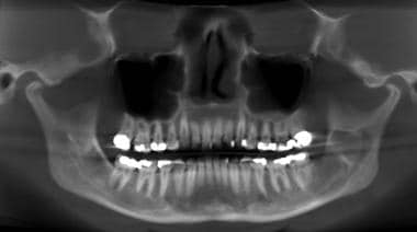
Panoramic radiography is a specialized type of dental X-ray imaging that captures a panoramic view of the entire mouth, including the teeth, upper and lower jaws, and surrounding structures. It uses a special machine that rotates around the head, capturing images as it moves. This technique provides a two-dimensional image that is helpful in diagnosing and planning treatment for various dental conditions such as impacted teeth, bone abnormalities, and jaw disorders.
The panoramic radiograph can also be used to assess the development and positioning of wisdom teeth, detect cysts or tumors in the jaws, and evaluate the effects of trauma or injury to the mouth. It is a valuable tool for dental professionals as it allows them to see a comprehensive view of the oral structures, which may not be visible with traditional X-ray techniques.
It's important to note that while panoramic radiography provides valuable information, it should be used in conjunction with other diagnostic tools and clinical examinations to ensure accurate diagnosis and treatment planning.
An impacted tooth is a condition where a tooth fails to erupt into the oral cavity within its expected time frame, resulting in its partial or complete entrapment within the jawbone or soft tissues. This commonly occurs with wisdom teeth (third molars) but can affect any tooth. Impacted teeth may cause problems such as infection, decay of adjacent teeth, gum disease, or cyst formation, and they may require surgical removal.
A Follicular Cyst is a type of cyst that forms within a follicle, which is the sac-like structure in the skin that contains and protects a hair root. In particular, it refers to a specific condition in the ovary where a follicle fails to rupture or release an egg after maturation, instead continuing to grow and fill with fluid, forming a cyst. These cysts are usually asymptomatic but can become large and cause symptoms such as pelvic pain or discomfort, irregular menstrual cycles, or abnormal vaginal bleeding. In most cases, follicular cysts resolve on their own within 2-3 menstrual cycles, but in rare cases, they may require medical intervention if they become complicated or do not resolve.
A fibroma is a benign (non-cancerous) tumor that consists primarily of fibrous or connective tissue. It can occur in various parts of the body, including the skin, mouth, and internal organs. The term "fibroma" is often used to describe any benign fibrous growth, but there are specific types of fibromas such as dermatofibroma (found in the skin), oral fibroma (found in the mouth), and benign fibrous histiocytoma (found in soft tissues).
It's important to note that while fibromas are generally harmless, they can cause discomfort or problems depending on their size and location. If a fibroma is causing issues or there's concern about its growth or malignancy, it should be evaluated by a healthcare professional for potential removal or further assessment.
An adenomatoid tumor is a benign (non-cancerous) neoplasm that typically arises in the serosal surfaces of the reproductive organs, such as the epididymis in men and the fallopian tube or uterus in women. These tumors are composed of epithelioid cells arranged in tubules, glands, or cysts, and they can sometimes be mistaken for malignant tumors due to their gross appearance. However, adenomatoid tumors are generally slow-growing and do not spread to other parts of the body. They are usually treated with surgical excision and have an excellent prognosis.
A myxoma is a type of benign (non-cancerous) tumor that develops in the heart, specifically in the heart's chambers or valves. It is the most common primary cardiac tumor in adults and typically affects the left atrium. Myxomas are composed of gelatinous, mucoid material and may have a stalk-like attachment to the endocardium (the inner lining of the heart).
Myxomas can vary in size and may cause symptoms such as shortness of breath, fatigue, chest pain, coughing, and fever. These symptoms are due to obstruction of blood flow within the heart or embolization (detachment and travel) of tumor fragments to other parts of the body. Surgical removal is usually required to treat myxomas, as they can lead to serious complications if left untreated.
Odontogenesis is the process of tooth development that involves the formation and calcification of teeth. It is a complex process that requires the interaction of several types of cells, including epithelial cells, mesenchymal cells, and odontoblasts. The process begins during embryonic development with the formation of dental lamina, which gives rise to the tooth bud. As the tooth bud grows and differentiates, it forms the various structures of the tooth, including the enamel, dentin, cementum, and pulp. Odontogenesis is completed when the tooth erupts into the oral cavity. Abnormalities in odontogenesis can result in developmental dental anomalies such as tooth agenesis, microdontia, or odontomas.
A focal infection is a focus or source of infection that can spread and cause harm to other parts of the body. A "focal infection, dental" refers to an infection that originates in the teeth or surrounding tissues of the mouth and then spreads to other parts of the body. This can occur when bacteria or other pathogens from a dental infection enter the bloodstream and travel to distant sites, where they can cause inflammation, tissue damage, and illness.
Dental focal infections can be caused by various conditions, such as tooth decay, periodontal disease, abscesses, or other oral infections. The bacteria involved in dental infections are often part of the normal oral flora but can become pathogenic under certain circumstances, such as when they gain access to deeper tissues or the bloodstream due to trauma, surgery, or poor oral hygiene.
If left untreated, dental focal infections can lead to serious health complications, including heart disease, brain abscesses, and other systemic infections. It is essential to maintain good oral hygiene and seek professional dental care to prevent and treat dental infections, reducing the risk of developing focal infections and related health issues.
Basal Cell Nevus Syndrome (BCNS), also known as Gorlin-Goltz Syndrome, is a rare genetic disorder that is characterized by the development of multiple basal cell carcinomas (BCCs), which are skin cancer tumors that arise from the basal cells in the outermost layer of the skin.
The syndrome is caused by mutations in the PTCH1 gene, which regulates the hedgehog signaling pathway involved in embryonic development and tissue growth regulation. The condition is inherited in an autosomal dominant manner, meaning that a child has a 50% chance of inheriting the mutated gene from an affected parent.
Individuals with BCNS typically develop hundreds to thousands of BCCs over their lifetime, often beginning in childhood or adolescence. They may also have other benign and malignant tumors, such as medulloblastomas (brain tumors), fibromas, and rhabdomyosarcomas.
Additional features of BCNS can include:
1. Facial abnormalities, such as a broad nasal bridge, widely spaced eyes, and pits or depressions on the palms and soles.
2. Skeletal abnormalities, such as spine deformities, rib anomalies, and jaw cysts.
3. Developmental delays and intellectual disabilities in some cases.
4. Increased risk of other cancers, including breast, ovarian, and lung cancer.
Early detection and management of BCCs and other tumors are crucial for individuals with BCNS to prevent complications and improve their quality of life. Regular dermatological examinations, sun protection measures, and surgical removal of tumors are common treatment approaches.
Mandibular diseases refer to conditions that affect the mandible, or lower jawbone. These diseases can be classified as congenital (present at birth) or acquired (developing after birth). They can also be categorized based on the tissues involved, such as bone, muscle, or cartilage. Some examples of mandibular diseases include:
1. Mandibular fractures: These are breaks in the lower jawbone that can result from trauma or injury.
2. Osteomyelitis: This is an infection of the bone and surrounding tissues, which can affect the mandible.
3. Temporomandibular joint (TMJ) disorders: These are conditions that affect the joint that connects the jawbone to the skull, causing pain and limited movement.
4. Mandibular tumors: These are abnormal growths that can be benign or malignant, and can develop in any of the tissues of the mandible.
5. Osteonecrosis: This is a condition where the bone tissue dies due to lack of blood supply, which can affect the mandible.
6. Cleft lip and palate: This is a congenital deformity that affects the development of the face and mouth, including the lower jawbone.
7. Mandibular hypoplasia: This is a condition where the lower jawbone does not develop properly, leading to a small or recessed chin.
8. Developmental disorders: These are conditions that affect the growth and development of the mandible, such as condylar hyperplasia or hemifacial microsomia.
A fibroma, ossifying is a benign (non-cancerous) tumor that typically develops in the periodontal ligament, which is the tissue that connects the tooth to the jawbone. This type of fibroma is characterized by the formation of bone-like tissue within the tumor. It usually appears as a firm, slow-growing nodule or mass that can cause pain or discomfort, particularly when biting down on the affected tooth.
The exact cause of ossifying fibromas is not well understood, but they are thought to arise from an overgrowth of cells in the periodontal ligament. They are more common in women than men and typically occur in people between the ages of 20 and 40. Treatment usually involves surgical removal of the tumor, along with any affected tissue or teeth. In some cases, recurrence may occur, so regular follow-up appointments with a dental professional are recommended.
A periodontal cyst, also known as a radicular cyst or dental cyst, is a type of odontogenic cyst that forms from the tissue of the periodontium, which surrounds and supports the teeth. It typically develops at the apex (tip) of a dead or non-vital tooth root and is filled with fluid. The cyst can grow slowly and painlessly, often going unnoticed until it becomes quite large or causes symptoms such as swelling, tenderness, or tooth mobility.
Periodontal cysts are usually asymptomatic and are often discovered during routine dental x-rays. If left untreated, they can eventually lead to the destruction of surrounding bone and tissue, potentially causing teeth to become loose or even fall out. Treatment typically involves surgical removal of the cyst along with the affected tooth, followed by careful monitoring to ensure that the cyst does not recur.
Cyst fluid refers to the fluid accumulated within a cyst, which is a closed sac-like or capsular structure, typically filled with liquid or semi-solid material. Cysts can develop in various parts of the body for different reasons, and the composition of cyst fluid may vary depending on the type of cyst and its location.
In some cases, cyst fluid might contain proteins, sugars, hormones, or even cells from the surrounding tissue. Infected cysts may have pus-like fluid, while cancerous or precancerous cysts might contain abnormal cells or tumor markers. The analysis of cyst fluid can help medical professionals diagnose and manage various medical conditions, including infections, inflammatory diseases, genetic disorders, and cancers.
It is important to note that the term 'cyst fluid' generally refers to the liquid content within a cyst, but the specific composition and appearance of this fluid may vary significantly depending on the underlying cause and type of cyst.
A periodontal abscess is a localized collection of pus in the tissues surrounding and supporting the teeth, caused by an infection. It's typically characterized by symptoms such as pain, swelling, redness, and sometimes drainage of pus from the affected area. The infection usually arises from dental plaque that accumulates on the teeth and gums, leading to periodontal disease. If left untreated, a periodontal abscess can result in tissue destruction, bone loss, and even tooth loss. Treatment typically involves draining the abscess, removing any infected tissue, and providing oral hygiene instruction to prevent future infections. In some cases, antibiotics may also be prescribed to help clear up the infection.
Mandibular prosthesis implantation is a dental surgical procedure that involves the placement of dental implants into the mandible (lower jawbone) to support and retain a prosthetic restoration, such as a denture or fixed bridge. This procedure is typically performed to restore oral function, aesthetics, and quality of life for patients who have lost all or most of their natural lower teeth due to injury, decay, or other reasons.
The implantation process typically involves several steps. First, the dental surgeon will carefully evaluate the patient's jawbone density and overall oral health to determine if they are a good candidate for the procedure. If so, the surgeon will then place one or more titanium implants into the mandible, using specialized surgical techniques to ensure proper placement and alignment.
After the implant(s) have been placed, the patient will typically undergo a healing period of several months, during which time the jawbone will gradually fuse with the implant(s) in a process called osseointegration. Once this process is complete, the surgeon will attach an abutment to each implant, which will serve as a connector between the implant and the prosthetic restoration.
Finally, the dental prosthesis (such as a denture or bridge) will be fabricated and attached to the abutments, providing a stable and secure replacement for the missing teeth. With proper care and maintenance, mandibular prosthesis implantation can provide a long-lasting and effective solution for patients with significant tooth loss.
Maxillary sinus neoplasms refer to abnormal growths or tumors that develop in the maxillary sinuses, which are located in the upper part of your cheekbones, below your eyes. These growths can be benign (non-cancerous) or malignant (cancerous).
Benign neoplasms may include conditions such as an osteoma (a benign bone tumor), a papilloma (a benign growth of the lining of the sinus), or a fibrous dysplasia (a condition where bone is replaced by fibrous tissue).
Malignant neoplasms, on the other hand, can be primary (originating in the maxillary sinuses) or secondary (spreading to the maxillary sinuses from another site in the body). Common types of malignant tumors that arise in the maxillary sinus include squamous cell carcinoma, adenocarcinoma, and mucoepidermoid carcinoma.
Symptoms of maxillary sinus neoplasms may include nasal congestion, nosebleeds, facial pain or numbness, vision changes, and difficulty swallowing or speaking. Treatment options depend on the type, size, and location of the tumor but may include surgery, radiation therapy, chemotherapy, or a combination of these approaches.
A third molar is the most posterior of the three molars present in an adult human dental arch. They are also commonly known as wisdom teeth, due to their late eruption period which usually occurs between the ages of 17-25, a time traditionally associated with gaining maturity and wisdom.
Anatomically, third molars have four cusps, making them the largest of all the teeth. However, not everyone develops third molars; some people may have one, two, three or no third molars at all. In many cases, third molars do not have enough space to fully erupt and align properly with the rest of the teeth, leading to impaction, infection, or other dental health issues. As a result, third molars are often extracted if they cause problems or if there is a risk they will cause problems in the future.
Dental enamel is the hard, outermost layer of a tooth that protects the dentin and pulp inside. It is primarily made up of minerals, mainly hydroxyapatite, and contains very little organic material. However, during the formation of dental enamel, proteins are synthesized and secreted by ameloblast cells, which help in the development and mineralization of the enamel. These proteins play a crucial role in the proper formation and structure of the enamel.
Some of the main dental enamel proteins include:
1. Amelogenin: This is the most abundant protein found in developing enamel, accounting for about 90% of the organic matrix. Amelogenin helps regulate the growth and organization of hydroxyapatite crystals during mineralization. It also plays a role in determining the final hardness and structure of the enamel.
2. Enamelin: This protein is the second most abundant protein in developing enamel, accounting for about 5-10% of the organic matrix. Enamelin is involved in the elongation and thickening of hydroxyapatite crystals during mineralization. It also helps maintain the stability of the enamel structure.
3. Ameloblastin: This protein is produced by ameloblast cells and is essential for proper enamel formation. Ameloblastin plays a role in regulating crystal growth, promoting adhesion between crystals, and maintaining the structural integrity of the enamel.
4. Tuftelin: This protein is found in both dentin and enamel but is more abundant in enamel. Tuftelin is involved in the initiation of mineralization and helps regulate crystal growth during this process.
5. Dentin sialophosphoprotein (DSPP): Although primarily associated with dentin formation, DSPP is also found in developing enamel. It plays a role in regulating crystal growth and promoting adhesion between crystals during mineralization.
After the formation of dental enamel is complete, these proteins are largely degraded and removed, leaving behind the highly mineralized and hard tissue that characterizes mature enamel. However, traces of these proteins may still be present in the enamel and could potentially play a role in its structure and properties.
Photomicrography is not a medical term per se, but it is a technique often used in the field of medicine and pathology. It refers to the process of taking photographs through a microscope, using specialized equipment and techniques to capture detailed images of specimens or structures that are too small to be seen by the naked eye. These images can be used for various purposes, such as medical research, diagnosis, education, and publication.
In summary, photomicrography is the photography of microscopic subjects, which can have many applications in the medical field.
Tumor markers are substances that can be found in the body and their presence can indicate the presence of certain types of cancer or other conditions. Biological tumor markers refer to those substances that are produced by cancer cells or by other cells in response to cancer or certain benign (non-cancerous) conditions. These markers can be found in various bodily fluids such as blood, urine, or tissue samples.
Examples of biological tumor markers include:
1. Proteins: Some tumor markers are proteins that are produced by cancer cells or by other cells in response to the presence of cancer. For example, prostate-specific antigen (PSA) is a protein produced by normal prostate cells and in higher amounts by prostate cancer cells.
2. Genetic material: Tumor markers can also include genetic material such as DNA, RNA, or microRNA that are shed by cancer cells into bodily fluids. For example, circulating tumor DNA (ctDNA) is genetic material from cancer cells that can be found in the bloodstream.
3. Metabolites: Tumor markers can also include metabolic products produced by cancer cells or by other cells in response to cancer. For example, lactate dehydrogenase (LDH) is an enzyme that is released into the bloodstream when cancer cells break down glucose for energy.
It's important to note that tumor markers are not specific to cancer and can be elevated in non-cancerous conditions as well. Therefore, they should not be used alone to diagnose cancer but rather as a tool in conjunction with other diagnostic tests and clinical evaluations.
The mandible, also known as the lower jaw, is the largest and strongest bone in the human face. It forms the lower portion of the oral cavity and plays a crucial role in various functions such as mastication (chewing), speaking, and swallowing. The mandible is a U-shaped bone that consists of a horizontal part called the body and two vertical parts called rami.
The mandible articulates with the skull at the temporomandibular joints (TMJs) located in front of each ear, allowing for movements like opening and closing the mouth, protrusion, retraction, and side-to-side movement. The mandible contains the lower teeth sockets called alveolar processes, which hold the lower teeth in place.
In medical terminology, the term "mandible" refers specifically to this bone and its associated structures.
Root resorption is a process that occurs when the body's own cells, called odontoclasts, break down and destroy the hard tissue of the tooth root. This can occur as a result of various factors such as trauma, infection, or orthodontic treatment. In some cases, it may be a normal part of the tooth development and eruption process in children. However, excessive or pathological root resorption can lead to weakening and loss of the tooth. It is often asymptomatic and discovered during routine dental x-rays.
Multiple primary neoplasms refer to the occurrence of more than one primary malignant tumor in an individual, where each tumor is unrelated to the other and originates from separate cells or organs. This differs from metastatic cancer, where a single malignancy spreads to multiple sites in the body. Multiple primary neoplasms can be synchronous (occurring at the same time) or metachronous (occurring at different times). The risk of developing multiple primary neoplasms increases with age and is associated with certain genetic predispositions, environmental factors, and lifestyle choices such as smoking and alcohol consumption.
Keratins are a type of fibrous structural proteins that constitute the main component of the integumentary system, which includes the hair, nails, and skin of vertebrates. They are also found in other tissues such as horns, hooves, feathers, and reptilian scales. Keratins are insoluble proteins that provide strength, rigidity, and protection to these structures.
Keratins are classified into two types: soft keratins (Type I) and hard keratins (Type II). Soft keratins are found in the skin and simple epithelial tissues, while hard keratins are present in structures like hair, nails, horns, and hooves.
Keratin proteins have a complex structure consisting of several domains, including an alpha-helical domain, beta-pleated sheet domain, and a non-repetitive domain. These domains provide keratin with its unique properties, such as resistance to heat, chemicals, and mechanical stress.
In summary, keratins are fibrous structural proteins that play a crucial role in providing strength, rigidity, and protection to various tissues in the body.
Retrospective studies, also known as retrospective research or looking back studies, are a type of observational study that examines data from the past to draw conclusions about possible causal relationships between risk factors and outcomes. In these studies, researchers analyze existing records, medical charts, or previously collected data to test a hypothesis or answer a specific research question.
Retrospective studies can be useful for generating hypotheses and identifying trends, but they have limitations compared to prospective studies, which follow participants forward in time from exposure to outcome. Retrospective studies are subject to biases such as recall bias, selection bias, and information bias, which can affect the validity of the results. Therefore, retrospective studies should be interpreted with caution and used primarily to generate hypotheses for further testing in prospective studies.
A Biological Specimen Bank, also known as a biobank or tissue bank, is a type of medical facility that collects, stores, and distributes biological samples for research purposes. These samples can include tissues, cells, DNA, blood, and other bodily fluids, and are often collected during medical procedures or from donors who have given their informed consent. The samples are then cataloged and stored in specialized conditions to preserve their quality and integrity.
Biobanks play a critical role in advancing medical research by providing researchers with access to large numbers of well-characterized biological samples. This allows them to study the underlying causes of diseases, develop new diagnostic tests and treatments, and evaluate the safety and effectiveness of drugs and other therapies. Biobanks may be established for specific research projects or as part of larger, more comprehensive efforts to build biomedical research infrastructure.
It is important to note that the use of biological specimens in research is subject to strict ethical guidelines and regulations, which are designed to protect the privacy and interests of donors and ensure that the samples are used responsibly and for legitimate scientific purposes.
Immunohistochemistry (IHC) is a technique used in pathology and laboratory medicine to identify specific proteins or antigens in tissue sections. It combines the principles of immunology and histology to detect the presence and location of these target molecules within cells and tissues. This technique utilizes antibodies that are specific to the protein or antigen of interest, which are then tagged with a detection system such as a chromogen or fluorophore. The stained tissue sections can be examined under a microscope, allowing for the visualization and analysis of the distribution and expression patterns of the target molecule in the context of the tissue architecture. Immunohistochemistry is widely used in diagnostic pathology to help identify various diseases, including cancer, infectious diseases, and immune-mediated disorders.
The Ki-67 antigen is a cellular protein that is expressed in all active phases of the cell cycle (G1, S, G2, and M), but not in the resting phase (G0). It is often used as a marker for cell proliferation and can be found in high concentrations in rapidly dividing cells. Immunohistochemical staining for Ki-67 can help to determine the growth fraction of a group of cells, which can be useful in the diagnosis and prognosis of various malignancies, including cancer. The level of Ki-67 expression is often associated with the aggressiveness of the tumor and its response to treatment.
Cyst20
- A periapical (radicular) cyst is the most common odontogenic cyst. (medscape.com)
- The second most common odontogenic cyst is the dentigerous cyst, which develops within the normal dental follicle that surrounds an unerupted tooth. (medscape.com)
- Prevalence and distribution of odontogenic cyst in Indian population: A 10-year retrospective study. (nepjol.info)
- What is the most common type of developmental odontogenic cyst? (easynotecards.com)
- The cyst was excised, and histopathologic examination revealed an odontogenic keratocyst (OKC). (easynotecards.com)
- And odontogenic tumor is the cyst formation in the jaw. (thoracentesis.science)
- Now combining both of these terms, adenomatoid odontogenic tumor is defined as the tumor or cyst which occurs in the jaws and orginiates from the enamel of jaws or dental lamina. (thoracentesis.science)
- Hybrid Odontogenic Tumor with a unique presentation of the Calcifying Epithelial Odontogenic Tumor, Adenomatoid Odontogenic Tumor, and Calcifying Odontogenic Cyst: A Case Report by: Nicolas Solano, et al. (uitm.edu.my)
- 9 Sala-Pérez S, Marco-Molina V, Gay-Escoda C. Squamous odontogenic tumor-like proliferation in a radicular cyst: a case report. (autopsyandcasereports.org)
- 11 Unal T, Gomel M, Gunel O. Squamous odontogenic tumor-like islands in a radicular cyst: report of a case. (autopsyandcasereports.org)
- Squamous cell carcinoma arising in a residual odontogenic cyst: case report. (autopsyandcasereports.org)
- 14 Oliveira JA, Costa IM, Loyola AM. Squamous odontogenic tumor-like proliferations (SOT-LP) versus intraosseous squamous cell carcinoma in residual cyst. (autopsyandcasereports.org)
- The odontogenic keratocyst (OKC) is a dilemmatic odontogenic developmental cyst of oral and maxillofacial region which has gained very special attention since last two decades. (journalcra.com)
- Previously classified under developmental odontogenic cyst of jaw by WHO in 1971 & 1992, OKC has been reclassified and renamed as keratocystic odontogenic tumor (KCOT) in the WHO classifications of head and neck tumors in 2005 due to its aggressive behavior, high recurrence rates and specific histological characterstics. (journalcra.com)
- a hybrid odontogenic tumor with a combination of the calcifying epithelial odontogenic tumor (CEOT) is present, adenomatoid odontogenic tumor (AOT) and calcifying odontogenic cyst (COC), located in the anterior ma. (uitm.edu.my)
- Hybrid Odontogenic Tumor of Calcifying Odontogenic Cyst and Ameloblastic Fibroma: a Case Report and Review of Literature by: Nazanin Mahdavi, et al. (uitm.edu.my)
- Most common follicular odontogenic cyst. (harvard.edu)
- Other lesions that may be considered in the differential diagnosis for the ameloblastic fibroma include the dentigerous cyst, adenomatoid odontogenic tumor, odontogenic keratocyst and the ameloblastic fibro-odontoma. (rdhmag.com)
- Most common odontogenic cyst and tumor reported was dentigerous cyst and ameloblastoma respectively. (oldcitypublishing.com)
- This study provides epidemiological information on odontogenic cyst and tumors at an institutional level. (oldcitypublishing.com)
Cysts and tumors5
- instead, it confines itself to an overview of the major odontogenic cysts and tumors with a brief discussion of other mandibular lesions that are often called cysts but are not true cystic lesions. (medscape.com)
- The frequency and incidence of odontogenic cysts and tumors are controversial and depends in the geographic location. (oldcitypublishing.com)
- The purpose of this study was to determine the prevalence of odontogenic cysts and tumors over a period of 10 years and to compare with other data reported around the world. (oldcitypublishing.com)
- Data for the study were obtained from the archives of the Department of Oral and Maxillofacial Pathology, diagnosed as the cases of Odontogenic cysts and tumors histopathologically, reported from January 2007 to March 2016. (oldcitypublishing.com)
- The relative frequency of these cysts and tumors can be analyzed at a global level to understand their prevalence, incidence, biological behaviour, and distribution. (oldcitypublishing.com)
Ameloblastoma6
- Ameloblastoma is a non-cancerous type of bone tumour. (mypathologyreport.ca)
- Immunoexpression of Wnt/β-catenin signaling pathway proteins in ameloblastoma and calcifying cystic odontogenic tumor. (harvard.edu)
- Morphological analysis identified odontogenic epithelial islands without dentin or enamel formation, suggesting a diagnosis of primary malignant ameloblastoma. (medpagetoday.com)
- Ameloblastoma is a benign epithelial odontogenic neoplasm which is common amongst the Yoruba ethinc group. (bvsalud.org)
- Background: Ameloblastoma is a benign epithelial odontogenic neoplasm which is common among the dwellers of sub-Saharan Africa. (bvsalud.org)
- Ameloblastoma, the most common epithelial odontogenic tumor, usually arises in the posterior mandible. (msdmanuals.com)
Keratocyst2
- There is debate over reclassification of the odontogenic keratocyst (OKC) as keratocystic odontogenic tumor (KCOT). (easynotecards.com)
- The Odontogenic Keratocyst is known for its aggressiveness, high recurrence rate and transformation of keratinized epithelia to non-keratinized squamous epithelium for which inflammation has been suggested to be responsible. (uwi.edu)
Keratocystic5
- The keratocystic odontogenic tumor (KOT) is a lesion that requires special considerations due to its clinical behavior and specific histopathological aspects. (bvsalud.org)
- The recurrence rate of 112 cases of Keratocystic Odontogenic Tumor (KCOT) were analyzed with regard to their location, size, locularity and treatment. (clinmedjournals.org)
- The Keratocystic Odontogenic Tumor (KCOT) is characterized by its high tendency to recur after surgical treatment. (clinmedjournals.org)
- Volumetric analysis of keratocystic odontogenic tumors and non-neoplastic jaw cysts - Comparison and its clinical relevance. (harvard.edu)
- Multilocularity as a radiographic marker of the keratocystic odontogenic tumor. (harvard.edu)
Epithelium6
- Odontogenic cysts are defined as epithelial-lined structures derived from odontogenic epithelium. (medscape.com)
- Both the mandibular mass and the lung nodule were histologically confirmed to be amyloid-producing odontogenic tumor based on the appearance of sheets and cords of the odontogenic epithelium disrupted by amorphous extracellular amyloid. (umn.edu)
- 1 The World Health Organization (WHO) in 2005 defined AOT as a tumor composed of odontogenic epithelium, presenting a variety of histo-architectural patterns, embedded in mature connective tissue stroma, and characterized by slow and progressive growth. (ac.ir)
- CEOT epithelium demonstrated variable expression levels for Notch1, 3, 4, Jagged1 and Delta1 suggesting upregulation of these molecules at sites of tumor differentiation. (um.edu.my)
- Histologically, the ameloblastic fibroma exhibits cords of odontogenic epithelium, often in an anastomosing arrangement. (rdhmag.com)
- On cone-beam computed with nests and cords of odontogenic epithelium tomography (CBCT), a hyperdense area delimi- in an ectomesenchyme as in the dental papilla. (bvsalud.org)
Dentigerous1
- Fluid sacs called dentigerous cysts or non-cancerous tumors like odontogenic keratocysts may arise from impacted wisdom tooth follicles. (cdhp.org)
Case of adenomatoid odont2
- So it is concluded that diagnosis is an important factor in case of adenomatoid odontogenic tumor. (thoracentesis.science)
- Here we report a case of adenomatoid odontogenic tumor (AOT) in the maxilla in a young girl aged 14 years and its surgical management. (uitm.edu.my)
Variant of adenomatoid odont1
- Surgical management of peripheral variant of adenomatoid odontogenic tumor: A rare case report with review by: Karuna Jindwani, et al. (uitm.edu.my)
Cystic3
- Different manifestations of calcifying cystic odontogenic tumor by: Estevam Rubens Utumi, et al. (uitm.edu.my)
- Using CT texture analysis to differentiate cystic and cystic-appearing odontogenic lesions. (harvard.edu)
- As with analysis of the patient's earlier masses, the cystic cells show odontogenic epithelial islands, but not dentin or enamel formation. (medpagetoday.com)
Neoplasm3
- An odontogenic tumor is a neoplasm of the cells or tissues that initiate odontogenic processes. (wikipedia.org)
- Squamous odontogenic tumor (SOT) is a rare benign neoplasm of the jaw that likely arises from remnants of the dental lamina. (autopsyandcasereports.org)
- Adenomatoid odontogenic tumor (AOT) is a relatively uncommon benign neoplasm of odontogenic epithelial origin, accounting for less than 5% of odontogenic tumors. (uniroma1.it)
Calcifying Epith2
- Dysregulation of Notch has been implicated in the tumorigenesis of some odontogenic neoplasms but its role in the calcifying epithelial odontogenic tumor (CEOT) remains unclarified. (um.edu.my)
- An aggressive presentation of adenomatoid odontogenic tumor associated with calcifying epithelial odontogenic tumor: A hybrid variant by: Shubhangi Mhaske, et al. (uitm.edu.my)
Hybrid Odontogenic Tumor1
- Hybrid Odontogenic Tumor with. (uitm.edu.my)
Lesions of the jaw1
- Odontogenic lesions of the jaw: A clinical? (nepjol.info)
Lesion3
- In enucleation, the mass or tissues which are involved in tumor are surgically removed without any dissection or cutting of lesion. (thoracentesis.science)
- Adenomatoid odontogenic tumor (AOT) is an uncommon benign odontogenic lesion, with debatable histogenesis and variable histopathology. (ac.ir)
- The fluctuations in the recurrence rate are explained by the variation in length of the follow-up period, the treatment modalities, the initial size of the lesion, its locularity, the number of cases included in the study, the pathology of the tumor (presence of orthokeratin or parakeratin), the skill of the surgeon and the association with nevoid basal cell carcinoma syndrome [ 5 , 7 - 14 ]. (clinmedjournals.org)
Mandible6
- Six years later, the dog presented for a new mass on the rostral mandible as well as a lung nodule without recurrence of the original maxillary tumor. (umn.edu)
- Almost two third cases of adenomatoid odontogenic tumor involves anterior maxilla and one third cases involves anterior mandible. (thoracentesis.science)
- Adenomatoid odontogenic tumor of the mandible by: Kailasam Subramaniam, et al. (uitm.edu.my)
- Mandible reconstruction is at times necessary as a result of trauma or from segmental resection defects due to, eg, tumors. (aofoundation.org)
- A 14-year-old boy presented to the dental defined ameloblastic fibro-odontoma (AFO) clinic of the São Leopoldo Mandic Dental as a benign odontogenic epithelial tumor with School and Research Center in April 2018 with ectomesenchyme undergoing variable inductive asymptomatic edema in the left and posterior changes during the formation of the enamel and region of the mandible. (bvsalud.org)
- The most common tumor of the mandible and maxilla is squamous cell carcinoma invading the bone through dental sockets. (msdmanuals.com)
Maxilla2
- A 1-year-old male Spinone Italiano dog was treated for an amyloid-producing odontogenic tumor on the right maxilla with a cytoreductive surgery followed by a definitive radiation protocol. (umn.edu)
- Use of application of navigation system to odontogenic benign tumors with maxilla. (biomedres.info)
Diagnosis5
- Diagnosis of odontogenic cysts and tumours requires detailed clinical, radiographical, and histopathological findings. (nepjol.info)
- The dentist's knowledge regarding odontogenic tumor lesions, especially KOT, is primary important in order to provide an accurate diagnosis of this lesions and to avoid their growth, which could result in significant injuries to the patient. (bvsalud.org)
- Diagnosis of adenomatoid odontogenic tumor is important to confirm it's presence and to select the most accurate type of treatment. (thoracentesis.science)
- The clinical differential diagnosis of a fibroma depends on its clinical presentation and location and includes giant cell fibroma, neurofibroma , peripheral giant cell granuloma , schwannoma, granular cell tumor, mucocele , and benign and malignant salivary gland tumors (eg, see Salivary Gland Neoplasms ). (medscape.com)
- Typically, these tumors are excised, particularly when the diagnosis is in doubt. (msdmanuals.com)
Salivary2
- Although often similar in radiographic presentation, malignant tumors (both primary and metastatic), benign salivary tumors , and vascular lesions are not addressed herein. (medscape.com)
- Cases with complete clinical details were included whereas non-odontogenic cysts, oral soft tissue, and salivary gland lesions were excluded. (nepjol.info)
Metastatic5
- This case illustrates the metastatic potential for amyloid-producing odontogenic tumor in dogs and asynchronous occurrence of multiple APOTs in the oral cavity. (umn.edu)
- Hirshberg A, Leibovich P, Buchner A. Metastatic tumors to the jawbones: analysis of 390 cases. (medscape.com)
- Allon I, Pessing A, Kaplan I, Allon DM, Hirshberg A. Metastatic tumors to the gingiva and the presence of teeth as a contributing factor: a literature analysis. (medscape.com)
- Primary tumor prevalence has an impact on the constituent ratio of metastases to the jaw but not on metastatic sites. (medscape.com)
- Metastatic tumors to the oral and maxillofacial region: a retrospective study of 19 cases in West China and review of the Chinese and English literature. (medscape.com)
Surgical3
- Treatment of adenomatoid odontogenic tumor involves surgical removal of cells. (thoracentesis.science)
- Odontogenic tumors: surgical pathology and management. (medlineplus.gov)
- Benign tumors may be observed and may not need surgical excision, although most tumors require resection with possible reconstruction. (msdmanuals.com)
Maxillofacial2
- Maxillofacial computed tomography scans were reviewed for presence of odontogenic disease. (physiciansweekly.com)
- Wright JM, Vered M. Update from the 4th edition of the World Health Organization classification of head and neck tumours: odontogenic and maxillofacial bone tumours. (nepjol.info)
Mandibular2
- Adenomatoid odontogenic tumor: As an unusual mandibular manifestation by: Neeraj Sharma, et al. (uitm.edu.my)
- This report describes a cancer patient, who had undergone chemotherapy and developed CNF of odontogenic origin to highlight the need for oral examination before commencement of chemotherapy.Case description: A 68 years old retired gardener who developed CNF from infected right permanent mandibular first and second molars. (bvsalud.org)
Epithelial tumor1
- An immature epithelial tumor of the JAW originating from the epithelial rests of Malassez or from other epithelial remnants of the ENAMEL from the developmental period. (curehunter.com)
Carcinoma2
- Objective: Laryngeal verrucous carcinoma (LVC) comprises 1% to 4% of all laryngeal tumors. (researchgate.net)
- MEN 2B patients manifest characteristic oral and facial features besides the neural crest cell-derived tumors, including medullary carcinoma, pheochromocytoma, mucosal neuroma, and ganglioneuromatosis of the gut. (qxmd.com)
Neoplasms1
- Odontogenic tumors (OTs) are a heterogeneous group of lesions that range from hamartomas to benign or malignant neoplasms. (scielo.edu.uy)
Enucleation4
- When the initial treatment was enucleation with peripheral ostectomy the recurrence was located in the periphery of the initial tumor. (clinmedjournals.org)
- When the treatment was marsupialization followed by enucleation and peripheral ostectomy, the recurrence was located in the periphery of the tumor reached after marsupialization, usually in area of adjacent teeth. (clinmedjournals.org)
- Patients presenting with small tumors were treated by enucleation and curettage. (clinmedjournals.org)
- In cases of large cysts, an incisional biopsy was first performed, and taking into consideration the proximity of the tumor to vital structures that needed to be preserved and the degree of cooperation of the patient, a decision was made regarding the mode of treatment: enucleation with peripheral ostectomy or marsupialization followed by enucleation with peripheral ostectomy in both modalities with or without the use of Carnoy's solution. (clinmedjournals.org)
Tumours7
- Fourth edition WHO 2017, classification of Head and Neck lesions, reclassified odontogenic cysts and tumours. (nepjol.info)
- To know relative frequency of odontogenic cysts and tumours according to WHO 2017 classification and to know their clinico-pathological characteristics in selected population of Nepal. (nepjol.info)
- Data were obtained conveniently from records of patients diagnosed with odontogenic cysts and tumours from April 2014-2021. (nepjol.info)
- In total of 163 biopsies, 120 (73.62%) cases were of odontogenic cysts and 43 (26.38%) cases were of odontogenic tumours. (nepjol.info)
- Prevalence of odontogenic cysts and tumours: A retrospective clinico-pathological study of 204 cases. (nepjol.info)
- Prevalence of odontogenic cysts and tumours on turkish sample according to latest classification of world health organisation: A 10-year retrospective study. (nepjol.info)
- Prevalence of odontogenic cysts and tumours among UAE population. (nepjol.info)
Radiographic1
- On radiographic imaging, this tumor appears as a dark colored region which is present around the erupted. (thoracentesis.science)
Periapical1
- Squamous odontogenic tumor-like proliferations in periapical cysts. (autopsyandcasereports.org)
Incidence2
- Incidence of Odontogenic Disease in Patients With Pott's Puffy Tumor. (physiciansweekly.com)
- This study sought to delineate the incidence of odontogenic disease in PPT, especially in cases where there is no history of facial trauma or prior frontal sinus surgery. (physiciansweekly.com)
Amyloid1
- Blackford Winders, C, Bell, CM & Goldschmidt, S 2020, ' Case Report: Amyloid-Producing Odontogenic Tumor With Pulmonary Metastasis in a Spinone Italiano-Proof of Malignant Potential ', Frontiers in Veterinary Science , vol. 7, 576376. (umn.edu)
Ameloblastic Fibroma3
- The ameloblastic fibroma is a benign odontogenic tumor of epithelial and mesenchymal origin. (rdhmag.com)
- The ameloblastic fibroma is an uncommon tumor. (rdhmag.com)
- The ameloblastic fibroma is a tumor and must be treated. (rdhmag.com)
Bone2
- Osteomyelitis with subperiosteal abscess of the frontal bone, or Pott's puffy tumor (PPT), is a rare but life-threatening condition. (physiciansweekly.com)
- This report presents to use navigation system to remove benign tumor that occurs in deep area of craniofacial bone completely in a minimally invasive way. (biomedres.info)
Oral4
- It can be performed for eyes, oral cavity and uterine tumors. (thoracentesis.science)
- Therefore, oral surgeons sometimes do not have enough operative fields during surgeries and anatomic changes caused by tumors and fractures make the surgeries more difficult. (biomedres.info)
- The application of navigation system in oral surgery has started in such cases that tumors are close to cranial base and have risks to damage important anatomical structures like nerves and vessels. (biomedres.info)
- some tumors are discovered on routine dental x-rays, whereas others are found on routine examinations of the oral cavity and teeth. (msdmanuals.com)
Tissues2
- The recurrent tumor was about a third of the size of the original, and involved only soft tissues. (medpagetoday.com)
- Odontoma, the most common odontogenic tumor, affects the dental follicle or the dental tissues and usually appears in the mandibles of young people. (msdmanuals.com)
Ameloblastomas1
- Malignant ameloblastomas are very rare, constituting less than 2% of all odontogenic tumors and roughly 4% of ameloblastomas, case authors note. (medpagetoday.com)
Gingiva1
- Management of a rare case of peripheral squamous odontogenic tumor of the gingiva. (autopsyandcasereports.org)
Depends on location1
- Treatment depends on location and tumor type. (msdmanuals.com)
Teeth3
- Adenomatoid odontogenic tumor is a dental tumor which involves the enamel if associated teeth. (thoracentesis.science)
- Although the tumor had not invaded her maxillary teeth and tongue muscles, its size interfered with full range of motion of the patient's lingual muscles. (medpagetoday.com)
- If not initially detected on x-ray, jaw tumors are diagnosed clinically because their growth causes swelling of the face, palate, or alveolar ridge (the part of the jaw supporting the teeth). (msdmanuals.com)