Ophthalmoscopes
Fundus Oculi
Lasers
Photography
Fluorescein Angiography
Retinal Diseases
Photoacoustic Techniques
Tomography, Optical Coherence
Optic Disk
Retina
Visual Field Tests
Optic Nerve Diseases
Retinopathy of Prematurity
Vitreous Body
Macula Lutea
Visual Acuity
Retinoscopes
Ophthalmology
Retinal Cone Photoreceptor Cells
Geographic Atrophy
Tomography
Fovea Centralis
Diagnostic Techniques, Ophthalmological
Vitreous Detachment
Diabetic Retinopathy
Glaucoma
Glaucoma, Open-Angle
Scotoma
Visual Fields
Choroid Diseases
Retinal Perforations
Retinal Drusen
Indocyanine Green
Optics and Photonics
Ocular Hypertension
Retinal Degeneration
Vision Disorders
Eye Infections, Parasitic
Scanning Laser Polarimetry
Vision Screening
Lenses
Retinal Ganglion Cells
Choroid
Vitreoretinopathy, Proliferative
Retinal Pigment Epithelium
Optical Phenomena
Nerve Fibers
Optic Disk Drusen
Pigment Epithelium of Eye
Photoreceptor Cells, Vertebrate
Gonioscopy
Eye Injuries
Retinitis
Macular Degeneration
Diagnostic Imaging
Retinal Detachment
Microscopy, Confocal
Blindness
Macular Edema
Tonometry, Ocular
Reproducibility of Results
Retinitis Pigmentosa
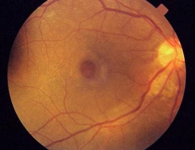
Ophthalmoscopic abnormalities in adults with falciparum malaria. (1/714)
We studied 424 adults with falciparum malaria admitted over 28 months. They were divided into three groups: cerebral malaria (n = 214); severe non-cerebral malaria (n = 58); and uncomplicated malaria (n = 152). Fundus examination was done daily from admission to discharge, and weekly thereafter in those with persistent changes. All patients were treated by a protocol based on WHO guidelines. Ophthalmoscopic abnormalities were: retinal haemorrhages, 40 (9.43%) (25 cerebral malaria, 10 severe non-cerebral and five uncomplicated malaria); papilloedema, 17 (7.94%) cerebral malaria and two uncomplicated malaria; blurring of disc margins, 25 (11.68%) cerebral and seven non-cerebral; retinal oedema, six (2.80%) cerebral and five non-cerebral malaria; disc pallor, five patients all with cerebral malaria; vitreous haemorrhage and hard exudate in one patient each, both cerebral malaria. Retinal haemorrhage was associated with cerebral malaria and severe non-cerebral malaria, especially with severe anaemia (p < 0.001), as compared to uncomplicated malaria (p < 0.01). The association of papilloedema and cerebral malaria was highly significant compared to severe non-cerebral malaria (p < 0.001). None of these findings was associated with statistically significant mortality, except disc pallor in cerebral malaria (p < 0.05). (+info)Gemfibrozil in a group of diabetics. (2/714)
A group of 14 diabetic patients was treated with gemfibrozil during a variable length of time ranging from nine to 23 weeks in order to establish if a lowering effect on the cholesterol and triglyceride levels could be achieved, as it had been in the case of another group of non-diabetic patients. The present results showed that: (1) The drug is remarkably well tolerated. (2) With doses ranging between 400 and 800 mg per day the magnitude of the effect of the drug was less than that observed in our previous trial with non-diabetic subjects. The effect upon triglycerides seemed to be reduced more than that upon cholesterol when compared with results in higher-dose studies. (3) In this group of diabetic patients (3 insulin dependent, 11 maturity-onset type) control of the diabetic condition was never impaired and appeared in some cases to be slightly improved by gemfibrozil. (4) There was no evidence of undesirable interaction with any of the anti-diabetic drugs used. (+info)High frequency of persistent hyperplastic primary vitreous and cataracts in p53-deficient mice. (3/714)
In order to investigate whether the p53 gene product plays a role in normal eye development, age matched p53-deficient mice and wild-type controls were sacrificed from day 2 to day 21 after birth. Eyes were paraffin-embedded and sectioned. Serial sections were taken at the level of the tunica vasculosa lentis and the hyaloid artery. The terminal dUTP nick-end labelling technique (TUNEL) was used to detect the number of cells displaying DNA fragmentation within these structures. Eyes were also prepared for scanning electron microscopy and resin embedded for semi-thin sections. Adult wild-type mice and p53-deficient mice were examined ophthalmoscopically in vivo. Ophthalmoscopical examination of mice completely deficient in p53 revealed them to be normal except for the persistence of the hyaloid vasculature, a structure that normally regresses during eye development. In adult animals there was also a high frequency of cataracts. Using morphological assessment and TUNEL we could show that in normal mice, regression of the primary vitreous, which includes the hyaloid artery, the vasa hyaloidea propria as well as the tunica vasculosa lentis, occurs via apoptotic cell death within 5 - 6 weeks after birth. The number of TUNEL-positive cells within these structures was significantly reduced in the p53-deficient mice in which parts of the hyaloid vasculature persisted and developed into a fibro-vascular retrolental plaque analogous to persistent hyperplastic primary vitreous (PHPV) described in humans. As in humans, PHPV in mice resulted in the development of cataracts. We have identified a role for p53-dependent apoptosis in the regression of the hyaloid vasculature and tunica vasculosa lentis. Our results provide further evidence for the importance of p53 in normal development and provide the first detailed evidence of its role in postnatal development in remodelling the developing eye. (+info)Radiation-induced leukocyte entrapment in the rat retinal microcirculation. (4/714)
PURPOSE: To evaluate the effects of irradiation on leukocyte dynamics in the rat retinal microcirculation. METHODS: Thirty-five Brown-Norway rats received a dose of 10 Gy irradiation in one fraction. Leukocyte dynamics were studied with acridine orange digital fluorography, in which a nuclear fluorescent dye of acridine orange is used and examined by scanning laser ophthalmoscope. This technique allowed visualization of fluorescent leukocytes in vivo. The leukocyte dynamics were evaluated at 0, 4, 7, 14, 30, and 60 days after the irradiation. RESULTS: Mean leukocyte velocity in the retinal capillaries decreased immediately. It partially recovered on day 4 but then gradually decreased up to the 2-month mark. Low-dose irradiation induced entrapment of leukocytes in the retinal microcirculation. The number of entrapped leukocytes gradually increased with time. The major retinal vessels significantly constricted immediately after irradiation. The diameter was reduced by 76% in arteries and 75% in veins, 2 months after irradiation. CONCLUSIONS: Entrapped leukocytes may be activated and exacerbate vascular injury and microinfarction and thus may participate in the pathogenesis of radiation retinopathy. (+info)Imaging features of intraventricular melanoma. (5/714)
We present the MR imaging findings in a patient with symptoms of increased intracranial pressure and a mass in the left lateral ventricle. The mass showed increased signal intensity on T1-weighted images and low signal intensity on T2-weighted images. The histologic diagnosis was that of melanoma, and detailed physical and funduscopic examinations disclosed no evidence of a primary lesion. We believe that the mass was a primary intraventricular melanoma, possibly arising from the choroid plexus, and we discuss the mechanisms that may be responsible for its occurrence in this location. (+info)Comparison of the cost-effectiveness of three approaches to screening for and treating sight-threatening diabetic retinopathy. (6/714)
The purpose of this study was to analyse and compare the costs involved in screening for and treating sight-threatening diabetic retinopathy in three different clinical settings. In the first setting, diabetologists screened using ophthalmoscopy and color photography, according to the St. Vincent Declaration guidelines, and selected patients for further assessment by a visiting ophthalmologist and for treatment in another hospital. In the second setting, all patients were regularly referred to ophthalmologists, either in the same hospital or elsewhere, for all aspects of eye care. In the third setting, screening was done again with ophthalmoscopy alone by diabetologists who followed the St. Vincent Declaration guidelines; however, further assessment and treatment were carried out in the eye department of the same hospital. Costs to the Italian National Health Service and to patients were calculated per screening performed and per patient subjected to laser treatment as a result of screening. A sensitivity analysis was then performed to simulate the costs of standardised patient populations going through the three different settings. It is concluded that absolute costs would be lower, both for the Italian National Health Service and for patients, if screening, assessment and treatment were all carried out in the same hospital. Equipping a diabetic clinic specially for screening would not be more expensive than delegating eye care to external parties, even for a hospital without an eye department. Moreover, delegating eye care more than doubles costs for patients. Screening for, assessing and treating sight-threatening diabetic retinopathy may be a cost-effective procedure for society as a whole in Italy. (+info)Inter- and intraobserver variation in the analysis of optic disc images: comparison of the Heidelberg retina tomograph and computer assisted planimetry. (7/714)
AIMS: The development of imaging and measurement techniques has brought the prospect of greater objectivity in the measurement of optic disc features, and therefore better agreement between observers. The purpose of this study was to quantify and compare the variation between observers using two measurement devices. METHODS: Optic disc photographs and images from the Heidelberg retina tomograph (HRT) of 30 eyes of 30 subjects were presented to six observers for analysis, and to one observer on five separate occasions. Agreement between observers was studied by comparing the analysis of each observer with the median result of the other five, and expressed as the mean difference and standard deviation of differences between the observer and the median. Inter- and intraobserver variation was calculated as a coefficient of variation (mean SD/mean x 100). RESULTS: For planimetry, agreement between observers was dependent on observer experience, for the HRT it was independent. Agreement between observers (SD of differences as a percentage of the median) for optic disc area was 4.0% to 7.2% (planimetry) and 3.3% to 6.0% (HRT), for neuroretinal rim area it was 10.8% to 21.0% (planimetry) and 5.2% to 9.6% (HRT). The mean interobserver coefficient of variation for optic disc area was 8.1% (planimetry) and 4.4% (HRT), for neuroretinal rim area it was 16.3% (planimetry) and 8.1% (HRT), and (HRT only) for rim volume was 16.3%, and reference height 9.1%. HRT variability was greater for the software version 1.11 reference plane than for version 1.10. The intraobserver coefficient of variation for optic disc area was 1.5% (planimetry) and 2.4% (HRT), for neuroretinal rim area it was 4.0% (planimetry) and 4.5% (HRT). CONCLUSIONS: Variation between observers is greatly reduced by the HRT when compared with planimetry. However, levels of variation, which may be clinically significant, remain for variables that depend on the subjective drawing of the disc margin. (+info)Effect of focal X-ray irradiation on experimental choroidal neovascularization. (8/714)
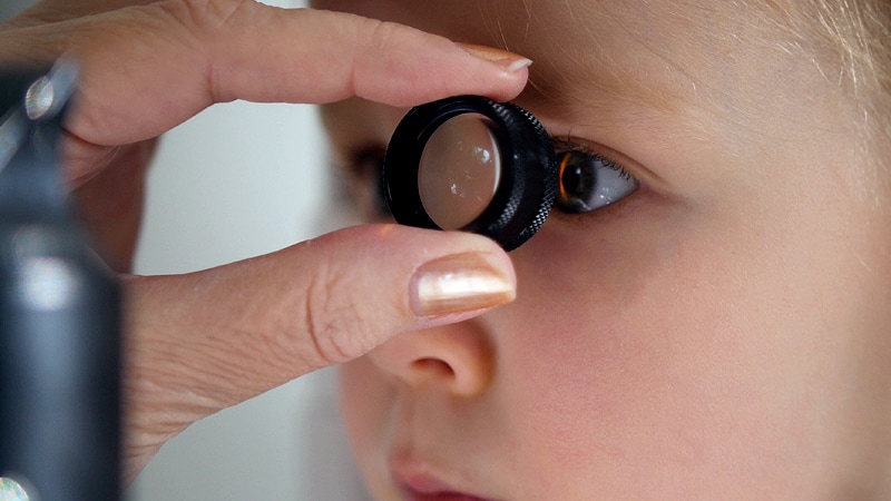
PURPOSE: Radiation therapy has been used to treat choroidal neovascularization (CNV) in patients with age-related macular degeneration. The in vivo effect of applying focal x-ray irradiation to the eye of rabbits with experimental CNV was investigated. METHODS: CNV was induced in the rabbit eyes by subretinal implantation of gelatin hydrogel microspheres impregnated with basic fibroblast growth factor. Three weeks after implantation, 17 of 34 eyes with CNV lesions accompanied by fluorescein leakage were irradiated with a single dose of 20 Gy; the other 17 eyes were not irradiated and served as the controls. The eyes were examined before irradiation and 1, 2, and 4 weeks after irradiation, by indirect ophthalmoscopy and fluorescein angiography. The degree of a decreasing amount of fluorescein leakage from the CNV lesions after irradiation was graded using a computerized image analysis system and was compared in the irradiated and nonirradiated eyes. These eyes were also examined histologically and immunohistochemically. RESULTS: Fluorescein leakage from the CNV lesions had significantly decreased in the eyes irradiated with 20 Gy compared with the control eyes, throughout the study period (P < 0.05). Histologic and immunohistochemical studies at 4 weeks after irradiation demonstrated that the degree of vascular formation and the number of vascular endothelial cells in the subretinal membrane of the irradiated eyes were less than those of the control eyes. CONCLUSIONS: Focal x-ray irradiation at the ocular region effectively reduced experimental CNV activity. These results support the possibility that radiation therapy may be beneficial in treating CNV. (+info)Ophthalmoscopy is a medical examination technique used by healthcare professionals to observe the interior structures of the eye, including the retina, optic disc, and vitreous humor. This procedure typically involves using an ophthalmoscope, a handheld device that consists of a light and magnifying lenses. The healthcare provider looks through the ophthalmoscope and directly observes the internal structures of the eye by illuminating them.
There are several types of ophthalmoscopy, including direct ophthalmoscopy, indirect ophthalmoscopy, and slit-lamp biomicroscopy. Each type has its own advantages and disadvantages, and they may be used in different situations depending on the specific clinical situation and the information needed.
Ophthalmoscopy is an important diagnostic tool for detecting and monitoring a wide range of eye conditions, including diabetic retinopathy, glaucoma, age-related macular degeneration, and other retinal disorders. It can also provide valuable information about the overall health of the individual, as changes in the appearance of the retina or optic nerve may indicate the presence of systemic diseases such as hypertension or diabetes.
An ophthalmoscope is a medical device used by healthcare professionals to examine the interior structures of the eye, including the retina, optic disc, and vitreous humor. It consists of a handle with a battery-powered light source and a head that contains lenses for focusing. When placed in contact with the patient's dilated pupil, the ophthalmoscope allows the examiner to visualize the internal structures of the eye and assess their health. Ophthalmoscopes are commonly used in routine eye examinations, as well as in the diagnosis and management of various eye conditions and diseases.
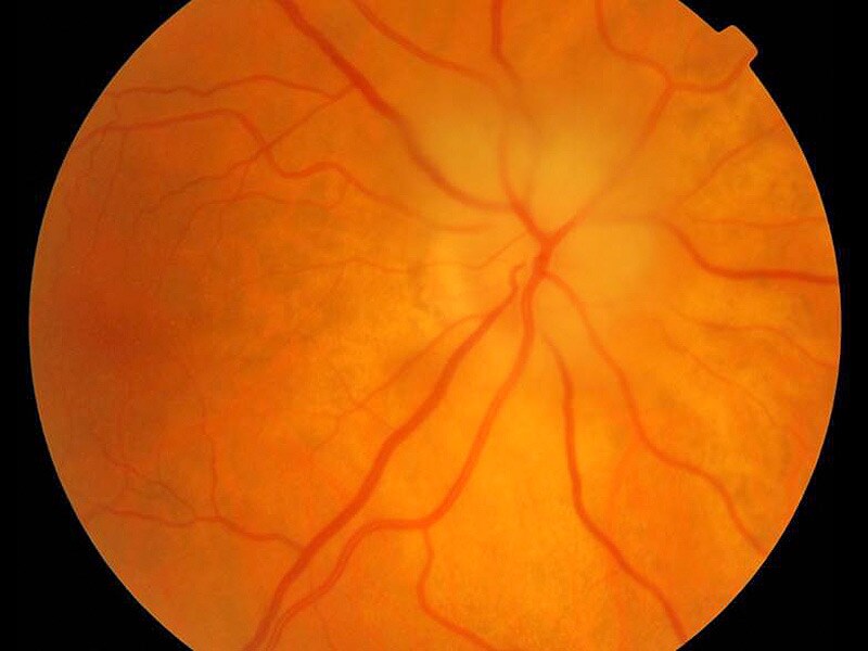
"Fundus Oculi" is a medical term that refers to the back part of the interior of the eye, including the optic disc, macula, fovea, retinal vasculature, and peripheral retina. It is the area where light is focused and then transmitted to the brain via the optic nerve, forming visual images. Examinations of the fundus oculi are crucial for detecting various eye conditions such as diabetic retinopathy, macular degeneration, glaucoma, and other retinal diseases. The examination is typically performed using an ophthalmoscope or a specialized camera called a retinal camera.
A laser is not a medical term per se, but a physical concept that has important applications in medicine. The term "LASER" stands for "Light Amplification by Stimulated Emission of Radiation." It refers to a device that produces and amplifies light with specific characteristics, such as monochromaticity (single wavelength), coherence (all waves moving in the same direction), and high intensity.
In medicine, lasers are used for various therapeutic and diagnostic purposes, including surgery, dermatology, ophthalmology, and dentistry. They can be used to cut, coagulate, or vaporize tissues with great precision, minimizing damage to surrounding structures. Additionally, lasers can be used to detect and measure physiological parameters, such as blood flow and oxygen saturation.
It's important to note that while lasers are powerful tools in medicine, they must be used by trained professionals to ensure safe and effective treatment.
I'm sorry for any confusion, but "photography" is not a term typically used in medical definitions. Photography refers to the art, application, or process of creating images by recording light or other electromagnetic radiation, either electronically by means of an image sensor, or chemically by means of a light-sensitive material such as photographic film.
If you're looking for a medical term related to imaging, there are several terms that might be relevant, such as:
1. Radiography: This is a technique using X-rays to visualize the internal structures of the body.
2. Ultrasonography: Also known as ultrasound, this is a diagnostic imaging technique using high-frequency sound waves to create images of the inside of the body.
3. Computed Tomography (CT): A type of imaging that uses X-rays to create detailed cross-sectional images of the body.
4. Magnetic Resonance Imaging (MRI): A type of imaging that uses magnetic fields and radio waves to create detailed images of the organs and tissues within the body.
5. Nuclear Medicine: This is a branch of medical imaging that uses small amounts of radioactive material to diagnose and treat diseases.
If you have any questions related to medical definitions or topics, feel free to ask!
Fluorescein angiography is a medical diagnostic procedure used in ophthalmology to examine the blood flow in the retina and choroid, which are the inner layers of the eye. This test involves injecting a fluorescent dye, Fluorescein, into a patient's arm vein. As the dye reaches the blood vessels in the eye, a specialized camera takes rapid sequences of photographs to capture the dye's circulation through the retina and choroid.
The images produced by fluorescein angiography can help doctors identify any damage to the blood vessels, leakage, or abnormal growth of new blood vessels. This information is crucial in diagnosing and managing various eye conditions such as age-related macular degeneration, diabetic retinopathy, retinal vein occlusions, and inflammatory eye diseases.
It's important to note that while fluorescein angiography is a valuable diagnostic tool, it does carry some risks, including temporary side effects like nausea, vomiting, or allergic reactions to the dye. In rare cases, severe adverse reactions can occur, so patients should discuss these potential risks with their healthcare provider before undergoing the procedure.
Retinal diseases refer to a group of conditions that affect the retina, which is the light-sensitive tissue located at the back of the eye. The retina is responsible for converting light into electrical signals that are sent to the brain and interpreted as visual images. Retinal diseases can cause vision loss or even blindness, depending on their severity and location in the retina.
Some common retinal diseases include:
1. Age-related macular degeneration (AMD): A progressive disease that affects the central part of the retina called the macula, causing blurred or distorted vision.
2. Diabetic retinopathy: A complication of diabetes that can damage the blood vessels in the retina, leading to vision loss.
3. Retinal detachment: A serious condition where the retina becomes separated from its underlying tissue, requiring immediate medical attention.
4. Macular edema: Swelling or thickening of the macula due to fluid accumulation, which can cause blurred vision.
5. Retinitis pigmentosa: A group of inherited eye disorders that affect the retina's ability to respond to light, causing progressive vision loss.
6. Macular hole: A small break in the macula that can cause distorted or blurry vision.
7. Retinal vein occlusion: Blockage of the retinal veins that can lead to bleeding, swelling, and potential vision loss.
Treatment for retinal diseases varies depending on the specific condition and its severity. Some treatments include medication, laser therapy, surgery, or a combination of these options. Regular eye exams are essential for early detection and treatment of retinal diseases.
Photoacoustic techniques, also known as optoacoustic techniques, refer to a group of diagnostic methods used in medicine and biology that combine the principles of laser-induced ultrasound and optical absorption contrast. These techniques involve illuminating a sample or tissue with a short laser pulse, which is absorbed by chromophores (light-absorbing molecules) within the tissue. The absorption of light energy leads to thermoelastic expansion and generates broadband acoustic waves, which are then detected using ultrasound transducers.
The resulting photoacoustic signals can provide information about the optical absorption properties, concentration, and distribution of chromophores in the tissue, such as hemoglobin, melanin, or exogenous contrast agents. Photoacoustic techniques have several advantages over traditional optical imaging methods, including deeper penetration depths (up to a few centimeters) and higher spatial resolution (down to a few micrometers).
There are various photoacoustic techniques, including photoacoustic microscopy (PAM), photoacoustic tomography (PAT), and functional photoacoustic imaging. These methods have shown great potential in various biomedical applications, such as cancer detection, blood oxygenation mapping, skin melanoma characterization, and brain function monitoring.
Optical coherence tomography (OCT) is a non-invasive imaging technique that uses low-coherence light to capture high-resolution cross-sectional images of biological tissues, particularly the retina and other ocular structures. OCT works by measuring the echo time delay of light scattered back from different depths within the tissue, creating a detailed map of the tissue's structure. This technique is widely used in ophthalmology to diagnose and monitor various eye conditions such as macular degeneration, diabetic retinopathy, and glaucoma.
The optic disk, also known as the optic nerve head, is the point where the optic nerve fibers exit the eye and transmit visual information to the brain. It appears as a pale, circular area in the back of the eye, near the center of the retina. The optic disk has no photoreceptor cells (rods and cones), so it is insensitive to light. It is an important structure to observe during eye examinations because changes in its appearance can indicate various ocular diseases or conditions, such as glaucoma, optic neuritis, or papilledema.
Eye diseases are a range of conditions that affect the eye or visual system, causing damage to vision and, in some cases, leading to blindness. These diseases can be categorized into various types, including:
1. Refractive errors: These include myopia (nearsightedness), hyperopia (farsightedness), astigmatism, and presbyopia, which affect the way light is focused on the retina and can usually be corrected with glasses or contact lenses.
2. Cataracts: A clouding of the lens inside the eye that leads to blurry vision, glare, and decreased contrast sensitivity. Cataract surgery is the most common treatment for this condition.
3. Glaucoma: A group of diseases characterized by increased pressure in the eye, leading to damage to the optic nerve and potential blindness if left untreated. Treatment includes medications, laser therapy, or surgery.
4. Age-related macular degeneration (AMD): A progressive condition that affects the central part of the retina called the macula, causing blurry vision and, in advanced stages, loss of central vision. Treatment may include anti-VEGF injections, laser therapy, or nutritional supplements.
5. Diabetic retinopathy: A complication of diabetes that affects the blood vessels in the retina, leading to bleeding, leakage, and potential blindness if left untreated. Treatment includes laser therapy, anti-VEGF injections, or surgery.
6. Retinal detachment: A separation of the retina from its underlying tissue, which can lead to vision loss if not treated promptly with surgery.
7. Amblyopia (lazy eye): A condition where one eye does not develop normal vision, often due to a misalignment or refractive error in childhood. Treatment includes correcting the underlying problem and encouraging the use of the weaker eye through patching or other methods.
8. Strabismus (crossed eyes): A misalignment of the eyes that can lead to amblyopia if not treated promptly with surgery, glasses, or other methods.
9. Corneal diseases: Conditions that affect the transparent outer layer of the eye, such as keratoconus, Fuchs' dystrophy, and infectious keratitis, which can lead to vision loss if not treated promptly.
10. Uveitis: Inflammation of the middle layer of the eye, which can cause vision loss if not treated promptly with anti-inflammatory medications or surgery.
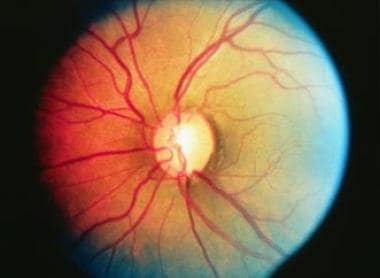
Hypertensive retinopathy is a term used to describe changes in the blood vessels and other structures in the retina that are caused by high blood pressure (hypertension). These changes can include narrowing of the blood vessels, thickening of their walls, and the formation of small bulges (microaneurysms) or bleeding. In severe cases, there may be swelling of the optic nerve and cotton wool spots, which are fluffy white patches that indicate areas where the blood supply to the retina has been disrupted.
Hypertensive retinopathy is usually asymptomatic in its early stages, but if it becomes advanced, it can lead to vision loss or even blindness. It is typically diagnosed by a doctor or eye care professional during an examination of the retina using specialized equipment such as an ophthalmoscope or a retinal camera. Treatment for hypertensive retinopathy usually involves controlling the underlying high blood pressure, which can help to prevent further damage to the retina and other structures in the eye.
Electroretinography (ERG) is a medical test used to evaluate the functioning of the retina, which is the light-sensitive tissue located at the back of the eye. The test measures the electrical responses of the retina to light stimulation.
During the procedure, a special contact lens or electrode is placed on the surface of the eye to record the electrical activity generated by the retina's light-sensitive cells (rods and cones) and other cells in the retina. The test typically involves presenting different levels of flashes of light to the eye while the electrical responses are recorded.
The resulting ERG waveform provides information about the overall health and function of the retina, including the condition of the photoreceptors, the integrity of the inner retinal layers, and the health of the retinal ganglion cells. This test is often used to diagnose and monitor various retinal disorders, such as retinitis pigmentosa, macular degeneration, and diabetic retinopathy.
Retinal vessels refer to the blood vessels that are located in the retina, which is the light-sensitive tissue that lines the inner surface of the eye. The retina contains two types of blood vessels: arteries and veins.
The central retinal artery supplies oxygenated blood to the inner layers of the retina, while the central retinal vein drains deoxygenated blood from the retina. These vessels can be visualized during a routine eye examination using an ophthalmoscope, which allows healthcare professionals to assess their health and any potential abnormalities.
Retinal vessels are essential for maintaining the health and function of the retina, and any damage or changes to these vessels can affect vision and lead to various eye conditions such as diabetic retinopathy, retinal vein occlusion, and hypertensive retinopathy.
The retina is the innermost, light-sensitive layer of tissue in the eye of many vertebrates and some cephalopods. It receives light that has been focused by the cornea and lens, converts it into neural signals, and sends these to the brain via the optic nerve. The retina contains several types of photoreceptor cells including rods (which handle vision in low light) and cones (which are active in bright light and are capable of color vision).
In medical terms, any pathological changes or diseases affecting the retinal structure and function can lead to visual impairment or blindness. Examples include age-related macular degeneration, diabetic retinopathy, retinal detachment, and retinitis pigmentosa among others.
A visual field test is a method used to measure an individual's entire scope of vision, which includes what can be seen straight ahead and in peripheral (or side) vision. During the test, the person being tested is asked to focus on a central point while gradually identifying the appearance of objects moving into their peripheral vision. The visual field test helps detect blind spots (scotomas) or gaps in the visual field, which can be caused by various conditions such as glaucoma, brain injury, optic nerve damage, or retinal disorders. It's an essential tool for diagnosing and monitoring eye-related diseases and conditions.
Optic nerve diseases refer to a group of conditions that affect the optic nerve, which transmits visual information from the eye to the brain. These diseases can cause various symptoms such as vision loss, decreased visual acuity, changes in color vision, and visual field defects. Examples of optic nerve diseases include optic neuritis (inflammation of the optic nerve), glaucoma (damage to the optic nerve due to high eye pressure), optic nerve damage from trauma or injury, ischemic optic neuropathy (lack of blood flow to the optic nerve), and optic nerve tumors. Treatment for optic nerve diseases varies depending on the specific condition and may include medications, surgery, or lifestyle changes.
Retinopathy of Prematurity (ROP) is a potentially sight-threatening proliferative retinal vascular disorder that primarily affects prematurely born infants, particularly those with low birth weight and/or young gestational age. It is characterized by the abnormal growth and development of retinal blood vessels due to disturbances in the oxygen supply and metabolic demands during critical phases of fetal development.
The condition can be classified into various stages (1-5) based on its severity, with stages 4 and 5 being more severe forms that may lead to retinal detachment and blindness if left untreated. The pathogenesis of ROP involves an initial phase of vessel loss and regression in the central retina, followed by a secondary phase of abnormal neovascularization, which can cause fibrosis, traction, and ultimately, retinal detachment.
ROP is typically managed with a multidisciplinary approach involving ophthalmologists, neonatologists, and pediatricians. Treatment options include laser photocoagulation, cryotherapy, intravitreal anti-VEGF injections, or even surgical interventions to prevent retinal detachment and preserve vision. Regular screening examinations are crucial for early detection and timely management of ROP in at-risk infants.
The vitreous body, also known simply as the vitreous, is the clear, gel-like substance that fills the space between the lens and the retina in the eye. It is composed mainly of water, but also contains collagen fibers, hyaluronic acid, and other proteins. The vitreous helps to maintain the shape of the eye and provides a transparent medium for light to pass through to reach the retina. With age, the vitreous can become more liquefied and may eventually separate from the retina, leading to symptoms such as floaters or flashes of light.
The macula lutea, often simply referred to as the macula or fovea centralis, is a part of the eye that is responsible for central vision and color perception. It's located in the center of the retina, the light-sensitive tissue at the back of the eye. The macula contains a high concentration of pigments called xanthophylls, which give it a yellowish color and protect the photoreceptor cells in this area from damage by blue light.
The central part of the macula is called the fovea, which is a small depression that contains only cones, the photoreceptor cells responsible for color vision and high visual acuity. The fovea is surrounded by the parafovea and the perifovea, which contain both cones and rods, the photoreceptor cells responsible for low-light vision and peripheral vision.
Damage to the macula can result in a loss of central vision and color perception, a condition known as age-related macular degeneration (AMD), which is a leading cause of blindness in older adults. Other conditions that can affect the macula include macular edema, macular holes, and macular pucker.
Mydriatics are medications that cause mydriasis, which is the dilation of the pupil. These drugs work by blocking the action of the muscarinic receptors in the iris, leading to relaxation of the circular muscle and constriction of the radial muscle, resulting in pupil dilation. Mydriatics are often used in eye examinations to facilitate examination of the interior structures of the eye. Commonly used mydriatic agents include tropicamide, phenylephrine, and cyclopentolate. It is important to note that mydriatics can have side effects such as blurred vision, photophobia, and accommodation difficulties, so patients should be advised accordingly.
Visual acuity is a measure of the sharpness or clarity of vision. It is usually tested by reading an eye chart from a specific distance, such as 20 feet (6 meters). The standard eye chart used for this purpose is called the Snellen chart, which contains rows of letters that decrease in size as you read down the chart.
Visual acuity is typically expressed as a fraction, with the numerator representing the testing distance and the denominator indicating the smallest line of type that can be read clearly. For example, if a person can read the line on the eye chart that corresponds to a visual acuity of 20/20, it means they have normal vision at 20 feet. If their visual acuity is 20/40, it means they must be as close as 20 feet to see what someone with normal vision can see at 40 feet.
It's important to note that visual acuity is just one aspect of overall vision and does not necessarily reflect other important factors such as peripheral vision, depth perception, color vision, or contrast sensitivity.
A retinoscope is an ophthalmic instrument used by eye care professionals to perform a retinoscopy examination, which is a subjective test used to estimate the refractive error and determine the prescription for eyeglasses or contact lenses. The retinoscope emits a beam of light that is shined onto the patient's retina, and the practitioner observes the reflection of the light (called the "reflex") as it passes through the pupil.
By adjusting the orientation and focus of the retinoscope's light, the practitioner can observe how the reflex changes, which provides information about the patient's refractive error. The retinoscope is an essential tool in eye examinations, particularly for children who may not be able to provide reliable answers to subjective questions about their vision.
Ophthalmology is a branch of medicine that deals with the diagnosis, treatment, and prevention of diseases and disorders of the eye and visual system. It is a surgical specialty, and ophthalmologists are medical doctors who complete additional years of training to become experts in eye care. They are qualified to perform eye exams, diagnose and treat eye diseases, prescribe glasses and contact lenses, and perform eye surgery. Some subspecialties within ophthalmology include cornea and external disease, glaucoma, neuro-ophthalmology, pediatric ophthalmology, retina and vitreous, and oculoplastics.
Retinal cone photoreceptor cells are specialized neurons located in the retina of the eye, responsible for visual phototransduction and color vision. They are one of the two types of photoreceptors, with the other being rods, which are more sensitive to low light levels. Cones are primarily responsible for high-acuity, color vision during daylight or bright-light conditions.
There are three types of cone cells, each containing different photopigments that absorb light at distinct wavelengths: short (S), medium (M), and long (L) wavelengths, which correspond to blue, green, and red light, respectively. The combination of signals from these three types of cones allows the human visual system to perceive a wide range of colors and discriminate between them. Cones are densely packed in the central region of the retina, known as the fovea, which provides the highest visual acuity.
Geographic atrophy is a medical term used to describe a specific pattern of degeneration of the retinal pigment epithelium (RPE) and the underlying choroidal tissue in the eye. This condition is often associated with age-related macular degeneration (AMD), which is a leading cause of vision loss in older adults.
In geographic atrophy, there are well-defined areas of RPE and choroidal atrophy that appear as pale, irregularly shaped patches in the central part of the retina known as the macula. These patches can grow larger over time and may lead to progressive vision loss. The exact cause of geographic atrophy is not fully understood, but it is thought to be related to oxidative stress, inflammation, and other age-related changes in the eye.
Currently, there are no effective treatments for geographic atrophy, although research is ongoing to find new ways to slow or halt its progression. Regular eye exams and monitoring by an ophthalmologist are important for people with AMD or geographic atrophy to help detect any changes in their vision and manage their condition effectively.
Tomography is a medical imaging technique used to produce cross-sectional images or slices of specific areas of the body. This technique uses various forms of radiation (X-rays, gamma rays) or sound waves (ultrasound) to create detailed images of the internal structures, such as organs, bones, and tissues. Common types of tomography include Computerized Tomography (CT), Positron Emission Tomography (PET), and Magnetic Resonance Imaging (MRI). The primary advantage of tomography is its ability to provide clear and detailed images of internal structures, allowing healthcare professionals to accurately diagnose and monitor a wide range of medical conditions.
The fovea centralis, also known as the macula lutea, is a small pit or depression located in the center of the retina, an light-sensitive tissue at the back of the eye. It is responsible for sharp, detailed vision (central vision) and color perception. The fovea contains only cones, the photoreceptor cells that are responsible for color vision and high visual acuity. It has a higher concentration of cones than any other area in the retina, allowing it to provide the greatest detail and color discrimination. The center of the fovea is called the foveola, which contains the highest density of cones and is avascular, meaning it lacks blood vessels to avoid interfering with the light passing through to the photoreceptor cells.
Diagnostic techniques in ophthalmology refer to the various methods and tests used by eye specialists (ophthalmologists) to examine, evaluate, and diagnose conditions related to the eyes and visual system. Here are some commonly used diagnostic techniques:
1. Visual Acuity Testing: This is a basic test to measure the sharpness of a person's vision. It typically involves reading letters or numbers from an eye chart at a specific distance.
2. Refraction Test: This test helps determine the correct lens prescription for glasses or contact lenses by measuring how light is bent as it passes through the cornea and lens.
3. Slit Lamp Examination: A slit lamp is a microscope that allows an ophthalmologist to examine the structures of the eye, including the cornea, iris, lens, and retina, in great detail.
4. Tonometry: This test measures the pressure inside the eye (intraocular pressure) to detect conditions like glaucoma. Common methods include applanation tonometry and non-contact tonometry.
5. Retinal Imaging: Several techniques are used to capture images of the retina, including fundus photography, fluorescein angiography, and optical coherence tomography (OCT). These tests help diagnose conditions like macular degeneration, diabetic retinopathy, and retinal detachments.
6. Color Vision Testing: This test evaluates a person's ability to distinguish between different colors, which can help detect color vision deficiencies or neurological disorders affecting the visual pathway.
7. Visual Field Testing: This test measures a person's peripheral (or side) vision and can help diagnose conditions like glaucoma, optic nerve damage, or brain injuries.
8. Pupillary Reactions Tests: These tests evaluate how the pupils respond to light and near objects, which can provide information about the condition of the eye's internal structures and the nervous system.
9. Ocular Motility Testing: This test assesses eye movements and alignment, helping diagnose conditions like strabismus (crossed eyes) or nystagmus (involuntary eye movement).
10. Corneal Topography: This non-invasive imaging technique maps the curvature of the cornea, which can help detect irregularities, assess the fit of contact lenses, and plan refractive surgery procedures.
Vitreous detachment, also known as posterior vitreous detachment (PVD), is a common age-related eye condition characterized by the separation of the vitreous gel from the retina. The vitreous is a clear, gel-like substance that fills the space between the lens and the retina in the eye. As we age, the vitreous may change in consistency, becoming more liquefied, leading to the formation of pockets of liquid within the gel.
In vitreous detachment, the posterior part of the vitreous closest to the retina begins to pull away from the retinal surface due to the shrinkage and liquefaction of the vitreous gel. This separation can cause symptoms such as floaters (spots or strands in the field of vision), flashes of light, or a decrease in vision sharpness. While vitreous detachment is typically not a serious condition on its own, it can sometimes lead to complications like retinal tears or retinal detachment, which require immediate medical attention.
A retinal hemorrhage is a type of bleeding that occurs in the blood vessels of the retina, which is the light-sensitive tissue located at the back of the eye. This condition can result from various underlying causes, including diabetes, high blood pressure, age-related macular degeneration, or trauma to the eye. Retinal hemorrhages can be categorized into different types based on their location and appearance, such as dot and blot hemorrhages, flame-shaped hemorrhages, or subhyaloid hemorrhages. Depending on the severity and cause of the hemorrhage, treatment options may vary from monitoring to laser therapy, medication, or even surgery. It is essential to consult an ophthalmologist for a proper evaluation and management plan if you suspect a retinal hemorrhage.
Diabetic retinopathy is a diabetes complication that affects the eyes. It's caused by damage to the blood vessels of the light-sensitive tissue at the back of the eye (retina).
At first, diabetic retinopathy may cause no symptoms or only mild vision problems. Eventually, it can cause blindness. The condition usually affects both eyes.
There are two main stages of diabetic retinopathy:
1. Early diabetic retinopathy. This is when the blood vessels in the eye start to leak fluid or bleed. You might not notice any changes in your vision at this stage, but it's still important to get treatment because it can prevent the condition from getting worse.
2. Advanced diabetic retinopathy. This is when new, abnormal blood vessels grow on the surface of the retina. These vessels can leak fluid and cause severe vision problems, including blindness.
Diabetic retinopathy can be treated with laser surgery, injections of medication into the eye, or a vitrectomy (a surgical procedure to remove the gel-like substance that fills the center of the eye). It's important to get regular eye exams to detect diabetic retinopathy early and get treatment before it causes serious vision problems.
Glaucoma is a group of eye conditions that damage the optic nerve, often caused by an abnormally high pressure in the eye (intraocular pressure). This damage can lead to permanent vision loss or even blindness if left untreated. The most common type is open-angle glaucoma, which has no warning signs and progresses slowly. Angle-closure glaucoma, on the other hand, can cause sudden eye pain, redness, nausea, and vomiting, as well as rapid vision loss. Other less common types of glaucoma also exist. While there is no cure for glaucoma, early detection and treatment can help slow or prevent further vision loss.
Open-angle glaucoma is a chronic, progressive type of glaucoma characterized by the gradual loss of optic nerve fibers and resulting in visual field defects. It is called "open-angle" because the angle where the iris meets the cornea (trabecular meshwork) appears to be normal and open on examination. The exact cause of this condition is not fully understood, but it is associated with increased resistance to the outflow of aqueous humor within the trabecular meshwork, leading to an increase in intraocular pressure (IOP). This elevated IOP can cause damage to the optic nerve and result in vision loss.
The onset of open-angle glaucoma is often asymptomatic, making regular comprehensive eye examinations crucial for early detection and management. Treatment typically involves lowering IOP using medications, laser therapy, or surgery to prevent further optic nerve damage and preserve vision.
A scotoma is a blind spot or area of reduced vision within the visual field. It's often surrounded by an area of less distinct vision and can be caused by various conditions such as eye diseases, neurological disorders, or brain injuries. A scotoma may be temporary or permanent, depending on its underlying cause.
There are different types of scotomas, including:
1. Central scotoma - a blind spot in the center of the visual field, often associated with conditions like age-related macular degeneration and diabetic retinopathy.
2. Paracentral scotoma - a blind spot located slightly away from the center of the visual field, which can be caused by optic neuritis or other optic nerve disorders.
3. Peripheral scotoma - a blind spot in the peripheral vision, often associated with retinal diseases like retinitis pigmentosa.
4. Absolute scotoma - a complete loss of vision in a specific area of the visual field.
5. Relative scotoma - a partial loss of vision in which some details can still be perceived, but not as clearly or vividly as in normal vision.
It is essential to consult an eye care professional if you experience any changes in your vision or notice a scotoma, as early detection and treatment can help prevent further vision loss.
Visual fields refer to the total area in which objects can be seen while keeping the eyes focused on a central point. It is the entire area that can be observed using peripheral (side) vision while the eye gazes at a fixed point. A visual field test is used to detect blind spots or gaps (scotomas) in a person's vision, which could indicate various medical conditions such as glaucoma, retinal damage, optic nerve disease, brain tumors, or strokes. The test measures both the central and peripheral vision and maps the entire area that can be seen when focusing on a single point.
The choroid is a part of the eye located between the retina and the sclera, which contains a large number of blood vessels that supply oxygen and nutrients to the outer layers of the retina. Choroid diseases refer to various medical conditions that affect the health and function of the choroid. Here are some examples:
1. Choroidal neovascularization (CNV): This is a condition where new blood vessels grow from the choroid into the retina, leading to fluid accumulation, bleeding, and scarring. CNV can cause vision loss and is often associated with age-related macular degeneration, myopia, and inflammatory eye diseases.
2. Chorioretinitis: This is an infection or inflammation of the choroid and retina, which can be caused by various microorganisms such as bacteria, viruses, fungi, or parasites. Symptoms may include blurred vision, floaters, light sensitivity, and eye pain.
3. Choroidal hemorrhage: This is a rare but serious condition where there is bleeding into the choroid, often caused by trauma, high blood pressure, or blood clotting disorders. It can lead to sudden vision loss and requires urgent medical attention.
4. Choroideremia: This is a genetic disorder that affects the choroid, retina, and optic nerve, leading to progressive vision loss. It is caused by mutations in the CHM gene and primarily affects males.
5. Central serous retinopathy (CSR): This is a condition where fluid accumulates under the retina, often in the macula, causing distortion or blurring of vision. While the exact cause is unknown, CSR is thought to be related to stress, steroid use, and other factors that affect the choroid's ability to regulate fluid.
6. Polypoidal choroidal vasculopathy (PCV): This is a condition where abnormal blood vessels form in the choroid, leading to serous or hemorrhagic detachment of the retina. PCV is often associated with age-related macular degeneration and can cause vision loss if left untreated.
These are just a few examples of choroidal disorders that can affect vision. If you experience any sudden changes in your vision, it's important to seek medical attention promptly.
A retinal perforation is a full-thickness break or hole in the retina, which is the light-sensitive tissue that lines the inner surface of the eye. This condition can lead to a serious complication called retinal detachment, where the retina separates from the underlying tissue, potentially resulting in vision loss if not promptly treated. Retinal perforations may be caused by trauma, certain eye conditions, or invasive eye procedures. Immediate medical attention is required for retinal perforations to prevent further damage and preserve vision.
Retinal drusen are yellow-white, deposits of extracellular material that accumulate beneath the retina, most commonly in the macula. They are a common age-related finding and can also be seen in various other conditions such as inherited retinal diseases. Drusen can vary in size and number, and their presence is often associated with an increased risk of developing age-related macular degeneration (AMD), a leading cause of vision loss in older adults. However, not all individuals with drusen will develop AMD, and the significance of drusen depends on factors such as size, number, and location. It's important to monitor drusen and have regular eye examinations to assess any changes or progression that may indicate a higher risk for developing AMD.
Indocyanine green (ICG) is a sterile, water-soluble, tricarbocyanine dye that is used as a diagnostic agent in medical imaging. It is primarily used in ophthalmology for fluorescein angiography to examine blood flow in the retina and choroid, and in cardiac surgery to assess cardiac output and perfusion. When injected into the body, ICG binds to plasma proteins and fluoresces when exposed to near-infrared light, allowing for visualization of various tissues and structures. It is excreted primarily by the liver and has a half-life of approximately 3-4 minutes in the bloodstream.
I'm sorry for any confusion, but "Optics and Photonics" is a broad field that encompasses various scientific and engineering disciplines. It doesn't have a specific medical definition, as it's not a term typically used in a medical context.
However, I can provide a general definition:
Optics is the study of light and its interactions with matter. This includes how light is produced, controlled, transmitted, and detected. It involves phenomena such as reflection, refraction, diffraction, and interference.
Photonics, on the other hand, is a branch of optics that deals with the generation, detection, and manipulation of individual photons, the basic units of light. Photonics is often applied to technologies such as lasers, fiber optics, and optical communications.
In a medical context, these fields might be used in various diagnostic and therapeutic applications, such as endoscopes, ophthalmic devices, laser surgery, and imaging technologies like MRI and CT scans. But the terms "Optics" and "Photonics" themselves are not medical conditions or treatments.
Ocular hypertension is a medical condition characterized by elevated pressure within the eye (intraocular pressure or IOP), which is higher than normal but not necessarily high enough to cause any visible damage to the optic nerve or visual field loss. It serves as a significant risk factor for developing glaucoma, a sight-threatening disease.
The normal range of intraocular pressure is typically between 10-21 mmHg (millimeters of mercury). Ocular hypertension is often defined as an IOP consistently above 21 mmHg, although some studies suggest that even pressures between 22-30 mmHg may not cause damage in all individuals. Regular monitoring and follow-up with an ophthalmologist are essential for people diagnosed with ocular hypertension to ensure early detection and management of any potential glaucomatous changes. Treatment options include medications, laser therapy, or surgery to lower the IOP and reduce the risk of glaucoma onset.
Retinal degeneration is a broad term that refers to the progressive loss of photoreceptor cells (rods and cones) in the retina, which are responsible for converting light into electrical signals that are sent to the brain. This process can lead to vision loss or blindness. There are many different types of retinal degeneration, including age-related macular degeneration, retinitis pigmentosa, and Stargardt's disease, among others. These conditions can have varying causes, such as genetic mutations, environmental factors, or a combination of both. Treatment options vary depending on the specific type and progression of the condition.
Vision disorders refer to a wide range of conditions that affect the visual system and result in various symptoms, such as blurry vision, double vision, distorted vision, impaired depth perception, and difficulty with visual tracking or focusing. These disorders can be categorized into several types, including:
1. Refractive errors: These occur when the shape of the eye prevents light from focusing directly on the retina, resulting in blurry vision. Examples include myopia (nearsightedness), hyperopia (farsightedness), astigmatism, and presbyopia (age-related loss of near vision).
2. Strabismus: Also known as crossed eyes or walleye, strabismus is a misalignment of the eyes where they point in different directions, which can lead to double vision or loss of depth perception.
3. Amblyopia: Often called lazy eye, amblyopia is a condition where one eye has reduced vision due to lack of proper visual development during childhood. It may be caused by strabismus, refractive errors, or other factors that interfere with normal visual development.
4. Accommodative disorders: These involve problems with the focusing ability of the eyes, such as convergence insufficiency (difficulty focusing on close objects) and accommodative dysfunction (inability to maintain clear vision at different distances).
5. Binocular vision disorders: These affect how the eyes work together as a team, leading to issues like poor depth perception, eye strain, and headaches. Examples include convergence insufficiency, divergence excess, and suppression.
6. Ocular motility disorders: These involve problems with eye movement, such as nystagmus (involuntary eye movements), strabismus, or restricted extraocular muscle function.
7. Visual processing disorders: These affect the brain's ability to interpret and make sense of visual information, even when the eyes themselves are healthy. Symptoms may include difficulty with reading, recognizing shapes and objects, and understanding spatial relationships.
8. Low vision: This term refers to significant visual impairment that cannot be fully corrected with glasses, contact lenses, medication, or surgery. It includes conditions like macular degeneration, diabetic retinopathy, glaucoma, and cataracts.
9. Blindness: Complete loss of sight in both eyes, which can be caused by various factors such as injury, disease, or genetic conditions.
Parasitic eye infections are conditions characterized by the invasion and infestation of the eye or its surrounding structures by parasites. These can be protozoans, helminths, or ectoparasites. Examples of such infections include Acanthamoeba keratitis, which is caused by a free-living amoeba found in water and soil; Toxoplasmosis, which is caused by the protozoan Toxoplasma gondii; Loiasis, which is caused by the parasitic filarial worm Loa loa; and Demodicosis, which is caused by the mite Demodex folliculorum. Symptoms can vary depending on the type of parasite but often include redness, pain, discharge, and vision changes. Treatment typically involves antiparasitic medications and sometimes surgery to remove the parasites or damaged tissue. Prevention measures include good hygiene practices and avoiding contact with contaminated water or soil.
Scanning Laser Polarimetry (SLP) is not primarily a medical term, but a technique that has been applied in medical research and diagnostics. It's a non-invasive method used to analyze the polarization of light as it interacts with biological tissues.
In a simpler sense, SLP uses laser light, which is polarized (meaning all the light waves vibrate in the same plane), to scan tissue. As this light interacts with the tissue, changes in the polarization of the light can occur due to various factors such as the structure and composition of the tissue. By analyzing these changes, SLP can provide information about the tissue's properties, which can be useful in the detection and diagnosis of certain medical conditions.
For example, it has been used in research related to diagnosing diseases like glaucoma and Alzheimer's disease by analyzing the polarization changes in the eye's retina and the brain's cortex, respectively. However, it's important to note that while SLP has shown promise in these areas, it is not yet widely used in clinical settings.
Vision screening is a quick and cost-effective method used to identify individuals who are at risk of vision problems or eye diseases. It is not a comprehensive eye examination, but rather an initial evaluation that helps to determine if a further, more in-depth examination by an eye care professional is needed. Vision screenings typically involve tests for visual acuity, distance and near vision, color perception, depth perception, and alignment of the eyes. The goal of vision screening is to detect potential vision issues early on, so that they can be treated promptly and effectively, thereby preventing or minimizing any negative impact on a person's overall vision and quality of life.
In the context of medical terminology, "lenses" generally refers to optical lenses used in various medical devices and instruments. These lenses are typically made of glass or plastic and are designed to refract (bend) light in specific ways to help magnify, focus, or redirect images. Here are some examples:
1. In ophthalmology and optometry, lenses are used in eyeglasses, contact lenses, and ophthalmic instruments to correct vision problems like myopia (nearsightedness), hypermetropia (farsightedness), astigmatism, or presbyopia.
2. In surgical microscopes, lenses are used to provide a magnified and clear view of the operating field during microsurgical procedures like ophthalmic, neurosurgical, or ENT (Ear, Nose, Throat) surgeries.
3. In endoscopes and laparoscopes, lenses are used to transmit light and images from inside the body during minimally invasive surgical procedures.
4. In ophthalmic diagnostic instruments like slit lamps, lenses are used to examine various structures of the eye in detail.
In summary, "lenses" in medical terminology refer to optical components that help manipulate light to aid in diagnosis, treatment, or visual correction.
Retinal Ganglion Cells (RGCs) are a type of neuron located in the innermost layer of the retina, the light-sensitive tissue at the back of the eye. These cells receive visual information from photoreceptors (rods and cones) via intermediate cells called bipolar cells. RGCs then send this visual information through their long axons to form the optic nerve, which transmits the signals to the brain for processing and interpretation as vision.
There are several types of RGCs, each with distinct morphological and functional characteristics. Some RGCs are specialized in detecting specific features of the visual scene, such as motion, contrast, color, or brightness. The diversity of RGCs allows for a rich and complex representation of the visual world in the brain.
Damage to RGCs can lead to various visual impairments, including loss of vision, reduced visual acuity, and altered visual fields. Conditions associated with RGC damage or degeneration include glaucoma, optic neuritis, ischemic optic neuropathy, and some inherited retinal diseases.
The choroid is a layer of the eye that contains blood vessels that supply oxygen and nutrients to the outer layers of the retina. It lies between the sclera (the white, protective coat of the eye) and the retina (the light-sensitive tissue at the back of the eye). The choroid is essential for maintaining the health and function of the retina, particularly the photoreceptor cells that detect light and transmit visual signals to the brain. Damage to the choroid can lead to vision loss or impairment.
Proliferative vitreoretinopathy (PVR) is a sight-threatening complication that can occur after open-globe eye injuries or retinal reattachment surgery. It is characterized by the abnormal growth and contraction of fibrous tissue on the surface of the retina and/or inside the vitreous cavity, which can cause distortion or detachment of the retina. This process can lead to visual impairment or even blindness if left untreated.
The term "proliferative" refers to the abnormal growth of cells (specifically, fibrous and inflammatory cells) on the retinal surface and within the vitreous cavity. These cells form membranes that can contract and cause traction on the retina, leading to distortion or detachment.
PVR is classified into three stages (A, B, and C) based on the extent of fibrous tissue formation and retinal changes. Stage A is characterized by the presence of cellular proliferation without any visible membranes or retinal changes. In stage B, fibrous membranes are present, but there is no retinal detachment. Finally, stage C involves the development of tractional retinal detachment due to the contraction of fibrous membranes.
Treatment for PVR typically involves additional surgical intervention to remove or release the fibrous tissue and reattach the retina. The prognosis for visual recovery depends on the severity and extent of the PVR, as well as the timing and success of treatment.
The retinal pigment epithelium (RPE) is a single layer of cells located between the photoreceptor cells of the retina and the choroid, which is a part of the eye containing blood vessels. The RPE plays a crucial role in maintaining the health and function of the photoreceptors by providing them with nutrients, removing waste products, and helping to regulate the light-sensitive visual pigments within the photoreceptors.
The RPE cells contain pigment granules that absorb excess light to prevent scattering within the eye and improve visual acuity. They also help to form the blood-retina barrier, which restricts the movement of certain molecules between the retina and the choroid, providing an important protective function for the retina.
Damage to the RPE can lead to a variety of eye conditions, including age-related macular degeneration (AMD), which is a leading cause of vision loss in older adults.
Intraocular pressure (IOP) is the fluid pressure within the eye, specifically within the anterior chamber, which is the space between the cornea and the iris. It is measured in millimeters of mercury (mmHg). The aqueous humor, a clear fluid that fills the anterior chamber, is constantly produced and drained, maintaining a balance that determines the IOP. Normal IOP ranges from 10-21 mmHg, with average values around 15-16 mmHg. Elevated IOP is a key risk factor for glaucoma, a group of eye conditions that can lead to optic nerve damage and vision loss if not treated promptly and effectively. Regular monitoring of IOP is essential in diagnosing and managing glaucoma and other ocular health issues.
Optical phenomena refer to the various observable patterns and effects that occur due to the interaction of light with the environment or with structures in our eye. These can include natural phenomena such as rainbows, mirages, and halos around the sun or moon, as well as visual artifacts created by the eye itself, such as afterimages, floaters, and flashes of light. Some optical phenomena are caused by the refraction, reflection, or interference of light waves, while others may result from abnormalities in the eye's structure or function. Understanding these phenomena can provide insight into the properties of light and the functioning of the visual system.
Nerve fibers are specialized structures that constitute the long, slender processes (axons) of neurons (nerve cells). They are responsible for conducting electrical impulses, known as action potentials, away from the cell body and transmitting them to other neurons or effector organs such as muscles and glands. Nerve fibers are often surrounded by supportive cells called glial cells and are grouped together to form nerve bundles or nerves. These fibers can be myelinated (covered with a fatty insulating sheath called myelin) or unmyelinated, which influences the speed of impulse transmission.
Optic disk drusen are small, calcified deposits that form within the optic nerve head, also known as the optic disc. They are made up of protein and calcium salts and can vary in size and number. These deposits can be seen on ophthalmic examination using an instrument called an ophthalmoscope.
Optic disk drusen are typically asymptomatic and are often discovered during routine eye examinations. However, in some cases, they may cause visual disturbances or even vision loss if they compress the optic nerve fibers. They can also increase the risk of developing other eye conditions such as glaucoma.
Optic disk drusen are more commonly found in individuals with a family history of the condition and tend to occur in younger people, typically before the age of 40. While there is no cure for optic disk drusen, regular eye examinations can help monitor any changes in the condition and manage any associated visual symptoms or complications.
The pigment epithelium of the eye, also known as the retinal pigment epithelium (RPE), is a layer of cells located between the photoreceptor cells of the retina and the choroid, which is the vascular layer of the eye. The RPE plays a crucial role in maintaining the health and function of the photoreceptors by providing them with nutrients, removing waste products, and helping to regulate the light that enters the eye.
The RPE cells contain pigment granules that absorb excess light, preventing it from scattering within the eye and improving visual acuity. They also help to create a barrier between the retina and the choroid, which is important for maintaining the proper functioning of the photoreceptors. Additionally, the RPE plays a role in the regeneration of visual pigments in the photoreceptor cells, allowing us to see in different light conditions.
Damage to the RPE can lead to various eye diseases and conditions, including age-related macular degeneration (AMD), which is a leading cause of vision loss in older adults.
Photoreceptor cells in vertebrates are specialized types of neurons located in the retina of the eye that are responsible for converting light stimuli into electrical signals. These cells are primarily responsible for the initial process of vision and have two main types: rods and cones.
Rods are more numerous and are responsible for low-light vision or scotopic vision, enabling us to see in dimly lit conditions. They do not contribute to color vision but provide information about the shape and movement of objects.
Cones, on the other hand, are less numerous and are responsible for color vision and high-acuity vision or photopic vision. There are three types of cones, each sensitive to different wavelengths of light: short (S), medium (M), and long (L) wavelengths, which correspond to blue, green, and red, respectively. The combination of signals from these three types of cones allows us to perceive a wide range of colors.
Both rods and cones contain photopigments that consist of a protein called opsin and a light-sensitive chromophore called retinal. When light hits the photopigment, it triggers a series of chemical reactions that ultimately lead to the generation of an electrical signal that is transmitted to the brain via the optic nerve. This process enables us to see and perceive our visual world.
Gonioscopy is a diagnostic procedure in ophthalmology used to examine the anterior chamber angle, which is the area where the iris and cornea meet. This examination helps to evaluate the drainage pathways of the eye for conditions such as glaucoma. A special contact lens called a goniolens is placed on the cornea during the procedure to allow the healthcare provider to visualize the angle using a biomicroscope. The lens may be coupled with a mirrored or prismatic surface to enhance the view of the angle. Gonioscopy can help detect conditions like narrow angles, closed angles, neovascularization, and other abnormalities that might contribute to glaucoma development or progression.
Eye injuries refer to any damage or trauma caused to the eye or its surrounding structures. These injuries can vary in severity and may include:
1. Corneal abrasions: A scratch or scrape on the clear surface of the eye (cornea).
2. Chemical burns: Occurs when chemicals come into contact with the eye, causing damage to the cornea and other structures.
3. Eyelid lacerations: Cuts or tears to the eyelid.
4. Subconjunctival hemorrhage: Bleeding under the conjunctiva, the clear membrane that covers the white part of the eye.
5. Hyphema: Accumulation of blood in the anterior chamber of the eye, which is the space between the cornea and iris.
6. Orbital fractures: Breaks in the bones surrounding the eye.
7. Retinal detachment: Separation of the retina from its underlying tissue, which can lead to vision loss if not treated promptly.
8. Traumatic uveitis: Inflammation of the uvea, the middle layer of the eye, caused by trauma.
9. Optic nerve damage: Damage to the optic nerve, which transmits visual information from the eye to the brain.
Eye injuries can result from a variety of causes, including accidents, sports-related injuries, violence, and chemical exposure. It is important to seek medical attention promptly for any suspected eye injury to prevent further damage and potential vision loss.
Equipment design, in the medical context, refers to the process of creating and developing medical equipment and devices, such as surgical instruments, diagnostic machines, or assistive technologies. This process involves several stages, including:
1. Identifying user needs and requirements
2. Concept development and brainstorming
3. Prototyping and testing
4. Design for manufacturing and assembly
5. Safety and regulatory compliance
6. Verification and validation
7. Training and support
The goal of equipment design is to create safe, effective, and efficient medical devices that meet the needs of healthcare providers and patients while complying with relevant regulations and standards. The design process typically involves a multidisciplinary team of engineers, clinicians, designers, and researchers who work together to develop innovative solutions that improve patient care and outcomes.
Retinitis is a medical term that refers to the inflammation of the retina, which is the light-sensitive tissue located at the back of the eye. The retina is responsible for converting light into electrical signals that are then sent to the brain and interpreted as visual images. Retinitis can be caused by various factors, including infections, autoimmune diseases, or genetic conditions.
The inflammation associated with retinitis can affect any part of the retina, but it typically involves the retinal pigment epithelium (RPE) and the photoreceptor cells (rods and cones). Depending on the severity and location of the inflammation, retinitis can cause a range of visual symptoms, such as blurry vision, floaters, loss of peripheral vision, or night blindness.
Retinitis is often distinguished from another condition called retinopathy, which refers to damage to the retina caused by diabetes or other systemic diseases. While both conditions can affect the retina and cause visual symptoms, retinitis is characterized by inflammation, while retinopathy is characterized by damage due to circulatory problems.
It's important to note that retinitis is a serious condition that requires prompt medical attention. If left untreated, it can lead to permanent vision loss or blindness. Treatment options for retinitis depend on the underlying cause and may include antibiotics, corticosteroids, or other immunosuppressive medications.
Macular degeneration, also known as age-related macular degeneration (AMD), is a medical condition that affects the central part of the retina, called the macula. The macula is responsible for sharp, detailed vision, which is necessary for activities such as reading, driving, and recognizing faces.
In AMD, there is a breakdown or deterioration of the macula, leading to gradual loss of central vision. There are two main types of AMD: dry (atrophic) and wet (exudative). Dry AMD is more common and progresses more slowly, while wet AMD is less common but can cause rapid and severe vision loss if left untreated.
The exact causes of AMD are not fully understood, but risk factors include age, smoking, family history, high blood pressure, obesity, and exposure to sunlight. While there is no cure for AMD, treatments such as vitamin supplements, laser therapy, and medication injections can help slow its progression and reduce the risk of vision loss.
Eye abnormalities refer to any structural or functional anomalies that affect the eye or its surrounding tissues. These abnormalities can be present at birth (congenital) or acquired later in life due to various factors such as injury, disease, or aging. Some examples of eye abnormalities include:
1. Strabismus: Also known as crossed eyes, strabismus is a condition where the eyes are misaligned and point in different directions.
2. Nystagmus: This is an involuntary movement of the eyes that can be horizontal, vertical, or rotatory.
3. Cataracts: A cataract is a clouding of the lens inside the eye that can cause vision loss.
4. Glaucoma: This is a group of eye conditions that damage the optic nerve and can lead to vision loss.
5. Retinal disorders: These include conditions such as retinal detachment, macular degeneration, and diabetic retinopathy.
6. Corneal abnormalities: These include conditions such as keratoconus, corneal ulcers, and Fuchs' dystrophy.
7. Orbital abnormalities: These include conditions such as orbital tumors, thyroid eye disease, and Graves' ophthalmopathy.
8. Ptosis: This is a condition where the upper eyelid droops over the eye.
9. Color blindness: A condition where a person has difficulty distinguishing between certain colors.
10. Microphthalmia: A condition where one or both eyes are abnormally small.
These are just a few examples of eye abnormalities, and there are many others that can affect the eye and its functioning. If you suspect that you have an eye abnormality, it is important to consult with an ophthalmologist for proper diagnosis and treatment.
An injection is a medical procedure in which a medication, vaccine, or other substance is introduced into the body using a needle and syringe. The substance can be delivered into various parts of the body, including into a vein (intravenous), muscle (intramuscular), under the skin (subcutaneous), or into the spinal canal (intrathecal or spinal).
Injections are commonly used to administer medications that cannot be taken orally, have poor oral bioavailability, need to reach the site of action quickly, or require direct delivery to a specific organ or tissue. They can also be used for diagnostic purposes, such as drawing blood samples (venipuncture) or injecting contrast agents for imaging studies.
Proper technique and sterile conditions are essential when administering injections to prevent infection, pain, and other complications. The choice of injection site depends on the type and volume of the substance being administered, as well as the patient's age, health status, and personal preferences.
Diagnostic imaging is a medical specialty that uses various technologies to produce visual representations of the internal structures and functioning of the body. These images are used to diagnose injury, disease, or other abnormalities and to monitor the effectiveness of treatment. Common modalities of diagnostic imaging include:
1. Radiography (X-ray): Uses ionizing radiation to produce detailed images of bones, teeth, and some organs.
2. Computed Tomography (CT) Scan: Combines X-ray technology with computer processing to create cross-sectional images of the body.
3. Magnetic Resonance Imaging (MRI): Uses a strong magnetic field and radio waves to generate detailed images of soft tissues, organs, and bones.
4. Ultrasound: Employs high-frequency sound waves to produce real-time images of internal structures, often used for obstetrics and gynecology.
5. Nuclear Medicine: Involves the administration of radioactive tracers to assess organ function or detect abnormalities within the body.
6. Positron Emission Tomography (PET) Scan: Uses a small amount of radioactive material to produce detailed images of metabolic activity in the body, often used for cancer detection and monitoring treatment response.
7. Fluoroscopy: Utilizes continuous X-ray imaging to observe moving structures or processes within the body, such as swallowing studies or angiography.
Diagnostic imaging plays a crucial role in modern medicine, allowing healthcare providers to make informed decisions about patient care and treatment plans.
Retinal detachment is a serious eye condition that occurs when the retina, a thin layer of tissue at the back of the eye responsible for processing light and sending visual signals to the brain, pulls away from its normal position. This can lead to significant vision loss or even blindness if not promptly treated. Retinal detachment can be caused by various factors such as aging, trauma, eye disease, or an inflammatory condition. Symptoms of retinal detachment may include sudden flashes of light, floaters, a shadow in the peripheral vision, or a curtain-like covering over part of the visual field. Immediate medical attention is necessary to prevent further damage and preserve vision.
Confocal microscopy is a powerful imaging technique used in medical and biological research to obtain high-resolution, contrast-rich images of thick samples. This super-resolution technology provides detailed visualization of cellular structures and processes at various depths within a specimen.
In confocal microscopy, a laser beam focused through a pinhole illuminates a small spot within the sample. The emitted fluorescence or reflected light from this spot is then collected by a detector, passing through a second pinhole that ensures only light from the focal plane reaches the detector. This process eliminates out-of-focus light, resulting in sharp images with improved contrast compared to conventional widefield microscopy.
By scanning the laser beam across the sample in a raster pattern and collecting fluorescence at each point, confocal microscopy generates optical sections of the specimen. These sections can be combined to create three-dimensional reconstructions, allowing researchers to study cellular architecture and interactions within complex tissues.
Confocal microscopy has numerous applications in medical research, including studying protein localization, tracking intracellular dynamics, analyzing cell morphology, and investigating disease mechanisms at the cellular level. Additionally, it is widely used in clinical settings for diagnostic purposes, such as analyzing skin lesions or detecting pathogens in patient samples.
Blindness is a condition of complete or near-complete vision loss. It can be caused by various factors such as eye diseases, injuries, or birth defects. Total blindness means that a person cannot see anything at all, while near-complete blindness refers to having only light perception or the ability to perceive the direction of light, but not able to discern shapes or forms. Legal blindness is a term used to define a certain level of visual impairment that qualifies an individual for government assistance and benefits; it usually means best corrected visual acuity of 20/200 or worse in the better eye, or a visual field no greater than 20 degrees in diameter.
Macular edema is a medical condition characterized by the accumulation of fluid in the macula, a small area in the center of the retina responsible for sharp, detailed vision. This buildup of fluid causes the macula to thicken and swell, which can distort central vision and lead to vision loss if not treated promptly. Macular edema is often a complication of other eye conditions such as diabetic retinopathy, age-related macular degeneration, retinal vein occlusion, or uveitis. It's important to note that while macular edema can affect anyone, it is more common in people with certain medical conditions like diabetes.
Ocular tonometry is a medical test used to measure the pressure inside the eye, also known as intraocular pressure (IOP). This test is an essential part of diagnosing and monitoring glaucoma, a group of eye conditions that can cause vision loss and blindness due to damage to the optic nerve from high IOP.
The most common method of ocular tonometry involves using a tonometer device that gently touches the front surface of the eye (cornea) with a small probe or prism. The device measures the amount of force required to flatten the cornea slightly, which correlates with the pressure inside the eye. Other methods of ocular tonometry include applanation tonometry, which uses a small amount of fluorescein dye and a blue light to measure the IOP, and rebound tonometry, which uses a lightweight probe that briefly touches the cornea and then bounces back to determine the IOP.
Regular ocular tonometry is important for detecting glaucoma early and preventing vision loss. It is typically performed during routine eye exams and may be recommended more frequently for individuals at higher risk of developing glaucoma, such as those with a family history of the condition or certain medical conditions like diabetes.
Reproducibility of results in a medical context refers to the ability to obtain consistent and comparable findings when a particular experiment or study is repeated, either by the same researcher or by different researchers, following the same experimental protocol. It is an essential principle in scientific research that helps to ensure the validity and reliability of research findings.
In medical research, reproducibility of results is crucial for establishing the effectiveness and safety of new treatments, interventions, or diagnostic tools. It involves conducting well-designed studies with adequate sample sizes, appropriate statistical analyses, and transparent reporting of methods and findings to allow other researchers to replicate the study and confirm or refute the results.
The lack of reproducibility in medical research has become a significant concern in recent years, as several high-profile studies have failed to produce consistent findings when replicated by other researchers. This has led to increased scrutiny of research practices and a call for greater transparency, rigor, and standardization in the conduct and reporting of medical research.
Retinitis pigmentosa (RP) is a group of rare, genetic disorders that involve a breakdown and loss of cells in the retina - a light-sensitive tissue located at the back of the eye. The retina converts light into electrical signals which are then sent to the brain and interpreted as visual images.
In RP, the cells that detect light (rods and cones) degenerate more slowly than other cells in the retina, leading to a progressive loss of vision. Symptoms typically begin in childhood with night blindness (difficulty seeing in low light), followed by a gradual narrowing of the visual field (tunnel vision). Over time, this can lead to significant vision loss and even blindness.
The condition is usually inherited and there are several different genes that have been associated with RP. The diagnosis is typically made based on a combination of genetic testing, family history, and clinical examination. Currently, there is no cure for RP, but researchers are actively working to develop new treatments that may help slow or stop the progression of the disease.
Prospective studies, also known as longitudinal studies, are a type of cohort study in which data is collected forward in time, following a group of individuals who share a common characteristic or exposure over a period of time. The researchers clearly define the study population and exposure of interest at the beginning of the study and follow up with the participants to determine the outcomes that develop over time. This type of study design allows for the investigation of causal relationships between exposures and outcomes, as well as the identification of risk factors and the estimation of disease incidence rates. Prospective studies are particularly useful in epidemiology and medical research when studying diseases with long latency periods or rare outcomes.
 Ophthalmoscopy
Scanning laser ophthalmoscopy
Point spread function
Louis de Wecker
Theodore John Dimitry Jr.
Adaptive optics
Canine glaucoma
Intraocular lymphoma
Toxoplasmic chorioretinitis
Diabetic retinopathy
Leukocoria
Visual impairment due to intracranial pressure
Fundus (eye)
Megalopapilla
Retina
William Gowers (neurologist)
Drusen
Heidelberg Retinal Tomography
Ernst Pflüger
Schwartz-Matsuo syndrome
Papilledema
Orbital emphysema
Biomedical engineering
Fundus photography
Robert Marcus Gunn
Blue field entoptic phenomenon
Cholesterol embolism
Karl Ernst Theodor Schweigger
Macula
Medical optical imaging
Indirect11
- Indirect ophthalmoscopy one that produces an inverted, or reversed, image of 2 to 5 times magnification. (wikipedia.org)
- View Fundus Examination in Pediatric Patients Using Indirect Ophthalmoscopy: Binocular Indirect Ophthalmoscopy (BIO), BIO With Scleral Indentation, BIO in Premature Infants on the publisher's website for pricing and purchasing information. (irma-international.org)
- The goal of this chapter is to provide the clinician with an understanding of binocular indirect ophthalmoscopy (BIO) and helpful clinical techniques for success in examining the fundus of the pediatric patient, including scleral depression. (irma-international.org)
- In indirect ophthalmoscopy, a real and inverted image is formed between the condensing lens and the observer. (eophtha.com)
- This was followed by series of innovations and methods of examination including the indirect method of ophthalmoscopy developed by Reute in 1852 (Fig:1b), binocular model by Marc-Antoine Giraud-Teulon (Fig:1c) and more practical hand-held reflex-free binocular ophthalmoscope by Allvar Gullstrand (Fig:1d). (eophtha.com)
- The technique of indirect binocular ophthalmoscopy starts with good dilatation of pupil with tropicamide and phenylephrine drops and examining fundus in the darkroom. (eophtha.com)
- In comparison to indirect ophthalmoscopy, however, the downside of this tool is the loss of the stereopsis character through examining via one eye at a time. (jfophth.com)
- All the participants underwent a dilated eye examination to detect DR by indirect ophthalmoscopy. (nih.gov)
- To measure the value of augmented reality technology usage to teach the medical students performing binocular indirect ophthalmoscopy. (vrmagic.com)
- Thirty-seven medical students were randomly assigned to the training of binocular indirect ophthalmoscopy either in the conventional way or with augmented reality ophthalmoscopy (ARO). (vrmagic.com)
- The main outcome measures were visual acuity, binocular indirect ophthalmoscopy score, adverse effects, and quality of life. (bvsalud.org)
Confocal2
- Aim To evaluate the efficiency of drusen detection by scanning laser ophthalmoscopy (SLO) using various infrared confocal apertures and differential contrast (DC) strategies. (bmj.com)
- Methods: Four hundred forty-one participants (832 eyes) in the Confocal Scanning Laser Ophthalmoscopy Ancillary Study to the Ocular Hypertension Treatment Study were included. (wustl.edu)
Retinal2
- Direct ophthalmoscopy is an extremely useful tool for detecting retinal, vascular, optic disc and choroidal pathologies. (jfophth.com)
- Owens DR, Gibbins RL, Lewis PA, Wall S, Allen JC, Morton R. Screening for diabetic retinopathy by general practitioners: ophthalmoscopy or retinal photography as 35 mm colour transparencies? (bvs.br)
Examination5
- Ophthalmoscopy is done as part of a routine physical or complete eye examination, mainly done by optometrists and ophthalmologists. (wikipedia.org)
- Ophthalmoscopy is an eye examination which allows an eye specialist to look at the back of an individual's eye. (eyepatient.net)
- Ophthalmoscopy: A Comprehensive Guide to Understanding Eye Examinations Ophthalmoscopy, also known as funduscopy, is a medical examination that allows eye doctors to view the inside of the eye, including the retina. (sellularhealth.com)
- Direct ophthalmoscopy has been proven to be an ophthalmologically time efficient mode of examination that is required also by multiple specialities e.g. (jfophth.com)
- It is essential, prior to the ophthalmoscopy examination, to inform the patient not to drive on the day of assessment as the vision will remain blurry for about 3-4 hours due to mydriatics application. (jfophth.com)
Fundus ophthalmoscopy1
- Learn more about fundus ophthalmoscopy . (notadryeye.org)
Approximately 15 times1
- It is of two major types: Direct ophthalmoscopy one that produces an upright, or unreversed, image of approximately 15 times magnification. (wikipedia.org)
Ophthalmoscope3
- Ophthalmoscopy, also called funduscopy, is a test that allows a health professional to see inside the fundus of the eye and other structures using an ophthalmoscope (or funduscope). (wikipedia.org)
- Each type of ophthalmoscopy has a special type of ophthalmoscope: The direct ophthalmoscope is an instrument about the size of a small flashlight (torch) with several lenses that can magnify up to about 15 times. (wikipedia.org)
- Ophthalmoscopy is conducted through a hand held device known as an ophthalmoscope. (eyepatient.net)
Tonometry1
- There are two tests: tonometry and ophthalmoscopy. (lonestarvision.com)
Fundoscopy1
- Direct ophthalmoscopy (fundoscopy) is a technique to examine the back of the eye. (jfophth.com)
Diabetic2
- Screening for diabetic retinopathy in a clinical setting: a comparison of direct ophthalmoscopy by primary care physicians with fundus photography. (bvs.br)
- Harding SP, Broadbent DM, Neoh C, White MC, Vora J. Sensitivity and specificity of photography and direct ophthalmoscopy in screening for sight threatening eye disease: the Liverpool Diabetic Eye Study. (bvs.br)
Examining the fundus1
- The technique of examining the fundus of the eye is called ophthalmoscopy. (eophtha.com)
Diagnosis1
- After diagnosis is made, screening protocols should be followed, including urinary catecholamines and ophthalmoscopy on an annual basis with MRI of the brain and spinal cord every 2 to 3 years, and yearly abdominal US with an additional abdominal CT scan every 2 to 3 years. (eyecancer.com)
Disorders1
- The eye care professional can use ophthalmoscopy to check for eye diseases and other disorders. (eyepatient.net)
Screening techniques1
- This refinement and updating of von Helmholtz's invention enabled ophthalmoscopy to become one of the most ubiquitous medical screening techniques in the world today. (wikipedia.org)
Approach2
- A second approach to scanning laser ophthalmoscopy uses an ellipsoidal mirror. (aao.org)
- An Easy Approach for Direct Ophthalmoscopy In 8 Steps! (jfophth.com)
Medical1
- Ophthalmoscopy" is a descriptor in the National Library of Medicine's controlled vocabulary thesaurus, MeSH (Medical Subject Headings) . (wakehealth.edu)
Optical1
- The journey of developing ophthalmoscopy began with an idea by the great physicist, Hermann von Helmholtz , who described the optical principles of direct ophthalmoscopy in 1851 (Fig:1a). (eophtha.com)
Photography3
- An alternative or complement to ophthalmoscopy is to perform a fundus photography, where the image can be analysed later by a professional. (wikipedia.org)
- Fundus Photography vs. Ophthalmoscopy Outcomes in the Emergency Department (FOTO-ED) Phase III: Web-based, In-service Training of Emergency Providers. (humaninsight.it)
- The Fundus photography vs. Ophthalmoscopy Trial Outcomes in the Emergency Department studies have demonstrated that emergency providers perform substantially better with fundus photography than direct ophthalmoscopy, but our web-based, in-service training did not result in further improvements at our institution. (humaninsight.it)
Times1
- At times, ophthalmoscopy may be a little uncomfortable, but it isn't painful. (eyepatient.net)
Good1
- Recent developments like scanning laser ophthalmoscopy can make good quality images through pupils as small as 2 millimeters, so dilating pupils is no longer needed with these devices. (wikipedia.org)
Major1
- This graph shows the total number of publications written about "Ophthalmoscopy" by people in this website by year, and whether "Ophthalmoscopy" was a major or minor topic of these publications. (wakehealth.edu)
Image1
- In direct ophthalmoscopy, a virtual and erect image of the fundus is seen. (eophtha.com)
Real1
- For testing student's skills, they had to examine a real person using a conventional ophthalmoscopy system and draw the optic disk. (vrmagic.com)
Patient1
- Figure 2: Ophthalmoscopy parts, patient side (4). (jfophth.com)
Efficient1
- The study provides evidence that a single ARO training is efficient to improve ophthalmoscopy skills. (vrmagic.com)
Conditions1
- In patients with headaches, the finding of swollen optic discs, or papilledema, on ophthalmoscopy is a key sign, as this indicates raised intracranial pressure (which could be due to hydrocephalus, benign intracranial hypertension (aka pseudotumor cerebri) or brain tumor, amongst other conditions. (wikipedia.org)
Laser1
- Adaptive optics scanning laser ophthalmoscopy for in vivo imaging of lamina cribrosa. (uh.edu)