Optic Nerve Diseases
Onchocerciasis, Ocular
Optic Nerve
Optic Neuritis
Olfactory Nerve Diseases
Vagus Nerve Diseases
Optic Nerve Injuries
Optic Disk
Hypoglossal Nerve Diseases
Vestibulocochlear Nerve Diseases
Glossopharyngeal Nerve Diseases
Trigeminal Nerve Diseases
Accessory Nerve Diseases
Facial Nerve Diseases
Abducens Nerve Diseases
Optic Chiasm
Trochlear Nerve Diseases
Optic Atrophy
Optic Nerve Neoplasms
Retinal Ganglion Cells
Nerve Fibers
Oculomotor Nerve Diseases
Cranial Nerve Diseases
Sciatic Nerve
Optic Nerve Glioma
Peripheral Nerves
Optic Neuropathy, Ischemic
Retina
Glaucoma
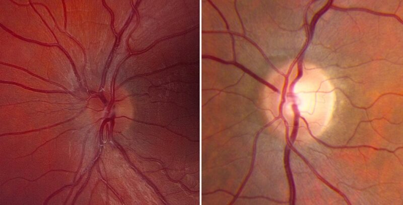
Papilledema
Optic Lobe, Nonmammalian
Optic Atrophies, Hereditary
Nerve Block
Eye
Cranial Nerve Neoplasms
Nerve Endings
Optic Atrophy, Hereditary, Leber
Optics and Photonics
Sural Nerve
Median Nerve
Evaluation of focal defects of the nerve fiber layer using optical coherence tomography. (1/852)
OBJECTIVE: To analyze glaucomatous eyes with known focal defects of the nerve fiber layer (NFL), relating optical coherence tomography (OCT) findings to clinical examination, NFL and stereoscopic optic nerve head (ONH) photography, and Humphrey 24-2 visual fields. DESIGN: Cross-sectional prevalence study. PARTICIPANTS: The authors followed 19 patients in the study group and 14 patients in the control group. INTERVENTION: Imaging with OCT was performed circumferentially around the ONH with a circle diameter of 3.4 mm using an internal fixation technique. One hundred OCT scan points taken within 2.5 seconds were analyzed. MAIN OUTCOME MEASURES: Measurements of NFL thickness using OCT were performed. RESULTS: In most eyes with focal NFL defects, OCTs showed significant thinning of the NFL in areas closely corresponding to focal defects visible on clinical examination, to red-free photographs, and to defects on the Humphrey visual fields. Optical coherence tomography enabled the detection of focal defects in the NFL with a sensitivity of 65% and a specificity of 81%. CONCLUSION: Analysis of NFL thickness in eyes with focal defects showed good structural and functional correlation with clinical parameters. Optical coherence tomography contributes to the identification of focal defects in the NFL that occur in early stages of glaucoma. (+info)Ophthalmic abnormalities in patients with cutaneous T-cell lymphoma. (2/852)
PURPOSE: To determine the frequency of ophthalmic abnormalities in patients with cutaneous T-cell lymphoma (mycosis fungoides and Sezary syndrome) and T-cell lymphoma involving the skin and to describe the clinical course of the disease with selected examples. METHODS: A computerized diagnostic retrieval system was used to identify all patients with T-cell lymphoma involving the skin who were examined at the Mayo Clinic (Rochester, Minnesota) between January 1, 1976 and December 31, 1990. The medical records of affected patients were reviewed. RESULTS: During the 15-year interval from 1976 through 1990, cutaneous T-cell lymphoma was diagnosed in 2,155 patients. Of these 2,155 patients, 42 (1.95%; 26 male and 16 female) had at least 1 ophthalmic abnormality attributable to the disease. The diagnoses in these 42 patients were mycosis fungoides in 19, clinical variants of T-cell lymphoma of the skin (most commonly, peripheral T-cell lymphoma) in 11, and Sezary syndrome in 12. Cicatricial eyelid ectropion was the most common finding, affecting 17 (40.4%) of the 42 patients. Thirty-seven patients had findings that, although probably not a direct consequence of cutaneous T-cell lymphoma, have been cataloged in previous studies. CONCLUSION: Although ophthalmic abnormalities in patients with cutaneous T-cell lymphoma are relatively uncommon, the manifestations of the disease are diverse and frequently difficult to treat. (+info)Acquired mitochondrial impairment as a cause of optic nerve disease. (3/852)
BACKGROUND: Blindness from an optic neuropathy recently occurred as an epidemic affecting 50,000 patients in Cuba (CEON) and had clinical features reminiscent of both tobacco-alcohol amblyopia (TAA) and Leber's hereditary optic neuropathy (Leber's; LHON). Selective damage to the papillomacular bundle was characteristic, and many patients also developed a peripheral neuropathy. Identified risk factors included vitamin deficiencies as well as exposure to methanol and cyanide. In all 3 syndromes, there is evidence that singular or combined insults to mitochondrial oxidative phosphorylation are associated with a clinically characteristic optic neuropathy. PURPOSE: First, to test the hypothesis that a common pathophysiologic mechanism involving impairment of mitochondria function and, consequently, axonal transport underlies both genetic optic nerve diseases such as Leber's and acquired toxic and nutritional deficiency optic neuropathies. According to this hypothesis, ATP depletion below a certain threshold leads to a blockage of orthograde axonal transport of mitochondria, which, in turn, leads to total ATP depletion and subsequent cell death. Second, to address several related questions, including (1) How does impaired energy production lead to optic neuropathy, particularly since it seems to relatively spare other metabolically active tissues, such as liver and heart? (2) Within the nervous system, why is the optic nerve, and most particularly the papillomacular bundle, so highly sensitive? Although there have been previous publications on the clinical features of the Cuban epidemic of blindness, the present hypothesis and the subsequent questions have not been previously addressed. METHODS: Patients in Cuba with epidemic optic neuropathy were personally evaluated through a comprehensive neuro-ophthalmologic examination. In addition, serum, lymphocytes for DNA analysis, cerebrospinal fluid (CSF), sural nerves, and eyes with attached optic nerves were obtained from Cuban patients, as well as from Leber's patients, for study. Finally, we developed an animal model to match the low serum folic acid and high serum formate levels found in the CEON patients, by administering to rats low doses of methanol after several months of a folic acid-deficient diet. Optic nerves and other tissues obtained from these rats were analyzed and compared with those from the Cuban patients. RESULTS: Patients from the Cuban epidemic of optic neuropathy with clinical evidence of a selective loss of the papillomacular bundle did much better once their nutritional status was corrected and exposure to toxins ceased. Patients with CEON often demonstrated low levels of folic acid and high levels of formate in their blood. Histopathologic studies demonstrated losses of the longest fibers (in the sural nerve) and those of smallest caliber (papillomacular bundle) in the optic nerve, with intra-axonal accumulations just anterior to the lamina cribrosa. Our animal model duplicated the serologic changes (low folic acid, high formate) as well as these histopathologic changes. Furthermore, ultrastructural examination of rat tissues demonstrated mitochondrial changes that further matched those seen on ultrastructural examination of tissues from patients with Leber's. CONCLUSION: Mitochondria can be impaired either genetically (as in Leber's) or through acquired insults (such as nutritional or toxic factors). Either may challenge energy production in all cells of the body. While this challenge may be met through certain compensatory mechanisms (such as in the size, shape, or number of the mitochondria), there exists in neurons a threshold which, once passed, leads to catastrophic changes. This threshold may be that point at which mitochondrial derangement leads to such ATP depletion that axonal transport is compromised, and decreased mitochondrial transport results in even further ATP depletion. Neurons are singularly dependent on the axonal transport of mitochondria. ( (+info)Diabetes mellitus: a risk factor in patients with Graves' orbitopathy. (4/852)
AIMS: To assess the prevalence of dysthyroid optic neuropathy (DON) in patients with diabetes mellitus (DM) and Graves' orbitopathy (GO) and to investigate the complications of surgery for GO in these patients. METHODS: The records of 482 consecutive patients with GO referred in a 5 year period were studied. Those patients who also had DM were selected for further study. The prevalence of insulin dependent diabetes mellitus (IDDM) and non-insulin dependent diabetes mellitus (NIDDM) was registered, as well as the prevalence and course of DON. In the patients who underwent surgery for GO the postoperative complications were recorded. RESULTS: Out of 482 patients with GO, 15 (3.1%) also had DM. Eight (1.7%) had IDDM, 7 (1.4%) had NIDDM. Five patients (33.3%) three with IDDM and two with NIDDM developed DON with 50% improvement of visual acuity after treatment, whereas in the whole population of 482 GO patients 19 had DON (3.9%), showing 69.4% improvement of vision after treatment. 10 patients with GO and DM were operated for GO; in one of them an optic atrophy developed as a result of a postoperative haemorrhage directly after a three wall orbital decompression by coronal approach. No other postoperative complications occurred. CONCLUSIONS: The prevalence of IDDM in patients with GO is higher than in the normal population. DON occurs much more frequently in patients with GO and DM than in the total group of GO patients and seems to have a worse visual prognosis. (+info)Dysgenesis of the internal carotid artery associated with transsphenoidal encephalocele: a neural crest syndrome? (5/852)
We describe two original cases of internal carotid artery dysgenesis associated with a malformative spectrum, which includes transsphenoidal encephalocele, optic nerve coloboma, hypopituitarism, and hypertelorism. Cephalic neural crest cells migrate to various regions in the head and neck where they contribute to the development of structures as diverse as the anterior skull base, the walls of the craniofacial arteries, the forebrain, and the face. Data suggest that the link between these rare malformations is abnormal neural crest development. (+info)Visual function and brain organization in non-decussating retinal-fugal fibre syndrome. (6/852)
Functional neuroimaging, psychophysical and electrophysiological investigations were performed in a patient with non-decussating retinal-fugal fibre syndrome, an inborn achiasmatic state in which the retinal projections of each eye map entirely to the ipsilateral primary visual cortex. Functional magnetic resonance imaging (fMRI) studies showed that for monocularly presented simple visual stimuli, only the ipsilateral striate cortex was activated. Within each hemisphere's striate cortex, the representation of the two hemifields overlapped extensively. Despite this gross miswiring, visual functions that require precise geometrical information (such as vernier acuity) were normal, and there was no evidence for the confounding of visual information between the overlapping ipsi-lateral and contralateral representations. Contrast sensitivity and velocity judgments were abnormal, but their dependence on the orientation and velocity of the targets suggests that this deficit was due to ocular instabilities, rather than the miswiring per se. There were no asymmetries in performance observed in visual search, visual naming or illusory contour perception. fMRI analysis of the latter two tasks under monocular viewing conditions indicated extensive bilateral activation of striate and prestriate areas. Thus, the remarkably normal visual behavior achieved by this patient is a result of both the plasticity of visual pathways, and efficient transfer of information between the hemispheres. (+info)Surgical management of symptomatic intrasellar arachnoid cysts--two case reports. (7/852)
Two patients with symptomatic intrasellar arachnoid cyst were successfully treated. A 67-year-old female with a cyst 20 mm in diameter developed headache and visual disturbance. She was treated by transsphenoidal surgery. A 59-year-old male with a cyst measuring 35 x 30 x 50 mm causing headache, visual disturbance, and deterioration of consciousness was managed by wide resection of the cyst wall via craniotomy. Postoperative courses in both patients were uneventful. Transsphenoidal surgery may be suitable for small to medium-sized cysts, although tight packing of the sella is mandatory to prevent leakage of cerebrospinal fluid. However, craniotomy is recommended for large intra- and suprasellar arachnoid cysts to avoid this complication, and to achieve sufficient communication between the cyst and the subarachnoid cistern. (+info)Idiopathic sclerotic inflammation of the orbit with left optic nerve compression in a patient with multifocal fibrosclerosis. (8/852)
We present the MR imaging findings in a 43-year-old male patient with bilateral idiopathic sclerosing inflammation of the orbit. Bilateral enhancing retrobulbar masses, with concentric compression of the retrobulbar segment of the left optic nerve, were seen. MR imaging proved to be the only means to distinguish between the different intraorbital structures and to determine the exact site of optic nerve compression. To our knowledge, this is the first documented case of MR imaging findings of this entity. (+info)Optic nerve diseases refer to a group of conditions that affect the optic nerve, which transmits visual information from the eye to the brain. These diseases can cause various symptoms such as vision loss, decreased visual acuity, changes in color vision, and visual field defects. Examples of optic nerve diseases include optic neuritis (inflammation of the optic nerve), glaucoma (damage to the optic nerve due to high eye pressure), optic nerve damage from trauma or injury, ischemic optic neuropathy (lack of blood flow to the optic nerve), and optic nerve tumors. Treatment for optic nerve diseases varies depending on the specific condition and may include medications, surgery, or lifestyle changes.
Onchocerciasis, Ocular is a medical condition that specifically refers to the eye manifestations caused by the parasitic infection, Onchocerca volvulus. Also known as "river blindness," this disease is spread through the bite of infected blackflies.
Ocular onchocerciasis affects various parts of the eye, including the conjunctiva, cornea, iris, and retina. The infection can cause symptoms such as itching, burning, and redness of the eyes. Over time, it may lead to more serious complications like punctate keratitis (small, scattered opacities on the cornea), cataracts, glaucoma, and ultimately, blindness.
The infection is diagnosed through a skin snip or blood test, which can detect the presence of microfilariae (the larval stage of the parasite) or antibodies against the parasite. Treatment typically involves administering oral medications such as ivermectin, which kills the microfilariae and reduces the risk of eye damage. However, it does not kill the adult worms, so multiple doses are often required to control the infection. In some cases, surgery may be necessary to remove advanced ocular lesions.
The optic nerve, also known as the second cranial nerve, is the nerve that transmits visual information from the retina to the brain. It is composed of approximately one million nerve fibers that carry signals related to vision, such as light intensity and color, from the eye's photoreceptor cells (rods and cones) to the visual cortex in the brain. The optic nerve is responsible for carrying this visual information so that it can be processed and interpreted by the brain, allowing us to see and perceive our surroundings. Damage to the optic nerve can result in vision loss or impairment.
Optic neuritis is a medical condition characterized by inflammation and damage to the optic nerve, which transmits visual information from the eye to the brain. This condition can result in various symptoms such as vision loss, pain with eye movement, color vision disturbances, and pupillary abnormalities. Optic neuritis may occur in isolation or be associated with other underlying medical conditions, including multiple sclerosis, neuromyelitis optica, and autoimmune disorders. The diagnosis typically involves a comprehensive eye examination, including visual acuity testing, dilated funduscopic examination, and possibly imaging studies like MRI to evaluate the optic nerve and brain. Treatment options may include corticosteroids or other immunomodulatory therapies to reduce inflammation and prevent further damage to the optic nerve.
Olfactory nerve diseases refer to conditions that affect the olfactory nerve, which is the first cranial nerve responsible for the sense of smell. These diseases can result in impaired or loss of smell (anosmia) and taste (ageusia), as well as distorted perception of smells (parosmia). The causes of olfactory nerve diseases can include trauma, infection, inflammation, neurological disorders, and exposure to certain chemicals. Some examples of specific olfactory nerve diseases include sinusitis, upper respiratory infections, head injuries, and neurodegenerative disorders such as Parkinson's disease and Alzheimer's disease. Treatment for these conditions depends on the underlying cause and may include medications, surgery, or lifestyle changes.
Vagus nerve diseases, also known as vagus nerve disorders, refer to conditions that affect the functioning of the vagus nerve. The vagus nerve is the tenth cranial nerve and extends from the brainstem to the abdomen, playing a crucial role in regulating various automatic functions of the body such as heart rate, digestion, respiratory rate, and sweating.
Diseases of the vagus nerve can result from various causes, including inflammation, infection, trauma, compression, or degeneration. Some common vagus nerve disorders include:
1. Vagus nerve dysfunction: This is a general term used to describe any abnormality in the functioning of the vagus nerve. Symptoms may vary depending on the specific functions affected but can include difficulty swallowing, hoarseness, voice changes, and abnormal heart rate or blood pressure.
2. Vagus nerve neuropathy: This is a condition that results from damage to the vagus nerve fibers. It can cause symptoms such as difficulty swallowing, voice changes, and abnormal digestive function.
3. Gastroparesis: This is a condition in which the stomach muscles fail to contract properly, leading to delayed gastric emptying. Vagus nerve dysfunction is a common cause of gastroparesis.
4. Orthostatic hypotension: This is a condition characterized by a drop in blood pressure when standing up from a sitting or lying down position. Vagus nerve dysfunction can contribute to this condition by causing an abnormal response in the heart rate and blood vessels.
5. Inflammatory disorders: Certain inflammatory conditions such as rheumatoid arthritis, lupus, and sarcoidosis can affect the vagus nerve and cause various symptoms.
Treatment for vagus nerve diseases depends on the underlying cause and may include medications, surgery, or lifestyle changes.
Optic nerve injuries refer to damages or trauma inflicted on the optic nerve, which is a crucial component of the visual system. The optic nerve transmits visual information from the retina to the brain, enabling us to see. Injuries to the optic nerve can result in various visual impairments, including partial or complete vision loss, decreased visual acuity, changes in color perception, and reduced field of view.
These injuries may occur due to several reasons, such as:
1. Direct trauma to the eye or head
2. Increased pressure inside the eye (glaucoma)
3. Optic neuritis, an inflammation of the optic nerve
4. Ischemia, or insufficient blood supply to the optic nerve
5. Compression from tumors or other space-occupying lesions
6. Intrinsic degenerative conditions affecting the optic nerve
7. Toxic exposure to certain chemicals or medications
Optic nerve injuries are diagnosed through a comprehensive eye examination, including visual acuity testing, slit-lamp examination, dilated fundus exam, and additional diagnostic tests like optical coherence tomography (OCT) and visual field testing. Treatment options vary depending on the cause and severity of the injury but may include medications, surgery, or vision rehabilitation.
The optic disk, also known as the optic nerve head, is the point where the optic nerve fibers exit the eye and transmit visual information to the brain. It appears as a pale, circular area in the back of the eye, near the center of the retina. The optic disk has no photoreceptor cells (rods and cones), so it is insensitive to light. It is an important structure to observe during eye examinations because changes in its appearance can indicate various ocular diseases or conditions, such as glaucoma, optic neuritis, or papilledema.
The hypoglossal nerve, also known as the 12th cranial nerve (CN XII), is primarily responsible for controlling tongue movements. Hypoglossal nerve diseases refer to conditions that affect this nerve and result in various tongue-related symptoms. These disorders can be congenital or acquired, and they may stem from different causes such as trauma, tumors, infections, inflammation, or degenerative processes.
Hypoglossal nerve diseases can present with the following symptoms:
1. Weakness or paralysis of the tongue muscles on one or both sides.
2. Deviation of the tongue towards the affected side when protruded.
3. Fasciculations (involuntary muscle twitches) or atrophy (wasting) of the tongue muscles.
4. Difficulty with speaking, swallowing, and chewing due to tongue weakness.
5. Changes in taste and sensation on the back of the tongue and throat.
Some specific hypoglossal nerve diseases include:
1. Hypoglossal nerve palsy: A condition characterized by unilateral or bilateral weakness or paralysis of the tongue due to damage to the hypoglossal nerve. Causes can include trauma, tumors, stroke, multiple sclerosis, or other neurological disorders.
2. Hypoglossal neuritis: Inflammation of the hypoglossal nerve, often caused by viral infections or autoimmune processes, leading to tongue weakness and atrophy.
3. Congenital hypoglossal nerve anomalies: Abnormal development of the hypoglossal nerve during fetal growth can result in various tongue-related symptoms and difficulties with speech and swallowing.
4. Tumors affecting the hypoglossal nerve: Both benign and malignant tumors, such as schwannomas or neurofibromas, can compress or infiltrate the hypoglossal nerve, causing weakness or paralysis.
5. Hypoglossal-facial anastomosis: A surgical procedure that connects the hypoglossal nerve to the facial nerve to restore facial movement in cases of facial nerve palsy. This connection can lead to tongue weakness as a side effect.
The vestibulocochlear nerve, also known as the 8th cranial nerve, is responsible for transmitting sound and balance information from the inner ear to the brain. Vestibulocochlear nerve diseases refer to conditions that affect this nerve and can result in hearing loss, vertigo, and balance problems.
These diseases can be caused by various factors, including genetics, infection, trauma, tumors, or degeneration. Some examples of vestibulocochlear nerve diseases include:
1. Vestibular neuritis: an inner ear infection that causes severe vertigo, nausea, and balance problems.
2. Labyrinthitis: an inner ear infection that affects both the vestibular and cochlear nerves, causing vertigo, hearing loss, and tinnitus.
3. Acoustic neuroma: a benign tumor that grows on the vestibulocochlear nerve, causing hearing loss, tinnitus, and balance problems.
4. Meniere's disease: a inner ear disorder that causes vertigo, hearing loss, tinnitus, and a feeling of fullness in the ear.
5. Ototoxicity: damage to the inner ear caused by certain medications or chemicals that can result in hearing loss and balance problems.
6. Vestibular migraine: a type of migraine that is associated with vertigo, dizziness, and balance problems.
Treatment for vestibulocochlear nerve diseases varies depending on the specific condition and its severity. It may include medication, physical therapy, surgery, or a combination of these approaches.
The glossopharyngeal nerve, also known as the ninth cranial nerve (CN IX), is primarily responsible for providing motor innervation to the stylopharyngeus muscle and sensory innervation to parts of the pharynx, middle ear, and posterior tongue. It also plays a role in the reflexive control of heart rate via the baroreceptors located in the carotid sinus.
Glossopharyngeal nerve diseases refer to conditions that affect the function of this nerve, leading to various symptoms. These diseases can be classified into two main categories: peripheral and central. Peripheral disorders are caused by damage or injury to the nerve itself, while central disorders result from problems in the brainstem where the glossopharyngeal nerve originates.
Some examples of glossopharyngeal nerve diseases include:
1. Glossopharyngeal neuralgia: A rare condition characterized by severe, stabbing pain in the throat, ear, or tongue, often triggered by swallowing or talking. This disorder may be caused by compression of the nerve by blood vessels or other structures.
2. Infections: Bacterial and viral infections can cause inflammation and damage to the glossopharyngeal nerve, leading to dysfunction. Examples include Lyme disease, herpes zoster (shingles), and meningitis.
3. Tumors: Benign or malignant growths in the head and neck region can compress and injure the glossopharyngeal nerve, resulting in symptoms related to its dysfunction.
4. Trauma: Direct trauma to the neck or skull base can damage the glossopharyngeal nerve, causing various deficits depending on the severity of the injury.
5. Neurological disorders: Conditions such as multiple sclerosis and stroke can affect the central connections of the glossopharyngeal nerve in the brainstem, leading to dysfunction.
6. Genetic conditions: Rare genetic disorders like Moersch-Woltman syndrome (also known as stiff person syndrome) can involve the glossopharyngeal nerve and cause symptoms related to its dysfunction.
Symptoms of glossopharyngeal nerve dysfunction may include difficulty swallowing, hoarseness, loss of taste on the back of the tongue, decreased sensation in the throat or ear, and pain in the neck, throat, or ear. Treatment for these conditions depends on the underlying cause and may involve medications, surgery, or other interventions to address the specific problem.
Trigeminal nerve diseases refer to conditions that affect the trigeminal nerve, which is one of the cranial nerves responsible for sensations in the face and motor functions such as biting and chewing. The trigeminal nerve has three branches: ophthalmic, maxillary, and mandibular, which innervate different parts of the face and head.
Trigeminal nerve diseases can cause various symptoms, including facial pain, numbness, tingling, or weakness. Some common trigeminal nerve diseases include:
1. Trigeminal neuralgia: A chronic pain condition that affects the trigeminal nerve, causing intense, stabbing, or electric shock-like pain in the face.
2. Hemifacial spasm: A neuromuscular disorder that causes involuntary muscle spasms on one side of the face, often affecting the muscles around the eye and mouth.
3. Trigeminal neuropathy: Damage or injury to the trigeminal nerve, which can result in numbness, tingling, or weakness in the face.
4. Herpes zoster oticus (Ramsay Hunt syndrome): A viral infection that affects the facial nerve and geniculate ganglion of the trigeminal nerve, causing facial paralysis, ear pain, and a rash around the ear.
5. Microvascular compression: Compression of the trigeminal nerve by a blood vessel, which can cause symptoms similar to trigeminal neuralgia.
Treatment for trigeminal nerve diseases depends on the specific condition and its severity. Treatment options may include medication, surgery, or radiation therapy.
The accessory nerve, also known as the 11th cranial nerve (CN XI), has both a cranial and spinal root and innervates the sternocleidomastoid muscle and trapezius muscle. Accessory nerve diseases refer to conditions that affect the function of this nerve, leading to weakness or paralysis of the affected muscles.
Some examples of accessory nerve diseases include:
1. Traumatic injury: Direct trauma to the neck or posterior scalene region can damage the spinal root of the accessory nerve. This can result in weakness or paralysis of the trapezius muscle, leading to difficulty with shoulder movement and pain.
2. Neuralgia: Accessory nerve neuralgia is a condition characterized by painful spasms or shooting pains along the course of the accessory nerve. It can be caused by nerve compression, inflammation, or injury.
3. Tumors: Tumors in the neck region, such as schwannomas or neurofibromas, can compress or invade the accessory nerve, leading to weakness or paralysis of the affected muscles.
4. Infections: Viral infections, such as poliovirus or West Nile virus, can cause inflammation and damage to the accessory nerve, resulting in weakness or paralysis.
5. Neuropathy: Accessory nerve neuropathy is a condition characterized by degeneration of the accessory nerve fibers due to various causes such as diabetes, autoimmune disorders, or exposure to toxins. This can result in weakness or paralysis of the affected muscles.
6. Congenital defects: Some individuals may be born with congenital defects that affect the development and function of the accessory nerve, leading to weakness or paralysis of the affected muscles.
Treatment for accessory nerve diseases depends on the underlying cause and can include physical therapy, medications, surgery, or a combination of these approaches.
Facial nerve diseases refer to a group of medical conditions that affect the function of the facial nerve, also known as the seventh cranial nerve. This nerve is responsible for controlling the muscles of facial expression, and it also carries sensory information from the taste buds in the front two-thirds of the tongue, and regulates saliva flow and tear production.
Facial nerve diseases can cause a variety of symptoms, depending on the specific location and extent of the nerve damage. Common symptoms include:
* Facial weakness or paralysis on one or both sides of the face
* Drooping of the eyelid and corner of the mouth
* Difficulty closing the eye or keeping it closed
* Changes in taste sensation or dryness of the mouth and eyes
* Abnormal sensitivity to sound (hyperacusis)
* Twitching or spasms of the facial muscles
Facial nerve diseases can be caused by a variety of factors, including:
* Infections such as Bell's palsy, Ramsay Hunt syndrome, and Lyme disease
* Trauma or injury to the face or skull
* Tumors that compress or invade the facial nerve
* Neurological conditions such as multiple sclerosis or Guillain-Barre syndrome
* Genetic disorders such as Moebius syndrome or hemifacial microsomia
Treatment for facial nerve diseases depends on the underlying cause and severity of the symptoms. In some cases, medication, physical therapy, or surgery may be necessary to restore function and relieve symptoms.
The abducens nerve, also known as the sixth cranial nerve, is responsible for controlling the lateral rectus muscle of the eye, which enables the eye to move outward. Abducens nerve diseases refer to conditions that affect this nerve and can result in various symptoms, primarily affecting eye movement.
Here are some medical definitions related to abducens nerve diseases:
1. Abducens Nerve Palsy: A condition characterized by weakness or paralysis of the abducens nerve, causing difficulty in moving the affected eye outward. This results in double vision (diplopia), especially when gazing towards the side of the weakened nerve. Abducens nerve palsy can be congenital, acquired, or caused by various factors such as trauma, tumors, aneurysms, infections, or diseases like diabetes and multiple sclerosis.
2. Sixth Nerve Palsy: Another term for abducens nerve palsy, referring to the weakness or paralysis of the sixth cranial nerve.
3. Internuclear Ophthalmoplegia (INO): A neurological condition affecting eye movement, often caused by a lesion in the medial longitudinal fasciculus (MLF), a bundle of nerve fibers that connects the abducens nucleus with the oculomotor nucleus. INO results in impaired adduction (inward movement) of the eye on the side of the lesion and nystagmus (involuntary eye movements) of the abducting eye on the opposite side when attempting to look towards the side of the lesion.
4. One-and-a-Half Syndrome: A rare neurological condition characterized by a combination of INO and internuclear ophthalmoplegia with horizontal gaze palsy on the same side, caused by damage to both the abducens nerve and the paramedian pontine reticular formation (PPRF). This results in limited or no ability to move the eyes towards the side of the lesion and impaired adduction of the eye on the opposite side.
5. Brainstem Encephalitis: Inflammation of the brainstem, which can affect the abducens nerve and other cranial nerves, leading to various neurological symptoms such as diplopia (double vision), ataxia (loss of balance and coordination), and facial weakness. Brainstem encephalitis can be caused by infectious agents, autoimmune disorders, or paraneoplastic syndromes.
6. Multiple Sclerosis (MS): An autoimmune disorder characterized by inflammation and demyelination of the central nervous system, including the brainstem and optic nerves. MS can cause various neurological symptoms, such as diplopia, nystagmus, and INO, due to damage to the abducens nerve and other cranial nerves.
7. Wernicke's Encephalopathy: A neurological disorder caused by thiamine (vitamin B1) deficiency, often seen in alcoholics or individuals with malnutrition. Wernicke's encephalopathy can affect the brainstem and cause various symptoms such as diplopia, ataxia, confusion, and oculomotor abnormalities.
8. Pontine Glioma: A rare type of brain tumor that arises from the glial cells in the pons (a part of the brainstem). Pontine gliomas can cause various neurological symptoms such as diplopia, facial weakness, and difficulty swallowing due to their location in the brainstem.
9. Brainstem Cavernous Malformation: A benign vascular lesion that arises from the small blood vessels in the brainstem. Brainstem cavernous malformations can cause various neurological symptoms such as diplopia, ataxia, and facial weakness due to their location in the brainstem.
10. Pituitary Adenoma: A benign tumor that arises from the pituitary gland, located at the base of the brain. Large pituitary adenomas can compress the optic nerves and cause various visual symptoms such as diplopia, visual field defects, and decreased vision.
11. Craniopharyngioma: A benign tumor that arises from the remnants of the Rathke's pouch, a structure that gives rise to the anterior pituitary gland. Craniopharyngiomas can cause various neurological and endocrine symptoms such as diplopia, visual field defects, headaches, and hormonal imbalances due to their location near the optic nerves and pituitary gland.
12. Meningioma: A benign tumor that arises from the meninges, the protective covering of the brain and spinal cord. Meningiomas can cause various neurological symptoms such as diplopia, headaches, and seizures depending on their location in the brain or spinal cord.
13. Chordoma: A rare type of malignant tumor that arises from the remnants of the notochord, a structure that gives rise to the spine during embryonic development. Chordomas can cause various neurological and endocrine symptoms such as diplopia, visual field defects, headaches, and hormonal imbalances due to their location near the brainstem and spinal cord.
14. Metastatic Brain Tumors: Malignant tumors that spread from other parts of the body to the brain. Metastatic brain tumors can cause various neurological symptoms such as diplopia, headaches, seizures, and cognitive impairment depending on their location in the brain.
15. Other Rare Brain Tumors: There are many other rare types of brain tumors that can cause diplopia or other neurological symptoms, including gliomas, ependymomas, pineal region tumors, and others. These tumors require specialized diagnosis and treatment by neuro-oncologists and neurosurgeons with expertise in these rare conditions.
In summary, diplopia can be caused by various brain tumors, including pituitary adenomas, meningiomas, chordomas, metastatic brain tumors, and other rare types of tumors. It is important to seek medical attention promptly if you experience diplopia or other neurological symptoms, as early diagnosis and treatment can improve outcomes and quality of life.
The optic chiasm is a structure in the brain where the optic nerves from each eye meet and cross. This allows for the integration of visual information from both eyes into the brain's visual cortex, creating a single, combined image of the visual world. The optic chiasm plays an important role in the processing of visual information and helps to facilitate depth perception and other complex visual tasks. Damage to the optic chiasm can result in various visual field deficits, such as bitemporal hemianopsia, where there is a loss of vision in the outer halves (temporal fields) of both eyes' visual fields.
The trochlear nerve, also known as the fourth cranial nerve (CN IV), is responsible for controlling the movement of the eye. It innervates the superior oblique muscle, which helps in depressing and rotating the eye downwards and outwards. Trochlear nerve diseases refer to conditions that affect this nerve and impair its function, leading to symptoms such as double vision (diplopia), vertical misalignment of the eyes, and difficulty with depth perception.
Trochlear nerve diseases can be caused by various factors, including trauma, compression, inflammation, infection, or tumors. Some common conditions that affect the trochlear nerve include:
1. Trochlear nerve palsy: This is a weakness or paralysis of the trochlear nerve, which can cause vertical and torsional diplopia, especially when looking downwards or to the side. It can be congenital or acquired due to trauma, compression, or other causes.
2. Aneurysm: Aneurysms in the vicinity of the trochlear nerve can compress or damage it, leading to palsy and diplopia.
3. Meningitis: Inflammation of the meninges (the membranes surrounding the brain and spinal cord) due to infection or other causes can affect the trochlear nerve and cause palsy.
4. Multiple sclerosis (MS): This is a chronic autoimmune disease that affects the central nervous system, including the cranial nerves. MS can cause demyelination of the trochlear nerve, leading to palsy and diplopia.
5. Diabetes: People with diabetes are at risk of developing diabetic neuropathy, which can affect any peripheral nerve, including the trochlear nerve.
6. Tumors: Space-occupying lesions in the brain or skull base, such as meningiomas, schwannomas, or pituitary adenomas, can compress the trochlear nerve and cause palsy.
The diagnosis of trochlear nerve diseases involves a thorough neurological examination, including assessment of eye movements and alignment. Imaging studies such as MRI or CT scans may be ordered to identify any structural lesions causing compression or damage to the nerve. Treatment depends on the underlying cause and may involve surgical intervention, medication, or observation.
Optic atrophy is a medical term that refers to the degeneration and shrinkage (atrophy) of the optic nerve, which transmits visual information from the eye to the brain. This condition can result in various vision abnormalities, including loss of visual acuity, color vision deficiencies, and peripheral vision loss.
Optic atrophy can occur due to a variety of causes, such as:
* Traumatic injuries to the eye or optic nerve
* Glaucoma
* Optic neuritis (inflammation of the optic nerve)
* Ischemic optic neuropathy (reduced blood flow to the optic nerve)
* Compression or swelling of the optic nerve
* Hereditary or congenital conditions affecting the optic nerve
* Toxins and certain medications that can damage the optic nerve.
The diagnosis of optic atrophy typically involves a comprehensive eye examination, including visual acuity testing, refraction assessment, slit-lamp examination, and dilated funduscopic examination to evaluate the health of the optic nerve. In some cases, additional diagnostic tests such as visual field testing, optical coherence tomography (OCT), or magnetic resonance imaging (MRI) may be necessary to confirm the diagnosis and determine the underlying cause.
There is no specific treatment for optic atrophy, but addressing the underlying cause can help prevent further damage to the optic nerve. In some cases, vision rehabilitation may be recommended to help patients adapt to their visual impairment.
Optic nerve neoplasms refer to abnormal growths or tumors that develop within or near the optic nerve. These tumors can be benign (non-cancerous) or malignant (cancerous).
Benign optic nerve neoplasms include optic nerve meningiomas and schwannomas, which originate from the sheaths surrounding the optic nerve. They usually grow slowly and may not cause significant vision loss, but they can lead to compression of the optic nerve, resulting in visual field defects or optic disc swelling (papilledema).
Malignant optic nerve neoplasms are rare but more aggressive. The most common type is optic nerve glioma, which arises from the glial cells within the optic nerve. These tumors can quickly damage the optic nerve and cause severe vision loss.
It's important to note that any optic nerve neoplasm requires prompt medical evaluation and treatment, as they can potentially lead to significant visual impairment or even blindness if left untreated.
Retinal Ganglion Cells (RGCs) are a type of neuron located in the innermost layer of the retina, the light-sensitive tissue at the back of the eye. These cells receive visual information from photoreceptors (rods and cones) via intermediate cells called bipolar cells. RGCs then send this visual information through their long axons to form the optic nerve, which transmits the signals to the brain for processing and interpretation as vision.
There are several types of RGCs, each with distinct morphological and functional characteristics. Some RGCs are specialized in detecting specific features of the visual scene, such as motion, contrast, color, or brightness. The diversity of RGCs allows for a rich and complex representation of the visual world in the brain.
Damage to RGCs can lead to various visual impairments, including loss of vision, reduced visual acuity, and altered visual fields. Conditions associated with RGC damage or degeneration include glaucoma, optic neuritis, ischemic optic neuropathy, and some inherited retinal diseases.
Nerve fibers are specialized structures that constitute the long, slender processes (axons) of neurons (nerve cells). They are responsible for conducting electrical impulses, known as action potentials, away from the cell body and transmitting them to other neurons or effector organs such as muscles and glands. Nerve fibers are often surrounded by supportive cells called glial cells and are grouped together to form nerve bundles or nerves. These fibers can be myelinated (covered with a fatty insulating sheath called myelin) or unmyelinated, which influences the speed of impulse transmission.
The oculomotor nerve, also known as the third cranial nerve (CN III), is responsible for controlling several important eye movements and functions. Oculomotor nerve diseases refer to conditions that affect this nerve and can lead to various symptoms related to eye movement and function. Here's a medical definition of oculomotor nerve diseases:
Oculomotor nerve diseases are a group of medical disorders characterized by the dysfunction or damage to the oculomotor nerve (CN III), resulting in impaired eye movements, abnormalities in pupillary response, and potential effects on eyelid position. These conditions can be congenital, acquired, or traumatic in nature and may lead to partial or complete paralysis of the nerve. Common oculomotor nerve diseases include oculomotor nerve palsy, third nerve ganglionopathies, and compressive oculomotor neuropathies caused by various pathologies such as aneurysms, tumors, or infections.
Cranial nerve diseases refer to conditions that affect the cranial nerves, which are a set of 12 pairs of nerves that originate from the brainstem and control various functions in the head and neck. These functions include vision, hearing, taste, smell, movement of the eyes and face, and sensation in the face.
Diseases of the cranial nerves can result from a variety of causes, including injury, infection, inflammation, tumors, or degenerative conditions. The specific symptoms that a person experiences will depend on which cranial nerve is affected and how severely it is damaged.
For example, damage to the optic nerve (cranial nerve II) can cause vision loss or visual disturbances, while damage to the facial nerve (cranial nerve VII) can result in weakness or paralysis of the face. Other common symptoms of cranial nerve diseases include pain, numbness, tingling, and hearing loss.
Treatment for cranial nerve diseases varies depending on the underlying cause and severity of the condition. In some cases, medication or surgery may be necessary to treat the underlying cause and relieve symptoms. Physical therapy or rehabilitation may also be recommended to help individuals regain function and improve their quality of life.
The sciatic nerve is the largest and longest nerve in the human body, running from the lower back through the buttocks and down the legs to the feet. It is formed by the union of the ventral rami (branches) of the L4 to S3 spinal nerves. The sciatic nerve provides motor and sensory innervation to various muscles and skin areas in the lower limbs, including the hamstrings, calf muscles, and the sole of the foot. Sciatic nerve disorders or injuries can result in symptoms such as pain, numbness, tingling, or weakness in the lower back, hips, legs, and feet, known as sciatica.
A nerve crush injury is a type of peripheral nerve injury that occurs when there is excessive pressure or compression applied to a nerve, causing it to become damaged or dysfunctional. This can happen due to various reasons such as trauma from accidents, surgical errors, or prolonged pressure on the nerve from tight casts, clothing, or positions.
The compression disrupts the normal functioning of the nerve, leading to symptoms such as numbness, tingling, weakness, or pain in the affected area. In severe cases, a nerve crush injury can cause permanent damage to the nerve, leading to long-term disability or loss of function. Treatment for nerve crush injuries typically involves relieving the pressure on the nerve, providing supportive care, and in some cases, surgical intervention may be necessary to repair the damaged nerve.
Nerve regeneration is the process of regrowth and restoration of functional nerve connections following damage or injury to the nervous system. This complex process involves various cellular and molecular events, such as the activation of support cells called glia, the sprouting of surviving nerve fibers (axons), and the reformation of neural circuits. The goal of nerve regeneration is to enable the restoration of normal sensory, motor, and autonomic functions impaired due to nerve damage or injury.
An Optic Nerve Glioma is a type of brain tumor that arises from the glial cells (supportive tissue) within the optic nerve. It is most commonly seen in children, particularly those with neurofibromatosis type 1 (NF1). These tumors are typically slow-growing and may not cause any symptoms, especially if they are small. However, as they grow larger, they can put pressure on the optic nerve, leading to vision loss or other visual disturbances. In some cases, these tumors can also affect nearby structures in the brain, causing additional neurological symptoms. Treatment options may include observation, chemotherapy, radiation therapy, or surgery, depending on the size and location of the tumor, as well as the patient's age and overall health.
Peripheral nerves are nerve fibers that transmit signals between the central nervous system (CNS, consisting of the brain and spinal cord) and the rest of the body. These nerves convey motor, sensory, and autonomic information, enabling us to move, feel, and respond to changes in our environment. They form a complex network that extends from the CNS to muscles, glands, skin, and internal organs, allowing for coordinated responses and functions throughout the body. Damage or injury to peripheral nerves can result in various neurological symptoms, such as numbness, weakness, or pain, depending on the type and severity of the damage.
Ischemic optic neuropathy (ION) is a medical condition that refers to the damage or death of the optic nerve due to insufficient blood supply. The optic nerve is responsible for transmitting visual information from the eye to the brain.
In ION, the blood vessels that supply the optic nerve become blocked or narrowed, leading to decreased blood flow and oxygen delivery to the nerve fibers. This results in inflammation, swelling, and ultimately, damage to the optic nerve. The damage can cause sudden, painless vision loss, often noticed upon waking up in the morning.
There are two types of ION: anterior ischemic optic neuropathy (AION) and posterior ischemic optic neuropathy (PION). AION affects the front part of the optic nerve, while PION affects the back part of the nerve. AION is further classified into arteritic and non-arteritic types, depending on whether it is caused by giant cell arteritis or not.
Risk factors for ION include age (most commonly occurring in people over 50), hypertension, diabetes, smoking, sleep apnea, and other cardiovascular diseases. Treatment options depend on the type and cause of ION and may include controlling underlying medical conditions, administering corticosteroids, or undergoing surgical procedures to improve blood flow.
An axon is a long, slender extension of a neuron (a type of nerve cell) that conducts electrical impulses (nerve impulses) away from the cell body to target cells, such as other neurons or muscle cells. Axons can vary in length from a few micrometers to over a meter long and are typically surrounded by a myelin sheath, which helps to insulate and protect the axon and allows for faster transmission of nerve impulses.
Axons play a critical role in the functioning of the nervous system, as they provide the means by which neurons communicate with one another and with other cells in the body. Damage to axons can result in serious neurological problems, such as those seen in spinal cord injuries or neurodegenerative diseases like multiple sclerosis.
The retina is the innermost, light-sensitive layer of tissue in the eye of many vertebrates and some cephalopods. It receives light that has been focused by the cornea and lens, converts it into neural signals, and sends these to the brain via the optic nerve. The retina contains several types of photoreceptor cells including rods (which handle vision in low light) and cones (which are active in bright light and are capable of color vision).
In medical terms, any pathological changes or diseases affecting the retinal structure and function can lead to visual impairment or blindness. Examples include age-related macular degeneration, diabetic retinopathy, retinal detachment, and retinitis pigmentosa among others.
Intraocular pressure (IOP) is the fluid pressure within the eye, specifically within the anterior chamber, which is the space between the cornea and the iris. It is measured in millimeters of mercury (mmHg). The aqueous humor, a clear fluid that fills the anterior chamber, is constantly produced and drained, maintaining a balance that determines the IOP. Normal IOP ranges from 10-21 mmHg, with average values around 15-16 mmHg. Elevated IOP is a key risk factor for glaucoma, a group of eye conditions that can lead to optic nerve damage and vision loss if not treated promptly and effectively. Regular monitoring of IOP is essential in diagnosing and managing glaucoma and other ocular health issues.
Glaucoma is a group of eye conditions that damage the optic nerve, often caused by an abnormally high pressure in the eye (intraocular pressure). This damage can lead to permanent vision loss or even blindness if left untreated. The most common type is open-angle glaucoma, which has no warning signs and progresses slowly. Angle-closure glaucoma, on the other hand, can cause sudden eye pain, redness, nausea, and vomiting, as well as rapid vision loss. Other less common types of glaucoma also exist. While there is no cure for glaucoma, early detection and treatment can help slow or prevent further vision loss.
Papilledema is a medical term that refers to swelling of the optic nerve head, also known as the disc, which is the point where the optic nerve enters the back of the eye (the retina). This swelling can be caused by increased pressure within the skull, such as from brain tumors, meningitis, or idiopathic intracranial hypertension. Papilledema is usually detected through a routine eye examination and may be accompanied by symptoms such as headaches, visual disturbances, and nausea. If left untreated, papilledema can lead to permanent vision loss.
The optic lobe in non-mammals refers to a specific region of the brain that is responsible for processing visual information. It is a part of the protocerebrum in the insect brain and is analogous to the mammalian visual cortex. The optic lobes receive input directly from the eyes via the optic nerves and are involved in the interpretation and integration of visual stimuli, enabling non-mammals to perceive and respond to their environment. In some invertebrates, like insects, the optic lobe is further divided into subregions, including the lamina, medulla, and lobula, each with distinct functions in visual processing.
Hereditary optic atrophies (HOAs) are a group of genetic disorders that cause degeneration of the optic nerve, leading to vision loss. The optic nerve is responsible for transmitting visual information from the eye to the brain. In HOAs, this nerve degenerates over time, resulting in decreased visual acuity, color vision deficits, and sometimes visual field defects.
There are several types of HOAs, including dominant optic atrophy (DOA), Leber hereditary optic neuropathy (LHON), autosomal recessive optic atrophy (AROA), and Wolfram syndrome. Each type has a different inheritance pattern and is caused by mutations in different genes.
DOA is the most common form of HOA and is characterized by progressive vision loss that typically begins in childhood or early adulthood. It is inherited in an autosomal dominant manner, meaning that a child has a 50% chance of inheriting the disease-causing mutation from an affected parent.
LHON is a mitochondrial disorder that primarily affects males and is characterized by sudden, severe vision loss that typically occurs in young adulthood. It is caused by mutations in the mitochondrial DNA and is inherited maternally.
AROA is a rare form of HOA that is inherited in an autosomal recessive manner, meaning that both copies of the gene must be mutated to cause the disease. It typically presents in infancy or early childhood with progressive vision loss.
Wolfram syndrome is a rare genetic disorder that affects multiple organs, including the eyes, ears, and endocrine system. It is characterized by diabetes insipidus, diabetes mellitus, optic atrophy, and hearing loss. It is inherited in an autosomal recessive manner.
There is currently no cure for HOAs, but treatments such as low-vision aids and rehabilitation may help to manage the symptoms. Research is ongoing to develop new therapies for these disorders.
Optic flow is not a medical term per se, but rather a term used in the field of visual perception and neuroscience. It refers to the pattern of motion of objects in the visual field that occurs as an observer moves through the environment. This pattern of motion is important for the perception of self-motion and the estimation of egocentric distance (the distance of objects in the environment relative to the observer). Optic flow has been studied in relation to various clinical populations, such as individuals with vestibular disorders or visual impairments, who may have difficulty processing optic flow information.
A nerve block is a medical procedure in which an anesthetic or neurolytic agent is injected near a specific nerve or bundle of nerves to block the transmission of pain signals from that area to the brain. This technique can be used for both diagnostic and therapeutic purposes, such as identifying the source of pain, providing temporary or prolonged relief, or facilitating surgical procedures in the affected region.
The injection typically contains a local anesthetic like lidocaine or bupivacaine, which numbs the nerve, preventing it from transmitting pain signals. In some cases, steroids may also be added to reduce inflammation and provide longer-lasting relief. Depending on the type of nerve block and its intended use, the injection might be administered close to the spine (neuraxial blocks), at peripheral nerves (peripheral nerve blocks), or around the sympathetic nervous system (sympathetic nerve blocks).
While nerve blocks are generally safe, they can have side effects such as infection, bleeding, nerve damage, or in rare cases, systemic toxicity from the anesthetic agent. It is essential to consult with a qualified medical professional before undergoing this procedure to ensure proper evaluation, technique, and post-procedure care.
The eye is the organ of sight, primarily responsible for detecting and focusing on visual stimuli. It is a complex structure composed of various parts that work together to enable vision. Here are some of the main components of the eye:
1. Cornea: The clear front part of the eye that refracts light entering the eye and protects the eye from harmful particles and microorganisms.
2. Iris: The colored part of the eye that controls the amount of light reaching the retina by adjusting the size of the pupil.
3. Pupil: The opening in the center of the iris that allows light to enter the eye.
4. Lens: A biconvex structure located behind the iris that further refracts light and focuses it onto the retina.
5. Retina: A layer of light-sensitive cells (rods and cones) at the back of the eye that convert light into electrical signals, which are then transmitted to the brain via the optic nerve.
6. Optic Nerve: The nerve that carries visual information from the retina to the brain.
7. Vitreous: A clear, gel-like substance that fills the space between the lens and the retina, providing structural support to the eye.
8. Conjunctiva: A thin, transparent membrane that covers the front of the eye and the inner surface of the eyelids.
9. Extraocular Muscles: Six muscles that control the movement of the eye, allowing for proper alignment and focus.
The eye is a remarkable organ that allows us to perceive and interact with our surroundings. Various medical specialties, such as ophthalmology and optometry, are dedicated to the diagnosis, treatment, and management of various eye conditions and diseases.
Cranial nerve neoplasms refer to abnormal growths or tumors that develop within or near the cranial nerves. These nerves are responsible for transmitting sensory and motor information between the brain and various parts of the head, neck, and trunk. There are 12 pairs of cranial nerves, each with a specific function and location in the skull.
Cranial nerve neoplasms can be benign or malignant and may arise from the nerve itself (schwannoma, neurofibroma) or from surrounding tissues that invade the nerve (meningioma, epidermoid cyst). The growth of these tumors can cause various symptoms depending on their size, location, and rate of growth. Common symptoms include:
* Facial weakness or numbness
* Double vision or other visual disturbances
* Hearing loss or tinnitus (ringing in the ears)
* Difficulty swallowing or speaking
* Loss of smell or taste
* Uncontrollable eye movements or drooping eyelids
Treatment for cranial nerve neoplasms depends on several factors, including the type, size, location, and extent of the tumor, as well as the patient's overall health. Treatment options may include surgery, radiation therapy, chemotherapy, or a combination of these approaches. Regular follow-up care is essential to monitor for recurrence or complications.
Nerve endings, also known as terminal branches or sensory receptors, are the specialized structures present at the termination point of nerve fibers (axons) that transmit electrical signals to and from the central nervous system (CNS). They primarily function in detecting changes in the external environment or internal body conditions and converting them into electrical impulses.
There are several types of nerve endings, including:
1. Free Nerve Endings: These are unencapsulated nerve endings that respond to various stimuli like temperature, pain, and touch. They are widely distributed throughout the body, especially in the skin, mucous membranes, and visceral organs.
2. Encapsulated Nerve Endings: These are wrapped by specialized connective tissue sheaths, which can modify their sensitivity to specific stimuli. Examples include Pacinian corpuscles (responsible for detecting deep pressure and vibration), Meissner's corpuscles (for light touch), Ruffini endings (for stretch and pressure), and Merkel cells (for sustained touch).
3. Specialised Nerve Endings: These are nerve endings that respond to specific stimuli, such as auditory, visual, olfactory, gustatory, and vestibular information. They include hair cells in the inner ear, photoreceptors in the retina, taste buds in the tongue, and olfactory receptors in the nasal cavity.
Nerve endings play a crucial role in relaying sensory information to the CNS for processing and initiating appropriate responses, such as reflex actions or conscious perception of the environment.
Hereditary Optic Atrophy, Leber type (LOA) is a mitochondrial DNA-associated inherited condition that primarily affects the optic nerve and leads to vision loss. It is characterized by the degeneration of retinal ganglion cells and their axons, which make up the optic nerve. This results in bilateral, painless, and progressive visual deterioration, typically beginning in young adulthood (14-35 years).
Leber's hereditary optic atrophy is caused by mutations in the mitochondrial DNA (mtDNA) gene MT-ND4 or MT-ND6. The condition follows a maternal pattern of inheritance, meaning that it is passed down through the mother's lineage.
The onset of LOA usually occurs in one eye first, followed by the second eye within weeks to months. Central vision is initially affected, leading to blurriness and loss of visual acuity. Color vision may also be impaired. The progression of the condition generally stabilizes after a few months, but complete recovery of vision is unlikely.
Currently, there is no cure for Leber's hereditary optic atrophy. Treatment focuses on managing symptoms and providing visual rehabilitation to help affected individuals adapt to their visual impairment.
I'm sorry for any confusion, but "Optics and Photonics" is a broad field that encompasses various scientific and engineering disciplines. It doesn't have a specific medical definition, as it's not a term typically used in a medical context.
However, I can provide a general definition:
Optics is the study of light and its interactions with matter. This includes how light is produced, controlled, transmitted, and detected. It involves phenomena such as reflection, refraction, diffraction, and interference.
Photonics, on the other hand, is a branch of optics that deals with the generation, detection, and manipulation of individual photons, the basic units of light. Photonics is often applied to technologies such as lasers, fiber optics, and optical communications.
In a medical context, these fields might be used in various diagnostic and therapeutic applications, such as endoscopes, ophthalmic devices, laser surgery, and imaging technologies like MRI and CT scans. But the terms "Optics" and "Photonics" themselves are not medical conditions or treatments.
The sural nerve is a purely sensory peripheral nerve in the lower leg and foot. It provides sensation to the outer ( lateral) aspect of the little toe and the adjacent side of the fourth toe, as well as a small portion of the skin on the back of the leg between the ankle and knee joints.
The sural nerve is formed by the union of branches from the tibial and common fibular nerves (branches of the sciatic nerve) in the lower leg. It runs down the calf, behind the lateral malleolus (the bony prominence on the outside of the ankle), and into the foot.
The sural nerve is often used as a donor nerve during nerve grafting procedures due to its consistent anatomy and relatively low risk for morbidity at the donor site.
The median nerve is one of the major nerves in the human body, providing sensation and motor function to parts of the arm and hand. It originates from the brachial plexus, a network of nerves that arise from the spinal cord in the neck. The median nerve travels down the arm, passing through the cubital tunnel at the elbow, and continues into the forearm and hand.
In the hand, the median nerve supplies sensation to the palm side of the thumb, index finger, middle finger, and half of the ring finger. It also provides motor function to some of the muscles that control finger movements, allowing for flexion of the fingers and opposition of the thumb.
Damage to the median nerve can result in a condition called carpal tunnel syndrome, which is characterized by numbness, tingling, and weakness in the hand and fingers.
Open-angle glaucoma is a chronic, progressive type of glaucoma characterized by the gradual loss of optic nerve fibers and resulting in visual field defects. It is called "open-angle" because the angle where the iris meets the cornea (trabecular meshwork) appears to be normal and open on examination. The exact cause of this condition is not fully understood, but it is associated with increased resistance to the outflow of aqueous humor within the trabecular meshwork, leading to an increase in intraocular pressure (IOP). This elevated IOP can cause damage to the optic nerve and result in vision loss.
The onset of open-angle glaucoma is often asymptomatic, making regular comprehensive eye examinations crucial for early detection and management. Treatment typically involves lowering IOP using medications, laser therapy, or surgery to prevent further optic nerve damage and preserve vision.
 Phosphene
Morning glory disc anomaly
Hypermetabolism
Mitochondrial optic neuropathies
Inebilizumab
Neuromyelitis optica spectrum disorder
Satralizumab
Chugai Pharmaceutical Co.
Barrie R. Jones
Christian Hamel
Behçet's disease
Alfredo Sadun
Photostress test
National Eye Institute
Visual field
Charles Davis (American athlete)
Eye surgery
Secondary glaucoma
Parkes Weber syndrome
Robert MacLaren
Glaucoma surgery
MT-ND4
Fyodorov Eye Microsurgery Complex
Optical coherence tomography
Optic papillitis
Billy Baxter (motorcyclist)
Semax
PAX2
Bella Nisan
Sensory loss
 A history of the optic nerve and its diseases | Eye
 Acquired mitochondrial impairment as a cause of optic nerve disease
 Mesenchymal stem cell therapy in retinal and optic nerve diseases: An update of clinical trials
 Coloboma of optic nerve - Getting a Diagnosis - Genetic and Rare Diseases Information Center
Image collections, International Foundation for Optic Nerve Disease
 Optic Nerve Disorders: MedlinePlus
 Optic nerve - structure, functions and diseases - Health 2023
 Optic nerve dysfunction in familial dysautonomia | Hereditary Ocular Diseases
 Multidimensional analysis and therapeutic development using patient iPSC-derived disease models of Wolfram syndrome - Optic...
Phosphene - Wikipedia
Diagnostic Accuracy of Optical Coherence Tomography and Scanning Laser Tomography for Identifying Glaucoma in Myopic Eyes
 Clinical - The University of Auckland
 Keep an Eye on Your Vision Health | CDC
 Toxic/Nutritional Optic Neuropathy: Background, Pathophysiology, Epidemiology
 Eye-Opening Study: Relationship Between Glaucoma and Poor Sleep - American Academy of Ophthalmology
 Daniel W Wang, MD| Comprehensive Ophthalmology | MedStar Health
 Neuro-Ophthalmology | LifeBridge Health | Main
What Is Glaucoma? Symptoms, Causes, Diagnosis, Treatment - American Academy of Ophthalmology
Childhood Optic Neuritis Differential Diagnoses
 AI in eye care: opportunities and limitations in Africa | World Economic Forum
Michael Joel Elman, MD| Comprehensive Ophthalmology | MedStar Health
Citation tools | American Journal of Neuroradiology
 Molecular Vision: Neuroprotective and neurite outgrowth effects of maltol on retinal ganglion cells under oxidative stress
Low-Tension Glaucoma: An Oxymoron in Ophthalmology
 Browsing University of Nottingham Research Data Management Service by Subject
 Ventricular Toxicity - 1549 Words | Bartleby
 amaurosis - definition and meaning
 Specsavers Horley Opticians & Hearing Centre
 Quinbisul - MyDr.com.au
 Blacks, Whites Benefit from Different Surgical Glaucoma Treatments | National Eye Institute
Neuritis24
- Optic neuritis is an inflammation of the optic nerve. (medlineplus.gov)
- Phosphenes that are induced by movement or sound may be associated with optic neuritis. (wikipedia.org)
- Childhood optic neuritis is marked by heterogeneity. (medscape.com)
- It may occur as a monophasic illness, recurrent isolated optic neuritis, and recurrent optic neuritis in the context of multifocal inflammatory CNS disease. (medscape.com)
- Since there may be a delay between the optic neuritis and the myelitis, and since the treatment will be different, suspicion for neuromyelitis optica should prompt laboratory study for NMO (neuromyelitis optica) antibody. (medscape.com)
- Myelin Oligodendrocyte Glycoprotein Serepositive Demyelinating disease (MOG IgG+) is a recently discovered cause of optic neuritis. (medscape.com)
- Patient presentation for optic neuritis has been found to have a bimodal distribution with younger patients less than 9 years of age exhibiting MOG-IgG in association with ADEM and older children with clinically isolated optic neuritis or NMO-SD phenotype. (medscape.com)
- Pediatric optic neuritis and risk of multiple sclerosis: Meta-analysis of observational studies. (medscape.com)
- Pediatric Optic Neuritis: What Is New. (medscape.com)
- The clinical features, MRI findings, and outcome of optic neuritis in children. (medscape.com)
- Risk factors for developing multiple sclerosis after childhood optic neuritis. (medscape.com)
- Pediatric Optic Neuritis Prospective Outcomes Study. (medscape.com)
- To demonstrate the relation between optic neuritis (ON) and systemic inflammation markers as neutrophil lymphocyte ratio (N/L ratio), platelet count, mean platelet volume (MPV), and red cell distribution width (RDW) and furthermore to evaluate the utilization of these markers to predict the frequency of the ON episodes. (hindawi.com)
- Optic neuritis (ON) is an acute demyelinating inflammatory disease of the optic nerve and primarily influences young females [ 1 , 2 ]. (hindawi.com)
- Dr. Shindler's research is examining potential mechanisms to provide neuroprotection of retinal ganglion cells in optic neuritis. (upenn.edu)
- Optic neuritis is an inflammatory disease of the optic nerve that often occurs as part of the neurodegenerative disease multiple sclerosis. (upenn.edu)
- 3 These episodes can be severe and lead to irreversible effects, such as optic neuritis, which causes vision loss and ultimately blindness. (optum.com)
- Over the past few years, there has been remarkable development in the area of optic neuritis. (lww.com)
- Antiaquaporin4 antibodies and antimyelin oligodendrocytes antibodies are now considered as distinct entities of optic neuritis with their specific clinical presentation, neuroimaging characteristics, treatment options, and course of the disease. (lww.com)
- Similarly, there has been a substantial change in the treatment of optic neuritis which was earlier limited to steroids and interferons. (lww.com)
- The development of new immunosuppressant drugs and monoclonal antibodies has reduced the relapses and improved the prognosis of optic neuritis as well as an associated systemic disease. (lww.com)
- This review article tends to provide an update on the approach and management of optic neuritis. (lww.com)
- The landscape of optic neuritis (ON) is rapidly changing with the discovery of new antibodies, advent of latest investigations, and revised diagnostic criteria. (lww.com)
- We searched PubMed and Medline for studies published during the last 10 years with the general term "optic neuritis" and specific terms like "typical optic neuritis," "atypical optic neuritis," "multiple sclerosis," "neuromyelitis optic neuritis," and "myelin oligodendrocyte glycoprotein antibody. (lww.com)
Glaucoma38
- Glaucoma is a group of diseases that are the leading cause of blindness in the United States. (medlineplus.gov)
- Glaucoma usually happens when the fluid pressure inside the eyes slowly rises and damages the optic nerve. (medlineplus.gov)
- Ruling out glaucoma in myopic eyes often poses a diagnostic challenge because of atypical optic disc morphology and visual field defects that can mimic glaucoma. (nih.gov)
- We determined whether neuroretinal rim assessment based on Bruch's membrane opening (BMO), rather than conventional optic disc margin (DM)-based assessment or retinal nerve fiber layer (RNFL) thickness, yielded higher diagnostic accuracy in myopic patients with glaucoma. (nih.gov)
- Myopic subjects with refraction error greater than -2 diopters (D) (spherical equivalent) and typical myopic optic disc morphology, with and without glaucoma, were recruited from a glaucoma clinic and a local optometry practice. (nih.gov)
- The final classification of myopic glaucoma or myopic control was based on consensus assessment by 3 clinicians of visual fields and optic disc photographs. (nih.gov)
- Bruch's membrane opening MRW is more sensitive than DM-RA and similar to RNFL thickness for the identification of glaucoma in myopic eyes and offers a valuable diagnostic tool for patients with glaucoma with myopic optic discs. (nih.gov)
- Glaucoma is a disease of the optic nerve . (aao.org)
- Damage to this nerve-which is responsible for sending signals from the eye to the brain so you can see-often goes unnoticed until an eye exam reveals the nerve damage and related vision loss caused by glaucoma. (aao.org)
- The study participants were glaucoma patients with evidence of optic nerve damage and vision loss in some portions of their visual field. (aao.org)
- People who slept for 10 or more hours a night were three times more likely to have glaucoma-related optic nerve damage than those who slept 7 hours a night. (aao.org)
- This is the age when early signs of eye disease (like glaucoma) and vision changes can begin. (aao.org)
- Glaucoma is a disease that damages your eye's optic nerve . (aao.org)
- People with "normal tension glaucoma" have eye pressure that is within normal ranges, but show signs of glaucoma, such as blind spots in their field of vision and optic nerve damage. (aao.org)
- Maltol has potential as a new neuroprotective therapeutic agent for oxidative stress-related ocular diseases, including glaucoma. (molvis.org)
- The first disease I'm going to mention is Glaucoma, according to Web MD last updated in 2015, Glaucoma is a condition that causes damage to your eye's optic nerve and gets worse over time. (bartleby.com)
- Researchers supported by the National Institutes of Health have discovered that black and white patients with advanced glaucoma respond differently to two surgical treatments for the disease. (nih.gov)
- A leading cause of irreversible vision loss in the United States, glaucoma affects about three million Americans, about half of whom maybe unaware they have the disease because of its lack of early symptoms. (nih.gov)
- The disease is three to four times as common in blacks as in whites, and blindness from glaucoma is six times as common in blacks than in whites. (nih.gov)
- Glaucoma is a group of diseases that damages the eye's optic nerve and can lead to blindness. (nih.gov)
- Glaucoma is easier to treat when found earlier and it is a highly manageable disease. (napervillemagazine.com)
- They also may improve vision in people with normal tension glaucoma, a form of the disease that damages the optic nerve. (aarp.org)
- Glaucoma is a progressive eye disease that can damage the optic nerve. (tuftsmedicalcenter.org)
- At present, approximately 3 million people are afflicted with glaucoma in the United States and about 50% of these individuals may not even know that they have the disease because of its asymptomatic nature. (tuftsmedicalcenter.org)
- January is Glaucoma Awareness Month, and ophthalmologists at New York Eye and Ear Infirmary of Mount Sinai are urging high-risk groups to get comprehensive eye exams for early detection of this degenerative eye disease. (news-medical.net)
- Glaucoma is a disease of the optic nerve that can eventually progress to blindness. (snpedia.com)
- Glaucoma is an eye disease that develops when eye pressure damages the optic nerve. (allaboutvision.com)
- Glaucoma is an eye disease that involves progressive optic nerve damage and vision loss, leading to blindness if undetected or untreated. (health.mil)
- The vast majority of glaucoma diagnoses in active component service members (97.6%) represent early stage disease (borderline or suspect glaucoma). (health.mil)
- Glaucoma refers to a group of eye diseases that can damage the optic nerve and can result in vision loss and blindness. (health.mil)
- Glaucoma-increase in fluid pressure inside the eye that leads to optic nerve damage and loss of vision. (senior.com)
- Glaucoma is a group of diseases that damage the eye's optic nerve, which carries visual signals from the eye to the brain. (nih.gov)
- Studies show that at least half of all people with glaucoma don't know they have this potentially blinding eye disease," says Dr. Paul Sieving, director of NIH's National Eye Institute. (nih.gov)
- Mediterranean in his opening address noted that glaucoma was a group of diseases that could steal sight without warning or symptoms. (who.int)
- In ophthalmology, OCT has applications in scanning the retina to predict and diagnose retinal diseases such as age-related macular degeneration (AMD) and diabetic retinopathy , and it can scan the optic nerve and nerve fiber layer to track damage from glaucoma. (medscape.com)
- 3) subjects with definite glaucoma or with glaucoma-related suspicious findings (e.g. increased cup to disc ratio, increased intraocular pressure, disc hemorrhage, and retinal nerve fibre layer thinning). (who.int)
- DME, glaucoma, and other retinal abnormalities) from OCT images, and providing a clinical triage suggestion to support real-world eye disease triage screening. (who.int)
- Glaucoma is a group of eye diseases and adolescents are estimated to be ma. (who.int)
Atrophy5
- Optic nerve atrophy is damage to the optic nerve. (medlineplus.gov)
- When bilateral visual acuity disturbances and visual field limitation appear, primary optic nerve atrophy is found. (medicalwholesome.com)
- Wolfram syndrome is a rare genetic disorder largely caused by pathogenic variants in the WFS1 gene and manifested by diabetes mellitus, optic nerve atrophy, and progressive neurodegeneration. (opticnervenetwork.com)
- A patient with advanced optic atrophy is less likely to recover visual function than a patient who does not have such pathologic changes. (medscape.com)
- I was diagnosed with Optic Atrophy when I was 14 years old. (isasurf.org)
Multiple sclerosis6
- citation needed] Less commonly, phosphenes can also be caused by some diseases of the retina and nerves, such as multiple sclerosis. (wikipedia.org)
- Certain diseases such as multiple sclerosis. (lifebridgehealth.org)
- The role of HHV-6 in acute multifocal neurologic disease in immunocompetent adults requires additional observation, and its role in multiple sclerosis is in question. (cdc.gov)
- Multiple sclerosis (MS) is a nervous system disease that affects the brain, optic nerve (which helps. (medlineplus.gov)
- Multiple sclerosis, or MS, is a chronic disease that affects the central nervous system (the brain and. (medlineplus.gov)
- An estimated 2.3 million people worldwide live with Multiple Sclerosis (MS). MS is a hard-to-predict disease of the central nervous system. (medlineplus.gov)
Spinal cord4
- The disease has particular predilection to the optic nerves, spinal cord, brainstem, basal ganglia, and cerebral and cerebellar hemispheres. (cdc.gov)
- MS is a disease that affects the spinal cord, brain and optic nerves. (yahoo.com)
- In this article we will explore the orphan drug market by examining neuromyelitis optica spectrum disorder (NMOSD), an autoimmune disease that primarily affects the optic nerves and spinal cord. (optum.com)
- of nerves in the brain and spinal cord. (msdmanuals.com)
Macular degeneration2
- Studies have connected a diet rich in antioxidant and anti-inflammatory foods with reduced risk of the most common age-related eye diseases including cataracts, diabetic retinopathy and age-related macular degeneration. (aarp.org)
- eSight users typically live with serious eye conditions such as macular degeneration, Stargardt's disease, diabetic retinopathy, optic nerve hypoplasia and more. (cnib.ca)
Neuropathy13
- In all 3 syndromes, there is evidence that singular or combined insults to mitochondrial oxidative phosphorylation are associated with a clinically characteristic optic neuropathy. (nih.gov)
- Second, to address several related questions, including (1) How does impaired energy production lead to optic neuropathy, particularly since it seems to relatively spare other metabolically active tissues, such as liver and heart? (nih.gov)
- Patients in Cuba with epidemic optic neuropathy were personally evaluated through a comprehensive neuro-ophthalmologic examination. (nih.gov)
- Patients from the Cuban epidemic of optic neuropathy with clinical evidence of a selective loss of the papillomacular bundle did much better once their nutritional status was corrected and exposure to toxins ceased. (nih.gov)
- optic neuropathy. (medicalwholesome.com)
- These disorders tend to be classified under the heading toxic/nutritional optic neuropathy, a syndrome characterized by papillomacular bundle damage within the optic nerves, central or cecocentral scotoma, and reduction of color vision. (medscape.com)
- Nutritional optic neuropathy is definitely more prevalent in regions of famine, such as in Africa, where it may take on epidemic proportions. (medscape.com)
- Historically, toxic optic neuropathy was formerly seen in children with chronic pulmonary conditions, such as cystic fibrosis, when treated with chloramphenicol. (medscape.com)
- Top box "Glaucomatous optic neuropathy" leads to 2 paths. (cdc.gov)
- On left side, it leads to Nyctohemeral IOP (with CCT determination) curve within normal range, which then leads to Non-IOP-related optic neuropathy and then to Assessment of the ocular and optic nerve perfusion pressure. (cdc.gov)
- The second of 2 paths from Glaucomatous optic neuropathy leads to IOP (with CCT determination) within normal range. (cdc.gov)
- Proposed flowchart of optic neuropathy, with specialist referral, according to the nychthemeral IOP characteristics. (cdc.gov)
- Although there are multiple reports of optic neuropathy caused by amiodarone, a major challenge is discerning whether the optic neuropathy has a direct causal relationship with amiodarone use or is due to other factors that increase ones risk in developing NAION. (bartleby.com)
Blindness8
- Greek and Roman humoral physiology needed a hollow optic nerve, the obstruction of which prevented the flow of visual spirit to and from the brain and resulted in blindness. (nature.com)
- His diseases and injuries are dangerous as they can lead to blindness. (medicalwholesome.com)
- Last fall, a team led by researchers at Children's Hospital of Philadelphia and University of Pennsylvania School of Medicine reported they had successfully injected a gene via a genetically engineered vector into the retinas of 12 patients suffering from a blindness-causing genetic disease called Leber's congenital amaurosis . (wordnik.com)
- As the disease worsens, the field of vision gradually narrows and blindness may result. (nih.gov)
- Uncontrolled diabetes can lead to heart disease and stroke, kidney disease, blindness from damage to the blood vessels of the retina (shown here), nerve damage, and impotence. (rxlist.com)
- Diabetic retinopathy is the most common diabetic eye disease and a leading cause of blindness in American adults. (senior.com)
- There are relatively few diseases that can cause complete blindness of the type Van der Paele suffered. (gresham.ac.uk)
- Deafness and blindness are generally thought to represent effects of pressure on nerves. (lu.se)
Ischemic1
- The optic nerve can be damaged by trauma, inflammation, compression, toxic and ischemic processes. (medicalwholesome.com)
Neuropathies7
- First, to test the hypothesis that a common pathophysiologic mechanism involving impairment of mitochondria function and, consequently, axonal transport underlies both genetic optic nerve diseases such as Leber's and acquired toxic and nutritional deficiency optic neuropathies. (nih.gov)
- Although these problems have been classified as optic neuropathies, in most of these entities, the primary lesion has not actually been localized to the optic nerve and may possibly originate in the retinal ganglion cells or nerve fiber layers, chiasm, or even the optic tracts. (medscape.com)
- In essence, the toxic and nutritional optic neuropathies actually are acquired mitochondrial optic neuropathies, similar in clinical picture to congenital mitochondrial optic neuropathies. (medscape.com)
- Toxic and nutritional optic neuropathies are not common in the United States. (medscape.com)
- Toxic optic neuropathies usually are associated with exposure of employees in a workplace, ingestion of materials/foods containing toxic substances, or systemic medications. (medscape.com)
- Any age may be affected by toxic optic neuropathies, but nutritional optic neuropathies are very rare in children, perhaps since drinking and smoking are much less frequent in this age group. (medscape.com)
- Dr. Shindler treats patients with neuro-ophthalmologic conditions, including optic neuropathies, papilledema, and binocular double vision. (upenn.edu)
Pathophysiology2
- OCT has also improved our understanding of disease progression and pathophysiology. (medscape.com)
- The etiology and the pathophysiology of Degos disease are unknown. (medscape.com)
Fibers10
- Histopathologic studies demonstrated losses of the longest fibers (in the sural nerve) and those of smallest caliber (papillomacular bundle) in the optic nerve, with intra-axonal accumulations just anterior to the lamina cribrosa. (nih.gov)
- The optic nerve is a bundle of more than 1 million nerve fibers that carry visual messages. (medlineplus.gov)
- Axons(neuron elements responsible for transmitting information from the cell body to subsequent neurons or effector cells) of multipolar cells form a layer of nerve fibers in the retina of the eye. (medicalwholesome.com)
- The optic nerve is made up of bundles of numerous nerve fibers. (medicalwholesome.com)
- Visual acuity is examined, visual field, color vision, pupil reaction to light and changes at the bottom of the eyes are assessed (this is where optic nerve discis located, i.e. the beginning of the fibers that make up this nerve) . (medicalwholesome.com)
- Problems related to the renewal of the optic nerve result from its location, difficult surgical access, complicated function and anatomical properties of the fibers. (medicalwholesome.com)
- The optic nerve is made of more than a million tiny nerve fibers. (aao.org)
- As these nerve fibers die, you will develop blind spots in your vision. (aao.org)
- You may not notice these blind spots until most of your optic nerve fibers have died. (aao.org)
- Overview of Demyelinating Disorders Most nerve fibers inside and outside the brain are wrapped with many layers of tissue composed of a fat (lipoprotein) called myelin. (msdmanuals.com)
Axonal2
- Extended high-dosage exposure may lead to folate or vitamin B-12 deficiency, resulting in accumulation of formic acid, inhibiting the electron transport chain and mitochondrial function, ultimately resulting in impairment of adenosine triphosphate (ATP)-mediated axonal transport within nerves, including the optic nerve. (medscape.com)
- Wnt signaling promotes axonal regeneration following optic nerve injury in the mouse. (neurotree.org)
Damages2
- The disease damages the optic nerve when fluid builds up in the front part of the eye, increasing eye pressure. (news-medical.net)
- The autoimmune reaction results in inflammation, which damages the myelin sheath and the nerve fiber under it. (msdmanuals.com)
Diagnosis5
- The process of getting a rare disease diagnosis can take several years. (nih.gov)
- In the diagnosis of diseases of the optic nerve, ophthalmological and neurological examinations are of the greatest importance. (medicalwholesome.com)
- Track change and predict trends in RNFL and GCC thickness and precisely measure angles to aid in disease diagnosis. (clarionmedical.com)
- Its usefulness stems from a single, comprehensive report that depicts: retinal thickness and GCC® thickness with normative comparison, symmetry analysis, FLV% and GLV%, proprietary Optovue GCC metrics that provide important information to aid in ocular disease diagnosis and management and eight high resolution B-scans. (clarionmedical.com)
- Considered one of the most prestigious prizes for biomedical research, the Lasker Awards honor individuals who have "made major advances in the understanding, diagnosis, treatment, cure, and prevention of human disease. (medscape.com)
Retina and optic2
- Eye diseases related to retina and optic nerve head (e.g. (who.int)
- Manual review of OCT images by professional graders or ophthalmologists;Index test:The current project aims to assess the performance of the AI-assisted system prospectively for automatically assessing image quality, identifying eye diseases related to retina and optic nerve head (e.g. (who.int)
Genetic1
- Research suggests that Degos disease is a genetic disease of complement or some other clotting factor or system. (medscape.com)
Acute4
- This case fits well in the spectrum of acute disseminated encephalomyelitis (ADEM), an inflammatory demyelinating disease of the central nervous systems of children and young adults, which occur in close temporal relationship with several infectious illnesses and immunizations ( 2 - 6 ). (cdc.gov)
- On autopsy, evidence of fulminant encephalitis with HHV-6 DNA demonstrated by PCR, immunohistochemical staining, or nucleic acid hybridization, confirms that HHV-6 causes acute CNS disease ( 8 ). (cdc.gov)
- I caution the casual reader who may conclude that using antiviral drugs against herpes viruses is recommended when acute mutlifocal neurologic disease clinically compatible with ADEM is indicated. (cdc.gov)
- however, there is a form of the disease called acute angle closure in which the trabecular meshwork becomes blocked, leading to a rapid rise in IOP and eye pain. (health.mil)
Cerebrospinal fluid1
- In addition, serum, lymphocytes for DNA analysis, cerebrospinal fluid (CSF), sural nerves, and eyes with attached optic nerves were obtained from Cuban patients, as well as from Leber's patients, for study. (nih.gov)
Congenital2
- It can also change in the course of many congenital diseases. (medicalwholesome.com)
- The FDA recently approved a drug to treat some inherited retinal diseases, which is the very first gene therapy available to treat a congenital condition. (napervillemagazine.com)
Cataract1
- Medieval physicians understood that the presence of a fixed dilated pupil indicated optic nerve obstruction, preventing the passage of visual spirit, and that cataract surgery in such cases would not restore sight. (nature.com)
Fundus1
- Participants were examined using fundus photography to see the optic nerve and automated visual field testing to check for areas of vision loss . (aao.org)
Axons2
- The mechanism of the neurotoxicity that occurs from the cardiac antiarrhythmic amiodarone also remains unclear, with researchers believing that it may relate to lipid accumulation in optic nerve axons, with subsequent impedance of axoplasmic flow. (medscape.com)
- Optic nerve regeneration in mammals: Regenerated or spared axons? (neurotree.org)
Damage13
- Damage to an optic nerve can cause vision loss . (medlineplus.gov)
- The examination of the optic nerve and determining the cause of its damage is preceded by an interview. (medicalwholesome.com)
- toxic damage to the optic nerve in poisoning with methyl alcohol, ethyl alcohol, nicotine. (medicalwholesome.com)
- Currently, there is no possibility of surgical treatment of optic nerve damage or its transplant. (medicalwholesome.com)
- The exact mechanism by which nutritional deficits damage the optic nerve has not been elucidated. (medscape.com)
- As a result, eye pressure builds and starts to damage the optic nerve. (aao.org)
- Regular eye exams are important to find early signs of damage to their optic nerve. (aao.org)
- This increased pressure can lead to optic nerve damage and reduced peripheral (side) vision. (nih.gov)
- Most often, optic nerve damage is caused by increased pressure in the eye, known as intraocular pressure. (tuftsmedicalcenter.org)
- Damage to nerve cells can lead to permanent vision loss in these patients. (upenn.edu)
- Dr. Shindler's studies are helping to identify new therapies to prevent nerve damage in this disease, and such therapies will also likely have benefits for other neurodegenerative diseases as well. (upenn.edu)
- Elevated pressure not relieved by treatment is believed to result in optic nerve damage in most affected individuals. (health.mil)
- The aim ers after reviewing the relevant visual field due to damage to the optic of the current study was to evaluate the literature. (who.int)
Pathology1
- The discovery of new antibodies has improved our understanding of the pathology of the disease. (lww.com)
Clinical4
- It is a powerful clinical tool that transforms the way you assess the retina, optic disc and cornea. (clarionmedical.com)
- I had been looking for an OCT for several years that would do all of the clinical tests that I needed for my patients with ocular disease, offer a wellness exam, and fit my budget. (clarionmedical.com)
- The AI-assisted system will finally provide a clinical triage suggestion (e.g., urgent referral, semi-urgent referral, non-urgent referral) for the identified eye diseases to facilitate timely treatment and management at tertiary eye clinics. (who.int)
- There are three clinical forms of the disease. (cdc.gov)
Peripheral4
- The optic nerve does not have specific features of the peripheral nerve because it belongs to the brain in terms of its structure and development. (medicalwholesome.com)
- Thanks to the methods of microsurgery, only partial reconstruction of mechanically damaged peripheral nerves is possible. (medicalwholesome.com)
- As the disease progresses, blind spots develop in your peripheral (side) vision. (aao.org)
- Peripheral or side vision gradually worsens without the patient realizing it as the disease progresses into later stages. (news-medical.net)
Neuromyelitis1
- 2 3 4 5 6 ] However, a more recent population-based survey conducted in urban Mangalore has shown a prevalence of 8.3/100,000 for MS and 2.6/100,000 for neuromyelitis optic spectrum diseases (NMOSD). (lww.com)
Diabetes mellitus1
- Neutrophil lymphocyte ratio (N/L ratio) is an effective, easy, and inexpensive method to determine systemic inflammation in various diseases such as cancer, hypertension, diabetes mellitus, and coronary artery disorders [ 10 , 11 ]. (hindawi.com)
Papillomacular bundle2
- 2) Within the nervous system, why is the optic nerve, and most particularly the papillomacular bundle, so highly sensitive? (nih.gov)
- Why certain other agents are toxic to the optic nerve, particularly the portion that comprises the papillomacular bundle, also remains largely unestablished. (medscape.com)
Methyl alcohol1
- The toxin methyl alcohol (methanol) causes focal delamination of the optic nerve in animal experiments and in humans. (medscape.com)
Diabetic Eye Disease2
- Diabetic Retinopathy - What is Diabetic Eye Disease? (senior.com)
- Diabetic eye disease refers to a group of eye problems that people with diabetes may face as a complication of diabetes. (senior.com)
Toxic4
- Causes include poor blood flow to the eye, disease, trauma, or exposure to toxic substances. (medlineplus.gov)
- Whether tobacco or ethanol has a direct toxic effect on the optic nerve remains unclear. (medscape.com)
- Whether an unusual configuration of the vascular supply of the optic nerve head in certain individuals predisposes them to the accumulation of toxic agents has been questioned. (medscape.com)
- Additionally, medications and toxic chemicals do not appear to induce Degos disease. (medscape.com)
Systemic7
- [ 1 ] This is supported by the fact that eculizumab, a C5 blocker, can effectively treat systemic Degos disease. (medscape.com)
- Degos disease occurs both in a limited benign, cutaneous form and in a potentially lethal multiorgan, systemic variant. (medscape.com)
- In the systemic variant of Degos disease, the gastrointestinal tract is affected in 50% of cases. (medscape.com)
- Intestinal perforation is the most severe complication and the most common cause of death in systemic Degos disease. (medscape.com)
- approximately 20% of cases of systemic Degos disease involve the CNS. (medscape.com)
- Unlike lupus, (1) Degos disease does not involve the face, (2) it does not respond to therapies such as corticosteroids that at least abate lupus, (3) it does not manifest with photosensitivity, (4) viral inclusions are present in some cells in patients with Degos disease, and (5) systemic Degos disease is universally fatal, usually within 1-2 years, whereas lupus (even if severe) takes years to be fatal. (medscape.com)
- [ 7 ] reiterated that malignant atrophic papulosis/Degos disease has both a (1) limited, cutaneous type and a (2) systemic, fatal variant. (medscape.com)
Ophthalmologist3
- A comprehensive dilated eye exam external icon external icon by an optometrist or ophthalmologist (eye doctor) is necessary to find eye diseases in the early stages when treatment to prevent vision loss is most effective. (cdc.gov)
- Having regular eye exams can help your ophthalmologist find this disease before you lose vision. (aao.org)
- For example, their ophthalmologist may notice something different about their optic nerve. (aao.org)
Inflammatory Diseases2
- Cell therapy is also applied for treating eye traumas and inflammatory diseases that require conservative therapy (conjunctivitis, keratitis, etc. (virtus.ua)
- Platelet activation occurs in inflammatory diseases and mean platelet volume (MPV) is a marker for it. (hindawi.com)
Glioma1
- optic nerve glioma (a slowly growing primary cancerous tumor originating from the glial nerve). (medicalwholesome.com)
Neurology1
- Demyelinating diseases were once considered rare in India with previous Indian studies showing that MS constitutes 0.32% to 1.58% of neurology admissions in hospitals, and a prevalence of approximately 1.33/100,000. (lww.com)
Severe2
- Primary nerve injuries that are severe enough to create neurotmetic or axonotmetic lesions, which result in Wallerian degeneration of the nerve, demonstrate fibrillation and positive sharp-wave discharges at rest. (medscape.com)
- The two more severe forms of the disease are visceral toxocariasis and ocular toxocariasis. (cdc.gov)
Examination1
- Tests for optic nerve disorders may include eye exams, ophthalmoscopy (an examination of the back of your eye), and imaging tests . (medlineplus.gov)
Retinal Vascular Diseases1
- He received fellowship training in Retinal Vascular Diseases at the Wilmer Eye Institute of the Johns Hopkins University and fellowship training in Diseases and Surgery of the Retina and Vitreous at Barnes Hospital/Retina Consultants, Ltd. of Washington University. (medstarhealth.org)
Prognosis1
- Information needed to answer any questions regarding nerve injury, muscle injury, muscle disease, localization, and prognosis can be obtained through electrodiagnostic testing. (medscape.com)
Epidemiology1
- This change in epidemiology could be because of greater awareness about the disease, better documentation, and also due to the development of low threshold for MRI among neurologists and ophthalmologists. (lww.com)
Axon3
- Retinal ganglion cell expression of cytokine enhances occupancy of NG2 cell-derived astrocytes at the nerve injury site: Implication for axon regeneration. (neurotree.org)
- Park KK , Luo X, Mooney SJ , Yungher BJ , Belin S , Wang C , Holmes MM , He Z . Retinal ganglion cell survival and axon regeneration after optic nerve injury in naked mole-rats. (neurotree.org)
- Advanced GCC imaging reveals ganglion cell and axon loss in optic nerve head disease. (clarionmedical.com)
Inflammation1
- Classifying Degos disease as a vasculitis may not be appropriate because inflammation of the vessel walls is minimal and because immune complexes have not been found in the vessel walls. (medscape.com)
Vascular3
- Some patients die of temporal arteritis due to vascular disease elsewhere in the body, which may cause heart attacks, stroke or bowel infarction. (gresham.ac.uk)
- [ 4 ] contended in 2004 that Degos disease may not be a specific entity but, rather, may represent a common end point to a variety of vascular insults, many of which have not been fully elucidated. (medscape.com)
- The patient displayed a pathologic phenomenon that was associated sickle cell disease (SCD), and included posterior retinal and macular vascular occlusions. (cdc.gov)
Ophthalmic3
- The intracranial part of the optic nerve is vascularized by the branches of the internal carotid artery (mainly the anterior cerebral artery and the ophthalmic artery). (medicalwholesome.com)
- In turn, the intraorbital part of the nerve supplies the central artery of the retina and the small arterioles extending from the ophthalmic artery. (medicalwholesome.com)
- The Ophthalmology Department of VIRTUS Clinic has wide technical possibilities to perform a thorough diagnostics of ophthalmic diseases and eyesight recovery. (virtus.ua)
Occurs1
- Symptomatic disease occurs when dead or dying larvae cause a reaction in the body. (cdc.gov)
Sheath6
- The term optic nerve sheath meningioma (ONSM) does not indicate a definite site of origin. (medscape.com)
- Secondary ONSMs are much more common than primary ONSMs, but the unqualified term "optic nerve sheath meningioma" ordinarily refers to primary ONSM. (medscape.com)
- These ectopic, extradural meningiomas do not appear to have a connection to the optic nerve sheath or the optic canal and do not appear to originate intracranially. (medscape.com)
- Demyelination is the destruction of the tissues that wrap around nerves, called the myelin sheath. (msdmanuals.com)
- Much like the insulation around an electrical wire, the myelin sheath enables nerve signals (electrical impulses) to be conducted along the nerve fiber with speed and accuracy. (msdmanuals.com)
- When the myelin sheath is damaged (called demyelination), nerves do not conduct electrical impulses normally. (msdmanuals.com)
Disorders1
- With some optic nerve disorders, you may get your vision back. (medlineplus.gov)
Cranial2
- The optic nerve is the second cranial nerve. (medicalwholesome.com)
- It is the 2nd cranial nerve, part of the visual pathway that conducts nerve impulses generated in the retina as a result of processing of visual stimuli. (medicalwholesome.com)
Fiber2
- Quantify the thickness of the retina, nerve fiber layer, ganglion cell complex (GCC) and the cornea. (clarionmedical.com)
- Meanwhile, at MIT's Lincoln Laboratory , Swanson was working on intersatellite laser communications and high-speed fiber-optic networks. (medscape.com)
Centers for Diseas5
- Centers for Disease Control and Prevention. (cdc.gov)
- The opinions expressed by authors contributing to this journal do not necessarily reflect the opinions of the U.S. Department of Health and Human Services, the Public Health Service, the Centers for Disease Control and Prevention, or the authors' affiliated institutions. (cdc.gov)
- The Centers for Disease Control and Prevention (CDC) cannot attest to the accuracy of a non-federal website. (cdc.gov)
- Announcer] This program is presented by the Centers for Disease Control and Prevention. (cdc.gov)
- I'm Dana Woodhall, a Medical Officer at the Centers for Disease Control and Prevention. (cdc.gov)
Visual5
- The acuity of central vision (at the optic disc) was explained by the concentration of visual spirit where the optic nerve met the retina. (nature.com)
- The growth of anatomy and influence of mechanical philosophy from the 17th century led to visual spirit being replaced with the concept of nerve force, which later became associated with electricity travelling along nerve fibres. (nature.com)
- Information on the dynamics of the increase in visual acuity deterioration and the occurrence of other symptoms as well as family history (family history of eye diseases) is of key importance. (medicalwholesome.com)
- Ophthalmology (New England Eye Center) at Tufts Medical Center in Boston specializes in the testing and treatment of all types of eye diseases and visual problems. (tuftsmedicalcenter.org)
- Examples of disease as shown in artworks will be examined, from the medical and surgical point of view as well as the historical and artistic ones, particularly visual loss as portrayed by artists from pre-historic times. (gresham.ac.uk)