Optic Nerve Glioma
Cranial Nerve Neoplasms
Optic Nerve Neoplasms
Glioma
Optic Nerve
Optic Nerve Diseases
Neurofibromatosis 1
Optic Nerve Injuries
Optic Disk
Optic Neuritis
Optic Chiasm
Optic Atrophy
Retinal Ganglion Cells
Nerve Fibers
Sciatic Nerve
Brain Neoplasms
Peripheral Nerves
Optic Neuropathy, Ischemic
Retina
Glaucoma
Papilledema
Optic Lobe, Nonmammalian
Optic Atrophies, Hereditary
Mutations affecting mRNA splicing are the most common molecular defects in patients with neurofibromatosis type 1. (1/43)
Neurofibromatosis type 1 (NF1) is one of the most common inherited disorders in humans and is caused by mutations in the NF1 gene. To date, the majority of the reported NF1 mutations are predicted to result in protein truncation, but very few studies have correlated the causative NF1 mutation with its effect at the mRNA level. We have applied a whole NF1 cDNA screening methodology to the study of 80 unrelated NF1 patients and have identified 44 different mutations, 32 being novel, in 52 of these patients. Mutations were detected in 87% of the familial cases, but in 51% of the sporadic ones. At least 15 of the 80 NF1 patients (19%) had recurrent mutations. The study shows that in 50% of the patients in whom the mutations were identified, these resulted in splicing alterations. Most of the splicing mutations did not involve the conserved AG/GT dinucleotides of the splice sites. One frameshift, two nonsense and two missense mutations were also responsible for alterations in mRNA splicing. The location and type of mutation within the NF1 gene, and its putative effect at the protein level, do not indicate any relationship to any specific clinical feature of NF1. The high proportion of aberrant spliced transcripts detected in NF1 patients stresses the importance of studying mutations at both the genomic and RNA level. It is possible that part of the clinical variability in NF1 could be due to mutations affecting mRNA splicing, which is the most common molecular defect in NF1. (+info)Optic pathway glioma: correlation of imaging findings with the presence of neurofibromatosis. (2/43)
BACKGROUND AND PURPOSE: Despite the benign histology of optic pathway glioma (OPG) (low-grade astrocytoma), its biological behavior is unpredictable, and it is unclear whether specific morphologic or anatomic patterns may be predictive of prognosis. It is also unclear whether OPG associated with neurofibromatosis (NF) is a distinct entity from non-NF-OPG. Our purpose was to describe the MR imaging features of OPG, compare the findings between patients with and those without NF, and identify prognostic imaging signs. METHODS: MR examinations of 91 patients with OPG (47 with NF and 44 without) were reviewed at presentation and during follow-up. The images were evaluated for size and extension of tumor, and imaging parameters. Statistical bivariate analysis was used to compare the patients with and those without NF, and Pearson correlation was used to evaluate the correlation between the different imaging parameters and prognosis. Kappa values were calculated to determine intraobserver and interobserver variability. RESULTS: The most common site of involvement in the NF group was the orbital nerve (66%), followed by the chiasm (62%). In the non-NF group, the chiasm was the most common site of involvement (91%); the orbital nerves were involved in only 32%. Extension beyond the optic pathway at diagnosis was uncommon in the NF group (2%) but frequent in the non-NF group (68%). In the NF group, the tumor was smaller and the original shape of the optic pathways was preserved (91% vs. 27% in the non-NF group). The presence of cystic components was significantly more common in the non-NF patients (66% vs. 9% in the NF group). During follow-up, half the NF patients remained stable, in contrast to 5% of the non-NF group. No statistical correlation was found between imaging features and biological behavior of the tumor. CONCLUSION: NF-OPG is a separate entity from non-NF-OPG, with different imaging features and prognosis, thereby warranting a specific diagnostic, clinical, and therapeutic approach. (+info)Neurofibromatosis type 1 and sporadic optic gliomas. (3/43)
AIMS: To compare the natural history of sporadic optic glioma with those associated with neurofibromatosis type 1 (NF1). METHODS: Optic glioma cases were identified using both the Manchester Children's Tumour Registry (CTR) and the North West Regional NF1 Database (NF1DB), with detailed information on natural history available from the former (in 34 of 36 cases identified). RESULTS: A total of 52 cases over a period of 41 years were identified. From the 34 whose natural history was known, almost all (n = 31) were symptomatic, with mean ages of presentation of 4.5 and 5.1 years for NF1 and sporadic cases respectively. The majority (n = 22) presented with visual impairment, seven of whom were blind in at least one eye. Sporadic cases were over twice as likely as NF1 to have visual impairment. Recurrence occurred in 12 patients. Fewer NF1 patients died as a direct result of their optic glioma, but overall mortality and 5 and 10 year survival rates between the two groups were similar. All five primary (non-metastatic) second central nervous system (CNS) tumours occurred in NF1 cases, two of these following radiotherapy. CONCLUSIONS: Symptomatic sporadic optic gliomas presented with impaired vision more frequently and were more aggressive than NF1 optic gliomas. Only optic glioma cases with NF1 were at risk of developing a second CNS tumour. Aggressive treatment of sporadic optic gliomas and early surveillance of NF1 optic gliomas may be required. The use of radiotherapy in these children requires further clarification. (+info)A patient with optic pathway glioma, scoliosis, Chiari type I malformation and syringomyelia: is it Neurofibromatosis type 1? (4/43)
A 22 years old girl had features of optic pathway glioma, scoliosis, Chiari type 1 malformation and cervical syringomyelia. She had no cutaneous lesions. We considered this combination to be more than coincidental and argue in favour of considering the case as a variant form of Neurofibromatosis type 1. The relevent literature in favour of our contention has been reviewed. (+info)Neuropsychological outcome in children with optic pathway tumours when first-line treatment is chemotherapy. (5/43)
Standard treatment of optic pathways gliomas consists of radiotherapy and surgery when feasible. Owing to the toxicity of irradiation, chemotherapy has emerged as an interesting therapeutic option, especially in young children. This study describes the neuropsychological profile of 27 children (aged between 1.5 and 15.7 years) with optic pathways gliomas treated with chemotherapy as first-line treatment. Eight of them also received radiotherapy as salvage treatment. Eight had neurofibromatosis type 1 (NF1). Intellectual outcome was preserved in children treated with chemotherapy only (mean=107+/-17) compared to children also receiving radiotherapy (mean IQ=88+/-24) or children having NF1 and treated with chemotherapy (mean IQ=80+/-13). Scores for abstract reasoning, mental arithmetic, chessboard/coding, perception, judgement of line orientation were lower in children irradiated than in those treated only by chemotherapy. Children with Nf1 showed subnormal IQ scores with marked impairment of short- and long-term memory. With respect to long-term neuropsychological outcome, our study shows that a chemotherapy-first strategy can preserve the intellectual outcome of these patients either by avoiding the need of radiotherapy or by delaying its use as much as possible. (+info)Optic nerve glioma in mice requires astrocyte Nf1 gene inactivation and Nf1 brain heterozygosity. (6/43)
Whereas biallelic neurofibromatosis 1 (NF1) inactivation is observed in NF1-associated gliomas, astrocyte-restricted Nf1 conditional knockout mice do not develop gliomas. These observations suggest that NF1 glioma formation requires additional cellular or genetic conditions. To determine the effect of an Nf1 heterozygous brain environment on NF1 glioma formation, we generated Nf1+/- mice lacking Nf1 expression in astrocytes. In contrast to astrocyte-restricted Nf1 conditional knockout mice, Nf1+/- mice lacking Nf1 in astrocytes develop optic nerve gliomas. This mouse model demonstrates that Nf1+/- cells contribute to the pathogenesis of gliomas in NF1 and provides a tool for the preclinical evaluation of potential therapeutic interventions for these tumors. (+info)Optic nerve glioma manifesting as intratumoral hemorrhage in a pregnant woman--case report. (7/43)
A 33-year-old pregnant woman presented with intratumoral hemorrhage in an astrocytoma of the left optic nerve and chiasm manifesting as poor vision in her left eye. Visual examination found no light perception in the left eye and deteriorated acuity with temporal hemianopsia in the right eye. Computed tomography demonstrated a round high-density mass in the suprasellar region. T2-weighted magnetic resonance (MR) imaging showed the center of the mass as mildly hypointense and T2-weighted MR imaging as hypointense. Both T1- and T2-weighted MR imaging showed the rim of the mass as hyperintense. She delivered a healthy baby by cesarean section. A left frontotemporal craniotomy was then performed. Incision of the lateral surface of the left optic nerve revealed clotted blood from the left optic nerve and the left side of the chiasm. No vascular malformation was noted in the hematoma cavity. Histological examination revealed a neoplasm composed of well-differentiated astrocytic cells. No Rosenthal fibers were identified. The patient made an uneventful postoperative recovery. Her right visual acuity returned to normal, but the temporal hemianopsia in the right eye and the blindness in the left eye persisted. Gliomas of the anterior visual pathways are rare, especially in adults. The hemorrhage may have been caused by the hypercoagulable state associated with pregnancy. Optic nerve astrocytic glioma with intratumoral hemorrhage should be considered in the differential diagnosis of suprasellar hematoma. (+info)Rapid development of optic glioma in a patient with hybrid phakomatosis: neurofibromatosis type 1 and tuberous sclerosis. (8/43)
Increased propensity for tumor formation in neurofibromatosis and tuberous sclerosis exists because of defective tumor-suppressor genes. Although different tumor-suppressor genes may be involved in neurofibromatosis and tuberous sclerosis, at the cellular level these genes share rather common enzymatic pathways. We believe these genetic malfunctions have resulted in a cumulative or additive effect for rapid growth of optic glioma in the following unusual case that has hybrid phakomatosis. (+info)An Optic Nerve Glioma is a type of brain tumor that arises from the glial cells (supportive tissue) within the optic nerve. It is most commonly seen in children, particularly those with neurofibromatosis type 1 (NF1). These tumors are typically slow-growing and may not cause any symptoms, especially if they are small. However, as they grow larger, they can put pressure on the optic nerve, leading to vision loss or other visual disturbances. In some cases, these tumors can also affect nearby structures in the brain, causing additional neurological symptoms. Treatment options may include observation, chemotherapy, radiation therapy, or surgery, depending on the size and location of the tumor, as well as the patient's age and overall health.
Cranial nerve neoplasms refer to abnormal growths or tumors that develop within or near the cranial nerves. These nerves are responsible for transmitting sensory and motor information between the brain and various parts of the head, neck, and trunk. There are 12 pairs of cranial nerves, each with a specific function and location in the skull.
Cranial nerve neoplasms can be benign or malignant and may arise from the nerve itself (schwannoma, neurofibroma) or from surrounding tissues that invade the nerve (meningioma, epidermoid cyst). The growth of these tumors can cause various symptoms depending on their size, location, and rate of growth. Common symptoms include:
* Facial weakness or numbness
* Double vision or other visual disturbances
* Hearing loss or tinnitus (ringing in the ears)
* Difficulty swallowing or speaking
* Loss of smell or taste
* Uncontrollable eye movements or drooping eyelids
Treatment for cranial nerve neoplasms depends on several factors, including the type, size, location, and extent of the tumor, as well as the patient's overall health. Treatment options may include surgery, radiation therapy, chemotherapy, or a combination of these approaches. Regular follow-up care is essential to monitor for recurrence or complications.
Optic nerve neoplasms refer to abnormal growths or tumors that develop within or near the optic nerve. These tumors can be benign (non-cancerous) or malignant (cancerous).
Benign optic nerve neoplasms include optic nerve meningiomas and schwannomas, which originate from the sheaths surrounding the optic nerve. They usually grow slowly and may not cause significant vision loss, but they can lead to compression of the optic nerve, resulting in visual field defects or optic disc swelling (papilledema).
Malignant optic nerve neoplasms are rare but more aggressive. The most common type is optic nerve glioma, which arises from the glial cells within the optic nerve. These tumors can quickly damage the optic nerve and cause severe vision loss.
It's important to note that any optic nerve neoplasm requires prompt medical evaluation and treatment, as they can potentially lead to significant visual impairment or even blindness if left untreated.
A glioma is a type of tumor that originates from the glial cells in the brain. Glial cells are non-neuronal cells that provide support and protection for nerve cells (neurons) within the central nervous system, including providing nutrients, maintaining homeostasis, and insulating neurons.
Gliomas can be classified into several types based on the specific type of glial cell from which they originate. The most common types include:
1. Astrocytoma: Arises from astrocytes, a type of star-shaped glial cells that provide structural support to neurons.
2. Oligodendroglioma: Develops from oligodendrocytes, which produce the myelin sheath that insulates nerve fibers.
3. Ependymoma: Originate from ependymal cells, which line the ventricles (fluid-filled spaces) in the brain and spinal cord.
4. Glioblastoma multiforme (GBM): A highly aggressive and malignant type of astrocytoma that tends to spread quickly within the brain.
Gliomas can be further classified based on their grade, which indicates how aggressive and fast-growing they are. Lower-grade gliomas tend to grow more slowly and may be less aggressive, while higher-grade gliomas are more likely to be aggressive and rapidly growing.
Symptoms of gliomas depend on the location and size of the tumor but can include headaches, seizures, cognitive changes, and neurological deficits such as weakness or paralysis in certain parts of the body. Treatment options for gliomas may include surgery, radiation therapy, chemotherapy, or a combination of these approaches.
The optic nerve, also known as the second cranial nerve, is the nerve that transmits visual information from the retina to the brain. It is composed of approximately one million nerve fibers that carry signals related to vision, such as light intensity and color, from the eye's photoreceptor cells (rods and cones) to the visual cortex in the brain. The optic nerve is responsible for carrying this visual information so that it can be processed and interpreted by the brain, allowing us to see and perceive our surroundings. Damage to the optic nerve can result in vision loss or impairment.
Optic nerve diseases refer to a group of conditions that affect the optic nerve, which transmits visual information from the eye to the brain. These diseases can cause various symptoms such as vision loss, decreased visual acuity, changes in color vision, and visual field defects. Examples of optic nerve diseases include optic neuritis (inflammation of the optic nerve), glaucoma (damage to the optic nerve due to high eye pressure), optic nerve damage from trauma or injury, ischemic optic neuropathy (lack of blood flow to the optic nerve), and optic nerve tumors. Treatment for optic nerve diseases varies depending on the specific condition and may include medications, surgery, or lifestyle changes.
Neurofibromatosis 1 (NF1) is a genetic disorder that affects the development and growth of nerve tissue. It's also known as von Recklinghausen disease. NF1 is characterized by the growth of non-cancerous tumors on the nerves, as well as skin and bone abnormalities.
The symptoms of Neurofibromatosis 1 can vary widely, even among members of the same family. Some common features include:
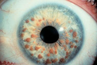
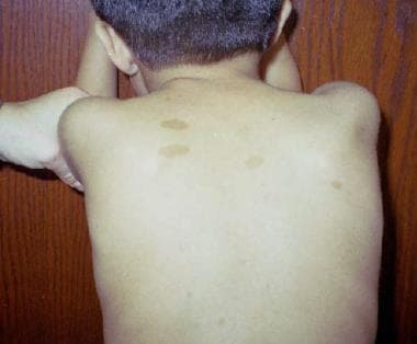
* Multiple café au lait spots (flat, light brown patches on the skin)
* Freckles in the underarms and groin area
* Benign growths on or under the skin called neurofibromas
* Larger, more complex tumors called plexiform neurofibromas
* Optic gliomas (tumors that form on the optic nerve)
* Distinctive bone abnormalities, such as a curved spine (scoliosis) or an enlarged head (macrocephaly)
* Learning disabilities and behavioral problems
Neurofibromatosis 1 is caused by mutations in the NF1 gene, which provides instructions for making a protein called neurofibromin. This protein helps regulate cell growth and division. When the NF1 gene is mutated, the production of neurofibromin is reduced or absent, leading to uncontrolled cell growth and the development of tumors.
NF1 is an autosomal dominant disorder, which means that a person has a 50% chance of inheriting the mutated gene from an affected parent. However, about half of all cases are the result of new mutations in the NF1 gene, and occur in people with no family history of the disorder.
There is currently no cure for Neurofibromatosis 1, but treatments are available to manage the symptoms and complications of the disease. These may include medications to control pain or reduce the size of tumors, surgery to remove tumors or correct bone abnormalities, and physical therapy to improve mobility and strength. Regular monitoring by a healthcare team experienced in treating Neurofibromatosis 1 is also important to detect any changes in the condition and provide appropriate care.
Optic nerve injuries refer to damages or trauma inflicted on the optic nerve, which is a crucial component of the visual system. The optic nerve transmits visual information from the retina to the brain, enabling us to see. Injuries to the optic nerve can result in various visual impairments, including partial or complete vision loss, decreased visual acuity, changes in color perception, and reduced field of view.
These injuries may occur due to several reasons, such as:
1. Direct trauma to the eye or head
2. Increased pressure inside the eye (glaucoma)
3. Optic neuritis, an inflammation of the optic nerve
4. Ischemia, or insufficient blood supply to the optic nerve
5. Compression from tumors or other space-occupying lesions
6. Intrinsic degenerative conditions affecting the optic nerve
7. Toxic exposure to certain chemicals or medications
Optic nerve injuries are diagnosed through a comprehensive eye examination, including visual acuity testing, slit-lamp examination, dilated fundus exam, and additional diagnostic tests like optical coherence tomography (OCT) and visual field testing. Treatment options vary depending on the cause and severity of the injury but may include medications, surgery, or vision rehabilitation.
The optic disk, also known as the optic nerve head, is the point where the optic nerve fibers exit the eye and transmit visual information to the brain. It appears as a pale, circular area in the back of the eye, near the center of the retina. The optic disk has no photoreceptor cells (rods and cones), so it is insensitive to light. It is an important structure to observe during eye examinations because changes in its appearance can indicate various ocular diseases or conditions, such as glaucoma, optic neuritis, or papilledema.
Optic neuritis is a medical condition characterized by inflammation and damage to the optic nerve, which transmits visual information from the eye to the brain. This condition can result in various symptoms such as vision loss, pain with eye movement, color vision disturbances, and pupillary abnormalities. Optic neuritis may occur in isolation or be associated with other underlying medical conditions, including multiple sclerosis, neuromyelitis optica, and autoimmune disorders. The diagnosis typically involves a comprehensive eye examination, including visual acuity testing, dilated funduscopic examination, and possibly imaging studies like MRI to evaluate the optic nerve and brain. Treatment options may include corticosteroids or other immunomodulatory therapies to reduce inflammation and prevent further damage to the optic nerve.
The optic chiasm is a structure in the brain where the optic nerves from each eye meet and cross. This allows for the integration of visual information from both eyes into the brain's visual cortex, creating a single, combined image of the visual world. The optic chiasm plays an important role in the processing of visual information and helps to facilitate depth perception and other complex visual tasks. Damage to the optic chiasm can result in various visual field deficits, such as bitemporal hemianopsia, where there is a loss of vision in the outer halves (temporal fields) of both eyes' visual fields.
Optic atrophy is a medical term that refers to the degeneration and shrinkage (atrophy) of the optic nerve, which transmits visual information from the eye to the brain. This condition can result in various vision abnormalities, including loss of visual acuity, color vision deficiencies, and peripheral vision loss.
Optic atrophy can occur due to a variety of causes, such as:
* Traumatic injuries to the eye or optic nerve
* Glaucoma
* Optic neuritis (inflammation of the optic nerve)
* Ischemic optic neuropathy (reduced blood flow to the optic nerve)
* Compression or swelling of the optic nerve
* Hereditary or congenital conditions affecting the optic nerve
* Toxins and certain medications that can damage the optic nerve.
The diagnosis of optic atrophy typically involves a comprehensive eye examination, including visual acuity testing, refraction assessment, slit-lamp examination, and dilated funduscopic examination to evaluate the health of the optic nerve. In some cases, additional diagnostic tests such as visual field testing, optical coherence tomography (OCT), or magnetic resonance imaging (MRI) may be necessary to confirm the diagnosis and determine the underlying cause.
There is no specific treatment for optic atrophy, but addressing the underlying cause can help prevent further damage to the optic nerve. In some cases, vision rehabilitation may be recommended to help patients adapt to their visual impairment.
Retinal Ganglion Cells (RGCs) are a type of neuron located in the innermost layer of the retina, the light-sensitive tissue at the back of the eye. These cells receive visual information from photoreceptors (rods and cones) via intermediate cells called bipolar cells. RGCs then send this visual information through their long axons to form the optic nerve, which transmits the signals to the brain for processing and interpretation as vision.
There are several types of RGCs, each with distinct morphological and functional characteristics. Some RGCs are specialized in detecting specific features of the visual scene, such as motion, contrast, color, or brightness. The diversity of RGCs allows for a rich and complex representation of the visual world in the brain.
Damage to RGCs can lead to various visual impairments, including loss of vision, reduced visual acuity, and altered visual fields. Conditions associated with RGC damage or degeneration include glaucoma, optic neuritis, ischemic optic neuropathy, and some inherited retinal diseases.
Nerve fibers are specialized structures that constitute the long, slender processes (axons) of neurons (nerve cells). They are responsible for conducting electrical impulses, known as action potentials, away from the cell body and transmitting them to other neurons or effector organs such as muscles and glands. Nerve fibers are often surrounded by supportive cells called glial cells and are grouped together to form nerve bundles or nerves. These fibers can be myelinated (covered with a fatty insulating sheath called myelin) or unmyelinated, which influences the speed of impulse transmission.
The sciatic nerve is the largest and longest nerve in the human body, running from the lower back through the buttocks and down the legs to the feet. It is formed by the union of the ventral rami (branches) of the L4 to S3 spinal nerves. The sciatic nerve provides motor and sensory innervation to various muscles and skin areas in the lower limbs, including the hamstrings, calf muscles, and the sole of the foot. Sciatic nerve disorders or injuries can result in symptoms such as pain, numbness, tingling, or weakness in the lower back, hips, legs, and feet, known as sciatica.
A nerve crush injury is a type of peripheral nerve injury that occurs when there is excessive pressure or compression applied to a nerve, causing it to become damaged or dysfunctional. This can happen due to various reasons such as trauma from accidents, surgical errors, or prolonged pressure on the nerve from tight casts, clothing, or positions.
The compression disrupts the normal functioning of the nerve, leading to symptoms such as numbness, tingling, weakness, or pain in the affected area. In severe cases, a nerve crush injury can cause permanent damage to the nerve, leading to long-term disability or loss of function. Treatment for nerve crush injuries typically involves relieving the pressure on the nerve, providing supportive care, and in some cases, surgical intervention may be necessary to repair the damaged nerve.
Brain neoplasms, also known as brain tumors, are abnormal growths of cells within the brain. These growths can be benign (non-cancerous) or malignant (cancerous). Benign brain tumors typically grow slowly and do not spread to other parts of the body. However, they can still cause serious problems if they press on sensitive areas of the brain. Malignant brain tumors, on the other hand, are cancerous and can grow quickly, invading surrounding brain tissue and spreading to other parts of the brain or spinal cord.
Brain neoplasms can arise from various types of cells within the brain, including glial cells (which provide support and insulation for nerve cells), neurons (nerve cells that transmit signals in the brain), and meninges (the membranes that cover the brain and spinal cord). They can also result from the spread of cancer cells from other parts of the body, known as metastatic brain tumors.
Symptoms of brain neoplasms may vary depending on their size, location, and growth rate. Common symptoms include headaches, seizures, weakness or paralysis in the limbs, difficulty with balance and coordination, changes in speech or vision, confusion, memory loss, and changes in behavior or personality.
Treatment for brain neoplasms depends on several factors, including the type, size, location, and grade of the tumor, as well as the patient's age and overall health. Treatment options may include surgery, radiation therapy, chemotherapy, targeted therapy, or a combination of these approaches. Regular follow-up care is essential to monitor for recurrence and manage any long-term effects of treatment.
Nerve regeneration is the process of regrowth and restoration of functional nerve connections following damage or injury to the nervous system. This complex process involves various cellular and molecular events, such as the activation of support cells called glia, the sprouting of surviving nerve fibers (axons), and the reformation of neural circuits. The goal of nerve regeneration is to enable the restoration of normal sensory, motor, and autonomic functions impaired due to nerve damage or injury.
Peripheral nerves are nerve fibers that transmit signals between the central nervous system (CNS, consisting of the brain and spinal cord) and the rest of the body. These nerves convey motor, sensory, and autonomic information, enabling us to move, feel, and respond to changes in our environment. They form a complex network that extends from the CNS to muscles, glands, skin, and internal organs, allowing for coordinated responses and functions throughout the body. Damage or injury to peripheral nerves can result in various neurological symptoms, such as numbness, weakness, or pain, depending on the type and severity of the damage.
Ischemic optic neuropathy (ION) is a medical condition that refers to the damage or death of the optic nerve due to insufficient blood supply. The optic nerve is responsible for transmitting visual information from the eye to the brain.
In ION, the blood vessels that supply the optic nerve become blocked or narrowed, leading to decreased blood flow and oxygen delivery to the nerve fibers. This results in inflammation, swelling, and ultimately, damage to the optic nerve. The damage can cause sudden, painless vision loss, often noticed upon waking up in the morning.
There are two types of ION: anterior ischemic optic neuropathy (AION) and posterior ischemic optic neuropathy (PION). AION affects the front part of the optic nerve, while PION affects the back part of the nerve. AION is further classified into arteritic and non-arteritic types, depending on whether it is caused by giant cell arteritis or not.
Risk factors for ION include age (most commonly occurring in people over 50), hypertension, diabetes, smoking, sleep apnea, and other cardiovascular diseases. Treatment options depend on the type and cause of ION and may include controlling underlying medical conditions, administering corticosteroids, or undergoing surgical procedures to improve blood flow.
An axon is a long, slender extension of a neuron (a type of nerve cell) that conducts electrical impulses (nerve impulses) away from the cell body to target cells, such as other neurons or muscle cells. Axons can vary in length from a few micrometers to over a meter long and are typically surrounded by a myelin sheath, which helps to insulate and protect the axon and allows for faster transmission of nerve impulses.
Axons play a critical role in the functioning of the nervous system, as they provide the means by which neurons communicate with one another and with other cells in the body. Damage to axons can result in serious neurological problems, such as those seen in spinal cord injuries or neurodegenerative diseases like multiple sclerosis.
The retina is the innermost, light-sensitive layer of tissue in the eye of many vertebrates and some cephalopods. It receives light that has been focused by the cornea and lens, converts it into neural signals, and sends these to the brain via the optic nerve. The retina contains several types of photoreceptor cells including rods (which handle vision in low light) and cones (which are active in bright light and are capable of color vision).
In medical terms, any pathological changes or diseases affecting the retinal structure and function can lead to visual impairment or blindness. Examples include age-related macular degeneration, diabetic retinopathy, retinal detachment, and retinitis pigmentosa among others.
Intraocular pressure (IOP) is the fluid pressure within the eye, specifically within the anterior chamber, which is the space between the cornea and the iris. It is measured in millimeters of mercury (mmHg). The aqueous humor, a clear fluid that fills the anterior chamber, is constantly produced and drained, maintaining a balance that determines the IOP. Normal IOP ranges from 10-21 mmHg, with average values around 15-16 mmHg. Elevated IOP is a key risk factor for glaucoma, a group of eye conditions that can lead to optic nerve damage and vision loss if not treated promptly and effectively. Regular monitoring of IOP is essential in diagnosing and managing glaucoma and other ocular health issues.
Glaucoma is a group of eye conditions that damage the optic nerve, often caused by an abnormally high pressure in the eye (intraocular pressure). This damage can lead to permanent vision loss or even blindness if left untreated. The most common type is open-angle glaucoma, which has no warning signs and progresses slowly. Angle-closure glaucoma, on the other hand, can cause sudden eye pain, redness, nausea, and vomiting, as well as rapid vision loss. Other less common types of glaucoma also exist. While there is no cure for glaucoma, early detection and treatment can help slow or prevent further vision loss.
Papilledema is a medical term that refers to swelling of the optic nerve head, also known as the disc, which is the point where the optic nerve enters the back of the eye (the retina). This swelling can be caused by increased pressure within the skull, such as from brain tumors, meningitis, or idiopathic intracranial hypertension. Papilledema is usually detected through a routine eye examination and may be accompanied by symptoms such as headaches, visual disturbances, and nausea. If left untreated, papilledema can lead to permanent vision loss.
The optic lobe in non-mammals refers to a specific region of the brain that is responsible for processing visual information. It is a part of the protocerebrum in the insect brain and is analogous to the mammalian visual cortex. The optic lobes receive input directly from the eyes via the optic nerves and are involved in the interpretation and integration of visual stimuli, enabling non-mammals to perceive and respond to their environment. In some invertebrates, like insects, the optic lobe is further divided into subregions, including the lamina, medulla, and lobula, each with distinct functions in visual processing.
Hereditary optic atrophies (HOAs) are a group of genetic disorders that cause degeneration of the optic nerve, leading to vision loss. The optic nerve is responsible for transmitting visual information from the eye to the brain. In HOAs, this nerve degenerates over time, resulting in decreased visual acuity, color vision deficits, and sometimes visual field defects.
There are several types of HOAs, including dominant optic atrophy (DOA), Leber hereditary optic neuropathy (LHON), autosomal recessive optic atrophy (AROA), and Wolfram syndrome. Each type has a different inheritance pattern and is caused by mutations in different genes.
DOA is the most common form of HOA and is characterized by progressive vision loss that typically begins in childhood or early adulthood. It is inherited in an autosomal dominant manner, meaning that a child has a 50% chance of inheriting the disease-causing mutation from an affected parent.
LHON is a mitochondrial disorder that primarily affects males and is characterized by sudden, severe vision loss that typically occurs in young adulthood. It is caused by mutations in the mitochondrial DNA and is inherited maternally.
AROA is a rare form of HOA that is inherited in an autosomal recessive manner, meaning that both copies of the gene must be mutated to cause the disease. It typically presents in infancy or early childhood with progressive vision loss.
Wolfram syndrome is a rare genetic disorder that affects multiple organs, including the eyes, ears, and endocrine system. It is characterized by diabetes insipidus, diabetes mellitus, optic atrophy, and hearing loss. It is inherited in an autosomal recessive manner.
There is currently no cure for HOAs, but treatments such as low-vision aids and rehabilitation may help to manage the symptoms. Research is ongoing to develop new therapies for these disorders.
Optic flow is not a medical term per se, but rather a term used in the field of visual perception and neuroscience. It refers to the pattern of motion of objects in the visual field that occurs as an observer moves through the environment. This pattern of motion is important for the perception of self-motion and the estimation of egocentric distance (the distance of objects in the environment relative to the observer). Optic flow has been studied in relation to various clinical populations, such as individuals with vestibular disorders or visual impairments, who may have difficulty processing optic flow information.
 Optic nerve glioma
Oncomodulin
Glioma
Optic nerve tumor
Nervous system neoplasm
Neurofibromatosis type I
Rory Guilday
Orbital cellulitis
List of MeSH codes (C04)
List of cancer types
List of MeSH codes (C10)
Pilocytic astrocytoma
Neurofibromin 1
Orbit (anatomy)
Legius syndrome
Juvenile myelomonocytic leukemia
Cranial nerves
Papilledema
Optic neuropathy
List of diseases (O)
Chiasmal syndrome
Phakomatosis
Role of microglia in disease
Eugène Devic
Endoscopic endonasal surgery
Megalopapilla
Brain tumor
Astroblastoma
List of MeSH codes (C11)
Neurofibromatosis
Optic nerve glioma - Wikipedia
 Optic Pathway (Optic Nerve) Glioma Imaging and Diagnosis: Practice Essentials, Computed Tomography, Magnetic Resonance Imaging
 Optic nerve glioma (Concept Id: C0346326)
- MedGen - NCBI
Optic nerve glioma (Concept Id: C0346326)
- MedGen - NCBI
Opthalmology Lec 56 Topic 47 Optic Nerve Glioma | Incus Quiz Private
glioma - Symptoms, Treatments and Resources for glioma
 Visual field: MedlinePlus Medical Encyclopedia
 Who can get brain cancer? Risk factors, genetics, and more
Fighting brain cancer with the Zika virus
Use of Gadolinium in MRI and MRA Linked to NSF/NFD in Patients With...
 Zimmerman Lecture - American Academy of Ophthalmology
 Optic Neuritis Clinical Guide | Cleveland Clinic
 Visual field
 Around the Eye in 365 Days - SLACK Books
 Conditions Treated | Brain Tumor Center
 Brain Tumor Treatment: Neuro-Oncology Expertise | UVA Health
 Clinical Profile of Unilateral Proptosis in a Tertiary Care Centre
 Neurofibromatosis Type 1 | Denver Health
 Brain Tumors | Boston Children's Hospital
 Toddler With Progressive Proptosis From Acute Myelogenous Leukemia
 Idiopathic reactive hyperplasia of the retinal pigment epithelium
 Clinical - The University of Auckland
 Kaiser Permanente Genetics Northern California
 Volume 63 Issue 24 | Cancer Research | American Association for Cancer Research
 Proton Therapy Survivor | Stephanie Mullins | MD Anderson Cancer Center
Strabismus - StatPearls - NCBI Bookshelf
Meningiomas Pathology: Definition, Overview, Etiology
Ashwagandha 60 caps - Trusted Ashwagandha OTC
 Roman G
 Yimei Li, PhD | Children's Hospital of Philadelphia
 Cranial Nerve Palsy | 3rd nerve palsy | 4th nerve palsy | 6th nerve palsy
Chiasm8
- Optic gliomas are usually pilocytic tumors, and can involve the optic nerve or optic chiasm. (wikipedia.org)
- While CT scans allow for optic nerve evaluation, MRI allows for intracranial evaluation to observe if the tumor has extended to other regions such as the optic chiasm & hypothalamus. (wikipedia.org)
- Once the optic chiasm is involved, the prognosis for life & vision worsens. (wikipedia.org)
- The diagnosis may be made with a high degree of confidence when the lesion involves the optic chiasm and retrochiasmatic optic pathway. (medscape.com)
- A glioma originating in the optic nerve or optic chiasm. (nih.gov)
- Portable MRI to assess optic chiasm decompression after endoscopic endonasal resection of sellar and suprasellar lesions. (yalemedicine.org)
- The optic nerve contains retinal ganglion cell axons that extend posteriorly from the globe, through the orbit and optic canal, to reach the optic chiasm. (medscape.com)
- Magnetic resonance imaging revealed an irregular, heterogeneously T2 hyperintense/T1 isointense mass in the region of the optic chiasm. (bvsalud.org)
Tumors7
- Optic pathway gliomas are benign tumors that are classified as pilocytic astrocytomas . (medscape.com)
- They constitute 50% of primary optic nerve tumors and 1.5-4% of all orbital tumors. (medscape.com)
- Less common but potentially more serious manifestations include optic nerve and other central nervous system gliomas, malignant peripheral nerve sheath tumors, scoliosis, tibial dysplasia, vasculopathy, and gastrointestinal, endocrine, or pulmonary disease. (nih.gov)
- This is where people have mutations on the APC , MLH1 , or PMS2 genes that increase the risk of brain tumors and gliomas. (medicalnewstoday.com)
- Gliomas are malignant brain tumors that arise from glia, brain cells that provide support for neurons and act as insulation between them. (stanfordchildrens.org)
- Sometimes tumors develop in the nerves that connect the brain to the eyes (optic nerves). (msdmanuals.com)
- Tumors compressing the optic nerve in the intracranial space may cause additional neurologic morbidity (eg, endocrine dysfunction, cranial nerve palsies, papilledema, stroke). (medscape.com)
Tumor8
- The most common type of brain tumor is glioma. (cincinnatichildrens.org)
- Gliomas also can be named according to the type of glial cells involved or the location of the tumor. (childrenshospital.org)
- An optic glioma is a tumor of the optic nerve (the nerve which controls vision). (kaiserpermanente.org)
- She went in to see a neurosurgeon who diagnosed her with a brain tumor known as optic nerve glioma, a cancer typically seen in children. (mdanderson.org)
- A tumor growing near the nerves that connect the eyes to the brain may alter vision. (stanfordchildrens.org)
- Brain tumor affects the optic nerve and can lead to vision loss, blurred or doubled vision, squinting, change in pupil size, etc. (wellbeingnutrition.com)
- Vestibular Schwannoma A vestibular schwannoma (also called an acoustic neuroma) is a noncancerous (benign) tumor that originates in the cells that wrap around the vestibular nerve (Schwann cells). (msdmanuals.com)
- Rarely, an intrinsic lesion of the optic nerve (ie, optic nerve glioma) can cause damage to the individual axons due to slow compression of the fascicles within the tumor. (medscape.com)
Neurofibromatosis5
- Optic nerve glioma (or optic glioma), a form of glioma which affects the optic nerve, is often one of the central nervous system manifestations of neurofibromatosis 1. (wikipedia.org)
- Optic gliomas are usually associated with neurofibromatosis type 1 in 30% of people with the condition. (wikipedia.org)
- The second patient was a child with neurofibromatosis type 1 who developed a pigmented peripapillary lesion following excision of an optic nerve glioma. (nih.gov)
- Risk of optic pathway glioma in children with neurofibromatosis type 1 and optic nerve tortuosity or nerve sheath thickening. (chop.edu)
- Neurofibromatosis is a group of genetic disorders in which many soft, fleshy growths of nerve tissue (neurofibromas) form under the skin and in other parts of the body, and flat spots that are the color of coffee with milk (café-au-lait spots) often develop on the skin. (msdmanuals.com)
Pathway2
- Optic nerve glioma (also known as optic pathway glioma) is the most common primary neoplasm of the optic nerve. (medscape.com)
- An injury to the optic nerve anywhere along this pathway by an extrinsic lesion is termed compressive optic neuropathy (CON). (medscape.com)
Neuritis14
- False-positive results can occur because of unilateral optic nerve enhancement or other unilateral disorders, such as optic meningioma, vascular lesions, neuritis, pseudotumor, and sarcoidosis. (medscape.com)
- What are the typical clinical features of optic neuritis? (clevelandclinic.org)
- Optic neuritis (ON) is a common manifestation of multiple sclerosis (MS), and refers to inflammation of the optic nerve. (clevelandclinic.org)
- The pain that occurs with optic neuritis is usually ocular, retroocular, periorbital, or a frontal headache. (clevelandclinic.org)
- How to approach the patient with suspected optic neuritis? (clevelandclinic.org)
- Funduscopic examination can appear normal acutely, but disc edema can be present in approximately one-third of patients (particularly those with anterior optic neuritis).1, 2 Optic disc pallor is generally seen weeks to months following onset of typical optic neuritis. (clevelandclinic.org)
- Imaging modalities used in the diagnosis of optic neuritis include orbital MRI and optical coherence tomography (OCT). These tools can be particularly helpful if the clinical history or physical examination findings are atypical for ON. (clevelandclinic.org)
- VEPs evaluate optic nerve function by calculating P100 latency and amplitude, which are generally abnormal in the setting of acute and remote optic neuritis. (clevelandclinic.org)
- Prolonged P100 latency is a characteristic of remote optic neuritis. (clevelandclinic.org)
- What is the neurologic differential diagnosis of optic neuritis? (clevelandclinic.org)
- It also leads to Discoid Lupus Erythematosus that forms a thick rash over the eyelids, Retinal Vasculitis that reduces the blood supply to the retina, and Optic Neuritis or Neuropathy, which is an inflammation around the optic nerve. (wellbeingnutrition.com)
- Optic neuritis is an early symptom of MS or multiple sclerosis. (wellbeingnutrition.com)
- There are other conditions too that can lead to optic neuritis other than MS. So, it doesn't always mean that the person with optic neuritis has or will get MS. (wellbeingnutrition.com)
- A delay in the diagnosis of compressive optic neuropathy is not uncommon since the vision loss is insidious, and the clinical findings may be missed or misinterpreted as a cataract, maculopathy, glaucoma, or optic neuritis. (medscape.com)
Tectal glioma3
- I am the mother of a three year old son who has a tectal glioma and secondary hydrocephalus. (medhelp.org)
- Anyone with Tectal Glioma? (medhelp.org)
- I'm looking to meet others with (or have loved ones with) a tectal glioma. (medhelp.org)
Pontine glioma1
- Diffuse intrinsic pontine glioma (DIPG) , also known as diffuse midline glioma (DMG). (cincinnatichildrens.org)
Neoplasm2
- A low-grade form of this neoplasm, benign optic glioma, occurs most often in pediatric patients. (medscape.com)
- Neoplasm is the most common cause of cranial nerve 6 and cranial nerve 3 palsies, while the decompensated congenital form is the the most common cause of acquired cranial nerve 4 palsy in children and adolescents. (eye.com.ph)
Compressive Optic Neuropathy8
- Compressive Optic Neuropathy. (slackbooks.com)
- The clinical hallmark of compressive optic neuropathy is slowly progressive vision loss. (medscape.com)
- Clinicians should consider compressive optic neuropathy in the differential diagnosis of unexplained, asymmetric visual acuity, normal or low-tension glaucoma, or visual symptoms responsive to corticosteroids. (medscape.com)
- The management of compressive optic neuropathy is often difficult, given the proximity of compressive lesions to critical neurovascular structures in the orbit and intracranial space. (medscape.com)
- Many of the conditions causing compressive optic neuropathy are also resistant to radiotherapy at doses tolerated by the globe and anterior visual pathways. (medscape.com)
- Compared to other ophthalmic conditions, compressive optic neuropathy (CON) is relatively rare, with an estimated incidence of about 4 cases per 100,000 individuals per year. (medscape.com)
- Compressive optic neuropathy (CON) typically causes permanent vision loss, particularly if optic atrophy is evident at the time of diagnosis. (medscape.com)
- Compressive optic neuropathy (CON) may occur at any age, but it is more common after age 30 years. (medscape.com)
Diffuse2
- In children, unenhanced CT scans typically reveal a marked, diffuse enlargement of the optic nerve, with characteristic kinking or bending. (medscape.com)
- Betsy is living with the consequences of damage to her pituitary gland as a result of being diagnosed with an inoperable diffuse optic nerve glioma . (braintumourresearch.org)
Neurofibromas1
- Neurofibromas develop along peripheral nerves-for example, under or within the skin and just outside the spinal cord. (msdmanuals.com)
Brain10
- Brain Stem Glioma - Treatment options? (medhelp.org)
- This exam is usually used to detect brain or nerve (neurologic) problems. (medlineplus.gov)
- Changes in the TP53 gene increase the risk of gliomas in the brain and other areas of the body, such as the breasts. (medicalnewstoday.com)
- Images may be taken of your nerves and brain. (denverhealth.org)
- The spinal nerves connect the brain with most parts of the body. (brainchild.org.au)
- Gliomas are categorized by where in the brain they are found and the specific type of glial cells - there are multiple types - that give rise to them. (stanfordchildrens.org)
- Brain stem glioma s are derived from the glial cells of the brain stem and occur most commonly in children between 5 and 10 years old. (stanfordchildrens.org)
- A child with a brain stem glioma may experience double vision, problems with walking or coordination, or difficulty moving their face or even one entire side of their body. (stanfordchildrens.org)
- A neurocutaneous syndrome causes problems that affect the brain, spine, and nerves (neuro) and the skin (cutaneous). (msdmanuals.com)
- the nerves outside the brain and spinal cord). (msdmanuals.com)
Retinal2
- OCT evaluates the optic nerve axonal integrity by measurement of the retinal nerve fiber layer (RNFL), and is generally used to evaluate for evidence of prior ON. (clevelandclinic.org)
- Key clinical message: Although rare, intraocular glioma is a differential diagnosis for hyphema, glaucoma, and retinal detachment. (bvsalud.org)
Cranial nerve palsies3
- 1 What are cranial nerve palsies? (eye.com.ph)
- 2 How do cranial nerve palsies present? (eye.com.ph)
- Congenital cranial nerve palsies usually present with a compensatory face turn, head tilt or chin-up position. (eye.com.ph)
Hemangioblastomas1
- Stereotactic irradiation may be effective for supratentorial hemangioblastomas including lesions of optic nerves. (bvsalud.org)
Lesion3
- In addition, fine calcification, which may help to identify a lesion as a meningioma rather than a glioma, is visualized best through CT scanning. (medscape.com)
- Similarly, a workup of incidentally discovered optic atrophy should include a neuroimaging study (eg, magnetic resonance imaging) to rule out a compressive lesion. (medscape.com)
- Optic nerve compression by an extrinsic lesion has been postulated to cause atrophy of ganglion cell axons either through ischemia or mechanical disruption of axonal transport. (medscape.com)
Sheath1
- Nerve impulses travel much faster in nerves with a myelin sheath than in those without one. (msdmanuals.com)
Neuropathy4
- The presence of hemorrhages or exudates on funduscopic examination is more suggestive of other, non-demyelinating etiologies of optic neuropathy and warrants ophthalmology evaluation. (clevelandclinic.org)
- Anterior ischemic optic neuropathy (AION), arteritic or nonarteritic. (clevelandclinic.org)
- Hypersensitive retinopathy (damage of the blood vessels in the eye, causing loss of vision or blurred vision), choroidopathy (fluid build-up around the retina causing a distorted vision), and neuropathy (blockage of blood flow that kills the nerve cells resulting in vision loss) are some eye and eyesight problems in the eye that occur due to high blood pressure. (wellbeingnutrition.com)
- Nonarteritic anterior ischemic optic neuropathy (nAION) is the second most common degenerative disease of the optic nerve. (bvsalud.org)
Benign2
- The tumor's slow & self-limiting growth indicates that it is not immediately problematic in most benign cases, with long-term studies showing that people with optic glioma may still have stable functional vision without intervention. (wikipedia.org)
- Hi, I am a 41 year old female with a "benign" Insula Glioma. (medhelp.org)
Intraorbital3
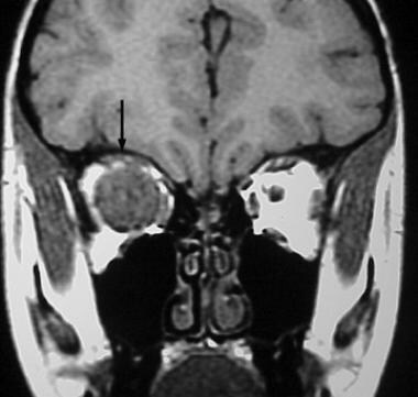
- Coronal noncontrast T1-weighted MRI reveals a large intraorbital mass (arrow) centered on the optic nerve. (medscape.com)
- The total length of the optic nerve averages 50 mm: 1 mm for the intraocular segment, 25 mm for the intraorbital segment, 10 mm for the intracanalicular segment, and 14 mm for the intracranial segment. (medscape.com)
- The authors describe a rare case of stereotactic irradiation of intraorbital hemangioblastoma of the optic nerve in a patient with Von Hippel-Lindau disease. (bvsalud.org)
Typically1
- In children, CON is typically caused by optic nerve glioma or rhabdomyosarcoma. (medscape.com)
Patient's1
- Axial T2-weighted MRI in a 46-year-old man demonstrates a mass in the lateral geniculate nucleus of the thalamus resulting from contiguous extension of the patient's known optic nerve glioma. (medscape.com)
Intraocular1
- Magnetic resonance imaging should be considered in cases of intraocular neoplasia, notably in those with incomplete surgical margins of the optic nerve. (bvsalud.org)
Extraocular muscles2
- Cranial nerve 3 supplies majority of the extraocular muscles, while cranial nerve 4 and 6 supplies the superior oblique and the lateral rectus, respectively. (eye.com.ph)
- The majority of cases encountered in clinical practice are due to Graves' orbitopathy, with compression of the optic nerve by the enlarged extraocular muscles at the orbital apex. (medscape.com)
Schwannomas1
- This condition occurs when mutations of the NF1 or NF2 gene increase the risk of cancers such as schwannomas , gliomas , and meningiomas . (medicalnewstoday.com)
Lesions1
- [ 9 , 10 ] Although MRI may reveal even subtle lesions of the optic nerve, CT scanning can detect a subtle erosion or expansion of the optic canal. (medscape.com)
Characteristic1
- Characteristic findings on clinical examination include reduced visual acuity, dyschromatopsia, a relative afferent pupillary defect, visual field defect, and optic atrophy (or edema). (medscape.com)
Visual field1
- The typical history of a sudden onset of scotoma without associated pain in conjunction with (sectorial) optic disc swelling, an afferent pupillary defect and a visual field defect are of decisive diagnostic importance. (bvsalud.org)
Mortality1
- Optic nerve gliomas have low mortality but extremely high prevalence of vision loss & eye-bulging exophthalmos) in children. (wikipedia.org)
Peripheral nervou1
- Cranial nerves are parts of the peripheral nervous system that supply the muscles of eye movement. (eye.com.ph)
Spinal cord1
- The spinal cord is part of the central nervous system and made up of bundles of nerve fibres. (brainchild.org.au)
Orbital2
- Orbital MRI with and without contrast, performed with fat saturation sequences, generally reveals enlargement and enhancement of the affected optic nerve in the acute setting. (clevelandclinic.org)
- The optic nerve is most vulnerable to injury by a compressive force where it is adjacent to bone or in a confined space (eg, orbital apex, optic canal). (medscape.com)
Examination2
- Cranial nerve function was fully intact, and neurological examination findings were normal. (contemporarypediatrics.com)
- Enucleation of the right globe was carried out, and histopathology examination revealed an optic nerve glioma with incomplete surgical margins. (bvsalud.org)
Edema1
- OCT at onset of ON is also potentially confounded by edema of the optic disc, which may lead to overestimates of baseline RNFL.4 Ganglion cell layer (GCL) thickness, another OCT measure, is not confounded by disc edema but declines in a similar time frame to RNFL and therefore may be more useful as a baseline measurement. (clevelandclinic.org)
Imaging1
- Optic nerve gliomas are diagnosed using magnetic resonance imaging (MRI) and CT scans. (wikipedia.org)
Clinical presentation1
- Optic gliomas alternate between periods of inactivity and growth, making their clinical presentation variable & clinical course unpredictable. (wikipedia.org)
Fibers2
- But once they started to look at the material, they realized that they were seeing nerve cells and fibers stained with a sharpness and readability not seen before, what one would call a technical breakthrough. (dnahelix.com)
- A nerve cell (neuron) consists of a large cell body and nerve fibers-one elongated extension (axon) for sending impulses and usually many branches (dendrites) for receiving impulses. (msdmanuals.com)
Grade1
- Description clinique avec aspect en résonance magnétique d'un gliome indéfini de haut grade du nerf optique avec extension intracrânienne. (bvsalud.org)
Vision2
- The main goal of treating optic gliomas is to preserve vision for as long as possible. (wikipedia.org)
- Optic sclerosis causes blurry and grey vision in one of the eyes. (wellbeingnutrition.com)
Gene1
- NF1 is caused by a change in a gene that normally makes proteins to control growth in the nerves. (denverhealth.org)