Orbital Fractures
Fracture Healing
Hip Fractures
Fracture Fixation, Internal
Fracture Fixation
Osteoporotic Fractures
Radius Fractures
Orbital Pseudotumor
Fractures, Spontaneous
Fractures, Stress
CSF orbitorrhoea with tension pneumocephalus. (1/65)
A seventy eight year old man sustained penetrating injury to right orbit about 15 years ago. Later he developed right orbital infection leading to phthisis bulbi. Two months before admission he developed CSF leak from the right orbit, tension pneumocephalous and meningitis. A rare case of CSF orbitorrhoea is reported here along with the discussion on mechanisms and management. (+info)Orbital blow-out fractures: correlation of preoperative computed tomography and postoperative ocular motility. (2/65)
BACKGROUND/PURPOSE: Although the management of orbital blow-out fractures was controversial for many years, refined imaging with computed tomography (CT) helped to narrow the poles of the debate. Many orbital surgeons currently recommend repair if fracture size portends late enophthalmos, or if diplopia has not substantially resolved within 2 weeks of the injury. While volumetric considerations have been generally well-served by this approach, ocular motility outcomes have been less than ideal. In one series, almost 50% of patients had residual diplopia 6 months after surgery. A fine network of fibrous septa that functionally unites the periosteum of the orbital floor, the inferior fibrofatty tissues, and the sheaths of the inferior rectus and oblique muscles was demonstrated by Koornneef. Entrapment between bone fragments of any of the components of this anatomic unit can limit ocular motility. Based on the pathogenesis of blow-out fractures, in which the fibrofatty-muscular complex is driven to varying degrees between bone fragments, some measure of soft tissue damage might be anticipated. Subsequent intrinsic fibrosis and contraction can tether globe movement, despite complete reduction of herniated orbital tissue from the fracture site. We postulated that the extent of this soft tissue damage might be estimated from preoperative imaging studies. METHODS: Study criteria included: retrievable coronal CT scans; fractures of the orbital floor without rim involvement, with or without extension into the medial wall; preoperative diplopia; surgical repair by a single surgeon; complete release of entrapped tissues; and postoperative ocular motility outcomes documented with binocular visual fields (BVFs). Thirty patients met all criteria. The CT scans and BVFs were assessed by different examiners among the authors. Fractures were classified into 3 general categories and 2 subtypes to reflect the severity of soft tissue damage within each category. "Trap-door" injuries, in which bone fragments appeared to have almost perfectly realigned, were classified as type I fractures. In the I-A subtype, no orbital tissue was visible on the sinus side of the fracture line. In the I-B subtype, soft tissue with the radiodensity of orbital fat was visible within the maxillary sinus. In type II fractures, bone fragments were distracted and soft tissue was displaced between them. In the II-A subtype, soft tissue displacement was less than, or proportional to, bone fragment distraction. In the II-B subtype, soft tissue displacement was greater than bone fragment distraction. In type III fractures, displaced bone fragments surrounded displaced soft tissue in all areas. In the III-A subtype, soft tissue and bone were moderately displaced. In the III-B subtype, both were markedly displaced. Motility outcomes were quantified by measuring the vertical excursion in BVFs. The interval between trauma and surgical repair was also determined. RESULTS: Among the 15 patients with a motility outcome in BVFs which was poorer than the median (86 degrees or less of single binocular vertical excursion), 4 patients (27%) had type A fractures; 11 patients (73%) had type B fractures. Among the 15 patients with a better outcome than the median (88 degrees or more), 10 patients (67%) had type A fractures; 5 patients (33%) had type B fractures. These differences became more defined as analysis moved away from the median. Among 5 patients with type B fractures and better than the median result in BVFs, 3 patients (60%) had surgical repair during the first week after injury. Among the 11 patients with type B fractures and less than the median result, 1 patient (9%) had repair during the first week. CONCLUSIONS: When the CT-depicted relationship between bone fragments and soft tissues is considered, a wide spectrum of injuries is subsumed under the rubric of blow-out fractures. In general, greater degrees of soft tissue incarceration or displacement, with presumably greater intrinsic damage and subsequent fibrosis, appear to result in poorer motility outcomes. Although this retrospective study does not conclusively prove its benefit, an urgent surgical approach to selected injuries should be considered. (+info)Mechanisms of orbital floor fractures: a clinical, experimental, and theoretical study. (3/65)
PURPOSE: The purpose of this study was to investigate the two accepted mechanisms of the orbital blow-out fracture (the hydraulic and the buckling theories) from a clinical, experimental, and theoretical standpoint. METHODS: Clinical cases in which blow-out fractures resulted from both a pure hydraulic mechanism and a pure buckling mechanism are presented. Twenty-one intact orbital floors were obtained from human cadavers. A metal rod was dropped, experimentally, onto each specimen until a fracture was produced, and the energy required in each instance was calculated. A biomathematical model of the human bony orbit, depicted as a thin-walled truncated conical shell, was devised. Two previously published (by the National Aeronautics Space Administration) theoretical structural engineering formulas for the fracture of thin-walled truncated conical shells were used to predict the energy required to fracture the bone of the orbital floor via the hydraulic and buckling mechanisms. RESULTS: Experimentally, the mean energy required to fracture the bone of the human cadaver orbital floor directly was 78 millijoules (mj) (range, 29-127 mj). Using the engineering formula for the hydraulic theory, the predicted theoretical energy is 71 mj (range, 38-120 mj); for the buckling theory, the predicted theoretical energy is 68 mj (range, 40-106 mj). CONCLUSION: Through this study, we have experimentally determined the amount of energy required to fracture the bone of the human orbital floor directly and have provided support for each mechanism of the orbital blow-out fracture from a clinical and theoretical basis. (+info)Strabismus due to flap tear of a rectus muscle. (4/65)
PURPOSE: To present a previously unreported avulsion-type injury of the rectus muscle, usually the inferior rectus, and detail its diagnosis and operative repair. METHODS: Thirty-five patients underwent repair of flap tears of 42 rectus muscles. The muscle abnormality was often subtle, with narrowing or thinning of the remaining attached global layer of muscle. The detached flap of external (orbital) muscle was found embedded in surrounding orbital fat and connective tissue. Retrieval and repair were performed in each case. RESULTS: Fourteen patients had orbital fractures, 7 had blunt trauma with no fracture, and 9 had suspected trauma but did not undergo computed tomographic scan. Five patients experienced this phenomenon following retinal detachment repair. Diagnostically, the predominant motility defect in 25 muscles was limitation toward the field of action of the muscle, presumably as a result of a tether created by the torn flap. These tethers simulated muscle palsy. Seventeen muscles were restricted away from their field of action, simulating entrapment. The direction taken by the flap during healing determined the resultant strabismus pattern. All patients presenting with gaze limitation toward an orbital fracture had flap tears. The worst results following flap tear repair were seen in patients who had undergone orbital fracture repair before presentation, patients who had undergone previous attempts at strabismus repair, and patients who experienced the longest intervals between the precipitating event and the repair. The best results were obtained in patients who underwent simultaneous fracture and strabismus repair or early strabismus repair alone. CONCLUSIONS: Avulsion-type flap tears of the extraocular muscles are a common cause of strabismus after trauma, and after repair for retinal detachment. Early repair produces the best results, but improvement is possible despite long delay. (+info)Antibiotics in orbital floor fractures. (5/65)
A short cut review was carried out to establish whether prophylactic antibiotics are indicated in patients with undisplaced maxillary or orbital floor fractures. Altogether 214 papers were found using the reported search, but none presented any evidence to answer the clinical question. More research is needed in this area and, in the mean time, local advice should be followed. (+info)Clinical analysis of internal orbital fractures in children. (6/65)
In order to describe the demographics, etiologic and clinical factors, and outcomes of orbital fractures in children, we have reviewed a case series of 17 patients under 18 years of age with internal orbital fractures (i.e., without involvement of the orbital rim) presenting to the Ghil hospital between March 2000 and June 2001. For 15 of the patients, we performed orbital wall reconstruction with Medpor barrier sheet implantation (thickness 1mm) through transconjunctival approach under endoscopic guidance, while maintaining mere observation on the other 2 patients. There were 14 male and 3 female patients. The most common cause of fractures was accident (7 cases). Inferior wall involvement was most commonly seen, and the trapdoor type fracture was the most common. Thirteen patients had extraocular muscle restriction, 9 had nausea/vomiting and 5 had bradycardia. Diplopia of 9 patients disappeared after 43 +/- 23 days. Nausea/vomiting and bradycardia disappeared rapidly after surgical intervention in all cases. These results suggest that trapdoor fractures with soft tissue entrapment are the most common in pediatric orbital wall fractures, and that most of them are associated with nausea/vomiting. We suggest that early diagnosis, and prompt surgical intervention are required for those patients with oculocardiac reflex. (+info)Transcaruncular approach to blowout fractures of the medial orbital wall. (7/65)
Transcutaneous and transconjunctival approaches are still frequently used to repair orbital wall fractures. However, medial orbital wall fracture remains a challenging area for plastic surgeons due to technical difficulties and postoperative scars. The transcaruncular approach is described and we present our experience with this approach to access the medial orbital wall in 10 patients with blowout fracture in the medial orbital region. All patients were corrected satisfactorily without cutaneous scar. The transcaruncular approach is a useful technique to repair medial orbital wall fractures. (+info)Intraorbital mucocele associated with old minor trauma--case report. (8/65)
A 46-year-old white man complained of swelling in the left orbital region. The only significant event in his medical history was minor trauma which occurred during ice hockey 15 years previously. On admission, the only clinical finding was left-sided exophthalmos. Computed tomography and magnetic resonance imaging revealed a left intraorbital cystic mass lesion. The cystic mass was completely removed through a left subfrontal extradural approach. There was no anatomical contact with the paranasal sinuses and the orbital walls were intact. The cystic mass was isolated in the orbital cavity. Histological examination confirmed the diagnosis of mucocele. Generally, the cause of mucocele is chronic sinusitis, but we suspect that the old minor trauma was the most likely cause in the present case. (+info)Orbital fractures refer to breaks in the bones that make up the eye socket, also known as the orbit. These bones include the maxilla, zygoma, frontal bone, and palatine bone. Orbital fractures can occur due to trauma, such as a blunt force injury or a penetrating wound.
There are several types of orbital fractures, including:
1. Blowout fracture: This occurs when the thin bone of the orbital floor is broken, often due to a direct blow to the eye. The force of the impact can cause the eyeball to move backward, breaking the bone and sometimes trapping the muscle that moves the eye (the inferior rectus).
2. Blow-in fracture: This type of fracture involves the breakage of the orbital roof, which is the bone that forms the upper boundary of the orbit. It typically occurs due to high-impact trauma, such as a car accident or a fall from a significant height.
3. Direct fracture: A direct fracture happens when there is a break in one or more of the bones that form the walls of the orbit. This type of fracture can result from a variety of traumas, including motor vehicle accidents, sports injuries, and assaults.
4. Indirect fracture: An indirect fracture occurs when the force of an injury is transmitted to the orbit through tissues surrounding it, causing the bone to break. The most common type of indirect orbital fracture is a blowout fracture.
Orbital fractures can cause various symptoms, including pain, swelling, bruising, and double vision. In some cases, the fracture may also lead to enophthalmos (sinking of the eye into the orbit) or telecanthus (increased distance between the inner corners of the eyes). Imaging tests, such as CT scans, are often used to diagnose orbital fractures and determine the best course of treatment. Treatment may include observation, pain management, and in some cases, surgery to repair the fracture and restore normal function.
Enophthalmos is a medical term that refers to the abnormal positioning of the eyeball within its socket, resulting in a posterior or backward displacement of the eye. This condition can occur due to various reasons such as trauma, surgical procedures, or diseases that affect the orbital tissues, including cancer, inflammation, or infection. Enophthalmos may lead to cosmetic concerns and visual disturbances, depending on its severity. A thorough examination by an ophthalmologist or an oculoplastic surgeon is necessary for accurate diagnosis and management of this condition.
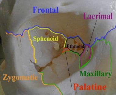
In medical terms, the orbit refers to the bony cavity or socket in the skull that contains and protects the eye (eyeball) and its associated structures, including muscles, nerves, blood vessels, fat, and the lacrimal gland. The orbit is made up of several bones: the frontal bone, sphenoid bone, zygomatic bone, maxilla bone, and palatine bone. These bones form a pyramid-like shape that provides protection for the eye while also allowing for a range of movements.
A bone fracture is a medical condition in which there is a partial or complete break in the continuity of a bone due to external or internal forces. Fractures can occur in any bone in the body and can vary in severity from a small crack to a shattered bone. The symptoms of a bone fracture typically include pain, swelling, bruising, deformity, and difficulty moving the affected limb. Treatment for a bone fracture may involve immobilization with a cast or splint, surgery to realign and stabilize the bone, or medication to manage pain and prevent infection. The specific treatment approach will depend on the location, type, and severity of the fracture.
Fracture healing is the natural process by which a broken bone repairs itself. When a fracture occurs, the body responds by initiating a series of biological and cellular events aimed at restoring the structural integrity of the bone. This process involves the formation of a hematoma (a collection of blood) around the fracture site, followed by the activation of inflammatory cells that help to clean up debris and prepare the area for repair.
Over time, specialized cells called osteoblasts begin to lay down new bone matrix, or osteoid, along the edges of the broken bone ends. This osteoid eventually hardens into new bone tissue, forming a bridge between the fracture fragments. As this process continues, the callus (a mass of newly formed bone and connective tissue) gradually becomes stronger and more compact, eventually remodeling itself into a solid, unbroken bone.
The entire process of fracture healing can take several weeks to several months, depending on factors such as the severity of the injury, the patient's age and overall health, and the location of the fracture. In some cases, medical intervention may be necessary to help promote healing or ensure proper alignment of the bone fragments. This may include the use of casts, braces, or surgical implants such as plates, screws, or rods.
Orbital diseases refer to a group of medical conditions that affect the orbit, which is the bony cavity in the skull that contains the eye, muscles, nerves, fat, and blood vessels. These diseases can cause various symptoms such as eyelid swelling, protrusion or displacement of the eyeball, double vision, pain, and limited extraocular muscle movement.
Orbital diseases can be broadly classified into inflammatory, infectious, neoplastic (benign or malignant), vascular, traumatic, and congenital categories. Some examples of orbital diseases include:
* Orbital cellulitis: a bacterial or fungal infection that causes swelling and inflammation in the orbit
* Graves' disease: an autoimmune disorder that affects the thyroid gland and can cause protrusion of the eyeballs (exophthalmos)
* Orbital tumors: benign or malignant growths that develop in the orbit, such as optic nerve gliomas, lacrimal gland tumors, and lymphomas
* Carotid-cavernous fistulas: abnormal connections between the carotid artery and cavernous sinus, leading to pulsatile proptosis and other symptoms
* Orbital fractures: breaks in the bones surrounding the orbit, often caused by trauma
* Congenital anomalies: structural abnormalities present at birth, such as craniofacial syndromes or dermoid cysts.
Proper diagnosis and management of orbital diseases require a multidisciplinary approach involving ophthalmologists, neurologists, radiologists, and other specialists.
A hip fracture is a medical condition referring to a break in the upper part of the femur (thigh) bone, which forms the hip joint. The majority of hip fractures occur due to falls or direct trauma to the area. They are more common in older adults, particularly those with osteoporosis, a condition that weakens bones and makes them more prone to breaking. Hip fractures can significantly impact mobility and quality of life, often requiring surgical intervention and rehabilitation.
Orbital neoplasms refer to abnormal growths or tumors that develop in the orbit, which is the bony cavity that contains the eyeball, muscles, nerves, fat, and blood vessels. These neoplasms can be benign (non-cancerous) or malignant (cancerous), and they can arise from various types of cells within the orbit.
Orbital neoplasms can cause a variety of symptoms depending on their size, location, and rate of growth. Common symptoms include protrusion or displacement of the eyeball, double vision, limited eye movement, pain, swelling, and numbness in the face. In some cases, orbital neoplasms may not cause any noticeable symptoms, especially if they are small and slow-growing.
There are many different types of orbital neoplasms, including:
1. Optic nerve glioma: a rare tumor that arises from the optic nerve's supportive tissue.
2. Orbital meningioma: a tumor that originates from the membranes covering the brain and extends into the orbit.
3. Lacrimal gland tumors: benign or malignant growths that develop in the lacrimal gland, which produces tears.
4. Orbital lymphangioma: a non-cancerous tumor that arises from the lymphatic vessels in the orbit.
5. Rhabdomyosarcoma: a malignant tumor that develops from the skeletal muscle cells in the orbit.
6. Metastatic tumors: cancerous growths that spread to the orbit from other parts of the body, such as the breast, lung, or prostate.
The diagnosis and treatment of orbital neoplasms depend on several factors, including the type, size, location, and extent of the tumor. Imaging tests, such as CT scans and MRI, are often used to visualize the tumor and determine its extent. A biopsy may also be performed to confirm the diagnosis and determine the tumor's type and grade. Treatment options include surgery, radiation therapy, chemotherapy, or a combination of these approaches.
A femoral fracture is a medical term that refers to a break in the thigh bone, which is the longest and strongest bone in the human body. The femur extends from the hip joint to the knee joint and is responsible for supporting the weight of the upper body and allowing movement of the lower extremity. Femoral fractures can occur due to various reasons such as high-energy trauma, low-energy trauma in individuals with weak bones (osteoporosis), or as a result of a direct blow to the thigh.
Femoral fractures can be classified into different types based on their location, pattern, and severity. Some common types of femoral fractures include:
1. Transverse fracture: A break that occurs straight across the bone.
2. Oblique fracture: A break that occurs at an angle across the bone.
3. Spiral fracture: A break that occurs in a helical pattern around the bone.
4. Comminuted fracture: A break that results in multiple fragments of the bone.
5. Open or compound fracture: A break in which the bone pierces through the skin.
6. Closed or simple fracture: A break in which the bone does not pierce through the skin.
Femoral fractures can cause severe pain, swelling, bruising, and difficulty walking or bearing weight on the affected leg. Diagnosis typically involves a physical examination, medical history, and imaging tests such as X-rays or CT scans. Treatment may involve surgical intervention, including the use of metal rods, plates, or screws to stabilize the bone, followed by rehabilitation and physical therapy to restore mobility and strength.
A spinal fracture, also known as a vertebral compression fracture, is a break in one or more bones (vertebrae) of the spine. This type of fracture often occurs due to weakened bones caused by osteoporosis, but it can also result from trauma such as a car accident or a fall.
In a spinal fracture, the front part of the vertebra collapses, causing the height of the vertebra to decrease, while the back part of the vertebra remains intact. This results in a wedge-shaped deformity of the vertebra. Multiple fractures can lead to a hunched forward posture known as kyphosis or dowager's hump.
Spinal fractures can cause pain, numbness, tingling, or weakness in the back, legs, or arms, depending on the location and severity of the fracture. In some cases, spinal cord compression may occur, leading to more severe symptoms such as paralysis or loss of bladder and bowel control.
Fracture fixation, internal, is a surgical procedure where a fractured bone is fixed using metal devices such as plates, screws, or rods that are implanted inside the body. This technique helps to maintain the alignment and stability of the broken bone while it heals. The implants may be temporarily or permanently left inside the body, depending on the nature and severity of the fracture. Internal fixation allows for early mobilization and rehabilitation, which can result in a faster recovery and improved functional outcome.
A comminuted fracture is a type of bone break where the bone is shattered into three or more pieces. This type of fracture typically occurs after high-energy trauma, such as a car accident or a fall from a great height. Commminuted fractures can also occur in bones that are weakened by conditions like osteoporosis or cancer. Because of the severity and complexity of comminuted fractures, they often require extensive treatment, which may include surgery to realign and stabilize the bone fragments using metal screws, plates, or rods.
Fracture fixation is a surgical procedure in orthopedic trauma surgery where a fractured bone is stabilized using various devices and techniques to promote proper healing and alignment. The goal of fracture fixation is to maintain the broken bone ends in correct anatomical position and length, allowing for adequate stability during the healing process.
There are two main types of fracture fixation:
1. Internal fixation: In this method, metal implants like plates, screws, or intramedullary rods are inserted directly into the bone to hold the fragments in place. These implants can be either removed or left in the body once healing is complete, depending on the type and location of the fracture.
2. External fixation: This technique involves placing pins or screws through the skin and into the bone above and below the fracture site. These pins are then connected to an external frame that maintains alignment and stability. External fixators are typically used when there is significant soft tissue damage, infection, or when internal fixation is not possible due to the complexity of the fracture.
The choice between internal and external fixation depends on various factors such as the type and location of the fracture, patient's age and overall health, surgeon's preference, and potential complications. Both methods aim to provide a stable environment for bone healing while minimizing the risk of malunion, nonunion, or deformity.
Osteoporotic fractures are breaks or cracks in bones that occur as a result of osteoporosis, a condition characterized by weak and brittle bones. Osteoporosis causes bones to lose density and strength, making them more susceptible to fractures, even from minor injuries or falls.
The most common types of osteoporotic fractures are:
1. Hip fractures: These occur when the upper part of the thigh bone (femur) breaks, often due to a fall. Hip fractures can be serious and may require surgery and hospitalization.
2. Vertebral compression fractures: These occur when the bones in the spine (vertebrae) collapse, causing height loss, back pain, and deformity. They are often caused by everyday activities, such as bending or lifting.
3. Wrist fractures: These occur when the bones in the wrist break, often due to a fall. Wrist fractures are common in older adults with osteoporosis.
4. Other fractures: Osteoporotic fractures can also occur in other bones, such as the pelvis, ribs, and humerus (upper arm bone).
Prevention is key in managing osteoporosis and reducing the risk of osteoporotic fractures. This includes getting enough calcium and vitamin D, engaging in regular weight-bearing exercise, avoiding smoking and excessive alcohol consumption, and taking medications as prescribed by a healthcare provider.
A radius fracture is a break in the bone that runs from the wrist to the elbow, located on the thumb side of the forearm. Radius fractures can occur as a result of a fall, direct blow to the forearm, or a high-energy collision such as a car accident. There are various types of radius fractures, including:
1. Distal radius fracture: A break at the end of the radius bone, near the wrist joint, which is the most common type of radius fracture.
2. Radial shaft fracture: A break in the middle portion of the radius bone.
3. Radial head and neck fractures: Breaks in the upper part of the radius bone, near the elbow joint.
4. Comminuted fracture: A complex radius fracture where the bone is broken into multiple pieces.
5. Open (compound) fracture: A radius fracture with a wound or laceration in the skin, allowing for communication between the outside environment and the fractured bone.
6. Intra-articular fracture: A radius fracture that extends into the wrist joint or elbow joint.
7. Torus (buckle) fracture: A stable fracture where one side of the bone is compressed, causing it to buckle or bend, but not break completely through.
Symptoms of a radius fracture may include pain, swelling, tenderness, bruising, deformity, limited mobility, and in some cases, numbness or tingling in the fingers. Treatment options depend on the type and severity of the fracture but can range from casting to surgical intervention with implant fixation.
Orbital pseudotumor, also known as orbital inflammatory syndrome or idiopathic orbital inflammation, is a non-specific term used to describe a group of conditions characterized by inflammation in the orbit (the bony cavity surrounding the eye) without any identifiable cause. It is not a true tumor, but rather an inflammatory reaction that can mimic the symptoms and signs of a tumor.
The condition can affect people of any age, although it is more common in middle-aged adults. The exact cause of orbital pseudotumor is unknown, but it is believed to be related to an abnormal immune response or inflammation triggered by various factors such as infections, trauma, or autoimmune disorders.
Symptoms of orbital pseudotumor may include eye pain, redness, swelling, protrusion of the eyeball (proptosis), double vision, and decreased vision. Diagnostic tests such as imaging studies (CT or MRI scans) and biopsy may be used to rule out other causes of orbital inflammation. Treatment typically involves corticosteroids to reduce inflammation, although other immunosuppressive medications may be necessary in severe cases. In some cases, the condition may resolve on its own without treatment.
Spontaneous fractures are bone breaks that occur without any identifiable trauma or injury. They are typically caused by underlying medical conditions that weaken the bones, making them more susceptible to breaking under normal stress or weight. The most common cause of spontaneous fractures is osteoporosis, a condition characterized by weak and brittle bones. Other potential causes include various bone diseases, certain cancers, long-term use of corticosteroids, and genetic disorders affecting bone strength.
It's important to note that while the term "spontaneous" implies that the fracture occurred without any apparent cause, it is usually the result of an underlying medical condition. Therefore, if you experience a spontaneous fracture, seeking medical attention is crucial to diagnose and manage the underlying cause to prevent future fractures and related complications.
Stress fractures are defined as small cracks or severe bruising in bones that occur from repetitive stress or overuse. They most commonly occur in weight-bearing bones, such as the legs and feet, but can also occur in the arms, hips, and back. Stress fractures differ from regular fractures because they typically do not result from a single, traumatic event. Instead, they are caused by repeated stress on the bone that results in microscopic damage over time. Athletes, military personnel, and individuals who engage in high-impact activities or have weak bones (osteoporosis) are at increased risk of developing stress fractures. Symptoms may include pain, swelling, tenderness, and difficulty walking or bearing weight on the affected bone.
 Orbital blowout fracture
Adam J. Oppenheimer
Superior orbital fissure
Orbital emphysema
BBS4
Eye injury
Nasolacrimal duct obstruction
The Box (Orbital song)
A. J. Burnett
Medial rectus muscle
Caldwell-Luc surgery
Skull fracture
Petey Williams
Eddie Kingston
Billy Joe Saunders
History of the South Korea national football team
Joel Embiid
Jalen Williams
South Korea national football team
Le Fort fracture of skull
Dextroscope
Kosei Tanaka
Alloplasty
Ryan Vogelsong
Sanada (wrestler)
S. Clay Wilson
Kenichi Ogata (shoot boxer)
Orbital lamina of ethmoid bone
Orbital x-ray
Tomonobu Shimizu
Orbital blowout fracture - Wikipedia
 Orbital Fractures: Practice Essentials, Problem, Epidemiology
 Orbital Socket Fractures: How to Identify and Treat Them
 Orbital floor fracture repair #2
 Pittsburgh Pirates' A.J. Burnett to undergo surgery for orbital fracture - ESPN
 Podcast - ICD 10 Coding for Orbital Fracture
 Joel Embiid injury update: 76ers star records double-double in return from orbital fracture | Sporting News Canada
Yordenis Ugas suffered fractured orbital against Errol Spence - Bad Left Hook
Use of bioresorbable implants for orbital fracture reconstruction | British Journal of Ophthalmology
Naoya Inoue dealing with fractured orbital after war with Nonito Donaire - Bad Left Hook
 ICD-10 Code for Fracture of medial orbital wall, right side, subsequent encounter for fracture with nonunion- S02.831K- Codify...
 Correlation between increased orbital volume and enophthalmos and diplopia in patients with fractures of the orbital floor or...
Orbital medial wall fractures: diagnosis and treatment - Italian Journal of Maxillofacial Surgery 2010 December;21(3):133-8 -...
NBA playoffs: Sixers' Joel Embiid out indefinitely after suffering orbital fracture in Game 6 vs. Toronto - Liberty Ballers
 Falling Umbrella Fractures Orbital Bone | Duke Health Referring Physicians
 Clinical Trials : Orbital Fractures
Orbital Fractures | Profiles RNS
 Treatment protocol for orbital fractures
 Fractures<...
Management of Orbital Fractures | Pocket Dentistry
Orbital Fractures E-Book - Imaging Science Today
 Nashville Orbital Fractures: Diagnosis, Treatment, and Oculoplastic Approaches
 Orbit, lateral orbital wall fracture
 FRACTURES OF THE ORBITAL FLOOR | JAMA Ophthalmology | JAMA Network
 Skull with Nasal and Orbital Facial Fractures : Medical Exhibit
 orbital fracture Archives - Pediatric EM Morsels
 Derrick Rose: Orbital Fracture Injury Precautions
Orbital Floor Fractures (Blowout): Background, History of the Procedure, Problem
 Orbital Trauma Boise | Eye Socket Fracture Idaho
 Fronto orbital complex fracture surgery recovery time
Trauma30
- With the trapdoor variant, there is a high frequency of extra-ocular muscle entrapment despite minimal signs of external trauma, a phenomenon that is referred to as a "white-eyed" orbital blowout fracture. (wikipedia.org)
- Orbital fractures are commonly seen with midfacial trauma. (medscape.com)
- Any or all of the orbital bones (eg, ethmoid, frontal, palatine, maxilla) may be involved in trauma, and fractures vary in their displacement and comminution. (medscape.com)
- Computed tomography (CT) scanning is considered the top choice in imaging studies for evaluating orbital trauma. (medscape.com)
- The majority of all orbital fractures - and all eye trauma in general - is caused by accidents and rarely from intentional violence. (webmd.com)
- In most cases, orbital fractures are caused by blunt force trauma, motor vehicle accidents and sometimes even physical assaults. (outsourcestrategies.com)
- Orbital floor fracture is an injury that occurs when a blow or trauma to the orbital rim pushes the bones back, causing the bones of the eye socket floor to buckle in a downward direction. (outsourcestrategies.com)
- Conclusion: The setting of a quality protocol for a trauma service is essentially important, since orbital fractures should be diagnosed accurately and quickly, seeking an appropriate treatment to minimize its after-effects. (bvsalud.org)
- The trauma of a fracture is multi-faceted - causing pain, reduced function and self-consciousness about your appearance in many cases. (drleanateston.com.au)
- These fractures are more common in adults, with a mean age of 32 years old, with the most common mechanism of injury being motor vehicle collision in adults and sport-related trauma in adolescents/children. (pocketdentistry.com)
- Changes to visual acuity or pupillary reactivity and dysfunction of the extraocular muscles are first assessed to determine the severity of the orbital trauma and need for surgical intervention. (pocketdentistry.com)
- Based on the findings, orbital trauma can be categorized into operative or nonoperative injuries and further defined based on urgency of repair as absolute (less than 24 hours), relative (within 24 hours, if possible), or delayed (within 2 weeks). (pocketdentistry.com)
- Isolated lateral orbital wall fractures are rare and only occur after isolated trauma to this anatomical structure. (aofoundation.org)
- Custom designed masks serve as a protective barrier from physical trauma to the orbital bones during healing. (ohniww.org)
- Orbital trauma is especially time sensitive in preserving vision and eye movement. (ohniww.org)
- In most cases of orbital trauma, a CT scan should be ordered to evaluate for any fractures to the orbital walls or damage to the adjacent brain tissues. (ohniww.org)
- In other cases, where trauma is more extensive, small incisions can be made under the eyelashes or to the inside of the eyelid in order to gain access to the orbital walls. (ohniww.org)
- Facial skeleton fractures can result from low-, medium-, or high-velocity trauma. (medscape.com)
- Another proposed mechanism that is less favored describes buckling of the orbital floor without displacement of orbital contents following high-velocity trauma. (medscape.com)
- A wide range of ocular trauma can occur concomitant with orbital fractures. (idahoeyelidandface.com)
- If we accept that impure fractures are the lead to of direct trauma to the orbital rim and resultant buckling of the floor, then we would expect similar rates of intraocular injury between pure and impure orbital fractures. (idahoeyelidandface.com)
- Al-Anezi M, Mahran H, Alomaym M, Salma Albati S, Alharbi M. Role of Titanium Mesh as aReconstruction Material for Orbital Floor Defects in Cases of Orbital Blowout Trauma, OHDM - 2018:17. (ipmj.org)
- Blunt force trauma and sharp force trauma due to assaults, athletic activities and falls can lead to canalicular lacerations, eyelid lacerations, orbital fractures and orbital hemorrhages. (naturalfacedr.com)
- The fragile nature of the orbital bones and adjacent sinuses make it highly likely for eye and orbit trauma to result in an orbital blowout fracture. (naturalfacedr.com)
- To prevent scarring and increase the procedure's success rate, an orbital blowout fracture should be repaired no later than 2 weeks after the trauma occurred. (naturalfacedr.com)
- Our team at LM Medical NYC in Greenwich Village and the Upper East Side offers orbital fracture treatment and facial reconstruction surgery to restore the function and appearance of the eyes after facial trauma. (lmmedicalnyc.com)
- WEBOF is a clinical diagnosis consisting of vertical diplopia, gaze restriction and nausea and/or vomiting in the setting of peri-orbital trauma in the pediatric and young-adult age group. (qxmd.com)
- Patients who have diplopia and/or pain with vertical movements of the eyes after blunt midfacial trauma should be suspected to have an orbital floor fracture. (thelmathinks.com)
- When researchers took into account age, gender and other health problems of the patient, the average costs of treating orbital trauma at the regional eye trauma center was $6,194, as compared to $12,692 at the other 21 hospitals. (thelmathinks.com)
- Blunt trauma (usually object smaller in diameter than orbit) pushes orbital contents posteriorly. (mhmedical.com)
Orbit26
- In pure orbital blowout fractures, the orbital rim (the most anterior bony margin of the orbit) is preserved, but with impure fractures, the orbital rim is also injured. (wikipedia.org)
- In children, the flexibility of the actively developing floor of the orbit fractures in a linear pattern that snaps backward. (wikipedia.org)
- Fracture severity ranges from small minimally displaced fractures of an isolated wall that require no surgical intervention to major disruption of the orbit as seen in the images below. (medscape.com)
- Orbit involvement is seen in various facial fracture patterns, including zygomaticomaxillary (ZMC), naso-orbito-ethmoid (NOE), frontal-sinus, Le Fort II, and Le Fort III fracture patterns. (medscape.com)
- Three-dimensional reconstructed images of the orbit are useful adjuncts in planning the surgical repair of complex fractures. (medscape.com)
- Orbital fractures occur when one or more of the bones around the eye ball called the orbit, or eye socket break. (outsourcestrategies.com)
- A large outdoor café umbrella toppled by high winds struck a 29-year-old man in the eye, causing a comminuted fracture of the orbit. (dukehealth.org)
- Fractures of the bones in the orbit, which include parts of the frontal, ethmoidal, lacrimal, and sphenoid bones and the maxilla and zygoma. (uchicago.edu)
- Oculoplastic surgeons perform orbital fracture reconstruction procedures, including various procedures on the tear duct, eyelids, orbit, and face. (tnoculoplastics.com)
- Figure 2: CT image of the head depicting an orbital fracture (right) and normal orbit (left). (ohniww.org)
- Orbital floor fractures can increase volume of the orbit with resultant hypoglobus and enophthalmos. (medscape.com)
- Recall that the "retropulsion" theory refers to a fracture of the orbital floor caused by sudden increase in intraorbital pressure (BLOW-OUT) which occurs when an object with sufficient force hits the aperture of the orbit and forces the soft orbital contents posteriorly. (idahoeyelidandface.com)
- Blows from a fist, for instance, or objects larger than the horizontal diameter of the orbit, are the most frequent cause of this type of fracture. (idahoeyelidandface.com)
- There was malpositioning of the medial wall of orbit fracture. (smbalaji.com)
- There was also a blowout fracture of the left floor of the orbit. (smbalaji.com)
- The medial wall of orbit fracture was addressed next. (smbalaji.com)
- Since the orbit and eyelid are complex structures, oculoplastic surgeons undergo specialized training to learn the nuances of eyelid and orbital treatments . (naturalfacedr.com)
- A blunt object larger than the orbital opening exerts force on the floor of the orbit or the medial wall, resulting in fractures of the thin bones. (aafp.org)
- A severe blow to the face can fracture any of several bones that form the orbit (the bony cavity that contains the eyeball, muscles, nerves, and blood vessels, as well as the structures that drain tears). (msdmanuals.com)
- Fractures of the floor (bottom) of the orbit (blowout fractures) are common, but fractures of other parts of the orbit also occur. (msdmanuals.com)
- This pressure can fracture one of the most fragile parts of the orbit, the part underneath the eyeball (orbital floor). (msdmanuals.com)
- Blowout fractures sometimes cause double vision, a sunken eyeball (particularly once the swelling resolves), an eyeball that is lower in the face, a decreased sensitivity to touch and pain around the cheek and upper lip (caused by injury to the nerves below the orbit), or an accumulation of air and/or blood in the tissues under the skin (subcutaneous emphysema and ecchymosis). (msdmanuals.com)
- Some people with a fracture of the orbit have no symptoms. (msdmanuals.com)
- However, most fractures of the orbit are painful, and the area swells because blood and fluid accumulate. (msdmanuals.com)
- Instead, the comparator group selected under regulatory guidance was comprised of patients who had received orbital atherectomy for severe coronary calcifications in the earlier, similarly designed ORBIT II trial, which led to FDA marketing approval of that technology. (medscape.com)
- The procedural success rate, defined as successful stent delivery with less than a 50% residual stenosis and no in-hospital MACE, was 92.4% in Disrupt CAD III, compared to 83.4% for orbital atherectomy in ORBIT II. (medscape.com)
Blowout22
- An orbital blowout fracture is a traumatic deformity of the orbital floor or medial wall that typically results from the impact of a blunt object larger than the orbital aperture, or eye socket. (wikipedia.org)
- The proximity of maxillary and ethmoidal sinus increases the susceptibility of the floor and medial wall for the orbital blowout fracture in these anatomical sites. (wikipedia.org)
- Therefore, medial wall blowout fractures are the second-most common, and superior wall, or roof and lateral wall, blowout fractures are uncommon and rare, respectively. (wikipedia.org)
- The two broad categories of blowout fractures are open door and trapdoor fractures. (wikipedia.org)
- The hinged orbital blowout fracture is a fracture with an edge of the fractured bone attached on either side. (wikipedia.org)
- However, it is known that pure blowout fractures most frequently involve the orbital floor. (wikipedia.org)
- These are also known as "blowout" fractures because the very thin base of the socket has formed a hole or crack. (webmd.com)
- The blowout fracture is the type of fracture that is a break of the thin inner wall or floor of the eye socket. (outsourcestrategies.com)
- Orbital injuries may be either blow-in or, more commonly, blowout patterns. (pocketdentistry.com)
- Note classic description of white eye blowout fracture. (pocketdentistry.com)
- Blowout fractures - These are often caused by getting hit with a baseball or fist and cause a break of the thin inner floor or wall of the eye socket. (tnoculoplastics.com)
- [ 1 ] In 1957, Smith and Regan described inferior rectus entrapment with decreased ocular motility in the setting of an orbital floor fracture and used the term blowout fracture. (medscape.com)
- Orbital blowout fractures are common in such accidents. (smbalaji.com)
- Access was gained to the orbital floor blowout fracture site. (smbalaji.com)
- Blowout fractures occur when the rim stays intact, but a crack forms in the wafer-thin bone that makes up the floor of the eye socket. (drleebottem.com)
- To review and evaluate the management of white-eyed blowout fractures (WEBOF) from Emergency Department (ED) triage through surgical repair. (qxmd.com)
- Retrospective chart review of consecutive cases of pediatric orbital blowout fracture requiring surgical repair at a large ophthalmologic referral center. (qxmd.com)
- The characteristics of patients with WEBOF and those with conventional orbital blowout fractures were compared, including: mechanism of injury, clinical presentation, ED management and referral patterns, and time to definitive treatment. (qxmd.com)
- Sixteen patients comprised the WEBOF study group, and 14 patients with conventional blowout fractures comprised the control group. (qxmd.com)
- A blowout fracture is a break of one or more of the bones that surround the eye. (thelmathinks.com)
- This review aims to develop an evidence-based pathway for isolated adult orbital blowout fractures. (liverpool.ac.uk)
- These types of injuries are known as blowout fractures. (msdmanuals.com)
Enophthalmos9
- however, because of the large defect in the orbital floor, late enophthalmos was predicted. (medscape.com)
- While some orbital fractures do not require surgery, large fractures or fractures that cause enophthalmos or diplopia do necessitate a surgical procedure. (naturalfacedr.com)
- Enophthalmos and fracture area of 22 patients was made inspection. (e-acfs.org)
- The extent of bone displacement seen on imaging can be helpful in determining if surgical repair is necessary, as a larger orbital floor fracture with greater displacement of orbital tissues will more likely result in enophthalmos and require surgical repair. (thelmathinks.com)
- Comparison of Postoperative Enophthalmos Between Fresh and Delayed Unilateral Orbital Fractures After Orbital Reconstruction With Titanium Mesh Using Computer-Assisted Navigation. (bvsalud.org)
- The sample was composed of 45 patients with post-traumatic unilateral enophthalmos who were divided into the fresh fracture group and the delayed fracture group. (bvsalud.org)
- The following parameters were measured with computed tomography images the degree of enophthalmos , orbital volume, and fracture defect area. (bvsalud.org)
- enophthalmos between T1 and T2) was similar in patients with fresh and delayed fractures. (bvsalud.org)
- Furthermore, the recurrence of enophthalmos is similar between the 2 groups, but it is higher in patients with orbital fractures involving 2 walls. (bvsalud.org)
Management of orbital8
- Large orbital floor fractures have less chance of restrictive strabismus due to nerve entrapment but a greater chance of enopthalmus There are a lot of controversies in the management of orbital fractures. (wikipedia.org)
- In the last thirty years, diagnostical imaging, surgical techniques, alloplastic materials, and surgical instruments development, have allowed a great progress in management of orbital fractures. (minervamedica.it)
- The aim of the present study is to focus on the progress and changes in the management of orbital medial wall fractures. (minervamedica.it)
- The surgical decision making in management of orbital fractures is largely dependent on patient-specific and surgeon-specific factors. (pocketdentistry.com)
- Postoperative complications in management of orbital fractures can be largely avoided with meticulous surgical technique. (pocketdentistry.com)
- Data from Boyette JR, Pemberton JD, Bonilla-Velez J. Management of orbital fractures: challenges and solutions. (pocketdentistry.com)
- This chapter will cover the clinical presentation, evaluation, examination findings, and management of orbital fractures organized from an anatomical perspective. (mhmedical.com)
- Al-Moraissi, EA, Thaller, SR & Ellis, E 2017, ' Subciliary vs. transconjunctival approach for the management of orbital floor and periorbital fractures: A systematic review and meta-analysis ', Journal of Cranio-Maxillofacial Surgery , vol. 45, no. 10, pp. 1647-1654. (uthscsa.edu)
Blow-out fractures1
- The literature favors the use of HAR% ratio, Field of Binocular Single Vision (FOBSV) and Exophthalmometer as the core tests that should form part of the standardized assessment for blow-out fractures (BOFs). (liverpool.ac.uk)
Bones21
- Most commonly, the inferior orbital wall, or the floor, is likely to collapse, because the bones of the roof and lateral walls are robust. (wikipedia.org)
- The orbital (eye) socket is a set of bones that surround and protect your eye. (webmd.com)
- The bones around the eye form the walls and floor - sides and bottom - of the orbital socket and vary in thickness. (webmd.com)
- Breaks to any of the bones in your eye socket are referred to as orbital socket fractures. (webmd.com)
- Orbital fractures can affect any of the bones surrounding the eye. (outsourcestrategies.com)
- Hand fractures can also be challenging, due to the large number of bones within the wrist and hand. (drleanateston.com.au)
- Orbital floor fracture - This type is caused by a blow to the rim of the eye socket, a fall, or a hard surface that pushes the bones back. (tnoculoplastics.com)
- When diagnosing an orbital fracture, an ophthalmologist carefully examines the injury, including the facial bones, eyelids, and surrounding soft tissue. (tnoculoplastics.com)
- This stock medical exhibit features an anterior view of the skull with severely comminuted fractures of the nasal bones and a fracture of the left orbital floor and lateral wall. (nucleusmedicalmedia.com)
- Once set into the correct position, it is important that the orbital bones do not move during healing. (ohniww.org)
- In some cases, small and thin flexible endoscopes are introduced through the nasal sinus and repairs can be made to the orbital bones without leaving any visible external scars. (ohniww.org)
- Floor fractures may occur in combination with zygomatic arch fractures, Le Fort type II or III midface fractures, or fractures of other orbital bones. (medscape.com)
- Most fractures occur in the posterior medial region that is comprised of the thinnest bones. (medscape.com)
- Intraocular injuries occur in a wide pattern when orbital bones are fractured. (idahoeyelidandface.com)
- He had suffered multiple fractures to the bones of the left upper and middle face. (smbalaji.com)
- Many materials have been used for treatment of orbital bones fractures and titanium mesh is widely used implant for this purpose therefore it worthwhile to study it. (ipmj.org)
- An orbital fracture is a serious break or cracks in one or more of the bones of the eye socket. (drdaneshrad.com)
- The facial bones that surround and protect the eyes are called the orbital sockets. (lmmedicalnyc.com)
- The most common orbital fractures affect the "floor" of the eye socket, or the bottom bones circling from the nose and under the eye. (lmmedicalnyc.com)
- Instead of fracturing, the bones flex outward, then return to their normal position. (thelmathinks.com)
- If the blunt force is strong enough, the orbital bones or other bones that are near the eye can fracture (which is the same as a break). (publicism.info)
Wall fractures5
- Combined floor and wall fractures. (webmd.com)
- Between 2005 and 2009, 11 (8 males, 3 females) consecutive patients with isolated medial orbital wall fractures were managed by authors (mean age 37.6 years, range 9-79 years). (minervamedica.it)
- Isolated lateral orbital wall fractures are very rare. (aofoundation.org)
- Some orbital wall fractures heal on their own, while others require surgery. (thelmathinks.com)
- Two types of surgery are used for orbital wall fractures: Traditional surgery, which requires an open incision. (thelmathinks.com)
Computerized tomography1
- Validity of multislice computerized tomography for diagnosis of maxillofacial fractures using an independent workstation. (uchicago.edu)
Type of fracture6
- The rim isn't broken in this type of fracture - just the floor is. (webmd.com)
- Surgery will be required to repair this type of fracture. (webmd.com)
- Most of the time this type of fracture is better when left alone. (webmd.com)
- People suffering from this type of fracture are more likely to have other injuries to the face, and possibly the optic nerve. (outsourcestrategies.com)
- This type of fracture pattern is seen in conjunction with the zygomatic complex fractures which is described in the section of the zygoma. (aofoundation.org)
- Which type of fracture is most likely to cause trismus? (thelmathinks.com)
Diplopia3
- Use of fracture size and soft tissue herniation on computed tomography to predict diplopia in isolated orbital floor fractures. (uchicago.edu)
- Although less common, orbital floor fractures in children are more likely to present with diplopia, extraocular muscle entrapment ,and nausea/emesis. (pocketdentistry.com)
- The inferior rectus muscle or orbital tissue can become entrapped within the fracture, resulting in tethering and restriction of gaze and diplopia. (medscape.com)
Bone21
- Assessing injury to the soft tissues and globe, as well as orbital and periorbital bone injury, is important. (medscape.com)
- This can be from a ball, fist, steering wheel, or anything else that hits you in the face with a lot of force and leads to a fractured orbital bone. (webmd.com)
- A frontal bone or sinus fracture is when the upper part of the rim breaks. (webmd.com)
- PITTSBURGH -- Pirates pitcher A.J. Burnett will undergo surgery on Friday to repair a fractured right orbital bone. (espn.com)
- Burnett isn't the first prominent professional athlete in Pittsburgh to sustain a fractured orbital bone. (espn.com)
- Pittsburgh Steelers linebacker James Harrison fractured his left orbital bone in a loss to Houston last October when the forepad in his helmet slammed down onto his eye. (espn.com)
- Dr. Sherrel Aston, a plastic surgeon at Lenox Hill Hospital in New York, said it takes about approximately one month to return to normal athletic activities following orbital bone surgery but could vary depending on the extent of the damage. (espn.com)
- A fracture is a broken bone in the socket involving the rim, the floor or both. (outsourcestrategies.com)
- Though minimally invasive endoscopic procedures are common, this one was unusual because of the deep comminuted fracture and the bone fragments embedded in the eye muscle," says Jang. (dukehealth.org)
- Orbital rim fracture - These types of fractures are caused by car accidents and, most of the time, affect the thick bone of the outer edges of the eye socket. (tnoculoplastics.com)
- If you delay seeing the doctor, fibrosis between the sinus mucosa, orbital tissues, and bone fragments may complicate the medical procedure. (tnoculoplastics.com)
- His left frontal bone had a depressed fracture. (smbalaji.com)
- A new mesh was then placed over the depressed frontal bone fracture. (smbalaji.com)
- An orbital fracture is a traumatic injury to the bone of the eye socket. (drleebottem.com)
- Once the fracture is visible to the surgeon, any shards of broken bone are removed, and muscles and tissue are repositioned as necessary. (drdaneshrad.com)
- The fractured orbital bone can pinch or restrict the tendons, muscles and tissues that are responsible for eye movement. (lmmedicalnyc.com)
- How do you know if you fracture your orbital bone? (thelmathinks.com)
- While not technically a bone break, trapdoor fractures can still lead to severe and sometimes permanent damage. (thelmathinks.com)
- Why does my orbital bone hurt? (thelmathinks.com)
- Orbital bone recoils faster than soft tissue, trapping soft tissue in defect. (mhmedical.com)
- Sometimes parts within the eye socket, such as a muscle attached to the eye, are forced through the fractured bone and become trapped-this happens most often in teens and young adults and requires urgent repair. (msdmanuals.com)
Facial fracture3
- Most patients with any form of facial fracture " such as orbital " will experience moderate to severe pain, which needs to be managed. (thelmathinks.com)
- It was at UFC 10 , which marked the UFC's return to a format featuring an eight-man tournament, that Don "The Predator" Frye suffered his most significant facial fracture against Mark "The Hammer" Coleman. (publicism.info)
- Dr. Charon has experience treating conditions like Facial Fracture, Vertigo and Broken Nose among other conditions at varying frequencies. (sharecare.com)
Injuries10
- The fractures can occur of pure floor, pure medial wall or combined floor and medial wall.They can occur with other injuries such as transfacial Le Fort fractures or zygomaticomaxillary complex fractures. (wikipedia.org)
- Smaller fractures are associated with a higher risk of entrapment of the nerve and therefore often smaller fracture are more serious injuries. (wikipedia.org)
- Motor vehicle accidents Falls Assault sports work-related injuries Any source of direct force There are two prevailing theories to how orbital fractures occur. (wikipedia.org)
- The blow-in pattern results from high-velocity injuries to anterior cranial base and lead to decreased orbital volume with downward and forward displacement of the globe. (pocketdentistry.com)
- Orbital fractures are traumatic injuries usually caused by assaults, sports, or accidents. (tnoculoplastics.com)
- While numerous studies have reminded the community of the importance of the ophthalmic examination in patients who sustain orbital fractures, to date, there have been only a handful of studies which focus on the incidence of intraocular injuries in patients with fracturesOcular Injury. (idahoeyelidandface.com)
- Of patients who sustained a pure orbital floor fracture, intraocular injuries occurred in 5.6%, compared with only 2% that sustained an impure fracture. (idahoeyelidandface.com)
- Intraocular injuries are more common in patients who sustained PURE orbital fractures than in patients with rim involvement (IMPURE) (p=0.05). (idahoeyelidandface.com)
- Orbital fractures are injuries frequently encountered both acutely in the Emergency Room as well as in the office as chronic conditions. (mhmedical.com)
- UFC and MMA veteran light heavyweight Brandon Vera has had some scary moments with facial injuries, including one significant orbital fracture. (publicism.info)
Eyelid2
- Pure orbital floor fractures, referred to as isolated floor fractures, result from impact injury to the globe and upper eyelid. (medscape.com)
- Depending on whether the eyelid muscles are injured, an orbital fracture can cause double vision. (drdaneshrad.com)
Reconstruction13
- A patient presented with an outcome fracture, and was treated by the reconstruction of the medial orbital wall using the nasal septum cartilage. (minervamedica.it)
- The facial symmetry is a specific measure of bodily symmetry and it influences judgements of aesthetic traits, therefore special attention and care must payed during orbital fracture treatment to consider every detail in the scenario of surgical reconstruction. (ipmj.org)
- The study included 13 patients (11 males and 2 females) presented to Oral and Maxillofacial Surgical Department in Al-Yarmook Teaching Hospital Iraq/ Baghdad in the period from January 2018 to June 2019 with traumatic orbital floor fracture and they underwent reconstruction with titanium mesh. (ipmj.org)
- The reconstruction of orbital floor using titanium mesh was successful in most of the cases and the resulted was good and satisfactory aesthetic and functional results. (ipmj.org)
- Gabrielli M F., Monnazzi M S, Passeri L A, Carvalho W R, Orbital Wall Reconstruction with Titanium Mesh. (ipmj.org)
- 10. Scolozzi P,Momjian A, Heuberger J, Andersen E,Broome M, Terzic A, Jaques B, Accuracy and Predictability in Use of AO Three-Dimensionally Preformed Titanium Mesh Plates for Posttraumatic Orbital Reconstruction: A Pilot StudyJ Craniofac Surg 2009;20:1108Y1113. (ipmj.org)
- The Evaluation of Complications of Titanium Mesh Reconstruction in Orbital Floor Fractures', Iraqi Postgraduate Medical Journal , 20(1), pp. 67-71. (ipmj.org)
- Hayder, G., Ismael, W. The Evaluation of Complications of Titanium Mesh Reconstruction in Orbital Floor Fractures. (ipmj.org)
- Fully automated analysis of orbital fracture shape and size with virtual reconstruction. (disior.com)
- However, if the fracture is causing eye dysfunction or disfigurement, orbital reconstruction surgery may be necessary. (lmmedicalnyc.com)
- If an eye socket fracture is causing eye movement issues, ongoing pain or disfigurement, orbital fracture reconstruction may be required. (lmmedicalnyc.com)
- Dr. Lesley Rabach is a double board certified facial plastic and reconstructive surgeon who provides orbital fracture repair surgery and facial reconstruction. (lmmedicalnyc.com)
- They underwent orbital reconstruction with standard preformed orbital implants and computer -assisted navigation system. (bvsalud.org)
Occur8
- Significant orbital emphysema from a communication with the maxillary sinus can occur. (medscape.com)
- Orbital rim fractures occur in the bony outer edges of the eye socket. (drleebottem.com)
- The rim is the thickest part of the socket, so rim fractures require a great deal of force to occur. (drleebottem.com)
- When blunt force impacts the eye or nose area, an orbital fracture can occur. (lmmedicalnyc.com)
- Orbital fractures can occur at various points in the eye sockets. (lmmedicalnyc.com)
- Zygomatic arch fractures tend to occur in 2-3 places along the arch. (thelmathinks.com)
- Double vision can occur because the eye is severely swollen or if one of the muscles that move the eye is trapped in the fracture. (msdmanuals.com)
- Bleeding can also occur in the eye socket (orbital hemorrhage) or the eyelids. (msdmanuals.com)
Complex fractures1
- Surgery offers permanent correction of complex fractures, and restores stability, normal function and an appealing appearance to visible areas like the face and hands. (drleanateston.com.au)
Isolated orbital floor fractures1
- Are We Overoperating on Isolated Orbital Floor Fractures? (lww.com)
Displaced orbital floor fracture1
- Minimally displaced orbital floor fracture. (medscape.com)
Socket fracture6
- What Causes an Orbital Socket Fracture? (webmd.com)
- The main cause of an orbital socket fracture is a hard hit to your face. (webmd.com)
- An orbital socket fracture is also called an eye socket fracture. (webmd.com)
- Symptoms of an orbital socket fracture will depend on the type and severity of the break. (webmd.com)
- If you have sustained an eye socket fracture and need treatment or repair, contact us at LM Medical NYC . (lmmedicalnyc.com)
- Symptoms of an eye socket fracture double vision or reduced vision. (thelmathinks.com)
Diagnosis4
- After the diagnosis of the maxillo-facial surgeon the treatment must be performed according to the orbital anatomical region involved. (bvsalud.org)
- Clinical diagnosis is based on meticulous examination of the eye, including patient vision and palpation of the orbital aperture. (aofoundation.org)
- The aim of this study was to investigate the distribution of different types of primary orbital tumors, histopathological diagnosis, and postoperative complications. (medscimonit.com)
- Diagnosis of an orbital fracture is suspected based on the symptoms and results of a physical examination. (msdmanuals.com)
Infraorbital3
- Although most pure orbital fractures affect the region medial to the infraorbital groove, any fracture type, size, or geometry is possible. (medscape.com)
- Purpose This study compared complications between subciliary and transconjunctival approaches to the infraorbital rim/orbital floor, using systematic review and meta-analysis. (uthscsa.edu)
- Randomized controlled and controlled (retrospective or prospective) clinical studies, with the aim of comparing subciliary to transconjunctival approaches in the management of infraorbital rim/orbital floor fractures, were included. (uthscsa.edu)
Postoperative1
- Prior to CT imaging, 2-D x-rays were considered sufficient for pre- and postoperative diagnostics in orbital fractures. (aofoundation.org)
Types of orbital1
- What Are the Types of Orbital Socket Fractures? (webmd.com)
Entrapment4
- Trapdoor orbital floor fracture and inferior rectus entrapment with minimal infraduction deficit and hypertropia. (uchicago.edu)
- Patient with entrapment of inferior rectus muscle due to left orbital floor fracture. (pocketdentistry.com)
- E ) Coronal CT demonstrating left orbital floor "trap-door" fracture with entrapment of inferior rectus muscle. (pocketdentistry.com)
- Use the Oculocardiac reflex when evaluating Pediatric Facial Fractures to look for ocular entrapment. (pedemmorsels.com)
Cone beam computed tomography1
- Model-based segmentation in orbital volume measurement with cone beam computed tomography and evaluation against current concepts. (disior.com)
Lateral2
- An inferior, lateral, or superior rim fracture may be an isolated injury, or it may be contiguous with an internal-wall fracture. (medscape.com)
- Much more common is a lateral orbital wall fracture together with a zygoma fracture (as shown). (aofoundation.org)
Left orbital fracture2
- Embiid suffered a left orbital fracture during the 2017-18 season that required surgery. (sportingnews.com)
- CHICAGO, October 6, 2015 - Chicago Bulls point guard Derrick Rose suffered a left orbital fracture during practice earlier this week after taking an elbow to the face. (ohniww.org)
Common orbital fractures1
- Orbital floor fractures are the most common orbital fractures encountered. (mhmedical.com)
Surgical repair1
- If there is no disfigurement of the socket, small fractures could heal without needing surgical repair. (lmmedicalnyc.com)
Traumatic3
- Orbital apex fractures are important to identify because of their association with damage to the neurovascular structures of the superior orbital fissure and optic canal (including traumatic optic neuropathy). (medscape.com)
- Zygomatic maxillary complex (ZMC), nasoorbitoethmoid (NOE), and frontal sinus fractures and traumatic optic neuropathy are discussed in other articles in this journal. (medscape.com)
- Sections of 1-mm thickness may be useful to assess optic-canal fractures and traumatic optic neuropathy. (medscape.com)
Floor or medial wall1
- Often, the fracture occurs in the orbital floor or medial wall. (naturalfacedr.com)
Surgery11
- If you have a mild fracture, you won't need surgery. (webmd.com)
- Indirect orbital fractures will only need surgery if another part of the eye has become trapped in the break or if more than 50% of the floor is broken. (webmd.com)
- Inoue said he would not require surgery to repair either fracture and that he will be reexamined by his doctor in a month to asses his progress. (badlefthook.com)
- Procedure: Orbital revision surgery Surgical revisions were performed under general anaesthesia. (digestivetracthealth.com)
- Surgery - When a surgical procedure is done, it repairs the tissue prolapse and the orbital wall, and in severe cases, sometimes, an implant is placed to create a new orbital wall. (tnoculoplastics.com)
- A 3D CT scan revealed malunion of his facial fractures from botched surgery. (smbalaji.com)
- Treatment of orbital fractures may begin with observation only, or surgery may immediately be deemed necessary. (drdaneshrad.com)
- Due to the small incisions that remain hidden inside of the eyelids, patients tend to recover quickly from orbital fracture surgery. (naturalfacedr.com)
- In some cases, an orbital fracture can heal without surgery. (lmmedicalnyc.com)
- Do you need surgery for orbital fracture? (thelmathinks.com)
- How much does orbital fracture surgery cost? (thelmathinks.com)
Emphysema2
- A common feature of an orbital wall fracture is intra-/periorbital air which appears clinically as emphysema (independent of whether the injury was penetrating or nonpenetrating). (aofoundation.org)
- Subcutaneous emphysema occurs if a fracture of the orbital floor allows air from the nose or sinuses to enter the tissues around the eye, particularly when people blow their nose. (msdmanuals.com)
Anatomical3
- The malunited fractures had to be refractured and replated in the correct anatomical position. (smbalaji.com)
- Fracture segments were aligned in normal anatomical positions. (smbalaji.com)
- Subjects that have a history of injury, infection, or deformity of at or near the anatomical site for planned product injection which may increase their risk for infection, injury, or complication related to the product (e.g., prior injury to blood vessels, lymphatics, history of orbital injury/fracture). (who.int)
Sinus1
- Fractures that involve the medial wall and floor may be considered open fractures, as laceration of the sinus mucosa is inevitable. (medscape.com)
Mild concussion5
- Ahead of Philadelphia's second-round matchup with Miami in the 2022 NBA Playoffs, Shams Charania of the Athletic broke the news that Embiid had suffered a right orbital fracture and mild concussion. (sportingnews.com)
- Sources: 76ers All-NBA star Joel Embiid suffered a right orbital fracture and mild concussion in series-clinching Game 6 win last night in Toronto. (sportingnews.com)
- The 76ers confirmed that Embiid has suffered a right orbital fracture and mild concussion. (sportingnews.com)
- On Friday evening, Shams Charania of The Athletic reported that superstar Joel Embiid suffered an orbital fracture and mild concussion during the Philadelphia 76ers ' Game 6 win over the Toronto Raptors on Thursday. (libertyballers.com)
- Joel Embiid suffered a right orbital fracture and mild concussion in last night's game vs. Toronto," read the Sixers' statement. (libertyballers.com)
Nasal1
- Orbital floor fractures alone or in conjunction with other facial skeletal fractures are the most commonly encountered midfacial fractures, second only to nasal fractures. (medscape.com)
Maxillary1
- Delayed maxillary sinusitis after orbital floor repair. (uchicago.edu)
Displacement2
- One of the great weaknesses of 2-D imaging is that in many cases a fracture is revealed, but not the degree of fracture displacement. (aofoundation.org)
- Since the advent of CT imaging, the surgeon can better define fractures, as well the degree of fracture displacement and the necessity for fracture reduction. (aofoundation.org)
Bony4
- This may be attributed to the honeycomb structure of the numerous bony septa of the ethmoid sinuses, which support the lamina papyracea, thus allowing it to withstand the sudden rise in intraorbital hydraulic pressure better than the orbital floor. (wikipedia.org)
- Once the orbital soft tissues are repositioned, an orbital implant is placed to completely cover the orbital bony defect, preventing malpositioning of the soft tissue and restoring the native bony orbital anatomic volume. (medscape.com)
- Orbital rim fracture, which is often caused by car accidents affects the thick bony outer edges of the eye socket. (outsourcestrategies.com)
- A bony step-off of the orbital rim and point tenderness are possible during palpation. (medscape.com)
Fixation3
- Over the past decade, rigid internal fixation has become the most frequently used technique in repair of floor fractures. (medscape.com)
- The old plates from the zygomaticomaxillary fracture fixation were removed. (smbalaji.com)
- Complex Orbital Fracture Repaire Using rigid fixation of internal orbital skeleton. (ipmj.org)
Tumors2
- We analyzed 122 patients (68 women and 54 men) with orbital tumors, hospitalized in the ENT Department of the Medical University of Silesia in Katowice during 1990-2013. (medscimonit.com)
- We characterized the occurrence and pathological profiles of orbital tumors. (medscimonit.com)
Severity2
- The type of treatment is determined by various factors, including the location and severity of the fracture, as well as the age and health of the patient. (drdaneshrad.com)
- During the consultation, New York oculoplastic surgeon Dr. Robert Schwarcz will examine the patient's face and eyes and assess the patient's orbital CT scan results to determine the injury's severity. (naturalfacedr.com)
Implant4
- A Supramid implant is then fashioned to cover the fracture. (uiowa.edu)
- In this case, it was determined that a Titan implant would be used due to the size the fracture. (uiowa.edu)
- The implant is then fixated to the inferior orbital rim with a 1 by 4 millimeter screw. (uiowa.edu)
- if needed, an implant is used to provide support and repair the fracture. (drdaneshrad.com)
Involve the orbital1
- Fractures may involve the orbital rim. (medscape.com)
Impure fractures1
- one theory (retropulsion) can explain variations of ocular findings in both pure and impure fractures. (idahoeyelidandface.com)
Walls1
- It states that the orbital rim buckles and transmits forces to the orbital walls, resulting in an orbital floor fracture. (idahoeyelidandface.com)
Zygomatic arch1
- His old zygomatic arch fracture was also stabilized with plates and screws. (smbalaji.com)
Zygomaticomaxillary1
- Malunion of his zygomaticomaxillary fracture had resulted in severe malocclusion. (smbalaji.com)
Increase in intraorbital2
- Orbital floor fractures are secondary to a sudden increase in intraorbital hydraulic pressure. (medscape.com)
- Resultant increase in intraorbital pressure causes fracture at weakest point: posteromedial orbital floor (hydraulic theory). (mhmedical.com)
Cause of orbital3
- Car accidents used to be a leading cause of orbital fractures, but the increased use of seatbelts and airbag requirements has greatly reduced this number. (webmd.com)
- Car accidents are a primary cause of orbital rim fractures. (drleebottem.com)
- Migraines and cluster headaches are a very common cause of orbital eye pain. (thelmathinks.com)
Treatment of orbital2
- There are several options for treatment of orbital fractures. (ohniww.org)
- Evaluate the complications of using titanium mesh in the treatment of orbital floor fractures. (ipmj.org)
Internal orbital skeleton2
- Fractures may be limited to the internal orbital skeleton. (medscape.com)
- This article focuses on fractures of the internal orbital skeleton. (medscape.com)
Nose1
- In addition to a fractured nose, "The Monster" suffered a fractured orbital that could delay his Top Rank debut . (badlefthook.com)