Ossification of Posterior Longitudinal Ligament
Longitudinal Ligaments
Ligaments
Cervical Vertebrae
Ligamentum Flavum
Spinal Cord Compression
Hyperostosis, Diffuse Idiopathic Skeletal
Decompression, Surgical
Laminectomy
Spinal Cord Diseases
Spondylosis
Spinal Osteophytosis
Spinal Fusion
Spinal Diseases
Intervertebral Disc
Quadriplegia
Ligaments, Articular
Thoracic Vertebrae
Intervertebral Disc Displacement
Radiculopathy
Tomography, X-Ray Computed
Lumbar Vertebrae
Magnetic Resonance Imaging
Periodontal Ligament
Treatment Outcome
Sternal splitting approach to upper thoracic lesions located anterior to the spinal cord. (1/57)
The sternal splitting approach for upper thoracic lesions located anterior to the spinal cord is described. The sternal splitting approach can be effectively applied to lesions from the T-1 to T-3 levels. The aortic arch prevents procedures below this level. The approach is straight toward the T1-3 vertebral bodies and provides good surgical orientation. The sternal splitting approach was applied to five patients with metastatic spinal tumors at the C7-T3 levels and three patients with ossification of the posterior longitudinal ligament at the T1-3 levels. No postoperative neurological deterioration occurred. Two patients had postoperative hoarseness. The sternal splitting approach to the upper thoracic spine is recommended for hard lesions, extensive lesions requiring radical resection, and lesions requiring postoperative stabilization with spinal instrumentation. (+info)Tetraparesis associated with ossification of the posterior longitudinal ligament of the cervical spine. (2/57)
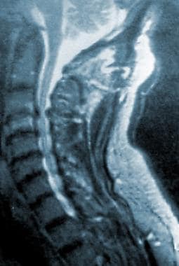
This is a case report of tetraparesis associated with extraordinarily severe ossification of the posterior longitudinal ligament (OPLL) of the cervical spine. There was no history of trauma. The object of this paper is to show that OPLL can progress relentlessly to a nearly complete quadriplegia even without trauma, but that adequate decompression can produce almost complete recovery. (+info)Laminoplasty: an evaluation of 24 cases. (3/57)
Cervical expansive laminoplasty has been advocated as an alternative procedure to laminectomy for the decompression of the cervical spine. It provides favourable cord decompression and stabilisation of the cervical spine and is a simpler and safer alternative to anterior fusion and laminectomy for myelopathy and myeloradiculopathy, due to multisegmental cervical spondylosis and ossified posterior longitudinal ligament. We report our experience in 24 patients with this procedure, 12 of whom had myelopathy and another 12 had myeloradiculopathy. The earliest symptom to improve was radicular pain or paraesthesia (75%). A reduction in spasticity was seen in 21 of the 24 patients (87.5%). Eleven patients had improvement in their motor power during a follow up period ranging from 1 month to 14 months. One patient deteriorated following the procedure and developed Brown Sequard features due to under riding of the lamina on the hinged side, another had severe post operative paraesthesias, while one patient had a CT scan evidence of 'closing of the door', without being symptomatic for it. The technique of the procedure is discussed and the pertinent literature reviewed. (+info)Surgery for ossification of the posterior longitudinal ligament of the cervical spine. (4/57)
The authors present their surgical experience with fifty seven cases of ossification of the posterior longitudinal ligament (OPLL) of the cervical spine, operated between January 1992 and January 1999. Continuous OPLL was seen in the majority of patients (40/57). Posterior decompressive surgery was performed in 18 patients, a median corpectomy and excision of the OPLL in 28 and anterior segmental decompression in 11 patients. One patient had a transient weakness of muscles supplied by the C5 myotome following a C4-C5 corpectomy. 84.2% of the patients showed improvement by at least one grade at the time of discharge. 92.8% of patients who underwent a corpectomy improved in the immediate post-operative period as compared to 90.9% of those who underwent an anterior segmental decompression and 83.3% of those who underwent a posterior decompressive procedure. 97.7% of the 44 patients followed-up between one and five years showed neurological improvement. Thirty-two patients (72.7 %) had regained normal or near normal neurological function and returned to their jobs. Good results were obtained when the surgical approach and the procedure adopted were individualised. (+info)Spinal cord and cauda equina compression in 'DISH'. (5/57)
Diffuse idiopathic skeletal hyperostosis (DISH) has long been regarded as a benign asymptomatic clinical entity with an innocuous clinical course. Precise information is lacking in the world literature. Authors report the results of a retrospective analysis of 74 cases of DISH. Eleven patients presented with progressive spinal cord or cauda equina compression. In nine cases ossified posterior longitudinal ligament (OPLL) and in two cases ossified ligamentum flavum (OLF) were primarily responsible. Surgically treated patients (eight) had far better outcome as compared to the patients managed conservatively, as they had refused surgery. 'DISH' is neither a benign condition, nor it always runs a innocuous clinical course. In fact, in about 15% of the cases, serious neurological manifestations occur, which may require a major neurosurgical intervention. (+info)Involvement of bone morphogenic protein-2 (BMP-2) in the pathological ossification process of the spinal ligament. (6/57)
OBJECTIVE: To investigate the function of bone morphogenic protein-2 (BMP-2) in the ossification of the spinal ligament (OSL). METHODS: Total RNA was prepared from the cultured spinal ligament cells from patients with OSL and analysed by reverse transcription-polymerase chain reaction using specific primers for BMP-2. BMP-2 mRNA expression in ligament tissues was examined by in situ hybridization. Spinal ligament cells from patients without OSL were treated with BMP-2 and examined for alkaline phosphatase activity. RESULTS: Expression of the BMP-2 gene was detected in cultured spinal ligament cells. In ligament tissues, BMP-2 mRNA was present in the chondrocyte-like cells in the fibrocartilage zone. Exogenous BMP-2 increased alkaline phosphatase activity in spinal ligament cells from patients without OSL. CONCLUSION: The BMP-2 gene is expressed in the spinal ligaments of OSL patients, and exogenous BMP-2 stimulates osteogenic differentiation of spinal ligament cells. The expression of BMP-2 in the spinal ligaments could be a clue in elucidating how heterotrophic osteogenesis develops in ligament tissue. (+info)Thoracic cord compression due to ossified hypertrophied ligamentum flavum. (7/57)
Ossified ligamentum flavum is increasingly appreciated as an important cause of thoracic myeloradiculopathy. Fifteen patients with age ranging from 30-61 years were studied. Fourteen presented with spastic paraparesis, and radiculopathy was the only complaint in one patient. Routine skiagrams and myelograms showed non-specific changes. Baseline CT and CT myelogram, however, documented the ossification of ligamentum flavum comprehensively. MRI was done in three patients. Multiple levels of the disease were seen in two cases. Four patients had ossified posterior longitudinal ligament. Thickened ligamentum flavum should be considered as an important cause of thoracic cord compression. (+info)Decrease in serum nucleotide pyrophosphatase activity in ankylosing spondylitis. (8/57)
OBJECTIVE: Ankylosing spondylitis (AS) is a prototype of a group of rheumatic diseases referred to as spondyloarthropathy. AS patients show marked ectopic ossification in the spine, occasionally resulting in so-called bamboo spine. Although a strong association with HLA-B27 has been reported, its aetiology remains undetermined. Another rheumatic disease, ossification of the posterior longitudinal ligament of the spine (OPLL), demonstrates ectopic ossification of the spinal ligaments very similar to that of AS. Recently, nucleotide pyrophosphatase (NPPS) was implicated in the aetiology of OPLL: an Npps mutation was found to cause OPLL in mice, and an association between a polymorphism of the human NPPS gene and OPLL was identified. The clinical similarities between AS and OPLL led us to hypothesize that NPPS may also be implicated in the aetiology of AS. To elucidate the role of NPPS in the pathogenesis of AS, we examined serum NPPS activity and the possible association of the NPPS gene with AS. METHODS: Forty-four Japanese patients with AS, 43 patients with OPLL, and age- and sex-matched normal volunteers took part in this study. We determined serum NPPS activity using high-performance liquid chromatography and examined the association between AS and NPPS using single nucleotide polymorphisms (SNPs) of the NPPS gene. RESULTS: Serum NPPS activity in AS patients was significantly decreased compared with the controls (P < 0.0001). However, there was no association between AS and NPPS gene SNPs. CONCLUSION: NPPS is implicated in the pathogenesis of AS. (+info)Ossification of the Posterior Longitudinal Ligament (OPLL) is a medical condition where there is abnormal growth and hardening (ossification) of the posterior longitudinal ligament in the spine. The posterior longitudinal ligament runs down the length of the spine, along the back of the vertebral bodies, and helps to maintain the stability and alignment of the spinal column.
In OPLL, the ossification of this ligament can cause narrowing of the spinal canal (spinal stenosis) and compression of the spinal cord or nerve roots. This condition is more commonly found in the cervical spine (neck), but it can also occur in the thoracic (chest) and lumbar (lower back) regions of the spine.
The symptoms of OPLL may include neck pain, stiffness, numbness, tingling, or weakness in the arms and/or legs, depending on the location and severity of the compression. In severe cases, it can lead to serious neurological deficits such as paralysis. The exact cause of OPLL is not fully understood, but it is believed to be related to genetic factors, aging, and mechanical stress on the spine.
Longitudinal ligaments, in the context of anatomy, refer to the fibrous bands that run lengthwise along the spine. They are named as such because they extend in the same direction as the long axis of the body. The main function of these ligaments is to provide stability and limit excessive movement in the spinal column.
There are three layers of longitudinal ligaments in the spine:
1. Anterior Longitudinal Ligament (ALL): This ligament runs down the front of the vertebral bodies, attached to their anterior aspects. It helps to prevent hyperextension of the spine.
2. Posterior Longitudinal Ligament (PLL): The PLL is located on the posterior side of the vertebral bodies and extends from the axis (C2) to the sacrum. Its primary function is to limit hyperflexion of the spine.
3. Ligamentum Flavum: Although not strictly a 'longitudinal' ligament, it is often grouped with them due to its longitudinal orientation. The ligamentum flavum is a pair of elastic bands that connect adjacent laminae (posterior bony parts) of the vertebral arch in the spine. Its main function is to maintain tension and stability while allowing slight movement between the vertebrae.
These longitudinal ligaments play an essential role in maintaining spinal alignment, protecting the spinal cord, and facilitating controlled movements within the spine.
Heterotopic ossification (HO) is a medical condition where bone tissue forms outside the skeleton, in locations where it does not typically exist. This process can occur in various soft tissues, such as muscles, tendons, ligaments, or even inside joint capsules. The abnormal bone growth can lead to pain, stiffness, limited range of motion, and, in some cases, loss of function in the affected area.
There are several types of heterotopic ossification, including:
1. Myositis ossificans - This form is often associated with trauma or injury, such as muscle damage from a fracture, surgery, or direct blow. It typically affects young, active individuals and usually resolves on its own within months to a few years.
2. Neurogenic heterotopic ossification (NHO) - Also known as "traumatic heterotopic ossification," this form is often linked to spinal cord injuries, brain injuries, or central nervous system damage. NHO can cause significant impairment and may require surgical intervention in some cases.
3. Fibrodysplasia ossificans progressiva (FOP) - This rare, genetic disorder causes progressive heterotopic ossification throughout the body, starting in early childhood. The condition significantly impacts mobility and quality of life, with no known cure.
The exact mechanisms behind heterotopic ossification are not fully understood, but it is believed that a combination of factors, including inflammation, tissue injury, and genetic predisposition, contribute to its development. Treatment options may include nonsteroidal anti-inflammatory drugs (NSAIDs), radiation therapy, physical therapy, or surgical removal of the abnormal bone growth, depending on the severity and location of the HO.
Ligaments are bands of dense, fibrous connective tissue that surround joints and provide support, stability, and limits the range of motion. They are made up primarily of collagen fibers arranged in a parallel pattern to withstand tension and stress. Ligaments attach bone to bone, and their function is to prevent excessive movement that could cause injury or dislocation.
There are two main types of ligaments: extracapsular and intracapsular. Extracapsular ligaments are located outside the joint capsule and provide stability to the joint by limiting its range of motion. Intracapsular ligaments, on the other hand, are found inside the joint capsule and help maintain the alignment of the joint surfaces.
Examples of common ligaments in the body include the anterior cruciate ligament (ACL) and posterior cruciate ligament (PCL) in the knee, the medial collateral ligament (MCL) and lateral collateral ligament (LCL) in the elbow, and the coracoacromial ligament in the shoulder.
Injuries to ligaments can occur due to sudden trauma or overuse, leading to sprains, strains, or tears. These injuries can cause pain, swelling, bruising, and limited mobility, and may require medical treatment such as immobilization, physical therapy, or surgery.
The cervical vertebrae are the seven vertebrae that make up the upper part of the spine, also known as the neck region. They are labeled C1 to C7, with C1 being closest to the skull and C7 connecting to the thoracic vertebrae in the chest region. The cervical vertebrae have unique structures to allow for a wide range of motion in the neck while also protecting the spinal cord and providing attachment points for muscles and ligaments.
The ligamentum flavum is a pair of elastic bands of tissue located in the spine. They connect the laminae, which are parts of the vertebral arch, from one vertebra to the next in the spine. These ligaments help maintain the stability and alignment of the vertebral column, allowing for a limited range of movement while preventing excessive motion that could cause injury. The elasticity of the ligamentum flavum also facilitates the return of the spinal column to its normal position after flexion.
These ligaments are named "flavum" because they have a yellowish color due to their high elastin content. They play an essential role in protecting the spinal cord and nerve roots from damage during movements of the spine. Any degeneration, thickening, or calcification of the ligamentum flavum may lead to conditions such as spinal stenosis, which can cause pain, numbness, or weakness in the back, legs, or arms.
Spinal cord compression is a medical condition that refers to the narrowing of the spinal canal, which puts pressure on the spinal cord and the nerves that branch out from it. This can occur due to various reasons such as degenerative changes in the spine, herniated discs, bone spurs, tumors, or fractures. The compression can lead to a range of symptoms including pain, numbness, tingling, weakness, or loss of bladder and bowel control. In severe cases, it can cause paralysis. Treatment options depend on the underlying cause and may include physical therapy, medication, surgery, or radiation therapy.
Diffuse Idiopathic Hyperostosis (DIH), also known as Forestier's Disease, is a non-inflammatory skeletal disorder characterized by the abnormal thickening and hardening (hyperostosis) of the bony portions of the spine and/or other parts of the skeleton. In DIH, there is an excessive formation of new bone along the edges of these bones, particularly at the sites where ligaments attach to the bones.
The term "idiopathic" indicates that the cause of this condition is currently unknown, while "diffuse" refers to its widespread involvement of multiple skeletal areas. The exact pathogenesis of DIH remains unclear; however, it has been suggested that there might be a connection with abnormal bone metabolism and/or localized inflammation.
DIH primarily affects middle-aged and older adults, with men being more commonly affected than women. Common symptoms include stiffness, pain, and limited mobility in the spine and joints. In some cases, DIH may also lead to complications such as spinal stenosis or nerve compression due to the excessive bone growth.
It is important to note that while hyperostosis can be a feature of various medical conditions, the term "Diffuse Idiopathic Skeletal Hyperostosis" specifically refers to this distinct clinical entity characterized by the widespread involvement of the skeleton and the absence of inflammation or other underlying causes.
Surgical decompression is a medical procedure that involves relieving pressure on a nerve or tissue by creating additional space. This is typically accomplished through the removal of a portion of bone or other tissue that is causing the compression. The goal of surgical decompression is to alleviate symptoms such as pain, numbness, tingling, or weakness caused by the compression.
In the context of spinal disorders, surgical decompression is often used to treat conditions such as herniated discs, spinal stenosis, or bone spurs that are compressing nerves in the spine. The specific procedure used may vary depending on the location and severity of the compression, but common techniques include laminectomy, discectomy, and foraminotomy.
It's important to note that surgical decompression is a significant medical intervention that carries risks such as infection, bleeding, and injury to surrounding tissues. As with any surgery, it should be considered as a last resort after other conservative treatments have been tried and found to be ineffective. A thorough evaluation by a qualified medical professional is necessary to determine whether surgical decompression is appropriate in a given case.
A laminectomy is a surgical procedure that involves the removal of the lamina, which is the back part of the vertebra that covers the spinal canal. This procedure is often performed to relieve pressure on the spinal cord or nerves caused by conditions such as herniated discs, spinal stenosis, or tumors. By removing the lamina, the surgeon can access the affected area and alleviate the compression on the spinal cord or nerves, thereby reducing pain, numbness, or weakness in the back, legs, or arms.
Laminectomy may be performed as a standalone procedure or in combination with other surgical techniques such as discectomy, foraminotomy, or spinal fusion. The specific approach and extent of the surgery will depend on the patient's individual condition and symptoms.
Spinal cord diseases refer to a group of conditions that affect the spinal cord, which is a part of the central nervous system responsible for transmitting messages between the brain and the rest of the body. These diseases can cause damage to the spinal cord, leading to various symptoms such as muscle weakness, numbness, pain, bladder and bowel dysfunction, and difficulty with movement and coordination.
Spinal cord diseases can be congenital or acquired, and they can result from a variety of causes, including infections, injuries, tumors, degenerative conditions, autoimmune disorders, and genetic factors. Some examples of spinal cord diseases include multiple sclerosis, spina bifida, spinal cord injury, herniated discs, spinal stenosis, and motor neuron diseases such as amyotrophic lateral sclerosis (ALS).
The treatment for spinal cord diseases varies depending on the underlying cause and severity of the condition. Treatment options may include medication, physical therapy, surgery, and rehabilitation. In some cases, the damage to the spinal cord may be irreversible, leading to permanent disability or paralysis.
Spondylosis is a general term that refers to degenerative changes in the spine, particularly in the joints (facets) between vertebrae and/or intervertebral discs. It's a common age-related condition, which can also be caused by stresses on the spine due to poor posture, repetitive movements, or injury.
The degenerative process often involves loss of hydration and elasticity in the intervertebral discs, leading to decreased disc height and potential disc herniation. This can cause narrowing of the spinal canal (spinal stenosis) or nerve root canal (foraminal stenosis), resulting in pressure on the spinal cord and/or nerves.
Spondylosis can occur throughout the spine, but it is most commonly found in the cervical (neck) and lumbar (lower back) regions. Symptoms may include pain, stiffness, numbness, tingling, or weakness in the neck, arms, legs, or back, depending on the location and severity of the degeneration. However, it's worth noting that many people with spondylosis might not experience any symptoms at all. Treatment options typically include pain management, physical therapy, and, in severe cases, surgery.
Spinal stenosis is a narrowing of the spinal canal or the neural foramina (the openings through which nerves exit the spinal column), typically in the lower back (lumbar) or neck (cervical) regions. This can put pressure on the spinal cord and/or nerve roots, causing pain, numbness, tingling, or weakness in the affected areas, often in the legs, arms, or hands. It's most commonly caused by age-related wear and tear, but can also be due to degenerative changes, herniated discs, tumors, or spinal injuries.
Spinal osteophytosis, also known as spinal osteophyte formation or bone spurs on the spine, refers to the abnormal growth of bony projections along the vertebral column's margins. These bony outgrowths develop due to degenerative changes, inflammation, or injury in the joints between the vertebrae (facet joints) and can cause stiffness, pain, and reduced mobility. In some cases, spinal osteophytosis may lead to complications such as spinal stenosis or nerve compression.
Spinal fusion is a surgical procedure where two or more vertebrae in the spine are fused together to create a solid bone. The purpose of this procedure is to restrict movement between the fused vertebrae, which can help reduce pain and stabilize the spine. This is typically done using bone grafts or bone graft substitutes, along with hardware such as rods, screws, or cages to hold the vertebrae in place while they heal together. The procedure may be recommended for various spinal conditions, including degenerative disc disease, spinal stenosis, spondylolisthesis, scoliosis, or fractures.
Spinal diseases refer to a range of medical conditions that affect the spinal column, which is made up of vertebrae (bones), intervertebral discs, facet joints, nerves, ligaments, and muscles. These diseases can cause pain, discomfort, stiffness, numbness, weakness, or even paralysis, depending on the severity and location of the condition. Here are some examples of spinal diseases:
1. Degenerative disc disease: This is a condition where the intervertebral discs lose their elasticity and height, leading to stiffness, pain, and decreased mobility.
2. Herniated disc: This occurs when the inner material of the intervertebral disc bulges or herniates out through a tear in the outer layer, causing pressure on the spinal nerves and resulting in pain, numbness, tingling, or weakness in the affected area.
3. Spinal stenosis: This is a narrowing of the spinal canal or the neural foramen (the openings where the spinal nerves exit the spinal column), which can cause pressure on the spinal cord or nerves and result in pain, numbness, tingling, or weakness.
4. Scoliosis: This is a curvature of the spine that can occur in children or adults, leading to an abnormal posture, back pain, and decreased lung function.
5. Osteoarthritis: This is a degenerative joint disease that affects the facet joints in the spine, causing pain, stiffness, and decreased mobility.
6. Ankylosing spondylitis: This is a chronic inflammatory disease that affects the spine and sacroiliac joints, leading to pain, stiffness, and fusion of the vertebrae.
7. Spinal tumors: These are abnormal growths that can occur in the spinal column, which can be benign or malignant, causing pain, neurological symptoms, or even paralysis.
8. Infections: Bacterial or viral infections can affect the spine, leading to pain, fever, and other systemic symptoms.
9. Trauma: Fractures, dislocations, or sprains of the spine can occur due to accidents, falls, or sports injuries, causing pain, neurological deficits, or even paralysis.
An intervertebral disc is a fibrocartilaginous structure found between the vertebrae of the spinal column in humans and other animals. It functions as a shock absorber, distributes mechanical stress during weight-bearing activities, and allows for varying degrees of mobility between adjacent vertebrae.
The disc is composed of two parts: the annulus fibrosus, which forms the tough, outer layer; and the nucleus pulposus, which is a gel-like substance in the center that contains proteoglycans and water. The combination of these components provides the disc with its unique ability to distribute forces and allow for movement.
The intervertebral discs are essential for the normal functioning of the spine, providing stability, flexibility, and protection to the spinal cord and nerves. However, they can also be subject to degeneration and injury, which may result in conditions such as herniated discs or degenerative disc disease.
Quadriplegia, also known as tetraplegia, is a medical condition characterized by paralysis affecting all four limbs and the trunk of the body. It results from damage to the cervical spinal cord, typically at levels C1-C8, which controls signals to the muscles in the arms, hands, trunk, legs, and pelvic organs. The extent of quadriplegia can vary widely, ranging from weakness to complete loss of movement and sensation below the level of injury. Other symptoms may include difficulty breathing, bowel and bladder dysfunction, and sexual dysfunction. The severity and prognosis depend on the location and extent of the spinal cord injury.
The spine, also known as the vertebral column, is a complex structure in the human body that is part of the axial skeleton. It is composed of 33 individual vertebrae (except in some people where there are fewer due to fusion of certain vertebrae), intervertebral discs, facet joints, ligaments, muscles, and nerves.
The spine has several important functions:
1. Protection: The spine protects the spinal cord, which is a major component of the nervous system, by enclosing it within a bony canal.
2. Support: The spine supports the head and upper body, allowing us to maintain an upright posture and facilitating movement of the trunk and head.
3. Movement: The spine enables various movements such as flexion (bending forward), extension (bending backward), lateral flexion (bending sideways), and rotation (twisting).
4. Weight-bearing: The spine helps distribute weight and pressure evenly across the body, reducing stress on individual vertebrae and other structures.
5. Blood vessel and nerve protection: The spine protects vital blood vessels and nerves that pass through it, including the aorta, vena cava, and spinal nerves.
The spine is divided into five regions: cervical (7 vertebrae), thoracic (12 vertebrae), lumbar (5 vertebrae), sacrum (5 fused vertebrae), and coccyx (4 fused vertebrae, also known as the tailbone). Each region has unique characteristics that allow for specific functions and adaptations to the body's needs.
Articular ligaments, also known as fibrous ligaments, are bands of dense, fibrous connective tissue that connect and stabilize bones to each other at joints. They help to limit the range of motion of a joint and provide support, preventing excessive movement that could cause injury. Articular ligaments are composed mainly of collagen fibers arranged in a parallel pattern, making them strong and flexible. They have limited blood supply and few nerve endings, which makes them less prone to injury but also slower to heal if damaged. Examples of articular ligaments include the anterior cruciate ligament (ACL) and posterior cruciate ligament (PCL) in the knee joint, and the medial collateral ligament (MCL) and lateral collateral ligament (LCL) in the elbow joint.
The thoracic vertebrae are the 12 vertebrae in the thoracic region of the spine, which is the portion between the cervical and lumbar regions. These vertebrae are numbered T1 to T12, with T1 being closest to the skull and T12 connecting to the lumbar region.
The main function of the thoracic vertebrae is to provide stability and support for the chest region, including protection for the vital organs within, such as the heart and lungs. Each thoracic vertebra has costal facets on its sides, which articulate with the heads of the ribs, forming the costovertebral joints. This connection between the spine and the ribcage allows for a range of movements while maintaining stability.
The thoracic vertebrae have a unique structure compared to other regions of the spine. They are characterized by having long, narrow bodies, small bony processes, and prominent spinous processes that point downwards. This particular shape and orientation of the thoracic vertebrae contribute to their role in limiting excessive spinal movement and providing overall trunk stability.
Intervertebral disc displacement, also known as a slipped disc or herniated disc, is a medical condition where the inner, softer material (nucleus pulposus) of the intervertebral disc bulges or ruptures through its outer, tougher ring (annulus fibrosus). This can put pressure on nearby nerves and cause pain, numbness, tingling, or weakness in the affected area, often in the lower back or neck. The displacement may also lead to inflammation and irritation of the surrounding spinal structures, further exacerbating the symptoms. The condition is typically caused by age-related wear and tear (degenerative disc disease) or sudden trauma.
Radiculopathy is a medical term that refers to the condition where there is damage or disturbance in the nerve roots as they exit the spinal column. These nerve roots, also known as radicles, can become damaged due to various reasons such as compression, inflammation, or injury, leading to a range of symptoms.
Radiculopathy may occur in any part of the spine, but it is most commonly found in the cervical (neck) and lumbar (lower back) regions. When the nerve roots in the cervical region are affected, it can result in symptoms such as neck pain, shoulder pain, arm pain, numbness, tingling, or weakness in the arms or fingers. On the other hand, when the nerve roots in the lumbar region are affected, it can cause lower back pain, leg pain, numbness, tingling, or weakness in the legs or feet.
The symptoms of radiculopathy can vary depending on the severity and location of the damage to the nerve roots. In some cases, the condition may resolve on its own with rest and conservative treatment. However, in more severe cases, medical intervention such as physical therapy, medication, or surgery may be necessary to alleviate the symptoms and prevent further damage.
X-ray computed tomography (CT or CAT scan) is a medical imaging method that uses computer-processed combinations of many X-ray images taken from different angles to produce cross-sectional (tomographic) images (virtual "slices") of the body. These cross-sectional images can then be used to display detailed internal views of organs, bones, and soft tissues in the body.
The term "computed tomography" is used instead of "CT scan" or "CAT scan" because the machines take a series of X-ray measurements from different angles around the body and then use a computer to process these data to create detailed images of internal structures within the body.
CT scanning is a noninvasive, painless medical test that helps physicians diagnose and treat medical conditions. CT imaging provides detailed information about many types of tissue including lung, bone, soft tissue and blood vessels. CT examinations can be performed on every part of the body for a variety of reasons including diagnosis, surgical planning, and monitoring of therapeutic responses.
In computed tomography (CT), an X-ray source and detector rotate around the patient, measuring the X-ray attenuation at many different angles. A computer uses this data to construct a cross-sectional image by the process of reconstruction. This technique is called "tomography". The term "computed" refers to the use of a computer to reconstruct the images.
CT has become an important tool in medical imaging and diagnosis, allowing radiologists and other physicians to view detailed internal images of the body. It can help identify many different medical conditions including cancer, heart disease, lung nodules, liver tumors, and internal injuries from trauma. CT is also commonly used for guiding biopsies and other minimally invasive procedures.
In summary, X-ray computed tomography (CT or CAT scan) is a medical imaging technique that uses computer-processed combinations of many X-ray images taken from different angles to produce cross-sectional images of the body. It provides detailed internal views of organs, bones, and soft tissues in the body, allowing physicians to diagnose and treat medical conditions.
The lumbar vertebrae are the five largest and strongest vertebrae in the human spine, located in the lower back region. They are responsible for bearing most of the body's weight and providing stability during movement. The lumbar vertebrae have a characteristic shape, with a large body in the front, which serves as the main weight-bearing structure, and a bony ring in the back, formed by the pedicles, laminae, and processes. This ring encloses and protects the spinal cord and nerves. The lumbar vertebrae are numbered L1 to L5, starting from the uppermost one. They allow for flexion, extension, lateral bending, and rotation movements of the trunk.
Medical Definition:
Magnetic Resonance Imaging (MRI) is a non-invasive diagnostic imaging technique that uses a strong magnetic field and radio waves to create detailed cross-sectional or three-dimensional images of the internal structures of the body. The patient lies within a large, cylindrical magnet, and the scanner detects changes in the direction of the magnetic field caused by protons in the body. These changes are then converted into detailed images that help medical professionals to diagnose and monitor various medical conditions, such as tumors, injuries, or diseases affecting the brain, spinal cord, heart, blood vessels, joints, and other internal organs. MRI does not use radiation like computed tomography (CT) scans.
The periodontal ligament, also known as the "PDL," is the soft tissue that connects the tooth root to the alveolar bone within the dental alveolus (socket). It consists of collagen fibers organized into groups called principal fibers and accessory fibers. These fibers are embedded into both the cementum of the tooth root and the alveolar bone, providing shock absorption during biting and chewing forces, allowing for slight tooth movement, and maintaining the tooth in its position within the socket.
The periodontal ligament plays a crucial role in the health and maintenance of the periodontium, which includes the gingiva (gums), cementum, alveolar bone, and the periodontal ligament itself. Inflammation or infection of the periodontal ligament can lead to periodontal disease, potentially causing tooth loss if not treated promptly and appropriately.
Treatment outcome is a term used to describe the result or effect of medical treatment on a patient's health status. It can be measured in various ways, such as through symptoms improvement, disease remission, reduced disability, improved quality of life, or survival rates. The treatment outcome helps healthcare providers evaluate the effectiveness of a particular treatment plan and make informed decisions about future care. It is also used in clinical research to compare the efficacy of different treatments and improve patient care.
The Posterior Cruciate Ligament (PCL) is one of the major ligaments in the knee, providing stability to the joint. It is a strong band of tissue located in the back of the knee, connecting the thighbone (femur) to the shinbone (tibia). The PCL limits the backward motion of the tibia relative to the femur and provides resistance to forces that tend to push the tibia backwards. It also assists in maintaining the overall alignment and function of the knee joint during various movements and activities. Injuries to the PCL are less common compared to injuries to the Anterior Cruciate Ligament (ACL) but can still occur due to high-energy trauma, such as motor vehicle accidents or sports incidents involving direct impact to the front of the knee.
 Ossification of the posterior longitudinal ligament
List of OMIM disorder codes
Collagen, type XI, alpha 2
Decompression (surgery)
Laminoplasty
Daichi Kitakata
OPLL
Natalie Merchant
Copenhagen disease
Takahiro Suwa
List of MeSH codes (C05)
List of MeSH codes (C23)
List of ICD-9 codes 710-739: diseases of the musculoskeletal system and connective tissue
Keep Your Courage
Diffuse idiopathic skeletal hyperostosis
Atlas (anatomy)
Ulna
Ischial tuberosity
Coccyx
Tibia
Navicular bone
Elbow
Carpal bones
Vertebral column
Cruciate ligament of atlas
Index of anatomy articles
Scapula
Intervertebral disc
Glossary of dinosaur anatomy
Glossary of bird terms
Ossification of the posterior longitudinal ligament - Wikipedia
 Ossification of Posterior Longitudinal Ligament (OPLL)
Ossification in the region of the posterior longitudinal ligament as a cause of cervical myelopathy. | Journal of Neurology,...
 Enhanced magnetic resonance imaging manifestations of paediatric intervertebral disc calcification combined with ossification...
Impact of diabetes mellitus on cervical spine surgery for ossification of the posterior longitudinal ligament<...
 The Role of Atlantoaxial Instability on Chiari, Ossification of the Posterior Longitudinal Ligament, Spondylosis and Stenosis. ...
 Ossification Of The Posterior Longitudinal Ligament Of Spine
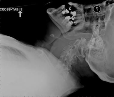
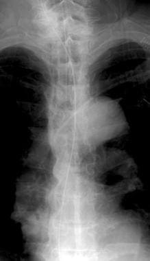
 Retropharyngeal Hematoma: Practice Essentials, Epidemiology, Etiology
LAMINOPLASTY FUSION FOR CERVICAL SPINAL CORD INJURY WITH OSSIFICATION OF POSTERIOR LONGITUDINAL LIGAMENT: Combination...
Ossification of posterior longitudinal ligament of cervical spine among omani patients referred for ct scan at a tertiary care...
![APERTO Lucent with SynergyDrive : Image Gallery | Fujifilm [India]](data:image/png;base64,iVBORw0KGgoAAAANSUhEUgAAABAAAAAQCAMAAAAoLQ9TAAAAVFBMVEX////39/f+/v4koIHf8ewAAACSkpLp6eng4ODu7u4qKir7+/shISE5OTnIyMg+Pj7Y6ua1tbWbm5sgICB0dHSrq6sKCgqHh4fY2NgbGxtISEjy8vLkHiJuAAAAZklEQVQYlXXMWQ6AIAxFUUDLJDI5y/73aYmKQeP9ak6aR+grQlmO5PKRAc9OSEyhXRCM6Z2LD3AYbPXBQYtz5oRuBBOsFTewScOMG6FAWkCvStH/jS/EbVcVJClYBSUE31Z50rw6ABnBA/O26wFTAAAAAElFTkSuQmCC) APERTO Lucent with SynergyDrive : Image Gallery | Fujifilm [India]
APERTO Lucent with SynergyDrive : Image Gallery | Fujifilm [India]
 Dr. Aaron Greenberg, MD - Orthopedic Spine Surgery Specialist in Franklin Lakes, NJ | Healthgrades
A new alarm point of transcranial electrical stimulation motor evoked potentials for intraoperative spinal cord monitoring: a...
MRI of the Spine, Second Edition, by Jeffrey S. Ross
 Addisu Mesfin, M.D. | UR Medicine
 Auto-ubiquitinylation kit - BML-UW0970 - Enzo Life Sciences
Journal of Neurosurgery: Spine Volume 6 Issue 4 () Journals
Browsing by Subject "Disease Progression"
 GEO Accession viewer
 7D | Radsource
 ICD-9-CM Codes for Spinal Deformity | BMUS: The Burden of Musculoskeletal Diseases in the United States
 Mechanical Stress-Induced IGF-1 Facilitates col-I and col-III Synthesis via the IGF-1R/AKT/mTORC1 Signaling Pathway
 Scoliosis - Doctors and departments - Mayo Clinic
HuGE Navigator|Genopedia|PHGKB
Rickets Imaging: Practice Essentials, Radiography
 A cell-autonomous requirement for neutral sphingomyelinase 2 in bone mineralization | Journal of Cell Biology | Rockefeller...
 Dr. Joseph A Graves, MD | Atlanta, GA | Grady Health
整形外科学講座 2021年|国立大学法人 浜松
 DR UMAPATI NARASINHA HEGDE - Home - Muljibhai Patel Urological Hospital
 References - SCIRE Professional
OPLL8
- Ossification of the posterior longitudinal ligament (OPLL) is a process of fibrosis, calcification, and ossification of the posterior longitudinal ligament of the spine, that may involve the spinal dura. (wikipedia.org)
- Paediatric IDC combined with ossification of the posterior longitudinal ligament (OPLL) is an even rarer condition, with only 8 cases described in detail to date. (biomedcentral.com)
- Ossification of the posterior longitudinal ligament (OPLL) mainly affects individuals of Asian descent aged 50-70 years and is a disease of unknown aetiology characterised by the replacement of ligamentous tissue by new ectopic bone [ 6 ]. (biomedcentral.com)
- This was a retrospective study of 10 cervical spinal cord injury (CSCI), concomitant with ossification of posterior longitudinal ligament (OPLL), that were treated by the author at Sanglah General Hospital-Bali during 2013-2014. (ejournals.ca)
- Objectives: We sought to evaluate the proportion of ossification of the posterior longitudinal ligament (OPLL) of the cervical spine and associated factors among Omani patients. (elsevierpure.com)
- Same concept holds good in the treatment of OPLL (ossification of posterior longitudinal ligament). (neurosurgical.tv)
- National Trends and Complications in the Surgical Management of Ossification of the Posterior Longitudinal Ligament (OPLL). (cornell.edu)
- Settlement after the start of trial for a woman paralyzed during spine surgery at an Illinois hospital when her neurosurgeon failed to diagnose and treat Ossification of the Posterior Longitudinal Ligament [OPLL]. (hurley-law.com)
Myelopathy2
- Ossification in the region of the posterior longitudinal ligament as a cause of cervical myelopathy. (bmj.com)
- Phenotypically, ttw mice develop excess calcification of the ligaments of the axial skeleton, resulting in myelopathy and an abnormal gait. (biomedcentral.com)
Stenosis5
- The Role of Atlantoaxial Instability on Chiari, Ossification of the Posterior Longitudinal Ligament, Spondylosis and Stenosis. (cornell.edu)
- Other rare causes of acquired spinal canal stenosis include epidural lipomatosis and ossification of the posterior longitudinal ligament and/or the ligamentum flavum. (jortho.org)
- Surgical management of incomplete cervical cord injury with stenosis secondary to ossification of the posterior longitudinal ligament. (stacksdiscovery.com)
- The patient required a circumferential procedure including a laminectomy/fusion followed by an anterior thoracic decompression to address both diffuse idiopathic skeletal hyperostosis (DISH) anteriorly and posterior stenosis. (surgicalneurologyint.com)
- For advanced spinal stenosis, surgery involves decompression of the disc and bone compression of the spinal cord, either from an anterior or posterior approach in conjunction with fusion of the affected levels. (msdmanuals.com)
Ligamentum2
- Disc changes, osteophyte formation, bulging of the inter-vertebral ligaments that include posterior longitudinal ligament and ligamentum flavum and consequent reduction in the spinal and root canal dimensions are all secondary to spinal instability. (neurosurgical.tv)
- Magnetic resonance imaging (MRI) and computed tomography (CT) studies documented severe T5-T9 diffuse idiopathic skeletal hyperostosis (DISH) resulting in marked circumferential cord compression attributed to multiple anterior ossified disc herniations and posterior ossification of the ligamentum flavum (OYL)/facet arthrosis. (surgicalneurologyint.com)
Thoracic ossification1
- We examined a 47-year-old woman after decompression for thoracic ossification of posterior longitudinal ligament was performed in another hospital. (whocc.org.cn)
Diffuse idiopathic skeletal hyperostosis2
- Background: Although diffuse idiopathic skeletal hyperostosis (DISH) is known to coexist with the ossification of spinal ligaments (OSLs), details of the radiographic relationship remain unclear. (elsevierpure.com)
- Japanese Organization of the Study for Ossification of Spinal Ligament (JOSL) 2021, ' Association between severity of diffuse idiopathic skeletal hyperostosis and ossification of other spinal ligaments in patients with ossification of the posterior longitudinal ligament ', Journal of Clinical Medicine , vol. 10, no. 20, 4690. (elsevierpure.com)
Decompression2
- Surgical management options include extensive cervical laminectomy with or without an additional posterior arthrodesis, anterior decompression and arthrodesis, and posterior cervical laminoplasty. (wikipedia.org)
- The surgical techniques used were laminectomy, laminectomy & discectomy, laminectomy, discectomy with foraminal decompression, laminectomy with posterior instrumentation, laminectomy with in situ fusion and posterior instrumentation. (jortho.org)
Lumbar1
- The complex anatomy of the lumbar spine is a remarkable combination of these strong vertebrae, multiple bony elements linked by joint capsules, and flexible ligaments/tendons, large muscles, and highly sensitive nerves. (medscape.com)
Tendons1
- [ 2 ] Patients with AS typically present with inflammatory back pain with varying degrees of associated enthesopathy (inflammation at sites where tendons, ligaments and joint capsule fibres attach to bone), peripheral arthritis and extra-articular manifestations. (medscape.com)
Prevalence1
- Emami Razavi S, Aryan A, Kazemi S, Rostamian A, Jahangiri A, Ghajarzadeh M. Prevalence of hip ossification and related clinical factors in cases with spinal cord injury. (scireproject.com)
Anterior2
- This discussion covers neck pain involving the posterior neck (not pain limited to the anterior neck) and low. (msdmanuals.com)
- Anterior slippage (anterolisthesis) is more common than posterior slippage (retrolisthesis). (msdmanuals.com)
Bone1
- Freed JH, Hahn H, Menter R, Dillon T. The use of the three-phase bone scan in the early diagnosis of heterotopic ossification (HO) and in the evaluation of didronel therapy. (scireproject.com)
Laminoplasty1
- Are clinical outcomes affected by laminoplasty method and K-line in patients with cervical ossification of posterior longitudinal ligament? (hku.hk)
Facet1
- Each vertebral arch is composed of 2 pedicles, 2 laminae, and 7 different bony processes (1 spinous, 4 articular, 2 transverse) (see the following image), joined together by facet joints and ligaments. (medscape.com)
Patients7
- Intravenous disodium etidronate therapy in spinal cord injury patients with heterotopic ossification. (scireproject.com)
- Banovac K, Gonzalez F, Evaluation and management of heterotopic ossification in patients with spinal cord injury. (scireproject.com)
- Risk factors for heterotopic ossification in patients with spinal cord injury: A case-control study of 264 patients. (scireproject.com)
- Durovic A, Miljkovic D, Brdareski Z, Plavsic A, Jevtic M. Pulse low intensity electromagnetic field as prophylaxis of heterotopic ossification in patients with traumatic spinal cord injury. (scireproject.com)
- Resection of heterotopic ossification in patients with spinal cord injuries. (scireproject.com)
- Diphosphonate treatment for heterotopic ossification in spinal cord injury patients. (scireproject.com)
- Genet F, Jourdan C, Lautridou C, Chehensse C, Minooee K, Denormandie P, Schnitzler A. The Impact of Preoperative Hip Heterotopic Ossification Extent on Recurrence in Patients with Head and Spinal Cord Injury: A Case Control Study. (scireproject.com)
Spinal Cord7
- Computed tomography (CT) and MRI revealed ossification of the intervertebral discs and posterior longitudinal ligament (PLL) at the C4/5 levels and an absence of obvious spinal cord compression. (biomedcentral.com)
- A comparison of heterotopic ossification treatment within the traumatic brain and spinal cord injured population: An evidence based systematic review. (scireproject.com)
- Treatment of heterotopic ossification after spinal cord injury. (scireproject.com)
- Banovac K, The effect of etidronate on late development of heterotopic ossification after spinal cord injury. (scireproject.com)
- Prevention of heterotopic ossification after spinal cord injury with indomethacin. (scireproject.com)
- Banovac K, Williams JM, Patrick LD, Levi A. Prevention of heterotopic ossification after spinal cord injury with COX-2 selective inhibitor (rofecoxib). (scireproject.com)
- Heterotopic ossification following hip replacement or spinal cord injury. (scireproject.com)
Study1
- By contrast, the patient of this case study manifested obvious enhancement of the calcified masses and ossified posterior longitudinal ligament (PLL), which differs considerably from the previously reported presentations. (biomedcentral.com)
Levels1
- Ossification indices were calculated as the sum of vertebral and intervertebral levels with OSL for each patient. (elsevierpure.com)
Injury1
- Arduini M, Mancini F, Farsetti P, Piperno A, Ippolito E. A new classification of peri-articular heterotopic ossification of the hip associated with neurological injury: 3D CT scan assessment and intra-operative findings. (scireproject.com)
Image1
- a and b: Sagittal (detailed) CT image demonstrating calcified herniated discs and diffuse ossification over larger parts of the spinal column. (surgicalneurologyint.com)