Osteitis Fibrosa Cystica
Acquired Hyperostosis Syndrome
Dry Socket
Ethmoid Sinusitis
Osteitis Deformans
Ethmoid Bone
Clavicle
Hyperostosis, Sternocostoclavicular
Renal Osteodystrophy
Paranasal Sinuses
Encyclopedias as Topic
Copyright
Organizations, Nonprofit
MedlinePlus
Computer Security
22-Oxacalcitriol ameliorates high-turnover bone and marked osteitis fibrosa in rats with slowly progressive nephritis. (1/119)
22-Oxacalcitriol ameliorates high-turnover bone and marked osteitis fibrosa in rats with slowly progressive nephritis. BACKGROUND: 22-Oxacalcitriol (OCT) is a unique vitamin D analogue with less calcemic activity than calcitriol, and it effectively suppresses parathyroid hormone (PTH) secretion in uremic rats. This study was performed to examine the long-term effect of intravenously administered OCT on high-turnover bone disease in model rats of slowly progressive renal failure. METHODS: Slowly progressive renal failure rats were made by a single injection of glycopeptide isolated from rat renal cortical tissues. At 250 days, glycopeptide-induced nephritis (GN) rats were divided into three groups with the same levels of serum creatinine and PTH, and they received either OCT (0.03 or 0.15 microg/kg body wt) or vehicle given intravenously three times per week for 15 weeks. RESULTS: Renal function of GN rats deteriorated very slowly but progressively, as assessed by the increase of serum creatinine concentration. At sacrifice, serum PTH levels, bone formation markers, bone resorption markers, and fibrosis volume were significantly elevated in vehicle-treated GN rats compared with those of sham-operated rats, suggesting the development of high-turnover bone disease with osteitis fibrosa. In contrast, in the GN-OCT 0.15 microg/kg group, these high PTH levels and high-turnover bone and fibrosis were significantly decreased. Such amelioration of bone abnormalities by OCT was not accompanied by either hypercalcemia or further deterioration of renal function. CONCLUSIONS: These data indicate that OCT may be a useful and safe agent not only for the suppression of PTH, but also for the amelioration of osteitis fibrosa and high-turnover bone without causing hypercalcemia in chronic dialysis patients. (+info)Antibody responses in patients with staphylococcal septicemia against two Staphylococcus aureus fibrinogen binding proteins: clumping factor and an extracellular fibrinogen binding protein. (2/119)
We analyzed the serum antibody responses against two Staphylococcus aureus fibrinogen binding proteins, the cell-bound clumping factor (Clf) and an extracellular fibrinogen binding protein (Efb). The material consisted of 105 consecutive serum samples from 41 patients suffering from S. aureus septicemia and 72 serum samples from healthy individuals. An enzyme-linked immunosorbent assay (ELISA) was developed. Healthy individuals showed variable levels of antibodies against the studied antigens, and cutoff levels (upper 95th percentile) against these antigens were determined. No correlation was seen between serum antibody levels against Clf and Efb. In acute-phase samples 27% of patients showed positive antibody levels against Clf and 10% showed positive levels against Efb, while in convalescent-phase samples 63% (26 of 41) showed a positive serology against Clf and 49% (20 of 41) showed a positive serology against Efb. Antibody levels against Efb were significantly lower in the acute-phase sera than in sera from healthy individuals (P = 0. 002). An antibody response against Clf was most frequent in patients suffering from osteitis plus septic arthritis and from endocarditis (80% positive). The antibody response against Efb appeared to develop later in the course of disease. A possible biological effect of measured antibodies was demonstrated with the help of an inhibition ELISA, in which both high-titer and low-titer sera inhibited the binding of bacteria to fibrinogen. In conclusion, we have demonstrated in vivo production of S. aureus fibrinogen binding proteins during deep S. aureus infections and a possible diagnostic and prophylactic role of the corresponding serum antibodies in such infections. (+info)Evolution of chronic recurrent multifocal osteitis toward spondylarthropathy over the long term. (3/119)
OBJECTIVE: To retrospectively assess, with a sufficiently long followup (mean 11.6 years; median 9 years), the long-term outcome of chronic recurrent multifocal osteitis (CRMO), a multifocal, inflammatory bone disease. METHODS: Patients included were 8 children/adolescents and 7 adults with no family history of rheumatic disease who had been diagnosed as having CRMO between 1979 and 1995. Ten patients had undergone at least 1 bone biopsy of the lesions, with histologic examination and multiple cultures. In 1996, in addition to an in-depth interview, 12 patients underwent an extensive physical examination, laboratory evaluation, HLA-A, B, C, and DR typing, bone radiography and scintigraphy, and computed tomography scan of the sternoclavicular and sacroiliac joints. RESULTS: Remission was observed in 3 patients. The other 12 patients developed various associations of vertebral (n = 10), sacroiliac (n = 6), anterior thoracic (n = 7), peripheral articular (n = 2), enthesopathic (n = 4), or dermatologic (palmoplantar pustulosis in 3 cases and psoriasis in 2) involvements. Spine involvement was the most common and occurred the earliest (median time to appearance after the onset of osteitis 5.63 years). Clinical sacroiliitis was always unilateral. No patients carried the HLA-B27 haplotype. CRMO responded well to nonsteroidal antiinflammatory drugs. Twelve patients met the European Spondylarthropathy Study Group criteria for spondylarthopathy. CONCLUSION: After 10 years, CRMO had usually evolved to spondylarthropathy, but with certain features not usually seen in the latter: predominantly, unilateral sacroiliitis, no familial form, and no link with HLA-B27. (+info)Growth factors in distraction osteogenesis. Immuno-histological pattern of TGF-beta1 and IGF-I in human callus induced by distraction osteogenesis. (4/119)
Although growth factors have been demonstrated during bone healing, their presence has not yet been confirmed in callus distraction. Therefore, in 3 patients we searched for cytokines during callus distraction. Bone biopsies were immuno-histochemically stained for TGF-beta1, IGF-I, TGF-beta type II receptor, IGF receptor, and proliferating cell nuclear antigen (PCNA). Histologically we found immature woven bone in the middle of the callus zone and increasing calcification and lamellar bone in the re-modelling zone. Osteoblasts and fibroblast-like cells in the middle zone, and osteoblasts in all zones stained for TGF-beta and its receptor. The number of positive staining cells related to proliferous activity as assessed both by PCNA, and by bone density in radiographs. IGF-I could be detected everywhere. In conclusion, growth factors are present in bone formation and in areas of re-modelling during callotasis. Their relation to proliferous activity and radiographic density supports their involvement in osteogenesis. (+info)Further studies of canine von Willebrand's disease. (5/119)
Additional characterization of von Willebrand's disease (VWD) in a family of German shepherd dogs is presented. Genetic studies of three generations of affected dogs indicate that about 50% of the progeny are affected if one parent has VWD and about 60% if both parents have the defect. Some of these progeny manifested an incomplete form of VWD, suggesting autosomal dominant inheritance with variable expressivity. The disease become progressively less severe with advancing age and repeated pregnancies. Ristocetin-induced platelet aggregation was significantly reduced in VWD dogs as compared with normal, thrombopathic, and hemophilic carrier dogs. Immunodiffusion and electroimmunodiffusion studies with rabbit anticanine factor VII showed the level of factor VII-related antigen to be low in VWD dogs but present in increased amounts in hemophilic dogs. VWD affected dogs had markedly delayed hemostatic plug formation, but their plugs appeared normal by light and electron microscopy. Their platelet nucleotides, ATP/ADP ration, and platelet protein content were normal. Platelet and fibrinogen survival times with [75Se] selenomethionine were also normal, although platelets from VWD dogs incorporated more radioactivity than did those from normal dogs or from dogs with incomplete VWD. (+info)A case report of synovitis, acne, pustulosis, hyperostosis and osteitis syndrome presenting with spondylodiscitis. (6/119)
SAPHO syndrome stands for synovitis, acne, pustulosis, hyperostosis and osteitis. The common site of skeletal lesions in this syndrome is the sternocostoclavicular area. Spondylodiscitis is rarely described in published studies. In general, skin lesions develop before the onset of skeletal lesions. We report a case of SAPHO syndrome in which spondylodiscitis developed more than 1 year before the onset of pustulosis. (+info)Incidence of pubic bone marrow oedema in Australian rules football players: relation to groin pain. (7/119)
OBJECTIVES: To examine the relation between the clinical features of groin pain and groin magnetic resonance imaging (MRI) appearances in a group largely comprising elite Australian Rules football players. The incidence of bone marrow oedema and other MRI findings in the pubic symphysis region was noted. The relation between a past history of groin pain and these other MRI findings was also examined. METHOD: In a prospective study, 116 male subjects (89 footballers, 17 umpires, 10 sedentary men) were examined before history taking and groin MRI. The clinical history was not known to the examiner (GMV) and radiologists (JPS, GTF). Clinical evidence of groin pain and examination findings were correlated with the presence of increased signal intensity within the pubic bone marrow. A past history of groin pain was correlated with the presence of other MRI findings such as cyst formation, fluid signal within the pubic symphysis disc, and irregularity of the pubic symphysis. RESULTS: Fifty two athletes (47 footballers, five umpires) had clinical features of groin pain with pubic symphysis and/or superior pubic ramus tenderness. A high incidence of increased signal intensity (77%) within the pubic bone marrow was identified in this group. There was an association between this group of athletes and the MRI finding of increased signal intensity (p<0.01). There was also an association between a past history of groin pain and the presence of other MRI findings (p<0.01). CONCLUSIONS: Athletes with groin pain and tenderness of the pubic symphysis and/or superior pubic ramus have clinical features consistent with the diagnosis of osteitis pubis. The increased signal intensity seen on MRI is due to pubic bone marrow oedema. An association exists between the clinical features of osteitis pubis and the MRI finding of pubic bone marrow oedema. A high incidence of pubic bone marrow oedema was also noted. Degenerative features visualised by MRI, such as subchondral cyst formation, were associated with a past history of groin pain. A stress injury to the pubic bone is the most likely explanation for these MRI findings and may be the cause of the clinical entity osteitis pubis. (+info)An unusual manifestation of acquired syphilis. (8/119)
Bone involvement is an unusual manifestation of acquired syphilis. We report a case of clinically apparent osteitis of the skull, secondary to acquired syphilis, which was the patient's presentation of human immunodeficiency virus infection. (+info)Osteitis is a medical term that refers to the inflammation of bone tissue. It can occur as a result of various conditions, such as infection (osteomyelitis), trauma, or autoimmune disorders. The symptoms of osteitis may include pain, swelling, warmth, and redness in the affected area, as well as fever and general malaise. Treatment typically involves addressing the underlying cause of the inflammation, which may involve antibiotics for infection or anti-inflammatory medications for other causes. In some cases, surgery may be necessary to remove infected or damaged bone tissue.
Osteitis fibrosa cystica is a medical condition that refers to the abnormal bone remodeling process characterized by increased bone resorption and formation, leading to bone thickening and weakening. It is also known as "von Recklinghausen's disease of bone" or "monostotic fibrous dysplasia."
This condition is typically caused by excessive production of parathyroid hormone (PTH) due to a benign or malignant tumor of the parathyroid gland, known as hyperparathyroidism. The overproduction of PTH leads to an imbalance in calcium and phosphorus metabolism, resulting in increased bone resorption and fibrous tissue deposition within the bone marrow.
The clinical features of osteitis fibrosa cystica include bone pain, fractures, bone deformities, and elevated levels of calcium and alkaline phosphatase in the blood. Radiographic findings may show characteristic "rugger jersey" or "salt and pepper" patterns of alternating areas of increased and decreased bone density.
Treatment typically involves surgical removal of the abnormal parathyroid gland tissue, followed by medical management to prevent further bone loss and promote healing.
Acquired hyperostosis syndrome is not a widely recognized medical term, and it may refer to several different conditions that involve abnormal bone growth or hardening. One possible condition that might be referred to as acquired hyperostosis syndrome is diffuse idiopathic skeletal hyperostosis (DISH).
Diffuse idiopathic skeletal hyperostosis is a non-inflammatory condition that affects the spine and other parts of the body. It is characterized by the calcification and ossification of ligaments and entheses, which are the sites where tendons or ligaments attach to bones. This process can lead to the formation of bony spurs or growths, called osteophytes, along the spine and other affected areas.
The exact cause of DISH is not known, but it is more common in older adults, males, and people with certain medical conditions such as diabetes and obesity. The symptoms of DISH can vary widely depending on the severity and location of the bone growths. Some people may experience stiffness, pain, or limited mobility in the affected areas, while others may have no symptoms at all.
It is important to note that there are many other conditions that can cause abnormal bone growth or hardening, so a proper medical evaluation is necessary to determine the underlying cause of any symptoms. If you have concerns about acquired hyperostosis syndrome or any other medical condition, you should speak with your healthcare provider for further guidance.
The pubic symphysis is the joint in the front of the pelvis that connects the two halves of the pelvic girdle, specifically the pubic bones. It's located at the lower part of the anterior (front) pelvic region. Unlike most joints, which are movable and contain synovial fluid, the pubic symphysis is a cartilaginous joint, also known as an amphiarthrosis.
The joint consists of fibrocartilaginous discs, ligaments, and the articular surfaces of the adjacent pubic bones. The fibrocartilaginous disc helps to absorb shock and reduce friction between the two bones. The main function of the pubic symphysis is to provide stability for the pelvis and transfer weight and forces from the upper body to the lower limbs during activities like walking, running, or jumping.
The pubic symphysis has a limited range of motion, allowing only slight movement in response to pressure or tension. During pregnancy and childbirth, the hormone relaxin is released, which increases the laxity of the pelvic joints, including the pubic symphysis, to accommodate the growing fetus and facilitate delivery. This increased mobility can sometimes lead to discomfort or pain in the area, known as symphysis pubis dysfunction (SPD) or pelvic girdle pain.
The pubic bone, also known as the pubis or pubic symphysis, is a part of the pelvis - the complex ring-like structure that forms the lower part of the trunk and supports the weight of the upper body. The pubic bone is the anterior (front) portion of the pelvic girdle, located at the bottom of the abdomen, and it connects to the other side at the pubic symphysis, a cartilaginous joint.
The pubic bone plays an essential role in supporting the lower limbs and providing attachment for various muscles involved in movements like walking, running, and jumping. It also protects some abdominal organs and contributes to the structure of the pelvic outlet, which is crucial during childbirth.
"Dry socket" is a common term used in dentistry to describe a condition that can occur after a tooth extraction. The medical term for dry socket is "alveolar osteitis." This condition arises when the blood clot that forms in the socket where the tooth was removed becomes dislodged or fails to form properly, exposing the bone and nerves underneath.
Dry socket can be quite painful, causing a throbbing sensation that may radiate to the ear, neck, or temple. It can also lead to bad breath and an unpleasant taste in the mouth. The exact cause of dry socket is not entirely clear, but several factors may increase the risk, including smoking, poor oral hygiene, using birth control pills, and having a history of dry socket.
Treatment for dry socket typically involves cleaning the socket and placing a medicated dressing to promote healing and relieve pain. Over-the-counter pain medications and warm compresses may also help alleviate discomfort. It is essential to follow your dentist's instructions carefully to prevent complications and promote proper healing.
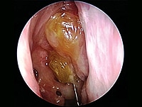
Ethmoid sinusitis is a medical condition that refers to the inflammation or infection of the ethmoid sinuses. The ethmoid sinuses are a pair of small, air-filled cavities located in the upper part of the nasal cavity, near the eyes. They are surrounded by delicate bone structures and are connected to the nasal cavity by narrow channels.
Ethmoid sinusitis can occur as a result of a viral, bacterial, or fungal infection, or it may be caused by allergies, environmental factors, or structural abnormalities in the nasal passages. When the ethmoid sinuses become inflamed or infected, they can cause symptoms such as:
* Nasal congestion or stuffiness
* Pain or pressure in the forehead, between the eyes, or in the cheeks
* Headaches or facial pain
* Thick, discolored nasal discharge
* Postnasal drip
* Coughing or sneezing
* Fever
* Fatigue
Ethmoid sinusitis can be acute (lasting for a short period of time) or chronic (persisting for several weeks or months). If left untreated, ethmoid sinusitis can lead to complications such as the spread of infection to other parts of the body, including the eyes and brain. Treatment for ethmoid sinusitis may include antibiotics, decongestants, nasal sprays, or surgery in severe cases.
Osteitis deformans, also known as Paget's disease of bone, is a chronic disorder of the bone characterized by abnormal turnover and remodeling of the bone. In this condition, the bone becomes enlarged, thickened, and deformed due to excessive and disorganized bone formation and resorption.
The process begins when the bone-remodeling cycle is disrupted, leading to an imbalance between the activity of osteoclasts (cells that break down bone) and osteoblasts (cells that form new bone). In Paget's disease, osteoclasts become overactive and increase bone resorption, followed by an overzealous response from osteoblasts, which attempt to repair the damage but do so in a disorganized manner.
The affected bones can become weakened, prone to fractures, and may cause pain, deformities, or other complications such as arthritis, hearing loss, or neurological symptoms if the skull or spine is involved. The exact cause of Paget's disease remains unknown, but it is believed that genetic and environmental factors play a role in its development.
Early diagnosis and treatment can help manage the symptoms and prevent complications associated with osteitis deformans. Treatment options include medications to slow down bone turnover, pain management, and orthopedic interventions when necessary.
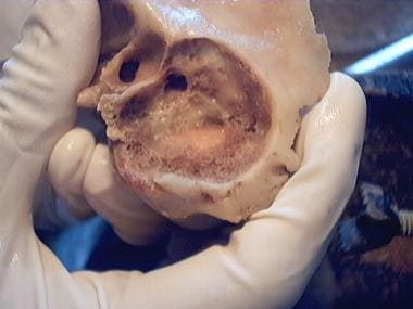
The ethmoid bone is a paired, thin, and lightweight bone that forms part of the skull's anterior cranial fossa and contributes to the formation of the orbit and nasal cavity. It is located between the frontal bone above and the maxilla and palatine bones below. The ethmoid bone has several important features:
1. Cribriform plate: This is the horizontal, sieve-like portion that forms part of the anterior cranial fossa and serves as the roof of the nasal cavity. It contains small openings (foramina) through which olfactory nerves pass.
2. Perpendicular plate: The perpendicular plate is a vertical structure that projects downward from the cribriform plate, forming part of the nasal septum and separating the left and right nasal cavities.
3. Superior and middle nasal conchae: These are curved bony projections within the lateral walls of the nasal cavity that help to warm, humidify, and filter incoming air.
4. Lacrimal bone: The ethmoid bone articulates with the lacrimal bone, forming part of the medial wall of the orbit.
5. Frontal process: This is a thin, vertical plate that articulates with the frontal bone above the orbit.
6. Sphenoidal process: The sphenoidal process connects the ethmoid bone to the sphenoid bone posteriorly.
The ethmoid bone plays a crucial role in protecting the brain and providing structural support for the eyes, as well as facilitating respiration by warming, humidifying, and filtering incoming air.
The clavicle, also known as the collarbone, is a long, slender bone that lies horizontally between the breastbone (sternum) and the shoulder blade (scapula). It is part of the shoulder girdle and plays a crucial role in supporting the upper limb. The clavicle has two ends: the medial end, which articulates with the sternum, and the lateral end, which articulates with the acromion process of the scapula. It is a common site of fracture due to its superficial location and susceptibility to direct trauma.
Hyperostosis, sternocostoclavicular, is a medical condition characterized by the abnormal thickening and hardening of the bone tissue in the sternocostoclavicular joint and surrounding areas. The sternocostoclavicular joint is where the clavicle (collarbone) meets the sternum (breastbone) and manubrium, and costae (ribs). This condition can result in pain, stiffness, and limited range of motion in the affected area. The exact cause of hyperostosis, sternocostoclavicular, is not fully understood, but it may be associated with trauma, inflammation, or genetic factors. In some cases, this condition may be asymptomatic and only discovered during imaging studies performed for other reasons. Treatment options typically include pain management, physical therapy, and in some cases, surgery to remove the excess bone growth.
Renal osteodystrophy is a bone disease that occurs in individuals with chronic kidney disease (CKD). It is characterized by abnormalities in the bones' structure and mineral composition due to disturbances in the metabolism of calcium, phosphorus, and vitamin D. These metabolic disturbances result from the kidneys' decreased ability to maintain balance in the levels of these minerals and hormones.
Renal osteodystrophy can manifest as several bone disorders, including:
1. Osteitis fibrosa cystica: Increased bone turnover due to excessive parathyroid hormone (PTH) production, leading to high levels of alkaline phosphatase and increased resorption of bones.
2. Adynamic bone disease: Decreased bone turnover due to reduced PTH levels, resulting in low bone formation rates and increased fracture risk.
3. Mixed uremic osteodystrophy: A combination of high and low bone turnover, with varying degrees of mineralization defects.
4. Osteomalacia: Defective mineralization of bones due to vitamin D deficiency or resistance, leading to soft and weak bones.
Symptoms of renal osteodystrophy may include bone pain, muscle weakness, fractures, deformities, and growth retardation in children. Diagnosis typically involves laboratory tests, imaging studies, and sometimes bone biopsies. Treatment focuses on correcting the metabolic imbalances through dietary modifications, medications (such as phosphate binders, vitamin D analogs, and calcimimetics), and addressing any secondary hyperparathyroidism if present.
Hyperostosis is a medical term that refers to an excessive growth or abnormal thickening of bone tissue. It can occur as a result of various conditions, such as inflammation, injury, or genetic disorders. The extra bone growth can cause pain, stiffness, and limited mobility in the affected area. In some cases, hyperostosis can also lead to deformities and other complications.
There are several types of hyperostosis, including:
1. Diffuse idiopathic skeletal hyperostosis (DISH): This is a condition that affects the spine, causing calcification and stiffening of the ligaments and bone spurs to form along the edges of the vertebrae. It is often asymptomatic but can cause pain and stiffness in some cases.
2. Flat bone hyperostosis: This type of hyperostosis affects the flat bones of the body, such as the skull, ribs, and pelvis. It can be caused by various conditions, including Paget's disease, fibrous dysplasia, and certain types of cancer.
3. Focal hyperostosis: This refers to localized areas of bone overgrowth that can occur in response to injury, infection, or inflammation. Examples include heterotopic ossification (the formation of bone in soft tissues) and Freiberg's infarction (a condition that affects the joint surface of the metatarsal bones in the foot).
4. Hyperostosis frontalis interna: This is a benign condition that causes thickening of the inner table of the frontal bone in the skull. It is more common in women and often asymptomatic but can cause headaches and other symptoms in some cases.
Treatment for hyperostosis depends on the underlying cause and severity of the condition. In some cases, no treatment may be necessary. However, if the condition causes pain or limits mobility, various treatments may be recommended, such as medication, physical therapy, or surgery.
Paranasal sinuses are air-filled cavities in the skull that surround the nasal cavity. There are four pairs of paranasal sinuses, including the maxillary, frontal, ethmoid, and sphenoid sinuses. These sinuses help to warm, humidify, and filter the air we breathe. They also contribute to our voice resonance and provide a slight cushioning effect for the skull. The openings of the paranasal sinuses lead directly into the nasal cavity, allowing mucus produced in the sinuses to drain into the nose. Infections or inflammation of the paranasal sinuses can result in conditions such as sinusitis.
Osteosclerosis is a medical term that refers to an abnormal thickening and increased density of bone tissue. This condition can occur as a result of various diseases or conditions, such as certain types of bone cancer, Paget's disease of bone, fluoride poisoning, or chronic infection of the bone. Osteosclerosis can also be seen in some benign conditions, such as osteopetrosis, which is a rare genetic disorder characterized by an excessively hard and dense skeleton.
In some cases, osteosclerosis may not cause any symptoms and may only be discovered on X-rays or other imaging studies. However, in other cases, it can lead to complications such as bone pain, fractures, or deformities. Treatment for osteosclerosis depends on the underlying cause of the condition and may include medications, surgery, or other therapies.
An encyclopedia is a comprehensive reference work containing articles on various topics, usually arranged in alphabetical order. In the context of medicine, a medical encyclopedia is a collection of articles that provide information about a wide range of medical topics, including diseases and conditions, treatments, tests, procedures, and anatomy and physiology. Medical encyclopedias may be published in print or electronic formats and are often used as a starting point for researching medical topics. They can provide reliable and accurate information on medical subjects, making them useful resources for healthcare professionals, students, and patients alike. Some well-known examples of medical encyclopedias include the Merck Manual and the Stedman's Medical Dictionary.
Copyright is a legal concept that gives the creator of an original work exclusive rights to its use and distribution, usually for a limited period of time. In the medical field, copyright protection can apply to various works such as medical textbooks, journal articles, educational materials, software, and multimedia presentations. It is important to note that copyright law seeks to strike a balance between protecting the rights of creators and promoting the progress of science and knowledge by allowing for limited use of copyrighted material under certain circumstances, such as fair use.
It's worth mentioning that while copyright protection can apply to medical works, there are also exceptions and limitations to copyright law that may allow for the use of copyrighted material without permission from the copyright owner in certain situations. For example, in the United States, the "fair use" doctrine allows for limited use of copyrighted material without obtaining permission from the copyright owner, depending on factors such as the purpose and character of the use, the nature of the copyrighted work, the amount and substantiality of the portion used, and the effect of the use upon the potential market for or value of the copyrighted work.
When using medical works that are protected by copyright, it is important to obtain permission from the copyright owner or ensure that the use falls under an exception or limitation to copyright law, such as fair use, in order to avoid infringing on the exclusive rights of the copyright owner.
Nonprofit organizations in the medical context are private entities that operate on a nonprofit basis and are typically dedicated to furthering a particular social, healthcare-related, or advocacy mission. They are usually tax-exempt and rely on donations, grants, and sometimes membership fees to support their work. Examples of nonprofit organizations in the medical field include hospitals, clinics, research institutions, patient advocacy groups, and health-related foundations. Their primary goal is to provide services or conduct activities that benefit the community or a specific group, rather than generating profits for shareholders or owners.
A patent, in the context of medicine and healthcare, generally refers to a government-granted exclusive right for an inventor to manufacture, use, or sell their invention for a certain period of time, typically 20 years from the filing date. In the medical field, patents may cover a wide range of inventions, including new drugs, medical devices, diagnostic methods, and even genetic sequences.
The purpose of patents is to provide incentives for innovation by allowing inventors to profit from their inventions. However, patents can also have significant implications for access to medical technologies and healthcare costs. For example, a patent on a life-saving drug may give the patent holder the exclusive right to manufacture and sell the drug, potentially limiting access and driving up prices.
It's worth noting that the patent system is complex and varies from country to country. In some cases, there may be ways to challenge or circumvent patents in order to increase access to medical technologies, such as through compulsory licensing or generic substitution.
MedlinePlus is not a medical term, but rather a consumer health website that provides high-quality, accurate, and reliable health information, written in easy-to-understand language. It is produced by the U.S. National Library of Medicine, the world's largest medical library, and is widely recognized as a trusted source of health information.
MedlinePlus offers information on various health topics, including conditions, diseases, tests, treatments, and wellness. It also provides access to drug information, medical dictionary, and encyclopedia, as well as links to clinical trials, medical news, and patient organizations. The website is available in both English and Spanish and can be accessed for free.
Computer security, also known as cybersecurity, is the protection of computer systems and networks from theft, damage, or unauthorized access to their hardware, software, or electronic data. This can include a wide range of measures, such as:
* Using firewalls, intrusion detection systems, and other technical safeguards to prevent unauthorized access to a network
* Encrypting sensitive data to protect it from being intercepted or accessed by unauthorized parties
* Implementing strong password policies and using multi-factor authentication to verify the identity of users
* Regularly updating and patching software to fix known vulnerabilities
* Providing security awareness training to employees to help them understand the risks and best practices for protecting sensitive information
* Having a incident response plan in place to quickly and effectively respond to any potential security incidents.
The goal of computer security is to maintain the confidentiality, integrity, and availability of computer systems and data, in order to protect the privacy and safety of individuals and organizations.
 Osteitis
Condensing osteitis
Rarefying osteitis
Osteitis pubis
Alveolar osteitis
Osteitis fibrosa cystica
Index of oral health and dental articles
John William Struthers
Hubert Maitland Turnbull
Charles Thurstan Holland
SAPHO syndrome
Typhoid fever
Endocrine bone disease
Coffin bone
Panosteitis
Brown tumor
Max Askanazy
Mycobacterium conceptionense
Renal osteodystrophy
Bacterial cold water disease
Paget's disease of bone
Dental extraction
Michelle Cosier
Munjed Al Muderis
Toothache
James Paget
James Paget University Hospital
Russell Morse Wilder
Mouthwash
Harald Maddadsson
Osteitis - Wikipedia
 Osteitis fibrosa: MedlinePlus Medical Encyclopedia
 Osteitis Pubis: Practice Essentials, Background, Anatomy
 OSTEITIS FIBROSA WITH INITIAL SYMPTOMS RESEMBLING RHEUMATIC INFECTION | The BMJ
 Osteitis Pubis Injury Guide | PhysioRoom Injury Advice
 Osteitis pubis and assessment of bone marrow edema at the pubic symphysis with MRI in an elite junior male soccer squad
Osteitis | Profiles RNS
 Osteitis Pubis
- Supacore
Osteitis Pubis
- Supacore
 Osteitis deformans Archives • LITFL
Osteitis Pubis | Heal My Injury
 Knee & Sports | Osteitis Pubis - Orthohub
 Tooth Extraction Risks
 Sound Pharmacy - Kieferosteitis - Jawbone Osteitis - Healing Sound
 Infected Implants - Osteitis Center Döbling, Vienna, Austria
Pediatric Mastoiditis Treatment & Management: Approach Considerations, Antimicrobial Therapy, Mastoidectomy, Tympanoplasty, and...
 Cases reported • Osteitis Deformans; Paget's Disease of Bone
 Condensing Osteitis: Symptoms, Causes, Treatment & More - Entirely Health
 Osteitis Condensans Ilii: Symptoms, Diagnosis and Treatment - Symptoma
 Osteitis Condensans Ilii Vs Sacroillitis - Clinical Maze Clinical Maze
 Chronic non bacterial osteitis- a multicentre study | Pediatric Rheumatology | Full Text
 How to help your Horse recover from Pedal Osteitis - Scoot Boots
Ostitis deforestation mans (Morbus Paget, Paget's bone disease)
 PSYCHOSIS IN PAGET'S DISEASE (OSTEITIS DEFORMANS) | Archives of Neurology & Psychiatry | JAMA Network
Severe deep infections and osteitis - Eric Senneville - Train-the-Foot Trainer Middle East
Osteitis Pubis: Hopefully My Last Post Related To This Nasty Injury - mataruga.com
 Can Impacted Wisdom Teeth Shift Other Teeth? (Potential Risks)
 Clinical risk factors for hamstring muscle strain injury: a prospective study with correlation of injury by magnetic resonance...
 38 CFR Appendix C to Part 4 - Appendix C to Part 4-Alphabetical Index of Disabilities | Electronic Code of Federal Regulations ...
 chronic osteitis of the navicular bone読み方, "chronic osteitis of the navicular bone"の発音
Pubis21
- Osteitis pubis is an inflammation of the pubic symphysis and surrounding muscle insertions. (medscape.com)
- This image shows classic sclerosis and lysis findings of osteitis pubis around pubic symphysis, with widening of symphysis. (medscape.com)
- The presenting symptoms of osteitis pubis can be almost any complaint about the groin or lower abdomen. (medscape.com)
- Physical findings for osteitis pubis can vary greatly. (medscape.com)
- What is a Osteitis Pubis Injury? (physioroom.com)
- Osteitis pubis is a painful overuse injury that affects the pelvis and most commonly occurs during kicking activities, ice skating and dance. (physioroom.com)
- The exact mechanism of the development of osteitis pubis remains unclear. (physioroom.com)
- Osteitis pubis is most common in sports where a large shearing force goes across the pubic symphysis. (physioroom.com)
- Others who tend to have a high risk of developing osteitis pubis include those with rheumatological diseases and expectant mothers. (physioroom.com)
- Osteitis pubis causes pain during and after exertion, which is often combined with tenderness to touch at the pubic symphysis area. (physioroom.com)
- The clinical signs and history of the patient are usually sufficient to raise the suspicion of osteitis pubis, but diagnostic investigations can confirm this condition. (physioroom.com)
- The x-rays are able to see widening or erosion at the pubic symphysis which is indicative of osteitis pubis. (physioroom.com)
- Frustratingly, osteitis pubis can be resistant to treatment and can last between 6 months and two years before symptoms resolve. (physioroom.com)
- To assess bone marrow edema at the pubic symphysis with magnetic resonance imaging (MRI), and its relation to training and osteitis pubis in an elite group of junior soccer players. (nih.gov)
- There was a greatly decreased risk of developing groin pain (osteitis pubis) with more training prior to entry of the AIS soccer program (odds ratio per 4 sessions of training, 0.003). (nih.gov)
- Substantial amounts of bone marrow edema at the pubic symphysis can occur in asymptomatic elite junior soccer players, but it is only weakly related to the development of osteitis pubis. (nih.gov)
- Progressing training loads more slowly in athletes presenting with low current training loads may be a useful strategy for the prevention of osteitis pubis in junior soccer players. (nih.gov)
- Multicenter Outcomes of Endoscopic Pubic Symphysectomy for Osteitis Pubis Associated With Femoroacetabular Impingement. (uchicago.edu)
- Osteitis pubis is a condition where there is inflammation present in the joint and surrounding tissues at the front of the pelvis, known as the pubic symphysis. (healmyinjury.com)
- Other factors, such as increasing age, being of aboriginal descent, or having a past history of knee injury or osteitis pubis, increase the risk of hamstring strain independently of previous posterior thigh injury. (bmj.com)
- An increased risk of hamstring muscle strain injury exists, independently of a past history of posterior thigh injury, in athletes who are older, have a past history of serious knee injury, have a past history of osteitis pubis, and are of aboriginal descent. (bmj.com)
Deformans2
- Paget's bone disease or osteitis deformans is a rare condition in which the processes of bone turnover runs amok. (healthanddisease.com)
- A disease of the skeletal system producing as much disturbance, especially of the external conformation of the skull, as osteitis deformans would naturally lead one to question its effects on the nervous system. (jamanetwork.com)
Alveolar16
- All healthy adults who presented with alveolar osteitis following tooth extraction over a 3 month period were invited to participate. (bris.ac.uk)
- Conclusion: PRGF® predictably treated alveolar osteitis following tooth extraction compared to the conventional standard treatment of Alvogyl® which has been used for many years. (bris.ac.uk)
- Local interventions for the management of alveolar osteitis (dry socket). (bvsalud.org)
- Alveolar osteitis ( dry socket ) is a complication of dental extractions more often involving mandibular molar teeth . (bvsalud.org)
- To assess the effects of local interventions used for the prevention and treatment of alveolar osteitis ( dry socket ) following tooth extraction . (bvsalud.org)
- Dry socket , or alveolar osteitis, can last for up to 7 days . (medicalnewstoday.com)
- Alveolar osteitis following surgical removal of mandibular third molars. (druglib.com)
- El curetaje y raspado del hueso alveolar no es recomendado, debido al intenso dolor que ocasiona despu s de haberse realizado y que adem s, no se tiene la certeza de que el nuevo co gulo sangu neo se mantenga en el sitio deseado. (medigraphic.com)
- Los enjuagues antis pticos, como la clorhexidina, usados antes y despu s de cualquier extracci n han demostrado tener buena eficacia en la prevenci n y tratamiento de la oste tis alveolar. (medigraphic.com)
- 2.- Charles RB, Abilene T, Alveolar osteitis by immediate placement of medicated parking. (medigraphic.com)
- 3.- G ksel SK, G nay Y, Zeynep S, Comparison of Alvogyl, Salicep Patch, and Low-Level Laser Therapy in the mangement of alveolar osteitis. (medigraphic.com)
- 4.- Corey CB, James AG, Sarah ER, Adam PS, Daniel ML, The efficacy of a topical anestehesic gel in the relief of pain associated with localizad alveolar osteitis. (medigraphic.com)
- 6.- Catellani JE, Harvey S, Erickson SH, Cherkin D. Effect of oral contraceptive cycle on dry socket (localized alveolar osteitis). (medigraphic.com)
- 7.- Larsen PE, Alveolar osteitis after surgical removal of impacted mandibular third molars. (medigraphic.com)
- 12.- Larsen PE, The effect of a clorhexidine rinse on the incidence of alveolar osteitis following the surgical removal of impacted mandibular third molars. (medigraphic.com)
- 13.- Morales TB, Osteitis alveolar (alve lo seco) despu s de la remoci n quir rgica de terceros molares inferiores impactados. (medigraphic.com)
Inflammation8
- Osteitis is inflammation of bone. (wikipedia.org)
- Kieferosteitis - Jawbone Osteitis is a bone inflammation marked by enlargement and pain. (sound-pharmacy.com)
- When it comes to the main causes of this dental condition, it's safe to say that inflammation and infections are considered to be the main culprits of condensing osteitis. (mgadental.com.au)
- Condensing osteitis is a condition that involves abnormal bone growth and lesions that may be due to infections and tooth inflammation. (entirelyhealth.com)
- Inflammation in the pedal bone can be the cause of severe lameness in your horse and the prognosis for "pedal osteitis" is often dire. (scootboots.com)
- Pedal Osteitis is inflammation in the pedal bone, also known as the coffin bone or distal phalanx, which is the very bottom bone inside the horse's hooves. (scootboots.com)
- The inflammation leading to pedal osteitis is most often non-septic, meaning that it is the result of chronic sole bruising from repeated concussion during work on hard surfaces. (scootboots.com)
- We call this an osteitis, which means inflammation in bone. (spondylitis.org)
Symptoms6
- However, a diagnosis of condensing osteitis may come as a surprise to a patient simply because there aren't any noticeable symptoms associated with this condition. (mgadental.com.au)
- That being said, here are some of the symptoms a patient should look out for in order to recognize condensing osteitis in time. (mgadental.com.au)
- The only two possible condensing osteitis symptoms are mild pain and irritation. (mgadental.com.au)
- This article will discuss with you what to know about condensing osteitis, including the symptoms, causes, treatment options, and more. (entirelyhealth.com)
- However, there are ways to help your horse manage the pain and even in some cases recover from pedal osteitis, by treating the cause rather than the symptoms. (scootboots.com)
- The symptoms of pedal osteitis are lameness, heat and foot soreness. (scootboots.com)
Pedal osteitis7
- Improper trimming and shoeing as well as navicular disease can also lead to pedal osteitis. (scootboots.com)
- Moreover, pedal osteitis is usually diagnosed following an x-ray revealing demineralisation - seen as uneven edges - of the pedal bone inside your horse's hoof. (scootboots.com)
- In the following, we will take a closer look at the causes of pedal osteitis. (scootboots.com)
- Knowing the causes will provide you with valuable knowledge about what to avoid, in order to prevent pedal osteitis from developing in the first place, or help your horse recover from pedal osteitis before it becomes chronic. (scootboots.com)
- Trauma is believed to be the main cause of pedal osteitis in horses. (scootboots.com)
- That is also the reason why remediate shoeing hasn't been very successful in helping horses with pedal osteitis. (scootboots.com)
- Common treatment of pedal osteitis in horses involves remediate shoeing, sometimes with a pad inserted to protect the sole of the hoof from concussion. (scootboots.com)
Condensans ilii5
- Osteitis condensans ilii results in recurrent axial low-back pain. (symptoma.com)
- Osteitis condensans ilii (OCI) is a benign cause of low-back pain that is self-limiting and has an unknown etiology. (symptoma.com)
- CONCLUSION: Osteitis condensans ilii is associated with tenderness during sacroiliac joint compression tests and should be considered in the differential diagnosis when sacroiliac joint tenderness is elicited. (symptoma.com)
- While usually an incidental finding, patients with osteitis condensans ilii are more likely to have sacroiliac joint tenderness compared to controls. (symptoma.com)
- Osteitis condensans ilii: a significant association with sacroiliac joint tenderness in women. (symptoma.com)
Bacterial osteitis1
- To understand the demographics, clinical features and treatment outcomes of Chronic Non-bacterial Osteitis (CNO) from three tertiary paediatric rheumatology services in the United Kingdom. (biomedcentral.com)
Condensing Osteitis12
- There are plenty of teeth-related issues people discover by chance, especially during regular visits to their dentists and one of those issues is definitely - condensing osteitis. (mgadental.com.au)
- Also, dentists do not consider condensing osteitis to be a common disease. (mgadental.com.au)
- Most dentists claim that condensing osteitis is usually asymptomatic. (mgadental.com.au)
- It should be stated that condensing osteitis is basically marked by abnormal bone hardening. (mgadental.com.au)
- However, there are other possible causes of condensing osteitis. (mgadental.com.au)
- The statistics say that 4 to 7 percent of people are suffering from condensing osteitis also known as focal sclerosing osteitis. (mgadental.com.au)
- An interesting fact is that, while not a common condition itself, dentists consider condensing osteitis to be one of the most frequent forms of jaw lesions. (mgadental.com.au)
- Like with many other dental issues, treatment for condensing osteitis depends on the underlying cause. (mgadental.com.au)
- There are plenty of condensing osteitis treatment methods a dentist can apply and they all work. (mgadental.com.au)
- However, the thing that makes condensing osteitis a complicated condition is the fact that it's usually asymptomatic. (mgadental.com.au)
- Condensing osteitis is not a very common dental condition. (entirelyhealth.com)
- Condensing osteitis can be discovered during a random dental check-up usually after an X-ray. (entirelyhealth.com)
Osteomyelitis3
- Of the 38 compensated BCG osteomyelitis/osteitis pa- intergenic spacer sequences for multispacer typing of Rickettsia tients, 18 were boys. (cdc.gov)
- 5 years of age have been sent to the national refer- a result of Mycobacterium bovis BCG osteomyelitis/osteitis to ence mycobacterial laboratory for BCG detection ( 2 ). (cdc.gov)
- Chronic nonbacterial osteitis (CNO) or chronic recurrent multifocal osteomyelitis (CRMO) is a rare auto inflammatory disorder characterised by the presence of sterile bone lesions [ 1 ]. (biomedcentral.com)
Socket1
- In some cases, this blood clot may be dislodged prematurely, which can lead to a condition known as osteitis or dry socket. (news-medical.net)
Fibrosa8
- Osteitis fibrosa is a complication of hyperparathyroidism , a condition in which overactive parathyroid glands cause certain bones to become abnormally weak and deformed. (medlineplus.gov)
- This is osteitis fibrosa. (medlineplus.gov)
- In rare cases, parathyroid cancer causes osteitis fibrosa. (medlineplus.gov)
- Osteitis fibrosa is now very rare in people who have hyperparathyroidism who have good access to medical care. (medlineplus.gov)
- Osteitis fibrosa may cause bone pain or tenderness. (medlineplus.gov)
- People with hyperparathyroidism are more likely to have osteopenia (thin bones) or osteoporosis (very thin bones) than to have full-blown osteitis fibrosa. (medlineplus.gov)
- Most of the bone problems from osteitis fibrosa can be reversed with surgery to remove the abnormal parathyroid gland(s). (medlineplus.gov)
- Osteitis Fibrosa Cystica defined by Medcinenet is "a condition that is associated with excessive parathyroid production (hyperparathyroidism), in which bone tissue becomes soft and deformed. (benjaminbarber.org)
Molars2
- The other name for this condition is focal sclerosing osteitis and it is known for causing harder, denser bones, which primarily affects molars in a patient's jaw area. (mgadental.com.au)
- It is also known as focal sclerosing osteitis, a dental condition known for causing harder, denser bones, which causes issues with the molars in your jaw area. (entirelyhealth.com)
SAPHO1
- 4. Patients with SAPHO (Synovitis-acne-pustulosis-hyperostosis-osteitis) syndrome. (who.int)
Chronic1
- As unifocal and/or non-recurrent forms have also been described the term Chronic Nonbacterial Osteitis is considered to be more appropriate than CRMO [ 3 ]. (biomedcentral.com)
MeSH1
- Osteitis" is a descriptor in the National Library of Medicine's controlled vocabulary thesaurus, MeSH (Medical Subject Headings) . (uchicago.edu)
People2
- This graph shows the total number of publications written about "Osteitis" by people in this website by year, and whether "Osteitis" was a major or minor topic of these publications. (uchicago.edu)
- Below are the most recent publications written about "Osteitis" by people in Profiles. (uchicago.edu)
Fibrosa cystica3
- Osteitis fibrosa cystica of the tibia. (mdwiki.org)
- Osteitis fibrosa cystica is defined as the classic skeletal manifestation of advanced hyperparathyroidism . (mdwiki.org)
- Osteitis fibrosa cystica is the result of unchecked hyperparathyroidism, or the overactivity of the parathyroid glands , which results in an overproduction of parathyroid hormone (PTH). (mdwiki.org)
Secondary to osteitis1
- Canine transmigration accompanying mandibular retrognathism secondary to osteitis. (nih.gov)
Synovitis3
- Synovitis, Acne, Pustulosis, Hyperostosis and Osteitis (SAPHO) Syndrome: A Brief Review of a Rare. (annals.edu.sg)
- SAPHO was proposed to refer to a combination of synovitis, acne, pustulosis, hyperostosis, and osteitis as a heading for these syndromes. (ajnr.org)
- Active inflammatory lesions such as bone marrow oedema (BMO)/osteitis, synovitis, enthesitis and capsulitis associated with SpA can be detected by MRI. (bmj.com)
Bone4
- Osteitis is inflammation of bone. (wikipedia.org)
- Osteitis fibrosa may cause bone pain or tenderness. (medlineplus.gov)
- Most of the bone problems from osteitis fibrosa can be reversed with surgery to remove the abnormal parathyroid gland(s). (medlineplus.gov)
- Osteitis fibrosis cystica (OFC), also known as osteitis fibrosa , osteodystrophia fibrosa , and von Recklinghausen's disease of bone (not to be confused with von Recklinghausen's disease, neurofibromatosis type I ), is caused by hyperparathyroidism , which is a surplus of parathyroid hormone from over-active parathyroid glands . (mdwiki.org)
Complication1
- Osteitis fibrosa is a complication of hyperparathyroidism , a condition in which overactive parathyroid glands cause certain bones to become abnormally weak and deformed. (medlineplus.gov)
Osteoporosis1
- People with hyperparathyroidism are more likely to have osteopenia (thin bones) or osteoporosis (very thin bones) than to have full-blown osteitis fibrosa. (medlineplus.gov)
Hyperparathyroidism1
- Osteitis fibrosa is now very rare in people who have hyperparathyroidism who have good access to medical care. (medlineplus.gov)
Vaccination4
- To investigate the incidence and increasing tendency of osteitis after BCG vaccination and, in addition, its clinical features, diagnostic methods and results of treatment. (nih.gov)
- 22 cases of Japanese children who received BCG vaccination between 1998 and 2007 and developed osteitis, and were reported in medical journals or meetings. (nih.gov)
- About 73% of cases of osteitis were seen from 9 to 18 months after receiving the vaccination. (nih.gov)
- Immunodeficiency might have some relationship to the development of osteitis after BCG vaccination. (nih.gov)
Sacroiliitis1
- Among these, the clear presence of BMO/osteitis was considered essential for defining active sacroiliitis. (bmj.com)
Treatment1
- We compared the treatment effectiveness between guselkumab and adalimumab in patients with pustulotic arthro-osteitis (PAO). (biomedcentral.com)
Cases1
- PPP is complicated by arthro-osteitis in approximately 10-45% of cases and is referred to as pustulotic arthro-osteitis (PAO) [ 3 , 4 ]. (biomedcentral.com)