Panniculitis, Peritoneal
Histiocytosis
Erythema Nodosum
Erythema Induratum
Pancreatitis, Alcoholic
Histiocytes
Panniculitis, Lupus Erythematosus
Erythema
Gnathostomiasis
Panniculitis, Nodular Nonsuppurative
Panniculitis
Skin Diseases
Lymphoma, T-Cell, Cutaneous
Dermatomyositis
Pancreatitis, Acute Necrotizing
Prednisolone
Remission, Spontaneous
Fatal Outcome
Biopsy
Skin
alpha 1-Antitrypsin
Cyclosporine
Immunosuppressive Agents
Tomography, X-Ray Computed
Hepatosplenic and subcutaneous panniculitis-like gamma/delta T cell lymphomas are derived from different Vdelta subsets of gamma/delta T lymphocytes. (1/103)
Gamma/delta T cell lymphomas (gamma/delta TCL) represent rare, often aggressive types of T cell malignancy that are clinically and pathologically diverse. Most gamma/delta TCL occur as a hepatosplenic or subcutaneous type. To date, analysis of the T cell receptor delta (TCRS) gene repertoire of hepatosplenic gamma/delta TCL (gamma/delta HSTCL) and subcutaneous panniculitis-like gamma/delta TCL (gamma/delta SPTCL) has been reported only in a limited number of cases. In this study we analyzed 11 gamma/delta HSTCL and 4 gamma/delta SPTCL by polymerase chain reaction and immunostaining to determine their usage of the Vdelta subtypes (Vdelta1-6). It is noteworthy that 10 of 11 gamma/delta HSTCL expressed the Vdelta1 gene. The remaining case also expressed T cell receptor delta (TCRS) as determined by flow cytometry and TCRdelta rearrangement in Southern blot. However, the Vdelta gene expressed by this lymphoma could not be determined, which suggests usage of an as yet unidentified Vdelta gene. In striking contrast to the gamma/delta HSTCL, all 4 gamma/delta SPTCL expressed the Vdelta2 gene. Our data demonstrate that gamma/delta HSTCL are preferentially derived from the Vdelta1 subset of gamma/delta T lymphocytes, whereas gamma/delta SPTCL are preferentially derived from the Vdelta2 subset. The pattern of Vdelta gene expression in HSTCL and SPTCL corresponds to the respective, predominant gamma/delta T cell subsets normally found in the spleen and skin. This finding suggests that gamma/delta TCL are derived from normal gamma/delta T lymphocytes which reside in the affected tissues. Furthermore, the selective, lymphoma type-specific Vdelta gene segment usage may provide a molecular tool to distinguish better among various types of gamma/delta TCL lymphoma particularly in the clinically advanced, widely disseminated cases. (+info)Mycobacterium marinum dermatitis and panniculitis with chronic pleuritis in a captive white whale (Delphinapterus leucas) with aortic rupture. (2/103)
A 16-year-old female white whale, Delphinapterus leucas, died after nearly 18 months of chronic lymphopenia and pyogranulomatous dermatitis. Necropsy revealed rupture of the aorta with hemorrhage into the cranial mediastinum and between fascial planes of the ventral neck musculature. Multiple foci of ulcerative dermatitis and panniculitis were present across the thorax and abdomen and surrounded the genital folds. In addition, there was a chronic proliferative pleuritis with over 20 liters of histiocytic exudate in the thoracic cavity. Acid-fast bacteria consistent with Mycobacterium sp. were identified in sections of skin lesions and in cytospins of pleural exudate. Cultures of pleura and 1 skin lesion collected at necropsy yielded sparse growth of an acid-fast bacillus with colony characteristics and morphology consistent with Mycobacterium marinum. Polymerase chain reaction-restriction fragment length polymorphism (PCR-RFLP) analysis confirmed the presence of M. marinum DNA in samples of skin. This is the first documented occurrence of mycobacteriosis in a white whale and is a unique presentation of mycobacterial dermatitis and panniculitis with chronic pleuritis in a cetacean. The improved PCR-RFLP protocol utilized in this case unifies techniques from several protocols to differentiate between species of Nocardia and rapidly growing mycobacteria clinically relevant to aquatic animals. (+info)Pyogranulomatous panniculitis in a cat associated with infection by the Mycobacterium fortuitum/peregrinum group. (3/103)
A 6-year-old, spayed female, American domestic shorthair was presented with a 10-month history of nodules on the dorsum. Diagnosis of pyogranulomatous panniculitis caused by Mycobacterium fortuitum/peregrinum group was achieved by using tissue culture, chromatography, and histopathologic examination. Pathological findings, diagnosis, and clinical management of the condition are discussed. (+info)Apoptosis and proliferation in subcutaneous panniculitis-like T-cell lymphoma. (4/103)
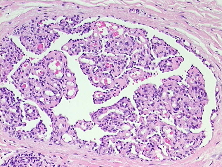
Subcutaneous panniculitis-like T cell lymphoma (SPTCL), designated recently as a distinct clinicopathologic entity in the World Health Organization Classification, is a neoplasm composed of cytotoxic T-cells that preferentially involves subcutaneous adipose tissue. Histologically, SPTCL is characterized by extensive karyorrhectic debris and tumor necrosis suggesting that apoptotic mechanisms are involved in its pathogenesis. We assessed the apoptotic index (AI) and proliferation rate (PR) of 13 cases of SPTCL by TUNEL test and Ki-67 immunostaining, respectively. We also immunohistochemically assessed for expression of BCL-2 (anti-apoptosis), BAX (pro-apoptosis), and P53 and correlated the results with apoptosis and proliferation. We detected a high AI (median 8.1%) in 11 cases of SPTCL, and 12 cases had low BCL-2 and high BAX expression. BCL-2 expression inversely correlated with AI (P <.001) and BAX (P <.001). We found a low PR (cutoff > or = 25%) in eight (61%) cases. There was an inverse correlation between AI and PR (r = -.58, P =.04). Ten cases were assessed for P53; immunostaining results were heterogeneous but P53 expression correlated with large cell cytologic features. Our findings demonstrate that SPTCLs have a high AI that may be explained by differential expression of BCL-2 and BAX in the neoplastic cells. (+info)Local and systemic effects of an allogeneic tumor cell vaccine combining transgenic human lymphotactin with interleukin-2 in patients with advanced or refractory neuroblastoma. (5/103)
In murine models, transgenic chemokine-cytokine tumor vaccines overcome many of the limitations of single-agent immunotherapy by producing the sequence of T-cell attraction followed by proliferation. The safety and immunologic effects of this approach in humans were tested in 21 patients with relapsed or refractory neuroblastoma. They received up to 8 subcutaneous injections of a vaccine combining lymphotactin (Lptn)- and interleukin-2 (IL-2)-secreting allogeneic neuroblastoma cells in a dose-escalating scheme. Severe adverse reactions were limited to reversible panniculitis in 5 patients and bone pain in 1 patient. Injection-site biopsies revealed increased cellularity caused by infiltration of CD4+ and CD8+ lymphocytes, eosinophils, and Langerhans cells. Systemically, the vaccine produced a 2-fold (P =.035) expansion of CD4+ T cells, a 3.5-fold (P =.039) expansion of natural killer (NK) cells, a 2.1-fold (P =.014) expansion of eosinophils, and a 1.6-fold (P =.049) increase in serum IL-5. When restimulated in vitro by the immunizing cell line, T cells collected after vaccination showed a 2.3-fold increase (P =.02) of T-helper (TH2)-type CD3+IL-4+ cells. Supernatant collected from restimulated cells showed increased amounts of IL-4 (11.4-fold; P =.021) and IL-5 (8.7-fold; P =.002). Six patients had significant increases in NK cytolytic activity. Fifteen patients made immunoglobulin G (IgG) antibodies that bound to the immunizing cell line. Measurable tumor responses included complete remission in 2 patients and partial response in 1 patient. Hence, allogeneic tumor cell vaccines combining transgenic Lptn with IL-2 appear to have little toxicity in humans and can induce an antitumor immune response. (+info)Pulmonary nocardiosis with bilateral diffuse granular lung shadows in a patient with subcutaneous panniculitic T-cell lymphoma. (6/103)
A 40-year-old woman undergoing prednisolone and cyclosporine therapy for subcutaneous panniculitic T-cell lymphoma complained of a cough for a few weeks. A chest X-ray revealed bilateral diffuse granular shadows. Additionally, the patient was discovered to have multiple subcutaneous abscesses. Gram-stained smears of sputum and pus from the abscess showing branched gram-positive rods led to a diagnosis of pulmonary nocardiosis with dissemination to the lungs and subcutaneous tissues. Combination therapy consisting of sulfamethoxazole/trimethoprim and panipenem/betamipron produced rapid improvement of radiographic abnormalities. It is suggested that pulmonary nocardiosis should be considered in the differential diagnosis of diffuse granular shadows on chest X-rays, especially in immunocompromised patients. (+info)Cytotoxic/natural killer cell cutaneous lymphomas. Report of EORTC Cutaneous Lymphoma Task Force Workshop. (7/103)
BACKGROUND: Cutaneous lymphomas expressing a cytotoxic or natural killer (NK) cell phenotype represent a group of lymphoproliferative disorders for which there is currently much confusion and little consensus regarding the best nomenclature and classification. METHODS: This study analyzes 48 cases of primary cutaneous lymphoma expressing cytotoxic proteins and/or the NK cell marker, CD56. These cases were collected for a workshop of the European Organization for Research and Treatment of Cancer Cutaneous Lymphoma Task Force, to better clarify the clinical, morphologic, and phenotypic features of these uncommon tumors. RESULTS: Several categories with different clinical and pathologic features were delineated: 1) aggressive, CD8+, epidermotropic, cytotoxic T-cell lymphoma; 2) mycosis fungoides, cytotoxic immunophenotype variant; 3) subcutaneous panniculitis-like T-cell lymphoma; 4) NK/T-cell lymphoma, nasal type; 5) CD4+, NK cell lymphoma; 6) blastoid NK cell lymphoma; (7) intravascular NK-like lymphoma; and 8) cytotoxic, peripheral T-cell lymphoma. CONCLUSIONS: Our data show that primary cutaneous cytotoxic/NK cell lymphomas include distinct groups of diseases, clinically, histologically, and biologically. Because the finding of a cytotoxic phenotype often has prognostic significance, the routine use of cytotoxic markers in the diagnosis and classification of cutaneous lymphomas should be expanded. (+info)Cytophagic histiocytic panniculitis with atypical lymphocytes in peripheral blood. (8/103)
A 26-year-old female patient developed high fever, hepatosplenomegaly and subcutaneous nodules. Atypical lymphocytes were present in the peripheral blood. Skin biopsy revealed lobular panniculitis. Bone marrow examination showed prominent phagocytosis by benign histiocytes. The diagnosis of cytophagic histiocytic panniculitis was made, and the disease has been well controlled with oral prednisolone. This is the first report of cytophagic histiocytic panniculitis with atypical lymphocytes in the peripheral blood, which are frequently seen in virus-associated hemophagocytic syndrome. This observation suggests that underlying viral infection may be one factor in the development of cytophagic histiocytic panniculitis. (+info)Peritoneal panniculitis is a rare inflammatory condition that affects the fatty tissue (panniculus) in the peritoneum, which is the thin membrane that lines the inside of the abdominal cavity and covers the organs within it. The condition is characterized by the accumulation of inflammatory cells in the fatty tissue, leading to nodular or diffuse enlargement and subsequent necrosis (death) of the adipose tissue.
Peritoneal panniculitis can occur as a primary disorder or secondary to other medical conditions such as malignancies, infections, autoimmune diseases, or reactions to medications. The exact cause of primary peritoneal panniculitis remains unclear. Symptoms may include abdominal pain, bloating, fever, weight loss, and elevated white blood cell count. Diagnosis typically involves imaging studies (such as CT or MRI scans) and confirmation through biopsy of the affected tissue. Treatment depends on the underlying cause but often includes corticosteroids and other immunosuppressive medications to manage the inflammation.
Histiocytosis is a term used to describe a group of rare disorders characterized by an abnormal increase in the number of histiocytes, which are a type of white blood cell that helps fight infection and helps in healing processes. These disorders can affect various organs and tissues in the body, leading to different symptoms and severity.
There are several types of histiocytosis, including Langerhans cell histiocytosis (LCH), Erdheim-Chester disease (ECD), and hemophagocytic lymphohistiocytosis (HLH). Each type has its own specific features and diagnostic criteria.
For example, LCH is characterized by the abnormal accumulation of Langerhans cells, a type of histiocyte found in the skin and mucous membranes. These cells can form tumors or lesions in various organs, such as the bones, lungs, liver, and skin.
HLH, on the other hand, is a life-threatening condition that occurs when there is an overactive immune response leading to excessive activation of histiocytes and other immune cells. This can result in fever, enlargement of the liver and spleen, and decreased blood cell counts.
The exact cause of histiocytosis is not fully understood, but it is believed to involve genetic mutations that lead to uncontrolled proliferation and accumulation of histiocytes. Treatment for histiocytosis depends on the type and severity of the disorder and may include chemotherapy, radiation therapy, immunosuppressive drugs, or stem cell transplantation.
Erythema nodosum is a type of inflammation that occurs in the fatty layer of the skin, causing painful, red or purple bumps (nodules) to form. It is a type of panniculitis, which refers to any condition that causes inflammation of the fatty layer of tissue beneath the skin.
Erythema nodosum is often associated with a variety of underlying conditions, such as infections (e.g., streptococcus, tuberculosis), medications (e.g., sulfa drugs, oral contraceptives), inflammatory bowel disease (e.g., Crohn's disease, ulcerative colitis), and pregnancy.
The bumps associated with erythema nodosum typically appear on the shins, ankles, knees, or other areas of the legs, although they can also occur on the arms, hands, or face. The bumps may be tender to the touch, warm, and swollen, and they may cause pain or discomfort when walking or standing for prolonged periods.
In most cases, erythema nodosum resolves on its own within a few weeks to several months, although symptoms can be managed with medications such as nonsteroidal anti-inflammatory drugs (NSAIDs) and corticosteroids. Treating the underlying condition is also important for resolving erythema nodosum and preventing recurrences.
Erythema induratum is a skin condition that is characterized by inflammation and hardening of the skin, usually occurring on the calves of the legs. It is also known as Bazin's disease. The condition typically affects young women and is thought to be related to tuberculosis infection.
The symptoms of erythema induratum include red, painful, and hard nodules or plaques on the skin that may ulcerate and form crusts. These lesions can be tender to the touch and may cause scarring. The condition often affects both legs and can be accompanied by fever, fatigue, and other systemic symptoms of tuberculosis.
The diagnosis of erythema induratum is typically made based on the clinical presentation of the skin lesions and confirmed with laboratory tests such as a biopsy or culture to detect tuberculosis infection. Treatment usually involves antibiotics to treat the underlying tuberculosis infection, as well as anti-inflammatory medications to manage the skin symptoms. In some cases, surgical removal of the lesions may be necessary.
Alcoholic pancreatitis is a specific type of pancreatitis, which is inflammation of the pancreas. This condition is caused by excessive and prolonged consumption of alcohol. The exact mechanism by which alcohol induces pancreatitis is not fully understood, but it is believed that alcohol causes damage to the cells of the pancreas, leading to inflammation. This can result in abdominal pain, nausea, vomiting, fever, and increased heart rate. Chronic alcoholic pancreatitis can also lead to serious complications such as diabetes, malnutrition, and pancreatic cancer. Treatment typically involves supportive care, such as hydration, pain management, and nutritional support, along with abstinence from alcohol. In severe cases, surgery may be necessary to remove damaged tissue or to relieve blockages in the pancreas.
Histiocytes are a type of immune cell that are part of the mononuclear phagocyte system. They originate from monocytes, which are derived from hematopoietic stem cells in the bone marrow. Histiocytes play an important role in the immune system by engulfing and destroying foreign substances, such as bacteria and viruses, as well as removing dead cells and other debris from the body. They can be found in various tissues throughout the body, including the skin, lymph nodes, spleen, and liver.
Histiocytes include several different types of cells, such as macrophages, dendritic cells, and Langerhans cells. These cells have different functions but all play a role in the immune response. For example, macrophages are involved in inflammation and tissue repair, while dendritic cells are important for presenting antigens to T cells and initiating an immune response.
Abnormal accumulations or dysfunction of histiocytes can lead to various diseases, such as histiocytosis, which is a group of disorders characterized by the abnormal proliferation and accumulation of histiocytes in various tissues.
Lupus erythematosus panniculitis (LEP), also known as lupus profundus, is a type of cutaneous lupus erythematosus (CLE) that affects the fatty layer under the skin (subcutaneous tissue). It is characterized by deep inflammation of the fatty tissue, leading to nodular or indurated (hardened) plaques and subcutaneous ulcerations. The lesions are typically tender and can be painful. LEP most commonly affects the face, trunk, and proximal extremities.
LEP is associated with systemic lupus erythematosus (SLE), but it can also occur in isolation without evidence of systemic involvement. It is estimated that approximately 1-10% of patients with SLE develop LEP. The exact pathogenesis of LEP remains unclear, but it is thought to involve an autoimmune response directed against the fatty tissue.
Histologically, LEP is characterized by lobular panniculitis with lymphocytic infiltration and fat necrosis. Direct immunofluorescence may show deposition of immune complexes along the blood vessels in the affected area.
Treatment of LEP can be challenging, and often requires a multidisciplinary approach involving dermatologists, rheumatologists, and other specialists. Systemic therapies such as corticosteroids, antimalarials, immunosuppressive agents, and biologics may be used to control the disease activity. In addition, local treatments such as intralesional steroid injections or surgical excision of lesions may be considered for refractory cases.
Cytophagocytosis is a medical term that refers to the process in which certain types of cells, particularly immune cells like macrophages, engulf and digest other smaller cells or particles. This process helps the body to eliminate foreign substances, cellular debris, and pathogens such as bacteria, viruses, and fungi.
During cytophagocytosis, the macrophage extends its pseudopodia (cytoplasmic extensions) to surround and engulf the target cell or particle, forming a vesicle called a phagosome. The phagosome then fuses with a lysosome, an organelle containing digestive enzymes, which breaks down the contents of the phagosome into smaller molecules that can be used by the macrophage for energy or eliminated as waste products.
Cytophagocytosis is an essential part of the immune system's defense mechanisms and plays a crucial role in maintaining tissue homeostasis and preventing infection and disease.
Erythema is a term used in medicine to describe redness of the skin, which occurs as a result of increased blood flow in the superficial capillaries. This redness can be caused by various factors such as inflammation, infection, trauma, or exposure to heat, cold, or ultraviolet radiation. In some cases, erythema may also be accompanied by other symptoms such as swelling, warmth, pain, or itching. It is a common finding in many medical conditions and can vary in severity from mild to severe.
Gnathostomiasis is a parasitic infection caused by the third-stage larvae of nematodes (roundworms) in the genus Gnathostoma. The infection typically occurs through the consumption of raw or undercooked freshwater fish, amphibians, or birds that contain the parasite's larvae.
The third-stage larvae penetrate the gastrointestinal tract and migrate to various tissues, including the skin, subcutaneous tissue, eyes, and central nervous system, causing cutaneous, ocular, or visceral lesions. The clinical manifestations of gnathostomiasis depend on the migration pathway and the organs involved.
Symptoms can range from mild dermatological reactions to severe neurological complications, such as eosinophilic meningitis or encephalitis. Diagnosis is often challenging due to its nonspecific clinical presentation and requires a high index of suspicion in travelers returning from endemic areas.
The disease is prevalent in Southeast Asia, East Asia, and Central and South America. Preventive measures include avoiding the consumption of raw or undercooked fish, amphibians, or birds in endemic regions and practicing good hygiene.
Nodular nonsuppurative panniculitis is a rare inflammatory condition that affects the subcutaneous fat tissue. The term "nonsuppurative" indicates that it does not involve pus formation or suppuration, unlike some other forms of panniculitis.
In nodular nonsuppurative panniculitis, multiple, firm, and occasionally tender nodules develop in the subcutaneous fat layer, usually on the lower extremities but can also occur on the abdomen, arms, and trunk. These nodules may vary in size from a few millimeters to several centimeters.
The etiology of nodular nonsuppurative panniculitis is not well understood, although it has been associated with various conditions such as autoimmune disorders (e.g., lupus erythematosus, rheumatoid arthritis), infections (e.g., hepatitis C, HIV), medications (e.g., bromocriptine, interferon), and malignancies (e.g., lymphoma).
Histologically, the condition is characterized by a lobular inflammatory infiltrate predominantly composed of lymphocytes, histiocytes, and occasionally plasma cells, with sparing of septa. The overlying skin may appear normal or show mild changes such as erythema or induration.
Treatment for nodular nonsuppurative panniculitis depends on the underlying cause. If an associated condition or medication is identified, addressing it may lead to resolution of the panniculitis. In cases where no specific cause is found, various treatments such as nonsteroidal anti-inflammatory drugs (NSAIDs), corticosteroids, immunosuppressive agents, and antimalarials have been used with varying success rates.
Panniculitis is a medical term that refers to inflammation of the subcutaneous fat, or the layer of fat located just beneath the skin. This condition can affect people of all ages and genders, although it is more commonly seen in middle-aged women. The inflammation can be caused by a variety of factors, including infections, autoimmune disorders, trauma, and medications.
The symptoms of panniculitis may include:
* Red, painful lumps or nodules under the skin
* Skin lesions that may be tender, warm, or bruised
* Swelling and redness in the affected area
* Fever, fatigue, and malaise (a general feeling of illness)
The diagnosis of panniculitis typically involves a physical examination, medical history, and sometimes a biopsy of the affected tissue. Treatment depends on the underlying cause of the inflammation and may include antibiotics, anti-inflammatory medications, or other therapies. In severe cases, hospitalization may be necessary to manage symptoms and prevent complications.
Skin diseases, also known as dermatological conditions, refer to any medical condition that affects the skin, which is the largest organ of the human body. These diseases can affect the skin's function, appearance, or overall health. They can be caused by various factors, including genetics, infections, allergies, environmental factors, and aging.
Skin diseases can present in many different forms, such as rashes, blisters, sores, discolorations, growths, or changes in texture. Some common examples of skin diseases include acne, eczema, psoriasis, dermatitis, fungal infections, viral infections, bacterial infections, and skin cancer.
The symptoms and severity of skin diseases can vary widely depending on the specific condition and individual factors. Some skin diseases are mild and can be treated with over-the-counter medications or topical creams, while others may require more intensive treatments such as prescription medications, light therapy, or even surgery.
It is important to seek medical attention if you experience any unusual or persistent changes in your skin, as some skin diseases can be serious or indicative of other underlying health conditions. A dermatologist is a medical doctor who specializes in the diagnosis and treatment of skin diseases.
Cutaneous T-cell lymphoma (CTCL) is a type of cancer that affects T-cells, a specific group of white blood cells called lymphocytes. These cells play a crucial role in the body's immune system and help protect against infection and disease. In CTCL, the T-cells become malignant and accumulate in the skin, leading to various skin symptoms and lesions.
CTCL is a subtype of non-Hodgkin lymphoma (NHL), which refers to a group of cancers that originate from lymphocytes. Within NHL, CTCL is categorized as a type of extranodal lymphoma since it primarily involves organs or tissues outside the lymphatic system, in this case, the skin.
The two most common subtypes of CTCL are mycosis fungoides and Sézary syndrome:
1. Mycosis fungoides (MF): This is the more prevalent form of CTCL, characterized by patches, plaques, or tumors on the skin. The lesions may be scaly, itchy, or change in size, shape, and color over time. MF usually progresses slowly, with early-stage disease often confined to the skin for several years before spreading to lymph nodes or other organs.
2. Sézary syndrome (SS): This is a more aggressive form of CTCL that involves not only the skin but also the blood and lymph nodes. SS is characterized by the presence of malignant T-cells, known as Sézary cells, in the peripheral blood. Patients with SS typically have generalized erythroderma (reddening and scaling of the entire body), pruritus (severe itching), lymphadenopathy (swollen lymph nodes), and alopecia (hair loss).
The diagnosis of CTCL usually involves a combination of clinical examination, skin biopsy, and immunophenotyping to identify the malignant T-cells. Treatment options depend on the stage and subtype of the disease and may include topical therapies, phototherapy, systemic medications, or targeted therapies.
Dermatomyositis is a medical condition characterized by inflammation and weakness in the muscles and skin. It is a type of inflammatory myopathy, which means that it causes muscle inflammation and damage. Dermatomyositis is often associated with a distinctive rash that affects the skin around the eyes, nose, mouth, fingers, and toes.
The symptoms of dermatomyositis can include:
* Progressive muscle weakness, particularly in the hips, thighs, shoulders, and neck
* Fatigue
* Difficulty swallowing or speaking
* Skin rash, which may be pink or purple and is often accompanied by itching
* Muscle pain and tenderness
* Joint pain and swelling
* Raynaud's phenomenon, a condition that affects blood flow to the fingers and toes
The exact cause of dermatomyositis is not known, but it is believed to be related to an autoimmune response in which the body's immune system mistakenly attacks healthy tissue. Treatment for dermatomyositis typically involves medications to reduce inflammation and suppress the immune system, as well as physical therapy to help maintain muscle strength and function.
Acute necrotizing pancreatitis is a severe and potentially life-threatening form of acute pancreatitis, which is an inflammatory condition of the pancreas. In acute necrotizing pancreatitis, there is widespread death (necrosis) of pancreatic tissue due to autodigestion caused by the activation and release of digestive enzymes within the pancreas. This condition can lead to systemic inflammation, organ failure, and infection of the necrotic areas in the pancreas. It typically has a more complicated clinical course and worse prognosis compared to acute interstitial pancreatitis, which is another form of acute pancreatitis without significant necrosis.
Prednisolone is a synthetic glucocorticoid drug, which is a class of steroid hormones. It is commonly used in the treatment of various inflammatory and autoimmune conditions due to its potent anti-inflammatory and immunosuppressive effects. Prednisolone works by binding to specific receptors in cells, leading to changes in gene expression that reduce the production of substances involved in inflammation, such as cytokines and prostaglandins.
Prednisolone is available in various forms, including tablets, syrups, and injectable solutions. It can be used to treat a wide range of medical conditions, including asthma, rheumatoid arthritis, inflammatory bowel disease, allergies, skin conditions, and certain types of cancer.
Like other steroid medications, prednisolone can have significant side effects if used in high doses or for long periods of time. These may include weight gain, mood changes, increased risk of infections, osteoporosis, diabetes, and adrenal suppression. As a result, the use of prednisolone should be closely monitored by a healthcare professional to ensure that its benefits outweigh its risks.
Spontaneous remission in a medical context refers to the disappearance or significant improvement of symptoms of a disease or condition without any specific treatment being administered. In other words, it's a situation where the disease resolves on its own, without any apparent cause. While spontaneous remission can occur in various conditions, it is relatively rare and not well understood. It's important to note that just because a remission occurs without treatment doesn't mean that medical care should be avoided, as many conditions can worsen or lead to complications if left untreated.
A fatal outcome is a term used in medical context to describe a situation where a disease, injury, or illness results in the death of an individual. It is the most severe and unfortunate possible outcome of any medical condition, and is often used as a measure of the severity and prognosis of various diseases and injuries. In clinical trials and research, fatal outcome may be used as an endpoint to evaluate the effectiveness and safety of different treatments or interventions.
A biopsy is a medical procedure in which a small sample of tissue is taken from the body to be examined under a microscope for the presence of disease. This can help doctors diagnose and monitor various medical conditions, such as cancer, infections, or autoimmune disorders. The type of biopsy performed will depend on the location and nature of the suspected condition. Some common types of biopsies include:
1. Incisional biopsy: In this procedure, a surgeon removes a piece of tissue from an abnormal area using a scalpel or other surgical instrument. This type of biopsy is often used when the lesion is too large to be removed entirely during the initial biopsy.
2. Excisional biopsy: An excisional biopsy involves removing the entire abnormal area, along with a margin of healthy tissue surrounding it. This technique is typically employed for smaller lesions or when cancer is suspected.
3. Needle biopsy: A needle biopsy uses a thin, hollow needle to extract cells or fluid from the body. There are two main types of needle biopsies: fine-needle aspiration (FNA) and core needle biopsy. FNA extracts loose cells, while a core needle biopsy removes a small piece of tissue.
4. Punch biopsy: In a punch biopsy, a round, sharp tool is used to remove a small cylindrical sample of skin tissue. This type of biopsy is often used for evaluating rashes or other skin abnormalities.
5. Shave biopsy: During a shave biopsy, a thin slice of tissue is removed from the surface of the skin using a sharp razor-like instrument. This technique is typically used for superficial lesions or growths on the skin.
After the biopsy sample has been collected, it is sent to a laboratory where a pathologist will examine the tissue under a microscope and provide a diagnosis based on their findings. The results of the biopsy can help guide further treatment decisions and determine the best course of action for managing the patient's condition.
In medical terms, the skin is the largest organ of the human body. It consists of two main layers: the epidermis (outer layer) and dermis (inner layer), as well as accessory structures like hair follicles, sweat glands, and oil glands. The skin plays a crucial role in protecting us from external factors such as bacteria, viruses, and environmental hazards, while also regulating body temperature and enabling the sense of touch.
Alpha 1-antitrypsin (AAT, or α1-antiproteinase, A1AP) is a protein that is primarily produced by the liver and released into the bloodstream. It belongs to a group of proteins called serine protease inhibitors, which help regulate inflammation and protect tissues from damage caused by enzymes involved in the immune response.
Alpha 1-antitrypsin is particularly important for protecting the lungs from damage caused by neutrophil elastase, an enzyme released by white blood cells called neutrophils during inflammation. In the lungs, AAT binds to and inhibits neutrophil elastase, preventing it from degrading the extracellular matrix and damaging lung tissue.
Deficiency in alpha 1-antitrypsin can lead to chronic obstructive pulmonary disease (COPD) and liver disease. The most common cause of AAT deficiency is a genetic mutation that results in abnormal folding and accumulation of the protein within liver cells, leading to reduced levels of functional AAT in the bloodstream. This condition is called alpha 1-antitrypsin deficiency (AATD) and can be inherited in an autosomal codominant manner. Individuals with severe AATD may require augmentation therapy with intravenous infusions of purified human AAT to help prevent lung damage.
Cyclosporine is a medication that belongs to a class of drugs called immunosuppressants. It is primarily used to prevent the rejection of transplanted organs, such as kidneys, livers, and hearts. Cyclosporine works by suppressing the activity of the immune system, which helps to reduce the risk of the body attacking the transplanted organ.
In addition to its use in organ transplantation, cyclosporine may also be used to treat certain autoimmune diseases, such as rheumatoid arthritis and psoriasis. It does this by suppressing the overactive immune response that contributes to these conditions.
Cyclosporine is available in capsule, oral solution, and injectable forms. Common side effects of the medication include kidney problems, high blood pressure, tremors, headache, and nausea. Long-term use of cyclosporine can also increase the risk of certain types of cancer and infections.
It is important to note that cyclosporine should only be used under the close supervision of a healthcare provider, as it requires regular monitoring of blood levels and kidney function.
Immunosuppressive agents are medications that decrease the activity of the immune system. They are often used to prevent the rejection of transplanted organs and to treat autoimmune diseases, where the immune system mistakenly attacks the body's own tissues. These drugs work by interfering with the immune system's normal responses, which helps to reduce inflammation and damage to tissues. However, because they suppress the immune system, people who take immunosuppressive agents are at increased risk for infections and other complications. Examples of immunosuppressive agents include corticosteroids, azathioprine, cyclophosphamide, mycophenolate mofetil, tacrolimus, and sirolimus.
A syndrome, in medical terms, is a set of symptoms that collectively indicate or characterize a disease, disorder, or underlying pathological process. It's essentially a collection of signs and/or symptoms that frequently occur together and can suggest a particular cause or condition, even though the exact physiological mechanisms might not be fully understood.
For example, Down syndrome is characterized by specific physical features, cognitive delays, and other developmental issues resulting from an extra copy of chromosome 21. Similarly, metabolic syndromes like diabetes mellitus type 2 involve a group of risk factors such as obesity, high blood pressure, high blood sugar, and abnormal cholesterol or triglyceride levels that collectively increase the risk of heart disease, stroke, and diabetes.
It's important to note that a syndrome is not a specific diagnosis; rather, it's a pattern of symptoms that can help guide further diagnostic evaluation and management.
X-ray computed tomography (CT or CAT scan) is a medical imaging method that uses computer-processed combinations of many X-ray images taken from different angles to produce cross-sectional (tomographic) images (virtual "slices") of the body. These cross-sectional images can then be used to display detailed internal views of organs, bones, and soft tissues in the body.
The term "computed tomography" is used instead of "CT scan" or "CAT scan" because the machines take a series of X-ray measurements from different angles around the body and then use a computer to process these data to create detailed images of internal structures within the body.
CT scanning is a noninvasive, painless medical test that helps physicians diagnose and treat medical conditions. CT imaging provides detailed information about many types of tissue including lung, bone, soft tissue and blood vessels. CT examinations can be performed on every part of the body for a variety of reasons including diagnosis, surgical planning, and monitoring of therapeutic responses.
In computed tomography (CT), an X-ray source and detector rotate around the patient, measuring the X-ray attenuation at many different angles. A computer uses this data to construct a cross-sectional image by the process of reconstruction. This technique is called "tomography". The term "computed" refers to the use of a computer to reconstruct the images.
CT has become an important tool in medical imaging and diagnosis, allowing radiologists and other physicians to view detailed internal images of the body. It can help identify many different medical conditions including cancer, heart disease, lung nodules, liver tumors, and internal injuries from trauma. CT is also commonly used for guiding biopsies and other minimally invasive procedures.
In summary, X-ray computed tomography (CT or CAT scan) is a medical imaging technique that uses computer-processed combinations of many X-ray images taken from different angles to produce cross-sectional images of the body. It provides detailed internal views of organs, bones, and soft tissues in the body, allowing physicians to diagnose and treat medical conditions.
Frostbite is a medical condition characterized by damage to body tissues caused by extreme cold. It most commonly affects the extremities, such as the fingers, toes, ears, and nose. This occurs when the skin and underlying tissues freeze, causing cellular injury and potentially leading to tissue death if not treated promptly.
The four stages of frostbite are:
1. Frostnip: The earliest stage of frostbite, characterized by cold, pale, or reddened skin. The affected area may also feel numb or tingly. At this stage, the damage is reversible with prompt rewarming.
2. Superficial Frostbite: At this stage, ice crystals form in the skin and underlying tissues, causing pain, swelling, and redness. The skin may appear white or waxy, and blisters may develop within 24-48 hours after rewarming.
3. Deep Frostbite: This is a more severe form of frostbite that affects the deeper tissues, such as muscles and tendons. The affected area becomes hard, cold, and numb, and the skin may appear blue or black. Large blisters filled with dark fluid may form, and there may be tissue death (gangrene) in severe cases.
4. Gangrene: This is the most severe stage of frostbite, characterized by tissue death and the potential need for amputation. The affected area turns black and becomes gangrenous, releasing a foul-smelling discharge.
Prevention measures include dressing appropriately for cold weather, covering exposed skin, staying dry, avoiding alcohol and tobacco use, and seeking shelter in extreme cold conditions. Prompt medical attention is necessary to prevent further tissue damage and potential amputation.
"Cold temperature" is a relative term and its definition can vary depending on the context. In general, it refers to temperatures that are lower than those normally experienced or preferred by humans and other warm-blooded animals. In a medical context, cold temperature is often defined as an environmental temperature that is below 16°C (60.8°F).
Exposure to cold temperatures can have various physiological effects on the human body, such as vasoconstriction of blood vessels near the skin surface, increased heart rate and metabolic rate, and shivering, which helps to generate heat and maintain body temperature. Prolonged exposure to extreme cold temperatures can lead to hypothermia, a potentially life-threatening condition characterized by a drop in core body temperature below 35°C (95°F).
It's worth noting that some people may have different sensitivities to cold temperatures due to factors such as age, health status, and certain medical conditions. For example, older adults, young children, and individuals with circulatory or neurological disorders may be more susceptible to the effects of cold temperatures.
Microfibrils are tiny, thread-like structures that are found in the extracellular matrix (the material that surrounds and supports cells) of many types of biological tissues. They are made up of bundles of long, thin proteins called fibrillins, which are joined together by other proteins such as microfibril-associated glycoproteins (MAGPs).
Microfibrils play an important role in providing structural support and elasticity to tissues. They are particularly abundant in the connective tissue that surrounds blood vessels, where they help to regulate the diameter of the vessels and maintain blood pressure. Microfibrils are also found in the elastic fibers of the lungs, skin, and other tissues, where they contribute to the ability of these tissues to stretch and recoil.
In addition to their structural roles, microfibrils have been shown to play a role in regulating cell behavior and signaling. For example, they can bind to growth factors and other signaling molecules, helping to control the activity of these molecules and influence cellular processes such as proliferation, differentiation, and migration.
Abnormalities in microfibril structure or function have been linked to a number of diseases, including Marfan syndrome, Loeys-Dietz syndrome, and cutis laxa. These conditions are characterized by problems with connective tissue strength and elasticity, which can lead to a range of symptoms such as skeletal abnormalities, cardiovascular disease, and skin fragility.
 Panniculitis
Septal panniculitis
Lupus erythematosus panniculitis
Neutrophilic lobular panniculitis
Atrophic connective tissue panniculitis
Lipodermatosclerosis
List of skin conditions
Weber-Christian disease
IgG4-related disease
Botfly
Idiopathic sclerosing mesenteritis
Subcutaneous fat necrosis of the newborn
Subcutaneous T-cell lymphoma
Acquired generalized lipodystrophy
Erythema induratum
Fat necrosis
Erythema nodosum
Panniculus adiposus
Histopathologic diagnosis of dermatitis
Alpha-1 antitrypsin deficiency
Sclerema neonatorum
Intravascular lymphomas
Nakajo syndrome
CANDLE syndrome
Frederick Parkes Weber
Discoid lupus erythematosus
Compression stockings
GATA2 deficiency
Max Rothmann
Acupuncture
Panniculitis - Wikipedia
 Cold Panniculitis Differential Diagnoses
Cold Panniculitis: Background, Pathophysiology, Etiology
 Panniculitis - Alpha-1 Foundation
 Photoclinic: 'Popsicle' Panniculitis
 Pancreatitis, Panniculitis, and Polyarthritis Syndrome | HSS Case
 Ice age: a case of cold panniculitis | ADC Fetal & Neonatal Edition
 Getting Under Your Skin: Alpha-1 Related Panniculitis - AlphaNet
M54.08 Panniculitis aff regions of neck/bk, sacr/sacrocygl region - ICD-10-CM Diagnosis Codes
 Mesenteric Panniculitis (MP) in CT - A Predictor of Malignancy? - University of Regensburg Publication Server
 Mesenteric Panniculitis or Sclerosing Mesenteritis | Page 28 | Mayo Clinic Connect
Mesenteric Panniculitis or Sclerosing Mesenteritis | Page 73 | Mayo Clinic Connect
Panniculitis | Profiles RNS
 Gastrointestinal Disease and Skin Problems
 Lupus Panniculitis - Derm In-Review
 Panniculitis - Dermatologic Disorders - MSD Manual Professional Edition
Pancreatic panniculitis due to pancreatic carcinoma<...
Subacute Nodular Migratory Panniculitis (Vilanova Disease) Differential Diagnoses
 Lobular panniculitis in patient with history of tuberculosis | Dermpedia
 Infantile onset panniculitis with uveitis and systemic granulomatosis - Global Genes
 View disorder Lipoatrophic panniculitis of the ankles in childhood
Mimicker of Renal Colic: Mesenteric Panniculitis<...
 Cytophagic histiocytic panniculitis: is it a macrophage activation syndrome in situ? | Pediatric Rheumatology | Full Text
 Draft Article - Billing and Coding: Trigger Point Injections (TPI) (DA57701)
 Dr. Robert Mellman, MD - Gastroenterology Specialist in Boca Raton, FL | Healthgrades
 Abdominoplasty, Suction Lipectomy, and Ventral Hernia Repair - Medical Clinical Policy Bulletins | Aetna
Relapsing Polychondritis Clinical Presentation: History, Physical Examination
 Dermatology | Austin Regional Clinic
Search Results: Breast, Rash or multiple lesions
 Otulipenia: MedlinePlus Genetics
Subacute Nodular Migratory1
- Vilanova X, Pinol Aguade J. Subacute Nodular Migratory Panniculitis. (medscape.com)
Mesenteric16
- Mesenteric Panniculitis (MP) in CT - A Predictor of Malignancy? (uni-regensburg.de)
- Purpose: The exact etiology of mesenteric panniculitis (MP) is still unknown and has been discussed in relation to different causes. (uni-regensburg.de)
- I thought I would start a discussion for patients with Mesenteric Panniculitis. (mayoclinic.org)
- From what I know, this auto-immune disorder has three stages to it, each with differing names (Mesenteric Panniculitis, Schlerosing Mesenteritis, Retractile Mesenteritis). (mayoclinic.org)
- I was diagnosed with mesenteric panniculitis on February 20 of this year after going to the emergency room in extreme pain and not being able to keep any food or liquid down. (mayoclinic.org)
- I was brought back to the rapid response room from triage because of a high heart rate which i wasn't having any chest pains at all and the e.r. doc sent me for a CT Scan which showed the mesenteric panniculitis. (mayoclinic.org)
- Hello, I have a couple of questions about Mesenteric Panniculitis. (mayoclinic.org)
- when they came back from the CAT scan all the doctor told me I had a very rare disease call mesenteric panniculitis and that she will refer me to a gastrologist I am at work right now but as I'm Walking the Floor I'm getting like cold sweats so I don't know what's going on with this disease. (mayoclinic.org)
- A Mesenteric panniculitis is an uncommon disorder with unknown etiology. (elsevierpure.com)
- We report computed tomography findings of mesenteric panniculitis in an adult patient with renal colic. (elsevierpure.com)
- Canan, A 2016, ' Mimicker of Renal Colic: Mesenteric Panniculitis ', Journal of Nepal Health Research Council , vol. 14, no. 33, pp. 132-134. (elsevierpure.com)
- Mesenteric panniculitis as a possible cause of fever of unknown origin. (bvsalud.org)
- Mesenteric panniculitis is a non-neoplastic condition involving inflammation and fibrosis of the small bowel mesentery . (bvsalud.org)
- The diagnosis of mesenteric panniculitis had been established 2 weeks prior based on an abdominal computerized tomography scan. (bvsalud.org)
- Extensive diagnostic investigations during his hospitalisation were unrevealing, and the symptoms were ultimately attributed to the mesenteric panniculitis . (bvsalud.org)
- This case suggests that mesenteric panniculitis merits consideration in the differential diagnosis of fever of unknown origin . (bvsalud.org)
Lymphoma4
- Manosca F, Ariga R, Bengana C, Reddy VB, Loew J, Gattuso P. Fine-needle aspiration of subcutaneous panniculitis-like T-cell lymphoma. (rush.edu)
- Cyclosporine A as a Primary Treatment for Panniculitis-like T Cell Lymphoma: A Case with a Long-Term Remission. (qxmd.com)
- Subcutaneous panniculitis-like T cell lymphoma (SPTL) is a distinctive cutaneous lymphoma characterized by an infiltration of subcutaneous tissue by neoplastic T cells, similar to panniculitis. (qxmd.com)
- These diseases include mycosis fungoides and subcutaneous panniculitis-like T-cell lymphoma. (dana-farber.org)
Lobular panniculitis4
- citation needed] There are thus four main histological subtypes: lobular panniculitis without vasculitis (acute panniculitis, previously termed Weber-Christian disease, systemic nodular panniculitis) lobular panniculitis with vasculitis septal panniculitis without vasculitis septal panniculitis with vasculitis Erythema induratum, or "Bazin disease", is a panniculitis on the back of the calves. (wikipedia.org)
- Infantile onset panniculitis with uveitis and systemic granulomatosis is a rare granulomatous autoinflammatroy disease characterized by infantile-onset widespread chronic recurrent progressive lobular panniculitis associated with panuveitis arthritis and severe systemic granulomatous inflammation. (globalgenes.org)
- Cytophagic histiocytic panniculitis (CHP) is a rare lobular panniculitis, characterized by subcutaneous proliferation of benign-appearing cytophagic histiocytes and lymphocytes often associated with systemic macrophage activation syndrome (MAS). (biomedcentral.com)
- Skin biopsy specimen revealed a lobular panniculitis with areas of necrosis in both patients. (biomedcentral.com)
Histiocytic panniculitis3
- citation needed] Non-vasculitis forms of panniculitis that may occur include: Cytophagic histiocytic panniculitis was first described in 1980 by Winkelmann as a chronic histiocytic disease of the subcutaneous adipose tissue, which is characterized clinically by tender erythematous nodules, recurrent high fever, malaise, jaundice, organomegaly, serosal effusions, pancytopenia, hepatic dysfunction and coagulation abnormalities. (wikipedia.org)
- Cytophagic histiocytic panniculitis often presents with multiple subcutaneous nodules and eventuates in a bleeding diathesis. (medscape.com)
- Cytophagic histiocytic panniculitis: is it a macrophage activation syndrome in situ? (biomedcentral.com)
Septal panniculitis2
- Erythema nodosum histologically represents the prototype of a septal panniculitis. (dermnetnz.org)
- The classic histopathologic presentation of erythema nodosum is that of a septal panniculitis with a mixed cellular infiltrate of lymphocytes , histiocytes , giant cells , and occasional eosinophils and a characteristic absence of vasculitis (figures 1-3). (dermnetnz.org)
Erythema Nodosum7
- Erythema Nodosum Erythema nodosum is a specific form of panniculitis characterized by tender, red or violet, palpable, subcutaneous nodules on the shins and occasionally other locations. (msdmanuals.com)
- Septal granulomatous panniculitis: comparison of the pathology of erythema nodosum migrans (migratory panniculitis) and chronic erythema nodosum. (medscape.com)
- Is erythema nodosum the same as panniculitis? (handlebar-online.com)
- Erythema nodosum is a type of panniculitis that affects subcutaneous fat in the skin, usually first evident as an outcropping of erythematous nodules that are highly sensitive to touch. (handlebar-online.com)
- Erythema nodosum (EN), is an inflammatory condition characterized by inflammation of the fat cells under the skin, resulting in tender red nodules or lumps that are usually seen on both shins. (handlebar-online.com)
- Erythema nodosum - a review of an uncommon panniculitis. (nih.gov)
- Erythema nodosum is the most common form of panniculitis . (dermnetnz.org)
Nodular2
- Cold panniculitis (CP) is an acute, nodular, erythematous eruption usually limited to areas exposed to the cold. (medscape.com)
- B and C) Nodular panniculitis lesion in legs. (hss.edu)
Equestrian cold panniculitis4
- Equestrian chilblain is also known as equestrian cold panniculitis. (medscape.com)
- Equestrian cold panniculitis in women. (medscape.com)
- Adachi A, Masaki S, Akiyama M. Equestrian cold panniculitis in a cold-storage-room worker. (medscape.com)
- Many patients classified as having cold panniculitis or equestrian cold panniculitis have forms of perniosis that represent dermal vascular injury rather than true panniculitis. (medscape.com)
Migratory2
- Vanegas ES, Cendejas RF, Mondragón A. A 41-year-old woman with migratory panniculitis. (medscape.com)
- These include skin symptoms, such as pancreatic panniculitis, acanthosis nigricans, livedo reticularis, necrolytic migratory erythema, cutaneous signs of hemorrhage, as in persons with severe acute pancreatitis, or the finding of cutaneous metastases of pancreatic carcinoma, which may be a sign of advanced disease. (nih.gov)
Alpha-1 Antitryp5
- Alpha-1 Antitrypsin Deficiency (Alpha-1) can cause panniculitis, an uncommon skin condition. (alpha1.org)
- This characteristic helps to distinguish it from other forms of panniculitis and its association with Alpha-1 Antitrypsin Deficiency. (alphanet.org)
- This realization suggests that the association between Alpha-1 Antitrypsin Deficiency and panniculitis is underdiagnosed in those that have yet to show lung or liver disease. (alphanet.org)
- Recent studies show treatment with the immunosuppressant Anakinra for panniculitis outside of Alpha-1 Antitrypsin Deficiency has proven effective. (alphanet.org)
- The true prevalence of Alpha-1-related panniculitis is still unknown, as diagnosis depends on awareness of the various presentations of diseases related to Alpha-1 Antitrypsin Deficiency, which is lacking even among pulmonologists. (alphanet.org)
Lupus2
- 492 Lupus erythematosus panniculitis, panniculitis associated with lupus erythematosus. (wikipedia.org)
- Juczynska K, Wozniacka A, Waszczykowska E, Zebrowska A. Lupus erythematosus panniculitis resistant to standard treatment, complicated with macrophage activation syndrome. (medscape.com)
Form of panniculitis4
- Microscopically there are epithelioid granulomas and vasculitis in the subcutaneous tissue, making it a form of panniculitis. (wikipedia.org)
- CHP is a rare and often fatal form of panniculitis with multisystem involvement. (wikipedia.org)
- 492 Lipodermatosclerosis is a form of panniculitis associated with chronic venous insufficiency that presents with brown indurations on the front of the shins. (wikipedia.org)
- Other forms include: Subcutaneous fat necrosis of the newborn, a form of panniculitis occurring in newborns that is usually self-resolving, that may be a result of hypoxic injury to relatively high levels of brown fat. (wikipedia.org)
Similar to panniculitis1
- Since several skin conditions can lead to symptoms and signs similar to panniculitis, the diagnosis of panniculitis depends on a positive skin biopsy. (alphanet.org)
Popsicle4
- 491 This condition has been described in children who suck ice or popsicles, and therefore is sometimes referred to as "popsicle panniculitis. (wikipedia.org)
- Popsicle panniculitis in a 5-month-old child on systemic prednisolone therapy. (medscape.com)
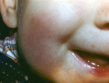
- The term 'popsicle' panniculitis, first used by Epstein, 1 describes a benign, cold-induced, subcutaneous fat necrosis of the cheeks in young children who suck on the frozen confection. (contemporarypediatrics.com)
- Popsicle panniculitis. (contemporarypediatrics.com)
Manifestation4
- Deleersnijder D, Betrains A, De Haes P, Peperstraete L, Buelens J, Blockmans D. Panniculitis as the first clinical manifestation of MPO-positive ANCA-associated vasculitis. (medscape.com)
- Panniculitis can be the initial manifestation of both benign and malignant conditions, presenting with or without arthritis. (hss.edu)
- Recognizing that panniculitis is an Alpha-1 manifestation is crucial to managing accessibility to effective therapies. (alphanet.org)
- Andrade R, Camacho CP, Manso AC & Sérgio M. (2021) Pancreatic Panniculitis: A Rare Manifestation of Pancreatic Disease. (scitcentral.com)
Necrosis6
- 494 Pancreatic panniculitis (also known as enzymatic panniculitis, Pancreatic fat necrosis, and subcutaneous fat necrosis) is a panniculitis most commonly associated with pancreatic carcinoma, and more rarely with anatomic pancreatic abnormalities, pseudocysts, or drug-induced pancreatitis. (wikipedia.org)
- Cold panniculitis needs to be distinguished from other disorders such as adiponecrosis subcutanea ( subcutaneous fat necrosis of the newborn , cold panniculitis of the newborn) and sclerema neonatorum . (medscape.com)
- In cold panniculitis of the newborn (adiponecrosis subcutanea), cold injury in the full-term newborn may occur with focal fat necrosis and a granulomatous and fibrous panniculitis in which the infiltrate usually contains multifocal histiocyte foreign body giant cells containing bifringent star-shaped crystals. (medscape.com)
- The pathophysiology of the panniculitis and arthritis is related to hyperlipasemia, in which lipase and other proteases are released into the bloodstream, causing adipose tissue necrosis and skin lesions. (hss.edu)
- Pancreatitis, panniculitis, and polyarthritis (PPP) syndrome: MRI features of intraosseous fat necrosis involving the feet and knees. (hss.edu)
- Fist described in 1883 by Chiari [1], Pancreatic panniculitis (PP), also known as pancreatic fat necrosis and enzymatic panniculitis, is a rare necrosis of subcutaneous adipocytes triggered by inflammatory, traumatic or malignant pancreatic disease. (scitcentral.com)
Inflammation7
- Panniculitis is a group of diseases whose hallmark is inflammation of subcutaneous adipose tissue (the fatty layer under the skin - panniculus adiposus). (wikipedia.org)
- In cold panniculitis, localized cold damage leads to inflammation of the subcutaneous adipose tissue and is particularly likely to occur in patients with chilblains or in paralyzed limbs affected by poliomyelitis. (medscape.com)
- Panniculitis is an aggravation of the fatty layer underneath the skin which occurs due to Crohn's disease (inflammation of the digestive system) or ulcerative colitis (inflammation of the colon and rectum). (news-medical.net)
- Panniculitis describes inflammation of the subcutaneous fat that can result from multiple causes. (msdmanuals.com)
- Panniculitis can be classified as lobular or septal depending on the principal site of the inflammation within the fat. (msdmanuals.com)
- Signs of systemic inflammation, such as fever and malaise, can accompany panniculitis. (msdmanuals.com)
- The excessive inflammation that results causes fever, diarrhea, panniculitis, and the other signs and symptoms of otulipenia. (medlineplus.gov)
Diagnosis7
- Differential diagnosis also includes poststeroid panniculitis and frostbite. (medscape.com)
- Cold panniculitis is a rare disorder in which the diagnosis probably is hampered by overlapping terminology. (medscape.com)
- A diagnosis was made of pancreatitis, panniculitis, and polyarthritis (PPP) syndrome, with a presumed pancreatic malignancy. (hss.edu)
- The workup for a panniculitis diagnosis recommends Alpha-1 testing, but because there is no diagnostic algorithm endorsed by any dermatological society, it is hard to know the true prevalence. (alphanet.org)
- The median age of diagnosis for panniculitis associated with the Alpha-1 ZZ-genotype is around 36 years. (alphanet.org)
- Diagnosis of panniculitis is by usually by clinical appearance and can be confirmed by biopsy. (msdmanuals.com)
- Rose C, Leverkus M, Fleischer M, Shimanovich I. Histopathology of panniculitis - aspects of biopsy techniques and difficulties in diagnosis. (medscape.com)
Nodules2
- Panniculitis is characterized by tender and erythematous subcutaneous nodules located over the extremities and sometimes over the posterior thorax, abdominal area, breasts, face, or buttocks. (msdmanuals.com)
- Diagnose panniculitis by clinical evaluation (including presence of tender, red, subcutaneous nodules) and confirm with excisional or incisional biopsy. (msdmanuals.com)
Pancreatic Disease2
- Pancreatic disease, panniculitis, polyarthrtitis syndrome successfully treated with total pancreatectomy: Case report and literature review. (hss.edu)
- Pancreatic panniculitis is a very rare complication associated with pancreatic disease, appearing in approximately 2 to 3% of all patients, most commonly those with acute or chronic pancreatitis, but also in patients with pancreatic carcinoma. (scitcentral.com)
Systemic3
- Panniculitis can also be classified based on the presence or absence of systemic symptoms. (wikipedia.org)
- Panniculitis without systemic disease can be a result of trauma or cold. (wikipedia.org)
- Newly diagnosed with Infantile onset panniculitis with uveitis and systemic granulomatosis? (globalgenes.org)
Uncommon2
- Panniculitis is an uncommon skin condition. (alpha1.org)
- Erythema induratum (EI) is an uncommon inflammatory disorder with alterations centered upon the subcutaneous fat tissue (panniculitis). (handlebar-online.com)
Neutrophilic1
- Young TK, Gutierrez D, Meehan SA, Pellett Madan R, Oza VS. Neutrophilic panniculitis arising from hematogenous spread of methicillin-resistant Staphylococcus aureus. (medscape.com)
Inflammatory3
- Restated, an inflammatory disorder primarily localized in the subcutaneous fat is termed a "panniculitis", a group of disorders that may be challenging both for the clinician and the dermatopathologist. (wikipedia.org)
- Panniculitis is an umbrella term for a group of different inflammatory conditions found in the subcutaneous fat of the skin - and a rare result for ZZ individuals and other severely deficient phenotypes. (alphanet.org)
- Treat panniculitis supportively and consider anti-inflammatory or immunosuppressive drug therapy, particularly if manifestations are severe. (msdmanuals.com)
Pathology1
- Pathology shows a septal necrotizing panniculitis with neutrophil infiltration. (hss.edu)
Pancreatitis1
- However, lipase levels are not the only factor implicated, as the relative frequency of pancreatitis with high serum lipase levels contrasts with the rarity of pancreatic panniculitis, and there are some PP cases in the literature that have been described with normal serum levels of all pancreatic enzymes [3], supporting the likely involvement of other unidentified factors in the causality of this entity. (scitcentral.com)
Occurs equally2
- Panniculitis occurs equally among men and women, usually developing at an average age of 40. (alpha1.org)
- Panniculitis occurs equally in men and women. (alphanet.org)
Rarity1
- The rarity of Alpha-1-related panniculitis means a lack of affordable and effective therapies. (alphanet.org)
Infancy1
- Cold panniculitis occurs during infancy and childhood and in adult women who are obese. (medscape.com)
Commonly1
- Adult women who are obese most commonly have cold panniculitis. (medscape.com)
Typically1
- A panniculitis outbreak is typically gradual. (alphanet.org)
Skin5
- Traumatic panniculitis is a panniculitis that occurs following trauma to the skin. (wikipedia.org)
- Panniculitis causes painful, raised red bumps to form on the skin. (alpha1.org)
- Panniculitis sometimes turns into deep ulcers in the skin where the tissue breaks down. (alpha1.org)
- Panniculitis spots sometimes develop after some kind of trauma or injury to the skin that would be minor in most people, such as vigorous exercise, intravenous injections, or cryosurgery (surgery that involves freezing the skin). (alpha1.org)
- But when the spots become deep ulcers in the skin with tissue breakdown it is known as necrotizing panniculitis. (alphanet.org)
Adipose1
- Cold panniculitis results from a cold injury to adipose tissue. (medscape.com)
Augmentation therapy2
- People with Alpha-1 who have panniculitis are sometimes treated with augmentation therapy. (alpha1.org)
- Antibiotics, chemotherapeutic agents, and Dapsone have also displayed varying levels of effectiveness in non-Alpha-1 panniculitis, so they may have some efficacy if augmentation therapy is not available. (alphanet.org)
Therapy3
- Cold Panniculitis After Ice Therapy for Supraventricular Tachycardia. (medscape.com)
- Cold Panniculitis Following Ice Therapy for Cardiac Arrhythmia. (medscape.com)
- The evidence for efficacy of this therapy in Alpha-1-related panniculitis is not strong, but it may be an alternative. (alphanet.org)
Descriptor1
- Panniculitis" is a descriptor in the National Library of Medicine's controlled vocabulary thesaurus, MeSH (Medical Subject Headings) . (rush.edu)
Patients3
- Patients with pancreatic malignancy and panniculitis (either with or without arthritis) seem to have a shorter median survival time than patients without this presentation [3]. (hss.edu)
- Il s'agit d'une étude transversale, monocentrique et descriptive, durant 12 mois, incluant les patients âgés d'au moins 18 ans admis en réanimation polyvalente pour un sepsis ou choc septique. (bvsalud.org)
- Cette étude documentaire, analyse les données des patients diagnostiqués et traités pour tuberculose de 2007 à 2017 en RDC. (bvsalud.org)
Cold6
- 492 Cold panniculitis is a panniculitis occurring after exposure to cold, most often seen in infants and young children. (wikipedia.org)
- Cold panniculitis: Adverse cutaneous effect of whole-body cryotherapy. (medscape.com)
- Quesada-Cortes A, Campos-Munoz L, Diaz-Diaz RM, Casado-Jimenez M. Cold panniculitis. (medscape.com)
- [ 4 ] Some overlap occurs, and cold panniculitis of the newborn has been associated with ice pack application. (medscape.com)
- Cold panniculitis is caused by cold injury in children and in women who are obese. (medscape.com)
- Ter Poorten JC, Hebert AA, Ilkiw R. Cold panniculitis in a neonate. (contemporarypediatrics.com)