Peripheral Nerves
Sciatic Nerve
Nerve Sheath Neoplasms
Peripheral Nervous System Diseases
Peripheral Nervous System Neoplasms
Schwann Cells
Nerve Fibers
Peripheral Nervous System
Sural Nerve
Tibial Nerve
Neural Conduction
Sciatic Neuropathy
Nerve Block
Median Nerve
Ulnar Nerve
Optic Nerve
Spinal Nerves
Neurofibroma
Myelin Sheath
Femoral Nerve
Facial Nerve
Spinal Nerve Roots
Nerve Tissue
Ganglia, Spinal
Neurofibromatosis 1
Neuralgia
Nerve Endings
Nerve Compression Syndromes
Neurilemmoma
Wallerian Degeneration
Radial Nerve
Axotomy
Nerve Fibers, Myelinated
Nerve Growth Factors
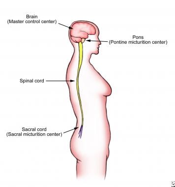
Spinal Cord
Diabetic Neuropathies
Nerve Growth Factor
Peroneal Nerve
Trigeminal Nerve
Rats, Sprague-Dawley
Cranial Nerves
Facial Nerve Injuries
Neuritis
Trauma, Nervous System
Myelin P0 Protein
Phrenic Nerve
Guided Tissue Regeneration
Hyperalgesia
Brachial Plexus
Sensory Receptor Cells
Immunohistochemistry
Nerve Degeneration
Peripheral Vascular Diseases
Ranvier's Nodes
Nerve Tissue Proteins
Demyelinating Diseases
Charcot-Marie-Tooth Disease
Ophthalmic Nerve
Myelin Proteins
Nerve Fibers, Unmyelinated
Polyneuropathies
Mandibular Nerve
Optic Nerve Injuries
Disease Models, Animal
Cats
Electromyography
Pain
S100 Proteins
Action Potentials
Cells, Cultured
Afferent Pathways
Early Growth Response Protein 2
Sympathetic Nervous System
Cochlear Nerve
Anesthetics, Local
Neurofibroma, Plexiform
Leukocytes, Mononuclear
Glossopharyngeal Nerve
Splanchnic Nerves
Cranial Nerve Neoplasms
Receptors, Nerve Growth Factor
Pain Measurement
Mice, Inbred C57BL
Skin
Peroneal Neuropathies
Nerve Transfer
Hindlimb
Peripheral Arterial Disease
Transcutaneous Electric Nerve Stimulation
Rats, Wistar
Maxillary Nerve
Microscopy, Electron
Muscle, Skeletal
Optic Nerve Diseases
Neurofibromatoses
Polyradiculoneuropathy
Hypoglossal Nerve
Neurofibromin 1
Recovery of Function
Neurofilament Proteins
Thoracic Nerves
Genes, Neurofibromatosis 1
Accessory Nerve
GAP-43 Protein
Lumbosacral Plexus
Oculomotor Nerve
RNA, Messenger
Electrophysiology
Central Nervous System
Nociceptors
Sensation
Abducens Nerve
Hyperesthesia
Facial Nerve Diseases
Posterior Horn Cells
Axonal Transport
Receptor, Nerve Growth Factor
Lingual Nerve
Mice, Transgenic
Olfactory Nerve
Mice, Knockout
Recurrent Laryngeal Nerve
Neurons
Anesthesia, Conduction
Neuritis, Autoimmune, Experimental
Tissue Transplantation
Evoked Potentials
Chondroitin ABC Lyase
Lymphoma, T-Cell, Peripheral
Neuroglia
Trigeminal Nerve Diseases
Guillain-Barre Syndrome
Ubiquitin Thiolesterase
Evoked Potentials, Somatosensory
Facial Paralysis
Nervous System Neoplasms
Nervous System
Reflex
Lidocaine
Calcitonin Gene-Related Peptide
Sarcoma, Synovial
T-Lymphocytes
Dose-Response Relationship, Drug
Octamer Transcription Factor-6
Rats, Inbred Strains
Nervous System Diseases
Lymphocytes
Electrodiagnosis
Abducens Nerve Diseases
Gene Expression Regulation
Microscopy, Electron, Transmission
Brachial Plexus Neuropathies
Reverse Transcriptase Polymerase Chain Reaction
Oculomotor Nerve Diseases
Musculocutaneous Nerve
Isaacs Syndrome
Mechanoreceptors
Spinal Cord Injuries
Cell Count
Retrograde Degeneration
Analysis of Variance
Injections, Spinal
Histocytochemistry
Brain
Flow Cytometry
Substance P
Dogs
Magnetic Resonance Imaging
Activating Transcription Factor 3
Chorda Tympani Nerve
Polyradiculoneuropathy, Chronic Inflammatory Demyelinating
Neurites
Blood-Nerve Barrier
Retinal Ganglion Cells
Signal Transduction
Synaptic Transmission
Gabapentin suppresses ectopic nerve discharges and reverses allodynia in neuropathic rats. (1/1775)
Repetitive ectopic discharges from injured afferent nerves play an important role in initiation and maintenance of neuropathic pain. Gabapentin is effective for treatment of neuropathic pain but the sites and mechanisms of its antinociceptive actions remain uncertain. In the present study, we tested a hypothesis that therapeutic doses of gabapentin suppress ectopic afferent discharge activity generated from injured peripheral nerves. Mechanical allodynia, induced by partial ligation of the sciatic nerve in rats, was determined by application of von Frey filaments to the hindpaw. Single-unit afferent nerve activity was recorded proximal to the ligated sciatic nerve site. Intravenous gabapentin, in a range of 30 to 90 mg/kg, significantly attenuated allodynia in nerve-injured rats. Furthermore, gabapentin, in the same therapeutic dose range, dose-dependently inhibited the ectopic discharge activity of 15 injured sciatic afferent nerve fibers through an action on impulse generation. However, the conduction velocity and responses of 12 normal afferent fibers to mechanical stimulation were not affected by gabapentin. Therefore, this study provides electrophysiological evidence that gabapentin is capable of suppressing the ectopic discharge activity from injured peripheral nerves. This action may contribute, at least in part, to the antiallodynic effect of gabapentin on neuropathic pain. (+info)Differential distribution of three members of a gene family encoding low voltage-activated (T-type) calcium channels. (2/1775)
Low voltage-activated (T-type) calcium currents are observed in many central and peripheral neurons and display distinct physiological and functional properties. Using in situ hybridization, we have localized central and peripheral nervous system expression of three transcripts (alpha1G, alpha1H, and alpha1I) of the T-type calcium channel family (CaVT). Each mRNA demonstrated a unique distribution, and expression of the three genes was largely complementary. We found high levels of expression of these transcripts in regions associated with prominent T-type currents, including inferior olivary and thalamic relay neurons (which expressed alpha1G), sensory ganglia, pituitary, and dentate gyrus granule neurons (alpha1H), and thalamic reticular neurons (alpha1I and alpha1H). Other regions of high expression included the Purkinje cell layer of the cerebellum, the bed nucleus of the stria terminalis, the claustrum (alpha1G), the olfactory tubercles (alpha1H and alpha1I), and the subthalamic nucleus (alpha1I and alpha1G). Some neurons expressed high levels of all three genes, including hippocampal pyramidal neurons and olfactory granule cells. Many brain regions showed a predominance of labeling for alpha1G, including the amygdala, cerebral cortex, rostral hypothalamus, brainstem, and spinal cord. Exceptions included the basal ganglia, which showed more prominent labeling for alpha1H and alpha1I, and the olfactory bulb, the hippocampus, and the caudal hypothalamus, which showed more even levels of all three transcripts. Our results are consistent with the hypothesis that differential gene expression underlies pharmacological and physiological heterogeneity observed in neuronal T-type calcium currents, and they provide a molecular basis for the study of T-type channels in particular neurons. (+info)Characterization of the transmembrane molecular architecture of the dystroglycan complex in schwann cells. (3/1775)
We have demonstrated previously 1) that the dystroglycan complex, but not the sarcoglycan complex, is expressed in peripheral nerve, and 2) that alpha-dystroglycan is an extracellular laminin-2-binding protein anchored to beta-dystroglycan in the Schwann cell membrane. In the present study, we investigated the transmembrane molecular architecture of the dystroglycan complex in Schwann cells. The cytoplasmic domain of beta-dystroglycan was co-localized with Dp116, the Schwann cell-specific isoform of dystrophin, in the abaxonal Schwann cell cytoplasm adjacent to the outer membrane. beta-dystroglycan bound to Dp116 mainly via the 15 C-terminal amino acids of its cytoplasmic domain, but these amino acids were not solely responsible for the interaction of these two proteins. Interestingly, the beta-dystroglycan-precipitating antibody precipitated only a small fraction of alpha-dystroglycan and did not precipitate laminin and Dp116 from the peripheral nerve extracts. Our results indicate 1) that Dp116 is a component of the submembranous cytoskeletal system that anchors the dystroglycan complex in Schwann cells, and 2) that the dystroglycan complex in Schwann cells is fragile compared with that in striated muscle cells. We propose that this fragility may be attributable to the absence of the sarcoglycan complex in Schwann cells. (+info)Analysis of optical signals evoked by peripheral nerve stimulation in rat somatosensory cortex: dynamic changes in hemoglobin concentration and oxygenation. (4/1775)
The origins of reflected light changes associated with neuronal activity (optical signals) were investigated in rat somatosensory cortex with optical imaging, microspectrophotometry, and laser-Doppler flowmetry, and dynamic changes in local hemoglobin concentration and oxygenation were focused on. Functional activation was carried out by 2-second, 5-Hz electrical stimulation of the hind limb under chloralose anesthesia. These measurements were performed at the contralateral parietal cortex through a thinned skull. Regional cortical blood flow (rCBF) started to rise 1.5 seconds after the stimulus onset, peaked at 3.5 seconds (26.7% +/- 9.7% increase over baseline), and returned to near baseline by 10 seconds. Optical signal responses at 577, 586, and 805 nm showed a monophasic increase in absorbance coincident with the increase in rCBF; however, the signal responses at 605 and 760 nm were biphasic (an early increase and late decrease in absorbance) and microanatomically heterogeneous. The spectral changes of absorbance indicated that the concentrations of both total hemoglobin and oxyhemoglobin increased together with rCBF; deoxyhemoglobin, increased slightly but distinctly (P = 0.016 at 1.0 seconds, P = 0.00038 at 1.5 seconds) just before rCBF increases, then decreased. The authors conclude that activity-related optical signals are greatly associated with a moment-to-moment adjustment of rCBF and metabolism to neuronal activity. (+info)The distribution of ganglioside-like moieties in peripheral nerves. (5/1775)
GM1 ganglioside has been implicated as a target of immune attack in some diseases of the peripheral nervous system. Anti-GM1 ganglioside antibodies are associated with certain acquired immune-mediated neuropathies. It is not clear how anti-GM1 antibodies cause nerve dysfunction and injury; however, sodium and/or potassium ion channel dysfunction at the node of Ranvier has been implicated. To gain insight into the pathogenesis of these neuropathies, we examined the distribution of GM1 ganglioside and Gal(beta1-3)GalNAc moieties in nerve fibres and their relationship to voltage-gated sodium and potassium (Kv1.1, 1.5) channels at the nodes of Ranvier in peripheral nerves from human, rat and dystrophic mice. Gal(beta1-3)GalNAc moieties were localized via the binding of cholera toxin and peanut agglutinin. As a control for the specificity of these findings, we compared the distribution of GM1 moieties to that of the ganglioside GT1b. Our study provides definitive evidence for the presence of Gal(beta1-3)GalNAc bearing moieties on the axolemmal surface of mature myelinated fibres and on Schwann cells. Gal(beta1-3)GalNAc binding sites did not have an obligatory co-localization with voltage-gated sodium channels or the potassium ion channels Kv1.1 and Kv1.5 and are thus not likely carried by these ion channels. In contrast with Gal(beta1-3)GalNAc, GT1b-like moieties are restricted to the axolemma. (+info)Salinomycin-induced polyneuropathy in cats: morphologic and epidemiologic data. (6/1775)
In April 1996, an outbreak of toxic polyneuropathy in cats occurred in the Netherlands. All cats had been fed one of two brands of dry cat food from one manufacturer. Chemical analyses of these foods, stomach contents, and liver and kidney of affected cats revealed contamination with the ionophor salinomycin. Epidemiologic and clinical data were collected from 823 cats, or about 1% of the cats at risk. In 21 affected cats, postmortem examination was performed. The affected cats had acute onset of lameness and paralysis of the hindlimbs followed by the forelimbs. Clinical and pathologic examination indicated a distal polyneuropathy involving both the sensory and motor nerves. (+info)Neurotrophin modulation of the monosynaptic reflex after peripheral nerve transection. (7/1775)
The effects of neurotrophin-3 (NT-3) and NT-4/5 on the function of axotomized group Ia afferents and motoneurons comprising the monosynaptic reflex pathway were investigated. The axotomized medial gastrocnemius (MG) nerve was provided with NT-3 or NT-4/5 for 8-35 d via an osmotic minipump attached to its central end at the time of axotomy. After this treatment, monosynaptic EPSPs were recorded intracellularly from MG or lateral gastrocnemius soleus (LGS) motoneurons in response to stimulation of the heteronymous nerve under pentobarbital anesthesia. Controls were preparations with axotomized nerves treated directly with vehicle; other axotomized controls were administered subcutaneous NT-3. Direct NT-3 administration (60 microgram/d) not only prevented the decline in EPSP amplitude from axotomized afferents (stimulate MG, record LGS) observed in axotomy controls but, after 5 weeks, led to EPSPs larger than those from intact afferents. These central changes were paralleled by recovery of group I afferent conduction velocity. Removal of NT-3 4-5 weeks after beginning treatment resulted in a decline of conduction velocity and EPSP amplitude within 1 week to values characteristic of axotomy. The increased synaptic efficacy after NT-3 treatment was associated with enhanced connectivity of single afferents to motoneurons. NT-4/5 induced modest recovery in group I afferent conduction velocity but not of the EPSPs they elicited. NT-3 or NT-4/5 had no effect on the properties of treated motoneurons or their monosynaptic EPSPs. We conclude that NT-3, and to a limited extent NT-4/5, promotes recovery of axotomized group Ia afferents but not axotomized motoneurons or the synapses on them. (+info)Relationships between lead absorption and peripheral nerve conduction velocities in lead workers. (8/1775)
The motor sensory, and mixed nerve conduction velocities of median and posterior tibial nerves were measured in 39 lead workers whose blood lead (PbB) concentrations ranged from 2 to 73 mug/100 g with anaverage of 29 mug/100 g. The PbB concentrations significantly correlated with the maximal motor nerve conduction velocities (MCV) and mixed nerve conduction velocities (MNCV) of the median nerve in the forearm and with the MCV of the posterior tibial nerve. Erythrocyte delta-aminolevulinic acid dehydratase (ALAD) activity correlated similarly with the MCV and MNCV of the median nerve in the forearm, and the 24-hour urinary lead excretion following the intravenous administration of CaEDTA (20 mg/kg) (lead mobilization test) correlated with the MNCV. But no parameter correlated with the sensory nerve conduction velocities. By multiple regression analysis, a combination of the three parameters of lead absorption was found to correlate significantly with the MCV and MNCV of the median nerve in the forearm. The MCVs of the median and posterior tibial nerves in lead workers were significantly delayed in the PbB range of 29-73 mug/100 g (mean 45), in the lead mobilization test range from 173 to 3,540 mug/day (mean 973), and the ALAD activity range from 4.4 to 19.4 u. (mean 14.0), respectively. (+info)Peripheral nerves are nerve fibers that transmit signals between the central nervous system (CNS, consisting of the brain and spinal cord) and the rest of the body. These nerves convey motor, sensory, and autonomic information, enabling us to move, feel, and respond to changes in our environment. They form a complex network that extends from the CNS to muscles, glands, skin, and internal organs, allowing for coordinated responses and functions throughout the body. Damage or injury to peripheral nerves can result in various neurological symptoms, such as numbness, weakness, or pain, depending on the type and severity of the damage.
Peripheral nerve injuries refer to damage or trauma to the peripheral nerves, which are the nerves outside the brain and spinal cord. These nerves transmit information between the central nervous system (CNS) and the rest of the body, including sensory, motor, and autonomic functions. Peripheral nerve injuries can result in various symptoms, depending on the type and severity of the injury, such as numbness, tingling, weakness, or paralysis in the affected area.
Peripheral nerve injuries are classified into three main categories based on the degree of damage:
1. Neuropraxia: This is the mildest form of nerve injury, where the nerve remains intact but its function is disrupted due to a local conduction block. The nerve fiber is damaged, but the supporting structures remain intact. Recovery usually occurs within 6-12 weeks without any residual deficits.
2. Axonotmesis: In this type of injury, there is damage to both the axons and the supporting structures (endoneurium, perineurium). The nerve fibers are disrupted, but the connective tissue sheaths remain intact. Recovery can take several months or even up to a year, and it may be incomplete, with some residual deficits possible.
3. Neurotmesis: This is the most severe form of nerve injury, where there is complete disruption of the nerve fibers and supporting structures (endoneurium, perineurium, epineurium). Recovery is unlikely without surgical intervention, which may involve nerve grafting or repair.
Peripheral nerve injuries can be caused by various factors, including trauma, compression, stretching, lacerations, or chemical exposure. Treatment options depend on the type and severity of the injury and may include conservative management, such as physical therapy and pain management, or surgical intervention for more severe cases.
The sciatic nerve is the largest and longest nerve in the human body, running from the lower back through the buttocks and down the legs to the feet. It is formed by the union of the ventral rami (branches) of the L4 to S3 spinal nerves. The sciatic nerve provides motor and sensory innervation to various muscles and skin areas in the lower limbs, including the hamstrings, calf muscles, and the sole of the foot. Sciatic nerve disorders or injuries can result in symptoms such as pain, numbness, tingling, or weakness in the lower back, hips, legs, and feet, known as sciatica.
Nerve sheath neoplasms are a group of tumors that arise from the cells surrounding and supporting the nerves. These tumors can be benign or malignant and include schwannomas, neurofibromas, and malignant peripheral nerve sheath tumors (MPNSTs). Schwannomas develop from the Schwann cells that produce the myelin sheath of the nerve, while neurofibromas arise from the nerve's supporting cells called fibroblasts. MPNSTs are cancerous tumors that can grow rapidly and invade surrounding tissues. Nerve sheath neoplasms can cause various symptoms depending on their location and size, including pain, numbness, weakness, or paralysis in the affected area.
Nerve regeneration is the process of regrowth and restoration of functional nerve connections following damage or injury to the nervous system. This complex process involves various cellular and molecular events, such as the activation of support cells called glia, the sprouting of surviving nerve fibers (axons), and the reformation of neural circuits. The goal of nerve regeneration is to enable the restoration of normal sensory, motor, and autonomic functions impaired due to nerve damage or injury.
Peripheral Nervous System (PNS) diseases, also known as Peripheral Neuropathies, refer to conditions that affect the functioning of the peripheral nervous system, which includes all the nerves outside the brain and spinal cord. These nerves transmit signals between the central nervous system (CNS) and the rest of the body, controlling sensations, movements, and automatic functions such as heart rate and digestion.
PNS diseases can be caused by various factors, including genetics, infections, toxins, metabolic disorders, trauma, or autoimmune conditions. The symptoms of PNS diseases depend on the type and extent of nerve damage but often include:
1. Numbness, tingling, or pain in the hands and feet
2. Muscle weakness or cramps
3. Loss of reflexes
4. Decreased sensation to touch, temperature, or vibration
5. Coordination problems and difficulty with balance
6. Sexual dysfunction
7. Digestive issues, such as constipation or diarrhea
8. Dizziness or fainting due to changes in blood pressure
Examples of PNS diseases include Guillain-Barre syndrome, Charcot-Marie-Tooth disease, diabetic neuropathy, and peripheral nerve injuries. Treatment for these conditions varies depending on the underlying cause but may involve medications, physical therapy, lifestyle changes, or surgery.
Peripheral nervous system (PNS) neoplasms refer to tumors that originate in the peripheral nerves, which are the nerves outside the brain and spinal cord. These tumors can be benign or malignant (cancerous). Benign tumors, such as schwannomas and neurofibromas, grow slowly and do not spread to other parts of the body. Malignant tumors, such as malignant peripheral nerve sheath tumors (MPNSTs), can invade nearby tissues and may metastasize (spread) to other organs.
PNS neoplasms can cause various symptoms depending on their location and size. Common symptoms include pain, weakness, numbness, or tingling in the affected area. In some cases, PNS neoplasms may not cause any symptoms until they become quite large. Treatment options for PNS neoplasms depend on several factors, including the type, size, and location of the tumor, as well as the patient's overall health. Treatment options may include surgery, radiation therapy, chemotherapy, or a combination of these approaches.
Schwann cells, also known as neurolemmocytes, are a type of glial cell that form the myelin sheath around peripheral nervous system (PNS) axons, allowing for the rapid and efficient transmission of nerve impulses. These cells play a crucial role in the maintenance and function of the PNS.
Schwann cells originate from the neural crest during embryonic development and migrate to the developing nerves. They wrap around the axons in a spiral fashion, forming multiple layers of myelin, which insulates the nerve fibers and increases the speed of electrical impulse transmission. Each Schwann cell is responsible for myelinating a single segment of an axon, with the gaps between these segments called nodes of Ranvier.
Schwann cells also provide structural support to the neurons and contribute to the regeneration of injured peripheral nerves by helping to guide the regrowth of axons to their targets. Additionally, Schwann cells can participate in immune responses within the PNS, such as releasing cytokines and chemokines to recruit immune cells during injury or infection.
A nerve crush injury is a type of peripheral nerve injury that occurs when there is excessive pressure or compression applied to a nerve, causing it to become damaged or dysfunctional. This can happen due to various reasons such as trauma from accidents, surgical errors, or prolonged pressure on the nerve from tight casts, clothing, or positions.
The compression disrupts the normal functioning of the nerve, leading to symptoms such as numbness, tingling, weakness, or pain in the affected area. In severe cases, a nerve crush injury can cause permanent damage to the nerve, leading to long-term disability or loss of function. Treatment for nerve crush injuries typically involves relieving the pressure on the nerve, providing supportive care, and in some cases, surgical intervention may be necessary to repair the damaged nerve.
Nerve fibers are specialized structures that constitute the long, slender processes (axons) of neurons (nerve cells). They are responsible for conducting electrical impulses, known as action potentials, away from the cell body and transmitting them to other neurons or effector organs such as muscles and glands. Nerve fibers are often surrounded by supportive cells called glial cells and are grouped together to form nerve bundles or nerves. These fibers can be myelinated (covered with a fatty insulating sheath called myelin) or unmyelinated, which influences the speed of impulse transmission.
The Peripheral Nervous System (PNS) is that part of the nervous system which lies outside of the brain and spinal cord. It includes all the nerves and ganglia ( clusters of neurons) outside of the central nervous system (CNS). The PNS is divided into two components: the somatic nervous system and the autonomic nervous system.
The somatic nervous system is responsible for transmitting sensory information from the skin, muscles, and joints to the CNS, and for controlling voluntary movements of the skeletal muscles.
The autonomic nervous system, on the other hand, controls involuntary actions, such as heart rate, digestion, respiratory rate, salivation, perspiration, pupillary dilation, and sexual arousal. It is further divided into the sympathetic and parasympathetic systems, which generally have opposing effects and maintain homeostasis in the body.
Damage to the peripheral nervous system can result in various medical conditions such as neuropathies, neuritis, plexopathies, and radiculopathies, leading to symptoms like numbness, tingling, pain, weakness, or loss of reflexes in the affected area.
The sural nerve is a purely sensory peripheral nerve in the lower leg and foot. It provides sensation to the outer ( lateral) aspect of the little toe and the adjacent side of the fourth toe, as well as a small portion of the skin on the back of the leg between the ankle and knee joints.
The sural nerve is formed by the union of branches from the tibial and common fibular nerves (branches of the sciatic nerve) in the lower leg. It runs down the calf, behind the lateral malleolus (the bony prominence on the outside of the ankle), and into the foot.
The sural nerve is often used as a donor nerve during nerve grafting procedures due to its consistent anatomy and relatively low risk for morbidity at the donor site.
The Tibial nerve is a major branch of the sciatic nerve that originates in the lower back and runs through the buttock and leg. It provides motor (nerve impulses that control muscle movement) and sensory (nerve impulses that convey information about touch, temperature, and pain) innervation to several muscles and skin regions in the lower limb.
More specifically, the Tibial nerve supplies the following structures:
1. Motor Innervation: The Tibial nerve provides motor innervation to the muscles in the back of the leg (posterior compartment), including the calf muscles (gastrocnemius and soleus) and the small muscles in the foot (intrinsic muscles). These muscles are responsible for plantarflexion (pointing the foot downward) and inversion (turning the foot inward) of the foot.
2. Sensory Innervation: The Tibial nerve provides sensory innervation to the skin on the sole of the foot, as well as the heel and some parts of the lower leg.
The Tibial nerve travels down the leg, passing behind the knee and through the calf, where it eventually joins with the common fibular (peroneal) nerve to form the tibial-fibular trunk. This trunk then divides into several smaller nerves that innervate the foot's intrinsic muscles and skin.
Damage or injury to the Tibial nerve can result in various symptoms, such as weakness or paralysis of the calf and foot muscles, numbness or tingling sensations in the sole of the foot, and difficulty walking or standing on tiptoes.
Neural conduction is the process by which electrical signals, known as action potentials, are transmitted along the axon of a neuron (nerve cell) to transmit information between different parts of the nervous system. This electrical impulse is generated by the movement of ions across the neuronal membrane, and it propagates down the length of the axon until it reaches the synapse, where it can then stimulate the release of neurotransmitters to communicate with other neurons or target cells. The speed of neural conduction can vary depending on factors such as the diameter of the axon, the presence of myelin sheaths (which act as insulation and allow for faster conduction), and the temperature of the environment.
Sciatic neuropathy is a condition that results from damage or injury to the sciatic nerve, which is the largest nerve in the human body. The sciatic nerve originates from the lower spine (lumbar and sacral regions) and travels down through the buttocks, hips, and legs to the feet.
Sciatic neuropathy can cause various symptoms, including pain, numbness, tingling, weakness, or difficulty moving the affected leg or foot. The pain associated with sciatic neuropathy is often described as sharp, shooting, or burning and may worsen with movement, coughing, or sneezing.
The causes of sciatic neuropathy include compression or irritation of the nerve due to conditions such as herniated discs, spinal stenosis, bone spurs, tumors, or piriformis syndrome. Trauma or injury to the lower back, hip, or buttocks can also cause sciatic neuropathy.
Diagnosing sciatic neuropathy typically involves a physical examination and medical history, as well as imaging tests such as X-rays, MRI, or CT scans to visualize the spine and surrounding structures. Treatment options may include pain management, physical therapy, steroid injections, or surgery, depending on the severity and underlying cause of the condition.
A nerve block is a medical procedure in which an anesthetic or neurolytic agent is injected near a specific nerve or bundle of nerves to block the transmission of pain signals from that area to the brain. This technique can be used for both diagnostic and therapeutic purposes, such as identifying the source of pain, providing temporary or prolonged relief, or facilitating surgical procedures in the affected region.
The injection typically contains a local anesthetic like lidocaine or bupivacaine, which numbs the nerve, preventing it from transmitting pain signals. In some cases, steroids may also be added to reduce inflammation and provide longer-lasting relief. Depending on the type of nerve block and its intended use, the injection might be administered close to the spine (neuraxial blocks), at peripheral nerves (peripheral nerve blocks), or around the sympathetic nervous system (sympathetic nerve blocks).
While nerve blocks are generally safe, they can have side effects such as infection, bleeding, nerve damage, or in rare cases, systemic toxicity from the anesthetic agent. It is essential to consult with a qualified medical professional before undergoing this procedure to ensure proper evaluation, technique, and post-procedure care.
The median nerve is one of the major nerves in the human body, providing sensation and motor function to parts of the arm and hand. It originates from the brachial plexus, a network of nerves that arise from the spinal cord in the neck. The median nerve travels down the arm, passing through the cubital tunnel at the elbow, and continues into the forearm and hand.
In the hand, the median nerve supplies sensation to the palm side of the thumb, index finger, middle finger, and half of the ring finger. It also provides motor function to some of the muscles that control finger movements, allowing for flexion of the fingers and opposition of the thumb.
Damage to the median nerve can result in a condition called carpal tunnel syndrome, which is characterized by numbness, tingling, and weakness in the hand and fingers.
The Ulnar nerve is one of the major nerves in the forearm and hand, which provides motor function to the majority of the intrinsic muscles of the hand (except for those innervated by the median nerve) and sensory innervation to the little finger and half of the ring finger. It originates from the brachial plexus, passes through the cubital tunnel at the elbow, and continues down the forearm, where it runs close to the ulna bone. The ulnar nerve then passes through the Guyon's canal in the wrist before branching out to innervate the hand muscles and provide sensation to the skin on the little finger and half of the ring finger.
The optic nerve, also known as the second cranial nerve, is the nerve that transmits visual information from the retina to the brain. It is composed of approximately one million nerve fibers that carry signals related to vision, such as light intensity and color, from the eye's photoreceptor cells (rods and cones) to the visual cortex in the brain. The optic nerve is responsible for carrying this visual information so that it can be processed and interpreted by the brain, allowing us to see and perceive our surroundings. Damage to the optic nerve can result in vision loss or impairment.
Spinal nerves are the bundles of nerve fibers that transmit signals between the spinal cord and the rest of the body. There are 31 pairs of spinal nerves in the human body, which can be divided into five regions: 8 cervical, 12 thoracic, 5 lumbar, 5 sacral, and 1 coccygeal. Each spinal nerve carries both sensory information (such as touch, temperature, and pain) from the periphery to the spinal cord, and motor information (such as muscle control) from the spinal cord to the muscles and other structures in the body. Spinal nerves also contain autonomic fibers that regulate involuntary functions such as heart rate, digestion, and blood pressure.
An axon is a long, slender extension of a neuron (a type of nerve cell) that conducts electrical impulses (nerve impulses) away from the cell body to target cells, such as other neurons or muscle cells. Axons can vary in length from a few micrometers to over a meter long and are typically surrounded by a myelin sheath, which helps to insulate and protect the axon and allows for faster transmission of nerve impulses.
Axons play a critical role in the functioning of the nervous system, as they provide the means by which neurons communicate with one another and with other cells in the body. Damage to axons can result in serious neurological problems, such as those seen in spinal cord injuries or neurodegenerative diseases like multiple sclerosis.
A neurofibroma is a benign (non-cancerous) tumor that develops from the nerve sheath, which is the protective covering around nerves. These tumors can grow anywhere on the body and can be found under the skin or deep inside the body. Neurofibromas can vary in size, and they may cause symptoms such as pain, numbness, or tingling if they press on nearby nerves.
Neurofibromas are a common feature of neurofibromatosis type 1 (NF1), a genetic disorder that affects approximately 1 in every 3,000 people worldwide. NF1 is characterized by the development of multiple neurofibromas and other tumors, as well as skin changes such as café-au-lait spots and freckling.
It's important to note that while most neurofibromas are benign, they can rarely undergo malignant transformation and become cancerous. If you have a neurofibroma or are concerned about your risk of developing one, it's important to seek medical advice from a healthcare professional who is familiar with this condition.
The myelin sheath is a multilayered, fatty substance that surrounds and insulates many nerve fibers in the nervous system. It is essential for the rapid transmission of electrical signals, or nerve impulses, along these nerve fibers, allowing for efficient communication between different parts of the body. The myelin sheath is produced by specialized cells called oligodendrocytes in the central nervous system (CNS) and Schwann cells in the peripheral nervous system (PNS). Damage to the myelin sheath, as seen in conditions like multiple sclerosis, can significantly impair nerve function and result in various neurological symptoms.
The femoral nerve is a major nerve in the thigh region of the human body. It originates from the lumbar plexus, specifically from the ventral rami (anterior divisions) of the second, third, and fourth lumbar nerves (L2-L4). The femoral nerve provides motor and sensory innervation to various muscles and areas in the lower limb.
Motor Innervation:
The femoral nerve is responsible for providing motor innervation to several muscles in the anterior compartment of the thigh, including:
1. Iliacus muscle
2. Psoas major muscle
3. Quadriceps femoris muscle (consisting of four heads: rectus femoris, vastus lateralis, vastus medialis, and vastus intermedius)
These muscles are involved in hip flexion, knee extension, and stabilization of the hip joint.
Sensory Innervation:
The sensory distribution of the femoral nerve includes:
1. Anterior and medial aspects of the thigh
2. Skin over the anterior aspect of the knee and lower leg (via the saphenous nerve, a branch of the femoral nerve)
The saphenous nerve provides sensation to the skin on the inner side of the leg and foot, as well as the medial malleolus (the bony bump on the inside of the ankle).
In summary, the femoral nerve is a crucial component of the lumbar plexus that controls motor functions in the anterior thigh muscles and provides sensory innervation to the anterior and medial aspects of the thigh and lower leg.
The facial nerve, also known as the seventh cranial nerve (CN VII), is a mixed nerve that carries both sensory and motor fibers. Its functions include controlling the muscles involved in facial expressions, taste sensation from the anterior two-thirds of the tongue, and secretomotor function to the lacrimal and salivary glands.
The facial nerve originates from the brainstem and exits the skull through the internal acoustic meatus. It then passes through the facial canal in the temporal bone before branching out to innervate various structures of the face. The main branches of the facial nerve include:
1. Temporal branch: Innervates the frontalis, corrugator supercilii, and orbicularis oculi muscles responsible for eyebrow movements and eyelid closure.
2. Zygomatic branch: Supplies the muscles that elevate the upper lip and wrinkle the nose.
3. Buccal branch: Innervates the muscles of the cheek and lips, allowing for facial expressions such as smiling and puckering.
4. Mandibular branch: Controls the muscles responsible for lower lip movement and depressing the angle of the mouth.
5. Cervical branch: Innervates the platysma muscle in the neck, which helps to depress the lower jaw and wrinkle the skin of the neck.
Damage to the facial nerve can result in various symptoms, such as facial weakness or paralysis, loss of taste sensation, and dry eyes or mouth due to impaired secretion.
Spinal nerve roots are the initial parts of spinal nerves that emerge from the spinal cord through the intervertebral foramen, which are small openings between each vertebra in the spine. These nerve roots carry motor, sensory, and autonomic fibers to and from specific regions of the body. There are 31 pairs of spinal nerve roots in total, with 8 cervical, 12 thoracic, 5 lumbar, 5 sacral, and 1 coccygeal pair. Each root has a dorsal (posterior) and ventral (anterior) ramus that branch off to form the peripheral nervous system. Irritation or compression of these nerve roots can result in pain, numbness, weakness, or loss of reflexes in the affected area.
Nerve tissue, also known as neural tissue, is a type of specialized tissue that is responsible for the transmission of electrical signals and the processing of information in the body. It is a key component of the nervous system, which includes the brain, spinal cord, and peripheral nerves. Nerve tissue is composed of two main types of cells: neurons and glial cells.
Neurons are the primary functional units of nerve tissue. They are specialized cells that are capable of generating and transmitting electrical signals, known as action potentials. Neurons have a unique structure, with a cell body (also called the soma) that contains the nucleus and other organelles, and processes (dendrites and axons) that extend from the cell body and are used to receive and transmit signals.
Glial cells, also known as neuroglia or glia, are non-neuronal cells that provide support and protection for neurons. There are several different types of glial cells, including astrocytes, oligodendrocytes, microglia, and Schwann cells. These cells play a variety of roles in the nervous system, such as providing structural support, maintaining the proper environment for neurons, and helping to repair and regenerate nerve tissue after injury.
Nerve tissue is found throughout the body, but it is most highly concentrated in the brain and spinal cord, which make up the central nervous system (CNS). The peripheral nerves, which are the nerves that extend from the CNS to the rest of the body, also contain nerve tissue. Nerve tissue is responsible for transmitting sensory information from the body to the brain, controlling muscle movements, and regulating various bodily functions such as heart rate, digestion, and respiration.
Spinal ganglia, also known as dorsal root ganglia, are clusters of nerve cell bodies located in the peripheral nervous system. They are situated along the length of the spinal cord and are responsible for transmitting sensory information from the body to the brain. Each spinal ganglion contains numerous neurons, or nerve cells, with long processes called axons that extend into the periphery and innervate various tissues and organs. The cell bodies within the spinal ganglia receive sensory input from these axons and transmit this information to the central nervous system via the dorsal roots of the spinal nerves. This allows the brain to interpret and respond to a wide range of sensory stimuli, including touch, temperature, pain, and proprioception (the sense of the position and movement of one's body).
Neurofibromatosis 1 (NF1) is a genetic disorder that affects the development and growth of nerve tissue. It's also known as von Recklinghausen disease. NF1 is characterized by the growth of non-cancerous tumors on the nerves, as well as skin and bone abnormalities.
The symptoms of Neurofibromatosis 1 can vary widely, even among members of the same family. Some common features include:
* Multiple café au lait spots (flat, light brown patches on the skin)
* Freckles in the underarms and groin area
* Benign growths on or under the skin called neurofibromas
* Larger, more complex tumors called plexiform neurofibromas
* Optic gliomas (tumors that form on the optic nerve)
* Distinctive bone abnormalities, such as a curved spine (scoliosis) or an enlarged head (macrocephaly)
* Learning disabilities and behavioral problems
Neurofibromatosis 1 is caused by mutations in the NF1 gene, which provides instructions for making a protein called neurofibromin. This protein helps regulate cell growth and division. When the NF1 gene is mutated, the production of neurofibromin is reduced or absent, leading to uncontrolled cell growth and the development of tumors.
NF1 is an autosomal dominant disorder, which means that a person has a 50% chance of inheriting the mutated gene from an affected parent. However, about half of all cases are the result of new mutations in the NF1 gene, and occur in people with no family history of the disorder.
There is currently no cure for Neurofibromatosis 1, but treatments are available to manage the symptoms and complications of the disease. These may include medications to control pain or reduce the size of tumors, surgery to remove tumors or correct bone abnormalities, and physical therapy to improve mobility and strength. Regular monitoring by a healthcare team experienced in treating Neurofibromatosis 1 is also important to detect any changes in the condition and provide appropriate care.
Neuralgia is a type of pain that occurs along the pathway of a nerve, often caused by damage or irritation to the nerve. It is typically described as a sharp, stabbing, burning, or electric-shock like pain that can be severe and debilitating. Neuralgia can affect any nerve in the body, but it most commonly occurs in the facial area (trigeminal neuralgia) or in the nerves related to the spine (postherpetic neuralgia). The pain associated with neuralgia can be intermittent or constant and may be worsened by certain triggers such as touch, temperature changes, or movement. Treatment for neuralgia typically involves medications to manage pain, as well as other therapies such as nerve blocks, surgery, or lifestyle modifications.
Nerve endings, also known as terminal branches or sensory receptors, are the specialized structures present at the termination point of nerve fibers (axons) that transmit electrical signals to and from the central nervous system (CNS). They primarily function in detecting changes in the external environment or internal body conditions and converting them into electrical impulses.
There are several types of nerve endings, including:
1. Free Nerve Endings: These are unencapsulated nerve endings that respond to various stimuli like temperature, pain, and touch. They are widely distributed throughout the body, especially in the skin, mucous membranes, and visceral organs.
2. Encapsulated Nerve Endings: These are wrapped by specialized connective tissue sheaths, which can modify their sensitivity to specific stimuli. Examples include Pacinian corpuscles (responsible for detecting deep pressure and vibration), Meissner's corpuscles (for light touch), Ruffini endings (for stretch and pressure), and Merkel cells (for sustained touch).
3. Specialised Nerve Endings: These are nerve endings that respond to specific stimuli, such as auditory, visual, olfactory, gustatory, and vestibular information. They include hair cells in the inner ear, photoreceptors in the retina, taste buds in the tongue, and olfactory receptors in the nasal cavity.
Nerve endings play a crucial role in relaying sensory information to the CNS for processing and initiating appropriate responses, such as reflex actions or conscious perception of the environment.
Nerve compression syndromes refer to a group of conditions characterized by the pressure or irritation of a peripheral nerve, causing various symptoms such as pain, numbness, tingling, and weakness in the affected area. This compression can occur due to several reasons, including injury, repetitive motion, bone spurs, tumors, or swelling. Common examples of nerve compression syndromes include carpal tunnel syndrome, cubital tunnel syndrome, radial nerve compression, and ulnar nerve entrapment at the wrist or elbow. Treatment options may include physical therapy, splinting, medications, injections, or surgery, depending on the severity and underlying cause of the condition.
A neurilemmoma, also known as schwannoma or peripheral nerve sheath tumor, is a benign, slow-growing tumor that arises from the Schwann cells, which produce the myelin sheath that surrounds and insulates peripheral nerves. These tumors can occur anywhere along the course of a peripheral nerve, but they most commonly affect the acoustic nerve (vestibulocochlear nerve), leading to a type of tumor called vestibular schwannoma or acoustic neuroma. Neurilemmomas are typically encapsulated and do not invade the surrounding tissue, although larger ones may cause pressure-related symptoms due to compression of nearby structures. Rarely, these tumors can undergo malignant transformation, leading to a condition called malignant peripheral nerve sheath tumor or neurofibrosarcoma.
Wallerian degeneration is a process that occurs following damage to the axons of neurons (nerve cells). After an axon is severed or traumatically injured, it undergoes a series of changes including fragmentation and removal of the distal segment of the axon, which is the part that is separated from the cell body. This process is named after Augustus Waller, who first described it in 1850.
The degenerative changes in the distal axon are characterized by the breakdown of the axonal cytoskeleton, the loss of myelin sheath (the fatty insulating material that surrounds and protects the axon), and the infiltration of macrophages to clear away the debris. These events lead to the degeneration of the distal axon segment, which is necessary for successful regeneration of the injured nerve.
Wallerian degeneration is a crucial process in the nervous system's response to injury, as it enables the regrowth of axons and the reestablishment of connections between neurons. However, if the regenerative capacity of the neuron is insufficient or the environment is not conducive to growth, functional recovery may be impaired, leading to long-term neurological deficits.
The Radial nerve is a major peripheral nerve in the human body that originates from the brachial plexus, which is a network of nerves formed by the union of the ventral rami (anterior divisions) of spinal nerves C5-T1. The radial nerve provides motor function to extensor muscles of the upper limb and sensation to parts of the skin on the back of the arm, forearm, and hand.
More specifically, the radial nerve supplies motor innervation to:
* Extensor muscles of the shoulder (e.g., teres minor, infraspinatus)
* Rotator cuff muscles
* Elbow joint stabilizers (e.g., lateral head of the triceps)
* Extensors of the wrist, fingers, and thumb
The radial nerve also provides sensory innervation to:
* Posterior aspect of the upper arm (from the lower third of the humerus to the elbow)
* Lateral forearm (from the lateral epicondyle of the humerus to the wrist)
* Dorsum of the hand (skin over the radial side of the dorsum, including the first web space)
Damage or injury to the radial nerve may result in various symptoms, such as weakness or paralysis of the extensor muscles, numbness or tingling sensations in the affected areas, and difficulty with extension movements of the wrist, fingers, and thumb. Common causes of radial nerve injuries include fractures of the humerus bone, compression during sleep or prolonged pressure on the nerve (e.g., from crutches), and entrapment syndromes like radial tunnel syndrome.
Axotomy is a medical term that refers to the surgical cutting or severing of an axon, which is the long, slender projection of a neuron (nerve cell) that conducts electrical impulses away from the cell body and toward other cells. Axons are a critical component of the nervous system, allowing for communication between different parts of the body.
Axotomy is often used in research settings to study the effects of axonal injury on neuronal function and regeneration. This procedure can provide valuable insights into the mechanisms underlying neurodegenerative disorders and potential therapies for nerve injuries. However, it is important to note that axotomy can also have significant consequences for the affected neuron, including changes in gene expression, metabolism, and overall survival.
Myelinated nerve fibers are neuronal processes that are surrounded by a myelin sheath, a fatty insulating substance that is produced by Schwann cells in the peripheral nervous system and oligodendrocytes in the central nervous system. This myelin sheath helps to increase the speed of electrical impulse transmission, also known as action potentials, along the nerve fiber. The myelin sheath has gaps called nodes of Ranvier where the electrical impulses can jump from one node to the next, which also contributes to the rapid conduction of signals. Myelinated nerve fibers are typically found in the peripheral nerves and the optic nerve, but not in the central nervous system (CNS) tracts that are located within the brain and spinal cord.
Afferent neurons, also known as sensory neurons, are a type of nerve cell that conducts impulses or signals from peripheral receptors towards the central nervous system (CNS), which includes the brain and spinal cord. These neurons are responsible for transmitting sensory information such as touch, temperature, pain, sound, and light to the CNS for processing and interpretation. Afferent neurons have specialized receptor endings that detect changes in the environment and convert them into electrical signals, which are then transmitted to the CNS via synapses with other neurons. Once the signals reach the CNS, they are processed and integrated with other information to produce a response or reaction to the stimulus.
Nerve Growth Factors (NGFs) are a family of proteins that play an essential role in the growth, maintenance, and survival of certain neurons (nerve cells). They were first discovered by Rita Levi-Montalcini and Stanley Cohen in 1956. NGF is particularly crucial for the development and function of the peripheral nervous system, which connects the central nervous system to various organs and tissues throughout the body.
NGF supports the differentiation and survival of sympathetic and sensory neurons during embryonic development. In adults, NGF continues to regulate the maintenance and repair of these neurons, contributing to neuroplasticity – the brain's ability to adapt and change over time. Additionally, NGF has been implicated in pain transmission and modulation, as well as inflammatory responses.
Abnormal levels or dysfunctional NGF signaling have been associated with various medical conditions, including neurodegenerative diseases (e.g., Alzheimer's and Parkinson's), chronic pain disorders, and certain cancers (e.g., small cell lung cancer). Therefore, understanding the role of NGF in physiological and pathological processes may provide valuable insights into developing novel therapeutic strategies for these conditions.
The spinal cord is a major part of the nervous system, extending from the brainstem and continuing down to the lower back. It is a slender, tubular bundle of nerve fibers (axons) and support cells (glial cells) that carries signals between the brain and the rest of the body. The spinal cord primarily serves as a conduit for motor information, which travels from the brain to the muscles, and sensory information, which travels from the body to the brain. It also contains neurons that can independently process and respond to information within the spinal cord without direct input from the brain.
The spinal cord is protected by the bony vertebral column (spine) and is divided into 31 segments: 8 cervical, 12 thoracic, 5 lumbar, 5 sacral, and 1 coccygeal. Each segment corresponds to a specific region of the body and gives rise to pairs of spinal nerves that exit through the intervertebral foramina at each level.
The spinal cord is responsible for several vital functions, including:
1. Reflexes: Simple reflex actions, such as the withdrawal reflex when touching a hot surface, are mediated by the spinal cord without involving the brain.
2. Muscle control: The spinal cord carries motor signals from the brain to the muscles, enabling voluntary movement and muscle tone regulation.
3. Sensory perception: The spinal cord transmits sensory information, such as touch, temperature, pain, and vibration, from the body to the brain for processing and awareness.
4. Autonomic functions: The sympathetic and parasympathetic divisions of the autonomic nervous system originate in the thoracolumbar and sacral regions of the spinal cord, respectively, controlling involuntary physiological responses like heart rate, blood pressure, digestion, and respiration.
Damage to the spinal cord can result in various degrees of paralysis or loss of sensation below the level of injury, depending on the severity and location of the damage.
Diabetic neuropathies refer to a group of nerve disorders that are caused by diabetes. High blood sugar levels can injure nerves throughout the body, but diabetic neuropathies most commonly affect the nerves in the legs and feet.
There are four main types of diabetic neuropathies:
1. Peripheral neuropathy: This is the most common type of diabetic neuropathy. It affects the nerves in the legs and feet, causing symptoms such as numbness, tingling, burning, or shooting pain.
2. Autonomic neuropathy: This type of neuropathy affects the autonomic nerves, which control involuntary functions such as heart rate, blood pressure, digestion, and bladder function. Symptoms may include dizziness, fainting, digestive problems, sexual dysfunction, and difficulty regulating body temperature.
3. Proximal neuropathy: Also known as diabetic amyotrophy, this type of neuropathy affects the nerves in the hips, thighs, or buttocks, causing weakness, pain, and difficulty walking.
4. Focal neuropathy: This type of neuropathy affects a single nerve or group of nerves, causing symptoms such as weakness, numbness, or pain in the affected area. Focal neuropathies can occur anywhere in the body, but they are most common in the head, torso, and legs.
The risk of developing diabetic neuropathies increases with the duration of diabetes and poor blood sugar control. Other factors that may contribute to the development of diabetic neuropathies include genetics, age, smoking, and alcohol consumption.
Nerve Growth Factor (NGF) is a small secreted protein that is involved in the growth, maintenance, and survival of certain neurons (nerve cells). It was the first neurotrophin to be discovered and is essential for the development and function of the nervous system. NGF binds to specific receptors on the surface of nerve cells and helps to promote their differentiation, axonal growth, and synaptic plasticity. Additionally, NGF has been implicated in various physiological processes such as inflammation, immune response, and wound healing. Deficiencies or excesses of NGF have been linked to several neurological disorders, including Alzheimer's disease, Parkinson's disease, and pain conditions.
The Peroneal nerve, also known as the common fibular nerve, is a branch of the sciatic nerve that supplies the muscles of the lower leg and provides sensation to the skin on the outer part of the lower leg and the top of the foot. It winds around the neck of the fibula (calf bone) and can be vulnerable to injury in this area, leading to symptoms such as weakness or numbness in the foot and leg.
The trigeminal nerve, also known as the fifth cranial nerve or CNV, is a paired nerve that carries both sensory and motor information. It has three major branches: ophthalmic (V1), maxillary (V2), and mandibular (V3). The ophthalmic branch provides sensation to the forehead, eyes, and upper portion of the nose; the maxillary branch supplies sensation to the lower eyelid, cheek, nasal cavity, and upper lip; and the mandibular branch is responsible for sensation in the lower lip, chin, and parts of the oral cavity, as well as motor function to the muscles involved in chewing. The trigeminal nerve plays a crucial role in sensations of touch, pain, temperature, and pressure in the face and mouth, and it also contributes to biting, chewing, and swallowing functions.
Motor neurons are specialized nerve cells in the brain and spinal cord that play a crucial role in controlling voluntary muscle movements. They transmit electrical signals from the brain to the muscles, enabling us to perform actions such as walking, talking, and swallowing. There are two types of motor neurons: upper motor neurons, which originate in the brain's motor cortex and travel down to the brainstem and spinal cord; and lower motor neurons, which extend from the brainstem and spinal cord to the muscles. Damage or degeneration of these motor neurons can lead to various neurological disorders, such as amyotrophic lateral sclerosis (ALS) and spinal muscular atrophy (SMA).
Sprague-Dawley rats are a strain of albino laboratory rats that are widely used in scientific research. They were first developed by researchers H.H. Sprague and R.C. Dawley in the early 20th century, and have since become one of the most commonly used rat strains in biomedical research due to their relatively large size, ease of handling, and consistent genetic background.
Sprague-Dawley rats are outbred, which means that they are genetically diverse and do not suffer from the same limitations as inbred strains, which can have reduced fertility and increased susceptibility to certain diseases. They are also characterized by their docile nature and low levels of aggression, making them easier to handle and study than some other rat strains.
These rats are used in a wide variety of research areas, including toxicology, pharmacology, nutrition, cancer, and behavioral studies. Because they are genetically diverse, Sprague-Dawley rats can be used to model a range of human diseases and conditions, making them an important tool in the development of new drugs and therapies.
Electric stimulation, also known as electrical nerve stimulation or neuromuscular electrical stimulation, is a therapeutic treatment that uses low-voltage electrical currents to stimulate nerves and muscles. It is often used to help manage pain, promote healing, and improve muscle strength and mobility. The electrical impulses can be delivered through electrodes placed on the skin or directly implanted into the body.
In a medical context, electric stimulation may be used for various purposes such as:
1. Pain management: Electric stimulation can help to block pain signals from reaching the brain and promote the release of endorphins, which are natural painkillers produced by the body.
2. Muscle rehabilitation: Electric stimulation can help to strengthen muscles that have become weak due to injury, illness, or surgery. It can also help to prevent muscle atrophy and improve range of motion.
3. Wound healing: Electric stimulation can promote tissue growth and help to speed up the healing process in wounds, ulcers, and other types of injuries.
4. Urinary incontinence: Electric stimulation can be used to strengthen the muscles that control urination and reduce symptoms of urinary incontinence.
5. Migraine prevention: Electric stimulation can be used as a preventive treatment for migraines by applying electrical impulses to specific nerves in the head and neck.
It is important to note that electric stimulation should only be administered under the guidance of a qualified healthcare professional, as improper use can cause harm or discomfort.
Cranial nerves are a set of twelve pairs of nerves that originate from the brainstem and skull, rather than the spinal cord. These nerves are responsible for transmitting sensory information (such as sight, smell, hearing, and taste) to the brain, as well as controlling various muscles in the head and neck (including those involved in chewing, swallowing, and eye movement). Each cranial nerve has a specific function and is named accordingly. For example, the optic nerve (cranial nerve II) transmits visual information from the eyes to the brain, while the vagus nerve (cranial nerve X) controls parasympathetic functions in the body such as heart rate and digestion.
Facial nerve injuries refer to damages or trauma inflicted on the facial nerve, also known as the seventh cranial nerve (CN VII). This nerve is responsible for controlling the muscles involved in facial expressions, eyelid movement, and taste sensation in the front two-thirds of the tongue.
There are two main types of facial nerve injuries:
1. Peripheral facial nerve injury: This type of injury occurs when damage affects the facial nerve outside the skull base, usually due to trauma from cuts, blunt force, or surgical procedures in the parotid gland or neck region. The injury may result in weakness or paralysis on one side of the face, known as Bell's palsy, and may also impact taste sensation and salivary function.
2. Central facial nerve injury: This type of injury occurs when damage affects the facial nerve within the skull base, often due to stroke, brain tumors, or traumatic brain injuries. Central facial nerve injuries typically result in weakness or paralysis only on the lower half of the face, as the upper motor neurons responsible for controlling the upper face receive innervation from both sides of the brain.
Treatment for facial nerve injuries depends on the severity and location of the damage. For mild to moderate injuries, physical therapy, protective eyewear, and medications like corticosteroids and antivirals may be prescribed. Severe cases might require surgical intervention, such as nerve grafts or muscle transfers, to restore function. In some instances, facial nerve injuries may heal on their own over time, particularly when the injury is mild and there is no ongoing compression or tension on the nerve.
Neuritis is a general term that refers to inflammation of a nerve or nerves, often causing pain, loss of function, and/or sensory changes. It can affect any part of the nervous system, including the peripheral nerves (those outside the brain and spinal cord) or the cranial nerves (those that serve the head and neck). Neuritis may result from various causes, such as infections, autoimmune disorders, trauma, toxins, or metabolic conditions. The specific symptoms and treatment depend on the underlying cause and the affected nerve(s).
Nervous system trauma, also known as neurotrauma, refers to damage or injury to the nervous system, including the brain and spinal cord. This type of trauma can result from various causes, such as vehicular accidents, sports injuries, falls, violence, or penetrating traumas. Nervous system trauma can lead to temporary or permanent impairments in sensory, motor, or cognitive functions, depending on the severity and location of the injury.
Traumatic brain injury (TBI) is a common form of nervous system trauma that occurs when an external force causes brain dysfunction. TBIs can be classified as mild, moderate, or severe, based on factors such as loss of consciousness, memory loss, and neurological deficits. Mild TBIs, also known as concussions, may not cause long-term damage but still require medical attention to ensure proper healing and prevent further complications.
Spinal cord injuries (SCI) are another form of nervous system trauma that can have severe consequences. SCI occurs when the spinal cord is damaged due to a sudden, traumatic blow or cut, causing loss of motor function, sensation, or autonomic function below the level of injury. The severity and location of the injury determine the extent of impairment, which can range from partial to complete paralysis.
Immediate medical intervention is crucial in cases of nervous system trauma to minimize secondary damage, prevent complications, and optimize recovery outcomes. Treatment options may include surgery, medication, rehabilitation, or a combination of these approaches.
Myelin P0 protein, also known as P0 or MPZ (myelin protein zero), is a major structural component of the myelin sheath in the peripheral nervous system. The myelin sheath is a multilayered membrane that surrounds and insulates nerve fibers to increase the speed of electrical impulse transmission.
P0 protein is a transmembrane glycoprotein, which means it spans the lipid bilayer of the myelin membrane and has sugar molecules (glycans) attached to it. It plays a crucial role in maintaining the compact structure of the myelin sheath by forming homodimers that interact with each other through their extracellular domains, creating tight junctions between the apposing layers of the myelin membrane.
P0 protein also contributes to the stability and integrity of the myelin sheath by interacting with other myelin proteins, such as connexin 32 and peripheral myelin protein 22 (PMP22). Mutations in the MPZ gene can lead to various peripheral neuropathies, including Charcot-Marie-Tooth disease type 1B and Dejerine-Sottas syndrome.
The phrenic nerve is a motor nerve that originates from the cervical spine (C3-C5) and descends through the neck to reach the diaphragm, which is the primary muscle used for breathing. The main function of the phrenic nerve is to innervate the diaphragm and control its contraction and relaxation, thereby enabling respiration.
Damage or injury to the phrenic nerve can result in paralysis of the diaphragm, leading to difficulty breathing and potentially causing respiratory failure. Certain medical conditions, such as neuromuscular disorders, spinal cord injuries, and tumors, can affect the phrenic nerve and impair its function.
A neuroma is not a specific type of tumor, but rather refers to a benign (non-cancerous) growth or swelling of nerve tissue. The most common type of neuroma is called a Morton's neuroma, which typically occurs between the third and fourth toes in the foot. It develops as a result of chronic irritation, compression, or trauma to the nerves leading to the toes, causing them to thicken and enlarge.
Morton's neuroma can cause symptoms such as pain, numbness, tingling, or burning sensations in the affected area. Treatment options for Morton's neuroma may include rest, ice, orthotics, physical therapy, medication, or in some cases, surgery. It is essential to consult a healthcare professional if you suspect you have a neuroma or are experiencing related symptoms.
Guided Tissue Regeneration (GTR) is a surgical procedure used in periodontics and implant dentistry that aims to regenerate lost periodontal tissues, such as the alveolar bone, cementum, and periodontal ligament, which have been destroyed due to periodontal disease or trauma. The goal of GTR is to restore the architectural and functional relationship between the teeth and their supporting structures.
The procedure involves placing a barrier membrane between the tooth root and the surrounding soft tissues, creating a protected space that allows the periodontal tissues to regenerate. The membrane acts as a physical barrier, preventing the rapid growth of epithelial cells and fibroblasts from the soft tissue into the defect area, while allowing the slower-growing cells derived from the periodontal ligament and bone to repopulate the space.
There are two main types of membranes used in GTR: resorbable and non-resorbable. Resorbable membranes are made of materials that degrade over time, eliminating the need for a second surgical procedure to remove them. Non-resorbable membranes, on the other hand, must be removed after a period of healing.
GTR has been shown to be effective in treating intrabony defects, furcation involvements, and ridge augmentations, among other applications. However, the success of GTR depends on various factors, including the patient's overall health, the size and location of the defect, and the surgeon's skill and experience.
Hyperalgesia is a medical term that describes an increased sensitivity to pain. It occurs when the nervous system, specifically the nociceptors (pain receptors), become excessively sensitive to stimuli. This means that a person experiences pain from a stimulus that normally wouldn't cause pain or experiences pain that is more intense than usual. Hyperalgesia can be a result of various conditions such as nerve damage, inflammation, or certain medications. It's an important symptom to monitor in patients with chronic pain conditions, as it may indicate the development of tolerance or addiction to pain medication.
The brachial plexus is a network of nerves that originates from the spinal cord in the neck region and supplies motor and sensory innervation to the upper limb. It is formed by the ventral rami (branches) of the lower four cervical nerves (C5-C8) and the first thoracic nerve (T1). In some cases, contributions from C4 and T2 may also be included.
The brachial plexus nerves exit the intervertebral foramen, pass through the neck, and travel down the upper chest before branching out to form major peripheral nerves of the upper limb. These include the axillary, radial, musculocutaneous, median, and ulnar nerves, which further innervate specific muscles and sensory areas in the arm, forearm, and hand.
Damage to the brachial plexus can result in various neurological deficits, such as weakness or paralysis of the upper limb, numbness, or loss of sensation in the affected area, depending on the severity and location of the injury.
Sensory receptor cells are specialized structures that convert physical stimuli from our environment into electrical signals, which are then transmitted to the brain for interpretation. These receptors can be found in various tissues throughout the body and are responsible for detecting sensations such as touch, pressure, temperature, taste, and smell. They can be classified into two main types: exteroceptors, which respond to stimuli from the external environment, and interoceptors, which react to internal conditions within the body. Examples of sensory receptor cells include hair cells in the inner ear, photoreceptors in the eye, and taste buds on the tongue.
Immunohistochemistry (IHC) is a technique used in pathology and laboratory medicine to identify specific proteins or antigens in tissue sections. It combines the principles of immunology and histology to detect the presence and location of these target molecules within cells and tissues. This technique utilizes antibodies that are specific to the protein or antigen of interest, which are then tagged with a detection system such as a chromogen or fluorophore. The stained tissue sections can be examined under a microscope, allowing for the visualization and analysis of the distribution and expression patterns of the target molecule in the context of the tissue architecture. Immunohistochemistry is widely used in diagnostic pathology to help identify various diseases, including cancer, infectious diseases, and immune-mediated disorders.
Nerve degeneration, also known as neurodegeneration, is the progressive loss of structure and function of neurons, which can lead to cognitive decline, motor impairment, and various other symptoms. This process occurs due to a variety of factors, including genetics, environmental influences, and aging. It is a key feature in several neurological disorders such as Alzheimer's disease, Parkinson's disease, Huntington's disease, and multiple sclerosis. The degeneration can affect any part of the nervous system, leading to different symptoms depending on the location and extent of the damage.
Peripheral Vascular Diseases (PVD) refer to a group of medical conditions that affect the blood vessels outside of the heart and brain. These diseases are characterized by a narrowing or blockage of the peripheral arteries, which can lead to reduced blood flow to the limbs, particularly the legs.
The primary cause of PVD is atherosclerosis, a buildup of fats, cholesterol, and other substances in and on the walls of the arteries, forming plaques that restrict blood flow. Other risk factors include smoking, diabetes, hypertension, high cholesterol levels, and a family history of vascular disease.
Symptoms of PVD can vary depending on the severity of the condition but may include leg pain or cramping during exercise (claudication), numbness or tingling in the legs, coldness or discoloration of the feet, sores or wounds that heal slowly or not at all, and in severe cases, gangrene.
PVD can increase the risk of heart attack and stroke, so it is essential to diagnose and treat the condition as early as possible. Treatment options include lifestyle changes such as quitting smoking, exercising regularly, and maintaining a healthy diet, medications to control symptoms and reduce the risk of complications, and surgical procedures such as angioplasty or bypass surgery to restore blood flow.
Denervation is a medical term that refers to the loss or removal of nerve supply to an organ or body part. This can occur as a result of surgical intervention, injury, or disease processes that damage the nerves leading to the affected area. The consequences of denervation depend on the specific organ or tissue involved, but generally, it can lead to changes in function, sensation, and muscle tone. For example, denervation of a skeletal muscle can cause weakness, atrophy, and altered reflexes. Similarly, denervation of an organ such as the heart can lead to abnormalities in heart rate and rhythm. In some cases, denervation may be intentional, such as during surgical procedures aimed at treating chronic pain or spasticity.
Ranvier's nodes, also known as nodes of Ranvier, are specialized structures in the nervous system. They are gaps in the myelin sheath, a fatty insulating substance that surrounds the axons of many neurons, leaving them exposed. These nodes play a crucial role in the rapid transmission of electrical signals along the neuron. The unmyelinated sections of the axon at the nodes have a higher concentration of voltage-gated sodium channels, which generate the action potential that propagates along the neuron. The myelinated segments between the nodes, called internodes, help to speed up this process by allowing the action potential to "jump" from node to node, a mechanism known as saltatory conduction. This process significantly increases the speed of neural impulse transmission, making it more efficient. Ranvier's nodes are named after Louis-Antoine Ranvier, a French histologist and physiologist who first described them in the late 19th century.
Nerve tissue proteins are specialized proteins found in the nervous system that provide structural and functional support to nerve cells, also known as neurons. These proteins include:
1. Neurofilaments: These are type IV intermediate filaments that provide structural support to neurons and help maintain their shape and size. They are composed of three subunits - NFL (light), NFM (medium), and NFH (heavy).
2. Neuronal Cytoskeletal Proteins: These include tubulins, actins, and spectrins that provide structural support to the neuronal cytoskeleton and help maintain its integrity.
3. Neurotransmitter Receptors: These are specialized proteins located on the postsynaptic membrane of neurons that bind neurotransmitters released by presynaptic neurons, triggering a response in the target cell.
4. Ion Channels: These are transmembrane proteins that regulate the flow of ions across the neuronal membrane and play a crucial role in generating and transmitting electrical signals in neurons.
5. Signaling Proteins: These include enzymes, receptors, and adaptor proteins that mediate intracellular signaling pathways involved in neuronal development, differentiation, survival, and death.
6. Adhesion Proteins: These are cell surface proteins that mediate cell-cell and cell-matrix interactions, playing a crucial role in the formation and maintenance of neural circuits.
7. Extracellular Matrix Proteins: These include proteoglycans, laminins, and collagens that provide structural support to nerve tissue and regulate neuronal migration, differentiation, and survival.
Demyelinating diseases are a group of disorders that are characterized by damage to the myelin sheath, which is the protective covering surrounding nerve fibers in the brain, optic nerves, and spinal cord. Myelin is essential for the rapid transmission of nerve impulses, and its damage results in disrupted communication between the brain and other parts of the body.
The most common demyelinating disease is multiple sclerosis (MS), where the immune system mistakenly attacks the myelin sheath. Other demyelinating diseases include:
1. Acute Disseminated Encephalomyelitis (ADEM): An autoimmune disorder that typically follows a viral infection or vaccination, causing widespread inflammation and demyelination in the brain and spinal cord.
2. Neuromyelitis Optica (NMO) or Devic's Disease: A rare autoimmune disorder that primarily affects the optic nerves and spinal cord, leading to severe vision loss and motor disability.
3. Transverse Myelitis: Inflammation of the spinal cord causing damage to both sides of one level (segment) of the spinal cord, resulting in various neurological symptoms such as muscle weakness, numbness, or pain, depending on which part of the spinal cord is affected.
4. Guillain-Barré Syndrome: An autoimmune disorder that causes rapid-onset muscle weakness, often beginning in the legs and spreading to the upper body, including the face and breathing muscles. It occurs when the immune system attacks the peripheral nerves' myelin sheath.
5. Central Pontine Myelinolysis (CPM): A rare neurological disorder caused by rapid shifts in sodium levels in the blood, leading to damage to the myelin sheath in a specific area of the brainstem called the pons.
These diseases can result in various symptoms, such as muscle weakness, numbness, vision loss, difficulty with balance and coordination, and cognitive impairment, depending on the location and extent of the demyelination. Treatment typically focuses on managing symptoms, modifying the immune system's response, and promoting nerve regeneration and remyelination when possible.
Charcot-Marie-Tooth disease (CMT) is a group of inherited disorders that cause nerve damage, primarily affecting the peripheral nerves. These are the nerves that transmit signals between the brain and spinal cord to the rest of the body. CMT affects both motor and sensory nerves, leading to muscle weakness and atrophy, as well as numbness or tingling in the hands and feet.
The disease is named after the three physicians who first described it: Jean-Martin Charcot, Pierre Marie, and Howard Henry Tooth. CMT is characterized by its progressive nature, meaning symptoms typically worsen over time, although the rate of progression can vary significantly among individuals.
There are several types of CMT, classified based on their genetic causes and patterns of inheritance. The two most common forms are CMT1 and CMT2:
1. CMT1: This form is caused by mutations in the genes responsible for the myelin sheath, which insulates peripheral nerves and allows for efficient signal transmission. As a result, demyelination occurs, slowing down nerve impulses and causing muscle weakness, particularly in the lower limbs. Symptoms usually begin in childhood or adolescence and include foot drop, high arches, and hammertoes.
2. CMT2: This form is caused by mutations in the genes responsible for the axons, the nerve fibers that transmit signals within peripheral nerves. As a result, axonal degeneration occurs, leading to muscle weakness and atrophy. Symptoms usually begin in early adulthood and progress more slowly than CMT1. They primarily affect the lower limbs but can also involve the hands and arms.
Diagnosis of CMT typically involves a combination of clinical evaluation, family history, nerve conduction studies, and genetic testing. While there is no cure for CMT, treatment focuses on managing symptoms and maintaining mobility and function through physical therapy, bracing, orthopedic surgery, and pain management.
The ophthalmic nerve, also known as the first cranial nerve or CN I, is a sensory nerve that primarily transmits information about vision, including light intensity and color, and sensation in the eye and surrounding areas. It is responsible for the sensory innervation of the upper eyelid, conjunctiva, cornea, iris, ciliary body, and nasal cavity. The ophthalmic nerve has three major branches: the lacrimal nerve, frontal nerve, and nasociliary nerve. Damage to this nerve can result in various visual disturbances and loss of sensation in the affected areas.
Myelin proteins are proteins that are found in the myelin sheath, which is a fatty (lipid-rich) substance that surrounds and insulates nerve fibers (axons) in the nervous system. The myelin sheath enables the rapid transmission of electrical signals (nerve impulses) along the axons, allowing for efficient communication between different parts of the nervous system.
There are several types of myelin proteins, including:
1. Proteolipid protein (PLP): This is the most abundant protein in the myelin sheath and plays a crucial role in maintaining the structure and function of the myelin sheath.
2. Myelin basic protein (MBP): This protein is also found in the myelin sheath and helps to stabilize the compact structure of the myelin sheath.
3. Myelin-associated glycoprotein (MAG): This protein is involved in the adhesion of the myelin sheath to the axon and helps to maintain the integrity of the myelin sheath.
4. 2'3'-cyclic nucleotide 3' phosphodiesterase (CNP): This protein is found in oligodendrocytes, which are the cells that produce the myelin sheath in the central nervous system. CNP plays a role in maintaining the structure and function of the oligodendrocytes.
Damage to myelin proteins can lead to demyelination, which is a characteristic feature of several neurological disorders, including multiple sclerosis (MS), Guillain-Barré syndrome, and Charcot-Marie-Tooth disease.
Unmyelinated nerve fibers, also known as unmyelinated axons or non-myelinated fibers, are nerve cells that lack a myelin sheath. Myelin is a fatty, insulating substance that surrounds the axon of many nerve cells and helps to increase the speed of electrical impulses traveling along the nerve fiber.
In unmyelinated nerve fibers, the axons are surrounded by a thin layer of Schwann cell processes called the endoneurium, but there is no continuous myelin sheath. Instead, the axons are packed closely together in bundles, with several axons lying within the same Schwann cell.
Unmyelinated nerve fibers tend to be smaller in diameter than myelinated fibers and conduct electrical impulses more slowly. They are commonly found in the autonomic nervous system, which controls involuntary functions such as heart rate, blood pressure, and digestion, as well as in sensory nerves that transmit pain and temperature signals.
Polyneuropathy is a medical condition that refers to the damage or dysfunction of peripheral nerves (nerves outside the brain and spinal cord) in multiple areas of the body. These nerves are responsible for transmitting sensory, motor, and autonomic signals between the central nervous system and the rest of the body.
In polyneuropathies, this communication is disrupted, leading to various symptoms depending on the type and extent of nerve damage. Commonly reported symptoms include:
1. Numbness or tingling in the hands and feet
2. Muscle weakness and cramps
3. Loss of reflexes
4. Burning or stabbing pain
5. Balance and coordination issues
6. Increased sensitivity to touch
7. Autonomic dysfunction, such as bowel, bladder, or digestive problems, and changes in blood pressure
Polyneuropathies can be caused by various factors, including diabetes, alcohol abuse, nutritional deficiencies, autoimmune disorders, infections, toxins, inherited genetic conditions, or idiopathic (unknown) causes. The treatment for polyneuropathy depends on the underlying cause and may involve managing underlying medical conditions, physical therapy, pain management, and lifestyle modifications.
The mandibular nerve is a branch of the trigeminal nerve (the fifth cranial nerve), which is responsible for sensations in the face and motor functions such as biting and chewing. The mandibular nerve provides both sensory and motor innervation to the lower third of the face, below the eye and nose down to the chin.
More specifically, it carries sensory information from the lower teeth, lower lip, and parts of the oral cavity, as well as the skin over the jaw and chin. It also provides motor innervation to the muscles of mastication (chewing), which include the masseter, temporalis, medial pterygoid, and lateral pterygoid muscles.
Damage to the mandibular nerve can result in numbness or loss of sensation in the lower face and mouth, as well as weakness or difficulty with chewing and biting.
Optic nerve injuries refer to damages or trauma inflicted on the optic nerve, which is a crucial component of the visual system. The optic nerve transmits visual information from the retina to the brain, enabling us to see. Injuries to the optic nerve can result in various visual impairments, including partial or complete vision loss, decreased visual acuity, changes in color perception, and reduced field of view.
These injuries may occur due to several reasons, such as:
1. Direct trauma to the eye or head
2. Increased pressure inside the eye (glaucoma)
3. Optic neuritis, an inflammation of the optic nerve
4. Ischemia, or insufficient blood supply to the optic nerve
5. Compression from tumors or other space-occupying lesions
6. Intrinsic degenerative conditions affecting the optic nerve
7. Toxic exposure to certain chemicals or medications
Optic nerve injuries are diagnosed through a comprehensive eye examination, including visual acuity testing, slit-lamp examination, dilated fundus exam, and additional diagnostic tests like optical coherence tomography (OCT) and visual field testing. Treatment options vary depending on the cause and severity of the injury but may include medications, surgery, or vision rehabilitation.
Animal disease models are specialized animals, typically rodents such as mice or rats, that have been genetically engineered or exposed to certain conditions to develop symptoms and physiological changes similar to those seen in human diseases. These models are used in medical research to study the pathophysiology of diseases, identify potential therapeutic targets, test drug efficacy and safety, and understand disease mechanisms.
The genetic modifications can include knockout or knock-in mutations, transgenic expression of specific genes, or RNA interference techniques. The animals may also be exposed to environmental factors such as chemicals, radiation, or infectious agents to induce the disease state.
Examples of animal disease models include:
1. Mouse models of cancer: Genetically engineered mice that develop various types of tumors, allowing researchers to study cancer initiation, progression, and metastasis.
2. Alzheimer's disease models: Transgenic mice expressing mutant human genes associated with Alzheimer's disease, which exhibit amyloid plaque formation and cognitive decline.
3. Diabetes models: Obese and diabetic mouse strains like the NOD (non-obese diabetic) or db/db mice, used to study the development of type 1 and type 2 diabetes, respectively.
4. Cardiovascular disease models: Atherosclerosis-prone mice, such as ApoE-deficient or LDLR-deficient mice, that develop plaque buildup in their arteries when fed a high-fat diet.
5. Inflammatory bowel disease models: Mice with genetic mutations affecting intestinal barrier function and immune response, such as IL-10 knockout or SAMP1/YitFc mice, which develop colitis.
Animal disease models are essential tools in preclinical research, but it is important to recognize their limitations. Differences between species can affect the translatability of results from animal studies to human patients. Therefore, researchers must carefully consider the choice of model and interpret findings cautiously when applying them to human diseases.
"Cat" is a common name that refers to various species of small carnivorous mammals that belong to the family Felidae. The domestic cat, also known as Felis catus or Felis silvestris catus, is a popular pet and companion animal. It is a subspecies of the wildcat, which is found in Europe, Africa, and Asia.
Domestic cats are often kept as pets because of their companionship, playful behavior, and ability to hunt vermin. They are also valued for their ability to provide emotional support and therapy to people. Cats are obligate carnivores, which means that they require a diet that consists mainly of meat to meet their nutritional needs.
Cats are known for their agility, sharp senses, and predatory instincts. They have retractable claws, which they use for hunting and self-defense. Cats also have a keen sense of smell, hearing, and vision, which allow them to detect prey and navigate their environment.
In medical terms, cats can be hosts to various parasites and diseases that can affect humans and other animals. Some common feline diseases include rabies, feline leukemia virus (FeLV), feline immunodeficiency virus (FIV), and toxoplasmosis. It is important for cat owners to keep their pets healthy and up-to-date on vaccinations and preventative treatments to protect both the cats and their human companions.
Electromyography (EMG) is a medical diagnostic procedure that measures the electrical activity of skeletal muscles during contraction and at rest. It involves inserting a thin needle electrode into the muscle to record the electrical signals generated by the muscle fibers. These signals are then displayed on an oscilloscope and may be heard through a speaker.
EMG can help diagnose various neuromuscular disorders, such as muscle weakness, numbness, or pain, and can distinguish between muscle and nerve disorders. It is often used in conjunction with other diagnostic tests, such as nerve conduction studies, to provide a comprehensive evaluation of the nervous system.
EMG is typically performed by a neurologist or a physiatrist, and the procedure may cause some discomfort or pain, although this is usually minimal. The results of an EMG can help guide treatment decisions and monitor the progression of neuromuscular conditions over time.
Pain is an unpleasant sensory and emotional experience associated with actual or potential tissue damage, or described in terms of such damage. It is a complex phenomenon that can result from various stimuli, such as thermal, mechanical, or chemical irritation, and it can be acute or chronic. The perception of pain involves the activation of specialized nerve cells called nociceptors, which transmit signals to the brain via the spinal cord. These signals are then processed in different regions of the brain, leading to the conscious experience of pain. It's important to note that pain is a highly individual and subjective experience, and its perception can vary widely among individuals.
S100 proteins are a family of calcium-binding proteins that are involved in the regulation of various cellular processes, including cell growth and differentiation, intracellular signaling, and inflammation. They are found in high concentrations in certain types of cells, such as nerve cells (neurons), glial cells (supporting cells in the nervous system), and skin cells (keratinocytes).
The S100 protein family consists of more than 20 members, which are divided into several subfamilies based on their structural similarities. Some of the well-known members of this family include S100A1, S100B, S100 calcium-binding protein A8 (S100A8), and S100 calcium-binding protein A9 (S100A9).
Abnormal expression or regulation of S100 proteins has been implicated in various pathological conditions, such as neurodegenerative diseases, cancer, and inflammatory disorders. For example, increased levels of S100B have been found in the brains of patients with Alzheimer's disease, while overexpression of S100A8 and S100A9 has been associated with the development and progression of certain types of cancer.
Therefore, understanding the functions and regulation of S100 proteins is important for developing new diagnostic and therapeutic strategies for various diseases.
An action potential is a brief electrical signal that travels along the membrane of a nerve cell (neuron) or muscle cell. It is initiated by a rapid, localized change in the permeability of the cell membrane to specific ions, such as sodium and potassium, resulting in a rapid influx of sodium ions and a subsequent efflux of potassium ions. This ion movement causes a brief reversal of the electrical potential across the membrane, which is known as depolarization. The action potential then propagates along the cell membrane as a wave, allowing the electrical signal to be transmitted over long distances within the body. Action potentials play a crucial role in the communication and functioning of the nervous system and muscle tissue.
"Cells, cultured" is a medical term that refers to cells that have been removed from an organism and grown in controlled laboratory conditions outside of the body. This process is called cell culture and it allows scientists to study cells in a more controlled and accessible environment than they would have inside the body. Cultured cells can be derived from a variety of sources, including tissues, organs, or fluids from humans, animals, or cell lines that have been previously established in the laboratory.
Cell culture involves several steps, including isolation of the cells from the tissue, purification and characterization of the cells, and maintenance of the cells in appropriate growth conditions. The cells are typically grown in specialized media that contain nutrients, growth factors, and other components necessary for their survival and proliferation. Cultured cells can be used for a variety of purposes, including basic research, drug development and testing, and production of biological products such as vaccines and gene therapies.
It is important to note that cultured cells may behave differently than they do in the body, and results obtained from cell culture studies may not always translate directly to human physiology or disease. Therefore, it is essential to validate findings from cell culture experiments using additional models and ultimately in clinical trials involving human subjects.
Afferent pathways, also known as sensory pathways, refer to the neural connections that transmit sensory information from the peripheral nervous system to the central nervous system (CNS), specifically to the brain and spinal cord. These pathways are responsible for carrying various types of sensory information, such as touch, temperature, pain, pressure, vibration, hearing, vision, and taste, to the CNS for processing and interpretation.
The afferent pathways begin with sensory receptors located throughout the body, which detect changes in the environment and convert them into electrical signals. These signals are then transmitted via afferent neurons, also known as sensory neurons, to the spinal cord or brainstem. Within the CNS, the information is further processed and integrated with other neural inputs before being relayed to higher cognitive centers for conscious awareness and response.
Understanding the anatomy and physiology of afferent pathways is essential for diagnosing and treating various neurological conditions that affect sensory function, such as neuropathies, spinal cord injuries, and brain disorders.
Early Growth Response Protein 2 (EGR2) is a transcription factor that belongs to the EGR family of proteins, which are involved in various biological processes such as cell proliferation, differentiation, and apoptosis. EGR2 is specifically known to play crucial roles in the development and function of the nervous system, including the regulation of neuronal survival, axon guidance, and myelination. It is also expressed in immune cells and has been implicated in the regulation of immune responses. Mutations in the EGR2 gene have been associated with certain neurological disorders and diseases, such as Charcot-Marie-Tooth disease type 1B and congenital hypomyelinating neuropathy.
The sympathetic nervous system (SNS) is a part of the autonomic nervous system that operates largely below the level of consciousness, and it functions to produce appropriate physiological responses to perceived danger. It's often associated with the "fight or flight" response. The SNS uses nerve impulses to stimulate target organs, causing them to speed up (e.g., increased heart rate), prepare for action, or otherwise respond to stressful situations.
The sympathetic nervous system is activated due to stressful emotional or physical situations and it prepares the body for immediate actions. It dilates the pupils, increases heart rate and blood pressure, accelerates breathing, and slows down digestion. The primary neurotransmitter involved in this system is norepinephrine (also known as noradrenaline).
The cochlear nerve, also known as the auditory nerve, is the sensory nerve that transmits sound signals from the inner ear to the brain. It consists of two parts: the outer spiral ganglion and the inner vestibular portion. The spiral ganglion contains the cell bodies of the bipolar neurons that receive input from hair cells in the cochlea, which is the snail-shaped organ in the inner ear responsible for hearing. These neurons then send their axons to form the cochlear nerve, which travels through the internal auditory meatus and synapses with neurons in the cochlear nuclei located in the brainstem.
Damage to the cochlear nerve can result in hearing loss or deafness, depending on the severity of the injury. Common causes of cochlear nerve damage include acoustic trauma, such as exposure to loud noises, viral infections, meningitis, and tumors affecting the nerve or surrounding structures. In some cases, cochlear nerve damage may be treated with hearing aids, cochlear implants, or other assistive devices to help restore or improve hearing function.
In the field of medicine, "time factors" refer to the duration of symptoms or time elapsed since the onset of a medical condition, which can have significant implications for diagnosis and treatment. Understanding time factors is crucial in determining the progression of a disease, evaluating the effectiveness of treatments, and making critical decisions regarding patient care.
For example, in stroke management, "time is brain," meaning that rapid intervention within a specific time frame (usually within 4.5 hours) is essential to administering tissue plasminogen activator (tPA), a clot-busting drug that can minimize brain damage and improve patient outcomes. Similarly, in trauma care, the "golden hour" concept emphasizes the importance of providing definitive care within the first 60 minutes after injury to increase survival rates and reduce morbidity.
Time factors also play a role in monitoring the progression of chronic conditions like diabetes or heart disease, where regular follow-ups and assessments help determine appropriate treatment adjustments and prevent complications. In infectious diseases, time factors are crucial for initiating antibiotic therapy and identifying potential outbreaks to control their spread.
Overall, "time factors" encompass the significance of recognizing and acting promptly in various medical scenarios to optimize patient outcomes and provide effective care.
Local anesthetics are a type of medication that is used to block the sensation of pain in a specific area of the body. They work by temporarily numbing the nerves in that area, preventing them from transmitting pain signals to the brain. Local anesthetics can be administered through various routes, including topical application (such as creams or gels), injection (such as into the skin or tissues), or regional nerve blocks (such as epidural or spinal anesthesia).
Some common examples of local anesthetics include lidocaine, prilocaine, bupivacaine, and ropivacaine. These medications can be used for a variety of medical procedures, ranging from minor surgeries (such as dental work or skin biopsies) to more major surgeries (such as joint replacements or hernia repairs).
Local anesthetics are generally considered safe when used appropriately, but they can have side effects and potential complications. These may include allergic reactions, toxicity (if too much is administered), and nerve damage (if the medication is injected into a nerve). It's important to follow your healthcare provider's instructions carefully when using local anesthetics, and to report any unusual symptoms or side effects promptly.
A plexiform neurofibroma is a type of neurofibroma, which is a benign tumor that develops from the nerve sheath. In a plexiform neurofibroma, the tumor grows along the nerves and can involve multiple fascicles, leading to a large, diffuse mass. These tumors can occur anywhere in the body but are most commonly found in the head, neck, and trunk.
Plexiform neurofibromas can be associated with neurofibromatosis type 1 (NF1), a genetic disorder that affects approximately 1 in every 3,000 people worldwide. In individuals with NF1, plexiform neurofibromas can cause significant morbidity, including disfigurement, pain, and functional impairment. Additionally, there is a small risk of malignant transformation into a type of cancer called malignant peripheral nerve sheath tumor (MPNST).
The diagnosis of plexiform neurofibromas is typically made based on clinical examination, medical history, and imaging studies such as MRI. A biopsy may be necessary to confirm the diagnosis. Treatment options for plexiform neurofibromas include surgery, radiation therapy, and medication. The choice of treatment depends on several factors, including the size and location of the tumor, the presence of symptoms, and the risk of malignant transformation.
Mononuclear leukocytes are a type of white blood cells (leukocytes) that have a single, large nucleus. They include lymphocytes (B-cells, T-cells, and natural killer cells), monocytes, and dendritic cells. These cells play important roles in the body's immune system, including defending against infection and disease, and participating in immune responses and surveillance. Mononuclear leukocytes can be found in the bloodstream as well as in tissues throughout the body. They are involved in both innate and adaptive immunity, providing specific and nonspecific defense mechanisms to protect the body from harmful pathogens and other threats.
The glossopharyngeal nerve, also known as the ninth cranial nerve (IX), is a mixed nerve that carries both sensory and motor fibers. It originates from the medulla oblongata in the brainstem and has several functions:
1. Sensory function: The glossopharyngeal nerve provides general sensation to the posterior third of the tongue, the tonsils, the back of the throat (pharynx), and the middle ear. It also carries taste sensations from the back one-third of the tongue.
2. Special visceral afferent function: The nerve transmits information about the stretch of the carotid artery and blood pressure to the brainstem.
3. Motor function: The glossopharyngeal nerve innervates the stylopharyngeus muscle, which helps elevate the pharynx during swallowing. It also provides parasympathetic fibers to the parotid gland, stimulating saliva production.
4. Visceral afferent function: The glossopharyngeal nerve carries information about the condition of the internal organs in the thorax and abdomen to the brainstem.
Overall, the glossopharyngeal nerve plays a crucial role in swallowing, taste, saliva production, and monitoring blood pressure and heart rate.
The splanchnic nerves are a set of nerve fibers that originate from the thoracic and lumbar regions of the spinal cord and innervate various internal organs. They are responsible for carrying both sensory information, such as pain and temperature, from the organs to the brain, and motor signals, which control the function of the organs, from the brain to the organs.
There are several splanchnic nerves, including the greater, lesser, and least splanchnic nerves, as well as the lumbar splanchnic nerves. These nerves primarily innervate the autonomic nervous system, which controls the involuntary functions of the body, such as heart rate, digestion, and respiration.
The greater splanchnic nerve arises from the fifth to the ninth thoracic ganglia and passes through the diaphragm to reach the abdomen. It innervates the stomach, esophagus, liver, pancreas, and adrenal glands.
The lesser splanchnic nerve arises from the tenth and eleventh thoracic ganglia and innervates the upper part of the small intestine, the pancreas, and the adrenal glands.
The least splanchnic nerve arises from the twelfth thoracic ganglion and innervates the lower part of the small intestine and the colon.
The lumbar splanchnic nerves arise from the first three or four lumbar ganglia and innervate the lower parts of the colon, the rectum, and the reproductive organs.
Cranial nerve neoplasms refer to abnormal growths or tumors that develop within or near the cranial nerves. These nerves are responsible for transmitting sensory and motor information between the brain and various parts of the head, neck, and trunk. There are 12 pairs of cranial nerves, each with a specific function and location in the skull.
Cranial nerve neoplasms can be benign or malignant and may arise from the nerve itself (schwannoma, neurofibroma) or from surrounding tissues that invade the nerve (meningioma, epidermoid cyst). The growth of these tumors can cause various symptoms depending on their size, location, and rate of growth. Common symptoms include:
* Facial weakness or numbness
* Double vision or other visual disturbances
* Hearing loss or tinnitus (ringing in the ears)
* Difficulty swallowing or speaking
* Loss of smell or taste
* Uncontrollable eye movements or drooping eyelids
Treatment for cranial nerve neoplasms depends on several factors, including the type, size, location, and extent of the tumor, as well as the patient's overall health. Treatment options may include surgery, radiation therapy, chemotherapy, or a combination of these approaches. Regular follow-up care is essential to monitor for recurrence or complications.
Nerve Growth Factor (NGF) receptors are a type of protein molecule found on the surface of certain cells, specifically those associated with the nervous system. They play a crucial role in the development, maintenance, and survival of neurons (nerve cells). There are two main types of NGF receptors:
1. Tyrosine Kinase Receptor A (TrkA): This is a high-affinity receptor for NGF and is primarily found on sensory neurons and sympathetic neurons. TrkA activation by NGF leads to the initiation of various intracellular signaling pathways that promote neuronal survival, differentiation, and growth.
2. P75 Neurotrophin Receptor (p75NTR): This is a low-affinity receptor for NGF and other neurotrophins. It can function as a coreceptor with Trk receptors to modulate their signals or act independently to mediate cell death under certain conditions.
Together, these two types of NGF receptors help regulate the complex interactions between neurons and their targets during development and throughout adult life.
Pain measurement, in a medical context, refers to the quantification or evaluation of the intensity and/or unpleasantness of a patient's subjective pain experience. This is typically accomplished through the use of standardized self-report measures such as numerical rating scales (NRS), visual analog scales (VAS), or categorical scales (mild, moderate, severe). In some cases, physiological measures like heart rate, blood pressure, and facial expressions may also be used to supplement self-reported pain ratings. The goal of pain measurement is to help healthcare providers better understand the nature and severity of a patient's pain in order to develop an effective treatment plan.
C57BL/6 (C57 Black 6) is an inbred strain of laboratory mouse that is widely used in biomedical research. The term "inbred" refers to a strain of animals where matings have been carried out between siblings or other closely related individuals for many generations, resulting in a population that is highly homozygous at most genetic loci.
The C57BL/6 strain was established in 1920 by crossing a female mouse from the dilute brown (DBA) strain with a male mouse from the black strain. The resulting offspring were then interbred for many generations to create the inbred C57BL/6 strain.
C57BL/6 mice are known for their robust health, longevity, and ease of handling, making them a popular choice for researchers. They have been used in a wide range of biomedical research areas, including studies of cancer, immunology, neuroscience, cardiovascular disease, and metabolism.
One of the most notable features of the C57BL/6 strain is its sensitivity to certain genetic modifications, such as the introduction of mutations that lead to obesity or impaired glucose tolerance. This has made it a valuable tool for studying the genetic basis of complex diseases and traits.
Overall, the C57BL/6 inbred mouse strain is an important model organism in biomedical research, providing a valuable resource for understanding the genetic and molecular mechanisms underlying human health and disease.
In medical terms, the skin is the largest organ of the human body. It consists of two main layers: the epidermis (outer layer) and dermis (inner layer), as well as accessory structures like hair follicles, sweat glands, and oil glands. The skin plays a crucial role in protecting us from external factors such as bacteria, viruses, and environmental hazards, while also regulating body temperature and enabling the sense of touch.
Peroneal neuropathies refer to conditions that cause damage or dysfunction to the peroneal nerve, which is a branch of the sciatic nerve. The peroneal nerve runs down the back of the leg and wraps around the fibula bone (the smaller of the two bones in the lower leg) before dividing into two branches that innervate the muscles and skin on the front and side of the lower leg and foot.
Peroneal neuropathies can cause various symptoms, including weakness or paralysis of the ankle and toe muscles, numbness or tingling in the top of the foot and along the outside of the lower leg, and difficulty lifting the foot (known as "foot drop"). These conditions can result from trauma, compression, diabetes, or other underlying medical conditions. Treatment for peroneal neuropathies may include physical therapy, bracing, medications to manage pain, and in some cases, surgery.
A nerve transfer is a surgical procedure where a functioning nerve is connected to an injured nerve to restore movement, sensation or function. The functioning nerve, called the donor nerve, usually comes from another less critical location in the body and has spare nerve fibers that can be used to reinnervate the injured nerve, called the recipient nerve.
During the procedure, a small section of the donor nerve is carefully dissected and prepared for transfer. The recipient nerve is also prepared by removing any damaged or non-functioning portions. The two ends are then connected using microsurgical techniques under a microscope. Over time, the nerve fibers from the donor nerve grow along the recipient nerve and reinnervate the muscles or sensory structures that were previously innervated by the injured nerve.
Nerve transfers can be used to treat various types of nerve injuries, including brachial plexus injuries, facial nerve palsy, and peripheral nerve injuries. The goal of the procedure is to restore function as quickly and efficiently as possible, allowing for a faster recovery and improved quality of life for the patient.
A hindlimb, also known as a posterior limb, is one of the pair of extremities that are located distally to the trunk in tetrapods (four-legged vertebrates) and include mammals, birds, reptiles, and amphibians. In humans and other primates, hindlimbs are equivalent to the lower limbs, which consist of the thigh, leg, foot, and toes.
The primary function of hindlimbs is locomotion, allowing animals to move from one place to another. However, they also play a role in other activities such as balance, support, and communication. In humans, the hindlimbs are responsible for weight-bearing, standing, walking, running, and jumping.
In medical terminology, the term "hindlimb" is not commonly used to describe human anatomy. Instead, healthcare professionals use terms like lower limbs or lower extremities to refer to the same region of the body. However, in comparative anatomy and veterinary medicine, the term hindlimb is still widely used to describe the corresponding structures in non-human animals.
Peripheral Arterial Disease (PAD) is a medical condition characterized by the narrowing or blockage of arteries that supply blood to the extremities, most commonly the legs. This results in reduced blood flow, leading to symptoms such as leg pain, cramping, numbness, or weakness during physical activity, and in severe cases, tissue damage or gangrene. PAD is often indicative of widespread atherosclerosis, which is the hardening and narrowing of arteries due to the buildup of fatty deposits called plaques. It's important to note that early detection and management can help prevent serious complications.
Transcutaneous Electrical Nerve Stimulation (TENS) is a non-invasive method of pain relief that involves the use of low-voltage electrical currents. A TENS device, which is usually small and portable, delivers these currents through electrodes that are placed on the skin near the site of pain. The electrical impulses stimulate nerve fibers, which can help to block the transmission of pain signals to the brain, thereby reducing the perception of pain.
TENS is thought to work through a number of different mechanisms, including the gate control theory of pain and the release of endorphins, which are natural painkillers produced by the body. It is generally considered safe, with few side effects, and can be used in conjunction with other forms of pain management.
TENS is often used to treat chronic pain conditions such as arthritis, fibromyalgia, and lower back pain, as well as acute pain from injuries or surgery. However, its effectiveness varies from person to person, and it may not work for everyone. It is important to consult with a healthcare provider before using TENS, particularly if you have any underlying medical conditions or are taking medication that could interact with the electrical currents.
"Wistar rats" are a strain of albino rats that are widely used in laboratory research. They were developed at the Wistar Institute in Philadelphia, USA, and were first introduced in 1906. Wistar rats are outbred, which means that they are genetically diverse and do not have a fixed set of genetic characteristics like inbred strains.
Wistar rats are commonly used as animal models in biomedical research because of their size, ease of handling, and relatively low cost. They are used in a wide range of research areas, including toxicology, pharmacology, nutrition, cancer, cardiovascular disease, and behavioral studies. Wistar rats are also used in safety testing of drugs, medical devices, and other products.
Wistar rats are typically larger than many other rat strains, with males weighing between 500-700 grams and females weighing between 250-350 grams. They have a lifespan of approximately 2-3 years. Wistar rats are also known for their docile and friendly nature, making them easy to handle and work with in the laboratory setting.
The maxillary nerve, also known as the second division of the trigeminal nerve (cranial nerve V2), is a primary sensory nerve that provides innervation to the skin of the lower eyelid, side of the nose, part of the cheek, upper lip, and roof of the mouth. It also supplies sensory fibers to the mucous membranes of the nasal cavity, maxillary sinus, palate, and upper teeth. Furthermore, it contributes motor innervation to the muscles involved in chewing (muscles of mastication), specifically the tensor veli palatini and tensor tympani. The maxillary nerve originates from the trigeminal ganglion and passes through the foramen rotundum in the skull before reaching its target areas.
Electron microscopy (EM) is a type of microscopy that uses a beam of electrons to create an image of the sample being examined, resulting in much higher magnification and resolution than light microscopy. There are several types of electron microscopy, including transmission electron microscopy (TEM), scanning electron microscopy (SEM), and reflection electron microscopy (REM).
In TEM, a beam of electrons is transmitted through a thin slice of the sample, and the electrons that pass through the sample are focused to form an image. This technique can provide detailed information about the internal structure of cells, viruses, and other biological specimens, as well as the composition and structure of materials at the atomic level.
In SEM, a beam of electrons is scanned across the surface of the sample, and the electrons that are scattered back from the surface are detected to create an image. This technique can provide information about the topography and composition of surfaces, as well as the structure of materials at the microscopic level.
REM is a variation of SEM in which the beam of electrons is reflected off the surface of the sample, rather than scattered back from it. This technique can provide information about the surface chemistry and composition of materials.
Electron microscopy has a wide range of applications in biology, medicine, and materials science, including the study of cellular structure and function, disease diagnosis, and the development of new materials and technologies.
Skeletal muscle, also known as striated or voluntary muscle, is a type of muscle that is attached to bones by tendons or aponeuroses and functions to produce movements and support the posture of the body. It is composed of long, multinucleated fibers that are arranged in parallel bundles and are characterized by alternating light and dark bands, giving them a striped appearance under a microscope. Skeletal muscle is under voluntary control, meaning that it is consciously activated through signals from the nervous system. It is responsible for activities such as walking, running, jumping, and lifting objects.
Optic nerve diseases refer to a group of conditions that affect the optic nerve, which transmits visual information from the eye to the brain. These diseases can cause various symptoms such as vision loss, decreased visual acuity, changes in color vision, and visual field defects. Examples of optic nerve diseases include optic neuritis (inflammation of the optic nerve), glaucoma (damage to the optic nerve due to high eye pressure), optic nerve damage from trauma or injury, ischemic optic neuropathy (lack of blood flow to the optic nerve), and optic nerve tumors. Treatment for optic nerve diseases varies depending on the specific condition and may include medications, surgery, or lifestyle changes.
Neurofibromatoses are a group of genetic disorders that primarily affect the nervous system. The term "neurofibromatosis" is often used to refer to two specific conditions: neurofibromatosis type 1 (NF1) and neurofibromatosis type 2 (NF2). These conditions are characterized by the growth of tumors on the nerves, called neurofibromas.
Neurofibromatosis type 1 (NF1): This is the most common form of neurofibromatosis, affecting about 1 in every 3,000 people worldwide. NF1 is caused by mutations in the NF1 gene and is characterized by the development of benign tumors on the nerves called neurofibromas. These tumors can develop anywhere on the body, including the skin, spinal cord, and brain. Other common features of NF1 include:
* Freckles in the underarms and groin area
* Lisch nodules (small, noncancerous growths) on the iris of the eye
* Bone abnormalities, such as scoliosis or bowing of the legs
* Learning disabilities or cognitive impairment
Neurofibromatosis type 2 (NF2): This form of neurofibromatosis is much rarer than NF1, affecting about 1 in every 30,000 people worldwide. NF2 is caused by mutations in the NF2 gene and is characterized by the development of benign tumors on the nerves that transmit sound from the inner ear to the brain (acoustic neuromas). These tumors can cause hearing loss, ringing in the ears, and balance problems. Other common features of NF2 include:
* Multiple schwannomas (tumors that develop on the protective covering of the nerves)
* Meningiomas (tumors that develop in the membranes surrounding the brain and spinal cord)
* Skin tumors called neurofibromas, although these are less common than in NF1
It is important to note that while neurofibromatoses can cause a range of symptoms and complications, most people with these conditions have a normal lifespan. With proper medical care and monitoring, it is possible to manage the symptoms and reduce the risk of complications.
Polyradiculoneuropathy is a medical term that refers to a condition affecting multiple nerve roots and peripheral nerves. It's a type of neuropathy, which is damage or disease affecting the peripheral nerves, and it involves damage to the nerve roots as they exit the spinal cord.
The term "poly" means many, "radiculo" refers to the nerve root, and "neuropathy" indicates a disorder of the nerves. Therefore, polyradiculoneuropathy implies that multiple nerve roots and peripheral nerves are affected.
This condition can result from various causes, such as infections (like Guillain-Barre syndrome), autoimmune disorders (such as lupus or rheumatoid arthritis), diabetes, cancer, or exposure to toxins. Symptoms may include weakness, numbness, tingling, or pain in the limbs, which can progress and become severe over time. Proper diagnosis and management are crucial for improving outcomes and preventing further nerve damage.
The hypoglossal nerve, also known as the 12th cranial nerve (CN XII), is primarily responsible for innervating the muscles of the tongue, allowing for its movement and function. These muscles include the intrinsic muscles that alter the shape of the tongue and the extrinsic muscles that position it in the oral cavity. The hypoglossal nerve also has some minor contributions to the innervation of two muscles in the neck: the sternocleidomastoid and the trapezius. These functions are related to head turning and maintaining head position. Any damage to this nerve can lead to weakness or paralysis of the tongue, causing difficulty with speech, swallowing, and tongue movements.
Neurofibromin 1 is a protein that is encoded by the NF1 gene in humans. Neurofibromin 1 acts as a tumor suppressor, helping to regulate cell growth and division. It plays an important role in the nervous system, where it helps to control the development and function of nerve cells. Mutations in the NF1 gene can lead to neurofibromatosis type 1 (NF1), a genetic disorder characterized by the growth of non-cancerous tumors on the nerves (neurofibromas) and other symptoms.
"Recovery of function" is a term used in medical rehabilitation to describe the process in which an individual regains the ability to perform activities or tasks that were previously difficult or impossible due to injury, illness, or disability. This can involve both physical and cognitive functions. The goal of recovery of function is to help the person return to their prior level of independence and participation in daily activities, work, and social roles as much as possible.
Recovery of function may be achieved through various interventions such as physical therapy, occupational therapy, speech-language therapy, and other rehabilitation strategies. The specific approach used will depend on the individual's needs and the nature of their impairment. Recovery of function can occur spontaneously as the body heals, or it may require targeted interventions to help facilitate the process.
It is important to note that recovery of function does not always mean a full return to pre-injury or pre-illness levels of ability. Instead, it often refers to the person's ability to adapt and compensate for any remaining impairments, allowing them to achieve their maximum level of functional independence and quality of life.
Neurofilament proteins (NFs) are type IV intermediate filament proteins that are specific to neurons. They are the major structural components of the neuronal cytoskeleton and play crucial roles in maintaining the structural integrity, stability, and diameter of axons. Neurofilaments are composed of three subunits: light (NFL), medium (NFM), and heavy (NFH) neurofilament proteins, which differ in their molecular weights. These subunits assemble into heteropolymers to form the neurofilament core, while the C-terminal tails of NFH and NFM extend outward from the core, interacting with other cellular components and participating in various neuronal functions. Increased levels of neurofilament proteins, particularly NFL, in cerebrospinal fluid (CSF) and blood are considered biomarkers for axonal damage and neurodegeneration in several neurological disorders, such as Alzheimer's disease, Parkinson's disease, amyotrophic lateral sclerosis (ALS), and multiple sclerosis (MS).
Thoracic nerves are the 12 paired nerves that originate from the thoracic segment (T1-T12) of the spinal cord. These nerves provide motor and sensory innervation to the trunk and abdomen, specifically to the muscles of the chest wall, the skin over the back and chest, and some parts of the abdomen. They also contribute to the formation of the sympathetic trunk, which is a part of the autonomic nervous system that regulates unconscious bodily functions such as heart rate and digestion. Each thoracic nerve emerges from the intervertebral foramen, a small opening between each vertebra, and splits into anterior and posterior branches to innervate the corresponding dermatomes and myotomes.
Neurofibromatosis 1 (NF1) is a genetic disorder caused by mutations in the NF1 gene, which is located on chromosome 17 and encodes the protein neurofibromin. Neurofibromin is a tumor suppressor protein that regulates cell growth and differentiation.
The NF1 gene mutation leads to the development of benign (non-cancerous) tumors on nerves and skin, called neurofibromas, as well as other clinical features such as café-au-lait spots (light brown patches on the skin), freckling in the axillary or inguinal regions, Lisch nodules (harmless growths on the iris of the eye), and skeletal abnormalities.
Neurofibromatosis 1 is an autosomal dominant disorder, which means that a person has a 50% chance of inheriting the mutated gene from an affected parent. However, up to 50% of cases result from new mutations in the NF1 gene and occur in people with no family history of the condition.
The clinical manifestations of Neurofibromatosis 1 can vary widely among individuals, even within the same family. The diagnosis is typically made based on clinical criteria established by the National Institutes of Health (NIH). Treatment is generally focused on managing symptoms and addressing complications as they arise, although surgery may be necessary to remove large or symptomatic tumors.
Pain threshold is a term used in medicine and research to describe the point at which a stimulus begins to be perceived as painful. It is an individual's subjective response and can vary from person to person based on factors such as their pain tolerance, mood, expectations, and cultural background.
The pain threshold is typically determined through a series of tests where gradually increasing levels of stimuli are applied until the individual reports feeling pain. This is often used in research settings to study pain perception and analgesic efficacy. However, it's important to note that the pain threshold should not be confused with pain tolerance, which refers to the maximum level of pain a person can endure.
The accessory nerve, also known as the eleventh cranial nerve (XI), has both a cranial and spinal component. It primarily controls the function of certain muscles in the back of the neck and shoulder.
The cranial part arises from nuclei in the brainstem and innervates some of the muscles that help with head rotation, including the sternocleidomastoid muscle. The spinal root originates from nerve roots in the upper spinal cord (C1-C5), exits the spine, and joins the cranial part to form a single trunk. This trunk then innervates the trapezius muscle, which helps with shoulder movement and stability.
Damage to the accessory nerve can result in weakness or paralysis of the affected muscles, causing symptoms such as difficulty turning the head, weak shoulder shrugging, or winged scapula (a condition where the shoulder blade protrudes from the back).
GAP-43 protein, also known as growth-associated protein 43 or B-50, is a neuronal protein that is highly expressed during development and axonal regeneration. It is involved in the regulation of synaptic plasticity, nerve impulse transmission, and neurite outgrowth. GAP-43 is localized to the growth cones of growing axons and is thought to play a role in the guidance and navigation of axonal growth during development and regeneration. It is a member of the calcium/calmodulin-dependent protein kinase substrate family and undergoes phosphorylation by several protein kinases, including PKC (protein kinase C), which regulates its function. GAP-43 has been implicated in various neurological disorders, such as Alzheimer's disease, Parkinson's disease, and schizophrenia.
The lumbosacral plexus is a complex network of nerves that arises from the lower part of the spinal cord, specifically the lumbar (L1-L5) and sacral (S1-S4) roots. This plexus is responsible for providing innervation to the lower extremities, including the legs, feet, and some parts of the abdomen and pelvis.
The lumbosacral plexus can be divided into several major branches:
1. The femoral nerve: It arises from the L2-L4 roots and supplies motor innervation to the muscles in the anterior compartment of the thigh, as well as sensation to the anterior and medial aspects of the leg and thigh.
2. The obturator nerve: It originates from the L2-L4 roots and provides motor innervation to the adductor muscles of the thigh and sensation to the inner aspect of the thigh.
3. The sciatic nerve: This is the largest nerve in the body, formed by the union of the tibial and common fibular (peroneal) nerves. It arises from the L4-S3 roots and supplies motor innervation to the muscles of the lower leg and foot, as well as sensation to the posterior aspect of the leg and foot.
4. The pudendal nerve: It originates from the S2-S4 roots and is responsible for providing motor innervation to the pelvic floor muscles and sensory innervation to the genital region.
5. Other smaller nerves, such as the ilioinguinal, iliohypogastric, and genitofemoral nerves, also arise from the lumbosacral plexus and supply sensation to various regions in the lower abdomen and pelvis.
Damage or injury to the lumbosacral plexus can result in significant neurological deficits, including muscle weakness, numbness, and pain in the lower extremities.
The oculomotor nerve, also known as the third cranial nerve (CN III), is a motor nerve that originates from the midbrain. It controls the majority of the eye muscles, including the levator palpebrae superioris muscle that raises the upper eyelid, and the extraocular muscles that enable various movements of the eye such as looking upward, downward, inward, and outward. Additionally, it carries parasympathetic fibers responsible for pupillary constriction and accommodation (focusing on near objects). Damage to this nerve can result in various ocular motor disorders, including strabismus, ptosis, and pupillary abnormalities.
Messenger RNA (mRNA) is a type of RNA (ribonucleic acid) that carries genetic information copied from DNA in the form of a series of three-base code "words," each of which specifies a particular amino acid. This information is used by the cell's machinery to construct proteins, a process known as translation. After being transcribed from DNA, mRNA travels out of the nucleus to the ribosomes in the cytoplasm where protein synthesis occurs. Once the protein has been synthesized, the mRNA may be degraded and recycled. Post-transcriptional modifications can also occur to mRNA, such as alternative splicing and addition of a 5' cap and a poly(A) tail, which can affect its stability, localization, and translation efficiency.
Electrophysiology is a branch of medicine that deals with the electrical activities of the body, particularly the heart. In a medical context, electrophysiology studies (EPS) are performed to assess abnormal heart rhythms (arrhythmias) and to evaluate the effectiveness of certain treatments, such as medication or pacemakers.
During an EPS, electrode catheters are inserted into the heart through blood vessels in the groin or neck. These catheters can record the electrical activity of the heart and stimulate it to help identify the source of the arrhythmia. The information gathered during the study can help doctors determine the best course of treatment for each patient.
In addition to cardiac electrophysiology, there are also other subspecialties within electrophysiology, such as neuromuscular electrophysiology, which deals with the electrical activity of the nervous system and muscles.
The Central Nervous System (CNS) is the part of the nervous system that consists of the brain and spinal cord. It is called the "central" system because it receives information from, and sends information to, the rest of the body through peripheral nerves, which make up the Peripheral Nervous System (PNS).
The CNS is responsible for processing sensory information, controlling motor functions, and regulating various autonomic processes like heart rate, respiration, and digestion. The brain, as the command center of the CNS, interprets sensory stimuli, formulates thoughts, and initiates actions. The spinal cord serves as a conduit for nerve impulses traveling to and from the brain and the rest of the body.
The CNS is protected by several structures, including the skull (which houses the brain) and the vertebral column (which surrounds and protects the spinal cord). Despite these protective measures, the CNS remains vulnerable to injury and disease, which can have severe consequences due to its crucial role in controlling essential bodily functions.
Nociceptors are specialized peripheral sensory neurons that detect and transmit signals indicating potentially harmful stimuli in the form of pain. They are activated by various noxious stimuli such as extreme temperatures, intense pressure, or chemical irritants. Once activated, nociceptors transmit these signals to the central nervous system (spinal cord and brain) where they are interpreted as painful sensations, leading to protective responses like withdrawing from the harmful stimulus or seeking medical attention. Nociceptors play a crucial role in our perception of pain and help protect the body from further harm.
In medical terms, sensation refers to the ability to perceive and interpret various stimuli from our environment through specialized receptor cells located throughout the body. These receptors convert physical stimuli such as light, sound, temperature, pressure, and chemicals into electrical signals that are transmitted to the brain via nerves. The brain then interprets these signals, allowing us to experience sensations like sight, hearing, touch, taste, and smell.
There are two main types of sensations: exteroceptive and interoceptive. Exteroceptive sensations involve stimuli from outside the body, such as light, sound, and touch. Interoceptive sensations, on the other hand, refer to the perception of internal bodily sensations, such as hunger, thirst, heartbeat, or emotions.
Disorders in sensation can result from damage to the nervous system, including peripheral nerves, spinal cord, or brain. Examples include numbness, tingling, pain, or loss of sensation in specific body parts, which can significantly impact a person's quality of life and ability to perform daily activities.
The abducens nerve, also known as the sixth cranial nerve (CN VI), is a motor nerve that controls the lateral rectus muscle of the eye. This muscle is responsible for moving the eye away from the midline (towards the temple) and enables the eyes to look towards the side while keeping them aligned. Any damage or dysfunction of the abducens nerve can result in strabismus, where the eyes are misaligned and point in different directions, specifically an adduction deficit, also known as abducens palsy or sixth nerve palsy.
Hyperesthesia is a medical term that refers to an increased sensitivity to sensory stimuli, including touch, pain, temperature, or sound. It can affect various parts of the body and can be a symptom of several different conditions, such as nerve damage, multiple sclerosis, or complex regional pain syndrome. Hyperesthesia can cause discomfort, pain, or even intense pain in response to light touch or other stimuli that would not normally cause such a reaction. Treatment for hyperesthesia depends on the underlying cause and may include medications, physical therapy, or other interventions.
Facial nerve diseases refer to a group of medical conditions that affect the function of the facial nerve, also known as the seventh cranial nerve. This nerve is responsible for controlling the muscles of facial expression, and it also carries sensory information from the taste buds in the front two-thirds of the tongue, and regulates saliva flow and tear production.
Facial nerve diseases can cause a variety of symptoms, depending on the specific location and extent of the nerve damage. Common symptoms include:
* Facial weakness or paralysis on one or both sides of the face
* Drooping of the eyelid and corner of the mouth
* Difficulty closing the eye or keeping it closed
* Changes in taste sensation or dryness of the mouth and eyes
* Abnormal sensitivity to sound (hyperacusis)
* Twitching or spasms of the facial muscles
Facial nerve diseases can be caused by a variety of factors, including:
* Infections such as Bell's palsy, Ramsay Hunt syndrome, and Lyme disease
* Trauma or injury to the face or skull
* Tumors that compress or invade the facial nerve
* Neurological conditions such as multiple sclerosis or Guillain-Barre syndrome
* Genetic disorders such as Moebius syndrome or hemifacial microsomia
Treatment for facial nerve diseases depends on the underlying cause and severity of the symptoms. In some cases, medication, physical therapy, or surgery may be necessary to restore function and relieve symptoms.
Posterior horn cells refer to the neurons located in the posterior (or dorsal) horn of the gray matter in the spinal cord. These cells are primarily responsible for receiving and processing sensory information from peripheral nerves, particularly related to touch, pressure, pain, and temperature. The axons of these cells form the ascending tracts that carry this information to the brain for further processing. It's worth noting that damage to posterior horn cells can result in various sensory deficits, such as those seen in certain neurological conditions.
Axonal transport is the controlled movement of materials and organelles within axons, which are the nerve fibers of neurons (nerve cells). This intracellular transport system is essential for maintaining the structural and functional integrity of axons, particularly in neurons with long axonal processes. There are two types of axonal transport: anterograde transport, which moves materials from the cell body toward the synaptic terminals, and retrograde transport, which transports materials from the synaptic terminals back to the cell body. Anterograde transport is typically slower than retrograde transport and can be divided into fast and slow components based on velocity. Fast anterograde transport moves vesicles containing neurotransmitters and their receptors, as well as mitochondria and other organelles, at speeds of up to 400 mm/day. Slow anterograde transport moves cytoskeletal elements, proteins, and RNA at speeds of 1-10 mm/day. Retrograde transport is primarily responsible for recycling membrane components, removing damaged organelles, and transmitting signals from the axon terminal to the cell body. Dysfunctions in axonal transport have been implicated in various neurodegenerative disorders, such as Alzheimer's disease, Parkinson's disease, and amyotrophic lateral sclerosis (ALS).
A nerve growth factor (NGF) receptor is a type of protein found on the surface of certain cells that selectively binds to NGF, a neurotrophin or a small signaling protein that promotes the growth and survival of nerve cells. There are two main types of NGF receptors: tyrosine kinase receptor A (TrkA) and p75 neurotrophin receptor (p75NTR). TrkA is a high-affinity receptor that activates various signaling pathways leading to the survival, differentiation, and growth of nerve cells. In contrast, p75NTR has lower affinity for NGF and can either promote or inhibit NGF signaling depending on its interactions with other proteins. Together, these two types of receptors help regulate the development, maintenance, and function of the nervous system.
Ligation, in the context of medical terminology, refers to the process of tying off a part of the body, usually blood vessels or tissue, with a surgical suture or another device. The goal is to stop the flow of fluids such as blood or other substances within the body. It is commonly used during surgeries to control bleeding or to block the passage of fluids, gases, or solids in various parts of the body.
The lingual nerve is a branch of the mandibular division of the trigeminal nerve (cranial nerve V). It provides general sensory innervation to the anterior two-thirds of the tongue, including taste sensation from the same region. It also supplies sensory innervation to the floor of the mouth and the lingual gingiva (gum tissue). The lingual nerve is closely associated with the submandibular and sublingual salivary glands and their ducts.
A muscle is a soft tissue in our body that contracts to produce force and motion. It is composed mainly of specialized cells called muscle fibers, which are bound together by connective tissue. There are three types of muscles: skeletal (voluntary), smooth (involuntary), and cardiac. Skeletal muscles attach to bones and help in movement, while smooth muscles are found within the walls of organs and blood vessels, helping with functions like digestion and circulation. Cardiac muscle is the specific type that makes up the heart, allowing it to pump blood throughout the body.
Transgenic mice are genetically modified rodents that have incorporated foreign DNA (exogenous DNA) into their own genome. This is typically done through the use of recombinant DNA technology, where a specific gene or genetic sequence of interest is isolated and then introduced into the mouse embryo. The resulting transgenic mice can then express the protein encoded by the foreign gene, allowing researchers to study its function in a living organism.
The process of creating transgenic mice usually involves microinjecting the exogenous DNA into the pronucleus of a fertilized egg, which is then implanted into a surrogate mother. The offspring that result from this procedure are screened for the presence of the foreign DNA, and those that carry the desired genetic modification are used to establish a transgenic mouse line.
Transgenic mice have been widely used in biomedical research to model human diseases, study gene function, and test new therapies. They provide a valuable tool for understanding complex biological processes and developing new treatments for a variety of medical conditions.
Physical stimulation, in a medical context, refers to the application of external forces or agents to the body or its tissues to elicit a response. This can include various forms of touch, pressure, temperature, vibration, or electrical currents. The purpose of physical stimulation may be therapeutic, as in the case of massage or physical therapy, or diagnostic, as in the use of reflex tests. It is also used in research settings to study physiological responses and mechanisms.
In a broader sense, physical stimulation can also refer to the body's exposure to physical activity or exercise, which can have numerous health benefits, including improving cardiovascular function, increasing muscle strength and flexibility, and reducing the risk of chronic diseases.
The olfactory nerve, also known as the first cranial nerve (I), is a specialized sensory nerve that is responsible for the sense of smell. It consists of thin, delicate fibers called olfactory neurons that are located in the upper part of the nasal cavity. These neurons have hair-like structures called cilia that detect and transmit information about odors to the brain.
The olfactory nerve has two main parts: the peripheral process and the central process. The peripheral process extends from the olfactory neuron to the nasal cavity, where it picks up odor molecules. These molecules bind to receptors on the cilia, which triggers an electrical signal that travels along the nerve fiber to the brain.
The central process of the olfactory nerve extends from the olfactory bulb, a structure at the base of the brain, to several areas in the brain involved in smell and memory, including the amygdala, hippocampus, and thalamus. Damage to the olfactory nerve can result in a loss of smell (anosmia) or distorted smells (parosmia).
A "knockout" mouse is a genetically engineered mouse in which one or more genes have been deleted or "knocked out" using molecular biology techniques. This allows researchers to study the function of specific genes and their role in various biological processes, as well as potential associations with human diseases. The mice are generated by introducing targeted DNA modifications into embryonic stem cells, which are then used to create a live animal. Knockout mice have been widely used in biomedical research to investigate gene function, disease mechanisms, and potential therapeutic targets.
The Recurrent Laryngeal Nerve (RLN) is a branch of the vagus nerve (cranial nerve X), which is a mixed sensory, motor, and autonomic nerve. The RLN has important functions in providing motor innervation to the intrinsic muscles of the larynx, except for the cricothyroid muscle, which is supplied by the external branch of the superior laryngeal nerve.
The recurrent laryngeal nerve supplies all the muscles that are responsible for adduction (bringing together) of the vocal cords, including the vocalis muscle, lateral cricoarytenoid, thyroarytenoid, and interarytenoid muscles. These muscles play a crucial role in voice production, coughing, and swallowing.
The right recurrent laryngeal nerve has a longer course than the left one. It loops around the subclavian artery in the chest before ascending to the larynx, while the left RLN hooks around the arch of the aorta. This anatomical course makes them vulnerable to injury during various surgical procedures, such as thyroidectomy and neck dissection, leading to potential voice impairment or vocal cord paralysis.
Neurons, also known as nerve cells or neurocytes, are specialized cells that constitute the basic unit of the nervous system. They are responsible for receiving, processing, and transmitting information and signals within the body. Neurons have three main parts: the dendrites, the cell body (soma), and the axon. The dendrites receive signals from other neurons or sensory receptors, while the axon transmits these signals to other neurons, muscles, or glands. The junction between two neurons is called a synapse, where neurotransmitters are released to transmit the signal across the gap (synaptic cleft) to the next neuron. Neurons vary in size, shape, and structure depending on their function and location within the nervous system.
Conduction anesthesia is a type of local anesthesia in which an anesthetic agent is administered near a peripheral nerve to block the transmission of painful stimuli. It is called "conduction" anesthesia because it works by blocking the conduction of nerve impulses along the nerve fibers.
There are several types of conduction anesthesia, including:
1. Infiltration anesthesia: In this technique, the anesthetic agent is injected directly into the tissue where the surgical procedure will be performed. This type of anesthesia can be used for minor surgeries such as wound closure or repair of simple lacerations.
2. Nerve block anesthesia: In this technique, the anesthetic agent is injected near a specific nerve or bundle of nerves to block sensation in a larger area of the body. For example, a brachial plexus block can be used to numb the arm and hand for procedures such as shoulder surgery or fracture reduction.
3. Field block anesthesia: In this technique, the anesthetic agent is injected around the periphery of the surgical site to create a "field" of anesthesia that blocks sensation in the area. This type of anesthesia is often used for procedures such as hernia repair or circumcision.
Conduction anesthesia has several advantages over general anesthesia, including reduced risk of complications, faster recovery time, and lower cost. However, it may not be appropriate for all types of surgical procedures or patients, and its effectiveness can vary depending on the skill of the practitioner and the individual patient's response to the anesthetic agent.
I'm sorry for any confusion, but "Autoimmune Experimental Neuritis" is not a widely recognized medical term or diagnosis. It seems that this term might be referring to a specific experimental model used in research to study autoimmune neuritis.
Neuritis generally refers to inflammation of a nerve or nerves, which can cause symptoms such as pain, tingling, numbness, or weakness. Autoimmune neuritis is a condition where the immune system mistakenly attacks the peripheral nerves, leading to these symptoms.
In research settings, an "experimental" model refers to a controlled study in a laboratory setting, often using animals, to investigate a particular medical condition or test new treatments. Therefore, "Autoimmune Experimental Neuritis" might refer to a specific animal model used to study the mechanisms and potential treatments of autoimmune neuritis.
However, without more context, it's difficult to provide a precise definition. If you have more information about where you encountered this term or its intended meaning, I would be happy to help further!
Tissue transplantation is a medical procedure where tissues from one part of the body or from another individual's body are removed and implanted in a recipient to replace damaged, diseased, or missing tissues. The tissues may include skin, bone, tendons, ligaments, heart valves, corneas, or even entire organs such as hearts, lungs, livers, and kidneys.
The donor tissue must be compatible with the recipient's body to reduce the risk of rejection, which is the immune system attacking and destroying the transplanted tissue. This often requires matching certain proteins called human leukocyte antigens (HLAs) found on the surface of most cells in the body.
Tissue transplantation can significantly improve a patient's quality of life or, in some cases, save their life. However, it does carry risks such as infection, bleeding, and rejection, which require careful monitoring and management.
The neuromuscular junction (NMJ) is the specialized synapse or chemical communication point, where the motor neuron's nerve terminal (presynaptic element) meets the muscle fiber's motor end plate (postsynaptic element). This junction plays a crucial role in controlling muscle contraction and relaxation.
At the NMJ, the neurotransmitter acetylcholine is released from the presynaptic nerve terminal into the synaptic cleft, following an action potential. Acetylcholine then binds to nicotinic acetylcholine receptors on the postsynaptic membrane of the muscle fiber, leading to the generation of an end-plate potential. If sufficient end-plate potentials are generated and summate, they will trigger an action potential in the muscle fiber, ultimately causing muscle contraction.
Dysfunction at the neuromuscular junction can result in various neuromuscular disorders, such as myasthenia gravis, where autoantibodies attack acetylcholine receptors, leading to muscle weakness and fatigue.
Evoked potentials (EPs) are medical tests that measure the electrical activity in the brain or spinal cord in response to specific sensory stimuli, such as sight, sound, or touch. These tests are often used to help diagnose and monitor conditions that affect the nervous system, such as multiple sclerosis, brainstem tumors, and spinal cord injuries.
There are several types of EPs, including:
1. Visual Evoked Potentials (VEPs): These are used to assess the function of the visual pathway from the eyes to the back of the brain. A patient is typically asked to look at a patterned image or flashing light while electrodes placed on the scalp record the electrical responses.
2. Brainstem Auditory Evoked Potentials (BAEPs): These are used to evaluate the function of the auditory nerve and brainstem. Clicking sounds are presented to one or both ears, and electrodes placed on the scalp measure the response.
3. Somatosensory Evoked Potentials (SSEPs): These are used to assess the function of the peripheral nerves and spinal cord. Small electrical shocks are applied to a nerve at the wrist or ankle, and electrodes placed on the scalp record the response as it travels up the spinal cord to the brain.
4. Motor Evoked Potentials (MEPs): These are used to assess the function of the motor pathways in the brain and spinal cord. A magnetic or electrical stimulus is applied to the brain or spinal cord, and electrodes placed on a muscle measure the response as it travels down the motor pathway.
EPs can help identify abnormalities in the nervous system that may not be apparent through other diagnostic tests, such as imaging studies or clinical examinations. They are generally safe, non-invasive procedures with few risks or side effects.
Chondroitin ABC lyase, also known as chondroitinase ABC or chondroitin sulfate eliminase, is an enzyme that breaks down chondroitin sulfate proteoglycans (CSPGs), which are major components of the extracellular matrix in various tissues including cartilage. CSPGs contain chondroitin sulfate chains, which are long, negatively charged polysaccharides composed of alternating sugars (N-acetylgalactosamine and glucuronic acid) with sulfate groups attached at specific positions.
Chondroitin ABC lyase cleaves chondroitin sulfate chains by removing a disaccharide unit from the polymer, resulting in the formation of unsaturated bonds between the remaining sugars. This enzymatic activity has been used in research to study the structure and function of CSPGs and their role in various biological processes, such as cell migration, tissue repair, and neural plasticity. Additionally, chondroitin ABC lyase has potential therapeutic applications for treating conditions associated with excessive accumulation of CSPGs, such as fibrosis and some neurological disorders.
T-cell peripheral lymphoma is a type of cancer that affects the T-cells, which are a type of white blood cell that plays a crucial role in the body's immune system. This type of lymphoma is called "peripheral" because it typically develops in T-cells that have matured and are found in various tissues and organs outside of the bone marrow, such as the lymph nodes, spleen, skin, and digestive tract.
Peripheral T-cell lymphomas (PTCL) are relatively rare and can be aggressive, with a tendency to spread quickly throughout the body. They can arise from different types of T-cells, leading to various subtypes of PTCL that may have different clinical features, treatment options, and prognoses.
Some common subtypes of peripheral T-cell lymphoma include:
1. PTCL, not otherwise specified (NOS): This is the most common subtype, accounting for about 25-30% of all PTCL cases. It includes cases that do not fit into any specific category or have features of more than one subtype.
2. Anaplastic large cell lymphoma (ALCL): ALCL can be further divided into two groups: systemic ALCL and cutaneous ALCL. Systemic ALCL is a more aggressive form, while cutaneous ALCL tends to be less aggressive and primarily affects the skin.
3. Angioimmunoblastic T-cell lymphoma (AITL): AITL is an aggressive subtype that often involves the lymph nodes and can affect other organs such as the spleen, liver, and bone marrow. It frequently presents with B symptoms (fever, night sweats, and weight loss) and abnormal blood tests.
4. Enteropathy-associated T-cell lymphoma (EATL): EATL is a rare but aggressive subtype that primarily affects the intestines, particularly in individuals with a history of celiac disease or gluten sensitivity.
5. Adult T-cell leukemia/lymphoma (ATLL): ATLL is caused by the human T-cell leukemia virus type 1 (HTLV-1) and primarily affects adults from regions where HTLV-1 is endemic, such as Japan, the Caribbean, and parts of Africa.
Treatment for PTCL depends on the specific subtype, stage, and individual patient factors. Common treatment options include chemotherapy, targeted therapy, immunotherapy, radiation therapy, stem cell transplantation, or a combination of these approaches. Clinical trials are also available for eligible patients to test new therapies and combinations.
Neuroglia, also known as glial cells or simply glia, are non-neuronal cells that provide support and protection for neurons in the nervous system. They maintain homeostasis, form myelin sheaths around nerve fibers, and provide structural support. They also play a role in the immune response of the central nervous system. Some types of neuroglia include astrocytes, oligodendrocytes, microglia, and ependymal cells.
Trigeminal nerve diseases refer to conditions that affect the trigeminal nerve, which is one of the cranial nerves responsible for sensations in the face and motor functions such as biting and chewing. The trigeminal nerve has three branches: ophthalmic, maxillary, and mandibular, which innervate different parts of the face and head.
Trigeminal nerve diseases can cause various symptoms, including facial pain, numbness, tingling, or weakness. Some common trigeminal nerve diseases include:
1. Trigeminal neuralgia: A chronic pain condition that affects the trigeminal nerve, causing intense, stabbing, or electric shock-like pain in the face.
2. Hemifacial spasm: A neuromuscular disorder that causes involuntary muscle spasms on one side of the face, often affecting the muscles around the eye and mouth.
3. Trigeminal neuropathy: Damage or injury to the trigeminal nerve, which can result in numbness, tingling, or weakness in the face.
4. Herpes zoster oticus (Ramsay Hunt syndrome): A viral infection that affects the facial nerve and geniculate ganglion of the trigeminal nerve, causing facial paralysis, ear pain, and a rash around the ear.
5. Microvascular compression: Compression of the trigeminal nerve by a blood vessel, which can cause symptoms similar to trigeminal neuralgia.
Treatment for trigeminal nerve diseases depends on the specific condition and its severity. Treatment options may include medication, surgery, or radiation therapy.
Guillain-Barré syndrome (GBS) is a rare autoimmune disorder in which the body's immune system mistakenly attacks the peripheral nervous system, leading to muscle weakness, tingling sensations, and sometimes paralysis. The peripheral nervous system includes the nerves that control our movements and transmit signals from our skin, muscles, and joints to our brain.
The onset of GBS usually occurs after a viral or bacterial infection, such as respiratory or gastrointestinal infections, or following surgery, vaccinations, or other immune system triggers. The exact cause of the immune response that leads to GBS is not fully understood.
GBS typically progresses rapidly over days or weeks, with symptoms reaching their peak within 2-4 weeks after onset. Most people with GBS experience muscle weakness that starts in the lower limbs and spreads upward to the upper body, arms, and face. In severe cases, the diaphragm and chest muscles may become weakened, leading to difficulty breathing and requiring mechanical ventilation.
The diagnosis of GBS is based on clinical symptoms, nerve conduction studies, and sometimes cerebrospinal fluid analysis. Treatment typically involves supportive care, such as pain management, physical therapy, and respiratory support if necessary. In addition, plasma exchange (plasmapheresis) or intravenous immunoglobulin (IVIG) may be used to reduce the severity of symptoms and speed up recovery.
While most people with GBS recover completely or with minimal residual symptoms, some may experience long-term disability or require ongoing medical care. The prognosis for GBS varies depending on the severity of the illness and the individual's age and overall health.
I'm sorry for any confusion, but "Ubiquitin Thiolesterase" is not a widely recognized medical term or a well-defined concept in the field of medicine. Ubiquitination, however, is a post-translational modification that plays a crucial role in various cellular processes, including protein degradation and regulation of signaling pathways.
Ubiquitin Thiolesterase could potentially refer to an enzyme that catalyzes the hydrolysis of a thioester bond between ubiquitin and a target protein. This process would be part of the ubiquitination cascade, where ubiquitin is transferred from one protein to another through various intermediates, including thioester bonds. However, I would recommend consulting primary literature or speaking with an expert in the field for more precise information on this topic.
'Animal behavior' refers to the actions or responses of animals to various stimuli, including their interactions with the environment and other individuals. It is the study of the actions of animals, whether they are instinctual, learned, or a combination of both. Animal behavior includes communication, mating, foraging, predator avoidance, and social organization, among other things. The scientific study of animal behavior is called ethology. This field seeks to understand the evolutionary basis for behaviors as well as their physiological and psychological mechanisms.
Somatosensory evoked potentials (SEPs) are electrical signals generated in the brain and spinal cord in response to the stimulation of peripheral nerves. These responses are recorded and measured to assess the functioning of the somatosensory system, which is responsible for processing sensations such as touch, temperature, vibration, and proprioception (the sense of the position and movement of body parts).
SEPs are typically elicited by applying electrical stimuli to peripheral nerves in the arms or legs. The resulting neural responses are then recorded using electrodes placed on the scalp or other locations on the body. These recordings can provide valuable information about the integrity and function of the nervous system, and are often used in clinical settings to diagnose and monitor conditions such as nerve damage, spinal cord injury, multiple sclerosis, and other neurological disorders.
SEPs can be further categorized based on the specific type of stimulus used and the location of the recording electrodes. For example, short-latency SEPs (SLSEPs) are those that occur within the first 50 milliseconds after stimulation, and are typically recorded from the scalp over the primary sensory cortex. These responses reflect the earliest stages of sensory processing and can be used to assess the integrity of the peripheral nerves and the ascending sensory pathways in the spinal cord.
In contrast, long-latency SEPs (LLSEPs) occur after 50 milliseconds and are typically recorded from more posterior regions of the scalp over the parietal cortex. These responses reflect later stages of sensory processing and can be used to assess higher-level cognitive functions such as attention, memory, and perception.
Overall, SEPs provide a valuable tool for clinicians and researchers seeking to understand the functioning of the somatosensory system and diagnose or monitor neurological disorders.
Facial paralysis is a loss of facial movement due to damage or dysfunction of the facial nerve (cranial nerve VII). This nerve controls the muscles involved in facial expressions, such as smiling, frowning, and closing the eyes. Damage to one side of the facial nerve can cause weakness or paralysis on that side of the face.
Facial paralysis can result from various conditions, including:
1. Bell's palsy - an idiopathic (unknown cause) inflammation of the facial nerve
2. Trauma - skull fractures, facial injuries, or surgical trauma to the facial nerve
3. Infections - Lyme disease, herpes zoster (shingles), HIV/AIDS, or bacterial infections like meningitis
4. Tumors - benign or malignant growths that compress or invade the facial nerve
5. Stroke - damage to the brainstem where the facial nerve originates
6. Congenital conditions - some people are born with facial paralysis due to genetic factors or birth trauma
Symptoms of facial paralysis may include:
* Inability to move one or more parts of the face, such as the eyebrows, eyelids, mouth, or cheeks
* Drooping of the affected side of the face
* Difficulty closing the eye on the affected side
* Changes in saliva and tear production
* Altered sense of taste
* Pain around the ear or jaw
* Speech difficulties due to weakened facial muscles
Treatment for facial paralysis depends on the underlying cause. In some cases, such as Bell's palsy, spontaneous recovery may occur within a few weeks to months. However, physical therapy, medications, and surgical interventions might be necessary in other situations to improve function and minimize complications.
Nervous system neoplasms are abnormal growths or tumors that occur within the nervous system, which includes the brain, spinal cord, and peripheral nerves. These tumors can be benign (non-cancerous) or malignant (cancerous), and their growth can compress or infiltrate surrounding tissues, leading to various neurological symptoms. The causes of nervous system neoplasms are not fully understood but may involve genetic factors, exposure to certain chemicals or radiation, and certain viral infections. Treatment options depend on the type, location, and size of the tumor and can include surgery, radiation therapy, chemotherapy, or a combination of these approaches.
The nervous system is a complex, highly organized network of specialized cells called neurons and glial cells that communicate with each other via electrical and chemical signals to coordinate various functions and activities in the body. It consists of two main parts: the central nervous system (CNS), including the brain and spinal cord, and the peripheral nervous system (PNS), which includes all the nerves and ganglia outside the CNS.
The primary function of the nervous system is to receive, process, and integrate information from both internal and external environments and then respond by generating appropriate motor outputs or behaviors. This involves sensing various stimuli through specialized receptors, transmitting this information through afferent neurons to the CNS for processing, integrating this information with other inputs and memories, making decisions based on this processed information, and finally executing responses through efferent neurons that control effector organs such as muscles and glands.
The nervous system can be further divided into subsystems based on their functions, including the somatic nervous system, which controls voluntary movements and reflexes; the autonomic nervous system, which regulates involuntary physiological processes like heart rate, digestion, and respiration; and the enteric nervous system, which is a specialized subset of the autonomic nervous system that controls gut functions. Overall, the nervous system plays a critical role in maintaining homeostasis, regulating behavior, and enabling cognition and consciousness.
A reflex is an automatic, involuntary and rapid response to a stimulus that occurs without conscious intention. In the context of physiology and neurology, it's a basic mechanism that involves the transmission of nerve impulses between neurons, resulting in a muscle contraction or glandular secretion.
Reflexes are important for maintaining homeostasis, protecting the body from harm, and coordinating movements. They can be tested clinically to assess the integrity of the nervous system, such as the knee-j jerk reflex, which tests the function of the L3-L4 spinal nerve roots and the sensitivity of the stretch reflex arc.
Lidocaine is a type of local anesthetic that numbs painful areas and is used to prevent pain during certain medical procedures. It works by blocking the nerves that transmit pain signals to the brain. In addition to its use as an anesthetic, lidocaine can also be used to treat irregular heart rates and relieve itching caused by allergic reactions or skin conditions such as eczema.
Lidocaine is available in various forms, including creams, gels, ointments, sprays, solutions, and injectable preparations. It can be applied directly to the skin or mucous membranes, or it can be administered by injection into a muscle or vein. The specific dosage and method of administration will depend on the reason for its use and the individual patient's medical history and current health status.
Like all medications, lidocaine can have side effects, including allergic reactions, numbness that lasts too long, and in rare cases, heart problems or seizures. It is important to follow the instructions of a healthcare provider carefully when using lidocaine to minimize the risk of adverse effects.
Calcitonin gene-related peptide (CGRP) is a neurotransmitter and vasodilator peptide that is widely distributed in the nervous system. It is encoded by the calcitonin gene, which also encodes calcitonin and catestatin. CGRP is produced and released by sensory nerves and plays important roles in pain transmission, modulation of inflammation, and regulation of blood flow.
CGRP exists as two forms, α-CGRP and β-CGRP, which differ slightly in their amino acid sequences but have similar biological activities. α-CGRP is found primarily in the central and peripheral nervous systems, while β-CGRP is expressed mainly in the gastrointestinal tract.
CGRP exerts its effects by binding to specific G protein-coupled receptors, which are widely distributed in various tissues, including blood vessels, smooth muscles, and sensory neurons. Activation of CGRP receptors leads to increased intracellular cyclic AMP levels, activation of protein kinase A, and subsequent relaxation of vascular smooth muscle, resulting in vasodilation.
CGRP has been implicated in several clinical conditions, including migraine, cluster headache, and inflammatory pain. Inhibition of CGRP signaling has emerged as a promising therapeutic strategy for the treatment of these disorders.
Synovial sarcoma is a rare type of cancer that typically develops in the soft tissues surrounding the joints, such as the synovial membrane, which lines the joint capsules. Despite its name, synovial sarcoma does not necessarily arise from the synovium. It is called so due to its resemblance to this tissue under a microscope.
This form of sarcoma primarily affects young adults and can be found in various parts of the body, but it most commonly occurs in the extremities, particularly near the knees. Synovial sarcoma is characterized by specific genetic changes that result in the formation of fusion proteins, which contribute to uncontrolled cell growth and tumor development.
There are two main subtypes of synovial sarcoma: monophasic and biphasic. Monophasic synovial sarcoma is composed of either spindle-shaped (spaghetti-like) cells or epithelioid (roundish) cells, while biphasic synovial sarcoma contains both types of cells. A third subtype, called poorly differentiated synovial sarcoma, has a more aggressive behavior and is composed of small round cells that do not resemble the typical spindle or epithelioid cells.
Treatment for synovial sarcoma usually involves surgical removal of the tumor, often followed by radiation therapy and/or chemotherapy to reduce the risk of recurrence and metastasis. The prognosis varies depending on factors such as the size and location of the tumor, the patient's age, and the presence of metastases at diagnosis.
T-lymphocytes, also known as T-cells, are a type of white blood cell that plays a key role in the adaptive immune system's response to infection. They are produced in the bone marrow and mature in the thymus gland. There are several different types of T-cells, including CD4+ helper T-cells, CD8+ cytotoxic T-cells, and regulatory T-cells (Tregs).
CD4+ helper T-cells assist in activating other immune cells, such as B-lymphocytes and macrophages. They also produce cytokines, which are signaling molecules that help coordinate the immune response. CD8+ cytotoxic T-cells directly kill infected cells by releasing toxic substances. Regulatory T-cells help maintain immune tolerance and prevent autoimmune diseases by suppressing the activity of other immune cells.
T-lymphocytes are important in the immune response to viral infections, cancer, and other diseases. Dysfunction or depletion of T-cells can lead to immunodeficiency and increased susceptibility to infections. On the other hand, an overactive T-cell response can contribute to autoimmune diseases and chronic inflammation.
A forelimb is a term used in animal anatomy to refer to the upper limbs located in the front of the body, primarily involved in movement and manipulation of the environment. In humans, this would be equivalent to the arms, while in quadrupedal animals (those that move on four legs), it includes the structures that are comparable to both the arms and legs of humans, such as the front legs of dogs or the forepaws of cats. The bones that make up a typical forelimb include the humerus, radius, ulna, carpals, metacarpals, and phalanges.
A dose-response relationship in the context of drugs refers to the changes in the effects or symptoms that occur as the dose of a drug is increased or decreased. Generally, as the dose of a drug is increased, the severity or intensity of its effects also increases. Conversely, as the dose is decreased, the effects of the drug become less severe or may disappear altogether.
The dose-response relationship is an important concept in pharmacology and toxicology because it helps to establish the safe and effective dosage range for a drug. By understanding how changes in the dose of a drug affect its therapeutic and adverse effects, healthcare providers can optimize treatment plans for their patients while minimizing the risk of harm.
The dose-response relationship is typically depicted as a curve that shows the relationship between the dose of a drug and its effect. The shape of the curve may vary depending on the drug and the specific effect being measured. Some drugs may have a steep dose-response curve, meaning that small changes in the dose can result in large differences in the effect. Other drugs may have a more gradual dose-response curve, where larger changes in the dose are needed to produce significant effects.
In addition to helping establish safe and effective dosages, the dose-response relationship is also used to evaluate the potential therapeutic benefits and risks of new drugs during clinical trials. By systematically testing different doses of a drug in controlled studies, researchers can identify the optimal dosage range for the drug and assess its safety and efficacy.
Octamer Transcription Factor-6 (OTF-6) is not a commonly used or widely accepted medical term. However, in the field of molecular biology, an octamer transcription factor refers to a protein that binds to specific octamer motifs in DNA and regulates gene transcription. The "6" likely refers to the specific isoform or variant of this transcription factor.
More specifically, OTF-6 may refer to the protein product of the SOX6 gene, which encodes a member of the SOX (SRY-related HMG box) family of transcription factors. These proteins contain a high mobility group (HMG) box DNA-binding domain and play critical roles in various developmental processes, including cell fate specification, organogenesis, and tumorigenesis.
The SOX6 protein can form homodimers or heterodimers with other SOX family members to bind to specific octamer motifs (consensus sequence: AACAAAG) in the regulatory regions of target genes. By modulating the expression of these target genes, OTF-6/SOX6 helps regulate various cellular processes, such as neurogenesis, chondrogenesis, and myogenesis.
It is essential to note that the term "Octamer Transcription Factor-6" may not be universally recognized or consistently used in scientific literature, so it is always best to refer to primary sources for precise definitions and contexts.
"Inbred strains of rats" are genetically identical rodents that have been produced through many generations of brother-sister mating. This results in a high degree of homozygosity, where the genes at any particular locus in the genome are identical in all members of the strain.
Inbred strains of rats are widely used in biomedical research because they provide a consistent and reproducible genetic background for studying various biological phenomena, including the effects of drugs, environmental factors, and genetic mutations on health and disease. Additionally, inbred strains can be used to create genetically modified models of human diseases by introducing specific mutations into their genomes.
Some commonly used inbred strains of rats include the Wistar Kyoto (WKY), Sprague-Dawley (SD), and Fischer 344 (F344) rat strains. Each strain has its own unique genetic characteristics, making them suitable for different types of research.
"Newborn animals" refers to the very young offspring of animals that have recently been born. In medical terminology, newborns are often referred to as "neonates," and they are classified as such from birth until about 28 days of age. During this time period, newborn animals are particularly vulnerable and require close monitoring and care to ensure their survival and healthy development.
The specific needs of newborn animals can vary widely depending on the species, but generally, they require warmth, nutrition, hydration, and protection from harm. In many cases, newborns are unable to regulate their own body temperature or feed themselves, so they rely heavily on their mothers for care and support.
In medical settings, newborn animals may be examined and treated by veterinarians to ensure that they are healthy and receiving the care they need. This can include providing medical interventions such as feeding tubes, antibiotics, or other treatments as needed to address any health issues that arise. Overall, the care and support of newborn animals is an important aspect of animal medicine and conservation efforts.
Nervous system diseases, also known as neurological disorders, refer to a group of conditions that affect the nervous system, which includes the brain, spinal cord, nerves, and muscles. These diseases can affect various functions of the body, such as movement, sensation, cognition, and behavior. They can be caused by genetics, infections, injuries, degeneration, or tumors. Examples of nervous system diseases include Alzheimer's disease, Parkinson's disease, multiple sclerosis, epilepsy, migraine, stroke, and neuroinfections like meningitis and encephalitis. The symptoms and severity of these disorders can vary widely, ranging from mild to severe and debilitating.
Lymphocytes are a type of white blood cell that is an essential part of the immune system. They are responsible for recognizing and responding to potentially harmful substances such as viruses, bacteria, and other foreign invaders. There are two main types of lymphocytes: B-lymphocytes (B-cells) and T-lymphocytes (T-cells).
B-lymphocytes produce antibodies, which are proteins that help to neutralize or destroy foreign substances. When a B-cell encounters a foreign substance, it becomes activated and begins to divide and differentiate into plasma cells, which produce and secrete large amounts of antibodies. These antibodies bind to the foreign substance, marking it for destruction by other immune cells.
T-lymphocytes, on the other hand, are involved in cell-mediated immunity. They directly attack and destroy infected cells or cancerous cells. T-cells can also help to regulate the immune response by producing chemical signals that activate or inhibit other immune cells.
Lymphocytes are produced in the bone marrow and mature in either the bone marrow (B-cells) or the thymus gland (T-cells). They circulate throughout the body in the blood and lymphatic system, where they can be found in high concentrations in lymph nodes, the spleen, and other lymphoid organs.
Abnormalities in the number or function of lymphocytes can lead to a variety of immune-related disorders, including immunodeficiency diseases, autoimmune disorders, and cancer.
Electrodiagnosis, also known as electromyography (EMG), is a medical diagnostic procedure that evaluates the health and function of muscles and nerves. It measures the electrical activity of skeletal muscles at rest and during contraction, as well as the conduction of electrical signals along nerves.
The test involves inserting a thin needle electrode into the muscle to record its electrical activity. The physician will ask the patient to contract and relax the muscle while the electrical activity is recorded. The resulting data can help diagnose various neuromuscular disorders, such as nerve damage or muscle diseases, by identifying abnormalities in the electrical signals.
Electrodiagnosis can be used to diagnose conditions such as carpal tunnel syndrome, peripheral neuropathy, muscular dystrophy, and amyotrophic lateral sclerosis (ALS), among others. It is a valuable tool in the diagnosis and management of neuromuscular disorders, helping physicians to develop appropriate treatment plans for their patients.
The abducens nerve, also known as the sixth cranial nerve, is responsible for controlling the lateral rectus muscle of the eye, which enables the eye to move outward. Abducens nerve diseases refer to conditions that affect this nerve and can result in various symptoms, primarily affecting eye movement.
Here are some medical definitions related to abducens nerve diseases:
1. Abducens Nerve Palsy: A condition characterized by weakness or paralysis of the abducens nerve, causing difficulty in moving the affected eye outward. This results in double vision (diplopia), especially when gazing towards the side of the weakened nerve. Abducens nerve palsy can be congenital, acquired, or caused by various factors such as trauma, tumors, aneurysms, infections, or diseases like diabetes and multiple sclerosis.
2. Sixth Nerve Palsy: Another term for abducens nerve palsy, referring to the weakness or paralysis of the sixth cranial nerve.
3. Internuclear Ophthalmoplegia (INO): A neurological condition affecting eye movement, often caused by a lesion in the medial longitudinal fasciculus (MLF), a bundle of nerve fibers that connects the abducens nucleus with the oculomotor nucleus. INO results in impaired adduction (inward movement) of the eye on the side of the lesion and nystagmus (involuntary eye movements) of the abducting eye on the opposite side when attempting to look towards the side of the lesion.
4. One-and-a-Half Syndrome: A rare neurological condition characterized by a combination of INO and internuclear ophthalmoplegia with horizontal gaze palsy on the same side, caused by damage to both the abducens nerve and the paramedian pontine reticular formation (PPRF). This results in limited or no ability to move the eyes towards the side of the lesion and impaired adduction of the eye on the opposite side.
5. Brainstem Encephalitis: Inflammation of the brainstem, which can affect the abducens nerve and other cranial nerves, leading to various neurological symptoms such as diplopia (double vision), ataxia (loss of balance and coordination), and facial weakness. Brainstem encephalitis can be caused by infectious agents, autoimmune disorders, or paraneoplastic syndromes.
6. Multiple Sclerosis (MS): An autoimmune disorder characterized by inflammation and demyelination of the central nervous system, including the brainstem and optic nerves. MS can cause various neurological symptoms, such as diplopia, nystagmus, and INO, due to damage to the abducens nerve and other cranial nerves.
7. Wernicke's Encephalopathy: A neurological disorder caused by thiamine (vitamin B1) deficiency, often seen in alcoholics or individuals with malnutrition. Wernicke's encephalopathy can affect the brainstem and cause various symptoms such as diplopia, ataxia, confusion, and oculomotor abnormalities.
8. Pontine Glioma: A rare type of brain tumor that arises from the glial cells in the pons (a part of the brainstem). Pontine gliomas can cause various neurological symptoms such as diplopia, facial weakness, and difficulty swallowing due to their location in the brainstem.
9. Brainstem Cavernous Malformation: A benign vascular lesion that arises from the small blood vessels in the brainstem. Brainstem cavernous malformations can cause various neurological symptoms such as diplopia, ataxia, and facial weakness due to their location in the brainstem.
10. Pituitary Adenoma: A benign tumor that arises from the pituitary gland, located at the base of the brain. Large pituitary adenomas can compress the optic nerves and cause various visual symptoms such as diplopia, visual field defects, and decreased vision.
11. Craniopharyngioma: A benign tumor that arises from the remnants of the Rathke's pouch, a structure that gives rise to the anterior pituitary gland. Craniopharyngiomas can cause various neurological and endocrine symptoms such as diplopia, visual field defects, headaches, and hormonal imbalances due to their location near the optic nerves and pituitary gland.
12. Meningioma: A benign tumor that arises from the meninges, the protective covering of the brain and spinal cord. Meningiomas can cause various neurological symptoms such as diplopia, headaches, and seizures depending on their location in the brain or spinal cord.
13. Chordoma: A rare type of malignant tumor that arises from the remnants of the notochord, a structure that gives rise to the spine during embryonic development. Chordomas can cause various neurological and endocrine symptoms such as diplopia, visual field defects, headaches, and hormonal imbalances due to their location near the brainstem and spinal cord.
14. Metastatic Brain Tumors: Malignant tumors that spread from other parts of the body to the brain. Metastatic brain tumors can cause various neurological symptoms such as diplopia, headaches, seizures, and cognitive impairment depending on their location in the brain.
15. Other Rare Brain Tumors: There are many other rare types of brain tumors that can cause diplopia or other neurological symptoms, including gliomas, ependymomas, pineal region tumors, and others. These tumors require specialized diagnosis and treatment by neuro-oncologists and neurosurgeons with expertise in these rare conditions.
In summary, diplopia can be caused by various brain tumors, including pituitary adenomas, meningiomas, chordomas, metastatic brain tumors, and other rare types of tumors. It is important to seek medical attention promptly if you experience diplopia or other neurological symptoms, as early diagnosis and treatment can improve outcomes and quality of life.
"Vasa nervorum" is a term used in anatomical and medical contexts to refer to the blood vessels that supply nerves with oxygen and nutrients. The term comes from Latin, where "vasa" means "vessels" or "ducts," and "nervorum" refers to "of the nerves."
These tiny blood vessels run within the nerve fascicles, which are bundles of nerve fibers surrounded by a layer of connective tissue. The vasa nervorum supply the nerve fibers, the supporting cells (such as Schwann cells), and the surrounding connective tissues with oxygen and nutrients. They also help remove waste products generated by normal cellular metabolism.
The health and functionality of the vasa nervorum are crucial for maintaining proper nerve function. Conditions that affect blood flow in these vessels, such as vasculitis or diabetes, can lead to nerve damage and various neurological disorders.
'Gene expression regulation' refers to the processes that control whether, when, and where a particular gene is expressed, meaning the production of a specific protein or functional RNA encoded by that gene. This complex mechanism can be influenced by various factors such as transcription factors, chromatin remodeling, DNA methylation, non-coding RNAs, and post-transcriptional modifications, among others. Proper regulation of gene expression is crucial for normal cellular function, development, and maintaining homeostasis in living organisms. Dysregulation of gene expression can lead to various diseases, including cancer and genetic disorders.
Transmission electron microscopy (TEM) is a type of microscopy in which an electron beam is transmitted through a ultra-thin specimen, interacting with it as it passes through. An image is formed from the interaction of the electrons with the specimen; the image is then magnified and visualized on a fluorescent screen or recorded on an electronic detector (or photographic film in older models).
TEM can provide high-resolution, high-magnification images that can reveal the internal structure of specimens including cells, viruses, and even molecules. It is widely used in biological and materials science research to investigate the ultrastructure of cells, tissues and materials. In medicine, TEM is used for diagnostic purposes in fields such as virology and bacteriology.
It's important to note that preparing a sample for TEM is a complex process, requiring specialized techniques to create thin (50-100 nm) specimens. These include cutting ultrathin sections of embedded samples using an ultramicrotome, staining with heavy metal salts, and positive staining or negative staining methods.
Brachial plexus neuropathies refer to a group of conditions that affect the brachial plexus, which is a network of nerves that originates from the spinal cord in the neck and travels down the arm. These nerves are responsible for providing motor and sensory function to the shoulder, arm, and hand.
Brachial plexus neuropathies can occur due to various reasons, including trauma, compression, inflammation, or tumors. The condition can cause symptoms such as pain, numbness, weakness, or paralysis in the affected arm and hand.
The specific medical definition of brachial plexus neuropathies is:
"A group of conditions that affect the brachial plexus, characterized by damage to the nerves that results in motor and/or sensory impairment of the upper limb. The condition can be congenital or acquired, with causes including trauma, compression, inflammation, or tumors."
Reverse Transcriptase Polymerase Chain Reaction (RT-PCR) is a laboratory technique used in molecular biology to amplify and detect specific DNA sequences. This technique is particularly useful for the detection and quantification of RNA viruses, as well as for the analysis of gene expression.
The process involves two main steps: reverse transcription and polymerase chain reaction (PCR). In the first step, reverse transcriptase enzyme is used to convert RNA into complementary DNA (cDNA) by reading the template provided by the RNA molecule. This cDNA then serves as a template for the PCR amplification step.
In the second step, the PCR reaction uses two primers that flank the target DNA sequence and a thermostable polymerase enzyme to repeatedly copy the targeted cDNA sequence. The reaction mixture is heated and cooled in cycles, allowing the primers to anneal to the template, and the polymerase to extend the new strand. This results in exponential amplification of the target DNA sequence, making it possible to detect even small amounts of RNA or cDNA.
RT-PCR is a sensitive and specific technique that has many applications in medical research and diagnostics, including the detection of viruses such as HIV, hepatitis C virus, and SARS-CoV-2 (the virus that causes COVID-19). It can also be used to study gene expression, identify genetic mutations, and diagnose genetic disorders.
The oculomotor nerve, also known as the third cranial nerve (CN III), is responsible for controlling several important eye movements and functions. Oculomotor nerve diseases refer to conditions that affect this nerve and can lead to various symptoms related to eye movement and function. Here's a medical definition of oculomotor nerve diseases:
Oculomotor nerve diseases are a group of medical disorders characterized by the dysfunction or damage to the oculomotor nerve (CN III), resulting in impaired eye movements, abnormalities in pupillary response, and potential effects on eyelid position. These conditions can be congenital, acquired, or traumatic in nature and may lead to partial or complete paralysis of the nerve. Common oculomotor nerve diseases include oculomotor nerve palsy, third nerve ganglionopathies, and compressive oculomotor neuropathies caused by various pathologies such as aneurysms, tumors, or infections.
The musculocutaneous nerve is a peripheral nerve that originates from the lateral cord of the brachial plexus, composed of contributions from the ventral rami of spinal nerves C5-C7. It provides motor innervation to the muscles in the anterior compartment of the upper arm: the coracobrachialis, biceps brachii, and brachialis. Additionally, it gives rise to the lateral antebrachial cutaneous nerve, which supplies sensory innervation to the skin on the lateral aspect of the forearm.
Isaac's syndrome, also known as neuromyotonia, is a rare neurological disorder characterized by continuous muscle fiber activity leading to stiffness, cramps, and delayed relaxation after contraction. This condition results from hyperexcitability of the peripheral nerves due to dysfunction of voltage-gated potassium channels.
The symptoms may include:
1. Muscle stiffness (rigidity)
2. Muscle twitching or cramping (myokymia)
3. Delayed relaxation after contraction (percussion myotonia)
4. Involuntary muscle activity (neuromyotonia)
5. Hyperhidrosis (excessive sweating)
6. Paresthesias (abnormal sensations)
Isaac's syndrome can be associated with other conditions, such as autoimmune disorders, paraneoplastic syndromes, or genetic factors. The diagnosis typically involves clinical examination, electromyography (EMG), and nerve conduction studies. Treatment options may include medications that reduce neuronal excitability, such as anticonvulsants, plasma exchange, or intravenous immunoglobulin therapy.
Mechanoreceptors are specialized sensory receptor cells that convert mechanical stimuli such as pressure, tension, or deformation into electrical signals that can be processed and interpreted by the nervous system. They are found in various tissues throughout the body, including the skin, muscles, tendons, joints, and internal organs. Mechanoreceptors can detect different types of mechanical stimuli depending on their specific structure and location. For example, Pacinian corpuscles in the skin respond to vibrations, while Ruffini endings in the joints detect changes in joint angle and pressure. Overall, mechanoreceptors play a crucial role in our ability to perceive and interact with our environment through touch, proprioception (the sense of the position and movement of body parts), and visceral sensation (awareness of internal organ activity).
Sensory thresholds are the minimum levels of stimulation that are required to produce a sensation in an individual, as determined through psychophysical testing. These tests measure the point at which a person can just barely detect the presence of a stimulus, such as a sound, light, touch, or smell.
There are two types of sensory thresholds: absolute and difference. Absolute threshold is the minimum level of intensity required to detect a stimulus 50% of the time. Difference threshold, also known as just noticeable difference (JND), is the smallest change in intensity that can be detected between two stimuli.
Sensory thresholds can vary between individuals and are influenced by factors such as age, attention, motivation, and expectations. They are often used in clinical settings to assess sensory function and diagnose conditions such as hearing or vision loss.
Stilbamidines are a class of chemical compounds that are primarily used as veterinary medicines, specifically as parasiticides for the treatment and prevention of ectoparasites such as ticks and lice in livestock animals. Stilbamidines belong to the family of chemicals known as formamidines, which are known to have insecticidal and acaricidal properties.
The most common stilbamidine compound is chlorphentermine, which has been used as an appetite suppressant in human medicine. However, its use as a weight loss drug was discontinued due to its addictive properties and potential for serious side effects.
It's important to note that Stilbamidines are not approved for use in humans and should only be used under the supervision of a veterinarian for the intended purpose of treating and preventing ectoparasites in animals.
Spinal cord injuries (SCI) refer to damage to the spinal cord that results in a loss of function, such as mobility or feeling. This injury can be caused by direct trauma to the spine or by indirect damage resulting from disease or degeneration of surrounding bones, tissues, or blood vessels. The location and severity of the injury on the spinal cord will determine which parts of the body are affected and to what extent.
The effects of SCI can range from mild sensory changes to severe paralysis, including loss of motor function, autonomic dysfunction, and possible changes in sensation, strength, and reflexes below the level of injury. These injuries are typically classified as complete or incomplete, depending on whether there is any remaining function below the level of injury.
Immediate medical attention is crucial for spinal cord injuries to prevent further damage and improve the chances of recovery. Treatment usually involves immobilization of the spine, medications to reduce swelling and pressure, surgery to stabilize the spine, and rehabilitation to help regain lost function. Despite advances in treatment, SCI can have a significant impact on a person's quality of life and ability to perform daily activities.
Neurosurgical procedures are operations that are performed on the brain, spinal cord, and peripheral nerves. These procedures are typically carried out by neurosurgeons, who are medical doctors with specialized training in the diagnosis and treatment of disorders of the nervous system. Neurosurgical procedures can be used to treat a wide range of conditions, including traumatic injuries, tumors, aneurysms, vascular malformations, infections, degenerative diseases, and congenital abnormalities.
Some common types of neurosurgical procedures include:
* Craniotomy: A procedure in which a bone flap is temporarily removed from the skull to gain access to the brain. This type of procedure may be performed to remove a tumor, repair a blood vessel, or relieve pressure on the brain.
* Spinal fusion: A procedure in which two or more vertebrae in the spine are fused together using bone grafts and metal hardware. This is often done to stabilize the spine and alleviate pain caused by degenerative conditions or spinal deformities.
* Microvascular decompression: A procedure in which a blood vessel that is causing pressure on a nerve is repositioned or removed. This type of procedure is often used to treat trigeminal neuralgia, a condition that causes severe facial pain.
* Deep brain stimulation: A procedure in which electrodes are implanted in specific areas of the brain and connected to a battery-operated device called a neurostimulator. The neurostimulator sends electrical impulses to the brain to help alleviate symptoms of movement disorders such as Parkinson's disease or dystonia.
* Stereotactic radiosurgery: A non-invasive procedure that uses focused beams of radiation to treat tumors, vascular malformations, and other abnormalities in the brain or spine. This type of procedure is often used for patients who are not good candidates for traditional surgery due to age, health status, or location of the lesion.
Neurosurgical procedures can be complex and require a high degree of skill and expertise. Patients considering neurosurgical treatment should consult with a qualified neurosurgeon to discuss their options and determine the best course of action for their individual situation.
"Cell count" is a medical term that refers to the process of determining the number of cells present in a given volume or sample of fluid or tissue. This can be done through various laboratory methods, such as counting individual cells under a microscope using a specialized grid called a hemocytometer, or using automated cell counters that use light scattering and electrical impedance techniques to count and classify different types of cells.
Cell counts are used in a variety of medical contexts, including hematology (the study of blood and blood-forming tissues), microbiology (the study of microscopic organisms), and pathology (the study of diseases and their causes). For example, a complete blood count (CBC) is a routine laboratory test that includes a white blood cell (WBC) count, red blood cell (RBC) count, hemoglobin level, hematocrit value, and platelet count. Abnormal cell counts can indicate the presence of various medical conditions, such as infections, anemia, or leukemia.
Retrograde degeneration is a medical term that refers to the process of degeneration or damage in neurons (nerve cells) that occurs backward from the site of injury or disease along the axon, which is the part of the neuron that transmits electrical signals to other neurons. This can lead to functional loss and may eventually result in the death of the neuron. Retrograde degeneration is often seen in neurodegenerative disorders such as Amyotrophic Lateral Sclerosis (ALS) and Alzheimer's disease, as well as in spinal cord injuries.
Analysis of Variance (ANOVA) is a statistical technique used to compare the means of two or more groups and determine whether there are any significant differences between them. It is a way to analyze the variance in a dataset to determine whether the variability between groups is greater than the variability within groups, which can indicate that the groups are significantly different from one another.
ANOVA is based on the concept of partitioning the total variance in a dataset into two components: variance due to differences between group means (also known as "between-group variance") and variance due to differences within each group (also known as "within-group variance"). By comparing these two sources of variance, ANOVA can help researchers determine whether any observed differences between groups are statistically significant, or whether they could have occurred by chance.
ANOVA is a widely used technique in many areas of research, including biology, psychology, engineering, and business. It is often used to compare the means of two or more experimental groups, such as a treatment group and a control group, to determine whether the treatment had a significant effect. ANOVA can also be used to compare the means of different populations or subgroups within a population, to identify any differences that may exist between them.
Analgesics are a class of drugs that are used to relieve pain. They work by blocking the transmission of pain signals in the nervous system, allowing individuals to manage their pain levels more effectively. There are many different types of analgesics available, including both prescription and over-the-counter options. Some common examples include acetaminophen (Tylenol), ibuprofen (Advil or Motrin), and opioids such as morphine or oxycodone.
The choice of analgesic will depend on several factors, including the type and severity of pain being experienced, any underlying medical conditions, potential drug interactions, and individual patient preferences. It is important to use these medications as directed by a healthcare provider, as misuse or overuse can lead to serious side effects and potential addiction.
In addition to their pain-relieving properties, some analgesics may also have additional benefits such as reducing inflammation (like in the case of nonsteroidal anti-inflammatory drugs or NSAIDs) or causing sedation (as with certain opioids). However, it is essential to weigh these potential benefits against the risks and side effects associated with each medication.
When used appropriately, analgesics can significantly improve a person's quality of life by helping them manage their pain effectively and allowing them to engage in daily activities more comfortably.
Capsaicin is defined in medical terms as the active component of chili peppers (genus Capsicum) that produces a burning sensation when it comes into contact with mucous membranes or skin. It is a potent irritant and is used topically as a counterirritant in some creams and patches to relieve pain. Capsaicin works by depleting substance P, a neurotransmitter that relays pain signals to the brain, from nerve endings.
Here is the medical definition of capsaicin from the Merriam-Webster's Medical Dictionary:
caпсаісіn : an alkaloid (C18H27NO3) that is the active principle of red peppers and is used in topical preparations as a counterirritant and analgesic.
Spinal injections, also known as epidural injections or intrathecal injections, are medical procedures involving the injection of medications directly into the spinal canal. The medication is usually delivered into the space surrounding the spinal cord (the epidural space) or into the cerebrospinal fluid that surrounds and protects the spinal cord (the subarachnoid space).
The medications used in spinal injections can include local anesthetics, steroids, opioids, or a combination of these. The purpose of spinal injections is to provide diagnostic information, therapeutic relief, or both. They are commonly used to treat various conditions affecting the spine, such as radicular pain (pain that radiates down the arms or legs), disc herniation, spinal stenosis, and degenerative disc disease.
Spinal injections can be administered using different techniques, including fluoroscopy-guided injections, computed tomography (CT) scan-guided injections, or with the help of a nerve stimulator. These techniques ensure accurate placement of the medication and minimize the risk of complications.
It is essential to consult a healthcare professional for specific information regarding spinal injections and their potential benefits and risks.
Histochemistry is the branch of pathology that deals with the microscopic localization of cellular or tissue components using specific chemical reactions. It involves the application of chemical techniques to identify and locate specific biomolecules within tissues, cells, and subcellular structures. This is achieved through the use of various staining methods that react with specific antigens or enzymes in the sample, allowing for their visualization under a microscope. Histochemistry is widely used in diagnostic pathology to identify different types of tissues, cells, and structures, as well as in research to study cellular and molecular processes in health and disease.
The brain is the central organ of the nervous system, responsible for receiving and processing sensory information, regulating vital functions, and controlling behavior, movement, and cognition. It is divided into several distinct regions, each with specific functions:
1. Cerebrum: The largest part of the brain, responsible for higher cognitive functions such as thinking, learning, memory, language, and perception. It is divided into two hemispheres, each controlling the opposite side of the body.
2. Cerebellum: Located at the back of the brain, it is responsible for coordinating muscle movements, maintaining balance, and fine-tuning motor skills.
3. Brainstem: Connects the cerebrum and cerebellum to the spinal cord, controlling vital functions such as breathing, heart rate, and blood pressure. It also serves as a relay center for sensory information and motor commands between the brain and the rest of the body.
4. Diencephalon: A region that includes the thalamus (a major sensory relay station) and hypothalamus (regulates hormones, temperature, hunger, thirst, and sleep).
5. Limbic system: A group of structures involved in emotional processing, memory formation, and motivation, including the hippocampus, amygdala, and cingulate gyrus.
The brain is composed of billions of interconnected neurons that communicate through electrical and chemical signals. It is protected by the skull and surrounded by three layers of membranes called meninges, as well as cerebrospinal fluid that provides cushioning and nutrients.
Flow cytometry is a medical and research technique used to measure physical and chemical characteristics of cells or particles, one cell at a time, as they flow in a fluid stream through a beam of light. The properties measured include:
* Cell size (light scatter)
* Cell internal complexity (granularity, also light scatter)
* Presence or absence of specific proteins or other molecules on the cell surface or inside the cell (using fluorescent antibodies or other fluorescent probes)
The technique is widely used in cell counting, cell sorting, protein engineering, biomarker discovery and monitoring disease progression, particularly in hematology, immunology, and cancer research.
Hyperesthesia is a medical term that refers to an increased sensitivity to sensory stimuli, including touch, pain, or temperature. It can affect various parts of the body and can be caused by different conditions, such as nerve damage, multiple sclerosis, or complex regional pain syndrome. Hyperesthesia can manifest as a heightened awareness of sensations, which can be painful or uncomfortable, and may interfere with daily activities. It is essential to consult a healthcare professional for an accurate diagnosis and appropriate treatment if experiencing symptoms of hyperesthesia.
Substance P is an undecapeptide neurotransmitter and neuromodulator, belonging to the tachykinin family of peptides. It is widely distributed in the central and peripheral nervous systems and is primarily found in sensory neurons. Substance P plays a crucial role in pain transmission, inflammation, and various autonomic functions. It exerts its effects by binding to neurokinin 1 (NK-1) receptors, which are expressed on the surface of target cells. Apart from nociception and inflammation, Substance P is also involved in regulating emotional behaviors, smooth muscle contraction, and fluid balance.
I believe there might be a misunderstanding in your question. "Dogs" is not a medical term or condition. It is the common name for a domesticated carnivore of the family Canidae, specifically the genus Canis, which includes wolves, foxes, and other extant and extinct species of mammals. Dogs are often kept as pets and companions, and they have been bred in a wide variety of forms and sizes for different purposes, such as hunting, herding, guarding, assisting police and military forces, and providing companionship and emotional support.
If you meant to ask about a specific medical condition or term related to dogs, please provide more context so I can give you an accurate answer.
Medical Definition:
Magnetic Resonance Imaging (MRI) is a non-invasive diagnostic imaging technique that uses a strong magnetic field and radio waves to create detailed cross-sectional or three-dimensional images of the internal structures of the body. The patient lies within a large, cylindrical magnet, and the scanner detects changes in the direction of the magnetic field caused by protons in the body. These changes are then converted into detailed images that help medical professionals to diagnose and monitor various medical conditions, such as tumors, injuries, or diseases affecting the brain, spinal cord, heart, blood vessels, joints, and other internal organs. MRI does not use radiation like computed tomography (CT) scans.
Activating Transcription Factor 3 (ATF3) is a protein involved in the regulation of gene expression. It belongs to the ATF/CREB family of basic region-leucine zipper (bZIP) transcription factors, which bind to specific DNA sequences and regulate the transcription of target genes.
ATF3 is known to be rapidly induced in response to various cellular stresses, such as oxidative stress, DNA damage, and inflammation. It can act as a transcriptional activator or repressor, depending on the context and the presence of other co-factors. ATF3 has been implicated in a variety of biological processes, including cell survival, differentiation, and apoptosis.
In the medical field, abnormal regulation of ATF3 has been linked to several diseases, such as cancer, neurodegenerative disorders, and autoimmune diseases. For example, ATF3 has been shown to play a role in tumorigenesis by regulating the expression of genes involved in cell proliferation, apoptosis, and metastasis. Additionally, ATF3 has been implicated in the pathogenesis of neurodegenerative disorders such as Alzheimer's disease and Parkinson's disease, where it may contribute to neuronal death and inflammation.
Overall, Activating Transcription Factor 3 is an important protein involved in the regulation of gene expression in response to cellular stress, and its dysregulation has been linked to several diseases.
The chorda tympani nerve is a branch of the facial nerve (cranial nerve VII) that has both sensory and taste functions. It carries taste sensations from the anterior two-thirds of the tongue and sensory information from the oral cavity, including touch, temperature, and pain.
Anatomically, the chorda tympani nerve originates from the facial nerve's intermediate nerve, which is located in the temporal bone of the skull. It then travels through the middle ear, passing near the tympanic membrane (eardrum) before leaving the skull via the petrotympanic fissure. From there, it joins the lingual nerve, a branch of the mandibular division of the trigeminal nerve (cranial nerve V), which carries the taste and sensory information to the brainstem for processing.
Clinically, damage to the chorda tympani nerve can result in loss of taste sensation on the anterior two-thirds of the tongue and altered sensations in the oral cavity. This type of injury can occur during middle ear surgery or as a result of various medical conditions that affect the facial nerve or its branches.
Chronic Inflammatory Demyelinating Polyradiculoneuropathy (CIDP) is a rare neurological disorder characterized by progressive and persistent inflammation of the peripheral nerves' myelin sheaths, leading to significant damage and impaired nerve function. Myelin is the fatty insulation that surrounds and protects nerve fibers, enabling efficient electrical conduction and communication between the brain, spinal cord, and muscles.
In CIDP, the immune system mistakenly attacks the myelin sheath, causing its gradual deterioration (demyelination) and subsequent impairment of nerve function. This results in symptoms such as progressive muscle weakness, numbness, tingling, or sensory loss affecting both sides of the body. The onset of CIDP can be either acute or insidious, with symptoms developing slowly over several months.
CIDP is typically classified into two categories based on the distribution of nerve involvement:
1. Distal acquired demyelinating symmetric (DADS) neuropathy: This form of CIDP affects the longest nerves first, leading to symmetrical sensory and motor disturbances in the feet and hands.
2. Asymmetric or multifocal acquired demyelinating sensory and motor neuropathy: In this form, the damage is more localized and asymmetrical, affecting various parts of the peripheral nervous system.
The diagnosis of CIDP relies on a combination of clinical presentation, electrodiagnostic studies (nerve conduction studies and electromyography), and supportive findings from cerebrospinal fluid analysis and nerve biopsy. Treatment usually involves immunosuppressive therapies to control the immune response and promote nerve recovery, such as corticosteroids, intravenous immunoglobulins, or plasma exchange. Early diagnosis and treatment can significantly improve outcomes and prevent long-term disability in patients with CIDP.
Neurites are extensions of a neuron (a type of cell in the nervous system) that can be either an axon or a dendrite. An axon is a thin, cable-like extension that carries signals away from the cell body, while a dendrite is a branching extension that receives signals from other neurons. Neurites play a crucial role in the communication between neurons and the formation of neural networks. They are involved in the transmission of electrical and chemical signals, as well as in the growth and development of the nervous system.
The Blood-Nerve Barrier (BNB) is a term that refers to the protective mechanism surrounding the nerves, primarily composed of the endothelial cells that make up the blood vessels within the nerve tissue. These cells are tightly joined together, forming a barrier that restricts the movement of substances from the bloodstream into the nerve tissue. This helps maintain a stable environment for the proper functioning of the nervous system.
The BNB is not as strict as the Blood-Brain Barrier (BBB), which surrounds the brain and has more specialized cells to regulate the entry of substances. Nevertheless, the BNB still plays an essential role in controlling the exchange of molecules between the bloodstream and nerve tissue, protecting the nerves from potentially harmful substances while allowing necessary nutrients to reach the nerve cells.
Retinal Ganglion Cells (RGCs) are a type of neuron located in the innermost layer of the retina, the light-sensitive tissue at the back of the eye. These cells receive visual information from photoreceptors (rods and cones) via intermediate cells called bipolar cells. RGCs then send this visual information through their long axons to form the optic nerve, which transmits the signals to the brain for processing and interpretation as vision.
There are several types of RGCs, each with distinct morphological and functional characteristics. Some RGCs are specialized in detecting specific features of the visual scene, such as motion, contrast, color, or brightness. The diversity of RGCs allows for a rich and complex representation of the visual world in the brain.
Damage to RGCs can lead to various visual impairments, including loss of vision, reduced visual acuity, and altered visual fields. Conditions associated with RGC damage or degeneration include glaucoma, optic neuritis, ischemic optic neuropathy, and some inherited retinal diseases.
Adrenergic fibers are a type of nerve fiber that releases neurotransmitters known as catecholamines, such as norepinephrine (noradrenaline) and epinephrine (adrenaline). These neurotransmitters bind to adrenergic receptors in various target organs, including the heart, blood vessels, lungs, glands, and other tissues, and mediate the "fight or flight" response to stress.
Adrenergic fibers can be classified into two types based on their neurotransmitter content:
1. Noradrenergic fibers: These fibers release norepinephrine as their primary neurotransmitter and are widely distributed throughout the autonomic nervous system, including the sympathetic and some parasympathetic ganglia. They play a crucial role in regulating cardiovascular function, respiration, metabolism, and other physiological processes.
2. Adrenergic fibers with dual innervation: These fibers contain both norepinephrine and epinephrine as neurotransmitters and are primarily located in the adrenal medulla. They release epinephrine into the bloodstream, which acts on distant target organs to produce a more widespread and intense "fight or flight" response than norepinephrine alone.
Overall, adrenergic fibers play a critical role in maintaining homeostasis and responding to stress by modulating various physiological functions through the release of catecholamines.
Signal transduction is the process by which a cell converts an extracellular signal, such as a hormone or neurotransmitter, into an intracellular response. This involves a series of molecular events that transmit the signal from the cell surface to the interior of the cell, ultimately resulting in changes in gene expression, protein activity, or metabolism.
The process typically begins with the binding of the extracellular signal to a receptor located on the cell membrane. This binding event activates the receptor, which then triggers a cascade of intracellular signaling molecules, such as second messengers, protein kinases, and ion channels. These molecules amplify and propagate the signal, ultimately leading to the activation or inhibition of specific cellular responses.
Signal transduction pathways are highly regulated and can be modulated by various factors, including other signaling molecules, post-translational modifications, and feedback mechanisms. Dysregulation of these pathways has been implicated in a variety of diseases, including cancer, diabetes, and neurological disorders.
Synaptic transmission is the process by which a neuron communicates with another cell, such as another neuron or a muscle cell, across a junction called a synapse. It involves the release of neurotransmitters from the presynaptic terminal of the neuron, which then cross the synaptic cleft and bind to receptors on the postsynaptic cell, leading to changes in the electrical or chemical properties of the target cell. This process is critical for the transmission of signals within the nervous system and for controlling various physiological functions in the body.
Molecular sequence data refers to the specific arrangement of molecules, most commonly nucleotides in DNA or RNA, or amino acids in proteins, that make up a biological macromolecule. This data is generated through laboratory techniques such as sequencing, and provides information about the exact order of the constituent molecules. This data is crucial in various fields of biology, including genetics, evolution, and molecular biology, allowing for comparisons between different organisms, identification of genetic variations, and studies of gene function and regulation.
Cranial nerve injuries refer to damages or trauma to one or more of the twelve cranial nerves (CN I through CN XII). These nerves originate from the brainstem and are responsible for transmitting sensory information (such as vision, hearing, smell, taste, and balance) and controlling various motor functions (like eye movement, facial expressions, swallowing, and speaking).
Cranial nerve injuries can result from various causes, including head trauma, tumors, infections, or neurological conditions. The severity of the injury may range from mild dysfunction to complete loss of function, depending on the extent of damage to the nerve. Treatment options vary based on the type and location of the injury but often involve a combination of medical management, physical therapy, surgical intervention, or rehabilitation.
Cell differentiation is the process by which a less specialized cell, or stem cell, becomes a more specialized cell type with specific functions and structures. This process involves changes in gene expression, which are regulated by various intracellular signaling pathways and transcription factors. Differentiation results in the development of distinct cell types that make up tissues and organs in multicellular organisms. It is a crucial aspect of embryonic development, tissue repair, and maintenance of homeostasis in the body.
In medical terms, the leg refers to the lower portion of the human body that extends from the knee down to the foot. It includes the thigh (femur), lower leg (tibia and fibula), foot, and ankle. The leg is primarily responsible for supporting the body's weight and enabling movements such as standing, walking, running, and jumping.
The leg contains several important structures, including bones, muscles, tendons, ligaments, blood vessels, nerves, and joints. These structures work together to provide stability, support, and mobility to the lower extremity. Common medical conditions that can affect the leg include fractures, sprains, strains, infections, peripheral artery disease, and neurological disorders.
Treatment outcome is a term used to describe the result or effect of medical treatment on a patient's health status. It can be measured in various ways, such as through symptoms improvement, disease remission, reduced disability, improved quality of life, or survival rates. The treatment outcome helps healthcare providers evaluate the effectiveness of a particular treatment plan and make informed decisions about future care. It is also used in clinical research to compare the efficacy of different treatments and improve patient care.
In medicine, "intractable pain" is a term used to describe pain that is difficult to manage, control or relieve with standard treatments. It's a type of chronic pain that continues for an extended period, often months or even years, and does not respond to conventional therapies such as medications, physical therapy, or surgery. Intractable pain can significantly affect a person's quality of life, causing emotional distress, sleep disturbances, and reduced mobility. It is essential to distinguish intractable pain from acute pain, which is typically sharp and short-lived, resulting from tissue damage or inflammation.
Intractable pain may be classified as:
1. Refractory pain: Pain that persists despite optimal treatment with various modalities, including medications, interventions, and multidisciplinary care.
2. Incurable pain: Pain caused by a progressive or incurable disease, such as cancer, for which no curative treatment is available.
3. Functional pain: Pain without an identifiable organic cause that does not respond to standard treatments.
Managing intractable pain often requires a multidisciplinary approach involving healthcare professionals from various fields, including pain specialists, neurologists, psychiatrists, psychologists, and physical therapists. Treatment options may include:
1. Adjuvant medications: Medications that are not primarily analgesics but have been found to help with pain relief, such as antidepressants, anticonvulsants, and muscle relaxants.
2. Interventional procedures: Minimally invasive techniques like nerve blocks, spinal cord stimulation, or intrathecal drug delivery systems that target specific nerves or areas of the body to reduce pain signals.
3. Psychological interventions: Techniques such as cognitive-behavioral therapy (CBT), mindfulness meditation, and relaxation training can help patients cope with chronic pain and improve their overall well-being.
4. Physical therapy and rehabilitation: Exercise programs, massage, acupuncture, and other physical therapies may provide relief for some types of intractable pain.
5. Complementary and alternative medicine (CAM): Techniques like yoga, tai chi, hypnosis, or biofeedback can be helpful in managing chronic pain.
6. Lifestyle modifications: Dietary changes, stress management, and quitting smoking may also contribute to improved pain management.
Carpal Tunnel Syndrome (CTS) is a common peripheral nerve disorder that affects the median nerve, which runs from the forearm into the hand through a narrow tunnel-like structure in the wrist called the carpal tunnel. The condition is caused by compression or pinching of the median nerve as it passes through this tunnel, leading to various symptoms such as numbness, tingling, and weakness in the hand and fingers.
The median nerve provides sensation to the thumb, index finger, middle finger, and half of the ring finger. It also controls some small muscles in the hand that allow for fine motor movements. When the median nerve is compressed or damaged due to CTS, it can result in a range of symptoms including:
1. Numbness, tingling, or burning sensations in the fingers (especially the thumb, index finger, middle finger, and half of the ring finger)
2. Pain or discomfort in the hand, wrist, or forearm
3. Weakness in the hand, leading to difficulty gripping objects or making a fist
4. A sensation of swelling or inflammation in the fingers, even if there is no visible swelling present
5. Nighttime symptoms that may disrupt sleep patterns
The exact cause of Carpal Tunnel Syndrome can vary from person to person, but some common risk factors include:
1. Repetitive hand and wrist motions (such as typing, writing, or using tools)
2. Prolonged exposure to vibrations (from machinery or power tools)
3. Wrist trauma or fractures
4. Pregnancy and hormonal changes
5. Certain medical conditions like diabetes, rheumatoid arthritis, and thyroid disorders
6. Obesity
7. Smoking
Diagnosis of Carpal Tunnel Syndrome typically involves a physical examination, medical history review, and sometimes specialized tests like nerve conduction studies or electromyography to confirm the diagnosis and assess the severity of the condition. Treatment options may include splinting, medication, corticosteroid injections, and in severe cases, surgery to relieve pressure on the median nerve.
Leprosy, also known as Hansen's disease, is a chronic infectious disease caused by the bacterium Mycobacterium leprae. It primarily affects the skin, peripheral nerves, mucosal surfaces of the upper respiratory tract, and the eyes. The disease mainly spreads through droplets from the nose and mouth of infected people.
Leprosy is characterized by granulomatous inflammation, which leads to the formation of distinctive skin lesions and nerve damage. If left untreated, it can cause progressive and permanent damage to the skin, nerves, limbs, and eyes. However, with early diagnosis and multidrug therapy (MDT), the disease can be cured, and disability can be prevented or limited.
The World Health Organization (WHO) classifies leprosy into two types based on the number of skin lesions and bacteriological index: paucibacillary (one to five lesions) and multibacillary (more than five lesions). This classification helps determine the appropriate treatment regimen.
Although leprosy is curable, it remains a public health concern in many developing countries due to its stigmatizing nature and potential for social exclusion of affected individuals.
The Fluorescent Antibody Technique (FAT) is a type of immunofluorescence assay used in laboratory medicine and pathology for the detection and localization of specific antigens or antibodies in tissues, cells, or microorganisms. In this technique, a fluorescein-labeled antibody is used to selectively bind to the target antigen or antibody, forming an immune complex. When excited by light of a specific wavelength, the fluorescein label emits light at a longer wavelength, typically visualized as green fluorescence under a fluorescence microscope.
The FAT is widely used in diagnostic microbiology for the identification and characterization of various bacteria, viruses, fungi, and parasites. It has also been applied in the diagnosis of autoimmune diseases and certain cancers by detecting specific antibodies or antigens in patient samples. The main advantage of FAT is its high sensitivity and specificity, allowing for accurate detection and differentiation of various pathogens and disease markers. However, it requires specialized equipment and trained personnel to perform and interpret the results.
Lymphocyte activation is the process by which B-cells and T-cells (types of lymphocytes) become activated to perform effector functions in an immune response. This process involves the recognition of specific antigens presented on the surface of antigen-presenting cells, such as dendritic cells or macrophages.
The activation of B-cells leads to their differentiation into plasma cells that produce antibodies, while the activation of T-cells results in the production of cytotoxic T-cells (CD8+ T-cells) that can directly kill infected cells or helper T-cells (CD4+ T-cells) that assist other immune cells.
Lymphocyte activation involves a series of intracellular signaling events, including the binding of co-stimulatory molecules and the release of cytokines, which ultimately result in the expression of genes involved in cell proliferation, differentiation, and effector functions. The activation process is tightly regulated to prevent excessive or inappropriate immune responses that can lead to autoimmunity or chronic inflammation.
The vestibular nerve, also known as the vestibulocochlear nerve or cranial nerve VIII, is a pair of nerves that transmit sensory information from the balance-sensing structures in the inner ear (the utricle, saccule, and semicircular canals) to the brain. This information helps the brain maintain balance and orientation of the head in space. The vestibular nerve also plays a role in hearing by transmitting sound signals from the cochlea to the brain.
Neuregulin-1 (NRG-1) is a growth factor that belongs to the neuregulin family and is involved in the development and function of the nervous system. It is a protein that is encoded by the NRG1 gene and is expressed in various tissues, including the brain. NRG-1 plays important roles in the regulation of neuronal survival, migration, differentiation, and synaptic plasticity. It acts as a ligand for the ErbB family of receptor tyrosine kinases, which are involved in intracellular signaling pathways that control various cellular processes. Abnormalities in NRG-1 signaling have been implicated in several neurological and psychiatric disorders, including schizophrenia, bipolar disorder, and Alzheimer's disease.
Aging is a complex, progressive and inevitable process of bodily changes over time, characterized by the accumulation of cellular damage and degenerative changes that eventually lead to increased vulnerability to disease and death. It involves various biological, genetic, environmental, and lifestyle factors that contribute to the decline in physical and mental functions. The medical field studies aging through the discipline of gerontology, which aims to understand the underlying mechanisms of aging and develop interventions to promote healthy aging and extend the human healthspan.
Myokymia is a medical term that refers to the involuntary, fine, worm-like muscle fasciculations or contractions, often described as "bag of worms" movement, seen beneath the skin. These rhythmic, persistent muscle twitches typically occur in small areas of a muscle and can be visible under the skin. Myokymia is generally not painful but can cause cosmetic concerns or discomfort due to the continuous movement.
Myokymia can result from various factors, including nerve injury, nerve compression, metabolic disorders, vitamin deficiencies, or certain medications. In some cases, it may be a symptom of neurological conditions like multiple sclerosis, motor neuron disease, or brainstem tumors. However, isolated myokymia is often idiopathic and not associated with any underlying medical condition.
It's essential to consult a healthcare professional if you experience persistent or worsening myokymia, as they can help determine the cause and recommend appropriate treatment options.
Sodium channels are specialized protein structures that are embedded in the membranes of excitable cells, such as nerve and muscle cells. They play a crucial role in the generation and transmission of electrical signals in these cells. Sodium channels are responsible for the rapid influx of sodium ions into the cell during the initial phase of an action potential, which is the electrical signal that travels along the membrane of a neuron or muscle fiber. This sudden influx of sodium ions causes the membrane potential to rapidly reverse, leading to the depolarization of the cell. After the action potential, the sodium channels close and become inactivated, preventing further entry of sodium ions and helping to restore the resting membrane potential.
Sodium channels are composed of a large alpha subunit and one or two smaller beta subunits. The alpha subunit forms the ion-conducting pore, while the beta subunits play a role in modulating the function and stability of the channel. Mutations in sodium channel genes have been associated with various inherited diseases, including certain forms of epilepsy, cardiac arrhythmias, and muscle disorders.
I believe there may be some confusion in your question. "Rabbits" is a common name used to refer to the Lagomorpha species, particularly members of the family Leporidae. They are small mammals known for their long ears, strong legs, and quick reproduction.
However, if you're referring to "rabbits" in a medical context, there is a term called "rabbit syndrome," which is a rare movement disorder characterized by repetitive, involuntary movements of the fingers, resembling those of a rabbit chewing. It is also known as "finger-chewing chorea." This condition is usually associated with certain medications, particularly antipsychotics, and typically resolves when the medication is stopped or adjusted.
A neurological examination is a series of tests used to evaluate the functioning of the nervous system, including both the central nervous system (the brain and spinal cord) and peripheral nervous system (the nerves that extend from the brain and spinal cord to the rest of the body). It is typically performed by a healthcare professional such as a neurologist or a primary care physician with specialized training in neurology.
During a neurological examination, the healthcare provider will assess various aspects of neurological function, including:
1. Mental status: This involves evaluating a person's level of consciousness, orientation, memory, and cognitive abilities.
2. Cranial nerves: There are 12 cranial nerves that control functions such as vision, hearing, smell, taste, and movement of the face and neck. The healthcare provider will test each of these nerves to ensure they are functioning properly.
3. Motor function: This involves assessing muscle strength, tone, coordination, and reflexes. The healthcare provider may ask the person to perform certain movements or tasks to evaluate these functions.
4. Sensory function: The healthcare provider will test a person's ability to feel different types of sensations, such as touch, pain, temperature, vibration, and proprioception (the sense of where your body is in space).
5. Coordination and balance: The healthcare provider may assess a person's ability to perform coordinated movements, such as touching their finger to their nose or walking heel-to-toe.
6. Reflexes: The healthcare provider will test various reflexes throughout the body using a reflex hammer.
The results of a neurological examination can help healthcare providers diagnose and monitor conditions that affect the nervous system, such as stroke, multiple sclerosis, Parkinson's disease, or peripheral neuropathy.
Sciatica is not a medical condition itself but rather a symptom of an underlying medical problem. It's typically described as pain that radiates along the sciatic nerve, which runs from your lower back through your hips and buttocks and down each leg.
The pain can vary widely, from a mild ache to a sharp, burning sensation or excruciating discomfort. Sometimes, the pain is severe enough to make moving difficult. Sciatica most commonly occurs when a herniated disk, bone spur on the spine, or narrowing of the spine (spinal stenosis) compresses part of the nerve.
While sciatica can be quite painful, it's not typically a sign of permanent nerve damage and can often be relieved with non-surgical treatments. However, if the pain is severe or persists for a long period, it's essential to seek medical attention as it could indicate a more serious underlying condition.
Reaction time, in the context of medicine and physiology, refers to the time period between the presentation of a stimulus and the subsequent initiation of a response. This complex process involves the central nervous system, particularly the brain, which perceives the stimulus, processes it, and then sends signals to the appropriate muscles or glands to react.
There are different types of reaction times, including simple reaction time (responding to a single, expected stimulus) and choice reaction time (choosing an appropriate response from multiple possibilities). These measures can be used in clinical settings to assess various aspects of neurological function, such as cognitive processing speed, motor control, and alertness.
However, it is important to note that reaction times can be influenced by several factors, including age, fatigue, attention, and the use of certain medications or substances.
Soft tissue neoplasms refer to abnormal growths or tumors that develop in the soft tissues of the body. Soft tissues include muscles, tendons, ligaments, fascia, nerves, blood vessels, fat, and synovial membranes (the thin layer of cells that line joints and tendons). Neoplasms can be benign (non-cancerous) or malignant (cancerous), and their behavior and potential for spread depend on the specific type of neoplasm.
Benign soft tissue neoplasms are typically slow-growing, well-circumscribed, and rarely spread to other parts of the body. They can often be removed surgically with a low risk of recurrence. Examples of benign soft tissue neoplasms include lipomas (fat tumors), schwannomas (nerve sheath tumors), and hemangiomas (blood vessel tumors).
Malignant soft tissue neoplasms, on the other hand, can grow rapidly, invade surrounding tissues, and may metastasize (spread) to distant parts of the body. They are often more difficult to treat than benign neoplasms and require a multidisciplinary approach, including surgery, radiation therapy, and chemotherapy. Examples of malignant soft tissue neoplasms include sarcomas, such as rhabdomyosarcoma (arising from skeletal muscle), leiomyosarcoma (arising from smooth muscle), and angiosarcoma (arising from blood vessels).
It is important to note that soft tissue neoplasms can occur in any part of the body, and their diagnosis and treatment require a thorough evaluation by a healthcare professional with expertise in this area.
Neoplasms of nerve tissue are abnormal growths or tumors that originate in the nervous system, including the brain, spinal cord, and peripheral nerves. These neoplasms can be benign or malignant (cancerous) and can cause a variety of symptoms depending on their location and size.
Benign nerve tissue neoplasms are typically slow-growing and do not spread to other parts of the body. Examples include schwannomas, neurofibromas, and meningiomas. These tumors arise from the supporting cells of the nervous system, such as Schwann cells, which produce the myelin sheath that insulates nerve fibers.
Malignant nerve tissue neoplasms, on the other hand, are cancerous and can invade nearby tissues and spread to other parts of the body. These tumors are less common than benign neoplasms and can be difficult to treat. Examples include glioblastoma multiforme, a highly aggressive brain cancer, and malignant peripheral nerve sheath tumors, which arise from the cells that surround peripheral nerves.
Symptoms of nerve tissue neoplasms can vary widely depending on their location and size. Some common symptoms include headaches, seizures, weakness or numbness in the limbs, difficulty with coordination or balance, and changes in vision, hearing, or speech. Treatment options for nerve tissue neoplasms may include surgery, radiation therapy, chemotherapy, or a combination of these approaches.
Laminin is a family of proteins that are an essential component of the basement membrane, which is a specialized type of extracellular matrix. Laminins are large trimeric molecules composed of three different chains: α, β, and γ. There are five different α chains, three different β chains, and three different γ chains that can combine to form at least 15 different laminin isoforms.
Laminins play a crucial role in maintaining the structure and integrity of basement membranes by interacting with other components of the extracellular matrix, such as collagen IV, and cell surface receptors, such as integrins. They are involved in various biological processes, including cell adhesion, differentiation, migration, and survival.
Laminin dysfunction has been implicated in several human diseases, including cancer, diabetic nephropathy, and muscular dystrophy.
In medical terms, the foot is the part of the lower limb that is distal to the leg and below the ankle, extending from the tarsus to the toes. It is primarily responsible for supporting body weight and facilitating movement through push-off during walking or running. The foot is a complex structure made up of 26 bones, 33 joints, and numerous muscles, tendons, ligaments, and nerves that work together to provide stability, balance, and flexibility. It can be divided into three main parts: the hindfoot, which contains the talus and calcaneus (heel) bones; the midfoot, which includes the navicular, cuboid, and cuneiform bones; and the forefoot, which consists of the metatarsals and phalanges that form the toes.
Glial Fibrillary Acidic Protein (GFAP) is a type of intermediate filament protein that is primarily found in astrocytes, which are a type of star-shaped glial cells in the central nervous system (CNS). These proteins play an essential role in maintaining the structural integrity and stability of astrocytes. They also participate in various cellular processes such as responding to injury, providing support to neurons, and regulating the extracellular environment.
GFAP is often used as a marker for astrocytic activation or reactivity, which can occur in response to CNS injuries, neuroinflammation, or neurodegenerative diseases. Elevated GFAP levels in cerebrospinal fluid (CSF) or blood can indicate astrocyte damage or dysfunction and are associated with several neurological conditions, including traumatic brain injury, stroke, multiple sclerosis, Alzheimer's disease, and Alexander's disease.
Rhizotomy is a surgical procedure where the root(s) of a nerve are cut. It is often used to treat chronic pain, spasticity, or other neurological symptoms that have not responded to other treatments. In some cases, only a portion of the nerve root may be severed (selective rhizotomy), while in others the entire root may be cut (root transaction). The specific nerves targeted during a rhizotomy depend on the individual patient's condition and symptoms.
This procedure is typically performed by a neurosurgeon, and it can be done through an open surgical approach or using minimally invasive techniques such as endoscopic or percutaneous approaches. After the surgery, patients may require physical therapy to help regain strength and mobility in the affected area. Potential risks of rhizotomy include numbness, weakness, and loss of reflexes in the areas served by the severed nerves.
The hypogastric plexus is a complex network of nerves located in the lower abdomen, near the aortic bifurcation. It plays a crucial role in the autonomic nervous system, primarily controlling the parasympathetic and sympathetic innervation to the pelvic viscera, including the descending colon, rectum, bladder, and reproductive organs. The hypogastric plexus is formed by the fusion of the superior and inferior hypogastric nerves, which originate from the lumbar and sacral spinal cord levels, respectively. Damage to this plexus can lead to various pelvic autonomic dysfunctions, such as urinary and fecal incontinence or sexual impairment.
Radial neuropathy, also known as radial nerve palsy, refers to damage or dysfunction of the radial nerve. The radial nerve provides motor function to the muscles in the back of the arm and sensation to the back of the hand and forearm. Damage to this nerve can result in weakness or paralysis of the wrist and finger extensors, causing difficulty with extending the wrist, fingers, and thumb. Additionally, there may be numbness or tingling sensations in the back of the hand and forearm. Radial neuropathy can occur due to various reasons such as trauma, compression, or certain medical conditions like diabetes.
Western blotting is a laboratory technique used in molecular biology to detect and quantify specific proteins in a mixture of many different proteins. This technique is commonly used to confirm the expression of a protein of interest, determine its size, and investigate its post-translational modifications. The name "Western" blotting distinguishes this technique from Southern blotting (for DNA) and Northern blotting (for RNA).
The Western blotting procedure involves several steps:
1. Protein extraction: The sample containing the proteins of interest is first extracted, often by breaking open cells or tissues and using a buffer to extract the proteins.
2. Separation of proteins by electrophoresis: The extracted proteins are then separated based on their size by loading them onto a polyacrylamide gel and running an electric current through the gel (a process called sodium dodecyl sulfate-polyacrylamide gel electrophoresis or SDS-PAGE). This separates the proteins according to their molecular weight, with smaller proteins migrating faster than larger ones.
3. Transfer of proteins to a membrane: After separation, the proteins are transferred from the gel onto a nitrocellulose or polyvinylidene fluoride (PVDF) membrane using an electric current in a process called blotting. This creates a replica of the protein pattern on the gel but now immobilized on the membrane for further analysis.
4. Blocking: The membrane is then blocked with a blocking agent, such as non-fat dry milk or bovine serum albumin (BSA), to prevent non-specific binding of antibodies in subsequent steps.
5. Primary antibody incubation: A primary antibody that specifically recognizes the protein of interest is added and allowed to bind to its target protein on the membrane. This step may be performed at room temperature or 4°C overnight, depending on the antibody's properties.
6. Washing: The membrane is washed with a buffer to remove unbound primary antibodies.
7. Secondary antibody incubation: A secondary antibody that recognizes the primary antibody (often coupled to an enzyme or fluorophore) is added and allowed to bind to the primary antibody. This step may involve using a horseradish peroxidase (HRP)-conjugated or alkaline phosphatase (AP)-conjugated secondary antibody, depending on the detection method used later.
8. Washing: The membrane is washed again to remove unbound secondary antibodies.
9. Detection: A detection reagent is added to visualize the protein of interest by detecting the signal generated from the enzyme-conjugated or fluorophore-conjugated secondary antibody. This can be done using chemiluminescent, colorimetric, or fluorescent methods.
10. Analysis: The resulting image is analyzed to determine the presence and quantity of the protein of interest in the sample.
Western blotting is a powerful technique for identifying and quantifying specific proteins within complex mixtures. It can be used to study protein expression, post-translational modifications, protein-protein interactions, and more. However, it requires careful optimization and validation to ensure accurate and reproducible results.
Ulnar neuropathies refer to conditions that cause damage or dysfunction to the ulnar nerve, which is one of the major nerves in the arm. The ulnar nerve runs down the forearm and through the wrist to the hand, where it provides sensation to the pinky finger and half of the ring finger, as well as motor function to the muscles that control finger movements.
Ulnar neuropathies can result from various causes, including trauma, compression, entrapment, or inflammation. Common symptoms include numbness, tingling, or weakness in the hand and fingers, particularly in the pinky and ring fingers. In more severe cases, muscle wasting and loss of dexterity may occur.
There are several types of ulnar neuropathies, depending on the location and cause of the nerve damage. For example, cubital tunnel syndrome is a type of ulnar neuropathy that results from compression of the ulnar nerve at the elbow, while ulnar nerve entrapment at the wrist (also known as Guyon's canal syndrome) can also cause ulnar neuropathies. Treatment options for ulnar neuropathies may include physical therapy, medication, or surgery, depending on the severity and underlying cause of the condition.
I'm sorry for any confusion, but "touch" is not a term that has a specific medical definition in the context you've provided. In a general sense, touch refers to the ability to perceive things through physically contacting them, which is a function of our nervous system. However, it's not a term used to describe a specific medical condition, diagnosis, treatment, or procedure. If you have any more specific context or question in mind, I'd be happy to try and help further!
Hypoglossal nerve injuries refer to damages or impairments to the twelfth cranial nerve, also known as the hypoglossal nerve. This nerve is primarily responsible for controlling the movements of the tongue.
An injury to this nerve can result in various symptoms, depending on the severity and location of the damage. These may include:
1. Deviation of the tongue to one side when protruded (usually away from the side of the lesion)
2. Weakness or paralysis of the tongue muscles
3. Difficulty with speaking, swallowing, and articulation
4. Changes in taste and sensation on the back of the tongue (in some cases)
Hypoglossal nerve injuries can occur due to various reasons, such as trauma, surgical complications, tumors, or neurological disorders like stroke or multiple sclerosis. Treatment for hypoglossal nerve injuries typically focuses on managing symptoms and may involve speech and language therapy, exercises to strengthen the tongue muscles, and, in some cases, surgical intervention.
Hereditary Sensory and Motor Neuropathy (HSMN) is a group of inherited disorders that affect the peripheral nerves, which are the nerves outside the brain and spinal cord. These nerves transmit information between the brain and muscles, as well as sensations such as touch, pain, heat, and cold.
HSMN is characterized by progressive degeneration of these peripheral nerves, leading to muscle weakness, numbness, and tingling sensations, particularly in the hands and feet. The condition can also affect the autonomic nervous system, which controls involuntary functions such as heart rate, blood pressure, and digestion.
HSMN is caused by genetic mutations that are inherited from one or both parents. There are several types of HSMN, each with its own specific symptoms, severity, and pattern of inheritance. The most common form is Charcot-Marie-Tooth disease (CMT), which affects both motor and sensory nerves.
Treatment for HSMN typically focuses on managing the symptoms and preventing complications. This may include physical therapy, bracing or orthopedic surgery to support weakened muscles, pain management, and lifestyle modifications such as avoiding activities that aggravate symptoms. There is currently no cure for HSMN, but ongoing research is aimed at developing new treatments and therapies to slow or halt the progression of the disease.
Muscle contraction is the physiological process in which muscle fibers shorten and generate force, leading to movement or stability of a body part. This process involves the sliding filament theory where thick and thin filaments within the sarcomeres (the functional units of muscles) slide past each other, facilitated by the interaction between myosin heads and actin filaments. The energy required for this action is provided by the hydrolysis of adenosine triphosphate (ATP). Muscle contractions can be voluntary or involuntary, and they play a crucial role in various bodily functions such as locomotion, circulation, respiration, and posture maintenance.
Microglia are a type of specialized immune cell found in the brain and spinal cord. They are part of the glial family, which provide support and protection to the neurons in the central nervous system (CNS). Microglia account for about 10-15% of all cells found in the CNS.
The primary role of microglia is to constantly survey their environment and eliminate any potentially harmful agents, such as pathogens, dead cells, or protein aggregates. They do this through a process called phagocytosis, where they engulf and digest foreign particles or cellular debris. In addition to their phagocytic function, microglia also release various cytokines, chemokines, and growth factors that help regulate the immune response in the CNS, promote neuronal survival, and contribute to synaptic plasticity.
Microglia can exist in different activation states depending on the nature of the stimuli they encounter. In a resting state, microglia have a small cell body with numerous branches that are constantly monitoring their surroundings. When activated by an injury, infection, or neurodegenerative process, microglia change their morphology and phenotype, retracting their processes and adopting an amoeboid shape to migrate towards the site of damage or inflammation. Based on the type of activation, microglia can release both pro-inflammatory and anti-inflammatory factors that contribute to either neuroprotection or neurotoxicity.
Dysregulation of microglial function has been implicated in several neurological disorders, including Alzheimer's disease, Parkinson's disease, multiple sclerosis, and Amyotrophic Lateral Sclerosis (ALS). Therefore, understanding the role of microglia in health and disease is crucial for developing novel therapeutic strategies to treat these conditions.
I couldn't find a medical definition specifically for "Diabetes Mellitus, Experimental." However, I can provide you with information about experimental diabetes research and its relevance to understanding the disease.
Experimental diabetes models are used in biomedical research to study the pathophysiology of diabetes mellitus and to test potential therapies or treatments. These models can be broadly categorized into two types: chemically-induced and genetically modified.
1. Chemically-induced diabetes models: These involve administering chemicals, such as alloxan or streptozotocin, to animals (commonly mice or rats) to destroy their pancreatic β-cells, which produce insulin. This results in hyperglycemia and symptoms similar to those seen in type 1 diabetes in humans.
2. Genetically modified diabetes models: These involve altering the genes of animals (commonly mice) to create a diabetes phenotype. Examples include non-obese diabetic (NOD) mice, which develop an autoimmune form of diabetes similar to human type 1 diabetes, and various strains of obese mice with insulin resistance, such as ob/ob or db/db mice, which model aspects of type 2 diabetes.
These experimental models help researchers better understand the mechanisms behind diabetes development and progression, identify new therapeutic targets, and test potential treatments before moving on to human clinical trials. However, it's essential to recognize that these models may not fully replicate all aspects of human diabetes, so findings from animal studies should be interpreted with caution.
Blood pressure is the force exerted by circulating blood on the walls of the blood vessels. It is measured in millimeters of mercury (mmHg) and is given as two figures:
1. Systolic pressure: This is the pressure when the heart pushes blood out into the arteries.
2. Diastolic pressure: This is the pressure when the heart rests between beats, allowing it to fill with blood.
Normal blood pressure for adults is typically around 120/80 mmHg, although this can vary slightly depending on age, sex, and other factors. High blood pressure (hypertension) is generally considered to be a reading of 130/80 mmHg or higher, while low blood pressure (hypotension) is usually defined as a reading below 90/60 mmHg. It's important to note that blood pressure can fluctuate throughout the day and may be affected by factors such as stress, physical activity, and medication use.
'Nervous system physiological phenomena' refer to the functions, activities, and processes that occur within the nervous system in a healthy or normal state. This includes:
1. Neuronal Activity: The transmission of electrical signals (action potentials) along neurons, which allows for communication between different cells and parts of the nervous system.
2. Neurotransmission: The release and binding of neurotransmitters to receptors on neighboring cells, enabling the transfer of information across the synapse or junction between two neurons.
3. Sensory Processing: The conversion of external stimuli into electrical signals by sensory receptors, followed by the transmission and interpretation of these signals within the central nervous system (brain and spinal cord).
4. Motor Function: The generation and execution of motor commands, allowing for voluntary movement and control of muscles and glands.
5. Autonomic Function: The regulation of internal organs and glands through the sympathetic and parasympathetic divisions of the autonomic nervous system, maintaining homeostasis within the body.
6. Cognitive Processes: Higher brain functions such as perception, attention, memory, language, learning, and emotion, which are supported by complex neural networks and interactions.
7. Sleep-Wake Cycle: The regulation of sleep and wakefulness through interactions between the brainstem, thalamus, hypothalamus, and basal forebrain, ensuring proper rest and recovery.
8. Development and Plasticity: The growth, maturation, and adaptation of the nervous system throughout life, including processes such as neuronal migration, synaptogenesis, and neural plasticity.
9. Endocrine Regulation: The interaction between the nervous system and endocrine system, with the hypothalamus playing a key role in controlling hormone release and maintaining homeostasis.
10. Immune Function: The communication between the nervous system and immune system, allowing for the coordination of responses to infection, injury, or stress.
Paresthesia is a medical term that describes an abnormal sensation such as tingling, numbness, prickling, or burning, usually in the hands, feet, arms, or legs. These sensations can occur without any obvious cause, often described as "pins and needles" or falling asleep in a limb. However, persistent paresthesia can be a sign of an underlying medical condition, such as nerve damage, diabetes, multiple sclerosis, or a vitamin deficiency. It is important to consult with a healthcare professional if experiencing persistent paresthesia to determine the cause and appropriate treatment.
Muscle denervation is a medical term that refers to the loss of nerve supply to a muscle or group of muscles. This can occur due to various reasons, such as injury to the nerves, nerve compression, or certain medical conditions like neuromuscular disorders. When the nerve supply to the muscle is interrupted, it can lead to muscle weakness, atrophy (wasting), and ultimately, paralysis.
In denervation, the communication between the nervous system and the muscle is disrupted, which means that the muscle no longer receives signals from the brain to contract and move. Over time, this can result in significant muscle wasting and disability, depending on the severity and extent of the denervation.
Denervation may be treated with various therapies, including physical therapy, medication, or surgical intervention, such as nerve grafting or muscle transfers, to restore function and prevent further muscle wasting. The specific treatment approach will depend on the underlying cause and severity of the denervation.
A decerebrate state is a medical condition that results from severe damage to the brainstem, specifically to the midbrain and above. This type of injury can cause motor responses characterized by rigid extension of the arms and legs, with the arms rotated outward and the wrists and fingers extended. The legs are also extended and the toes pointed downward. These postures are often referred to as "decerebrate rigidity" or "posturing."
The decerebrate state is typically seen in patients who have experienced severe trauma, such as a car accident or gunshot wound, or who have suffered from a large stroke or other type of brain hemorrhage. It can also occur in some cases of severe hypoxia (lack of oxygen) to the brain, such as during cardiac arrest or drowning.
The decerebrate state is a serious medical emergency that requires immediate treatment. If left untreated, it can lead to further brain damage and even death. Treatment typically involves providing supportive care, such as mechanical ventilation to help with breathing, medications to control blood pressure and prevent seizures, and surgery to repair any underlying injuries or bleeding. In some cases, patients may require long-term rehabilitation to regain lost function and improve their quality of life.
In the context of medicine and physiology, vibration refers to the mechanical oscillation of a physical body or substance with a periodic back-and-forth motion around an equilibrium point. This motion can be produced by external forces or internal processes within the body.
Vibration is often measured in terms of frequency (the number of cycles per second) and amplitude (the maximum displacement from the equilibrium position). In clinical settings, vibration perception tests are used to assess peripheral nerve function and diagnose conditions such as neuropathy.
Prolonged exposure to whole-body vibration or hand-transmitted vibration in certain occupational settings can also have adverse health effects, including hearing loss, musculoskeletal disorders, and vascular damage.
Monoclonal antibodies are a type of antibody that are identical because they are produced by a single clone of cells. They are laboratory-produced molecules that act like human antibodies in the immune system. They can be designed to attach to specific proteins found on the surface of cancer cells, making them useful for targeting and treating cancer. Monoclonal antibodies can also be used as a therapy for other diseases, such as autoimmune disorders and inflammatory conditions.
Monoclonal antibodies are produced by fusing a single type of immune cell, called a B cell, with a tumor cell to create a hybrid cell, or hybridoma. This hybrid cell is then able to replicate indefinitely, producing a large number of identical copies of the original antibody. These antibodies can be further modified and engineered to enhance their ability to bind to specific targets, increase their stability, and improve their effectiveness as therapeutic agents.
Monoclonal antibodies have several mechanisms of action in cancer therapy. They can directly kill cancer cells by binding to them and triggering an immune response. They can also block the signals that promote cancer growth and survival. Additionally, monoclonal antibodies can be used to deliver drugs or radiation directly to cancer cells, increasing the effectiveness of these treatments while minimizing their side effects on healthy tissues.
Monoclonal antibodies have become an important tool in modern medicine, with several approved for use in cancer therapy and other diseases. They are continuing to be studied and developed as a promising approach to treating a wide range of medical conditions.
Evoked potentials, motor, are a category of tests used in clinical neurophysiology to measure the electrical activity generated by the nervous system in response to a stimulus that specifically activates the motor pathways. These tests can help assess the integrity and function of the motor neurons, which are responsible for controlling voluntary muscle movements.
During a motor evoked potentials test, electrodes are placed on the scalp or directly on the surface of the brain or spinal cord. A stimulus is then applied to the motor cortex or peripheral nerves, causing the muscles to contract. The resulting electrical signals are recorded and analyzed to evaluate the conduction velocity, amplitude, and latency of the motor responses.
Motor evoked potentials tests can be useful in diagnosing various neurological conditions, such as multiple sclerosis, spinal cord injuries, and motor neuron diseases. They can also help monitor the progression of these conditions and assess the effectiveness of treatments.
Tibial neuropathy refers to damage or dysfunction of the tibial nerve, which is one of the major nerves in the leg. The tibial nerve provides motor and sensory innervation to the lower leg, ankle, and foot muscles, as well as the skin on the sole of the foot.
Tibial neuropathy can result from various causes, including trauma, compression, diabetes, or other systemic diseases that affect the nerves. The symptoms of tibial neuropathy may include pain, numbness, tingling, or weakness in the affected leg and foot. In severe cases, it can lead to muscle wasting and difficulty walking.
The diagnosis of tibial neuropathy typically involves a thorough physical examination, including a neurological assessment, as well as electrical testing of nerve function (nerve conduction studies and electromyography). Treatment depends on the underlying cause but may include medication, physical therapy, or surgery in some cases.
A phenotype is the physical or biochemical expression of an organism's genes, or the observable traits and characteristics resulting from the interaction of its genetic constitution (genotype) with environmental factors. These characteristics can include appearance, development, behavior, and resistance to disease, among others. Phenotypes can vary widely, even among individuals with identical genotypes, due to differences in environmental influences, gene expression, and genetic interactions.
I'm happy to help! "Rats, Inbred Lew" is a specific strain of laboratory rats that have been inbred for research purposes. The "Lew" part of the name refers to the location where they were first developed, the Lewis Institute in Lake Bluff, Illinois, USA.
Inbreeding is a process of mating closely related individuals over many generations to create a genetically homogeneous population. This results in a high degree of genetic similarity among members of the strain, making them ideal for use as experimental models because any differences observed between individuals are more likely to be due to the experimental manipulation rather than genetic variation.
Inbred Lew rats have been widely used in biomedical research, particularly in studies related to hypertension and cardiovascular disease. They exhibit a number of unique characteristics that make them useful for these types of studies, including their susceptibility to developing high blood pressure when fed a high-salt diet or given certain drugs.
It's important to note that while inbred strains like Lew rats can be very useful tools for researchers, they are not perfect models for human disease. Because they have been bred in a controlled environment and selected for specific traits, they may not respond to experimental manipulations in the same way that humans or other animals would. Therefore, it's important to interpret findings from these studies with caution and consider multiple lines of evidence before drawing any firm conclusions.
I'm sorry for any confusion, but "Ditiocarb" is not a recognized medical term or abbreviation in standard medical or clinical contexts. It's possible that you may have made a typo or are referring to a term from a different field. If you have more information or if there's a specific context in which you encountered this term, I'd be happy to help further!
Sensation disorders are conditions that affect the nervous system's ability to receive and interpret sensory information from the environment. These disorders can affect any of the five senses, including sight, hearing, touch, taste, and smell. They can result in symptoms such as numbness, tingling, pain, or loss of sensation in various parts of the body.
Some common types of sensation disorders include:
1. Neuropathy: A disorder that affects the nerves, often causing numbness, tingling, or pain in the hands and feet.
2. Central pain syndrome: A condition that results from damage to the brain or spinal cord, leading to chronic pain.
3. Tinnitus: A ringing or buzzing sound in the ears that can be a symptom of an underlying hearing disorder.
4. Ageusia: The loss of taste sensation, often caused by damage to the tongue or nerves that transmit taste information to the brain.
5. Anosmia: The loss of smell sensation, which can result from a variety of causes including injury, infection, or neurological disorders.
Sensation disorders can have significant impacts on a person's quality of life and ability to perform daily activities. Treatment may involve medication, physical therapy, or other interventions aimed at addressing the underlying cause of the disorder.
Monocytes are a type of white blood cell that are part of the immune system. They are large cells with a round or oval shape and a nucleus that is typically indented or horseshoe-shaped. Monocytes are produced in the bone marrow and then circulate in the bloodstream, where they can differentiate into other types of immune cells such as macrophages and dendritic cells.
Monocytes play an important role in the body's defense against infection and tissue damage. They are able to engulf and digest foreign particles, microorganisms, and dead or damaged cells, which helps to clear them from the body. Monocytes also produce cytokines, which are signaling molecules that help to coordinate the immune response.
Elevated levels of monocytes in the bloodstream can be a sign of an ongoing infection, inflammation, or other medical conditions such as cancer or autoimmune disorders.
TrkA (Tropomyosin receptor kinase A) is a type of receptor tyrosine kinase that binds to and is activated by the nerve growth factor (NGF). It is a transmembrane protein found on the surface of certain neurons, and plays an important role in the development, maintenance, and function of the nervous system.
Once NGF binds to TrkA, it activates a series of intracellular signaling pathways that promote the survival, differentiation, and growth of these neurons. TrkA has been found to be particularly important in the development and maintenance of nociceptive (pain-sensing) neurons, and is a target for the treatment of chronic pain.
Reference values, also known as reference ranges or reference intervals, are the set of values that are considered normal or typical for a particular population or group of people. These values are often used in laboratory tests to help interpret test results and determine whether a patient's value falls within the expected range.
The process of establishing reference values typically involves measuring a particular biomarker or parameter in a large, healthy population and then calculating the mean and standard deviation of the measurements. Based on these statistics, a range is established that includes a certain percentage of the population (often 95%) and excludes extreme outliers.
It's important to note that reference values can vary depending on factors such as age, sex, race, and other demographic characteristics. Therefore, it's essential to use reference values that are specific to the relevant population when interpreting laboratory test results. Additionally, reference values may change over time due to advances in measurement technology or changes in the population being studied.
Up-regulation is a term used in molecular biology and medicine to describe an increase in the expression or activity of a gene, protein, or receptor in response to a stimulus. This can occur through various mechanisms such as increased transcription, translation, or reduced degradation of the molecule. Up-regulation can have important functional consequences, for example, enhancing the sensitivity or response of a cell to a hormone, neurotransmitter, or drug. It is a normal physiological process that can also be induced by disease or pharmacological interventions.
A ganglion is a cluster of neuron cell bodies in the peripheral nervous system. Ganglia are typically associated with nerves and serve as sites for sensory processing, integration, and relay of information between the periphery and the central nervous system (CNS). The two main types of ganglia are sensory ganglia, which contain pseudounipolar neurons that transmit sensory information to the CNS, and autonomic ganglia, which contain multipolar neurons that control involuntary physiological functions.
Examples of sensory ganglia include dorsal root ganglia (DRG), which are associated with spinal nerves, and cranial nerve ganglia, such as the trigeminal ganglion. Autonomic ganglia can be further divided into sympathetic and parasympathetic ganglia, which regulate different aspects of the autonomic nervous system.
It's worth noting that in anatomy, "ganglion" refers to a group of nerve cell bodies, while in clinical contexts, "ganglion" is often used to describe a specific type of cystic structure that forms near joints or tendons, typically in the wrist or foot. These ganglia are not related to the peripheral nervous system's ganglia but rather are fluid-filled sacs that may cause discomfort or pain due to their size or location.
Electric stimulation therapy, also known as neuromuscular electrical stimulation (NMES) or electromyostimulation, is a therapeutic treatment that uses electrical impulses to stimulate muscles and nerves. The electrical signals are delivered through electrodes placed on the skin near the target muscle group or nerve.
The therapy can be used for various purposes, including:
1. Pain management: Electric stimulation can help reduce pain by stimulating the release of endorphins, which are natural painkillers produced by the body. It can also help block the transmission of pain signals to the brain.
2. Muscle rehabilitation: NMES can be used to prevent muscle atrophy and maintain muscle tone in individuals who are unable to move their muscles due to injury or illness, such as spinal cord injuries or stroke.
3. Improving circulation: Electric stimulation can help improve blood flow and reduce swelling by contracting the muscles and promoting the movement of fluids in the body.
4. Wound healing: NMES can be used to promote wound healing by increasing blood flow, reducing swelling, and improving muscle function around the wound site.
5. Muscle strengthening: Electric stimulation can be used to strengthen muscles by causing them to contract and relax repeatedly, which can help improve muscle strength and endurance.
It is important to note that electric stimulation therapy should only be administered under the guidance of a trained healthcare professional, as improper use can cause harm or discomfort.
Bupivacaine is a long-acting local anesthetic drug, which is used to cause numbness or loss of feeling in a specific area of the body during certain medical procedures such as surgery, dental work, or childbirth. It works by blocking the nerves that transmit pain signals to the brain.
Bupivacaine is available as a solution for injection and is usually administered directly into the tissue surrounding the nerve to be blocked (nerve block) or into the spinal fluid (epidural). The onset of action of bupivacaine is relatively slow, but its duration of action is long, making it suitable for procedures that require prolonged pain relief.
Like all local anesthetics, bupivacaine carries a risk of side effects such as allergic reactions, nerve damage, and systemic toxicity if accidentally injected into a blood vessel or given in excessive doses. It should be used with caution in patients with certain medical conditions, including heart disease, liver disease, and neurological disorders.
A mutation is a permanent change in the DNA sequence of an organism's genome. Mutations can occur spontaneously or be caused by environmental factors such as exposure to radiation, chemicals, or viruses. They may have various effects on the organism, ranging from benign to harmful, depending on where they occur and whether they alter the function of essential proteins. In some cases, mutations can increase an individual's susceptibility to certain diseases or disorders, while in others, they may confer a survival advantage. Mutations are the driving force behind evolution, as they introduce new genetic variability into populations, which can then be acted upon by natural selection.
Neurofibrils are thin, thread-like structures found within the cytoplasm of nerve cells (neurons). They are primarily composed of various proteins and are involved in maintaining the structure and function of neurons. Neurofibrils include two types: neurofilaments and microtubule-associated protein tau (TAU) proteins.
Neurofilaments are intermediate filaments that provide structural support to neurons, while TAU proteins are involved in microtubule assembly, stability, and intracellular transport. Abnormal accumulation and aggregation of these proteins can lead to neurodegenerative disorders, such as Alzheimer's disease, Parkinson's disease, and amyotrophic lateral sclerosis (ALS).
Perineuronal satellite cells are a type of glial cell that surround and enwrap the neurons in the peripheral nervous system. They are called "satellite" cells because they appear to be clustered around the neuron like satellites orbiting a planet. These cells play important roles in maintaining the homeostasis of the neural microenvironment, providing structural support, and contributing to the regulation of neurotransmitter synthesis, uptake, and metabolism. They also have the ability to proliferate and differentiate into other cell types under certain conditions, making them a potential source for cell-based therapies in nerve injuries and neurodegenerative diseases.
Membrane proteins are a type of protein that are embedded in the lipid bilayer of biological membranes, such as the plasma membrane of cells or the inner membrane of mitochondria. These proteins play crucial roles in various cellular processes, including:
1. Cell-cell recognition and signaling
2. Transport of molecules across the membrane (selective permeability)
3. Enzymatic reactions at the membrane surface
4. Energy transduction and conversion
5. Mechanosensation and signal transduction
Membrane proteins can be classified into two main categories: integral membrane proteins, which are permanently associated with the lipid bilayer, and peripheral membrane proteins, which are temporarily or loosely attached to the membrane surface. Integral membrane proteins can further be divided into three subcategories based on their topology:
1. Transmembrane proteins, which span the entire width of the lipid bilayer with one or more alpha-helices or beta-barrels.
2. Lipid-anchored proteins, which are covalently attached to lipids in the membrane via a glycosylphosphatidylinositol (GPI) anchor or other lipid modifications.
3. Monotopic proteins, which are partially embedded in the membrane and have one or more domains exposed to either side of the bilayer.
Membrane proteins are essential for maintaining cellular homeostasis and are targets for various therapeutic interventions, including drug development and gene therapy. However, their structural complexity and hydrophobicity make them challenging to study using traditional biochemical methods, requiring specialized techniques such as X-ray crystallography, nuclear magnetic resonance (NMR) spectroscopy, and single-particle cryo-electron microscopy (cryo-EM).
Neurologic mutant mice are genetically engineered or spontaneously mutated rodents that are used as models to study various neurological disorders and conditions. These mice have specific genetic modifications or mutations that affect their nervous system, leading to phenotypes that resemble human neurological diseases.
Some examples of neurologic mutant mice include:
1. Alzheimer's disease models: Mice that overexpress genes associated with Alzheimer's disease, such as the amyloid precursor protein (APP) or presenilin 1 (PS1), to study the pathogenesis and potential treatments of this disorder.
2. Parkinson's disease models: Mice that have genetic mutations in genes associated with Parkinson's disease, such as alpha-synuclein or parkin, to investigate the mechanisms underlying this condition and develop new therapies.
3. Huntington's disease models: Mice that carry an expanded CAG repeat in the huntingtin gene to replicate the genetic defect seen in humans with Huntington's disease and study disease progression and treatment strategies.
4. Epilepsy models: Mice with genetic mutations that cause spontaneous seizures or increased susceptibility to seizures, used to investigate the underlying mechanisms of epilepsy and develop new treatments.
5. Stroke models: Mice that have surgical induction of stroke or genetic modifications that increase the risk of stroke, used to study the pathophysiology of stroke and identify potential therapeutic targets.
Neurologic mutant mice are essential tools in biomedical research, allowing scientists to investigate the complex interactions between genes and the environment that contribute to neurological disorders. These models help researchers better understand disease mechanisms, develop new therapies, and test their safety and efficacy before moving on to clinical trials in humans.
A biopsy is a medical procedure in which a small sample of tissue is taken from the body to be examined under a microscope for the presence of disease. This can help doctors diagnose and monitor various medical conditions, such as cancer, infections, or autoimmune disorders. The type of biopsy performed will depend on the location and nature of the suspected condition. Some common types of biopsies include:
1. Incisional biopsy: In this procedure, a surgeon removes a piece of tissue from an abnormal area using a scalpel or other surgical instrument. This type of biopsy is often used when the lesion is too large to be removed entirely during the initial biopsy.
2. Excisional biopsy: An excisional biopsy involves removing the entire abnormal area, along with a margin of healthy tissue surrounding it. This technique is typically employed for smaller lesions or when cancer is suspected.
3. Needle biopsy: A needle biopsy uses a thin, hollow needle to extract cells or fluid from the body. There are two main types of needle biopsies: fine-needle aspiration (FNA) and core needle biopsy. FNA extracts loose cells, while a core needle biopsy removes a small piece of tissue.
4. Punch biopsy: In a punch biopsy, a round, sharp tool is used to remove a small cylindrical sample of skin tissue. This type of biopsy is often used for evaluating rashes or other skin abnormalities.
5. Shave biopsy: During a shave biopsy, a thin slice of tissue is removed from the surface of the skin using a sharp razor-like instrument. This technique is typically used for superficial lesions or growths on the skin.
After the biopsy sample has been collected, it is sent to a laboratory where a pathologist will examine the tissue under a microscope and provide a diagnosis based on their findings. The results of the biopsy can help guide further treatment decisions and determine the best course of action for managing the patient's condition.
"Motor activity" is a general term used in the field of medicine and neuroscience to refer to any kind of physical movement or action that is generated by the body's motor system. The motor system includes the brain, spinal cord, nerves, and muscles that work together to produce movements such as walking, talking, reaching for an object, or even subtle actions like moving your eyes.
Motor activity can be voluntary, meaning it is initiated intentionally by the individual, or involuntary, meaning it is triggered automatically by the nervous system without conscious control. Examples of voluntary motor activity include deliberately lifting your arm or kicking a ball, while examples of involuntary motor activity include heartbeat, digestion, and reflex actions like jerking your hand away from a hot stove.
Abnormalities in motor activity can be a sign of neurological or muscular disorders, such as Parkinson's disease, cerebral palsy, or multiple sclerosis. Assessment of motor activity is often used in the diagnosis and treatment of these conditions.
In situ hybridization (ISH) is a molecular biology technique used to detect and localize specific nucleic acid sequences, such as DNA or RNA, within cells or tissues. This technique involves the use of a labeled probe that is complementary to the target nucleic acid sequence. The probe can be labeled with various types of markers, including radioisotopes, fluorescent dyes, or enzymes.
During the ISH procedure, the labeled probe is hybridized to the target nucleic acid sequence in situ, meaning that the hybridization occurs within the intact cells or tissues. After washing away unbound probe, the location of the labeled probe can be visualized using various methods depending on the type of label used.
In situ hybridization has a wide range of applications in both research and diagnostic settings, including the detection of gene expression patterns, identification of viral infections, and diagnosis of genetic disorders.
The somatosensory cortex is a part of the brain located in the postcentral gyrus of the parietal lobe, which is responsible for processing sensory information from the body. It receives and integrates tactile, proprioceptive, and thermoception inputs from the skin, muscles, joints, and internal organs, allowing us to perceive and interpret touch, pressure, pain, temperature, vibration, position, and movement of our body parts. The somatosensory cortex is organized in a map-like manner, known as the sensory homunculus, where each body part is represented according to its relative sensitivity and density of innervation. This organization allows for precise localization and discrimination of tactile stimuli across the body surface.
Ataxia is a medical term that refers to a group of disorders affecting coordination, balance, and speech. It is characterized by a lack of muscle control during voluntary movements, causing unsteady or awkward movements, and often accompanied by tremors. Ataxia can affect various parts of the body, such as the limbs, trunk, eyes, and speech muscles. The condition can be congenital or acquired, and it can result from damage to the cerebellum, spinal cord, or sensory nerves. There are several types of ataxia, including hereditary ataxias, degenerative ataxias, cerebellar ataxias, and acquired ataxias, each with its own specific causes, symptoms, and prognosis. Treatment for ataxia typically focuses on managing symptoms and improving quality of life, as there is no cure for most forms of the disorder.
Gene expression is the process by which the information encoded in a gene is used to synthesize a functional gene product, such as a protein or RNA molecule. This process involves several steps: transcription, RNA processing, and translation. During transcription, the genetic information in DNA is copied into a complementary RNA molecule, known as messenger RNA (mRNA). The mRNA then undergoes RNA processing, which includes adding a cap and tail to the mRNA and splicing out non-coding regions called introns. The resulting mature mRNA is then translated into a protein on ribosomes in the cytoplasm through the process of translation.
The regulation of gene expression is a complex and highly controlled process that allows cells to respond to changes in their environment, such as growth factors, hormones, and stress signals. This regulation can occur at various stages of gene expression, including transcriptional activation or repression, RNA processing, mRNA stability, and translation. Dysregulation of gene expression has been implicated in many diseases, including cancer, genetic disorders, and neurological conditions.
Neuromuscular diseases are a group of disorders that involve the peripheral nervous system, which includes the nerves and muscles outside of the brain and spinal cord. These conditions can affect both children and adults, and they can be inherited or acquired. Neuromuscular diseases can cause a wide range of symptoms, including muscle weakness, numbness, tingling, pain, cramping, and twitching. Some common examples of neuromuscular diseases include muscular dystrophy, amyotrophic lateral sclerosis (ALS), peripheral neuropathy, and myasthenia gravis. The specific symptoms and severity of these conditions can vary widely depending on the underlying cause and the specific muscles and nerves that are affected. Treatment for neuromuscular diseases may include medications, physical therapy, assistive devices, or surgery, depending on the individual case.
NAV1.8 (SCN10A) voltage-gated sodium channel is a type of ion channel found in excitable cells such as neurons and some types of immune cells. These channels play a crucial role in the generation and transmission of electrical signals in the form of action potentials. The NAV1.8 subtype, specifically, is primarily expressed in peripheral nervous system tissues, including sensory neurons responsible for pain perception.
NAV1.8 voltage-gated sodium channels are composed of four homologous domains (I-IV), each containing six transmembrane segments (S1-S6). The S4 segment in each domain functions as a voltage sensor, moving in response to changes in the membrane potential. When the membrane potential becomes more positive (depolarized), the S4 segment moves outward, which opens the channel and allows sodium ions (Na+) to flow into the cell. This influx of Na+ ions further depolarizes the membrane, leading to the rapid upstroke of the action potential.
The NAV1.8 channels are known for their unique biophysical properties, including slow activation and inactivation kinetics, as well as relative resistance to tetrodotoxin (TTX), a neurotoxin that blocks most voltage-gated sodium channels. These characteristics make NAV1.8 channels particularly important for generating and maintaining the electrical excitability of nociceptive neurons, which are responsible for transmitting pain signals from the periphery to the central nervous system.
Mutations in the SCN10A gene, which encodes the NAV1.8 channel, have been associated with various pain-related disorders, such as inherited erythromelalgia and small fiber neuropathies, highlighting their significance in pain physiology and pathophysiology.
The brainstem is the lower part of the brain that connects to the spinal cord. It consists of the midbrain, pons, and medulla oblongata. The brainstem controls many vital functions such as heart rate, breathing, and blood pressure. It also serves as a relay center for sensory and motor information between the cerebral cortex and the rest of the body. Additionally, several cranial nerves originate from the brainstem, including those that control eye movements, facial movements, and hearing.
CD (cluster of differentiation) antigens are cell-surface proteins that are expressed on leukocytes (white blood cells) and can be used to identify and distinguish different subsets of these cells. They are important markers in the field of immunology and hematology, and are commonly used to diagnose and monitor various diseases, including cancer, autoimmune disorders, and infectious diseases.
CD antigens are designated by numbers, such as CD4, CD8, CD19, etc., which refer to specific proteins found on the surface of different types of leukocytes. For example, CD4 is a protein found on the surface of helper T cells, while CD8 is found on cytotoxic T cells.
CD antigens can be used as targets for immunotherapy, such as monoclonal antibody therapy, in which antibodies are designed to bind to specific CD antigens and trigger an immune response against cancer cells or infected cells. They can also be used as markers to monitor the effectiveness of treatments and to detect minimal residual disease (MRD) after treatment.
It's important to note that not all CD antigens are exclusive to leukocytes, some can be found on other cell types as well, and their expression can vary depending on the activation state or differentiation stage of the cells.
Neurofibromatosis 2 (NF2) is a genetic disorder characterized by the development of non-cancerous tumors in the nervous system, particularly on the nerves related to hearing and balance. It's also known as central neurofibromatosis or bilateral acoustic neuroma syndrome.
The primary feature of NF2 is the growth of schwannomas, which are tumors that develop from the cells surrounding nerve fibers. These typically grow on the vestibular nerve, leading to hearing loss, ringing in the ears (tinnitus), and balance problems. Bilateral acoustic neuromas (schwannomas affecting both vestibular nerves) are a hallmark of this condition.
Other common features include:
1. Meningiomas: These are tumors that grow in the meninges, the protective layers surrounding the brain and spinal cord.
2. Ependymomas: These are tumors that develop from the ependymal cells lining the ventricles (fluid-filled spaces) in the brain or the spinal cord canal.
3. Neurofibromas: Unlike in Neurofibromatosis type 1, these are less common and typically don't become cancerous.
4. Skin changes: While not as prevalent as in NF1, some people with NF2 may have skin freckles, café-au-lait spots, or skin tumors.
5. Eye problems: Some individuals may experience cataracts, retinal abnormalities, or optic nerve tumors (optic gliomas).
6. Other potential symptoms: Headaches, facial weakness or numbness, and difficulty swallowing or speaking.
NF2 is an autosomal dominant disorder, meaning that a person has a 50% chance of inheriting the condition if one of their parents has it. However, about half of all NF2 cases result from spontaneous genetic mutations with no family history of the disorder.
Sensory ganglia are clusters of nerve cell bodies located outside the central nervous system (the brain and spinal cord). They are primarily associated with sensory neurons, which are responsible for transmitting sensory information from various parts of the body to the central nervous system.
In humans, there are two main types of sensory ganglia: dorsal root ganglia and cranial nerve ganglia. Dorsal root ganglia are located along the spinal cord and contain the cell bodies of sensory neurons that innervate the skin, muscles, joints, and other tissues of the body. These neurons transmit information about touch, temperature, pain, and proprioception (the sense of the position and movement of the body).
Cranial nerve ganglia are associated with the cranial nerves, which are responsible for transmitting sensory information from the head and neck to the brain. For example, the trigeminal ganglion is a cranial nerve ganglion that contains the cell bodies of neurons that transmit sensory information from the face, mouth, and other structures of the head.
Overall, sensory ganglia play a critical role in our ability to perceive and interact with the world around us by transmitting important sensory information to the brain for processing.
PC12 cells are a type of rat pheochromocytoma cell line, which are commonly used in scientific research. Pheochromocytomas are tumors that develop from the chromaffin cells of the adrenal gland, and PC12 cells are a subtype of these cells.
PC12 cells have several characteristics that make them useful for research purposes. They can be grown in culture and can be differentiated into a neuron-like phenotype when treated with nerve growth factor (NGF). This makes them a popular choice for studies involving neuroscience, neurotoxicity, and neurodegenerative disorders.
PC12 cells are also known to express various neurotransmitter receptors, ion channels, and other proteins that are relevant to neuronal function, making them useful for studying the mechanisms of drug action and toxicity. Additionally, PC12 cells can be used to study the regulation of cell growth and differentiation, as well as the molecular basis of cancer.
Polymerase Chain Reaction (PCR) is a laboratory technique used to amplify specific regions of DNA. It enables the production of thousands to millions of copies of a particular DNA sequence in a rapid and efficient manner, making it an essential tool in various fields such as molecular biology, medical diagnostics, forensic science, and research.
The PCR process involves repeated cycles of heating and cooling to separate the DNA strands, allow primers (short sequences of single-stranded DNA) to attach to the target regions, and extend these primers using an enzyme called Taq polymerase, resulting in the exponential amplification of the desired DNA segment.
In a medical context, PCR is often used for detecting and quantifying specific pathogens (viruses, bacteria, fungi, or parasites) in clinical samples, identifying genetic mutations or polymorphisms associated with diseases, monitoring disease progression, and evaluating treatment effectiveness.
Analgesia is defined as the absence or relief of pain in a patient, achieved through various medical means. It is derived from the Greek word "an-" meaning without and "algein" meaning to feel pain. Analgesics are medications that are used to reduce pain without causing loss of consciousness, and they work by blocking the transmission of pain signals to the brain.
Examples of analgesics include over-the-counter medications such as acetaminophen (Tylenol) and nonsteroidal anti-inflammatory drugs (NSAIDs) like ibuprofen (Advil, Motrin) and naproxen (Aleve). Prescription opioid painkillers, such as oxycodone (OxyContin, Percocet) and hydrocodone (Vicodin), are also used for pain relief but carry a higher risk of addiction and abuse.
Analgesia can also be achieved through non-pharmacological means, such as through nerve blocks, spinal cord stimulation, acupuncture, and other complementary therapies. The choice of analgesic therapy depends on the type and severity of pain, as well as the patient's medical history and individual needs.
Regional blood flow (RBF) refers to the rate at which blood flows through a specific region or organ in the body, typically expressed in milliliters per minute per 100 grams of tissue (ml/min/100g). It is an essential physiological parameter that reflects the delivery of oxygen and nutrients to tissues while removing waste products. RBF can be affected by various factors such as metabolic demands, neural regulation, hormonal influences, and changes in blood pressure or vascular resistance. Measuring RBF is crucial for understanding organ function, diagnosing diseases, and evaluating the effectiveness of treatments.
An adjuvant in anesthesia refers to a substance or drug that is added to an anesthetic medication to enhance its effects, make it last longer, or improve the overall quality of anesthesia. Adjuvants do not produce analgesia or anesthesia on their own but work synergistically with other anesthetics to achieve better clinical outcomes.
There are several types of adjuvants used in anesthesia, including:
1. Opioids: These are commonly used adjuvants that enhance the analgesic effect of anesthetic drugs. Examples include fentanyl, sufentanil, and remifentanil.
2. Alpha-2 agonists: Drugs like clonidine and dexmedetomidine are used as adjuvants to provide sedation, analgesia, and anxiolysis. They also help reduce the requirement for other anesthetic drugs, thus minimizing side effects.
3. Ketamine: This NMDA receptor antagonist is used as an adjuvant to provide analgesia and amnesia. It can be used in subanesthetic doses to improve the quality of analgesia during general anesthesia or as a sole anesthetic for procedural sedation.
4. Local anesthetics: When used as an adjuvant, local anesthetics can prolong the duration of postoperative analgesia and reduce the requirement for opioids. Examples include bupivacaine, ropivacaine, and lidocaine.
5. Neostigmine: This cholinesterase inhibitor is used as an adjuvant to reverse the neuromuscular blockade produced by non-depolarizing muscle relaxants at the end of surgery.
6. Dexamethasone: A corticosteroid used as an adjuvant to reduce postoperative nausea and vomiting, inflammation, and pain.
7. Magnesium sulfate: This non-competitive NMDA receptor antagonist is used as an adjuvant to provide analgesia, reduce opioid consumption, and provide neuroprotection in certain surgical settings.
The choice of adjuvants depends on the type of surgery, patient factors, and the desired clinical effects.
Cell movement, also known as cell motility, refers to the ability of cells to move independently and change their location within tissue or inside the body. This process is essential for various biological functions, including embryonic development, wound healing, immune responses, and cancer metastasis.
There are several types of cell movement, including:
1. **Crawling or mesenchymal migration:** Cells move by extending and retracting protrusions called pseudopodia or filopodia, which contain actin filaments. This type of movement is common in fibroblasts, immune cells, and cancer cells during tissue invasion and metastasis.
2. **Amoeboid migration:** Cells move by changing their shape and squeezing through tight spaces without forming protrusions. This type of movement is often observed in white blood cells (leukocytes) as they migrate through the body to fight infections.
3. **Pseudopodial extension:** Cells extend pseudopodia, which are temporary cytoplasmic projections containing actin filaments. These protrusions help the cell explore its environment and move forward.
4. **Bacterial flagellar motion:** Bacteria use a whip-like structure called a flagellum to propel themselves through their environment. The rotation of the flagellum is driven by a molecular motor in the bacterial cell membrane.
5. **Ciliary and ependymal movement:** Ciliated cells, such as those lining the respiratory tract and fallopian tubes, have hair-like structures called cilia that beat in coordinated waves to move fluids or mucus across the cell surface.
Cell movement is regulated by a complex interplay of signaling pathways, cytoskeletal rearrangements, and adhesion molecules, which enable cells to respond to environmental cues and navigate through tissues.
Cytokines are a broad and diverse category of small signaling proteins that are secreted by various cells, including immune cells, in response to different stimuli. They play crucial roles in regulating the immune response, inflammation, hematopoiesis, and cellular communication.
Cytokines mediate their effects by binding to specific receptors on the surface of target cells, which triggers intracellular signaling pathways that ultimately result in changes in gene expression, cell behavior, and function. Some key functions of cytokines include:
1. Regulating the activation, differentiation, and proliferation of immune cells such as T cells, B cells, natural killer (NK) cells, and macrophages.
2. Coordinating the inflammatory response by recruiting immune cells to sites of infection or tissue damage and modulating their effector functions.
3. Regulating hematopoiesis, the process of blood cell formation in the bone marrow, by controlling the proliferation, differentiation, and survival of hematopoietic stem and progenitor cells.
4. Modulating the development and function of the nervous system, including neuroinflammation, neuroprotection, and neuroregeneration.
Cytokines can be classified into several categories based on their structure, function, or cellular origin. Some common types of cytokines include interleukins (ILs), interferons (IFNs), tumor necrosis factors (TNFs), chemokines, colony-stimulating factors (CSFs), and transforming growth factors (TGFs). Dysregulation of cytokine production and signaling has been implicated in various pathological conditions, such as autoimmune diseases, chronic inflammation, cancer, and neurodegenerative disorders.
Neurofibrosarcoma is a rare type of soft tissue sarcoma, which is a cancer that develops in the soft tissues of the body such as fat, muscle, tendons, blood vessels, and nerves. Neurofibrosarcoma specifically arises from the nerve sheath cells, also known as the Schwann cells, that cover and protect the peripheral nerves.
This type of cancer typically forms a painless mass or tumor in the affected area, which can grow and invade nearby tissues and organs over time. Neurofibrosarcoma can occur anywhere in the body but is most commonly found in the arms, legs, trunk, or head and neck region.
Neurofibrosarcoma can be classified into two main types: conventional and malignant peripheral nerve sheath tumor (MPNST). Conventional neurofibrosarcoma is more common and tends to occur in older adults, while MPNST is a more aggressive form that is associated with genetic disorders such as neurofibromatosis type 1.
Treatment for neurofibrosarcoma typically involves surgical removal of the tumor, along with radiation therapy and/or chemotherapy to help prevent recurrence and spread of the cancer. The prognosis for neurofibrosarcoma varies depending on several factors, including the size and location of the tumor, the patient's age and overall health, and the stage of the disease at diagnosis.
Blood cells are the formed elements in the blood, including red blood cells (erythrocytes), white blood cells (leukocytes), and platelets (thrombocytes). These cells are produced in the bone marrow and play crucial roles in the body's functions. Red blood cells are responsible for carrying oxygen to tissues and carbon dioxide away from them, while white blood cells are part of the immune system and help defend against infection and disease. Platelets are cell fragments that are essential for normal blood clotting.
Myelin Basic Protein (MBP) is a key structural protein found in the myelin sheath, which is a multilayered membrane that surrounds and insulates nerve fibers (axons) in the nervous system. The myelin sheath enables efficient and rapid transmission of electrical signals (nerve impulses) along the axons, allowing for proper communication between different neurons.
MBP is one of several proteins responsible for maintaining the structural integrity and organization of the myelin sheath. It is a basic protein, meaning it has a high isoelectric point due to its abundance of positively charged amino acids. MBP is primarily located in the intraperiod line of the compact myelin, which is a region where the extracellular leaflets of the apposing membranes come into close contact without fusing.
MBP plays crucial roles in the formation, maintenance, and repair of the myelin sheath:
1. During development, MBP helps mediate the compaction of the myelin sheath by interacting with other proteins and lipids in the membrane.
2. MBP contributes to the stability and resilience of the myelin sheath by forming strong ionic bonds with negatively charged phospholipids in the membrane.
3. In response to injury or disease, MBP can be cleaved into smaller peptides that act as chemoattractants for immune cells, initiating the process of remyelination and repair.
Dysregulation or damage to MBP has been implicated in several demyelinating diseases, such as multiple sclerosis (MS), where the immune system mistakenly attacks the myelin sheath, leading to its degradation and loss. The presence of autoantibodies against MBP is a common feature in MS patients, suggesting that an abnormal immune response to this protein may contribute to the pathogenesis of the disease.
Cell proliferation is the process by which cells increase in number, typically through the process of cell division. In the context of biology and medicine, it refers to the reproduction of cells that makes up living tissue, allowing growth, maintenance, and repair. It involves several stages including the transition from a phase of quiescence (G0 phase) to an active phase (G1 phase), DNA replication in the S phase, and mitosis or M phase, where the cell divides into two daughter cells.
Abnormal or uncontrolled cell proliferation is a characteristic feature of many diseases, including cancer, where deregulated cell cycle control leads to excessive and unregulated growth of cells, forming tumors that can invade surrounding tissues and metastasize to distant sites in the body.
Peripheral Blood Stem Cell Transplantation (PBSCT) is a medical procedure that involves the transplantation of stem cells, which are immature cells found in the bone marrow that can develop into different types of blood cells. In PBSCT, these stem cells are collected from the peripheral blood instead of directly from the bone marrow.
The process begins with mobilization, where a growth factor medication is given to the donor to stimulate the release of stem cells from the bone marrow into the peripheral blood. After several days, the donor's blood is then removed through a procedure called apheresis, where the stem cells are separated and collected while the remaining blood components are returned to the donor.
The collected stem cells are then infused into the recipient's bloodstream, where they migrate to the bone marrow and begin to repopulate, leading to the production of new blood cells. This procedure is often used as a treatment for various malignant and non-malignant disorders, such as leukemia, lymphoma, multiple myeloma, and aplastic anemia.
PBSCT offers several advantages over traditional bone marrow transplantation, including faster engraftment, lower risk of graft failure, and reduced procedure-related morbidity. However, it also has its own set of challenges, such as the potential for increased incidence of chronic graft-versus-host disease (GVHD) and the need for more stringent HLA matching between donor and recipient.
Tissue distribution, in the context of pharmacology and toxicology, refers to the way that a drug or xenobiotic (a chemical substance found within an organism that is not naturally produced by or expected to be present within that organism) is distributed throughout the body's tissues after administration. It describes how much of the drug or xenobiotic can be found in various tissues and organs, and is influenced by factors such as blood flow, lipid solubility, protein binding, and the permeability of cell membranes. Understanding tissue distribution is important for predicting the potential effects of a drug or toxin on different parts of the body, and for designing drugs with improved safety and efficacy profiles.
Tolonium Chloride, also known as Toluidine Blue O, is a basic thiazine metachromatic dye that is used in medical and research settings. It is often used as a diagnostic agent in procedures such as the Toluidine Blue Test for identifying cancerous or precancerous cells in the cervix, oral mucosa, and other tissues. The dye selectively binds to acidic components in the extracellular matrix of neoplastic cells, making them more visible under a microscope. It is also used in research to study cell membrane permeability and lysosomal function. Please note that the use of Tolonium Chloride should be under medical supervision and professional guidance.
Femoral neuropathy is a medical condition that affects the femoral nerve, which is one of the largest nerves in the body. It originates from the lumbar plexus in the lower back and supplies sensation to the front of the thigh and controls the muscles that help straighten the leg and move the knee.
Femoral neuropathy can result from various causes, including nerve compression, trauma, diabetes, tumors, or surgical injury. The symptoms of femoral neuropathy may include numbness, tingling, or weakness in the thigh, difficulty lifting the leg or walking, and decreased knee reflexes.
Diagnosis of femoral neuropathy typically involves a physical examination, medical history, and diagnostic tests such as nerve conduction studies or an MRI to identify any underlying causes. Treatment for femoral neuropathy depends on the cause but may include physical therapy, pain management, and in some cases, surgery.
Clonidine is an medication that belongs to a class of drugs called centrally acting alpha-agonist hypotensives. It works by stimulating certain receptors in the brain and lowering the heart rate, which results in decreased blood pressure. Clonidine is commonly used to treat hypertension (high blood pressure), but it can also be used for other purposes such as managing withdrawal symptoms from opioids or alcohol, treating attention deficit hyperactivity disorder (ADHD), and preventing migraines. It can be taken orally in the form of tablets or transdermally through a patch applied to the skin. As with any medication, clonidine should be used under the guidance and supervision of a healthcare provider.
A genetic modifier refers to a gene that influences the expression or penetrance of another gene. In other words, it is a gene that can change the way that a particular genetic trait is expressed. Genetic modifiers do not cause the trait itself, but rather modify its appearance or severity. They can either increase (enhancer) or decrease (suppressor) the effect of the primary gene in question. Modifier genes can help explain why two individuals with the same genetic mutation may have different symptoms or severity of a particular genetic condition.
Somatosensory disorders are a category of neurological conditions that affect the somatosensory system, which is responsible for receiving and processing sensory information from the body. These disorders can result in abnormal or distorted perception of touch, temperature, pain, vibration, position, movement, and pressure.
Somatosensory disorders can be caused by damage to or dysfunction of the peripheral nerves, spinal cord, or brain. They can manifest as a variety of symptoms, including numbness, tingling, burning sensations, hypersensitivity to touch, loss of sensation, and difficulty with coordination and balance.
Examples of somatosensory disorders include peripheral neuropathy, complex regional pain syndrome (CRPS), and dysesthesias. Treatment for these conditions may involve medication, physical therapy, or other interventions aimed at managing symptoms and improving quality of life.
An autonomic nerve block is a medical procedure that involves injecting a local anesthetic or other medication into or near the nerves that make up the autonomic nervous system. This type of nerve block is used to diagnose and treat certain medical conditions that affect the autonomic nervous system, such as neuropathy or complex regional pain syndrome (CRPS).
The autonomic nervous system is responsible for controlling many involuntary bodily functions, such as heart rate, blood pressure, digestion, and body temperature. It is made up of two parts: the sympathetic nervous system and the parasympathetic nervous system. The sympathetic nervous system is responsible for preparing the body for "fight or flight" responses, while the parasympathetic nervous system helps the body relax and rest.
An autonomic nerve block can be used to diagnose a problem with the autonomic nervous system by temporarily blocking the nerves' signals and observing how this affects the body's functions. It can also be used to treat pain or other symptoms caused by damage to the autonomic nerves. The injection is usually given in the area near the spine, and the specific location will depend on the nerves being targeted.
It is important to note that an autonomic nerve block is a medical procedure that should only be performed by a qualified healthcare professional. As with any medical procedure, there are risks and benefits associated with an autonomic nerve block, and it is important for patients to discuss these with their doctor before deciding whether this treatment is right for them.
Laryngeal nerve injuries refer to damages or injuries to the recurrent laryngeal nerve (RLN) and/or the superior laryngeal nerve (SLN), which are the primary nerves that supply the larynx, or voice box. These nerves play crucial roles in controlling the vocal cord movements and protecting the airway during swallowing.
The recurrent laryngeal nerve provides motor function to all intrinsic muscles of the larynx, except for the cricothyroid muscle, which is innervated by the superior laryngeal nerve. The RLN also carries sensory fibers from a small area of the mucous membrane below the vocal folds.
Injuries to these nerves can result in voice changes, breathing difficulties, and swallowing problems. Depending on the severity and location of the injury, patients may experience hoarseness, weak voice, breathy voice, coughing while swallowing, or even complete airway obstruction in severe cases. Laryngeal nerve injuries can occur due to various reasons, such as surgical complications (e.g., thyroid, esophageal, and cardiovascular surgeries), neck trauma, tumors, infections, or iatrogenic causes.
Mercury poisoning, specifically affecting the nervous system, is also known as erethism or cerebral mercurialism. It is a condition that results from prolonged exposure to mercury or its compounds, which can lead to serious neurological and psychiatric symptoms. The central nervous system is particularly sensitive to mercury's toxic effects.
The symptoms of mercury poisoning affecting the nervous system may include:
1. Personality changes: This might include increased irritability, excitability, or emotional lability.
2. Cognitive impairment: There can be issues with memory, attention, and concentration, leading to difficulties in learning and performing complex tasks.
3. Neuromuscular symptoms: These may include tremors, fine motor coordination problems, and muscle weakness. In severe cases, it might lead to ataxia (loss of balance and coordination) or even paralysis.
4. Sensory impairment: Mercury poisoning can cause sensory disturbances such as numbness, tingling, or pain in the extremities (peripheral neuropathy). Additionally, visual and auditory disturbances might occur.
5. Speech and hearing problems: Changes in speech patterns, including slurred speech, or difficulties with hearing may also be present.
6. Mood disorders: Depression, anxiety, and other psychiatric symptoms can develop as a result of mercury poisoning.
7. Insomnia: Sleep disturbances are common in individuals exposed to mercury.
It is important to note that these symptoms might not appear immediately after exposure to mercury but could take months or even years to develop, depending on the severity and duration of exposure. If you suspect mercury poisoning, seek medical attention promptly for proper diagnosis and treatment.
Cyclohexanecarboxylic acids are a type of organic compound that consists of a cyclohexane ring, which is a six-carbon saturated hydrocarbon, substituted with a carboxylic acid group (-COOH). This group contains a carbon atom double bonded to an oxygen atom and single bonded to a hydroxyl group (-OH).
The cyclohexane ring can be in various forms, including the chair, boat, or twist-boat conformations, depending on the orientation of its constituent atoms. The carboxylic acid group can ionize to form a carboxylate anion, which is negatively charged and has a deprotonated hydroxyl group.
Cyclohexanecarboxylic acids have various applications in industry and research, including as intermediates in the synthesis of other chemicals, solvents, and pharmaceuticals. They can also be found naturally in some plants and microorganisms.
Trigeminal neuralgia is a chronic pain condition that affects the trigeminal nerve, which is one of the largest nerves in the head. It carries sensations from the face to the brain.
Medically, trigeminal neuralgia is defined as a neuropathic disorder characterized by episodes of intense, stabbing, electric shock-like pain in the areas of the face supplied by the trigeminal nerve (the ophthalmic, maxillary, and mandibular divisions). The pain can be triggered by simple activities such as talking, eating, brushing teeth, or even touching the face lightly.
The condition is more common in women over 50, but it can occur at any age and in either gender. While the exact cause of trigeminal neuralgia is not always known, it can sometimes be related to pressure on the trigeminal nerve from a nearby blood vessel or other causes such as multiple sclerosis. Treatment typically involves medications, surgery, or a combination of both.
A synapse is a structure in the nervous system that allows for the transmission of signals from one neuron (nerve cell) to another. It is the point where the axon terminal of one neuron meets the dendrite or cell body of another, and it is here that neurotransmitters are released and received. The synapse includes both the presynaptic and postsynaptic elements, as well as the cleft between them.
At the presynaptic side, an action potential travels down the axon and triggers the release of neurotransmitters into the synaptic cleft through exocytosis. These neurotransmitters then bind to receptors on the postsynaptic side, which can either excite or inhibit the receiving neuron. The strength of the signal between two neurons is determined by the number and efficiency of these synapses.
Synapses play a crucial role in the functioning of the nervous system, allowing for the integration and processing of information from various sources. They are also dynamic structures that can undergo changes in response to experience or injury, which has important implications for learning, memory, and recovery from neurological disorders.
Ischemia is the medical term used to describe a lack of blood flow to a part of the body, often due to blocked or narrowed blood vessels. This can lead to a shortage of oxygen and nutrients in the tissues, which can cause them to become damaged or die. Ischemia can affect many different parts of the body, including the heart, brain, legs, and intestines. Symptoms of ischemia depend on the location and severity of the blockage, but they may include pain, cramping, numbness, weakness, or coldness in the affected area. In severe cases, ischemia can lead to tissue death (gangrene) or organ failure. Treatment for ischemia typically involves addressing the underlying cause of the blocked blood flow, such as through medication, surgery, or lifestyle changes.
"Skin physiological phenomena" is not a standard medical term with a specific definition. However, I can provide some information about the general concepts that might be encompassed by this term.
Physiological phenomena refer to the functions and processes that occur in living organisms. When it comes to the skin, there are many different physiological phenomena that take place, including:
1. Barrier function: The skin acts as a barrier to protect the body from external elements such as bacteria, viruses, chemicals, and UV radiation.
2. Temperature regulation: The skin helps regulate body temperature through sweat production and blood flow.
3. Sensation: The skin contains nerve endings that allow us to feel touch, pressure, pain, and temperature.
4. Vitamin D synthesis: The skin can produce vitamin D when exposed to sunlight.
5. Moisture regulation: The skin helps maintain the body's moisture balance by producing sweat and preventing water loss.
6. Immunological function: The skin plays a role in the immune system by providing a physical barrier and containing immune cells that help fight off infections.
7. Excretion: The skin eliminates waste products through sweat.
8. Wound healing: The skin has the ability to repair itself after injury, through a complex process involving inflammation, tissue regeneration, and remodeling.
Therefore, "skin physiological phenomena" could refer to any or all of these functions and processes that take place in the skin.
Autologous transplantation is a medical procedure where cells, tissues, or organs are removed from a person, stored and then returned back to the same individual at a later time. This is different from allogeneic transplantation where the tissue or organ is obtained from another donor. The term "autologous" is derived from the Greek words "auto" meaning self and "logos" meaning study.
In autologous transplantation, the patient's own cells or tissues are used to replace or repair damaged or diseased ones. This reduces the risk of rejection and eliminates the need for immunosuppressive drugs, which are required in allogeneic transplants to prevent the body from attacking the foreign tissue.
Examples of autologous transplantation include:
* Autologous bone marrow or stem cell transplantation, where stem cells are removed from the patient's blood or bone marrow, stored and then reinfused back into the same individual after high-dose chemotherapy or radiation therapy to treat cancer.
* Autologous skin grafting, where a piece of skin is taken from one part of the body and transplanted to another area on the same person.
* Autologous chondrocyte implantation, where cartilage cells are harvested from the patient's own knee, cultured in a laboratory and then implanted back into the knee to repair damaged cartilage.
The medulla oblongata is a part of the brainstem that is located in the posterior portion of the brainstem and continues with the spinal cord. It plays a vital role in controlling several critical bodily functions, such as breathing, heart rate, and blood pressure. The medulla oblongata also contains nerve pathways that transmit sensory information from the body to the brain and motor commands from the brain to the muscles. Additionally, it is responsible for reflexes such as vomiting, swallowing, coughing, and sneezing.
In a medical context, "hot temperature" is not a standard medical term with a specific definition. However, it is often used in relation to fever, which is a common symptom of illness. A fever is typically defined as a body temperature that is higher than normal, usually above 38°C (100.4°F) for adults and above 37.5-38°C (99.5-101.3°F) for children, depending on the source.
Therefore, when a medical professional talks about "hot temperature," they may be referring to a body temperature that is higher than normal due to fever or other causes. It's important to note that a high environmental temperature can also contribute to an elevated body temperature, so it's essential to consider both the body temperature and the environmental temperature when assessing a patient's condition.
Confocal microscopy is a powerful imaging technique used in medical and biological research to obtain high-resolution, contrast-rich images of thick samples. This super-resolution technology provides detailed visualization of cellular structures and processes at various depths within a specimen.
In confocal microscopy, a laser beam focused through a pinhole illuminates a small spot within the sample. The emitted fluorescence or reflected light from this spot is then collected by a detector, passing through a second pinhole that ensures only light from the focal plane reaches the detector. This process eliminates out-of-focus light, resulting in sharp images with improved contrast compared to conventional widefield microscopy.
By scanning the laser beam across the sample in a raster pattern and collecting fluorescence at each point, confocal microscopy generates optical sections of the specimen. These sections can be combined to create three-dimensional reconstructions, allowing researchers to study cellular architecture and interactions within complex tissues.
Confocal microscopy has numerous applications in medical research, including studying protein localization, tracking intracellular dynamics, analyzing cell morphology, and investigating disease mechanisms at the cellular level. Additionally, it is widely used in clinical settings for diagnostic purposes, such as analyzing skin lesions or detecting pathogens in patient samples.
Efferent pathways refer to the neural connections that carry signals from the central nervous system (CNS), which includes the brain and spinal cord, to the peripheral effectors such as muscles and glands. These pathways are responsible for the initiation and control of motor responses, as well as regulating various autonomic functions.
Efferent pathways can be divided into two main types:
1. Somatic efferent pathways: These pathways carry signals from the CNS to the skeletal muscles, enabling voluntary movements and postural control. The final common pathway for somatic motor innervation is the alpha-motor neuron, which synapses directly onto skeletal muscle fibers.
2. Autonomic efferent pathways: These pathways regulate the function of internal organs, smooth muscles, and glands. They are further divided into two subtypes: sympathetic and parasympathetic. The sympathetic system is responsible for the 'fight or flight' response, while the parasympathetic system promotes rest and digestion. Both systems use a two-neuron chain to transmit signals from the CNS to the effector organs. The preganglionic neuron has its cell body in the CNS and synapses with the postganglionic neuron in an autonomic ganglion located near the effector organ. The postganglionic neuron then innervates the target organ or tissue.
In summary, efferent pathways are the neural connections that carry signals from the CNS to peripheral effectors, enabling motor responses and regulating various autonomic functions. They can be divided into somatic and autonomic efferent pathways, with further subdivisions within the autonomic system.
Macrophages are a type of white blood cell that are an essential part of the immune system. They are large, specialized cells that engulf and destroy foreign substances, such as bacteria, viruses, parasites, and fungi, as well as damaged or dead cells. Macrophages are found throughout the body, including in the bloodstream, lymph nodes, spleen, liver, lungs, and connective tissues. They play a critical role in inflammation, immune response, and tissue repair and remodeling.
Macrophages originate from monocytes, which are a type of white blood cell produced in the bone marrow. When monocytes enter the tissues, they differentiate into macrophages, which have a larger size and more specialized functions than monocytes. Macrophages can change their shape and move through tissues to reach sites of infection or injury. They also produce cytokines, chemokines, and other signaling molecules that help coordinate the immune response and recruit other immune cells to the site of infection or injury.
Macrophages have a variety of surface receptors that allow them to recognize and respond to different types of foreign substances and signals from other cells. They can engulf and digest foreign particles, bacteria, and viruses through a process called phagocytosis. Macrophages also play a role in presenting antigens to T cells, which are another type of immune cell that helps coordinate the immune response.
Overall, macrophages are crucial for maintaining tissue homeostasis, defending against infection, and promoting wound healing and tissue repair. Dysregulation of macrophage function has been implicated in a variety of diseases, including cancer, autoimmune disorders, and chronic inflammatory conditions.
Retrospective studies, also known as retrospective research or looking back studies, are a type of observational study that examines data from the past to draw conclusions about possible causal relationships between risk factors and outcomes. In these studies, researchers analyze existing records, medical charts, or previously collected data to test a hypothesis or answer a specific research question.
Retrospective studies can be useful for generating hypotheses and identifying trends, but they have limitations compared to prospective studies, which follow participants forward in time from exposure to outcome. Retrospective studies are subject to biases such as recall bias, selection bias, and information bias, which can affect the validity of the results. Therefore, retrospective studies should be interpreted with caution and used primarily to generate hypotheses for further testing in prospective studies.
Cell adhesion molecules (CAMs) are a type of protein that mediates the attachment or binding of cells to their surrounding extracellular matrix or to other cells. Neuronal cell adhesion molecules (NCAMs) are a specific subtype of CAMs that are primarily expressed on neurons and play crucial roles in the development, maintenance, and function of the nervous system.
NCAMs are involved in various processes such as cell recognition, migration, differentiation, synaptic plasticity, and neural circuit formation. They can interact with other NCAMs or other types of CAMs to form homophilic or heterophilic bonds, respectively. The binding of NCAMs can activate intracellular signaling pathways that regulate various cellular responses.
NCAMs are classified into three major families based on their molecular structure: the immunoglobulin superfamily (Ig-CAMs), the cadherin family, and the integrin family. The Ig-CAMs include NCAM1 (also known as CD56), which is a glycoprotein with multiple extracellular Ig-like domains and intracellular signaling motifs. The cadherin family includes N-cadherin, which mediates calcium-dependent cell-cell adhesion. The integrin family includes integrins such as α5β1 and αVβ3, which mediate cell-matrix adhesion.
Abnormalities in NCAMs have been implicated in various neurological disorders, including schizophrenia, Alzheimer's disease, and autism spectrum disorder. Therefore, understanding the structure and function of NCAMs is essential for developing therapeutic strategies to treat these conditions.
Neuronal plasticity, also known as neuroplasticity or neural plasticity, refers to the ability of the brain and nervous system to change and adapt as a result of experience, learning, injury, or disease. This can involve changes in the structure, organization, and function of neurons (nerve cells) and their connections (synapses) in the central and peripheral nervous systems.
Neuronal plasticity can take many forms, including:
* Synaptic plasticity: Changes in the strength or efficiency of synaptic connections between neurons. This can involve the formation, elimination, or modification of synapses.
* Neural circuit plasticity: Changes in the organization and connectivity of neural circuits, which are networks of interconnected neurons that process information.
* Structural plasticity: Changes in the physical structure of neurons, such as the growth or retraction of dendrites (branches that receive input from other neurons) or axons (projections that transmit signals to other neurons).
* Functional plasticity: Changes in the physiological properties of neurons, such as their excitability, responsiveness, or sensitivity to stimuli.
Neuronal plasticity is a fundamental property of the nervous system and plays a crucial role in many aspects of brain function, including learning, memory, perception, and cognition. It also contributes to the brain's ability to recover from injury or disease, such as stroke or traumatic brain injury.
A biological marker, often referred to as a biomarker, is a measurable indicator that reflects the presence or severity of a disease state, or a response to a therapeutic intervention. Biomarkers can be found in various materials such as blood, tissues, or bodily fluids, and they can take many forms, including molecular, histologic, radiographic, or physiological measurements.
In the context of medical research and clinical practice, biomarkers are used for a variety of purposes, such as:
1. Diagnosis: Biomarkers can help diagnose a disease by indicating the presence or absence of a particular condition. For example, prostate-specific antigen (PSA) is a biomarker used to detect prostate cancer.
2. Monitoring: Biomarkers can be used to monitor the progression or regression of a disease over time. For instance, hemoglobin A1c (HbA1c) levels are monitored in diabetes patients to assess long-term blood glucose control.
3. Predicting: Biomarkers can help predict the likelihood of developing a particular disease or the risk of a negative outcome. For example, the presence of certain genetic mutations can indicate an increased risk for breast cancer.
4. Response to treatment: Biomarkers can be used to evaluate the effectiveness of a specific treatment by measuring changes in the biomarker levels before and after the intervention. This is particularly useful in personalized medicine, where treatments are tailored to individual patients based on their unique biomarker profiles.
It's important to note that for a biomarker to be considered clinically valid and useful, it must undergo rigorous validation through well-designed studies, including demonstrating sensitivity, specificity, reproducibility, and clinical relevance.
A neurilemma, also known as a schwannoma or neurolemmoma, is a type of benign tumor that arises from the nerve sheath. Specifically, it develops from the Schwann cells, which produce the myelin sheath that insulates and protects the nerves. Neurilemmomas can occur anywhere in the body where there are nerves, but they most commonly affect the cranial nerves, particularly the eighth cranial nerve (the vestibulocochlear nerve). They can also be found along the spine and in the extremities.
Neurilemmomas typically appear as solitary, slow-growing, and well-circumscribed masses that do not usually cause pain or other symptoms unless they compress nearby structures. In some cases, however, they may cause hearing loss, tinnitus, balance problems, or facial nerve paralysis when they affect the cranial nerves. Treatment typically involves surgical removal of the tumor, and the prognosis is generally good, with a low risk of recurrence.
Chemoreceptor cells are specialized sensory neurons that detect and respond to chemical changes in the internal or external environment. They play a crucial role in maintaining homeostasis within the body by converting chemical signals into electrical impulses, which are then transmitted to the central nervous system for further processing and response.
There are two main types of chemoreceptor cells:
1. Oxygen Chemoreceptors: These cells are located in the carotid bodies near the bifurcation of the common carotid artery and in the aortic bodies close to the aortic arch. They monitor the levels of oxygen, carbon dioxide, and pH in the blood and respond to decreases in oxygen concentration or increases in carbon dioxide and hydrogen ions (indicating acidity) by increasing their firing rate. This signals the brain to increase respiratory rate and depth, thereby restoring normal oxygen levels.
2. Taste Cells: These chemoreceptor cells are found within the taste buds of the tongue and other areas of the oral cavity. They detect specific tastes (salty, sour, sweet, bitter, and umami) by interacting with molecules from food. When a tastant binds to receptors on the surface of a taste cell, it triggers a series of intracellular signaling events that ultimately lead to the generation of an action potential. This information is then relayed to the brain, where it is interpreted as taste sensation.
In summary, chemoreceptor cells are essential for maintaining physiological balance by detecting and responding to chemical stimuli in the body. They play a critical role in regulating vital functions such as respiration and digestion.
The Obturator Nerve is a nerve that originates from the lumbar plexus, specifically from the ventral rami of spinal nerves L2-L4. It travels through the pelvis and exits the pelvic cavity via the obturator foramen, hence its name. The obturator nerve provides motor innervation to the muscles in the medial compartment of the thigh, specifically the adductor muscles (adductor longus, adductor brevis, adductor magnus, gracilis, and obturator externus). It also provides sensory innervation to a small area on the inner aspect of the thigh.
Neuropeptides are small protein-like molecules that are used by neurons to communicate with each other and with other cells in the body. They are produced in the cell body of a neuron, processed from larger precursor proteins, and then transported to the nerve terminal where they are stored in secretory vesicles. When the neuron is stimulated, the vesicles fuse with the cell membrane and release their contents into the extracellular space.
Neuropeptides can act as neurotransmitters or neuromodulators, depending on their target receptors and the duration of their effects. They play important roles in a variety of physiological processes, including pain perception, appetite regulation, stress response, and social behavior. Some neuropeptides also have hormonal functions, such as oxytocin and vasopressin, which are produced in the hypothalamus and released into the bloodstream to regulate reproductive and cardiovascular function, respectively.
There are hundreds of different neuropeptides that have been identified in the nervous system, and many of them have multiple functions and interact with other signaling molecules to modulate neural activity. Dysregulation of neuropeptide systems has been implicated in various neurological and psychiatric disorders, such as chronic pain, addiction, depression, and anxiety.
Membrane glycoproteins are proteins that contain oligosaccharide chains (glycans) covalently attached to their polypeptide backbone. They are integral components of biological membranes, spanning the lipid bilayer and playing crucial roles in various cellular processes.
The glycosylation of these proteins occurs in the endoplasmic reticulum (ER) and Golgi apparatus during protein folding and trafficking. The attached glycans can vary in structure, length, and composition, which contributes to the diversity of membrane glycoproteins.
Membrane glycoproteins can be classified into two main types based on their orientation within the lipid bilayer:
1. Type I (N-linked): These glycoproteins have a single transmembrane domain and an extracellular N-terminus, where the oligosaccharides are predominantly attached via asparagine residues (Asn-X-Ser/Thr sequon).
2. Type II (C-linked): These glycoproteins possess two transmembrane domains and an intracellular C-terminus, with the oligosaccharides linked to tryptophan residues via a mannose moiety.
Membrane glycoproteins are involved in various cellular functions, such as:
* Cell adhesion and recognition
* Receptor-mediated signal transduction
* Enzymatic catalysis
* Transport of molecules across membranes
* Cell-cell communication
* Immunological responses
Some examples of membrane glycoproteins include cell surface receptors (e.g., growth factor receptors, cytokine receptors), adhesion molecules (e.g., integrins, cadherins), and transporters (e.g., ion channels, ABC transporters).
An amino acid sequence is the specific order of amino acids in a protein or peptide molecule, formed by the linking of the amino group (-NH2) of one amino acid to the carboxyl group (-COOH) of another amino acid through a peptide bond. The sequence is determined by the genetic code and is unique to each type of protein or peptide. It plays a crucial role in determining the three-dimensional structure and function of proteins.
Tetrodotoxin (TTX) is a potent neurotoxin that is primarily found in certain species of pufferfish, blue-ringed octopuses, and other marine animals. It blocks voltage-gated sodium channels in nerve cell membranes, leading to muscle paralysis and potentially respiratory failure. TTX has no known antidote, and medical treatment focuses on supportive care for symptoms. Exposure can occur through ingestion, inhalation, or skin absorption, depending on the route of toxicity.
Follow-up studies are a type of longitudinal research that involve repeated observations or measurements of the same variables over a period of time, in order to understand their long-term effects or outcomes. In medical context, follow-up studies are often used to evaluate the safety and efficacy of medical treatments, interventions, or procedures.
In a typical follow-up study, a group of individuals (called a cohort) who have received a particular treatment or intervention are identified and then followed over time through periodic assessments or data collection. The data collected may include information on clinical outcomes, adverse events, changes in symptoms or functional status, and other relevant measures.
The results of follow-up studies can provide important insights into the long-term benefits and risks of medical interventions, as well as help to identify factors that may influence treatment effectiveness or patient outcomes. However, it is important to note that follow-up studies can be subject to various biases and limitations, such as loss to follow-up, recall bias, and changes in clinical practice over time, which must be carefully considered when interpreting the results.
The optic disk, also known as the optic nerve head, is the point where the optic nerve fibers exit the eye and transmit visual information to the brain. It appears as a pale, circular area in the back of the eye, near the center of the retina. The optic disk has no photoreceptor cells (rods and cones), so it is insensitive to light. It is an important structure to observe during eye examinations because changes in its appearance can indicate various ocular diseases or conditions, such as glaucoma, optic neuritis, or papilledema.
Cell survival refers to the ability of a cell to continue living and functioning normally, despite being exposed to potentially harmful conditions or treatments. This can include exposure to toxins, radiation, chemotherapeutic drugs, or other stressors that can damage cells or interfere with their normal processes.
In scientific research, measures of cell survival are often used to evaluate the effectiveness of various therapies or treatments. For example, researchers may expose cells to a particular drug or treatment and then measure the percentage of cells that survive to assess its potential therapeutic value. Similarly, in toxicology studies, measures of cell survival can help to determine the safety of various chemicals or substances.
It's important to note that cell survival is not the same as cell proliferation, which refers to the ability of cells to divide and multiply. While some treatments may promote cell survival, they may also inhibit cell proliferation, making them useful for treating diseases such as cancer. Conversely, other treatments may be designed to specifically target and kill cancer cells, even if it means sacrificing some healthy cells in the process.
Immunoelectron microscopy (IEM) is a specialized type of electron microscopy that combines the principles of immunochemistry and electron microscopy to detect and localize specific antigens within cells or tissues at the ultrastructural level. This technique allows for the visualization and identification of specific proteins, viruses, or other antigenic structures with a high degree of resolution and specificity.
In IEM, samples are first fixed, embedded, and sectioned to prepare them for electron microscopy. The sections are then treated with specific antibodies that have been labeled with electron-dense markers, such as gold particles or ferritin. These labeled antibodies bind to the target antigens in the sample, allowing for their visualization under an electron microscope.
There are several different methods of IEM, including pre-embedding and post-embedding techniques. Pre-embedding involves labeling the antigens before embedding the sample in resin, while post-embedding involves labeling the antigens after embedding. Post-embedding techniques are generally more commonly used because they allow for better preservation of ultrastructure and higher resolution.
IEM is a valuable tool in many areas of research, including virology, bacteriology, immunology, and cell biology. It can be used to study the structure and function of viruses, bacteria, and other microorganisms, as well as the distribution and localization of specific proteins and antigens within cells and tissues.
Brain-Derived Neurotrophic Factor (BDNF) is a type of protein called a neurotrophin, which is involved in the growth and maintenance of neurons (nerve cells) in the brain. BDNFA is encoded by the BDNF gene and is widely expressed throughout the central nervous system. It plays an essential role in supporting the survival of existing neurons, encouraging the growth and differentiation of new neurons and synapses, and contributing to neuroplasticity - the ability of the brain to change and adapt as a result of experience. Low levels of BDNF have been associated with several neurological disorders, including depression, Alzheimer's disease, and Huntington's disease.
Neurotrophin 3 (NT-3) is a protein that belongs to the family of neurotrophic factors, which are essential for the growth, survival, and differentiation of neurons. NT-3 specifically plays a crucial role in the development and maintenance of the nervous system, particularly in the peripheral nervous system. It has high affinity binding to two receptors: TrkC and p75NTR. The activation of these receptors by NT-3 promotes the survival and differentiation of sensory neurons, motor neurons, and some sympathetic neurons. Additionally, it contributes to the regulation of synaptic plasticity and neural circuit formation during development and in adulthood.
A chick embryo refers to the developing organism that arises from a fertilized chicken egg. It is often used as a model system in biological research, particularly during the stages of development when many of its organs and systems are forming and can be easily observed and manipulated. The study of chick embryos has contributed significantly to our understanding of various aspects of developmental biology, including gastrulation, neurulation, organogenesis, and pattern formation. Researchers may use various techniques to observe and manipulate the chick embryo, such as surgical alterations, cell labeling, and exposure to drugs or other agents.
Transient receptor potential vanilloid (TRPV) cation channels are a subfamily of transient receptor potential (TRP) channels, which are non-selective cation channels that play important roles in various physiological processes such as nociception, thermosensation, and mechanosensation. TRPV channels are activated by a variety of stimuli including temperature, chemical ligands, and mechanical forces.
TRPV channels are composed of six transmembrane domains with intracellular N- and C-termini. The TRPV subfamily includes six members: TRPV1 to TRPV6. Among them, TRPV1 is also known as the vanilloid receptor 1 (VR1) and is activated by capsaicin, the active component of hot chili peppers, as well as noxious heat. TRPV2 is activated by noxious heat and mechanical stimuli, while TRPV3 and TRPV4 are activated by warm temperatures and various chemical ligands. TRPV5 and TRPV6 are primarily involved in calcium transport and are activated by low pH and divalent cations.
TRPV channels play important roles in pain sensation, neurogenic inflammation, and temperature perception. Dysfunction of these channels has been implicated in various pathological conditions such as chronic pain, inflammatory diseases, and cancer. Therefore, TRPV channels are considered promising targets for the development of novel therapeutics for these conditions.
Peripheral catheterization is a medical procedure that involves the insertion of a thin, flexible tube (catheter) into a peripheral vein, which is a blood vessel located outside of the chest and abdomen. This type of catheterization is typically performed to administer medications, fluids, or nutritional support, or to monitor various physiological parameters such as central venous pressure.
Peripheral catheters are usually inserted into veins in the hands or arms, although they can also be placed in other peripheral veins. The procedure is typically performed using aseptic technique to minimize the risk of infection. Once the catheter is in place, it may be secured with a dressing or suture to prevent movement and dislodgement.
Peripheral catheterization is a relatively safe and common procedure that is routinely performed in hospitals, clinics, and other healthcare settings. However, like any medical procedure, it carries a small risk of complications such as infection, bleeding, or damage to the vein or surrounding tissues.
Skin temperature is the measure of heat emitted by the skin, which can be an indicator of the body's core temperature. It is typically lower than the body's internal temperature and varies depending on factors such as environmental temperature, blood flow, and physical activity. Skin temperature is often used as a vital sign in medical settings and can be measured using various methods, including thermal scanners, digital thermometers, or mercury thermometers. Changes in skin temperature may also be associated with certain medical conditions, such as inflammation, infection, or nerve damage.
In medical terms, a hand is the part of the human body that is attached to the forearm and consists of the carpus (wrist), metacarpus, and phalanges. It is made up of 27 bones, along with muscles, tendons, ligaments, and other soft tissues. The hand is a highly specialized organ that is capable of performing a wide range of complex movements and functions, including grasping, holding, manipulating objects, and communicating through gestures. It is also richly innervated with sensory receptors that provide information about touch, temperature, pain, and proprioception (the sense of the position and movement of body parts).
The trigeminal ganglion, also known as the semilunar or Gasserian ganglion, is a sensory ganglion (a cluster of nerve cell bodies) located near the base of the skull. It is a part of the trigeminal nerve (the fifth cranial nerve), which is responsible for sensation in the face and motor functions such as biting and chewing.
The trigeminal ganglion contains the cell bodies of sensory neurons that carry information from three major branches of the trigeminal nerve: the ophthalmic, maxillary, and mandibular divisions. These divisions provide sensation to different areas of the face, head, and oral cavity, including the skin, mucous membranes, muscles, and teeth.
Damage to the trigeminal ganglion or its nerve branches can result in various sensory disturbances, such as pain, numbness, or tingling in the affected areas. Conditions like trigeminal neuralgia, a disorder characterized by intense, stabbing facial pain, may involve the trigeminal ganglion and its associated nerves.
CD4-positive T-lymphocytes, also known as CD4+ T cells or helper T cells, are a type of white blood cell that plays a crucial role in the immune response. They express the CD4 receptor on their surface and help coordinate the immune system's response to infectious agents such as viruses and bacteria.
CD4+ T cells recognize and bind to specific antigens presented by antigen-presenting cells, such as dendritic cells or macrophages. Once activated, they can differentiate into various subsets of effector cells, including Th1, Th2, Th17, and Treg cells, each with distinct functions in the immune response.
CD4+ T cells are particularly important in the immune response to HIV (human immunodeficiency virus), which targets and destroys these cells, leading to a weakened immune system and increased susceptibility to opportunistic infections. The number of CD4+ T cells is often used as a marker of disease progression in HIV infection, with lower counts indicating more advanced disease.
Inflammation is a complex biological response of tissues to harmful stimuli, such as pathogens, damaged cells, or irritants. It is characterized by the following signs: rubor (redness), tumor (swelling), calor (heat), dolor (pain), and functio laesa (loss of function). The process involves the activation of the immune system, recruitment of white blood cells, and release of inflammatory mediators, which contribute to the elimination of the injurious stimuli and initiation of the healing process. However, uncontrolled or chronic inflammation can also lead to tissue damage and diseases.
Immunoenzyme techniques are a group of laboratory methods used in immunology and clinical chemistry that combine the specificity of antibody-antigen reactions with the sensitivity and amplification capabilities of enzyme reactions. These techniques are primarily used for the detection, quantitation, or identification of various analytes (such as proteins, hormones, drugs, viruses, or bacteria) in biological samples.
In immunoenzyme techniques, an enzyme is linked to an antibody or antigen, creating a conjugate. This conjugate then interacts with the target analyte in the sample, forming an immune complex. The presence and amount of this immune complex can be visualized or measured by detecting the enzymatic activity associated with it.
There are several types of immunoenzyme techniques, including:
1. Enzyme-linked Immunosorbent Assay (ELISA): A widely used method for detecting and quantifying various analytes in a sample. In ELISA, an enzyme is attached to either the capture antibody or the detection antibody. After the immune complex formation, a substrate is added that reacts with the enzyme, producing a colored product that can be measured spectrophotometrically.
2. Immunoblotting (Western blot): A method used for detecting specific proteins in a complex mixture, such as a protein extract from cells or tissues. In this technique, proteins are separated by gel electrophoresis and transferred to a membrane, where they are probed with an enzyme-conjugated antibody directed against the target protein.
3. Immunohistochemistry (IHC): A method used for detecting specific antigens in tissue sections or cells. In IHC, an enzyme-conjugated primary or secondary antibody is applied to the sample, and the presence of the antigen is visualized using a chromogenic substrate that produces a colored product at the site of the antigen-antibody interaction.
4. Immunofluorescence (IF): A method used for detecting specific antigens in cells or tissues by employing fluorophore-conjugated antibodies. The presence of the antigen is visualized using a fluorescence microscope.
5. Enzyme-linked immunosorbent assay (ELISA): A method used for detecting and quantifying specific antigens or antibodies in liquid samples, such as serum or culture supernatants. In ELISA, an enzyme-conjugated detection antibody is added after the immune complex formation, and a substrate is added that reacts with the enzyme to produce a colored product that can be measured spectrophotometrically.
These techniques are widely used in research and diagnostic laboratories for various applications, including protein characterization, disease diagnosis, and monitoring treatment responses.
Musculoskeletal development is a process that involves the growth and development of the muscles, bones, joints, and related tissues from birth through adulthood. This complex process is regulated by genetic, environmental, and behavioral factors and is critical for overall health, mobility, and quality of life.
During musculoskeletal development, bones grow in length and diameter, muscle mass increases, and joints become stronger and more stable. The process involves the coordinated growth and maturation of various tissues, including cartilage, tendons, ligaments, and nerves. Proper nutrition, physical activity, and injury prevention are essential for optimal musculoskeletal development.
Abnormalities in musculoskeletal development can lead to a range of conditions, such as muscular dystrophy, osteoporosis, scoliosis, and joint injuries. These conditions can have significant impacts on an individual's physical function, mobility, and overall health, making it essential to promote healthy musculoskeletal development throughout the lifespan.
'Staining and labeling' are techniques commonly used in pathology, histology, cytology, and molecular biology to highlight or identify specific components or structures within tissues, cells, or molecules. These methods enable researchers and medical professionals to visualize and analyze the distribution, localization, and interaction of biological entities, contributing to a better understanding of diseases, cellular processes, and potential therapeutic targets.
Medical definitions for 'staining' and 'labeling' are as follows:
1. Staining: A process that involves applying dyes or stains to tissues, cells, or molecules to enhance their contrast and reveal specific structures or components. Stains can be categorized into basic stains (which highlight acidic structures) and acidic stains (which highlight basic structures). Common staining techniques include Hematoxylin and Eosin (H&E), which differentiates cell nuclei from the surrounding cytoplasm and extracellular matrix; special stains, such as PAS (Periodic Acid-Schiff) for carbohydrates or Masson's trichrome for collagen fibers; and immunostains, which use antibodies to target specific proteins.
2. Labeling: A process that involves attaching a detectable marker or tag to a molecule of interest, allowing its identification, quantification, or tracking within a biological system. Labels can be direct, where the marker is directly conjugated to the targeting molecule, or indirect, where an intermediate linker molecule is used to attach the label to the target. Common labeling techniques include fluorescent labels (such as FITC, TRITC, or Alexa Fluor), enzymatic labels (such as horseradish peroxidase or alkaline phosphatase), and radioactive labels (such as ³²P or ¹⁴C). Labeling is often used in conjunction with staining techniques to enhance the specificity and sensitivity of detection.
Together, staining and labeling provide valuable tools for medical research, diagnostics, and therapeutic development, offering insights into cellular and molecular processes that underlie health and disease.
A chronic disease is a long-term medical condition that often progresses slowly over a period of years and requires ongoing management and care. These diseases are typically not fully curable, but symptoms can be managed to improve quality of life. Common chronic diseases include heart disease, stroke, cancer, diabetes, arthritis, and COPD (chronic obstructive pulmonary disease). They are often associated with advanced age, although they can also affect children and younger adults. Chronic diseases can have significant impacts on individuals' physical, emotional, and social well-being, as well as on healthcare systems and society at large.
A cell line is a culture of cells that are grown in a laboratory for use in research. These cells are usually taken from a single cell or group of cells, and they are able to divide and grow continuously in the lab. Cell lines can come from many different sources, including animals, plants, and humans. They are often used in scientific research to study cellular processes, disease mechanisms, and to test new drugs or treatments. Some common types of human cell lines include HeLa cells (which come from a cancer patient named Henrietta Lacks), HEK293 cells (which come from embryonic kidney cells), and HUVEC cells (which come from umbilical vein endothelial cells). It is important to note that cell lines are not the same as primary cells, which are cells that are taken directly from a living organism and have not been grown in the lab.
There is no medical definition for "dog diseases" as it is too broad a term. However, dogs can suffer from various health conditions and illnesses that are specific to their species or similar to those found in humans. Some common categories of dog diseases include:
1. Infectious Diseases: These are caused by viruses, bacteria, fungi, or parasites. Examples include distemper, parvovirus, kennel cough, Lyme disease, and heartworms.
2. Hereditary/Genetic Disorders: Some dogs may inherit certain genetic disorders from their parents. Examples include hip dysplasia, elbow dysplasia, progressive retinal atrophy (PRA), and degenerative myelopathy.
3. Age-Related Diseases: As dogs age, they become more susceptible to various health issues. Common age-related diseases in dogs include arthritis, dental disease, cancer, and cognitive dysfunction syndrome (CDS).
4. Nutritional Disorders: Malnutrition or improper feeding can lead to various health problems in dogs. Examples include obesity, malnutrition, and vitamin deficiencies.
5. Environmental Diseases: These are caused by exposure to environmental factors such as toxins, allergens, or extreme temperatures. Examples include heatstroke, frostbite, and toxicities from ingesting harmful substances.
6. Neurological Disorders: Dogs can suffer from various neurological conditions that affect their nervous system. Examples include epilepsy, intervertebral disc disease (IVDD), and vestibular disease.
7. Behavioral Disorders: Some dogs may develop behavioral issues due to various factors such as anxiety, fear, or aggression. Examples include separation anxiety, noise phobias, and resource guarding.
It's important to note that regular veterinary care, proper nutrition, exercise, and preventative measures can help reduce the risk of many dog diseases.
B-lymphocytes, also known as B-cells, are a type of white blood cell that plays a key role in the immune system's response to infection. They are responsible for producing antibodies, which are proteins that help to neutralize or destroy pathogens such as bacteria and viruses.
When a B-lymphocyte encounters a pathogen, it becomes activated and begins to divide and differentiate into plasma cells, which produce and secrete large amounts of antibodies specific to the antigens on the surface of the pathogen. These antibodies bind to the pathogen, marking it for destruction by other immune cells such as neutrophils and macrophages.
B-lymphocytes also have a role in presenting antigens to T-lymphocytes, another type of white blood cell involved in the immune response. This helps to stimulate the activation and proliferation of T-lymphocytes, which can then go on to destroy infected cells or help to coordinate the overall immune response.
Overall, B-lymphocytes are an essential part of the adaptive immune system, providing long-lasting immunity to previously encountered pathogens and helping to protect against future infections.
A medical definition of the wrist is the complex joint that connects the forearm to the hand, composed of eight carpal bones arranged in two rows. The wrist allows for movement and flexibility in the hand, enabling us to perform various activities such as grasping, writing, and typing. It also provides stability and support for the hand during these movements. Additionally, numerous ligaments, tendons, and nerves pass through or near the wrist, making it susceptible to injuries and conditions like carpal tunnel syndrome.
Postoperative pain is defined as the pain or discomfort experienced by patients following a surgical procedure. It can vary in intensity and duration depending on the type of surgery performed, individual pain tolerance, and other factors. The pain may be caused by tissue trauma, inflammation, or nerve damage resulting from the surgical intervention. Proper assessment and management of postoperative pain is essential to promote recovery, prevent complications, and improve patient satisfaction.
An animal model in medicine refers to the use of non-human animals in experiments to understand, predict, and test responses and effects of various biological and chemical interactions that may also occur in humans. These models are used when studying complex systems or processes that cannot be easily replicated or studied in human subjects, such as genetic manipulation or exposure to harmful substances. The choice of animal model depends on the specific research question being asked and the similarities between the animal's and human's biological and physiological responses. Examples of commonly used animal models include mice, rats, rabbits, guinea pigs, and non-human primates.
Muscle weakness is a condition in which muscles cannot develop the expected level of physical force or power. This results in reduced muscle function and can be caused by various factors, including nerve damage, muscle diseases, or hormonal imbalances. Muscle weakness may manifest as difficulty lifting objects, maintaining posture, or performing daily activities. It is essential to consult a healthcare professional for proper diagnosis and treatment of muscle weakness.
Neural pathways, also known as nerve tracts or fasciculi, refer to the highly organized and specialized routes through which nerve impulses travel within the nervous system. These pathways are formed by groups of neurons (nerve cells) that are connected in a series, creating a continuous communication network for electrical signals to transmit information between different regions of the brain, spinal cord, and peripheral nerves.
Neural pathways can be classified into two main types: sensory (afferent) and motor (efferent). Sensory neural pathways carry sensory information from various receptors in the body (such as those for touch, temperature, pain, and vision) to the brain for processing. Motor neural pathways, on the other hand, transmit signals from the brain to the muscles and glands, controlling movements and other effector functions.
The formation of these neural pathways is crucial for normal nervous system function, as it enables efficient communication between different parts of the body and allows for complex behaviors, cognitive processes, and adaptive responses to internal and external stimuli.
The Parasympathetic Nervous System (PNS) is the part of the autonomic nervous system that primarily controls vegetative functions during rest, relaxation, and digestion. It is responsible for the body's "rest and digest" activities including decreasing heart rate, lowering blood pressure, increasing digestive activity, and stimulating sexual arousal. The PNS utilizes acetylcholine as its primary neurotransmitter and acts in opposition to the Sympathetic Nervous System (SNS), which is responsible for the "fight or flight" response.
A leukocyte count, also known as a white blood cell (WBC) count, is a laboratory test that measures the number of leukocytes in a sample of blood. Leukocytes are a vital part of the body's immune system and help fight infection and inflammation. A high or low leukocyte count may indicate an underlying medical condition, such as an infection, inflammation, or a bone marrow disorder. The normal range for a leukocyte count in adults is typically between 4,500 and 11,000 cells per microliter (mcL) of blood. However, the normal range can vary slightly depending on the laboratory and the individual's age and sex.
In medical terms, fingers are not specifically defined as they are common anatomical structures. However, I can provide you with a general anatomy definition:
Fingers are the terminal parts of the upper limb in primates, including humans, consisting of four digits (thumb, index, middle, and ring fingers) and one opposable thumb. They contain bones called phalanges, connected by joints that allow for movement and flexibility. Each finger has a nail, nerve endings for sensation, and blood vessels to supply nutrients and oxygen. Fingers are crucial for various activities such as grasping, manipulating objects, and tactile exploration of the environment.
"Cold temperature" is a relative term and its definition can vary depending on the context. In general, it refers to temperatures that are lower than those normally experienced or preferred by humans and other warm-blooded animals. In a medical context, cold temperature is often defined as an environmental temperature that is below 16°C (60.8°F).
Exposure to cold temperatures can have various physiological effects on the human body, such as vasoconstriction of blood vessels near the skin surface, increased heart rate and metabolic rate, and shivering, which helps to generate heat and maintain body temperature. Prolonged exposure to extreme cold temperatures can lead to hypothermia, a potentially life-threatening condition characterized by a drop in core body temperature below 35°C (95°F).
It's worth noting that some people may have different sensitivities to cold temperatures due to factors such as age, health status, and certain medical conditions. For example, older adults, young children, and individuals with circulatory or neurological disorders may be more susceptible to the effects of cold temperatures.
A fatal outcome is a term used in medical context to describe a situation where a disease, injury, or illness results in the death of an individual. It is the most severe and unfortunate possible outcome of any medical condition, and is often used as a measure of the severity and prognosis of various diseases and injuries. In clinical trials and research, fatal outcome may be used as an endpoint to evaluate the effectiveness and safety of different treatments or interventions.
Leukocytes, also known as white blood cells (WBCs), are a crucial component of the human immune system. They are responsible for protecting the body against infections and foreign substances. Leukocytes are produced in the bone marrow and circulate throughout the body in the bloodstream and lymphatic system.
There are several types of leukocytes, including:
1. Neutrophils - These are the most abundant type of leukocyte and are primarily responsible for fighting bacterial infections. They contain enzymes that can destroy bacteria.
2. Lymphocytes - These are responsible for producing antibodies and destroying virus-infected cells, as well as cancer cells. There are two main types of lymphocytes: B-lymphocytes and T-lymphocytes.
3. Monocytes - These are the largest type of leukocyte and help to break down and remove dead or damaged tissues, as well as microorganisms.
4. Eosinophils - These play a role in fighting parasitic infections and are also involved in allergic reactions and inflammation.
5. Basophils - These release histamine and other chemicals that cause inflammation in response to allergens or irritants.
An abnormal increase or decrease in the number of leukocytes can indicate an underlying medical condition, such as an infection, inflammation, or a blood disorder.
Paralysis is a loss of muscle function in part or all of your body. It can be localized, affecting only one specific area, or generalized, impacting multiple areas or even the entire body. Paralysis often occurs when something goes wrong with the way messages pass between your brain and muscles. In most cases, paralysis is caused by damage to the nervous system, especially the spinal cord. Other causes include stroke, trauma, infections, and various neurological disorders.
It's important to note that paralysis doesn't always mean a total loss of movement or feeling. Sometimes, it may just cause weakness or numbness in the affected area. The severity and extent of paralysis depend on the underlying cause and the location of the damage in the nervous system.
A base sequence in the context of molecular biology refers to the specific order of nucleotides in a DNA or RNA molecule. In DNA, these nucleotides are adenine (A), guanine (G), cytosine (C), and thymine (T). In RNA, uracil (U) takes the place of thymine. The base sequence contains genetic information that is transcribed into RNA and ultimately translated into proteins. It is the exact order of these bases that determines the genetic code and thus the function of the DNA or RNA molecule.
Tissue scaffolds, also known as bioactive scaffolds or synthetic extracellular matrices, refer to three-dimensional structures that serve as templates for the growth and organization of cells in tissue engineering and regenerative medicine. These scaffolds are designed to mimic the natural extracellular matrix (ECM) found in biological tissues, providing a supportive environment for cell attachment, proliferation, differentiation, and migration.
Tissue scaffolds can be made from various materials, including naturally derived biopolymers (e.g., collagen, alginate, chitosan, hyaluronic acid), synthetic polymers (e.g., polycaprolactone, polylactic acid, poly(lactic-co-glycolic acid)), or a combination of both. The choice of material depends on the specific application and desired properties, such as biocompatibility, biodegradability, mechanical strength, and porosity.
The primary functions of tissue scaffolds include:
1. Cell attachment: Providing surfaces for cells to adhere, spread, and form stable focal adhesions.
2. Mechanical support: Offering a structural framework that maintains the desired shape and mechanical properties of the engineered tissue.
3. Nutrient diffusion: Ensuring adequate transport of nutrients, oxygen, and waste products throughout the scaffold to support cell survival and function.
4. Guided tissue growth: Directing the organization and differentiation of cells through spatial cues and biochemical signals.
5. Biodegradation: Gradually degrading at a rate that matches tissue regeneration, allowing for the replacement of the scaffold with native ECM produced by the cells.
Tissue scaffolds have been used in various applications, such as wound healing, bone and cartilage repair, cardiovascular tissue engineering, and neural tissue regeneration. The design and fabrication of tissue scaffolds are critical aspects of tissue engineering, aiming to create functional substitutes for damaged or diseased tissues and organs.
Nonparametric statistics is a branch of statistics that does not rely on assumptions about the distribution of variables in the population from which the sample is drawn. In contrast to parametric methods, nonparametric techniques make fewer assumptions about the data and are therefore more flexible in their application. Nonparametric tests are often used when the data do not meet the assumptions required for parametric tests, such as normality or equal variances.
Nonparametric statistical methods include tests such as the Wilcoxon rank-sum test (also known as the Mann-Whitney U test) for comparing two independent groups, the Wilcoxon signed-rank test for comparing two related groups, and the Kruskal-Wallis test for comparing more than two independent groups. These tests use the ranks of the data rather than the actual values to make comparisons, which allows them to be used with ordinal or continuous data that do not meet the assumptions of parametric tests.
Overall, nonparametric statistics provide a useful set of tools for analyzing data in situations where the assumptions of parametric methods are not met, and can help researchers draw valid conclusions from their data even when the data are not normally distributed or have other characteristics that violate the assumptions of parametric tests.
A cadaver is a deceased body that is used for medical research or education. In the field of medicine, cadavers are often used in anatomy lessons, surgical training, and other forms of medical research. The use of cadavers allows medical professionals to gain a deeper understanding of the human body and its various systems without causing harm to living subjects. Cadavers may be donated to medical schools or obtained through other means, such as through consent of the deceased or their next of kin. It is important to handle and treat cadavers with respect and dignity, as they were once living individuals who deserve to be treated with care even in death.
Functional laterality, in a medical context, refers to the preferential use or performance of one side of the body over the other for specific functions. This is often demonstrated in hand dominance, where an individual may be right-handed or left-handed, meaning they primarily use their right or left hand for tasks such as writing, eating, or throwing.
However, functional laterality can also apply to other bodily functions and structures, including the eyes (ocular dominance), ears (auditory dominance), or legs. It's important to note that functional laterality is not a strict binary concept; some individuals may exhibit mixed dominance or no strong preference for one side over the other.
In clinical settings, assessing functional laterality can be useful in diagnosing and treating various neurological conditions, such as stroke or traumatic brain injury, where understanding any resulting lateralized impairments can inform rehabilitation strategies.
A chemical sympathectomy is a medical procedure that involves the use of chemicals to interrupt the function of the sympathetic nervous system. The sympathetic nervous system is a part of the autonomic nervous system that regulates various involuntary physiological responses, such as heart rate, blood pressure, and sweating.
In a chemical sympathectomy, an anesthetic or neurolytic agent is injected into or around the sympathetic nerve trunks to block the transmission of nerve impulses. This procedure can be performed to treat various medical conditions, such as hyperhidrosis (excessive sweating), Raynaud's phenomenon, and certain types of pain.
The effects of a chemical sympathectomy are usually temporary, lasting several months to a year or more, depending on the type of agent used and the specific technique employed. Potential complications of this procedure include nerve damage, bleeding, infection, and puncture of surrounding organs.
A case-control study is an observational research design used to identify risk factors or causes of a disease or health outcome. In this type of study, individuals with the disease or condition (cases) are compared with similar individuals who do not have the disease or condition (controls). The exposure history or other characteristics of interest are then compared between the two groups to determine if there is an association between the exposure and the disease.
Case-control studies are often used when it is not feasible or ethical to conduct a randomized controlled trial, as they can provide valuable insights into potential causes of diseases or health outcomes in a relatively short period of time and at a lower cost than other study designs. However, because case-control studies rely on retrospective data collection, they are subject to biases such as recall bias and selection bias, which can affect the validity of the results. Therefore, it is important to carefully design and conduct case-control studies to minimize these potential sources of bias.
Antibodies are proteins produced by the immune system in response to the presence of a foreign substance, such as a bacterium or virus. They are capable of identifying and binding to specific antigens (foreign substances) on the surface of these invaders, marking them for destruction by other immune cells. Antibodies are also known as immunoglobulins and come in several different types, including IgA, IgD, IgE, IgG, and IgM, each with a unique function in the immune response. They are composed of four polypeptide chains, two heavy chains and two light chains, that are held together by disulfide bonds. The variable regions of the heavy and light chains form the antigen-binding site, which is specific to a particular antigen.
Dystroglycans are a type of protein that play a crucial role in the structure and function of the muscle membrane (sarcolemma). They are an essential component of the dystrophin-glycoprotein complex, which helps maintain the stability and integrity of the sarcolemma during muscle contraction and relaxation.
Dystroglycans consist of two subunits: alpha-dystroglycan and beta-dystroglycan. Alpha-dystroglycan is a large, heavily glycosylated protein that extends from the intracellular space to the extracellular matrix, where it interacts with various extracellular matrix proteins such as laminin and agrin. Beta-dystroglycan, on the other hand, spans the muscle membrane and binds to dystrophin, a cytoskeletal protein that helps maintain the structural integrity of the sarcolemma.
Mutations in genes encoding for proteins involved in the glycosylation of alpha-dystroglycan can lead to a group of genetic disorders known as congenital muscular dystrophies, which are characterized by muscle weakness, hypotonia, and developmental delays. These disorders include Walker-Warburg syndrome, Fukuyama congenital muscular dystrophy, and Muscle-Eye-Brain disease, among others.
Prospective studies, also known as longitudinal studies, are a type of cohort study in which data is collected forward in time, following a group of individuals who share a common characteristic or exposure over a period of time. The researchers clearly define the study population and exposure of interest at the beginning of the study and follow up with the participants to determine the outcomes that develop over time. This type of study design allows for the investigation of causal relationships between exposures and outcomes, as well as the identification of risk factors and the estimation of disease incidence rates. Prospective studies are particularly useful in epidemiology and medical research when studying diseases with long latency periods or rare outcomes.
Optic nerve neoplasms refer to abnormal growths or tumors that develop within or near the optic nerve. These tumors can be benign (non-cancerous) or malignant (cancerous).
Benign optic nerve neoplasms include optic nerve meningiomas and schwannomas, which originate from the sheaths surrounding the optic nerve. They usually grow slowly and may not cause significant vision loss, but they can lead to compression of the optic nerve, resulting in visual field defects or optic disc swelling (papilledema).
Malignant optic nerve neoplasms are rare but more aggressive. The most common type is optic nerve glioma, which arises from the glial cells within the optic nerve. These tumors can quickly damage the optic nerve and cause severe vision loss.
It's important to note that any optic nerve neoplasm requires prompt medical evaluation and treatment, as they can potentially lead to significant visual impairment or even blindness if left untreated.
The retina is the innermost, light-sensitive layer of tissue in the eye of many vertebrates and some cephalopods. It receives light that has been focused by the cornea and lens, converts it into neural signals, and sends these to the brain via the optic nerve. The retina contains several types of photoreceptor cells including rods (which handle vision in low light) and cones (which are active in bright light and are capable of color vision).
In medical terms, any pathological changes or diseases affecting the retinal structure and function can lead to visual impairment or blindness. Examples include age-related macular degeneration, diabetic retinopathy, retinal detachment, and retinitis pigmentosa among others.
Thoracic neoplasms refer to abnormal growths or tumors that develop in the thorax, which is the area of the body that includes the chest and lungs. These neoplasms can be benign (non-cancerous) or malignant (cancerous). Malignant thoracic neoplasms are often referred to as lung cancer, but they can also include other types of cancer such as mesothelioma, thymoma, and esophageal cancer.
Thoracic neoplasms can cause various symptoms depending on their location and size. Common symptoms include coughing, chest pain, shortness of breath, hoarseness, and difficulty swallowing. Treatment options for thoracic neoplasms depend on the type, stage, and location of the tumor, as well as the patient's overall health. Treatment may include surgery, radiation therapy, chemotherapy, targeted therapy, or a combination of these approaches.
Connective tissue is a type of biological tissue that provides support, strength, and protection to various structures in the body. It is composed of cells called fibroblasts, which produce extracellular matrix components such as collagen, elastin, and proteoglycans. These components give connective tissue its unique properties, including tensile strength, elasticity, and resistance to compression.
There are several types of connective tissue in the body, each with its own specific functions and characteristics. Some examples include:
1. Loose or Areolar Connective Tissue: This type of connective tissue is found throughout the body and provides cushioning and support to organs and other structures. It contains a large amount of ground substance, which allows for the movement and gliding of adjacent tissues.
2. Dense Connective Tissue: This type of connective tissue has a higher concentration of collagen fibers than loose connective tissue, making it stronger and less flexible. Dense connective tissue can be further divided into two categories: regular (or parallel) and irregular. Regular dense connective tissue, such as tendons and ligaments, has collagen fibers that run parallel to each other, providing great tensile strength. Irregular dense connective tissue, such as the dermis of the skin, has collagen fibers arranged in a more haphazard pattern, providing support and flexibility.
3. Adipose Tissue: This type of connective tissue is primarily composed of fat cells called adipocytes. Adipose tissue serves as an energy storage reservoir and provides insulation and cushioning to the body.
4. Cartilage: A firm, flexible type of connective tissue that contains chondrocytes within a matrix of collagen and proteoglycans. Cartilage is found in various parts of the body, including the joints, nose, ears, and trachea.
5. Bone: A specialized form of connective tissue that consists of an organic matrix (mainly collagen) and an inorganic mineral component (hydroxyapatite). Bone provides structural support to the body and serves as a reservoir for calcium and phosphate ions.
6. Blood: Although not traditionally considered connective tissue, blood does contain elements of connective tissue, such as plasma proteins and leukocytes (white blood cells). Blood transports nutrients, oxygen, hormones, and waste products throughout the body.
Efferent neurons are specialized nerve cells that transmit signals from the central nervous system (CNS), which includes the brain and spinal cord, to effector organs such as muscles or glands. These signals typically result in a response or action, hence the term "efferent," derived from the Latin word "efferre" meaning "to carry away."
Efferent neurons are part of the motor pathway and can be further classified into two types:
1. Somatic efferent neurons: These neurons transmit signals to skeletal muscles, enabling voluntary movements and posture maintenance. They have their cell bodies located in the ventral horn of the spinal cord and send their axons through the ventral roots to innervate specific muscle fibers.
2. Autonomic efferent neurons: These neurons are responsible for controlling involuntary functions, such as heart rate, digestion, respiration, and pupil dilation. They have a two-neuron chain arrangement, with the preganglionic neuron having its cell body in the CNS (brainstem or spinal cord) and synapsing with the postganglionic neuron in an autonomic ganglion near the effector organ. Autonomic efferent neurons can be further divided into sympathetic, parasympathetic, and enteric subdivisions based on their functions and innervation patterns.
In summary, efferent neurons are a critical component of the nervous system, responsible for transmitting signals from the CNS to various effector organs, ultimately controlling and coordinating numerous bodily functions and responses.
Kv1.1 potassium channel, also known as KCNA1, is a type of voltage-gated potassium channel that plays a crucial role in the regulation of electrical excitability in neurons and other excitable cells. It is encoded by the KCNA1 gene located on chromosome 12p13.
The Kv1.1 channel is composed of four α-subunits, each containing six transmembrane domains with a pore-forming region between the fifth and sixth domains. These channels are responsible for the rapid repolarization of action potentials in neurons, which helps to control the frequency and pattern of neural activity.
Mutations in the KCNA1 gene have been associated with various neurological disorders, including episodic ataxia type 1 (EA1) and familial hemiplegic migraine (FHM). EA1 is characterized by brief episodes of cerebellar ataxia, myokymia, and neuromyotonia, while FHM is a severe form of migraine with aura that can cause temporary paralysis on one side of the body.
Overall, Kv1.1 potassium channels play an essential role in maintaining normal neural excitability and are critical for proper neurological function.
Locomotion, in a medical context, refers to the ability to move independently and change location. It involves the coordinated movement of the muscles, bones, and nervous system that enables an individual to move from one place to another. This can include walking, running, jumping, or using assistive devices such as wheelchairs or crutches. Locomotion is a fundamental aspect of human mobility and is often assessed in medical evaluations to determine overall health and functioning.
A motor endplate, also known as the neuromuscular junction, is the site where a motor neuron's axon terminal synapses with a muscle fiber. It is a specialized chemical synapse that allows for the transmission of electrical signals from the nervous system to the skeletal muscles, resulting in muscle contraction. The motor endplate is composed of several structures including the presynaptic membrane, which contains neurotransmitter-filled vesicles, and the postsynaptic membrane, which contains numerous nicotinic acetylcholine receptors. When an action potential reaches the axon terminal, it triggers the release of acetylcholine into the synaptic cleft, where it binds to receptors on the postsynaptic membrane and causes the opening of ion channels, leading to the generation of an endplate potential that can trigger muscle contraction.
The autonomic nervous system (ANS) is a component of the peripheral nervous system that regulates involuntary physiological functions, such as heart rate, digestion, respiratory rate, pupillary response, urination, and sexual arousal. The autonomic pathways refer to the neural connections and signaling processes that allow the ANS to carry out these functions.
The autonomic pathways consist of two main subdivisions: the sympathetic nervous system (SNS) and the parasympathetic nervous system (PNS). These systems have opposing effects on many organs, with the SNS generally stimulating activity and the PNS inhibiting it. The enteric nervous system, which controls gut function, is sometimes considered a third subdivision of the ANS.
The sympathetic pathway originates in the thoracic and lumbar regions of the spinal cord, with preganglionic neurons synapsing on postganglionic neurons in paravertebral ganglia or prevertebral ganglia. The parasympathetic pathway originates in the brainstem (cranial nerves III, VII, IX, and X) and the sacral region of the spinal cord (S2-S4), with preganglionic neurons synapsing on postganglionic neurons near or within the target organ.
Acetylcholine is the primary neurotransmitter used in both the sympathetic and parasympathetic pathways, although norepinephrine (noradrenaline) is also released by some postganglionic sympathetic neurons. The specific pattern of neural activation and inhibition within the autonomic pathways helps maintain homeostasis and allows for adaptive responses to changes in the internal and external environment.
Membrane potential is the electrical potential difference across a cell membrane, typically for excitable cells such as nerve and muscle cells. It is the difference in electric charge between the inside and outside of a cell, created by the selective permeability of the cell membrane to different ions. The resting membrane potential of a typical animal cell is around -70 mV, with the interior being negative relative to the exterior. This potential is generated and maintained by the active transport of ions across the membrane, primarily through the action of the sodium-potassium pump. Membrane potentials play a crucial role in many physiological processes, including the transmission of nerve impulses and the contraction of muscle cells.
The forearm is the region of the upper limb between the elbow and the wrist. It consists of two bones, the radius and ulna, which are located side by side and run parallel to each other. The forearm is responsible for movements such as flexion, extension, supination, and pronation of the hand and wrist.
Immunophenotyping is a medical laboratory technique used to identify and classify cells, usually in the context of hematologic (blood) disorders and malignancies (cancers), based on their surface or intracellular expression of various proteins and antigens. This technique utilizes specific antibodies tagged with fluorochromes, which bind to the target antigens on the cell surface or within the cells. The labeled cells are then analyzed using flow cytometry, allowing for the detection and quantification of multiple antigenic markers simultaneously.
Immunophenotyping helps in understanding the distribution of different cell types, their subsets, and activation status, which can be crucial in diagnosing various hematological disorders, immunodeficiencies, and distinguishing between different types of leukemias, lymphomas, and other malignancies. Additionally, it can also be used to monitor the progression of diseases, evaluate the effectiveness of treatments, and detect minimal residual disease (MRD) during follow-up care.
An amide is a functional group or a compound that contains a carbonyl group (a double-bonded carbon atom) and a nitrogen atom. The nitrogen atom is connected to the carbonyl carbon atom by a single bond, and it also has a lone pair of electrons. Amides are commonly found in proteins and peptides, where they form amide bonds (also known as peptide bonds) between individual amino acids.
The general structure of an amide is R-CO-NHR', where R and R' can be alkyl or aryl groups. Amides can be classified into several types based on the nature of R and R' substituents:
* Primary amides: R-CO-NH2
* Secondary amides: R-CO-NHR'
* Tertiary amides: R-CO-NR''R'''
Amides have several important chemical properties. They are generally stable and resistant to hydrolysis under neutral or basic conditions, but they can be hydrolyzed under acidic conditions or with strong bases. Amides also exhibit a characteristic infrared absorption band around 1650 cm-1 due to the carbonyl stretching vibration.
In addition to their prevalence in proteins and peptides, amides are also found in many natural and synthetic compounds, including pharmaceuticals, dyes, and polymers. They have a wide range of applications in chemistry, biology, and materials science.
Hand injuries refer to any damage or harm caused to the structures of the hand, including the bones, joints, muscles, tendons, ligaments, nerves, blood vessels, and skin. These injuries can result from various causes such as trauma, overuse, or degenerative conditions. Examples of hand injuries include fractures, dislocations, sprains, strains, cuts, burns, and insect bites. Symptoms may vary depending on the type and severity of the injury, but they often include pain, swelling, stiffness, numbness, weakness, or loss of function in the hand. Proper diagnosis and treatment are crucial to ensure optimal recovery and prevent long-term complications.
Acetylcholinesterase (AChE) is an enzyme that catalyzes the hydrolysis of acetylcholine (ACh), a neurotransmitter, into choline and acetic acid. This enzyme plays a crucial role in regulating the transmission of nerve impulses across the synapse, the junction between two neurons or between a neuron and a muscle fiber.
Acetylcholinesterase is located in the synaptic cleft, the narrow gap between the presynaptic and postsynaptic membranes. When ACh is released from the presynaptic membrane and binds to receptors on the postsynaptic membrane, it triggers a response in the target cell. Acetylcholinesterase rapidly breaks down ACh, terminating its action and allowing for rapid cycling of neurotransmission.
Inhibition of acetylcholinesterase leads to an accumulation of ACh in the synaptic cleft, prolonging its effects on the postsynaptic membrane. This can result in excessive stimulation of cholinergic receptors and overactivation of the cholinergic system, which may cause a range of symptoms, including muscle weakness, fasciculations, sweating, salivation, lacrimation, urination, defecation, bradycardia, and bronchoconstriction.
Acetylcholinesterase inhibitors are used in the treatment of various medical conditions, such as Alzheimer's disease, myasthenia gravis, and glaucoma. However, they can also be used as chemical weapons, such as nerve agents, due to their ability to disrupt the nervous system and cause severe toxicity.
The term "lower extremity" is used in the medical field to refer to the portion of the human body that includes the structures below the hip joint. This includes the thigh, lower leg, ankle, and foot. The lower extremities are responsible for weight-bearing and locomotion, allowing individuals to stand, walk, run, and jump. They contain many important structures such as bones, muscles, tendons, ligaments, nerves, and blood vessels.
Home infusion therapy is a healthcare service where patients receive administered medications, fluids, or nutritional support through a vein (intravenous), beneath the skin (subcutaneous), or into the spinal fluid (intrathecal) in their own homes. This treatment modality is an alternative to receiving such therapies in a hospital or other healthcare facility. It allows patients to receive medical care while maintaining their comfort and independence in a familiar environment. Home infusion therapy can be used for various conditions, including infections that require antibiotics or antifungals, pain management, hydration, chemotherapy, and other specialized infusions.
The process typically involves the placement of a catheter or needle, often with the help of a home healthcare nurse, who also provides training to the patient or their caregiver for self-administration. A pharmacist is responsible for preparing and compounding the medications, ensuring their sterility, stability, and accurate dosing. Home infusion therapy services may also include regular monitoring, assessment, and communication with the prescribing physician to manage the patient's treatment plan effectively.
Home infusion therapy has been shown to improve patient outcomes, increase satisfaction, and reduce healthcare costs compared to traditional inpatient care. It is a valuable option for patients who require ongoing therapies but prefer to recover or manage their conditions at home.
I must clarify that the term "Guinea Pigs" is not typically used in medical definitions. However, in colloquial or informal language, it may refer to people who are used as the first to try out a new medical treatment or drug. This is known as being a "test subject" or "in a clinical trial."
In the field of scientific research, particularly in studies involving animals, guinea pigs are small rodents that are often used as experimental subjects due to their size, cost-effectiveness, and ease of handling. They are not actually pigs from Guinea, despite their name's origins being unclear. However, they do not exactly fit the description of being used in human medical experiments.
The Autonomic Nervous System (ANS) is a part of the peripheral nervous system that operates largely below the level of consciousness and controls visceral functions. It is divided into two main subdivisions: the sympathetic and parasympathetic nervous systems, which generally have opposing effects and maintain homeostasis in the body.
The Sympathetic Nervous System (SNS) prepares the body for stressful or emergency situations, often referred to as the "fight or flight" response. It increases heart rate, blood pressure, respiratory rate, and metabolic rate, while also decreasing digestive activity. This response helps the body respond quickly to perceived threats.
The Parasympathetic Nervous System (PNS), on the other hand, promotes the "rest and digest" state, allowing the body to conserve energy and restore itself after the stress response has subsided. It decreases heart rate, blood pressure, and respiratory rate, while increasing digestive activity and promoting relaxation.
These two systems work together to maintain balance in the body by adjusting various functions based on internal and external demands. Disorders of the Autonomic Nervous System can lead to a variety of symptoms, such as orthostatic hypotension, gastroparesis, and cardiac arrhythmias, among others.
Histological techniques are a set of laboratory methods and procedures used to study the microscopic structure of tissues, also known as histology. These techniques include:
1. Tissue fixation: The process of preserving tissue specimens to maintain their structural integrity and prevent decomposition. This is typically done using formaldehyde or other chemical fixatives.
2. Tissue processing: The preparation of fixed tissues for embedding by removing water, fat, and other substances that can interfere with sectioning and staining. This is usually accomplished through a series of dehydration, clearing, and infiltration steps.
3. Embedding: The placement of processed tissue specimens into a solid support medium, such as paraffin or plastic, to facilitate sectioning.
4. Sectioning: The cutting of thin slices (usually 4-6 microns thick) from embedded tissue blocks using a microtome.
5. Staining: The application of dyes or stains to tissue sections to highlight specific structures or components. This can be done through a variety of methods, including hematoxylin and eosin (H&E) staining, immunohistochemistry, and special stains for specific cell types or molecules.
6. Mounting: The placement of stained tissue sections onto glass slides and covering them with a mounting medium to protect the tissue from damage and improve microscopic visualization.
7. Microscopy: The examination of stained tissue sections using a light or electron microscope to observe and analyze their structure and composition.
These techniques are essential for the diagnosis and study of various diseases, including cancer, neurological disorders, and infections. They allow pathologists and researchers to visualize and understand the cellular and molecular changes that occur in tissues during disease processes.
Café-au-lait spots are light to dark brown, flat patches on the skin that are benign and usually harmless. The term "café-au-lait" means "coffee with milk," which describes the color of these spots. They can vary in size from a few millimeters to several centimeters in diameter and can appear anywhere on the body, although they are most commonly found on the trunk and buttocks.
While café-au-lait spots are common and can occur in up to 20% of the general population, having multiple (more than six) such spots, especially if they are large or present at birth, may be a sign of an underlying medical condition, such as neurofibromatosis type 1 (NF1), a genetic disorder that affects the growth and development of nerve tissue.
Therefore, it is essential to monitor café-au-lait spots and report any changes or concerns to a healthcare provider.
A "mutant strain of mice" in a medical context refers to genetically engineered mice that have specific genetic mutations introduced into their DNA. These mutations can be designed to mimic certain human diseases or conditions, allowing researchers to study the underlying biological mechanisms and test potential therapies in a controlled laboratory setting.
Mutant strains of mice are created through various techniques, including embryonic stem cell manipulation, gene editing technologies such as CRISPR-Cas9, and radiation-induced mutagenesis. These methods allow scientists to introduce specific genetic changes into the mouse genome, resulting in mice that exhibit altered physiological or behavioral traits.
These strains of mice are widely used in biomedical research because their short lifespan, small size, and high reproductive rate make them an ideal model organism for studying human diseases. Additionally, the mouse genome has been well-characterized, and many genetic tools and resources are available to researchers working with these animals.
Examples of mutant strains of mice include those that carry mutations in genes associated with cancer, neurodegenerative disorders, metabolic diseases, and immunological conditions. These mice provide valuable insights into the pathophysiology of human diseases and help advance our understanding of potential therapeutic interventions.
 Classification of peripheral nerves - Wikipedia
 Traumatic Peripheral Nerve Lesions: Background
 Peripheral Nerve Clinic - Overview - Mayo Clinic
 Peripheral nerve sheath tumors | IVIS
 Peripheral nerve regeneration | Universitätsklinikum Freiburg
 Peripheral Nerve Injury
Peripheral nerve tumors - Symptoms and causes - Mayo Clinic
 Nerve Problems or Peripheral Neuropathy | Cancer.Net
 Division of Spine and Peripheral Nerve Surgery | Neurosurgery | Stanford Medicine
 Neuropathy (Disorders of the Peripheral Nerve)</span><i class="icon" aria-hidden=...
Peripheral Nerve Surgery Archives - WPS News
 Peripheral Nerve Tumor Surgery Clinical Trials - Mayo Clinic Research
 Thieme E-Journals - Journal of Brachial Plexus and Peripheral Nerve Injury / Abstract
 Peripheral nerve injuries | Condition | UAMS Health
 Functional brain training alleviates chemotherapy-induced peripheral nerve damage in cancer survivors | MD Anderson Cancer...
Orphanet: Malignant peripheral nerve sheath tumor with perineurial differentiation
 Master Gene Orchestrates Regeneration of Damaged Peripheral Nerves - Neuroscience News
 Safety and efficacy of peripheral nerve stimulation of the occipital nerves for the management of chronic migraine: results...
 The Safety of EXPAREL ® (Bupivacaine Liposome Injectable Suspension) Administered by Peripheral Nerve Block in Rabbits and Dogs
 Engineered neural tissue for peripheral nerve repair - Open Research Online
AIDS TO THE INVESTIGATION OF PERIPHERAL NERVE INJURIES | Journal of Neurology, Neurosurgery & Psychiatry
 Post-Cesarean Delivery Analgesia and the Role of Peripheral Nerve Blocks: A Review
 Peripheral Nervous System Histology - Peripheral nerve - histology slide
Figure 2 - Accumulation of L-type Bovine Prions in Peripheral Nerve Tissues - Volume 16, Number 7-July 2010 - Emerging...
246605000 - Peripheral nerve finding - SNOMED CT
The 3-Minute Musculoskeletal and Peripheral Nerve Exam (Paperback) | Tattered Cover Book Store
 Frontiers | Commentary: Selective Fiber Degeneration in the Peripheral Nerve of a Patient With Severe Complex Regional Pain...
 Intraoral presentation of multiple malignant peripheral nerve sheath tumors associated with neurofibromatosis-1 - Amrita Vishwa...
 Peripheral Nerve Stimulator for Post-Surgical Pain Management
 Local infiltration analgesia adds no clinical benefit in pain control to peripheral nerve blocks after total knee arthroplasty ...
Sciatic nerve15
- My husband was diagnosed in March 2008 with high grade stage 3 aggressive Malignant Peripheral Nerve Sheath Tumor/soft tissue sarcoma a month AFTER the grapefruit sized tumor was surgically removed from his sciatic nerve in his pelvis. (cancer.org)
- We report three cases of HB occurring in peripheral nerve, two intradural tumors arising in a C4 and a cauda equina nerve root, respectively, and a third lesion in the sciatic nerve at mid thigh. (nih.gov)
- In the third case, prominent extension of the tumor within epineurium permitted a microsurgical resection with sparing of sciatic nerve fascicles. (nih.gov)
- Schwann cells in the intact sciatic nerve express high amounts of ciliary neurotrophic factor (CNTF), but 7 days after injury to the nerve expression dramatically decreases. (nih.gov)
- Patients in AP group received ultrasound guided adductor canal and popliteal block and patients in AG group received ultrasound guided adductor canal and transgluteal sciatic nerve block. (srce.hr)
- Time to sciatic nerve block onset was signifi cantly shorter in AP group (mean AP 4.45 min vs. AG 7.80 min). (srce.hr)
- In conclusion, both peripheral nerve block combinations provided adequate level of postoperative analgesia but adductor canal block and transgluteal sciatic nerve block combination required longer execution time, patients reported transgluteal application to be painful, and their verall satisfaction was lower. (srce.hr)
- Inocula from selected tissues-obex (A), sciatic nerve (B), adrenal gland (C), branchial nerve plexus (D), and vagus nerve cervical part (E)-were prepared from cattle euthanized at 10 (code 8515, circle), 12 (code 498, square), and 16 (code 5566, triangle) months postinoculation were and inoculated intracerebrally into mice transgenic for bovine prion protein. (cdc.gov)
- The effect on peripheral nerve regeneration of 4-methylcatechol (MC), which is a potent inducer of nerve growth factor (NGF) synthesis in vitro and in vivo, was examined in a sciatic nerve-lesioned animal model. (aspetjournals.org)
- The sciatic nerve of adult male Wistar rats was transected and both of the cut ends were then inserted into sterile silicone tubes that were subsequently attached to an intervening silicone chamber. (aspetjournals.org)
- Here we show that α4 integrins are expressed on dorsal root ganglion neuron cell bodies and growth cones in the sciatic nerve during regeneration and that the interaction of α4 integrin with alternatively spliced isoforms of recombinant fibronectins containing the α4 binding site enhances neurite outgrowth in dorsal root ganglion neurons. (uea.ac.uk)
- The team selected the sciatic nerve in rats as a model for regeneration, as it contains a complex, mixed nerve (sensory and motor) system. (nih.gov)
- They created 10 mm gaps in the sciatic nerve and treated the animals with customized, 3-D printed nerve scaffolds. (nih.gov)
- In this hematoxylin and eosin-stained image of a longitudinal section of the sciatic nerve, the typical appearance of axonal degeneration referred to as Wallerian degeneration is evident. (nih.gov)
- They used optical coherence tomography (OCT), an imaging approach that measures the quantity and direction of light deflected and reflected by tissue, to monitor blood flow and blood vessel size during electrical stimulation of the sciatic nerve. (nih.gov)
Regeneration9
- An assessment of current techniques for inducing axon regeneration and neurological recovery following peripheral nerve trauma. (medscape.com)
- The regeneration of peripheral nerves comprises complicated steps involving a set of cellular and molecular events in distal nerve stumps with axonal sprouting and remyelination. (wjgnet.com)
- Advancements in medical science have shed light on the innovative nerve repair devices that create a microenvironment for faster nerve regeneration. (prnewswire.co.uk)
- 4-Methylcatechol, an inducer of nerve growth factor synthesis, enhances peripheral nerve regeneration across nerve gaps. (aspetjournals.org)
- These experiments suggest an important role for α4 integrin and paxillin in peripheral nerve regeneration and show how alternative splicing of fibronectin may provide a mechanism to enhance repair after injury. (uea.ac.uk)
- Nerve regeneration requires a complex interplay of physical and chemical cues. (nih.gov)
- A team led by Dr. Michael C. McAlpine at the University of Minnesota, Blake N. Johnson at Virginia Tech, and Dr. Xiaofeng Jia at the University of Maryland and Johns Hopkins University has been investigating techniques for guiding nerve regeneration. (nih.gov)
- This represents an important proof of concept of the 3-D printing of custom nerve guides for the regeneration of complex nerve injuries," McAlpine says. (nih.gov)
- 3D Printed Anatomical Nerve Regeneration Pathways. (nih.gov)
Stimulation22
- Large-amplitude compound muscle action potential (CMAP) response was recorded from the right biceps muscle after intraoperative direct bipolar stimulation of the proximal right musculocutaneous nerve at low stimulus intensities (3.9 mA). (medscape.com)
- Cheng AS, Hung LK, Wong JM, Lau H, Chan J. A prospective study of early tactile stimulation after digital nerve repair. (springer.com)
- The global peripheral nerve stimulation (PNS) market was valued at USD 750.36 million in 2018 and is expected to exhibit a solid 7.7% CAGR over the forecast period from 2019 to 2024, according to the latest research report from Market Research Future (MRFR). (medgadget.com)
- The global peripheral nerve stimulation market is profiled in detail in the report, including a detailed analysis of the market's major drivers and restraints. (medgadget.com)
- The leading players operating in the global peripheral nerve stimulation market are also profiled in the report. (medgadget.com)
- The report thus gives readers a complete overview of the global peripheral nerve stimulation market, including economic and competitive trends likely to determine the market's growth prospects over the forecast period. (medgadget.com)
- Peripheral nerve stimulation devices are especially used in neurological diseases such as epilepsy, Alzheimer's, Parkinson's, and others. (medgadget.com)
- Increasing awareness about neurodegenerative diseases is also likely to be a key driver for the peripheral nerve stimulation market over the forecast period. (medgadget.com)
- The increasing geriatric population is also likely to drive the development of nerve repair devices in the coming years, leading to steady growth of the peripheral nerve stimulation market over the forecast period. (medgadget.com)
- The global peripheral nerve stimulation market is segmented on the basis of product type, end user, and region. (medgadget.com)
- By product type, the global peripheral nerve stimulation market is segmented into transcutaneous and implantable. (medgadget.com)
- By end use, the global peripheral nerve stimulation market is segmented into hospitals and clinics, ambulatory centers, specialty centers, and others. (medgadget.com)
- North America is likely to remain the leading regional market for peripheral nerve stimulation devices over the forecast period. (medgadget.com)
- The presence of major market players in North America and the growing geriatric population in the region are likely to be the major drivers for the peripheral nerve stimulation market in the region over the forecast period. (medgadget.com)
- The growing geriatric population in the region is likely to drive the prevalence of neurodegenerative diseases such as Alzheimer's and Parkinson's in the region, which is likely to be a major driver for the peripheral nerve stimulation market in the region. (medgadget.com)
- We used the model to simulate a) the stimulation of a nerve trunk model with a cuff electrode, and b) the propagation of action potentials along the axons. (yale.edu)
- 1 . Capllonch-Juan M, Sepulveda F (2020) Modelling the effects of ephaptic coupling on selectivity and response patterns during artificial stimulation of peripheral nerves. (yale.edu)
- Implantable Subcutaneous Peripheral Nerve Stimulation Improves Degenerative Ataxia. (bvsalud.org)
- This study investigates the clinical and neurophysiologic outcomes of the use of subcutaneous cortex stimulation (in keeping with the established protocol of peripheral nerve stimulation applied in chronic intractable pain ) in degenerative ataxia . (bvsalud.org)
- Central neuromodulation based on peripheral stimulation is considered part of neurosurgical clinical practice for the treatment of a variety of neurological disorders . (bvsalud.org)
- A team at SPARC awardee institution Massachusetts General Hospital, led by Dr. Benjamin Vakoc, worked with scientists in the medical device laboratories at the Food and Drug Administration (FDA) to develop a novel approach for predicting and preventing nerve injury caused by electrical stimulation in real time. (nih.gov)
- They then compared OCT observations during stimulation with established analyses of nerve function and tissue structure to correlate the changes with nerve injury. (nih.gov)
Spinal cord10
- Together, these two systems provide information regarding the location and status of the limbs, organs, and the remainder of the body to the central nervous system (CNS) via nerves and ganglia present outside of the spinal cord and brain. (wikipedia.org)
- Langley rejected that the sympathetic nerves possessed a particular relationship to the 'sympathies,' and aptly pointed out that the presence of ganglial nerves in both the spinal cord and cranium made the 'ganglionic' connections of the peripheral nerves a pointless, if not misleading, term. (wikipedia.org)
- While there are some similarities, peripheral nerve injuries are different from spinal cord injuries . (childrenshospital.org)
- On the bottom left is a dorsal root ganglion (dark purple), which is a cluster of sensory nerve bodies normally found just outside the spinal cord. (nih.gov)
- [ 1 ] Regional anesthesia is broken down into neuraxial blocks, which focus on the spinal cord and nerve roots within the thecal sac, and peripheral nerve blocks (PNBs), where local anesthetics are used to provide analgesia to peripheral nerves. (medscape.com)
- The peripheral nerves extend from the brain and spinal cord out to the rest of the body. (nih.gov)
- In the new study, NEI researchers explored the role of myocilin in other tissues in these mice, focusing on a peripheral nerve that connects the spinal cord to muscles in the leg. (nih.gov)
- Axonopathy" is a generic term describing a variety of lesions of the axon occurring in brain, spinal cord, and peripheral nerve. (nih.gov)
- The presence of swollen axons in the brain is not generally accompanied by dramatic distension of the myelin sheath such as those seen in spinal cord and peripheral nerves. (nih.gov)
- The nerves that connect the brain and spinal cord to organs in the body are composed of long cable-like bundles of fibers called axons, wrapped in a supportive layer of connective tissue that contains blood vessels essential for nerve health and function. (nih.gov)
Malignant peripheral nerve sheath tumours2
- We studied 123 patients with malignant peripheral nerve sheath tumours (MPNSTs) between 1979 and 2002. (hindawi.com)
- Malignant peripheral nerve sheath tumours (MPNSTs) are aggressive, locally invasive soft tissue sarcomas, typically presenting as a rapidly growing and painful lump. (hindawi.com)
Injuries21
- If peripheral neuropathy causes you to lose feeling in your feet, you may not notice pressure or injuries that lead to blisters and sores. (nih.gov)
- Peripheral neuropathy can make you more likely to lose your balance and fall, which can increase your chance of fractures and other injuries. (nih.gov)
- Evaluation and management of peripheral nerve injuries requires a thorough knowledge of neuroanatomy, neurophysiology, and electrodiagnostic medicine. (medscape.com)
- [ 1 ] Instead, this article emphasizes the use of various electrodiagnostic techniques in the evaluation and management of nerve injuries in general. (medscape.com)
- Nerve Injuries: Operative Results for Major Nerve Injuries. (medscape.com)
- The role of somatosensory evoked potentials and nerve conduction studies in the surgical management of brachial plexus injuries. (medscape.com)
- Prognostic factors for outcome after median, ulnar, and combined medianeulnar nerve injuries: a prospective study. (springer.com)
- Nerve injuries interfere with these signals and can lead to a loss of movement or feeling in the affected area. (childrenshospital.org)
- Answers to questions about nerve injuries, how some severe nerve damage can be repaired, and why it's important to seek treatment promptly. (childrenshospital.org)
- Some nerve injuries are temporary - if the injury is mild to moderate, a nerve may repair itself. (childrenshospital.org)
- Some nerve injuries are painful, but other nerve injuries reduce the sensation of pain. (childrenshospital.org)
- These fibers can get damaged during accidents, falls, or sports injuries that stretch, compress, or rupture the nerve. (childrenshospital.org)
- Many nerve injuries happen at the same time as bone or muscle injuries. (childrenshospital.org)
- Nerve injuries can happen during surgery, for instance, if a child is positioned in a way that stretches or puts too much pressure on a nerve or if a nerve is cut during the operation. (childrenshospital.org)
- How are peripheral nerve injuries diagnosed? (childrenshospital.org)
- How are peripheral nerve injuries treated without surgery? (childrenshospital.org)
- LAS VEGAS , Jan. 5, 2021 /PRNewswire/ -- The increase in the PNI market can be attributed to an increase in the incidence of the injuries of peripheral nerves. (prnewswire.co.uk)
- Injuries to Peripheral nerves, responsible for sending a message from the brain across the body, can be a result of traumatic injuries, infections, exposure to toxins, falls, or can manifest as morbidity. (prnewswire.co.uk)
- The treatment of the Peripheral Nerve Injuries varies depending upon the severity and location of the nerve injuries. (prnewswire.co.uk)
- Physiotherapy is very important to promote the recovery of peripheral nerve injuries regardless of whether surgery is required. (manchesterneurophysio.co.uk)
- The hope is this work will one day lead to grafts that can be used to treat people whose nerves have been damaged by car accidents or other traumatic injuries. (nih.gov)
Surgery14
- Nerve transfers and neurotization in peripheral nerve injury, from surgery to rehabilitation. (medscape.com)
- Kliot M, Slimp J. Techniques for assessment of peripheral nerve function at surgery. (medscape.com)
- Differentiate nerve injury in continuity from nerve transection is crucial element in treatment plan as neurapraxia and axonotmesis have good chance of recovery, while neurotmesis need surgery. (scirp.org)
- The aim was to compare the quality of postoperative analgesia and incidence of adverse effects using two different peripheral nerve block combinations in patients undergoing reconstructive knee surgery under spinal anesthesia. (srce.hr)
- The first part concerns advanced peripheral nerve surgery, and the second minimally invasive spinal surgery. (ajnr.org)
- Part 1-"Advanced Peripheral Nerve Surgery"-has an introductory chapter on the contentious issue of neurolysis of the injured peripheral nerve and is written by one the masters of peripheral nerve surgery: Professor Hanno Millesi. (ajnr.org)
- The other chapters are written mostly by groups from Italy and include various peripheral nerve problems ranging from a vascular thoracic outlet syndrome to endoscopic carpal tunnel surgery to suprascapular nerve entrapment. (ajnr.org)
- Part 1 would be of interest to a surgeon who specializes in peripheral nerve surgery, but it is unlikely to be of much relevance to a neuroradiology audience. (ajnr.org)
- Facial nerve schwannomas are often found incidentally during routine middle ear or mastoid surgery. (medscape.com)
- If a nerve is not healing properly surgery may be required to repair the damaged section. (manchesterneurophysio.co.uk)
- Surgery to relieve pressure on the nerve may help if the symptoms get worse, or if there is proof that part of the nerve is wasting away. (medlineplus.gov)
- The primary goal of this article was to describe the diagnosis-based approach for the utilization of preoperative peripheral nerve blocks in perioperative care for orthopaedic trauma surgery procedures based on the experience and current practice at our center. (medscape.com)
- Dr. Tollestrup explains being a pioneer in Pain-focused Peripheral Nerve Surgery. (nevadanervesurgery.org)
- Peripheral nerve surgery is a subspecialty that provides surgical treatment of peripheral nerve problems. (drducicplasticsurgery.com)
Fibers6
- In 1898, British scientist John Newport Langley first coined the term "autonomic" in classifying the connections of nerve fibers to peripheral nerve cells. (wikipedia.org)
- Nerves are made up of bundles of fibers surrounded by a protective sheath. (childrenshospital.org)
- We built a computational model of a peripheral nerve trunk in which the interstitial space between the fibers and the tissues is modelled using a resistor network, thus enabling distance-dependent ephaptic coupling between myelinated axons and between fascicles as well. (yale.edu)
- Some of the positively stained cells appeared to be Schwann cells, a type of glial cell that helps keep peripheral nerve fibers alive. (hhv-6foundation.org)
- In the 10 controls, muscle showed type 2 atrophy in all patients, necrotic muscle fibers in 1, MHC-1 expression in nonnecrotic/nonregenerating fibers in 3, MxA expression on capillaries in 2, and inflammatory cells in none, and nerves showed no inflammatory cells or MxA expression. (neurology.org)
- They found that myocilin appears to promote the formation of myelin-the insulation that protects nerve fibers and enables them to rapidly transmit electrical signals. (nih.gov)
Stretching or com1
- In older kids, stretching or compressing the brachial plexus nerves - during high-impact sports, for instance - can cause burners and stingers. (childrenshospital.org)
Injury24
- The purpose of this article is not to describe the clinical features of every conceivable nerve injury. (medscape.com)
- USG exceptionally has an important role to play in evaluation of patients with suspected nerve injury, as MRI may not differentiate neural contusion from nerve disruption. (scirp.org)
- Neurapraxic injury is seen as swollen nerve with hypoechoic appearance. (scirp.org)
- Seddon defined 3 grades of nerve injury neurapraxia, axonotmesis, and neurotmesis based on the extent of injury to the three structural components of the peripheral nerve. (scirp.org)
- Neurapraxia is injury with maintenance of nerve continuity. (scirp.org)
- APPEARANCE with USG: USG demonstrates the site of injury, differentiate type of injury e.g.: nerve injury in continuity from nerve transection, detects foreign bodies, neuroma and scarring, in case with transection provides the distance between the stumps as is crucial in surgical plan. (scirp.org)
- The impact of a peripheral nerve injury (PNI) on an individual's function ranges from moderate and temporary to significant and life changing, depending upon the severity and location of the injury and patient-specific factors. (springer.com)
- Rehabilitation following injury to a peripheral nerve is essential to optimize function and outcomes. (springer.com)
- Davis K, Taylor K, Anastakis K. Nerve injury triggers changes in the brain. (springer.com)
- Duff SV, Estilow T. Chapter 45: Therapist's management of peripheral nerve injury. (springer.com)
- Peripheral nerve injury and repair. (springer.com)
- What is a peripheral nerve injury? (childrenshospital.org)
- When an injury interferes with communication between a nerve and a muscle in the arm or leg, it's called a peripheral nerve injury . (childrenshospital.org)
- What are the symptoms of peripheral nerve injury in children and teens? (childrenshospital.org)
- Symptoms of nerve injury depend on which nerve is damaged and how severely. (childrenshospital.org)
- What causes peripheral nerve injury in babies, children, and teens? (childrenshospital.org)
- During a difficult birth, the network of nerves located between the neck and shoulder can get damaged, a condition called brachial plexus birth injury . (childrenshospital.org)
- Diagnosing a nerve injury begins with a physical exam. (childrenshospital.org)
- To determine whether this change occurs only in the region of the injury or throughout the whole nerve we examined the spatial and temporal expression of CNTF after a crush injury. (nih.gov)
- DelveInsight Peripheral Nerve Injury epidemiological analysis demonstrated PNI incidence in 2020 to be 3,948,533 in the 7MM (the US. (prnewswire.co.uk)
- Jones I, Novikova LN, Novikov LN, Renardy M, Ullrich A, Wiberg M, Carlsson L, Kingham PJ (2018) Regenerative effects of human embryonic stem cell-derived neural crest cells for treatment of peripheral nerve injury. (atsbio.com)
- The management of a peripheral nerve injury varies depending on the cause, type and degree of the nerve injury. (manchesterneurophysio.co.uk)
- Treatment for peripheral nerve damage depends on the specific injury. (nih.gov)
- This approach points to new real-time biomarkers that could be used to calibrate bioelectronic medicine therapies in order to predict and prevent nerve injury. (nih.gov)
Disorders9
- MR neurography and muscle MR imaging for image diagnosis of disorders affecting the peripheral nerves and musculature. (medscape.com)
- Johns Hopkins neuropathy experts know the potentially debilitating effects of diabetic neuropathy and other nerve disorders. (hopkinsmedicine.org)
- Charcot-Marie-Tooth disease (CMT) is one of the most common inherited nerve disorders. (hnf-cure.org)
- Diseases affecting the entire body (systemic disorders) can also cause isolated nerve damage. (medlineplus.gov)
- Glaucoma is a group of disorders that damage the optic nerve, which is the connection between the eyes and the brain. (nih.gov)
- Until now, there was little reason to suspect this new role for myocilin, since mutations in the myocilin gene do not appear to cause peripheral nerve disorders. (nih.gov)
- OHSU's Nerve Center provides comprehensive care and the most advanced treatment options for patients with peripheral nerve disorders. (ohsu.edu)
- Our neuropathy specialists are dedicated to helping patients with nerve disorders and diseases. (ohsu.edu)
- Bioelectronic medicine, the use of therapeutic devices to modulate electrical activity in nerves and improve organ function, has the potential to treat a wide variety of diseases and conditions, such as heart arrhythmias, gastrointestinal disorders, and type II diabetes. (nih.gov)
Cranial nerves2
- Brown WF, Veitch J. AAEM minimonograph #42: intraoperative monitoring of peripheral and cranial nerves. (medscape.com)
- Diabetes can affect peripheral sensory and motor nerves, thoracic nerves, cranial nerves, or autonomic nerves, manifesting itself in multiple ways. (hopkinsmedicine.org)
Tumors1
- Malignant peripheral nerve sheath tumors (MPNSTs) are a type of sarcoma with poor prognosis due to their complex genetic changes, invasive growth, and insensitivity to chemo- and radiotherapies. (oncotarget.com)
Myelination1
- Myocilin Mediates Myelination in the Peripheral Nervous System through ErbB2/3 Signaling. (nih.gov)
Abstract1
- abstract = "We have shown previously that repair in the peripheral nervous system is associated with a reversion to an embryonic pattern of alternative splicing of the extracellular matrix molecule fibronectin. (uea.ac.uk)
Ganglia3
- They were surprised to learn that it could be detected in the peripheral nerve ganglia, and hope to use their new animal model to explore how HHV-7 might play a role in demyelinating diseases. (hhv-6foundation.org)
- The KR4 antibody used to stain for human herpesvirus 7 showed positive staining for cells in peripheral ganglia tissues, including glial cells, as well as cells in the macaque salivary gland. (hhv-6foundation.org)
- Positive staining using the HHV-7 KR4 antibody (brown) in salivary gland tissue (left) and peripheral nerve ganglia (right). (hhv-6foundation.org)
Autonomic3
- The classification of peripheral nerves in the peripheral nervous system (PNS) groups the nerves into two main groups, the somatic and the autonomic nervous systems. (wikipedia.org)
- The nerve classifications of the autonomic nervous system created by the traditional methodologies have remained mostly unchanged over the last 100 years. (wikipedia.org)
- This prospective observational study, conducted for 24 years on patients with Diabetes type 1, demonstrated for the first time that peripheral and autonomic nerve dysfunction can be completely prevented by long-term near-normoglycemia, maintained from the diagnosis of type 1 diabetes. (medoc-web.com)
Neuropathy28
- What is peripheral neuropathy? (nih.gov)
- Peripheral neuropathy is a type of nerve damage that typically affects the feet and legs and sometimes affects the hands and arms. (nih.gov)
- Up to one-half of people with diabetes have peripheral neuropathy. (nih.gov)
- Over time, high blood glucose, also called blood sugar, and high levels of fats, such as triglycerides , in the blood from diabetes can damage your nerves and the small blood vessels that nourish your nerves, leading to peripheral neuropathy. (nih.gov)
- What are the symptoms of peripheral neuropathy? (nih.gov)
- If you have peripheral neuropathy, you may feel burning or tingling, like "pins and needles," in your feet. (nih.gov)
- What problems does peripheral neuropathy cause? (nih.gov)
- Peripheral neuropathy can cause foot problems that lead to blisters and sores. (nih.gov)
- The chronic pain of peripheral neuropathy can also lead to grief, anxiety, and depression . (nih.gov)
- How do doctors diagnose peripheral neuropathy? (nih.gov)
- Doctors diagnose peripheral neuropathy based on your symptoms, family and medical history, a physical exam, and tests. (nih.gov)
- What tests do doctors use to diagnose peripheral neuropathy? (nih.gov)
- How can I prevent the problems caused by peripheral neuropathy? (nih.gov)
- You can prevent the problems caused by peripheral neuropathy by managing your diabetes , which means managing your blood glucose, blood pressure, and cholesterol. (nih.gov)
- The purpose of this Funding Opportunity Announcement (FOA) is to encourage basic biologic research on damage to the peripheral nervous system instigated by pharmacologic cancer treatments, known as chemotherapy-induced peripheral neuropathy (CIPN). (nih.gov)
- Research on peripheral neuropathy is mainly focused on nerve damage associated with diabetes and inherited neuropathies and the area of CIPN remains understudied. (nih.gov)
- Ulnar neuropathy occurs when there is damage to the ulnar nerve. (medlineplus.gov)
- 5Th Cranial Nerve Pain If you have lost feeling in your extremities, you may be suffering from peripheral neuropathy. (mpoi.net)
- These are the most common symptoms of Peripheral Neuropathy, a vexing disorder brought on by damage to the peripheral nervous system which today affects tens of millions of Americans, and many more millions of people around the globe. (mpoi.net)
- The number of peripheral neuropathy sufferers just cited only reflects the tip of the iceberg. (mpoi.net)
- Indeed, in years to come peripheral neuropathy, which is right now increasing in prevalence at a mind-blowing rate, has the potential to become one of the most widespread scourges humanity has yet known . (mpoi.net)
- Because peripheral neuropathy strikes individuals without pity. (mpoi.net)
- As a previous neuropathy patient myself, I have recently published a simple, proven, step-by-step neuropathy treatment program based on my successes in resolving the peripheral neuropathy condition. (mpoi.net)
- It is critically important for every neuropathy sufferer to understand that all cases of neuropathy will do better if the damaged peripheral sensory nerves in the lower legs, feet, and hands, and the environment in which they are found, can be made healthier, less toxic, and more whole. (mpoi.net)
- Medical schools, researchers, and neuropathy organizations estimate that between 20-30 million cases of peripheral neuropathy exist within the borders of the United States, with millions more being diagnosed each year. (mpoi.net)
- Already, a full 8% of the millions of Medicare patients have peripheral neuropathy as a secondary diagnosis in their doctor provided diagnostic record. (mpoi.net)
- And these figures do not even speculate about the untold number of undiagnosed cases (peripheral neuropathy is still largely under the radar of many physicians and the media in general), or the millions of new developing cases whose symptoms are just beginning to manifest themselves, nor do they take into account the additional millions upon millions of peripheral neuropathy patients who live in other countries around the world. (mpoi.net)
- This neuropathy occurs due to the ulnar nerve going under a lot of pressure. (drducicplasticsurgery.com)
Surgical2
- Elliot D. Surgical management of painful peripheral nerves. (springer.com)
- Surgical therapy required excision of the affected nerve roots in the first two cases. (nih.gov)
Tumours1
- Peripheral nerves are affected by a number of disease processes like trauma, infection, inflammation, benign and malignant tumours, as well as entrapment neuropathies. (scirp.org)
Diagnosis3
- Ultrasound as a useful tool in the diagnosis and management of truamtic nerve lesions. (medscape.com)
- Prompt diagnosis and treatment are therefore necessary to prevent progression of audiologic, vestibular, and facial nerve dysfunction, which may also be present. (medscape.com)
- The OHSU Nerve Center provides diagnosis and treatment for all types of nerve problems. (ohsu.edu)
Tissue8
- This lesion is composed of fibrous and fatty tissue that infiltrates peripheral nerves, typically on the volar aspect of the upper extremities of children and young adults. (medscape.com)
- All three patients had a painless soft tissue mass of the wrist and/or hand, which followed nerve distribution, and only one patient had neurologic symptoms due to compression. (medscape.com)
- Recognize anatomy and ultrasound characteristics of the nerves and surrounding tissue to assist with management of focal neuropathies. (gcus.com)
- This model then served as a template for a specialized, custom-built 3-D printer to manufacture a structure mimicking the exact geometry of the original nerve tissue. (nih.gov)
- Conclusions Muscle and nerve tissue demonstrated inflammatory/immune-mediated damage likely related to release of cytokines. (neurology.org)
- Close examination of all regions of white matter and nerve tissue is required to correctly discover and interpret this type of lesion. (nih.gov)
- As with any therapeutic, safety is enhanced by understanding and reducing potential unintended effects, including tissue damage to stimulated nerves. (nih.gov)
- In order to capture a clear image of tissue damage, they created a custom 3D-printed device to simultaneously stimulate the nerve while also stabilizing it to prevent blurry images caused by breathing and muscle movement. (nih.gov)
Peroneal2
- Dislocating the knee can also damage a nerve called the peroneal nerve, which can lead to foot drop and a loss of sensation in part of the foot. (childrenshospital.org)
- The optimal electrode addressing protocol suggested by the simulations was validated in vivo on the tibial and peroneal fascicles of rat sciatic peripheral nerves (N=3) against MicroCT reference images. (ucl.ac.uk)
Neuropathies2
- Focal Peripheral Neuropathies. (medscape.com)
- USG can diagnose and localize various pathologies that involve the peripheral nerves such as traumatic, inflammatory, infective, neoplastic, and traumatic neuropathies. (scirp.org)
Sensory and motor1
- They found they could also help direct sensory and motor nerve branches to grow along separate paths by incorporating specific biochemical cues into different parts of the structure. (nih.gov)
Blood vessels1
- Nowhere else in the body is there such an amazing and complex functioning of bones, joints, muscles, tendons, nerves, blood vessels and skin as in the hand. (eur.nl)
Focus on peripheral2
- Preclinical research proposals that focus on peripheral neuropathic pain and other neurosensory symptoms such as paresthesias and peripheral anesthesia are invited. (nih.gov)
- The Peripheral Nerve Network University of Toronto is a collaborative multidisciplinary group with a focus on peripheral nerve research, education and clinical practice. (uoftplasticsurgery.ca)
Symptoms2
- Our patients have access to other treatments, including new medications to control symptoms, procedures to alleviate pain and techniques to promote nerve growth. (hopkinsmedicine.org)
- Preclinical research that focuses not only on peripheral neuropathic pain but also on neurosensory symptoms such as paresthesias and peripheral anesthesias is invited. (nih.gov)
Brachial3
- Assessment of the brachial plexus and the phrenic nerve. (medscape.com)
- Lidocaine hydrochloride injection is indicated for production of local or regional anesthesia by infiltration techniques such as percutaneous injection and intravenous regional anesthesia by peripheral nerve block techniques such as brachial plexus and intercostal and by central neural techniques such as lumbar and caudal epidural blocks, when the accepted procedures for these techniques as described in standard textbooks are observed. (nih.gov)
- Age-related axonopathy occurs commonly in the cauda equina, ventral spinal nerve roots, ventral and lateral spinal funiculi, sciatic and brachial nerves, and lower brainstem. (nih.gov)
20181
- Devi, M. and Meena, A. (2018) Role of Ultrasonography in Evaluation of Peripheral Nerves. (scirp.org)
Surgically2
- Peripheral nerve stimulators comprise a small electrical device that is surgically placed next to one of the peripheral nerves. (medgadget.com)
- She was bed-ridden when she found Dr. Tim Tollestrup, a peripheral nerve surgeon who surgically disconnected the nerve damaged during her knee arthroplasties. (nevadanervesurgery.org)
Chronic2
- Since HHV-6A and HHV-6B can smolder in the brain and other organs without circulating in the peripheral blood or plasma, identifying chronic infection is a challenge. (hhv-6foundation.org)
- Hansen's disease is an infectious, chronic bacterial disease that primarily affects the skin and peripheral nerves. (cdc.gov)
Researchers4
- Previous researchers had utilized different terms such as "the sympathetic nerves" [Winslow et al. (wikipedia.org)
- This FOA is intended to stimulate neuroscience researchers to apply their expertise for researching the functional and structural peripheral nerve damage incurred by cancer treatments. (nih.gov)
- The researchers removed a section and prepared a cast of the nerve to image using a 3-D light-scanning technique. (nih.gov)
- Thanks to these data, the researchers are now examining whether myocilin plays a similar role in the optic nerve. (nih.gov)
Tissues3
- Bioassay using nerve tissues obtained from bovine spongiform encephalopathy JP24 prion-inoculated cattle. (cdc.gov)
- Stem cell isolation and expansion for peripheral nerve repair (PNR) can be achieved using a wide diversity of prenatal and adult tissues, such as bone marrow or brain tissues. (wjgnet.com)
- Objective To explore the spectrum of skeletal muscle and nerve pathology of patients who died after severe acute respiratory syndrome coronavirus 2 (SARS-CoV-2) infection and to assess for direct viral invasion of these tissues. (neurology.org)
Muscles2
- On dynamic examination, the nerves show sliding movement over the muscles and tendons. (scirp.org)
- New techniques have made it possible to repair damaged nerves and restore full or partial function to the affected muscles. (childrenshospital.org)
Pathology1
- An altered movement or contour deformity during movement of the nerve gives us a clue to diagnose pathology. (scirp.org)
Proximal2
Lesions1
- Cite this: Lipofibromatous Hamartoma and Related Peripheral Nerve Lesions - Medscape - Aug 01, 2000. (medscape.com)
Treatment5
- Lundborg G, Björkman A, Rosén B. Enhanced sensory relearning after nerve repair by using repeated forearm anaesthesia: aspects on time dynamics of treatment. (springer.com)
- Blended Peripheral Nerve Ultrasound with Human Cadaver Lab CME Course is designed to provide a comprehensive approach to the use of ultrasound for the assessment and treatment of peripheral nerves including a comprehensive discussion of the sonographic appearance and recognition of upper and lower limb peripheral nerves, ultrasound guided injection techniques for peripheral nerve injections, regenerative medicine procedures, and the use of diagnostic ultrasound in conjunction with electrophysiologic techniques. (gcus.com)
- Nerve grafting is the gold standard of the PNI treatment. (prnewswire.co.uk)
- Treatment for facial nerve schwannomas involves either conservative observation, conventional microsurgery, or stereotactic radiosurgery. (medscape.com)
- We know you're unique, and create an individual nerve pain treatment plan for you. (ohsu.edu)
Ulnar nerve4
- Ulnar nerve dysfunction is a problem with one of the nerves that travel from the shoulder to the hand, called the ulnar nerve. (medlineplus.gov)
- Damage to one nerve group, such as the ulnar nerve, is called mononeuropathy . (medlineplus.gov)
- An elbow pad if the ulnar nerve is injured at the elbow. (medlineplus.gov)
- The ulnar nerve is found to be compressed mostly at the level of the patient's elbow and is. (drducicplasticsurgery.com)
Conduction Velocity1
- Thermal and Vibration Quantitative Sensory Testing (QST) , Nerve Conduction Velocity (NCV) and Heart Rate Variability (HRV) assessments. (medoc-web.com)
Scaffolds2
- Effects of pore orientation on in-vitro properties of retinoic acid-loaded PLGA/gelatin scaffolds for artificial peripheral nerve application. (scienceopen.com)
- A novel 3-D printing approach was used to create custom scaffolds that helped damaged rat nerves regenerate and improved the animals' ability to walk. (nih.gov)
Diseases2
- The field of nerve repair devices is likely to stay prominent in the global medical devices market in the coming years, as it is a lucrative revenue generator, due to the increasing prevalence of neurodegenerative diseases. (medgadget.com)
- Over 500 scientists, clinicians and CMT industry leaders that work specifically on nerve diseases presented their current research projects. (hnf-cure.org)
Axons2
- These results suggest that MC stimulates de novo synthesis of NGF (and/or NGF-related molecules), which results in the enhancement of sprouting and maturation of peripheral axons. (aspetjournals.org)
- To the right are the nuclei (light purple) and axons (green) of motor neurons, which are the nerve cells involved in forming key signaling networks. (nih.gov)
Myelin5
- Immunohistochemical analysis of normal nerve revealed CNTF localized in outer portion of the cytoplasm of myelin-forming Schwann cells. (nih.gov)
- When damage destroys the nerve covering ( myelin sheath) or part of the nerve itself, nerve signaling is slowed or prevented. (medlineplus.gov)
- Mice lacking myocilin had thinner myelin in their peripheral nerves. (nih.gov)
- They reason that an abnormal myelin sheath could make the optic nerve susceptible to damage, leading to glaucoma. (nih.gov)
- This radioautographic study was designed to localize the cytological sites involved in the incorporation of a lipid precursor into the myelin and the myelin-related cell of the peripheral nervous system. (scienceopen.com)
Median1
- Magnetic resonance neurography studies of the median nerve before and after carpal tunnel decompression. (medscape.com)
Dysfunction1
- If the cause of the nerve dysfunction can be found and successfully treated, there is a good chance of a full recovery. (medlineplus.gov)
Similarities1
- Much of the original nerve classification done by Langley, and Gaskell by extension, was based on pharmacological responses of nerves throughout the body as well as the gross anatomical similarities of nerves. (wikipedia.org)
Neural2
- OBJECTIVE: The main objective of this study was to investigate which injection pattern led to the best imaging of fascicular compound activity in fast neural EIT of peripheral nerve using an external cylindrical 2x14-electrodes cuff. (ucl.ac.uk)
- Objective: To determine if differentiated neural crest cells are promising supporting cell candidates to aid in peripheral nerve repair. (atsbio.com)
Schwann1
- Here, we demonstrate that PI4KA is required by Schwann cells to properly myelinate the murine peripheral nervous system (PNS). (nih.gov)
Treatments2
- It examines differences in physician prescribing of two breast cancer pain treatments, peripheral nerve block (PNB) and opioids, for Black and White women with like levels of health insurance coverage and socioeconomic status (SES). (columbia.edu)
- We focus on early intervention and advanced nerve treatments to give you the best possible results and help you function better, so you can return to your normal activities with a minimum of nerve pain or discomfort. (ohsu.edu)
Cubital tunnel s1
- When the nerve compressed in the elbow, a problem called cubital tunnel syndrome may result. (medlineplus.gov)
Patients6
- Because of the frequent association of choristomas with the facial nerve, temporary or permanent palsy has been reported in 25% of patients after tumor resection. (medscape.com)
- Instructional guide for patients using a Peripheral Nerve Block Catheter. (eduhealth.ca)
- Methods Psoas muscle and femoral nerve sampled from 35 consecutive autopsies of patients who died after SARS-CoV-2 infection and 10 SARS-CoV-2-negative controls were examined under light microscopy. (neurology.org)
- Abnormal expression of myxovirus resistance protein A (MxA) was present on capillaries in muscle in 9 patients and in nerve in 7 patients. (neurology.org)
- SARS-CoV-2 immunohistochemistry was negative in muscle and nerve in all patients. (neurology.org)
- Classification of Evidence This study provides Class IV evidence that muscle and nerve biopsies document a variety of pathologic changes in patients dying of coronavirus disease 2019 (COVID-19). (neurology.org)
Damage9
- This type of diabetes-related nerve damage can also cause changes to the shape of your feet and toes. (nih.gov)
- Your doctor may perform tests to rule out other causes of nerve damage, such as a blood test to check for thyroid problems, kidney disease , or low vitamin B12 levels. (nih.gov)
- Optimum control of diabetes to prevent further nerve damage, developed by experts from the Johns Hopkins Comprehensive Diabetes Center . (hopkinsmedicine.org)
- These specialists have deep experience repairing peripheral nerve damage in the hands, arms, legs, and feet. (childrenshospital.org)
- But if the nerve damage is severe, and nerve signals aren't restored in 12 to 18 months, the muscle will never regain function. (childrenshospital.org)
- If you suspect your child has nerve damage, it's important to bring them to a doctor who specializes in nerve repair as soon as possible. (childrenshospital.org)
- If an athlete dislocates their shoulder , for instance, they may also damage the nerve that runs through the shoulder (the axillary nerve) and not be able to move their arm in certain directions. (childrenshospital.org)
- Some medical conditions such as carpal tunnel syndrome, diabetes , and lupus can cause nerve damage. (childrenshospital.org)
- Mononeuropathy means there is damage to a single nerve. (medlineplus.gov)