Perivascular Epithelioid Cell Neoplasms
Epithelioid Cells
Angiomyolipoma
Adenomyosis
Neoplasms, Plasma Cell
Neoplasms, Connective and Soft Tissue
Round Ligament
Tuberous Sclerosis
Epithelial Cells
Clear cell "sugar" tumor of the lung: a well-enhanced mass with an early washout pattern on dynamic contrast-enhanced computed tomography. (1/32)
(+info)Perivascular epithelioid cell tumor (PEComa) of abdominal cavity from falciform ligament: a case report. (2/32)
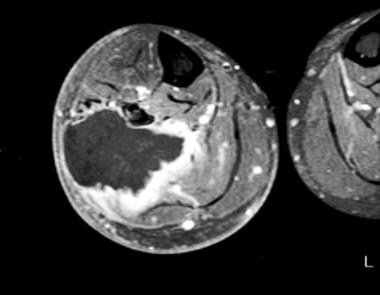
(+info)Malignant perivascular epithelioid cell tumour of the round ligament mimics leiomyoma on computed tomography. (3/32)
We report a case of a 45-year-old woman who had a palpable mass in the left lower quadrant of the abdomen. Computed tomography (CT) showed a circumscribed mass in the left round ligament of the uterus. The mass had heterogeneous density and enhancement accompanied by mottled calcification, which was initially identified as a leiomyoma. A histopathological examination revealed a malignant perivascular epithelioid cell tumour (PEComa), which is a rare soft tissue tumour. This case demonstrates that the appearance of malignant PEComa on the CT can mimic leiomyoma, which is the most common tumour of the round ligament. To the best of our knowledge, this is the first report of a CT appearance of this round ligament tumour. The radiological features and differential diagnosis are discussed. (+info)Pancreatic PEComa (sugar tumor): MDCT and EUS features. (4/32)
CONTEXT: PEComas (tumors showing perivascular epithelioid cell differentiation) of the pancreas are exceedingly rare. CASE REPORT: We herein report on a 60-year-old female who noticed a bulge in her right upper quadrant while exercising. Subsequent multidetector-row CT scan showed a 3.5 cm well-defined, encapsulated, hypovascular, solid tumor in the body of the pancreas. Endoscopic ultrasound demonstrated a mixed hypo- and hyper-echoic, well-defined, heterogeneous tumor. CONCLUSIONS: Although three pancreatic PEComas (sugar tumors) have been described previously, to the best of our knowledge, this is the first report of a pancreatic PEComa with illustration of its multidetector-row CT and endoscopic ultrasound features in the radiological literature. (+info)Perivascular epithelioid cell tumor (PEComa) of the liver diagnosed by contrast-enhanced ultrasonography. (5/32)
Perivascular epithelioid cell (PEC) is a unique cell which expresses both myogenic and melanocytic markers, and forms PEComa. A 36-year-old woman presented with a 35 mm-diameter liver tumor. MRI showed poor fat component in the tumor. Contrast-enhanced ultrasonography using the newly developed enhancing reagent, Sonazoid, clearly demonstrated early-phase enhancement of the tumor and rapid drainage of the reagent to veins, suggesting a PEComa. Lateral segmentectomy of the liver was performed. Histologically, epithelioid tumor cells around the vessels were immunostained with both HMB-45 and alpha-smooth muscle actin, confirming the diagnosis of PEComa. No recurrence has been found for 18 months following the operation. (+info)Clinical activity of mTOR inhibition with sirolimus in malignant perivascular epithelioid cell tumors: targeting the pathogenic activation of mTORC1 in tumors. (6/32)
(+info)A perivascular epithelioid cell tumor of the stomach: an unsuspected diagnosis. (7/32)
Perivascular epithelioid cell tumor (PEComa) is a rare mesenchymal neoplasia and currently well recognized as a distinct entity with characteristic morphological, immunohistochemical and molecular findings. We report a case of PEComa arising in the antrum of a 71-year-old female with melena. The tumor, located predominantly in the submucosa as a well delimited nodule, measured 3.0 cm in diameter and was completely resected, with no evidence of the disease elsewhere. Histologically, it was composed predominantly of eosinophilic epithelioid cells arranged in small nests commonly related to variably sized vessels, with abundant extracellular material, moderate nuclear variation and discrete mitotic activity. No necrosis, angiolymphatic invasion or perineural infiltration was seen. Tumor cells were uniformly positive for vimentin, smooth muscle actin, desmin and melan A. Although unusual, PEComa should be considered in the differential diagnosis of gastric neoplasia with characteristic epithelioid and oncocytic features and prominent vasculature. (+info)Perivascular epithelioid cell tumor (PEComa) of the uterus with aggressive behavior at presentation. (8/32)
Perivascular epithelioid cell tumor (PEComa) is a rare mesenchymal tumor composed of histologically and immunohistochemically distinctive perivascular epithelioid cells (PECs). Both benign and malignant tumors have been identified, but the criteria for diagnosis of malignancy have not been fully established due to the rarity of the tumor. We report on a case of uterine PEComa in a 33-year old woman with lymph node metastasis at presentation. The tumor had the characteristic histologic features of PEComa with cytologic atypia, mitotic activity of 2/10 high power field (HPF), and necrosis; it exhibited immunopositivity for HMB-45, calponin and desmin and was negative for melan-A. The patient received neoadjuvant chemotherapy, debulking surgery and adjuvant chemotherapy. No evidence of recurrence or metastasis was apparent 8 months after surgery. (+info)Perivascular Epithelioid Cell Neoplasms (PEComas) are a rare group of mesenchymal tumors that demonstrate unique clinical and pathological features. These neoplasms are characterized by the proliferation of perivascular epithelioid cells (PECs), which are distinctive cells with an epithelioid appearance and a close association with blood vessel walls.
PEComas can occur in various organs, such as the kidney, liver, lung, pancreas, and gastrointestinal tract, but they most commonly involve the uterus. The World Health Organization (WHO) recognizes three main types of PEComas: epithelioid angiomyolipoma, clear cell "sugar" tumor, and lymphangioleiomyomatosis (LAM).
PEComas exhibit a wide range of clinical behaviors, from benign to malignant. Malignant PEComas typically display features such as infiltrative growth, high cellularity, nuclear atypia, increased mitotic activity, and necrosis. The pathogenesis of PEComas is not well understood, but recent studies suggest that they may be related to the TSC1 or TSC2 gene mutations, which are also associated with tuberous sclerosis complex (TSC), a genetic disorder characterized by benign tumors in multiple organs.
Diagnosis of PEComas is based on histopathological examination and immunohistochemical staining. The typical immunophenotype of PECs includes positivity for both melanocytic markers (such as HMB-45 and Melan-A) and smooth muscle markers (such as actin and desmin).
Treatment options for PEComas depend on the tumor's location, size, and clinical behavior. Surgical resection is the primary treatment modality for localized, symptomatic, or malignant PEComas. In some cases, systemic therapy with mammalian target of rapamycin (mTOR) inhibitors may be considered, particularly in metastatic or recurrent tumors.
Epithelioid cells are a type of cell that can be found in certain types of tissue in the body, including connective tissue and some organs. These cells have a characteristic appearance under a microscope, with an enlarged, oval or round shape and a pale, abundant cytoplasm. They may also have a nucleus that is centrally located and has a uniform, rounded shape.
Epithelioid cells are often seen in the context of inflammation or disease, particularly in relation to granulomatous disorders such as sarcoidosis and tuberculosis. In these conditions, epithelioid cells can form clusters known as granulomas, which are a hallmark of the diseases. The exact function of epithelioid cells is not fully understood, but they are thought to play a role in the immune response and may help to contain and eliminate foreign substances or pathogens from the body.
Angiomyolipoma is a type of benign tumor that occurs most commonly in the kidney. It is composed of blood vessels (angio-), smooth muscle cells (myo-), and fat cells (lipo-). Angiomyolipomas are usually associated with the genetic disorder tuberous sclerosis complex, but they can also occur spontaneously or as a result of other genetic conditions.
These tumors can vary in size and may cause symptoms such as pain, blood in the urine, or a palpable mass in the abdomen if they grow large enough. In some cases, angiomyolipomas may also be at risk for rupture and bleeding, particularly if they are larger than 4 cm in size.
Treatment options for angiomyolipomas include surveillance with imaging tests, medication to reduce the risk of bleeding, or surgical removal of the tumor. The choice of treatment depends on factors such as the size and location of the tumor, the presence of symptoms, and the patient's overall health.
Adenomyosis is a medical condition that affects the uterus (womb). It occurs when the tissue that normally lines the uterus (endometrium) grows into the muscular wall of the uterus. This causes the uterine wall to thicken and can result in heavy, prolonged menstrual periods, as well as pain during menstruation, sexual intercourse, or pelvic examinations.
In some cases, adenomyosis may cause no symptoms at all, while in others it can lead to significant discomfort and impact a woman's quality of life. The exact causes of adenomyosis are not fully understood, but it is thought to be related to hormonal factors, inflammation, and previous uterine trauma or surgery.
Adenomyosis is typically diagnosed through imaging tests such as ultrasound or MRI, although in some cases a biopsy may be necessary to confirm the diagnosis. Treatment options for adenomyosis include medication to manage pain and reduce heavy menstrual bleeding, hormonal therapies to help regulate menstrual cycles, and in severe cases, surgery to remove all or part of the uterus (hysterectomy).
Melanoma-specific antigens are proteins or other molecules that are present on melanoma cells but not normally found on healthy cells in the body. These antigens can be recognized by the immune system as foreign and trigger an immune response, making them potential targets for immunotherapy treatments for melanoma.
There are two main types of melanoma-specific antigens: tumor-specific antigens (TSAs) and tumor-associated antigens (TAAs). TSAs are unique to cancer cells and are not found on normal cells, while TAAs are overexpressed or mutated versions of proteins that are also present in normal cells.
Examples of melanoma-specific antigens include Melan-A/MART-1, gp100, and tyrosinase. These antigens have been studied extensively as targets for cancer vaccines, adoptive cell therapy, and other immunotherapy approaches to treat melanoma.
Plasma cell neoplasms are a type of cancer that originates from plasma cells, which are a type of white blood cell found in the bone marrow. These cells are responsible for producing antibodies to help fight off infections. When plasma cells become cancerous and multiply out of control, they can form a tumor called a plasmacytoma.
There are two main types of plasma cell neoplasms: solitary plasmacytoma and multiple myeloma. Solitary plasmacytoma is a localized tumor that typically forms in the bone, while multiple myeloma is a systemic disease that affects multiple bones and can cause a variety of symptoms such as bone pain, fatigue, and anemia.
Plasma cell neoplasms are diagnosed through a combination of tests, including blood tests, imaging studies, and bone marrow biopsy. Treatment options depend on the stage and extent of the disease, but may include radiation therapy, chemotherapy, and stem cell transplantation.
Neoplasms of connective and soft tissue are abnormal growths or tumors that develop in the body's supportive tissues, such as cartilage, tendons, ligaments, fascia, and fat. These neoplasms can be benign (non-cancerous) or malignant (cancerous).
Benign connective and soft tissue neoplasms include:
- Lipomas: slow-growing, fatty tumors that develop under the skin.
- Fibromas: firm, benign tumors that develop in connective tissue such as tendons or ligaments.
- Nevi (plural of nevus): benign growths made up of cells called melanocytes, which produce pigment.
Malignant connective and soft tissue neoplasms include:
- Sarcomas: a type of cancer that develops in the body's supportive tissues such as muscle, bone, fat, cartilage, or blood vessels. There are many different types of sarcomas, including liposarcoma (fatty tissue), rhabdomyosarcoma (muscle), and osteosarcoma (bone).
- Desmoid tumors: a rare type of benign tumor that can become aggressive and invade surrounding tissues. While not considered cancerous, desmoid tumors can cause significant morbidity due to their tendency to grow and infiltrate nearby structures.
Connective and soft tissue neoplasms can present with various symptoms depending on their location and size. Treatment options include surgery, radiation therapy, chemotherapy, or a combination of these modalities. Regular follow-up care is essential to monitor for recurrence or metastasis (spread) of the tumor.
The round ligament is a cord-like structure in the female pelvis that extends from the uterus to the labia majora. It is one of the major ligaments that support the uterus and helps to maintain its position within the pelvis. The round ligament is composed of fibrous tissue and smooth muscle, and it plays a role in maintaining the tone and shape of the uterus.
During pregnancy, the round ligament can become stretched and thickened as the uterus grows and expands. This can sometimes cause discomfort or pain, particularly on one side of the pelvis. In some cases, the round ligament may also contribute to the development of certain gynecological conditions, such as uterine prolapse or urinary incontinence.
It is important for healthcare providers to consider the round ligament when evaluating and treating female reproductive health issues, as it can have a significant impact on the function and positioning of the uterus and other pelvic organs.
Tuberous Sclerosis Complex (TSC) is a rare genetic disorder that causes non-cancerous (benign) tumors to grow in many parts of the body. These tumors can affect the brain, skin, heart, kidneys, eyes, and lungs. The signs and symptoms of TSC can vary widely, depending on where the tumors develop and how severely a person is affected.
The condition is caused by mutations in either the TSC1 or TSC2 gene, which regulate a protein that helps control cell growth and division. When these genes are mutated, the protein is not produced correctly, leading to excessive cell growth and the development of tumors.
TSC is typically diagnosed based on clinical symptoms, medical imaging, and genetic testing. Treatment for TSC often involves a multidisciplinary approach, with specialists in neurology, dermatology, cardiology, nephrology, pulmonology, and ophthalmology working together to manage the various symptoms of the condition. Medications, surgery, and other therapies may be used to help control seizures, developmental delays, skin abnormalities, and other complications of TSC.
Epithelial cells are types of cells that cover the outer surfaces of the body, line the inner surfaces of organs and glands, and form the lining of blood vessels and body cavities. They provide a protective barrier against the external environment, regulate the movement of materials between the internal and external environments, and are involved in the sense of touch, temperature, and pain. Epithelial cells can be squamous (flat and thin), cuboidal (square-shaped and of equal height), or columnar (tall and narrow) in shape and are classified based on their location and function.
 Perivascular epithelioid cell tumour
Angiocentric glioma
Juxtaglomerular cell tumor
Schwannoma
Low-grade fibromyxoid sarcoma
Acral myxoinflammatory fibroblastic sarcoma
Angiomyolipoma
List of skin conditions
 Perivascular Epithelioid Cell Neoplasms
Summary Report | CureHunter
Perivascular Epithelioid Cell Neoplasms
Summary Report | CureHunter
Perivascular epithelioid cell tumour - Wikipedia
 Ectopic prolactin secretion from a perivascular epithelioid cell tumor (PEComa)
 Carcinosarcoma of the Sigmoid Colon: Report of a Case | Case Reports in Gastroenterology | Karger Publishers
 Pathology of Uterus Smooth Muscle Tumors: Definition, Epidemiology, Location
 Adrenal lipomatous tumours: a 30 year clinicopathological experience at a single institution | Journal of Clinical Pathology
 Uterine Sarcoma, Version 1.2016 in: Journal of the National Comprehensive Cancer Network Volume 13 Issue 11 (2015)
Lymphangioleiomyomatosis Imaging and Diagnosis: Practice Essentials, Radiography, Computed Tomography
 MeSH Browser
Oral Malignant Melanoma: Overview, Etiology and Pathophysiology, Epidemiology
 Division of Oncology - Research output
- Research Profiles at Washington University School of Medicine
Division of Oncology - Research output
- Research Profiles at Washington University School of Medicine
Orthopaedic Surgery & Rehabilitation - Research output - Research Nebraska
Wilson Lao - Research output - Taipei Medical University
Centers, HSCD - Scholarly Works - Arizona Board of Regents
Kelley, S.<...
 LAM Health Project - National Organization for Rare Disorders
 Case 3: Quarter 4, 2022
 Radiological features of perivascular epithelioid cell tumours (PEComas) in a paediatric patient - Case report
 Wolffian Ducts | Harvard Catalyst Profiles | Harvard Catalyst
Multidisciplinary Approach In Advanced Perivascular Epithelioid Cell Tumour (PEComa): A Case Report
Familial Adenomatous Polyposis - Symptoms, Causes, Treatment | NORD
 Pesquisa | Portal Regional da BVS
 Perivascular epithelioid cell tumor of the uterine cervix identified on the liquid-based cytology: a case report | Diagnostic...
 Journal of Postgraduate Gynecology & Obstetrics: June 2014
MedPharmRes - By University of Medicine and Pharmacy at Ho Chi Minh City
 Discussion | ASC Education
June | 2014 | Syk Signaling
Lymphangioleiomyomatosis Imaging: Overview, Radiography, Computed Tomography
PEComas8
- abdominopelvic sarcoma of perivascular epitheloid cells primary extrapulmonary sugar tumour Thus, it has been advocated that the above could be classified PEComas. (wikipedia.org)
- Perivascular epithelioid cell tumors (PEComas) are an uncommon family of soft tissue tumors with dual myoid-melanocytic differentiation. (nih.gov)
- [ 1 ] LAM was classified as 1 of 3 forms of PEComataous tumor (arising from perivascular epithelioid cells), along with benign PEComas (including clear cell tumor) and malignant PEComas. (medscape.com)
- Karska K, Kozioł I, Leśniewska M, Budzyńska J, Woźniak M. Radiological features of perivascular epithelioid cell tumours (PEComas) in a paediatric patient - Case report. (jpccr.eu)
- Background: Perivascular epithelioid cell tumours (PEComas) are mesenchymal neoplasms with variable biological behaviour, ranging from benign to extremely aggressive diseases able to metastasize. (fortuneonline.org)
- Perivascular epithelioid cell tumors (PEComas) are a rare group of mesenchymal neoplasms characterized by the presence of histologically and immunohistochemically distinctive perivascular epithelioid cells [ 1 ], which can occur in any part of the body. (biomedcentral.com)
- Perivascular epithelioid cell tumours (PEComas) belongs to the family of mesenchymal neoplasms that can occur in many organs, but rarely found in liver. (medpharmres.com)
- [ 1 ] LAM was classified as one of three forms of PEComataous tumor (arising from perivascular epithelioid cells), along with benign PEComas (including clear cell tumor), and malignant PEComas. (medscape.com)
Tumors10
- A family of mesenchymal tumors composed of histologically and immunohistochemically distinctive perivascular epithelioid cells. (curehunter.com)
- Although PEComa family tumors commonly demonstrate a perivascular growth pattern, pericyte antigen expression has not yet been examined among this unique tumor group. (nih.gov)
- Previously, we demonstrated that a subset of perivascular soft tissue tumors exhibit a striking pericytic immunophenotype, with diffuse expression of αSMA, CD146, and PDGFRβ. (nih.gov)
- Renal angiomyolipoma (renal AML) is a rare benign mesenchymal neoplasm of the kidney that is a member of the perivascular epithelioid cell tumors (PEComa) family. (cytopathology.org)
- The inflammatory AML should be distinguished from other tumors with inflammatory background such as inflammatory myofibroblastic tumor and follicular dendritic cell tumor and deserves wider recognition for its occurrence as a primary hepatic tumor. (biomedcentral.com)
- According to the predominant component, growth pattern, cell type, and other features, the tumors were subcategorized into trabecular, pelioid and inflammatory variants. (biomedcentral.com)
- Of these, inflammatory or pelioid pattern usually presents as a focal finding within the tumor, but very rarely, they become the predominant pattern [ 4 ], creating great diagnostic confusion with other tumors such as inflammatory myofibroblastic tumor (IMT), follicular dendritc cell (FDC) tumor and other hepatic mesenchymal neoplasms. (biomedcentral.com)
- Their behavior ranges from that of indolent, low-grade tumors, such as epithelioid hemangioendothelioma, to that of lethal, high-grade angiosarcomas or hemangiopericytomas. (basicmedicalkey.com)
- It is thought that leiomyoma is the result of either transformation of normal uterine muscle cells into abnormal cells through somatic mutations , or through the growth of abnormal uterine muscle cells into tumors. (wikidoc.org)
- Leptomeningeal infiltration of the brain or spinal cord by neoplastic cells may occur as complication of solid or hematopoietic tumors such as non-Hodgkin lymphoma. (cnr.it)
Renal Cell Carc1
- Reappraisal of Morphologic Differences Between Renal Medullary Carcinoma, Collecting Duct Carcinoma, and Fumarate Hydratase-deficient Renal Cell Carcinoma. (uchicago.edu)
Tumours8
- Perivascular epithelioid cell tumour, also known as PEComa or PEC tumour, is a family of mesenchymal tumours consisting of perivascular epithelioid cells (PECs). (wikipedia.org)
- The cell type from which these tumours originate remains unknown. (wikipedia.org)
- Perivascular epithelioid cell tumour (PEComa) is a rare family of mesenchymal tumours composed of epithelioid cells. (jpccr.eu)
- Imaging features of primary and metastatic malignant perivascular epithelioid cell tumours. (jpccr.eu)
- MR imaging is frequently performed with MRA to distinguish between vascular malformations, vascular tumours, and perivascular tumours. (springer.com)
- Some vascular tumours preferentially affect the hand, such as pyogenic granulomas or spindle cell haemangiomas associated with Maffucci syndrome. (springer.com)
- Glomus tumours are the most frequent perivascular tumours of the hand. (springer.com)
- The purpose of this article is to describe the state-of-the-art acquisition protocols and illustrate the different patterns of vascular lesions and perivascular tumours of the hand. (springer.com)
Angiomyolipoma5
- PECs bear significant histologic and immunohistochemical similarity to: angiomyolipoma, clear-cell sugar tumour (CCST), lymphangioleiomyomatosis, and, clear-cell myomelanocytic tumour of ligamentum teres/falciform ligament. (wikipedia.org)
- Typical angiomyolipoma (AML) specimens showed variable expression of pericyte antigens among both perivascular and myoid-appearing cells. (nih.gov)
- Based on the WHO classification, renal epithelioid angiomyolipoma (renal eAML) is composed of at least 80% predominantly round to polygonal epithelioid cells (1). (cytopathology.org)
- Angiomyolipoma (AML) is a rare mesenchymal neoplasm of the tumor, composed of a varying heterogeneous mixture of three tissue components: blood vessels, smooth muscle and adipose cells. (biomedcentral.com)
- Hepatic angiomyolipoma is a rare, benign, hepatic mesenchymal neoplasm found in both males and females, and most commonly in adult females. (biomedcentral.com)
Tumour5
- Perivascular epithelioid cell tumour: dynamic CT, MRI and clinicopathological characteristics-analysis of 32 cases and review of the literature. (jpccr.eu)
- Handa A, Fujita K, Kono T, Komori K, Hirobe S, Fukuzawa R. Radiological findings of perivascular epithelioid cell tumour (PEComa) of the falciform ligament. (jpccr.eu)
- Zhao J, Teng H, Zhao R, Ding W, Yu K, Zhu L, Zhang J, Han Y. Malignant perivascular epithelioid cell tumour of the lung synchronous with a primary adenocarcinoma: one case report and review of the literature. (jpccr.eu)
- Xuesong D, Hong G, Weiguo Z. Bladder Perivascular Epithelioid Cell Tumour: Dynamic CT and MRI Presentation of 2 Cases With 2-year Follow-up and Review of the Literature. (jpccr.eu)
- The complex and often confusing history, histology and histogenesis of mesonephric, STK11 adnexal tumour and mesonephric-like neoplasms of the upper female genital tract (including broad ligament). (harvard.edu)
Lymphangioleiomyomatosis5
- Lymphangioleiomyomatosis (LAM) is a rare idiopathic disease that affects women and is characterized by nonneoplastic peribronchial, perivascular, and perilymphatic proliferation of atypical smooth muscle resulting in vascular and airway obstruction, cyst formation, and a progressive decline in lung function. (medscape.com)
- Vasohibin-1 and -2 in pulmonary lymphangioleiomyomatosis (LAM) cells associated with angiogenic and prognostic factors. (ailam.it)
- Cross talk between LAM cells and fibroblasts may influence alveolar epithelial cell behavior in lymphangioleiomyomatosis. (ailam.it)
- Mast-Cell Tryptase Release Contributes to Disease Progression in Lymphangioleiomyomatosis. (ailam.it)
- Isolation and characterisation of lymphatic endothelial cells from lung tissues affected by lymphangioleiomyomatosis. (ailam.it)
Hepatic2
- We herein present one case of hepatic AML exhibiting prominent inflammatory cells in the background, which happened in a 61-year-old Chinese female patient, without signs of tuberous sclerosis. (biomedcentral.com)
- Histologically similar to those in the kidney, hepatic AML consists of a mixture of myoid cells, adipose tissue and thick-walled vessels. (biomedcentral.com)
Spindle-shaped4
- The tumor was a huge lesion occupying the inside of the lumen, and histopathological findings revealed that the tumor, the main part of which lay beneath the mucous membrane, had a transitional image composed of both spindle-shaped atypical cells and sarcomatoid shape. (karger.com)
- The most common identified cells are spindle-shaped cells with or without epithelioid components. (cytopathology.org)
- Cutaneous biopsy revealed RAE characterized by the proliferation of epithelioid and spindle-shaped cells in superficial and middermis lining vascular channels, arranged in clusters, and sometimes displaying an intravascular growth pattern. (thedoctorsdoctor.com)
- On microscopic histopathological analysis, elongated and spindle-shaped cells with a cigar-shaped nucleus are characteristic findings of leiomyoma. (wikidoc.org)
Differentiation4
- Diestelkamp T, Mikes Z, Wilson-Smith R, Germaine P. Radiological findings of two neoplasms with perivascular epithelioid cell differentiation. (jpccr.eu)
- In brief, vascular endothelial growth factor-A (VEGF-A), which is a part of a large superfamily of growth factors, is required for differentiation of endothelial precursor cells. (basicmedicalkey.com)
- The prospero related homeobox-1 (PROX1) transcriptional factor and vascular endothelial growth factor receptor-3 (VEGFR3) are responsible for their differentiation into lymphatic endothelial cells. (basicmedicalkey.com)
- Lung cDC2s from CD109-/- mice had a poor ability to induce cytokine production in ex vivo DC-T cell cocultures with high expression of RUNX3, resulting in suppression of Th2 differentiation. (nagoya-u.ac.jp)
Malignant3
- The diagnosis of ectopic pituitary hormone secretion requires abnormally high circulating hormone levels, absence of a pituitary tumor, and localization of the hormone in question to the extrapituitary malignant neoplasm. (nih.gov)
- Although rare, malignant transformation of nevi to melanoma involves the clonal expansion of cells that acquire a selective growth advantage. (medscape.com)
- Ischiorectal fossa: benign and malignant neoplasms of this "ignored" radiological anatomical space. (jpccr.eu)
Atypical2
- Microscopically, we observed some loosely cohesive atypical cells arranged in single or clusters and sheets, which exhibited epithelioid morphology with abundant clear cytoplasm. (biomedcentral.com)
- Distinct round nucleoli were visible in some cells (Fig. 1 C), notably with numerous melanin pigments in the cytoplasm (Fig. 1 D). The primary diagnosis was atypical cells which were suspected to be melanoma. (biomedcentral.com)
Granular3
- PECs consist of perivascular epithelioid cells with a clear/granular cytoplasm and central round nucleus without prominent nucleoli. (wikipedia.org)
- The cell block section showed single or clusters of medium-sized epithelioid cells in a background of fibrinoid fluid, with abundant clear or granular eosinophilic cytoplasm. (biomedcentral.com)
- These kind of neoplasms consist of nests and also fascicles of apparent in order to granular epithelioid and/or spindled cells using a regular set up all around veins. (vx-661modulator.com)
Pathogenesis1
- The pathogenesis of leiomyoma is characterized by benign smooth muscle neoplasm . (wikidoc.org)
Routinely express1
- The neoplastic cells routinely express pan-cytokeratin and neuroendocrine markers chromogranin and synaptophysin (focal to diffuse staining). (pbpath.org)
Lung3
- Although historically LAM has been considered an interstitial lung disease, it is now considered to be a low-grade destructive metastasizing neoplasm. (medscape.com)
- Large lung volumes and interstitial disease on plain film also can be seen with Langerhans cell histiocytosis, sarcoidosis, and extrinsic allergic alveolitis. (medscape.com)
- Am J Physiol Lung Cell Mol Physiol 2022;1;322(2):L283-L293. (ailam.it)
Lymphocytic3
- His past medical history was significant for subclinical B-cell chronic lymphocytic leukemia (CLL), which had never been treated. (cdlib.org)
- Two skin biopsies showed common features of a perivascular and periadnexal lymphocytic infiltrate in the superficial to mid-dermis. (cdlib.org)
- Perivascular and periadnexal lymphocytic infiltrate in the superficial to mid-dermis. (cdlib.org)
Nuclei8
- The biopsy showed infiltrating neoplastic cells characterized by cytologically bland nuclei and abundant finely vacuolated, clear cytoplasm embedded in a collagenized/fibrotic stroma [Figure 1A/B]. No necrosis or mitotic figures were identified. (pbpath.org)
- The tumor cells demonstrated clear/foamy vacuolated cytoplasm and small uniform nuclei with finely dispersed chromatin [Figure 4]. (pbpath.org)
- tumor cells are characterized by round to pyknotic nuclei with fine chromatin and inconspicuous nucleoli. (pbpath.org)
- Microscopically, some loosely cohesive epithelioid cells were uniform with abundant clear cytoplasm, showing predominantly round or oval nuclei with finely stippled chromatin. (biomedcentral.com)
- The epithelioid cells were uniform and approximately the same size as the parabasal cells (Fig. 1 A&B), showing predominantly round or oval nuclei with finely stippled chromatin. (biomedcentral.com)
- A few spindled nuclei and melanin pigments were also identified (Fig. 2 A). IHC demonstrated that the epithelioid cells were positive for HMB45 andTFE3 (Fig. 2 B&C), focally positive for Melan-A, while negative for S-100, SOX-10, AE1/AE3, EMA, Desmin, SMA, H-caldesmon. (biomedcentral.com)
- Bare nuclei, intranuclear inclusions, and multinucleated giant cells are present in some cases (2). (cytopathology.org)
- The histopathological examination revealed a neoplasm characterized by a densely collagenized stroma with alternating zones of cellularity, consisting of small spindled or ovoid cells with moderate amounts of eosinophilic cytoplasm and nuclei with fine chromatin and inconspicuous nucleoli, clustered around thin-walled capillaries ( Figure 5 ). (medcraveonline.com)
Occur1
- Although vulva and vagina are the most common sites of angiomyofibroblastoma, this neoplasm can rarely occur in the pelvic and extrapelvic tissues. (medcraveonline.com)
Immunohistochemically1
- Immunohistochemically, the bland spindle cells that line these spaces are strongly positive for vimentin and CD34 and negative for cytokeratin and factor VIII. (health.am)
Uterine2
- Perivascular epithelioid cell tumor (PEComa) occurring in the female genital tract are rare, and typically found in the uterine corpus. (biomedcentral.com)
- Proteogenomic landscape of uterine leiomyomas from hereditary leiomyomatosis and renal cell cancer patients. (uchicago.edu)
Findings1
- The cytopathological features were well correlated with cell block and histopathological findings. (biomedcentral.com)
Endothelial cells8
- One effective strategy for treating atherosclerosis is to inhibit the injury of vascular endothelial cells (VECs) induced by oxidized low-density lipoprotein (oxLDL) and high glucose (HG). (bvsalud.org)
- Reactive angioendotheliomatosis (RAE) is a rare benign cutaneous vascular proliferation characterized by intravascular hyperplasia of endothelial cells and tuft-like proliferation of vessels. (thedoctorsdoctor.com)
- Histopathologically, it is characterized by a proliferation of endothelial cells within vascular lumina resulting in the obliteration of the involved vessels. (thedoctorsdoctor.com)
- It consists of violaceous and purpuric plaques histopathologically characterized by diffuse proliferation of endothelial cells interstitially arranged between collagen bundles of the reticular dermis. (thedoctorsdoctor.com)
- These interacting tasks are carried out by a system that is ubiquitous to all vertebrates and comprises two treelike branched systems of tubules known as blood and lymphatic vessels lined by highly specialized endothelial cells. (basicmedicalkey.com)
- 8 The sprouting of endothelial cells is regulated by Notch signaling receptors and their Delta-like 4 (DLL4) ligand. (basicmedicalkey.com)
- Precurser lymphatic endothelial cells form a distinctive cluster in mid-gestation embryos on the dorsal side of the jugular vein. (basicmedicalkey.com)
- 5 , 9 , 17 Although endothelial cells of blood vessels and lymphatics are somewhat similar, the lymphatics lack a continuous basement membrane and are not tightly sealed by intercellular junctions, permitting the free access of interstitial tissue fluid into the vessel. (basicmedicalkey.com)
Cytoplasm3
- Distinct round nucleoli were visible in some cells, notably with numerous melanin pigments in the cytoplasm. (biomedcentral.com)
- The cytologic characteristics of the tumor can provide sufficient clues for PEComa diagnosis, which includes loosely cohesive, epithelioid morphology with abundant clear or eosinophilic cytoplasm, low-grade nuclear atypia, cytoplasmic melanin pigments. (biomedcentral.com)
- The tumor cells were spindled and histiocytoid in shape, with slightly eosinophilic cytoplasm, and arranged along the vessels or scattered among the inflammatory background. (biomedcentral.com)
Morphology1
- AML samples with predominant epithelioid morphology showed a marked reduction in or the absence of immunoreactivity for pericyte markers. (nih.gov)
Liver1
- It is an indolent disease characterized by the accumulation of mature monoclonal B cells in the blood and bone marrow, often also involving the spleen, liver, and lymph nodes. (cdlib.org)
Histiocytoid1
- The biopsy from the right neck also showed a prominent granulomatous component with aggregates of histiocytoid cells forming perivascular epithelioid granulomas (Figure 4). (cdlib.org)
Infiltration1
- Histologically, the striking feature was the infiltration of numerous inflammatory cells in the background, including small lymphocytes, plasma cells, and eosnophils. (biomedcentral.com)
HMB452
- Upon immunohistochemistry (IHC), the tumor cells were positive for HMB45 and TFE3, focally positive for MelanA, while negative for muscle marker. (biomedcentral.com)
- Tumor cells are diffusely immunoreactive with HMB45. (medpharmres.com)
Fibroblasts1
- and epithelioid fibroblasts embedded in dense fibrous stroma. (health.am)
Langerhans1
- Cystic fibrosis and Langerhans cell histiocytosis (eosinophilic granuloma) share this feature. (medscape.com)
Proliferation1
- LAM is characterized by the spread and uncontrolled growth (proliferation) of specialized cells (smooth muscle cells) in certain organs of the body, especially the lungs. (rarediseases.org)
Exhibit1
- Epithelioid cells may exhibit variable degrees of nuclear atypia including pleomorphism and enlarged sizes with prominent nucleoli. (cytopathology.org)
Epithelial cell1
- We carried out transfection experiments using three pancreatic cancer cell lines (MiaPaCa-2, BxPC-3, and SW1990) and one pancreatic duct epithelial cell line (HPDE6c7). (nagoya-u.ac.jp)
Antigen1
- The standard vascular markers used in pathologic differential diagnosis of vascular conditions include von Willebrand factor (vWF), factor VIII-associated protein, platelet endothelial cell adhesion molecule-1 (CD31), human hematopoietic progenitor cell antigen (CD34), v- ets avian erythroblastosis virus E26 oncogene homolog (ERG), human herpes virus 8 (HHV-8), latency-associated nuclear antigen (LANA1), podoplanin (D2-40), and PROX1. (basicmedicalkey.com)
Neoplastic cells3
- The neoplastic cells were positive for pan-CK (strong and diffuse), synaptophysin (strong and diffuse) [Figure 2], chromogranin (strong but focal) and inhibin (strong and diffuse) while negative for CK7, RCC, PAX-8, S-100, Melan-A (MART-1), and beta catenin. (pbpath.org)
- Sections showed neoplastic cells arranged in nests/clusters and cords, surrounded by marked collagenized/fibrotic stroma [Figure 3]. (pbpath.org)
- Immunohistochemical staining showing positivity of neoplastic cells for CD20 (A), CD23 (B), CD5 (C), and CD43 (D). (cdlib.org)
Papillary1
- Papillary tumor of the pineal region (PTPR) is a neuroectodermal tumor thought to originate from cells of the subcommissural organ. (thieme-connect.de)
Smooth1
- The tumor cells were positive immunostaining for HMB-45, Melan-A, and smooth muscle actin. (biomedcentral.com)
Lesions1
- Morphologically, corresponding lesions on the skin seen in other places are composed of the unifying mobile or portable, the perivascular epithelioid cell (PEC). (vx-661modulator.com)
Immunohistochemical1
- However, given the patient's history of CLL, a panel of immunohistochemical stains was performed on both biopsies revealing aggregates and individual CD20+ B cells in both perivascular and periadnexal locations. (cdlib.org)
Eosinophilic1
- CD109 on Dendritic Cells Regulates Airway Hyperreactivity and Eosinophilic Airway Inflammation. (nagoya-u.ac.jp)