Persistent Hyperplastic Primary Vitreous
Vitreous Body
Posterior Capsulotomy
Hyperplasia
Lens, Crystalline
Prenatal ultrasonographic diagnosis of persistent hyperplastic primary vitreous. (1/13)
(+info)A nonautonomous role for retinal frizzled-5 in regulating hyaloid vitreous vasculature development. (2/13)
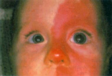
(+info)Bilateral persistent hyperplastic primary vitreous. (3/13)
A case of bilateral persistent hyperplastic primary vitreous (PHPV) in a 3-month-old male infant, who had bilateral leukokoria, is presented. The child was referred for imaging with a clinical suspicion of retinoblastoma. Gray-scale ultrasound evaluation revealed an echogenic band in the posterior segment of both globes, extending from the posterior surface of the lens capsule to the optic disc. Doppler examination revealed the presence of arterial flow in the band in both globes. Associated echogenic hemorrhage was also seen, which was confirmed by computed tomography. Most cases of PHPV are sporadic and unilateral, and bilateral PHPV is rare. The imaging features in this case suggest the diagnosis of bilateral PHPV and differentiate it from retinoblastoma. This entity, although infrequent, should be considered in the differential diagnosis while evaluating bilateral leukokoria. (+info)A developmental defect in astrocytes inhibits programmed regression of the hyaloid vasculature in the mammalian eye. (4/13)
(+info)Infant Aphakia Treatment Study: effects of persistent fetal vasculature on outcome at 1 year of age. (5/13)
(+info)Congenital fibrovascular pupillary membranes: clinical and histopathologic findings. (6/13)
(+info)The deletion of Math5 disrupts retinal blood vessel and glial development in mice. (7/13)
(+info)Deletion of HIF-1alpha partially rescues the abnormal hyaloid vascular system in Cited2 conditional knockout mouse eyes. (8/13)
PURPOSE: Cited2 (CBP/p300-interacting transactivators with glutamic acid (E) and aspartic acid (D)-rich tail 2) is a member of a new family of transcriptional modulators. Cited2 null embryos exhibit hyaloid hypercellularity consisting of aberrant vasculature in the eye. The purpose of the study is to address whether abnormal lenticular development is a primary defect of Cited2 deletion and whether deletion of hypoxia inducible factor (HIF)-1alpha or an HIF-1alpha target gene, vascular endothelial growth factor (VEGF), could rescue abnormal hyaloid vascular system (HVS) in Cited2 deficient adult eyes. METHODS: Le-Cre specific Cited2 knockout (Cited2(CKO)) mice with or without deletion of HIF-1alpha or VEGF were generated by standard Cre-Lox methods. Eyes collected from six-eight weeks old mice were characterized by Real Time PCR and immunohistological staining. RESULTS: Cited2(CKO) mice had smaller lenses, abnormal lens stalk formation, and failed regression of the HVS in the adult eye. The eye phenotype had features similar to persistent hyperplastic primary vitreous (PHPV), a human congenital eye disorder leading to abnormal lenticular development. Deletion of HIF-1alpha or VEGF in Cited2 knockout eyes partially rescued the abnormal HVS but had no effect on the smaller lens and abnormal lens stalk differentiation. Intravitreal injection of Topotecan (TPT), a compound that inhibits HIF-1alpha expression, partially eliminated HVS defects in Cited2(CKO) lenses. CONCLUSIONS: Abnormal HVS is a primary defect in Cited2 knockout mice, resulting in part from dysregulated functions of HIF-1 and VEGF. The Cited2(CKO) mouse line could be used as a novel disease model for PHPV and as an in vivo model for testing potential HIF-1 inhibitors. (+info)Persistent Hyperplastic Primary Vitreous (PHPV) is a rare congenital eye condition that occurs during fetal development. It is characterized by the failure of the primary vitreous, a gel-like substance in the eye, to completely regress or disappear. Instead, the primary vitreous persists and undergoes hyperplasia, leading to the formation of abnormal tissue within the eye.
In PHPV, this persistent tissue can cause various problems, including a small pupil, a cloudy area in the center of the lens (cataract), a white mass behind the lens, and abnormal blood vessels growing from the retina towards the center of the eye. These abnormalities can lead to visual impairment or even blindness, depending on the severity of the condition.
PHPV is typically diagnosed during infancy or early childhood, through a comprehensive eye examination that includes a detailed view of the internal structures of the eye using a specialized lens (slit lamp) and other diagnostic tests. Treatment options may include surgery to remove the abnormal tissue and improve vision, but the success of treatment depends on the extent and location of the PHPV.
Eye abnormalities refer to any structural or functional anomalies that affect the eye or its surrounding tissues. These abnormalities can be present at birth (congenital) or acquired later in life due to various factors such as injury, disease, or aging. Some examples of eye abnormalities include:
1. Strabismus: Also known as crossed eyes, strabismus is a condition where the eyes are misaligned and point in different directions.
2. Nystagmus: This is an involuntary movement of the eyes that can be horizontal, vertical, or rotatory.
3. Cataracts: A cataract is a clouding of the lens inside the eye that can cause vision loss.
4. Glaucoma: This is a group of eye conditions that damage the optic nerve and can lead to vision loss.
5. Retinal disorders: These include conditions such as retinal detachment, macular degeneration, and diabetic retinopathy.
6. Corneal abnormalities: These include conditions such as keratoconus, corneal ulcers, and Fuchs' dystrophy.
7. Orbital abnormalities: These include conditions such as orbital tumors, thyroid eye disease, and Graves' ophthalmopathy.
8. Ptosis: This is a condition where the upper eyelid droops over the eye.
9. Color blindness: A condition where a person has difficulty distinguishing between certain colors.
10. Microphthalmia: A condition where one or both eyes are abnormally small.
These are just a few examples of eye abnormalities, and there are many others that can affect the eye and its functioning. If you suspect that you have an eye abnormality, it is important to consult with an ophthalmologist for proper diagnosis and treatment.
The vitreous body, also known simply as the vitreous, is the clear, gel-like substance that fills the space between the lens and the retina in the eye. It is composed mainly of water, but also contains collagen fibers, hyaluronic acid, and other proteins. The vitreous helps to maintain the shape of the eye and provides a transparent medium for light to pass through to reach the retina. With age, the vitreous can become more liquefied and may eventually separate from the retina, leading to symptoms such as floaters or flashes of light.
A posterior capsulotomy is a surgical procedure that involves making an opening in the back part (posterior) of the lens capsule, which is a thin, clear membrane that holds the lens in place inside the eye. This procedure is typically performed to treat after-cataract, also known as posterior capsular opacification (PCO), which can cause vision loss or disturbance after cataract surgery. During cataract surgery, the cloudy natural lens of the eye is removed and replaced with an artificial intraocular lens (IOL). However, sometimes the back part of the lens capsule may become hazy or opaque over time, leading to visual symptoms similar to those experienced before cataract surgery.
In a posterior capsulotomy, a laser (usually a YAG laser) is used to create an opening in the cloudy posterior capsule, allowing light to pass through and restoring clear vision. The procedure is typically quick, painless, and performed as an outpatient procedure in a doctor's office or clinic. Patients may experience some side effects such as floaters, glare, or flashes of light after the procedure, but these usually resolve within a few days or weeks.
Hyperplasia is a medical term that refers to an abnormal increase in the number of cells in an organ or tissue, leading to an enlargement of the affected area. It's a response to various stimuli such as hormones, chronic irritation, or inflammation. Hyperplasia can be physiological, like the growth of breast tissue during pregnancy, or pathological, like in the case of benign or malignant tumors. The process is generally reversible if the stimulus is removed. It's important to note that hyperplasia itself is not cancerous, but some forms of hyperplasia can increase the risk of developing cancer over time.
The crystalline lens is a biconvex transparent structure in the eye that helps to refract (bend) light rays and focus them onto the retina. It is located behind the iris and pupil and is suspended by small fibers called zonules that connect it to the ciliary body. The lens can change its shape to accommodate and focus on objects at different distances, a process known as accommodation. With age, the lens may become cloudy or opaque, leading to cataracts.
A cataract is a clouding of the natural lens in the eye that affects vision. This clouding can cause vision to become blurry, faded, or dim, making it difficult to see clearly. Cataracts are a common age-related condition, but they can also be caused by injury, disease, or medication use. In most cases, cataracts develop gradually over time and can be treated with surgery to remove the cloudy lens and replace it with an artificial one.
 Persistent fetal vasculature
 Persistent hyperplastic primary vitreous
 Persistent hyperplastic primary vitreous: congenital malformation of the eye - PubMed
 Clinical manifestations and pathological characteristics of retrolental membranes secondary to persistent hyperplastic primary...
 Peripheral Anterior Synechia Workup: Approach Considerations, Laboratory Studies, Imaging Studies
 Pupil - white spots: MedlinePlus Medical Encyclopedia
 Retinoblastoma - Causes, Symptoms, Diagnosis, Treatment & Prevention
 BVA - Hereditary Eye Disease Scheme for dogs
 Pupil - white spots Information | Mount Sinai - New York
 S. Afr. J. radiol. (Online) -
vol.18 número1
S. Afr. J. radiol. (Online) -
vol.18 número1 Conditions of the Lens - WSAVA 2003 Congress - VIN
 Persistent pupillary membrane and secondary angle closure glaucoma: a case report | Pediatric Oncall Journal
 NDP-Related Retinopathies - GeneReviews® - NCBI Bookshelf
 microphthalmos | Hereditary Ocular Diseases
 Retinopathy of prematurity - wikidoc
 MeSH Browser
 DeCS
DeCS Dobermann - Dogs of Britain| Natural Dog Treats
 Tmem38b Mouse Gene Details | transmembrane protein 38B | International Mouse Phenotyping Consortium