Persistent Hyperplastic Primary Vitreous
Vitreous Body
Posterior Capsulotomy
Hyperplasia
Lens, Crystalline
Prenatal ultrasonographic diagnosis of persistent hyperplastic primary vitreous. (1/13)
(+info)A nonautonomous role for retinal frizzled-5 in regulating hyaloid vitreous vasculature development. (2/13)
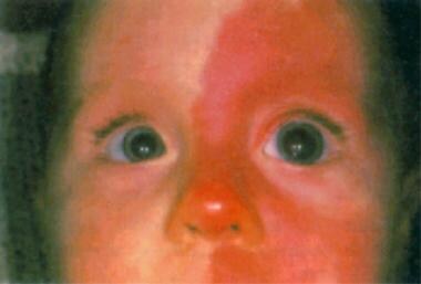
(+info)Bilateral persistent hyperplastic primary vitreous. (3/13)
A case of bilateral persistent hyperplastic primary vitreous (PHPV) in a 3-month-old male infant, who had bilateral leukokoria, is presented. The child was referred for imaging with a clinical suspicion of retinoblastoma. Gray-scale ultrasound evaluation revealed an echogenic band in the posterior segment of both globes, extending from the posterior surface of the lens capsule to the optic disc. Doppler examination revealed the presence of arterial flow in the band in both globes. Associated echogenic hemorrhage was also seen, which was confirmed by computed tomography. Most cases of PHPV are sporadic and unilateral, and bilateral PHPV is rare. The imaging features in this case suggest the diagnosis of bilateral PHPV and differentiate it from retinoblastoma. This entity, although infrequent, should be considered in the differential diagnosis while evaluating bilateral leukokoria. (+info)A developmental defect in astrocytes inhibits programmed regression of the hyaloid vasculature in the mammalian eye. (4/13)
(+info)Infant Aphakia Treatment Study: effects of persistent fetal vasculature on outcome at 1 year of age. (5/13)
(+info)Congenital fibrovascular pupillary membranes: clinical and histopathologic findings. (6/13)
(+info)The deletion of Math5 disrupts retinal blood vessel and glial development in mice. (7/13)
(+info)Deletion of HIF-1alpha partially rescues the abnormal hyaloid vascular system in Cited2 conditional knockout mouse eyes. (8/13)
PURPOSE: Cited2 (CBP/p300-interacting transactivators with glutamic acid (E) and aspartic acid (D)-rich tail 2) is a member of a new family of transcriptional modulators. Cited2 null embryos exhibit hyaloid hypercellularity consisting of aberrant vasculature in the eye. The purpose of the study is to address whether abnormal lenticular development is a primary defect of Cited2 deletion and whether deletion of hypoxia inducible factor (HIF)-1alpha or an HIF-1alpha target gene, vascular endothelial growth factor (VEGF), could rescue abnormal hyaloid vascular system (HVS) in Cited2 deficient adult eyes. METHODS: Le-Cre specific Cited2 knockout (Cited2(CKO)) mice with or without deletion of HIF-1alpha or VEGF were generated by standard Cre-Lox methods. Eyes collected from six-eight weeks old mice were characterized by Real Time PCR and immunohistological staining. RESULTS: Cited2(CKO) mice had smaller lenses, abnormal lens stalk formation, and failed regression of the HVS in the adult eye. The eye phenotype had features similar to persistent hyperplastic primary vitreous (PHPV), a human congenital eye disorder leading to abnormal lenticular development. Deletion of HIF-1alpha or VEGF in Cited2 knockout eyes partially rescued the abnormal HVS but had no effect on the smaller lens and abnormal lens stalk differentiation. Intravitreal injection of Topotecan (TPT), a compound that inhibits HIF-1alpha expression, partially eliminated HVS defects in Cited2(CKO) lenses. CONCLUSIONS: Abnormal HVS is a primary defect in Cited2 knockout mice, resulting in part from dysregulated functions of HIF-1 and VEGF. The Cited2(CKO) mouse line could be used as a novel disease model for PHPV and as an in vivo model for testing potential HIF-1 inhibitors. (+info)Persistent Hyperplastic Primary Vitreous (PHPV) is a rare congenital eye condition that occurs during fetal development. It is characterized by the failure of the primary vitreous, a gel-like substance in the eye, to completely regress or disappear. Instead, the primary vitreous persists and undergoes hyperplasia, leading to the formation of abnormal tissue within the eye.
In PHPV, this persistent tissue can cause various problems, including a small pupil, a cloudy area in the center of the lens (cataract), a white mass behind the lens, and abnormal blood vessels growing from the retina towards the center of the eye. These abnormalities can lead to visual impairment or even blindness, depending on the severity of the condition.
PHPV is typically diagnosed during infancy or early childhood, through a comprehensive eye examination that includes a detailed view of the internal structures of the eye using a specialized lens (slit lamp) and other diagnostic tests. Treatment options may include surgery to remove the abnormal tissue and improve vision, but the success of treatment depends on the extent and location of the PHPV.
Eye abnormalities refer to any structural or functional anomalies that affect the eye or its surrounding tissues. These abnormalities can be present at birth (congenital) or acquired later in life due to various factors such as injury, disease, or aging. Some examples of eye abnormalities include:
1. Strabismus: Also known as crossed eyes, strabismus is a condition where the eyes are misaligned and point in different directions.
2. Nystagmus: This is an involuntary movement of the eyes that can be horizontal, vertical, or rotatory.
3. Cataracts: A cataract is a clouding of the lens inside the eye that can cause vision loss.
4. Glaucoma: This is a group of eye conditions that damage the optic nerve and can lead to vision loss.
5. Retinal disorders: These include conditions such as retinal detachment, macular degeneration, and diabetic retinopathy.
6. Corneal abnormalities: These include conditions such as keratoconus, corneal ulcers, and Fuchs' dystrophy.
7. Orbital abnormalities: These include conditions such as orbital tumors, thyroid eye disease, and Graves' ophthalmopathy.
8. Ptosis: This is a condition where the upper eyelid droops over the eye.
9. Color blindness: A condition where a person has difficulty distinguishing between certain colors.
10. Microphthalmia: A condition where one or both eyes are abnormally small.
These are just a few examples of eye abnormalities, and there are many others that can affect the eye and its functioning. If you suspect that you have an eye abnormality, it is important to consult with an ophthalmologist for proper diagnosis and treatment.
The vitreous body, also known simply as the vitreous, is the clear, gel-like substance that fills the space between the lens and the retina in the eye. It is composed mainly of water, but also contains collagen fibers, hyaluronic acid, and other proteins. The vitreous helps to maintain the shape of the eye and provides a transparent medium for light to pass through to reach the retina. With age, the vitreous can become more liquefied and may eventually separate from the retina, leading to symptoms such as floaters or flashes of light.
A posterior capsulotomy is a surgical procedure that involves making an opening in the back part (posterior) of the lens capsule, which is a thin, clear membrane that holds the lens in place inside the eye. This procedure is typically performed to treat after-cataract, also known as posterior capsular opacification (PCO), which can cause vision loss or disturbance after cataract surgery. During cataract surgery, the cloudy natural lens of the eye is removed and replaced with an artificial intraocular lens (IOL). However, sometimes the back part of the lens capsule may become hazy or opaque over time, leading to visual symptoms similar to those experienced before cataract surgery.
In a posterior capsulotomy, a laser (usually a YAG laser) is used to create an opening in the cloudy posterior capsule, allowing light to pass through and restoring clear vision. The procedure is typically quick, painless, and performed as an outpatient procedure in a doctor's office or clinic. Patients may experience some side effects such as floaters, glare, or flashes of light after the procedure, but these usually resolve within a few days or weeks.
Hyperplasia is a medical term that refers to an abnormal increase in the number of cells in an organ or tissue, leading to an enlargement of the affected area. It's a response to various stimuli such as hormones, chronic irritation, or inflammation. Hyperplasia can be physiological, like the growth of breast tissue during pregnancy, or pathological, like in the case of benign or malignant tumors. The process is generally reversible if the stimulus is removed. It's important to note that hyperplasia itself is not cancerous, but some forms of hyperplasia can increase the risk of developing cancer over time.
The crystalline lens is a biconvex transparent structure in the eye that helps to refract (bend) light rays and focus them onto the retina. It is located behind the iris and pupil and is suspended by small fibers called zonules that connect it to the ciliary body. The lens can change its shape to accommodate and focus on objects at different distances, a process known as accommodation. With age, the lens may become cloudy or opaque, leading to cataracts.
A cataract is a clouding of the natural lens in the eye that affects vision. This clouding can cause vision to become blurry, faded, or dim, making it difficult to see clearly. Cataracts are a common age-related condition, but they can also be caused by injury, disease, or medication use. In most cases, cataracts develop gradually over time and can be treated with surgery to remove the cloudy lens and replace it with an artificial one.
 Persistent fetal vasculature
Coats' disease
Axenfeld-Rieger syndrome
Welsh Corgi
Murder of Brittanee Drexel
 Persistent Hyperplastic Primary Vitreous | Harvard Catalyst Profiles | Harvard Catalyst
Persistent fetal vasculature - Wikipedia
 Bilateral persistent hyperplastic primary vitreous: an Egyptian family supporting a rare autosomal dominant inheritance |...
 Pupil - white spots: MedlinePlus Medical Encyclopedia
 Juvenile Glaucoma: Background, Pathophysiology, Epidemiology
Retinal Dysplasia Mimicking Intraocular Tumor: MR Imaging Findings with Histopathologic Correlation | American Journal of...
 Persistent Fetal Vasculature
 Ocular Pathology of Fukuyama Congenital Muscular Dystrophy | IntechOpen
 Trpm1 Mouse Gene Details | transient receptor potential cation channel, subfamily M, member 1 | International Mouse Phenotyping...
 Molecular Vision: Analysis of FOXD3 sequence variation in
human ocular disease
Molecular Vision: Analysis of FOXD3 sequence variation in
human ocular disease
 Brittanee Drexel
 Eye Facts - SPECKLES FOR KIDS
 Cataract extraction. Medical search
Secondary Congenital Glaucoma: Background, Pathophysiology, Epidemiology
Ptprk Mouse Gene Details | protein tyrosine phosphatase receptor type K | International Mouse Phenotyping Consortium
 Beginners Guide To Retinoblastoma - Medrenaline
Retinoblastoma and Simulating Lesions | Ento Key
Bio2Vec
Bio2Vec
 Canine Health Schemes - Dog Breeding Reform Group - DBRG
 The Debate Over the Blue Eyed Pitbull Explained - Dog Food Care
![Buphthalmos[Clinical Features] OR 1641795[uid] - MedGen -...](data:image/png;base64,iVBORw0KGgoAAAANSUhEUgAAABAAAAAQCAYAAAAf8/9hAAAB1ElEQVQ4jaWSPWgTcRjGf/ehuWhobO2JxGJRY3taTTRV2yoqSpW6iIWO4iAoUsRBioNDKUWKLU7i4KA4OfhVREQnETRia03k7IdiS0LaQYKJQg3mLtfc30GySNUDn/V5nx/vy/vAf0pqad3db2xquiBJku93s2Tb2eEHdw1rTcsxol23sObTjN7oIp9KVmaU9kMdTxcLAyiqGtA0bfms+XKQULSdQG2EmnUx0q9ughAA8p/CFW0IN3Sv0vUI5p2zIMpUrd5JeP/Jii//80ZJUlrb9lyV8qn3zI5dB8A4MoBWtcITAKBmZe3eRmPzccYf9uIUsyzx6zQd7fMMAIjFdgxpkuPy4clFANbu6qa6fouybXtznxeAoqoBn0/zz5kvBqVQ5DBasJ5gXaPnDQAWFpwCkiwLZekyAMp2wTPAsqy5d8nEZcIHThPQo7jlIua9854BibdvekqKX8PouARAOn6F+c8pT4Bc7svz6U8f77O1cwDVV439PcPU4yHw8AUhhDPyOn4OfWOMuuZfBZp41INTLACorhC2/Jc2zsxMX8vl8lMcPBUHFL5mnpEZGa748sS42esKYS8WLtl2NjE22s/6fScIhtr48W2S5O0zIFwvp3vST6Z+myCvkaonAAAAAElFTkSuQmCC) "Buphthalmos"[Clinical Features] OR 1641795[uid] - MedGen -...
"Buphthalmos"[Clinical Features] OR 1641795[uid] - MedGen -...
 General Info | Staffordshire Bull Terrier 1935 - Staffordshire Bull Terrier 1935
 Clinical Ophthalmic Echography | Κωνσταντάρας
 PHPV from Smartphone Fundoscopy. - Retina Image Bank
Vol 13 No 3 (2014) | Asian Journal of Ophthalmology
 Azfar Siddiqui Mohammed
Atlas of Veterinary Surgical Pathology
 Microphthalmia Type Lenz (Oculofaciocardiodental syndrome): Symptoms, Diagnosis and Treatment - Symptoma
PHPV12
- Persistent hyperplastic primary vitreous (PHPV) is also referred to as persistent fetal vasculature (PFV) or persistent fetal vasculature syndrome (PFVS), OMIM 611308 (autosomal dominant PHPV) or OMIM 611311 (autosomal recessive PHPV). (institut-vision.org)
- PHPV is a congenital anomaly of the eye that results following failure of embryological primary vitreous and hyaloid vasculature to regress. (institut-vision.org)
- The purely anterior PHPV is also known as persistent tunica vasculosa lentis and persistent posterior fetal fibrovascular sheath of the lens. (institut-vision.org)
- Persistent hyperplastic primary vitreous (PHPV), also known as persistent fetal vasculature, is a rare congenital developmental malformation of the eye, caused by the failure of regression of the primary vitreous. (nih.gov)
- Persistent hyperplastic primary vitreous (PHPV) is a congenital eye disease of children, because its clinical manifestations are nonspecific, it is easy to be misdiagnosed as congenital cataract and the prognosis is poor.There has been more researches on PHPV clinical characteristics than its histopathological feature and immunochemistry. (cjeo-journal.org)
- PHPV is usually misdiagnosed as congenital cataract and shows a poor postoperative BCVA.PHPV occurs mainly due to an incomplete regression of the embryonic vitreous and hyaloid vasculature and eventual abnormality of lens. (cjeo-journal.org)
- Persistent fetal vasculature (PFV), also known as persistent fetal vasculature syndrome (PFSV), and until 1997 known primarily as persistent hyperplastic primary vitreous (PHPV), is a rare congenital anomaly which occurs when blood vessels within the developing eye, known as the embryonic hyaloid vasculature network, fail to regress as they normally would in-utero after the eye is fully developed. (wikipedia.org)
- The situation is also called persistent fetal vasculature (Goldberg 1997 ), which has been coined as a new term and advocated to be used in place of PHPV. (springeropen.com)
- The persistent fetal vasculature or PHPV is classified largely into two types, posterior type and anterior type, based on the clinical pictures. (springeropen.com)
- Dehghan M.H., Mashayekhi A., XX1st meeting of the club Jules Gonin 28 august- 1september 1998, Edinburgh - Scotland 3 - Outcomes of surgical (pars plicata and limbal lensectomy, vitrectomy) and non-surgical management of persistent hyperplastic primary vitreous (PHPV). (drugstorepdfsearch.com)
- Following this, it has been decided by the Working Party that as of 1st January 2020 your breed will no longer be listed under Schedule B of the eye scheme for Multifocal retinal dysplasia (MRD), Persistent hyperplastic primary vitreous (PHPV), Hereditary cataracts (HC) and Persistent pupillary membranes (PPM). (southernfinnishlapphundsociety.co.uk)
- Other features that have been reported in association with HARD Syndrome include coloboma, Persistent Hyperplastic Primary Vitreous (PHPV) also known as Persistent Fetal Vasculature, cataracts, glaucoma, buphthalmos, anterior chamber dysgenesis, optic atrophy, and optic nerve hypoplasia. (mhmedical.com)
Vasculature7
- Persistent fetal vasculature heightens the lifelong risk of glaucoma, cataracts, intraocular hemorrhages, and Retinal detachments, accounting for the visual loss of nearly 5% of the blind community in the developed world. (wikipedia.org)
- The presentation of persistent fetal vasculature is generally classified into three forms: purely anterior, purely posterior, or a mix of both. (wikipedia.org)
- Persistent fetal vasculature manifests exclusively in newborn infants, generally within two weeks of birth, although it may not be diagnosed until much later. (wikipedia.org)
- The spectrum of NDP -related retinopathies appears to be a continuum with considerable overlap, ranging from Norrie disease, NDP -related persistent fetal vasculature, NDP -related familial exudative vitreoretinopathy, NDP -related advanced retinopathy of prematurity, and NDP -related Coats disease. (nih.gov)
- A developmental ocular anomaly in which the primary VITREOUS BODY and its surrounding hyaloid vasculature failed to regress. (nih.gov)
- To describe the incidence of unilateral congenital cataract associated with minimal (ultrasonically undetectable) levels of persistent fetal vasculature in the first 18 months of the life and to report surgical methods for intraocular lens implantation, using 25-gauge vitrectomy system. (springeropen.com)
- Unilateral congenital cataract in the first 12 months of the life has a high incidence for the association with anterior type of persistent fetal vasculature which could not be detected by preoperative ultrasound examinations. (springeropen.com)
Posterior3
- It has previously been called persistent tunica vasculosa lentis, persistent posterior fetal fibrovascular sheath of the lens, congenital retinal septum, and ablatio falciformis congenita. (institut-vision.org)
- The indications included congenital rubella cataract with microphthalmos, traumatic cataract, persistent anterior and posterior hyperplastic primary vitreous, postcataract pupillary membranes, and postcataract vitreous prolapse with cystoid mascular edema. (nih.gov)
- In contrast, 3 children with unilateral cataract at the age younger than 12 months showed white fibrous tissue in the anterior vitreous integrated with the posterior lens capsule while the other 3 children with unilateral cataract at the age from 12 to 18 months did not have vitreous abnormalities. (springeropen.com)
Retinoblastoma2
- Retinoblastoma is the most common primary cancer of the eye occurring in childhood , with a frequency of 1:14,000 to 1:20,000 live births depending on the country, and accounts for 3% of all childhood cancers. (medindia.net)
- These second primary malignancies are the most common cause of death following retinoblastoma in developed countries. (medindia.net)
Abnormalities2
- Abnormalities of the zonular fibers that normally hold the lens in position may occur, resulting in luxation of the lens from its normal position into the anterior chamber or into the vitreous humor. (vin.com)
- No patient showed vitreous abnormalities on ultrasound examinations before the surgery. (springeropen.com)
Glaucoma8
- It is characterized by persistence of various portions of the primary vitreous (embryonic hyaloid vascular system) with hyperplasia of the associated embryonic connective tissue, and associated with microphthalmia, cataract and glaucoma. (institut-vision.org)
- Liang YB, Wang NL, Rong SS, Thomas R. Initial Treatment for Primary Angle-Closure Glaucoma in China. (medscape.com)
- Scanning electron microscopy of the trabecular meshwork: understanding the pathogenesis of primary angle closure glaucoma. (medscape.com)
- This entity is one of a group of pediatric glaucomas known as primary developmental glaucoma. (medscape.com)
- There are two types of inherited glaucoma, Primary Closed Angle Glaucoma (PCAG/PACG) and Primary Open Angle Glaucoma (POAG). (bva.co.uk)
- R V C, S K, Venkatesh R, Shankar L G. Persistent Pupillary Membrane and Secondary Angle Closure Glaucoma: A Case Report. (pediatriconcall.com)
- The prevalence of primary congenital glaucoma (PCG) is one in 3,300 live births and PCG accounts for 4.2% of all childhood blindness in Indian population.5 We report a rare case of unilateral persistent pupillary membrane with secondary glaucoma in a child. (pediatriconcall.com)
- Elevated intraocular pressure (IOP) is the only modifiable risk factor for primary open-angle glaucoma (POAG). (bvsalud.org)
Cataract2
- Primary glaucomas are congenital and can be associated with syndromes whereas secondary glaucomas are associated with other ocular comorbidities like uveitis, congenital cataract surgery. (pediatriconcall.com)
- Ten children with bilateral cataract underwent lensectomy in both eyes with a 25-gauge vitreous cutter under irrigation with a 25-gauge infusion cannula, inserted from two side ports at the corneal limbus. (springeropen.com)
Hereditary1
- The disorder known as persistent hyperplastic primary vitreous is generally not considered hereditary since it usually occurs unilaterally and sporadically. (arizona.edu)
Syndrome1
- Persistent Fetal Vascular Syndrome is also known as Persistent Hyperplastic Primary Vitreous that can cause a traction retinal detachment difficult to differentiate but typically unilateral. (wikidoc.org)
Lens3
- The fluids in the eye are divided by the lens into the vitreous humor (behind the lens) and the aqueous humor (in front of the lens). (mountsinai.org)
- The lens is seated in the patellar fossa, a shallow depression in the vitreous body. (vin.com)
- Anterior segment evaluation showed redness, increased size of the cornea compared to LE, brownish strands of tissue covering the pupil, arranged in a cart wheel manner suggestive of persistent pupillary membrane (PPM), clear lens in RE (Figure 1). (pediatriconcall.com)
Cataracts1
- In contrast, unilateral cataracts are sporadic and sometimes associated with other anomalies such as persistent hyperplastic primary vitreous (Morrison et al. (springeropen.com)
Pupillary membrane2
- On examination he had larger cornea, brownish strands of tissue arranged in a cartwheel pattern suggestive of persistent pupillary membrane (PPM) in pupillary area in RE. (pediatriconcall.com)
- Persistent pupillary membrane (PPM) represents a relatively common congenital ocular anomaly seen in 95% of neonates and 20% of adult population 1 that appears as fine iris strands along the pupil. (pediatriconcall.com)
Cavity1
- Theories suggest a mechanical loss of support to the trabecular meshwork (TM) after a lensectomy, in addition to the chemical alteration of the TM morphology and gene expression due to inflammatory mediators from the vitreous cavity. (eyewiki.org)
Corneal1
- Corneal opacity is observed from birth in Peters anomaly and is the primary cause for reduced visual acuity (VA). The corneal opacity may obstruct the central visual axis, causing sensory deprivation amblyopia. (gene.vision)
Vascular1
- Research undertaken in this lab examined the role of p19Arf in vascular remodeling and development of the disease persistent hyperplastic primary vitreous. (nih.gov)
Dysplasia1
- Persistent hyperplastic main vitreous and recessive oculo-dento-osseous dysplasia. (thequantumdrift.com)
Leukocoria2
- The most common cause of leukocoria is the primary vitreous used in the formation of the eye during fetal development, which in PFV remains in part or in whole within the eye upon birth, and whose tissue is hazy and scarred. (wikipedia.org)
- Some patients in addition have a vascularized hyperplastic vitreous and often present with blindness and a congenital leukocoria. (arizona.edu)
Secondary1
- 4 Paediatric glaucomas can be either primary or secondary. (pediatriconcall.com)
Limbal1
- Mashayekhi A., Ahamdieh H., soheilian M.,Azarmina M., Dehghan M.H. 5th Iranian congress of ophthalmology Thran IR iran 1995 9 - Primary capsulectomy and anterior vitrectomy combined with lensectomy and PCIOL implantation in children: limbal VS pars plana approach. (drugstorepdfsearch.com)
Uveitis1
- DEFINITION Masquerade syndromes comprise a group of disorders - simulating a chronic idiopathic uveitis - having an underlying primary cause that is not immune mediated and that is associated with an apparent clinical picture of intraocular inflammation They are usually poorly, if not at all, responsive to corticosteroid treatment. (kipdf.com)
Angle2
- Peripheral anterior synechia reduce extent of angle widening after laser peripheral iridotomy in eyes with primary angle closure. (medscape.com)
- Phacoemulsification and goniosynechialysis in the management of unresponsive primary angle closure. (medscape.com)
Consult1
- Your experience may be different from others, and you should consult your primary care provider (PCP) for more information. (nih.gov)
Data1
- The second day of the meeting will be devoted to critical assessment of the health effects that can be justifiably attributed to asbestos and vitreous fibers and to identifying critical data gaps and research needs that would further enlighten this subject (Topics #2 and #3). (cdc.gov)
Rare1
- Evidence for autosomal dominant inheritance of persistent hyperplastic primary vitreous comes from rare families with an apparent vertical transmission of the condition. (arizona.edu)
Care2
- An 11 year old boy presented to our primary eye care centre with defective vision in right eye (RE), which he realized accidentally while closing his left eye (LE). (pediatriconcall.com)
- The SBT is currently a popular breed in the UK, recently identified as the second most common purebred in the wider general dog population under primary veterinary care [ 2 ]. (biomedcentral.com)