Pigmentation Disorders
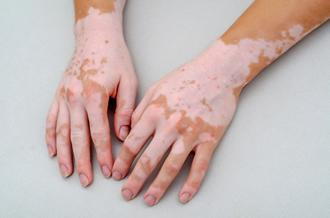
Vitiligo
Melanins
Peutz-Jeghers Syndrome
Dental Amalgam
Death Certificates
An L1 element intronic insertion in the black-eyed white (Mitf[mi-bw]) gene: the loss of a single Mitf isoform responsible for the pigmentary defect and inner ear deafness. (1/348)
Waardenburg syndrome type 2 (WS2) is an autosomal dominant disorder characterized by a combination of pigmentary and auditory abnormalities. Approximately 20% of WS2 cases are associated with mutations in the gene encoding microphthalmia-associated transcription factor (MITF). MITF plays a critical role in the development of both neural-crest-derived melanocytes and optic cup-derived retinal pigmented epithelium (RPE); the loss of a functional Mitf in mice results in complete absence of all pigment cells, which in turn induces microphthalmia and inner ear deafness. The black-eyed white Mitf mi-bw homozygous mouse normally has a pigmented RPE but lacks melanocytes essential for the pigmentation of the body and hearing. We show here that Mitf mi-bw is caused by an insertion into intron 3 of a 7.2 kb novel L1 element, L1bw, which belongs to an actively retrotransposing TF subfamily. The L1bw insertion reduces the amount of mRNAs for two Mitf isoforms, Mitf-A and Mitf-H, by affecting their overall expression levels and pre-mRNA splicing patterns, while it abolishes mRNA expression of another isoform, Mitf-M, which is specifically expressed in neural-crest-derived melanocytes. The consequence of the L1 insertion in the black-eyed white Mitf mi-bw mouse is that the developmental programme for RPE cells proceeds normally, most likely because of the presence of residual, full-length Mitf-A and Mitf-H proteins, whereas the lack of Mitf-M results in loss of the melanocyte population. The results suggest that melanocyte development depends critically on a single Mitf isoform, Mitf-M, and raise the possibility that specific mutations affecting MITF-M, the human equivalent of Mitf-M, may be responsible for a subset of WS2 conditions. (+info)A case of melanonychia caused by Exophiala dermatitidis. (2/348)
We report a case of a healthy 61-year-old woman with discoloration of the nail on her right big toe. We first treated her with topical steroid and urea under suspected diagnosis of nail eczema, but the lesion remained. In culture, black, shiny, pasty and yeast-like colonies grew repeatedly. Examination of debris from her nail showed dematiaceous spherical cells and hyphal elements. Microscopically, annelloconidia were produced at the apical ends of anellidic conidiogenous cells. This colony grew at 40C. Mitochondrial DNA restriction fragment length polymorphism was analysed in this strain and its restriction pattern confirmed the isolate to be Exophiala dermatitidis. Based on these findings, we diagnosed this nail deformity as fungal melanonychia due to Exophiala dermatitidis. This is the third reported case of this disease. (+info)Altered gene expression in melanocytes exposed to 4-tertiary butyl phenol (4-TBP): upregulation of the A2b adenosine receptor 1. (3/348)
Exposure to phenolic agents contributes to the development of occupational vitiligo. Proposed as a causative factor for leukoderma in vivo, the para-substituted phenol 4-tertiary butyl phenol was chosen to investigate early cellular events responsible for selective disappearance of melanocytes from the epidermis of individuals sensitive to such agents. To this end, differential display of melanocyte mRNA isolated from three separate cultures was performed following a 12 h exposure of cells to 250 microM 4-tertiary butyl phenol or to vehicle alone. Fragments of cDNA representing differentially expressed messages were cloned and subsequently confirmed by reverse dot blotting. Alignment analysis revealed that the L30 ribosomal protein was upregulated by the treatment, potentially reflecting altered levels of protein synthesis in response to stress. In addition, a gene sequence upregulated following exposure to 4-tertiary butyl phenol was identified as the A2b receptor (a P1 receptor for adenosine). Differential expression of this gene was confirmed in an RNase protection assay. By reverse transcription-polymerase chain reaction, the gene was shown to be expressed in keratinocytes and fibroblasts as well. Flow cytometry confirmed differential expression in melanocytes and fibroblasts, but not in keratinocytes. Interestingly, it has been reported that P1 purinoceptor stimulation can induce apoptosis. This is in concordance with results reported elsewhere demonstrating induction of apoptosis by 4-tertiary butyl phenol in human melanocytes, as well as with morphologic changes observed in this study in cells exposed to 250 microM 4-tertiary butyl phenol for 72 h. In conclusion, differential display is useful to establish melanocyte components involved in the cellular response to phenolic agents. (+info)Epidemic dropsy in India. (4/348)
Epidemic dropsy is a clinical state resulting from use of edible oils adulterated with Argemone mexicana oil. Sanguinarine and dehydrosanguinarine are two major toxic alkaloids of Argemone oil, which cause widespread capillary dilatation, proliferation and increased capillary permeability. Leakage of the protein-rich plasma component into the extracellular compartment leads to the formation of oedema. The haemodynamic consequences of this vascular dilatation and permeability lead to a state of relative hypovolemia with a constant stimulus for fluid and salt conservation by the kidneys. Illness begins with gastroenteric symptoms followed by cutaneous erythema and pigmentation. Respiratory symptoms such as cough, shortness of breath and orthopnoea progressing to frank right-sided congestive cardiac failure are seen. Mild to moderate anaemia, hypoproteinaemia, mild to moderate renal azotemia, retinal haemorrhages, and glaucoma are common manifestations. There is no specific therapy. Removal of the adulterated oil and symptomatic treatment of congestive cardiac failure and respiratory symptoms, along with administration of antioxidants and multivitamins, remain the mainstay of treatment. Selective cultivation of yellow mustard, strict enforcement of the Indian Food Adulteration Act, and exemplary punishment to unscrupulous traders are the main preventive measures. (+info)Terminal osseous dysplasia with pigmentary defects maps to human chromosome Xq27.3-xqter. (5/348)
We have identified a four-generation family with 10 affected females manifesting one or more of the following features: osseous dysplasia involving the metacarpals, metatarsals, and phalanges leading to brachydactyly, camptodactyly, and other digital deformities; pigmentary defects on the face and scalp; and multiple frenula. There were no affected males. We performed X-inactivation studies on seven affected females, using a methylation assay at the androgen receptor locus; all seven demonstrated preferential inactivation of their maternal chromosomes carrying the mutation, and two unaffected females showed a random pattern. These findings indicate that this disorder is linked to the X chromosome. To map the gene for this disorder, we analyzed DNA from nine affected females and five unaffected individuals, using 40 polymorphic markers evenly distributed throughout the X chromosome. Two-point and multipoint linkage analyses using informative markers excluded most of the X chromosome and demonstrated linkage to a region on the long arm between DXS548 and Xqter. A maximum LOD score of 3.16 at recombination fraction 0 was obtained for five markers mapping to Xq27.3-Xq28. The mapping data should facilitate the identification of the molecular basis of this disorder. (+info)An unbalanced submicroscopic translocation t(8;16)(q24.3;p13.3)pat associated with tuberous sclerosis complex, adult polycystic kidney disease, and hypomelanosis of Ito. (6/348)
We report on a familial submicroscopic translocation involving chromosomes 8 and 16. The proband of the family had a clinical picture suggestive of a large deletion in the chromosome 16p13.3 area, as he was affected with tuberous sclerosis complex (TSC) and had alpha thalassaemia trait, and his half brother, who also had TSC, may have suffered additionally from polycystic kidney disease (PKD). FISH studies provided evidence for a familial translocation t(8;16)(q24.3;p13.3) with an unbalanced form in the proband and a balanced form in the father and in a paternal aunt. The unbalanced translocation caused the index patient to be deleted for the chromosome 16p13.3-pter region, with the most proximal breakpoint described to date for terminal 16p deletions. In addition, FISH analysis showed a duplication for the distal 8q region. Since the index patient also had hypomelanosis of Ito (HI), either of the chromosomal areas involved in the translocation may be a candidate region for an HI determining gene. Furthermore, it is noteworthy that both carriers of the balanced translocation showed a nodular goitre, while the proband has hypothyroidism. (+info)Problematic pigmented lesions: approach to diagnosis. (7/348)
A number of pigmented lesions are difficult to classify and raise the possibility of a melanoma diagnosis. Care should be exercised to exclude non-melanocytic lesions, and benign melanocytic entities, both of which can mimic melanoma histologically. In addition, the possibility of the lesion being a melanoma variant or epidermotropic metastasis should be considered. There will still be some cases that are difficult to resolve. These usually fall into one of three categories: atypical junctional melanocytic lesion versus early melanoma; naevus versus naevoid melanoma; and atypical Spitz, cellular blue, and deep penetrating naevi versus thick melanoma. These will pose problems even for experts. The atypical Spitz lesions are perhaps the most important category because they tend to be from younger individuals, the differential diagnosis is thick melanoma, and there is no single discriminating histological feature. (+info)A quantitative evaluation of pigmented skin lesions using the L*a*b* color coordinates. (8/348)
The evaluation of pigmentary skin lesions by clinical doctors has been based on subjective and qualitative judgements. Observations have mostly relied on visual inspection, making the effects of treatment difficult to evaluate with any precision. For this reason there is a real need for an objective method to evaluate prognosis after treatment. Recent scientific measurements such as reflectance spectrophotometry and reflectance colorimetry have provided accurate quantitative color information about skin lesions, but these techniques are costly and difficult to apply in the clinical field. The purpose of this study was to develop a simple and cost-effective way of evaluating treatment results. We have developed a software program using the L*a*b* color coordinate system to quantify the effect of treatment and have successfully demonstrated its clinical usefulness. Our method compares the relative color difference between normal skin and skin lesions before and after treatment, instead of measuring the absolute color of skin lesions. The accuracy of our quantitative color analysis was confirmed by the simulated images of hemangioma and ota nevus. Clinical efficacy was also confirmed through a blind test involving 3 clinicians who were asked to grade the treatment effects of 13 cases of hemangioma and 7 cases of ota nevus. These subjective clinical grades correlated well with the treatment results obtained using the proposed color analysis system (Correlation coefficient = 0.84). (+info)Pigmentation disorders are conditions that affect the production or distribution of melanin, the pigment responsible for the color of skin, hair, and eyes. These disorders can cause changes in the color of the skin, resulting in areas that are darker (hyperpigmentation) or lighter (hypopigmentation) than normal. Examples of pigmentation disorders include melasma, age spots, albinism, and vitiligo. The causes, symptoms, and treatments for these conditions can vary widely, so it is important to consult a healthcare provider for an accurate diagnosis and treatment plan.
Vitiligo is a medical condition characterized by the loss of pigmentation in patches of skin, resulting in irregular white depigmented areas. It's caused by the destruction of melanocytes, the cells responsible for producing melanin, which gives our skin its color. The exact cause of vitiligo is not fully understood, but it's thought to be an autoimmune disorder where the immune system mistakenly attacks and destroys melanocytes. It can affect people of any age, gender, or ethnicity, although it may be more noticeable in people with darker skin tones. The progression of vitiligo is unpredictable and can vary from person to person. Treatment options include topical creams, light therapy, oral medications, and surgical procedures, but the effectiveness of these treatments varies depending on the individual case.
Pigmentation, in a medical context, refers to the coloring of the skin, hair, or eyes due to the presence of pigment-producing cells called melanocytes. These cells produce a pigment called melanin, which determines the color of our skin, hair, and eyes.
There are two main types of melanin: eumelanin and pheomelanin. Eumelanin is responsible for brown or black coloration, while pheomelanin produces a red or yellow hue. The amount and type of melanin produced by melanocytes can vary from person to person, leading to differences in skin color and hair color.
Changes in pigmentation can occur due to various factors such as genetics, exposure to sunlight, hormonal changes, inflammation, or certain medical conditions. For example, hyperpigmentation refers to an excess production of melanin that results in darkened patches on the skin, while hypopigmentation is a condition where there is a decreased production of melanin leading to lighter or white patches on the skin.
Skin pigmentation is the coloration of the skin that is primarily determined by two types of melanin pigments, eumelanin and pheomelanin. These pigments are produced by melanocytes, which are specialized cells located in the epidermis. Eumelanin is responsible for brown or black coloration, while pheomelanin produces a red or yellow hue.
The amount and distribution of melanin in the skin can vary depending on genetic factors, age, sun exposure, and various other influences. Increased production of melanin in response to UV radiation from the sun helps protect the skin from damage, leading to darkening or tanning of the skin. However, excessive sun exposure can also cause irregular pigmentation, such as sunspots or freckles.
Abnormalities in skin pigmentation can result from various medical conditions, including albinism (lack of melanin production), vitiligo (loss of melanocytes leading to white patches), and melasma (excessive pigmentation often caused by hormonal changes). These conditions may require medical treatment to manage or improve the pigmentation issues.
Melanin is a pigment that determines the color of skin, hair, and eyes in humans and animals. It is produced by melanocytes, which are specialized cells found in the epidermis (the outer layer of the skin) and the choroid (the vascular coat of the eye). There are two main types of melanin: eumelanin and pheomelanin. Eumelanin is a black or brown pigment, while pheomelanin is a red or yellow pigment. The amount and type of melanin produced by an individual can affect their skin and hair color, as well as their susceptibility to certain diseases, such as skin cancer.
Peutz-Jeghers Syndrome (PJS) is a rare genetic disorder characterized by the development of benign tumors called hamartomas in the gastrointestinal tract and pigmented macules on the skin and mucous membranes. The syndrome is caused by mutations in the STK11/LKB1 gene, which is involved in regulating cell growth and division.
Individuals with PJS have an increased risk of developing various types of cancer, including gastrointestinal tract cancers, breast cancer, ovarian cancer, lung cancer, and cervical cancer. The diagnosis of PJS is typically made based on the presence of characteristic clinical features, such as multiple pigmented macules on the skin and mucous membranes, and a history of benign gastrointestinal tumors or family history of PJS.
Management of PJS involves regular surveillance for gastrointestinal tumors and cancer screening, as well as genetic counseling and testing for family members who may be at risk. Treatment options depend on the location and size of the tumors and may include endoscopic removal or surgery.
Dental amalgam is a commonly used dental filling material that consists of a mixture of metals, including silver, tin, copper, and mercury. The mercury binds the other metals together to form a strong, durable, and stable restoration that is resistant to wear and tear. Dental amalgam has been used for over 150 years to fill cavities and repair damaged teeth, and it remains a popular choice among dentists due to its strength, durability, and affordability.
However, there has been some controversy surrounding the use of dental amalgam due to concerns about the potential health effects of mercury exposure. While the majority of scientific evidence suggests that dental amalgam is safe for most people, some individuals may be more sensitive to mercury and may experience adverse reactions. As a result, some dentists may recommend alternative filling materials, such as composite resin or gold, for certain patients.
Overall, dental amalgam is a safe and effective option for filling cavities and restoring damaged teeth, but it is important to discuss any concerns or questions with a qualified dental professional.
A death certificate is a formal legal document that records the date, location, and cause of a person's death. It is typically issued by a medical professional, such as a physician or medical examiner, and is used to establish the fact of death for legal purposes. The information on a death certificate may be used for a variety of purposes, including settling the deceased person's estate, assisting with insurance claims, and supporting public health surveillance and research.
In order to complete a death certificate, the medical professional must determine the cause of death and any significant contributing conditions. This may involve reviewing the deceased person's medical history, conducting a physical examination, and ordering laboratory tests or autopsy. The cause of death is typically described using standardized codes from the International Classification of Diseases (ICD).
It is important to note that the information on a death certificate is considered confidential and is protected by law. Only authorized individuals, such as the deceased person's next of kin or legal representative, are permitted to access the document.
A hamartoma is a benign tumor-like growth that is composed of an unusual mixture of cells and tissues that are normally found in the affected area. These growths can occur anywhere in the body, but they are most commonly found in the skin, lungs, and brain. Hamartomas are typically slow growing and do not spread to other parts of the body (metastasize). They are usually harmless, but in some cases, they may cause symptoms or complications depending on their size and location. In general, hamartomas do not require treatment unless they are causing problems.
Hyperpigmentation6
- Background There is little published information about segmental hypo- and hyperpigmentation pigmentation disorder (SegPD) although it is a relatively common problem in paediatric dermatology. (medscape.com)
- We propose reviving the term 'segmental pigmentation disorder' coined by Metzker and colleagues to describe children with segmental and block-like hypo-/hyperpigmentation with midline demarcation. (medscape.com)
- Skin pigmentation disorders including hyperpigmentation and albinism. (nih.gov)
- Drug-induced pigmentation and/or hyperpigmentation may be caused by numerous drugs through a number of differing mechanisms. (logicalimages.com)
- A common form of pigmentation is hyperpigmentation, when the body produces too much melatonin. (ag3derm.com)
- Sir, Acanthosis nigricans (AN) is a skin disorder characterised by skin hyperpigmentation and thickening. (e-ijd.org)
Segmental Pigmentation Disorder6
- Segmental Pigmentation Disorder: Clinical Manifestations and Epidemiological Features in 144 patients, a Retrospective Case-control Study. (nih.gov)
- [ 1 ] introduced the term 'segmental pigmentation disorder' (SegPD) when reporting on a case series of 30 children accrued over a 4-year period. (medscape.com)
- Cite this: Segmental Pigmentation Disorder - Medscape - Jun 01, 2010. (medscape.com)
- Segmental Pigmentation Disorder (SegPD) is an uncommon skin condition affecting young children. (pedraresearch.org)
- Segmental Pigmentation Disorder consists of white and brown pigmented birthmarks that appear as large patches on the body. (pedraresearch.org)
- The underlying cause of Segmental Pigmentation Disorder is not known, but it suspected to be due to genetic changes in these different areas of skin color, in the way that the skin cells produce pigment. (pedraresearch.org)
Overview of Pigmentation Disorders1
- Overview of Pigmentation Disorders Melanin is the brownish pigment responsible for the color of skin, hair, and the iris of the eyes. (merckmanuals.com)
Vitiligo8
- Vitiligo is an acquired depigmenting skin disorder in which pigment is lost from the skin in patches. (arlingtonvaderm.com)
- He told the talk show host that he had vitiligo, a disorder that destroyed his skin pigmentation. (cnn.com)
- A family of compounds derived from black pepper extract have shown potential in animal studies to be effective in treating vitiligo, a skin pigmentation disorder. (sciencedaily.com)
- Vitiligo is a disorder associated with pigmentation of the skin. (diethealthclub.com)
- Vitiligo is an auto immune disorder. (diethealthclub.com)
- Individuals with vitiligo-suffering parents are more prone to the disorder. (diethealthclub.com)
- Nonsegmental vitiligo is a skin disorder characterized by a loss of pigmentation, or color, of the skin, mucosa, and hair. (nursingcenter.com)
- Vitiligo is an acquired disorder of pigmentation characterized by depigmented or hypopigmented macules caused by loss of epidermal melanocytes. (e-ijd.org)
Oculocutaneous albinism3
- Oculocutaneous albinism (OCA) is a clearly defined set of seven types of genetic mutations which reduce or completely prevent the synthesis of eumelanin or pheomelanin , resulting in reduced pigmentation. (wikipedia.org)
- Hermansky-Pudlak syndrome (HPS) consists of a group of genetically heterogeneous disorders which share the clinical findings of oculocutaneous albinism, a platelet storage pool deficiency, and some degree of ceroid lipofuscinosis. (nih.gov)
- Sharma's award will support research of oculocutaneous albinism (OCA), a group of genetic disorders that affect production of the skin pigment melanin. (nih.gov)
Human pigmentation1
- A polymorphism in the agouti signaling protein gene is associated with human pigmentation. (nature.com)
Melanoma2
- Patients with oral melanoma often recall having a previous pigmentation in the same area months to years before the melanoma diagnosis, and the condition may even have elicited a prior comment from physicians or dentists. (medscape.com)
- The four major mechanisms leading to increased oral pigmentation are discussed in detail: physiologic pigmentation, systemic diseases (eg, Peutz-Jeghers syndrome [PJS]), oral mucosal insults (eg, amalgam tattoo), and neoplastic processes (eg, melanoma). (medscape.com)
Acanthosis1
- People who have a disorder called acanthosis nigricans develop darkened and thickened skin in the underarms, on the nape of the neck, and in skinfolds. (merckmanuals.com)
Pigmentary Disorders1
- Shining Light on Autophagy in Skin Pigmentation and Pigmentary Disorders. (nih.gov)
Melanocytes4
- Physiologic pigmentation, also known as racial or ethnic pigmentation, is an increased production of melanin pigment by melanocytes in dark-skinned individuals. (medscape.com)
- [ 5 ] The pigmentation is due to an increased melanocytic activity rather than an increase in the number of melanocytes. (medscape.com)
- Melanocytes are the cells that produce pigmentation in the skin. (sciencedaily.com)
- Melanocytes or the pigment-making cells are destroyed in this disorder. (diethealthclub.com)
Melanin production1
- This uneven melanin production results in spots of pigmentation known as freckles. (merckmanuals.com)
Oral Mucosa3
- Buchner A. Amalgam tattoo (amalgam pigmentation) of the oral mucosa: clinical manifestations, diagnosis and treatment. (medscape.com)
- Black and Brown: Non-neoplastic Pigmentation of the Oral Mucosa. (medscape.com)
- Tripe palms along with pigmentation over her genital and oral mucosa were seen [Figure 3] . (e-ijd.org)
Melasma2
- The smallest amount of UV exposure may cause melasma to recur after fading which explains why the pigmentation of melasma worsens in summer. (fairfieldderm.com)
- Melasma - this is a pigmentation problem which often appears during pregnancy as a result of hormonal changes in the body. (thelasertreatmentclinic.com)
SegPD1
- To define the spectrum of this disorder more accurately, we retrospectively reviewed the records of 39 children with SegPD. (medscape.com)
Etiology1
- Early biopsy of focal pigmentations of undetermined etiology is extremely important in order to detect oral melanomas at an early stage. (medscape.com)
Physiologic1
- Lambertini M, Patrizi A, Ravaioli GM, Dika E. Oral pigmentation in physiologic conditions, post-inflammatory affections and systemic diseases. (medscape.com)
Abnormal1
- Abnormal skin growths and abnormal pigmentation of the skin may be present at birth or develop later in life. (childrensnational.org)
Bipolar Disorder2
- When your roommate has bipolar disorder, there are things to consider. (healthyplace.com)
- When your roommate has bipolar disorder -- or any other mental illness -- they may, unwittingly, be responsible for taking care of you and managing their own reactions to your symptoms ( Effects of Bipolar Disorder on Family and Friends ). (healthyplace.com)
Brownish1
- Increased melanin most often produces a brownish pigmentation in the dermis. (logicalimages.com)
Systemic3
- No association with systemic disorders, such as neurological involvement, was found. (medscape.com)
- [ 7 ] The tendency to develop pigmentation appears to be genetically determined, but color intensity might be influenced by smoking, hormones, systemic medications, and physical factors. (medscape.com)
- Systemic Lupus Erythematosus (SLE) Systemic lupus erythematosus is a chronic autoimmune inflammatory connective tissue disorder that can involve joints, kidneys, skin, mucous membranes, and blood vessel walls. (merckmanuals.com)
Uneven1
- One of the main causes of uneven skin tone is pigmentation disorders, where your skin produces too much or not enough melanin. (cetaphil.com)
Treatments2
- I suffer from post inflammation pigmentation from adult acne for several years and literally have tried a whole variety of topical treatments, prescriptive, lasers etc. (thelasertreatmentclinic.com)
- Before you invest in more expensive treatments for pigmentation disorders, it may be useful to try these home remedies first to see if they can treat the problem. (ag3derm.com)
Regulation1
- Skin pigmentation process and its regulation. (nih.gov)
Mutants2
- Pigmentation mutants in various species are highly informative about basic genetic and developmental pathways, and provide important clues to the processes of photoprotection, cancer predisposition and even human evolution. (nature.com)
- Mutants were analyzed for pigmentation defects and retinal disease by histology, immunohistochemistry, and transmission electron microscopy.Phenocopy and rescue experiments determined that a loss of Vps11 results in the platinum phenotype. (nih.gov)
Albinism1
- With the exception of albinism, most of these pigmentation disorders can be effectively treated using laser treatment. (thelasertreatmentclinic.com)
Clinical7
- Local treatment now rules the global market due to better clinical results in patients with pigmentation problems. (eu.org)
- Each gene responsible for a subset of HPS or a related disorder codes for a protein which almost certainly plays a pivotal role in vesicular trafficking, inextricably linking clinical and cell biological interests in this group of diseases. (nih.gov)
- Question 1: What is the specific clinical disorder to be studied? (cdc.gov)
- Question 2: What are the clinical findings defining this disorder? (cdc.gov)
- The specific clinical disorder is primary iron overload of adult onset sufficient to cause significant morbidity and mortality. (cdc.gov)
- The original clinical diagnosis of hereditary hemochromatosis was based on the triad of hepatic cirrhosis, diabetes mellitus, and skin pigmentation. (cdc.gov)
- Low vitamin D intake can be related to seasonal affective disorder," says Alan Manevitz, MD, clinical psychiatrist at Lenox Hill Hospital in New York City. (qualityhealth.com)
Ultraviolet2
- They are responsible for the pigmentation of skin and hair, and thereby contribute to the appearance of skin and provide protection from damage by ultraviolet radiation. (nature.com)
- Similar to silver , a gold preparation used parenterally for a long period may rarely produce a permanent skin pigmentation - especially if the skin is exposed to sunlight or artificial ultraviolet radiation. (wikidoc.org)
Lentigines1
- Noonan syndrome with multiple lentigines (NSML) is a very rare inherited disorder. (nih.gov)
Exposed to sunlight1
- Chlorpromazine is known, in rare instances, to induceskin pigmentation in areas exposed to sunlight. (psychiatrist.com)
Freckles1
- Freckles are a result of variation in skin pigmentation in which pigment is not released evenly. (ag3derm.com)
Symptoms2
Amalgam1
- Multiple causes are known, and they may range from simple iatrogenic mechanisms, such as implantation of dental amalgam, to complex medical disorders, such as Peutz-Jeghers syndrome (PJS) and Addison disease. (medscape.com)
Underproduction2
- Most pigmentation disorders involve the underproduction or overproduction of melanin. (wikipedia.org)
- Pigmentation disorders are caused by an overproduction or underproduction of melanin. (ag3derm.com)
Patches2
- Some pigmentation disorders affect just patches of skin. (medlineplus.gov)
- Research reveals the greater incidence of this disorder, in individuals suffering from autoimmune diseases, such as pernicious anemia, hyperthyroidism and alopecia areata (patches of baldness). (diethealthclub.com)
Darker1
- Pigmentation can cause skin to appear discoloured or blotchy, & can make areas of the skin appear lighter or darker than normal. (ag3derm.com)
Acne1
- Acne is a disorder of the hair follicles and oil (sebaceous) glands that become clogged. (childrensnational.org)
Sunlight1
- For example, a person may look yellow because of liver problems, slightly blue because of breathing problems, bruised because of blood disorders, or pink or red because of skin problems or extra sensitivity to sunlight. (cancer.org)
Autosomal4
- PJS is an autosomal dominant disorder characterized by intestinal hamartomatous polyps in association with mucocutaneous melanocytic macules. (medscape.com)
- Juvenile hemochromatosis ( HFE2 ) is a rare autosomal recessive disorder associated with a gene mapped to the long arm of chromosome 1. (cdc.gov)
- Carney complex is inherited in an autosomal dominant pattern, which means one copy of the altered gene in each cell is sufficient to cause the disorder. (nih.gov)
- Autosomal Dominant Genetic disorders determined by a single gene (Mendelian disorders) are easiest to analyze and the most well understood. (merckmanuals.com)
Genetic Disorders1
- ture of genetic disorders, genetic consultation and testing, The authors thank the other members of the Genetics Home gene therapy, and genomic research. (nih.gov)
Autism spectrum1
- To evaluate the electroretinogram waveform in autism spectrum disorder (ASD) and attention deficit hyperactivity disorder (ADHD) using a discrete wavelet transform (DWT) approach. (frontiersin.org)
Affects2
- Pigmentation is what affects the color of skin. (medicalnewstoday.com)
- Leber congenital amaurosis (LCA) is an eye disorder that primarily affects the retina. (nih.gov)
Dermatologist1
- A specialized UV lamp known as a Wood's lamp may be used by the dermatologist to accentuate differences in pigmentation and sun damaged skin. (fairfieldderm.com)
Mucous1
- There are certain classes of drugs that are more commonly implicated in causing skin, mucous membrane, and nail pigmentation. (logicalimages.com)
Relatively common1
- Oral pigmentation is a relatively common condition that may involve any portion of the oral cavity. (medscape.com)
Dermatological1
- He also pioneered X-ray therapy for dermatological disorders. (uc.edu)
Unusual1
- Almost all people with Carney complex have areas of unusual skin pigmentation. (nih.gov)