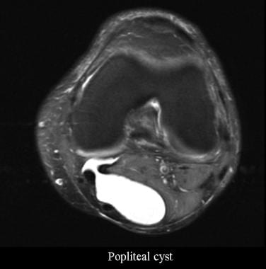
Popliteal Cyst
Synovial Cyst
Cysts
Periarticular lesions detected on magnetic resonance imaging: prevalence in knees with and without symptoms. (1/29)
OBJECTIVE: To evaluate, using magnetic resonance imaging (MRI), the prevalence of periarticular lesions in older persons with or without knee pain, and to assess the association of these lesions with knee pain. METHODS: Subjects ages 45 years and older, with or without knee pain, were recruited from Veterans Affairs medical centers and from the community. Weight-bearing posteroanterior, skyline, and lateral radiographs were obtained in all subjects. Subjects were divided into 3 groups: those with radiographic OA (ROA) and knee pain (n = 376), those with ROA and no knee pain (n = 51), and those with neither ROA nor knee pain (n = 24). A single knee (the more symptomatic one in subjects with knee pain) was imaged with a 1.5T scanner using T1- and T2-weighted and proton-density spin-echo imaging sequences. MRIs were read for the presence of periarticular lesions, which were categorized (according to their general location) as being either peripatellar (prepatellar, superficial infrapatellar, deep infrapatellar) or "other periarticular lesions" (semimembranosus-tibial collateral ligament bursitis, anserine bursitis, iliotibial band syndrome, tibiofibular cyst). RESULTS: Patients with knee pain had more severe radiographic disease than did subjects who were asymptomatic. Peripatellar lesions (prepatellar or superficial infrapatellar) were present in 12.1% of the patients with knee pain and ROA, in 20.5% of the patients with ROA and no knee pain, and in 0% of subjects with neither ROA nor knee pain (P = 0.116). However, other periarticular lesions were present in 14.9% of patients with both ROA and knee pain, in only 3.9% of patients with ROA but no knee pain, and in 0% of the group with no knee pain and no ROA (P = 0.004). CONCLUSION: Although peripatellar lesions are equally common among subjects with knee pain and those without knee pain, other periarticular lesions (including bursitis and iliotibial band syndrome) are significantly more common among subjects with knee pain and may contribute to pain in these individuals. (+info)Valgus and varus deformity after wide-local excision, brachytherapy and external beam irradiation in two children with lower extremity synovial cell sarcoma: case report. (2/29)
BACKGROUND: Limb-salvage is a primary objective in the management of extremity soft-tissue sarcoma in adults and children. Wide-local excision combined with radiation therapy is effective in achieving local tumor control with acceptable morbidity and good functional outcomes for most patients. CASE PRESENTATION: Two cases of deformity after wide-local excision, brachytherapy and external beam irradiation for lower-extremity synovial cell sarcoma are presented and discussed to highlight contributing factors, time course of radiation effects and orthopedic management. In an effort to spare normal tissues from the long-term effects of radiation therapy, more focal irradiation techniques have been applied to patients with musculoskeletal tumors including brachytherapy and conformal radiation therapy. As illustrated in this report, the use of these techniques results in the asymmetric irradiation of growth plates and contributes to the development of valgus or varus deformity and leg-length discrepancies. CONCLUSIONS: Despite good functional outcomes, progressive deformity in both patients required epiphysiodesis more than 3 years after initial management. There is a dearth of information related to the effects of radiation therapy on the musculoskeletal system in children. Because limb-sparing approaches are to be highlighted in the next generation of cooperative group protocols for children with musculoskeletal tumors, documentation of the effects of surgery and radiation therapy will lead to improved decision making in the selection of the best treatment approach and in the follow-up of these patients. (+info)Cystic adventitial disease: a trap for the unwary. (3/29)
Cystic adventitial disease is an uncommon condition. A case of cystic adventitial disease of the popliteal artery is reported in a young man who has been followed up for 14 years after surgical treatment. Early recognition and treatment of the condition will prevent progression to popliteal thrombosis and critical ischaemia. However, diagnosis of the condition is difficult. Characteristic features in the presenting history, such as fluctuation in severity of symptoms, sudden onset after vigorous activity and delayed recovery time after cessation of exercise are identified, which should help the clinician avoid misdiagnosis and delayed diagnosis of the condition. The clinician is also warned of the associated misleading clinical features such as the presence of normal peripheral pulses and normal ankle pressures in some cases of CAD. (+info)Ultrasonographic scan in knee pain in athletes. (4/29)
Fifty-two knees were examined using real-time high-definition ultrasonography with a 7.5 MHz probe. The extra-articular structures were easily visualized and diagnosis of patellar tendon lesions and Baker's cysts formulated. While the meniscal cartilages were shown as a homogeneous triangular structure between the femoral condyle and the tibial plateau, no lesions were detected. Deeper intra-articular structures, such as the cruciate ligaments, were not shown by the scan, thus their evaluation was not possible. Given its low cost, wide availability, non-invasiveness and patients' acceptability of the technique, ultrasonography may play an important role in the diagnosis of soft tissue lesions in and around the knee joint. (+info)Cystic adventitial disease of the popliteal artery: an argument for the developmental theory. (5/29)
Cystic adventitial disease is a rare non-atheromatous cause of popliteal artery disease. We report a case of a 54-year-old patient with claudication of the right calf caused by cystic adventitial disease. Intra-operatively, a communication between the adventitia and the knee joint was identified. Connections between the adventitial cyst and the nearby joint have been reported in the literature that support the developmental theory. This theory suggests that cystic adventitial disease is a developmental manifestation of mucin-secreting cells derived from the mesenchyme of the adjacent joint. This case is the first, to our knowledge, in which a communication between joint and adventitia has been clearly documented by operative findings. (+info)Painful swollen leg--think beyond deep vein thrombosis or Baker's cyst. (6/29)
(+info)What is the clinical and ethical importance of incidental abnormalities found by knee MRI? (7/29)
(+info)Sclerotherapy of Baker's cyst with imaging confirmation of resolution. (8/29)
BACKGROUND: Baker's cysts are commonly encountered in pain management practices. OBJECTIVE: To ascertain if sclerotherapy treatment of a Baker's cyst could produce objectively verifiable MRI imaging changes. DESIGN: Case report. METHODS: A 52-year-old white male with a posterior horn of the medial meniscus tear and a large Baker's cyst who had failed conservative care and drainage was imaged before treatment with sclerosing. Three injections of 12.5% dextrose and anesthetic with sodium morrhuate were injected intraarticular into the right knee after drainage. RESULTS: The Baker's cyst resolved on both postoperative imaging after the completion of care as well as on physical examination. CONCLUSIONS: Prolotherapy in this case study seemed to be an effective treatment for Baker's cyst in this patient. (+info)A Popliteal cyst, also known as Baker's cyst, is a fluid-filled sac that develops behind the knee, in the popliteal fossa. It forms when synovial fluid from the knee joint extends through a tear in the joint capsule, creating a visible bulge. The cyst may cause discomfort, swelling, or pain, especially when fully extended or flexed. In some cases, it can rupture and cause further complications, such as increased pain and inflammation in the calf region. Treatment options for Popliteal cysts include physical therapy, corticosteroid injections, and, in severe cases, surgical intervention to repair the underlying joint issue and remove the cyst.
A Synovial Cyst is a type of benign cyst that typically develops in the synovium, which is the membrane that lines and lubricates joint capsules. These cysts are filled with synovial fluid, which is the same lubricating fluid found inside joints. They usually form as a result of degenerative changes, trauma, or underlying joint diseases such as osteoarthritis.
Synovial cysts commonly occur in the spine (particularly in the facet joints), but they can also develop in other areas of the body, including the knees, hips, and hands. While synovial cysts are generally not harmful, they may cause discomfort or pain if they press on nearby nerves or restrict movement in the affected joint. Treatment options for synovial cysts range from conservative measures like physical therapy and pain management to surgical intervention in severe cases.
A cyst is a closed sac, having a distinct membrane and division between the sac and its surrounding tissue, that contains fluid, air, or semisolid material. Cysts can occur in various parts of the body, including the skin, internal organs, and bones. They can be caused by various factors, such as infection, genetic predisposition, or blockage of a duct or gland. Some cysts may cause symptoms, such as pain or discomfort, while others may not cause any symptoms at all. Treatment for cysts depends on the type and location of the cyst, as well as whether it is causing any problems. Some cysts may go away on their own, while others may need to be drained or removed through a surgical procedure.
Cyst fluid refers to the fluid accumulated within a cyst, which is a closed sac-like or capsular structure, typically filled with liquid or semi-solid material. Cysts can develop in various parts of the body for different reasons, and the composition of cyst fluid may vary depending on the type of cyst and its location.
In some cases, cyst fluid might contain proteins, sugars, hormones, or even cells from the surrounding tissue. Infected cysts may have pus-like fluid, while cancerous or precancerous cysts might contain abnormal cells or tumor markers. The analysis of cyst fluid can help medical professionals diagnose and manage various medical conditions, including infections, inflammatory diseases, genetic disorders, and cancers.
It is important to note that the term 'cyst fluid' generally refers to the liquid content within a cyst, but the specific composition and appearance of this fluid may vary significantly depending on the underlying cause and type of cyst.
Baker's Cysts24
- Unknown causes - Baker's cysts can sometimes develop in children for no apparent reason. (singaporesportsclinic.com)
- Baker's cysts (also known as Baker cysts or popliteal cysts) are very common in people with rheumatoid arthritis (RA). (myrateam.com)
- There are several ways to manage Baker's cysts, both at home and with medical treatment. (myrateam.com)
- What Are Baker's Cysts? (myrateam.com)
- Named after 19th century surgeon William Morrant Baker - credited with first describing the condition - Baker's cysts can also occur as the result of injury or other disease, including osteoarthritis. (myrateam.com)
- In many cases, Baker's cysts do not cause any symptoms . (myrateam.com)
- Many myRAteam members have described what Baker's cysts feel like. (myrateam.com)
- One member shared how their Baker's cysts were "hurting really badly all day and hard as a rock and very big. (myrateam.com)
- Some members find that the pain from Baker's cysts can come and go, while others experience no pain at all. (myrateam.com)
- On rare occasions, Baker's cysts may cause complications . (myrateam.com)
- How Are Baker's Cysts Diagnosed? (myrateam.com)
- Although common in RA, Baker's cysts may elude clinical detection . (myrateam.com)
- In adults, Baker's cysts usually arise from almost any form of knee arthritis (e.g., rheumatoid arthritis) or cartilage (particularly a meniscus) tear. (wikipedia.org)
- Baker's cysts in children do not point to underlying joint disease. (wikipedia.org)
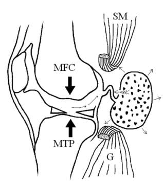
- Baker's cysts arise between the tendons of the medial head of the gastrocnemius and the semimembranosus muscles. (wikipedia.org)
- Most Baker's cysts maintain this direct communication with the synovial cavity of the knee, but sometimes, the new cyst pinches off. (wikipedia.org)
- Baker's cyst on MRI, sagittal image Baker's cyst on MRI, sagittal image Baker's cysts usually require no treatment unless they are symptomatic. (wikipedia.org)
- Baker's cysts in children, unlike in older people, nearly always disappear with time, and rarely require excision. (wikipedia.org)
- Popliteal cysts (also called Baker's cysts) are most frequently characterized by the enlargement of the gastrocnemius-semi-membranosus bursa in the posteromedial region of the knee. (jksrr.org)
- Ganglion cysts and Baker's cysts sometimes recur after surgery. (healthline.com)
- Several studies have shown an excellent success rate of communication enlargement surgery for popliteal cysts (Baker's cysts). (springeropen.com)
- Accordingly, popliteal cysts are also known as Baker's cysts. (springeropen.com)
- Sonographic detection of baker's cysts: comparison with MR imaging. (ptandme.com)
- When severe enough to hamper daily activities, the allopathic medical community approaches Baker's cysts with both surgical and non-surgical solutions. (integrativehealthcare.org)
Fossa8
- The most common mass in the popliteal fossa, Baker cyst, also termed popliteal cyst, results from fluid distention of the gastrocnemio-semimembranosus bursa, which is located in the medial aspect of the popliteal fossa. (medscape.com)
- The majority of patients with Baker cysts are asymptomatic, but knee joint pain and stiffness and a palpable mass in the medial popliteal fossa are not uncommon. (medscape.com)
- Background: Popliteal cysts are common and present as asymptomatic lumps in the medial popliteal fossa. (scirp.org)
- Conclusion: The cystic lesions in the medial aspect of the popliteal fossa can be misdiagnosed. (scirp.org)
- Diagnostic imaging approach to posteromedial knee (medial popliteal fossa) masses. (scirp.org)
- Baker cysts are enlarged bursae in the popliteal fossa. (msdmanuals.com)
- Baker cysts are enlarged bursae that develop from an accumulation of synovial fluid in the popliteal fossa. (msdmanuals.com)
- Exam normal except mild popliteal fossa swelling. (amssm.org)
Osteoarthritis5
- Baker cysts can be associated with conditions such as osteoarthritis of the knee, meniscal tears, rheumatoid arthritis, Charcot joints, and synovial disorders of the knee. (medscape.com)
- However, they should be obtained early in the evaluation, as they are useful for detecting other conditions commonly found in association with popliteal cysts, such as osteoarthritis, inflammatory arthritis, and loose bodies. (medscape.com)
- Osteoarthritis is the most common cause where 50% of people develop a Baker's Cyst knee. (singaporesportsclinic.com)
- Thus, popliteal cysts are almost never an isolated pathology in adult knees and are almost always associated with another pathology of the knee joint such as a meniscal tear and osteoarthritis 8 - 10) . (jksrr.org)
- The usual causes of Baker cysts are prior injury, rheumatoid arthritis, osteoarthritis, or overuse of the knee. (msdmanuals.com)
Synovial fluid8
- In 1877, Baker described 8 cases of periarticular cysts caused by synovial fluid that had escaped from the knee joint and formed a new sac outside the joint. (medscape.com)
- The inflammation in the leg was attributed to the synovial fluid collecting in the calf following the rupture of the cyst, although a diagnosis of cellulitis still was being considered, and so intravenous clindamycin was continued. (consultant360.com)
- A Baker's cyst forms when the envelope surrounding the joint ( joint capsule ) tightens, leading it to produce too much synovial fluid. (myrateam.com)
- In some cases, a Baker's cyst may rupture (burst) and leak synovial fluid into the calf area, which can cause redness, warmth, and pain or the sensation that water is running down your calf . (myrateam.com)
- Baker cyst is a buildup of joint fluid (synovial fluid) that forms a swelling behind the knee. (medlineplus.gov)
- Diagnosis is confirmed by ultrasonography, although if needed and there is no suspicion of a popliteal artery aneurysm then aspiration of synovial fluid from the cyst may be undertaken with care. (wikipedia.org)
- Moreover, the presence of a valve and the existence of an effusion create unidirectional flow of synovial fluid from the articular cavity to the cyst, which is one of the fundamental factors responsible for the formation and persistence of cysts 6) . (jksrr.org)
- Synovial fluid flows from the joint toward the cyst with extension of the knee. (msdmanuals.com)
Size of the cyst2
- There may be a decrease in range of motion caused by pain or by the size of the cyst. (medlineplus.gov)
- The size of the scar depends on several factors, including the size of the cyst. (healthline.com)
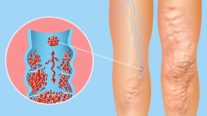
Deep venous thr1
- Care must be taken to differentiate ruptured Baker cysts from deep venous thrombosis (DVT). (medscape.com)
Knee joint9
- Case-Presentation: Popliteal Cyst: On ultrasound: characteristic neck communicating with knee joint. (scirp.org)
- A Baker's cyst, also called a popliteal cyst , is usually the result of a problem with your knee joint, such as arthritis or a cartilage tear. (singaporesportsclinic.com)
- Joint x-ray - this will not show the cyst but can show the presence of arthritis in the knee joint that may be causing the problem. (singaporesportsclinic.com)
- The synovial sac of the knee joint can, under certain circumstances, produce a posterior bulge, into the popliteal space, the space behind the knee. (wikipedia.org)
- They were first reported by Adams in 1840 [ 1 ], and Baker described in 1877 that this synovial cyst communicates with the knee joint and is often associated with other intra-articular lesions [ 3 ]. (springeropen.com)
- This is a fluid collection in a cyst that bulges out from the knee joint. (lvhn.org)
- Most Baker cysts accumulate fluid from the adjacent knee joint space. (msdmanuals.com)
- Baker cysts can develop without knee joint communication (eg, from the gastrocnemius-semimembranous bursa) in children. (msdmanuals.com)
- The most common site of posterior knee soreness and inflammation is the popliteal bursa, which is positioned behind the knee joint. (pinoyathletics.info)
Asymptomatic2
- Baker cysts may be asymptomatic but become noticeable when they become swollen (eg, ≥ 5 cm). (msdmanuals.com)
- Asymptomatic cysts do not require treatment. (msdmanuals.com)
Aneurysm2
- Thrombosed Popliteal Aneurysm: Lamellated appearance-high/low signal intensity on T2. (scirp.org)
- Be aware that lumps in the back of the knee are most likely a Popliteal Cyst but might possibly be a tumor or an aneurysm (swelling in an artery). (singaporesportsclinic.com)
Bursa4
- This fluid can accumulate in the back of your knee (called the popliteal bursa ) and cause a Baker's cyst to develop. (myrateam.com)
- The Baker's cyst is a distension of the bursa subtendinea and is caused by noninfectious knee effusion secondary to arthrosis, meniscal tears, trauma, rheumatoid arthritis, gout, or any other form of sinovitis, like rheumatoid arthritis. (uniroma1.it)
- Popliteal cysts are characterized by the enlargement of the gastrocnemius-semimembranosus bursa in the posteromedial region of the knee [ 13 ]. (springeropen.com)
- In children, Bakers cyst may be related to a problem with the bursa. (ptandme.com)
Fluid12
- Ultrasonography revealed a ruptured popliteal cyst in the boy's left knee with fluid tracking down to the calf. (consultant360.com)
- A Baker's cyst is a fluid-filled cyst that causes a bulge and a feeling of tightness behind your knee. (singaporesportsclinic.com)
- Both conditions can cause your knee to produce too much fluid, which can lead to a Baker's cyst. (singaporesportsclinic.com)
- Injury - trauma or injury to the knee can cause a build-up of fluid (effusion), which triggers Baker's cyst. (singaporesportsclinic.com)
- Shining a light through the cyst (transillumination) can show that the growth is fluid filled. (medlineplus.gov)
- A Baker's cyst, also known as a popliteal cyst, is a type of fluid collection behind the knee. (wikipedia.org)
- For this procedure, a doctor will insert a thin needle into the cyst to drain the fluid. (healthline.com)
- Finally, the irrigation fluid is suctioned, and the reduced cyst is visualized by ultrasound. (springeropen.com)
- A popliteal cyst, also known as a Baker's cyst, is a fluid-filled swelling that causes a lump at the back of the knee, leading to tightness and restricted movement. (samarpanphysioclinic.com)
- A Bakers cyst is a buildup of joint fluid behind the knee. (ptandme.com)
- A Bakers cyst develops when there is too much of this fluid. (ptandme.com)
- This will show that the cyst is filled with fluid and not solid. (ptandme.com)
Removal of the cyst1
- Surgical removal of the cyst will result in a scar. (healthline.com)
Ganglion cysts2
- In addition, US can differentiate these cysts from popliteal aneurysms and ganglion cysts. (medscape.com)
- The differential diagnosis of posterior knee lesions is broad and includes cystic lesions (other bursae, meniscal cysts, ganglion cysts and popliteal cysts). (scirp.org)
Lesions3
- In this article, we review the chief US and MR characteristics of popliteal cysts and some biopsy proven cases of mimics of popliteal cysts in each of the broad categories of cystic tumors, vascular lesions and synovial based cystic lesions. (scirp.org)
- They were first described by Adams 1) in 1840, and Baker 2) described the association of popliteal cysts with intra-articular lesions in 1877. (jksrr.org)
- Because most lesions originate from the follicular infundibulum, the more general term epidermoid cyst is favored. (medscape.com)
Intra-articular1
- Several studies on the pathogenesis of popliteal cysts have shown that they are associated with intra-articular pathology and valvular mechanism 5 - 7) . (jksrr.org)
Ultrasonography3
- [ 11 ] On ultrasonography, myxoid liposarcomas appear as complex, hypoechoic masses that do not meet the criteria for a simple cyst. (medscape.com)
- Subsequently, a contrast dye (indigo carmine) is injected into the popliteal cyst percutaneously using ultrasonography. (springeropen.com)
- If clinical findings are inconclusive (eg, if cysts are small or to differentiate them from deep vein thromboses), ultrasonography can be done. (msdmanuals.com)
Light through the cyst1
- The doctor may also shine a special light through the cyst. (ptandme.com)
Enlarging Baker cyst1
- In the Bunsen-valve mechanism, the enlarging Baker cyst exerts mass effect (feathered arrow) on the slitlike communication between the joint and the cyst, trapping effusion. (medscape.com)
Bakers Cyst1
- There is no known way to prevent a Bakers cyst. (ptandme.com)
Rupture4
- What do you suspect is the underlying cause of the boy's popliteal cyst rupture? (consultant360.com)
- Sometimes, the cyst may break open (rupture), causing pain, swelling, and bruising on the back of the knee and calf. (medlineplus.gov)
- Rupture of a Baker's cyst may also cause bruising below the medial malleolus of the ankle (Crescent sign). (wikipedia.org)
- A Baker's cyst can rupture and produce acute pain behind the knee and in the calf and swelling of the calf muscles. (wikipedia.org)
Meniscal cysts1
- [ 1 ] The ability to detect Baker cysts is near 100%, but ultrasound lacks the specificity to differentiate Baker cysts from meniscal cysts or myxoid tumors. (medscape.com)
Show the cyst1
- X-rays will not show the cyst or a meniscal tear , but they will show other problems that may be present, including arthritis. (medlineplus.gov)
Baker15
- Valvular mechanism of Baker cyst. (medscape.com)
- Effusion and fibrin are pumped (large arrows) into the Baker cyst (long, thin arrows). (medscape.com)
- A Baker cyst is seen medially (arrowhead). (medscape.com)
- Axial, T2-weighted magnetic resonance image of the knee shows effusion, synovial proliferation (white arrowhead), and a Baker cyst that contains debris (black arrowhead). (medscape.com)
- Contrast-enhanced, axial computed tomography (CT) scan of the knee shows multiple gaslike lucencies within a Baker cyst and synovial enhancement. (medscape.com)
- Administration of low-molecular-weight heparin to treat suspected DVT can lead to compartment syndrome in patients with Baker cysts. (medscape.com)
- In the past, Baker cysts were commonly detected by conventional arthrography, but disadvantages include invasiveness and the use of ionizing radiation. (medscape.com)
- [ 1 , 11 ] Ultrasound has largely replaced arthrography as the initial assessment for Baker cysts and is an easy-to-use, rapid, relatively inexpensive examination to employ in this setting. (medscape.com)
- A Baker cyst is caused by swelling in the knee. (medlineplus.gov)
- It is important to know whether pain or swelling is caused by a Baker cyst or a blood clot. (medlineplus.gov)
- A Baker cyst will not cause any long-term harm, but it can be annoying and painful. (medlineplus.gov)
- The symptoms of Baker cysts often come and go. (medlineplus.gov)
- Most Baker cysts are small and are do not cause symptoms. (msdmanuals.com)
- Consider ruptured Baker cyst in patients, particularly those with knee arthritis, who have suspected calf deep vein thrombosis. (msdmanuals.com)
- Nonsteroidal anti-inflammatory drugs (NSAIDs) are the primary treatment for symptomatic Baker cysts. (msdmanuals.com)
Posterior1
- The superficial femoral and popliteal veins in the thighs and the posterior tibial and peroneal veins in the calves are most commonly affected. (merckmanuals.com)
Symptoms7
- What are the symptoms of Baker's Cyst? (singaporesportsclinic.com)
- Note that some of the symptoms of a Baker's cyst - such as swelling, warmth, and redness - may resemble the symptoms of RA . (myrateam.com)
- Symptoms of a ruptured Baker's cyst may resemble those of a blood clot in a vein in the leg (deep vein thrombosis). (myrateam.com)
- If your rheumatologist suspects you may have a Baker's cyst, they will likely start by asking about your symptoms . (myrateam.com)
- It is very rare that the symptoms are actually coming from the cyst. (wikipedia.org)
- Learn more about a Baker's cyst and its symptoms, as well as how massage therapy can assist in the relief from this sometimes uncomfortable condition. (integrativehealthcare.org)
- Also, find out which massage techniques should be incorporated into a session when a client has a Baker's cyst, and why it is important for bodyworkers to familiarize themselves with the signs, symptoms and risk factors associated with a deep vein thrombosis. (integrativehealthcare.org)
Ultrasound3
- This study describes a simple ultrasound-guided arthroscopic technique to manage popliteal cysts and reduce postoperative pain. (springeropen.com)
- Additionally, a periarticular multimodal drug injection is administered into the septum and inner wall of the cyst under ultrasound guidance. (springeropen.com)
- Ultrasound-guided arthroscopic surgery for popliteal cysts can ensure reproducibility and be effective for postoperative pain relief. (springeropen.com)
Calf5
- The photo showed the boy's red calf and popliteal area following a case of poison ivy contact dermatitis on his left leg. (consultant360.com)
- The redness of the calf had nearly resolved, but the popliteal area still was markedly red. (consultant360.com)
- If the cyst breaks open, pain may significantly increase with swelling of the calf. (wikipedia.org)
- If the cyst breaks open, pain may increase, and there may be swelling of the calf. (wikipedia.org)
- A burst cyst commonly causes calf pain, swelling and redness that may mimic thrombophlebitis. (wikipedia.org)
Articular cavity1
- Therefore, the key to a successful surgery is the closure or enlargement of the communication between the cyst and the articular cavity [ 30 ]. (springeropen.com)
Inflammation3
- If necessary, the cyst can be aspirated to reduce its size, then injected with a corticosteroid to reduce inflammation. (wikipedia.org)
- Incomplete cyst removal can also cause scarring in the area and incite local inflammation. (healthline.com)
- Inflammation is mediated in part by the horny material contained in epidermoid cysts. (medscape.com)
Arthroscopic1
- To compare the clinical outcomes of the arthroscopic treatments for popliteal cysts with and without cystectomy. (jksrr.org)
Back of the knee4
- Popliteal artery blockage - A blockage of the artery deep in the back of the knee can cause pain and decreased blood flow to the leg. (myrateam.com)
- A Baker's Cyst or Popliteal cyst is a prominent swelling at the back of the knee. (sportsinjuryclinic.net)
- A Baker's Cyst, or Popliteal Cyst, is a swelling extending out from the back of the knee, often about the size of a golf ball. (sportsrehab.app)
- According to Ben Benjamin, Ph.D., a Baker's cyst is actually not a cyst or an injury at the back of the knee, although it could be mistaken for either. (integrativehealthcare.org)
Lateral1
- The extracapsular ligaments or external ligaments are the patellar ligament, medial collateral ligament (MCL), lateral collateral ligament (LCLs), oblique popliteal ligament, and arcuate popliteal ligament. (medscape.com)
Medial2
- The communicating neck of the popliteal cyst is along the line of least resistance―between heads of medial gastrocnemius and semimembranosus and is the chief identifying feature of a popliteal cyst. (scirp.org)
- A specimen from a cadaver of a Baker's cyst in popliteal space Baker's cyst on axial MRI with communicating channel between the semimembranosus muscle and the medial head of the gastrocnemius muscle. (wikipedia.org)
Discomfort4
- They will be able to work with you to find the best ways of managing the cyst and any potential discomfort. (myrateam.com)
- A large cyst may cause some discomfort or stiffness. (medlineplus.gov)
- Surgical excision is reserved for cysts that cause a great amount of discomfort to the patient. (wikipedia.org)
- By focusing on the probable underlying knee problem, the swelling and discomfort of a Baker's cyst can typically be relieved. (integrativehealthcare.org)
Surgical2
- During a surgical operation the surgeon can remove the swollen tissue (synovium) that leads to the cyst formation. (singaporesportsclinic.com)
- Handy 4) reported that a connection was observed in 30%-50% of cadaveric dissections, 55% of open surgical excisions, 37% of knee diagnostic arthroscopies, and 50% of arthrograms of normal knee joints, even without a popliteal cyst. (jksrr.org)
Occur1
- Popliteal cysts occur most often in adults between the ages of 55 and 70 and in children between 4 and 7 years old. (integrativehealthcare.org)
Epidermoid17
- Drainage is not the preferred approach for epidermoid or pilar cysts of the skin. (healthline.com)
- Epidermoid cysts represent the most common cutaneous cysts. (medscape.com)
- Historically, epidermoid cysts have been referred to by various terms, including follicular infundibular cysts, epidermal cysts, and epidermal inclusion cysts. (medscape.com)
- The term epidermal inclusion cyst refers specifically to an epidermoid cyst that is the result of the implantation of epidermal elements in the dermis. (medscape.com)
- Finally, the term milia refers to very small, superficial epidermoid cysts. (medscape.com)
- Epidermoid cysts result from the proliferation of epidermal cells within a circumscribed space of the dermis. (medscape.com)
- In addition, epidermoid cysts express cytokeratins 1 and 10, which are constituents of the suprabasilar layers of the epidermis. (medscape.com)
- The manner in which carcinomas may arise within epidermoid cysts is unclear. (medscape.com)
- In a series of epidermoid cysts with carcinoma, immunohistochemical results for HPV were negative, suggesting that HPV is not likely to play a role in the development in squamous cell carcinoma (SCC) in epidermoid cysts. (medscape.com)
- Pigmentation of epidermoid cysts is common in individuals with dark skin. (medscape.com)
- In a study of Indian patients with epidermoid cysts, 63% of the cysts contained melanin pigment. (medscape.com)
- Epidermoid cysts are approximately twice as common in men as in women. (medscape.com)
- Small epidermoid cysts known as milia are common in the neonatal period. (medscape.com)
- Handa U, Kumar S, Mohan H. Aspiration cytology of epidermoid cyst of terminal phalanx. (medscape.com)
- Aloi F, Tomasini C, Pippione M. Mycosis fungoides and eruptive epidermoid cysts: a unique response of follicular and eccrine structures. (medscape.com)
- Delacretaz J. Keratotic basal-cell carcinoma arising from an epidermoid cyst. (medscape.com)
- King LA, Barr RJ, Gottschalk HR. Mycosis fungoides with underlying epidermoid cysts. (medscape.com)
Injury1
- MRIs can help the provider see the cyst and look for any meniscal injury or other problems that caused the cyst. (medlineplus.gov)
Diagnosis1
- But some cysts may require a medical diagnosis and treatment. (healthline.com)
Treatment8
- If unsure always seek advise by calling us now at (65) 66532628 - 24HR Hotline for baker's cyst (popliteal) cyst treatment appointment. (singaporesportsclinic.com)
- If the cyst is painful, the goal of treatment is to correct the problem that is causing the cyst. (medlineplus.gov)
- Most cysts on the skin are harmless and resolve without treatment. (healthline.com)
- Cysts are typically harmless and don't always require treatment. (healthline.com)
- It can be difficult to identify a cyst versus a boil , skin abscess , or something else that may need treatment. (healthline.com)
- Your doctor may recommend other treatment depending on the type and location of the cyst. (healthline.com)
- Many Bakers cysts resolve on their own without treatment. (ptandme.com)
- Rest and elevation are crucial to any Baker's cyst treatment plan. (integrativehealthcare.org)
Stiffness1
- Patients complain of worsening pain, increased knee stiffness, and decreased range of motion as the cyst becomes larger. (msdmanuals.com)
Redness1
- These cysts may grow in size, causing or worsening redness and swelling. (myrateam.com)
Painful3
- Another wrote that theirs are "very painful and put added pressure on the joints," while a third said that their cyst interferes with their sleep: "It's so painful at night. (myrateam.com)
- The cyst can be painful when you bend or extend your knee. (samarpanphysioclinic.com)
- If a cyst is painful or interferes with daily activities, physical therapy may be helpful to strengthen the muscles around the knee and reduce the swelling. (ptandme.com)
Bulge1
- In order to correct the problem, physicians treating a Baker's cyst typically search for the underlying cause of the bulge. (integrativehealthcare.org)