Postcholecystectomy Syndrome
Sphincter of Oddi
Common Bile Duct Diseases
Flatulence
Technetium Tc 99m Disofenin
Ampulla of Vater
Sphincterotomy, Endoscopic
Transduodenal sphincteroplasty and transampullary septectomy for sphincter of Oddi dysfunction. (1/11)
BACKGROUND: The diagnosis and management of sphincter of Oddi dysfunction are controversial issues. Both surgical and endoscopic series report modest success in the treatment of this condition. There is evidence from endoscopic series that the Milwaukee classification could predict the clinical outcome after sphincterotomy. We reviewed our long-term results of surgical sphincter ablation for sphincter of Oddi dysfunction, in order to correlate outcome with underlining pathology (biliary versus pancreatic) and Milwaukee biliary group classification. PATIENTS AND METHODS: During a 10 year period (1987-1996), 36 patients with either biliary (n = 26) or pancreatic (n = 10) presentation of suspected sphincter of Oddi dysfunction were selected for surgery according to a standard protocol of investigation and management. All patients were classified according to the Milwaukee classification for the biliary group or its version for the pancreatic group and had transduodenal sphincteroplasty and transampullary septectomy. RESULTS: Despite a trend towards a better outcome in the biliary group (good result 62%, moderate 23%, poor 15%) compared to the pancreatic (good result 40%, moderate 40%, poor 20%) the difference was not statistically significant (P = 0.48). Milwaukee classification for the biliary group correlated well with a favourable outcome (P < 0.05). CONCLUSIONS: The modest outcome despite careful patient selection for surgery emphasises the need for more objective diagnostic tools. Milwaukee classification appears to be of good predictive value, and a good result can be anticipated in type I or even type II patients. The trend towards a better outcome in the biliary group may reflect the weakness of a drainage procedure to treat patients with parenchymal pancreatic disease. (+info)Scintigraphy versus manometry in patients with suspected biliary sphincter of Oddi dysfunction. (2/11)
INTRODUCTION: Sphincter of Oddi (SO) manometry is at present the "gold standard" investigation for patients with suspected biliary SO dysfunction. Non-invasive scintigraphy in cholecystectomised patients using a complex scoring system or the transit time from the hepatic hilum to the duodenum (HDTT) have been promoted as sensitive and specific alternatives. AIM: To evaluate the scintigraphic scoring system and HDTT in patients with suspected biliary SO dysfunction undergoing SO manometry. METHODS: Cholecystectomised patients undergoing SO manometry for persistent biliary-type pain, as defined by the Rome II criteria, for which all other causes had been excluded, were prospectively studied. Scintigraphy with cholecystokinin octapeptide infusion was performed within a month prior to manometry. Scoring of the scans and measurement of HDTT was performed by independent blinded observers. Manometry of the biliary sphincter was performed per-endoscopically and defined as abnormal if basal pressure was > or = 40 mm Hg. RESULTS: Thirty two patients were enrolled (30 females, mean age 45.1 years). Three patients were excluded from analysis because manometry from the bile duct was not technically possible. Eight patients had abnormal manometry. Scintigraphic scoring had a sensitivity of 25-38%, a specificity of 86-89%, positive predictive value (PPV) of 40-60%, and a negative predictive value (NPV) of 75-79%. The coefficient of variation for interobserver variation in scores was 0.72. HDTT sensitivity was 13%, specificity 95%, PPV 50%, and NPV 74%. CONCLUSIONS: Our findings indicate that scintigraphy using these methods of analysis correlates poorly with manometry in post cholecystectomy patients with suspected biliary SO dysfunction. (+info)Endoscopic diagnosis and treatment of post-cholecystectomy syndrome. (3/11)
OBJECTIVE: To assess the value of endoscopy in the etiological diagnosis and treatment of post- cholecystectomy syndrome (PCS). METHODS: 386 patients with PCS were given endoscopic retrograde cholangiopancreatography (ERCP). Having been made the etiology clear, patients with choledocholithiasis were subjected to endoscopic sphincterotomy (EST) or endoscopic papillary balloon dilatation (EPBD) to extract stones, those with papillary inflammatory stricture to EST or EPBD, those with papillary diverticulum and sphincter of Oddi dysfunction (SOD) to EPBD, those with papillary tumor and hepatobiliary tumor to endoscopic metal biliary endoprosthese (EMBE), and those with biliary stricture in the mid bile duct, purulent cholangitis, choledocholithiasis (stones not extracted one time) and bile leakage to endoscopic nose-biliary drainage (ENBD). RESULTS: ERCP was performed successfully in 371 patients (96.1%). No abnormalities were found endoscopically in 30 patients. In 243 patients with choledocholithiasis, 235 had stones removed after one to three times. Thirty-nine patients with papillary inflammatory stricture were successfully treated with EST or EPBD. Nine patients with papillary diverticulum which oppressed the papillary opening and 7 patients with SOD were also successfully treated with EPBD. In 16 patients with stricture in the mid bile duct, 11 showed improvement after ENBD. Six patients with papillary tumor and 5 patients with hepatobiliary stricture after EMBE showed significant alleviation of jaundice. Six patients with bile leakage caused by cholecystectomy received ENBD successfully, avoiding re-operation. Six patients developed gastroduodenal ulcer and 4 residual stones in the cholecystic duct. Complications occurred in 21 patients (5.7%). CONCLUSIONS: ERCP may detect the etiology of post-cholecystectomy syndrome at early stage, and therapeutic measures can be taken accordingly in clinical practice. (+info)Patients' quality of life after laparoscopic or open cholecystectomy. (4/11)
OBJECTIVE: This study was aimed at evaluating and comparing the quality of life in patients who underwent laparoscopic and open cholecystectomy for chronic cholecystolithiasis. METHODS: The study included 25 patients with laparoscopic cholecystectomy (LC group) and 26 with open cholecystectomy (OC group). The quality of life was measured with the Gastrointestinal Quality of Life Index (GLQI) preoperatively, thereafter regularly at 2, 5, 10 and 16 weeks after the operation. RESULTS: The mean preoperative overall GLQI scores were 112.5 and 110.3 in LC and OC group respectively (P>0.05). In the LC group, the mean overall GLQI score reduced slightly to 110.0 two weeks after the operation (P>0.05). The LC group showed significant improvement in overall score and in the aspects of symptomatology, emotional and physiological status from 5 to 16 weeks postoperatively. In the OC group, the GLQI score reduced to 102.0 two weeks after surgery (P<0.05). Significant reductions were shown in the aspects of symptomatology, physiological and social status. The GLQI scores returned to the preoperative level of 115.6 ten weeks after the operation (P>0.05). The patients experienced significant improvements of GLQI sixteen weeks after OC operation (P<0.01~0.05). Within the 10 postoperative weeks, the LC group had significantly higher GLQI scores than the OC group (P<0.05). CONCLUSIONS: LC can improve the quality of life postoperatively better and more rapidly than OC. The assessment of quality of life assessment is a valid method for measuring the effects of surgical treatment. (+info)Laparoscopic management of remnant cystic duct calculi: a retrospective study. (5/11)
(+info)Post-cholecystectomy syndrome: spectrum of biliary findings at magnetic resonance cholangiopancreatography. (6/11)
(+info)Post-cholecystectomy Mirizzi's syndrome: magnetic resonance cholangiopancreatography demonstration. (7/11)
(+info)Clinical features and treatment of sump syndrome following hepaticojejunostomy. (8/11)
BACKGROUND: Cholangitis after Roux-en-Y hepaticojejunostomy is usually caused by anastomotic stricture. A small number of cases present without evidence of obstruction and are ascribed to reflux of gastro-intestinal content into the biliary tree above the anastomosis (sump syndrome). Despite prophylactic rotating antibiotic therapy, the cholangitic episode may be severe and life-threatening. METHODS: From 2001 to 2006, six patients who had undergone an end-to-side hepaticojejunostomy presented to our institution with recurrent episodes of biliary sepsis. Anastomotic stricture was excluded by liver MRI/MRCP and percutaneous transhepatic cholangiogram (PTC). Barium meal showed reflux of contrast into the biliary tree in all patients. Three patients had a short jejunal Roux limb (less than 50 cm) on pre-operative imaging. RESULTS: Five patients underwent surgery and two of them had two operations. One patient had a Tsuchida antireflux valve and subsequently underwent lengthening of the Roux loop. Three patients had lengthening of the Roux loop; one underwent re-do hepaticojejunostomy and one had concomitant revision of the hepaticojejunostomy and lengthening of the Roux loop. The latter underwent further lengthening of the Roux loop. Three patients are cholangitis-free 6, 36 and 60 months after surgery; two still experience mild episodes of cholangitis. CONCLUSIONS: An adequate length of the Roux loop is important to prevent reflux. However, Roux loop lengthening to 70 cm or more does not always resolve the problem and cholangitis, although generally less frequent and severe, may recur despite appropriate reconstructive or antireflux surgery. In these cases, life-long rotating antibiotics is the only available measure. (+info)Postcholecystectomy Syndrome is a condition that occurs in some patients following the surgical removal of the gallbladder (cholecystectomy). The syndrome encompasses a variety of symptoms such as abdominal pain, bloating, gas, indigestion, and diarrhea, which can be caused by several factors including:
1. Abnormal functioning or motility of the sphincter of Oddi (a muscle that controls the flow of bile and pancreatic juice into the small intestine)
2. Formation of gallstones in the bile ducts (choledocholithiasis)
3. Biliary dyskinesia (impaired functioning of the biliary tract muscles)
4. Persistent or recurrent infection or inflammation of the bile ducts (biliopathy)
5. Formation of abnormal bile-filled pouches (biliolethiasis or bile duct cysts)
6. Changes in bowel habits due to altered enterohepatic circulation of bile acids
The symptoms of Postcholecystectomy Syndrome can vary in severity and frequency, and they may appear soon after the surgery or develop months or even years later. The diagnosis of this condition typically involves a comprehensive medical evaluation, including a detailed history, physical examination, laboratory tests, and imaging studies such as ultrasound, CT scan, MRI, or endoscopic retrograde cholangiopancreatography (ERCP).
Treatment options for Postcholecystectomy Syndrome depend on the underlying cause of the symptoms and may include medications, dietary modifications, endoscopic procedures, or surgery. In some cases, the syndrome may resolve on its own without any specific treatment.
The Sphincter of Oddi is a muscular valve that controls the flow of bile and pancreatic juice from the pancreatic and bile ducts into the duodenum, which is the first part of the small intestine. It is named after Ruggero Oddi, an Italian physiologist who discovered it in 1887. The Sphincter of Oddi has two parts: the sphincter papillae, which surrounds the common opening of the pancreatic and bile ducts into the duodenum, and the sphincter choledochus, which is located more proximally in the bile duct. The contraction and relaxation of these muscles help regulate the release of digestive enzymes from the pancreas and the flow of bile from the liver to aid in digestion.
Cholecystectomy is a medical procedure to remove the gallbladder, a small pear-shaped organ located on the right side of the abdomen, just beneath the liver. The primary function of the gallbladder is to store and concentrate bile, a digestive fluid produced by the liver. During a cholecystectomy, the surgeon removes the gallbladder, usually due to the presence of gallstones or inflammation that can cause pain, infection, or other complications.
There are two primary methods for performing a cholecystectomy:
1. Open Cholecystectomy: In this traditional surgical approach, the surgeon makes an incision in the abdomen to access and remove the gallbladder. This method is typically used when there are complications or unique circumstances that make laparoscopic surgery difficult or risky.
2. Laparoscopic Cholecystectomy: This is a minimally invasive surgical procedure where the surgeon makes several small incisions in the abdomen, through which a thin tube with a camera (laparoscope) and specialized surgical instruments are inserted. The surgeon then guides these tools to remove the gallbladder while viewing the internal structures on a video monitor.
After the gallbladder is removed, bile flows directly from the liver into the small intestine through the common bile duct, and the body continues to function normally without any significant issues.
Common bile duct diseases refer to conditions that affect the common bile duct, a tube that carries bile from the liver and gallbladder into the small intestine. Some common examples of common bile duct diseases include:
1. Choledocholithiasis: This is the presence of stones (calculi) in the common bile duct, which can cause blockage, inflammation, and infection.
2. Cholangitis: This is an infection or inflammation of the common bile duct, often caused by obstruction due to stones, tumors, or strictures.
3. Common bile duct cancer (cholangiocarcinoma): This is a rare but aggressive cancer that arises from the cells lining the common bile duct.
4. Biliary strictures: These are narrowing or scarring of the common bile duct, which can be caused by injury, inflammation, or surgery.
5. Benign tumors: Non-cancerous growths in the common bile duct can also cause blockage and other symptoms.
Symptoms of common bile duct diseases may include abdominal pain, jaundice (yellowing of the skin and eyes), fever, chills, nausea, vomiting, and dark urine or light-colored stools. Treatment depends on the specific condition and severity but may include medications, endoscopic procedures, surgery, or a combination of these approaches.
Flatulence is the medical term for the release of intestinal gas from the rectum, commonly known as passing gas or farting. It is a normal bodily function that occurs when the body digests food in the stomach and intestines.
During digestion, the body breaks down food into nutrients that can be absorbed into the bloodstream. However, not all food particles can be fully broken down, and some of them reach the large intestine, where they are fermented by bacteria. This fermentation process produces gases such as nitrogen, oxygen, carbon dioxide, hydrogen, and methane.
The buildup of these gases in the digestive tract can cause discomfort, bloating, and the urge to pass gas. The average person passes gas about 10-20 times a day, although this can vary widely from person to person.
While flatulence is a normal bodily function, excessive or frequent passing of gas can be a sign of an underlying digestive issue such as irritable bowel syndrome (IBS), lactose intolerance, or gastrointestinal infections. If you are experiencing persistent or severe symptoms, it is recommended to consult with a healthcare professional for further evaluation and treatment.
Technetium Tc 99m Disofenin is not a medical condition, but rather a radiopharmaceutical used in diagnostic imaging. It is a radioactive tracer used in nuclear medicine scans, specifically for liver and biliary system imaging. The compound consists of the radioisotope Technetium-99m (Tc-99m) bonded to the pharmaceutical Disofenin.
The Tc-99m is a gamma emitter with a half-life of 6 hours, making it ideal for diagnostic imaging. When administered to the patient, the compound is taken up by the liver and excreted into the bile ducts and gallbladder, allowing medical professionals to visualize these structures using a gamma camera. This can help detect various conditions such as tumors, gallstones, or obstructions in the biliary system.
It's important to note that Technetium Tc 99m Disofenin is used diagnostically and not for therapeutic purposes. The radiation exposure from this compound is generally low and considered safe for diagnostic use. However, as with any medical procedure involving radiation, the benefits and risks should be carefully weighed and discussed with a healthcare professional.
A syndrome, in medical terms, is a set of symptoms that collectively indicate or characterize a disease, disorder, or underlying pathological process. It's essentially a collection of signs and/or symptoms that frequently occur together and can suggest a particular cause or condition, even though the exact physiological mechanisms might not be fully understood.
For example, Down syndrome is characterized by specific physical features, cognitive delays, and other developmental issues resulting from an extra copy of chromosome 21. Similarly, metabolic syndromes like diabetes mellitus type 2 involve a group of risk factors such as obesity, high blood pressure, high blood sugar, and abnormal cholesterol or triglyceride levels that collectively increase the risk of heart disease, stroke, and diabetes.
It's important to note that a syndrome is not a specific diagnosis; rather, it's a pattern of symptoms that can help guide further diagnostic evaluation and management.
The ampulla of Vater, also known as hepatopancreatic ampulla, is a dilated portion of the common bile duct where it joins the main pancreatic duct and empties into the second part of the duodenum. It serves as a conduit for both bile from the liver and digestive enzymes from the pancreas to reach the small intestine, facilitating the digestion and absorption of nutrients. The ampulla of Vater is surrounded by a muscular sphincter, the sphincter of Oddi, which controls the flow of these secretions into the duodenum.
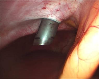
Endoscopic sphincterotomy is a medical procedure that involves the use of an endoscope (a flexible tube with a light and camera) to cut the papilla of Vater, which contains the sphincter of Oddi muscle. This procedure is typically performed to treat gallstones or to manage other conditions related to the bile ducts or pancreatic ducts.
The sphincterotomy helps to widen the opening of the papilla, allowing stones or other obstructions to pass through more easily. It may also be used to relieve pressure and pain caused by spasms of the sphincter of Oddi muscle. The procedure is usually done under sedation or anesthesia and carries a risk of complications such as bleeding, infection, perforation, and pancreatitis.
 Postcholecystectomy syndrome
 Postcholecystectomy Syndrome: Practice Essentials, Pathophysiology and Etiology, Epidemiology
 Postcholecystectomy Syndrome - Hepatic and Biliary Disorders - MSD Manual Professional Edition
 Post-cholecystectomy Syndrome: Causes, Symptoms And Treatment
 Wikizero - <span class="mw-page-title-main">Postcholecystectomy syndrome...
 Open Research: Mucocele of the gall bladder stump: a cause of post-cholecystectomy syndrome
 The most frequent symptoms of postcholecystectomy syndrome for cholelithiasis patients older than 40 years of age
 Relation of coffee, green tea, and caffeine intake to gallstone disease in middle-aged Japanese men
 Biliary Tract Disorders, Gallbladder Disorders, & Gallstone Pancreatitis | ACG
 Edward C. McCarron, MD| Surgical Oncology | MedStar Health
 Porcelain gallbladder: Symptoms, causes, and more
 An Approach to Gastrointestinal Bleeding | PPT
 urofacial syndrome - Ontology Browser - Rat Genome Database
 Bile: Your New BFF | Digestion | Articles | Magazine
 Food, fibre, bile acids and the pelvic floor: An integrated low risk low cost approach to managing irritable bowel syndrome
 Buy ➤Bilinorm tea for 4,84 $ in the shop rusmedicines
 Gallstones: Should I Have Gallbladder Surgery? | The Children's Hospital at Montefiore
 Cholelithiasis