Pycnodysostosis
Cathepsin K
Bone Diseases, Developmental
Anemia, Myelophthisic
Maxillofacial Abnormalities
Cranial Fontanelles
Cathepsins
Dwarfism
Chromosomes, Human, Pair 1
Pyknodysostosis: oral findings and differential diagnosis. (1/3)
Pyknodysostosis is a rare, genetic, autosomal recessive condition characterized by short stature, generalized bone sclerosis, and oral manifestations such as maxillary atresia and an increase of the mandibular angle. The main purpose of this article was to report a case of pyknodysostosis, describing the characteristic orofacial findings of the disease and discussing the differential diagnosis. (+info)Clinical and animal research findings in pycnodysostosis and gene mutations of cathepsin K from 1996 to 2011. (2/3)
(+info)Ichthyosis vulgaris and pycnodysostosis: an unusual occurrence. (3/3)
(+info)Pycnodysostosis is a rare genetic disorder characterized by skeletal dysplasia (abnormal development of the bones) and distinctive facial features. The condition is caused by mutations in the CTSK gene, which provides instructions for making an enzyme called cathepsin K. This enzyme is responsible for breaking down collagen, a protein that provides structure and strength to connective tissues throughout the body.
In people with pycnodysostosis, the lack of functional cathepsin K leads to the accumulation of abnormal bone matrix, which results in bones that are dense but fragile and prone to fractures. The condition is also associated with a number of other skeletal abnormalities, including:
* Short stature
* A prominent forehead (frontal bossing)
* A broad, flat nasal bridge
* A small chin (micrognathia)
* A narrow mouth
* A high-arched palate
* Dental abnormalities, such as delayed tooth eruption and thickened dental enamel
* Hypoplastic or aplastic clavicles (collarbones)
* Short fingers and toes
* Multiple fractures, particularly in the long bones of the arms and legs
Pycnodysostosis is typically diagnosed in childhood based on clinical features and confirmed with genetic testing. There is no cure for the condition, but treatment is focused on managing symptoms and preventing complications. This may include:
* Orthopedic interventions to correct skeletal abnormalities or treat fractures
* Dental care to address dental abnormalities and prevent tooth decay
* Speech therapy to help with any speech difficulties caused by the narrow mouth and high-arched palate
* Genetic counseling for affected individuals and their families.
Dysostosis is a term used to describe a group of genetic disorders that are characterized by abnormal development and formation of one or more bones in the body. The condition is typically present at birth (congenital) and can affect any bone, but it most commonly involves the bones of the skull, face, hands, and feet.
The term "dysostosis" comes from the Greek words "dys," meaning difficult or abnormal, and "osteon," meaning bone. Dysostoses are usually caused by mutations in specific genes that regulate bone development. These genetic changes can be inherited from one or both parents or can occur spontaneously during fetal development.
There are many different types of dysostoses, each with its own set of symptoms and characteristics. Some common examples include:
1. Cleidocranial Dysplasia: This is a rare genetic disorder that affects the development of the skull and collarbones (cleido). People with cleidocranial dysplasia may have a larger than normal head, wide-set eyes, a prominent forehead, and underdeveloped or missing collarbones.
2. Acrocephalopolysyndactyly Type II: Also known as ACPS II or Greig cephalopolysyndactyly syndrome, this disorder is characterized by a pointed skull (acrocephaly), extra fingers and toes (polydactyly), and wide-set eyes.
3. Osteogenesis Imperfecta: This is a group of genetic disorders that affect the body's production of collagen, a protein that helps to strengthen bones. People with osteogenesis imperfecta have fragile bones that break easily, often as a result of minor trauma.
4. Diastrophic Dysplasia: This is a rare genetic disorder that affects the development of the bones and cartilage in the body. People with diastrophic dysplasia may have short limbs, a deformed spine, and a characteristic "hitchhiker's thumb" appearance.
5. Thanatophoric Dysplasia: This is a severe genetic disorder that affects the development of the bones in the body. People with thanatophoric dysplasia have very short limbs, a small chest, and a deformed skull. The condition is often fatal in infancy or early childhood.
These are just a few examples of the many different types of skeletal dysplasias that exist. While some forms of these disorders can be managed with medical treatment and therapy, others may require surgery or other interventions to help improve quality of life. In some cases, genetic counseling and testing may be recommended for individuals who are considering starting a family and have a history of skeletal dysplasia in their family.
Cathepsin K is a proteolytic enzyme, which belongs to the family of papain-like cysteine proteases. It is primarily produced by osteoclasts, which are specialized cells responsible for bone resorption. Cathepsin K plays a crucial role in the degradation and remodeling of the extracellular matrix, particularly in bone tissue.
This enzyme is capable of breaking down various proteins, including collagen, elastin, and proteoglycans, which are major components of the bone matrix. By doing so, cathepsin K helps osteoclasts to dissolve and remove mineralized and non-mineralized bone matrix during the process of bone resorption.
Apart from its function in bone metabolism, cathepsin K has also been implicated in several pathological conditions, such as osteoporosis, rheumatoid arthritis, and tumor metastasis to bones. Inhibitors of cathepsin K are being investigated as potential therapeutic agents for the treatment of these disorders.
Developmental bone diseases are a group of medical conditions that affect the growth and development of bones. These diseases are present at birth or develop during childhood and adolescence, when bones are growing rapidly. They can result from genetic mutations, hormonal imbalances, or environmental factors such as poor nutrition.
Some examples of developmental bone diseases include:
1. Osteogenesis imperfecta (OI): Also known as brittle bone disease, OI is a genetic disorder that affects the body's production of collagen, a protein necessary for healthy bones. People with OI have fragile bones that break easily and may also experience other symptoms such as blue sclerae (whites of the eyes), hearing loss, and joint laxity.
2. Achondroplasia: This is the most common form of dwarfism, caused by a genetic mutation that affects bone growth. People with achondroplasia have short limbs and a large head relative to their body size.
3. Rickets: A condition caused by vitamin D deficiency or an inability to absorb or use vitamin D properly. This leads to weak, soft bones that can bow or bend easily, particularly in children.
4. Fibrous dysplasia: A rare bone disorder where normal bone is replaced with fibrous tissue, leading to weakened bones and deformities.
5. Scoliosis: An abnormal curvature of the spine that can develop during childhood or adolescence. While not strictly a developmental bone disease, scoliosis can be caused by various underlying conditions such as cerebral palsy, muscular dystrophy, or spina bifida.
Treatment for developmental bone diseases varies depending on the specific condition and its severity. Treatment may include medication, physical therapy, bracing, or surgery to correct deformities and improve function. Regular follow-up with a healthcare provider is essential to monitor growth, manage symptoms, and prevent complications.
Myelophthisic anemia is a type of anemia that occurs when the bone marrow becomes replaced or damaged by fibrosis, tumor infiltration, or other disorders, leading to decreased production of blood cells. This results in a decrease in all three types of blood cells - red blood cells, white blood cells, and platelets.
The symptoms of myelophthisic anemia may include fatigue, weakness, shortness of breath, frequent infections, and easy bruising or bleeding. The diagnosis is typically made through a combination of medical history, physical examination, complete blood count (CBC), and bone marrow aspiration and biopsy. Treatment for myelophthisic anemia depends on the underlying cause and may include chemotherapy, radiation therapy, surgery, or supportive care with transfusions of red blood cells or platelets.
Maxillofacial abnormalities, also known as craniofacial anomalies, refer to a broad range of structural and functional disorders that affect the development of the skull, face, jaws, and related soft tissues. These abnormalities can result from genetic factors, environmental influences, or a combination of both. They can vary in severity, from minor cosmetic issues to significant impairments of vital functions such as breathing, speaking, and eating.
Examples of maxillofacial abnormalities include cleft lip and palate, craniosynostosis (premature fusion of the skull bones), hemifacial microsomia (underdevelopment of one side of the face), and various other congenital anomalies. These conditions may require multidisciplinary treatment involving surgeons, orthodontists, speech therapists, and other healthcare professionals to address both functional and aesthetic concerns.
Cranial fontanelles, also known as "soft spots," are the membrane-covered spaces between the bones of a newborn or infant's skull. There are six fontanelles in total: two anterior (frontal) fontanelles, two posterior (occipital) fontanelles, and two smaller sphenoid and mastoid fontanelles.
The anterior fontanelle is the most prominent and is located towards the front of the head. It typically measures about 1 to 2 inches in diameter at birth and closes by around 18-24 months of age as the bones of the skull grow together. The posterior fontanelle is smaller, located towards the back of the head, and usually closes by around 2 months of age.
The fontanelles allow for the baby's brain to grow rapidly during the first few months of life, and they also provide some flexibility during childbirth, allowing the skull bones to overlap and make it easier for the baby to pass through the birth canal. It is important to handle a newborn gently, especially around the fontanelles, as they are still developing and can be injured easily.
Cathepsins are a type of proteolytic enzymes, which are found in lysosomes and are responsible for breaking down proteins inside the cell. They are classified as papain-like cysteine proteases and play important roles in various physiological processes, including tissue remodeling, antigen presentation, and apoptosis (programmed cell death). There are several different types of cathepsins, including cathepsin B, C, D, F, H, K, L, S, V, and X/Z, each with distinct substrate specificities and functions.
Dysregulation of cathepsins has been implicated in various pathological conditions, such as cancer, neurodegenerative diseases, and inflammatory disorders. For example, overexpression or hyperactivation of certain cathepsins has been shown to contribute to tumor invasion and metastasis, while their inhibition has been explored as a potential therapeutic strategy in cancer treatment. Similarly, abnormal levels of cathepsins have been linked to the progression of neurodegenerative diseases like Alzheimer's and Parkinson's, making them attractive targets for drug development.
Dwarfism is a medical condition that is characterized by short stature, typically with an adult height of 4 feet 10 inches (147 centimeters) or less. It is caused by a variety of genetic and medical conditions that affect bone growth, including skeletal dysplasias, hormonal deficiencies, and chromosomal abnormalities.
Skeletal dysplasias are the most common cause of dwarfism and are characterized by abnormalities in the development and growth of bones and cartilage. Achondroplasia is the most common form of skeletal dysplasia, accounting for about 70% of all cases of dwarfism. It is caused by a mutation in the fibroblast growth factor receptor 3 (FGFR3) gene and results in short limbs, a large head, and a prominent forehead.
Hormonal deficiencies, such as growth hormone deficiency or hypothyroidism, can also cause dwarfism if they are not diagnosed and treated early. Chromosomal abnormalities, such as Turner syndrome (monosomy X) or Down syndrome (trisomy 21), can also result in short stature and other features of dwarfism.
It is important to note that people with dwarfism are not "dwarves" - the term "dwarf" is a medical and sociological term used to describe individuals with this condition, while "dwarves" is a term often used in fantasy literature and media to refer to mythical beings. The use of the term "dwarf" can be considered disrespectful or offensive to some people with dwarfism, so it is important to use respectful language when referring to individuals with this condition.
Human chromosome pair 1 refers to the first pair of chromosomes in a set of 23 pairs found in the cells of the human body, excluding sex cells (sperm and eggs). Each cell in the human body, except for the gametes, contains 46 chromosomes arranged in 23 pairs. These chromosomes are rod-shaped structures that contain genetic information in the form of DNA.
Chromosome pair 1 is the largest pair, making up about 8% of the total DNA in a cell. Each chromosome in the pair consists of two arms - a shorter p arm and a longer q arm - connected at a centromere. Chromosome 1 carries an estimated 2,000-2,500 genes, which are segments of DNA that contain instructions for making proteins or regulating gene expression.
Defects or mutations in the genes located on chromosome 1 can lead to various genetic disorders and diseases, such as Charcot-Marie-Tooth disease type 1A, Huntington's disease, and certain types of cancer.
Consanguinity is a medical and genetic term that refers to the degree of genetic relationship between two individuals who share common ancestors. Consanguineous relationships exist when people are related by blood, through a common ancestor or siblings who have children together. The closer the relationship between the two individuals, the higher the degree of consanguinity.
The degree of consanguinity is typically expressed as a percentage or fraction, with higher values indicating a closer genetic relationship. For example, first-degree relatives, such as parents and children or full siblings, share approximately 50% of their genes and have a consanguinity coefficient of 0.25 (or 25%).
Consanguinity can increase the risk of certain genetic disorders and birth defects in offspring due to the increased likelihood of sharing harmful recessive genes. The risks depend on the degree of consanguinity, with closer relationships carrying higher risks. It is important for individuals who are planning to have children and have a history of consanguinity to consider genetic counseling and testing to assess their risk of passing on genetic disorders.
 Pycnodysostosis
List of OMIM disorder codes
Cathepsin K
Melorheostosis
Osteoclast
Henri de Toulouse-Lautrec
Wormian bones
Lysosomal storage disease
List of diseases (P)
Racquet nail
Index of trauma and orthopaedics articles
Pycnodysostosis - Wikipedia
 Een subtrochantaire femurfractuur bij een patiënte met pycnodysostosis | mijn-bsl
 Etiology
 Expanded Carrier Screening | Thermo Fisher Scientific - US
Hygeia as Muse - Volume 14, Number 3-March 2008 - Emerging Infectious Diseases journal - CDC
Homepage | BMJ Case Reports
 UKE - Physician - Martin Horstmann
 Flashes of Fiction - 365tomorrows
 Baby’s Pregnancy Calendar
 urofacial syndrome - Ontology Browser - Rat Genome Database
Specific PHGKB|Rare Diseases PHGKB|PHGKB
 Results for cd02621
 QHerit® carrier screening
 Vol 6, Issue 2 - Page 5 - Journal of Natural Science, Biology and Medicine
 NIH/Osteoporosis and Related Bone Diseases National Resource Center - National Organization for Rare Disorders
 Journal of Pediatric Sciences » Cilt: 4 Sayı: 1
 Picnodisostose
 Krabbe disease | Radiology Reference Article | Radiopaedia.org
Fucosidosis | Radiology Reference Article | Radiopaedia.org
 Factors that Affect the Osteoclastogenesis of RAW264.7 Cells
 Rat CTSK(Cathepsin K) ELISA Kit - SR Biosystem
 online second opinion - Surgery Second Opinion
Lysosomal Storage Diseases | Profiles RNS
March | 2016 | Dna-Pk Inhibitors
 9 Of The Biggest Changes 'Outlander' Made From The Books
 Thesis research methodology º NCC Dept. of MAT/CSC/ITE
 Newest 'pathology' Questions - Psychology & Neuroscience Stack Exchange
 Update on ectodermal dysplasias clinical classification. - PDF Download Free
 Eye Diseases, Hereditary (medical concept explorer)
Patient with pycnodysostosis4
- Kundu ZS, Marya KM, Devgan A, Yadav V, Rohilla S. Subtrochanteric fracture managed by intramedullary nail in a patient with pycnodysostosis. (bsl.nl)
- Yuasa T, Maeda K, Kaneko K, Yoshikata K. Total Hip Arthroplasty after treatment of an atypical subtrochanteric femoral fracture in a patient with pycnodysostosis. (bsl.nl)
- Atypical femur fractures in a patient with pycnodysostosis: a case report. (bsl.nl)
- We had a case of a middle childhood female patient with pycnodysostosis and a femur fracture. (bvsalud.org)
Patients with pycnodysostosis2
- Though elastic nailing is preferred for paediatric long bone fractures, surgeons must be prepared for extremely sclerotic cortices and a narrow medullary canal when dealing with patients with pycnodysostosis. (bvsalud.org)
- RESULTS: We describe first clinical and genetic characteristics of three Russian patients with pycnodysostosis from unrelated families. (bvsalud.org)
Possibly pycnodysostosis1
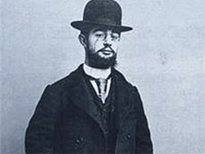
- Modern physicians attribute this to an unknown genetic disorder, possibly pycnodysostosis (sometimes known as Toulouse-Lautrec Syndrome), or a variant disorder along the lines of osteopetrosis, achondroplasia, or osteogenesis imperfecta. (artvee.com)
Toulouse-Lautrec Syndrome1
- In both the show and books, Colum suffers from Pycnodysostosis (Toulouse-Lautrec syndrome) and comes to Claire for some permanent relief for the pain. (thedipp.com)
Short stature2
- Author: Majoki "You are suffering from all the hallmarks of pycnodysostosis: fragile bones, short stature, large head, weak chin. (365tomorrows.com)
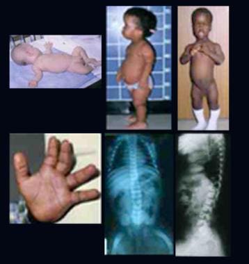
- Pycnodysostosis is characterized by short-limbed short stature, typical facial appearance (convex nasal ridge and small jaw with obtuse mandibular angle), osteosclerosis with increased bone fragility, acroosteolysis of the distal phalanges, delayed closure of the cranial sutures, and dysplasia of the clavicle. (nih.gov)
Fractures2
Fracture4
- Bor N, Rubin G, Rozen N. Fracture management in pycnodysostosis: 27 years of follow-up. (bsl.nl)
- Pycnodysostosis is a rare genetic condition that leads to generalised bony sclerosis and increased fracture risk. (bvsalud.org)
- CASE PRESENTATION: We present the case of a 4-year-old girl diagnosed with pycnodysostosis and associated pathological tibial fracture. (bvsalud.org)
- CONCLUSION: The presentation of pycnodysostosis as tibial fracture is rare and there is limited literature on its management. (bvsalud.org)
Genetic1
- Due to the limited number of exons of the CTSK gene that causes pycnodysostosis, a cheaper genetic testing called Sanger sequencing can be employed to confirm the diagnosis. (wikipedia.org)
Clinical1
- The polymorphism of the clinical manifestations of pycnodysostosis and low prevalence of the disorder lead to the difficulties with early. (bvsalud.org)
Typical facial1
- Pycnodysostosis is one of those disorders which has a typical facial gestalt and can be clinically identified in the majority of cases. (wikipedia.org)
Disorder2
- The molecular basis of pycnodysostosis was elucidated in 1996 by Gelb and collaborators and the disorder results from biallelic pathogenic mutation in CTSK gene (OMIM * 601105). (wikipedia.org)
- BACKGROUND: Pycnodysostosis is a rare autosomal recessive lysosomal disorder of bone characterized by diffuse skeletal condensation with thickening of the cortex and narrowing of the medullary canal. (bvsalud.org)
Disease3
- Pycnodysostosis (from Greek: πυκνός (puknos) meaning "dense", dys ("defective"), and ostosis ("condition of the bone")), is a lysosomal storage disease of the bone caused by a mutation in the gene that codes the enzyme cathepsin K. It is also known as PKND and PYCD. (wikipedia.org)
- Pycnodysostosis is a rare autosomal recessive disease with osteoarticular manifestations of great relevance in anesthetic practice. (bvsalud.org)
- Pycnodysostosis is a rare disease with very few reported cased all over the world. (bvsalud.org)
Variant1
- BACKGROUND: Pycnodysostosis (PD, OMIM # 265800) is a rare variant of skeletal dysplasia with an autosomal recessive type of inheritance, characterized by a combination of specific features such as disproportionate nanism, generalized osteosclerosis, and distinct craniofacial dysmorphism. (bvsalud.org)
Management1
- The treatment of pycnodysostosis is currently based on symptomatic management and no active trials are in place for a curative approach. (wikipedia.org)
Features1
- Cathepsin K analysis in a pycnodysostosis cohort: demographic, genotypic and phenotypic features. (bsl.nl)
Female patient with pycnodysostosis2
- Led by Professor Yrjö T. Konttinen of Helsinki University Central Hospital in Helskinki, Finland , the study involved a 55-year-old female patient with pycnodysostosis who also developed psoriatic arthritis. (medscape.com)
- We had a case of a middle childhood female patient with pycnodysostosis and a femur fracture. (bvsalud.org)
Congenital1
- Pycnodysostosis and cardiac congenital defects: a rare association. (escardio.org)
Bone disorder1
- Pycnodysostosis is a rare recessive autosomal bone disorder. (jomos.org)
Clinical3
- Pycnodysostosis should be suspected in probands with the following clinical, radiographic, and laboratory findings. (nih.gov)
- Two clinical cases of pycnodysostosis with typical maxillofacial complications are presented. (jomos.org)
- Çapan E, Turan S, Kılıçoğlu H. Clinical And Cephalometric Analysis Of Three Cases With Pycnodysostosis (Case Report). (lingualortodontidernegi.org.tr)
CTSK1
- The molecular basis of pycnodysostosis was elucidated in 1996 by Gelb and collaborators and the disorder results from biallelic pathogenic mutation in CTSK gene (OMIM * 601105). (wikipedia.org)
Osteosclerosis1
- Formal diagnostic criteria for pycnodysostosis have not been established, however the radiographic features of acroosteolysis, osteosclerosis, and loss of the normal angle of the jaw are almost pathognomonic. (nih.gov)
Rare1
- Born into the aristocracy , Toulouse-Lautrec broke both his legs around the time of his adolescence and, due to the rare condition pycnodysostosis , was very short as an adult due to his undersized legs. (sartle.com)
Bones1
- She died after a few hours and histopathological studies identified extramedullary haematopoiesis in the liver, little lamellar bone formation, decreased osteoclasts, abnormally thickened bony trabeculae with retained cartilage in long bones, and diminished marrow spaces similar to those seen in dense bone diseases such as osteopetrosis and pycnodysostosis. (bmj.com)
Disorders1
- Pycnodysostosis is one of those disorders which has a typical facial gestalt and can be clinically identified in the majority of cases. (wikipedia.org)
Case Report1
- 34. A Case Report of Pycnodysostosis Associated with Multiple Pituitary Hormone Deficiencies and Response to Treatment. (nih.gov)
Patient1
- Cite this: Erosive Arthritis in a Patient With Pycnodysostosis - Medscape - Nov 01, 2008. (medscape.com)
Child1
- 37. Novel mutation and white matter involvement in an Indian child with pycnodysostosis. (nih.gov)
Time1
- Pycnodysostosis also causes problems that may become evident with time. (wikipedia.org)