Shaken Baby Syndrome
Eye Infections, Parasitic
Papilledema
Child Abuse
Cerebral Hemorrhage
Fundus Oculi
Pneumoencephalography
Neuroleptanalgesia
Subarachnoid Hemorrhage
Retinal Diseases
Battered Child Syndrome
Craniocerebral Trauma
Eye Injuries
Fluorescein Angiography
Intracranial Hemorrhages
Philadelphia
Vitreous Body
Postpartum Hemorrhage
Gastrointestinal Hemorrhage
Ophthalmoscopic abnormalities in adults with falciparum malaria. (1/232)
We studied 424 adults with falciparum malaria admitted over 28 months. They were divided into three groups: cerebral malaria (n = 214); severe non-cerebral malaria (n = 58); and uncomplicated malaria (n = 152). Fundus examination was done daily from admission to discharge, and weekly thereafter in those with persistent changes. All patients were treated by a protocol based on WHO guidelines. Ophthalmoscopic abnormalities were: retinal haemorrhages, 40 (9.43%) (25 cerebral malaria, 10 severe non-cerebral and five uncomplicated malaria); papilloedema, 17 (7.94%) cerebral malaria and two uncomplicated malaria; blurring of disc margins, 25 (11.68%) cerebral and seven non-cerebral; retinal oedema, six (2.80%) cerebral and five non-cerebral malaria; disc pallor, five patients all with cerebral malaria; vitreous haemorrhage and hard exudate in one patient each, both cerebral malaria. Retinal haemorrhage was associated with cerebral malaria and severe non-cerebral malaria, especially with severe anaemia (p < 0.001), as compared to uncomplicated malaria (p < 0.01). The association of papilloedema and cerebral malaria was highly significant compared to severe non-cerebral malaria (p < 0.001). None of these findings was associated with statistically significant mortality, except disc pallor in cerebral malaria (p < 0.05). (+info)Perifoveal vascular leakage and macular oedema after intracapsular cataract extraction. (2/232)
Perifoveal capillary leakage of fluorescein was demonstrated in 60 per cent of 50 eyes when angiography was performed two weeks after cataract extraction. Repeat angiography six weeks postoperatively in 17 eyes demonstrated persistence of already established leakage in 11 of 12 eyes and no new leakage in five eyes previously negative. Cystoid macular oedema with visual acuity of less than 20/40 six weeks postoperatively occurred in five eyes (10 per cent). Eyes of patients with vascular disease and those patients of 60 years or older were found to have altered vascular permeability significantly more frequently. Inflammation was no more severe or prevalent in those patients who demonstrated leakage and no inflammation was clinically apparent in 10 of 11 eyes demonstrating dye leakage six weeks postoperatively. We conclude that the constitutional factors of age and vascular disease are of prime importance in causing altered vascular permeability in the early postoperative period after cataract extraction; factors causing sustained leakage with reduction of visual acuity were not demonstrated. (+info)White-centred retinal haemorrhages (Roth spots). (3/232)
Roth spots (white-centred retinal haemorrhages) were classically described as septic emboli lodged in the retina of patients with subacute bacterial endocarditis. Indeed many have considered Roth spots pathognomonic for this condition. More recent histological evidence suggests, however, that they are not foci of bacterial abscess. Instead, they are nonspecific and may be found in many other diseases. A review of the histology and the pathogenesis of these white-centred haemorrhages will be provided, along with the work-up of the differential diagnosis. (+info)New animal model for human ocular toxocariasis: ophthalmoscopic observation. (4/232)
BACKGROUND/AIMS: Although human ocular toxocariasis causes severe vision defect, little is known about its aetiology, diagnosis, and treatment. To develop a new animal model for human ocular toxocariasis, ophthalmological findings of fundi in Mongolian gerbils, Meriones unguiculatus, and BALB/c mice were investigated following infection with Toxocara canis. METHODS: Using an ophthalmoscope, which was specifically developed to observe the fundi of small animals, ocular changes of fundi of 20 gerbils and 11 mice were monitored after oral infection with embryonated eggs of T canis. RESULTS: Vitreous, choroidal, and retinal haemorrhages were consistently observed in Mongolian gerbils, but rarely in mice. Severe exudative lesions and vasculitis were often present in gerbils but not in mice. Migrating larvae were also frequently observed in gerbils. CONCLUSION: Mongolian gerbils are more appropriate animal model for human ocular toxocariasis than previously used experimental animal such as mice, guinea pigs, rabbits, and monkeys because of its high susceptibility of ocular infection. (+info)Air bags and ocular injuries. (5/232)
PURPOSE: This investigation retrospectively examined ocular injuries associated with air bag deployment to gain a better appreciation of potential risk factors in motor vehicle accidents. National statistics regarding the efficacy of air bags were reviewed. METHODS: Review of the literature from 1991 to 1998 identified 44 articles describing 97 patients with air-bag-induced ocular injuries. Variables extracted from each case were age, sex, height, position in the car, eye wear, vehicle impact speed, visual acuity, and specific ocular injuries. RESULTS: Corneal abrasions occurred in 49% of occupants, hyphemas in 43%, vitreous or retinal hemorrhages in 25%, and retinal tears or detachments in 15%. The globe was ruptured in 10 patients. Patients involved in higher-speed accidents (over 30 mph) sustained a greater percentage of vitreous or retinal hemorrhages and traumatic cataracts, while those at slower speeds were more prone to retinal tears or detachments. In a subset of 14 patients with serious ocular injuries, the impact speed of 11 patients was recorded at 30 mph or less. Slower speed may be a risk factor for some ocular injuries. Occupant height was not a significant factor. National statistics confirm that air bags reduce fatalities in motor vehicle accidents. However, children sitting in the front seat without a seat belt and infants in passenger-side rear-facing car seats are at risk for fatal injury. CONCLUSION: Air bags combined with seat belts are an effective means of reducing injury and death in adults during motor vehicle accidents. However, this study has documented a wide variety of ocular injuries associated with air bag deployment. It is hoped that researchers can develop modifications that continue to save lives while minimizing additional harm. (+info)A 12-year ophthalmologic experience with the shaken baby syndrome at a regional children's hospital. (6/232)
PURPOSE: To examine the ophthalmologic experience with the shaken baby syndrome (SBS) at one medical center, including clinical findings, autopsy findings, and the visual outcome of survivors. METHODS: One hundred sixteen patients admitted from 1987 to 1998 for subdural hematomas of the brain secondary to abuse were included. RESULTS: Retinal hemorrhages were detected in 84% of the children, but this important finding had been missed often by nonophthalmologists. Poor visual response, poor pupillary response, and retinal hemorrhage correlated strongly with demise of the child. One child who died had pigmented retinal scars from previous abuse, a condition not previously observed histopathologically. The clinical and autopsy findings varied somewhat, probably because of the differing conditions for examination. No correlation could be made between computerized tomography scans done during life and the subdural hemorrhage of the optic nerve found on autopsy. Half of the surviving patients were known to have good vision. One fourth of the patients had poor vision, largely due to cerebral visual impairment from bilateral injury posterior to the optic chiasm. Severe neurologic impairment correlated highly with loss of vision. CONCLUSION: This series provides information on the frequency of eye findings in SBS patients. No fundus finding is pathognomonic for SBS. When retinal hemorrhages are found in young children, the likelihood that abuse occurred is very high. The difficulty that nonophthalmologists have in detecting retinal hemorrhage may be an important limiting factor in finding these children so they may be protected from further abuse. (+info)Polymerase chain reaction for detection of Mycobacterium tuberculosis in epiretinal membrane in Eales' disease. (7/232)
PURPOSE: Tuberculous etiology has been suggested in Eales' disease. Because epiretinal membrane (ERM) is formed on the inner surface of the retina in Eales' disease, it could be the most appropriate intraocular specimen for investigation. Therefore, a nested polymerase chain reaction (nPCR), which detects MPB64 gene of Mycobacterium tuberculosis on the archival specimens of ERM of well-documented Eales' and non-Eales' patients, was applied and the results compared. METHODS: nPCR technique was standardized, and the sensitivity and specificity of the primers were determined. nPCR technique was applied to tissue sections obtained from formalin-fixed and paraffin-embedded tissues of ERM from 23 patients with Eales' disease and 27 noninfective and non-Eales' disease patients as controls. RESULTS: nPCR technique was specific for M. tuberculosis genome and sensitive enough to detect 0.25 fg (corresponding to the presence of a single bacillus). Eleven (47.8%) ERM of 23 Eales' disease and 3 (11.1%) of 27 controls were positive for M. tuberculosis genome. The difference between the two groups was statistically significant (P = 0.001), indicating association of this bacterium with Eales' disease. CONCLUSIONS: The demonstration of the presence of M. tuberculosis DNA by nPCR technique in significant number of ERM of Eales' disease compared with the controls further emphasizes the probable role of this bacterium in the pathogenesis of this enigmatic clinical condition. (+info)Domestic violence: the shaken adult syndrome. (8/232)
A case of domestic violence is reported. The patient presented with the triad of injuries associated with the shaking of infants: retinal haemorrhages, subdural haematoma, and patterned bruising; this has been described as the shaken adult syndrome. This case report reflects the difficulties in diagnosing domestic violence in the accident and emergency setting. (+info)A retinal hemorrhage is a type of bleeding that occurs in the blood vessels of the retina, which is the light-sensitive tissue located at the back of the eye. This condition can result from various underlying causes, including diabetes, high blood pressure, age-related macular degeneration, or trauma to the eye. Retinal hemorrhages can be categorized into different types based on their location and appearance, such as dot and blot hemorrhages, flame-shaped hemorrhages, or subhyaloid hemorrhages. Depending on the severity and cause of the hemorrhage, treatment options may vary from monitoring to laser therapy, medication, or even surgery. It is essential to consult an ophthalmologist for a proper evaluation and management plan if you suspect a retinal hemorrhage.
Shaken Baby Syndrome (SBS), also known as Abusive Head Trauma, is a form of inflicted injury that occurs when a baby or young child is violently shaken. This can lead to severe brain damage, blindness, hearing loss, developmental delays, seizures, and even death. The shaking causes the baby's fragile brain to move back and forth inside the skull, resulting in bruised brain tissues, bleeding in the brain, and detachment of the retinas. It's important to note that even brief periods of shaking can result in severe consequences. SBS is a form of child abuse and should be reported immediately to authorities.
A Vitreous Hemorrhage is a medical condition where there is bleeding into the vitreous cavity of the eye. The vitreous cavity is the space in the eye that is filled with a clear, gel-like substance called the vitreous humor. This substance helps to maintain the shape of the eye and transmit light to the retina.
When a vitreous hemorrhage occurs, blood cells from the bleeding mix with the vitreous humor, causing it to become cloudy or hazy. As a result, vision can become significantly impaired, ranging from mildly blurry to complete loss of vision depending on the severity of the bleed.
Vitreous hemorrhages can occur due to various reasons such as trauma, retinal tears or detachments, diabetic retinopathy, age-related macular degeneration, and other eye conditions that affect the blood vessels in the eye. Treatment for vitreous hemorrhage depends on the underlying cause and may include observation, laser surgery, or vitrectomy (a surgical procedure to remove the vitreous humor and stop the bleeding).
Parasitic eye infections are conditions characterized by the invasion and infestation of the eye or its surrounding structures by parasites. These can be protozoans, helminths, or ectoparasites. Examples of such infections include Acanthamoeba keratitis, which is caused by a free-living amoeba found in water and soil; Toxoplasmosis, which is caused by the protozoan Toxoplasma gondii; Loiasis, which is caused by the parasitic filarial worm Loa loa; and Demodicosis, which is caused by the mite Demodex folliculorum. Symptoms can vary depending on the type of parasite but often include redness, pain, discharge, and vision changes. Treatment typically involves antiparasitic medications and sometimes surgery to remove the parasites or damaged tissue. Prevention measures include good hygiene practices and avoiding contact with contaminated water or soil.
Papilledema is a medical term that refers to swelling of the optic nerve head, also known as the disc, which is the point where the optic nerve enters the back of the eye (the retina). This swelling can be caused by increased pressure within the skull, such as from brain tumors, meningitis, or idiopathic intracranial hypertension. Papilledema is usually detected through a routine eye examination and may be accompanied by symptoms such as headaches, visual disturbances, and nausea. If left untreated, papilledema can lead to permanent vision loss.
Hemorrhage is defined in the medical context as an excessive loss of blood from the circulatory system, which can occur due to various reasons such as injury, surgery, or underlying health conditions that affect blood clotting or the integrity of blood vessels. The bleeding may be internal, external, visible, or concealed, and it can vary in severity from minor to life-threatening, depending on the location and extent of the bleeding. Hemorrhage is a serious medical emergency that requires immediate attention and treatment to prevent further blood loss, organ damage, and potential death.
Child abuse is a broad term that refers to any form of physical, emotional, or sexual mistreatment or neglect that causes harm to a child's health, development, or dignity. According to the World Health Organization (WHO), child abuse includes:
1. Physical abuse: Non-accidental injuries caused by hitting, kicking, shaking, burning, or otherwise harming a child's body.
2. Sexual abuse: Any sexual activity involving a child, such as touching or non-touching behaviors, exploitation, or exposure to pornographic material.
3. Emotional abuse: Behaviors that harm a child's emotional well-being and self-esteem, such as constant criticism, humiliation, threats, or rejection.
4. Neglect: Failure to provide for a child's basic needs, including food, clothing, shelter, medical care, education, and emotional support.
Child abuse can have serious short-term and long-term consequences for the physical, emotional, and psychological well-being of children. It is a violation of their fundamental human rights and a public health concern that requires prevention, early detection, and intervention.
A cerebral hemorrhage, also known as an intracranial hemorrhage or intracerebral hemorrhage, is a type of stroke that results from bleeding within the brain tissue. It occurs when a weakened blood vessel bursts and causes localized bleeding in the brain. This bleeding can increase pressure in the skull, damage nearby brain cells, and release toxic substances that further harm brain tissues.
Cerebral hemorrhages are often caused by chronic conditions like hypertension (high blood pressure) or cerebral amyloid angiopathy, which weakens the walls of blood vessels over time. Other potential causes include trauma, aneurysms, arteriovenous malformations, illicit drug use, and brain tumors. Symptoms may include sudden headache, weakness, numbness, difficulty speaking or understanding speech, vision problems, loss of balance, and altered level of consciousness. Immediate medical attention is required to diagnose and manage cerebral hemorrhage through imaging techniques, supportive care, and possible surgical interventions.
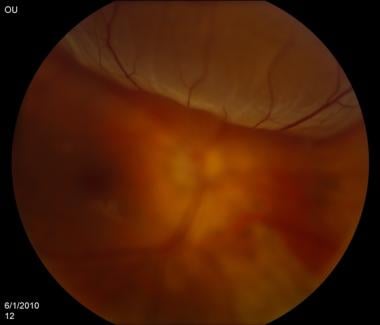
"Fundus Oculi" is a medical term that refers to the back part of the interior of the eye, including the optic disc, macula, fovea, retinal vasculature, and peripheral retina. It is the area where light is focused and then transmitted to the brain via the optic nerve, forming visual images. Examinations of the fundus oculi are crucial for detecting various eye conditions such as diabetic retinopathy, macular degeneration, glaucoma, and other retinal diseases. The examination is typically performed using an ophthalmoscope or a specialized camera called a retinal camera.
I'm sorry for any confusion, but "Pneumoradiography" is not a recognized term in the field of medicine or radiology. It seems that there might be a mix-up with the terminologies.
However, you may be referring to "Pneumonic Radiography," which means a chest X-ray used to diagnose pneumonia. Pneumonia is an infection or inflammation of the lung tissue, and a chest X-ray can help identify areas of increased density in the lungs that could indicate the presence of pneumonia.
If you meant something else, please provide more context or clarify your question, and I would be happy to help further.
I'm sorry for any confusion, but "mountaineering" is not a medical term. It is a term used to describe the sport or recreational activity of climbing mountains.
However, if we are talking about potential medical aspects related to mountaineering, it would be appropriate to discuss terms like "altitude sickness," "hypothermia," or "frostbite" which are conditions that a mountaineer might experience during their activities. But without further context, I can't provide a specific medical definition directly related to 'mountaineering'.
Pneumoencephalography is a diagnostic procedure that is rarely used today, due to the development of less invasive techniques. It involves the introduction of air or another gas into the ventricular system or subarachnoid space of the brain, followed by X-ray imaging to visualize the structures and any abnormalities within the intracranial cavity.
The primary purpose of this procedure was to diagnose conditions affecting the brain's ventricles, such as hydrocephalus, tumors, or inflammation. The introduction of air into the cranium allowed for better visualization of these structures and any potential abnormalities. However, due to its invasive nature, risks associated with the procedure, and the availability of non-invasive imaging techniques like CT and MRI scans, pneumoencephalography has fallen out of favor in modern medicine.
Neuroleptanalgesia is a clinical state produced by the combined use of a neuroleptic (a drug that dampens down the activity of the brain, leading to decreased awareness of one's surroundings and reduced ability to initiate movements) and an analgesic (a pain-relieving drug). This combination results in a state of dissociative analgesia, where the patient remains conscious but detached from their environment, with reduced perception of pain. It has been used in certain medical procedures as an alternative to general anesthesia.
The term 'neurolept' refers to drugs that have a pronounced effect on the nervous system, reducing psychomotor agitation and emotional reactivity. Examples of neuroleptic drugs include phenothiazines (such as chlorpromazine), butyrophenones (such as haloperidol), and diphenylbutylpiperidines (such as pimozide).
Analgesics, on the other hand, are medications that primarily target pain perception pathways in the nervous system. Common examples include opioids (such as morphine or fentanyl) and non-opioid analgesics (such as acetaminophen or ibuprofen).
The combination of neuroleptic and analgesic drugs is used to achieve a balance between pain relief, sedation, and preservation of the patient's ability to communicate and cooperate during medical procedures. However, due to potential side effects such as respiratory depression, neuroleptanalgesia requires careful monitoring and management by anesthesiologists or other trained medical professionals.
A subarachnoid hemorrhage is a type of stroke that results from bleeding into the space surrounding the brain, specifically within the subarachnoid space which contains cerebrospinal fluid (CSF). This space is located between the arachnoid membrane and the pia mater, two of the three layers that make up the meninges, the protective covering of the brain and spinal cord.
The bleeding typically originates from a ruptured aneurysm, a weakened area in the wall of a cerebral artery, or less commonly from arteriovenous malformations (AVMs) or head trauma. The sudden influx of blood into the CSF-filled space can cause increased intracranial pressure, irritation to the brain, and vasospasms, leading to further ischemia and potential additional neurological damage.
Symptoms of a subarachnoid hemorrhage may include sudden onset of severe headache (often described as "the worst headache of my life"), neck stiffness, altered mental status, nausea, vomiting, photophobia, and focal neurological deficits. Rapid diagnosis and treatment are crucial to prevent further complications and improve the chances of recovery.
Retinal diseases refer to a group of conditions that affect the retina, which is the light-sensitive tissue located at the back of the eye. The retina is responsible for converting light into electrical signals that are sent to the brain and interpreted as visual images. Retinal diseases can cause vision loss or even blindness, depending on their severity and location in the retina.
Some common retinal diseases include:
1. Age-related macular degeneration (AMD): A progressive disease that affects the central part of the retina called the macula, causing blurred or distorted vision.
2. Diabetic retinopathy: A complication of diabetes that can damage the blood vessels in the retina, leading to vision loss.
3. Retinal detachment: A serious condition where the retina becomes separated from its underlying tissue, requiring immediate medical attention.
4. Macular edema: Swelling or thickening of the macula due to fluid accumulation, which can cause blurred vision.
5. Retinitis pigmentosa: A group of inherited eye disorders that affect the retina's ability to respond to light, causing progressive vision loss.
6. Macular hole: A small break in the macula that can cause distorted or blurry vision.
7. Retinal vein occlusion: Blockage of the retinal veins that can lead to bleeding, swelling, and potential vision loss.
Treatment for retinal diseases varies depending on the specific condition and its severity. Some treatments include medication, laser therapy, surgery, or a combination of these options. Regular eye exams are essential for early detection and treatment of retinal diseases.
Battered Child Syndrome is a medical condition in which a child has been physically abused and harmed, often over a period of time. It is also known as Non-accidental Injury (NAI) or Inflicted Traumatic Injury. The syndrome is characterized by a pattern of injuries, including bruises, fractures, burns, and internal injuries, which are not consistent with the history provided by the caregiver.
The symptoms of Battered Child Syndrome may include:
1. Unexplained or inconsistent explanations for injuries
2. Multiple injuries in various stages of healing
3. Injuries to different parts of the body, such as the ears, mouth, and genitals
4. Frequent visits to the emergency department or doctor's office for treatment of injuries
5. Delayed seeking of medical attention for serious injuries
6. Behavioral changes, such as fearfulness, regression, or aggression
7. Developmental delays or learning difficulties
8. Failure to thrive (poor growth and weight gain)
The diagnosis of Battered Child Syndrome is made by a healthcare professional based on the history, physical examination, and any diagnostic tests that may be necessary. The syndrome is a serious form of child abuse that requires immediate intervention and protection for the child. Treatment typically involves medical care for injuries, counseling and support for the child and family, and reporting the abuse to child protective services or law enforcement agencies.
Craniocerebral trauma, also known as traumatic brain injury (TBI), is a type of injury that occurs to the head and brain. It can result from a variety of causes, including motor vehicle accidents, falls, sports injuries, violence, or other types of trauma. Craniocerebral trauma can range in severity from mild concussions to severe injuries that cause permanent disability or death.
The injury typically occurs when there is a sudden impact to the head, causing the brain to move within the skull and collide with the inside of the skull. This can result in bruising, bleeding, swelling, or tearing of brain tissue, as well as damage to blood vessels and nerves. In severe cases, the skull may be fractured or penetrated, leading to direct injury to the brain.
Symptoms of craniocerebral trauma can vary widely depending on the severity and location of the injury. They may include headache, dizziness, confusion, memory loss, difficulty speaking or understanding speech, changes in vision or hearing, weakness or numbness in the limbs, balance problems, and behavioral or emotional changes. In severe cases, the person may lose consciousness or fall into a coma.
Treatment for craniocerebral trauma depends on the severity of the injury. Mild injuries may be treated with rest, pain medication, and close monitoring, while more severe injuries may require surgery, intensive care, and rehabilitation. Prevention is key to reducing the incidence of craniocerebral trauma, including measures such as wearing seat belts and helmets, preventing falls, and avoiding violent situations.
Ophthalmoscopy is a medical examination technique used by healthcare professionals to observe the interior structures of the eye, including the retina, optic disc, and vitreous humor. This procedure typically involves using an ophthalmoscope, a handheld device that consists of a light and magnifying lenses. The healthcare provider looks through the ophthalmoscope and directly observes the internal structures of the eye by illuminating them.
There are several types of ophthalmoscopy, including direct ophthalmoscopy, indirect ophthalmoscopy, and slit-lamp biomicroscopy. Each type has its own advantages and disadvantages, and they may be used in different situations depending on the specific clinical situation and the information needed.
Ophthalmoscopy is an important diagnostic tool for detecting and monitoring a wide range of eye conditions, including diabetic retinopathy, glaucoma, age-related macular degeneration, and other retinal disorders. It can also provide valuable information about the overall health of the individual, as changes in the appearance of the retina or optic nerve may indicate the presence of systemic diseases such as hypertension or diabetes.
Eye injuries refer to any damage or trauma caused to the eye or its surrounding structures. These injuries can vary in severity and may include:
1. Corneal abrasions: A scratch or scrape on the clear surface of the eye (cornea).
2. Chemical burns: Occurs when chemicals come into contact with the eye, causing damage to the cornea and other structures.
3. Eyelid lacerations: Cuts or tears to the eyelid.
4. Subconjunctival hemorrhage: Bleeding under the conjunctiva, the clear membrane that covers the white part of the eye.
5. Hyphema: Accumulation of blood in the anterior chamber of the eye, which is the space between the cornea and iris.
6. Orbital fractures: Breaks in the bones surrounding the eye.
7. Retinal detachment: Separation of the retina from its underlying tissue, which can lead to vision loss if not treated promptly.
8. Traumatic uveitis: Inflammation of the uvea, the middle layer of the eye, caused by trauma.
9. Optic nerve damage: Damage to the optic nerve, which transmits visual information from the eye to the brain.
Eye injuries can result from a variety of causes, including accidents, sports-related injuries, violence, and chemical exposure. It is important to seek medical attention promptly for any suspected eye injury to prevent further damage and potential vision loss.
Altitude sickness, also known as mountain sickness or hypobaropathy, is a condition that can occur when you travel to high altitudes (usually above 8000 feet or 2400 meters) too quickly. At high altitudes, the air pressure is lower and there is less oxygen available for your body to use. This can lead to various symptoms such as:
1. Headache
2. Dizziness or lightheadedness
3. Shortness of breath
4. Rapid heart rate
5. Nausea or vomiting
6. Fatigue or weakness
7. Insomnia
8. Swelling of the hands, feet, and face
9. Confusion or difficulty with coordination
There are three types of altitude sickness: acute mountain sickness (AMS), high-altitude pulmonary edema (HAPE), and high-altitude cerebral edema (HACE). AMS is the mildest form, while HAPE and HACE can be life-threatening.
Preventive measures include gradual ascent to allow your body time to adjust to the altitude, staying hydrated, avoiding alcohol and heavy meals, and taking it easy during the first few days at high altitudes. If symptoms persist or worsen, immediate medical attention is necessary.
Fluorescein angiography is a medical diagnostic procedure used in ophthalmology to examine the blood flow in the retina and choroid, which are the inner layers of the eye. This test involves injecting a fluorescent dye, Fluorescein, into a patient's arm vein. As the dye reaches the blood vessels in the eye, a specialized camera takes rapid sequences of photographs to capture the dye's circulation through the retina and choroid.
The images produced by fluorescein angiography can help doctors identify any damage to the blood vessels, leakage, or abnormal growth of new blood vessels. This information is crucial in diagnosing and managing various eye conditions such as age-related macular degeneration, diabetic retinopathy, retinal vein occlusions, and inflammatory eye diseases.
It's important to note that while fluorescein angiography is a valuable diagnostic tool, it does carry some risks, including temporary side effects like nausea, vomiting, or allergic reactions to the dye. In rare cases, severe adverse reactions can occur, so patients should discuss these potential risks with their healthcare provider before undergoing the procedure.
Retinal vessels refer to the blood vessels that are located in the retina, which is the light-sensitive tissue that lines the inner surface of the eye. The retina contains two types of blood vessels: arteries and veins.
The central retinal artery supplies oxygenated blood to the inner layers of the retina, while the central retinal vein drains deoxygenated blood from the retina. These vessels can be visualized during a routine eye examination using an ophthalmoscope, which allows healthcare professionals to assess their health and any potential abnormalities.
Retinal vessels are essential for maintaining the health and function of the retina, and any damage or changes to these vessels can affect vision and lead to various eye conditions such as diabetic retinopathy, retinal vein occlusion, and hypertensive retinopathy.
Intracranial hemorrhage (ICH) is a type of stroke caused by bleeding within the brain or its surrounding tissues. It's a serious medical emergency that requires immediate attention and treatment. The bleeding can occur in various locations:
1. Epidural hematoma: Bleeding between the dura mater (the outermost protective covering of the brain) and the skull. This is often caused by trauma, such as a head injury.
2. Subdural hematoma: Bleeding between the dura mater and the brain's surface, which can also be caused by trauma.
3. Subarachnoid hemorrhage: Bleeding in the subarachnoid space, which is filled with cerebrospinal fluid (CSF) and surrounds the brain. This type of ICH is commonly caused by the rupture of an intracranial aneurysm or arteriovenous malformation.
4. Intraparenchymal hemorrhage: Bleeding within the brain tissue itself, which can be caused by hypertension (high blood pressure), amyloid angiopathy, or trauma.
5. Intraventricular hemorrhage: Bleeding into the brain's ventricular system, which contains CSF and communicates with the subarachnoid space. This type of ICH is often seen in premature infants but can also be caused by head trauma or aneurysm rupture in adults.
Symptoms of intracranial hemorrhage may include sudden severe headache, vomiting, altered consciousness, confusion, seizures, weakness, numbness, or paralysis on one side of the body, vision changes, or difficulty speaking or understanding speech. Rapid diagnosis and treatment are crucial to prevent further brain damage and potential long-term disabilities or death.
I'm sorry for any confusion, but "Philadelphia" is not a medical term. It is a place name, specifically referring to a large city in the state of Pennsylvania, USA. However, it could be used in a medical context if someone were discussing a treatment or procedure that was first developed or primarily practiced in Philadelphia.
If you're looking for a medical term, I'd be happy to help. Could you please provide more details?
Eye diseases are a range of conditions that affect the eye or visual system, causing damage to vision and, in some cases, leading to blindness. These diseases can be categorized into various types, including:
1. Refractive errors: These include myopia (nearsightedness), hyperopia (farsightedness), astigmatism, and presbyopia, which affect the way light is focused on the retina and can usually be corrected with glasses or contact lenses.
2. Cataracts: A clouding of the lens inside the eye that leads to blurry vision, glare, and decreased contrast sensitivity. Cataract surgery is the most common treatment for this condition.
3. Glaucoma: A group of diseases characterized by increased pressure in the eye, leading to damage to the optic nerve and potential blindness if left untreated. Treatment includes medications, laser therapy, or surgery.
4. Age-related macular degeneration (AMD): A progressive condition that affects the central part of the retina called the macula, causing blurry vision and, in advanced stages, loss of central vision. Treatment may include anti-VEGF injections, laser therapy, or nutritional supplements.
5. Diabetic retinopathy: A complication of diabetes that affects the blood vessels in the retina, leading to bleeding, leakage, and potential blindness if left untreated. Treatment includes laser therapy, anti-VEGF injections, or surgery.
6. Retinal detachment: A separation of the retina from its underlying tissue, which can lead to vision loss if not treated promptly with surgery.
7. Amblyopia (lazy eye): A condition where one eye does not develop normal vision, often due to a misalignment or refractive error in childhood. Treatment includes correcting the underlying problem and encouraging the use of the weaker eye through patching or other methods.
8. Strabismus (crossed eyes): A misalignment of the eyes that can lead to amblyopia if not treated promptly with surgery, glasses, or other methods.
9. Corneal diseases: Conditions that affect the transparent outer layer of the eye, such as keratoconus, Fuchs' dystrophy, and infectious keratitis, which can lead to vision loss if not treated promptly.
10. Uveitis: Inflammation of the middle layer of the eye, which can cause vision loss if not treated promptly with anti-inflammatory medications or surgery.
The vitreous body, also known simply as the vitreous, is the clear, gel-like substance that fills the space between the lens and the retina in the eye. It is composed mainly of water, but also contains collagen fibers, hyaluronic acid, and other proteins. The vitreous helps to maintain the shape of the eye and provides a transparent medium for light to pass through to reach the retina. With age, the vitreous can become more liquefied and may eventually separate from the retina, leading to symptoms such as floaters or flashes of light.
Postpartum hemorrhage (PPH) is a significant obstetrical complication defined as the loss of more than 500 milliliters of blood within the first 24 hours after childbirth, whether it occurs vaginally or through cesarean section. It can also be defined as a blood loss of more than 1000 mL in relation to the amount of blood lost during the procedure and the patient's baseline hematocrit level.
Postpartum hemorrhage is classified into two types: primary (early) PPH, which occurs within the first 24 hours after delivery, and secondary (late) PPH, which happens between 24 hours and 12 weeks postpartum. The most common causes of PPH are uterine atony, trauma to the genital tract, retained placental tissue, and coagulopathy.
Uterine atony is the inability of the uterus to contract effectively after delivery, leading to excessive bleeding. Trauma to the genital tract can occur during childbirth, causing lacerations or tears that may result in bleeding. Retained placental tissue refers to the remnants of the placenta left inside the uterus, which can cause infection and heavy bleeding. Coagulopathy is a condition where the blood has difficulty clotting, leading to uncontrolled bleeding.
Symptoms of PPH include excessive vaginal bleeding, low blood pressure, increased heart rate, decreased urine output, and signs of shock such as confusion, rapid breathing, and pale skin. Treatment for PPH includes uterotonics, manual removal of retained placental tissue, repair of genital tract lacerations, blood transfusions, and surgery if necessary.
Preventing PPH involves proper antenatal care, monitoring high-risk pregnancies, active management of the third stage of labor, and prompt recognition and treatment of any bleeding complications during or after delivery.
Gastrointestinal (GI) hemorrhage is a term used to describe any bleeding that occurs in the gastrointestinal tract, which includes the esophagus, stomach, small intestine, large intestine, and rectum. The bleeding can range from mild to severe and can produce symptoms such as vomiting blood, passing black or tarry stools, or having low blood pressure.
GI hemorrhage can be classified as either upper or lower, depending on the location of the bleed. Upper GI hemorrhage refers to bleeding that occurs above the ligament of Treitz, which is a point in the small intestine where it becomes narrower and turns a corner. Common causes of upper GI hemorrhage include gastritis, ulcers, esophageal varices, and Mallory-Weiss tears.
Lower GI hemorrhage refers to bleeding that occurs below the ligament of Treitz. Common causes of lower GI hemorrhage include diverticulosis, colitis, inflammatory bowel disease, and vascular abnormalities such as angiodysplasia.
The diagnosis of GI hemorrhage is often made based on the patient's symptoms, medical history, physical examination, and diagnostic tests such as endoscopy, CT scan, or radionuclide scanning. Treatment depends on the severity and cause of the bleeding and may include medications, endoscopic procedures, surgery, or a combination of these approaches.
A newborn infant is a baby who is within the first 28 days of life. This period is also referred to as the neonatal period. Newborns require specialized care and attention due to their immature bodily systems and increased vulnerability to various health issues. They are closely monitored for signs of well-being, growth, and development during this critical time.
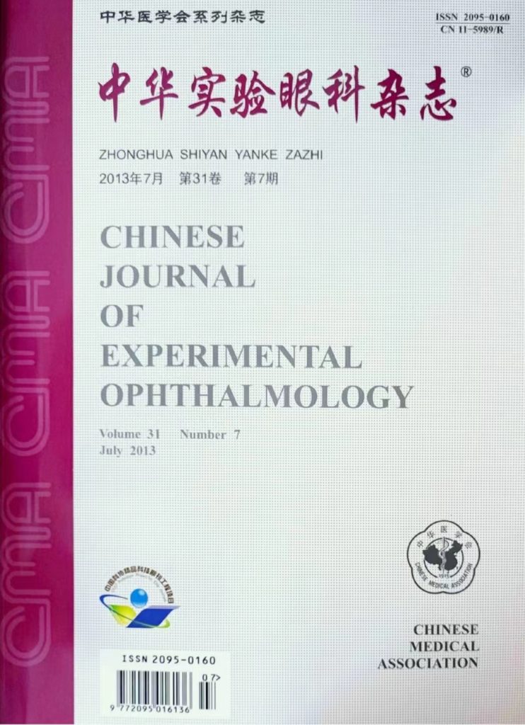
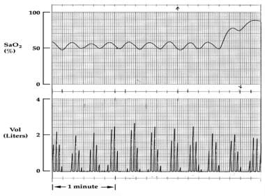
 Retinal haemorrhage
Vitreous hemorrhage
Shaken baby syndrome
Bungee jumping
Child abuse
Operative vaginal delivery
Intraocular hemorrhage
Arlie W. Schorger
Jane D. Kivlin
Smallpox
Phacoemulsification
Fuchs spot
Systemic scleroderma
End organ damage
Canid alphaherpesvirus 1
Mothball
Hypertensive leukoencephalopathy
Hyperviscosity syndrome
Alvacir Raposo
Retinopathy
Eales disease
Mount Everest
Scleral reinforcement surgery
Terson syndrome
Blast-related ocular trauma
Bacillus cereus
Eddie Mayo
Coats' disease
Sickle cell retinopathy
Charles Snead Houston
Retinal haemorrhage - Wikipedia
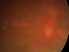
 Altitude-Related Disorders: Background, Sleep at High Altitude, High-Altitude Retinal Hemorrhage
 Retinal hemorrhages in newborn piglets following cardiopulmonary resuscitation
Retinal Hemorrhages in 4 Patients with Dengue Fever - Volume 11, Number 5-May 2005 - Emerging Infectious Diseases journal - CDC
 Retinal Hemorrhages in 4 Patients with Dengue Fever
Can convulsions alone cause retinal haemorrhages in infants? | British Journal of Ophthalmology
 Severe Retinal Hemorrhages With Retinoschisis Are Not Pathognomonic for Abusive Head Trauma | American Academy of Forensic...
 An infant with subdural hematoma and retinal hemorrhages: does von Willebrand disease explain the findings? | Forensic Science,...
 Information for Retinal haemorrhage
 Retinal haemorrhage in P falciparum malaria - MORU Tropical Health Network
 High-Altitude Retinal Hemorrhage | The Atlas of Emergency Medicine, 5e | AccessMedicine | McGraw Hill Medical
Retinal hemorrhage - WikEM
 Retinal Hemorrhage | Eye Patient
Retinal Haemorrhaging - Parents Accused
Bilateral retinal hemorrhages and disk edema in migraine<...
Pediatric Streptococcus-Associated Brain Abscesses and Empyemas
 Rhegmatogenous/Tractional Retinal Detachment and Vitreous Hemorrhage Associated With Branch Retinal Vein Occlusion in a...
Chester Porphyria: Practice Essentials, Background, Pathophysiology
 Dr. Joshua Powell, MD - Ophthalmology Specialist in Norman, OK | Healthgrades
 Fourth Year Medical Students
 Selective Estrogen Receptor Modulators: Drug Class, Uses, Side Effects, Drug Names
 1957. Dr M.KAUFFMAN : Retinal hemorrhages in the newborn. - ATIDE - Accouchement Traumatique Invalidité & Décès de l'Enfant
Intracranial Hemorrhage Clinical Presentation: History, Physical, Causes
Argon laser for subhyoid retinal haemorrhage | The Journal of Ophthalmology of Eastern, Central and Southern Africa
Bilateral retinal hemorrhages and macular edema in a patient with dengue fever associated with cerebral hemorrhage Joshi RS,...
 Retinal vein occlusion: MedlinePlus Medical Encyclopedia
 Blue light-induced retinal lesions, intraretinal vascular leakage and edema formation in the all-cone mouse retina | Cell Death...
 Dr. Jaime Jimenez Agosto, MD, Ophthalmology Specialist - Hattiesburg, MS | Sharecare
Occlusion17
- In older children and adults, retinal hemorrhage can be caused by several medical conditions such as hypertension, retinal vein occlusion (a blockage of a retinal vein), anemia, leukemia or diabetes. (wikipedia.org)
- This is known as a retinal artery occlusion, or eye stroke. (aao.org)
- It is sometimes caused by a retinal vein occlusion. (aao.org)
- Retinal vein occlusion is a blockage of the small veins that carry blood away from the retina. (medlineplus.gov)
- Retinal vein occlusion is most often caused by hardening of the arteries ( atherosclerosis ) and the formation of a blood clot. (medlineplus.gov)
- The risk of these disorders increases with age, therefore retinal vein occlusion most often affects older people. (medlineplus.gov)
- People with retinal vein occlusion often regain useful vision. (medlineplus.gov)
- Retinal vein occlusion is a sign of a general blood vessel (vascular) disease. (medlineplus.gov)
- Measures used to prevent other blood vessel diseases may decrease the risk for retinal vein occlusion. (medlineplus.gov)
- Controlling diabetes may help prevent retinal vein occlusion. (medlineplus.gov)
- Retinal vein occlusion (RVO) is an interruption of the normal venous drainage from the retinal tissue. (bmj.com)
- Hypertension, diabetes mellitus, atherosclerosis, and glaucoma are major risk factors for the development of central retinal vein occlusion (CRVO) or branch retinal vein occlusion (BRVO) in older patients. (bmj.com)
- For an uncomplicated retinal vein occlusion, whether it is ischemic or nonischemic, management consists of close monitoring to detect complications and treatment of underlying risk factors. (bmj.com)
- Characteristically, in the retina proximal to the occlusion, the affected venous system is tortuous and dilated, and there are several intraretinal hemorrhages and retinal edema. (bmj.com)
- Which of the following on funduscopic examination is most characteristic of central retinal vein occlusion? (google.com)
- Central retinal vein occlusion presents with sudden , painless , monocular vision loss with the classic " blood and thunder " appearance and retinal hemorrhages . (google.com)
- The post Podcast Ep 51: Glioblastoma, Kerion, Antipsychotics, Urine Dipstick, Tetalogy of Fallot, Central Retinal Vein Occlusion appeared first on RoshReview.com . (google.com)
Detachment3
- Occupational lifting tasks and retinal detachment in non-myopics and myopics: extended analysis of a case-control study. (medscape.com)
- Bjerrum SS, Mikkelsen KL, La Cour M. Risk of Pseudophakic Retinal Detachment in 202?226 Patients Using the Fellow Nonoperated Eye as Reference. (medscape.com)
- McNamara D. Cataract surgery may up retinal detachment risk 4-fold. (medscape.com)
Vitreous4
- Retinal haemorrhaging can be caused in many ways including direct trauma to the eye, raised intravascular pressure, raised intracranial pressure, raised central venous pressure or interaction with the vitreous jelly caused by the effects of shaking. (parentsaccused.co.uk)
- 4 Ulbig M.W., Mangouritsas G., Rothbacher H.H. Long-term results after drainage of premacular subhyaloid hemorrhage into the vitreous with a pulsed Nd:YAG laser. (coecsa.org)
- There were cells in the vitreous on each side, and the funduscopic examination (Figure) demonstrated slight optic disc edema on each side, with adjacent retinal hemorrhage on the left. (medscape.com)
- 2. Presence of macular fibrosis or retinal epithelial tear, clinically relevant myopic degeneration, or vitreous hemorrhage a. (who.int)
Edema7
- High-altitude pulmonary edema (HAPE) and high-altitude cerebral edema (HACE) are the most ominous of these symptoms, whereas acute mountain sickness (AMS), retinal hemorrhages, and peripheral edema are milder forms of the disease. (medscape.com)
- A 27-year-old patient with severe migraine developed bilateral disk edema and retinal hemorrhages during a prolonged attack of his illness, lasting at least two months. (johnshopkins.edu)
- The combination of bilateral disk edema and diffuse superficial retinal hemorrhages was considered to be a manifestation of prolonged valsalva efforts accompanying the migraine attack and of not persistent raised intracranial pressure. (johnshopkins.edu)
- We report a case of bilateral retinal hemorrhages with macular edema in a patient with dengue fever (DF). (thepajo.org)
- Isolated cases of retinal hemorrhages and macular edema have been reported in the literature. (thepajo.org)
- Herein, we report a case of bilateral retinal hemorrhages with macular edema in a patient with neurological complications due to DF. (thepajo.org)
- This was accompanied by retinal swelling and the appearance of cystoid spaces in both inner and ONLs of R91W;Nrl −/− mice indicating edema in affected areas. (nature.com)
Uveitis1
- Whereas retinal haemorrhage and uveitis are known adverse reactions to angiogenesis inhibitors, the reported cases of blindness and death should heighten awareness of potential safety issues associated with VEGF inhibitors for the treatment of proliferative eye disorders. (smw.ch)
Intracerebral4
- Acute seizures after intracerebral hemorrhage: a factor in progressive midline shift and outcome. (medscape.com)
- CT angiography "spot sign" predicts hematoma expansion in acute intracerebral hemorrhage. (medscape.com)
- Mayer SA, Brun NC, Begtrup K. Recombinant activated factor VII for acute intracerebral hemorrhage. (medscape.com)
- The high incidence of intracerebral hemorrhage illustrates the clinical differences from other hereditary cerebral small-vessel diseases, such as cerebral autosomal dominant arteriopathy with subcortical infarcts and leukoencephalopathy (CADASIL), cerebral autosomal recessive arteriopathy with subcortical infarcts and leukoencephalopathy (CARASIL), and hereditary endotheliopathy, retinopathy, nephropathy, and stroke (HERNS) 3 . (nature.com)
Blood vessels2
- Any rupture in the retinal surface can damage the tiny blood vessels and the result is bleeding or retinal hemorrhage. (medindia.net)
- Furthermore, image analysis provides a simple and noninvasive visualization of the retinal blood vessels in those high risk ophthalmologic medical conditions [ 1 - 3 ]. (hindawi.com)
Ophthalmology1
- Ophthalmology opinion revealed retinal hemorrhages. (bvsalud.org)
Vascular5
- Anti-vascular endothelial growth factor (VEGF) drugs like Avastin and Lucentis have also been shown to repair retinal hemorrhaging in diabetic patients and patients with hemorrhages associated with new vessel growth. (wikipedia.org)
- Fundoscopic examination showed bilateral blot hemorrhages within the vascular arcades in all 4 patients. (cdc.gov)
- 6 7 By contrast, vascular endothelial growth factor (VEGF) appears to stimulate retinal vasodilatation and neovascularization. (arvojournals.org)
- These extracted markers or characterized fundus digital image features provide insights and relates quantitative retinal vascular topography abnormalities to various pathologies such as diabetic retinopathy, macular degeneration, hypertensive retinopathy, transient ischemic attack, neovascular glaucoma, and cardiovascular diseases. (hindawi.com)
- Some distinct changes in the retinal microvasculature are recognized as the preindicator of subsequent vascular incidents like ischemic stroke or acute stroke [ 10 ]. (hindawi.com)
Cause retinal2
Bilateral retinal1
- An 11-month-old girl presented to hospital with a massive subdural haematoma and bilateral retinal haemorrhages following an allegedly minor fall. (springer.com)
Subdural Hemorrhage2
- 2019. Dr V.VLASYUK : Subdural Hemorrhage. (atide-asso.org)
- Repetitive back-and-forth head rotation from vigorous shaking is purported to be a central mechanism responsible for diffuse white matter injur y, subdural hemorrhage, and retinal hemorrhage in some cases of abusive head trauma (AHT) in young children. (cdc.gov)
Association between retinal2
- The generally accepted body of medical literature is strongly in favor of the tight association between retinal hemorrhages and child abuse, and there is no controversy among mainstream medicine. (wikipedia.org)
- But the Cardiovascular Health Study stated that there is no association between retinal arteriolar caliber (diameter) and stroke but rather there is a close association between stroke and the larger venular caliber (diameter) [ 13 ]. (hindawi.com)
High altitude2
- High-altitude retinal hemorrhages (HARHs) are rarely symptomatic, but if found over the macula, these hemorrhages may cause temporary blindness. (mhmedical.com)
- Fundoscopic appearance of high-altitude retinal hemorrhage. (mhmedical.com)
Cerebral4
- Lobar hemorrhage due to cerebral amyloid angiopathy may be preceded by prodromal symptoms of focal numbness, tingling, or weakness. (medscape.com)
- 2016. Dr C-L.FRANCOEUR, S-A.MAYER : Management of delayed cerebral ischemia after subarachnoid hemorrhage. (atide-asso.org)
- This patient also had cerebral hemorrhage. (thepajo.org)
- COL4A1 -related disorders are characterized by a higher incidence of cerebral hemorrhage than other hereditary cerebral small vessel diseases. (nature.com)
Changes of retinal2
- The paper [ 16 ] searched MEDLINE and EMBASE to find out the relation between microvascular changes of retinal microvasculature and prevalence or incident of stroke. (hindawi.com)
- In our series of 7 cases, we documented impaired visual acuity, central visual field defects, circumscribed and sometimes complex changes of retinal reflectivity, and intraretinal fluid. (aerzteblatt.de)
Causes retinal1
- To determine whether conventional cardiopulmonary resuscitation causes retinal hemorrhages in piglets. (nih.gov)
Infants5
- Retinal hemorrhage is strongly associated with child abuse in infants and young children and often leaves such abused infants permanently blind. (wikipedia.org)
- In infants, retinal hemorrhages (RH) are highly associated with child abuse. (wikipedia.org)
- Preventive measures such as regular prenatal care and monitoring of infants with high risks of the disorder may be done to avoid further complications of retinal hemorrhages in infants. (wikipedia.org)
- Our research shows that you see the hemorrhages in a variety of different situations in infants, children and adults,' said Dr. Lantz, senior researcher involved in the study. (medindia.net)
- Retinal haemorrhages in infants with vWD have not been previously reported. (springer.com)
Intracranial hemorrhage2
- Intracranial hemorrhage and rebleeding in suspected victims of abusive head trauma: addressing the forensic controversies. (springer.com)
- Immediate and delayed traumatic intracranial hemorrhage in patients with head trauma and preinjury warfarin or clopidogrel use. (medscape.com)
Intraventricular hemorrhage2
- The IVH score: a novel tool for estimating intraventricular hemorrhage volume: clinical and research implications. (medscape.com)
- Strong correlations have been found between low neonatal serum concentrations of IGF-1 and poor brain and retinal growth as well as poor general growth with multiorgan morbidities, such as intraventricular hemorrhage, retinopathy of prematurity, bronchopulmonary dysplasia, and necrotizing enterocolitis. (lu.se)
Head Trauma1
- Predictors of long-term visual outcome following retinal hemorrhage from abusive head trauma. (upenn.edu)
Diabetic3
- 2 Morse L.S., Chapman C.B., Eliott D. Subretinal hemorrhages in proliferative diabetic retinopathy. (coecsa.org)
- According to the study of [ 12 ] with a multiethnic cohort, retinal arteriolar narrowing and retinopathy of diabetic free people have an association with increased risk of acute stroke. (hindawi.com)
- History of or presence of retinal disease other than GA: diabetic retinopathy, central serous chorioretinopathy, inherited retinal degeneration, toxic maculopathies (ie, hydroxychloroquine maculopathy), arterial and venous occlusive disease, macular hole that is present or has been previously repaired, or choroidal melanoma. (who.int)
Visual acuity2
- She returned at age 6 with decreased visual acuity OD due to retinal traction by the PFV fibrovascular stalk (Figure 1A). (uiowa.edu)
- The spectrum of damage ranged from focal photoreceptor defects to macular foramina and retinal hemorrhages associated with loss of visual acuity and central scotoma. (aerzteblatt.de)
Shaken baby syn1
- If non-accidental head injury (or shaken baby syndrome) is suspected, it is likely that an ophthalmologist (specialist eye doctor) will examine your child's eyes to see if there are any retinal haemorrhages present. (parentsaccused.co.uk)
Diseases1
- Retinal microvascular abnormalities like microaneurysm, arteriovenous nicking, haemorrhages, and vessel caliber are considered as associative to the stroke and indicative of death from stroke and IHD (Ischemic Heart Diseases) [ 1 ]. (hindawi.com)
Severe4
- Features that make retinal haemorrhaging more severe can include whether they are present in one or both eyes, whether they are in every layer of the retina or whether they extend to the outside of the retina or are just focused around the centre. (parentsaccused.co.uk)
- When due to NAI, retinal haemorrhages were often present in both eyes and were more severe. (parentsaccused.co.uk)
- To present a case of severe pre-retinal hemorrhage that was treated with Argon laser posterior hylodotomy. (coecsa.org)
- Recent years have seen a marked increase in laser-pointer-related injuries, which sometimes involve severe retinal damage and irreversible visual impairment. (aerzteblatt.de)
Intraretinal hemorrhages1
- Fundus examination of both eyes showed preretinal and intraretinal hemorrhages. (thepajo.org)
Acute mountain1
- Retinal hemorrhages are common above 5200 m (17,000 ft) and are not usually associated with acute mountain sickness (AMS). (mhmedical.com)
Posterior1
- 3 Raymond L.A. Neodymium:YAG laser treatment for hemorrhages under the internal limiting membrane and posterior hyaloid face in the macula. (coecsa.org)
Dengue fever4
- Rarely, retinal hemorrhages affecting patients with dengue fever are reported. (cdc.gov)
- We report 4 patients with dengue fever complicated by retinal hemorrhages who were hospitalized in our institution in June and July 2004. (cdc.gov)
- In the same period, retinal hemorrhages were diagnosed in 4 dengue fever patients in our hospital. (cdc.gov)
- We report 4 patients with retinal hemorrhages that developed during hospitalization for dengue fever. (cdc.gov)
Sudden3
- Some symptoms may include: Seeing floaters in the vision Seeing cobwebs in the vision Seeing haze or shadows Distorted vision Rapid flashes of light in peripheral vision Red tint to vision Blurriness Sudden blindness Headache In adults, retinal hemorrhages are largely spontaneous, secondary to chronic medical conditions such as hypertension. (wikipedia.org)
- A hemorrhage can lead to blind spots and gradual or sudden loss of vision. (aao.org)
- Retinal vein occlusions are usually painless, sudden, and unilateral causes of vision loss. (bmj.com)
Subhyaloid1
- 6 Khadka D., Bhandari S., Bajimaya S., Thapa R., Paudyal G., Pradhan E. Nd:YAG laser hyaloidotomy in the management of Premacular Subhyaloid Hemorrhage. (coecsa.org)
Vein occlusions1
- Retinal vein occlusions preferred practice pattern. (medlineplus.gov)
Vasculature2
- Recently, we generated R91W;Nrl −/− double-mutant mice, which display a well-ordered all-cone retina with normal retinal vasculature and a strong photopic function that generates useful vision. (nature.com)
- Analysis of the human fundus eye images has become the key point for diagnosing the various pathologies of retinal vasculature. (hindawi.com)
Diagnosis2
Fundus4
- A retinal hemorrhage is generally diagnosed by using an ophthalmoscope or fundus camera in order to examine the inside of the eye. (wikipedia.org)
- B) Color Fundus Photograph OD at age 19 status post pars plana vitrectomy demonstrating the amputated stump of the white fibrovascular stalk with resolution of retinal traction. (uiowa.edu)
- One of the most important subfields of biomedical engineering is the analysis of fundus retinal images. (hindawi.com)
- The fundus retinal images are directly captured from human eye that includes some other landmarks like microcirculation system of the retina, macula, optic disc, fovea, microaneurysm, and exudates [ 4 ]. (hindawi.com)
Choroidal1
- There is evidence of peripapillary and macular retinal traction and subretinal fluid based on fine retinal folds and loss of visible choroidal detail underlying the stalk. (uiowa.edu)
Detachments1
- Controversies in the management of primary retinal detachments. (medscape.com)
Rarely1
- Research has found that retinal haemorrhages are rarely caused during accidental injury and are much more common in NAI. (parentsaccused.co.uk)
BRVO1
- Blockage of smaller veins (branch veins or BRVO) in the retina often occurs in places where retinal arteries that have been thickened or hardened by atherosclerosis cross over and place pressure on a retinal vein. (medlineplus.gov)
20161
- 2016. Dr N-F.CALLAWAY et al : Retinal and optic nerve hemorrhages in the newborn infant. (atide-asso.org)
Bleeding occurs1
- Retinal hemorrhage (UK English: retinal haemorrhage) is a disorder of the eye in which bleeding occurs in the retina, the light sensitive tissue, located on the back wall of the eye. (wikipedia.org)
Ocular1
- Injur y metrics were the occurrence and extent of axonal injur y (AI), extra-axial hemorrhage (EAH), red cell neuronal/axonal change (RCNAC), and ocular injur y (OI). (cdc.gov)
Incidence2
- Retinal hemorrhages may reflect the rising incidence of dengue in Singapore or may be caused by changes in the predominant serotype of the dengue virus. (cdc.gov)
- Incidence, distribution, and duration of birth-related retinal hemorrhages: a prospective study. (springer.com)
Examination2
- A new study has now found out that retinal examination of a child, to determine the location and number of hemorrhages may not be a sure sign of abuse. (medindia.net)
- Anterior segment examination showed subconjunctival hemorrhage in both eyes with normal pupil size pupil that reacted to light. (thepajo.org)