Retinal Vasculitis
Vasculitis
Systemic Vasculitis
Tuberculosis, Ocular
Behcet Syndrome
Retinal Diseases
Panuveitis
Uveitis
Retinitis
Fluorescein Angiography
Anti-Neutrophil Cytoplasmic Antibody-Associated Vasculitis
Uveitis, Anterior
Fundus Oculi
Vasculitis, Central Nervous System
Polyarteritis Nodosa
Prednisolone
Antigen-Antibody Complex
Visual Acuity
Full panretinal photocoagulation and early vitrectomy improve prognosis of retinal vasculitis associated with tuberculoprotein hypersensitivity (Eales' disease). (1/37)
BACKGROUND/AIMS: Eales' disease is an uncommon vasoproliferative retinal disease affecting otherwise healthy young men that is characterised by obliterative retinal periphlebitis, with sequelae such as recurrent vitreous haemorrhage and traction retinal detachment. This study was undertaken to determine whether visual prognosis of Eales' disease could be improved by appropriate medical and surgical treatment. METHODS: The authors retrospectively studied 30 patients (46 eyes) who were treated from 1992 to 2001. Recorded data included patient age, sex, race, medical history, medications, results of the ophthalmological examination, results of diagnostic laboratory evaluation, and details of systemic and surgical treatments. The mean follow up was 10.6 months. RESULTS: 19 patients (23 eyes) who presented with active periphlebitis received systemic steroids and antituberculous therapy. Extensive full panretinal photocoagulation was performed in 21 eyes that presented with new vessel formation and peripheral capillary closure with or without vitreous haemorrhage. Vitrectomy and endolaser panretinal photocoagulation was necessary in 15 eyes, for severe non-clearing vitreous haemorrhage in 11 eyes and vitreous haemorrhage with traction retinal detachment in four eyes. Complete regression of the disease was achieved in all eyes. Vitrectomy resulted in a significant visual improvement with 14 of the 15 eyes (93.3%) achieving > or =20/200 visual acuity. Overall, the distribution of visual acuities among eyes improved from presentation to final follow up, with 36.4% of eyes having 20/40 or better acuity at presentation compared with 63.6% of eyes by final follow up. CONCLUSIONS: These results suggest that aggressive treatment of Eales' disease with systemic steroids and antituberculous therapy, full panretinal photocoagulation and early vitrectomy, when necessary, may result in improving the anatomic and visual outcome. (+info)Frosted branch angiitis associated with rapidly progressive glomerulonephritis. (2/37)
Simultaneous occurrence of frosted branch angiitis and immune-mediated rapidly progressive glomerulonephritis is reported. The two diseases possibly share a common immune mechanism. Patients of frosted branch angiitis should undergo complete systemic evaluation including renal function tests even if the patient is systemically asymptomatic. (+info)Human recombinant interferon alfa-2a for the treatment of Behcet's disease with sight threatening posterior or panuveitis. (3/37)
BACKGROUND: Behcet's disease is a multisystem vasculitis of unknown origin. Standard treatment mainly comprises systemic immunosuppressive agents. Ocular involvement, mostly posterior uveitis with retinal vasculitis, leads to blindness in 20-50% of the involved eyes within 5 years. The efficacy of interferon alfa-2a was studied in patients with sight threatening posterior uveitis or retinal vasculitis. METHODS: 50 patients were included in this open, non-randomised, uncontrolled prospective study. Recombinant human interferon alfa-2a (rhIFNalpha-2a) was applied at a dose of 6 million units subcutaneously daily. Dose reduction was performed according to a decision tree until discontinuation. Disease activity was evaluated every 2 weeks by the Behcet's disease activity scoring system and the uveitis scoring system. RESULTS: Response rate of the ocular manifestations was 92% (three non-responder, one incomplete response). Mean visual acuity rose significantly from 0.56 to 0.84 at week 24 (p<0.0001). Posterior uveitis score of the affected eyes fell by 46% every week (p<0.001). Remission of retinal inflammation was achieved by week 24. Mean Behcet's disease activity score fell from 5.8 to 3.3 at week 24 and further to 2.8 at week 52. After a mean observation period of 36.4 months (range 12-72), 20 patients (40%) are off treatment and disease free for 7-58 months (mean 29.5). In the other patients maintenance IFN dosage is three million units three times weekly. CONCLUSIONS: rhIFNalpha-2a is effective in ocular Behcet's disease, leading to significant improvement of vision and complete remission of ocular vasculitis in the majority of the patients. (+info)Recurrent anterior uveitis and healed retinal vasculitis associated with multiple sclerosis. (4/37)
We describe the occurrence of anterior uveitis with healed retinal vasculitis in an Asian-Indian woman. She had features of anterior uveitis and healed retinal vasculitis. This rare disease in India may be associated with intraocular inflammation. (+info)Optic neuritis and retinal vasculitis as primary manifestations of systemic lupus erythematosus. (5/37)
Systemic lupus erythematosus (SLE) is a common multisystem disorder. However, retinal vasculitis as a primary manifestation of SLE is uncommon, accounting for only 4% of causes of retinal vasculitis. The postulated mechanism appeared to be vaso-occlusion of the retinal arterioles by thrombosis, with resultant ischaemia. Optic neuropathy in SLE is also rare, with a prevalence of 1%. This is a case report of a young lady who presented to us with retinal vasculitis as her initial presentation of SLE. Interestingly, the pathologic mechanism appeared to be inflammatory and not vaso-occlusive. (+info)Is granuloma annulare related to intermediate uveitis with retinal vasculitis? (6/37)
AIM: To report on eight patients with severe idiopathic intermediate uveitis (IU) and granuloma annulare (GA), a self limiting cutaneous condition of unknown aetiology. METHODS: Retrospective case series. Clinical ophthalmic and dermatological data were studied and fluorescein angiography and skin biopsies were reviewed. RESULTS: All patients with idiopathic IU had similar ocular features (eight with vitritis, seven with retinal vasculitis) and developed complications such as cystoid macular oedema (n=5), cataract (n=4), and glaucoma (n=3). Systemic diseases were not found, but a localised type of GA was observed in all. CONCLUSION: Seven out of eight patients with IU and GA developed severe retinal vasculitis. Further studies are needed for a better understanding of this association, a common pathogenesis, and its eventual clinical consequences. (+info)IFN-gamma-regulated Toxoplasma gondii distribution and load in the murine eye. (7/37)
PURPOSE: To establish a mouse model of ocular toxoplasmosis in both wild type (WT) and immunocompromised hosts and to clarify the effects of interferon (IFN)-gamma on the infectivity of Toxoplasma gondii in various parts of the eye. METHODS: Susceptible WT C57BL/6, resistant WT BALB/c, and IFN-gamma knockout (GKO) mice were infected with cysts of T. gondii perorally. The tissues were harvested for molecular and histopathologic studies. Analysis included a quantitative competitive polymerase chain reaction (QC-PCR) assay and reverse transcription (RT)-PCR for IFN-gamma and stage conversion markers. All animals underwent ophthalmic examinations including fluorescein angiography (FA). RESULTS: In WT C57BL/6 mice, T. gondii was detected in tissue in the following order: brain, retina, choroid, sclera, and optic nerve (ON). The highest T. gondii load was observed in the posterior retina, and was much greater than that in WT BALB/c mice. In GKO mice, disseminated infection was evident, and the T. gondii load was highest in the choroid and ON. IFN-gamma mRNA expression in WT C57BL/6 mice was higher than that in WT BALB/c mice after infection. Tachyzoites existed in GKO mice, whereas bradyzoites existed in WT C57BL/6 mice. FA showed dye leakage from the retinal capillaries of GKO mice. CONCLUSIONS: The T. gondii load in the retina in the susceptible WT strain continued to increase, unlike in the resistant WT strain. IFN-gamma was shown to regulate the T. gondii load and interconversion in the eye. A toxoplasmic vasculitis model was established with GKO mice and assay systems with QC-PCR and FA. (+info)Susac syndrome: report of four cases and review of the literature. (8/37)
Susac syndrome is a rare disease of unknown pathogenesis. It is caused by a microangiopathy affecting the arterioles of the brain, retina, and cochlea, giving the classic clinical triad of subacute encephalopathy, visual loss secondary to retinal branch occlusions, and sensorineural hearing loss. The features of four cases of this syndrome are presented. MR imaging, retinal fluorescein angiography, and audiography findings enable diagnosis. Early therapy may reduce sequelae and improve recovery. (+info)Retinal vasculitis is a medical condition characterized by inflammation of the blood vessels in the retina, which is the light-sensitive tissue located at the back of the eye. This condition can cause damage to the retina and may lead to vision loss if not treated promptly. The inflammation can affect both the small and large blood vessels in the retina and can occur as a result of various systemic diseases or infections, including autoimmune disorders, tuberculosis, syphilis, and toxoplasmosis. In some cases, retinal vasculitis may also be associated with uveitis, which is inflammation of the middle layer of the eye. Treatment typically involves addressing the underlying cause of the inflammation and may include corticosteroids or other immunosuppressive therapies to reduce inflammation and prevent further damage to the retina.
Vasculitis is a group of disorders characterized by inflammation of the blood vessels, which can cause changes in the vessel walls including thickening, narrowing, or weakening. These changes can restrict blood flow, leading to organ and tissue damage. The specific symptoms and severity of vasculitis depend on the size and location of the affected blood vessels and the extent of inflammation. Vasculitis can affect any organ system in the body, and its causes can vary, including infections, autoimmune disorders, or exposure to certain medications or chemicals.
Systemic vasculitis is a group of disorders characterized by inflammation of the blood vessels (vasculitis) that can affect various organs and systems throughout the body. This condition can cause damage to the walls of the blood vessels, leading to narrowing, blockage, or weakening of the vessel walls, which can further result in reduced blood flow, tissue damage, and organ dysfunction.
The symptoms of systemic vasculitis depend on the severity and location of the affected blood vessels. They may include fever, fatigue, weight loss, joint pain, skin rashes or lesions, muscle weakness, nerve damage, and organ dysfunction such as kidney failure, lung disease, or gastrointestinal bleeding.
Systemic vasculitis can be caused by various factors, including infections, autoimmune diseases, medications, and underlying medical conditions. The diagnosis of systemic vasculitis typically involves a combination of physical examination, laboratory tests, imaging studies, and sometimes biopsy of the affected tissue. Treatment may include corticosteroids, immunosuppressive drugs, and other medications to control inflammation and prevent organ damage.
Ocular tuberculosis (OTB) is a form of extrapulmonary tuberculosis (TB), which results from the spread of Mycobacterium tuberculosis complex bacteria outside the lungs. In ocular tuberculosis, these bacteria primarily affect the eye and its surrounding structures.
The most common form of OTB is tubercular uveitis, which involves inflammation of the uveal tract (iris, ciliary body, and choroid). Other forms of OTB include:
* Tubercular conjunctivitis: Inflammation of the conjunctiva, the mucous membrane that covers the front part of the eye and lines the inside of the eyelids.
* Tubercular keratitis: Inflammation of the cornea, the transparent outer layer at the front of the eye.
* Tubercular scleritis: Inflammation of the sclera, the white protective coating of the eye.
* Tubercular episcleritis: Inflammation of the episclera, a thin layer of tissue between the conjunctiva and sclera.
* Tubercular dacryoadenitis: Inflammation of the lacrimal gland, which produces tears.
* Tubercular optic neuritis: Inflammation of the optic nerve, which transmits visual information from the eye to the brain.
Diagnosis of OTB can be challenging due to its varied clinical presentations and the need for laboratory confirmation. A definitive diagnosis typically requires the isolation of Mycobacterium tuberculosis from ocular tissues or fluids, which may involve invasive procedures. In some cases, a presumptive diagnosis might be made based on clinical findings, epidemiological data, and response to anti-tuberculous therapy.
Treatment for OTB usually involves a standard anti-tuberculosis regimen consisting of multiple drugs (isoniazid, rifampin, ethambutol, and pyrazinamide) for at least six months. Corticosteroids or other immunosuppressive agents might be used concomitantly to manage inflammation and prevent tissue damage. Close monitoring is essential to ensure treatment adherence, assess response to therapy, and detect potential side effects.
Behçet syndrome is a rare inflammatory disease that can cause symptoms in various parts of the body. It's characterized by recurrent mouth sores (aphthous ulcers), genital sores, and inflammation of the eyes (uveitis). The condition may also cause skin lesions, joint pain and swelling, and inflammation of the digestive tract, brain, or spinal cord.
The exact cause of Behçet syndrome is not known, but it's thought to be an autoimmune disorder, in which the body's immune system mistakenly attacks its own healthy cells and tissues. The condition tends to affect men more often than women and typically develops during a person's 20s or 30s.
There is no cure for Behçet syndrome, but treatments can help manage symptoms and prevent complications. Treatment options may include medications such as corticosteroids, immunosuppressants, and biologics to reduce inflammation, as well as pain relievers and other supportive therapies.
Retinal diseases refer to a group of conditions that affect the retina, which is the light-sensitive tissue located at the back of the eye. The retina is responsible for converting light into electrical signals that are sent to the brain and interpreted as visual images. Retinal diseases can cause vision loss or even blindness, depending on their severity and location in the retina.
Some common retinal diseases include:
1. Age-related macular degeneration (AMD): A progressive disease that affects the central part of the retina called the macula, causing blurred or distorted vision.
2. Diabetic retinopathy: A complication of diabetes that can damage the blood vessels in the retina, leading to vision loss.
3. Retinal detachment: A serious condition where the retina becomes separated from its underlying tissue, requiring immediate medical attention.
4. Macular edema: Swelling or thickening of the macula due to fluid accumulation, which can cause blurred vision.
5. Retinitis pigmentosa: A group of inherited eye disorders that affect the retina's ability to respond to light, causing progressive vision loss.
6. Macular hole: A small break in the macula that can cause distorted or blurry vision.
7. Retinal vein occlusion: Blockage of the retinal veins that can lead to bleeding, swelling, and potential vision loss.
Treatment for retinal diseases varies depending on the specific condition and its severity. Some treatments include medication, laser therapy, surgery, or a combination of these options. Regular eye exams are essential for early detection and treatment of retinal diseases.
Panuveitis is a medical term that refers to inflammation that affects the entire uveal tract, including the iris, ciliary body, and choroid. The uveal tract is the middle layer of the eye between the inner retina and the outer fibrous tunic (sclera). Panuveitis can also affect other parts of the eye, such as the vitreous, retina, and optic nerve.
The symptoms of panuveitis may include redness, pain, light sensitivity, blurred vision, floaters, and decreased visual acuity. The condition can be caused by various factors, including infections, autoimmune diseases, trauma, or unknown causes (idiopathic). Treatment typically involves the use of corticosteroids to reduce inflammation, as well as addressing any underlying cause if identified. If left untreated, panuveitis can lead to complications such as cataracts, glaucoma, and retinal damage, which can result in permanent vision loss.
Intermediate uveitis is a type of uveitis that affects the vitreous cavity and peripheral retina. It is characterized by the presence of inflammatory cells in the vitreous, called vitritis, and sometimes also by snowbanking or peripheral lesions in the retina. Intermediate uveitis can cause vision loss due to cystoid macular edema, epiretinal membrane formation, or complications such as glaucoma or cataract. The onset of intermediate uveitis is often insidious and the course can be chronic, with recurrent episodes of inflammation. The exact cause of intermediate uveitis is often unknown, but it can be associated with systemic diseases such as sarcoidosis, multiple sclerosis, or Lyme disease.
Retinal vessels refer to the blood vessels that are located in the retina, which is the light-sensitive tissue that lines the inner surface of the eye. The retina contains two types of blood vessels: arteries and veins.
The central retinal artery supplies oxygenated blood to the inner layers of the retina, while the central retinal vein drains deoxygenated blood from the retina. These vessels can be visualized during a routine eye examination using an ophthalmoscope, which allows healthcare professionals to assess their health and any potential abnormalities.
Retinal vessels are essential for maintaining the health and function of the retina, and any damage or changes to these vessels can affect vision and lead to various eye conditions such as diabetic retinopathy, retinal vein occlusion, and hypertensive retinopathy.
A retinal hemorrhage is a type of bleeding that occurs in the blood vessels of the retina, which is the light-sensitive tissue located at the back of the eye. This condition can result from various underlying causes, including diabetes, high blood pressure, age-related macular degeneration, or trauma to the eye. Retinal hemorrhages can be categorized into different types based on their location and appearance, such as dot and blot hemorrhages, flame-shaped hemorrhages, or subhyaloid hemorrhages. Depending on the severity and cause of the hemorrhage, treatment options may vary from monitoring to laser therapy, medication, or even surgery. It is essential to consult an ophthalmologist for a proper evaluation and management plan if you suspect a retinal hemorrhage.
An eye hemorrhage, also known as subconjunctival hemorrhage, is a condition where there is bleeding in the eye, specifically under the conjunctiva which is the clear membrane that covers the white part of the eye (sclera). This membrane has tiny blood vessels that can rupture and cause blood to accumulate, leading to a visible red patch on the surface of the eye.
Eye hemorrhages are usually painless and harmless, and they often resolve on their own within 1-2 weeks without any treatment. However, if they occur frequently or are accompanied by other symptoms such as vision changes, pain, or sensitivity to light, it is important to seek medical attention as they could indicate a more serious underlying condition. Common causes of eye hemorrhages include trauma, high blood pressure, blood thinners, and aging.
Uveitis is the inflammation of the uvea, the middle layer of the eye between the retina and the white of the eye (sclera). The uvea consists of the iris, ciliary body, and choroid. Uveitis can cause redness, pain, and vision loss. It can be caused by various systemic diseases, infections, or trauma. Depending on the part of the uvea that's affected, uveitis can be classified as anterior (iritis), intermediate (cyclitis), posterior (choroiditis), or pan-uveitis (affecting all layers). Treatment typically includes corticosteroids and other immunosuppressive drugs to control inflammation.
Retinitis is a medical term that refers to the inflammation of the retina, which is the light-sensitive tissue located at the back of the eye. The retina is responsible for converting light into electrical signals that are then sent to the brain and interpreted as visual images. Retinitis can be caused by various factors, including infections, autoimmune diseases, or genetic conditions.
The inflammation associated with retinitis can affect any part of the retina, but it typically involves the retinal pigment epithelium (RPE) and the photoreceptor cells (rods and cones). Depending on the severity and location of the inflammation, retinitis can cause a range of visual symptoms, such as blurry vision, floaters, loss of peripheral vision, or night blindness.
Retinitis is often distinguished from another condition called retinopathy, which refers to damage to the retina caused by diabetes or other systemic diseases. While both conditions can affect the retina and cause visual symptoms, retinitis is characterized by inflammation, while retinopathy is characterized by damage due to circulatory problems.
It's important to note that retinitis is a serious condition that requires prompt medical attention. If left untreated, it can lead to permanent vision loss or blindness. Treatment options for retinitis depend on the underlying cause and may include antibiotics, corticosteroids, or other immunosuppressive medications.
Fluorescein angiography is a medical diagnostic procedure used in ophthalmology to examine the blood flow in the retina and choroid, which are the inner layers of the eye. This test involves injecting a fluorescent dye, Fluorescein, into a patient's arm vein. As the dye reaches the blood vessels in the eye, a specialized camera takes rapid sequences of photographs to capture the dye's circulation through the retina and choroid.
The images produced by fluorescein angiography can help doctors identify any damage to the blood vessels, leakage, or abnormal growth of new blood vessels. This information is crucial in diagnosing and managing various eye conditions such as age-related macular degeneration, diabetic retinopathy, retinal vein occlusions, and inflammatory eye diseases.
It's important to note that while fluorescein angiography is a valuable diagnostic tool, it does carry some risks, including temporary side effects like nausea, vomiting, or allergic reactions to the dye. In rare cases, severe adverse reactions can occur, so patients should discuss these potential risks with their healthcare provider before undergoing the procedure.
Anti-Neutrophil Cytoplasmic Antibody (ANCA)-Associated Vasculitis (AAV) is a group of autoimmune diseases characterized by inflammation and damage to small blood vessels, particularly capillaries, venules, and arterioles. The condition is named after the presence of ANCAs in the patient's serum, which are autoantibodies that target specific proteins in the neutrophil cytoplasm.
AAV includes several subtypes, including:
1. Granulomatosis with Polyangiitis (GPA, formerly known as Wegener's granulomatosis) - a form of AAV that typically affects the respiratory tract and kidneys, characterized by the presence of granulomas (clusters of inflammatory cells).
2. Microscopic Polyangiitis (MPA) - a form of AAV that primarily affects small vessels in various organs, such as the kidneys, lungs, and skin.
3. Eosinophilic Granulomatosis with Polyangiitis (EGPA, formerly known as Churg-Strauss syndrome) - a form of AAV that involves asthma, allergies, and eosinophilia (an increased number of eosinophils in the blood), along with vasculitis affecting various organs.
The exact cause of ANCA-Associated Vasculitis is not fully understood, but it is believed to involve an interplay between genetic factors, environmental triggers, and dysregulation of the immune system. The condition can lead to a wide range of symptoms depending on which organs are affected, including fever, fatigue, weight loss, joint pain, skin rashes, cough, shortness of breath, nosebleeds, and kidney problems. Treatment typically involves immunosuppressive medications to control inflammation and prevent further damage to the affected organs.
Anterior uveitis is a medical term that refers to the inflammation of the front portion of the uvea, which is the middle layer of the eye. The uvea includes the iris (the colored part of the eye), the ciliary body (a structure behind the iris that helps focus light onto the retina), and the choroid (a layer of blood vessels that supplies oxygen and nutrients to the retina).
Anterior uveitis is characterized by inflammation of the iris and/or the ciliary body, leading to symptoms such as redness, pain, sensitivity to light, blurred vision, and a small pupil. The condition can be caused by various factors, including infections, autoimmune diseases, trauma, or unknown causes (idiopathic).
Treatment of anterior uveitis typically involves the use of topical corticosteroids to reduce inflammation and cycloplegics to relieve pain and prevent spasms of the ciliary muscle. In some cases, oral medications may be necessary to control the inflammation. Prompt treatment is important to prevent complications such as glaucoma, cataracts, or permanent vision loss.
A Vitreous Hemorrhage is a medical condition where there is bleeding into the vitreous cavity of the eye. The vitreous cavity is the space in the eye that is filled with a clear, gel-like substance called the vitreous humor. This substance helps to maintain the shape of the eye and transmit light to the retina.
When a vitreous hemorrhage occurs, blood cells from the bleeding mix with the vitreous humor, causing it to become cloudy or hazy. As a result, vision can become significantly impaired, ranging from mildly blurry to complete loss of vision depending on the severity of the bleed.
Vitreous hemorrhages can occur due to various reasons such as trauma, retinal tears or detachments, diabetic retinopathy, age-related macular degeneration, and other eye conditions that affect the blood vessels in the eye. Treatment for vitreous hemorrhage depends on the underlying cause and may include observation, laser surgery, or vitrectomy (a surgical procedure to remove the vitreous humor and stop the bleeding).
"Fundus Oculi" is a medical term that refers to the back part of the interior of the eye, including the optic disc, macula, fovea, retinal vasculature, and peripheral retina. It is the area where light is focused and then transmitted to the brain via the optic nerve, forming visual images. Examinations of the fundus oculi are crucial for detecting various eye conditions such as diabetic retinopathy, macular degeneration, glaucoma, and other retinal diseases. The examination is typically performed using an ophthalmoscope or a specialized camera called a retinal camera.
Vasculitis, Central Nervous System (CNS), refers to a group of disorders characterized by inflammation of blood vessels within the brain and/or spinal cord. This inflammation can cause damage to the blood vessel walls, leading to narrowing, blocking or weakening of the vessels, and in some cases, formation of aneurysms or rupture of the vessels.
The causes of CNS vasculitis are varied and can include infections, autoimmune diseases, medications, and unknown factors. The symptoms of CNS vasculitis depend on the severity and location of the inflammation, and may include headache, seizures, stroke-like symptoms (such as weakness or numbness in the face, arms, or legs), cognitive changes, and in severe cases, coma.
Diagnosis of CNS vasculitis typically involves a combination of clinical evaluation, imaging studies (such as MRI or angiography), and laboratory tests (including blood tests and analysis of cerebrospinal fluid). Treatment may involve corticosteroids, immunosuppressive medications, and/or other therapies aimed at reducing inflammation and preventing further damage to the blood vessels.
Polyarteritis nodosa (PAN) is a rare, systemic necrotizing vasculitis that affects medium-sized and small muscular arteries. It is characterized by inflammation and damage to the walls of the arteries, leading to the formation of microaneurysms (small bulges in the artery wall) and subsequent narrowing or complete occlusion of the affected vessels. This can result in tissue ischemia (reduced blood flow) and infarction (tissue death), causing a wide range of clinical manifestations that vary depending on the organs involved.
The exact cause of PAN remains unclear, but it is believed to involve an autoimmune response triggered by various factors such as infections or exposure to certain drugs. The diagnosis of PAN typically requires a combination of clinical findings, laboratory tests, and imaging studies, often supported by histopathological examination of affected tissues. Treatment usually involves the use of immunosuppressive medications to control inflammation and prevent further damage to the arteries and organs.
Prednisolone is a synthetic glucocorticoid drug, which is a class of steroid hormones. It is commonly used in the treatment of various inflammatory and autoimmune conditions due to its potent anti-inflammatory and immunosuppressive effects. Prednisolone works by binding to specific receptors in cells, leading to changes in gene expression that reduce the production of substances involved in inflammation, such as cytokines and prostaglandins.
Prednisolone is available in various forms, including tablets, syrups, and injectable solutions. It can be used to treat a wide range of medical conditions, including asthma, rheumatoid arthritis, inflammatory bowel disease, allergies, skin conditions, and certain types of cancer.
Like other steroid medications, prednisolone can have significant side effects if used in high doses or for long periods of time. These may include weight gain, mood changes, increased risk of infections, osteoporosis, diabetes, and adrenal suppression. As a result, the use of prednisolone should be closely monitored by a healthcare professional to ensure that its benefits outweigh its risks.
An antigen-antibody complex is a type of immune complex that forms when an antibody binds to a specific antigen. An antigen is any substance that triggers an immune response, while an antibody is a protein produced by the immune system to neutralize or destroy foreign substances like antigens.
When an antibody binds to an antigen, it forms a complex that can be either soluble or insoluble. Soluble complexes are formed when the antigen is small and can move freely through the bloodstream. Insoluble complexes, on the other hand, are formed when the antigen is too large to move freely, such as when it is part of a bacterium or virus.
The formation of antigen-antibody complexes plays an important role in the immune response. Once formed, these complexes can be recognized and cleared by other components of the immune system, such as phagocytes, which help to prevent further damage to the body. However, in some cases, the formation of large numbers of antigen-antibody complexes can lead to inflammation and tissue damage, contributing to the development of certain autoimmune diseases.
Visual acuity is a measure of the sharpness or clarity of vision. It is usually tested by reading an eye chart from a specific distance, such as 20 feet (6 meters). The standard eye chart used for this purpose is called the Snellen chart, which contains rows of letters that decrease in size as you read down the chart.
Visual acuity is typically expressed as a fraction, with the numerator representing the testing distance and the denominator indicating the smallest line of type that can be read clearly. For example, if a person can read the line on the eye chart that corresponds to a visual acuity of 20/20, it means they have normal vision at 20 feet. If their visual acuity is 20/40, it means they must be as close as 20 feet to see what someone with normal vision can see at 40 feet.
It's important to note that visual acuity is just one aspect of overall vision and does not necessarily reflect other important factors such as peripheral vision, depth perception, color vision, or contrast sensitivity.
 Retinal vasculitis
Q fever
Late congenital syphilitic oculopathy
ARR3
Central retinal artery occlusion
Canid alphaherpesvirus 1
Eales disease
Behçet's disease
Leptospirosis
Methylprednisolone
Birdshot chorioretinopathy
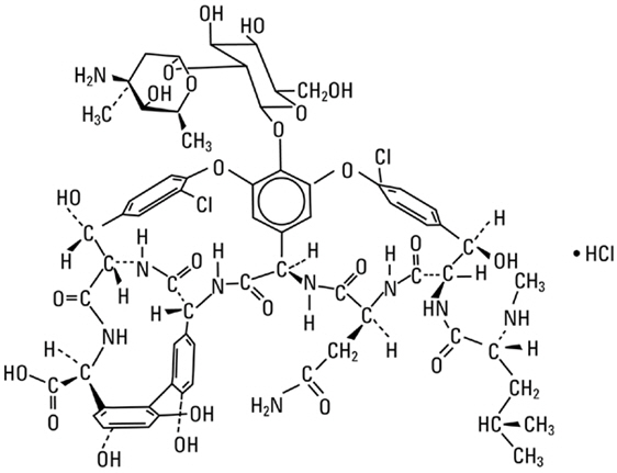
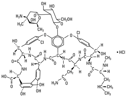
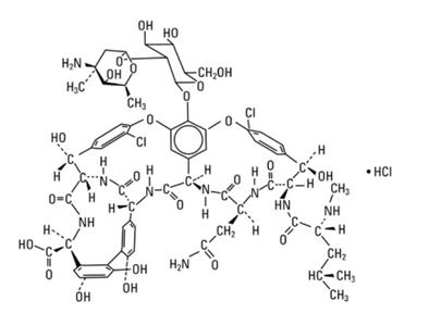
Vancomycin
List of systemic diseases with ocular manifestations
Systemic vasculitis
Brolucizumab
Retinal vasculopathy with cerebral leukoencephalopathy and systemic manifestations
Ehrlichiosis (canine)
Relapsing polychondritis
List of MeSH codes (C14)
Progressive outer retinal necrosis
Amaurosis fugax
Outline of cardiology
List of diseases (R)
Acute posterior multifocal placoid pigment epitheliopathy
Outline of emergency medicine
Sickle cell retinopathy
Takayasu's arteritis
Intraparenchymal hemorrhage
List of dog diseases
Granulomatous meningoencephalitis
Retinal vasculitis - Wikipedia
RETINAL VASCULITIS | British Journal of Ophthalmology
Rickettsia sibirica subsp. mongolitimonae Infection and Retinal Vasculitis - Volume 14, Number 4-April 2008 - Emerging...
 Apellis Releases Update on Retinal Vasculitis Associated with Pegcetacoplan Injection
 Syfovre Vision Side Effects | Retinal Vasculitis Eye Inflammation
 Retinal vasculitis | List of High Impact Articles | PPts | Journals | Videos
 Retinal Vasculitis; Vasculitis, Retinal
 Idiopathic Occlusive Retinal Vasculitis (Late Stage) - Retina Image Bank
 Uveitis Evaluation and Treatment: Approach to the Uveitis Workup, Choosing the Correct Diagnostic Test, Evidence-based Medicine...
H2-T25 MGI Mouse Gene Detail - MGI:1890744 - histocompatibility-2, T region locus 25
![Moore SM[au] - Search Results - PubMed](data:image/png;base64,iVBORw0KGgoAAAANSUhEUgAAABAAAAAQCAMAAAAoLQ9TAAAARVBMVEVHcEwoU45gYmYAUpQAUpRPYGVgYmZLXnJgYmYAUZUAUpRJXnIAUpQAUpRgYmYAUpRgYmZgYmZhYmYAUpQAUpQAUpRgYmaDiPJuAAAAFXRSTlMADOJ+6QewGO8/uTRqtH7GdFJ11p1bCL3TAAAAZUlEQVQYlV2PVw7AIAxDTeney7n/UcsoldX3E+VJOAboEi7MBpHWMs1ADlG8u7UYWauwyZFeRQVPOhG2o+aiwhByJxUx91Jxhje3iJSqGfHuLKI0+0TpXvY1twCOPlFh5pa/++MB0vIOBm+1zaoAAAAASUVORK5CYII=) Moore SM[au] - Search Results - PubMed
Moore SM[au] - Search Results - PubMed
Branch Retinal Artery Occlusion (BRAO) Clinical Presentation: History, Physical, Causes
 CDC - Clinical Advisory: Ocular Syphilis in the United States
Naclerio C[au] - Search Results - PubMed
 SciELO - Brazil - Red eyes in the necropsy floor: twenty cases of hyphema in dogs and
cats Red eyes in the...
SciELO - Brazil - Red eyes in the necropsy floor: twenty cases of hyphema in dogs and
cats Red eyes in the...
 Pediatric Rheumatology | Cleveland Clinic Children's
Advanced Search Results - Public Health Image Library(PHIL)
 Retinal vein occlusion - Symptoms, diagnosis and treatment | BMJ Best Practice US
 IVHDM in refractory & severe pediatric uveitis | OPTH
Neurosyphilis: Overview of Syphilis of the CNS, Pathophysiology of Syphilis, Epidemiology of Syphilis
Safety of Beovu: A Lingering Issue Which Novartis Attempts to Counter with Recent Study Results - Drug Injury Watch
 Phase 3 CheckMate -67T Trial of Subcutaneous Nivolumab (nivolumab and hyaluronidase) Meets Co-Primary Endpoints in Advanced or...
Phacoanaphylaxis: Background, Pathophysiology, Epidemiology
 Tattoo-associated uveitis | Eye
 KEYTRUDA® (pembrolizumab) Plus Chemotherapy Significantly Improved Overall Survival Versus Chemotherapy Alone as First-Line...
Merck Receives Positive EU CHMP Opinion for KEYTRUDA® (pembrolizumab) as First-Line Treatment in Adult Patients With Metastatic...
Granulomatosis with Polyangiitis (GPA, formerly Wegener Granulomatosis): Practice Essentials, Background, Etiology
 User talk:Tony.Ching.AAO - EyeWiki
 Syfovre™ Lawsuit
Occlusion9
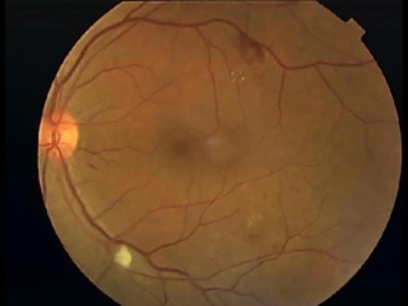
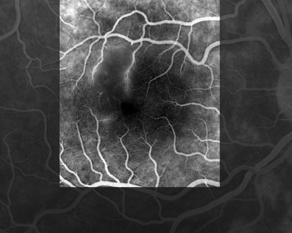
- Measurement of visual acuity and results of a slit-lamp examination were within normal limits, but a funduscopic examination showed a white retinal macular lesion that corresponded in a fluorescein angiograph to an area of retinal ischemia induced by vascular inflammation and subsequent occlusion ( Figure ). (cdc.gov)
- Patients with branch retinal artery occlusion (BRAO) typically present with acute, unilateral, painless, partial visual loss. (medscape.com)
- Retinal vein occlusion (RVO) is an interruption of the normal venous drainage from the retinal tissue. (bmj.com)
- Hypertension, diabetes mellitus, atherosclerosis, and glaucoma are major risk factors for the development of central retinal vein occlusion (CRVO) or branch retinal vein occlusion (BRVO) in older patients. (bmj.com)
- For an uncomplicated retinal vein occlusion, whether it is ischemic or nonischemic, management consists of close monitoring to detect complications and treatment of underlying risk factors. (bmj.com)
- Characteristically, in the retina proximal to the occlusion, the affected venous system is tortuous and dilated, and there are several intraretinal hemorrhages and retinal edema. (bmj.com)
- The intraocular inflammation safety signal led to an update to Beovu's U.S. and [European Union (EU)] labels, which now include information about retinal vasculitis and retinal vascular occlusion, which can cause blindness. (drug-injury.com)
- exudation of material into the subretinal space from retinal vessels (such as in hypertension, central retinal venous occlusion, vasculitis, papilledema). (institut-vision.org)
- vasculitis lead to occlusion of the vessels genital ulcerations, unilateral oedema or aneurysm formation [ 7 ]. (who.int)
Uveitis6
- Eales disease, pars planitis, birdshot retinochoroidopathy (autoimmune bilateral posterior uveitis), and Fuchs heterochromic iridocyclitis (FHI) can also cause retinal vasculitis. (wikipedia.org)
- William J. Johnson, M.D., of the Wolfe Clinic in Iowa, reported on Syfovre vision side effects cases ranging from hemorrhagic occlusive vasculitis-type inflammation with continued blindness to uveitis to vision loss to symptomatic floaters. (lamblawoffice.com)
- Additional manifestations may include anterior uveitis, optic neuropathy, retinal vasculitis and interstitial keratitis. (cdc.gov)
- The case definition for an ocular syphilis case is as follows: a person with clinical symptoms or signs consistent with ocular disease (i.e. uveitis, panuveitis, diminished visual acuity, blindness, optic neuropathy, interstitial keratitis, anterior uveitis, and retinal vasculitis) with syphilis of any stage. (cdc.gov)
- Background Behçet's syndrome (BS) is a multifactorial, polygenic, autoinflammatory vasculitis characterized by recurrent oral and genital ulcers, uveitis, skin lesions, and arthritis. (researchsquare.com)
- In the update, Apellis confirmed a seventh event of retinal vasculitis resulting from SYFOVRE treatment as determined by Apellis's internal safety committee and external retina/uveitis specialists. (woodtv.com)
Events of retinal vasculitis6
- An update from Apellis reports 10 confirmed events of retinal vasculitis and 2 suspected events associated with pegcetacoplan injection for geographic atrophy. (consultantlive.com)
- Apellis Pharmaceuticals has shared an update on the launch of pegcetacoplan injection (SYFOVRE) for geographic atrophy (GA) secondary to age-related macular degeneration (AMD) , including an update on the events of retinal vasculitis associated with the therapy. (consultantlive.com)
- At that time, the company provided an update on injection kits with the use of pegcetacoplan injection and confirmed 8 events of retinal vasculitis. (consultantlive.com)
- To this date, there have been 10 confirmed events of retinal vasculitis (7 occlusive 3 non-occlusive) and 2 suspected events. (consultantlive.com)
- Zero events of retinal vasculitis were reported by investigators or identified by an independent reading center in the Phase 3 clinical trials. (pharmexec.com)
- Finally, on July 29, 2023, Apellis provided an update on the Company's review of the six events of retinal vasculitis reported by the ASRS concerning SYFOVRE treatments. (woodtv.com)
Inflammation10
- Retinal vasculitis is inflammation of the vascular branches of the retinal artery, caused either by primary ocular disease processes, or as a specific presentation of any systemic form of vasculitis such as Behçet's disease, sarcoidosis, multiple sclerosis, or any form of systemic necrotizing vasculitis such as temporal arteritis, polyarteritis nodosa, and granulomatosis with polyangiitis, or due to lupus erythematosus, or rheumatoid arthritis. (wikipedia.org)
- citation needed] Ophthalmic examination may reveal neovascularization (creation of new vessels in the retina), retinal vessel narrowing, retinal vessel cuffing, retinal hemorrhage, or possible vitritis (inflammation of the vitreous body) or choroiditis (inflammation of the choroid). (wikipedia.org)
- At present, we are investigating Syfovre drug injury lawsuits against Apellis Pharmaceuticals, Inc., the drug company responsible for Syfovre, for patients who developed occlusive retinal vasculitis, eye inflammation, vision loss, or blindness after receiving Syfovre injections. (lamblawoffice.com)
- It is the inflammation of the vascular branches of the retinal artery resulted either by primary ocular disease processes or a defined presentation of any systemic form of vasculitis including behcet's disease, sarcoidosis, multiple sclerosis or any other form of systemic nectrozing vasculitis like temporal arteritis, polyarteritis nodosa and granulomatosis with polyangiitis. (hilarispublisher.com)
- [ 1 ] Its hallmark features include necrotizing granulomatous inflammation and pauci-immune vasculitis in small- and medium-sized blood vessels. (medscape.com)
- Syfovre™ is a drug that is injected into the eye to slow retinal damage from geographic atrophy (GA). However, some users may experience serious side effects from these injections that may lead to eye inflammation and vision loss. (sokolovelaw.com)
- Occlusive retinal vasculitis is a type of inflammation that hinders blood flow to the retina and may result in severe vision loss. (sokolovelaw.com)
- Granulomatosis with Polyangiitis (GPA) Granulomatosis with polyangiitis is characterized by necrotizing granulomatous inflammation, small- and medium-sized vessel vasculitis, and focal necrotizing glomerulonephritis, often with crescent. (merckmanuals.com)
- the vessel inflammation (true vasculitis) is only part of the pathophysiology and there is predominant parenchymal inflammation in a characteristic pattern that involves specific organs. (merckmanuals.com)
- Specifically, the ASRS indicated that physicians have reported cases of eye inflammation in patients treated with SYFOVRE, including six instances of occlusive retinal vasculitis, a type of inflammation that blocks blood flow through the vessels that feed the retina and potentially results in blindness. (woodtv.com)
Syfovre8
- Our most recent Drug Injury Watch article, "Apellis Updates Number of Syfovre Retinal Vasculitis Cases But Provides No Further 'Explanation'", was posted last month, in mid-October 2023. (lamblawoffice.com)
- While the injections were approved by the U.S. Food and Drug Administration (FDA) in February 2023, a safety committee advising the American Society of Retinal Specialists (ASRS) sounded the alarm regarding dangerous side effects of Syfovre in July 2023 . (sokolovelaw.com)
- The group found six cases of patients experiencing occlusive retinal vasculitis just a few days after their first injection of Syfovre. (sokolovelaw.com)
- However, patients may experience occlusive retinal vasculitis within 1 to 2 weeks of their first Syfovre injection . (sokolovelaw.com)
- Syfovre linked to multiple cases of retinal vasculitis. (pharmexec.com)
- Prompted by recent reports of retinal vasculitis being linked to Syfovre, its recently approved eye injection treatment, Apellis has provided an update on its own review of potential manufacturing issues. (pharmexec.com)
- Later that day, after the market closed, Apellis issued a statement explaining that, of the six occurrences of vasculitis following SYFOVRE treatment, "two of the events were confirmed as occlusive, one was confirmed as non-occlusive, and the remaining three were undetermined based on limited information and lack of imaging. (woodtv.com)
- The clinical trials designed by Apellis for its SYFOVRE therapeutic treatment failed to identify cases of retinal vasculitis in patients receiving injections. (mystateline.com)
Artery2
- Funduscopic examination shows retinal whitening along the distribution of the affected artery. (medscape.com)
- Narrowed branch retinal artery, boxcarring, segmentation of the blood columns, cotton-wool spots, and emboli are other possible findings. (medscape.com)
Ischemic5
- These extracted markers or characterized fundus digital image features provide insights and relates quantitative retinal vascular topography abnormalities to various pathologies such as diabetic retinopathy, macular degeneration, hypertensive retinopathy, transient ischemic attack, neovascular glaucoma, and cardiovascular diseases. (hindawi.com)
- Some distinct changes in the retinal microvasculature are recognized as the preindicator of subsequent vascular incidents like ischemic stroke or acute stroke [ 10 ]. (hindawi.com)
- Rotterdam cohort study also came into a decision after a long-term observation that the retinal venular diameter is associated with any stroke or ischemic stroke [ 14 ]. (hindawi.com)
- Retinal microvascular abnormalities like microaneurysm, arteriovenous nicking, haemorrhages, and vessel caliber are considered as associative to the stroke and indicative of death from stroke and IHD (Ischemic Heart Diseases) [ 1 ]. (hindawi.com)
- There were no cases of occlusive or non-occlusive retinal vasculitis or ischemic neuropathy. (ophthalmologytimes.com)
Detachment2
- Significant liquefaction of the vitreous gel may lead to vitreous detachment (usually termed posterior vitreous detachment or PVD), which often precipitates RRD by producing tractional forces necessary to generate retinal breaks. (institut-vision.org)
- Due to rotational eye movements, gravitational and inertial forces or contracture of intraocular fibroproliferative tissue, vitreous currents force fluid through the retinal breaks and progressively extend the retinal detachment. (institut-vision.org)
Occlusive vasculitis2
- Fluorescein angiograph of the right eye of the patient showing retinal occlusive vasculitis with arteriolar leakage at late phase. (cdc.gov)
- I think it's important to know that there is a risk of retinal vasculitis or occlusive vasculitis when using pegcetacoplan, so it's important to consent your patients to the risk," Dilsher Dhoot, MD of California Retina Consultants told HCPLive at the ASRS 41st Annual Meeting. (consultantlive.com)
Ocular3
- We report a case in a pregnant woman with ocular vasculitis. (cdc.gov)
- They pointed out that while ocular manifestations have been reported in association with various types of vasculitis, there seems to be no routine ophthalmologic examinations for patients with those diseases. (ophthalmologytimes.com)
- The investigators concluded, "Conducting routine ophthalmologic examinations in patients diagnosed with vasculitis to assess the retina and choroid by measuring parameters like the choroidal thickness, the choroidal vascularity index, area and perimeter of the foveal avascular zone, and the circularity index could be beneficial, because it may detect pathological changes before any ocular symptoms alarm the patients. (ophthalmologytimes.com)
Hemorrhagic occlusive2
- Many surgeons are looking for alternatives to vancomycin for intracameral prophylaxis because of its association with the rare but sight-threatening complication of hemorrhagic occlusive retinal vasculitis. (medscape.com)
- Hemorrhagic occlusive retinal vasulitis is an extremely rare but devastating complication following uncomplicated surgery where intraocular vancomycin was delivered. (medscape.com)
Vascular2
- For those patients who present with only vasculitis of the retinal vessels, great investigative effort (Chest X-ray, blood test, urinary analysis, vascular biopsy, ophthalmology assessment, etc.) should be undertaken to ensure that a systemic disease is not the hidden culprit. (wikipedia.org)
- Se caracteriza por FIBROSIS de la papred vascular y TROMBOSIS oclusiva que provocan ISQUEMIA y ulceraciones en los dedos y las extremidades. (bvsalud.org)
Vessels4
- CNS manifestations include vasculitis of small to medium-sized vessels of the brain or spinal cord and granulomatous masses that involve the orbit, optic nerve, meninges, or brain. (medscape.com)
- Cutaneous Vasculitis Cutaneous vasculitis refers to vasculitis affecting small- or medium-sized vessels in the skin and subcutaneous tissue but not the internal organs. (merckmanuals.com)
- Furthermore, image analysis provides a simple and noninvasive visualization of the retinal blood vessels in those high risk ophthalmologic medical conditions [ 1 - 3 ]. (hindawi.com)
- Retinal breaks may develop spontaneously in areas of strong vitreoretinal adhesion, typically along retinal vessels, or in patients with certain predisposing conditions, such as lattice retinal degeneration. (institut-vision.org)
Choroidal2
- Because of this gap, they investigated retinal and choroidal abnormalities in patients with primary vasculitis in a prospective and observation study. (ophthalmologytimes.com)
- Szydelko-PaśkoU,Przeździecka-Dołyk J, Andrzej Dolyk, et al.Evaluation of choroidal and retinal features in patients with primary vasculitis-an original optical coherence tomography and optical coherence tomography angiography study. (ophthalmologytimes.com)
ASRS1
- All identified reported adverse events are submitted by Apellis to the FDA, and the company is in communication with the American Society of Retina Specialists (ASRS) on these reported cases of retinal vasculitis. (consultantlive.com)
Retina2
- Drug-induced vasculitis may involve retina as well, as seen in methamphetamine induced vasculitis. (wikipedia.org)
- The fundus retinal images are directly captured from human eye that includes some other landmarks like microcirculation system of the retina, macula, optic disc, fovea, microaneurysm, and exudates [ 4 ]. (hindawi.com)
Abnormalities1
- Lattice degeneration is one of the most important vitreoretinal abnormalities associated with an increased likelihood of retinal tears and RD. Approximately 30% of patients with RD also have lattice degeneration. (institut-vision.org)
Vessel4
- Some forms of vasculitis are characterized by giant cells in the vessel wall. (merckmanuals.com)
- Leukocytoclastic vasculitis is a histopathologic term used to describe findings in small-vessel vasculitis. (merckmanuals.com)
- Various diagnostic techniques are used to analyze retinal microvasculature image to enable geometric features measurements such as vessel tortuosity, branching angles, branching coefficient, vessel diameter, and fractal dimension. (hindawi.com)
- Many disorders, such as cutaneous creased and the patient was discharged small vessel vasculitis, inflammatory eye in good condition. (who.int)
Systemic2
- Various causes of bleeding disorders were found related to secondary hyphema: in decreasing order of frequency, they included vasculitis (8/15), systemic hypertension (5/15), and acquired coagulopathies (2/15). (scielo.br)
- Vasculitis due to feline infectious peritonitis accounted for half of the cases (n=3) of systemic hyphema in cats. (scielo.br)
Vasculature1
- Analysis of the human fundus eye images has become the key point for diagnosing the various pathologies of retinal vasculature. (hindawi.com)
Ophthalmology1
- Journal of Vasculitis, Journal of Clinical & Experimental Ophthalmology, Journal of Pulmonary & Respiratory Medicine, Journal of Neuroinfectious Diseases, Journal of Neonatal Biology, Progress in Retinal and Eye Research, Retinal Cases and Brief Reports, Journal of Clinical & Experimental Ophthalmology. (hilarispublisher.com)
Cutaneous1
- Cutaneous vasculitis may be limited to the. (merckmanuals.com)
Arteriolar2
- According to the study of [ 12 ] with a multiethnic cohort, retinal arteriolar narrowing and retinopathy of diabetic free people have an association with increased risk of acute stroke. (hindawi.com)
- But the Cardiovascular Health Study stated that there is no association between retinal arteriolar caliber (diameter) and stroke but rather there is a close association between stroke and the larger venular caliber (diameter) [ 13 ]. (hindawi.com)
Painless2
- Retinal vasculitis presents as painless, decrease of visual acuity (blurry vision), visual floaters, scotomas (dark spot in vision), decreased ability to distinguish colors, and metamorphopsia (distortion of images such as linear images). (wikipedia.org)
- Retinal vein occlusions are usually painless, sudden, and unilateral causes of vision loss. (bmj.com)
Patients6
- Within the confirmed retinal vasculitis events, 6 patients have recovered vision either fully or partially, while 3 patients have severe vision impairment unlikely to be resolved, and 1 outcome is still pending. (consultantlive.com)
- In a study of 70 patients with retinal emboli, 40 were found to have cholesterol emboli, 8 platelet-fibrin emboli, 6 calcific emboli, and 1 possible myxomatous embolus. (medscape.com)
- Our facility may offer patients the opportunity to participate in state-of-the-art clinical trials for juvenile arthritis and vasculitis . (clevelandclinic.org)
- Hypercoagulability and vasculitis are important risk factors for the development of CRVO or BRVO in younger patients. (bmj.com)
- The study included 41 patients (78 eyes) with 5 types of primary vasculitis, ie, Takayasu's arteritis, giant cell arteritis, Buerger's disease, granulomatosis with polyangiitis, and polyarteritis nodosa. (ophthalmologytimes.com)
- A significant proportion of patients with acute PVD develop an associated retinal tear that can lead to RD and, if left untreated, permanent vision loss. (institut-vision.org)
APELLIS3
- Iapoce C. APELLIS shares update on Pegcetacoplan injection kits, retinal vasculitis events. (consultantlive.com)
- https://www.hcplive.com/view/apellis-update-pegcetacoplan-injection-kits-retinal-vasculitis-events. (consultantlive.com)
- Apellis also stated that the Company is evaluating an eighth reported event of retinal vasculitis, which the Company had not yet confirmed. (woodtv.com)
Granulomatosis with polyangiitis2
- Behçet's disease Common Variable Immune Deficiency Eales disease Granulomatosis with polyangiitis Idiopathic Retinal Vasculitis Aneurysms and Neuroretinitis Lupus erythematosus Multiple sclerosis Polyarteritis nodosa Q fever Rheumatoid arthritis Sarcoidosis Temporal arteritis Retinal vasculitis is very rare as the only presenting symptom. (wikipedia.org)
- Eosinophilic granulomatosis with polyangiitis (formerly known as Churg-Strauss vasculitis). (clevelandclinic.org)
Degeneration2
- By targeting C5, ACP has the potential to decrease activity of the complement system that causes the degeneration of retinal cells and potentially slow the progression of GA. (ophthalmologytimes.com)
- Atrophic retinal holes have round shape and gradual onset, are often within patches of lattice degeneration and are not associated vitreoretinal traction. (institut-vision.org)
Posterior1
- Only 15% of retinal breaks develop posterior to the equator. (institut-vision.org)
Macular1
- Moschos MM, Guex-Crosier Y . Retinal vasculitis and cystoid macular edema after body tattooing: a case report. (nature.com)
Onset3
- Wide- field image of the right eye of a 28-year-old woman with idiopathic occlusive retinal vasculitis 6 months after the onset. (asrs.org)
- Dialysis is a traumatic (or in some cases congenital) circumferential retinal tear by the ora serrata that has linear shape, acute onset and most commonly occurs in young individuals. (institut-vision.org)
- The age moptysisofvaryingdegrees(upto500 formed and a mild retinal vasculitis was of disease onset is usually in the second mL) is the most common and predomi- found via ophthalmoscopy. (who.int)
Cases2
- According to a company press release, evidence suggests manufacturing issues did not play a role in the cases of retinal vasculitis. (pharmexec.com)
- sev- and potential vectors/ reservoirs of this rolides (azithromycin, clarithromycin, en new cases and review of the literature. (cdc.gov)
Infectious2
- Infectious pathogens such as Mycobacterium tuberculosis, visceral larva migrans (Toxocara canis & Toxocara cati) can also cause retinal vasculitis. (wikipedia.org)
- Intravitreal administration of corticosteroid and immunosuppressants in a case non infectious retinal vasculitis Antimicrobial therapy is required in the case of infectious retinal vasculitis Lynn K. Gordon, M.D., Ph.D. (January 2003). (wikipedia.org)
Diseases1
- We are a regional, national, and international referral center for families seeking initial evaluation or second opinion services for pediatric rheumatologic conditions, including juvenile arthritis, vasculitis and childhood autoinflammatory diseases. (clevelandclinic.org)
Infection1
- Secondary vasculitis may be triggered by an infection, a drug, or a toxin or may occur as part of another inflammatory disorder or cancer. (merckmanuals.com)
Diagnosis1
- Differential of the lower limb, blurred vision and Behçet disease (BD) is a chronic inflam- diagnosis of Behçet-induced vasculitis polyarthralgia. (who.int)
Injection1
- Estimates suggest the real-world rate of retinal vasculitis is rare at 0.01% per injection. (consultantlive.com)
Pathophysiology1
- Recent evidence indicates that the intrinsically photosensitive retinal ganglion cells play a key role in the pathophysiology of photophobia. (researchgate.net)
Primary1
- Primary vasculitis has no known cause. (merckmanuals.com)