Rhabditida
Papillomatous pastern dermatitis with spirochetes and Pelodera strongyloides in a Tennessee Walking Horse. (1/12)
Papillomatous digital dermatitis is a common disease in cattle. The pastern dermatitis observed in a horse shared many of the gross characteristics of papillomatous digital dermatitis in cattle. Lesions included a mixture of proliferative and erosive changes, with a verrucose appearance in some areas. Microscopic similarities included pseudoepitheliomatous and papillomatous epidermal hyperplasia with hyperkeratosis, spongiosis of the epidermis, and intraepidermal spirochetes. The horse was also concurrently infected with Pelodera strongyloides. Papillomatous digital dermatitis in cattle is associated with poor husbandry practices. The environment of the affected horse was heavily contaminated with urine, manure, and other organic debris. Verrucous pododermatitis of horses may be the same as or similar to bovine papillomatous digital dermatitis, and these conditions have similar etiologies. (+info)Radiculomeningomyelitis due to Halicephalobus gingivalis in a horse. (2/12)
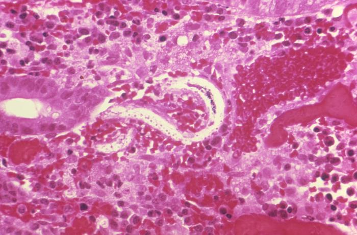
An adult horse was euthanatized following a clinical diagnosis of cauda equina neuritis. Significant gross postmortem and histopathologic findings were limited to the sacral spinal cord and cauda equina. The sacral spinal cord, meninges, and spinal nerve roots were expanded and partially effaced by sclerosing granulomatous inflammation with necrosis. The lesion contained numerous nematode larvae and fewer adults with a rhabditiform esophagus having a corpus, isthmus, and valved bulb. Female nematodes were amphidelphic and didelphic with reflexed ovaries. These morphologic features confirm Halicephalobus gingivalis as a novel cause of clinical signs in this case of cauda equina neuritis. (+info)Pelodera (syn. Rhabditis) strongyloides as a cause of dermatitis--a report of 11 dogs from Finland. (3/12)
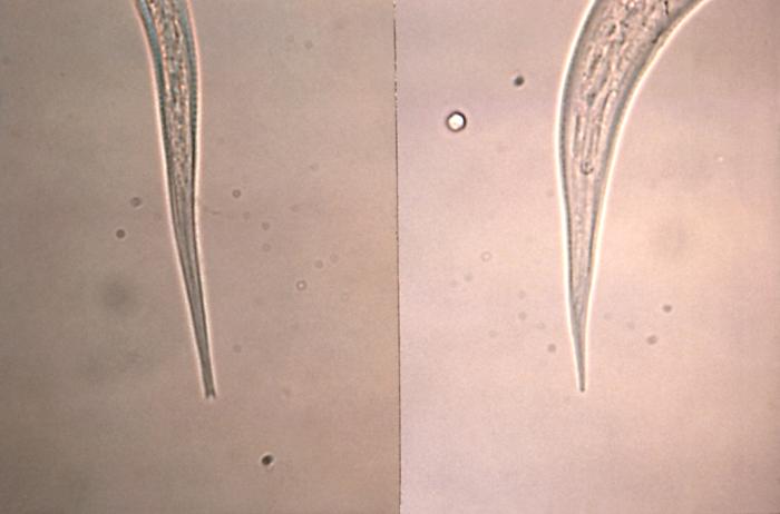
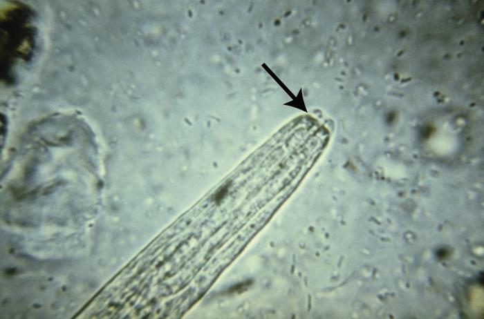
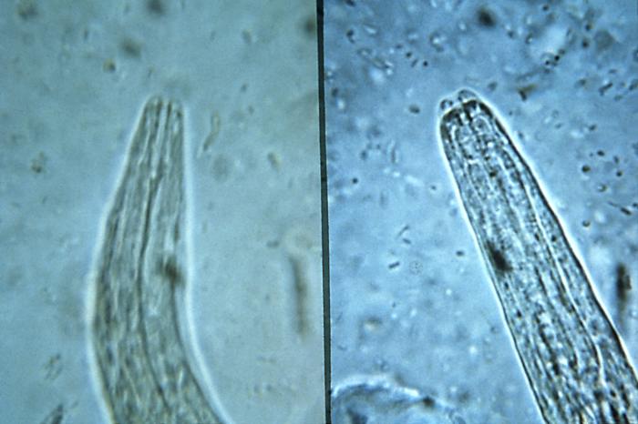
BACKGROUND: Pelodera (Rhabditis) strongyloides is a small saprophytic nematode that lives in decaying organic matter. On rare occasions, it can invade the mammalian skin, causing a pruritic, erythematous, alopecic and crusting dermatitis on skin sites that come into contact with the ground. Diagnosis of the disease is based on case history (a dog living outdoors on damp straw bedding) with characteristic skin lesions and on the demonstration of typical larvae in skin scrapings or biopsy. Pelodera (rhabditic) dermatitis cases have been reported mainly from Central European countries and the United States. CASE PRESENTATION: During 1975-1999, we verified 11 canine cases of Pelodera dermatitis in Finland. The cases were confirmed by identifying Pelodera larvae in scrapings. Biopsies for histopathology were obtained from three cases, and typical histopathological lesions (epidermal hyperplasia, epidermal and follicular hyperkeratosis, folliculitis and furunculosis with large numbers of nematode larvae of 25-40 microm of diameter within hair follicles) were present. The Pelodera strongyloides dermatitica strain from the first verified case in Finland has been maintained in ordinary blood agar in our laboratory since 1975. Light microscopy (LM) and scanning electron microscopy (SEM) studies were employed to obtain detailed morphological information about the causative agent. The rhabditiform oesophagus at all developmental stages, the morphology of the anterior end of the nematode, copulatory bursa and spicules of the male and the tail of the female were the most important morphological features for identifying P. strongyloides. CONCLUSION: These cases show that Pelodera dermatitis occurs in Finland, and also farther north than described earlier in the literature. This condition should be considered when a dog living outdoors has typical skin lesions situated at sites in contact with the ground as the main presenting clinical feature. The fastest and easiest way to confirm the diagnosis is to demonstrate typical larvae in skin scrapings. In uncertain cases, skin biopsy and culturing of the worms are recommended as supplementary diagnostic procedures. (+info)Halicephalobus gingivalis-associated meningoencephalitis in a Thoroughbred foal. (4/12)
A 13-week-old Thoroughbred colt from central Kentucky was euthanized after an acute onset of ataxia, blindness, head tremors, leaning to the right, recumbency, and seizures. Microscopically, there was a verminous meningoencephalitis characterized by an eosinophilic and granulomatous inflammatory reaction primarily affecting the cerebellum. Dispersed within regions of inflammation were numerous cross and longitudinal sections of intact and degenerative small nematodes. The nematodes had dorsoflexed ovaries and ventroflexed vulvas, which are distinguishing features of Halicephalobus gingivalis. Intact nematodes, compatible with H. gingivalis, also were recovered and identified from portions of the brain that had been frozen for 5-week post-necropsy examination via tissue maceration and additional laboratory techniques. (+info)The sugar glider (Petaurus breviceps): a laboratory host for the nematode Parastrongyloides trichosuri. (5/12)
(+info)Unsuccessful treatment of a horse with mandibular granulomatous osteomyelitis due to Halicephalobus gingivalis. (6/12)
An 8-year-old horse was presented with a submandibular swelling. Biopsy of the lesion indicated granulomatous osteomyelitis due to Halicephalobus gingivalis. In the absence of evidence of involvement of the central nervous system at the time of diagnosis, the horse was treated with ivermectin. Unfortunately, the horse did not survive. (+info)Assessing the influence of the entomopathogenic nematode Heterorhabditis baujardi LPP7 (Rhabiditina) on embryogenesis and hatching of the plant-parasitic nematode Meloidogyne mayaguensis (Tylenchina). (7/12)
(+info)Subterranean, herbivore-induced plant volatile increases biological control activity of multiple beneficial nematode species in distinct habitats. (8/12)
(+info)Rhabditida is an order of nematode (roundworm) parasites that can infect humans and other animals. Rhabditida infections in humans are typically caused by the accidental ingestion or inhalation of infective stages of these parasites, which can be found in contaminated food, water, or soil.
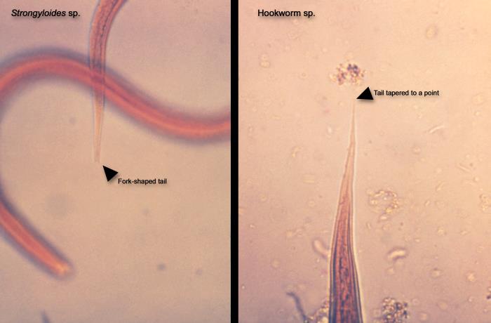
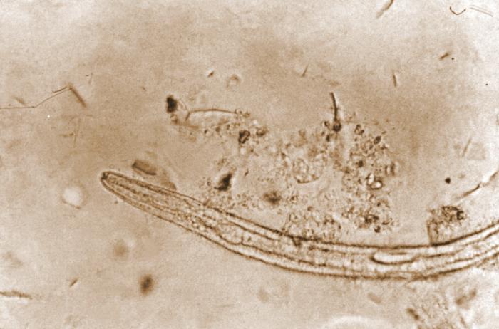
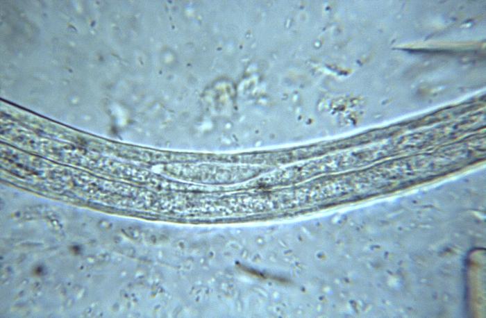
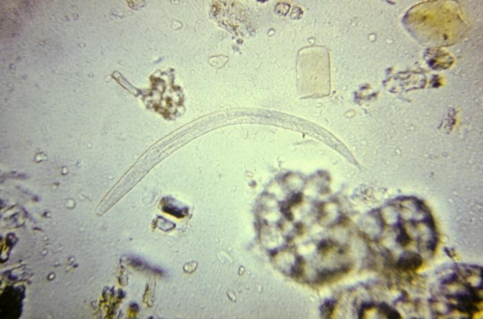
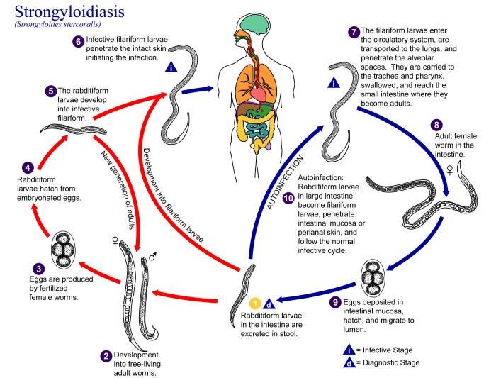
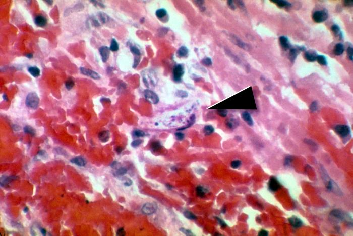
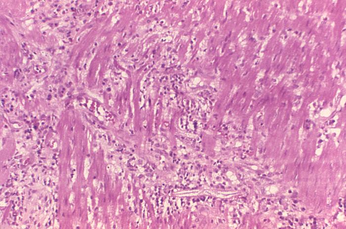
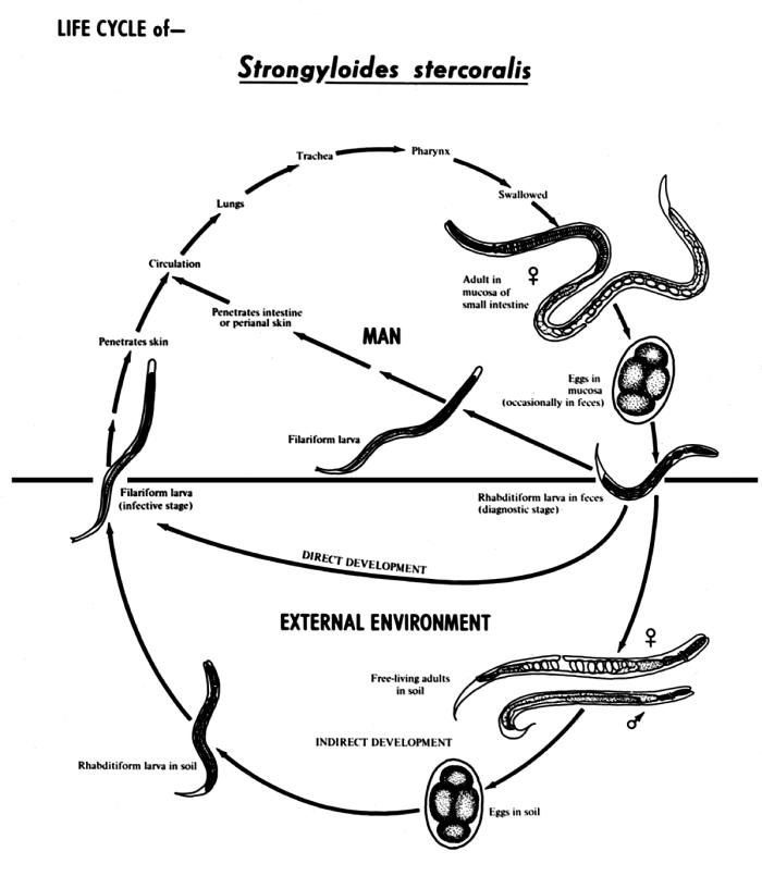
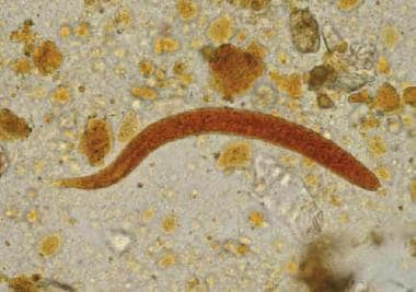
The most common Rhabditida infection in humans is strongyloidiasis, which is caused by the nematode Strongyloides stercoralis. This parasite can infect the small intestine and cause symptoms such as abdominal pain, diarrhea, and skin rashes. In severe cases, strongyloidiasis can lead to a life-threatening condition called hyperinfection syndrome, in which large numbers of larvae invade various organs throughout the body.
Other Rhabditida species that can infect humans include Ancylostoma duodenale and Necator americanus, which cause hookworm infection, and Enterobius vermicularis, which causes pinworm infection.
Preventing Rhabditida infections involves practicing good hygiene, such as washing hands thoroughly with soap and water, avoiding contact with contaminated soil or feces, and cooking food thoroughly before eating it. Treatment for Rhabditida infections typically involves administering anti-parasitic medications to kill the parasites.
Rhabditida is an order of nematodes, or roundworms. These are microscopic worms that have a long, slender, and unsegmented body. Rhabditida includes both free-living and parasitic species. Some free-living species live in soil and decaying organic matter, where they play an important role in the breakdown of organic material.
Parasitic species of Rhabditida can infect a wide range of hosts, including humans, animals, and plants. They can cause various diseases and conditions, depending on the species and the location of the infection. For example, some parasitic Rhabditida species can infect the gastrointestinal tract and cause diarrhea, abdominal pain, and other symptoms. Other species can infect the lungs and cause respiratory problems.
Rhabditida are characterized by several distinctive features, including a mouth equipped with three lips and teeth, and a unique reproductive system that allows them to reproduce both sexually and asexually. They are also known for their ability to form resistant structures called resting spores, which can survive in unfavorable conditions and germinate when conditions improve.
It's worth noting that the classification of nematodes is complex and constantly evolving, so different sources may use slightly different terminology or groupings when discussing Rhabditida and other orders of nematodes.
Nematoda is a phylum of pseudocoelomate, unsegmented worms with a round or filiform body shape. They are commonly known as roundworms or threadworms. Nematodes are among the most diverse and numerous animals on earth, with estimates of over 1 million species, of which only about 25,000 have been described.
Nematodes are found in a wide range of habitats, including marine, freshwater, and terrestrial environments. Some nematode species are free-living, while others are parasitic, infecting a variety of hosts, including plants, animals, and humans. Parasitic nematodes can cause significant disease and economic losses in agriculture, livestock production, and human health.
The medical importance of nematodes lies primarily in their role as parasites that infect humans and animals. Some common examples of medically important nematodes include:
* Ascaris lumbricoides (human roundworm)
* Trichuris trichiura (whipworm)
* Ancylostoma duodenale and Necator americanus (hookworms)
* Enterobius vermicularis (pinworm or threadworm)
* Wuchereria bancrofti, Brugia malayi, and Loa loa (filarial nematodes that cause lymphatic filariasis, onchocerciasis, and loiasis, respectively)
Nematode infections can cause a range of clinical symptoms, depending on the species and the location of the parasite in the body. Common symptoms include gastrointestinal disturbances, anemia, skin rashes, and lymphatic swelling. In some cases, nematode infections can lead to serious complications or even death if left untreated.
Medical management of nematode infections typically involves the use of anthelmintic drugs, which are medications that kill or expel parasitic worms from the body. The choice of drug depends on the species of nematode and the severity of the infection. In some cases, preventive measures such as improved sanitation and hygiene can help reduce the risk of nematode infections.
 Rhabditida Infections | Harvard Catalyst Profiles | Harvard Catalyst
 Advanced Search Results - Public Health Image Library(PHIL)
 List of diseases (R) - Wikipedia
 Strongyloidiasis: Background, Pathophysiology, Etiology
 Pesquisa | Portal Regional da BVS
 Helminthic Therapy (Hookworms) for Hair Loss? A Scientific Review | 2021
 5:12 am
 Thelaziosis - Thelazia callipaeda. Ocular parasitism in dogs and cats with risk of transmission to people: Microscopic exam;...
 Bjorn Victor's Universiteit Gent Doctoral Thesis Template - Overleaf, 在线LaTeX编辑器
 Zoonotic parasites associated with predation by dogs and cats | Parasites & Vectors | Full Text
 Frontiers | External Immune Inhibitory Efficiency of External Secretions and Their Metabolic Profiling in Red Palm Weevil,...
 IOBC-WPRS Bulletin Vol. 162, 2023 - IOBC-WPRS
 Items where Subject is "Q Science | QL Zoology" - IBB PAS Repository
 Bursaphelenchus xylophilus, the pinewood nematode: its significance and a historical review
 Mark H. Ellisman - Publications
 Vien Sot ret Ky Sinh Trung - Con trung Quy Nhon
 S-EPMC8039977 - First report on molecular identification of |i|Anisakis simplex|/i| in |i|Oncorhynchus nerka|/i| from the fish...
 Search Results
 Zafar Handoo : USDA ARS
 Robbie Rae |
Liverpool John Moores University
Robbie Rae |
Liverpool John Moores University References - UC Nursery and Floriculture Alliance